
نوروبیولوژی به زبان ساده؛ به دنیای نوروبیولوژی خوش آمدید


دعای مطالعه [ نمایش ]
بِسْمِ الله الرَّحْمنِ الرَّحیمِ
اَللّهُمَّ اَخْرِجْنى مِنْ ظُلُماتِ الْوَهْمِ
خدايا مرا بيرون آور از تاريكىهاى وهم،
وَ اَكْرِمْنى بِنُورِ الْفَهْمِ
و به نور فهم گرامى ام بدار،
اَللّهُمَّ افْتَحْ عَلَيْنا اَبْوابَ رَحْمَتِكَ
خدايا درهاى رحمتت را به روى ما بگشا،
وَانْشُرْ عَلَيْنا خَزائِنَ عُلُومِكَ بِرَحْمَتِكَ يا اَرْحَمَ الرّاحِمينَ
و خزانههاى علومت را بر ما باز كن به امید رحمتت اى مهربانترين مهربانان.
کتاب «نوروبیولوژی به زبان ساده» (Neurobiology For Dummies) نوشتهی دکتر فرانک آمتور و آن بی. ثایبر، یکی از منابع برجسته و معتبر در معرفی مبانی علوم اعصاب برای علاقهمندان و دانشجویان این حوزه است. نویسندگان در این اثر کوشیدهاند تا با زبانی روان و ساده، پیچیدگیهای مغز و سیستم عصبی را برای مخاطب قابل درک سازند.
ترجمهی دقیق و علمی این کتاب ارزشمند توسط برند علمی «آیندهنگاران مغز» به مدیریت داریوش طاهری، فرصتی تازه برای فارسیزبانان ایجاد کرده است تا به شکلی علمی و ساختارمند با مرزهای نوین دانش علوم اعصاب آشنا شوند. این ترجمه نه تنها دسترسی گسترده به دانش را ممکن ساخته، بلکه رسالتی علمی در جهت ارتقای آموزش، فهم عمیقتر عملکرد مغز و سیستم عصبی و همچنین توسعه روشهای نوین در حوزه سلامت عصبی را دنبال میکند.
» کتاب نوروبیولوژی به زبان ساده
» » فصل ۱: به دنیای نوروبیولوژی خوش آمدید
در حال ویرایش
» Neurobiology For Dummies
»» CHAPTER 1: Welcome to the World of Neurobiology
Getting Started with Neurobiology
I IN THIS PART
شروع کار با نوروبیولوژی
در این بخش
Find out how the nervous system is organized and what its functions are.
دریابید که سیستم عصبی چگونه سازماندهی شده و عملکردهای آن چیست.
Look at the important molecules in neurons and glia and how genes are regulated.
به مولکولهای مهم در نورونها و گلیا و نحوه تنظیم ژنها نگاهی بیندازید.
Recognize how neurons and glial cells are formed and organized during development.
نحوه تشکیل و سازماندهی نورونها و سلولهای گلیال در طول رشد را تشخیص دهید.
Appreciate the mechanisms that neurons use in electrical signaling.
مکانیسمهایی را که نورونها در سیگنالدهی الکتریکی استفاده میکنند، درک کنید.
See how neurons use chemical signaling to communicate.
ببینید که نورونها چگونه از سیگنالدهی شیمیایی برای برقراری ارتباط استفاده میکنند.
Discover how different types of neurons and responses comprise the nervous system.
کشف کنید که چگونه انواع مختلف نورونها و پاسخها سیستم عصبی را تشکیل میدهند.
IN THIS CHAPTER
» Getting to know how neurons evolved
» Seeing how the nervous system is organized
» Meeting neurons and glial cells
» Taking a tour around the brain
در این فصل
» آشنایی با چگونگی تکامل نورونها
» بررسی چگونگی سازماندهی سیستم عصبی
» آشنایی با نورونها و سلولهای گلیال
» گشت و گذاری در مغز
Chapter 1
Welcome to the World of Neurobiology
What makes you you? Your brain, most people would answer. Then what is it about your brain that makes you you? The brain is made of neurons and glial cells. Worms and bees have brains with neurons and glia. So do dogs and chimpanzees. What about the brain distinguishes these animals from each other, and for that matter, one human from another? Is it more neurons, different neurons, special neural circuits? And what happens when these neurons, glia, and circuits are disrupted or dysfunctional?
فصل ۱
به دنیای نوروبیولوژی خوش آمدید
چه چیزی شما را «شما» میکند؟ اکثر مردم پاسخ میدهند: مغز شما. پس چه چیزی در مغز شما شما را «شما» میکند؟ مغز از نورونها و سلولهای گلیال ساخته شده است. کرمها و زنبورها مغزی با نورونها و گلیا دارند. سگها و شامپانزهها هم همینطور. چه چیزی در مغز این حیوانات را از یکدیگر و در واقع یک انسان را از دیگری متمایز میکند؟ آیا نورونهای بیشتر، نورونهای متفاوت، مدارهای عصبی خاص است؟ و وقتی این نورونها، گلیا و مدارها مختل یا ناکارآمد میشوند، چه اتفاقی میافتد؟
Neurobiologists like the two of us want to answer these questions. Thousands of us at universities, research institutes, and pharmaceutical companies all over the world are working on these questions. We have many hypotheses and data sets. This book, in a way, is a progress report on the efforts.
متخصصان نوروبیولوژی مانند ما دو نفر میخواهیم به این سؤالات پاسخ دهیم. هزاران نفر از ما در دانشگاهها، مؤسسات تحقیقاتی و شرکتهای داروسازی در سراسر جهان روی این سؤالات کار میکنیم. ما فرضیهها و مجموعه دادههای زیادی داریم. این کتاب، به نوعی، گزارش پیشرفت در مورد این تلاشها است.
Virtually all neurobiologists believe that intelligence comes from nervous systems that are broadly programmed by genes and fine tuned by experience. Generally, the human genetic program creates a brain with more cortical neurons than other animals have, allowing for richer experience to produce a unique kind of cognition and intelligence.
تقریباً همه متخصصان علوم اعصاب معتقدند که هوش از سیستمهای عصبی ناشی میشود که به طور گسترده توسط ژنها برنامهریزی شده و با تجربه تنظیم دقیق میشوند. به طور کلی، برنامه ژنتیکی انسان مغزی با نورونهای قشری بیشتر از سایر حیوانات ایجاد میکند که امکان تجربه غنیتر را برای تولید نوع منحصر به فردی از شناخت و هوش فراهم میکند.
Here we introduce you to the basics of neurobiology and give you a jumping off point to use this book in understanding the human nervous system.
در اینجا شما را با اصول اولیه نوروبیولوژی آشنا میکنیم و نقطه شروعی برای استفاده از این کتاب در درک سیستم عصبی انسان در اختیارتان قرار میدهیم.
Starting with Neurobiology Just the Basics
Neurobiology is the study of neurons, glia, and the nervous system. It involves many fields of study including physiology, anatomy, biochemistry, molecular biology, cognitive and behavioral psychology, and artificial intelligence. The basic goals of neurobiology are to describe how the nervous system operates in terms of what neurons and glial cells do, how they’re built, how they work, and what happens when their functions are disrupted.
شروع با نوروبیولوژی فقط اصول اولیه
نوروبیولوژی مطالعه نورونها، گلیا و سیستم عصبی است. این رشته شامل بسیاری از زمینههای مطالعاتی از جمله فیزیولوژی، آناتومی، بیوشیمی، زیستشناسی مولکولی، روانشناسی شناختی و رفتاری و هوش مصنوعی است. اهداف اساسی نوروبیولوژی توصیف نحوه عملکرد سیستم عصبی از نظر عملکرد نورونها و سلولهای گلیال، نحوه ساخت آنها، نحوه کار آنها و آنچه هنگام اختلال در عملکرد آنها اتفاق میافتد، است.
Neurons are the detecting and signaling cells of the nervous system that use electrical and chemical signaling to communicate. Glial cells are the supporting cells that provide metabolic, structural, and functional support.
نورونها سلولهای تشخیصدهنده و سیگنالدهنده سیستم عصبی هستند که از سیگنالهای الکتریکی و شیمیایی برای برقراری ارتباط استفاده میکنند. سلولهای گلیال سلولهای پشتیبان هستند که پشتیبانی متابولیکی، ساختاری و عملکردی را فراهم میکنند.
EVOLVING THE NERVOUS SYSTEM
Many of the DNA sequences, proteins, and reactions that exist in neurons and glia are similar to those in single-celled organisms. This apparent conservation of genes and biochemistry is an important argument for life having a common origin.
تکامل سیستم عصبی
بسیاری از توالیهای DNA، پروتئینها و واکنشهایی که در نورونها و گلیا وجود دارند، مشابه توالیهای موجود در موجودات تک سلولی هستند. این حفظ ظاهری ژنها و بیوشیمی، استدلال مهمی برای منشأ مشترک حیات است.
Cells have membranes that separate their insides from the external environment. Receptors embedded in the membrane enable cells to respond to external signals. Receptor responses include biochemical cascades inside the cell, and, in neurons particularly, electrical activity.
سلولها دارای غشاهایی هستند که درون آنها را از محیط خارجی جدا میکند. گیرندههای تعبیه شده در غشا، سلولها را قادر میسازند تا به سیگنالهای خارجی پاسخ دهند. پاسخهای گیرنده شامل آبشارهای بیوشیمیایی درون سلول و به ویژه در نورونها، فعالیت الکتریکی است.
Animals are multicellular eukaryotic organisms that evolved around 600 million years ago. Early animals evolved different types of cells that are specialized to do things like secrete hormones or digestive enzymes and undergo contraction. As animals increased cells and got bigger, their sensor cells became separated from other cells and movement required coordination.
حیوانات، ارگانیسمهای یوکاریوتی چند سلولی هستند که حدود ۶۰۰ میلیون سال پیش تکامل یافتهاند. حیوانات اولیه انواع مختلفی از سلولها را تکامل دادند که برای انجام کارهایی مانند ترشح هورمونها یا آنزیمهای گوارشی و انقباض تخصص یافتهاند. با افزایش سلولها و بزرگتر شدن حیوانات، سلولهای حسگر آنها از سایر سلولها جدا شدند و حرکت آنها نیاز به هماهنگی داشت.
Neurons evolved between about 600 to 550 million years ago, most likely from epithelial cells. This probably occurred when animals needed to send sensory information over longer distances and control contraction of muscles for movement. Neurons developed as electrical signaling cells that formed nerve nets in cnidarian and/or ctenophore evolution.
نورونها بین حدود ۶۰۰ تا ۵۵۰ میلیون سال پیش، به احتمال زیاد از سلولهای اپیتلیال، تکامل یافتند. این احتمالاً زمانی رخ داده است که حیوانات نیاز به ارسال اطلاعات حسی در فواصل طولانیتر و کنترل انقباض عضلات برای حرکت داشتند. نورونها به عنوان سلولهای سیگنالدهنده الکتریکی توسعه یافتند که شبکههای عصبی را در تکامل کیسهتنان و/یا شانهداران تشکیل دادند.
Later, the evolution of special sense organs and nerve ganglia occurred several times in bilaterans (the clade that gave rise to crustaceans, insects, and vertebrates). There’s still much debate over the origin of neurons and nervous systems in the tree of life.
بعدها، تکامل اندامهای حسی ویژه و گانگلیونهای عصبی چندین بار در دوشاخهسانان (کلادی که باعث پیدایش سختپوستان، حشرات و مهرهداران شد) رخ داد. هنوز بحثهای زیادی در مورد منشأ نورونها و سیستمهای عصبی در درخت حیات وجود دارد.
Neurons evolved extensions called axons and dendrites that allow them to communicate rapidly, specifically, and over long distances. Action potentials are electrical signals transmitted along the axon. The axon forms connections called synapses with its targets (the majority on dendrites) where chemical communication occurs and typically produces electrical responses. The following sections discuss when and how neurons most likely evolved during animal evolution.
نورونها زائدههایی به نام آکسون و دندریت را تکامل دادند که به آنها امکان برقراری ارتباط سریع، اختصاصی و در فواصل طولانی را میدهد. پتانسیلهای عمل، سیگنالهای الکتریکی هستند که در امتداد آکسون منتقل میشوند. آکسون با اهداف خود (که اکثراً روی دندریتها هستند) ارتباطاتی به نام سیناپس تشکیل میدهد که در آنجا ارتباط شیمیایی رخ میدهد و معمولاً پاسخهای الکتریکی تولید میکند. بخشهای بعدی به زمان و چگونگی تکامل نورونها به احتمال زیاد در طول تکامل حیوانات میپردازند.
Arriving at a nervous system
Electrical signaling in the form of action potentials and cell to cell communication evolved in single celled organisms before multicellularity evolved. Some nonmetazoan single celled organisms express genes for secreted molecules and synaptic related proteins. Evolving neurons adapted functions that single cells use to interact with the environment and other cells.
رسیدن به یک سیستم عصبی
سیگنالدهی الکتریکی به شکل پتانسیلهای عمل و ارتباط سلول به سلول در موجودات تک سلولی قبل از تکامل چند سلولی تکامل یافت. برخی از موجودات تک سلولی غیرمتازوآ ژنهایی را برای مولکولهای ترشح شده و پروتئینهای مرتبط با سیناپس بیان میکنند. نورونهای در حال تکامل، عملکردهایی را که سلولهای تک سلولی برای تعامل با محیط و سایر سلولها استفاده میکنند، تطبیق دادهاند.
The next step was the extension of an axon, from one cell to distant cells where a specific signaling substance, called a neurotransmitter, is released. Now, instead of a multicellular signaling soup, there are circuits. Electrical and chemical signaling allowed for rapid communication across the distances from one end of an animal to another for sensing the surrounding environment and performing coordinated movements.
مرحله بعدی، گسترش یک آکسون، از یک سلول به سلولهای دورتر بود که در آن یک ماده سیگنالدهنده خاص، به نام انتقالدهنده عصبی، آزاد میشود. اکنون، به جای یک سوپ سیگنالدهنده چند سلولی، مدارها وجود دارند. سیگنالدهی الکتریکی و شیمیایی امکان ارتباط سریع را در فواصل مختلف از یک سر حیوان به سر دیگر برای حس کردن محیط اطراف و انجام حرکات هماهنگ فراهم میکرد.
Coordinating responses in simple circuits
Nervous systems are complex and hard to study. The human brain is estimated to contain about 86 billion neurons and about the same number of glial cells. An average neuron receives a thousand or more synapses, meaning there are more than a hundred trillion synapses in the human brain. Neurobiologists don’t really know yet how single neurons work and don’t know, and can’t count, all of the connections between neurons.
هماهنگی پاسخها در مدارهای ساده
سیستمهای عصبی پیچیده و مطالعه آنها دشوار است. تخمین زده میشود که مغز انسان حدود ۸۶ میلیارد نورون و تقریباً به همین تعداد سلول گلیال دارد. یک نورون به طور متوسط هزار یا بیشتر سیناپس دریافت میکند، به این معنی که بیش از صد تریلیون سیناپس در مغز انسان وجود دارد. متخصصان مغز و اعصاب هنوز واقعاً نمیدانند که نورونهای منفرد چگونه کار میکنند و تمام اتصالات بین نورونها را نمیدانند و نمیتوانند بشمارند.
Studying invertebrates
People often wonder why scientists study the nervous systems of flies, worms, squids, and slugs. The reason is they have advantages in that the cells are fewer, bigger, and/or more amenable to genetic manipulation. Alan Hodgkin and Andrew Huxley won the Nobel Prize for deducing the ionic basis of the action potential in the squid giant axon. Eric Kandel won the Nobel Prize for identifying memory mechanisms in the sea slug Aplysia.
مطالعه بیمهرگان
مردم اغلب تعجب میکنند که چرا دانشمندان سیستم عصبی مگسها، کرمها، ماهیهای مرکب و حلزونها را مطالعه میکنند. دلیل آن این است که آنها مزایایی دارند، از جمله اینکه سلولها کمتر، بزرگتر و/یا بیشتر در معرض دستکاری ژنتیکی هستند. آلن هاجکین و اندرو هاکسلی به خاطر کشف اساس یونی پتانسیل عمل در آکسون غولپیکر ماهی مرکب جایزه نوبل را دریافت کردند. اریک کندل به خاطر شناسایی مکانیسمهای حافظه در حلزون دریایی آپلیزیا جایزه نوبل را دریافت کرد.
Many invertebrates such as worms and insects have several hundred to a few thousand neurons. This vastly simplifies the problem of working out a complete neural circuit, including which neurotransmitters are used by which neurons, what responses are produced and how activity is integrated.
بسیاری از بیمهرگان مانند کرمها و حشرات چند صد تا چند هزار نورون دارند. این امر، مشکل بررسی یک مدار عصبی کامل، از جمله اینکه کدام انتقالدهندههای عصبی توسط کدام نورونها استفاده میشوند، چه پاسخهایی تولید میشوند و چگونه فعالیتها یکپارچه میشوند را تا حد زیادی ساده میکند.
Recent progress has been made in making model systems from mammals, studying intact brains and behavior, and using brain slices, neural tissue cultures, and brain organoids and that can be investigated using molecular biology, microscopy, TIP and electrical techniques.
پیشرفتهای اخیر در ساخت سیستمهای مدل از پستانداران، مطالعه مغز و رفتار سالم، و استفاده از برشهای مغز، کشت بافت عصبی و ارگانوئیدهای مغزی حاصل شده است و میتوان آنها را با استفاده از زیستشناسی مولکولی، میکروسکوپی، TIP و تکنیکهای الکتریکی بررسی کرد.
Introducing Neurons and Glia
When neurobiologists talk about the nervous system, we often use the term neural. Neural refers to any part of the nervous system that involves neurons or glia. Part 1 of this book gives you more detailed information about neurons and glia. Here we describe how neurons and glia are functionally organized in the nervous system.
معرفی نورونها و گلیا
وقتی متخصصان نوروبیولوژی در مورد سیستم عصبی صحبت میکنند، اغلب از اصطلاح عصبی استفاده میکنند. عصبی به هر بخشی از سیستم عصبی اشاره دارد که شامل نورونها یا گلیا میشود. بخش اول این کتاب اطلاعات دقیقتری در مورد نورونها و گلیا به شما میدهد. در اینجا نحوه سازماندهی عملکردی نورونها و گلیا در سیستم عصبی را شرح میدهیم.
Beginning with functional anatomy
The nervous system has two main components that are connected and communicate with each other:
با آناتومی عملکردی شروع میکنیم
سیستم عصبی دو جزء اصلی دارد که به هم متصل هستند و با یکدیگر ارتباط برقرار میکنند:
» Central nervous system (CNS): The CNS is composed of the brain and spinal cord.
» سیستم عصبی مرکزی (CNS): سیستم عصبی مرکزی از مغز و نخاع تشکیل شده است.
» Peripheral nervous system (PNS): The PNS includes the spinal nerves, cranial nerves, ganglia, and the enteric nervous system.
» سیستم عصبی محیطی (PNS): PNS شامل اعصاب نخاعی، اعصاب جمجمهای، گانگلیونها و سیستم عصبی رودهای است.
Neurons in the PNS are responsible for detecting and relaying sensory signals in the external world (somatic sensory system) and in the internal body (visceral sensory system). The PNS then relays that information to the CNS.
نورونهای سیستم عصبی محیطی (PNS) مسئول تشخیص و انتقال سیگنالهای حسی در دنیای بیرون (سیستم حسی پیکری) و در بدن داخلی (سیستم حسی احشایی) هستند. سپس PNS آن اطلاعات را به سیستم عصبی مرکزی (CNS) منتقل میکند.
The CNS receives sensory information and uses it for perception and homeostasis. These are crucial for the other main functions of the CNS: to control the body’s movements and produce survival responses and behaviors. To help control and regulate this output, the mammalian CNS also provides emotion and cognitive functions.
سیستم عصبی مرکزی (CNS) اطلاعات حسی را دریافت میکند و از آن برای ادراک و هموستاز استفاده میکند. این اطلاعات برای سایر عملکردهای اصلی سیستم عصبی مرکزی (CNS) بسیار مهم هستند: کنترل حرکات بدن و ایجاد پاسخها و رفتارهای بقا. برای کمک به کنترل و تنظیم این خروجی، سیستم عصبی مرکزی پستانداران همچنین عملکردهای احساسی و شناختی را فراهم میکند.
The CNS sends output signals from motor neurons that extend axons in the nerves (the PNS) to your muscles, organs, and glands that control their contraction, relaxation, or secretion. The output in the PNS has two main divisions:
سیستم عصبی مرکزی سیگنالهای خروجی را از نورونهای حرکتی که آکسونها را در اعصاب (PNS) امتداد میدهند، به عضلات، اندامها و غدد شما ارسال میکند که انقباض، آرامش یا ترشح آنها را کنترل میکنند. خروجی در PNS دو بخش اصلی دارد:
» The somatic system controls skeletal muscles to control our movements.
» The autonomic system controls organs and glands to regulate internal functions.
» سیستم سوماتیک عضلات اسکلتی را برای کنترل حرکات ما کنترل میکند.
» سیستم اتونومیک اندامها و غدد را برای تنظیم عملکردهای داخلی کنترل میکند.
Establishing the cells
Both neurons and glial cells have a cell body where the nucleus and cellular organelles are located. Cellular organelles include mitochondria, the endoplasmic reticulum, Golgi complex, and lysosomes. The nucleus contains the chromosomes and is where RNA is produced. Organelles carry out biochemical activities including energy metabolism and protein synthesis. Figure 1-1 gives you a snapshot of typical neurons and glial cells that we discuss in the following sections. We describe the biochemistry and cell biology of neurons and glia in Chapter 2.
تشکیل سلولها
هم نورونها و هم سلولهای گلیال دارای جسم سلولی هستند که هسته و اندامکهای سلولی در آن قرار دارند. اندامکهای سلولی شامل میتوکندری، شبکه آندوپلاسمی، کمپلکس گلژی و لیزوزومها هستند. هسته حاوی کروموزومها است و جایی است که RNA تولید میشود. اندامکها فعالیتهای بیوشیمیایی از جمله متابولیسم انرژی و سنتز پروتئین را انجام میدهند. شکل 1-1 تصویری کلی از نورونها و سلولهای گلیال معمولی را که در بخشهای بعدی مورد بحث قرار میدهیم، به شما ارائه میدهد. ما بیوشیمی و زیستشناسی سلولی نورونها و گلیا را در فصل 2 شرح میدهیم.
Neurons and glia are derived from parts of ectoderm that form the neural tube and neural crest. Prenatal development involves hard wired genetic programs. Postnatal development involves the sculpting of synaptic connections and depends on experience and activity. Chapter 3 focuses on the development of the nervous system.
نورونها و گلیاها از بخشهایی از اکتودرم که لوله عصبی و تاج عصبی را تشکیل میدهند، مشتق میشوند. رشد قبل از تولد شامل برنامههای ژنتیکی پیچیده است. رشد پس از تولد شامل شکلگیری اتصالات سیناپسی است و به تجربه و فعالیت بستگی دارد. فصل 3 بر رشد سیستم عصبی تمرکز دارد.
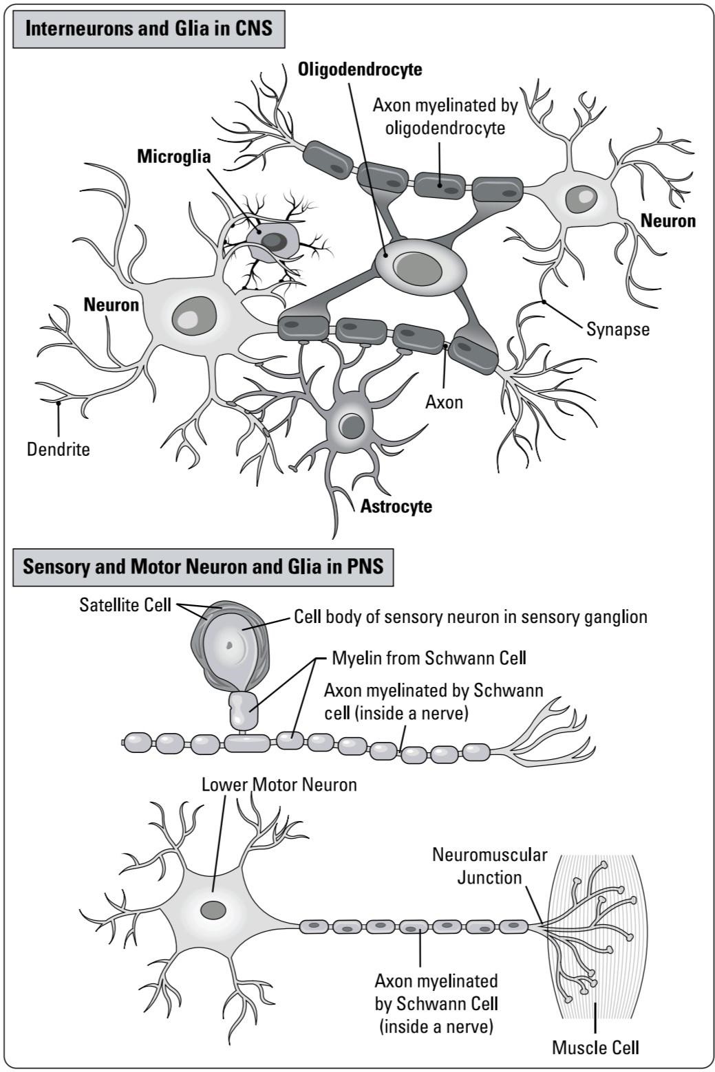
FIGURE 1-1: Typical neurons and supporting glial cells.
شکل ۱-۱: نورونهای معمولی و سلولهای گلیال پشتیبان.
Specializing the functions
Neurons, also referred to as nerve cells or neuronal cells, are the main signaling cells of the nervous system. Functionally, neurons can be classified as the following:
تخصصی کردن عملکردها
نورونها که به عنوان سلولهای عصبی یا سلولهای نورونی نیز شناخته میشوند، سلولهای اصلی سیگنالدهی سیستم عصبی هستند. از نظر عملکردی، نورونها را میتوان به صورت زیر طبقهبندی کرد:
» Sensory neurons: Sensory neurons detect external and internal stimuli. The majority are located in the PNS and they transmit information to the CNS.
» نورونهای حسی: نورونهای حسی محرکهای خارجی و داخلی را تشخیص میدهند. اکثر آنها در سیستم عصبی محیطی (PNS) قرار دارند و اطلاعات را به سیستم عصبی مرکزی (CNS) منتقل میکنند.
» Motor neurons: Motor neurons send signals out to control muscle contraction. They’re found in both the CNS and PNS.
» نورونهای حرکتی: نورونهای حرکتی سیگنالهایی را برای کنترل انقباض عضلات ارسال میکنند. آنها هم در سیستم عصبی مرکزی (CNS) و هم در سیستم عصبی محیطی (PNS) یافت میشوند.
» Interneurons: Interneurons include projection (or principal neurons) and local circuit neurons. They’re located in the CNS where they integrate information for perception, cognition, and/or the control of movement and behaviors for survival and homeostasis.
» نورونهای رابط: نورونهای رابط شامل نورونهای طرحریزی (یا نورونهای اصلی) و نورونهای مدار محلی هستند. آنها در سیستم عصبی مرکزی (CNS) قرار دارند و در آنجا اطلاعات را برای ادراک، شناخت و/یا کنترل حرکت و رفتارها برای بقا و هموستاز ادغام میکنند.
Neurons can also be classified based on the neurotransmitter they make and release, the effect they produce, and their morphology.
نورونها را میتوان بر اساس انتقالدهنده عصبی که میسازند و آزاد میکنند، اثری که تولید میکنند و مورفولوژیشان نیز طبقهبندی کرد.
Glial cells are also referred to as neuroglia or glia. Microglia are the resident immune cells in the CNS. Astrocytes are located in the CNS where they provide metabolic support for neurons and other glia. Some astrocytes surround synapses where they regulate ion and neurotransmitter levels. Other astrocytes extend processes called end feet to surround brain capillaries where they transport nutrients into the CNS.
سلولهای گلیال همچنین به عنوان نوروگلیا یا گلیا شناخته میشوند. میکروگلیا سلولهای ایمنی ساکن در سیستم عصبی مرکزی (CNS) هستند. آستروسیتها در سیستم عصبی مرکزی قرار دارند و در آنجا پشتیبانی متابولیکی را برای نورونها و سایر گلیاها فراهم میکنند. برخی از آستروسیتها سیناپسها را احاطه کردهاند که در آنجا سطح یونها و انتقالدهندههای عصبی را تنظیم میکنند. سایر آستروسیتها زوائدی به نام پاهای انتهایی را گسترش میدهند تا مویرگهای مغز را احاطه کنند و در آنجا مواد مغذی را به سیستم عصبی مرکزی منتقل کنند.
Forming the barriers
Brain capillaries are formed by vascular endothelial cells that make tight junctions. These junctions prevent the direct diffusion of water soluble nutrients from the blood into the CNS and forms the blood-brain barrier. It’s the reason the CNS requires astrocytes to transport nutrients between capillaries and neurons. Another type of glial cell called pericytes surround the capillaries and help regulate the capillary diameter or vasodilation. Microglia are the resident immune cells in the CNS that help with the detection and removal of pathogens and damaged cells.
تشکیل موانع
مویرگهای مغز توسط سلولهای اندوتلیال عروقی تشکیل میشوند که اتصالات محکمی ایجاد میکنند. این اتصالات از انتشار مستقیم مواد مغذی محلول در آب از خون به سیستم عصبی مرکزی جلوگیری میکنند و سد خونی-مغزی را تشکیل میدهند. به همین دلیل است که سیستم عصبی مرکزی برای انتقال مواد مغذی بین مویرگها و نورونها به آستروسیتها نیاز دارد. نوع دیگری از سلولهای گلیال به نام پریسیتها، مویرگها را احاطه کرده و به تنظیم قطر مویرگ یا گشاد شدن عروق کمک میکنند. میکروگلیاها سلولهای ایمنی ساکن در سیستم عصبی مرکزی هستند که به تشخیص و حذف عوامل بیماریزا و سلولهای آسیبدیده کمک میکنند.
In the CNS, about half of the axons are myelinated by oligodendrocytes. Myelination involves the wrapping of an axon by many layers of the oligodendrocyte cell membrane. In the PNS, the majority of axons are myelinated within the nerve where the Schwann cells provide the myelin membrane. Myelination of axons increases the speed and efficiency of action potentials traveling along the axon.
در سیستم عصبی مرکزی (CNS)، حدود نیمی از آکسونها توسط الیگودندروسیتها میلیندار شدهاند. میلینسازی شامل پیچیدن یک آکسون توسط لایههای زیادی از غشای سلولی الیگودندروسیت است. در سیستم عصبی محیطی (PNS)، اکثر آکسونها در داخل عصب میلیندار هستند، جایی که سلولهای شوان غشای میلین را فراهم میکنند. میلیندار شدن آکسونها سرعت و کارایی پتانسیلهای عمل در حال حرکت در امتداد آکسون را افزایش میدهد.
The brain contains fluid-filled tubes inside called the ventricles, which are continuous with the central canal in the spinal cord. The ventricles are lined by ependymal cells and contain a region called the choroid plexus formed by specialized ependymal cells and capillaries. The ependymal cells synthesize and transport cerebrospinal fluid (CSF) inside the ventricles.
مغز حاوی لولههای پر از مایعی به نام بطنها است که در امتداد کانال مرکزی نخاع قرار دارند. بطنها توسط سلولهای اپاندیمی پوشیده شدهاند و حاوی ناحیهای به نام شبکه کوروئید هستند که توسط سلولهای اپاندیمی تخصصی و مویرگها تشکیل شده است. سلولهای اپاندیمی مایع مغزی نخاعی (CSF) را در داخل بطنها سنتز و منتقل میکنند.
Electrifying the membrane
All cells have a membrane potential, a difference in electrical charge across the membrane. Neurons use changes in the membrane potential for communication. The axon is where an action potential is produced and conducted to the presynaptic region. We describe the basis of electrical signaling and action potentials in Chapter 4.
برق رسانی به غشاء
همه سلولها دارای پتانسیل غشاء هستند، که به معنی تفاوت در بار الکتریکی در دو طرف غشاء است. نورونها از تغییرات پتانسیل غشاء برای ارتباط استفاده میکنند. آکسون جایی است که پتانسیل عمل تولید و به ناحیه پیش سیناپسی هدایت میشود. ما اساس سیگنالینگ الکتریکی و پتانسیلهای عمل را در فصل ۴ شرح میدهیم.
Communicating with targets
The dendrites are typically branched and are the main receiving processes in neurons. Many dendrites contain small bumps or protrusions called dendritic spines. Dendrites and dendritic spines receive incoming signals from other neurons at synapses. These electrical signals are also described in Chapter 4.
ارتباط با اهداف
دندریتها معمولاً شاخهدار هستند و فرآیندهای اصلی دریافت در نورونها محسوب میشوند. بسیاری از دندریتها حاوی برآمدگیها یا برآمدگیهای کوچکی به نام خارهای دندریتیک هستند. دندریتها و خارهای دندریتیک سیگنالهای ورودی را از سایر نورونها در سیناپسها دریافت میکنند. این سیگنالهای الکتریکی نیز در فصل ۴ توضیح داده شدهاند.
At the synapse, action potentials are converted into release of chemicals called neurotransmitters. Neurotransmitters activate receptors on the target cell. We describe the mechanism of synaptic transmission in Chapter 5 and neurotransmitter systems in Chapter 6.
در سیناپس، پتانسیلهای عمل به آزادسازی مواد شیمیایی به نام انتقالدهندههای عصبی تبدیل میشوند. انتقالدهندههای عصبی گیرندههای سلول هدف را فعال میکنند. ما مکانیسم انتقال سیناپسی را در فصل 5 و سیستمهای انتقالدهنده عصبی را در فصل 6 شرح میدهیم.
Protecting and Nourishing the Parts
Here we explain how the nervous system is protected by non-neural structures, how it receives its blood supply, and what nerves contain.
محافظت و تغذیه اعضا
در اینجا توضیح میدهیم که چگونه سیستم عصبی توسط ساختارهای غیر عصبی محافظت میشود، چگونه خون دریافت میکند و چه اعصابی در آن وجود دارد.
Safeguarding the CNS
The brain is encased in the skull bone and the spinal cord is surrounded by the bones of the vertebral column. Underneath the bones are the meninges, three layers of membranes that cover the brain and spinal cord. We describe the meninges in Chapter 3.
محافظت از سیستم عصبی مرکزی
مغز در استخوان جمجمه قرار دارد و نخاع توسط استخوانهای ستون فقرات احاطه شده است. در زیر استخوانها، مننژها قرار دارند، سه لایه غشایی که مغز و نخاع را میپوشانند. مننژها را در فصل 3 شرح میدهیم.
Providing the supplies
The brain and spinal cord are highly vascularized, containing many arteries, veins, arterioles, venules, and capillaries. The main blood supply to the front and middle regions of the brain is from the internal carotid arteries. The blood supply to the posterior brain and spinal cord is from the vertebral arteries.
فراهم کردن ملزومات
مغز و نخاع بسیار عروقی هستند و حاوی شریانها، وریدها، سرخرگها، سیاهرگچهها و مویرگهای زیادی میباشند. خونرسانی اصلی به نواحی جلویی و میانی مغز از طریق شریانهای کاروتید داخلی انجام میشود. خونرسانی به مغز و نخاع خلفی از طریق شریانهای مهرهای انجام میشود.
The CNS capillaries are different from the rest of the body’s general capillaries in that they, together with astrocytes and pericytes, form a blood brain barrier. The brain contains many venous plexuses that receive the blood and drain it into the jugular veins. A lymphatic system in the meninges mediates the movement of waste and immune cells.
مویرگهای سیستم عصبی مرکزی با بقیه مویرگهای عمومی بدن متفاوت هستند، زیرا آنها به همراه آستروسیتها و پریسیتها، سد خونی-مغزی را تشکیل میدهند. مغز حاوی شبکههای وریدی زیادی است که خون را دریافت کرده و آن را به رگهای ژوگولار تخلیه میکنند. یک سیستم لنفاوی در مننژها واسطه حرکت مواد زائد و سلولهای ایمنی است.
The ventricles inside the brain synthesize and circulate CSF. Some CSF is diverted around the brain where the meninges transfers the CSF to the blood. The rest of the CSF flows in the central canal inside the spinal cord.
بطنهای داخل مغز، مایع مغزی نخاعی (CSF) را سنتز و به گردش در میآورند. مقداری از مایع مغزی نخاعی در اطراف مغز منحرف میشود، جایی که مننژها، مایع مغزی نخاعی را به خون منتقل میکنند. بقیه مایع مغزی نخاعی در کانال مرکزی داخل نخاع جریان دارد.
Supporting the PNS
In the PNS, the nerves are bundles of axons that are covered with layers of connective tissue. Nerves also contain the myelinating Schwann cells and blood vessels. The axons in nerves originate or terminate in the spinal cord or brainstem.
پشتیبانی از سیستم عصبی محیطی (PNS)
در سیستم عصبی محیطی، اعصاب دستههایی از آکسونها هستند که با لایههایی از بافت همبند پوشیده شدهاند. اعصاب همچنین حاوی سلولهای شوان میلینساز و رگهای خونی هستند. آکسونهای اعصاب از نخاع یا ساقه مغز سرچشمه میگیرند یا به آن ختم میشوند.
In the PNS, ganglia are groups neuron cell bodies clustered in regions outside the spinal cord or brainstem (refer to Chapter 7). The PNS is divided into the somatic, autonomic (visceral), and enteric nervous systems. The two types of ganglia are sensory ganglia and autonomic ganglia. The enteric nervous system contains both sensory and motor neurons in the gastrointestinal tracts.
در سیستم عصبی محیطی (PNS)، گانگلیونها گروههایی از اجسام سلولی نورونی هستند که در مناطقی خارج از نخاع یا ساقه مغز تجمع یافتهاند (به فصل 7 مراجعه کنید). سیستم عصبی محیطی به سیستمهای عصبی سوماتیک، اتونومیک (احشایی) و رودهای تقسیم میشود. دو نوع گانگلیون، گانگلیونهای حسی و گانگلیونهای اتونومیک هستند. سیستم عصبی رودهای شامل نورونهای حسی و حرکتی در مجاری گوارشی است.
Navigating Around the Nervous System
The brain and spinal cord are formed by groups of neurons that work together to connect to other neurons through tracts. Examining the CNS in fixed sections, you see regions the following regions:
پیمایش در سیستم عصبی
مغز و نخاع توسط گروههایی از نورونها تشکیل شدهاند که با همکاری یکدیگر از طریق مسیرهایی به نورونهای دیگر متصل میشوند. با بررسی بخشهای ثابت سیستم عصبی مرکزی، مناطقی از جمله مناطق زیر را مشاهده میکنید:
» Gray matter contains neuronal cell bodies, dendrites, synapses, and astrocytes.
» White matter contains myelinated axons, oligodendrocytes, and astrocytes.
» ماده خاکستری شامل جسم سلولی نورونها، دندریتها، سیناپسها و آستروسیتها است.
» ماده سفید شامل آکسونهای میلیندار، الیگودندروسیتها و آستروسیتها است.
These sections explain the gray and white matter regions in the CNS.
این بخشها نواحی ماده خاکستری و سفید در سیستم عصبی مرکزی (CNS) را توضیح میدهند.
Organizing gray matter
Gray matter in the CNS is organized in several ways:
سازماندهی ماده خاکستری
ماده خاکستری در سیستم عصبی مرکزی (CNS) به چندین روش سازماندهی شده است:
» In the brain the cortical gray matter is layered and found in the cerebral cortex and cerebellar cortex.
» در مغز، ماده خاکستری قشری لایه لایه است و در قشر مغز و قشر مخچه یافت میشود.
» The other groups of neurons in the brain are called nuclei (a single nucleus). Subcortical brain structures, including the basal ganglia, thalamus, hypothalamus, amygdala, and brainstem, are formed by many separate nuclei.
» گروههای دیگر نورونها در مغز، هسته (یک هسته واحد) نامیده میشوند. ساختارهای زیرقشری مغز، از جمله عقدههای قاعدهای، تالاموس، هیپوتالاموس، آمیگدال و ساقه مغز، توسط هستههای جداگانه زیادی تشکیل شدهاند.
» In the spinal cord, the gray matter is located in the horns. The ventral horns contain motor neurons while the dorsal horn contains sensory relay neurons. Both regions contain spinal interneurons.
» در نخاع، ماده خاکستری در شاخها قرار دارد. شاخهای شکمی حاوی نورونهای حرکتی هستند در حالی که شاخ پشتی حاوی نورونهای رله حسی است. هر دو ناحیه حاوی نورونهای رابط نخاعی هستند.
Tracking along white matter
Within the CNS, axon tracts form the white matter. There are different types of tracts in the brain that connect neurons in one cortical region to another cortical region or to subcortical areas. In the spinal cord, the white matter is located in the regions around the white matter and contains the axons of all the ascending sensory and descending motor tracts.
ردیابی در امتداد ماده سفید
در داخل سیستم عصبی مرکزی، رشتههای آکسونی، ماده سفید را تشکیل میدهند. انواع مختلفی از رشتهها در مغز وجود دارند که نورونها را در یک ناحیه قشری به ناحیه قشری دیگر یا به نواحی زیرقشری متصل میکنند. در نخاع، ماده سفید در نواحی اطراف ماده سفید قرار دارد و شامل آکسونهای تمام رشتههای حسی صعودی و حرکتی نزولی است.
Dissecting functions of the CNS
Looking at the mammalian brain from the outside, shown in Figure 1-2, you can see three major parts:
تشریح عملکردهای سیستم عصبی مرکزی
با نگاه به مغز پستانداران از بیرون، که در شکل ۱-۲ نشان داده شده است، میتوانید سه بخش اصلی را مشاهده کنید:
» Forebrain: The largest part of the brain, it contains the cerebrum and subcortical nuclei. The cerebrum is composed of the cerebral cortices and cerebral white matter. Subcortical structures including the basal ganglia, amygdala, thalamus, hypothalamus, and pituitary gland.
» مغز پیشین: بزرگترین بخش مغز، حاوی مخ و هستههای زیرقشری است. مخ از قشرهای مغزی و ماده سفید مغزی تشکیل شده است. ساختارهای زیرقشری شامل گانگلیونهای پایه، آمیگدال، تالاموس، هیپوتالاموس و غده هیپوفیز هستند.
» Brain stem: The midbrain is located underneath the thalamus. Below that is the pons, and below that the medulla that ends at the top of the spinal cord. The brain stem is the oldest part of the brain that’s essential for survival responses and homeostasis.
» ساقه مغز: مغز میانی در زیر تالاموس قرار دارد. در زیر آن پل مغزی و در زیر آن بصل النخاع قرار دارد که در بالای نخاع ختم میشود. ساقه مغز قدیمیترین بخش مغز است که برای پاسخهای بقا و هموستاز ضروری است.
» Cerebellum: It sticks out from the back of the pons in the brainstem. It contains as many neurons as the rest of the brain combined. Though it evolved first in vertebrates, the cerebellum continued to evolve as mammals added the larger neocortex. It functions in motor control.
» مخچه: از پشت پل مغزی در ساقه مغز بیرون زده است. به اندازه بقیه مغز نورون دارد. اگرچه مخچه ابتدا در مهرهداران تکامل یافت، اما با اضافه شدن نئوکورتکس بزرگتر در پستانداران، به تکامل خود ادامه داد. این بخش در کنترل حرکت نقش دارد.

FIGURE 1-2: Views of the human brain.
شکل ۱-۲: نماهایی از مغز انسان.
Looking at the cerebrum
The cerebrum contains the cerebral cortex and the underlying cerebral white matter. Figure 1-2 shows that from the outside you see many gyri (singular: gyrus), which are ridges, and many sulci (singular: sulcus), which are grooves that give the human and some mammalian brains their folded appearance. Folding allowed more cortical areas to fit inside the skull during evolution.
نگاهی به مخ
مخ شامل قشر مغز و ماده سفید مغزی زیرین است. شکل ۱-۲ نشان میدهد که از بیرون، تعداد زیادی شکنج (مفرد: شکنج) که برآمدگی هستند و تعداد زیادی شیار (مفرد: شیار) که شیارهایی هستند که به مغز انسان و برخی از پستانداران ظاهری چینخورده میدهند، میبینید. چینخوردگی باعث شده است که در طول تکامل، نواحی قشری بیشتری در داخل جمجمه جای بگیرند.
The longitudinal fissure separates the left and right cerebral hemispheres. Deep sulci separate the cerebrum into four lobes on each side, called the frontal, parietal, temporal, and occipital lobe, named for the cranial bones that overlies each (see the next section for more about these lobes). The central sulcus separates the frontal and parietal lobes while the lateral sulcus separates the temporal lobe from the other three lobes.
شیار طولی، نیمکرههای چپ و راست مغز را از هم جدا میکند. شیارهای عمیق، مخ را در هر طرف به چهار لوب تقسیم میکنند که لوبهای پیشانی، آهیانه، گیجگاهی و پسسری نامیده میشوند و نام آنها از استخوانهای جمجمهای که روی هر یک از آنها قرار دارند، گرفته شده است (برای اطلاعات بیشتر در مورد این لوبها به بخش بعدی مراجعه کنید). شیار مرکزی، لوبهای پیشانی و آهیانه را از هم جدا میکند در حالی که شیار جانبی، لوب گیجگاهی را از سه لوب دیگر جدا میکند.
Figuring out the lobes’ functions
Figure 1-2 also illustrates the lobes of the cerebrum. Here is a brief overview (Chapter 11 examines the functional anatomy of the lobes in greater detail):
بررسی عملکرد لوبها
شکل ۱-۲ همچنین لوبهای مخ را نشان میدهد. در اینجا مروری مختصر بر آنها داریم (فصل ۱۱ آناتومی عملکردی لوبها را با جزئیات بیشتری بررسی میکند):
» Frontal lobe: It contains the motor cortex, prefrontal cortex, frontal eye fields, and Broca’s area. The frontal lobe operates primarily to program and organize goal directed movements, speech, and executive functions.
» لوب پیشانی: شامل قشر حرکتی، قشر پیشپیشانی، میدانهای بینایی پیشانی و ناحیه بروکا است. لوب پیشانی عمدتاً برای برنامهریزی و سازماندهی حرکات هدفمند، گفتار و عملکردهای اجرایی عمل میکند.
» Parietal lobe: It contains the somatosensory cortex and posterior parietal cortex, a sensory association area that integrates somatosensory, visual, and auditory inputs.
» لوب آهیانه: شامل قشر حسی-پیکری و قشر آهیانه خلفی، یک ناحیه ارتباط حسی است که ورودیهای حسی-پیکری، بینایی و شنوایی را ادغام میکند.
» Temporal lobe: It contains the auditory cortex, hippocampus, and amygdala. The temporal lobe functions in processing emotion, deciphering auditory information, understanding language, and encoding memory.
» لوب گیجگاهی: شامل قشر شنوایی، هیپوکامپ و آمیگدال است. لوب گیجگاهی در پردازش احساسات، رمزگشایی اطلاعات شنوایی، درک زبان و رمزگذاری حافظه نقش دارد.
» Occipital lobe: It contains the visual cortex composed of multiple visual processing areas.
» لوب پس سری: شامل قشر بینایی است که از چندین ناحیه پردازش بینایی تشکیل شده است.
Situating underneath the cortex
Underneath and within the cerebral white matter are the subcortical areas. They include the thalamus, hypothalamus, basal ganglia, cerebellum, and brainstem. We introduce the functions of these areas in the next section and in Part 2 of this book.
زیر قشر مغز قرار دارد
در زیر و درون ماده سفید مغز، نواحی زیرقشری قرار دارند. این نواحی شامل تالاموس، هیپوتالاموس، عقدههای قاعدهای، مخچه و ساقه مغز میشوند. ما عملکرد این نواحی را در بخش بعدی و در بخش دوم این کتاب معرفی میکنیم.
Moving the Correct Way
An important function of the brain is the control of motor systems. Motor systems involve somatic control of skeletal muscles. They also control contraction of the heart and smooth muscles in organs and tissues by the autonomic motor system. Part 2 of this book focuses on mechanisms and control of movements and behaviors. Here we introduce the basics.
حرکت به روش صحیح
یکی از عملکردهای مهم مغز، کنترل سیستمهای حرکتی است. سیستمهای حرکتی شامل کنترل سوماتیک عضلات اسکلتی هستند. آنها همچنین انقباض قلب و عضلات صاف در اندامها و بافتها را توسط سیستم حرکتی خودکار کنترل میکنند. بخش دوم این کتاب بر مکانیسمها و کنترل حرکات و رفتارها تمرکز دارد. در اینجا اصول اولیه را معرفی میکنیم.
Making the movements
Upper motor neurons from the motor cortex send axons to the lower motor neurons and nearby interneurons. The lower motor neurons provide the output to skeletal muscles to control their contraction (see Chapter 7).
انجام حرکات
نورونهای حرکتی فوقانی از قشر حرکتی، آکسونها را به نورونهای حرکتی تحتانی و نورونهای رابط مجاور میفرستند. نورونهای حرکتی تحتانی، خروجی عضلات اسکلتی را برای کنترل انقباض آنها فراهم میکنند (به فصل 7 مراجعه کنید).
Controlling the output
The motor cortex contains areas involved in control of motor output. The primary motor cortex provides direct motor commands to produce movements. The supplementary motor area and premotor cortex receive inputs from cortical and sub cortical areas and control the output from the primary motor cortex.
کنترل خروجی
قشر حرکتی شامل نواحی درگیر در کنترل خروجی حرکتی است. قشر حرکتی اولیه، دستورات حرکتی مستقیم را برای تولید حرکات فراهم میکند. ناحیه حرکتی تکمیلی و قشر پیشحرکتی، ورودیها را از نواحی قشری و زیرقشری دریافت کرده و خروجی قشر حرکتی اولیه را کنترل میکنند.
The basal ganglia provide action selection and inhibit unwanted movements. The cerebellum is necessary for learning movement sequences and detecting and correcting errors to coordinate movements. Chapter 8 discusses these brain regions in motor control.
عقدههای قاعدهای انتخاب عمل را فراهم میکنند و حرکات ناخواسته را مهار میکنند. مخچه برای یادگیری توالیهای حرکتی و تشخیص و اصلاح خطاها برای هماهنگی حرکات ضروری است. فصل ۸ این نواحی مغز را در کنترل حرکتی مورد بحث قرار میدهد.
Focusing on older parts
Both the spinal cord and brainstem function like subcontractors of the brain to execute the brain’s instructions and report on their progress. They contain lower motor neurons whose axons control muscle cell contraction. They receive sensory inputs for both local circuit reflexes and for relaying sensory information to the thalamus, which then transmits to the sensory cortices.
تمرکز بر بخشهای قدیمیتر
هم نخاع و هم ساقه مغز مانند پیمانکاران فرعی مغز عمل میکنند تا دستورالعملهای مغز را اجرا کرده و پیشرفت آنها را گزارش دهند. آنها حاوی نورونهای حرکتی تحتانی هستند که آکسونهای آنها انقباض سلولهای عضلانی را کنترل میکنند. آنها ورودیهای حسی را برای رفلکسهای مدار موضعی و همچنین برای انتقال اطلاعات حسی به تالاموس دریافت میکنند که سپس به قشرهای حسی منتقل میشود.
The brainstem and spinal cord are also essential in survival and homeostasis. Both control the autonomic nervous system involved in regulation of heartrate, blood pressure, and respiration. The sympathetic system prepares us for action in the fight-or-flight mode, while the parasympathetic system organizes resources for digestion and the conservation of energy. Chapter 9 describes the basic organization and functions of these regions.
ساقه مغز و نخاع نیز در بقا و هموستاز ضروری هستند. هر دو سیستم عصبی خودکار را که در تنظیم ضربان قلب، فشار خون و تنفس نقش دارد، کنترل میکنند. سیستم سمپاتیک ما را برای عمل در حالت جنگ یا گریز آماده میکند، در حالی که سیستم پاراسمپاتیک منابع لازم برای هضم و حفظ انرژی را سازماندهی میکند. فصل 9 سازماندهی و عملکردهای اساسی این مناطق را شرح میدهد.
Ensuring homeostasis
The hypothalamus receives input from many brain areas and controls homeostatic processes including regulation of temperature and feeding. It provides control of the autonomic nervous system and circadian rhythms, the body’s internal clock. It also links the brain with the endocrine system by controlling the secretion of hormones from the pituitary gland.
تضمین هموستاز
هیپوتالاموس از بسیاری از نواحی مغز ورودی دریافت میکند و فرآیندهای هموستاتیک از جمله تنظیم دما و تغذیه را کنترل میکند. این غده کنترل سیستم عصبی خودکار و ریتمهای شبانهروزی، ساعت داخلی بدن، را بر عهده دارد. همچنین با کنترل ترشح هورمونها از غده هیپوفیز، مغز را به سیستم غدد درونریز متصل میکند.
The cingulate cortex is involved in processing pain, error, and conflict detection, processing and regulating emotions, regulating autonomic functions, social behavior, and empathy. Regions involved in memory provide key inputs for our survival responses and cognition (see Chapter 15).
قشر کمربندی در پردازش درد، خطا و تشخیص تعارض، پردازش و تنظیم احساسات، تنظیم عملکردهای خودکار، رفتار اجتماعی و همدلی نقش دارد. مناطقی که در حافظه دخیل هستند، ورودیهای کلیدی برای پاسخهای بقا و شناخت ما را فراهم میکنند (به فصل ۱۵ مراجعه کنید).
The amygdala is an emotion processing center for emotions like fear and motivation. It’s where fear memories are encoded. Its outputs contribute to regulation of autonomic and endocrine functions, as well as executive functions such as decision making and emotional control.
آمیگدال یک مرکز پردازش احساسات برای احساساتی مانند ترس و انگیزه است. جایی است که خاطرات ترس رمزگذاری میشوند. خروجیهای آن به تنظیم عملکردهای خودکار و غدد درونریز و همچنین عملکردهای اجرایی مانند تصمیمگیری و کنترل احساسات کمک میکند.
The reticular formation is a diffuse network of neurons and axon tracts that runs through the brainstem up into portions of the thalamus. It controls body state through processes such as controlling wakefulness versus sleep, alertness, and homeostatic mechanisms such as heart rate.
تشکیلات مشبک، شبکهای پراکنده از نورونها و رشتههای آکسونی است که از ساقه مغز تا بخشهایی از تالاموس امتداد دارد. این تشکیلات از طریق فرآیندهایی مانند کنترل بیداری در مقابل خواب، هوشیاری و مکانیسمهای هومئوستاتیک مانند ضربان قلب، وضعیت بدن را کنترل میکند.
Chapter 10 describes survival and homeostatic functions of the brainstem and limbic system. We discuss the specific functions of these brain areas and how they interact with other parts of the brain.
فصل 10 به شرح عملکردهای بقا و هومئوستاتیک ساقه مغز و سیستم لیمبیک میپردازد. ما در مورد عملکردهای خاص این نواحی مغز و نحوه تعامل آنها با سایر قسمتهای مغز بحث میکنیم.
Completing the circuit
The neocortex dominates the mammalian brain in volume, particularly in primates. It has the same six-layered structure virtually everywhere, with the similar cell types and cortical minicolumn circuits. It’s responsible for producing sensory perception and controlling voluntary movement and cognitive functions. Chapter 11 describes the neocortex and its functions.
تکمیل مدار
نئوکورتکس از نظر حجم، مغز پستانداران، به ویژه در نخستیسانان را در بر میگیرد. این بخش تقریباً در همه جا ساختار شش لایهای یکسانی دارد، با انواع سلولها و مدارهای ستونی کوچک قشری مشابه. این بخش مسئول ایجاد ادراک حسی و کنترل حرکات ارادی و عملکردهای شناختی است. فصل 11 نئوکورتکس و عملکردهای آن را شرح میدهد.
Chapter 12 focuses on the types of injury, degeneration, and dysfunction that can compromise the control or production of movement. We describe the most common developmental disorders and movement disorders that involve motor systems.
فصل ۱۲ بر انواع آسیب، تخریب و اختلال عملکرد که میتوانند کنترل یا تولید حرکت را به خطر بیندازند، تمرکز دارد. ما شایعترین اختلالات رشدی و اختلالات حرکتی که سیستمهای حرکتی را درگیر میکنند، شرح میدهیم.
Perceiving the World, Thinking, and Remembering
Part 3 describes how sensory systems generate perception and how brain areas cooperate to provide cognitive functions, including executive functions, language, and memory. Chapter 18 discusses neurodevelopmental and mental disorders that impact cognitive functions. Here we give a brief overview.
درک جهان، تفکر و به خاطر سپردن
بخش ۳ توضیح میدهد که چگونه سیستمهای حسی ادراک را ایجاد میکنند و چگونه نواحی مغز برای ارائه عملکردهای شناختی، از جمله عملکردهای اجرایی، زبان و حافظه، با هم همکاری میکنند. فصل ۱۸ اختلالات عصبی-رشدی و روانی را که بر عملکردهای شناختی تأثیر میگذارند، مورد بحث قرار میدهد. در اینجا مروری مختصر ارائه میدهیم.
Seeing and hearing is believing
In many mammals, visual processing dominates the brain. In both vision and audition, high-order brain areas process complex inputs, such as face specific neurons in inferotemporal cortex in vision, and language specific areas in audition such as Wernicke’s area. Chapter 13 focuses on the visual and auditory sensory systems.
دیدن و شنیدن، باور کردن است
در بسیاری از پستانداران، پردازش بصری بر مغز غالب است. در هر دو حوزه بینایی و شنوایی، نواحی سطح بالای مغز، ورودیهای پیچیده را پردازش میکنند، مانند نورونهای مختص چهره در قشر گیجگاهی تحتانی در بینایی، و نواحی مختص زبان در شنوایی مانند ناحیه ورنیکه. فصل ۱۳ بر سیستمهای حسی بینایی و شنوایی تمرکز دارد.
Sensing touch, smell, and taste
In the somatosensory system, sensation is composed of sensory receptors including mechanoreceptors, thermoreceptors, and nociceptors in the skin, muscles, and joints. These allow you to perceive touch, temperature, and painful stimuli.
حس لامسه، بو و طعم
در سیستم حسی-پیکری، حس از گیرندههای حسی شامل گیرندههای مکانیکی، گیرندههای حرارتی و گیرندههای درد در پوست، عضلات و مفاصل تشکیل شده است. این گیرندهها به شما امکان میدهند لمس، دما و محرکهای دردناک را درک کنید.
The sense of smell is mediated by olfactory receptor cells in the nose while the sense of taste is mediated by receptors mostly in taste buds on the tongue. Your perception of flavor is formed by a combination of taste and smell. Chapter 14 examines somatosensory, smell, and taste systems in more detail.
حس بویایی توسط سلولهای گیرنده بویایی در بینی و حس چشایی توسط گیرندههایی که عمدتاً در جوانههای چشایی روی زبان قرار دارند، ایجاد میشود. درک شما از طعم غذا با ترکیبی از طعم و بو شکل میگیرد. فصل ۱۴ سیستمهای حسی-پیکری، بویایی و چشایی را با جزئیات بیشتری بررسی میکند.
Focusing on learning, memory, and plasticity
A hallmark of mammals is they can change their behavior through learning and memory (see Chapter 15). The short-term representation of an event and its context occurs in the prefrontal cortex as working memory. The hippocampus encodes long-term declarative memory for facts and events and spatial memory. The amygdala encodes fear memory. The basal ganglia and cerebellum encode procedural memory for habits and motor skills.
تمرکز بر یادگیری، حافظه و انعطافپذیری
یکی از ویژگیهای بارز پستانداران این است که میتوانند رفتار خود را از طریق یادگیری و حافظه تغییر دهند (به فصل ۱۵ مراجعه کنید). بازنمایی کوتاهمدت یک رویداد و زمینه آن در قشر جلوی مغز به عنوان حافظه کاری رخ میدهد. هیپوکامپ حافظه اعلانی بلندمدت را برای حقایق و رویدادها و حافظه فضایی رمزگذاری میکند. آمیگدال حافظه ترس را رمزگذاری میکند. عقدههای قاعدهای و مخچه حافظه رویهای را برای عادات و مهارتهای حرکتی رمزگذاری میکنند.
The encoding of memory involves synaptic plasticity in each of the brain areas identified. Research suggests that long term memory is stored in cortical circuits in the cerebral cortex. This means that the neocortex is involved in not only the integration and processing of sensory, motor, and cognitive information, but also in its storage as well.
رمزگذاری حافظه شامل انعطافپذیری سیناپسی در هر یک از نواحی شناساییشده مغز است. تحقیقات نشان میدهد که حافظه بلندمدت در مدارهای قشری در قشر مغز ذخیره میشود. این بدان معناست که نئوکورتکس نه تنها در ادغام و پردازش اطلاعات حسی، حرکتی و شناختی، بلکه در ذخیرهسازی آن نیز نقش دارد.
Controlling with the executive brain
The frontal lobes are responsible for planning and executing behavior (refer to Chapter 16). During evolution there was a relative expansion of the frontal lobe compared to the rest of the brain in humans compared to other primates, and primates compared to other mammals.
کنترل با مغز اجرایی
لوبهای پیشانی مسئول برنامهریزی و اجرای رفتار هستند (به فصل ۱۶ مراجعه کنید). در طول تکامل، در انسانها در مقایسه با سایر نخستیسانان و در نخستیسانان در مقایسه با سایر پستانداران، گسترش نسبی لوب پیشانی در مقایسه با بقیه مغز وجود داشته است.
The motor cortex controls motor neurons for regulation of contraction of skeletal muscles. The frontal eye fields control eye movements, and Broca’s area mediates language production. The prefrontal cortex produces executive functions for performing goal directed movements and controlling behavior.
قشر حرکتی، نورونهای حرکتی را برای تنظیم انقباض عضلات اسکلتی کنترل میکند. میدانهای بینایی پیشانی حرکات چشم را کنترل میکنند و ناحیه بروکا واسطه تولید زبان است. قشر جلوی مغز، عملکردهای اجرایی را برای انجام حرکات هدفمند و کنترل رفتار ایجاد میکند.
Thinking about cognition
Several cognitive functions, including language, are lateralized in humans. The capacity for learning language is innate in humans and involves Wernicke’s and Broca’s areas. Neurobiology research is working to understand how human language systems operate. Chapter 17 examines the components of the human brain areas most important for cognitive functions.
تفکر در مورد شناخت
چندین عملکرد شناختی، از جمله زبان، در انسان به صورت جانبی عمل میکنند. ظرفیت یادگیری زبان در انسان ذاتی است و نواحی ورنیکه و بروکا را درگیر میکند. تحقیقات نوروبیولوژی در تلاش است تا نحوه عملکرد سیستمهای زبانی انسان را درک کند. فصل ۱۷ اجزای نواحی مغز انسان را که برای عملکردهای شناختی مهمتر هستند، بررسی میکند.
Intelligence is the ability to acquire and apply knowledge and skills. The prefrontal cortex is involved in higher-order cognitive processes such as working memory, planning, decision-making, reasoning, problem-solving, and goal setting. The prefrontal cortex also acts in behavioral self-control and emotional control. These cognitive functions, together with your long term declarative memory, form the components of intelligence.
هوش توانایی کسب و به کارگیری دانش و مهارتها است. قشر جلوی مغز در فرآیندهای شناختی سطح بالاتر مانند حافظه کاری، برنامهریزی، تصمیمگیری، استدلال، حل مسئله و تعیین هدف نقش دارد. قشر جلوی مغز همچنین در خودکنترلی رفتاری و کنترل عاطفی عمل میکند. این عملکردهای شناختی، همراه با حافظه بلندمدت شما، اجزای هوش را تشکیل میدهند.
Consciousness is subjective and includes being aware of your sensations, thoughts, experiences, and emotions. Researchers have studied brain mechanisms that may underlie consciousness and have proposed many hypotheses about the basis of consciousness.
هوشیاری امری ذهنی است و شامل آگاهی از احساسات، افکار، تجربیات و عواطف شما میشود. محققان مکانیسمهای مغزی را که ممکن است زیربنای هوشیاری باشند، مطالعه کردهاند و فرضیههای زیادی در مورد اساس هوشیاری ارائه دادهاند.



