
فیزیولوژی پزشکی گایتون و هال؛ سیستم ادراری: آناتومی عملکردی و تشکیل ادرار توسط کلیهها

دعای مطالعه [ نمایش ]
بِسْمِ الله الرَّحْمنِ الرَّحیمِ
اَللّهُمَّ اَخْرِجْنى مِنْ ظُلُماتِ الْوَهْمِ
خدايا مرا بيرون آور از تاريكىهاى وهم،
وَ اَكْرِمْنى بِنُورِ الْفَهْمِ
و به نور فهم گرامى ام بدار،
اَللّهُمَّ افْتَحْ عَلَيْنا اَبْوابَ رَحْمَتِكَ
خدايا درهاى رحمتت را به روى ما بگشا،
وَانْشُرْ عَلَيْنا خَزائِنَ عُلُومِكَ بِرَحْمَتِكَ يا اَرْحَمَ الرّاحِمينَ
و خزانههاى علومت را بر ما باز كن به امید رحمتت اى مهربانترين مهربانان.
کتاب «فیزیولوژی پزشکی گایتون وهال» بهعنوان یکی از جامعترین و معتبرترین منابع در حوزه علوم پزشکی، همچنان مرجع کلیدی برای درک عملکرد پیچیده بدن انسان است. این اثر با تکیه بر تازهترین پژوهشها و توضیحات دقیق از سازوکارهای فیزیولوژیک، پلی میان علوم پایه پزشکی و کاربردهای بالینی ایجاد میکند و نقشی بیبدیل در آموزش، پژوهش و ارتقای دانش سلامت ایفا مینماید.
ترجمه دقیق و علمیاین شاهکار توسط برند علمیآیندهنگاران مغز به مدیریت داریوش طاهری، دسترسی فارسیزبانان به مرزهای نوین دانش فیزیولوژی را ممکن ساخته و رسالتی علمیبرای ارتقای آموزش پزشکی، فهم عمیقتر سازوکارهای بدن و توسعه روشهای نوین در حوزه سلامت فراهم آورده است.
» کتاب فیزیولوژی پزشکی گایتون وهال
» » فصل ۲۶: سیستم ادراری: آناتومیعملکردی و تشکیل ادرار توسط کلیهها
در حال ویرایش
» Guyton and Hall Textbook of Medical Physiology
»» CHAPTER 26: The Urinary System: Functional Anatomy and Urine Formation by the Kidneys

MULTIPLE FUNCTIONS OF THE KIDNEYS
Most people are familiar with one important function of the kidneys-to rid the body of waste materials that are ingested or produced by metabolism. A second function that is especially critical is to control the volume and electrolyte composition of the body fluids. For water and virtually all electrolytes in the body, the balance between intake (due to ingestion or metabolic production) and output (due to excretion or metabolic consumption) is maintained largely by the kidneys. This regulatory function of the kidneys maintains the stable internal environment necessary for the cells to perform their various activities.
عملکردهای چندگانه کلیهها
اکثر مردم با یکی از عملکردهای مهم کلیهها آشنا هستند و آن این است که بدن از مواد زائدی که توسط متابولیسم بلعیده شده یا تولید میشود، خلاص شود. عملکرد دوم که به ویژه حیاتی است، کنترل حجم و ترکیب الکترولیت مایعات بدن است. برای آب و تقریباً تمام الکترولیتهای بدن، تعادل بین دریافت (به دلیل مصرف یا تولید متابولیک) و خروجی (به دلیل دفع یا مصرف متابولیک) تا حد زیادی توسط کلیهها حفظ میشود. این عملکرد تنظیمیکلیهها محیط داخلی پایدار لازم را برای سلولها برای انجام فعالیتهای مختلف خود حفظ میکند.
The kidneys perform their most critical functions by filtering the plasma and removing substances from the filtrate at variable rates, depending on the needs of the body. Ultimately, the kidneys clear unwanted substances from the filtrate (and therefore from the blood) by excreting them in the urine while returning substances that are needed back to the blood.
کلیهها با فیلتر کردن پلاسما و حذف مواد از فیلتر با سرعتهای متغیر، بسته به نیاز بدن، حیاتی ترین وظایف خود را انجام میدهند. در نهایت، کلیهها مواد ناخواسته را از فیلتر (و در نتیجه از خون) با دفع آنها از طریق ادرار پاک میکنند و در عین حال مواد مورد نیاز را به خون باز میگرداند.
Although this chapter and the next few chapters focus mainly on the control of renal excretion of water, electrolytes, and metabolic waste products, the kidneys serve many important homeostatic functions, including the following:
اگرچه این فصل و چند فصل بعدی عمدتاً بر کنترل دفع کلیوی آب، الکترولیتها و مواد زائد متابولیک تمرکز دارند، کلیهها بسیاری از عملکردهای مهم هموستاتیک را انجام میدهند، از جمله موارد زیر:
• Excretion of metabolic waste products and foreign chemicals
• Regulation of water and electrolyte balances
• Regulation of body fluid osmolality and electrolyte concentrations
• Regulation of arterial pressure
• Regulation of acid-base balance
• Regulation of erythrocyte production
• Secretion, metabolism, and excretion of hormones
• Gluconeogenesis
• دفع مواد زائد متابولیک و مواد شیمیایی خارجی
• تنظیم تعادل آب و الکترولیت
• تنظیم اسمولالیته مایع بدن و غلظت الکترولیت
• تنظیم فشار شریانی
• تنظیم تعادل اسید و باز
• تنظیم تولید گلبولهای قرمز
• ترشح، متابولیسم و دفع هورمونها
• گلوکونئوژنز
Excretion of Metabolic Waste Products, Foreign Chemicals, Drugs, and Hormone Metabolites. The kidneys are the primary means for eliminating most of the waste products of metabolism that are no longer needed by the body. These products include urea (from the metabolism of amino acids), creatinine (from muscle creatine), uric acid (from nucleic acids), end products of hemoglobin breakdown (e.g., bilirubin), and metabolites of various hormones. These waste products must be eliminated from the body as rapidly as they are produced. The kidneys also eliminate most toxins and other foreign sub- stances that are produced by the body or ingested, such as pesticides, drugs, and food additives.
دفع مواد زائد متابولیک، مواد شیمیایی خارجی، داروها و متابولیتهای هورمونی. کلیهها ابزار اولیه برای از بین بردن بیشتر مواد زائد متابولیسم هستند که دیگر مورد نیاز بدن نیستند. این محصولات شامل اوره (از متابولیسم اسیدهای آمینه)، کراتینین (از کراتین ماهیچه)، اسید اوریک (از اسیدهای نوکلئیک)، محصولات نهایی تجزیه هموگلوبین (به عنوان مثال، بیلی روبین) و متابولیتهای هورمونهای مختلف است. این مواد زائد باید به همان سرعتی که تولید میشوند از بدن دفع شوند. کلیهها همچنین اکثر سموم و سایر مواد خارجی را که توسط بدن تولید میشوند یا بلعیده میشوند مانند آفت کشها، داروها و افزودنیهای غذایی از بین میبرند.
Regulation of Water and Electrolyte Balances. For maintenance of homeostasis, excretion of water and electrolytes must match intake precisely. If intake exceeds excretion, the amount of that substance in the body will increase. If intake is less than excretion, the amount of that substance in the body will decrease. Although temporary (or cyclic) imbalances of water and electrolytes may occur in various physiological and pathophysiological conditions associated with altered intake or renal excretion, the maintenance of life depends on restoration of water and electrolyte balance.
تنظیم تعادل آب و الکترولیت. برای حفظ هموستاز، دفع آب و الکترولیتها باید دقیقاً مطابق با میزان دریافتی باشد. اگر مصرف بیش از دفع شود، مقدار آن ماده در بدن افزایش مییابد. اگر دریافت کمتر از دفع باشد، مقدار آن ماده در بدن کاهش مییابد. اگرچه عدم تعادل موقتی (یا چرخه ای) آب و الکترولیتها ممکن است در شرایط مختلف فیزیولوژیکی و پاتوفیزیولوژیکی مرتبط با تغییر مصرف یا دفع کلیوی رخ دهد، حفظ حیات به بازیابی تعادل آب و الکترولیت بستگی دارد.
Intake of water and many electrolytes is usually governed by a person’s eating and drinking habits, requiring the kidneys to adjust their excretion rates to match the intakes of various substances. Figure 26-1 shows the response of the kidneys to a sudden 10-fold increase in sodium intake from a low level of 30 mEq/day to a high level of 300 mEq/day. Within 2 to 3 days after raising the sodium intake, renal excretion also increases to about 300 mEq/day so that the balance between intake and output is rapidly re-established. However, during the 2 to 3 days of renal adaptation to the high sodium intake, there is a modest accumulation of sodium that raises extracellular fluid volume slightly and triggers hormonal changes and other compensatory responses that signal the kidneys to increase their sodium excretion.
مصرف آب و بسیاری از الکترولیتها معمولاً توسط عادات خوردن و آشامیدن فرد کنترل میشود و کلیهها را ملزم میکند تا میزان دفع خود را متناسب با میزان دریافتی مواد مختلف تنظیم کنند. شکل 26-1 پاسخ کلیهها را به افزایش 10 برابری ناگهانی دریافت سدیم از سطح پایین 30 میلی اکی والان در روز به سطح بالای 300 میلی اکی والان در روز نشان میدهد. در عرض 2 تا 3 روز پس از افزایش مصرف سدیم، دفع کلیوی نیز به حدود 300 میلی اکی والان در روز افزایش مییابد تا تعادل بین دریافت و خروجی به سرعت برقرار شود. با این حال، در طی 2 تا 3 روز سازگاری کلیه با مصرف زیاد سدیم، تجمع متوسطی از سدیم وجود دارد که حجم مایع خارج سلولی را کمیافزایش میدهد و تغییرات هورمونی و سایر پاسخهای جبرانی را ایجاد میکند که به کلیهها سیگنال میدهد که دفع سدیم خود را افزایش دهند.
The capability of the kidneys to alter sodium excretion in response to changes in sodium intake is tremendous. Experimental studies have shown that in many people, sodium intake can be increased to 1500 mEq/day (more than 10 times normal) or decreased to 10 mEq/ day (<0.1 of normal), with relatively small changes in extracellular fluid volume or plasma sodium concentration. This phenomenon is also true for water and for most other electrolytes, such as chloride, potassium, calcium, hydrogen, magnesium, and phosphate ions. In the next few chapters, we discuss the specific mechanisms that permit the kidneys to perform these amazing feats of homeostasis.
توانایی کلیهها برای تغییر دفع سدیم در پاسخ به تغییرات در دریافت سدیم بسیار زیاد است. مطالعات تجربی نشان داده است که در بسیاری از افراد، مصرف سدیم را میتوان با تغییرات نسبتاً کمیدر حجم مایع خارج سلولی یا غلظت سدیم پلاسما به 1500 میلی اکی والان در روز (بیش از 10 برابر نرمال) یا به 10 میلی اکی والان در روز کاهش داد (کمتر از 0.1 مقدار طبیعی). این پدیده برای آب و برای اکثر الکترولیتهای دیگر مانند یونهای کلرید، پتاسیم، کلسیم، هیدروژن، منیزیم و فسفات نیز صادق است. در چند فصل بعدی، مکانیسمهای خاصی را مورد بحث قرار میدهیم که به کلیهها اجازه میدهد تا این شاهکارهای شگفتانگیز هموستاز را انجام دهند.

Figure 26-1. Effect of increasing sodium intake 10-fold (from 30 to 300 mEq/day) on urinary sodium excretion and extracellular fluid volume. The shaded areas represent the net sodium retention or net sodium loss, determined by the difference between sodium intake and sodium excretion.
شکل 26-1. تأثیر افزایش 10 برابری دریافت سدیم (از 30 تا 300 میلی اکی والان در روز) بر دفع سدیم ادراری و حجم مایع خارج سلولی. نواحی سایه دار نشان دهنده احتباس خالص سدیم یا از دست دادن خالص سدیم است که با تفاوت بین مصرف سدیم و دفع سدیم تعیین میشود.
Regulation of Arterial Pressure. As discussed in Chapter 19, the kidneys play a dominant role in long- term regulation of arterial pressure by excreting variable amounts of sodium and water. The kidneys also contribute to short-term arterial pressure regulation by secreting hormones and vasoactive factors or substances (e.g., renin) that lead to the formation of vasoactive products (e.g., angiotensin II). Regulation of Acid-Base Balance. The kidneys con- tribute to acid-base regulation, along with the lungs and body fluid buffers, by excreting acids and by regulating the body fluid buffer stores. The kidneys are the only means of eliminating certain types of acids from the body, such as sulfuric acid and phosphoric acid, which are generated by the metabolism of proteins.
تنظیم فشار شریانی. همانطور که در فصل 19 بحث شد، کلیهها با دفع مقادیر متغیر سدیم و آب، نقش غالب را در تنظیم طولانی مدت فشار شریانی ایفا میکنند. کلیهها همچنین با ترشح هورمونها و فاکتورهای وازواکتیو یا مواد (مانند رنین) که منجر به تشکیل محصولات وازواکتیو (مانند آنژیوتانسین II) میشود، به تنظیم فشار شریانی کوتاه مدت کمک میکنند.تنظیم تعادل اسید و باز. کلیهها به همراه ریهها و بافرهای مایع بدن، با دفع اسیدها و تنظیم ذخایر بافر مایع بدن، به تنظیم اسید-باز کمک میکنند. کلیهها تنها وسیله دفع انواع خاصی از اسیدها از بدن هستند، مانند اسید سولفوریک و اسید فسفریک که در اثر متابولیسم پروتئینها تولید میشوند.
Regulation of Erythrocyte Production. The kidneys secrete erythropoietin, which stimulates production of red blood cells by hematopoietic stem cells in the bone marrow, as discussed in Chapter 33. One important stimulus for erythropoietin secretion by the kidneys is hypoxia. The kidneys normally account for almost all the erythropoietin secreted into the circulation. In people with severe kidney disease or who have had their kidneys removed and have been placed on hemodialysis, severe anemia develops as a result of decreased erythropoietin production.
تنظیم تولید گلبولهای قرمز. کلیهها اریتروپویتین ترشح میکنند که باعث تحریک تولید گلبولهای قرمز توسط سلولهای بنیادی خون ساز در مغز استخوان میشود، همانطور که در فصل 33 بحث شد. یکی از محرکهای مهم برای ترشح اریتروپویتین توسط کلیهها هیپوکسی است. کلیهها به طور معمول تقریباً تمام اریتروپویتین ترشح شده در گردش خون را تشکیل میدهند. در افراد مبتلا به بیماری شدید کلیوی یا افرادی که کلیههای خود را برداشته اند و تحت همودیالیز قرار گرفته اند، کم خونی شدید در نتیجه کاهش تولید اریتروپویتین ایجاد میشود.
Regulation of 1,25-Dihydroxyvitamin D3 Production. The kidneys produce 1,25-dihydroxyvitamin D3 (calcitriol), the active form of vitamin D, by hydroxylating this vitamin at the “number 1” position. Calcitriol is essential for normal calcium deposition in bone and calcium reabsorption by the gastrointestinal tract. As discussed in Chapter 80, calcitriol plays an important role in calcium and phosphate regulation.
تنظیم 1،25-دی هیدروکسی ویتامین D3 تولید. کلیهها 1,25-دی هیدروکسی ویتامین D3 (کلسیتریول)، شکل فعال ویتامین D را با هیدروکسیل کردن این ویتامین در موقعیت “شماره 1” تولید میکنند. کلسیتریول برای رسوب طبیعی کلسیم در استخوان و بازجذب کلسیم توسط دستگاه گوارش ضروری است. همانطور که در فصل 80 بحث شد، کلسیتریول نقش مهمیدر تنظیم کلسیم و فسفات دارد.
Glucose Synthesis. The kidneys synthesize glucose from amino acids and other precursors during prolonged fasting, a process referred to as gluconeogenesis. The kidneys’ capacity to add glucose to the blood during prolonged periods of fasting rivals that of the liver.
سنتز گلوکز. کلیهها گلوکز را از اسیدهای آمینه و سایر پیش سازها در طول روزه داری طولانی سنتز میکنند، فرآیندی که به آن گلوکونئوژنز گفته میشود. ظرفیت کلیهها برای افزودن گلوکز به خون در طول دورههای طولانی روزه داری با کبد رقابت میکند.
With chronic kidney disease or acute failure of the kidneys, these homeostatic functions are disrupted, and severe abnormalities of body fluid volumes and composition rapidly occur. With complete renal failure, enough potassium, acids, fluid, and other substances accumulate in the body to cause death within a few days unless clinical interventions such as hemodialysis are initiated to restore, at least partially, the body fluid and electrolyte balances.
با بیماری مزمن کلیوی یا نارسایی حاد کلیهها، این عملکردهای هموستاتیک مختل میشوند و ناهنجاریهای شدید در حجم و ترکیب مایعات بدن به سرعت رخ میدهد. با نارسایی کامل کلیه، پتاسیم، اسیدها، مایعات و سایر مواد به اندازه کافی در بدن جمع میشود که در عرض چند روز باعث مرگ میشود، مگر اینکه مداخلات بالینی مانند همودیالیز برای بازگرداندن، حداقل تا حدی، تعادل مایعات و الکترولیتهای بدن آغاز شود.
PHYSIOLOGIC ANATOMY OF THE KIDNEYS
GENERAL ORGANIZATION OF THE KIDNEYS AND URINARY TRACT
The two kidneys lie on the posterior wall of the abdomen, outside the peritoneal cavity (Figure 26-2). Each kidney of the adult human weighs about 150 grams and is about the size of a clenched fist. The medial side of each kidney contains an indented region called the hilum through which pass the renal artery and vein, lymphatics, nerve supply, and ureter, which carries the final urine from the kidney to the bladder, where it is stored until the bladder is emptied. The kidney is surrounded by a tough fibrous capsule that protects its delicate inner structures.
آناتومیفیزیولوژیکی کلیهها
سازمان کلی کلیهها و مجاری ادراری
دو کلیه بر روی دیواره خلفی شکم، خارج از حفره صفاقی قرار دارند (شکل 26-2). وزن هر کلیه انسان بالغ حدود 150 گرم و به اندازه یک مشت گره کرده است. سمت داخلی هر کلیه شامل یک ناحیه فرورفته به نام هیلوم است که از طریق آن شریان و ورید کلیوی، مجرای لنفاوی، منبع عصبی و حالب عبور میکند که ادرار نهایی را از کلیه به مثانه میبرد و در آنجا تا تخلیه مثانه ذخیره میشود. کلیه توسط یک کپسول فیبری سخت احاطه شده است که از ساختارهای داخلی ظریف آن محافظت میکند.
If the kidney is bisected from top to bottom, the two major regions that can be visualized are the outer cortex and the inner medulla regions. The medulla is divided into 8 to 10 cone-shaped masses of tissue called renal pyramids. The base of each pyramid originates at the border between the cortex and medulla and terminates in the papilla, which projects into the space of the renal pelvis, a funnel-shaped continuation of the upper end of the ureter. The outer border of the pelvis is divided into open-ended pouches called major calyces that extend downward and divide into minor calyces, which collect urine from the tubules of each papilla. The walls of the calyces, pelvis, and ureter contain contractile elements that propel the urine toward the bladder, where urine is stored until it is emptied by micturition, discussed later in this chapter.
اگر کلیه از بالا به پایین دو نیم شود، دو ناحیه اصلی که قابل مشاهده هستند عبارتند از: قشر بیرونی و قسمت داخلی مدولا. مدولا به 8 تا 10 توده مخروطی شکل به نام هرم کلیوی تقسیم میشود. قاعده هر هرم از مرز بین قشر و مدولا سرچشمه میگیرد و به پاپیلا ختم میشود که به فضای لگن کلیوی، ادامه قیفیشکل انتهای فوقانی حالب، ختم میشود. مرز بیرونی لگن به کیسههایی با انتهای باز به نام کالیس ماژور تقسیم میشود که به سمت پایین امتداد یافته و به کالیسهای کوچک تقسیم میشوند که ادرار را از لولههای هر پاپیلا جمعآوری میکنند. دیوارههای کالیس، لگن و حالب حاوی عناصر انقباضی هستند که ادرار را به سمت مثانه سوق میدهند، جایی که ادرار تا زمانی که با دفع ادرار تخلیه شود، ذخیره میشود.

Figure 26-2. General organization of the kidneys and the urinary system.
شکل 26-2. سازمان کلی کلیهها و سیستم ادراری.
RENAL BLOOD SUPPLY
Blood flow to the two kidneys is normally about 22% of the cardiac output, or 1100 ml/min. The renal artery enters the kidney through the hilum and then branches progressively to form the interlobar arteries, arcuate arteries, interlobular arteries (also called radial arteries), and afferent arterioles, which lead to the glomerular capillaries, where large amounts of fluid and solutes (except the plasma proteins) are filtered to begin urine formation (Figure 26-3). The distal ends of the capillaries of each glomerulus coalesce to form the efferent arteriole, which leads to a second capillary network, the peritubular capillaries, that surrounds the renal tubules.
تامین خون کلیه
جریان خون به دو کلیه به طور معمول حدود ۲۲٪ از برونده قلبی یا ۱۱۰۰ میلیلیتر در دقیقه است. شریان کلیوی از طریق ناف کلیه وارد آن میشود و سپس به تدریج شاخه میشود تا شریانهای بین لوبی، شریانهای قوسی، شریانهای بین لوبولی (که شریانهای شعاعی نیز نامیده میشوند) و شریانچههای آوران را تشکیل دهد که به مویرگهای گلومرولی منتهی میشوند، جایی که مقادیر زیادی مایع و مواد محلول (به جز پروتئینهای پلاسما) فیلتر میشوند تا تشکیل ادرار آغاز شود (شکل ۲۶-۳). انتهای دیستال مویرگهای هر گلومرول به هم میپیوندند و شریانچه وابران را تشکیل میدهند که به یک شبکه مویرگی دوم، مویرگهای اطراف لولهای، منتهی میشود که لولههای کلیوی را احاطه میکند.
The renal circulation is unique in having two capillary beds, the glomerular and peritubular capillaries, which are arranged in series and are separated by the efferent arterioles. These arterioles help regulate the hydrostatic pressure in both sets of capillaries. High hydrostatic pressure in the glomerular capillaries (~60 mm Hg) causes rapid fluid filtration, whereas a much lower hydrostatic pressure in the peritubular capillaries (~13 mm Hg) permits rapid fluid reabsorption. By adjusting the resistance of the afferent and efferent arterioles, the kidneys can regulate the hydrostatic pressure in the glomerular and the peritubular capillaries, thereby changing the rate of glomerular filtration, tubular reabsorption, or both in response to the body’s homeostatic demands.
گردش کلیوی در داشتن دو بستر مویرگی، مویرگهای گلومرولی و پری لوله ای، که به صورت متوالی مرتب شده اند و توسط شریانهای وابران از هم جدا شده اند، منحصر به فرد است. این شریانها به تنظیم فشار هیدرواستاتیک در هر دو مجموعه مویرگها کمک میکنند. فشار هیدرواستاتیک بالا در مویرگهای گلومرولی (~60 میلی متر جیوه) باعث تصفیه سریع مایع میشود، در حالی که فشار هیدرواستاتیک بسیار پایین تر در مویرگهای اطراف لوله ای (~13 میلی متر جیوه) امکان بازجذب سریع مایع را فراهم میکند. با تنظیم مقاومت شریانهای آوران و وابران، کلیهها میتوانند فشار هیدرواستاتیک را در مویرگهای گلومرولی و اطراف لولهای تنظیم کنند، در نتیجه سرعت فیلتراسیون گلومرولی، بازجذب لولهای یا هر دو را در پاسخ به نیازهای هموستاتیک بدن تغییر میدهند.
The peritubular capillaries empty into the vessels of the venous system, which run parallel to the arteriolar vessels. The blood vessels of the venous system progressively form the interlobular vein, arcuate vein, interlobar vein, and renal vein, which leaves the kidney beside the renal artery and ureter.
مویرگهای اطراف لوله ای به رگهای سیستم وریدی که موازی با عروق شریانی هستند تخلیه میشوند. رگهای خونی سیستم وریدی به تدریج ورید بین لوبولار، ورید کمانی، ورید بین لوبار و ورید کلیوی را تشکیل میدهند که کلیه را در کنار شریان کلیوی و حالب ترک میکند.

Figure 26-3. Section of the human kidney showing the major vessels that supply the blood flow to the kidney and a schematic of the microcirculation of each nephron.
شکل 26-3. بخشی از کلیه انسان که عروق اصلی را نشان میدهد که جریان خون را به کلیه میرسانند و شماتیکی از میکروسیرکولاسیون هر نفرون را نشان میدهد.
THE NEPHRON IS THE FUNCTIONAL UNIT OF THE KIDNEY
Each human kidney contains about 800,000 to 1,000,000 nephrons, each of which is capable of forming urine. The kidney cannot regenerate new nephrons. Therefore, with renal injury, disease, or normal aging, the number of nephrons gradually decreases. After age 40 years, the number of functioning nephrons usually decreases about 10% every 10 years; thus, at age 80 years, many people have 40% fewer functioning nephrons than they did at age 40 years. This loss is not life-threatening because adaptive changes in the remaining nephrons allow them to excrete the proper amounts of water, electrolytes, and waste products, as discussed in Chapter 32.
نفرون واحد عملکردی کلیه است
هر کلیه انسان دارای حدود 800000 تا 1000000 نفرون است که هر کدام قادر به تشکیل ادرار هستند. کلیه نمیتواند نفرونهای جدید را بازسازی کند. بنابراین، با آسیب کلیوی، بیماری یا پیری طبیعی، تعداد نفرونها به تدریج کاهش مییابد. پس از 40 سالگی، تعداد نفرونهای فعال معمولاً هر 10 سال حدود 10 درصد کاهش مییابد. بنابراین، در سن 80 سالگی، بسیاری از افراد نسبت به سن 40 سالگی، 40٪ نفرونهای فعال کمتری دارند. این از دست دادن تهدیدی برای زندگی نیست زیرا تغییرات تطبیقی در نفرونهای باقیمانده به آنها اجازه میدهد تا مقادیر مناسبی از آب، الکترولیتها و مواد زائد را دفع کنند، همانطور که در فصل 32 بحث شد.
Each nephron contains (1) a tuft of glomerular capillaries called the glomerulus, through which large amounts of fluid are filtered from the blood, and (2) a long tubule in which the filtered fluid is converted into urine on its way to the pelvis of the kidney (see Figure 26-3).
هر نفرون شامل (1) دسته ای از مویرگهای گلومرولی به نام گلومرول است که از طریق آن مقادیر زیادی مایع از خون فیلتر میشود، و (2) یک لوله طولانی که در آن مایع تصفیه شده در مسیر خود به لگن کلیه به ادرار تبدیل میشود (شکل 26-3 را ببینید).
The glomerulus contains a network of branching and anastomosing glomerular capillaries that, compared with other capillaries, have high hydrostatic pressure (~60 mm Hg). The glomerular capillaries are covered by epithelial cells, and the total glomerulus is encased in Bowman’s capsule.
گلومرول حاوی شبکه ای از مویرگهای گلومرولی منشعب و آناستوموز است که در مقایسه با مویرگهای دیگر، فشار هیدرواستاتیک بالایی (~60 میلی متر جیوه) دارند. مویرگهای گلومرولی توسط سلولهای اپیتلیال پوشیده شده و گلومرول کل در کپسول بومن محصور شده است.
Fluid filtered from the glomerular capillaries flows into Bowman’s capsule and then into the proximal tubule, which lies in the cortex of the kidney (Figure 26-4). From the proximal tubule, fluid flows into the loop of Henle, which dips into the renal medulla. Each loop consists of a descending and an ascending limb. The walls of the descending limb and lower end of the ascending limb are very thin and therefore are called the thin segment of the loop of Henle. After the ascending limb of the loop returns partway back to the cortex, its wall becomes much thicker; this segment is referred to as the thick segment of the ascending limb.
مایع فیلتر شده از مویرگهای گلومرولی به داخل کپسول بومن و سپس به لوله پروگزیمال که در قشر کلیه قرار دارد جریان مییابد (شکل 26-4). از لوله پروگزیمال، مایع به حلقه هنله جریان مییابد که به داخل بصل النخاع کلیه فرو میرود. هر حلقه از یک اندام نزولی و صعودی تشکیل شده است. دیوارههای اندام نزولی و انتهای پایینی اندام صعودی بسیار نازک هستند و به همین دلیل به آن بخش نازک حلقه هنله میگویند. پس از بازگشت اندام صعودی حلقه به قشر مغز، دیواره آن بسیار ضخیم تر میشود. این بخش به عنوان بخش ضخیم اندام صعودی نامیده میشود.
At the end of the thick ascending limb is a short segment that has in its wall a plaque of specialized epithelial cells, known as the macula densa. As discussed later, the macula densa plays an important role in controlling nephron function. Beyond the macula. densa, fluid enters the distal tubule, which, like the proximal tubule, lies in the renal cortex. The distal tubule is followed by the connecting tubule and cortical collecting tubule, which lead to the cortical collecting duct. The initial parts of 8 to 10 cortical collecting ducts join to form a single, larger collecting duct that runs downward into the medulla and becomes the medullary collecting duct. The collecting ducts merge to form progressively larger ducts that eventually empty into the renal pelvis through the tips of the renal papillae. In each kidney, there are about 250 of these very large collecting ducts, each of which collects urine from about 4000 nephrons.
در انتهای اندام صعودی ضخیم قطعه کوتاهی وجود دارد که در دیواره آن پلاکی از سلولهای اپیتلیال تخصصی به نام ماکولا دنسا وجود دارد. همانطور که بعداً بحث شد، ماکولا دنسا نقش مهمیدر کنترل عملکرد نفرون دارد. فراتر از ماکولا. densa، مایع وارد لوله دیستال میشود که مانند لوله پروگزیمال در قشر کلیه قرار دارد. لوله دیستال توسط لوله اتصال و لوله جمع کننده قشری دنبال میشود که به مجرای جمع کننده کورتیکال منتهی میشود. قسمتهای اولیه 8 تا 10 مجرای جمع کننده قشر به هم میپیوندند و یک مجرای جمع کننده بزرگتر را تشکیل میدهند که به سمت پایین به داخل مدولا میرود و به مجرای جمع کننده مدولاری تبدیل میشود. مجاری جمعآوری با هم ادغام میشوند و مجاری به تدریج بزرگتر را تشکیل میدهند که در نهایت از طریق نوک پاپیلاهای کلیوی وارد لگن کلیه میشوند. در هر کلیه، حدود 250 عدد از این مجرای جمع آوری بسیار بزرگ وجود دارد که هر کدام از آنها ادرار حدود 4000 نفرون را جمع آوری میکند.

Figure 26-4. Basic tubular segments of the nephron. The relative lengths of the different tubular segments are not drawn to scale.
شکل 26-4. بخشهای لوله ای اصلی نفرون. طول نسبی بخشهای لولهای مختلف به مقیاس کشیده نمیشود.
Regional Differences in Nephron Structure: Cortical and Juxtamedullary Nephrons. Although each nephron has all the components described earlier, there are some differences, depending on how deep the nephron lies within the kidney mass. The nephrons that have glomeruli located in the outer cortex are called cortical nephrons; they have short loops of Henle that penetrate only a short distance into the medulla (Figure 26-5).
تفاوتهای منطقهای در ساختار نفرون: نفرونهای قشری و جانبی. اگرچه هر نفرون دارای تمام اجزایی است که قبلاً توضیح داده شد، اما بسته به میزان عمق نفرون در داخل توده کلیه، تفاوتهایی وجود دارد. نفرونهایی که دارای گلومرول در قشر بیرونی هستند، نفرونهای قشری نامیده میشوند. آنها حلقههای کوتاهی از هنله دارند که فقط در فاصله کوتاهی به داخل مدولا نفوذ میکنند (شکل 26-5).
About 20% to 30% of the nephrons have glomeruli that lie deep in the renal cortex near the medulla and are called juxtamedullary nephrons. These nephrons have long loops of Henle that dip deeply into the medulla, in some cases all the way to the tips of the renal papillae.
حدود 20 تا 30 درصد نفرونها دارای گلومرولهایی هستند که در اعماق قشر کلیه و نزدیک بصل النخاع قرار دارند و نفرون juxtamedullary نامیده میشوند. این نفرونها دارای حلقههای بلند هنله هستند که عمیقاً در بصل النخاع فرو میروند، در برخی موارد تا انتهای پاپیلای کلیه.
The vascular structures supplying the juxtamedullary nephrons also differ from those supplying the cortical nephrons. For the cortical nephrons, the entire tubular system is surrounded by an extensive network of peritubular capillaries. For the juxtamedullary nephrons, long efferent arterioles extend from the glomeruli down into the outer medulla and then divide into specialized peritubular capillaries called vasa recta, which extend down- ward into the medulla, lying side by side with the loops of Henle. Like the loops of Henle, the vasa recta return toward the cortex and empty into the cortical veins. This specialized network of capillaries in the medulla plays an essential role in the formation of a concentrated urine, discussed in Chapter 29.
ساختارهای عروقی تامین کننده نفرونهای کنار هم با ساختارهای تامین کننده نفرونهای قشر مغز متفاوت است. برای نفرونهای قشر مغز، کل سیستم لوله ای توسط شبکه گسترده ای از مویرگهای اطراف احاطه شده است. برای نفرونهای مجاوری، شریانهای وابران بلند از گلومرولها به سمت مدولای بیرونی امتداد مییابند و سپس به مویرگهای اطراف لولهای تخصصی به نام vasa recta تقسیم میشوند که به سمت پایین به داخل بصل النخاع کشیده میشوند و در کنار حلقههای هنله قرار دارند. مانند حلقههای هنله، وازا رکتا به سمت قشر باز میگردد و به داخل وریدهای قشری تخلیه میشود. این شبکه تخصصی مویرگها در بصل النخاع نقش اساسی در تشکیل ادرار غلیظ ایفا میکند که در فصل 29 مورد بحث قرار گرفت.
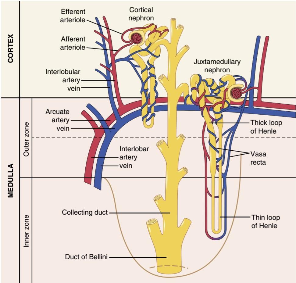
Figure 26-5. Schematic of relationships between blood vessels and tubular structures and differences between cortical and juxtamedullary nephrons.
شکل 26-5. شماتیک روابط بین رگهای خونی و ساختارهای لوله ای و تفاوت بین نفرونهای قشری و کنار هم.
MICTURITION
Micturition is the process whereby the urinary bladder empties when it becomes filled. This process involves two main steps. First, the bladder fills progressively until the tension in its walls rises above a threshold level. This tension elicits the second step, which is a nervous reflex called the micturition reflex that empties the bladder or, if this fails, at least causes a conscious desire to urinate. Although the micturition reflex is an autonomic spinal cord reflex, it can also be inhibited or facilitated by centers in the cerebral cortex or brain stem.
مدفوع
شیردهی فرآیندی است که طی آن مثانه پس از پر شدن تخلیه میشود. این فرآیند شامل دو مرحله اصلی است. ابتدا مثانه به تدریج پر میشود تا زمانی که کشش در دیوارههای آن از سطح آستانه بالاتر رود. این تنش مرحله دوم را ایجاد میکند، که یک رفلکس عصبی به نام رفلکس ادرار است که مثانه را تخلیه میکند یا در صورت عدم موفقیت، حداقل باعث میل آگاهانه برای ادرار میشود. اگرچه رفلکس ادرار یک رفلکس خودمختار نخاع است، اما میتواند توسط مراکزی در قشر مغز یا ساقه مغز مهار یا تسهیل شود.
PHYSIOLOGIC ANATOMY OF THE BLADDER
The urinary bladder, shown in Figure 26-6, is a smooth muscle chamber composed of two main parts: (1) the body, which is the major part of the bladder in which urine collects; and (2) the neck, which is a funnel-shaped extension of the body, passing inferiorly and anteriorly into the urogenital triangle and connecting with the urethra. The lower part of the bladder neck is also called the posterior urethra because of its relationship to the urethra.
آناتومیفیزیولوژیکی مثانه
مثانه، که در شکل 26-6 نشان داده شده است، یک محفظه ماهیچه صاف است که از دو قسمت اصلی تشکیل شده است: (1) بدن، که قسمت اصلی مثانه است که ادرار در آن جمع میشود. و (2) گردن که امتداد قیفی شکل بدن است که از پایین و جلو به داخل مثلث ادراری تناسلی عبور کرده و به مجرای ادرار متصل میشود. قسمت پایینی گردن مثانه به دلیل ارتباط آن با مجرای ادرار، مجرای ادرار خلفی نیز نامیده میشود.
The smooth muscle of the bladder is called the detrusor muscle. Its muscle fibers extend in all directions and, when contracted, can increase the pressure in the bladder to 40 to 60 mm Hg. Thus, contraction of the detrusor muscle is a major step in emptying the bladder. Smooth muscle cells of the detrusor muscle fuse with one another so that low-resistance electrical pathways exist from one muscle cell to the other. Therefore, an action potential can spread throughout the detrusor muscle, from one muscle cell to the next, to cause contraction of the entire bladder at once.
عضله صاف مثانه را عضله دترسور مینامند. فیبرهای عضلانی آن در همه جهات گسترش مییابند و در صورت انقباض میتوانند فشار مثانه را تا 40 تا 60 میلی متر جیوه افزایش دهند. بنابراین، انقباض عضله دترسور گام مهمیدر تخلیه مثانه است. سلولهای ماهیچه صاف عضله دترسور با یکدیگر ترکیب میشوند به طوری که مسیرهای الکتریکی با مقاومت کم از یک سلول عضلانی به سلول دیگر وجود دارد. بنابراین، یک پتانسیل عمل میتواند در سراسر عضله دترسور، از یک سلول عضلانی به سلول دیگر، گسترش یابد و باعث انقباض کل مثانه به یکباره شود.
On the posterior wall of the bladder, lying immediately above the bladder neck, is a small triangular area called the trigone. At the lowermost apex of the trigone, the bladder neck opens into the posterior urethra, and the two ureters enter the bladder at the uppermost angles of the trigone. The trigone can be identified by the fact that its mucosa, the inner lining of the bladder, is smooth, in contrast to the remaining bladder mucosa, which is folded to form rugae.
در دیواره خلفی مثانه، بلافاصله بالای گردن مثانه، یک ناحیه مثلثی کوچک به نام تریگون وجود دارد. در پایین ترین راس تریگون، گردن مثانه به سمت مجرای ادرار خلفی باز میشود و دو حالب در بالاترین زوایای تریگون وارد مثانه میشوند. تریگون را میتوان با این واقعیت تشخیص داد که مخاط آن، پوشش داخلی مثانه، صاف است، برخلاف مخاط باقیمانده مثانه، که برای تشکیل روگا تا شده است.
Each ureter, as it enters the bladder, courses obliquely through the detrusor muscle and then passes another 1 to 2 centimeters beneath the bladder mucosa before emptying into the bladder.
هر حالب، هنگام ورود به مثانه، به صورت مایل از عضله دترسور عبور میکند و سپس قبل از تخلیه به مثانه، 1 تا 2 سانتی متر دیگر از زیر مخاط مثانه عبور میکند.
The bladder neck (posterior urethra) is 2 to 3 centimeters long, and its wall is composed of detrusor muscle interlaced with a large amount of elastic tissue. The muscle in this area is called the internal sphincter. Its natural tone normally keeps the bladder neck and posterior urethra empty of urine and, therefore, prevents emptying of the bladder until the pressure in the main part of the bladder rises above a critical threshold.
گردن مثانه (پیشابراه خلفی) 2 تا 3 سانتی متر طول دارد و دیواره آن از عضله دترسور در هم آمیخته با مقدار زیادی بافت الاستیک تشکیل شده است. عضله این ناحیه اسفنکتر داخلی نامیده میشود. رنگ طبیعی آن معمولاً گردن مثانه و مجرای ادرار خلفی را خالی از ادرار نگه میدارد و بنابراین تا زمانی که فشار در قسمت اصلی مثانه از آستانه بحرانی بالاتر برود از تخلیه مثانه جلوگیری میکند.
Beyond the posterior urethra, the urethra passes through the urogenital diaphragm, which contains a layer of muscle called the external sphincter of the bladder. This muscle is a voluntary skeletal muscle, in contrast to the muscle of the bladder body and bladder neck, which is entirely smooth muscle. The external sphincter muscle is under voluntary control of the nervous system and can be used to consciously prevent urination, even when involuntary controls are attempting to empty the bladder.
فراتر از مجرای ادرار خلفی، مجرای ادرار از دیافراگم ادراری تناسلی عبور میکند که حاوی لایه ای از عضله به نام اسفنکتر خارجی مثانه است. این عضله یک عضله اسکلتی ارادی است، برخلاف عضله بدن مثانه و گردن مثانه که کاملاً ماهیچه صاف است. عضله اسفنکتر خارجی تحت کنترل ارادی سیستم عصبی است و میتوان از آن برای جلوگیری آگاهانه از ادرار استفاده کرد، حتی زمانی که کنترلهای غیرارادی سعی در تخلیه مثانه دارند.
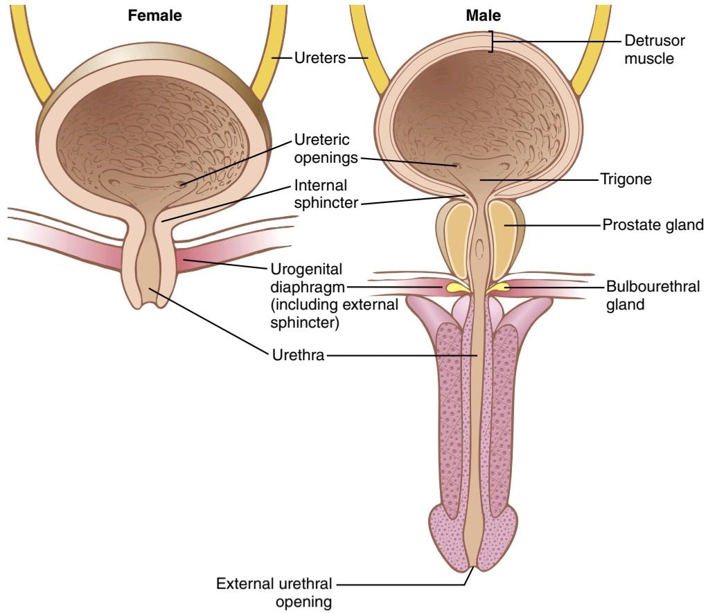
Figure 26-6. Anatomy of the urinary bladder and urethra in males and females.
شکل 26-6. آناتومیمثانه و مجرای ادرار در مردان و زنان.
Innervation of the Bladder. The principal nerve supply of the bladder is via the pelvic nerves, which connect with the spinal cord through the sacral plexus, mainly connecting with cord segments S2 and S3 (Figure 26-7). Coursing through the pelvic nerves are both sensory nerve fibers and motor nerve fibers. The sensory fibers detect the degree of stretch in the bladder wall. Stretch signals from the posterior urethra are especially strong and are mainly responsible for initiating the reflexes that cause bladder emptying.
عصب دهی مثانه. تامین عصب اصلی مثانه از طریق اعصاب لگنی است که از طریق شبکه خاجی به نخاع متصل میشود و عمدتاً با بخشهای S2 و S3 طناب متصل میشود (شکل 26-7). از طریق اعصاب لگن هم رشتههای عصبی حسی و هم رشتههای عصبی حرکتی عبور میکنند. فیبرهای حسی میزان کشش دیواره مثانه را تشخیص میدهند. سیگنالهای کششی از مجرای ادرار خلفی به ویژه قوی هستند و عمدتاً مسئول شروع رفلکسهایی هستند که باعث تخلیه مثانه میشوند.
The motor nerves transmitted in the pelvic nerves are parasympathetic fibers. These fibers terminate on ganglion cells located in the wall of the bladder. Short post- ganglionic nerves then innervate the detrusor muscle.
اعصاب حرکتی منتقل شده در اعصاب لگن رشتههای پاراسمپاتیک هستند. این رشتهها به سلولهای گانگلیونی واقع در دیواره مثانه ختم میشوند. اعصاب کوتاه پس از گانگلیونی سپس عضله دترسور را عصب دهی میکنند.
In addition to the pelvic nerves, two other types of innervation are important in bladder function. Most important are the skeletal motor fibers transmitted through the pudendal nerve to the external bladder sphincter. These fibers are somatic nerve fibers that innervate and control the voluntary skeletal muscle of the sphincter. Also, the bladder receives sympathetic innervation from the sympathetic chain through the hypogastric nerves, connecting mainly with the L2 segment of the spinal cord. These sympathetic fibers stimulate mainly the blood vessels and have little to do with bladder contraction. Some sensory nerve fibers also pass via the sympathetic nerves and may be important in the sensation of fullness and, in some cases, pain.
علاوه بر اعصاب لگن، دو نوع دیگر عصب دهی در عملکرد مثانه مهم هستند. مهمترین آنها فیبرهای حرکتی اسکلتی هستند که از طریق عصب پودندال به اسفنکتر خارجی مثانه منتقل میشوند. این رشتهها رشتههای عصبی جسمانی هستند که عضله اسکلتی ارادی اسفنکتر را عصب دهی و کنترل میکنند. همچنین، مثانه عصب سمپاتیک را از زنجیره سمپاتیک از طریق اعصاب هیپوگاستر دریافت میکند، که عمدتا با بخش L2 نخاع متصل میشود. این رشتههای سمپاتیک عمدتاً رگهای خونی را تحریک میکنند و ارتباط چندانی با انقباض مثانه ندارند. برخی از رشتههای عصبی حسی نیز از طریق اعصاب سمپاتیک عبور میکنند و ممکن است در احساس پری و در برخی موارد درد مهم باشند.

Figure 26-7. Innervation of the urinary bladder.
شکل 26-7. عصب دهی مثانه.
URINE TRANSPORT FROM THE KIDNEYS THROUGH THE URETERS AND INTO THE BLADDER
Urine that is expelled from the bladder has essentially the same composition as fluid flowing out of the collecting ducts. There are no significant changes in the composition of urine as it flows through the renal calyces and ureters to the bladder.
انتقال ادرار از کلیهها از طریق حالب و به مثانه
ادراری که از مثانه خارج میشود، اساساً ترکیبی مشابه مایعی دارد که از مجاری جمع کننده خارج میشود. هیچ تغییر قابل توجهی در ترکیب ادرار وجود ندارد زیرا از طریق کالیسها و حالبهای کلیه به مثانه جریان مییابد.
Urine flowing from the collecting ducts into the renal calyces stretches the calyces and increases their inherent pacemaker activity, which in turn initiates peristaltic contractions that spread to the renal pelvis and then down- ward along the length of the ureter, thereby forcing urine from the renal pelvis toward the bladder. In adults, the ureters are normally 25 to 35 centimeters (10-14 inches) long.
ادراری که از مجاری جمعآوری به داخل کالیسهای کلیوی جریان مییابد، کالیسها را کشیده و فعالیت ضربانساز ذاتی آنها را افزایش میدهد، که به نوبه خود باعث شروع انقباضات پریستالتیک میشود که به لگن کلیه و سپس در طول حالب به سمت پایین گسترش مییابد، در نتیجه ادرار را از لگنچه کلیه به سمت مثانه مجبور میکند. در بزرگسالان، حالبها به طور معمول 25 تا 35 سانتی متر (10-14 اینچ) طول دارند.
The walls of the ureters contain smooth muscle and are innervated by sympathetic and parasympathetic nerves, as well as by an intramural plexus of neurons and nerve fibers that extends along the entire length of the ureters. As with other visceral smooth muscle, peristaltic contractions in the ureter are enhanced by parasympathetic stimulation and inhibited by sympathetic stimulation.
دیواره حالبها حاوی عضله صاف است و توسط اعصاب سمپاتیک و پاراسمپاتیک و همچنین توسط یک شبکه درون دیواری از نورونها و رشتههای عصبی که در تمام طول حالبها امتداد دارد عصب دهی میشود. مانند سایر عضلات صاف احشایی، انقباضات پریستالتیک در حالب با تحریک پاراسمپاتیک افزایش یافته و با تحریک سمپاتیک مهار میشود.
The ureters enter the bladder through the detrusor muscle in the trigone region of the bladder, as shown in Figure 26-6. Normally, the ureters course obliquely for several centimeters through the bladder wall. The normal tone of the detrusor muscle in the bladder wall tends to compress the ureter, thereby preventing backflow (reflux) of urine from the bladder when pressure builds up in the bladder during micturition or bladder compression. Each peristaltic wave along the ureter increases the pressure within the ureter so that the region passing through the bladder wall opens and allows urine to flow into the bladder.
حالبها همانطور که در شکل 26-6 نشان داده شده است از طریق عضله دترسور در ناحیه تریگون مثانه وارد مثانه میشوند. به طور معمول، حالبها به مدت چند سانتی متر به صورت مورب از دیواره مثانه عبور میکنند. تون نرمال عضله دترسور در دیواره مثانه تمایل دارد حالب را فشرده کند و در نتیجه از برگشت (رفلاکس) ادرار از مثانه در هنگام افزایش فشار در مثانه در حین اجابت مزاج یا فشرده شدن مثانه جلوگیری میکند. هر موج پریستالتیک در طول حالب فشار داخل حالب را افزایش میدهد به طوری که ناحیه ای که از دیواره مثانه میگذرد باز میشود و اجازه میدهد ادرار به داخل مثانه جریان یابد.
In some people, the distance that the ureter courses through the bladder wall is less than normal, and thus contraction of the bladder during micturition does not always lead to complete occlusion of the ureter. As a result, some of the urine in the bladder is propelled backward into the ureter, a condition called vesicoureteral reflux. Such reflux can lead to enlargement of the ureters and, if severe, can increase the pressure in the renal calyces and structures of the renal medulla, causing damage to these regions.
در برخی از افراد فاصله ای که حالب از دیواره مثانه عبور میکند کمتر از حد طبیعی است و بنابراین انقباض مثانه در حین دفع ادرار همیشه منجر به انسداد کامل حالب نمیشود. در نتیجه، مقداری از ادرار موجود در مثانه به سمت عقب به داخل حالب هدایت میشود، وضعیتی که به آن رفلاکس مثانه میگویند. چنین ریفلاکسی میتواند منجر به بزرگ شدن حالبها شود و در صورت شدید، فشار را در کالیسها و ساختارهای مدولای کلیه افزایش داده و باعث آسیب به این نواحی شود.
Pain Sensation in the Ureters and the Ureterorenal Reflex. The ureters are extensively supplied with pain nerve fibers. When a ureter becomes blocked (e.g., by a ureteral stone), intense reflex constriction occurs, which is associated with severe pain. Also, the pain impulses cause a sympathetic reflex back to the kidney to constrict the renal arterioles, thereby decreasing urine output from the kidney. This effect is called the ureterorenal reflex and is important for attenuating flow of fluid into the pelvis of a kidney with a blocked ureter.
احساس درد در حالبها و رفلکس حالب. حالبها به طور گسترده ای با رشتههای عصبی درد تامین میشوند. هنگامیکه حالب مسدود میشود (مثلاً توسط سنگ حالب)، انقباض شدید رفلکس رخ میدهد که با درد شدید همراه است. همچنین، تکانههای درد باعث ایجاد رفلکس سمپاتیک به کلیه میشود تا شریانهای کلیوی را منقبض کند و در نتیجه خروجی ادرار از کلیه را کاهش دهد. این اثر، رفلکس حالب – ادراری نامیده میشود و برای کاهش جریان مایع به داخل لگن کلیه با حالب مسدود شده مهم است.
Filling of the Bladder and Bladder Wall Tone-the Cystometrogram
Figure 26-8 shows the approximate changes in intravesicular pressure as the bladder fills with urine. When there is no urine in the bladder, the intravesicular pressure is about 0, but by the time 30 to 50 milliliters of urine have collected, the pressure rises to 5 to 10 centimeters of water. Additional urine-200 to 300 milliliters- can collect with only a small additional rise in pressure; this constant level of pressure is caused by intrinsic tone of the bladder wall. Beyond 300 to 400 milliliters, collection of more urine in the bladder causes the pressure to rise rapidly.
پر کردن مثانه و تون دیواره مثانه – سیستومتروگرام
شکل 26-8 تغییرات تقریبی فشار داخل وزیکولی را با پر شدن مثانه از ادرار نشان میدهد. هنگامیکه ادرار در مثانه وجود ندارد، فشار داخل مثانه حدود 0 است، اما زمانی که 30 تا 50 میلی لیتر ادرار جمع شود، فشار به 5 تا 10 سانتی متر آب میرسد. ادرار اضافی – 200 تا 300 میلی لیتر – میتواند تنها با یک افزایش کوچک اضافی در فشار جمع شود. این سطح ثابت فشار ناشی از تون درونی دیواره مثانه است. بیش از 300 تا 400 میلی لیتر، جمع آوری ادرار بیشتر در مثانه باعث افزایش سریع فشار میشود.
Superimposed on the tonic pressure changes during filling of the bladder are periodic acute increases in pressure that last from a few seconds to more than 1 minute. The pressure peaks may rise only a few centimeters of water or may rise to more than 100 centimeters of water. These pressure peaks are called micturition waves in the cystometrogram and are caused by the micturition reflex.
بر تغییرات فشار تونیک در طی پر شدن مثانه، افزایش حاد دوره ای فشار که از چند ثانیه تا بیش از 1 دقیقه طول میکشد، اضافه میشود. پیکهای فشار ممکن است فقط چند سانتی متر از آب بالا برود یا ممکن است به بیش از 100 سانتی متر آب برسد. این پیکهای فشار در سیستومتروگرام امواج دفعی نامیده میشوند و در اثر رفلکس ادرار ایجاد میشوند.

Figure 26-8. A normal cystometrogram, showing also acute pressure waves (dashed spikes) caused by micturition reflexes.
شکل 26-8. سیستومتروگرام نرمال، همچنین امواج فشار حاد (خوشههای بریده بریده) ناشی از رفلکسهای دفع ادرار را نشان میدهد.
MICTURITION REFLEX
Referring again to Figure 26-8, one can see that as the bladder fills, many superimposed micturition contractions begin to appear, as shown by the dashed spikes. These are the result of a stretch reflex initiated by sensory stretch receptors in the bladder wall, especially by the receptors in the posterior urethra when this area begins to fill with urine at the higher bladder pressures. Sensory signals from the bladder stretch receptors are conducted to the sacral segments of the cord through the pelvic nerves and then reflexively back again to the bladder through the parasympathetic nerve fibers by way of these same nerves.
رفلکس ادرار
با مراجعه مجدد به شکل 26-8، میتوان دید که با پر شدن مثانه، بسیاری از انقباضات مدفوع روی هم ظاهر میشوند، همانطور که توسط میخهای بریده بریده نشان داده شده است. اینها نتیجه یک رفلکس کششی هستند که توسط گیرندههای کشش حسی در دیواره مثانه، به ویژه توسط گیرندههای مجرای ادرار خلفی زمانی که این ناحیه شروع به پر شدن از ادرار در فشارهای بالاتر مثانه میکند، آغاز میشود. سیگنالهای حسی از گیرندههای کشش مثانه از طریق اعصاب لگنی به بخشهای خاجی طناب منتقل میشوند و سپس از طریق همین اعصاب مجدداً از طریق رشتههای عصبی پاراسمپاتیک به مثانه بازمیگردند.
When the bladder is only partially filled, these micturition contractions usually relax spontaneously after a fraction of a minute, the detrusor muscles stop contracting, and pressure falls back to the baseline. As the bladder continues to fill, the micturition reflexes become more frequent and cause greater contractions of the detrusor muscle.
هنگامیکه مثانه فقط تا حدی پر است، این انقباضات دفع ادرار معمولاً پس از کسری از دقیقه به طور خود به خود شل میشوند، ماهیچههای دترسور منقبض میشوند و فشار به سطح پایه باز میگردد. با ادامه پر شدن مثانه، رفلکسهای دفع ادرار بیشتر میشوند و باعث انقباضات بیشتر عضله دترسور میشوند.
Once a micturition reflex begins, it is self-regenerative. That is, initial contraction of the bladder activates the stretch receptors to cause a greater increase in sensory impulses from the bladder and posterior urethra, which causes a further increase in reflex contraction of the bladder; thus, the cycle is repeated again and again until the bladder has reached a strong degree of contraction. Then, after a few seconds to more than a minute, the self- regenerative reflex begins to fatigue, and the regenerative cycle of the micturition reflex ceases, permitting the bladder to relax.
هنگامیکه یک رفلکس ادرار شروع میشود، خود بازسازی میشود. یعنی انقباض اولیه مثانه گیرندههای کششی را فعال میکند تا باعث افزایش بیشتر تکانههای حسی از مثانه و مجرای ادرار خلفی شود که باعث افزایش بیشتر انقباض رفلکس مثانه میشود. بنابراین، این چرخه بارها و بارها تکرار میشود تا زمانی که مثانه به درجه قوی از انقباض برسد. سپس، پس از چند ثانیه تا بیش از یک دقیقه، رفلکس خود بازسازی شروع به خستگی میکند و چرخه بازسازی رفلکس ادرار متوقف میشود و به مثانه اجازه میدهد تا آرام شود.
Thus, the micturition reflex is a single complete cycle of the following: (1) progressive and rapid increase of pressure; (2) a period of sustained pressure; and (3) return of the pressure to the basal tone of the bladder. Once a micturition reflex has occurred but has not succeeded in emptying the bladder, the nervous elements of this reflex usually remain in an inhibited state for a few minutes to 1 hour or more before another micturition reflex occurs. As the bladder becomes filled more and more, micturition reflexes occur more and more often and powerfully.
بنابراین، رفلکس ادرار یک چرخه کامل از موارد زیر است: (1) افزایش تدریجی و سریع فشار. (2) یک دوره فشار پایدار؛ و (3) بازگشت فشار به تون پایه مثانه. هنگامیکه یک رفلکس ادرار رخ میدهد اما موفق به تخلیه مثانه نشده است، عناصر عصبی این رفلکس معمولاً برای چند دقیقه تا 1 ساعت یا بیشتر در حالت مهار شده باقی میمانند قبل از اینکه رفلکس ادراری دیگری رخ دهد. همانطور که مثانه بیشتر و بیشتر پر میشود، رفلکسهای دفع ادرار بیشتر و بیشتر و قوی تر اتفاق میافتد.
Once the micturition reflex becomes powerful enough, it causes another reflex, which passes through the pudendal nerves to the external sphincter to inhibit it. If this inhibition is more potent in the brain than the voluntary constrictor signals to the external sphincter, urination will occur. If not, urination will not occur until the bladder fills still further, and the micturition reflex becomes more powerful.
هنگامیکه رفلکس ادرار به اندازه کافی قوی شد، باعث ایجاد رفلکس دیگری میشود که از طریق اعصاب پودندال به اسفنکتر خارجی میگذرد تا آن را مهار کند. اگر این مهار در مغز بیشتر از سیگنالهای منقبض کننده اختیاری به اسفنکتر خارجی باشد، ادرار رخ میدهد. در غیر این صورت، تا زمانی که مثانه بیشتر پر نشود و رفلکس ادرار قوی تر شود، ادرار رخ نمیدهد.
Facilitation or Inhibition of Micturition by the Brain. The micturition reflex is an autonomic spinal cord reflex, but it can be inhibited or facilitated by centers in the brain. These centers include the following: (1) strong facilitative and inhibitory centers in the brain stem, located mainly in the pons; and (2) several centers located in the cerebral cortex that are mainly inhibitory but can become excitatory.
تسهیل یا مهار ادرار توسط مغز. رفلکس ادرار یک بازتاب خودکار نخاعی است، اما میتواند توسط مراکزی در مغز مهار یا تسهیل شود. این مراکز شامل موارد زیر است: (1) مراکز تسهیل کننده و بازدارنده قوی در ساقه مغز که عمدتاً در پونز قرار دارند. و (2) چندین مرکز واقع در قشر مغز که عمدتاً مهاری هستند اما میتوانند تحریک کننده شوند.
The micturition reflex is the basic cause of micturition, but the higher centers normally exert final control of micturition, as follows:
رفلکس مدفوع علت اصلی اجابت مزاج است، اما مراکز بالاتر معمولاً کنترل نهایی دفع ادرار را به شرح زیر اعمال میکنند:
1. The higher centers keep the micturition reflex partially inhibited, except when micturition is desired.
1. مراکز بالاتر رفلکس ادرار را تا حدی مهار میکنند، مگر در مواقعی که ادرار مورد نظر باشد.
2. The higher centers can prevent micturition, even if the micturition reflex occurs, by tonic contraction of the external bladder sphincter until a convenient time presents itself.
2. مراکز بالاتر میتوانند با انقباض تونیک اسفنکتر خارجی مثانه تا زمانی که زمان مناسبی به وجود آید، از دفع ادرار جلوگیری کنند، حتی اگر رفلکس ادرار رخ دهد.
3. When it is time to urinate, the cortical centers can facilitate the sacral micturition centers to help initiate a micturition reflex and, at the same time, inhibit the external urinary sphincter so that urination can occur.
3. هنگامیکه زمان ادرار فرا میرسد، مراکز قشری میتوانند مراکز ادراری خاجی را تسهیل کنند تا به شروع رفلکس ادرار کمک کنند و در عین حال، اسفنکتر خارجی ادرار را مهار کنند تا ادرار رخ دهد.
Voluntary urination is usually initiated in the following way. First, a person voluntarily contracts the abdominal muscles, which increases pressure in the bladder and allows extra urine to enter the bladder neck and posterior urethra under pressure, thus stretching their walls. This action stimulates the stretch receptors, which excites the micturition reflex and simultaneously inhibits the external urethral sphincter. Ordinarily, all the urine will be emptied, with rarely more than 5 to 10 milliliters left in the bladder.
ادرار ارادی معمولاً به روش زیر شروع میشود. ابتدا، فرد به طور داوطلبانه عضلات شکم را منقبض میکند که باعث افزایش فشار در مثانه میشود و اجازه میدهد ادرار اضافی تحت فشار وارد گردن مثانه و مجرای ادرار خلفی شود و در نتیجه دیوارههای آنها کشیده شود. این عمل گیرندههای کششی را تحریک میکند که رفلکس ادرار را تحریک میکند و به طور همزمان اسفنکتر خارجی مجرای ادرار را مهار میکند. به طور معمول، تمام ادرار تخلیه میشود و به ندرت بیش از 5 تا 10 میلی لیتر در مثانه باقی میماند.
Abnormalities of Micturition
Atonic Bladder and Incontinence Caused by Destruction of Sensory Nerve Fibers. Micturition reflex contraction cannot occur if the sensory nerve fibers from the bladder to the spinal cord are destroyed, thereby preventing transmission of stretch signals from the bladder. When this happens, a person loses bladder control, despite intact efferent fibers from the cord to the bladder and despite intact neurogenic connections within the brain. Instead of emptying periodically, the bladder fills to capacity and overflows a few drops at a time through the urethra. This occurrence is called over- flow incontinence.
ناهنجاریهای دفع ادرار
مثانه آتونیک و بی اختیاری ناشی از تخریب رشتههای عصبی حسی. اگر رشتههای عصبی حسی از مثانه به نخاع از بین بروند، انقباض رفلکس ادرار اتفاق نمیافتد و در نتیجه از انتقال سیگنالهای کششی از مثانه جلوگیری میشود. هنگامیکه این اتفاق میافتد، با وجود فیبرهای وابران دست نخورده از بند ناف به مثانه و با وجود اتصالات عصبی دست نخورده در مغز، کنترل مثانه را از دست میدهد. به جای تخلیه دوره ای، مثانه تا حد ظرفیت پر میشود و هر بار چند قطره از مجرای ادرار سرریز میشود. این اتفاق بی اختیاری سرریز نامیده میشود.
A common cause of atonic bladder is crush injury to the sacral region of the spinal cord. Certain diseases can also cause damage to the dorsal root nerve fibers that enter the spinal cord. For example, syphilis can cause constrictive fibrosis around the dorsal root nerve fibers, destroying them. This condition is called tabes dorsalis, and the resulting bladder condition is called tabetic bladder.
یکی از علل شایع مثانه آتونیک آسیب له شدن ناحیه خاجی نخاع است. برخی از بیماریها همچنین میتوانند باعث آسیب به رشتههای عصبی ریشه پشتی شوند که وارد نخاع میشوند. به عنوان مثال، سیفلیس میتواند باعث فیبروز انقباضی در اطراف رشتههای عصبی ریشه پشتی شود و آنها را از بین ببرد. به این حالت تبس دورسالیس و وضعیت مثانه حاصله را مثانه تبتیک میگویند.
Automatic Bladder Caused by Spinal Cord Damage Above the Sacral Region. If the spinal cord is damaged above the sacral region but the sacral cord segments are still intact, typical micturition reflexes can still occur. However, they are no longer controlled by the brain. During the first few days to several weeks after the damage to the cord has occurred, the micturition reflexes are suppressed because of the state of spinal shock caused by the sudden loss of facilitative impulses from the brain stem and cerebrum. However, if the bladder is emptied periodically by catheterization to prevent bladder injury caused by overstretching of the bladder, the excitability of the micturition reflex gradually increases until typical micturition reflexes return; then, periodic (but unannounced) bladder emptying occurs.
مثانه خودکار ناشی از آسیب نخاع در بالای ناحیه خاجی. اگر طناب نخاعی در بالای ناحیه خاجی آسیب دیده باشد اما بخشهای طناب خاجی هنوز دست نخورده باشند، رفلکسهای معمولی ادرار همچنان ممکن است رخ دهد. با این حال، آنها دیگر توسط مغز کنترل نمیشوند. در طی چند روز اول تا چند هفته پس از آسیب به بند ناف، رفلکسهای دفع ادرار به دلیل حالت شوک نخاعی ناشی از از دست دادن ناگهانی تکانههای تسهیل کننده از ساقه مغز و مخ، سرکوب میشوند. با این حال، اگر مثانه بهطور دورهای با کاتتریزاسیون برای جلوگیری از آسیب مثانه ناشی از کشیدگی بیش از حد مثانه تخلیه شود، تحریکپذیری رفلکس ادرار به تدریج افزایش مییابد تا زمانی که رفلکسهای ادراری معمولی بازگردند. سپس، تخلیه مثانه دوره ای (اما اعلام نشده) رخ میدهد.
Uninhibited Neurogenic Bladder Caused by Lack of Inhibitory Signals From the Brain. Another abnormality of micturition is the so-called uninhibited neurogenic bladder, which results in frequent and relatively uncontrolled micturition. This condition derives from partial damage in the spinal cord or the brain stem that interrupts most of the inhibitory signals. Therefore, facilitative impulses passing continually down the cord keep the sacral centers so excitable that even a small quantity of urine elicits an uncontrollable micturition reflex, thereby promoting frequent urination.
مثانه نوروژنیک مهار نشده ناشی از فقدان سیگنالهای بازدارنده از مغز. یکی دیگر از ناهنجاریهای دفع ادرار مثانه نوروژنیک مهار نشده است که منجر به دفع مکرر و نسبتاً کنترل نشده میشود. این وضعیت ناشی از آسیب جزئی در نخاع یا ساقه مغز است که بیشتر سیگنالهای بازدارنده را قطع میکند. بنابراین، تکانههای تسهیلکننده که به طور مداوم از بند ناف عبور میکنند، مراکز خاجی را چنان تحریکپذیر نگه میدارند که حتی مقدار کمیادرار باعث ایجاد رفلکس ادرار غیرقابل کنترل میشود و در نتیجه باعث تکرر ادرار میشود.
URINE FORMATION RESULTS FROM GLOMERULAR FILTRATION, TUBULAR REABSORPTION, AND TUBULAR SECRETION
The rates at which different substances are excreted in the urine represent the sum of three renal processes, shown in Figure 26-9: (1) glomerular filtration of substances in the blood; (2) reabsorption of substances from the renal tubules into the blood; and (3) secretion of substances from the blood into the renal tubules as follows: Urinary excretion rate = Filtration rate – Reabsorption rate + Secretion rate
نتایج تشکیل ادرار از فیلتراسیون گلومرولی، بازجذب لولهای و ترشح لولهای
سرعت دفع مواد مختلف در ادرار مجموع سه فرآیند کلیوی را نشان میدهد که در شکل 26-9 نشان داده شده است: (1) فیلتراسیون گلومرولی مواد در خون. (2) بازجذب مواد از لولههای کلیوی به خون. و (3) ترشح مواد از خون به داخل لولههای کلیوی به شرح زیر است: سرعت دفع ادرار = سرعت فیلتراسیون – سرعت بازجذب + سرعت ترشح
Urine formation begins when a large amount of fluid that is virtually free of protein is filtered from the glomerular capillaries into Bowman’s capsule. Most substances in the plasma, except for proteins, are freely filtered, so their concentration in the glomerular filtrate in Bowman’s capsule is almost the same as in the plasma. As filtered fluid leaves Bowman’s capsule and passes through the tubules, it is modified by reabsorption of water and specific solutes back into the blood or by secretion of other substances from the peritubular capillaries into the tubules.
تشکیل ادرار زمانی آغاز میشود که مقدار زیادی مایع که عملاً عاری از پروتئین است، از مویرگهای گلومرولی به داخل کپسول بومن فیلتر شود. اکثر مواد موجود در پلاسما، به جز پروتئینها، آزادانه فیلتر میشوند، بنابراین غلظت آنها در مایع فیلتر شده گلومرولی در کپسول بومن تقریباً مشابه پلاسما است. هنگامیکه مایع فیلتر شده کپسول بومن را ترک میکند و از توبولها عبور میکند، با بازجذب آب و املاح خاص به داخل خون یا با ترشح سایر مواد از مویرگهای اطراف توبول به داخل توبولها، تغییر شکل میدهد.
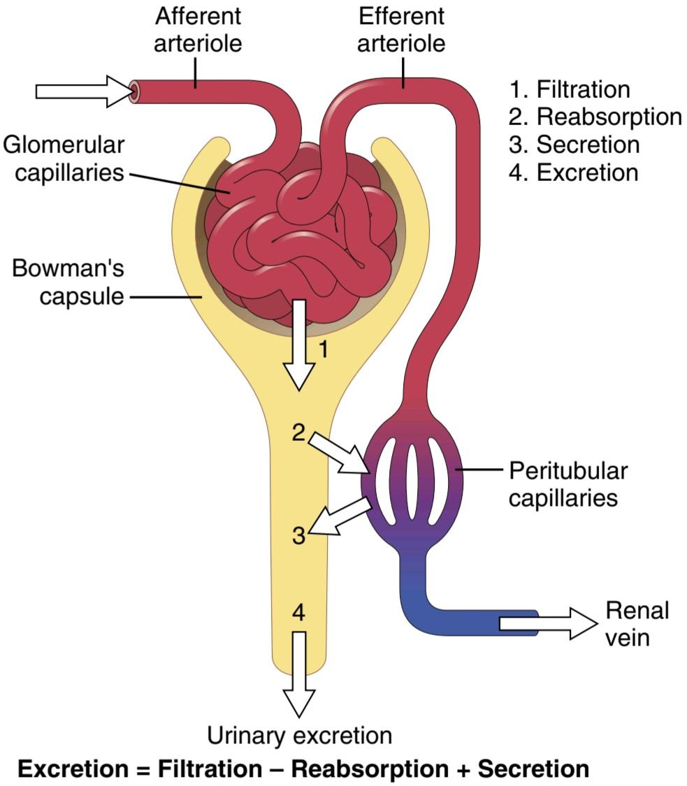
Figure 26-9. Basic kidney processes that determine the composition of the urine. The urinary excretion rate of a substance is equal to the rate at which the substance is filtered minus its reabsorption rate plus the rate at which it is secreted from the peritubular capillary blood into the tubules.
شکل 26-9. فرآیندهای کلیوی اساسی که ترکیب ادرار را تعیین میکند. سرعت دفع ادرار یک ماده برابر است با سرعت فیلتر شدن ماده منهای نرخ بازجذب آن به اضافه سرعت ترشح آن از خون مویرگی دور لوله ای به داخل لولهها.
Figure 26-10 shows the renal handling of four hypothetical substances. The substance shown in panel A is freely filtered by the glomerular capillaries but is neither reabsorbed nor secreted. Therefore, its excretion rate is equal to the rate at which it was filtered. Certain waste products in the body, such as creatinine, are handled by the kidneys in this manner, allowing excretion of essentially all that is filtered.
شکل 26-10 عملکرد کلیوی چهار ماده فرضی را نشان میدهد. ماده نشان داده شده در پانل A آزادانه توسط مویرگهای گلومرولی فیلتر میشود اما نه دوباره جذب میشود و نه ترشح میشود. بنابراین میزان دفع آن برابر با سرعتی است که فیلتر شده است. برخی از مواد زائد در بدن، مانند کراتینین، توسط کلیهها به این روش مدیریت میشوند و اجازه میدهند اساساً تمام موارد فیلتر شده دفع شوند.
In panel B, the substance is freely filtered but is also partly reabsorbed from the tubules back into the blood. Therefore, the rate of urinary excretion is less than the rate of filtration at the glomerular capillaries. In this case, the excretion rate is calculated as the filtration rate minus the reabsorption rate. This pattern is typical for many of the electrolytes of the body, such as sodium and chloride ions.
در پانل B، این ماده آزادانه فیلتر میشود، اما همچنین تا حدی از لولهها دوباره به خون بازجذب میشود. بنابراین، سرعت دفع ادرار کمتر از سرعت فیلتراسیون در مویرگهای گلومرولی است. در این حالت میزان دفع به صورت نرخ فیلتراسیون منهای نرخ بازجذب محاسبه میشود. این الگو برای بسیاری از الکترولیتهای بدن مانند یونهای سدیم و کلرید معمولی است.
In panel C, the substance is freely filtered at the glomerular capillaries but is not excreted into the urine because all the filtered substance is reabsorbed from the tubules back into the blood. This pattern occurs for some of the nutritional substances in the blood, such as amino acids and glucose, allowing them to be conserved in the body fluids.
در پانل C، ماده آزادانه در مویرگهای گلومرولی فیلتر میشود اما به ادرار دفع نمیشود، زیرا تمام ماده فیلتر شده از لولهها دوباره به داخل خون جذب میشود. این الگو برای برخی از مواد مغذی موجود در خون مانند اسیدهای آمینه و گلوکز رخ میدهد و به آنها اجازه میدهد در مایعات بدن حفظ شوند.
The substance in panel D is freely filtered at the glomerular capillaries and is not reabsorbed, but additional quantities of this substance are secreted from the peritubular capillary blood into the renal tubules. This pattern often occurs for organic acids and bases, permitting them to be cleared from the blood rapidly and excreted in large amounts in the urine. The excretion rate in this case is calculated as filtration rate plus tubular secretion rate.
ماده موجود در پانل D آزادانه در مویرگهای گلومرولی فیلتر میشود و دوباره جذب نمیشود، اما مقادیر بیشتری از این ماده از خون مویرگی اطراف لوله به داخل لولههای کلیوی ترشح میشود. این الگو اغلب برای اسیدها و بازهای آلی رخ میدهد و به آنها اجازه میدهد تا به سرعت از خون پاک شوند و در مقادیر زیاد از طریق ادرار دفع شوند. میزان دفع در این مورد به صورت نرخ فیلتراسیون به اضافه میزان ترشح لوله ای محاسبه میشود.
For each substance in the plasma, a particular combination of filtration, reabsorption, and secretion occurs. The rate at which the substance is excreted in the urine depends on the relative rates of these three basic renal processes.
برای هر ماده در پلاسما، ترکیب خاصی از فیلتراسیون، بازجذب و ترشح رخ میدهد. سرعت دفع این ماده از طریق ادرار به سرعت نسبی این سه فرآیند کلیوی بستگی دارد.

Figure 26-10. Renal handling of four hypothetical substances. A, The substance is freely filtered but not reabsorbed. B, The substance is freely filtered, but part of the filtered load is reabsorbed back in the blood. C, The substance is freely filtered but is not excreted in the urine because all the filtered substance is reabsorbed from the tubules into the blood. D, The substance is freely filtered and is not reabsorbed but is secreted from the peritubular capillary blood into the renal tubules.
شکل 26-10. جابجایی کلیه چهار ماده فرضی. الف، این ماده آزادانه فیلتر میشود اما دوباره جذب نمیشود. ب، این ماده آزادانه فیلتر میشود، اما بخشی از بار فیلتر شده دوباره در خون جذب میشود. ج، این ماده آزادانه فیلتر میشود اما از طریق ادرار دفع نمیشود زیرا تمام ماده فیلتر شده از لولهها به خون بازجذب میشود. د، این ماده آزادانه فیلتر میشود و دوباره جذب نمیشود، اما از خون مویرگی اطراف لوله به داخل لولههای کلیوی ترشح میشود.
FILTRATION, REABSORPTION, AND SECRETION OF DIFFERENT SUBSTANCES
In general, tubular reabsorption is quantitatively more important than tubular secretion in the formation of urine, but secretion plays an important role in determining the amounts of potassium and hydrogen ions and a few other substances that are excreted in the urine. Most substances that must be cleared from the blood, especially the end products of metabolism, such as urea, creatinine, uric acid, and urates, are poorly reabsorbed and are there- fore excreted in large amounts in the urine. Certain foreign substances and drugs are also poorly reabsorbed but, in addition, are secreted from the blood into the tubules, so their excretion rates are high. Conversely, electrolytes, such as sodium ions, chloride ions, and bicarbonate ions, are highly reabsorbed, so only small amounts appear in the urine. Certain nutritional substances, such as amino acids and glucose, are completely reabsorbed from the tubules and do not appear in the urine, even though large amounts are filtered by the glomerular capillaries.
فیلتراسیون، بازجذب و ترشح مواد مختلف
به طور کلی، بازجذب لولهای از نظر کمیمهمتر از ترشح لولهای در تشکیل ادرار است، اما ترشح نقش مهمیدر تعیین مقدار یونهای پتاسیم و هیدروژن و چند ماده دیگر دارد که در ادرار دفع میشوند. بیشتر موادی که باید از خون پاک شوند، به ویژه محصولات نهایی متابولیسم، مانند اوره، کراتینین، اسید اوریک و اوراتها، بازجذب ضعیفی دارند و بنابراین به مقدار زیادی از طریق ادرار دفع میشوند. برخی از مواد خارجی و داروها نیز بازجذب ضعیفی دارند، اما علاوه بر این، از خون به داخل لولهها ترشح میشوند، بنابراین میزان دفع آنها بالاست. برعکس، الکترولیتها مانند یونهای سدیم، یونهای کلرید و یونهای بیکربنات به شدت بازجذب میشوند، بنابراین فقط مقادیر کمیدر ادرار ظاهر میشوند. برخی از مواد مغذی مانند اسیدهای آمینه و گلوکز به طور کامل از لولهها بازجذب میشوند و در ادرار ظاهر نمیشوند، حتی اگر مقادیر زیادی توسط مویرگهای گلومرولی فیلتر شوند.
Each of the processes-glomerular filtration, tubular reabsorption, and tubular secretion-is regulated according to the needs of the body. For example, when there is excess sodium in the body, the rate at which sodium is filtered usually increases slightly, and a smaller fraction of the filtered sodium is reabsorbed, causing increased urinary excretion of sodium.
هر یک از فرآیندها – فیلتراسیون گلومرولی، بازجذب لوله ای، و ترشح لوله ای – بر اساس نیاز بدن تنظیم میشود. به عنوان مثال، هنگامیکه سدیم اضافی در بدن وجود دارد، سرعت فیلتر شدن سدیم معمولاً کمیافزایش مییابد و بخش کوچکتری از سدیم فیلتر شده دوباره جذب میشود و باعث افزایش دفع سدیم از طریق ادرار میشود.
For most substances, the rates of filtration and reabsorption are extremely large relative to the rates of excretion. Therefore, even slight changes of filtration or reabsorption can lead to relatively large changes in renal excretion. For example, an increase in the glomerular filtration rate (GFR) of only 10% (from 180 to 198 L/day) would raise the urine volume by 13-fold (from 1.5 to 19.5 L/day) if tubular reabsorption remained constant. In reality, changes in glomerular filtration and tubular reabsorption usually act in a coordinated manner to produce the necessary changes in renal excretion.
برای اکثر مواد، سرعت فیلتراسیون و بازجذب نسبت به سرعت دفع بسیار زیاد است. بنابراین، حتی تغییرات جزئی فیلتراسیون یا بازجذب میتواند منجر به تغییرات نسبتاً زیادی در دفع کلیوی شود. برای مثال، افزایش نرخ فیلتراسیون گلومرولی (GFR) تنها به میزان 10 درصد (از 180 به 198 لیتر در روز) اگر بازجذب لوله ای ثابت بماند، حجم ادرار را 13 برابر (از 1.5 به 19.5 لیتر در روز) افزایش میدهد. در واقع، تغییرات در فیلتراسیون گلومرولی و بازجذب لوله ای معمولاً به صورت هماهنگ عمل میکنند تا تغییرات لازم را در دفع کلیوی ایجاد کنند.
Why Are Large Amounts of Solutes Filtered and Then Reabsorbed by the Kidneys? One might question the wisdom of filtering such large amounts of water and solutes and then reabsorbing most of these substances. One advantage of a high GFR is that it allows the kidneys to remove waste products rapidly from the body that depend mainly on glomerular filtration for their excretion. Most waste products are poorly reabsorbed by the tubules and, therefore, depend on a high GFR for effective removal from the body.
چرا مقادیر زیادی از املاح فیلتر شده و سپس توسط کلیهها بازجذب میشوند؟ ممکن است حکمت فیلتر کردن چنین مقادیر زیادی آب و املاح و سپس جذب مجدد بیشتر این مواد مورد سوال قرار گیرد. یکی از مزایای GFR بالا این است که به کلیهها اجازه میدهد تا مواد زائد را به سرعت از بدن خارج کنند که عمدتاً برای دفع آنها به فیلتراسیون گلومرولی بستگی دارد. اکثر مواد زائد به خوبی توسط لولهها بازجذب میشوند و بنابراین برای حذف موثر از بدن به GFR بالا وابسته هستند.
A second advantage of a high GFR is that it allows all the body fluids to be filtered and processed by the kidneys many times each day. Because the entire plasma volume is only about 3 liters, whereas the GFR is about 180 L/ day, the entire plasma can be filtered and processed about 60 times each day. This high GFR allows the kidneys to control the volume and composition of the body fluids precisely and rapidly.
مزیت دوم GFR بالا این است که اجازه میدهد تمام مایعات بدن بارها در روز توسط کلیهها فیلتر و پردازش شوند. از آنجایی که کل حجم پلاسما فقط حدود 3 لیتر است، در حالی که GFR حدود 180 لیتر در روز است، کل پلاسما را میتوان حدود 60 بار در روز فیلتر و پردازش کرد. این GFR بالا به کلیهها اجازه میدهد تا حجم و ترکیب مایعات بدن را به طور دقیق و سریع کنترل کنند.
کلیک کنید «Bibliography: فهرست کتب مربوطه»
see the bibliography for chapter 27 to 32
🚀 با ما همراه شوید!
تازهترین مطالب و آموزشهای مغز و اعصاب را از دست ندهید. با فالو کردن کانال تلگرام آیندهنگاران مغز، از ما حمایت کنید!


