
فیزیولوژی پزشکی گایتون و هال؛ تنظیم محفظههای مایع بدن: مایعات خارج سلولی و داخل سلولی؛ ادم


دعای مطالعه [ نمایش ]
بِسْمِ الله الرَّحْمنِ الرَّحیمِ
اَللّهُمَّ اَخْرِجْنى مِنْ ظُلُماتِ الْوَهْمِ
خدايا مرا بيرون آور از تاريكىهاى وهم،
وَ اَكْرِمْنى بِنُورِ الْفَهْمِ
و به نور فهم گرامى ام بدار،
اَللّهُمَّ افْتَحْ عَلَيْنا اَبْوابَ رَحْمَتِكَ
خدايا درهاى رحمتت را به روى ما بگشا،
وَانْشُرْ عَلَيْنا خَزائِنَ عُلُومِكَ بِرَحْمَتِكَ يا اَرْحَمَ الرّاحِمينَ
و خزانههاى علومت را بر ما باز كن به امید رحمتت اى مهربانترين مهربانان.
کتاب «فیزیولوژی پزشکی گایتون و هال» بهعنوان یکی از جامعترین و معتبرترین منابع در حوزه علوم پزشکی، همچنان مرجع کلیدی برای درک عملکرد پیچیده بدن انسان است. این اثر با تکیه بر تازهترین پژوهشها و توضیحات دقیق از سازوکارهای فیزیولوژیک، پلی میان علوم پایه پزشکی و کاربردهای بالینی ایجاد میکند و نقشی بیبدیل در آموزش، پژوهش و ارتقای دانش سلامت ایفا مینماید.
ترجمه دقیق و علمی این شاهکار توسط برند علمی آیندهنگاران مغز به مدیریت داریوش طاهری، دسترسی فارسیزبانان به مرزهای نوین دانش فیزیولوژی را ممکن ساخته و رسالتی علمی برای ارتقای آموزش پزشکی، فهم عمیقتر سازوکارهای بدن و توسعه روشهای نوین در حوزه سلامت فراهم آورده است.
» کتاب فیزیولوژی پزشکی گایتون و هال
» » فصل ۲۵: تنظیم محفظههای مایع بدن: مایعات خارج سلولی و داخل سلولی؛ ادم
در حال ویرایش
» Guyton and Hall Textbook of Medical Physiology
»» CHAPTER 25: Regulation of Body Fluid Compartments: Extracellular and Intracellular Fluids; Edema

The maintenance of a relatively constant volume and stable composition of the body fluids is essential for homeostasis. Some of the most common and important problems in clinical medicine arise because of abnormalities in the control systems that maintain this relative constancy of the body fluids. In this chapter and in the following chapters on the kidneys, we discuss overall regulation of body fluid volume, constituents of the extracellular fluid, acid-base balance, and control of fluid exchange between extracellular and intracellular compartments.
حفظ حجم نسبتاً ثابت و ترکیب پایدار مایعات بدن برای هموستاز ضروری است. برخی از رایج ترین و مهم ترین مشکلات در پزشکی بالینی به دلیل وجود ناهنجاری در سیستم های کنترلی است که این ثبات نسبی مایعات بدن را حفظ می کند. در این فصل و در فصلهای بعدی درباره کلیهها، به تنظیم کلی حجم مایع بدن، اجزای تشکیلدهنده مایع خارج سلولی، تعادل اسید و باز و کنترل تبادل مایع بین بخشهای خارج سلولی و درون سلولی میپردازیم.
FLUID INTAKE AND OUTPUT ARE BALANCED DURING STEADY-STATE CONDITIONS
The relative constancy of the body fluids is remarkable, because there is continuous exchange of fluid and solutes with the external environment, as well as within the different body compartments. For example, fluid added to the body is highly variable and must be carefully matched by an equal output of water from the body to prevent body fluid volumes from increasing or decreasing.
مصرف و خروجی مایعات در شرایط پایدار متعادل هستند
ثبات نسبی مایعات بدن قابل توجه است، زیرا تبادل مداوم مایعات و املاح با محیط خارجی و همچنین در قسمت های مختلف بدن وجود دارد. به عنوان مثال، مایع اضافه شده به بدن بسیار متغیر است و باید به دقت با خروجی برابر آب از بدن مطابقت داده شود تا از افزایش یا کاهش حجم مایعات بدن جلوگیری شود.
DAILY INTAKE OF WATER
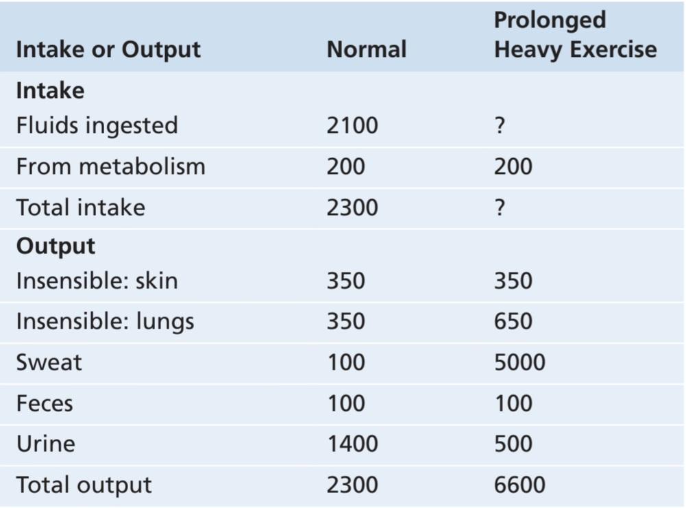
Water is added to the body by two major sources: (1) it is ingested in the form of liquids or water in food, which together normally add about 2100 ml/day to the body fluids; and (2) it is synthesized in the body by oxidation of carbohydrates, adding about 200 ml/day. These mechanisms provide a total water intake of about 2300 ml/day (Table 25-1). However, intake of water is highly variable among different people and even within the same person on different days, depending on climate, habits, and level of physical activity.
مصرف روزانه آب
آب توسط دو منبع اصلی به بدن اضافه می شود: (1) به شکل مایعات یا آب موجود در غذا وارد بدن می شود که معمولاً روزانه 2100 میلی لیتر به مایعات بدن اضافه می شود. و (2) با اکسیداسیون کربوهیدرات ها در بدن سنتز می شود و حدود 200 میلی لیتر در روز به آن اضافه می شود. این مکانیسم ها کل آب مصرفی حدود 2300 میلی لیتر در روز را فراهم می کنند (جدول 25-1). با این حال، بسته به آب و هوا، عادات و سطح فعالیت بدنی، مصرف آب در بین افراد مختلف و حتی در یک فرد در روزهای مختلف بسیار متغیر است.
Table 25-1 Daily Intake and Output of Water (ml/day)
جدول 25-1 میزان مصرف و دفع روزانه آب (میلی لیتر در روز)
DAILY LOSS OF BODY WATER
Insensible Water Loss. Some water losses cannot be precisely regulated. For example, humans experience continuous water loss by evaporation from the respiratory tract and diffusion through the skin, which together account for about 700 ml/day of water loss under normal conditions. This loss is termed insensible water loss because we are not consciously aware of it, even though it occurs continually in all living people.
از دست دادن روزانه آب بدن
از دست دادن غیر محسوس آب. برخی از تلفات آب را نمی توان به طور دقیق تنظیم کرد. به عنوان مثال، انسان با تبخیر از مجرای تنفسی و انتشار از طریق پوست، از دست دادن مداوم آب را تجربه می کند که مجموعاً حدود 700 میلی لیتر در روز از دست دادن آب را در شرایط عادی تشکیل می دهد. این از دست دادن آب نامحسوس نامیده می شود زیرا ما آگاهانه از آن آگاه نیستیم، حتی اگر به طور مداوم در همه افراد زنده رخ دهد.
Insensible water loss through the skin occurs independently of sweating and is present even in people who are born without sweat glands; the average water loss by diffusion through the skin is about 300 to 400 ml/day. This loss is minimized by the cholesterol-filled, cornified layer of the skin, which provides a barrier against excessive loss by diffusion. When the cornified layer becomes denuded, as occurs with extensive burns, the rate of evaporation can increase as much as 10-fold, to 3 to 5 L/ day. For this reason, persons with burns must be given large amounts of fluid, usually intravenously, to balance fluid loss.
از دست دادن غیر محسوس آب از طریق پوست مستقل از تعریق رخ می دهد و حتی در افرادی که بدون غدد عرق متولد می شوند نیز وجود دارد. متوسط از دست دادن آب از طریق انتشار در پوست حدود 300 تا 400 میلی لیتر در روز است. این ضایعات توسط لایهای پر از کلسترول و کورنیه شده پوست به حداقل میرسد، که مانعی در برابر از دست دادن بیش از حد توسط انتشار ایجاد میکند. هنگامی که لایه کورنیفیک شده برهنه می شود، همانطور که با سوختگی های گسترده اتفاق می افتد، سرعت تبخیر می تواند تا 10 برابر افزایش یابد و به 3 تا 5 لیتر در روز برسد. به همین دلیل، به افراد مبتلا به سوختگی باید مقادیر زیادی مایعات، معمولاً به صورت داخل وریدی، داده شود تا از دست دادن مایعات متعادل شود.
Insensible water loss through the respiratory tract normally averages about 300 to 400 ml/day. As air enters the respiratory tract, it becomes saturated with moisture to a vapor pressure of about 47 mm Hg before it is expelled. Because the vapor pressure of the inspired air is usually less than 47 mm Hg, water is continuously lost through the lungs with respiration. In cold weather, the atmospheric vapor pressure decreases to nearly 0, causing an even greater loss of water from the lungs as the temperature decreases. This process explains the dry feeling in the respiratory passages in cold weather.
از دست دادن غیر محسوس آب از طریق دستگاه تنفسی به طور معمول حدود 300 تا 400 میلی لیتر در روز است. با ورود هوا به مجرای تنفسی، قبل از خروج، با رطوبت اشباع می شود و فشار بخار آن به حدود 47 میلی متر جیوه می رسد. از آنجایی که فشار بخار هوای دمیده شده معمولاً کمتر از 47 میلی متر جیوه است، آب به طور مداوم از طریق ریه ها با تنفس از دست می رود. در هوای سرد، فشار بخار اتمسفر به نزدیک به 0 کاهش می یابد و با کاهش دما باعث از دست دادن حتی بیشتر آب از ریه ها می شود. این فرآیند احساس خشکی در مجاری تنفسی را در هوای سرد توضیح می دهد.
Fluid Loss in Sweat. The amount of water lost by sweating is highly variable, depending on physical activity and environmental temperature. The volume of sweat normally is about 100 ml/day, but in very hot weather or during heavy exercise, fluid loss in sweat occasionally increases to 1 to 2 L/hour. This fluid loss would rapidly deplete the body fluids if intake were not also increased by activating the thirst mechanism, as discussed in Chapter 29.
از دست دادن مایعات در عرق. مقدار آب از دست رفته در اثر تعریق بسته به فعالیت بدنی و دمای محیط بسیار متغیر است. حجم عرق به طور معمول حدود 100 میلی لیتر در روز است، اما در هوای بسیار گرم یا در حین ورزش سنگین، از دست دادن مایعات در عرق گهگاه به 1 تا 2 لیتر در ساعت افزایش می یابد. این از دست دادن مایعات به سرعت باعث تخلیه مایعات بدن می شود، اگر میزان مصرف با فعال کردن مکانیسم تشنگی، همانطور که در فصل 29 بحث شد، افزایش نیابد.
Water Loss in Feces. Only a small amount of water (100 ml/day) normally is lost in the feces. This loss can increase to several liters a day in people with severe diarrhea. Therefore, severe diarrhea can be life-threatening if not corrected within a few days.
از دست دادن آب در مدفوع. فقط مقدار کمی آب (100 میلی لیتر در روز) به طور معمول در مدفوع از دست می رود. این کاهش در افراد مبتلا به اسهال شدید می تواند به چندین لیتر در روز افزایش یابد. بنابراین، اسهال شدید اگر ظرف چند روز اصلاح نشود، می تواند تهدید کننده زندگی باشد.
Water Loss by the Kidneys. The remaining water loss from the body occurs in the urine excreted by the kidneys. Multiple mechanisms control the rate of urine excretion. The most important means whereby the body maintains balance between water intake and output, as well as a balance between intake and output of most electrolytes in the body, is by controlling the rate at which the kidneys excrete these substances. For example, urine volume can be as low as 0.5 L/day in a dehydrated person or as high as 20 L/day in a person who has been drinking tremendous amounts of water.
از دست دادن آب توسط کلیه ها. آب باقیمانده از بدن از طریق ادرار دفع شده توسط کلیه ها اتفاق می افتد. مکانیسم های متعددی سرعت دفع ادرار را کنترل می کنند. مهمترین وسیله ای که بدن به وسیله آن تعادل بین دریافت و خروجی آب و همچنین تعادل بین دریافت و خروجی اکثر الکترولیت ها در بدن را حفظ می کند، کنترل سرعت دفع این مواد از کلیه ها است. به عنوان مثال، حجم ادرار در یک فرد کمآب میتواند به 0.5 لیتر در روز یا در فردی که مقادیر زیادی آب مینوشیده به 20 لیتر در روز برسد.
This variability of intake is also true for most of the electrolytes of the body, such as sodium, chloride, and potassium. In some people, sodium intake may be as low as 20 mEq/day, whereas in others, sodium intake may be as high as 300 to 500 mEq/day. The kidneys have the task of adjusting the excretion rate of water and electrolytes to match the intake of these substances precisely, as well as compensating for excessive losses of fluids and electrolytes that occur in certain disease states. In Chapters 26 through 32, we discuss the mechanisms that allow the kidneys to perform these remarkable tasks.
این تنوع در مصرف برای اکثر الکترولیت های بدن مانند سدیم، کلرید و پتاسیم نیز صادق است. در برخی افراد، مصرف سدیم ممکن است تا 20 میلی اکی والان در روز باشد، در حالی که در برخی دیگر، مصرف سدیم ممکن است به 300 تا 500 میلی اکی والان در روز برسد. کلیه ها وظیفه دارند میزان دفع آب و الکترولیت ها را به گونه ای تنظیم کنند که دقیقاً با دریافت این مواد مطابقت داشته باشد و همچنین اتلاف بیش از حد مایعات و الکترولیت ها را که در شرایط بیماری خاص اتفاق می افتد، جبران کند. در فصلهای 26 تا 32، مکانیسمهایی را مورد بحث قرار میدهیم که به کلیهها اجازه میدهد تا این وظایف قابل توجه را انجام دهند.
BODY FLUID COMPARTMENTS
The total body fluid is distributed mainly between two compartments, the extracellular fluid and the intracellular fluid (Figure 25-1). The extracellular fluid is divided into the interstitial fluid and the blood plasma.
محفظه های مایعات بدن
کل مایع بدن عمدتاً بین دو بخش، مایع خارج سلولی و مایع درون سلولی توزیع می شود (شکل 25-1). مایع خارج سلولی به مایع بینابینی و پلاسمای خون تقسیم می شود.
There is another small compartment of fluid that is referred to as transcellular fluid. This compartment includes fluid in the synovial, peritoneal, pericardial, and intraocular spaces, as well as the cerebrospinal fluid; it is usually considered to be a specialized type of extracellular fluid, although in some cases its composition may differ markedly from that of the plasma or interstitial fluid. All the transcellular fluids together constitute about 1 to 2 liters.
محفظه کوچک دیگری از مایع وجود دارد که به آن مایع بین سلولی می گویند. این بخش شامل مایع در فضاهای سینوویال، صفاقی، پریکارد و داخل چشم و همچنین مایع مغزی نخاعی است. معمولاً به عنوان یک نوع تخصصی مایع خارج سلولی در نظر گرفته می شود، اگرچه در برخی موارد ممکن است ترکیب آن به طور قابل توجهی با پلاسما یا مایع بین بافتی متفاوت باشد. تمام مایعات بین سلولی با هم حدود 1 تا 2 لیتر را تشکیل می دهند.
In a 70-kg adult man, the total body water is about 60% of the body weight, or about 42 liters. This percentage depends on age, sex, and degree of obesity. As a person grows older, the percentage of total body weight that is fluid gradually decreases. This decrease is due in part to the fact that aging is usually associated with an increased percentage of the body weight being fat, which decreases the percentage of water in the body.
در یک مرد بالغ 70 کیلوگرمی، کل آب بدن حدود 60 درصد وزن بدن یا حدود 42 لیتر است. این درصد به سن، جنس و درجه چاقی بستگی دارد. با افزایش سن، درصد وزن کل بدن که مایع است به تدریج کاهش می یابد. این کاهش تا حدی به دلیل
این واقعیت که افزایش سن معمولا با افزایش درصد چربی در وزن بدن همراه است که باعث کاهش درصد آب در بدن می شود.
Because women normally have a greater percentage of body fat compared with men, their total body water averages about 50% of the body weight. In premature and newborn babies, the total body water ranges from 70% to 75% of body weight. Therefore, when discussing average body fluid compartments, we should realize that variations exist, depending on age, sex, and percentage of body fat.
از آنجایی که زنان به طور معمول درصد بیشتری از چربی بدن در مقایسه با مردان دارند، کل آب بدن آنها به طور متوسط حدود 50 درصد وزن بدن است. در نوزادان نارس و تازه متولد شده، کل آب بدن بین 70 تا 75 درصد وزن بدن متغیر است. بنابراین، هنگام بحث در مورد میانگین قسمت های مایع بدن، باید متوجه باشیم که بسته به سن، جنس و درصد چربی بدن، تغییراتی وجود دارد.
In many other countries, the average body weight (and fat mass) has increased rapidly during the past 30 years. The average body weight for adult men older than 20 years in the United States is estimated to be approximately 88.8 kg (~196 pounds), and for adult women it is 77.4 kg (~170 pounds). Therefore, data discussed for an average 70-kg man in this and other chapters would need to be adjusted accordingly when considering body fluid compartments in most people.
در بسیاری از کشورهای دیگر، میانگین وزن بدن (و توده چربی) طی 30 سال گذشته به سرعت افزایش یافته است. میانگین وزن بدن مردان بالغ بالای 20 سال در ایالات متحده تقریباً 88.8 کیلوگرم (~ 196 پوند) و برای زنان بالغ 77.4 کیلوگرم (~170 پوند) تخمین زده می شود. بنابراین، دادههایی که برای یک مرد متوسط 70 کیلوگرمی در این فصل و سایر فصلها مورد بحث قرار گرفتهاند، باید بر این اساس هنگام در نظر گرفتن محفظههای مایع بدن در بیشتر افراد تنظیم شوند.

Figure 25-1. Summary of body fluid regulation, including the major body fluid compartments and the membranes that separate these compartments. The values shown are for an average 70-kg man.
شکل 25-1. خلاصه ای از تنظیم مایعات بدن، از جمله بخش های اصلی مایع بدن و غشاهایی که این بخش ها را از هم جدا می کنند. مقادیر نشان داده شده برای یک مرد متوسط 70 کیلوگرمی است.
INTRACELLULAR FLUID COMPARTMENT
About 28 of the 42 liters of fluid in the body are inside the trillions of cells and is collectively called the intracellular fluid. Thus, the intracellular fluid constitutes about 40% of the total body weight in an “average” person.
محفظه مایعات داخل سلولی
حدود 28 لیتر از 42 لیتر مایع بدن در داخل تریلیون ها سلول قرار دارد و در مجموع مایع درون سلولی نامیده می شود. بنابراین، مایع درون سلولی حدود 40 درصد از کل وزن بدن یک فرد “متوسط” را تشکیل می دهد.
The fluid of each cell contains its individual mixture of different constituents, but the concentrations of these substances are similar from one cell to another. In fact, the composition of cell fluids is remarkably similar, even in different animals, ranging from the most primitive microorganisms to humans. For this reason, the intracellular fluid of all the different cells together is considered to be one large fluid compartment.
مایع هر سلول حاوی ترکیب جداگانه ای از اجزای مختلف است، اما غلظت این مواد از سلولی به سلول دیگر مشابه است. در واقع، ترکیب مایعات سلولی حتی در حیوانات مختلف، از ابتداییترین میکروارگانیسمها گرفته تا انسان، به طرز قابل ملاحظهای مشابه است. به همین دلیل، مایع درون سلولی تمام سلول های مختلف با هم به عنوان یک محفظه مایع بزرگ در نظر گرفته می شود.
EXTRACELLULAR FLUID COMPARTMENT
All the fluids outside the cells are collectively called the extracellular fluid. Together these fluids account for about 20% of the body weight, or about 14 liters in a 70-kg man. The two largest compartments of the extracellular fluid are the interstitial fluid, which makes up more than three-fourths (11 liters) of the extracellular fluid, and the plasma, which makes up almost one-fourth of the extracellular fluid, or about 3 liters. The plasma is the noncellular part of the blood; it exchanges substances continuously with the interstitial fluid through the pores of the capillary membranes. These pores are highly permeable to almost all solutes in the extracellular fluid, except the proteins. Therefore, the extracellular fluids are constantly mixing, so the plasma and interstitial fluids have about the same composition, except for proteins, which have a higher concentration in the plasma.
محفظه مایعات خارج سلولی
تمام مایعات خارج از سلول ها در مجموع مایع خارج سلولی نامیده می شوند. این مایعات روی هم حدود 20 درصد وزن بدن یا حدود 14 لیتر در یک مرد 70 کیلوگرمی را تشکیل می دهند. دو بخش بزرگ مایع خارج سلولی عبارتند از مایع بینابینی که بیش از سه چهارم (11 لیتر) مایع خارج سلولی را تشکیل می دهد و پلاسما که تقریباً یک چهارم مایع خارج سلولی یا حدود 3 لیتر را تشکیل می دهد. پلاسما قسمت غیر سلولی خون است. از طریق منافذ غشاهای مویرگی به طور مداوم مواد را با مایع بینابینی مبادله می کند. این منافذ تقریباً به تمام املاح موجود در مایع خارج سلولی به جز پروتئین ها نفوذپذیری بالایی دارند. بنابراین، مایعات خارج سلولی دائماً در حال مخلوط شدن هستند، بنابراین پلاسما و مایعات بینابینی تقریباً ترکیب یکسانی دارند، به جز پروتئین ها که غلظت بیشتری در پلاسما دارند.
BLOOD VOLUME
Blood contains extracellular fluid (the fluid in plasma) and intracellular fluid (the fluid in the red blood cells). However, blood is considered to be a separate fluid compartment because it is contained in a chamber of its own, the circulatory system. The blood volume is especially important in the control of cardiovascular dynamics.
حجم خون
خون حاوی مایع خارج سلولی (مایع موجود در پلاسما) و مایع درون سلولی (مایع موجود در گلبول های قرمز) است. با این حال، خون به عنوان یک محفظه مایع جداگانه در نظر گرفته می شود، زیرا در یک محفظه مخصوص به خود، یعنی سیستم گردش خون قرار دارد. حجم خون به ویژه در کنترل پویایی قلب و عروق بسیار مهم است.
The average blood volume of adults is about 7% of body weight, or about 5 liters. About 60% of the blood is plasma and 40% is red blood cells, but these percentages can vary considerably in different people, depending on sex, weight, and other factors.
متوسط حجم خون بزرگسالان حدود 7 درصد وزن بدن یا حدود 5 لیتر است. حدود 60 درصد خون را پلاسما و 40 درصد را گلبول های قرمز تشکیل می دهد، اما این درصدها در افراد مختلف بسته به جنس، وزن و عوامل دیگر می تواند به طور قابل توجهی متفاوت باشد.
Hematocrit (Packed Red Blood Cell Volume). The hematocrit is the fraction of the blood composed of red blood cells, as determined by centrifuging blood in a hematocrit tube until the cells become tightly packed in the bottom of the tube. Because the centrifuge does not completely pack the red blood cells together, about 3% to 4% of the plasma remains entrapped among the cells, and the true hematocrit is only about 96% of the measured hematocrit.
هماتوکریت (حجم گلبول قرمز بسته). هماتوکریت کسری از خون است که از گلبول های قرمز تشکیل شده است که با سانتریفیوژ کردن خون در لوله هماتوکریت تا زمانی که سلول ها در ته لوله محکم بسته شوند تعیین می شود. از آنجایی که سانتریفیوژ گلبول های قرمز را به طور کامل در کنار هم قرار نمی دهد، حدود 3 تا 4 درصد از پلاسما در بین سلول ها محبوس می ماند و هماتوکریت واقعی تنها حدود 96 درصد هماتوکریت اندازه گیری شده است.
In men, the measured hematocrit is normally about 0.40, and in women, it is about 0.36. In persons with severe anemia, the hematocrit may fall as low as 0.10, a value that is barely sufficient to sustain life. Conversely, in persons with some conditions, excessive production of red blood cells occurs, resulting in polycythemia. In these persons, the hematocrit can rise to 0.65.
در مردان، هماتوکریت اندازه گیری شده به طور معمول حدود 0.40 و در زنان حدود 0.36 است. در افراد مبتلا به کم خونی شدید، هماتوکریت ممکن است تا 0.10 کاهش یابد، مقداری که به سختی برای حفظ زندگی کافی است. برعکس، در افراد مبتلا به برخی شرایط، تولید بیش از حد گلبول های قرمز خون رخ می دهد که منجر به پلی سیتمی می شود. در این افراد، هماتوکریت می تواند تا 0.65 افزایش یابد.
CONSTITUENTS OF EXTRACELLULAR AND INTRACELLULAR FLUIDS
Comparisons of the composition of the extracellular fluid, including the plasma and interstitial fluid, and the intracellular fluid are shown in Figures 25-2 and 25-3 and in Table 25-2.
اجزای تشکیل دهنده مایعات خارج سلولی و درون سلولی
مقایسه ترکیب مایع خارج سلولی شامل پلاسما و مایع بین بافتی و مایع داخل سلولی در شکل های 25-2 و 25-3 و جدول 25-2 نشان داده شده است.

Figure 25-2. Major cations and anions of the intracellular and extracellular fluids. The concentrations of Ca2+ and Mg2+ represent the sum of these two ions. The concentrations shown represent the total of free ions and complexed ions.
شکل 25-2. کاتیون ها و آنیون های اصلی مایعات درون سلولی و خارج سلولی. غلظت های +Ca2 و +Mg2 نشان دهنده مجموع این دو یون است. غلظت های نشان داده شده مجموع یون های آزاد و یون های کمپلکس را نشان می دهد.
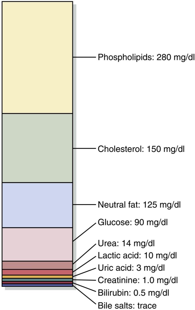
Figure 25-3. Nonelectrolytes of the plasma.
شکل 25-3. غیر الکترولیت های پلاسما
Similar Ionic Composition of Plasma and Interstitial Fluid
Because the plasma and interstitial fluid are separated only by highly permeable capillary membranes, their ionic composition is similar. The most important difference between these two compartments is the higher concentration of protein in the plasma; because the capillaries have a low permeability to the plasma proteins, only small amounts of proteins are leaked into the interstitial spaces in most tissues.
ترکیب یونی مشابه پلاسما و مایع بینابینی
از آنجایی که پلاسما و مایع بینابینی فقط توسط غشاهای مویرگی بسیار نفوذپذیر جدا می شوند، ترکیب یونی آنها مشابه است. مهمترین تفاوت بین این دو بخش، غلظت بالاتر پروتئین در پلاسما است. از آنجایی که مویرگ ها نفوذپذیری پایینی نسبت به پروتئین های پلاسما دارند، فقط مقادیر کمی از پروتئین ها به فضاهای بینابینی در بیشتر بافت ها نشت می کنند.
Because of the Donnan effect, the concentration of positively charged ions (cations) is slightly greater (~2%) in plasma than in interstitial fluid. Plasma proteins have a net negative charge and therefore tend to bind cations such as sodium and potassium ions, thus holding extra amounts of these cations in the plasma, along with the plasma proteins. Conversely, negatively charged ions (anions) tend to have a slightly higher concentration in interstitial fluid compared with plasma because the negative charges of the plasma proteins repel the negatively charged anions. For practical purposes, however, the concentrations of ions in interstitial fluid and plasma are considered to be about equal.
به دلیل اثر دانان، غلظت یونهای با بار مثبت (کاتیونها) در پلاسما کمی بیشتر از مایع بینابینی (~2%) است. پروتئین های پلاسما دارای بار منفی خالص هستند و بنابراین تمایل به اتصال کاتیون هایی مانند یون های سدیم و پتاسیم دارند، بنابراین مقادیر اضافی این کاتیون ها را همراه با پروتئین های پلاسما در پلاسما نگه می دارند. برعکس، یونهای دارای بار منفی (آنیونها) در مقایسه با پلاسما غلظت کمی بالاتر در مایع بینبافتی دارند، زیرا بارهای منفی پروتئینهای پلاسما آنیونهای با بار منفی را دفع میکنند. با این حال، برای اهداف عملی، غلظت یون ها در مایع بینابینی و پلاسما تقریباً برابر در نظر گرفته می شود.
Referring again to Figure 25-2, one can see that the extracellular fluid, including the plasma and interstitial fluid, contains large amounts of sodium and chloride ions and reasonably large amounts of bicarbonate ions but only small quantities of potassium, calcium, magnesium, phosphate, and organic acid ions. The composition of extracellular fluid is carefully regulated by various mechanisms, but especially by the kidneys, as discussed later. This regulation allows the cells to remain continually bathed in a fluid that contains the proper concentration of electrolytes and nutrients for optimal cell function.
با مراجعه مجدد به شکل 2-25، می توان دریافت که مایع خارج سلولی، از جمله پلاسما و مایع بین بافتی، حاوی مقادیر زیادی یون سدیم و کلرید و مقادیر نسبتاً زیادی یون بی کربنات است اما فقط مقادیر کمی پتاسیم، کلسیم، منیزیم، فسفات و اسیدهای آلی دارد. ترکیب مایع خارج سلولی به دقت توسط مکانیسم های مختلف تنظیم می شود، به ویژه توسط کلیه ها، همانطور که بعداً بحث شد. این تنظیم به سلول ها اجازه می دهد تا به طور مداوم در مایعی که حاوی غلظت مناسب الکترولیت ها و مواد مغذی برای عملکرد بهینه سلول است، غوطه ور بمانند.
Table 25-2 Osmolar Substances in Extracellular and Intracellular Fluids
جدول 25-2 مواد اسمولی در سیالات خارج سلولی و درون سلولی

INTRACELLULAR FLUID CONSTITUENTS
The intracellular fluid is separated from the extracellular fluid by a cell membrane that is highly permeable to water but is not permeable to most electrolytes in the body. In contrast to the extracellular fluid, the intracellular fluid. contains only small quantities of sodium and chloride ions and almost no calcium ions. Instead, it contains large amounts of potassium and phosphate ions plus moderate quantities of magnesium and sulfate ions, all of which have low concentrations in the extracellular fluid. Also, cells contain large amounts of protein—almost four times as much as in the plasma.
ترکیبات مایع درون سلولی
مایع درون سلولی توسط یک غشای سلولی که نفوذپذیری بالایی در برابر آب دارد، از مایع خارج سلولی جدا میشود، اما برای اکثر الکترولیتهای بدن قابل نفوذ نیست. بر خلاف مایع خارج سلولی، مایع درون سلولی. حاوی مقدار کمی یون سدیم و کلرید و تقریباً هیچ یون کلسیمی ندارد. در عوض، حاوی مقادیر زیادی یون پتاسیم و فسفات به علاوه مقادیر متوسطی از یونهای منیزیم و سولفات است که همگی غلظتهای پایینی در مایع خارج سلولی دارند. همچنین، سلولها حاوی مقادیر زیادی پروتئین هستند – تقریباً چهار برابر پلاسما.
MEASUREMENT OF BODY FLUID COMPARTMENT VOLUMES- INDICATOR-DILUTION PRINCIPLE
The volume of a fluid compartment in the body can be measured by placing an indicator substance in the com- partment, allowing it to disperse evenly throughout the compartment’s fluid, and then analyzing the extent to which the substance becomes diluted. Figure 25-4 shows this indicator-dilution method of measuring the volume of a fluid compartment. This method is based on the conservation of mass principle, which means that the total mass of a substance after dispersion in the fluid compartment will be the same as the total mass injected into the compartment.
اندازه گیری حجم های محفظه مایعات بدن – شاخص – اصل رقیق سازی
حجم یک محفظه مایع در بدن را میتوان با قرار دادن یک ماده نشانگر در محفظه اندازهگیری کرد و به آن اجازه داد تا به طور مساوی در سراسر مایع محفظه پراکنده شود و سپس میزان رقیق شدن ماده را تجزیه و تحلیل کرد. شکل 25-4 این روش رقیق سازی شاخص را برای اندازه گیری حجم یک محفظه سیال نشان می دهد. این روش بر اساس اصل بقای جرم است، به این معنی که جرم کل یک ماده پس از پراکندگی در محفظه سیال برابر با کل جرم تزریق شده به محفظه خواهد بود.
In the example shown in Figure 25-4, a small amount of dye or other substance contained in the syringe is injected into a chamber, and the substance is allowed to disperse throughout the chamber until it becomes mixed in equal concentrations in all areas. Then a sample of fluid containing the dispersed substance is removed, and the concentration is analyzed chemically, photoelectrically, or by other means. If none of the substance leaks out of the compartment, the total mass of substance in the compartment (Volume B x Concentration B) will equal the total mass of the substance injected (Volume A x Concentration A). By simple rearrangement of the equation, one can calculate the unknown volume of chamber B as follows:
در مثال نشان داده شده در شکل 25-4، مقدار کمی رنگ یا ماده دیگری که در سرنگ وجود دارد به داخل یک محفظه تزریق میشود و اجازه داده میشود تا در سراسر محفظه پراکنده شود تا زمانی که در غلظتهای مساوی در تمام قسمتها مخلوط شود. سپس نمونهای از مایع حاوی ماده پراکنده برداشته میشود و غلظت آن بهصورت شیمیایی، فوتوالکتریک یا به روشهای دیگر آنالیز میشود. اگر هیچ یک از مواد از محفظه نشت نکند، جرم کل ماده در محفظه (حجم B x غلظت B) برابر با جرم کل ماده تزریق شده (حجم A x غلظت A) خواهد بود. با بازآرایی ساده معادله، می توان حجم مجهول محفظه B را به صورت زیر محاسبه کرد:

For this calculation, one needs to know the following: (1) the total amount of substance injected into the chamber (the numerator of the equation); and (2) the concentration of the fluid in the chamber after the substance has been dispersed (the denominator).
برای این محاسبه، باید موارد زیر را بدانیم: (1) مقدار کل ماده تزریق شده به محفظه (شمارگر معادله). و (2) غلظت سیال در محفظه پس از پراکندگی ماده (مخرج).
For example, if 1 milliliter of a solution containing 10 mg/ml of dye is dispersed into chamber B, and the final concentration in the chamber is 0.01 mg/ml of fluid, the unknown volume of the chamber can be calculated as follows:
به عنوان مثال، اگر 1 میلی لیتر از محلول حاوی 10 میلی گرم در میلی لیتر رنگ در محفظه B پراکنده شود و غلظت نهایی در محفظه 0.01 میلی گرم بر میلی لیتر مایع باشد، حجم مجهول محفظه را می توان به صورت زیر محاسبه کرد:

This method can be used to measure the volume of virtually any compartment in the body as long as the following occur: (1) the indicator disperses evenly throughout the compartment; (2) the indicator disperses only in the compartment that is being measured; and (3) the indicator is not metabolized or excreted. If the indicator is metabolized or excreted, correction must be made for loss of the indicator from the body. Several substances can be used to measure the volume of each of the different body fluids.
این روش را می توان برای اندازه گیری حجم تقریباً هر محفظه در بدن تا زمانی که موارد زیر رخ می دهد استفاده کرد: (1) نشانگر به طور مساوی در سراسر محفظه پخش می شود. (2) نشانگر فقط در محفظه ای که اندازه گیری می شود پراکنده می شود. و (3) شاخص متابولیزه یا دفع نمی شود. اگر اندیکاتور متابولیزه یا دفع شود، باید برای از دست دادن اندیکاتور از بدن اصلاح شود. برای اندازه گیری حجم هر یک از مایعات مختلف بدن می توان از چندین ماده استفاده کرد.

Figure 25-4 Indicator-dilution method for measuring fluid volumes.
شکل 25-4 روش رقیقسازی با شاخص برای اندازهگیری حجم مایعات.
DETERMINATION OF VOLUMES OF SPECIFIC BODY FLUID COMPARTMENTS
Measurement of Total Body Water. Radioactive water (tritium, 3H2O) or heavy water (deuterium, 2H2O) can be used to measure total body water. These forms of water mix with the total body water within a few hours after being injected into the blood, and the dilution principle can be used to calculate total body water (Table 25-3). Another substance that has been used to measure total body water is antipyrine, which is very lipidsoluble, rapidly penetrates cell membranes, and distributes uniformly throughout the intracellular and extracellular compartments.
تعیین حجم محفظه های مایعات خاص بدن
اندازه گیری کل آب بدن. برای اندازه گیری کل آب بدن می توان از آب رادیواکتیو (تریتیوم، 3H2O) یا آب سنگین (دوتریوم، 2H2O) استفاده کرد. این اشکال آب با کل آب بدن ظرف چند ساعت پس از تزریق به خون مخلوط میشوند و از اصل رقیقسازی میتوان برای محاسبه کل آب بدن استفاده کرد (جدول 25-3). ماده دیگری که برای اندازه گیری کل آب بدن مورد استفاده قرار می گیرد آنتی پیرین است که بسیار محلول در چربی است، به سرعت در غشای سلولی نفوذ می کند و به طور یکنواخت در قسمت های درون سلولی و خارج سلولی توزیع می شود.
Table 25-3 Measurement of Body Fluid Volumes Volume
جدول 25-3 اندازه گیری حجم مایعات بدن

Measurement of Extracellular Fluid Volume. The volume of extracellular fluid can be estimated using any of several substances that disperse in the plasma and interstitial fluid but do not readily permeate the cell membrane. These include radioactive sodium, radioactive chloride, radioactive iothalamate, thiosulfate ion, and inulin. When any one of these substances is injected into the blood, it usually disperses almost completely throughout the extracellular fluid within 30 to 60 minutes. Some of these substances, however, such as radioactive sodium, may diffuse into the cells in small amounts. Therefore, one frequently speaks of the sodium space or inulin space instead of calling the measurement the true extracellular fluid volume.
اندازه گیری حجم مایع خارج سلولی. حجم مایع خارج سلولی را می توان با استفاده از هر یک از چندین ماده که در پلاسما و مایع بینابینی پراکنده می شوند، اما به راحتی در غشای سلولی نفوذ نمی کنند، تخمین زد. اینها عبارتند از سدیم رادیواکتیو، کلرید رادیواکتیو، یوتالامات رادیواکتیو، یون تیوسولفات و اینولین. هنگامی که هر یک از این مواد به خون تزریق می شود، معمولاً در عرض 30 تا 60 دقیقه تقریباً به طور کامل در مایع خارج سلولی پخش می شود. با این حال، برخی از این مواد، مانند سدیم رادیواکتیو، ممکن است به مقدار کم در سلولها پخش شوند. بنابراین، اغلب از فضای سدیم یا فضای اینولین صحبت می شود به جای اینکه اندازه گیری را حجم واقعی مایع خارج سلولی بنامیم.
Calculation of Intracellular Volume. The intracellular volume cannot be measured directly. However, it can be calculated as follows:
محاسبه حجم داخل سلولی. حجم داخل سلولی را نمی توان به طور مستقیم اندازه گیری کرد. اما می توان آن را به صورت زیر محاسبه کرد:

Measurement of Plasma Volume. Plasma volume can be measured using a substance that does not readily penetrate capillary membranes but remains in the vascular system after injection. One of the most commonly used substances for measuring plasma volume is serum albumin labeled with radioactive iodine (1251-albumin) or with a dye that avidly binds to the plasma proteins, such as Evans blue dye (also called T-1824).
اندازه گیری حجم پلاسما. حجم پلاسما را می توان با استفاده از ماده ای اندازه گیری کرد که به راحتی در غشاهای مویرگی نفوذ نمی کند اما پس از تزریق در سیستم عروقی باقی می ماند. یکی از رایجترین مواد مورد استفاده برای اندازهگیری حجم پلاسما، آلبومین سرم است که با ید رادیواکتیو (آلبومین 1251) یا با رنگی که مشتاقانه به پروتئینهای پلاسما متصل میشود، مانند رنگ آبی Evans (که T-1824 نیز نامیده میشود) برچسبگذاری شده است.
Calculation of Interstitial Fluid Volume. Interstitial fluid volume cannot be measured directly, but it can be calculated as follows:
محاسبه حجم مایع بینابینی. حجم مایع بینابینی را نمی توان مستقیماً اندازه گیری کرد، اما می توان آن را به صورت زیر محاسبه کرد:

Measurement of Blood Volume. If one measures the hematocrit (the fraction of the total blood volume composed of cells) and plasma volume using the methods described earlier, blood volume can also be calculated using the following equation:
اندازه گیری حجم خون. اگر هماتوکریت (کسری از حجم کل خون متشکل از سلول ها) و حجم پلاسما را با استفاده از روش هایی که قبلا توضیح داده شد اندازه گیری کنیم، حجم خون نیز می تواند با استفاده از معادله زیر محاسبه شود:

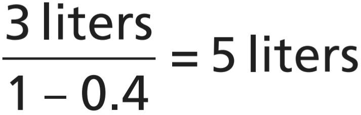
For example, if the plasma volume is 3 liters and hematocrit is 0.40, the total blood volume would be calculated as follows:
به عنوان مثال، اگر حجم پلاسما 3 لیتر و هماتوکریت 0.40 باشد، حجم کل خون به صورت زیر محاسبه می شود:
Another way to measure blood volume is to inject red blood cells that have been labeled with radioactive material into the circulation. After these mix in the circulation, the radioactivity of a mixed blood sample can be measured, and the total blood volume can be calculated using the indicator-dilution principle. One substance that can used to label the red blood cells is radioactive chromium (51Cr), which binds tightly with the red blood cells.
روش دیگر برای اندازه گیری حجم خون تزریق گلبول های قرمز است که با مواد رادیواکتیو برچسب گذاری شده اند به گردش خون. پس از این مخلوط در گردش خون، رادیواکتیویته یک نمونه خون مخلوط را می توان اندازه گیری کرد و حجم کل خون را می توان با استفاده از اصل نشانگر-رقت محاسبه کرد. یکی از موادی که میتوان برای برچسبگذاری گلبولهای قرمز خون استفاده کرد، کروم رادیواکتیو (51Cr) است که محکم با گلبولهای قرمز خون متصل میشود.
FLUID EXCHANGE AND OSMOTIC EQUILIBRIUM BETWEEN INTRACELLULAR AND EXTRACELLULAR FLUID
A frequent problem in treating seriously ill patients is maintaining adequate fluids in one or both of the intracellular and extracellular compartments. As discussed in Chapter 16 and later in this chapter, the relative amounts of extracellular fluid distributed between the plasma and interstitial spaces are determined mainly by the balance of hydrostatic and colloid osmotic forces across the capillary membranes.
تبادل سیالات و تعادل اسموتیک بین سیالات درون سلولی و خارجسلولی
یکی از مشکلات رایج در درمان بیماران شدیداً بیمار، حفظ مایعات کافی در یک یا هر دو بخش درون سلولی و خارج سلولی است. همانطور که در فصل 16 و بعد در این فصل بحث شد، مقادیر نسبی مایع خارج سلولی توزیع شده بین پلاسما و فضاهای بین بافتی عمدتاً توسط تعادل نیروهای اسمزی هیدرواستاتیک و کلوئیدی در سراسر غشاهای مویرگی تعیین می شود.
The distribution of fluid between intracellular and extracellular compartments, in contrast, is determined mainly by the osmotic effect of smaller solutes-especially sodium, chloride, and other electrolytes-acting across the cell membrane. The reason for this is that the cell membranes are highly permeable to water but relatively impermeable to even small ions such as sodium and chloride. Therefore, water moves across the cell membrane rapidly, and the intracellular fluid remains isotonic with the extracellular fluid.
در مقابل، توزیع مایع بین محفظه های درون سلولی و خارج سلولی عمدتاً توسط اثر اسمزی املاح کوچکتر – به ویژه سدیم، کلرید و سایر الکترولیت هایی که در سراسر غشای سلولی عمل می کنند، تعیین می شود. دلیل این امر این است که غشاهای سلولی نسبت به آب بسیار نفوذپذیر هستند اما حتی به یونهای کوچکی مانند سدیم و کلرید نسبتاً غیرقابل نفوذ هستند. بنابراین، آب به سرعت در غشای سلولی حرکت می کند و مایع درون سلولی با مایع خارج سلولی ایزوتونیک باقی می ماند.
In the next section, we discuss the interrelations between intracellular and extracellular fluid volumes and the osmotic factors that can cause shifts of fluid between these two compartments.
در بخش بعدی، ما در مورد روابط متقابل بین حجم مایع درون سلولی و خارج سلولی و عوامل اسمزی که می توانند باعث جابجایی مایع بین این دو بخش شوند، بحث می کنیم.
BASIC PRINCIPLES OF OSMOSIS AND OSMOTIC PRESSURE
The basic principles of osmosis and osmotic pressure were presented in Chapter 4. Therefore, we review here only the most important aspects of these principles as they apply to volume regulation.
اصول اساسی اسمز و فشار اسموتیک
اصول اساسی اسمز و فشار اسمزی در فصل 4 ارائه شد. بنابراین، ما در اینجا تنها مهمترین جنبه های این اصول را که در تنظیم حجم اعمال می شوند، مرور می کنیم.
Because cell membranes are relatively impermeable to most solutes but are highly permeable to water (i.e., they are selectively permeable), whenever there is a higher concentration of solute on one side of the cell membrane, water diffuses across the membrane toward the region of higher solute concentration. Thus, if a solute such as sodium chloride is added to the extracellular fluid, water rapidly diffuses from the cells through the cell membranes into the extracellular fluid until the water concentration on both sides of the membrane becomes equal. Conversely, if a solute such as sodium chloride is removed from the extracellular fluid, water diffuses from the extracellular fluid through the cell membranes and into the cells.
از آنجایی که غشای سلولی نسبت به اکثر املاح نسبتاً نفوذناپذیر هستند اما در برابر آب بسیار نفوذپذیر هستند (یعنی به طور انتخابی نفوذپذیر هستند)، هرگاه غلظت بیشتری از املاح در یک طرف غشای سلولی وجود داشته باشد، آب در سراسر غشاء به سمت منطقه با غلظت املاح بالاتر پخش می شود. بنابراین، اگر یک املاح مانند کلرید سدیم به مایع خارج سلولی اضافه شود، آب به سرعت از سلول ها از طریق غشای سلولی به مایع خارج سلولی پخش می شود تا زمانی که غلظت آب در دو طرف غشاء برابر شود. برعکس، اگر یک املاح مانند کلرید سدیم از مایع خارج سلولی خارج شود، آب از مایع خارج سلولی از طریق غشای سلولی و به داخل سلول ها پخش می شود.
Osmolality and Osmolarity. The osmolal concentration of a solution is called osmolality when the concentration is expressed as osmoles per kilogram of water; it is called osmolarity when it is expressed as osmoles per liter of solution. In dilute solutions such as the body fluids, these two terms can be used almost synonymously because the differences are small. Most of the calculations used clinically and the calculations expressed in the next several chapters are based on osmolarities rather than osmolalities.
اسمولاریته و اسمولاریته. غلظت اسمول یک محلول زمانی اسمولالیته نامیده می شود که غلظت آن به صورت اسمول بر کیلوگرم آب بیان شود. اسمولاریته نامیده می شود که به صورت اسمول در لیتر محلول بیان شود. در محلول های رقیق مانند مایعات بدن، این دو عبارت را می توان تقریباً مترادف به کار برد زیرا تفاوت ها کم است. بیشتر محاسباتی که از نظر بالینی استفاده می شوند و محاسباتی که در چندین فصل بعدی بیان می شوند، بر اساس اسمولاریته ها هستند تا اسمولالیته ها.
Calculation of the Osmolarity and Osmotic Pressure of a Solution. Using the van’t Hoff law, one can calculate the potential osmotic pressure of a solution, assuming that the cell membrane is impermeable to the solute. For example, the osmotic pressure of a 0.9% sodium chloride solution is calculated as follows. A 0.9% solution means that there is 0.9 gram of sodium chloride per 100 milliliters of solution, or 9 g/L. Because the molecular weight of sodium chloride is 58.5 g/mol, the molarity of the solution is 9 g/L divided by 58.5 g/mol, or about 0.154 mol/L. Because each molecule of sodium chloride is equal to 2 osmoles, the osmolarity of the solution is 0.154 x 2, or 0.308 Osm/L. Therefore, the osmolarity of this solution is 308 mOsm/L. The potential osmotic pressure of this solution would therefore be 308 mOsm/L x 19.3 mm Hg/ mOsm/L, or 5944 mm Hg.
محاسبه اسمولاریته و فشار اسمزی یک محلول. با استفاده از قانون وانت هاف، می توان فشار اسمزی بالقوه یک محلول را محاسبه کرد، با این فرض که غشای سلولی نسبت به املاح نفوذ ناپذیر است. به عنوان مثال، فشار اسمزی محلول کلرید سدیم 0.9٪ به صورت زیر محاسبه می شود. محلول 0.9٪ به این معنی است که در هر 100 میلی لیتر محلول 0.9 گرم کلرید سدیم یا 9 گرم در لیتر وجود دارد. از آنجایی که وزن مولکولی کلرید سدیم 58.5 گرم بر مول است، مولاریته محلول 9 گرم در لیتر تقسیم بر 58.5 گرم در مول یا حدود 0.154 مول در لیتر است. از آنجایی که هر مولکول کلرید سدیم برابر با 2 اسمول است، اسمولاریته محلول 0.154 x 2 یا 0.308 Osm/L است. بنابراین اسمولاریته این محلول 308 mOsm/L است. بنابراین فشار اسمزی بالقوه این محلول 308 mOsm/L x 19.3 mm Hg/mOsm/L یا 5944 mm Hg خواهد بود.
This calculation is an approximation, because sodium and chloride ions do not behave entirely independently in solution as a result of interionic attraction between them. One can correct for these deviations from the predictions of van’t Hoff’s law by using a correction factor called the osmotic coefficient. For sodium chloride, the osmotic coefficient is about 0.93. Therefore, the actual osmolarity of a 0.9% sodium chloride solution is 308 x 0.93, or about 286 mOsm/L. For practical reasons, the osmotic coefficients of different solutes are sometimes neglected in determining the osmolarity and osmotic pressures of physiologic solutions.
این محاسبه تقریبی است، زیرا یونهای سدیم و کلرید در محلول بهطور کامل بهطور مستقل رفتار نمیکنند، زیرا در نتیجه جاذبه بین یونی بین آنها وجود دارد. می توان این انحرافات از پیش بینی های قانون وانت هاف را با استفاده از یک ضریب تصحیح به نام ضریب اسمزی تصحیح کرد. برای کلرید سدیم، ضریب اسمزی حدود 0.93 است. بنابراین، اسمولاریته واقعی محلول کلرید سدیم 0.9% 308 x 0.93 یا حدود 286 mOsm/L است. به دلایل عملی، گاهی اوقات ضرایب اسمزی املاح مختلف در تعیین اسمولاریته و فشار اسمزی محلول های فیزیولوژیک نادیده گرفته می شود.
Osmolarity of Body Fluids. Referring back to Table 25-2, note the approximate osmolarity of the various osmotically active substances in plasma, interstitial fluid, and intracellular fluid. About 80% of the total osmolarity of the interstitial fluid and plasma is due to sodium and chloride ions, whereas for intracellular fluid, almost half the osmolarity is due to potassium ions, and the remainder is divided among many other intracellular substances.
اسمولاریته مایعات بدن. با مراجعه به جدول 25-2، اسمولاریته تقریبی مواد مختلف اسمزی فعال در پلاسما، مایع بینابینی و مایع درون سلولی را یادداشت کنید. حدود 80 درصد از کل اسمولاریته مایع بین بافتی و پلاسما به دلیل یون های سدیم و کلرید است، در حالی که برای مایع درون سلولی، تقریباً نیمی از اسمولاریته به دلیل یون های پتاسیم است و باقی مانده بین بسیاری از مواد داخل سلولی دیگر تقسیم می شود.
As shown in Table 25-2, the total osmolarity of each of the three compartments is about 300 mOsm/L, with the plasma being about 1 mOsm/L greater than that of the interstitial and intracellular fluids. The slight difference between plasma and interstitial fluid is caused by the osmotic effects of the plasma proteins, which maintain about 20 mm Hg greater pressure in the capillaries than in the surrounding interstitial spaces, as discussed in Chapter 16.
همانطور که در جدول 25-2 نشان داده شده است، اسمولاریته کل هر یک از سه محفظه حدود 300 mOsm/L است که پلاسما حدود 1 mOsm/L بیشتر از مایعات بینابینی و درون سلولی است. تفاوت جزئی بین پلاسما و مایع بینابینی ناشی از اثرات اسمزی پروتئین های پلاسما است که حدود 20 میلی متر جیوه فشار بیشتری را در مویرگ ها نسبت به فضاهای بینابینی اطراف حفظ می کند، همانطور که در فصل 16 بحث شد.
Corrected Osmolar Activity of Body Fluids. At the bottom of Table 25-2 are shown corrected osmolar activities of plasma, interstitial fluid, and intracellular fluid. The reason for these corrections is that cations and anions exert interionic attraction, which can cause a slight decrease in the osmotic activity of the dissolved substances.
فعالیت اسمولی تصحیح شده مایعات بدن. در پایین جدول 25-2 فعالیت های اسمولی اصلاح شده پلاسما، مایع بینابینی و مایع درون سلولی نشان داده شده است. دلیل این اصلاحات این است که کاتیون ها و آنیون ها جاذبه بین یونی دارند که می تواند باعث کاهش جزئی در فعالیت اسمزی مواد محلول شود.
Osmotic Equilibrium Between Intracellular and Extracellular Fluids
High osmotic pressures can develop across the cell membrane with relatively small changes in the concentrations of solutes in the extracellular fluid. As discussed earlier, for each milliosmole concentration gradient of an impermeant solute (one that will not permeate the cell membrane), about 19.3 mm Hg of osmotic pressure is exerted across the cell membrane. If the cell membrane is exposed to pure water, and the osmolarity of intracellular fluid is 282 mOsm/L, the potential osmotic pressure that can develop across the cell membrane is more than 5400 mm Hg. This demonstrates the large force that can move water across the cell membrane when the intracellular and extracellular fluids are not in osmotic equilibrium. As a result of these forces, relatively small changes in the concentration of impermeant solutes in the extracellular fluid can cause large changes in cell volume.
تعادل اسمزی بین سیالات درون سلولی و خارج سلولی
فشار اسمزی بالا می تواند در سراسر غشای سلولی با تغییرات نسبتاً کمی در غلظت املاح در مایع خارج سلولی ایجاد شود. همانطور که قبلاً بحث شد، برای هر گرادیان غلظت میلیاسمول یک املاح غیرقابل نفوذ (محلی که در غشای سلولی نفوذ نمیکند)، حدود 19.3 میلیمتر جیوه فشار اسمزی در سراسر غشای سلولی اعمال میشود. اگر غشای سلولی در معرض آب خالص قرار گیرد و اسمولاریته مایع درون سلولی 282 mOsm/L باشد، فشار اسمزی بالقوه ای که می تواند در سراسر غشای سلول ایجاد شود بیش از 5400 میلی متر جیوه است. این نشان دهنده نیروی زیادی است که می تواند آب را در غشای سلولی حرکت دهد، زمانی که مایعات درون سلولی و خارج سلولی در تعادل اسمزی نیستند. در نتیجه این نیروها، تغییرات نسبتاً کوچک در غلظت املاح غیرقابل نفوذ در مایع خارج سلولی می تواند باعث تغییرات زیادی در حجم سلول شود.
Isotonic, Hypotonic, and Hypertonic Fluids. The effects of different concentrations of impermeant solutes in the extracellular fluid on cell volume are shown in Figure 25-5. If a cell is placed in a solution of impermeant solutes having an osmolarity of 282 mOsm/L, the cells will not shrink or swell because the water concentration in the intracellular and extracellular fluids is equal, and the solutes cannot enter or leave the cell. Such a solution is said to be isotonic because it neither shrinks nor swells the cells. Examples of isotonic solutions include a 0.9% solution of sodium chloride or a 5% glucose solution. These solutions are important in clinical medicine because they can be infused into the blood without the danger of upsetting the osmotic equilibrium between the intracellular and extra- cellular fluids.
مایعات ایزوتونیک، هیپوتونیک و هیپرتونیک. اثرات غلظت های مختلف املاح غیرقابل نفوذ در مایع خارج سلولی بر حجم سلول در شکل 25-5 نشان داده شده است. اگر سلولی در محلولی از املاح غیرقابل نفوذ با اسمولاریته 282 mOsm/L قرار گیرد، سلولها منقبض یا متورم نمیشوند زیرا غلظت آب در مایعات درون سلولی و خارج سلولی برابر است و املاح نمیتوانند وارد سلول یا خارج شوند. گفته می شود که چنین محلولی ایزوتونیک است زیرا نه سلول ها را کوچک می کند و نه متورم می کند. نمونه هایی از محلول های ایزوتونیک شامل محلول 0.9٪ کلرید سدیم یا محلول 5٪ گلوکز است. این محلول ها در پزشکی بالینی مهم هستند زیرا می توانند بدون خطر برهم زدن تعادل اسمزی بین مایعات داخل سلولی و خارج سلولی به خون تزریق شوند.
If a cell is placed into a hypotonic solution that has a lower concentration of impermeant solutes (<282 mOsm/L), water will diffuse into the cell, causing it to swell; water will continue to diffuse into the cell, diluting the intracellular fluid while also concentrating the extracellular fluid until both solutions have about the same osmolarity. Solutions of sodium chloride with a concentration of less than 0.9% are hypotonic and cause cells to swell.
اگر سلولی در محلول هیپوتونیکی که غلظت کمتری از املاح غیرقابل نفوذ دارد (<282 mOsm/L) قرار داده شود، آب به داخل سلول منتشر میشود و باعث متورم شدن آن میشود. آب به انتشار در سلول ادامه می دهد و مایع درون سلولی را رقیق می کند و در عین حال مایع خارج سلولی را متمرکز می کند تا زمانی که هر دو محلول تقریباً اسمولاریته یکسانی داشته باشند. محلول های کلرید سدیم با غلظت کمتر از 0.9 درصد هیپوتونیک هستند و باعث متورم شدن سلول ها می شوند.
If a cell is placed in a hypertonic solution having a higher concentration of impermeant solutes, water will flow out of the cell into the extracellular fluid, concentrating the intracellular fluid and diluting the extracellular fluid. In this case, the cell will shrink until the two concentrations become equal. Sodium chloride solutions of more than 0.9% are hypertonic.
اگر سلولی در محلول هایپرتونیک با غلظت بیشتری از املاح غیرقابل نفوذ قرار داده شود، آب از سلول به مایع خارج سلولی می ریزد و مایع درون سلولی را متمرکز و مایع خارج سلولی را رقیق می کند. در این حالت سلول تا زمانی که دو غلظت برابر شوند منقبض می شود. محلول های کلرید سدیم بیش از 0.9 درصد هیپرتونیک هستند.

Figure 25-5. Effects of isotonic (A), hypertonic (B), and hypotonic (C) solutions on cell volume.
شکل 25-5. اثرات محلول های ایزوتونیک (A)، هیپرتونیک (B) و هیپوتونیک (C) بر حجم سلول.
Isosmotic, Hyperosmotic, and Hypo-Osmotic Fluids. The terms isotonic, hypotonic, and hypertonic refer to whether solutions will cause a change in cell volume. The tonicity of solutions depends on the concentration of impermeant solutes. Some solutes, however, can permeate the cell membrane. Solutions with an osmolarity the same as the cell are called isosmotic, regardless of whether the solute can penetrate the cell membrane.
سیالات ایزوسموز، هیپراسموتیک و هیپواسموتیک. اصطلاحات ایزوتونیک، هیپوتونیک و هیپرتونیک به این موضوع اشاره دارد که آیا محلول ها باعث تغییر در حجم سلول می شوند یا خیر. تونسیته محلول ها بستگی به غلظت املاح نامرغوب دارد. با این حال، برخی از املاح می توانند در غشای سلولی نفوذ کنند. محلول هایی که اسمولاریته مشابه سلول دارند، ایزوسمتیک نامیده می شوند، صرف نظر از اینکه آیا املاح می تواند به غشای سلول نفوذ کند یا خیر.
The terms hyperosmotic and hypo-osmotic refer to solutions that have a higher or lower osmolarity, respectively, compared with the normal extracellular fluid, without regard for whether the solute permeates the cell membrane. Highly permeating substances, such as urea, can cause transient shifts in fluid volume between the intracellular and extracellular fluids but, given enough time, the concentrations of these substances eventually become equal in the two compartments and have little effect on intracellular volume under steady-state conditions.
اصطلاحات هیپراسموتیک و هیپواسموتیک به محلولی اطلاق می شود که در مقایسه با مایع خارج سلولی طبیعی، به ترتیب اسمولاریته بالاتر یا کمتری دارند، بدون توجه به اینکه آیا املاح در غشای سلول نفوذ می کند یا خیر. مواد بسیار نفوذپذیر مانند اوره میتوانند باعث تغییرات گذرا در حجم مایع بین مایعات درون سلولی و خارج سلولی شوند، اما با توجه به زمان کافی، غلظت این مواد در نهایت در دو بخش برابر میشود و تأثیر کمی بر حجم داخل سلولی در شرایط پایدار دارند.
Osmotic Equilibrium Between Intracellular and Extracellular Fluids Is Rapidly Attained. Transfer of fluid across the cell membrane occurs so rapidly that any differences in osmolarities between these two compartments are usually corrected within seconds or, at the most, minutes. This rapid movement of water across the cell membrane does not mean that complete equilibrium occurs between the intracellular and extracellular compartments throughout the whole body within the same short period. This is because fluid usually enters the body through the gut and must be transported by the blood to all tissues before complete osmotic equilibrium can occur. It usually takes about 30 minutes to achieve osmotic equilibrium everywhere in the body after drinking water.
تعادل اسمزی بین مایعات درون سلولی و خارج سلولی به سرعت حاصل می شود. انتقال مایع از طریق غشای سلولی آنقدر سریع اتفاق می افتد که هر گونه تفاوت اسمولاریته بین این دو بخش معمولاً در عرض چند ثانیه یا حداکثر چند دقیقه اصلاح می شود. این حرکت سریع آب در سراسر غشای سلولی به این معنی نیست که تعادل کامل بین بخش های درون سلولی و خارج سلولی در سراسر بدن در یک دوره کوتاه اتفاق می افتد. این به این دلیل است که مایع معمولاً از طریق روده وارد بدن می شود و قبل از ایجاد تعادل اسمزی کامل باید توسط خون به تمام بافت ها منتقل شود. معمولاً حدود 30 دقیقه طول می کشد تا پس از نوشیدن آب به تعادل اسمزی در همه جای بدن برسیم.
VOLUME AND OSMOLALITY OF EXTRACELLULAR AND INTRACELLULAR FLUIDS IN ABNORMAL STATES
Some of the different factors that can cause extracellular and intracellular volumes to change markedly are excess ingestion or renal retention of water, dehydration, intravenous infusion of different types of solutions, loss of large amounts of fluid from the gastrointestinal tract, and loss of abnormal amounts of fluid by sweating or through the kidneys.
حجم و اسمولالیته سیالات خارج سلولی و درون سلولی در حالت غیر طبیعی ایالات
برخی از عوامل مختلفی که می توانند باعث تغییر محسوس حجم خارج سلولی و داخل سلولی شوند عبارتند از: مصرف بیش از حد یا احتباس کلیوی آب، کم آبی، انفوزیون داخل وریدی انواع مختلف محلول ها، از دست دادن مقدار زیادی مایع از دستگاه گوارش، و از دست دادن مقادیر غیر طبیعی مایع در اثر تعریق یا از طریق کلیه ها.
The changes in intracellular and extracellular fluid volumes and the types of therapy that should be instituted can be calculated if the following basic principles are kept in mind:
تغییرات در حجم مایع درون سلولی و خارج سلولی و انواع درمانی که باید انجام شود را می توان با رعایت اصول اساسی زیر محاسبه کرد:
1. Water moves rapidly across cell membranes; therefore, the osmolarities of intracellular and extracellular fluids remain almost exactly equal to each other, except for a few minutes after a change in one of the compartments.
1. آب به سرعت در غشاهای سلولی حرکت می کند. بنابراین، اسمولاریتههای مایعات درون سلولی و خارج سلولی تقریباً دقیقاً برابر یکدیگر باقی میمانند، مگر چند دقیقه پس از تغییر در یکی از محفظهها.
2. Cell membranes are almost completely impermeable to many solutes, such as sodium and chloride; therefore, the number of osmoles in the extracellular or intracellular fluid generally remains relatively constant unless solutes are added to or lost from the extracellular compartment.
2. غشاهای سلولی تقریباً به طور کامل در برابر بسیاری از املاح مانند سدیم و کلرید غیرقابل نفوذ هستند. بنابراین، تعداد اسمول ها در مایع خارج سلولی یا درون سلولی معمولاً نسبتاً ثابت می ماند مگر اینکه املاح به محفظه خارج سلولی اضافه یا از بین بروند.
With these basic principles in mind, we can analyze the effects of different abnormal fluid conditions on extra- cellular and intracellular fluid volumes and osmolarities.
با در نظر گرفتن این اصول اولیه، میتوانیم اثرات شرایط مختلف مایع غیرطبیعی را بر حجم و اسمولاریتههای مایع خارج سلولی و درون سلولی تجزیه و تحلیل کنیم.
Effect of Adding Saline Solution to the Extracellular Fluid
If isotonic saline is added to the extracellular fluid compartment, the osmolarity of the extracellular fluid does not change. The main effect is an increase in extracellular fluid volume (Figure 25-6A). The sodium and chloride largely remain in the extracellular fluid because the cell membrane behaves as though it were virtually imperme- able to the sodium chloride.
اثر افزودن محلول نمکی به مایع خارج سلولی
اگر سالین ایزوتونیک به قسمت مایع خارج سلولی اضافه شود، اسمولاریته مایع خارج سلولی تغییر نمی کند. اثر اصلی افزایش حجم مایع خارج سلولی است (شکل 25-6A). سدیم و کلرید تا حد زیادی در مایع خارج سلولی باقی میمانند، زیرا غشای سلولی طوری رفتار میکند که انگار عملاً در برابر کلرید سدیم غیرقابل نفوذ است.
If a hypertonic solution is added to the extracellular fluid, the extracellular osmolarity increases and causes osmosis of water out of the cells and into the extracellular compartment (see Figure 25-6B). Again, almost all the added sodium chloride remains in the extracellular compartment, and fluid diffuses from the cells into the extracellular space to achieve osmotic equilibrium. The net effect is an increase in extracellular volume (greater than the volume of fluid added), a decrease in intracellular volume, and a rise in osmolarity in both compartments.
اگر یک محلول هیپرتونیک به مایع خارج سلولی اضافه شود، اسمولاریته خارج سلولی افزایش می یابد و باعث اسمز آب از سلول ها و داخل محفظه خارج سلولی می شود (شکل 25-6B را ببینید). مجدداً، تقریباً تمام کلرید سدیم اضافه شده در محفظه خارج سلولی باقی می ماند و مایع از سلول ها به فضای خارج سلولی برای رسیدن به تعادل اسمزی منتشر می شود. اثر خالص افزایش حجم خارج سلولی (بیشتر از حجم مایع اضافه شده)، کاهش حجم درون سلولی و افزایش اسمولاریته در هر دو بخش است.
If a hypotonic solution is added to the extracellular fluid, the osmolarity of the extracellular fluid decreases, and some of the extracellular water diffuses into the cells until the intracellular and extracellular compartments have the same osmolarity (see Figure 25-6C). Both the intracellular and extracellular volumes are increased by the addition of hypotonic fluid, although the intracellular volume increases to a greater extent.
اگر یک محلول هیپوتونیک به مایع خارج سلولی اضافه شود، اسمولاریته مایع خارج سلولی کاهش مییابد و مقداری از آب خارج سلولی به داخل سلولها پخش میشود تا جایی که محفظههای درون سلولی و خارج سلولی اسمولاریته یکسانی داشته باشند (شکل 25-6C را ببینید). هر دو حجم داخل سلولی و خارج سلولی با افزودن مایع هیپوتونیک افزایش می یابد، اگرچه حجم داخل سلولی به میزان بیشتری افزایش می یابد.
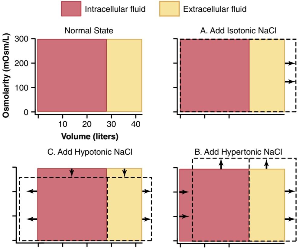
Figure 25-6. Effect of adding isotonic (A), hypertonic (B), and hypotonic solutions (A) to the extracellular fluid after osmotic equilibrium. The normal state is indicated by the solid lines, and the shifts from normal are shown by the shaded areas. The volumes of intracellular and extracellular fluid compartments are shown in the abscissa of each diagram, and the osmolarities of these compartments are shown on the ordinates.
شکل 25-6. اثر افزودن محلول های ایزوتونیک (A)، هیپرتونیک (B) و هیپوتونیک (A) به مایع خارج سلولی پس از تعادل اسمزی. حالت نرمال با خطوط توپر و جابجایی ها از حالت عادی با مناطق سایه دار نشان داده می شود. حجم محفظه های مایع درون سلولی و خارج سلولی در ابسیسا هر نمودار نشان داده شده است و اسمولاریته این محفظه ها روی دستورات نشان داده شده است.
Calculation of Fluid Shifts and Osmolarities After Infusion of Hypertonic Saline Solution. We can calculate the sequential effects of infusing different solutions on extracellular and intracellular fluid volumes and osmolarities. For example, if 2 liters of a hypertonic 3.0% sodium chloride solution were infused into the extracellular fluid compartment of a 70-kg patient whose initial plasma osmolarity is 280 mOsm/L, what would be the intracellular and extracellular fluid volumes and osmolarities after osmotic equilibrium?
محاسبه شیفت و اسمولاریته سیال پس از انفوزیون محلول سالین هیپرتونیک. ما میتوانیم اثرات متوالی تزریق محلولهای مختلف را بر حجم و اسمولاریتههای مایع خارج سلولی و درون سلولی محاسبه کنیم. به عنوان مثال، اگر 2 لیتر محلول کلرید سدیم 0/3 درصد هیپرتونیک به محفظه مایع خارج سلولی یک بیمار 70 کیلوگرمی که اسمولاریته اولیه پلاسمایی آن 280 mOsm/L است تزریق شود، حجم مایع درون سلولی و خارج سلولی و تعادلهای اسمولاریوماتیک پس از آن چقدر خواهد بود؟
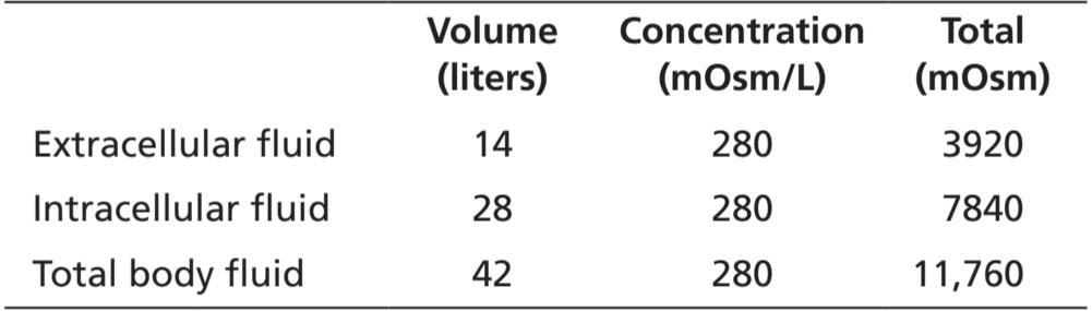
The first step is to calculate the initial conditions, including the volume, concentration, and total milliosmoles in each compartment. Assuming that extracellular fluid volume is 20% of body weight, and intracellular fluid volume is 40% of body weight, the following volumes and concentrations can be calculated.
اولین مرحله محاسبه شرایط اولیه، از جمله حجم، غلظت و مجموع میلیمول در هر محفظه است. با فرض اینکه حجم مایع خارج سلولی 20 درصد وزن بدن و حجم مایع داخل سلولی 40 درصد وزن بدن باشد، می توان حجم ها و غلظت های زیر را محاسبه کرد.
Step 1. Initial Conditions
مرحله 1. شرایط اولیه
Next, we calculate the total milliosmoles added to the extracellular fluid in 2 liters of 3.0% sodium chloride. A 3.0% solution means that there are 3.0 g/100 ml, or 30 grams of sodium chloride per liter. Because the molecular weight of sodium chloride is about 58.5 g/mol, this means that there is about 0.5128 mole of sodium chloride per liter of solution. For 2 liters of solution, this would be 1.0256 mole of sodium chloride. Because 1 mole of sodium chloride is equal to approximately 2 osmoles (sodium chloride has two osmotically active particles per mole), the net effect of adding 2 liters of this solution is to add 2051 milliosmoles of sodium chloride to the extracellular fluid.
در مرحله بعد، کل میلی مول های اضافه شده به مایع خارج سلولی را در 2 لیتر کلرید سدیم 0/3 درصد محاسبه می کنیم. محلول 3.0٪ به این معنی است که 3.0 گرم در 100 میلی لیتر یا 30 گرم کلرید سدیم در هر لیتر وجود دارد. از آنجایی که وزن مولکولی سدیم کلرید حدود 58.5 گرم بر مول است، به این معنی است که در هر لیتر محلول حدود 0.5128 مول کلرید سدیم وجود دارد. برای 2 لیتر محلول، این مقدار 1.0256 مول کلرید سدیم است. از آنجایی که 1 مول کلرید سدیم تقریباً برابر با 2 اسمول است (کلرید سدیم در هر مول دو ذره فعال اسمزی دارد)، اثر خالص افزودن 2 لیتر از این محلول، افزودن 2051 میلی مول کلرید سدیم به مایع خارج سلولی است.
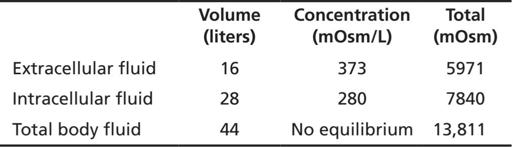
In step 2, we calculate the instantaneous effect of add- ing 2051 milliosmoles of sodium chloride to the extra- cellular fluid along with 2 liters of volume. There would be no change in the intracellular fluid concentration or volume, and there would be no osmotic equilibrium. In the extracellular fluid, however, there would be an addi- tional 2051 milliosmoles of total solute, yielding a total of 5971 milliosmoles. Because the extracellular compart- ment now has 16 liters of volume, the concentration can be calculated by dividing 5971 milliosmoles by 16 liters to yield a concentration of about 373 mOsm/L. Thus, the following values would occur instantly after adding the solution.
در مرحله 2، ما اثر آنی اضافه کردن 2051 میلی مول کلرید سدیم به مایع خارج سلولی به همراه 2 لیتر حجم را محاسبه می کنیم. هیچ تغییری در غلظت یا حجم مایع درون سلولی وجود نخواهد داشت و تعادل اسمزی وجود نخواهد داشت. با این حال، در مایع خارج سلولی، 2051 میلیاس مول کل املاح اضافی وجود خواهد داشت که در مجموع 5971 میلیاسمول به دست میآید. از آنجایی که محفظه خارج سلولی اکنون 16 لیتر حجم دارد، می توان غلظت آن را با تقسیم 5971 میلی مول بر 16 لیتر محاسبه کرد تا غلظتی در حدود 373 mOsm/L بدست آورد. بنابراین، مقادیر زیر بلافاصله پس از افزودن محلول رخ می دهد.
Step 2. Instantaneous Effect of Adding 2 Liters of 3.0% Sodium Chloride
مرحله 2. اثر آنی افزودن 2 لیتر کلرید سدیم 3.0 درصد
In the third step, we calculate the volumes and concentrations that would be found within a few minutes after osmotic equilibrium develops. In this case, the concentrations in the intracellular and extracellular fluid compartments would be equal and can be calculated by dividing the total milliosmoles in the body, 13,811, by the total volume, which is now 44 liters. This calculation yields a concentration of 313.9 mOsm/L. Therefore, all the body fluid compartments will have this same concentration after osmotic equilibrium. Assuming that no solute or water has been lost from the body, and that there is no movement of sodium chloride into or out of the cells, we then calculate the volumes of the intracellular and extracellular compartments. The intracellular fluid volume is calculated by dividing the total milliosmoles in the intracellular fluid (7840) by the concentration (313.9 mOsm/L) to yield a volume of 24.98 liters. Extracellular fluid volume is calculated by dividing the total milliosmoles in extracellular fluid (5971) by the concentration (313.9 mOsm/L) to yield a volume of 19.02 liters. Again, these calculations are based on the assumption that the sodium chloride added to the extracellular fluid remains there and does not move into the cells.
در مرحله سوم، حجم ها و غلظت هایی را که در عرض چند دقیقه پس از ایجاد تعادل اسمزی پیدا می شوند، محاسبه می کنیم. در این حالت، غلظت در محفظه مایع درون سلولی و خارج سلولی برابر است و می توان با تقسیم کل میلی مول در بدن، 13811، بر حجم کل، که اکنون 44 لیتر است، محاسبه کرد. این محاسبه غلظت 313.9 mOsm/L را به دست می دهد. بنابراین، تمام محفظه های مایع بدن پس از تعادل اسمزی همان غلظت را خواهند داشت. با فرض اینکه هیچ املاح یا آب از بدن از بین نرفته است و هیچ حرکتی از کلرید سدیم به داخل یا خارج سلول ها وجود ندارد، سپس حجم بخش های درون سلولی و خارج سلولی را محاسبه می کنیم. حجم مایع درون سلولی با تقسیم کل میلی مول در مایع درون سلولی (7840) بر غلظت (313.9 mOsm/L) محاسبه می شود تا حجمی معادل 24.98 لیتر به دست آید. حجم مایع خارج سلولی با تقسیم کل میلی مول در مایع خارج سلولی (5971) بر غلظت (313.9 mOsm/L) محاسبه می شود تا حجمی معادل 19.02 لیتر به دست آید. باز هم، این محاسبات بر اساس این فرض است که کلرید سدیم اضافه شده به مایع خارج سلولی در آنجا باقی می ماند و به داخل سلول ها حرکت نمی کند.
Step 3. Effect of Adding 2 Liters of 3.0% Sodium Chloride After Osmotic Equilibrium
مرحله 3. اثر افزودن 2 لیتر کلرید سدیم 3.0% پس از تعادل اسمزی
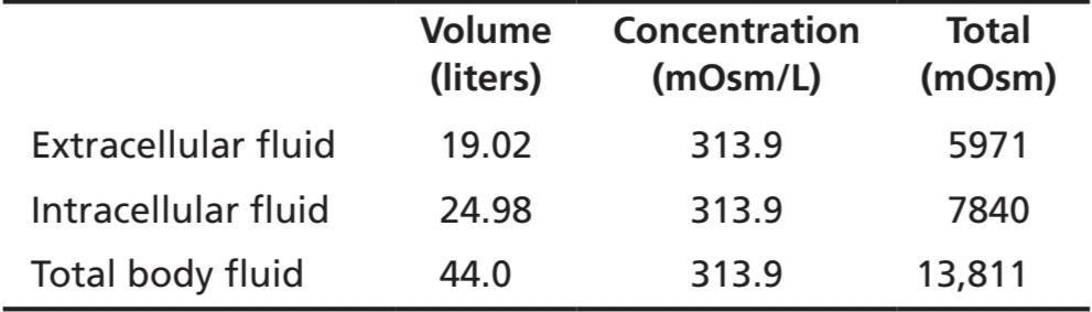
Thus, one can see from this example that adding 2 liters of a hypertonic sodium chloride solution causes more than a 5-liter increase in extracellular fluid volume while decreasing intracellular fluid volume by almost 3 liters.
بنابراین، از این مثال می توان دریافت که افزودن 2 لیتر محلول کلرید سدیم هیپرتونیک باعث افزایش بیش از 5 لیتری حجم مایع خارج سلولی می شود در حالی که حجم مایع داخل سلولی را تقریباً 3 لیتر کاهش می دهد.
This method of calculating changes in intracellular and extracellular fluid volumes and osmolarities can be applied to virtually any clinical problem of fluid volume regulation. The reader should be familiar with such calculations because an understanding of the mathematical aspects of osmotic equilibrium between intracellular and extracellular fluid compartments is essential for under- standing almost all fluid abnormalities of the body and their treatment.
این روش محاسبه تغییرات در حجم مایعات درون سلولی و خارج سلولی و اسمولاریته را می توان تقریباً برای هر مشکل بالینی تنظیم حجم مایع اعمال کرد. خواننده باید با چنین محاسباتی آشنا باشد زیرا درک جنبه های ریاضی تعادل اسمزی بین محفظه مایع درون سلولی و خارج سلولی برای درک تقریباً تمام ناهنجاری های مایع بدن و درمان آنها ضروری است.
GLUCOSE AND OTHER SOLUTIONS ADMINISTERED FOR NUTRITIVE PURPOSES
Several types of solutions are administered intravenously to provide nutrition to people who cannot otherwise ingest adequate amounts of nutrition. Glucose solutions are widely used; amino acid and homogenized fat solutions are used to a lesser extent. When these solutions are administered, their concentrations of osmotically active substances are usually adjusted nearly to isotonicity, or they are given slowly enough that they do not upset the osmotic equilibrium of the body fluids.
گلوکز و سایر محلول های تجویز شده برای اهداف تغذیه ای
انواع مختلفی از محلول ها به صورت داخل وریدی تجویز می شوند تا برای افرادی که در غیر این صورت نمی توانند مقادیر کافی از مواد غذایی را مصرف کنند، تغذیه کنند. محلول های گلوکز به طور گسترده استفاده می شود. اسید آمینه و محلول های چربی هموژن به میزان کمتری استفاده می شود. هنگامی که این محلول ها تجویز می شوند، غلظت مواد فعال اسمزی آنها معمولاً تقریباً به ایزوتونیک تنظیم می شود، یا به قدری آهسته داده می شوند که تعادل اسمزی مایعات بدن را بر هم نزند.
After the glucose or other nutrients are metabolized, an excess of water often remains, especially if additional fluid is ingested. Ordinarily, the kidneys excrete this fluid in the form of dilute urine. The net result, therefore, is the addition of only nutrients to the body.
پس از متابولیزه شدن گلوکز یا سایر مواد مغذی، اغلب آب اضافی باقی می ماند، به خصوص اگر مایع اضافی مصرف شود. به طور معمول کلیه ها این مایع را به صورت ادرار رقیق دفع می کنند. بنابراین، نتیجه خالص، افزودن تنها مواد مغذی به بدن است.
A 5% glucose solution, which is nearly isosmotic, is often used to treat dehydration. Because the solution is isosmotic, it can be infused intravenously without causing red blood cell swelling, as would occur with an infusion of pure water. Glucose in the solution is rapidly transported into the cells and metabolized. Therefore, infusion of a 5% glucose solution reduces extracellular fluid osmolarity and therefore helps correct the increase in extracellular fluid osmolarity associated with dehydration.
محلول گلوکز 5 درصد که تقریباً هم اسمزی است، اغلب برای درمان کم آبی استفاده می شود. از آنجایی که محلول ایزوسموز است، می توان آن را بدون ایجاد تورم گلبول های قرمز به صورت داخل وریدی انفوزیون کرد، همانطور که با تزریق آب خالص اتفاق می افتد. گلوکز موجود در محلول به سرعت به داخل سلول ها منتقل شده و متابولیزه می شود. بنابراین، انفوزیون محلول گلوکز 5 درصد اسمولاریته مایع خارج سلولی را کاهش می دهد و بنابراین به اصلاح افزایش اسمولاریته مایع خارج سلولی مرتبط با کم آبی کمک می کند.
CLINICAL ABNORMALITIES OF FLUID VOLUME REGULATION: HYPONATREMIA AND HYPERNATREMIA
A measurement that is readily available to the clinician for evaluating a patient’s fluid status is the plasma sodium concentration. Plasma osmolarity is not routinely measured but, because sodium and its associated anions (mainly chloride) account for more than 90% of the solute in the extracellular fluid, plasma sodium concentration is a reasonable indicator of plasma osmolarity under many conditions. When plasma sodium concentration is reduced more than a few milliequivalents below normal (about 142 mEq/L), a person is said to have hyponatremia. When plasma sodium concentration is elevated above normal, a person is said to have hypernatremia.
ناهنجاری های بالینی تنظیم حجم مایعات: هیپوناترمی و هیپرناترمی
اندازه گیری که به راحتی در دسترس پزشک برای ارزیابی وضعیت مایع بیمار است، غلظت سدیم پلاسما است. اسمولاریته پلاسما به طور معمول اندازه گیری نمی شود، اما از آنجایی که سدیم و آنیون های مرتبط با آن (عمدتاً کلرید) بیش از 90 درصد املاح موجود در مایع خارج سلولی را تشکیل می دهند، غلظت سدیم پلاسما شاخص معقولی برای اسمولاریته پلاسما در بسیاری از شرایط است. هنگامی که غلظت سدیم پلاسما بیش از چند میلی اکی والان کمتر از حد طبیعی (حدود 142 میلی اکی والان در لیتر) کاهش می یابد، گفته می شود که فرد دچار هیپوناترمی است. هنگامی که غلظت سدیم پلاسما بالاتر از حد طبیعی است، گفته می شود که فرد مبتلا به هیپرناترمی است.
Causes of Hyponatremia: Excess Water or Loss of Sodium
Decreased plasma sodium concentration can result from loss of sodium from the extracellular fluid or addition of excess water to the extracellular fluid (Table 25-4). A primary loss of sodium usually results in hyponatremia and dehydration and is associated with decreased extracellular fluid volume. Conditions that can cause hyponatremia as a result of the loss of sodium include diarrhea and vomiting. Overuse of diuretics that inhibit the ability of the kidneys to conserve sodium and certain types of sodium-wasting kidney diseases can also cause modest degrees of hyponatremia. Addison disease, which results from decreased secretion of the hormone aldosterone, impairs the ability of the kidneys to reabsorb sodium and can cause a modest degree of hyponatremia.
علل هیپوناترمی: آب اضافی یا از دست دادن سدیم
کاهش غلظت سدیم پلاسما می تواند ناشی از از دست دادن سدیم از مایع خارج سلولی یا اضافه شدن آب اضافی به مایع خارج سلولی باشد (جدول 25-4). از دست دادن اولیه سدیم معمولاً منجر به هیپوناترمی و کم آبی بدن می شود و با کاهش حجم مایع خارج سلولی همراه است. شرایطی که می تواند باعث هیپوناترمی در نتیجه از دست دادن سدیم شود شامل اسهال و استفراغ است. استفاده بیش از حد از دیورتیک ها که توانایی کلیه ها در حفظ سدیم و انواع خاصی از بیماری های کلیوی هدر دهنده سدیم را مهار می کنند نیز می تواند باعث درجات متوسطی از هیپوناترمی شود. بیماری آدیسون که از کاهش ترشح هورمون آلدوسترون ناشی میشود، توانایی کلیهها برای بازجذب سدیم را مختل میکند و میتواند باعث ایجاد درجات متوسطی از هیپوناترمی شود.
Hyponatremia can also be associated with excess water retention, which dilutes the sodium in the extracellular fluid, a condition referred to as hyponatremiaoverhydration. For example, excessive secretion of antidiuretic hormone (ADH), which increases water reabsorption by the kidney tubules, can lead to hyponatremia and overhydration.
هیپوناترمی همچنین می تواند با احتباس بیش از حد آب همراه باشد که باعث رقیق شدن سدیم در مایع خارج سلولی می شود، وضعیتی که به عنوان هیپوناترمی- هیدراتاسیون بیش از حد شناخته می شود. به عنوان مثال، ترشح بیش از حد هورمون آنتی دیورتیک (ADH)، که جذب مجدد آب توسط لوله های کلیه را افزایش می دهد، می تواند منجر به هیپوناترمی و هیدراتاسیون بیش از حد شود.
Table 25-4 Abnormalities of Body Fluid Volume Regulation: Hyponatremia and Hypernatremia
جدول 25-4 ناهنجاری های تنظیم حجم مایعات بدن: هیپوناترمی و هیپرناترمی

Hyponatremia Causes Cell Edema
Rapid changes in cell volume as a result of hyponatremia can have profound effects on tissue and organ function, especially the brain. A rapid reduction in plasma sodium concentration, for example, can cause brain cell edema and neurological symptoms, including headache, nausea, lethargy, and disorientation. If plasma sodium concentration rapidly falls below 115 to 120 mmol/L, brain swelling may lead to seizures, coma, permanent brain damage, and death. Because the skull is rigid, the brain cannot increase its volume by more than about 10% without it being forced down the neck (herniation), which can lead to permanent brain injury and death.
هیپوناترمی باعث ادم سلولی می شود
تغییرات سریع در حجم سلول در نتیجه هیپوناترمی می تواند اثرات عمیقی بر عملکرد بافت ها و اندام ها، به ویژه مغز داشته باشد. برای مثال، کاهش سریع غلظت سدیم پلاسما میتواند باعث ادم سلولهای مغزی و علائم عصبی، از جمله سردرد، حالت تهوع، بیحالی و بیحسی شود. اگر غلظت سدیم پلاسما به سرعت به زیر 115 تا 120 میلی مول در لیتر برسد، تورم مغز ممکن است منجر به تشنج، کما، آسیب دائمی مغز و مرگ شود. از آنجایی که جمجمه سفت است، مغز نمی تواند بیش از 10 درصد حجم خود را بدون فشار دادن به گردن (فتق) افزایش دهد که می تواند منجر به آسیب دائمی مغز و مرگ شود.
When hyponatremia evolves more slowly, over several days, the brain and other tissues respond by transporting sodium, chloride, potassium, and organic solutes, such as glutamate, from the cells into the extracellular compartment. This response attenuates osmotic flow of water into the cells and swelling of the tissues (Figure 25-7).
هنگامی که هیپوناترمی کندتر، طی چند روز تکامل مییابد، مغز و سایر بافتها با انتقال سدیم، کلرید، پتاسیم و املاح آلی مانند گلوتامات از سلولها به داخل بخش خارج سلولی پاسخ میدهند. این پاسخ باعث کاهش جریان اسمزی آب به داخل سلول ها و تورم بافت ها می شود (شکل 25-7).
Transport of solutes from the cells during slowly developing hyponatremia, however, can make the brain vulnerable to injury if the hyponatremia is corrected too rapidly. When hypertonic solutions are added too rapidly to correct hyponatremia, this intervention can outpace the brain’s ability to recapture the solutes lost from the cells and may lead to osmotic injury of the neurons that is associated with demyelination, a loss of the myelin sheath from nerves. This osmosis-mediated demyelination of neurons can be avoided by limiting the correction of chronic hyponatremia to less than 10 to 12 mmol/L in 24 hours and to less than 18 mmol/L in 48 hours. This slow rate of correction permits the brain to recover the osmoles that were lost from the cells as a result of adaptation to chronic hyponatremia.
با این حال، انتقال املاح از سلول ها در طول هیپوناترمی به آرامی، می تواند مغز را در برابر آسیب آسیب پذیر کند، اگر هیپوناترمی خیلی سریع اصلاح شود. هنگامی که محلول های هیپرتونیک خیلی سریع برای اصلاح هیپوناترمی اضافه می شود، این مداخله می تواند از توانایی مغز برای بازپس گیری املاح از دست رفته از سلول ها پیشی بگیرد و ممکن است منجر به آسیب اسمزی نورون ها شود که با دمیلیناسیون، از دست دادن غلاف میلین از اعصاب همراه است. با محدود کردن اصلاح هیپوناترمی مزمن به کمتر از 10 تا 12 میلی مول در لیتر در 24 ساعت و به کمتر از 18 میلی مول در لیتر در 48 ساعت، می توان از این دمیلیناسیون نورون ها با واسطه اسمز جلوگیری کرد. این سرعت آهسته اصلاح به مغز اجازه میدهد تا اسمولهایی را که در نتیجه سازگاری با هیپوناترمی مزمن از سلولها از دست دادهاند، بازیابی کند.
Hyponatremia is the most common electrolyte disorder encountered in clinical practice and may occur in up to 15% to 25% of hospitalized patients.
هیپوناترمی شایع ترین اختلال الکترولیتی است که در عمل بالینی با آن مواجه می شود و ممکن است در 15 تا 25 درصد از بیماران بستری در بیمارستان رخ دهد.
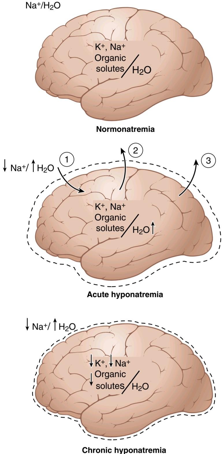
Figure 25-7. Brain cell volume regulation during hyponatremia. During acute hyponatremia, caused by loss of Nat or excess H2O, there is diffusion of H2O into the cells (1) and swelling of the brain tissue (indicated by the dashed lines). This process stimulates transport of Na+, K+, and organic solutes out of the cells (2), which then causes water diffusion out of the cells (3). With chronic hyponatremia, the brain swelling is attenuated by transport of solutes from the cells.
شکل 25-7. تنظیم حجم سلول های مغز در طول هیپوناترمی در طول هیپوناترمی حاد، ناشی از از دست دادن +Na یا H2O اضافی، انتشار H2O به سلول ها (1) و تورم بافت مغز (که با خطوط بریده نشان داده می شود) وجود دارد. این فرآیند انتقال Na+، +K و املاح آلی را به خارج از سلول ها تحریک می کند (2)، که سپس باعث انتشار آب از سلول ها می شود (3). در هیپوناترمی مزمن، تورم مغز با انتقال املاح از سلول ها کاهش می یابد.
Causes of Hypernatremia: Water Loss or Excess Sodium
An increased plasma sodium concentration, which also causes increased osmolarity, can be due to loss of water from the extracellular fluid, which concentrates the sodium ions, or excess sodium in the extracellular fluid. Primary loss of water from the extracellular fluid results in hypernatremia and dehydration. This condition can occur from an inability to secrete ADH, which is needed for the kidneys to conserve water. As a result of lack of ADH, the kidneys excrete large amounts of dilute urine (a disorder referred to as central diabetes insipidus), causing dehydration and increased concentration of sodium chloride in the extracellular fluid. In certain types of renal diseases, the kidneys cannot respond to ADH, causing a type of nephrogenic diabetes insipidus. A more common cause of hypernatremia associated with decreased extracellular fluid volume is simple dehydration caused by water intake that is less than water loss, as can occur with sweating during prolonged heavy exercise.
علل هایپرناترمی: از دست دادن آب یا سدیم اضافی
افزایش غلظت سدیم پلاسما، که باعث افزایش اسمولاریته نیز می شود، می تواند به دلیل از دست دادن آب از مایع خارج سلولی باشد که یون های سدیم را متمرکز می کند، یا سدیم اضافی در مایع خارج سلولی. از دست دادن اولیه آب از مایع خارج سلولی منجر به هایپرناترمی و کم آبی می شود. این وضعیت می تواند ناشی از ناتوانی در ترشح ADH باشد، که برای کلیه ها برای حفظ آب مورد نیاز است. در نتیجه کمبود ADH، کلیه ها مقادیر زیادی ادرار رقیق را دفع می کنند (اختلالی که به دیابت بی مزه مرکزی گفته می شود)، که باعث کم آبی و افزایش غلظت کلرید سدیم در مایع خارج سلولی می شود. در انواع خاصی از بیماری های کلیوی، کلیه ها نمی توانند به ADH پاسخ دهند و باعث ایجاد نوعی دیابت بی مزه نفروژنیک می شود. یکی از علل شایع هیپرناترمی مرتبط با کاهش حجم مایع خارج سلولی، کم آبی ساده ناشی از مصرف آب است که کمتر از از دست دادن آب است، همانطور که می تواند با تعریق در طول ورزش سنگین طولانی مدت رخ دهد.
Hypernatremia can also occur when excessive sodium chloride is added to the extracellular fluid. This often results in hypernatremia-overhydration because excess extracellular sodium chloride is also usually associated with at least some degree of water retention by the kidneys. For example, excessive secretion of the sodium- retaining hormone aldosterone can cause a mild degree of hypernatremia and overhydration. The reason that the hypernatremia is not more severe is that the sodium retention also stimulates secretion of ADH and causes the kidneys to reabsorb greater amounts of water.
هیپرناترمی همچنین می تواند زمانی رخ دهد که کلرید سدیم بیش از حد به مایع خارج سلولی اضافه شود. این اغلب منجر به هیپرناترمی بیش از حد هیدراتاسیون می شود، زیرا کلرید سدیم خارج سلولی اضافی نیز معمولاً با حداقل درجاتی از احتباس آب توسط کلیه ها همراه است. به عنوان مثال، ترشح بیش از حد هورمون نگهدارنده سدیم آلدوسترون می تواند باعث درجات خفیف هیپرناترمی و هیدراتاسیون بیش از حد شود. دلیل اینکه هیپرناترمی شدیدتر نیست این است که احتباس سدیم ترشح ADH را نیز تحریک می کند و باعث می شود کلیه ها مقادیر بیشتری آب را بازجذب کنند.
Thus, in analyzing abnormalities of plasma sodium concentration and deciding on proper therapy, one should first determine whether the abnormality is caused by a primary loss or gain of sodium or a primary loss or gain of water.
بنابراین، در تجزیه و تحلیل ناهنجاری های غلظت سدیم پلاسما و تصمیم گیری در مورد درمان مناسب، ابتدا باید مشخص شود که آیا این ناهنجاری ناشی از کاهش یا افزایش اولیه سدیم است یا از دست دادن یا افزایش اولیه آب.
Hypernatremia Causes Cell Shrinkage
Hypernatremia is much less common than hyponatremia and severe symptoms usually occur only with rapid and large increases in the plasma sodium concentration above 158 to 160 mmol/L. Hypernatremia usually promotes intense thirst and stimulates secretion of ADH, which both protect against a large increase in plasma and extra- cellular fluid sodium, as discussed in Chapter 29. However, severe hypernatremia can occur in patients with hypothalamic lesions that impair their sense of thirst, in infants who may not have ready access to water, in older patients with altered mental status, or in persons with diabetes insipidus.
هیپرناترمی باعث کوچک شدن سلول می شود
هیپرناترمی بسیار کمتر از هیپوناترمی است و علائم شدید معمولاً تنها با افزایش سریع و زیاد غلظت سدیم پلاسما بالای 158 تا 160 میلی مول در لیتر رخ می دهد. هیپرناترمی معمولاً تشنگی شدید را افزایش میدهد و ترشح ADH را تحریک میکند، که هر دو در برابر افزایش زیاد سدیم پلاسما و مایع خارج سلولی محافظت میکند، همانطور که در فصل 29 بحث شد. با دیابت بی مزه
Correction of hypernatremia can be achieved by administering a hypo-osmotic sodium chloride or dextrose solution. However, it is prudent to correct the hypernatremia slowly in patients who have had chronic increases in their plasma sodium concentration because hypernatremia also activates defense mechanisms that protect the cell from changes in volume. These defenses are the opposite of those that occur for hyponatremia and consist of mechanisms that increase the intracellular concentration of sodium and other solutes.
اصلاح هیپرناترمی را می توان با تجویز محلول کلرید سدیم یا دکستروز هیپواسموتیک به دست آورد. با این حال، اصلاح آهسته هیپرناترمی در بیمارانی که افزایش مزمن غلظت سدیم پلاسما داشته اند، عاقلانه است، زیرا هیپرناترمی همچنین مکانیسم های دفاعی را فعال می کند که سلول را از تغییرات حجم محافظت می کند. این دفاعها برعکس مواردی هستند که برای هیپوناترمی رخ میدهند و شامل مکانیسمهایی هستند که غلظت سدیم و سایر املاح درون سلولی را افزایش میدهند.
EDEMA: EXCESS FLUID IN THE TISSUES
Edema refers to the presence of excess fluid in the body tissues. In many cases, edema occurs mainly in the extra- cellular fluid compartment, but it can also involve intracellular fluid accumulation.
ادم: مایعات اضافی در بافت ها
ادم به وجود مایع اضافی در بافت های بدن اشاره دارد. در بسیاری از موارد، ادم عمدتاً در محفظه مایع خارج سلولی رخ می دهد، اما می تواند شامل تجمع مایع درون سلولی نیز باشد.
INTRACELLULAR EDEMA
Three conditions are especially prone to cause intracellular swelling: (1) hyponatremia, as discussed earlier; (2) depression of the metabolic systems of the tissues; and (3) lack of adequate nutrition to the cells. For example, when blood flow to a tissue is decreased, the delivery of oxygen and nutrients is reduced. If blood flow becomes too low to maintain normal tissue metabolism, the cell membrane ionic pumps become depressed, and sodium ions that normally leak into the interior of the cell can no longer be pumped out of the cells. The excess intracellular sodium ions then cause osmosis of water into the cells. Sometimes this process can increase intracellular volume of a tissue area-even of an entire ischemic leg, for example to two to three times normal. When such an increase in intracellular volume occurs, it is usually a prelude to death of the tissue.
ادم داخل سلولی
سه وضعیت به ویژه مستعد ایجاد تورم درون سلولی هستند: (1) هیپوناترمی، همانطور که قبلاً بحث شد. (2) افسردگی سیستم های متابولیک بافت ها. و (3) عدم تغذیه کافی برای سلول ها. به عنوان مثال، هنگامی که جریان خون به بافت کاهش می یابد، تحویل اکسیژن و مواد مغذی کاهش می یابد. اگر جریان خون برای حفظ متابولیسم طبیعی بافت خیلی کم شود، پمپهای یونی غشای سلولی ضعیف میشوند و یونهای سدیم که معمولاً به داخل سلول نشت میکنند، دیگر نمیتوانند از سلولها پمپ شوند. یون های سدیم داخل سلولی اضافی سپس باعث اسمز آب به داخل سلول ها می شود. گاهی اوقات این فرآیند می تواند حجم داخل سلولی یک ناحیه بافتی را افزایش دهد – حتی کل یک پای ایسکمیک را، به عنوان مثال تا دو تا سه برابر طبیعی. وقتی چنین افزایشی در حجم داخل سلولی رخ می دهد، معمولاً مقدمه ای برای مرگ بافت است.
Intracellular edema can also occur in inflamed tissues. Inflammation usually increases cell membrane permeability, allowing sodium and other ions to diffuse into the interior of the cell, with subsequent osmosis of water into the cells.
ادم داخل سلولی نیز می تواند در بافت های ملتهب ایجاد شود. التهاب معمولاً نفوذپذیری غشای سلولی را افزایش میدهد و به سدیم و سایر یونها اجازه میدهد تا به داخل سلول نفوذ کنند و متعاقباً آب را به داخل سلولها اسمز کنند.
EXTRACELLULAR EDEMA
Extracellular edema occurs when excess fluid accumulates in the extracellular spaces. There are two general causes of extracellular edema: (1) abnormal leakage of fluid from the plasma to the interstitial spaces across the capillaries; and (2) failure of the lymphatics to return fluid from the interstitium back into the blood, often called lymphedema. The most common clinical cause of interstitial fluid accumulation is excessive capillary fluid filtration.
ادم خارج سلولی
ادم خارج سلولی زمانی رخ می دهد که مایع اضافی در فضاهای خارج سلولی جمع شود. دو علت کلی برای ادم خارج سلولی وجود دارد: (1) نشت غیر طبیعی مایع از پلاسما به فضاهای بینابینی در سراسر مویرگها. و (2) ناتوانی لنفاوی در بازگشت مایع از بینابینی به خون، که اغلب به آن ادم لنفاوی می گویند. شایع ترین علت بالینی تجمع مایع بینابینی، فیلتراسیون بیش از حد مایع مویرگی است.
Factors That Can Increase Capillary Filtration
To understand the causes of excessive capillary filtration, it is useful to review the determinants of capillary filtration discussed in Chapter 16. Mathematically, the capillary filtration rate can be expressed as follows:
عواملی که می توانند فیلتراسیون مویرگی را افزایش دهند
برای درک علل فیلتراسیون مویرگی بیش از حد، مرور عوامل تعیین کننده فیلتراسیون مویرگی که در فصل 16 بحث شد مفید است. از نظر ریاضی، نرخ فیلتراسیون مویرگی را می توان به صورت زیر بیان کرد:

where K is the capillary filtration coefficient (the product of the permeability and surface area of the capillaries), Pe is the capillary hydrostatic pressure, Pif is the interstitial fluid hydrostatic pressure, л, is the capillary plasma colloid osmotic pressure, and if is the interstitial fluid colloid osmotic pressure. From this equation, one can see that any one of the following changes can increase the capillary filtration rate:
که در آن K ضریب فیلتراسیون مویرگی (محصول نفوذپذیری و سطح مویرگها)، Pe فشار هیدرواستاتیک مویرگی، Pif فشار هیدرواستاتیک مایع بینابینی، л، فشار اسمزی کلوئیدی پلاسمای مویرگی و اگر فشار اسمزی کلوئیدی مایع بینابینی است. از این معادله می توان دریافت که هر یک از تغییرات زیر می تواند نرخ فیلتراسیون مویرگی را افزایش دهد:
• Increased capillary filtration coefficient.
• Increased capillary hydrostatic pressure.
• Decreased plasma colloid osmotic pressure
• افزایش ضریب فیلتراسیون مویرگی.
• افزایش فشار هیدرواستاتیک مویرگی.
• کاهش فشار اسمزی کلوئیدی پلاسما
Lymphedema Failure of Lymph Vessels. to Return Fluid and Protein to the Blood
When lymphatic function is greatly impaired as a result of blockage or loss of the lymph vessels, edema can become especially severe because plasma proteins that leak into the interstitium cannot be removed in any other way. The rise in protein concentration raises the colloid osmotic pressure of interstitial fluid, which draws even more fluid out of the capillaries.
لنف ادم نارسایی عروق لنفاوی. برای بازگرداندن مایعات و پروتئین به خون
هنگامی که عملکرد لنفاوی در نتیجه انسداد یا از دست دادن عروق لنفاوی به شدت مختل می شود، ادم می تواند به ویژه شدید شود زیرا پروتئین های پلاسما که به داخل بینابینی نشت می کنند را نمی توان به روش دیگری خارج کرد. افزایش غلظت پروتئین فشار اسمزی کلوئیدی مایع بینابینی را افزایش می دهد که حتی مایع بیشتری را از مویرگ ها خارج می کند.
Blockage of lymph flow can be especially severe with infections of the lymph nodes, such as occurs with infection by filarial nematodes (Wuchereria bancrofti), which are microscopic threadlike worms. The adult worms live in the human lymph system and are spread from person to person by mosquitoes. People with filarial infections can have severe lymphedema and elephantiasis and men can have swelling of the scrotum, called hydrocele. Lymphatic filariasis affects more than 120 million people in 80 countries throughout the tropics and subtropics of Asia, Africa, the Western Pacific, and parts of the Caribbean and South America.
انسداد جریان لنفاوی می تواند به ویژه با عفونت های غدد لنفاوی شدید باشد، مانند عفونت توسط نماتدهای فیلاریال (Wuchereria bancrofti)، که کرم های نخ مانند میکروسکوپی هستند. کرم های بالغ در سیستم لنفاوی انسان زندگی می کنند و توسط پشه ها از فردی به فرد دیگر منتقل می شوند. افراد مبتلا به عفونت فیلاریال ممکن است به لنف ادم و فیل شدید مبتلا شوند و مردان ممکن است تورم کیسه بیضه به نام هیدروسل داشته باشند. فیلاریازیس لنفاوی بیش از 120 میلیون نفر را در 80 کشور در سراسر مناطق گرمسیری و نیمه گرمسیری آسیا، آفریقا، غرب اقیانوس آرام و بخش هایی از کارائیب و آمریکای جنوبی تحت تاثیر قرار می دهد.
Lymphedema can also occur in persons who have certain types of cancer or after surgery in which lymph vessels are removed or obstructed. For example, large numbers of lymph vessels are removed during a radical mastectomy, impairing the removal of protein and fluid from the breast and arm areas and causing edema and swelling of the tissue spaces. A few lymph vessels eventually regrow after this type of surgery; thus, the interstitial edema is usually temporary.
لنف ادم همچنین می تواند در افرادی که دارای انواع خاصی از سرطان هستند یا پس از جراحی که در آن عروق لنفاوی برداشته شده یا مسدود شده است، رخ دهد. به عنوان مثال، تعداد زیادی از رگهای لنفاوی در طول ماستکتومی رادیکال برداشته میشوند که باعث اختلال در برداشتن پروتئین و مایع از نواحی پستان و بازو میشود و باعث ادم و تورم فضاهای بافتی میشود. چند رگ لنفاوی در نهایت پس از این نوع جراحی دوباره رشد می کنند. بنابراین، ادم بینابینی معمولاً موقتی است.
Summary of Causes of Extracellular Edema
A large number of conditions can cause fluid accumulation in the interstitial spaces by abnormal leaking of fluid from the capillaries or by preventing the lymphatics from returning fluid from the interstitium back to the circulation. The following is a partial list of conditions that can cause extracellular edema by these two types of abnormalities:
خلاصه ای از علل ادم خارج سلولی
تعداد زیادی از شرایط می توانند با نشت غیر طبیعی مایع از مویرگ ها یا با جلوگیری از بازگرداندن مایعات لنفاوی از بینابینی به گردش خون، باعث تجمع مایع در فضاهای بینابینی شوند. در زیر فهرستی جزئی از شرایطی وجود دارد که میتوانند با این دو نوع ناهنجاری باعث ادم خارج سلولی شوند:
I. Increased capillary pressure
I. افزایش فشار مویرگی
A. Excessive kidney retention of salt and water
1. Acute or chronic kidney failure
2. Mineralocorticoid excess
الف. احتباس بیش از حد نمک و آب کلیه
1. نارسایی حاد یا مزمن کلیه
2. مینرالوکورتیکوئید اضافی
B. High venous pressure and venous constriction
1. Heart failure
2. Venous obstruction
3. Failure of venous pumps
a) Paralysis of muscles
b) Immobilization of parts of the body
c) Failure of venous valves
ب. فشار وریدی بالا و انقباض وریدی
1. نارسایی قلبی
2. انسداد وریدی
3. خرابی پمپ های وریدی
الف) فلج عضلات
ب) بی حرکتی اعضای بدن
ج) نارسایی دریچه های وریدی
C. Decreased arteriolar resistance
1. Excessive body heat
2. Insufficiency of sympathetic nervous system
3. Vasodilator drugs
ج. کاهش مقاومت شریانی
1. گرمای بیش از حد بدن
2. نارسایی سیستم عصبی سمپاتیک
3. داروهای گشادکننده عروق
II. Decreased plasma proteins
II. کاهش پروتئین های پلاسما
A. Loss of proteins in urine (nephrotic syndrome)
B. Loss of protein from denuded skin areas
1. Burns
2. Wounds
الف. از دست دادن پروتئین در ادرار (سندرم نفروتیک)
ب. از دست دادن پروتئین از نواحی برهنه شده پوست
1. سوختگی
2. زخم
C. Failure to produce proteins
1. Liver disease (e.g., cirrhosis)
2. Serious protein or caloric malnutrition
ج. عدم تولید پروتئین
1. بیماری کبد (به عنوان مثال، سیروز)
2. سوء تغذیه جدی پروتئین یا کالری
III. Increased capillary permeability
III. افزایش نفوذپذیری مویرگی
A. Immune reactions that cause release of histamine and other immune products
B. Toxins
C. Bacterial infections
D. Vitamin deficiency, especially vitamin C
E. Prolonged ischemia
F. Burns
الف. واکنش های ایمنی که باعث آزاد شدن هیستامین و سایر محصولات ایمنی می شوند
ب. سموم
ج. عفونت های باکتریایی
د. کمبود ویتامین به خصوص ویتامین C
ه. ایسکمی طولانی مدت
و. برنز
IV. Blockage of lymph return
IV. انسداد بازگشت لنفاوی
A. Cancer
B. Infections (e.g., filarial nematodes)
C. Surgery
D.Congenital absence or abnormality of lymphatic vessels
الف. سرطان
ب. عفونت ها (مانند نماتدهای فیلاریال)
ج. جراحی
د. عدم وجود یا ناهنجاری مادرزادی عروق لنفاوی
Edema Caused by Heart Failure.One of the most serious and common causes of edema is heart failure, which was discussed in Chapter 22. In heart failure, the heart fails to pump blood normally from the veins into the arteries, which raises venous and capillary pressures, causing increased capillary filtration. In addition, the arterial pressure tends to fall, causing decreased excretion of salt and water by the kidneys, which causes still more edema. Also, blood flow to the kidneys is reduced in persons with heart failure, and this reduced blood flow stimulates secretion of renin, causing increased formation of angiotensin II and aldosterone, which both cause additional salt and water retention by the kidneys. In advanced heart failure, increased secretion of ADH stimulates water reabsorption by the renal tubules, leading to hyponatremia as well as intracellular and extracellular edema. Thus, in persons with untreated heart failure, all these factors acting together can cause serious generalized edema.
ادم ناشی از نارسایی قلبی. یکی از جدی ترین و شایع ترین علل ادم نارسایی قلبی است که در فصل 22 مورد بحث قرار گرفت. در نارسایی قلبی، قلب نمی تواند خون را به طور طبیعی از وریدها به داخل شریان ها پمپ کند، که فشار وریدی و مویرگی را افزایش می دهد و باعث افزایش فیلتراسیون مویرگی می شود. علاوه بر این، فشار شریانی تمایل به کاهش دارد و باعث کاهش دفع نمک و آب توسط کلیه ها می شود که باعث ادم بیشتر می شود. همچنین جریان خون به کلیه ها در افراد مبتلا به نارسایی قلبی کاهش می یابد و این کاهش جریان خون باعث تحریک ترشح رنین می شود و باعث افزایش تشکیل آنژیوتانسین II و آلدوسترون می شود که هر دو باعث احتباس نمک و آب اضافی توسط کلیه ها می شوند. در نارسایی پیشرفته قلبی، افزایش ترشح ADH باعث تحریک جذب مجدد آب توسط لوله های کلیوی می شود که منجر به هیپوناترمی و همچنین ادم داخل سلولی و خارج سلولی می شود. بنابراین، در افراد مبتلا به نارسایی قلبی درمان نشده، همه این عوامل با هم عمل میکنند و میتوانند باعث ادم عمومی جدی شوند.
In patients with left-sided heart failure but without significant failure of the right side of the heart, blood is pumped into the lungs normally by the right side of the heart but cannot escape easily from the pulmonary veins to the left side of the heart because this part of the heart has been greatly weakened. Consequently, all the pulmonary vascular pressures, including pulmonary capillary pressure, rise far above normal, causing serious and lifethreatening pulmonary edema. When untreated, fluid accumulation in the lungs can rapidly progress, causing death within a few hours.
در بیماران مبتلا به نارسایی قلبی سمت چپ اما بدون نارسایی قابل توجه در سمت راست قلب، خون به طور معمول توسط سمت راست قلب به ریه ها پمپ می شود اما نمی تواند به راحتی از وریدهای ریوی به سمت چپ قلب فرار کند زیرا این قسمت از قلب بسیار ضعیف شده است. در نتیجه، تمام فشارهای عروقی ریوی، از جمله فشار مویرگ ریوی، بسیار بالاتر از حد طبیعی است و باعث ادم ریوی جدی و تهدید کننده زندگی می شود. در صورت عدم درمان، تجمع مایع در ریه ها می تواند به سرعت پیشرفت کند و در عرض چند ساعت باعث مرگ شود.
Edema Caused by Decreased Kidney Excretion of Salt and Water. Most sodium chloride added to the blood remains in the extracellular compartment, and only a small amount enters the cells. Therefore, in kidney diseases that compromise urinary excretion of salt and water, large amounts of sodium chloride and water are added to the extracellular fluid. Most of this salt and water leaks from the blood into the interstitial spaces, but some remains in the blood. The main effects of this are as follows: (1) widespread increases in interstitial fluid volume (extracellular edema); and (2) hypertension because of the increase in blood volume, as explained in Chapter 19. As an example, in children who have acute glomerulonephritis, in which the renal glomeruli are injured by inflammation and therefore fail to filter adequate amounts of fluid, serious extracellular fluid edema also develops; along with the edema, severe hypertension usually develops.
ادم ناشی از کاهش دفع نمک و آب از کلیه. بیشتر کلرید سدیم اضافه شده به خون در قسمت خارج سلولی باقی می ماند و تنها مقدار کمی از آن وارد سلول ها می شود. بنابراین در بیماری های کلیوی که دفع نمک و آب از طریق ادرار را به خطر می اندازند، مقدار زیادی کلرید سدیم و آب به مایع خارج سلولی اضافه می شود. بیشتر این نمک و آب از خون به فضاهای بینابینی نشت می کند، اما مقداری در خون باقی می ماند. اثرات اصلی این امر به شرح زیر است: (1) افزایش گسترده در حجم مایع بینابینی (ادم خارج سلولی). و (2) فشار خون بالا به دلیل افزایش حجم خون، همانطور که در فصل 19 توضیح داده شد. به عنوان مثال، در کودکانی که گلومرولونفریت حاد دارند، که در آن گلومرول های کلیوی در اثر التهاب آسیب می بینند و بنابراین نمی توانند مقادیر کافی مایع را فیلتر کنند، ادم مایع خارج سلولی جدی نیز ایجاد می شود. همراه با ادم، فشار خون شدید معمولا ایجاد می شود.
Edema Caused by Decreased Plasma Proteins. Failure to produce normal amounts of proteins or leakage of proteins from the plasma causes the plasma colloid osmotic pressure to fall. This leads to increased capillary filtration throughout the body and extracellular edema.
ادم ناشی از کاهش پروتئین های پلاسما. عدم تولید مقادیر طبیعی پروتئین یا نشت پروتئین از پلاسما باعث کاهش فشار اسمزی کلوئیدی پلاسما می شود. این منجر به افزایش فیلتراسیون مویرگی در سراسر بدن و ادم خارج سلولی می شود.
One of the most important causes of decreased plasma protein concentration is loss of proteins in the urine in certain kidney diseases, a condition referred to as nephrotic syndrome. Multiple types of renal diseases can damage the membranes of the renal glomeruli, causing the membranes to become leaky to the plasma proteins and often allowing large quantities of these proteins to pass into the urine. When this loss exceeds the ability of the body to synthesize proteins, a reduction in plasma protein concentration occurs. Serious generalized edema occurs when the plasma protein concentration falls below 2.5 g/100 ml.
یکی از مهم ترین دلایل کاهش غلظت پروتئین پلاسما، از دست دادن پروتئین در ادرار در برخی بیماری های کلیوی است که به آن سندرم نفروتیک گفته می شود. انواع مختلفی از بیماریهای کلیوی میتوانند به غشای گلومرولهای کلیوی آسیب برسانند و باعث نشت غشاها به پروتئینهای پلاسما شوند و اغلب اجازه میدهند مقادیر زیادی از این پروتئینها وارد ادرار شوند. هنگامی که این کاهش از توانایی بدن برای سنتز پروتئین ها بیشتر شود، کاهش غلظت پروتئین پلاسما رخ می دهد. ادم عمومی جدی زمانی رخ می دهد که غلظت پروتئین پلاسما به کمتر از 2.5 گرم در 100 میلی لیتر برسد.
Cirrhosis of the liver is another condition that reduces plasma protein concentration. Cirrhosis means the development of large amounts of fibrous tissue among the liver parenchymal cells. One result is failure of these cells to produce sufficient plasma proteins, leading to decreased plasma colloid osmotic pressure and the generalized edema that accompanies this condition.
سیروز کبدی بیماری دیگری است که غلظت پروتئین پلاسما را کاهش می دهد. سیروز به معنای ایجاد مقادیر زیادی بافت فیبری در بین سلول های پارانشیمی کبد است. یکی از نتایج این است که این سلول ها در تولید پروتئین های کافی پلاسما شکست می خورند که منجر به کاهش فشار اسمزی کلوئیدی پلاسما و ادم عمومی همراه با این وضعیت می شود.
Another way that cirrhosis causes edema is that the liver fibrosis sometimes compresses the abdominal portal venous drainage vessels as they pass through the liver before emptying back into the general circulation. Blockage of this portal venous outflow raises capillary hydro- static pressure throughout the gastrointestinal area and further increases fluid filtration out of the plasma into the intra-abdominal areas. When this occurs, the combined effects of decreased plasma protein concentration and high portal capillary pressures cause transudation of large amounts of fluid and protein into the abdominal cavity, a condition referred to as ascites.
راه دیگری که سیروز باعث ادم می شود این است که فیبروز کبدی گاهی اوقات عروق تخلیه ورید پورتال شکمی را در حین عبور از کبد قبل از تخلیه مجدد به گردش خون عمومی فشرده می کند. انسداد این جریان خروجی ورید پورتال، فشار هیدرواستاتیک مویرگی را در سراسر ناحیه گوارشی افزایش میدهد و بیشتر فیلتراسیون مایع را از پلاسما به داخل نواحی داخل شکمی افزایش میدهد. هنگامی که این اتفاق می افتد، اثرات ترکیبی کاهش غلظت پروتئین پلاسما و فشار بالای مویرگی پورتال باعث انتقال مقادیر زیادی مایع و پروتئین به داخل حفره شکمی می شود که به آن آسیت می گویند.
SAFETY FACTORS THAT NORMALLY PREVENT EDEMA
Even though many disturbances can cause edema, the abnormality must usually be severe before serious edema develops. The reason the abnormality must be severe is that three major safety factors prevent excessive fluid accumulation in the interstitial spaces: (1) low compliance of the interstitium when interstitial fluid pressure is in the negative pressure range; (2) the ability of lymph flow to increase 10- to 50-fold; and (3) washdown of the interstitial fluid protein concentration, which reduces interstitial fluid colloid osmotic pressure as capillary filtration increases.
عوامل ایمنی که به طور معمول از ادم جلوگیری می کنند
حتی اگر بسیاری از اختلالات می توانند باعث ادم شوند، این ناهنجاری معمولاً باید قبل از ایجاد ادم جدی شدید باشد. دلیل اینکه این ناهنجاری باید شدید باشد این است که سه عامل ایمنی اصلی از تجمع بیش از حد مایع در فضاهای بین بافتی جلوگیری می کند: (1) انطباق کم بینابینی هنگامی که فشار مایع بینابینی در محدوده فشار منفی است. (2) توانایی جریان لنفاوی برای افزایش 10 تا 50 برابری. و (3) شستشوی غلظت پروتئین مایع بینابینی، که فشار اسمزی کلوئیدی مایع بینابینی را با افزایش فیلتراسیون مویرگی کاهش می دهد.
Safety Factor Caused by Low Compliance of the Interstitium in the Negative Pressure Range
In Chapter 16, we noted that interstitial fluid hydrostatic pressure in loose subcutaneous tissues of the body is slightly less than atmospheric pressure, averaging about -3 mm Hg. This slight suction in the tissues helps hold the tissues together. Figure 25-8 shows the approximate relationships between different levels of interstitial fluid pressure and interstitial fluid volume, as extrapolated to people from animal studies. Note in Figure 25-8 that as long as the interstitial fluid pressure is in the negative range, small changes in interstitial fluid volume are associated with relatively large changes in interstitial fluid hydrostatic pressure. Therefore, in the negative pressure range, the compliance of the tissues, defined as the change in volume per millimeter of Hg pressure change, is low.
عامل ایمنی ناشی از انطباق کم بینابینی در محدوده فشار منفی
در فصل 16، اشاره کردیم که فشار هیدرواستاتیک مایع بینابینی در بافت های شل زیر جلدی بدن اندکی کمتر از فشار اتمسفر است و به طور متوسط حدود 3- میلی متر جیوه است. این مکش اندک در بافت ها به نگه داشتن بافت ها در کنار هم کمک می کند. شکل 25-8 روابط تقریبی بین سطوح مختلف فشار مایع بین بافتی و حجم مایع بین بافتی را نشان می دهد، همانطور که از مطالعات حیوانی برای افراد برون یابی شده است. در شکل 25-8 توجه داشته باشید که تا زمانی که فشار سیال بینابینی در محدوده منفی است، تغییرات جزئی در حجم مایع بین بافتی با تغییرات نسبتاً زیادی در فشار هیدرواستاتیک مایع بینابینی همراه است. بنابراین، در محدوده فشار منفی، انطباق بافت ها که به عنوان تغییر حجم در هر میلی متر تغییر فشار جیوه تعریف می شود، کم است.
How does the low compliance of the tissues in the negative pressure range act as a safety factor against edema? To answer this question, recall the determinants of capillary filtration discussed previously. When interstitial fluid hydrostatic pressure increases, this increased pressure tends to oppose further capillary filtration. Therefore, as long as the interstitial fluid hydrostatic pressure is in the negative pressure range, small increases in interstitial fluid volume cause relatively large increases in interstitial fluid hydrostatic pressure, opposing further filtration of fluid into the tissues.
انطباق کم بافت ها در محدوده فشار منفی چگونه به عنوان یک عامل ایمنی در برابر ادم عمل می کند؟ برای پاسخ به این سوال، عوامل تعیین کننده فیلتراسیون مویرگی که قبلاً مورد بحث قرار گرفت را به یاد آورید. هنگامی که فشار هیدرواستاتیک مایع بینابینی افزایش می یابد، این فشار افزایش یافته تمایل دارد با فیلتراسیون مویرگی بیشتر مخالفت کند. بنابراین، تا زمانی که فشار هیدرواستاتیک مایع بینابینی در محدوده فشار منفی است، افزایش های جزئی در حجم مایع بین بافتی باعث افزایش نسبتاً زیاد فشار هیدرواستاتیک مایع بینابینی می شود که با فیلتراسیون بیشتر مایع به داخل بافت ها مخالفت می کند.
Because the normal interstitial fluid hydrostatic pressure is 3 mm Hg, the interstitial fluid hydrostatic pressure must increase by about 3 mm Hg before large amounts of fluid will begin to accumulate in the tissues. Therefore, the safety factor against edema is a change of interstitial fluid pressure of about 3 mm Hg. In the positive tissue pressure range, this safety factor against edema is lost because of the large increase in compliance of the tissues.
از آنجایی که فشار هیدرواستاتیک مایع بین بافتی طبیعی 3 میلی متر جیوه است، فشار هیدرواستاتیک مایع بین بافتی باید حدود 3 میلی متر جیوه افزایش یابد تا مقادیر زیادی مایع در بافت ها تجمع پیدا کند. بنابراین، عامل ایمنی در برابر ادم، تغییر فشار مایع بینابینی حدود 3 میلی متر جیوه است. در محدوده فشار بافتی مثبت، این فاکتور ایمنی در برابر ادم به دلیل افزایش زیاد انطباق بافت ها از بین می رود.

Figure 25-8. Relationship between interstitial fluid hydrostatic pres- sure and interstitial fluid volumes, including total volume, free fluid volume, and gel fluid volume, for loose tissues such as skin. Note that significant amounts of free fluid occur only when the interstitial fluid pressure becomes positive. (Modified from Guyton AC, Granger HJ, Taylor AE: Interstitial fluid pressure. Physiol Rev 51:527, 1971.)
شکل 25-8. رابطه بین فشار هیدرواستاتیک مایع بینابینی و حجم مایع بینابینی، از جمله حجم کل، حجم مایع آزاد و حجم مایع ژل، برای بافت های شل مانند پوست. توجه داشته باشید که مقادیر قابل توجهی از مایع آزاد تنها زمانی رخ می دهد که فشار مایع بینابینی مثبت شود. (اصلاح شده از Guyton AC، Granger HJ، Taylor AE: فشار مایع بینابینی. Physiol Rev 51:527، 1971.)
Importance of Interstitial Gel in Preventing Fluid Accumulation in the Interstitium. Note in Figure 25-8 that in normal tissues with negative interstitial fluid pressure, virtually all the fluid in the interstitium is in gel form. That is, the fluid is bound in a proteoglycan meshwork so that there are virtually no free fluid spaces larger than a few hundredths of a micrometer in diameter. The importance of the gel is that it prevents fluid from flowing easily through the tissues because of impediment from the brush pile of trillions of proteoglycan filaments. Also, when the interstitial fluid pressure falls to very negative values, the gel does not contract very much because the meshwork of proteoglycan filaments offers an elastic resistance to compression. In the negative fluid pressure range, the interstitial fluid volume does not change much, regardless of whether the degree of suction is only a few mm Hg negative pressure or 10 to 20 mm Hg negative pressure. In other words, the compliance of the tissues is very low in the negative pressure range.
اهمیت ژل بینابینی در جلوگیری از تجمع مایعات در بینابینی. در شکل 25-8 توجه داشته باشید که در بافت های نرمال با فشار مایع بینابینی منفی، تقریباً تمام مایع بینابینی به شکل ژل است. یعنی سیال در یک شبکه پروتئوگلیکان بسته شده است به طوری که عملاً هیچ فضای آزاد سیال بزرگتر از چند صدم میکرومتر در قطر وجود ندارد. اهمیت ژل در این است که از جریان آسان مایع در بافت ها جلوگیری می کند زیرا مانع از انبوه برس از تریلیون ها رشته پروتئوگلیکان می شود. همچنین، هنگامی که فشار مایع بینابینی به مقادیر بسیار منفی کاهش مییابد، ژل خیلی منقبض نمیشود، زیرا شبکه رشتههای پروتئوگلیکان مقاومت الاستیکی در برابر فشار ایجاد میکند. در محدوده فشار منفی سیال، حجم مایع بین بافتی تغییر چندانی نمی کند، صرف نظر از اینکه درجه مکش تنها چند میلی متر جیوه فشار منفی باشد یا 10 تا 20 میلی متر جیوه فشار منفی. به عبارت دیگر، انطباق بافت ها در محدوده فشار منفی بسیار کم است.
By contrast, when interstitial fluid pressure rises to the positive pressure range, there is a tremendous accumulation of free fluid in the tissues. In this pressure range, the tissues are compliant, allowing large amounts of fluid to accumulate, with relatively small additional increases in interstitial fluid hydrostatic pressure. Most of the extra fluid that accumulates is free fluid because it pushes the brush pile of proteoglycan filaments apart. Therefore, the fluid can flow freely through the tissue spaces because it is not in gel form. When this free flow of fluid occurs, the edema is said to be pitting edema because one can press the thumb against the tissue area and push the fluid out of the area. When the thumb is removed, a pit is left in the skin for a few seconds until the fluid flows back from the surrounding tissues. This type of edema is distinguished from nonpitting edema, which occurs when the tissue cells swell instead of the interstitium or when the fluid in the interstitium becomes clotted with fibrinogen so that it cannot move freely within the tissue spaces.
در مقابل، هنگامی که فشار مایع بینابینی به محدوده فشار مثبت افزایش می یابد، تجمع فوق العاده ای از مایع آزاد در بافت ها وجود دارد. در این محدوده فشار، بافتها منطبق هستند و به مقدار زیادی مایع اجازه میدهند تا با افزایش نسبتاً کمی اضافی در فشار هیدرواستاتیک مایع بینابینی جمع شوند. بیشتر مایع اضافی که انباشته می شود، مایع آزاد است زیرا انبوه رشته های پروتئوگلیکان را از هم جدا می کند. بنابراین، مایع می تواند آزادانه در فضاهای بافتی جریان یابد زیرا به شکل ژل نیست. هنگامی که این جریان آزاد مایع رخ می دهد، گفته می شود ادم ادم حفره ای است زیرا می توان انگشت شست را به ناحیه بافت فشار داد و مایع را از ناحیه خارج کرد. هنگامی که انگشت شست برداشته می شود، یک گودال برای چند ثانیه در پوست باقی می ماند تا مایع از بافت های اطراف برگردد. این نوع ادم از ادم بدون حفره متمایز می شود، که زمانی رخ می دهد که سلول های بافتی به جای بینابینی متورم می شوند یا زمانی که مایع موجود در بینابینی با فیبرینوژن لخته می شود به طوری که نمی تواند آزادانه در فضاهای بافتی حرکت کند.
Importance of Proteoglycan Filaments as a Spacer for Cells and in Preventing Rapid Flow of Fluid in Tissues. The proteoglycan filaments, along with much larger collagen fibrils in the interstitial spaces, act as a spacer between the cells. Nutrients and ions do not diffuse readily through cell membranes; therefore, without adequate spacing between the cells, these nutrients, electrolytes, and cell waste products cannot be rapidly exchanged between the blood capillaries and cells located at a distance from one another.
اهمیت رشته های پروتئوگلیکان به عنوان یک فاصله دهنده برای سلول ها و در جلوگیری از جریان سریع مایع در بافت ها. رشته های پروتئوگلیکان، همراه با فیبریل های کلاژن بسیار بزرگتر در فضاهای بینابینی، به عنوان یک فاصله بین سلول ها عمل می کنند. مواد مغذی و یون ها به آسانی از طریق غشای سلولی پخش نمی شوند. بنابراین، بدون فاصله کافی بین سلولها، این مواد مغذی، الکترولیتها و مواد زائد سلولی نمیتوانند به سرعت بین مویرگهای خون و سلولهایی که در فاصلهای از یکدیگر قرار دارند مبادله شوند.
The proteoglycan filaments also prevent fluid from flowing too easily through the tissue spaces. If it were not for the proteoglycan filaments, the simple act of a person standing up would cause large amounts of interstitial fluid to flow from the upper body to the lower body. When too much fluid accumulates in the interstitium, as occurs in edema, this extra fluid creates large channels that allow the fluid to flow readily through the interstitium. Therefore, when severe edema occurs in the legs, the edema fluid often can be decreased by simply elevating the legs.
رشته های پروتئوگلیکان همچنین از جریان بیش از حد مایع در فضاهای بافتی جلوگیری می کند. اگر رشتههای پروتئوگلیکان نبودند، عمل ساده ایستادن فرد باعث میشد مقادیر زیادی مایع بینابینی از بالاتنه به پایینتنه جاری شود. هنگامی که مایع بیش از حد در بینابینی انباشته می شود، همانطور که در ادم اتفاق می افتد، این مایع اضافی کانال های بزرگی ایجاد می کند که به مایع اجازه می دهد تا به راحتی از طریق بینابینی جریان یابد. بنابراین، هنگامی که ادم شدید در پاها رخ می دهد، مایع ادم را اغلب می توان با بالا بردن ساده پاها کاهش داد.
Even though fluid does not flow easily through the tissues in the presence of the compacted proteoglycan filaments, different substances within the fluid can diffuse through the tissues at least 95% as easily as they normally diffuse. Therefore, the usual diffusion of nutrients to the cells and the removal of waste products from the cells are not com- promised by the proteoglycan filaments of the interstitium.
حتی اگر مایع در حضور رشته های پروتئوگلیکان فشرده شده به راحتی در بافت ها جریان نداشته باشد، مواد مختلف درون مایع می توانند حداقل 95 درصد به راحتی در بافت ها پخش شوند. بنابراین، انتشار معمول مواد مغذی به سلولها و حذف مواد زائد از سلولها توسط رشتههای پروتئوگلیکان بینابینی به خطر نمیافتد.
Increased Lymph Flow as a Safety Factor Against Edema
A major function of the lymphatic system is to return the fluid and proteins filtered from the capillaries into the interstitium to the circulation. Without this continuous return of the filtered proteins and fluid to the blood, the plasma volume would be rapidly depleted, and interstitial edema would occur.
افزایش جریان لنفاوی به عنوان یک عامل ایمنی در برابر ادم
عملکرد اصلی سیستم لنفاوی این است که مایع و پروتئین های فیلتر شده از مویرگ ها به داخل بینابینی را به گردش خون بازگرداند. بدون این بازگشت مداوم پروتئین ها و مایعات فیلتر شده به خون، حجم پلاسما به سرعت کاهش می یابد و ادم بینابینی رخ می دهد.
The lymphatics act as a safety factor against edema because lymph flow can increase 10- to 50-fold when fluid begins to accumulate in the tissues. This increased lymph flow allows the lymphatics to carry away large amounts of fluid and proteins in response to increased capillary filtration, preventing the interstitial pressure from rising into the positive pressure range. The safety factor caused by increased lymph flow has been calculated to be about 7 mm Hg.
غدد لنفاوی به عنوان یک عامل ایمنی در برابر ادم عمل می کنند زیرا جریان لنفاوی می تواند 10 تا 50 برابر با شروع تجمع مایع در بافت ها افزایش یابد. این افزایش جریان لنفاوی به غدد لنفاوی اجازه میدهد تا مقادیر زیادی مایع و پروتئین را در پاسخ به افزایش فیلتراسیون مویرگی با خود ببرند و از افزایش فشار بین بافتی به محدوده فشار مثبت جلوگیری میکنند. ضریب ایمنی ناشی از افزایش جریان لنفاوی حدود 7 میلی متر جیوه محاسبه شده است.
Washdown of Interstitial Fluid Protein as a Safety Factor Against Edema
As increased amounts of fluid are filtered into the interstitium, the interstitial fluid pressure increases, causing increased lymph flow. In most tissues, the protein concentration of the interstitium decreases as lymph flow is increased because larger amounts of protein are carried away than can be filtered out of the capillaries. The reason for this phenomenon is that the capillaries are relatively impermeable to proteins compared with the lymph vessels. Therefore, the proteins are washed out of the interstitial fluid as lymph flow increases.
شستشوی پروتئین مایع بینابینی به عنوان یک عامل ایمنی در برابر ادم
با افزایش مقادیر مایع به داخل بینابینی، فشار مایع بینابینی افزایش می یابد و باعث افزایش جریان لنفاوی می شود. در بیشتر بافتها، با افزایش جریان لنفاوی، غلظت پروتئین بینابینی کاهش مییابد، زیرا مقادیر بیشتری از پروتئین از مویرگها خارج میشود. دلیل این پدیده این است که مویرگ ها نسبت به پروتئین ها نسبت به عروق لنفاوی نفوذناپذیر هستند. بنابراین، با افزایش جریان لنفاوی، پروتئین ها از مایع بینابینی شسته می شوند.
Because the interstitial fluid colloid osmotic pressure caused by the proteins tends to draw fluid out of the capillaries, decreasing the interstitial fluid proteins lowers the net filtration force across the capillaries and tends to prevent further accumulation of fluid. The safety factor from this effect has been calculated to be about 7 mm Hg.
از آنجایی که فشار اسمزی کلوئیدی مایع بین بافتی ناشی از پروتئین ها تمایل دارد مایع را از مویرگ ها بیرون بکشد، کاهش پروتئین های مایع بینابینی نیروی خالص تصفیه را در سراسر مویرگ ها کاهش می دهد و از تجمع بیشتر مایع جلوگیری می کند. ضریب ایمنی ناشی از این اثر حدود 7 میلی متر جیوه محاسبه شده است.
SUMMARY OF SAFETY FACTORS THAT PREVENT EDEMA
Putting together all the safety factors against edema, we find the following:
1. The safety factor caused by low tissue compliance in the negative pressure range is about 3 mm Hg.
2. The safety factor caused by increased lymph flow is about 7 mm Hg.
3. The safety factor caused by washdown of proteins from the interstitial spaces is about 7 mm Hg.
خلاصه ای از عوامل ایمنی که از ادم جلوگیری می کنند
با کنار هم قرار دادن تمام عوامل ایمنی در برابر ادم، موارد زیر را می یابیم:
1. ضریب ایمنی ناشی از انطباق بافت کم در محدوده فشار منفی حدود 3 میلی متر جیوه است.
2. فاکتور ایمنی ناشی از افزایش جریان لنفاوی حدود 7 میلی متر جیوه است.
3. فاکتور ایمنی ناشی از شستشوی پروتئین ها از فضاهای بینابینی حدود 7 میلی متر جیوه است.
Therefore, the total safety factor against edema is about 17 mm Hg. This means that the capillary pressure in a peripheral tissue could theoretically rise by 17 mm Hg, or approximately double the normal value, before marked edema would occur.
بنابراین ضریب ایمنی کل در برابر ادم حدود 17 میلی متر جیوه است. این بدان معنی است که فشار مویرگی در بافت محیطی از نظر تئوری می تواند 17 میلی متر جیوه یا تقریباً دو برابر مقدار طبیعی افزایش یابد، قبل از اینکه ادم مشخصی ایجاد شود.
FLUIDS IN POTENTIAL SPACES OF THE BODY
Some examples of potential spaces are the pleural, pericardial, peritoneal, and synovial cavities, including both the joint cavities and the bursae. Virtually all these potential spaces have surfaces that almost touch each other, with only a thin layer of fluid in between, and the surfaces slide over each other. To facilitate the sliding, a viscous proteinaceous fluid lubricates the surfaces.
سیالات در فضاهای بالقوه بدن
برخی از نمونههای فضاهای بالقوه حفرههای پلور، پریکاردیال، صفاقی و سینوویال از جمله حفرههای مفصلی و بورسها هستند. تقریباً همه این فضاهای بالقوه دارای سطوحی هستند که تقریباً با یکدیگر تماس دارند و فقط یک لایه نازک مایع در بین آنها وجود دارد و سطوح روی یکدیگر می لغزند. برای تسهیل در لغزش، یک مایع پروتئینی چسبناک سطوح را روان می کند.
Fluid Is Exchanged Between Capillaries and Potential Spaces. The surface membrane of a potential space usually does not offer significant resistance to the passage of fluids, electrolytes, or even proteins, which all move back and forth between the space and interstitial fluid in the surrounding tissue with relative ease. Therefore, each potential space is in reality a large tissue space. Consequently, fluid in the capillaries adjacent to the potential space diffuses not only into the interstitial fluid but also into the potential space.
سیال بین مویرگ ها و فضاهای بالقوه رد و بدل می شود. غشای سطحی یک فضای پتانسیل معمولاً مقاومت قابل توجهی در برابر عبور مایعات، الکترولیتها یا حتی پروتئینها که همگی با سهولت نسبی بین فضا و مایع میان بافتی در بافت اطراف به جلو و عقب حرکت میکنند، نشان نمیدهد. بنابراین، هر فضای بالقوه در واقع یک فضای بافت بزرگ است. در نتیجه، مایع در مویرگ های مجاور فضای پتانسیل نه تنها به مایع بینابینی، بلکه در فضای پتانسیل نیز منتشر می شود.
Lymphatic Vessels Drain Protein From the Potential Spaces. Proteins collect in the potential spaces because of leakage out of the capillaries, similar to the collection of protein in the interstitial spaces throughout the body. The protein must be removed through lymphatics or other channels and returned to the circulation. Each potential space is directly or indirectly connected with lymph vessels. In some cases, such as the pleural cavity and peritoneal cavity, large lymph vessels arise directly from the cavity itself.
رگ های لنفاوی پروتئین را از فضاهای بالقوه تخلیه می کنند. پروتئین ها به دلیل نشت از مویرگ ها در فضاهای بالقوه جمع می شوند، مشابه تجمع پروتئین در فضاهای بینابینی در سراسر بدن. پروتئین باید از طریق مجاری لنفاوی یا کانال های دیگر خارج شود و به گردش خون بازگردد. هر فضای بالقوه به طور مستقیم یا غیر مستقیم با عروق لنفاوی مرتبط است. در برخی موارد، مانند حفره پلور و حفره صفاقی، عروق لنفاوی بزرگ مستقیماً از خود حفره ایجاد می شوند.
Edema Fluid in the Potential Spaces Is Called Effusion. When edema occurs in the subcutaneous tissues adjacent to the potential space, edema fluid usually collects in the potential space as well; this fluid is called effusion. Thus, lymph blockage or any of the multiple abnormalities that can cause excessive capillary filtration can cause effusion in the same way that interstitial edema is caused. The abdominal cavity is especially prone to collect effusion fluid, and in this case, the effusion is called ascites. In serious cases, 20 liters or more of ascitic fluid can accumulate.
سیال ادم در فضاهای بالقوه افیوژن نامیده می شود. هنگامی که ادم در بافت های زیر جلدی مجاور فضای بالقوه رخ می دهد، مایع ادم معمولاً در فضای بالقوه نیز جمع می شود. این مایع را افیوژن می نامند. بنابراین، انسداد لنفاوی یا هر یک از ناهنجاریهای متعددی که میتواند باعث فیلتراسیون بیش از حد مویرگی شود، میتواند به همان روشی که ادم بینابینی ایجاد میشود، باعث افیوژن شود. حفره شکمی به ویژه مستعد جمع آوری مایع افیوژن است و در این حالت، افیوژن آسیت نامیده می شود. در موارد جدی، 20 لیتر یا بیشتر مایع آسیتی می تواند جمع شود.
The other potential spaces, such as the pleural cavity, pericardial cavity, and joint spaces, can become seriously swollen when generalized edema is present. Also, injury or local infection in any one of the cavities often blocks the lymph drainage, causing isolated swelling in the cavity.
سایر فضاهای بالقوه، مانند حفره پلور، حفره پریکارد و فضاهای مفصلی، در صورت وجود ادم عمومی می توانند به طور جدی متورم شوند. همچنین، آسیب یا عفونت موضعی در هر یک از حفرهها اغلب زهکشی لنفاوی را مسدود میکند و باعث تورم جداگانه در حفره میشود.
The dynamics of fluid exchange in the pleural cavity are discussed in detail in Chapter 39. These dynamics are mainly representative of all the other potential spaces as well. The normal fluid pressure in most or all of the potential spaces in the nonedematous state is negative in the same way that this pressure is negative (subatmo- spheric) in loose subcutaneous tissue. For example, the interstitial fluid hydrostatic pressure is normally about -7 to -8 mm Hg in the pleural cavity, -3 to −5 mm Hg in the joint spaces, and -5 to -6 mm Hg in the pericardial cavity.
دینامیک تبادل مایع در حفره پلور به تفصیل در فصل 39 مورد بحث قرار گرفته است. این دینامیک ها عمدتاً نماینده تمام فضاهای بالقوه دیگر نیز هستند. فشار طبیعی مایع در اکثر یا همه فضاهای بالقوه در حالت غیرادماتوز منفی است، همان طور که این فشار در بافت شل زیر جلدی منفی است (زیر هوا). به عنوان مثال، فشار هیدرواستاتیک مایع بینابینی معمولاً در حفره پلور 7- تا 8- میلی متر جیوه، در فضاهای مفصلی 3- تا 5- میلی متر جیوه و در حفره پریکارد 5- تا 6- میلی متر جیوه است.
کلیک کنید «Bibliography: فهرست کتب مربوطه»
Adrogué HJ, Madias NE: The challenge of hyponatremia. J Am Soc Nephrol 23:1140, 2012.
Aukland K: Why don’t our feet swell in the upright position? News Physiol Sci 9:214, 1994.
Berl T: Vasopressin antagonists. N Engl J Med 372:2207, 2015.
Bhave G, Neilson EG: Body fluid dynamics: back to the future. J Am Soc Nephrol 22:2166, 2011.
Breslin JW, Yang Y, Scallan JP, Sweat RS, Adderley SP, Murfee WL: Lymphatic vessel network structure and physiology. Compr Physiol 9:207, 2018.
Cifarelli V, Eichmann A: The intestinal lymphatic system: functions and metabolic implications. Cell Mol Gastroenterol Hepatol 7:503, 2019.
Damkier HH, Brown PD, Praetorius J: Cerebrospinal fluid secretion by the choroid plexus. Physiol Rev 93:1847, 2013.
Gankam Kengne F, Decaux G: Hyponatremia and the brain. Kidney Int Rep 3(24), 2017.
Guyton AC, Granger HJ, Taylor AE: Interstitial fluid pressure. Physiol Rev 1:527, 1971.
Jones DP: Syndrome of inappropriate secretion of antidiuretic hor- mone and hyponatremia. Pediatr Rev 39:27, 2018.
Jovanovich AJ, Berl T: Where vaptans do and do not fit in the treat- ment of hyponatremia. Kidney Int 83:563, 2013.
Jussila L, Alitalo K: Vascular growth factors and lymphangiogenesis, Physiol Rev 82:673, 2002.
Liamis G, Filippatos TD, Elisaf MS: Evaluation and treatment of hypernatremia: a practical guide for physicians. Postgrad Med 128:299, 2016.
Petrova TV, Koh GY: Organ-specific lymphatic vasculature: From de- velopment to pathophysiology. J Exp Med 215:35, 2018.
Schrier RW, Sharma S, Shchekochikhin D: Hyponatraemia: more than just a marker of disease severity? Nat Rev Nephrol 9:37, 2013.
Sterns RH: Treatment of severe hyponatremia. Clin J Am Soc Nephrol 13:641, 2018.
Sterns RHP: Disorders of plasma sodium-causes, consequences, and correction. N Engl J Med 372:55, 2015.
🚀 با ما همراه شوید!
تازهترین مطالب و آموزشهای مغز و اعصاب را از دست ندهید. با فالو کردن کانال تلگرام آیندهنگاران مغز، از ما حمایت کنید!



