
فیزیولوژی پزشکی گایتون و هال؛ شوک گردش خون و درمان آن


دعای مطالعه [ نمایش ]
بِسْمِ الله الرَّحْمنِ الرَّحیمِ
اَللّهُمَّ اَخْرِجْنى مِنْ ظُلُماتِ الْوَهْمِ
خدايا مرا بيرون آور از تاريكىهاى وهم،
وَ اَكْرِمْنى بِنُورِ الْفَهْمِ
و به نور فهم گرامى ام بدار،
اَللّهُمَّ افْتَحْ عَلَيْنا اَبْوابَ رَحْمَتِكَ
خدايا درهاى رحمتت را به روى ما بگشا،
وَانْشُرْ عَلَيْنا خَزائِنَ عُلُومِكَ بِرَحْمَتِكَ يا اَرْحَمَ الرّاحِمينَ
و خزانههاى علومت را بر ما باز كن به امید رحمتت اى مهربانترين مهربانان.
کتاب «فیزیولوژی پزشکی گایتون و هال» بهعنوان یکی از جامعترین و معتبرترین منابع در حوزه علوم پزشکی، همچنان مرجع کلیدی برای درک عملکرد پیچیده بدن انسان است. این اثر با تکیه بر تازهترین پژوهشها و توضیحات دقیق از سازوکارهای فیزیولوژیک، پلی میان علوم پایه پزشکی و کاربردهای بالینی ایجاد میکند و نقشی بیبدیل در آموزش، پژوهش و ارتقای دانش سلامت ایفا مینماید.
ترجمه دقیق و علمی این شاهکار توسط برند علمی آیندهنگاران مغز به مدیریت داریوش طاهری، دسترسی فارسیزبانان به مرزهای نوین دانش فیزیولوژی را ممکن ساخته و رسالتی علمی برای ارتقای آموزش پزشکی، فهم عمیقتر سازوکارهای بدن و توسعه روشهای نوین در حوزه سلامت فراهم آورده است.
» Guyton and Hall Textbook of Medical Physiology
»» CHAPTER 24: Circulatory Shock and Its Treatment

Circulatory Shock and Its Treatment
Circulatory shock means generalized inadequate blood flow through the body to the extent that the body tissues are damaged, especially because too little oxygen and other nutrients are delivered to the tissue cells. Even the cardiovascular system itself-the heart musculature, walls of the blood vessels, vasomotor system, and other circulatory parts begins to deteriorate, so the shock, once begun, is prone to become progressively worse.
شوک گردش خون به معنای جریان ناکافی عمومی خون در بدن است تا حدی که بافت های بدن آسیب ببینند، به ویژه به این دلیل که اکسیژن و سایر مواد مغذی بسیار کمی به سلول های بافتی می رسد. حتی خود سیستم قلبی عروقی – ماهیچههای قلب، دیوارههای رگهای خونی، سیستم وازوموتور و سایر بخشهای گردش خون شروع به زوال میکنند، بنابراین شوک پس از شروع، مستعد بدتر شدن تدریجی است.
PHYSIOLOGICAL CAUSES OF SHOCK
CIRCULATORY SHOCK CAUSED BY DECREASED CARDIAC OUTPUT
Shock usually results from inadequate cardiac output. Therefore, any condition that reduces the cardiac output far below normal may lead to circulatory shock. Two types of factors can severely reduce cardiac output:
علل فیزیولوژیکی شوک
شوک گردش خون ناشی از کاهش برون ده قلبی
شوک معمولاً از برون ده قلبی ناکافی ناشی می شود. بنابراین، هر شرایطی که برون ده قلبی را بسیار کمتر از حد طبیعی کاهش دهد، ممکن است منجر به شوک گردش خون شود. دو نوع عامل می توانند برون ده قلبی را به شدت کاهش دهند:
1. Cardiac abnormalities that decrease the ability of the heart to pump blood. These abnormalities include in particular myocardial infarction but also toxic states of the heart, severe heart valve dysfunction, heart arrhythmias, and other conditions. The circulatory shock that results from diminished cardiac pumping ability is called cardiogenic shock. This condition is discussed in Chapter 22, where it is noted that as many as 70% of people who experience cardiogenic shock do not survive.
1. ناهنجاری های قلبی که توانایی قلب برای پمپاژ خون را کاهش می دهد. این ناهنجاریها شامل انفارکتوس میوکارد خاص، اما همچنین حالتهای سمی قلب، اختلال در عملکرد شدید دریچههای قلب، آریتمیهای قلبی و سایر شرایط میشود. شوک گردش خون ناشی از کاهش توانایی پمپاژ قلب، شوک قلبی نامیده می شود. این وضعیت در فصل 22 مورد بحث قرار گرفته است، جایی که ذکر شده است که 70٪ از افرادی که شوک قلبی را تجربه می کنند زنده نمی مانند.
2. Factors that decrease venous return also decrease cardiac output because the heart cannot pump blood that does not flow into it. The most common cause of decreased venous return is diminished blood volume, but venous return can also be reduced as a result of decreased vascular tone, especially of the venous blood reservoirs, or obstruction to blood flow at some point in the circulation, especially in the venous return pathway to the heart.
2. عواملی که بازگشت وریدی را کاهش می دهند، برون ده قلبی را نیز کاهش می دهند، زیرا قلب نمی تواند خونی را که به داخل آن جریان نمی یابد پمپاژ کند. شایع ترین علت کاهش بازگشت وریدی، کاهش حجم خون است، اما بازگشت وریدی نیز می تواند در نتیجه کاهش تون عروق، به ویژه مخازن خون وریدی، یا انسداد جریان خون در نقطه ای از گردش خون، به ویژه در مسیر بازگشت وریدی به قلب، کاهش یابد.
CIRCULATORY SHOCK WITHOUT DIMINISHED CARDIAC OUTPUT
Occasionally, cardiac output is normal or even more than normal, yet the person is in a state of circulatory shock. This situation can result from the following: (1) excessive metabolic rate, so even a normal cardiac output is inadequate; or (2) abnormal tissue perfusion patterns, so most of the cardiac output is passing through blood vessels besides those that supply the local tissues with nutrition.
شوک گردش خون بدون کاهش برون ده قلبی
گاهی اوقات، برون ده قلبی طبیعی یا حتی بیشتر از حد طبیعی است، با این حال فرد در حالت شوک گردش خون قرار دارد. این وضعیت می تواند ناشی از موارد زیر باشد: (1) نرخ متابولیک بیش از حد، بنابراین حتی برون ده قلبی طبیعی ناکافی است. یا (2) الگوهای پرفیوژن بافتی غیرطبیعی، بنابراین بیشتر برون ده قلبی علاوه بر عروق خونی که تغذیه بافت های محلی را تامین می کنند، از طریق رگ های خونی عبور می کند.
The specific causes of shock are discussed later in this chapter. For the present, it is important to note that all of them lead to inadequate delivery of nutrients to critical tissues and critical organs, as well as inadequate removal of cellular waste products from the tissues.
علل خاص شوک در ادامه این فصل مورد بحث قرار می گیرد. در حال حاضر، توجه به این نکته مهم است که همه آنها منجر به تحویل ناکافی مواد مغذی به بافتها و اندامهای حیاتی و همچنین حذف ناکافی مواد زائد سلولی از بافتها میشوند.
WHAT HAPPENS TO THE ARTERIAL PRESSURE IN CIRCULATORY SHOCK?
In the minds of many physicians, the arterial pressure level is the principal measure of adequacy of circulatory function. However, the arterial pressure can often be seriously misleading. At times, a person may be in severe shock and still have an almost normal arterial pressure because of powerful nervous reflexes that keep the pres- sure from falling. At other times, the arterial pressure can fall to half of normal, but the person still has normal tissue perfusion and is not in shock.
چه اتفاقی برای فشار شریانی در شوک گردش خون می افتد؟
در ذهن بسیاری از پزشکان، سطح فشار شریانی معیار اصلی کفایت عملکرد گردش خون است. با این حال، فشار شریانی اغلب می تواند به طور جدی گمراه کننده باشد. گاهی ممکن است فرد در شوک شدید باشد و به دلیل رفلکسهای عصبی قوی که از افت فشار جلوگیری میکند، همچنان فشار شریانی تقریباً طبیعی داشته باشد. در مواقع دیگر، فشار شریانی می تواند به نصف حد طبیعی کاهش یابد، اما فرد همچنان پرفیوژن بافت طبیعی دارد و در شوک نیست.
In most types of shock, especially shock caused by severe blood loss, the arterial blood pressure decreases at the same time the cardiac output decreases, although usually not as much.
در اکثر انواع شوک، به ویژه شوک ناشی از از دست دادن شدید خون، فشار خون شریانی همزمان با کاهش برون ده قلبی کاهش می یابد، اگرچه معمولاً نه به اندازه.
TISSUE DETERIORATION IS THE END RESULT OF CIRCULATORY SHOCK
Once circulatory shock reaches a critical state of severity, regardless of its initiating cause, the shock itself leads to more shock. That is, the inadequate blood flow causes the body tissues to begin deteriorating, including the heart and circulatory system. This deterioration causes even greater decreases in cardiac output, and a vicious cycle ensues, with progressively increasing circulatory shock, less adequate tissue perfusion, and more shock until death occurs. It is with this late stage of circulatory shock that we are especially concerned because appropriate physiological treatment can often reverse the rapid slide to death.
زوال بافت نتیجه نهایی شوک گردش خون است
هنگامی که شوک گردش خون به یک وضعیت بحرانی از شدت می رسد، صرف نظر از علت شروع آن، خود شوک منجر به شوک بیشتری می شود. یعنی جریان ناکافی خون باعث می شود بافت های بدن از جمله قلب و سیستم گردش خون شروع به تخریب کنند. این بدتر شدن باعث کاهش حتی بیشتر در برون ده قلبی می شود و یک چرخه معیوب با افزایش تدریجی شوک گردش خون، پرفیوژن بافتی کمتر و شوک بیشتر تا زمان مرگ رخ می دهد. در مورد این مرحله اواخر شوک گردش خون است که ما به ویژه نگران هستیم زیرا درمان فیزیولوژیکی مناسب اغلب می تواند لغزش سریع را به سمت مرگ معکوس کند.
STAGES OF SHOCK
Because the characteristics of circulatory shock change with different degrees of severity, shock is often divided into the following three major stages:
مراحل شوک
از آنجایی که ویژگی های شوک گردش خون با درجات مختلف شدت تغییر می کند، شوک اغلب به سه مرحله اصلی زیر تقسیم می شود:
1. A nonprogressive stage (sometimes called the compensated stage), in which the normal circulatory compensatory mechanisms eventually cause full recovery without help from outside therapy.
1. مرحله غیرپیشرونده (گاهی اوقات مرحله جبرانی نامیده می شود)، که در آن مکانیسم های جبرانی طبیعی گردش خون در نهایت بدون کمک درمان خارجی باعث بهبودی کامل می شود.
2. A progressive stage, in which, without therapy, the shock becomes steadily worse until death occurs.
2. مرحله پیشرونده، که در آن، بدون درمان، شوک به طور پیوسته بدتر می شود تا زمانی که مرگ رخ دهد.
3. An irreversible stage, in which the shock has progressed to such an extent that all forms of known therapy are inadequate to save the person’s life even though, for the moment, the person is still alive.
3. مرحله برگشت ناپذیر، که در آن شوک به حدی پیشرفت کرده است که تمام اشکال درمان شناخته شده برای نجات جان فرد ناکافی است، حتی اگر در حال حاضر، فرد هنوز زنده است.
We will now discuss the stages of circulatory shock caused by decreased blood volume, which illustrate the basic principles. Then we will consider special characteristics of shock initiated by other causes.
اکنون در مورد مراحل شوک گردش خون ناشی از کاهش حجم خون بحث خواهیم کرد که اصول اولیه را نشان می دهد. سپس ما ویژگی های خاص شوک ناشی از علل دیگر را در نظر خواهیم گرفت.
SHOCK CAUSED BY HYPOVOLEMIA- HEMORRHAGIC SHOCK
Hypovolemia means diminished blood volume. Hemorrhage is the most common cause of hypovolemic shock. Hemorrhage decreases the filling pressure of the circulation and, as a consequence, decreases venous return. As a result, the cardiac output falls below normal, and shock may ensue.
شوک ناشی از هیپوولمی – شوک هموراژیک
هیپوولمی به معنای کاهش حجم خون است. خونریزی شایع ترین علت شوک هیپوولمیک است. خونریزی فشار پر شدن گردش خون را کاهش می دهد و در نتیجه بازگشت وریدی را کاهش می دهد. در نتیجه برون ده قلبی کمتر از حد طبیعی می شود و ممکن است شوک ایجاد شود.
Relationship of Bleeding Volume to Cardiac Output and Arterial Pressure
Figure 24-1 shows the approximate effects on cardiac output and arterial pressure of removing blood from the circulatory system over a period of about 30 minutes. About 10% of the total blood volume can be removed with almost no effect on arterial pressure or cardiac output, but greater blood loss usually diminishes the cardiac output first and later the arterial pressure, both of which fall to zero when about 40% to 45% of the total blood volume has been removed.
رابطه حجم خونریزی با برون ده قلبی و فشار شریانی
شکل 24-1 اثرات تقریبی برون ده قلبی و فشار شریانی حذف خون از سیستم گردش خون را در مدت زمان حدود 30 دقیقه نشان می دهد. تقریباً 10٪ از حجم کل خون را می توان تقریباً بدون هیچ تأثیری بر فشار شریانی یا برون ده قلبی برداشت، اما از دست دادن خون بیشتر معمولاً برون ده قلبی را کاهش می دهد و بعداً فشار شریانی را کاهش می دهد که هر دو با حذف حدود 40 تا 45 درصد از کل حجم خون به صفر می رسند.
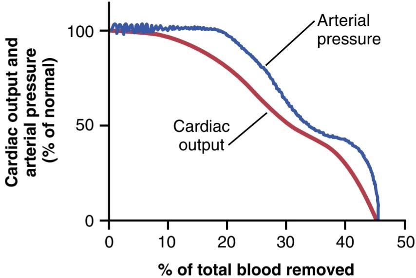
Figure 24-1 Effect of hemorrhage on cardiac output and arterial pressure.
شکل 24-1 تأثیر خونریزی بر برون ده قلبی و فشار شریانی.
Sympathetic Reflex Compensations in Shock-Their Special Value to Maintain Arterial Pressure. The de- crease in arterial pressure after hemorrhage, as well as decreases in pressures in the pulmonary arteries and veins in the thorax, cause powerful sympathetic reflexes (initiated mainly by the arterial baroreceptors and other vascular stretch receptors, as explained in Chapter 18). These reflexes stimulate the sympathetic vasoconstrictor system in most tissues of the body, resulting in three important effects:
جبرانهای رفلکس سمپاتیک در شوک – ارزش ویژه آنها برای حفظ فشار شریانی. کاهش فشار شریانی پس از خونریزی، و همچنین کاهش فشار در شریانهای ریوی و سیاهرگهای قفسه سینه، باعث ایجاد رفلکسهای سمپاتیک قوی میشود (که عمدتاً توسط بارورسپتورهای شریانی و سایر گیرندههای کشش عروقی آغاز میشوند، همانطور که در فصل 18 توضیح داده شد). این رفلکس ها سیستم منقبض کننده عروق سمپاتیک را در اکثر بافت های بدن تحریک می کنند و در نتیجه سه اثر مهم را به همراه دارند:
1. The arterioles constrict in most parts of the systemic circulation, thereby increasing the total peripheral resistance.
1. شریان ها در بیشتر قسمت های گردش خون سیستمیک منقبض می شوند و در نتیجه مقاومت کلی محیطی افزایش می یابد.
2. The veins and venous reservoirs constrict, thereby helping maintain adequate venous return, despite diminished blood volume.
2. سیاهرگها و مخازن وریدی منقبض میشوند و در نتیجه با وجود کاهش حجم خون، به حفظ بازگشت وریدی کافی کمک میکنند.
3. Heart activity increases markedly, sometimes in- creasing the heart rate from the normal value of 72 beats/min to as high as 160 to 180 beats/min.
3. فعالیت قلب به طور قابل توجهی افزایش می یابد، گاهی اوقات ضربان قلب از مقدار طبیعی 72 ضربه در دقیقه به 160 تا 180 ضربه در دقیقه افزایش می یابد.
In the absence of the sympathetic reflexes, only 15% to 20% of the blood volume can be removed over a period of 30 minutes before a person dies; in contrast, a person can sustain a 30% to 40% loss of blood volume when the reflexes are intact. Therefore, these reflexes extend the amount of blood loss that can occur without causing death to about twice that which is possible in their absence.
در غیاب رفلکس های سمپاتیک، تنها 15 تا 20 درصد حجم خون را می توان در مدت 30 دقیقه قبل از مرگ فرد برداشت. در مقابل، زمانی که رفلکس ها دست نخورده باشند، یک فرد می تواند 30 تا 40 درصد از دست دادن حجم خون را تحمل کند. بنابراین، این رفلکس ها میزان از دست دادن خون را که می تواند بدون ایجاد مرگ اتفاق بیفتد، تا حدود دو برابر میزان از دست دادن خون در غیاب آنها افزایش می دهد.
Greater Effect of Sympathetic Nervous Reflexes in Maintaining Arterial Pressure Than in Maintaining Cardiac Output. Referring again to Figure 24-1, note that the arterial pressure is maintained at or near normal levels in the hemorrhaging person longer than is the cardiac output. The reason for this difference is that the sympathetic reflexes are geared more for maintaining arterial pressure than for maintaining cardiac output. They increase the arterial pressure mainly by increasing the total peripheral resistance, which has no beneficial effect on cardiac output. However, the sympathetic constriction of the veins is important to keep venous return and cardiac output from falling too much, in addition to their role in maintaining arterial pressure.
تأثیر بیشتر رفلکس های عصبی سمپاتیک در حفظ فشار شریانی نسبت به حفظ برون ده قلبی. با مراجعه مجدد به شکل 24-1، توجه داشته باشید که فشار شریانی در سطح طبیعی یا نزدیک به سطح طبیعی در فرد خونریزی دهنده بیشتر از برون ده قلبی حفظ می شود. دلیل این تفاوت این است که رفلکس های سمپاتیک بیشتر برای حفظ فشار شریانی هستند تا برای حفظ برون ده قلبی. آنها فشار شریانی را عمدتاً با افزایش مقاومت کلی محیطی افزایش می دهند که تأثیر مفیدی بر برون ده قلبی ندارد. با این حال، انقباض سمپاتیک وریدها علاوه بر نقش آنها در حفظ فشار شریانی، برای جلوگیری از افت بیش از حد بازگشت وریدی و برون ده قلبی مهم است.
Especially interesting is the second plateau occurring at about 50 mm Hg in the arterial pressure curve of Figure 24-1. This second plateau results from activation of the central nervous system ischemic response, which causes extreme stimulation of the sympathetic nervous system when the brain begins to experience lack of oxygen or excess buildup of carbon dioxide, as discussed in Chapter 18. This effect of the central nervous system ischemic response can be called the “last-ditch stand” of the sympathetic reflexes in their attempt to keep the arterial pressure from falling too low.
به خصوص جالب توجه فلات دوم در حدود 50 میلی متر جیوه در منحنی فشار شریانی شکل 24-1 است. این فلات دوم ناشی از فعال شدن پاسخ ایسکمیک سیستم عصبی مرکزی است که باعث تحریک شدید سیستم عصبی سمپاتیک می شود، زمانی که مغز شروع به تجربه کمبود اکسیژن یا تجمع بیش از حد دی اکسید کربن می کند، همانطور که در فصل 18 بحث شد. از پایین افتادن خیلی پایین
Protection of Coronary and Cerebral Blood Flow by the Reflexes. A special value of the maintenance of normal arterial pressure, even in the presence of decreasing cardiac output, is protection of blood flow through the coronary and cerebral circulations. The sympathetic stimulation does not cause significant constriction of the cerebral or cardiac vessels. In addition, in both vascular beds, local blood flow autoregulation is excellent, which prevents moderate decreases in arterial pressure from significantly decreasing their blood flows. Therefore, blood flow through the heart and brain is maintained essentially at normal levels as long as the mean arterial pressure does not fall below about 70 mm Hg, despite the fact that blood flow in some other areas of the body might be decreased to as little as one-third to one-quarter normal by this time because of vasoconstriction.
محافظت از جریان خون کرونر و مغز توسط رفلکس ها. یک ارزش ویژه برای حفظ فشار شریانی طبیعی، حتی در صورت کاهش برون ده قلبی، محافظت از جریان خون از طریق گردش خون کرونر و مغز است. تحریک سمپاتیک باعث انقباض قابل توجهی در عروق مغزی یا قلبی نمی شود. علاوه بر این، در هر دو بستر عروقی، خودتنظیمی جریان خون موضعی عالی است، که از کاهش متوسط فشار شریانی و کاهش قابل توجه جریان خون آنها جلوگیری می کند. بنابراین، جریان خون در قلب و مغز تا زمانی که فشار متوسط شریانی کمتر از حدود 70 میلیمتر جیوه نباشد، اساساً در سطوح طبیعی حفظ میشود، علیرغم این واقعیت که جریان خون در برخی مناطق دیگر بدن ممکن است تا این زمان به دلیل انقباض عروق به یک سوم تا یک چهارم طبیعی کاهش یابد.
PROGRESSIVE AND NONPROGRESSIVE HEMORRHAGIC SHOCK
Figure 24-2 shows an experiment that demonstrates the effects of different degrees of sudden acute hem- orrhage on the subsequent course of arterial pressure. The animals in this experiment were anesthetized and bled rapidly until their arterial pressures fell to different levels. The animals whose pressures fell immediately to no lower than 45 mm Hg (groups I, II, and III) all eventually recovered; the recovery occurred rapidly if the pressure fell only slightly (group I) but occurred slowly if it fell almost to the 45-mm Hg level (group III). When the arterial pressure fell below 45 mm Hg (groups IV, V, and VI), all the animals died, although many of them hovered between life and death for hours before the circulatory system deteriorated to the stage of death.
شوک هموراژیک پیشرونده و غیرپیشرونده
شکل 24-2 آزمایشی را نشان می دهد که اثرات درجات مختلف خونریزی حاد ناگهانی را بر روند بعدی فشار شریانی نشان می دهد. حیوانات در این آزمایش بیهوش شدند و تا زمانی که فشار شریانی آنها به سطوح مختلف کاهش یافت، به سرعت خونریزی کردند. حیواناتی که فشار آنها بلافاصله به کمتر از 45 میلیمتر جیوه کاهش یافت (گروههای I، II و III) همگی در نهایت بهبود یافتند. اگر فشار فقط اندکی کاهش یابد (گروه I) بهبود به سرعت رخ می دهد، اما اگر تقریباً به سطح 45 میلی متر جیوه (گروه III) کاهش یابد، به آرامی رخ می دهد. هنگامی که فشار شریانی به زیر 45 میلیمتر جیوه رسید (گروههای IV، V و VI)، همه حیوانات مردند، اگرچه بسیاری از آنها ساعتها قبل از اینکه سیستم گردش خون به مرحله مرگ بدتر شود، بین زندگی و مرگ معلق ماندند.
This experiment demonstrates that the circulatory system can recover as long as the degree of hemorrhage is no greater than a certain critical amount. Crossing this critical threshold by even a few milliliters of blood loss makes the eventual difference between life and death. Thus, hemorrhage beyond a certain critical level causes shock to become progressive. That is, the shock itself causes still more shock, and the condition becomes a vicious cycle that eventually leads to deterioration of the circulation and to death.
این آزمایش نشان می دهد که سیستم گردش خون می تواند بهبود یابد تا زمانی که درجه خونریزی از مقدار بحرانی معینی بیشتر نباشد. عبور از این آستانه بحرانی حتی با چند میلی لیتر از دست دادن خون، تفاوت نهایی بین زندگی و مرگ را ایجاد می کند. بنابراین، خونریزی فراتر از یک سطح بحرانی خاص باعث می شود شوک پیشرونده شود. یعنی خود شوک باعث شوک بیشتر می شود و این وضعیت به یک چرخه معیوب تبدیل می شود که در نهایت منجر به بدتر شدن گردش خون و مرگ می شود.
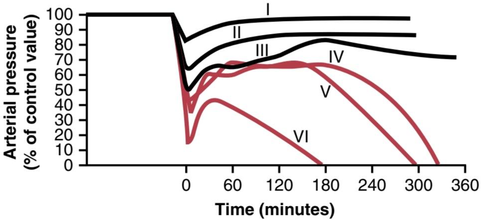
Figure 24-2 Time course of arterial pressure in dogs after different degrees of acute hemorrhage. Each curve represents average results from six dogs (curves I-VI).
شکل 24-2 دوره زمانی فشار شریانی در سگ ها پس از درجات مختلف خونریزی حاد. هر منحنی نشان دهنده نتایج متوسط از شش سگ است (منحنی I-VI).
Nonprogressive Shock-Compensated Shock
If shock is not severe enough to cause its own progression, the person eventually recovers. Therefore, shock of this lesser degree is called nonprogressive shock or compensated shock, meaning that the sympathetic reflexes and other factors compensate enough to prevent further deterioration of the circulation.
شوک غیر پیشرونده جبران شده با شوک
اگر شوک به اندازه کافی شدید نباشد که باعث پیشرفت خود شود، فرد در نهایت بهبود می یابد. بنابراین شوک با این درجه کمتر، شوک غیرپیشرونده یا شوک جبرانی نامیده می شود، به این معنی که رفلکس های سمپاتیک و سایر عوامل به اندازه کافی جبران می کنند تا از بدتر شدن بیشتر گردش خون جلوگیری کنند.
The factors that cause a person to recover from moderate degrees of shock are the negative feedback control mechanisms of the circulation that attempt to return cardiac output and arterial pressure back to normal levels. They include the following:
عواملی که باعث بهبودی فرد از شوک درجات متوسط می شود مکانیسم های کنترل بازخورد منفی گردش خون است که تلاش می کند برون ده قلبی و فشار شریانی را به سطح طبیعی بازگرداند. آنها شامل موارد زیر است:
1. Baroreceptor reflexes, which elicit powerful sympathetic stimulation of the circulation
1. رفلکس های گیرنده بارور، که باعث تحریک سمپاتیک قوی گردش خون می شوند.
2. Central nervous system ischemic response, which elicits even more powerful sympathetic stimulation throughout the body but is not activated significantly until the arterial pressure falls below 50 mm Hg
2. پاسخ ایسکمیک سیستم عصبی مرکزی، که تحریک سمپاتیک قویتری را در سراسر بدن ایجاد میکند، اما تا زمانی که فشار شریانی به زیر 50 میلیمتر جیوه نرسد، به طور قابل توجهی فعال نمیشود.
3. Reverse stress-relaxation of the circulatory system, which causes the blood vessels to contract around the diminished blood volume so that the blood volume that is available more adequately fills the circulation
3. استرس-آرامش معکوس سیستم گردش خون، که باعث می شود رگ های خونی در اطراف حجم خون کاهش یافته منقبض شوند تا حجم خونی که در دسترس است به اندازه کافی گردش خون را پر کند.
4. Increased secretion of renin by the kidneys and formation of angiotensin II, which constricts the peripheral arterioles and also causes decreased output of water and salt by the kidneys, both of which help prevent progression of shock
4. افزایش ترشح رنین توسط کلیه ها و تشکیل آنژیوتانسین II که شریان های محیطی را منقبض می کند و همچنین باعث کاهش خروجی آب و نمک توسط کلیه ها می شود که هر دو به جلوگیری از پیشرفت شوک کمک می کنند.
5. Increased secretion by the posterior pituitary gland of vasopressin (antidiuretic hormone), which constricts the peripheral arterioles and veins and greatly increases water retention by the kidneys
5. افزایش ترشح وازوپرسین (هورمون ضد ادرار) توسط غده هیپوفیز خلفی که شریان ها و وریدهای محیطی را منقبض می کند و احتباس آب توسط کلیه ها را به شدت افزایش می دهد.
6. Increased secretion by the adrenal medullae of epinephrine and norepinephrine, which constricts the peripheral arterioles and veins and increases the heart rate
6. افزایش ترشح اپی نفرین و نوراپی نفرین توسط بصل الکلیوی آدرنال که شریان ها و سیاهرگ های محیطی را منقبض می کند و ضربان قلب را افزایش می دهد.
7. Compensatory mechanisms that return the blood volume back toward normal, including absorption of large quantities of fluid from the intestinal tract, absorption of fluid into the blood capillaries from the interstitial spaces of the body, conservation of water and salt by the kidneys, and increased thirst and increased appetite for salt, which make the person drink water and eat salty foods if they are able to do so
7. مکانیسم های جبرانی که حجم خون را به حالت طبیعی برمی گرداند، از جمله جذب مقدار زیادی مایع از مجرای روده، جذب مایع به مویرگ های خونی از فضاهای بین بافتی بدن، حفظ آب و نمک توسط کلیه ها و افزایش تشنگی و افزایش اشتها برای نمک، که باعث می شود فرد در صورت نوشیدن آب و غذای شور قادر به نوشیدن آب و نمک باشد.
The sympathetic reflexes and increased secretion of catecholamines by the adrenal medullae provide rapid help toward bringing about recovery because they become maximally activated within 30 seconds to a few minutes after hemorrhage.
رفلکسهای سمپاتیک و افزایش ترشح کاتکول آمینها توسط بصلالعدلی آدرنال کمک سریعی به بهبودی میکنند، زیرا در عرض 30 ثانیه تا چند دقیقه پس از خونریزی حداکثر فعال میشوند.
The angiotensin and vasopressin mechanisms, as well as the reverse stress-relaxation that causes contraction of the blood vessels and venous reservoirs, all require 10 to 60 minutes to respond completely, but they aid greatly in increasing the arterial pressure or increasing the circulatory filling pressure, thereby increasing the return of blood to the heart.
مکانیسمهای آنژیوتانسین و وازوپرسین و همچنین آرامش معکوس استرس که باعث انقباض رگهای خونی و مخازن وریدی میشود، همگی به 10 تا 60 دقیقه برای پاسخ کامل نیاز دارند، اما به افزایش فشار شریانی یا افزایش فشار پر شدن گردش خون کمک زیادی میکنند و در نتیجه بازگشت خون به قلب را افزایش میدهند.
Finally, readjustment of blood volume by absorption of fluid from the interstitial spaces and intestinal tract, as well as oral ingestion and absorption of additional quantities of water and salt, may require from 1 to 48 hours, but recovery eventually takes place, provided the shock does not become severe enough to enter the progressive stage.
در نهایت، تنظیم مجدد حجم خون با جذب مایع از فضاهای بینابینی و مجرای روده، و همچنین مصرف خوراکی و جذب مقادیر اضافی آب و نمک، ممکن است از 1 تا 48 ساعت نیاز داشته باشد، اما بهبودی در نهایت صورت میگیرد، مشروط بر اینکه شوک به اندازه کافی شدید نباشد تا وارد مرحله پیشرونده شود.
Progressive Shock-Caused by Vicious Cycle of Cardiovascular Deterioration
Figure 24-3 shows some of the positive feedbacks that further depress cardiac output in shock, thus causing the shock to become progressive. Some of the more important feedbacks are described in the following sections.
شوک پیشرونده ناشی از چرخه معیوب زوال قلبی عروقی
شکل 24-3 برخی از بازخوردهای مثبت را نشان می دهد که برون ده قلبی را در شوک کاهش می دهد و در نتیجه شوک را پیشرونده می کند. برخی از بازخوردهای مهمتر در بخشهای زیر توضیح داده شدهاند.
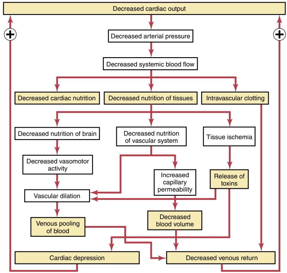
Figure 24-3 Different types of positive feedback that can lead to the progression of shock.
شکل 24-3 انواع مختلف بازخورد مثبت که می تواند منجر به پیشرفت شوک شود.
Cardiac Depression. When the arterial pressure falls low enough, coronary blood flow decreases below that required for adequate nutrition of the myocardium, weakening the heart muscle and decreasing the cardiac output more. Thus, a positive feedback cycle has developed, whereby the shock becomes more and more severe.
افسردگی قلبی. هنگامی که فشار شریانی به اندازه کافی پایین میآید، جریان خون کرونر کمتر از مقدار مورد نیاز برای تغذیه کافی میوکارد کاهش مییابد و عضله قلب را ضعیف میکند و برون ده قلبی را بیشتر کاهش میدهد. بنابراین، یک چرخه بازخورد مثبت ایجاد شده است که به موجب آن شوک بیشتر و شدیدتر می شود.
Figure 24-4 shows cardiac output curves extrapolated to the human heart from studies in experimental animals, demonstrating progressive deterioration of the heart at different times after the onset of shock. An anesthetized animal was bled until the arterial pressure fell to 30 mm Hg, and the pressure was held at this level by further bleeding or retransfusion of blood, as required. Note from the second curve in the figure that there was little deterioration of the heart during the first 2 hours, but by 4 hours, the heart had deteriorated about 40%. Then, rap- idly, during the last hour of the experiment (after 4 hours of low coronary blood pressure), the heart deteriorated completely.
شکل 24-4 منحنیهای برون ده قلبی را نشان میدهد که از مطالعات روی حیوانات آزمایشی به قلب انسان برونداده شدهاند، و زوال پیشرونده قلب را در زمانهای مختلف پس از شروع شوک نشان میدهند. یک حیوان بیهوش تا زمانی که فشار شریانی به 30 میلیمتر جیوه کاهش یابد، خونریزی شد و فشار در این سطح با خونریزی بیشتر یا انتقال مجدد خون در صورت لزوم حفظ شد. از منحنی دوم در شکل توجه کنید که در 2 ساعت اول وخامت کمی قلب وجود داشت، اما در 4 ساعت، قلب حدود 40٪ تحلیل رفته بود. سپس به سرعت در آخرین ساعت آزمایش (پس از 4 ساعت کاهش فشار خون کرونری)، قلب به طور کامل بدتر شد.
Thus, one of the important features of progressive shock, whether it is hemorrhagic in origin or has another cause, is eventual progressive deterioration of the heart. In the early stages of shock, this deterioration plays very little role in the condition of the person, partly because deterioration of the heart is not severe during the first hour or so of shock, but mainly because the heart has reserve capability that normally allows it to pump 300% to 400% more blood than is required by the body for adequate tissue nutrition. In the latest stages of shock, however, deterioration of the heart is probably the most important factor in the final lethal progression of the shock.
بنابراین، یکی از ویژگیهای مهم شوک پیشرونده، چه منشأ هموراژیک داشته باشد و چه علت دیگری داشته باشد، زوال تدریجی قلب است. در مراحل اولیه شوک، این وخامت نقش بسیار کمی در وضعیت فرد ایفا میکند، تا حدی به این دلیل که وخامت قلب در طول یک ساعت اولیه شوک شدید نیست، اما عمدتاً به این دلیل که قلب دارای قابلیت ذخیره است که به طور معمول به آن اجازه میدهد 300 تا 400 درصد بیشتر از آنچه بدن برای تغذیه مناسب بافت نیاز دارد، خون پمپاژ کند. با این حال، در آخرین مراحل شوک، بدتر شدن قلب احتمالاً مهمترین عامل در پیشرفت نهایی کشنده شوک است.

Figure 24-4 Cardiac output curves of the heart at different times after hemorrhagic shock begins. (These curves have been extrapolated to the human heart from data obtained in dog experiments by Dr. J.W. Crowell.)
شکل 24-4 منحنی های برون ده قلبی در زمان های مختلف پس از شروع شوک هموراژیک. (این منحنی ها بر اساس داده های به دست آمده در آزمایشات سگ توسط دکتر J.W. Crowell به قلب انسان تعمیم داده شده است.)
Vasomotor Failure. In the early stages of shock, various circulatory reflexes cause intense activity of the sympathetic nervous system. This activity helps delay depression of cardiac output and especially helps prevent decreased arterial pressure. However, there comes a point when diminished blood flow to the brain’s vasomotor center depresses the center so much that it, too, becomes progressively less active and, finally, totally inactive. For example, during the first 4 to 8 minutes, complete circulatory arrest to the brain causes the most intense of all sympathetic discharges, but by the end of 10 to 15 minutes, the vasomotor center becomes so depressed that no further evidence of sympathetic discharge can be demonstrated. Fortunately, the vasomotor center usually does not fail in the early stages of shock if the arterial pressure remains above 30 mm Hg.
نارسایی وازوموتور. در مراحل اولیه شوک، رفلکس های مختلف گردش خون باعث فعالیت شدید سیستم عصبی سمپاتیک می شود. این فعالیت به تاخیر انداختن برون ده قلبی کمک می کند و به ویژه به جلوگیری از کاهش فشار شریانی کمک می کند. با این حال، زمانی فرا می رسد که کاهش جریان خون به مرکز وازوموتور مغز، آن مرکز را به قدری تحت فشار قرار می دهد که آن نیز به تدریج کمتر فعال و در نهایت کاملاً غیرفعال می شود. به عنوان مثال، در طول 4 تا 8 دقیقه اول، توقف کامل گردش خون در مغز باعث شدیدترین ترشحات سمپاتیک می شود، اما در پایان 10 تا 15 دقیقه، مرکز وازوموتور چنان افسرده می شود که هیچ شواهد دیگری از ترشح سمپاتیک نمی توان نشان داد. خوشبختانه، اگر فشار شریانی بالای 30 میلی متر جیوه باقی بماند، معمولاً مرکز وازوموتور در مراحل اولیه شوک از کار نمی افتد.
Blockage of Very Small Vessels by Sludged Blood. In time, blockage occurs in many of the very small blood vessels in the circulatory system, and this blockage also causes the shock to progress. The initiating cause of this blockage is sluggish blood flow in the microvessels. Because tissue metabolism continues despite the low flow, large amounts of acid, both carbonic acid and lactic acid, continue to empty into the local blood vessels and greatly increase the local acidity of the blood. This acidic effect, plus other deterioration products from the ischemic tissues, causes local blood agglutination, resulting in minute blood clots and leading to very small plugs in the small vessels. Even if the vessels do not become plugged, an increased tendency for the blood cells to stick to one another makes it more difficult for blood to flow through the microvasculature, giving rise to the term sludged blood.
انسداد عروق بسیار کوچک توسط خون لجن شده. با گذشت زمان، انسداد در بسیاری از رگ های خونی بسیار کوچک در سیستم گردش خون رخ می دهد و این انسداد همچنین باعث پیشرفت شوک می شود. علت شروع این انسداد، جریان خون کند در رگهای کوچک است. از آنجایی که متابولیسم بافت با وجود جریان کم ادامه می یابد، مقادیر زیادی اسید، هم اسید کربنیک و هم اسید لاکتیک، همچنان در رگ های خونی موضعی تخلیه می شوند و اسیدیته موضعی خون را تا حد زیادی افزایش می دهند. این اثر اسیدی، به علاوه سایر محصولات زوال بافتهای ایسکمیک، باعث آگلوتیناسیون موضعی خون میشود، که منجر به لختههای خونی کوچک میشود و منجر به ایجاد پلاگهای بسیار کوچک در رگهای کوچک میشود. حتی اگر رگها مسدود نشوند، افزایش تمایل سلولهای خونی به چسبیدن به یکدیگر جریان خون را از طریق ریز عروق دشوارتر میکند و اصطلاح خون لجنشده را به وجود میآورد.
Increased Capillary Permeability. After many hours of capillary hypoxia and lack of other nutrients, the permeability of the capillaries gradually increases, and large quantities of fluid begin to transude into the tissues. This phenomenon decreases the blood volume even more, with a resultant further decrease in cardiac output, making the shock still more severe. Capillary hypoxia does not cause increased capillary permeability until the late stages of prolonged shock.
افزایش نفوذپذیری مویرگی. پس از ساعتها هیپوکسی مویرگی و کمبود مواد مغذی دیگر، نفوذپذیری مویرگها به تدریج افزایش مییابد و مقادیر زیادی مایع شروع به ترانسود به داخل بافتها میکند. این پدیده حجم خون را حتی بیشتر کاهش می دهد و در نتیجه برون ده قلبی را کاهش می دهد و شوک را همچنان شدیدتر می کند. هیپوکسی مویرگی تا مراحل پایانی شوک طولانی مدت باعث افزایش نفوذپذیری مویرگی نمی شود.
Release of Toxins by Ischemic Tissue. Shock has been suggested to cause tissues to release toxic substances, such as histamine, serotonin, and tissue enzymes, that cause further deterioration of the circulatory system. Experimental studies have proved the significance of at least one toxin, endotoxin, in some types of shock.
آزادسازی سموم توسط بافت ایسکمیک. شوک برای ترشح مواد سمی مانند هیستامین، سروتونین و آنزیم های بافتی که باعث زوال بیشتر سیستم گردش خون می شود، پیشنهاد شده است. مطالعات تجربی اهمیت حداقل یک سم به نام اندوتوکسین را در برخی از انواع شوک ثابت کرده است.
Cardiac Depression Caused by Endotoxin. Endotoxin is released from the bodies of dead gram-negative bacteria in the intestines. Diminished blood flow to the intestines often causes enhanced formation and absorption of this toxic substance. The circulating toxin then causes increased cellular metabolism, despite inadequate nutrition of the cells, which has a specific effect on the heart muscle, causing cardiac depression. Endotoxin can play a major role in some types of shock, especially septic shock, discussed later in this chapter.
افسردگی قلبی ناشی از اندوتوکسین. اندوتوکسین از بدن باکتری های گرم منفی مرده در روده آزاد می شود. کاهش جریان خون به روده اغلب باعث افزایش تشکیل و جذب این ماده سمی می شود. سپس سم در گردش باعث افزایش متابولیسم سلولی می شود، علیرغم تغذیه ناکافی سلول ها، که تأثیر خاصی بر عضله قلب دارد و باعث افسردگی قلبی می شود. اندوتوکسین می تواند نقش عمده ای در برخی از انواع شوک، به ویژه شوک سپتیک، که در ادامه این فصل مورد بحث قرار می گیرد، ایفا کند.
Generalized Cellular Deterioration. As shock becomes severe, many signs of generalized cellular deterioration occur throughout the body. One organ especially affected is the liver, as illustrated in Figure 24-5. The liver is especially affected mainly because of the lack of enough nutrients to support the normally high rate of metabolism in liver cells, but also partly because of the exposure of the liver cells to any vascular toxin or other abnormal metabolic factor occurring in shock.
زوال عمومی سلولی. همانطور که شوک شدید می شود، بسیاری از علائم زوال سلولی عمومی در سراسر بدن رخ می دهد. همانطور که در شکل 24-5 نشان داده شده است، یکی از اعضایی که به ویژه تحت تأثیر قرار می گیرد، کبد است. کبد عمدتاً به دلیل کمبود مواد مغذی کافی برای حمایت از میزان متابولیسم بالا در سلولهای کبدی تحت تأثیر قرار میگیرد، اما تا حدی به دلیل قرار گرفتن سلولهای کبدی در معرض هر گونه سم عروقی یا سایر عوامل متابولیک غیرطبیعی که در شوک رخ میدهد، تحت تأثیر قرار میگیرد.
Among the damaging cellular effects that are known to occur in most body tissues are the following:
از جمله اثرات مخرب سلولی که در بیشتر بافتهای بدن رخ میدهد، موارد زیر است:
1. Active transport of sodium and potassium through the cell membrane is greatly diminished. As a result, sodium and chloride accumulate in the cells, and potassium is lost from the cells. In addition, the cells begin to swell.
1. انتقال فعال سدیم و پتاسیم از طریق غشای سلولی تا حد زیادی کاهش می یابد. در نتیجه، سدیم و کلرید در سلولها جمع میشوند و پتاسیم از سلولها از بین میرود. علاوه بر این، سلول ها شروع به متورم شدن می کنند.
2. Mitochondrial activity in the liver cells, as well as in many other tissues of the body, becomes severely depressed.
2. فعالیت میتوکندری در سلول های کبد و همچنین در بسیاری از بافت های دیگر بدن به شدت افسرده می شود.
3. Lysosomes in the cells in widespread tissue areas begin to break open, with intracellular release of hydrolases, which cause further intracellular deterioration.
3. لیزوزوم ها در سلول ها در نواحی بافت گسترده شروع به شکستن می کنند، با آزاد شدن درون سلولی هیدرولازها، که باعث زوال بیشتر داخل سلولی می شود.
4. Cellular metabolism of nutrients, such as glucose, eventually becomes greatly depressed in the last stages of shock. The actions of some hormones are depressed as well, including almost 100% depression of the actions of insulin.
4. متابولیسم سلولی مواد مغذی، مانند گلوکز، در نهایت در آخرین مراحل شوک به شدت افسرده می شود. عملکرد برخی از هورمون ها نیز افسرده است، از جمله کاهش تقریباً 100 درصدی عملکرد انسولین.
All these effects contribute to further deterioration of many organs of the body, including especially the following: (1) the liver, with depression of its many metabolic and detoxification functions; (2) the lungs, with eventual development of pulmonary edema and poor ability to oxygenate the blood; and (3) the heart, thereby further depressing its contractility.
همه این اثرات به زوال بیشتر بسیاری از اندامهای بدن، از جمله موارد زیر کمک میکنند: (1) کبد، همراه با کاهش عملکردهای متابولیکی و سمزدایی آن. (2) ریه ها، با ایجاد ادم ریوی و توانایی ضعیف برای اکسیژن رسانی به خون. و (3) قلب، در نتیجه انقباض آن را بیشتر کاهش می دهد.
Patchy Areas of Tissue Necrosis Caused by Patchy Blood Flows in Different Organs. Not all cells of the body are equally damaged by shock because some tissues have better blood supplies than others. For example, the cells adjacent to the arterial ends of capillaries receive better nutrition than cells adjacent to the venous ends of the same capillaries. Therefore, more nutritive deficiency occurs around the venous ends of capillaries than elsewhere. Figure 24-5 shows necrosis in the center of a liver lobule, the portion of the lobule that is the last to be exposed to the blood as it passes through the liver sinusoids.
نواحی تکه تکه نکروز بافتی ناشی از جریان خون تکه تکه در اندام های مختلف. همه سلول های بدن به یک اندازه در اثر شوک آسیب نمی بینند، زیرا برخی از بافت ها خون بهتری نسبت به سایرین دارند. به عنوان مثال، سلول های مجاور انتهای شریانی مویرگ ها تغذیه بهتری نسبت به سلول های مجاور انتهای وریدی همان مویرگ ها دریافت می کنند. بنابراین، کمبود مواد مغذی در اطراف انتهای وریدی مویرگ ها بیشتر از جاهای دیگر رخ می دهد. شکل 24-5 نکروز را در مرکز یک لوبول کبد نشان می دهد، بخشی از لوبول که آخرین لوبولی است که هنگام عبور از سینوسی های کبد در معرض خون قرار می گیرد.
Similar punctate lesions occur in heart muscle, although here a definite repetitive pattern, such as occurs in the liver, cannot be demonstrated. Nevertheless, the cardiac lesions play an important role in leading to the final irreversible stage of shock. Deteriorative lesions also occur in the kidneys, especially in the epithelium of the kidney tubules, leading to kidney failure and occasionally uremic death several days later. Deterioration of the lungs also often leads to respiratory distress and death several days later, called the shock lung syndrome.
ضایعات نقطهگذاری مشابهی در عضله قلب رخ میدهد، اگرچه در اینجا یک الگوی تکراری مشخص، مانند آنچه در کبد رخ میدهد، نمیتوان نشان داد. با این وجود، ضایعات قلبی نقش مهمی در منجر به آخرین مرحله غیر قابل برگشت شوک دارند. ضایعات بدتر نیز در کلیه ها به خصوص در اپیتلیوم لوله های کلیه ایجاد می شود که منجر به نارسایی کلیه و گاهاً مرگ اورمیک چند روز بعد می شود. بدتر شدن ریه ها نیز اغلب منجر به ناراحتی تنفسی و مرگ چند روز بعد می شود که به آن سندرم شوک ریه می گویند.
Acidosis in Shock. Metabolic derangements that occur in shocked tissue can lead to acidosis throughout the body. This results from poor delivery of oxygen to the tissues, which greatly diminishes oxidative metabolism of the foodstuffs. When this occurs, the cells obtain most of their energy by the anaerobic process of glycolysis, which leads to excess lactic acid in the blood. In addition, poor blood flow through tissues prevents normal removal of carbon dioxide. The carbon dioxide reacts locally in the cells with water to form high concentrations of intracellular carbonic acid, which, in turn, reacts with various tissue chemicals to form additional intracellular acidic substances. Thus, another deteriorative effect of shock is generalized and local tissue acidosis, leading to further progression of the shock.
اسیدوز در شوک. اختلالات متابولیک که در بافت شوک شده رخ می دهد می تواند منجر به اسیدوز در سراسر بدن شود. این امر ناشی از تحویل ضعیف اکسیژن به بافت ها است که متابولیسم اکسیداتیو مواد غذایی را تا حد زیادی کاهش می دهد. هنگامی که این اتفاق می افتد، سلول ها بیشتر انرژی خود را از طریق فرآیند بی هوازی گلیکولیز به دست می آورند که منجر به اسید لاکتیک اضافی در خون می شود. علاوه بر این، جریان خون ضعیف در بافت ها مانع از حذف طبیعی دی اکسید کربن می شود. دی اکسید کربن به صورت موضعی در سلول ها با آب واکنش می دهد و غلظت بالایی از اسید کربنیک درون سلولی را تشکیل می دهد که به نوبه خود با مواد شیمیایی بافتی مختلف واکنش داده و مواد اسیدی داخل سلولی اضافی را تشکیل می دهد. بنابراین، یکی دیگر از اثرات مخرب شوک، اسیدوز بافتی عمومی و موضعی است که منجر به پیشرفت بیشتر شوک می شود.
Positive Feedback Deterioration of Tissues in Shock and Vicious Cycle of Progressive Shock. All the factors just discussed that can lead to further progression of shock are types of positive feedback—that is, each in- crease in the degree of shock causes a further increase in the shock. However, positive feedback does not necessarily lead to a vicious cycle. Development of a vicious cycle depends on the intensity of the positive feedback. In mild degrees of shock, the negative feedback mechanisms of the circulation, including sympathetic reflexes, reverse stress-relaxation mechanism of the blood reservoirs, and absorption of fluid into the blood from the interstitial spaces, can easily overcome the positive feedback influences and, therefore, cause recovery. In severe shock, however, the deteriorative feedback mechanisms become more and more powerful, leading to such rapid deterioration of the circulation that all the normal negative feedback systems of circulatory control acting together can- not return the cardiac output to normal.
بازخورد مثبت بدتر شدن بافت ها در شوک و چرخه باطل شوک پیشرونده. تمام عواملی که اخیراً مورد بحث قرار گرفت و میتوانند منجر به پیشرفت بیشتر شوک شوند، انواع بازخورد مثبت هستند، یعنی هر افزایش در درجه شوک باعث افزایش بیشتر شوک میشود. با این حال، بازخورد مثبت لزوما به یک چرخه معیوب منجر نمی شود. توسعه یک چرخه معیوب به شدت بازخورد مثبت بستگی دارد. در درجات خفیف شوک، مکانیسم های بازخورد منفی گردش خون، از جمله رفلکس های سمپاتیک، مکانیسم معکوس استرس-آرامش مخازن خون، و جذب مایع به خون از فضاهای بینابینی، می توانند به راحتی بر تأثیرات بازخورد مثبت غلبه کنند و در نتیجه باعث بهبودی شوند. با این حال، در شوک شدید، مکانیسمهای بازخورد بدتر قویتر و قویتر میشوند و منجر به بدتر شدن سریع گردش خون میشوند که تمام سیستمهای بازخورد منفی طبیعی کنترل گردش خون که با هم عمل میکنند نمیتوانند برون ده قلبی را به حالت عادی برگردانند.
Considering once again the principles of positive feedback and vicious cycles discussed in Chapter 1, one can readily understand why there is a critical cardiac output level above which a person in shock recovers and below which a person enters a vicious cycle of circulatory deterioration that proceeds until death.
با در نظر گرفتن یک بار دیگر اصول بازخورد مثبت و چرخههای معیوب که در فصل 1 مورد بحث قرار گرفت، میتوان به راحتی درک کرد که چرا یک سطح برون ده قلبی بحرانی وجود دارد که بالاتر از آن فرد در شوک بهبود مییابد و در زیر آن فرد وارد یک چرخه معیوب از زوال گردش خون میشود که تا زمان مرگ ادامه دارد.

Figure 24-5 Necrosis of the central portion of a liver lobule during severe circulatory shock. (Courtesy Dr. J.W. Crowell.)
شکل 24-5 نکروز بخش مرکزی لوبول کبد در طول شوک شدید گردش خون. (با احترام از دکتر J.W. Crowell.)
IRREVERSIBLE SHOCK
After shock has progressed to a certain stage, transfusion or any other type of therapy becomes incapable of saving the person’s life. The person is then said to be in the irreversible stage of shock. Ironically, even in this irreversible stage, therapy can, on rare occasions, return the arterial pressure and even the cardiac output to normal or near normal for short periods, but the circulatory system nevertheless continues to deteriorate, and death ensues in another few minutes to few hours.
شوک برگشت ناپذیر
پس از اینکه شوک به مرحله خاصی رسید، تزریق خون یا هر نوع درمان دیگری در نجات جان فرد ناتوان می شود. سپس گفته می شود که فرد در مرحله برگشت ناپذیر شوک قرار دارد. از قضا، حتی در این مرحله غیرقابل برگشت، درمان در موارد نادر می تواند فشار شریانی و حتی برون ده قلبی را برای دوره های کوتاهی به حالت طبیعی یا نزدیک به نرمال برگرداند، اما با این وجود، سیستم گردش خون همچنان رو به وخامت است و مرگ در چند دقیقه تا چند ساعت دیگر رخ می دهد.
Figure 24-6 shows that transfusion during the irreversible stage can sometimes cause cardiac output (as well as the arterial pressure) to return to nearly normal. However, the cardiac output soon begins to fall again, and subsequent transfusions have less and less effect. By this time, multiple deteriorative changes have occurred in the heart muscle cells that may not necessarily affect the heart’s immediate ability to pump blood but, over a long period, depress heart pumping enough to cause death. Beyond a certain point, so much tissue damage has occurred, so many destructive enzymes have been released into the body fluids, so much acidosis has developed, and so many other destructive factors are now in progress that even a normal cardiac output for a few minutes cannot reverse the continuing deterioration. Therefore, in severe shock, a stage is eventually reached at which the person will die, even though vigorous therapy might still return the cardiac output to normal for short periods.
شکل 24-6 نشان می دهد که تزریق خون در مرحله برگشت ناپذیر گاهی اوقات می تواند باعث شود که برون ده قلبی (و همچنین فشار شریانی) تقریباً به حالت عادی برگردد. با این حال، برون ده قلبی به زودی دوباره شروع به کاهش می کند و تزریق های بعدی اثر کمتر و کمتری دارند. در این زمان، تغییرات مخرب متعددی در سلولهای عضله قلب رخ داده است که ممکن است لزوماً بر توانایی آنی قلب برای پمپاژ خون تأثیر نگذارد، اما در یک دوره طولانی، پمپاژ قلب را به اندازهای کاهش میدهد که باعث مرگ شود. فراتر از یک نقطه خاص، آسیب بافتی بسیار زیادی رخ داده است، آنزیم های مخرب زیادی در مایعات بدن آزاد شده اند، اسیدوز بسیار زیادی ایجاد شده است، و بسیاری از عوامل مخرب دیگر در حال حاضر در حال پیشرفت هستند که حتی یک برون ده قلبی طبیعی برای چند دقیقه نمی تواند ادامه بدتر شدن را معکوس کند. بنابراین، در شوک شدید، در نهایت به مرحلهای میرسد که در آن فرد میمیرد، حتی اگر درمان شدید همچنان برون ده قلبی را برای دورههای کوتاهی به حالت عادی بازگرداند.

Figure 24-6 Failure of transfusion to prevent death in irreversible shock.
شکل 24-6 عدم انتقال خون برای جلوگیری از مرگ در شوک غیرقابل برگشت.
Depletion of Cellular High-Energy Phosphate Reserves in Irreversible Shock. The high-energy phosphate reserves in the tissues of the body, especially in the liver and heart, are greatly diminished in severe shock. Essentially all the creatine phosphate has been degraded, and almost all the adenosine triphosphate has downgraded to adenosine diphosphate, adenosine monophosphate and, eventually, adenosine. Much of this adenosine then diffuses out of the cells into the circulating blood and is converted into uric acid, a substance that cannot re-enter the cells to reconstitute the adenosine phosphate system. New adenosine can be synthesized at a rate of only about 2% of the normal cellular amount an hour, meaning that once the high-energy phosphate stores of the cells are depleted, they are difficult to replenish.
کاهش ذخایر فسفات پرانرژی سلولی در شوک برگشت ناپذیر. ذخایر فسفات پرانرژی در بافت های بدن، به ویژه در کبد و قلب، در شوک شدید تا حد زیادی کاهش می یابد. اساساً تمام کراتین فسفات تجزیه شده است و تقریباً تمام آدنوزین تری فسفات به آدنوزین دی فسفات، آدنوزین مونوفسفات و در نهایت آدنوزین تنزل یافته است. سپس بیشتر این آدنوزین از سلولها به داخل خون در گردش پخش میشود و به اسید اوریک تبدیل میشود، مادهای که نمیتواند دوباره وارد سلولها شود تا سیستم آدنوزین فسفات را بازسازی کند. آدنوزین جدید را می توان با سرعتی حدود 2% از مقدار طبیعی سلولی در ساعت سنتز کرد، به این معنی که وقتی ذخایر فسفات پرانرژی سلول ها تخلیه شد، دوباره پر کردن آنها دشوار است.
Thus, one of the most devastating end results in shock, and the one that is perhaps most significant for development of the final state of irreversibility, is cellular depletion of these high-energy compounds.
بنابراین، یکی از مخرب ترین نتایج نهایی در شوک، و یکی از مهمترین آنها برای ایجاد حالت نهایی برگشت ناپذیری، تخلیه سلولی این ترکیبات پر انرژی است.
HYPOVOLEMIC SHOCK CAUSED BY PLASMA LOSS
Loss of plasma from the circulatory system, even without loss of red blood cells, can sometimes be severe enough to reduce the total blood volume markedly, causing typical hypovolemic shock similar in almost all details to that caused by hemorrhage. Severe plasma loss occurs in the following conditions:
شوک هیپوولمیک ناشی از از دست دادن پلاسما
از دست دادن پلاسما از سیستم گردش خون، حتی بدون از دست دادن گلبول های قرمز، گاهی اوقات می تواند به اندازه ای شدید باشد که حجم کل خون را به طور قابل توجهی کاهش دهد و باعث ایجاد شوک هیپوولمیک معمولی تقریباً در تمام جزئیات مشابه با خونریزی ناشی از خونریزی شود. از دست دادن شدید پلاسما در شرایط زیر رخ می دهد:
1. Intestinal obstruction may cause severely reduced plasma volume. Distention of the intestine in intestinal obstruction partly blocks venous blood flow in the intestinal walls, which increases intestinal capillary pressure, causing fluid to leak from the capillaries into the intestinal walls and intestinal lumen. Because the lost fluid has high protein content, the result is reduced total blood plasma protein, as well as reduced plasma volume.
1. انسداد روده ممکن است باعث کاهش شدید حجم پلاسما شود. اتساع روده در انسداد روده تا حدودی جریان خون وریدی را در دیوارههای روده مسدود میکند که باعث افزایش فشار مویرگ روده میشود و باعث نشت مایع از مویرگها به دیوارههای روده و مجرای روده میشود. از آنجایی که مایع از دست رفته محتوای پروتئین بالایی دارد، نتیجه کاهش پروتئین کل پلاسمای خون و همچنین کاهش حجم پلاسما است.
2. Severe burns or other denuding conditions of the skin cause loss of plasma through the denuded skin areas so that the plasma volume becomes markedly reduced.
2. سوختگی های شدید یا سایر شرایط برهنه شدن پوست باعث از بین رفتن پلاسما از طریق نواحی برهنه شده پوست می شود به طوری که حجم پلاسما به طور قابل توجهی کاهش می یابد.
The hypovolemic shock that results from plasma loss has almost the same characteristics as the shock caused by hemorrhage, except for one additional complicating factor: the blood viscosity increases greatly as a result of increased red blood cell concentration in the remaining blood, and this increase in viscosity exacerbates the slug- gishness of blood flow.
شوک هیپوولمیک ناشی از از دست دادن پلاسما تقریباً همان خصوصیات شوک ناشی از خونریزی را دارد، به جز یک عامل پیچیده دیگر: ویسکوزیته خون در نتیجه افزایش غلظت گلبول های قرمز خون در خون باقی مانده، به شدت افزایش می یابد و این افزایش ویسکوزیته، کندی جریان خون را تشدید می کند.
Loss of fluid from all fluid compartments of the body is called dehydration; this condition can also reduce the blood volume and cause hypovolemic shock similar to that resulting from hemorrhage. Some of the causes of this type of shock are the following: (1) excessive sweating; (2) fluid loss in severe diarrhea or vomiting; (3) excess loss of fluid by the kidneys; (4) inadequate intake of fluid and electrolytes; or (5) destruction of the adrenal cortices, with loss of aldosterone secretion and consequent failure of the kidneys to reabsorb sodium, chloride, and water, which occurs in the absence of the adrenocortical hor- mone aldosterone.
از دست دادن مایعات از تمام محفظه های مایع بدن، کم آبی نامیده می شود. این وضعیت همچنین می تواند حجم خون را کاهش دهد و باعث ایجاد شوک هیپوولمیک مشابه با خونریزی ناشی از خونریزی شود. برخی از علل این نوع شوک عبارتند از: (1) تعریق بیش از حد. (2) از دست دادن مایعات در اسهال یا استفراغ شدید. (3) از دست دادن بیش از حد مایع توسط کلیه ها. (4) مصرف ناکافی مایعات و الکترولیت ها. یا (5) تخریب قشر آدرنال، با از دست دادن ترشح آلدوسترون و در نتیجه نارسایی کلیه ها در بازجذب سدیم، کلرید و آب، که در غیاب هورمون آلدوسترون قشر آدرنال رخ می دهد.
HYPOVOLEMIC SHOCK CAUSED BY TRAUMA
One of the most common causes of circulatory shock is trauma to the body. Often, the shock results simply from hemorrhage caused by the trauma, but it can also occur even without hemorrhage because extensive contusion of the body can damage the capillaries sufficiently to allow excessive loss of plasma into the tissues. This phenomenon results in greatly reduced plasma volume, with resultant hypovolemic shock.
شوک هیپوولمیک ناشی از تروما
یکی از شایع ترین علل شوک گردش خون، ضربه به بدن است. اغلب، شوک صرفاً از خونریزی ناشی از تروما ناشی میشود، اما میتواند حتی بدون خونریزی نیز رخ دهد، زیرا کوفتگی وسیع بدن میتواند به اندازهای به مویرگها آسیب برساند که باعث از دست رفتن بیش از حد پلاسما در بافتها شود. این پدیده منجر به کاهش شدید حجم پلاسما و شوک هیپوولمیک می شود.
Various attempts have been made to implicate toxic factors released by the traumatized tissues as one of the causes of shock after trauma. However, cross-transfusion experiments with normal animals have failed to show significant toxic elements. Traumatic shock, therefore, seems to result mainly from hypovolemia, although there might also be a moderate degree of concomitant neurogenic shock caused by loss of vasomotor tone, as discussed next.
تلاش های مختلفی انجام شده است تا عوامل سمی آزاد شده توسط بافت های آسیب دیده به عنوان یکی از علل شوک پس از تروما در نظر گرفته شود. با این حال، آزمایشهای تزریق متقابل با حیوانات عادی نتوانسته است عناصر سمی قابل توجهی را نشان دهد. بنابراین، به نظر می رسد شوک تروماتیک عمدتاً از هیپوولمی ناشی می شود، اگرچه ممکن است درجات متوسطی از شوک عصبی همزمان ناشی از از دست دادن تون وازوموتور نیز وجود داشته باشد، همانطور که در ادامه بحث شد.
NEUROGENIC SHOCK-INCREASED VASCULAR CAPACITY
Shock occasionally occurs without any loss of blood volume. Instead, the vascular capacity increases so much that even the normal amount of blood is incapable of filling the circulatory system adequately. One of the major causes of this condition is sudden loss of vasomotor tone throughout the body, resulting especially in massive dilation of the veins. The resulting condition is known as neurogenic shock.
شوک نوروژنیک افزایش ظرفیت عروقی
شوک گاهی اوقات بدون از دست دادن حجم خون رخ می دهد. در عوض، ظرفیت عروقی آنقدر افزایش مییابد که حتی مقدار طبیعی خون نیز قادر به پر کردن سیستم گردش خون به اندازه کافی نیست. یکی از دلایل اصلی این بیماری از دست دادن ناگهانی تون وازوموتور در سراسر بدن است که به ویژه در نتیجه گشاد شدن شدید سیاهرگ ها می باشد. شرایط ایجاد شده به عنوان شوک عصبی شناخته می شود.
The role of vascular capacity in helping regulate circulatory function was discussed in Chapter 15, where it was noted that an increase in vascular capacity or a decrease in blood volume reduces the mean systemic filling pres- sure, which reduces venous return to the heart. Diminished venous return caused by vascular dilation is called venous pooling of blood.
نقش ظرفیت عروقی در کمک به تنظیم عملکرد گردش خون در فصل 15 مورد بحث قرار گرفت، جایی که اشاره شد که افزایش ظرفیت عروقی یا کاهش حجم خون، فشار متوسط پر شدن سیستمیک را کاهش می دهد، که بازگشت وریدی به قلب را کاهش می دهد. کاهش بازگشت وریدی ناشی از گشاد شدن عروق، تجمع وریدی خون نامیده می شود.
Causes of Neurogenic Shock. Some neurogenic factors that can cause loss of vasomotor tone include the following:
علل شوک نوروژنیک. برخی از عوامل عصبی که می توانند باعث از دست دادن تون وازوموتور شوند عبارتند از:
1. Deep general anesthesia often depresses the vasomotor center enough to cause vasomotor paralysis, with resulting neurogenic shock.
1. بیهوشی عمومی عمیق اغلب مرکز وازوموتور را به اندازه کافی تحت فشار قرار می دهد تا باعث فلج وازوموتور و شوک عصبی در نتیجه شود.
2. Spinal anesthesia, especially when this extends all the way up the spinal cord, blocks the sympathetic nervous outflow from the nervous system and can be a potent cause of neurogenic shock.
2. بی حسی نخاعی، به خصوص زمانی که این بی حسی تا طناب نخاعی گسترش می یابد، خروج عصبی سمپاتیک از سیستم عصبی را مسدود می کند و می تواند یک علت قوی شوک عصبی باشد.
3. Brain damage is often a cause of vasomotor paralysis. Many patients who have had a brain concussion or contusion of the basal regions of the brain experience profound neurogenic shock. Also, even though brain ischemia for a few minutes almost al- ways causes extreme vasomotor stimulation and in- creased blood pressure, prolonged ischemia (lasting >5-10 minutes) can cause the opposite effect-total inactivation of the vasomotor neurons in the brain stem, with a consequent decrease in arterial pres- sure and development of severe neurogenic shock.
3. آسیب مغزی اغلب علت فلج وازوموتور است. بسیاری از بیمارانی که دچار ضربه مغزی یا کوفتگی نواحی پایه مغز شده اند، شوک عصبی عمیقی را تجربه می کنند. همچنین، اگرچه ایسکمی مغز برای چند دقیقه تقریباً همیشه باعث تحریک شدید وازوموتور و افزایش فشار خون میشود، ایسکمی طولانی مدت (بیش از 10-5 دقیقه) میتواند اثر معکوس و غیرفعال کامل نورونهای وازوموتور در ساقه مغز و در نتیجه کاهش فشار شریانی و ایجاد شوک عصبی شدید را ایجاد کند.
ANAPHYLACTIC SHOCK AND HISTAMINE SHOCK
Anaphylaxis is an allergic condition in which cardiac output and arterial pressure often decrease drastically. This condition is discussed in Chapter 35. It results primarily from an antigen-antibody reaction that rapidly occurs after an antigen to which the person is sensitive enters the circulation. One of the principal effects is to cause the basophils in the blood and mast cells in the pericapillary tissues to release histamine or a histamine-like substance. The histamine causes the following: (1) an increase in vascular capacity because of venous dilation, thus causing a marked decrease in venous return; (2) dilation of the arterioles, resulting in greatly reduced arterial pressure; and (3) greatly increased capillary permeability, with rapid loss of fluid and protein into the tissue spaces. The net effect is a great reduction in venous return and, some- times, such serious shock that the person may die within minutes.
شوک آنافیلاکسی و شوک هیستامینی
آنافیلاکسی یک وضعیت آلرژیک است که در آن برون ده قلبی و فشار شریانی اغلب به شدت کاهش می یابد. این وضعیت در فصل 35 مورد بحث قرار گرفته است. این وضعیت عمدتاً ناشی از واکنش آنتی ژن-آنتی بادی است که به سرعت پس از ورود آنتی ژنی که فرد به آن حساس است، به گردش خون رخ می دهد. یکی از اثرات اصلی این است که باعث می شود بازوفیل های خون و ماست سل ها در بافت های پریکاپیلاری هیستامین یا یک ماده مشابه هیستامین آزاد کنند. هیستامین باعث موارد زیر می شود: (1) افزایش ظرفیت عروقی به دلیل اتساع وریدی، در نتیجه باعث کاهش قابل توجه در بازگشت وریدی می شود. (2) اتساع شریان ها، که منجر به کاهش شدید فشار شریانی می شود. و (3) نفوذپذیری مویرگی را با از دست دادن سریع مایع و پروتئین در فضاهای بافتی به شدت افزایش داد. اثر خالص کاهش چشمگیر بازگشت وریدی و گاهی چنان شوک جدی است که ممکن است فرد در عرض چند دقیقه بمیرد.
Intravenous injection of large amounts of histamine causes histamine shock, which has characteristics almost identical to those of anaphylactic shock.
تزریق داخل وریدی مقادیر زیادی هیستامین باعث ایجاد شوک هیستامینی می شود که ویژگی هایی تقریباً مشابه با شوک آنافیلاکتیک دارد.
SEPTIC SHOCK
Septic shock refers to a bacterial infection widely disseminated to many areas of the body, with the infection being carried through the blood from one tissue to another and causing extensive damage. There are many varieties of septic shock because of the many types of bacterial infections that can cause it, and because infection in different parts of the body produces different effects. Most cases of septic shock, however, are caused by Gram-positive bacteria, followed by endotoxin-producing Gram-negative bacteria.
شوک سپتیک
شوک سپتیک به یک عفونت باکتریایی اشاره دارد که به طور گسترده در بسیاری از نواحی بدن منتشر می شود و عفونت از طریق خون از یک بافت به بافت دیگر منتقل می شود و باعث آسیب گسترده می شود. انواع مختلفی از شوک سپتیک وجود دارد، زیرا انواع مختلفی از عفونتهای باکتریایی میتوانند باعث ایجاد آن شوند و به این دلیل که عفونت در قسمتهای مختلف بدن اثرات متفاوتی دارد. با این حال، بیشتر موارد شوک سپتیک توسط باکتری های گرم مثبت و به دنبال آن باکتری های گرم منفی تولید کننده اندوتوکسین ایجاد می شود.
Septic shock is extremely important to the clinician because, other than cardiogenic shock, septic shock is currently the most frequent cause of shock-related death in the hospital.
شوک سپتیک برای پزشک بسیار مهم است زیرا به غیر از شوک قلبی، شوک سپتیک در حال حاضر شایع ترین علت مرگ ناشی از شوک در بیمارستان است.
Some of the typical causes of septic shock include the following:
برخی از علل معمول شوک سپتیک عبارتند از:
1. Peritonitis caused by spread of infection from the uterus and fallopian tubes, sometimes resulting from an instrumental abortion performed under unsterile conditions
1. پریتونیت ناشی از انتشار عفونت از رحم و لوله های فالوپ، که گاهی در نتیجه سقط جنین ابزاری در شرایط غیراستریل ایجاد می شود.
2. Peritonitis resulting from rupture of the gastrointestinal system, sometimes caused by intestinal dis- ease or by wounds
2. پریتونیت ناشی از پارگی دستگاه گوارش، که گاهی به دلیل بیماری روده یا زخم ایجاد می شود.
3. Generalized bodily infection resulting from spread of a skin infection such as streptococcal or staphylococcal infection
3. عفونت عمومی بدن ناشی از گسترش عفونت پوستی مانند عفونت استرپتوکوک یا استافیلوکوکی
4. Generalized gangrenous infection resulting specifically from gas gangrene bacilli, spreading first through peripheral tissues and finally via the blood to the internal organs, especially the liver
4. عفونت گانگرنوز عمومی ناشی از باسیل های گانگرن گازی که ابتدا از طریق بافت های محیطی و در نهایت از طریق خون به اندام های داخلی به ویژه کبد گسترش می یابد.
5. Infection spreading into the blood from the kidney or urinary tract, often caused by colon bacilli.
5. انتشار عفونت از کلیه یا مجاری ادراری به خون که اغلب توسط باسیل های روده بزرگ ایجاد می شود.
Special Features of Septic Shock. Because of the multiple types of septic shock, it is difficult to categorize this condition. The following features are often observed:
ویژگی های خاص شوک سپتیک .به دلیل انواع متعدد شوک سپتیک، طبقه بندی این وضعیت دشوار است. ویژگی های زیر اغلب مشاهده می شود:
1. High fever
2. Often marked vasodilation throughout the body, especially in the infected tissues
1. تب بالا
2. اتساع عروقی اغلب مشخص در سراسر بدن، به ویژه در بافت های عفونی
3. High cardiac output in perhaps half of patients, caused by arteriolar dilation in the infected tissues and by high metabolic rate and vasodilation elsewhere in the body, resulting from bacterial toxin stimulation of cellular metabolism and from a high body temperature
3. برون ده قلبی بالا در شاید نیمی از بیماران، ناشی از اتساع شریانی در بافت های عفونی و با سرعت متابولیک بالا و اتساع عروق در سایر نقاط بدن، ناشی از تحریک سموم باکتریایی متابولیسم سلولی و دمای بالای بدن است.
4. Sludging of the blood, caused by red cell agglutination in response to degenerating tissues
4. لجن شدن خون، ناشی از آگلوتیناسیون گلبول های قرمز در پاسخ به بافت های در حال تخریب
5. Development of micro-blood clots in widespread areas of the body, a condition called disseminated intravascular coagulation; also, this causes the blood clotting factors to be used up, so hemorrhaging occurs in many tissues, especially in the gut wall of the intestinal tract
5. ایجاد لخته های میکرو خون در نواحی گسترده بدن، وضعیتی به نام انعقاد داخل عروقی منتشر. همچنین، این باعث می شود که فاکتورهای انعقاد خون مصرف شوند، بنابراین خونریزی در بسیاری از بافت ها، به ویژه در دیواره روده دستگاه روده رخ می دهد.
In early stages of septic shock, the patient usually does not have signs of circulatory collapse but only signs of the bacterial infection. As the infection becomes more severe, the circulatory system usually becomes involved because of direct extension of the infection or secondarily as a result of toxins from the bacteria, with resultant loss of plasma into the infected tissues through deteriorating blood capillary walls. There finally comes a point at which deterioration of the circulation becomes progressive in the same way that progression occurs in all other types of shock. The end stages of septic shock are not greatly different from the end stages of hemorrhagic shock, even though the initiating factors are markedly different in the two conditions.
در مراحل اولیه شوک سپتیک، بیمار معمولاً علائمی از فروپاشی گردش خون ندارد، بلکه فقط علائم عفونت باکتریایی را دارد. همانطور که عفونت شدیدتر می شود، سیستم گردش خون معمولاً به دلیل گسترش مستقیم عفونت یا به طور ثانویه در نتیجه سموم از باکتری ها درگیر می شود و در نتیجه از دست دادن پلاسما به بافت های عفونی از طریق تخریب دیواره های مویرگ خون درگیر می شود. سرانجام به نقطه ای می رسد که در آن بدتر شدن گردش خون به همان شیوه ای که پیشرفت در سایر انواع شوک رخ می دهد، پیش رونده می شود. مراحل پایانی شوک سپتیک تفاوت زیادی با مراحل پایانی شوک هموراژیک ندارد، حتی اگر عوامل شروع کننده در این دو شرایط به طور قابل توجهی متفاوت باشند.
PHYSIOLOGY OF TREATMENT IN SHOCK
REPLACEMENT THERAPY
فیزیولوژی درمان در شوک
درمان جایگزین
Blood and Plasma Transfusion. If a person is in shock caused by hemorrhage, the best possible therapy is usually transfusion of whole blood. If the shock is caused by plasma loss, the best therapy is administration of plasma. When dehydration is the cause, administration of an appropriate electrolyte solution can correct the shock.
انتقال خون و پلاسما. اگر فردی در شوک ناشی از خونریزی باشد، بهترین درمان ممکن معمولاً تزریق خون کامل است. اگر شوک ناشی از از دست دادن پلاسما باشد، بهترین درمان تجویز پلاسما است. وقتی علت کم آبی بدن باشد، تجویز محلول الکترولیت مناسب می تواند شوک را اصلاح کند.
Whole blood is not always available, such as under battlefield conditions. Plasma can usually substitute adequately for whole blood because it increases the blood volume and restores normal hemodynamics. Plasma can- not restore a normal hematocrit, but the body can usually stand a decrease in hematocrit to about half of normal before serious consequences result if cardiac output is adequate. Therefore, in emergency conditions, it is reasonable to use plasma in place of whole blood for treatment of hemorrhagic or most other types of hypovolemic shock.
خون کامل همیشه در دسترس نیست، مثلاً در شرایط میدان جنگ. پلاسما معمولاً می تواند به اندازه کافی جایگزین خون کامل شود زیرا حجم خون را افزایش می دهد و همودینامیک طبیعی را بازیابی می کند. پلاسما نمی تواند یک هماتوکریت طبیعی را بازیابی کند، اما بدن معمولاً می تواند کاهش هماتوکریت را تا حدود نصف مقدار طبیعی تحمل کند، قبل از اینکه اگر برون ده قلبی کافی باشد، عواقب جدی ایجاد شود. بنابراین، در شرایط اضطراری، استفاده از پلاسما به جای خون کامل برای درمان هموراژیک یا بیشتر انواع دیگر شوک هیپوولمیک منطقی است.
Sometimes, plasma is unavailable. In these cases, various plasma substitutes have been developed that perform almost exactly the same hemodynamic functions as plasma. One of these substitutes is dextran solution.
گاهی اوقات، پلاسما در دسترس نیست. در این موارد، جایگزینهای پلاسما مختلفی ساخته شدهاند که تقریباً همان عملکردهای همودینامیک پلاسما را انجام میدهند. یکی از این جایگزین ها محلول دکستران است.
Dextran Solution as a Plasma Substitute. The principal requirement of a truly effective plasma substitute is that it remain in the circulatory system—that is, it does not filter through the capillary pores into the tissue spaces. In addition, the solution must be nontoxic and must contain appropriate electrolytes to prevent derangement of the body’s extracellular fluid electrolytes on administration.
محلول دکستران به عنوان یک جایگزین پلاسما. نیاز اصلی یک جایگزین واقعی پلاسما این است که در سیستم گردش خون باقی بماند – یعنی از طریق منافذ مویرگی به داخل فضاهای بافتی فیلتر نشود. علاوه بر این، محلول باید غیرسمی باشد و باید حاوی الکترولیت های مناسب باشد تا از اختلال الکترولیت های مایع خارج سلولی بدن در هنگام تجویز جلوگیری کند.
To remain in the circulation, the plasma substitute must contain some substance that has a large enough molecular size to exert colloid osmotic pressure. One substance developed for this purpose is dextran, a large polysaccharide polymer of glucose. Dextrans of appropriate molecular size do not pass through the capillary pores and, therefore, can replace plasma proteins as colloid osmotic agents.
برای باقی ماندن در گردش خون، جایگزین پلاسما باید حاوی مقداری ماده باشد که اندازه مولکولی کافی برای اعمال فشار اسمزی کلوئیدی داشته باشد. یکی از مواد ساخته شده برای این منظور دکستران است، یک پلیمر پلی ساکارید بزرگ از گلوکز. دکسترها با اندازه مولکولی مناسب از منافذ مویرگی عبور نمی کنند و بنابراین می توانند جایگزین پروتئین های پلاسما به عنوان عوامل اسمزی کلوئیدی شوند.
Few toxic reactions have been observed when using purified dextran to provide colloid osmotic pressure; therefore, solutions containing this substance have been used as a substitute for plasma in fluid replacement therapy.
واکنشهای سمی کمی هنگام استفاده از دکستران خالص برای ایجاد فشار اسمزی کلوئیدی مشاهده شده است. بنابراین، محلول های حاوی این ماده به عنوان جایگزین پلاسما در درمان جایگزینی مایعات استفاده شده است.
TREATMENT OF NEUROGENIC AND ANAPHYLACTIC SHOCK WITH SYMPATHOMIMETIC DRUGS
A sympathomimetic drug is a drug that mimics sympathetic stimulation. These drugs include norepinephrine, epinephrine, and a large number of long-acting drugs that have the same basic effects as epinephrine and norepinephrine.
درمان شوک نوروژنیک و آنافیلاکسی با داروهای سمپاتومیمتیک
داروی سمپاتومیمتیک دارویی است که تحریک سمپاتیک را تقلید می کند. این داروها شامل نوراپی نفرین، اپی نفرین و تعداد زیادی از داروهای طولانی اثر هستند که اثرات اساسی مشابه اپی نفرین و نوراپی نفرین دارند.
In two types of shock, sympathomimetic drugs have proven to be especially beneficial. The first of these is neurogenic shock, in which the sympathetic nervous system is severely depressed. Administering a sympathomimetic drug takes the place of the diminished sympathetic actions and can often restore full circulatory function.
در دو نوع شوک، داروهای سمپاتومیمتیک به ویژه مفید هستند. اولین مورد آن شوک نوروژنیک است که در آن سیستم عصبی سمپاتیک به شدت افسرده می شود. تجویز یک داروی سمپاتومیمتیک جایگزین اعمال سمپاتیک کاهش یافته می شود و اغلب می تواند عملکرد کامل گردش خون را بازیابی کند.
The second type of shock in which sympathomimetic drugs are valuable is anaphylactic shock, in which excess histamine plays a prominent role. The sympathomimetic drugs have a vasoconstrictor effect that opposes the vaso- dilating effect of histamine. Therefore, epinephrine, norepinephrine, or other sympathomimetic drugs are often lifesaving.
دومین نوع شوک که در آن داروهای سمپاتومیمتیک ارزشمند هستند، شوک آنافیلاکتیک است که هیستامین اضافی در آن نقش برجسته ای دارد. داروهای سمپاتومیمتیک دارای اثر منقبض کننده عروق هستند که با اثر گشادکننده عروق هیستامین مخالف است. بنابراین، اپی نفرین، نوراپی نفرین یا سایر داروهای سمپاتومیمتیک اغلب نجات دهنده هستند.
Sympathomimetic drugs have not proved to be very valuable in hemorrhagic shock. The reason is that in this type of shock, the sympathetic nervous system is almost always maximally activated by the circulatory reflexes; so much norepinephrine and epinephrine are already circulating in the blood that sympathomimetic drugs have essentially no additional beneficial effect.
داروهای سمپاتومیمتیک در شوک هموراژیک چندان ارزشمند نیستند. دلیل آن این است که در این نوع شوک، سیستم عصبی سمپاتیک تقریباً همیشه حداکثر توسط رفلکس های گردش خون فعال می شود. آنقدر نوراپی نفرین و اپی نفرین در خون در گردش هستند که داروهای سمپاتومیمتیک اساساً هیچ اثر مفید اضافی ندارند.
OTHER THERAPY
درمان دیگر
Treatment by the Head-Down Position. When the pressure falls too low in most types of shock, especially in hemorrhagic and neurogenic shock, placing the patient with the head at least 12 inches lower than the feet helps in promoting venous return, thereby also increasing cardiac output. This head-down position is the first essential step in the treatment of many types of shock.
درمان با وضعیت سر به پایین. هنگامی که فشار در بسیاری از انواع شوک، به ویژه در شوک هموراژیک و نوروژنیک بسیار کم میشود، قرار دادن بیمار با سر حداقل 12 اینچ پایینتر از پاها به بهبود بازگشت وریدی و در نتیجه افزایش برون ده قلبی کمک میکند. این وضعیت سر به پایین اولین مرحله ضروری در درمان بسیاری از انواع شوک است.
Oxygen Therapy. Because a major deleterious effect of most types of shock is too little delivery of oxygen to the tissues, giving the patient oxygen to breathe can be of benefit in some cases. However, this intervention frequently is far less beneficial than one might expect because the problem in most types of shock is not inadequate oxygenation of the blood by the lungs but inadequate transport of the blood after it is oxygenated.
اکسیژن درمانی. از آنجایی که یک اثر مضر عمده اکثر انواع شوک، تحویل کم اکسیژن به بافت ها است، دادن اکسیژن به بیمار برای تنفس می تواند در برخی موارد مفید باشد. با این حال، این مداخله اغلب بسیار کمتر از آن چیزی است که انتظار می رود سودمند باشد، زیرا مشکل در اکثر انواع شوک، اکسیژن رسانی ناکافی خون توسط ریه ها نیست، بلکه انتقال ناکافی خون پس از اکسیژن رسانی است.
Treatment With Glucocorticoids. Glucocorticoids- adrenal cortex hormones that control glucose metabolism-are frequently given to patients in severe shock for several reasons: (1) experiments have shown empirically that glucocorticoids frequently increase the strength of the heart in the late stages of shock; (2) glucocorticoids stabilize lysosomes in tissue cells and thereby prevent the release of lysosomal enzymes into the cytoplasm of the cells, thus preventing deterioration from this source; and (3) glucocorticoids might aid in the metabolism of glucose by the severely damaged cells.
درمان با گلوکوکورتیکوئیدها. گلوکوکورتیکوئیدها – هورمونهای قشر آدرنال که متابولیسم گلوکز را کنترل میکنند – اغلب به دلایل مختلفی به بیماران شوک شدید داده میشود: (1) آزمایشها بهطور تجربی نشان دادهاند که گلوکوکورتیکوئیدها اغلب قدرت قلب را در مراحل پایانی شوک افزایش میدهند. (2) گلوکوکورتیکوئیدها لیزوزومها را در سلولهای بافتی تثبیت میکنند و در نتیجه از آزاد شدن آنزیمهای لیزوزومی در سیتوپلاسم سلولها جلوگیری میکنند، بنابراین از زوال این منبع جلوگیری میکنند. و (3) گلوکوکورتیکوئیدها ممکن است به متابولیسم گلوکز توسط سلول های آسیب دیده کمک کنند.
CIRCULATORY ARREST
A condition closely allied to circulatory shock is circulatory arrest, in which all blood flow stops. This condition can occur, for example, as a result of cardiac arrest or ventricular fibrillation.
دستگیری گردش خون
وضعیتی که نزدیک به شوک گردش خون است، توقف گردش خون است که در آن تمام جریان خون متوقف می شود. این وضعیت می تواند به عنوان مثال در نتیجه ایست قلبی یا فیبریلاسیون بطنی رخ دهد.
Ventricular fibrillation can usually be stopped by strong electroshock of the heart, the basic principles of which are described in Chapter 13.
فیبریلاسیون بطنی معمولاً می تواند با الکتروشوک قوی قلب متوقف شود که اصول اولیه آن در فصل 13 توضیح داده شده است.
In the case of complete cardiac arrest, a normal cardiac rhythm can sometimes be restored by immediately applying cardiopulmonary resuscitation procedures while at the same time supplying the patient’s lungs with adequate quantities of ventilatory oxygen.
در مورد ایست کامل قلبی، گاهی اوقات میتوان با اعمال فوری روشهای احیای قلبی ریوی، ریتم طبیعی قلب را بازیابی کرد و در عین حال، ریههای بیمار را با مقادیر کافی اکسیژن تهویه تامین کرد.
Effect of Circulatory Arrest on the Brain
A special challenge in circulatory arrest is to prevent detrimental effects of the arrest on the brain. In general, more than 5 to 8 minutes of total circulatory arrest can cause at least some degree of permanent brain damage in more than half of patients. Circulatory arrest for as long as 10 to 15 minutes almost always permanently destroys significant amounts of mental capacity.
اثر توقف گردش خون بر روی مغز
یک چالش ویژه در ایست گردش خون، جلوگیری از اثرات مضر توقف بر روی مغز است. به طور کلی، بیش از 5 تا 8 دقیقه توقف کامل گردش خون می تواند حداقل درجاتی از آسیب دائمی مغز را در بیش از نیمی از بیماران ایجاد کند. ایست گردش خون به مدت 10 تا 15 دقیقه تقریباً همیشه به طور دائمی مقدار قابل توجهی از ظرفیت ذهنی را از بین می برد.
For many years, it was thought that this detrimental effect on the brain was caused by the acute cerebral hypoxia that occurs during circulatory arrest. However, experiments have shown that if blood clots are prevented from occurring in the blood vessels of the brain, this will also prevent much of the early deterioration of the brain during circulatory arrest. For example, in animal experiments, all the blood was removed from the animal’s blood vessels at the beginning of circulatory arrest and then replaced at the end of circulatory arrest so that no intravascular blood clotting could occur. In this experiment, the brain was usually able to withstand up to 30 minutes of circulatory arrest without permanent brain damage. Also, administration of heparin or streptokinase (to prevent blood coagulation) before cardiac arrest was shown to increase the survivability of the brain up to two to four times longer than usual.
برای سالها تصور میشد که این اثر مضر بر مغز ناشی از هیپوکسی حاد مغزی است که در حین توقف گردش خون رخ میدهد. با این حال، آزمایشها نشان دادهاند که اگر از ایجاد لختههای خون در رگهای خونی مغز جلوگیری شود، از بسیاری از زوال زودهنگام مغز در طول توقف گردش خون نیز جلوگیری میکند. به عنوان مثال، در آزمایشهای حیوانی، در ابتدای توقف گردش خون، تمام خون از رگهای خونی حیوان خارج میشد و سپس در پایان توقف گردش خون جایگزین میشد تا لخته شدن خون داخل عروقی رخ ندهد. در این آزمایش، مغز معمولا قادر بود تا 30 دقیقه توقف گردش خون را بدون آسیب دائمی مغز تحمل کند. همچنین نشان داده شد که تجویز هپارین یا استرپتوکیناز (برای جلوگیری از انعقاد خون) قبل از ایست قلبی، بقای مغز را تا دو تا چهار برابر بیشتر از حد معمول افزایش می دهد.
کلیک کنید «Bibliography: فهرست کتب مربوطه»
Angus DC, van der Poll T: Severe sepsis and septic shock. N Engl J Med 369:840, 2013.
Buckley MS, Barletta JF, Smithburger PL, Radosevich JJ, Kane-Gill SL: Catecholamine vasopressor support sparing strategies in vasodila- tory shock. Pharmacotherapy 39:382, 2019.
Cecconi M, Evans L, Levy M, Rhodes A: Sepsis and septic shock. Lan- cet 392:75, 2018.
Cannon JW: Hemorrhagic shock. N Engl J Med 378:370, 2018.
Crowell JW, Smith EE: Oxygen deficit and irreversible hemorrhagic
shock. Am J Physiol 206:313, 1964.
Galli SJ, Tsai M, Piliponsky AM: The development of allergic inflam- mation. Nature 454:445, 2008.
Guyton AC, Jones CE, Coleman TG: Circulatory Physiology: Cardiac Output and Its Regulation. Philadelphia: WB Saunders, 1973. Huet O, Chin-Dusting JP: Septic shock: desperately seeking treat- ment. Clin Sci (Lond) 126:31, 2014.
Hunt BJ: Bleeding and coagulopathies in critical care. N Engl J Med 370:847, 2014.
Kar B, Basra SS, Shah NR, Loyalka P: Percutaneous circulatory support in cardiogenic shock: interventional bridge to recovery. Circulation 125:1809, 2012.
Lieberman PL: Recognition and first-line treatment of anaphylaxis. Am J Med 127(1 Suppl):S6, 2014.
Myburgh JA, Mythen MG: Resuscitation fluids. N Engl J Med 369:1243, 2013.
Nakamura T, Murata T: Regulation of vascular permeability in ana- phylaxis. Br J Pharmacol 175:2538, 2018.
Prescott HC, Angus DC: Enhancing recovery from sepsis: a review. JAMA 319:62, 2018.
Reynolds HR, Hochman J: Cardiogenic shock: current concepts and improving outcomes. Circulation 117:686, 2008.
Siddall E, Khatri M, Radhakrishnan J: Capillary leak syndrome: etiolo- gies, pathophysiology, and management. Kidney Int 92:37, 2017.
Simons FE, Sheikh A: Anaphylaxis: the acute episode and beyond. BMJ 2013 Feb 12;346:1602. doi: 10.1136/bmj.f602.
🚀 با ما همراه شوید!
تازهترین مطالب و آموزشهای مغز و اعصاب را از دست ندهید. با فالو کردن کانال تلگرام آیندهنگاران مغز، از ما حمایت کنید!



