
فیزیولوژی پزشکی گایتون و هال؛ دریچههای قلب و صداهای قلب؛ نقایص دریچهای و مادرزادی قلب


دعای مطالعه [ نمایش ]
بِسْمِ الله الرَّحْمنِ الرَّحیمِ
اَللّهُمَّ اَخْرِجْنى مِنْ ظُلُماتِ الْوَهْمِ
خدايا مرا بيرون آور از تاريكىهاى وهم،
وَ اَكْرِمْنى بِنُورِ الْفَهْمِ
و به نور فهم گرامى ام بدار،
اَللّهُمَّ افْتَحْ عَلَيْنا اَبْوابَ رَحْمَتِكَ
خدايا درهاى رحمتت را به روى ما بگشا،
وَانْشُرْ عَلَيْنا خَزائِنَ عُلُومِكَ بِرَحْمَتِكَ يا اَرْحَمَ الرّاحِمينَ
و خزانههاى علومت را بر ما باز كن به امید رحمتت اى مهربانترين مهربانان.
کتاب «فیزیولوژی پزشکی گایتون و هال» بهعنوان یکی از جامعترین و معتبرترین منابع در حوزه علوم پزشکی، همچنان مرجع کلیدی برای درک عملکرد پیچیده بدن انسان است. این اثر با تکیه بر تازهترین پژوهشها و توضیحات دقیق از سازوکارهای فیزیولوژیک، پلی میان علوم پایه پزشکی و کاربردهای بالینی ایجاد میکند و نقشی بیبدیل در آموزش، پژوهش و ارتقای دانش سلامت ایفا مینماید.
ترجمه دقیق و علمی این شاهکار توسط برند علمی آیندهنگاران مغز به مدیریت داریوش طاهری، دسترسی فارسیزبانان به مرزهای نوین دانش فیزیولوژی را ممکن ساخته و رسالتی علمی برای ارتقای آموزش پزشکی، فهم عمیقتر سازوکارهای بدن و توسعه روشهای نوین در حوزه سلامت فراهم آورده است.
» کتاب فیزیولوژی پزشکی گایتون و هال
» » فصل ۲۳: دریچههای قلب و صداهای قلب؛ نقایص دریچهای و مادرزادی قلب
در حال ویرایش
» Guyton and Hall Textbook of Medical Physiology
»» CHAPTER 23: Heart Valves and Heart Sounds; Valvular and Congenital Heart Defects
 Function of the heart valves was discussed in Chapter 9, where we pointed out that closing of the valves causes audible sounds. Ordinarily, no audible sounds occur when the valves open. In this chapter, we first discuss the factors that cause the sounds in the heart under normal and abnormal conditions. Then we discuss the overall circulatory changes that occur when valvular or congenital heart defects are present.
عملکرد دریچه های قلب در فصل 9 مورد بحث قرار گرفت، جایی که اشاره کردیم که بسته شدن دریچه ها باعث ایجاد صداهای قابل شنیدن می شود. معمولاً هنگام باز شدن دریچه ها هیچ صدای قابل شنیدنی ایجاد نمی شود. در این فصل، ابتدا عواملی را که در شرایط عادی و غیرعادی باعث ایجاد صدا در قلب میشوند، مورد بحث قرار میدهیم. سپس تغییرات کلی گردش خون را که هنگام وجود نقص دریچه ای یا مادرزادی قلب رخ می دهد، مورد بحث قرار می دهیم.
HEART SOUNDS
صدای قلب
NORMAL HEART SOUNDS
When listening to a normal heart with a stethoscope, one hears a sound usually described as “lub, dub, lub, dub.” The “lub” is associated with closure of the atrioventricular (A-V) valves at the beginning of systole, and the “dub” is associated with closure of the semilunar (aortic and pulmonary) valves at the end of systole. The “lub” sound is called the first heart sound, and the “dub” is called the second heart sound, because the normal pumping cycle of the heart is considered to start when the A-V valves close at the onset of ventricular systole (Audio 23-1).
صداهای طبیعی قلب
هنگام گوش دادن به یک قلب معمولی با گوشی پزشکی، صدایی را می شنود که معمولاً به عنوان “لوب، دوب، لوب، داب” توصیف می شود. “Lub” با بسته شدن دریچه های دهلیزی (A-V) در ابتدای سیستول، و “داب” با بسته شدن دریچه های نیمه قمری (آئورت و ریوی) در انتهای سیستول همراه است. صدای “لوب” اولین صدای قلب و صدای “داب” دومین صدای قلب نامیده می شود، زیرا چرخه پمپاژ طبیعی قلب زمانی شروع می شود که دریچه های A-V در شروع سیستول بطنی بسته می شوند (صوتی 23-1).
The First Heart Sound Is Associated With Closure of A-V. The main cause of the first heart sound is vibration of the taut valves immediately after closure, along with vibration of the adjacent walls of the heart and major vessels around the heart. That is, in generating the first heart sound, contraction of the ventricles first causes sudden backflow of blood against the A-V valves (tricuspid and mitral valves), causing them to close and bulge toward the atria until the chordae tendineae abruptly stop the back bulging. The elastic tautness of the chordae tendineae and valves then causes the back-surging blood to bounce forward again into each respective ventricle. This mechanism causes the blood and the ventricular walls, as well as the taut valves, to vibrate and causes vibrating turbulence in the blood. The vibrations travel through the adjacent tissues to the chest wall, where they can be heard as sound by using the stethoscope.
اولین صدای قلب با بسته شدن A-V مرتبط است. علت اصلی اولین صدای قلب، لرزش دریچه های تنگ بلافاصله پس از بسته شدن، همراه با لرزش دیواره های مجاور قلب و عروق اصلی اطراف قلب است. یعنی در ایجاد اولین صدای قلب، انقباض بطن ها ابتدا باعث برگشت ناگهانی خون در برابر دریچه های A-V (دریچه های سه لتی و میترال) می شود و باعث بسته شدن و برآمدگی آن ها به سمت دهلیزها می شود تا زمانی که تاندون های وتر به طور ناگهانی برآمدگی پشت را متوقف کنند. سفتی الاستیک تاندینه ها و دریچه ها باعث می شود که خون برگشتی دوباره به سمت جلو به داخل هر بطن مربوطه جهش کند. این مکانیسم باعث ارتعاش خون و دیواره های بطن و همچنین دریچه های محکم می شود و باعث تلاطم ارتعاشی در خون می شود. ارتعاشات از طریق بافت های مجاور به دیواره قفسه سینه می رسد، جایی که با استفاده از گوشی پزشکی می توان آنها را به صورت صدا شنید.
The Second Heart Sound Is Associated With Closure of the Aortic and Pulmonary Valves. The second heart sound results from sudden closure of the semilunar valves (aortic and pulmonary valves) at the end of systole. When the semilunar valves close, they bulge backward toward the ventricles, and their elastic stretch recoils the blood back into the arteries, which causes a short period of reverberation of blood back and forth between the walls of the arteries and the semilunar valves, as well as between these valves and the ventricular walls. The vibrations occurring in the arterial walls are then transmitted mainly along the arteries. When the vibrations of the vessels or ventricles come into contact with a sounding board, such as the chest wall, they create sound that can be heard using a stethoscope.
صدای قلب دوم با بسته شدن دریچه های آئورت و ریوی مرتبط است. صدای دوم قلب ناشی از بسته شدن ناگهانی دریچه های نیمه ماه (دریچه های آئورت و ریوی) در انتهای سیستول است. هنگامی که دریچههای نیمه قمری بسته میشوند، به سمت عقب به سمت بطنها برآمده میشوند و کشش الاستیک آنها خون را به داخل شریانها پس میکشد، که باعث یک دوره کوتاه بازتاب خون بین دیوارههای شریانها و دریچههای نیمه قمری و همچنین بین این دریچهها و دیوارههای بطنی میشود. سپس ارتعاشات رخ داده در دیواره شریان ها عمدتاً در امتداد شریان ها منتقل می شود. هنگامی که ارتعاشات عروق یا بطن ها با یک تخته صدا مانند دیواره قفسه سینه تماس پیدا می کند، صدایی ایجاد می کند که با گوشی پزشکی قابل شنیدن است.
Duration and Pitch of First and Second Heart Sounds. The duration of each of the heart sounds is slightly more than 0.10 second, with the first sound about 0.14 second and the second about 0.11 second. The reason for the shorter second sound is that the semilunar valves are tauter than the A-V valves, so they vibrate for a shorter time than do the A-V valves.
مدت و میزان صدای قلب اول و دوم. مدت زمان هر یک از صداهای قلب کمی بیشتر از 0.10 ثانیه است که صدای اول حدود 0.14 ثانیه و صدای دوم حدود 0.11 ثانیه است. دلیل کوتاهتر بودن صدای دوم این است که دریچههای نیمه قمری نسبت به دریچههای A-V سفتتر هستند، بنابراین نسبت به دریچههای A-V برای مدت زمان کوتاهتری ارتعاش میکنند.
The audible range of frequency (pitch) in the first and second heart sounds, as shown in Figure 23-1, begins at the lowest frequency the ear can detect, about 40 cycles/ sec, and goes up above 500 cycles/sec. When a special electronic apparatus is used to record these sounds, a larger proportion of the recorded sound is at frequencies and sound levels below the audible range, going down to 3 to 4 cycles/sec and peaking at about 20 cycles/sec, as illustrated by the lower shaded area in Figure 23-1. For this reason, major portions of the heart sounds can be recorded electronically by phonocardiography, even though they cannot be heard with a stethoscope.
محدوده قابل شنیدن فرکانس (بلندی) در صداهای قلب اول و دوم، همانطور که در شکل 23-1 نشان داده شده است، از کمترین فرکانسی که گوش می تواند تشخیص دهد شروع می شود، حدود 40 سیکل در ثانیه، و به بالای 500 سیکل در ثانیه می رسد. هنگامی که از یک دستگاه الکترونیکی ویژه برای ضبط این صداها استفاده می شود، بخش بیشتری از صدای ضبط شده در فرکانس ها و سطوح صوتی زیر محدوده قابل شنیدن است، به 3 تا 4 سیکل در ثانیه کاهش می یابد و در حدود 20 سیکل در ثانیه به حداکثر می رسد، همانطور که در ناحیه سایه دار پایین در شکل 23-1 نشان داده شده است. به همین دلیل، بخش عمده ای از صداهای قلب را می توان به صورت الکترونیکی با فونوکاردیوگرافی ضبط کرد، حتی اگر با گوشی پزشکی قابل شنیدن نباشد.
The second heart sound normally has a higher frequency than the first heart sound for two reasons: (1) the tautness of the semilunar valves in comparison with the much less taut A-V valves; and (2) the greater elastic coefficient of the taut arterial walls, which provides the principal vibrating chambers for the second sound, in comparison with the much looser, less elastic ventricular chambers, which provides the vibrating system for the first heart sound. The clinician uses these differences to distinguish special characteristics of the two respective sounds.
صدای قلب دوم معمولاً به دو دلیل فرکانس بالاتری نسبت به صدای قلب اول دارد: (1) سفت بودن دریچههای نیمه قمری در مقایسه با دریچههای A-V بسیار کمتر. و (2) ضریب الاستیک بیشتر دیوارههای شریانی محکم، که محفظههای ارتعاشی اصلی را برای صدای دوم فراهم میکند، در مقایسه با محفظههای بطنی بسیار شلتر و کمتر الاستیک، که سیستم ارتعاشی را برای صدای قلب اول فراهم میکند. پزشک از این تفاوت ها برای تشخیص ویژگی های خاص دو صدای مربوطه استفاده می کند.
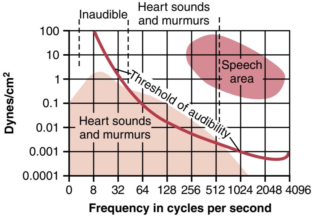
Figure 23-1 Amplitude of different frequency vibrations in the heart sounds and heart murmurs in relationship to the threshold of audibility, showing that the range of sounds that can be heard is between 40 and 520 cycles/sec. (Modified from Butterworth JS, Chassin JL, McGrath JJ: Cardiac Auscultation, 2nd ed. New York: Grune & Stratton, 1960.)
شکل 23-1 دامنه ارتعاشات فرکانس مختلف در صداهای قلب و سوفل قلب در رابطه با آستانه شنوایی، نشان می دهد که دامنه صداهایی که می توان شنید بین 40 تا 520 سیکل در ثانیه است. (اصلاح شده از Butterworth JS, Chassin JL, McGrath JJ: Cardiac Auscultation, 2nd ed. New York: Grune & Stratton, 1960.)
The Third Heart Sound Occurs at the Beginning of the Middle Third of Diastole. Occasionally, a weak, rumbling third heart sound is heard at the beginning of the middle third of diastole. A logical but unproved explanation of this sound is oscillation of blood back and forth between the walls of the ventricles initiated by inrushing blood from the atria. This is analogous to running water from a faucet into a paper sack, with the inrushing water reverberating back and forth between the walls of the sack to cause vibrations in its walls. The reason the third heart sound does not occur until the middle third of diastole is believed to be that in the early part of diastole, the ventricles are not filled sufficiently to create even the small amount of elastic tension necessary for reverberation. The frequency of this sound is usually so low that the ear can- not hear it, yet it can often be recorded in the phonocardiogram. The third heart sound may be normally present in children, adolescents, and young adults but generally indicates systolic heart failure in older adults.
صدای قلب سوم در ابتدای یک سوم میانی دیاستول رخ می دهد. گاهی در ابتدای یک سوم میانی دیاستول، صدای سوم قلب ضعیف و خروشان شنیده می شود. یک توضیح منطقی اما اثبات نشده از این صدا، نوسان خون به جلو و عقب بین دیوارههای بطن است که با هجوم خون از دهلیزها آغاز میشود. این شبیه به جاری شدن آب از یک شیر آب به یک کیسه کاغذی است، با آب هجومی که بین دیواره های گونی طنین انداز می شود و باعث ایجاد لرزش در دیواره های آن می شود. اعتقاد بر این است که دلیل اینکه صدای سوم قلب تا یک سوم میانی دیاستول رخ نمی دهد این است که در اوایل دیاستول، بطن ها به اندازه کافی پر نمی شوند تا حتی مقدار کمی کشش الاستیک لازم برای طنین ایجاد شود. فرکانس این صدا معمولاً آنقدر کم است که گوش نمی تواند آن را بشنود، اما اغلب می توان آن را در فونوکاردیوگرام ضبط کرد. صدای سوم قلب ممکن است به طور معمول در کودکان، نوجوانان و بزرگسالان جوان وجود داشته باشد اما به طور کلی نشان دهنده نارسایی سیستولیک قلب در بزرگسالان مسن است.
Atrial Contraction-Fourth Heart Sound. An atrial heart sound can sometimes be recorded in the phonocar- diogram, but it can almost never be heard with a stethoscope because of its weakness and very low frequency- usually 20 cycles/sec or less. This sound occurs when the atria contract, and presumably, it is caused by the inrush of blood into the ventricles, which initiates vibrations similar to those of the third heart sound. A fourth heart sound is common in persons who derive benefit from atrial contraction for ventricular filling as a result of decreased ventricular wall compliance and increased resistance to ventricular filling. For example, a fourth heart sound is often heard in older patients with left ventricular hypertrophy.
انقباض دهلیزی-صدای چهارم قلب. گاهی اوقات صدای قلب دهلیزی را می توان در فونوکاردیوگرام ثبت کرد، اما به دلیل ضعف و فرکانس بسیار پایین آن تقریباً هرگز با گوشی پزشکی شنیده نمی شود – معمولاً 20 سیکل در ثانیه یا کمتر. این صدا زمانی رخ می دهد که دهلیزها منقبض می شوند و احتمالاً در اثر هجوم خون به بطن ها ایجاد می شود که باعث ایجاد ارتعاشاتی مشابه ارتعاشات صدای سوم قلب می شود. صدای چهارم قلب در افرادی که از انقباض دهلیزی برای پر شدن بطن در نتیجه کاهش انطباق دیواره بطن و افزایش مقاومت در برابر پر شدن بطن سود می برند، رایج است. برای مثال، صدای چهارم قلب اغلب در بیماران مسنتر مبتلا به هیپرتروفی بطن چپ شنیده میشود.
Chest Surface Areas for Auscultation of Normal Heart Sounds. Listening to the sounds of the body, usually with the aid of a stethoscope, is called auscultation. Figure 23-2 shows the areas of the chest wall from which the different heart valvular sounds can best be distinguished. Although the sounds from all the valves can be heard from all these areas, the cardiologist distinguishes the sounds from the different valves by a process of elimination. That is, he or she moves the stethoscope from one area to another, noting the loudness of the sounds in different areas and gradually picking out the sound components from each valve.
نواحی سطح قفسه سینه برای سمع صداهای طبیعی قلب. گوش دادن به صداهای بدن، معمولاً با کمک گوشی پزشکی، سمع نامیده می شود. شکل 23-2 نواحی دیواره قفسه سینه را نشان می دهد که صداهای مختلف دریچه قلب را می توان به بهترین وجه از آنها تشخیص داد. اگرچه صداهای همه دریچهها از همه این نواحی شنیده میشود، اما متخصص قلب، صداها را از دریچههای مختلف با فرآیند حذف تشخیص میدهد. به این معنی که گوشیپزشکی را از یک ناحیه به ناحیه دیگر حرکت میدهد و به بلندی صداها در نواحی مختلف توجه میکند و به تدریج اجزای صدا را از هر دریچه انتخاب میکند.
The areas for listening to the different heart sounds are not directly over the valves themselves. The aortic area is upward along the aorta because of sound transmission up the aorta, and the pulmonic area is upward along the pulmonary artery. The tricuspid area is over the right ventricle, and the mitral area is over the apex of the left ventricle, which is the portion of the heart nearest the surface of the chest; the heart is rotated so that the remainder of the left ventricle lies more posteriorly.
نواحی گوش دادن به صداهای مختلف قلب مستقیماً بالای خود دریچه ها نیست. ناحیه آئورت به دلیل انتقال صدا به بالای آئورت در امتداد آئورت به سمت بالا است و ناحیه ریوی در امتداد شریان ریوی به سمت بالا است. ناحیه سه لتی روی بطن راست و ناحیه میترال بالای بطن چپ قرار دارد که نزدیکترین قسمت قلب به سطح قفسه سینه است. قلب به گونه ای می چرخد که باقی مانده بطن چپ بیشتر در عقب قرار گیرد.

Figure 23-2 Chest areas from which sounds each valve are heard best.
شکل 23-2 نواحی قفسه سینه که صدای هر دریچه از آنها بهتر شنیده میشود.
Phonocardiogram. If a microphone specially designed to detect low-frequency sound is placed on the chest, the heart sounds can be amplified and recorded by a high-speed recording apparatus. The recording is called a phonocardiogram, and the heart sounds appear as waves, as shown schematically in Figure 23- 3. Recording A is an example of normal heart sounds, showing the vibrations of the first, second, and third heart sounds and even the very weak atrial sound. Note specifically that the third and atrial heart sounds are each a very low rumble. The third heart sound can be recorded in only one-third to one-half of people, and the atrial heart sound can be recorded in perhaps one-fourth of people.
فونوکاردیوگرام. اگر میکروفونی که مخصوص تشخیص صدای با فرکانس پایین طراحی شده است روی قفسه سینه قرار داده شود، صداهای قلب را می توان با یک دستگاه ضبط با سرعت بالا تقویت و ضبط کرد. ضبط صدا فونوکاردیوگرام نامیده می شود و صداهای قلب به صورت امواج ظاهر می شوند، همانطور که به صورت شماتیک در شکل 23-3 نشان داده شده است. ضبط A نمونه ای از صداهای طبیعی قلب است که ارتعاشات صداهای قلب اول، دوم و سوم و حتی صدای بسیار ضعیف دهلیزی را نشان می دهد. به طور خاص توجه داشته باشید که صدای قلب سوم و دهلیزی هر کدام یک صدای بسیار کم است. صدای سوم قلب را می توان تنها در یک سوم تا نیمی از افراد ضبط کرد و صدای قلب دهلیزی را می توان در یک چهارم افراد ثبت کرد.

Figure 23-3 A-F, Phonocardiograms from normal and abnormal hearts.
شکل 23-3 A-F، فونوکاردیوگرام از قلب های طبیعی و غیر طبیعی.
VALVULAR LESIONS
Rheumatic Valvular Lesions
Rheumatic fever is an autoimmune disease in which the heart valves are likely to be damaged or destroyed. The disease is usually initiated by a streptococcal toxin.
ضایعات دریچه ای
ضایعات دریچه ای روماتیسمی
تب روماتیسمی یک بیماری خودایمنی است که در آن دریچه های قلب احتمالا آسیب دیده یا از بین می روند. این بیماری معمولاً توسط یک سم استرپتوکوک شروع می شود.
The sequence of events almost always begins with a preliminary streptococcal infection caused specifically by group A hemolytic streptococci. These bacteria initially cause a sore throat, scarlet fever, or middle ear infection. However, the streptococci also release several different proteins against which the person’s reticuloendothelial system produces antibodies. The antibodies react not only with the streptococcal protein but also with other protein tissues of the body, often causing severe immunologic damage. These reactions continue to take place as long as the antibodies persist in the blood-1 year or more.
توالی وقایع تقریباً همیشه با یک عفونت استرپتوککی اولیه که به طور خاص توسط استرپتوکوک های همولیتیک گروه A ایجاد می شود، شروع می شود. این باکتری ها در ابتدا باعث گلودرد، مخملک یا عفونت گوش میانی می شوند. با این حال، استرپتوکوک ها چندین پروتئین مختلف را نیز آزاد می کنند که سیستم رتیکولواندوتلیال فرد علیه آنها آنتی بادی تولید می کند. آنتی بادی ها نه تنها با پروتئین استرپتوکوک بلکه با سایر بافت های پروتئینی بدن نیز واکنش نشان می دهند و اغلب باعث آسیب شدید ایمنی می شوند. تا زمانی که آنتی بادی ها در خون 1 سال یا بیشتر باقی بمانند، این واکنش ها ادامه می یابد.
Rheumatic fever particularly causes damage in certain susceptible areas, such as the heart valves. The degree of heart valve damage is directly correlated with the concentration and persistence of the antibodies. The principles of immunity that relate to this type of reaction are discussed in Chapter 35, and it is noted in Chapter 32 that acute glomerular nephritis of the kidneys has a similar immunologic basis.
تب روماتیسمی بهویژه در نواحی حساس مانند دریچههای قلب آسیب میبیند. میزان آسیب دریچه قلب به طور مستقیم با غلظت و ماندگاری آنتی بادی ها در ارتباط است. اصول ایمنی مربوط به این نوع واکنش در فصل 35 مورد بحث قرار می گیرد و در فصل 32 ذکر می شود که نفریت گلومرولار حاد کلیه ها دارای پایه ایمونولوژیک مشابه است.
In persons with rheumatic fever, large hemorrhagic, fibrinous, bulbous lesions grow along the inflamed edges of the heart valves. Because the mitral valve undergoes more trauma during valvular action than any of the other valves, it is the one most often seriously damaged, and the aortic valve is the second most frequently damaged. The right heart valves-that is, the tricuspid and pulmonary valves are usually affected much less severely, probably because the low-pressure stresses that act on these valves are slight compared with the high-pressure stresses that act on the left heart valves.
در افراد مبتلا به تب روماتیسمی، ضایعات هموراژیک، فیبرینی و پیازی بزرگ در امتداد لبههای ملتهب دریچههای قلب رشد میکنند. از آنجایی که دریچه میترال در طول عملکرد دریچه ای بیش از سایر دریچه ها آسیب می بیند، این دریچه اغلب آسیب جدی می بیند و دریچه آئورت دومین آسیب دیدگی است. دریچههای قلب راست – یعنی دریچههای سه لتی و ریوی معمولاً با شدت کمتری تحت تأثیر قرار میگیرند، احتمالاً به این دلیل که تنشهای فشار پایینی که روی این دریچهها اعمال میشود در مقایسه با تنشهای فشار بالا که روی دریچههای قلب چپ تأثیر میگذارند، اندک است.
The lesions of acute rheumatic fever frequently occur on adjacent valve leaflets simultaneously, so the edges of the leaflets become stuck together. Then, weeks, months, or years later, the lesions become scar tissue, permanently fusing portions of adjacent valve leaflets. Also, the free edges of the leaflets, which are normally filmy and free- flapping, often become solid scarred masses.
ضایعات تب روماتیسمی حاد اغلب به طور همزمان روی لت های دریچه مجاور ایجاد می شود، بنابراین لبه های برگچه ها به هم چسبیده می شوند. سپس، هفتهها، ماهها یا سالها بعد، ضایعات به بافت اسکار تبدیل میشوند و به طور دائم بخشهایی از لتهای دریچه مجاور را با هم ترکیب میکنند. همچنین، لبههای آزاد برگچهها که معمولاً لایهای هستند و آزاد میشوند، اغلب به تودههای اسکار جامد تبدیل میشوند.
A valve in which the leaflets adhere to one another so extensively that blood cannot flow through it normally is said to be stenosed. Conversely, when the valve edges are so destroyed by scar tissue that they cannot close as the ventricles contract, regurgitation (backflow) of blood occurs when the valve should be closed. Stenosis usually does not occur without the coexistence of at least some degree of regurgitation, and vice versa.
گفته می شود دریچه ای که در آن برگچه ها به قدری به یکدیگر می چسبند که خون به طور معمول نمی تواند از آن عبور کند تنگی دارد. برعکس، زمانی که لبههای دریچه به قدری توسط بافت اسکار تخریب میشوند که نمیتوانند با انقباض بطنها بسته شوند، برگشت خون (جریان برگشتی) زمانی اتفاق میافتد که دریچه باید بسته شود. تنگی معمولاً بدون همزیستی حداقل درجاتی از نارسایی رخ نمی دهد و بالعکس.
Aging and Aortic Valve Stenosis
With aging, the aortic valve often thickens, becomes calcified and stiffer, and may partially obstruct outflow from the left ventricle. With increased life expectancy and aging of the population, aortic valve stenosis has become the most common heart valve disease.
پیری و تنگی دریچه آئورت
با افزایش سن، دریچه آئورت اغلب ضخیم می شود، کلسیفیه و سفت تر می شود و ممکن است تا حدی مانع خروج از بطن چپ شود. با افزایش امید به زندگی و افزایش سن جمعیت، تنگی دریچه آئورت به شایع ترین بیماری دریچه قلب تبدیل شده است.
Stenosis of a previously normal aortic valve, often called senile calcific aortic valve stenosis, is characterized by valve calcium deposition and ossification, which lead to narrowing of the aortic valve orifice. As a compensatory response to the increased workload imposed on the heart by the stenotic aortic valve, the left ventricle under- goes concentric hypertrophy. This type of hypertrophy is associated with increased left ventricular wall thickness, which permits the heart to pump with greater vigor against the partially obstructed outflow. An increasing pressure gradient then develops across the calcified valve, reaching 75 to 100 mm Hg in severe cases of aortic valve stenosis.
تنگی دریچه آئورت که قبلاً طبیعی بود، که اغلب تنگی دریچه آئورت کلسیفیک سالخورده نامیده میشود، با رسوب کلسیم دریچه و استخوانبندی مشخص میشود که منجر به باریک شدن روزنه دریچه آئورت میشود. به عنوان یک پاسخ جبرانی به افزایش بار کاری که توسط دریچه آئورت تنگی بر قلب تحمیل می شود، بطن چپ دچار هیپرتروفی متحدالمرکز می شود. این نوع هیپرتروفی با افزایش ضخامت دیواره بطن چپ همراه است که به قلب اجازه میدهد با قدرت بیشتری در برابر جریان خروجی نسبی انسداد پمپاژ کند. سپس یک گرادیان فشار فزاینده در سراسر دریچه کلسیفیه ایجاد می شود و در موارد شدید تنگی دریچه آئورت به 75 تا 100 میلی متر جیوه می رسد.
The hypertrophied left ventricle also becomes more fibrotic and tends to be ischemic because of impaired microcirculatory perfusion, although some patients may also have atherosclerosis of the coronary arteries. The ejection fraction may be normal, and the patient may be able to maintain adequate cardiac output under resting conditions, but with even moderate exercise, symptoms of heart failure may appear. As the stenosis progressively worsens, there are reductions in systolic heart function and inability of the left ventricle to develop enough pressure to pump effectively against the load imposed by the partially obstructed aortic valve. Consequently, symptoms of congestive heart failure appear, with reductions in stroke volume and cardiac output.
بطن چپ هیپرتروفی شده نیز فیبروتیکتر میشود و به دلیل اختلال در پرفیوژن میکروسیرکولاتوری تمایل به ایسکمیک دارد، اگرچه برخی از بیماران ممکن است به آترواسکلروز شریانهای کرونر نیز مبتلا باشند. کسر جهشی ممکن است طبیعی باشد، و بیمار ممکن است بتواند برون ده قلبی کافی را در شرایط استراحت حفظ کند، اما با ورزش حتی متوسط، علائم نارسایی قلبی ممکن است ظاهر شود. با بدتر شدن تدریجی تنگی، کاهش در عملکرد قلب سیستولیک و ناتوانی بطن چپ در ایجاد فشار کافی برای پمپاژ موثر در برابر بار تحمیل شده توسط دریچه آئورت نیمه مسدود شده وجود دارد. در نتیجه، علائم نارسایی احتقانی قلب با کاهش حجم سکته مغزی و برون ده قلبی ظاهر می شود.
Calcific aortic valve stenosis usually does not become severe enough to draw clinical attention until after age 70. Important symptoms of aortic valve stenosis are exertion-related angina, reduced exercise tolerance, and congestive heart failure. Shortness of breath (dyspnea) is due to increased left ventricular filling pressure or inability to increase cardiac output adequately with exercise. Early recognition and management of aortic stenosis are important because untreated symptomatic aortic valve stenosis is progressive and will ultimately be fatal.
تنگی دریچه آئورت کلسیفیک معمولاً تا سن 70 سالگی آنقدر شدید نمی شود که توجه بالینی را به خود جلب کند. علائم مهم تنگی دریچه آئورت عبارتند از آنژین مربوط به فعالیت، کاهش تحمل ورزش و نارسایی احتقانی قلب. تنگی نفس (تنگی نفس) به دلیل افزایش فشار پر شدن بطن چپ یا ناتوانی در افزایش برون ده قلبی به اندازه کافی با ورزش است. تشخیص زودهنگام و مدیریت تنگی آئورت مهم است زیرا تنگی دریچه آئورت علامتدار درمان نشده پیشرونده است و در نهایت کشنده خواهد بود.
The development of transcatheter aortic valve replacement technologies has provided new therapeutic opportunities, especially for older patients, in whom traditional surgical procedures cannot be performed or are associated with high risk.
توسعه فنآوریهای جایگزینی دریچه آئورت ترانس کاتتر، فرصتهای درمانی جدیدی را بهویژه برای بیماران مسنتر، که در آنها روشهای جراحی سنتی نمیتوان انجام داد یا با خطر بالایی همراه است، فراهم کرده است.
Heart Murmurs Caused by Valvular Lesions
As shown by the phonocardiograms in Figure 23-3, many abnormal heart sounds, known as heart murmurs, occur when abnormalities of the valves are present, as discussed here.
سوفل قلب ناشی از ضایعات دریچه ای
همانطور که توسط فونوکاردیوگرام در شکل 23-3 نشان داده شده است، بسیاری از صداهای غیرطبیعی قلب، که به عنوان سوفل قلبی شناخته می شوند، هنگامی رخ می دهند که ناهنجاری دریچه ها وجود داشته باشد، همانطور که در اینجا بحث شد.
Systolic Murmur of Aortic Stenosis. In persons with aortic stenosis, blood is ejected from the left ventricle through only a small fibrous opening of the aortic valve. Because of the resistance to ejection, the blood pressure in the left ventricle sometimes rises as high as 300 mm Hg; the pressure in the aorta is still normal. Thus, a nozzle effect is created during systole, with blood jetting at tremendous velocity through the small opening of the valve. This phenomenon causes severe turbulence of the blood in the root of the aorta. The turbulent blood impinging against the aortic walls causes intense vibration, and a loud murmur occurs during systole (see recording B, Figure 23-3; Audio 23-2) and is transmitted throughout the superior thoracic aorta and even into the large arteries of the neck. This sound is harsh, and in persons with severe stenosis it may be so loud that it can be heard several feet away from the patient. Also, the sound vibrations can often be felt with the hand on the upper chest and lower neck, a phenomenon known as a thrill.
سوفل سیستولیک تنگی آئورت. در افراد مبتلا به تنگی آئورت، خون از بطن چپ تنها از طریق یک دهانه فیبری کوچک دریچه آئورت خارج می شود. به دلیل مقاومت در برابر جهش، فشار خون در بطن چپ گاهی تا 300 میلی متر جیوه افزایش می یابد. فشار در آئورت هنوز طبیعی است. بنابراین، یک اثر نازل در طول سیستول ایجاد می شود، با جهش خون با سرعت فوق العاده از طریق دهانه کوچک دریچه. این پدیده باعث تلاطم شدید خون در ریشه آئورت می شود. خون آشفته ای که به دیواره های آئورت برخورد می کند باعث ارتعاش شدید می شود و سوفل بلندی در طول سیستول رخ می دهد (به ضبط B، شکل 23-3؛ صوت 23-2 مراجعه کنید) و در سراسر آئورت سینه ای فوقانی و حتی به شریان های بزرگ گردن منتقل می شود. این صدا خشن است و در افرادی که تنگی شدید دارند ممکن است آنقدر بلند باشد که در فاصله چند فوتی بیمار شنیده شود. همچنین، ارتعاشات صدا را اغلب می توان با دست در قسمت بالای سینه و پایین گردن احساس کرد، پدیده ای که به عنوان هیجان شناخته می شود.
Diastolic Murmur of Aortic Regurgitation. In aortic regurgitation, no abnormal sound is heard during systole, but during diastole, blood flows backward from the highpressure aorta into the left ventricle, causing a “blowing” murmur of relatively high pitch, with a swishing quality heard maximally over the left ventricle (see recording D, Figure 23-3; Audio 23-3). This murmur results from the turbulence of blood jetting backward into the blood al- ready in the low-pressure diastolic left ventricle.
سوفل دیاستولیک نارسایی آئورت. در نارسایی آئورت، هیچ صدای غیرطبیعی در طول سیستول شنیده نمیشود، اما در طول دیاستول، خون از آئورت پرفشار به سمت عقب به داخل بطن چپ جریان مییابد که باعث ایجاد سوفل «دمنده» با صدای نسبتاً بالا میشود، با کیفیت صدایی که حداکثر از روی بطن چپ شنیده میشود(به ضبط D، شکل 23-3؛ فایل صوتی 23-3 مراجعه کنید). این سوفل ناشی از تلاطم جهش خون به سمت عقب به داخل خون در بطن چپ دیاستولیک کم فشار است.
Systolic Murmur of Mitral Regurgitation. In persons with mitral regurgitation, blood flows backward through the mitral valve into the left atrium during systole. This backward flow also causes a high-frequency blowing, swishing sound (see recording C, Figure 23-3; Audio 23-4) similar to that of aortic regurgitation but occurring during systole rather than diastole. It is transmitted most strongly into the left atrium. However, the left atrium is so deep within the chest that it is difficult to hear this sound directly over the atrium. As a result, the sound of mitral regurgitation is transmitted to the chest wall mainly through the left ventricle to the apex of the heart.
سوفل سیستولیک نارسایی میترال. در افراد مبتلا به نارسایی میترال، خون در طی سیستول از طریق دریچه میترال به سمت عقب به دهلیز چپ جریان می یابد. این جریان رو به عقب همچنین باعث ایجاد صدای وزش با فرکانس بالا می شود (به ضبط C، شکل 23-3؛ صوت 23-4 مراجعه کنید) شبیه به نارسایی آئورت اما در طول سیستول به جای دیاستول رخ می دهد. به شدت به دهلیز چپ منتقل می شود. با این حال، دهلیز چپ به قدری در داخل قفسه سینه قرار دارد که شنیدن این صدا به طور مستقیم از بالای دهلیز دشوار است. در نتیجه، صدای نارسایی میترال عمدتاً از طریق بطن چپ به بالای قلب به دیواره قفسه سینه منتقل می شود.
Diastolic Murmur of Mitral Stenosis. In persons with mitral stenosis, blood passes with difficulty through the stenosed mitral valve from the left atrium into the left ventricle, and because the pressure in the left atrium seldom rises above 30 mm Hg, a large pressure differential forcing blood from the left atrium into the left ventricle does not develop. Consequently, the abnormal sounds heard in mitral stenosis (see recording E, Figure 23-3; Audio 23-5) are usually weak and of very low frequency, so most of the sound spectrum is below the low-frequency end of human hearing.
سوفل دیاستولیک تنگی میترال. در افراد مبتلا به تنگی میترال، خون به سختی از طریق دریچه میترال تنگ شده از دهلیز چپ به بطن چپ میرود و چون فشار در دهلیز چپ به ندرت از 30 میلیمتر جیوه بالاتر میرود، اختلاف فشار زیادی که خون را از دهلیز چپ به بطن چپ میکشد، ایجاد نمیشود. در نتیجه، صداهای غیرطبیعی شنیده شده در تنگی میترال (به ضبط E، شکل 23-3؛ صوت 23-5 مراجعه کنید) معمولاً ضعیف و فرکانس بسیار پایینی دارند، بنابراین بیشتر طیف صدا زیر انتهای فرکانس پایین شنوایی انسان است.
During the early part of diastole, a left ventricle with a stenotic mitral valve has so little blood in it, and its walls are so flabby, that blood does not reverberate back and forth between the walls of the ventricle. For this reason, even in persons with severe mitral stenosis, no murmur may be heard during the first third of diastole. Then, after partial filling, the ventricle has stretched enough for blood to reverberate, and a low rumbling murmur begins.
در اوایل دیاستول، یک بطن چپ با دریچه میترال تنگی دارای خون بسیار کمی است و دیوارههای آن به قدری شل است که خون بین دیوارههای بطن به عقب و جلو طنین نمیزند. به همین دلیل، حتی در افرادی که تنگی شدید میترال دارند، ممکن است در یک سوم اول دیاستول هیچ سوفلی شنیده نشود. سپس، پس از پر شدن نسبی، بطن به اندازه کافی کشیده می شود که خون طنین انداز می کند و سوفل خفیف شروع می شود.
Phonocardiograms of Valvular Murmurs. Phonocardiograms B, C, D, and E of Figure 23-3 show, respectively, idealized records obtained from patients with aortic stenosis, mitral regurgitation, aortic regurgitation, and mitral stenosis. It is obvious from these phonocardiograms that the aortic stenotic lesion causes the loudest murmur, and the mitral stenotic lesion causes the weakest murmur. The phonocardiograms show how the intensity of the murmurs varies during different portions of systole and diastole, and the relative timing of each murmur is also evident. Note especially that the murmurs of aortic stenosis and mitral regurgitation occur only during systole, whereas the murmurs of aortic regurgitation and mitral stenosis occur only during diastole.
فونوکاردیوگرام سوفل دریچه ای. فونوکاردیوگرام های B، C، D و E در شکل 23-3 به ترتیب سوابق ایده آلی را نشان می دهد که از بیماران مبتلا به تنگی آئورت، نارسایی میترال، نارسایی آئورت و تنگی میترال به دست آمده است. از این فونوکاردیوگرام ها مشخص است که ضایعه تنگی آئورت باعث بلندترین سوفل و ضایعه تنگی میترال ضعیف ترین سوفل را ایجاد می کند. فونوکاردیوگرام ها نشان می دهند که چگونه شدت سوفل در قسمت های مختلف سیستول و دیاستول تغییر می کند و زمان نسبی هر سوفل نیز مشهود است. به ویژه توجه داشته باشید که سوفل تنگی آئورت و نارسایی میترال فقط در طول سیستول رخ می دهد، در حالی که سوفل نارسایی آئورت و تنگی میترال فقط در طول دیاستول رخ می دهد.
ABNORMAL CIRCULATORY DYNAMICS IN VALVULAR HEART DISEASE
CIRCULATORY DYNAMICS IN AORTIC STENOSIS AND AORTIC REGURGITATION
In aortic stenosis, the contracting left ventricle fails to empty adequately, whereas in aortic regurgitation, blood flows backward into the ventricle from the aorta after the ventricle has just pumped the blood into the aorta. Therefore, in both cases, the net stroke volume output of the heart is reduced.
دینامیک گردش خون غیر طبیعی در بیماری دریچه ای قلب
دینامیک گردش خون در تنگی آئورت و نارسایی آئورت
در تنگی آئورت، بطن چپ منقبض به اندازه کافی تخلیه نمی شود، در حالی که در نارسایی آئورت، پس از اینکه بطن تازه خون را به داخل آئورت پمپاژ کرد، خون از آئورت به سمت عقب به داخل بطن جریان می یابد. بنابراین، در هر دو مورد، خروجی خالص حجم ضربه ای قلب کاهش می یابد.
Several important compensations take place that can ameliorate the severity of the circulatory defects. Some of these compensations are described in the following sections.
چندین جبران مهم انجام می شود که می تواند شدت نقایص گردش خون را بهبود بخشد. برخی از این غرامت ها در بخش های زیر توضیح داده شده است.
Hypertrophy of Left Ventricle. In both aortic stenosis and aortic regurgitation, the left ventricular musculature hypertrophies because of the increased ventricular workload. In regurgitation, the left ventricular chamber also enlarges to hold all the regurgitant blood from the aorta. This type of hypertrophy, with enlargement of the ventricular chamber, is often referred to as eccentric hypertrophy (Figure 23-4). Sometimes, the left ventricular muscle mass increases fourfold to fivefold, creating a tremendously large left side of the heart.
هیپرتروفی بطن چپ. هم در تنگی آئورت و هم در نارسایی آئورت، به دلیل افزایش بار کاری بطن، عضله بطن چپ هیپرتروفی میشود. در رگورژیتاسیون، محفظه بطن چپ نیز بزرگ می شود تا تمام خون نارسایی آئورت را در خود نگه دارد. این نوع هیپرتروفی، با بزرگ شدن محفظه بطنی، اغلب به عنوان هیپرتروفی خارج از مرکز شناخته می شود (شکل 23-4). گاهی اوقات، توده عضلانی بطن چپ چهار تا پنج برابر افزایش می یابد و یک سمت چپ فوق العاده بزرگ قلب ایجاد می کند.
When the aortic valve is seriously stenosed, the hyper- trophied muscle may allow the left ventricle to develop as much as 400 mm Hg of intraventricular pressure at systolic peak. This type of concentric hypertrophy is associated with thickened ventricular walls and a smaller ventricular chamber (see Figure 23-4) and also occurs in other circumstances where afterload of the heart is increased, such as in chronic hypertension.
هنگامی که دریچه آئورت به طور جدی تنگ می شود، عضله هیپرتروفی شده ممکن است به بطن چپ اجازه دهد تا فشار داخل بطنی 400 میلی متر جیوه در اوج سیستولیک ایجاد کند. این نوع هیپرتروفی متحدالمرکز با دیوارههای بطنی ضخیمتر و یک محفظه بطنی کوچکتر همراه است (شکل 23-4 را ببینید) و همچنین در شرایط دیگری که بار اضافی قلب افزایش مییابد، مانند فشار خون مزمن، رخ میدهد.
In persons with severe aortic regurgitation, sometimes the enlarged left ventricle can pump a stroke volume out- put as great as 250 milliliters, although as much as 75% of this blood returns to the ventricle during diastole, and only 25% flows through the aorta to the body.
در افراد مبتلا به نارسایی شدید آئورت، گاهی اوقات بطن چپ بزرگ شده میتواند حجم ضربهای را تا 250 میلیلیتر پمپ کند، اگرچه 75 درصد از این خون در طول دیاستول به بطن باز میگردد و تنها 25 درصد آن از طریق آئورت به بدن جریان مییابد.
Increase in Blood Volume. Another effect that helps compensate for the diminished net pumping by the left ventricle is increased blood volume. This increased volume results from the following: (1) an initial slight decrease in arterial pressure; (2) nervous reflexes and hormonal changes induced by the decrease in pressure. These mechanisms together diminish renal output of urine, causing blood volume to increase and the mean arterial pressure to return to normal. Also, red blood cell mass eventually increases because of a slight degree of tissue hypoxia.
افزایش حجم خون. اثر دیگری که به جبران کاهش پمپاژ خالص توسط بطن چپ کمک می کند افزایش حجم خون است. این افزایش حجم ناشی از موارد زیر است: (1) کاهش جزئی اولیه در فشار شریانی. (2) رفلکس های عصبی و تغییرات هورمونی ناشی از کاهش فشار. این مکانیسم ها با هم برون ده کلیه ادرار را کاهش می دهند و باعث افزایش حجم خون و بازگشت فشار متوسط شریانی به حالت عادی می شوند. همچنین، توده گلبول های قرمز در نهایت به دلیل درجه کمی از هیپوکسی بافتی افزایش می یابد.
The increase in blood volume tends to increase venous return to the heart, which, in turn, causes the left ventricle to pump with the extra power required to overcome the abnormal pumping dynamics.
افزایش حجم خون تمایل به افزایش بازگشت وریدی به قلب دارد که به نوبه خود باعث می شود بطن چپ با قدرت اضافی مورد نیاز برای غلبه بر دینامیک پمپاژ غیرطبیعی پمپاژ کند.
Aortic Valvular Lesions May Be Associated With Inadequate Coronary Blood Flow. When a person has stenosis of the aortic valve, the ventricular muscle must develop a high tension to create the high intraventricular pressure needed to force blood through the stenosed valve. This action increases the workload and oxygen consumption of the ventricle, necessitating increased coronary blood flow to deliver this oxygen. The high wall tension of the ventricle, however, causes marked decreases in coronary flow during systole, particularly in the subendocardial vessels. Intraventricular diastolic pressure is also increased when there is aortic valve stenosis, and this increased pressure may cause compression of the inner layers of the heart muscle and reduced coronary blood flow. Thus, severe aortic valve stenosis often causes ischemia of the heart muscle.
ضایعات دریچه آئورت ممکن است با جریان خون ناکافی کرونر مرتبط باشد. هنگامی که فردی دچار تنگی دریچه آئورت می شود، عضله بطنی باید کشش بالایی ایجاد کند تا فشار داخل بطنی بالایی را ایجاد کند که برای عبور خون از دریچه تنگ شده لازم است. این عمل باعث افزایش حجم کار و مصرف اکسیژن در بطن می شود و نیاز به افزایش جریان خون کرونری برای رساندن این اکسیژن دارد. کشش بالای دیواره بطن، با این حال، باعث کاهش قابل توجه جریان کرونر در طول سیستول، به ویژه در عروق ساب اندوکارد می شود. فشار دیاستولیک داخل بطنی نیز در صورت وجود تنگی دریچه آئورت افزایش می یابد و این افزایش فشار ممکن است باعث فشرده شدن لایه های داخلی عضله قلب و کاهش جریان خون کرونر شود. بنابراین، تنگی شدید دریچه آئورت اغلب باعث ایسکمی عضله قلب می شود.
With aortic regurgitation, the intraventricular diastolic pressure also increases, compressing the inner layer of the heart muscle and decreasing coronary blood flow. Aortic diastolic pressure decreases during aortic regurgitation, which can also decrease coronary blood flow and cause ischemia of the heart muscle.
با نارسایی آئورت، فشار دیاستولیک داخل بطنی نیز افزایش مییابد و لایه داخلی عضله قلب را فشرده میکند و جریان خون کرونر را کاهش میدهد. فشار دیاستولیک آئورت در هنگام نارسایی آئورت کاهش مییابد که میتواند جریان خون کرونر را نیز کاهش دهد و باعث ایسکمی عضله قلب شود.

Figure 23-4 Comparison of the left ventricle in a normal heart, a heart with eccentric left ventricular hypertrophy (LVH), and a heart with concentric LVH. Left ventricular mass increases in eccentric and concentric LVH but there is an increase in left ventricular cavity size in ec- Chronic hypertension centric LVH, whereas wall thickness increases in concentric LVH.
شکل 23-4 مقایسه بطن چپ در قلب طبیعی، قلبی با هیپرتروفی بطن چپ غیرعادی (LVH) و قلب با LVH متحدالمرکز. توده بطن چپ در LVH برونمرکز و متمرکز افزایش مییابد، اما در LVH مرکزی فشار خون مزمن، افزایش در اندازه حفره بطن چپ وجود دارد، در حالی که ضخامت دیواره در LVH متحدالمرکز افزایش مییابد.
Eventual Failure of Left Ventricle and Development of Pulmonary Edema. In the early stages of aortic steno- sis or aortic regurgitation, the intrinsic ability of the left ventricle to adapt to increasing loads prevents significant abnormalities in circulatory function in the person during rest, other than the increased work output required of the left ventricle. Therefore, considerable degrees of aortic stenosis or aortic regurgitation often occur before the person knows that he or she has serious heart disease (e.g., a resting left ventricular systolic pressure as high as 200 mm Hg in persons with aortic stenosis or a left ventricular stroke volume output as high as twice normal in persons with aortic regurgitation).
نارسایی احتمالی بطن چپ و ایجاد ادم ریوی. در مراحل اولیه تنگی آئورت یا نارسایی آئورت، توانایی ذاتی بطن چپ برای انطباق با بارهای فزاینده، از ناهنجاری های قابل توجهی در عملکرد گردش خون در فرد در حین استراحت، به غیر از افزایش بازده کار مورد نیاز بطن چپ، جلوگیری می کند. بنابراین، درجات قابل توجهی از تنگی آئورت یا نارسایی آئورت اغلب قبل از اینکه فرد بداند که به بیماری قلبی جدی مبتلا است رخ میدهد (مثلاً فشار سیستولیک بطن چپ در حالت استراحت تا 200 میلیمتر جیوه در افراد مبتلا به تنگی آئورت یا حجم سکته مغزی بطن چپ با حجم خروجی سکته مغزی دوبرابر یا عادی در افراد عادی).
Beyond a critical stage in these aortic valve lesions, the left ventricle finally cannot keep up with the work demand. As a consequence, the left ventricle dilates and cardiac output begins to fall; blood simultaneously dams up in the left atrium and in the lungs behind the failing left ventricle. The left atrial pressure rises progressively, and at mean left atrial pressures above 25 to 40 mm Hg, serious edema appears in the lungs, as discussed in detail in Chapter 39.
فراتر از یک مرحله بحرانی در این ضایعات دریچه آئورت، بطن چپ در نهایت نمی تواند با تقاضای کار مطابقت داشته باشد. در نتیجه، بطن چپ گشاد می شود و برون ده قلبی شروع به کاهش می کند. خون به طور همزمان در دهلیز چپ و در ریه ها در پشت بطن چپ از کار افتاده می ریزد. فشار دهلیز چپ به تدریج افزایش می یابد و در فشارهای دهلیز چپ بالای 25 تا 40 میلی متر جیوه، ادم جدی در ریه ها ظاهر می شود، همانطور که در فصل 39 به تفصیل مورد بحث قرار گرفت.
DYNAMICS OF MITRAL STENOSIS AND MITRAL REGURGITATION
In persons with mitral stenosis, blood flow from the left atrium into the left ventricle is impeded and, in persons with mitral regurgitation, much of the blood that has flowed into the left ventricle during diastole leaks back into the left atrium during systole rather than being pumped into the aorta. Therefore, either of these conditions reduces net movement of blood from the left atrium into the left ventricle.
دینامیک تنگی میترال و نارسایی میترال
در افراد مبتلا به تنگی میترال، جریان خون از دهلیز چپ به داخل بطن چپ مختل می شود و در افراد مبتلا به نارسایی میترال، بیشتر خونی که در طول دیاستول به بطن چپ جریان یافته است، به جای پمپ شدن به آئورت، در طی سیستول به دهلیز چپ نشت می کند. بنابراین، هر یک از این شرایط حرکت خالص خون را از دهلیز چپ به بطن چپ کاهش می دهد.
Pulmonary Edema in Mitral Valvular Disease. The buildup of blood in the left atrium causes a progressive increase in left atrial pressure, eventually resulting in the development of serious pulmonary edema. Ordinarily, lethal edema does not occur until the mean left atrial pressure rises above 25 mm Hg, and sometimes as high as 40 mm Hg, because the lung lymphatic vessels enlarge many times and can carry fluid rapidly away from the lung tissues.
ادم ریوی در بیماری دریچه میترال. تجمع خون در دهلیز چپ باعث افزایش تدریجی فشار دهلیز چپ می شود و در نهایت منجر به ایجاد ادم ریوی جدی می شود. به طور معمول، ادم کشنده تا زمانی که میانگین فشار دهلیز چپ به بالای ۲۵ میلیمتر جیوه و گاهی به ۴۰ میلیمتر جیوه برسد، رخ نمیدهد، زیرا عروق لنفاوی ریه بارها بزرگ میشوند و میتوانند مایع را به سرعت از بافتهای ریه دور کنند.
Enlarged Left Atrium and Atrial Fibrillation. The high left atrial pressure in mitral valvular disease also causes progressive enlargement of the left atrium, which in- creases the distance that the cardiac electrical excitatory impulse must travel in the atrial wall. This pathway may eventually become so long that it predisposes to the development of excitatory signal circus movements, as discussed in Chapter 13. Therefore, in late stages of mitral valvular disease, especially in mitral stenosis, atrial fibrillation often occurs. This development further reduces the pumping effectiveness of the heart and causes further cardiac debility.
بزرگ شدن دهلیز چپ و فیبریلاسیون دهلیزی. فشار بالای دهلیز چپ در بیماری دریچه میترال همچنین باعث بزرگ شدن پیشرونده دهلیز چپ می شود که فاصله ای را که تکانه تحریک الکتریکی قلبی باید در دیواره دهلیز طی کند را افزایش می دهد. این مسیر ممکن است در نهایت آنقدر طولانی شود که مستعد ایجاد حرکات سیرک سیگنال تحریکی باشد، همانطور که در فصل 13 بحث شد. بنابراین، در مراحل پایانی بیماری دریچه میترال، به ویژه در تنگی میترال، فیبریلاسیون دهلیزی اغلب رخ می دهد. این پیشرفت کارایی پمپاژ قلب را بیشتر کاهش می دهد و باعث ناتوانی بیشتر قلبی می شود.
Compensation in Early Mitral Valvular Disease. As also occurs in aortic valvular disease and in many types of congenital heart disease, the blood volume increases in mitral valvular disease principally because of diminished excretion of water and salt by the kidneys. This increased blood volume increases venous return to the heart, thereby helping overcome the effect of the cardiac debility. Therefore, after compensation, cardiac output may fall only minimally until the late stages of mitral valvular disease, even though the left atrial pressure is rising.
جبران در بیماری دریچه ای اولیه میترال. همانطور که در بیماری دریچه آئورت و در بسیاری از انواع بیماری های مادرزادی قلبی نیز اتفاق می افتد، حجم خون در بیماری دریچه میترال عمدتاً به دلیل کاهش دفع آب و نمک توسط کلیه ها افزایش می یابد. این افزایش حجم خون بازگشت وریدی به قلب را افزایش می دهد و در نتیجه به غلبه بر اثر ناتوانی قلبی کمک می کند. بنابراین، پس از جبران، برون ده قلبی ممکن است تا مراحل پایانی بیماری دریچه میترال به حداقل برسد، حتی اگر فشار دهلیز چپ در حال افزایش باشد.
As the left atrial pressure rises, blood begins to dam up in the lungs, eventually all the way back to the pulmonary artery. In addition, incipient edema of the lungs causes pulmonary arteriolar constriction. These two effects together increase systolic pulmonary arterial pressure and also right ventricular pressure, sometimes to as high as 60 mm Hg, which is more than double normal. This increased pressure, in turn, causes hypertrophy of the right side of the heart, which partially compensates for its increased workload.
با افزایش فشار دهلیز چپ، خون در ریهها شروع به تجمع میکند و در نهایت به شریان ریوی بازمیگردد. علاوه بر این، ادم اولیه ریه ها باعث انقباض شریان های ریوی می شود. این دو اثر با هم باعث افزایش فشار شریانی ریوی سیستولیک و همچنین فشار بطن راست می شود که گاهی تا 60 میلی متر جیوه می رسد که بیش از دو برابر طبیعی است. این افزایش فشار به نوبه خود باعث هیپرتروفی سمت راست قلب می شود که تا حدی افزایش حجم کار آن را جبران می کند.
CIRCULATORY DYNAMICS DURING EXERCISE IN PATIENTS WITH VALVULAR LESIONS
During exercise, increased blood flow is required to provide additional oxygen and nutrients to the exercising muscles and remove waste products of increased metabolism. Consequently, increased quantities of venous blood are returned to the heart from the peripheral circulation. Therefore, all the dynamic abnormalities that occur in the different types of valvular heart disease become exacerbated. Even in persons with mild valvular heart disease, in which the symptoms may be unrecognizable at rest, severe symptoms often develop during heavy exercise. For example, in patients with aortic valvular lesions, exercise can cause acute left ventricular failure followed by acute pulmonary edema. Also, in patients with mitral disease, exercise can cause so much damming of blood in the lungs that serious or even lethal pulmonary edema may ensue in as little as 10 minutes.
دینامیک گردش خون در حین ورزش در بیماران مبتلا به ضایعات دریچه ای
در حین ورزش، افزایش جریان خون برای تامین اکسیژن و مواد مغذی اضافی به عضلات در حال تمرین و حذف مواد زائد ناشی از افزایش متابولیسم مورد نیاز است. در نتیجه، مقادیر بیشتری از خون وریدی از گردش خون محیطی به قلب باز می گردد. بنابراین، تمام ناهنجاری های دینامیکی که در انواع مختلف بیماری دریچه ای قلب رخ می دهد تشدید می شود. حتی در افراد مبتلا به بیماری خفیف دریچه ای قلب، که در آن علائم ممکن است در حالت استراحت غیرقابل تشخیص باشد، علائم شدید اغلب در طول ورزش سنگین ایجاد می شود. به عنوان مثال، در بیماران مبتلا به ضایعات دریچه آئورت، ورزش می تواند باعث نارسایی حاد بطن چپ و به دنبال آن ادم حاد ریوی شود. همچنین، در بیماران مبتلا به بیماری میترال، ورزش میتواند باعث آسیب زیادی به خون در ریهها شود که ادم ریوی جدی یا حتی کشنده ممکن است در کمتر از 10 دقیقه ایجاد شود.
Even in mild to moderate cases of valvular disease, the patient’s cardiac reserve diminishes in proportion to the severity of the valvular dysfunction. That is, the cardiac output does not increase as much as it should during exercise. Therefore, the muscles of the body fatigue rapidly because of too little increase in muscle blood flow.
حتی در موارد خفیف تا متوسط بیماری دریچه ای، ذخیره قلبی بیمار متناسب با شدت اختلال عملکرد دریچه کاهش می یابد. یعنی برون ده قلبی در حین ورزش آنطور که باید افزایش نمی یابد. بنابراین، ماهیچه های بدن به دلیل افزایش بسیار کم در جریان خون عضلانی، به سرعت خسته می شوند.
ABNORMAL CIRCULATORY DYNAMICS IN CONGENITAL HEART DEFECTS
Occasionally, the heart or its associated blood vessels are malformed during fetal life; the defect is called a congenital anomaly. There are three major types of congenital anomalies of the heart and its associated vessels: (1) stenosis of the channel of blood flow at some point in the heart or in a closely allied major blood vessel; (2) an anomaly that allows blood to flow backward from the left side of the heart or aorta to the right side of the heart or pulmonary artery, thus failing to flow through the systemic circulation, called a left-to-right shunt; and (3) an anomaly that allows blood to flow directly from the right side of the heart into the left side of the heart, thus failing to flow through the lungs, called a right-to-left shunt.
دینامیک گردش خون غیر طبیعی در نقایص مادرزادی قلب
گاهی اوقات، قلب یا رگ های خونی مرتبط با آن در طول زندگی جنینی بدشکل می شوند. این نقص ناهنجاری مادرزادی نامیده می شود. سه نوع عمده از ناهنجاری های مادرزادی قلب و عروق مرتبط با آن وجود دارد: (1) تنگی کانال جریان خون در نقطه ای از قلب یا در یک رگ خونی اصلی نزدیک به آن. (2) یک ناهنجاری که اجازه می دهد خون به سمت عقب از سمت چپ قلب یا آئورت به سمت راست قلب یا شریان ریوی جریان یابد، بنابراین نمی تواند از طریق گردش خون سیستمیک جریان یابد، به نام شانت چپ به راست. و (3) یک ناهنجاری که اجازه می دهد خون به طور مستقیم از سمت راست قلب به سمت چپ قلب جریان یابد، بنابراین در ریه ها جریان پیدا نمی کند، به نام شانت راست به چپ.
The effects of the different stenotic lesions are easily understood. For instance, congenital aortic valve stenosis results in the same dynamic effects as aortic valve stenosis caused by other valvular lesions, namely, cardiac hyper- trophy, heart muscle ischemia, reduced cardiac output, and a tendency to develop serious pulmonary edema.
اثرات ضایعات مختلف تنگی به راحتی قابل درک است. به عنوان مثال، تنگی مادرزادی دریچه آئورت همان اثرات پویایی را دارد که تنگی دریچه آئورت ناشی از سایر ضایعات دریچهای، یعنی هیپرتروفی قلب، ایسکمی عضله قلب، کاهش برون ده قلبی، و تمایل به ایجاد ادم ریوی جدی است.
Another type of congenital stenosis is coarctation of the aorta, often occurring above the level of the diaphragm. This stenosis causes the arterial pressure in the upper part of the body (above the level of the coarctation) to be much greater than the pressure in the lower body because of the great resistance to blood flow through the coarctation to the lower body; part of the blood must go around the coarctation through small collateral arteries, as discussed in Chapter 19.
نوع دیگری از تنگی مادرزادی کوآرکتاسیون آئورت است که اغلب بالاتر از سطح دیافراگم رخ می دهد. این تنگی باعث می شود که فشار شریانی در قسمت فوقانی بدن (بالاتر از سطح کوآرکتاسیون) بسیار بیشتر از فشار در قسمت تحتانی بدن باشد زیرا مقاومت زیادی در برابر جریان خون از طریق کوآرکتاسیون به پایین بدن وجود دارد. همانطور که در فصل 19 مورد بحث قرار گرفت، بخشی از خون باید از طریق شریان های جانبی کوچک به اطراف کوآرکتاسیون برود.
PATENT DUCTUS ARTERIOSUS-A LEFT- TO-RIGHT SHUNT
During fetal life, the lungs are collapsed, and the elastic compression of the lungs that keeps the alveoli collapsed keeps most of the lung blood vessels collapsed as well. Therefore, resistance to blood flow through the lungs is so great that the pulmonary arterial pressure is high in the fetus. Also, because of low resistance to blood flow from the aorta through the large vessels of the placenta, the pressure in the aorta of the fetus is lower than normal-in fact, lower than in the pulmonary artery. This phenomenon causes almost all the pulmonary arterial blood to flow through a special artery present in the fetus that connects the pulmonary artery with the aorta (Figure 23-5), the ductus arteriosus, thus bypassing the lungs. This mechanism allows immediate recirculation of the blood through the systemic arteries of the fetus without the blood going through the lungs. This lack of blood flow through the lungs is not detrimental to the fetus because the blood is oxygenated by the placenta.
مجرای شریانی باز شده-یک شانت چپ به راست
در طول زندگی جنینی، ریهها از بین میروند و فشردگی الاستیک ریهها که آلوئولها را به هم میریزد، بیشتر رگهای خونی ریه را نیز فرو میریزد. بنابراین مقاومت در برابر جریان خون از طریق ریه ها به حدی است که فشار شریانی ریوی در جنین زیاد است. همچنین، به دلیل مقاومت کم در برابر جریان خون از آئورت از طریق عروق بزرگ جفت، فشار آئورت جنین کمتر از حد طبیعی است – در واقع کمتر از شریان ریوی است. این پدیده باعث می شود که تقریباً تمام خون شریانی ریوی از طریق یک شریان خاص موجود در جنین جریان یابد که شریان ریوی را با آئورت (شکل 5-23) یعنی مجرای شریانی وصل می کند و در نتیجه ریه ها را دور می زند. این مکانیسم امکان گردش مجدد خون را از طریق شریان های سیستمیک جنین بدون عبور خون از ریه ها فراهم می کند. این عدم جریان خون از طریق ریه ها برای جنین مضر نیست زیرا خون توسط جفت اکسیژن رسانی می شود.
Closure of Ductus Arteriosus After Birth. As soon as a baby is born and begins to breathe, the lungs inflate. Not only do the alveoli fill with air, but also the resistance to blood flow through the pulmonary vascular tree decreases tremendously, allowing the pulmonary arterial pressure to fall. Simultaneously, the aortic pressure rises because of sudden cessation of blood flow from the aorta through the placenta. Thus, the pressure in the pulmonary artery falls, while that in the aorta rises. As a result, forward blood flow through the ductus arteriosus ceases suddenly at birth and, in fact, blood begins to flow backward through the ductus, from the aorta into the pulmonary artery. This new state of backward blood flow causes the ductus arteriosus to be- come occluded within a few hours to a few days in most babies, so blood flow through the ductus does not persist. The ductus is believed to close because the oxygen concentration of the aortic blood now flowing through it is about twice as high as that of the blood flowing from the pulmonary artery into the ductus during fetal life. The oxygen presumably constricts the muscle in the ductus wall. This phenomenon is discussed further in Chapter 84.
بسته شدن مجرای شریانی پس از تولد. به محض اینکه نوزاد متولد می شود و شروع به تنفس می کند، ریه ها باد می کنند. نه تنها آلوئول ها با هوا پر می شوند، بلکه مقاومت در برابر جریان خون از طریق درخت عروق ریوی نیز به شدت کاهش می یابد و به کاهش فشار شریان ریوی اجازه می دهد. به طور همزمان، فشار آئورت به دلیل قطع ناگهانی جریان خون از آئورت از طریق جفت افزایش می یابد. بنابراین، فشار در شریان ریوی کاهش می یابد، در حالی که فشار در آئورت افزایش می یابد. در نتیجه، جریان خون به جلو از مجرای شریانی به طور ناگهانی در بدو تولد متوقف می شود و در واقع، خون از طریق مجرا به سمت عقب جریان می یابد، از آئورت به شریان ریوی. این وضعیت جدید جریان خون به عقب باعث می شود که مجرای شریانی در عرض چند ساعت تا چند روز در اکثر نوزادان مسدود شود، بنابراین جریان خون از طریق مجرا ادامه پیدا نمی کند. اعتقاد بر این است که مجرا بسته می شود زیرا غلظت اکسیژن خون آئورتی که اکنون در آن جریان دارد تقریباً دو برابر بیشتر از خونی است که در طول زندگی جنینی از شریان ریوی به مجرای جریان دارد. احتمالاً اکسیژن عضله دیواره مجرا را منقبض می کند. این پدیده در فصل 84 بیشتر مورد بحث قرار گرفته است.
Unfortunately, in about 1 of every 5500 babies, the ductus does not close, causing the condition known as patent ductus arteriosus, which is shown in Figure 23-5.
متأسفانه در حدود 1 مورد از هر 5500 نوزاد، مجرا بسته نمی شود و باعث ایجاد وضعیتی می شود که به عنوان مجرای شریانی باز شده شناخته می شود که در شکل 23-5 نشان داده شده است.
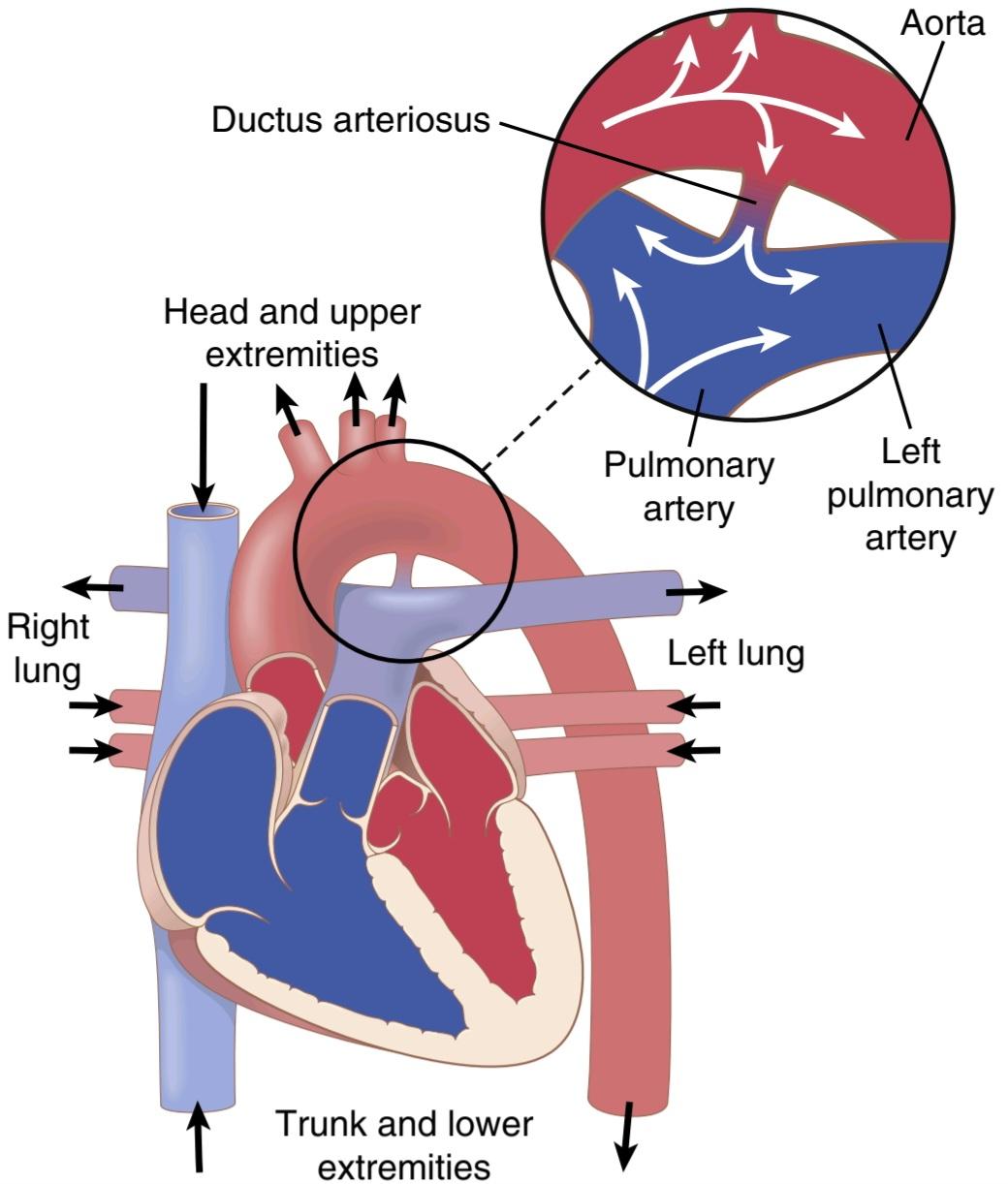
Figure 23-5 Patent ductus arteriosus, showing by the blue color that venous blood changes into oxygenated blood at different points in the circulation. Inset, Backflow of blood from the aorta into the pulmonary artery and then through the lungs for a second time.
شکل 23-5 مجرای شریانی ثبت شده که با رنگ آبی نشان می دهد که خون وریدی در نقاط مختلف گردش خون به خون اکسیژن دار تبدیل می شود. ورود، برگشت خون از آئورت به شریان ریوی و سپس از طریق ریه ها برای بار دوم.
Dynamics of the Circulation With a Persistent Patent Ductus
During the early months of an infant’s life, a patent ductus usually does not cause severely abnormal function. How- ever, as the child grows older, the differential between the high pressure in the aorta and the lower pressure in the pulmonary artery progressively increases, with a cor- responding increase in the backward flow of blood from the aorta into the pulmonary artery. Also, the high aortic blood pressure usually causes the diameter of the partially open ductus to increase with time, making the condition even worse.
دینامیک گردش خون با یک مجرای ثبت اختراع پایدار
در ماه های اولیه زندگی نوزاد، مجرای باز شده معمولاً باعث عملکرد غیر طبیعی شدید نمی شود. با این حال، همانطور که کودک بزرگتر می شود، تفاوت بین فشار بالا در آئورت و فشار پایین در شریان ریوی به تدریج افزایش می یابد و به همین ترتیب جریان خون به عقب از آئورت به شریان ریوی افزایش می یابد. همچنین، فشار خون بالا آئورت معمولاً باعث میشود که قطر مجرای نیمه باز با گذشت زمان افزایش یابد و وضعیت را بدتر کند.
Recirculation Through the Lungs. In an older child with a patent ductus, one-half to two-thirds of the aortic blood flows backward through the ductus into the pulmonary artery, then through the lungs, and finally back into the left ventricle and aorta, passing through the lungs and left side of the heart two or more times for every one time that it passes through the systemic circulation. People with this condition do not show cyanosis until later in life, when the heart fails or the lungs become congested. Indeed, early in life, the arterial blood is often better oxygenated than normal because of the extra times that it passes through the lungs.
گردش مجدد از طریق ریه ها. در یک کودک بزرگتر با مجرای باز، نیم تا دو سوم خون آئورت از طریق مجرای به داخل شریان ریوی، سپس از طریق ریه ها، و در نهایت به بطن چپ و آئورت برمی گردد و برای هر بار عبور از سیستم گردش خون، دو بار یا بیشتر از ریه ها و سمت چپ قلب می گذرد. افراد مبتلا به این عارضه تا اواخر عمر، زمانی که قلب از کار می افتد یا ریه ها دچار احتقان می شوند، سیانوز را نشان نمی دهند. در واقع، در اوایل زندگی، خون شریانی معمولاً به دلیل دفعات اضافی که از ریه ها عبور می کند، بهتر از حالت عادی اکسیژن دار می شود.
Diminished Cardiac and Respiratory Reserve. The major effects of patent ductus arteriosus on the patient are decreased cardiac and respiratory reserve. The left ventricle is pumping about two or more times the normal cardiac output, and the maximum that it can pump after hypertrophy of the heart has occurred is about four to seven times normal. Therefore, during exercise, the net blood flow through the remainder of the body can never increase to the levels required for strenuous activity. With even moderately strenuous exercise, the person is likely to become weak and may even faint from momentary heart failure.
کاهش ذخیره قلبی و تنفسی. اثرات عمده مجرای شریانی باز بر روی بیمار کاهش ذخیره قلبی و تنفسی است. بطن چپ حدود دو یا چند برابر برون ده طبیعی قلب پمپاژ می کند و حداکثر مقداری که می تواند پس از بروز هیپرتروفی قلب پمپاژ کند حدود چهار تا هفت برابر طبیعی است. بنابراین، در طول ورزش، جریان خالص خون در بقیه بدن هرگز نمی تواند به سطوح مورد نیاز برای فعالیت شدید افزایش یابد. با ورزش های شدید حتی نسبتاً شدید، فرد احتمالاً ضعیف می شود و حتی ممکن است در اثر نارسایی قلبی لحظه ای غش کند.
The high pressures in the pulmonary vessels caused by excess flow through the lungs may also lead to pulmonary congestion and pulmonary edema. As a result of the excessive load on the heart, and especially because the pulmonary congestion becomes progressively more severe with age, most patients with uncorrected patent ductus die from heart disease between the ages of 20 and 40 years.
فشار زیاد در رگ های ریوی ناشی از جریان بیش از حد از ریه ها نیز ممکن است منجر به احتقان ریوی و ادم ریوی شود. در نتیجه بار بیش از حد بر روی قلب، و به ویژه به دلیل اینکه احتقان ریوی با افزایش سن به تدریج شدیدتر می شود، اکثر بیماران مبتلا به مجرای باز شده اصلاح نشده بین 20 تا 40 سالگی بر اثر بیماری قلبی می میرند.
Heart Sounds: Machinery Murmur
In a newborn infant with patent ductus arteriosus, occa- sionally no abnormal heart sounds are heard because the quantity of reverse blood flow through the ductus may be insufficient to cause a heart murmur. However, as the baby grows older, to 1 to 3 years of age, a harsh blowing murmur begins to be heard in the pulmonary artery area of the chest, as shown in recording F, Figure 23-3. This sound is much more intense during systole when the aortic pressure is high and much less intense during diastole when the aortic pressure falls low, so that the murmur waxes and wanes with each beat of the heart, creating the so-called machinery murmur.
صدای قلب: سوفل ماشینی
در یک نوزاد تازه متولد شده با مجرای شریانی باز، گهگاه صدای قلب غیرطبیعی شنیده نمی شود، زیرا ممکن است مقدار جریان خون معکوس از طریق مجرا برای ایجاد سوفل قلبی کافی نباشد. با این حال، همانطور که در ثبت F، شکل 23-3 نشان داده شده است، همانطور که کودک بزرگتر می شود، تا 1 تا 3 سالگی، یک سوفل دمنده شدید در ناحیه شریان ریوی قفسه سینه شنیده می شود. این صدا در هنگام سیستول زمانی که فشار آئورت بالا است بسیار شدیدتر است و در هنگام دیاستول هنگامی که فشار آئورت پایین میآید بسیار شدیدتر است، به طوری که سوفل با هر ضربان قلب کاهش مییابد و به اصطلاح سوفل ماشینی ایجاد میکند.
Surgical Treatment
Surgical treatment of patent ductus arteriosus (PDA) is simple; one need only ligate the patent ductus or divide it and then close the two ends. This procedure was one of the first successful heart surgeries ever performed. Catheter-based devices often are used to close PDAs in infants or children who are large enough to have the procedure. A small metal coil or other occlusion device is passed up through the catheter and placed in the PDA to block blood flow through the vessel.
درمان جراحی
درمان جراحی مجرای شریانی باز شده (PDA) ساده است. فقط کافی است مجرای باز شده را ببندید یا آن را تقسیم کنید و سپس دو سر را ببندید. این روش یکی از اولین جراحی های موفقیت آمیز قلب بود که تاکنون انجام شده است. دستگاه های مبتنی بر کاتتر اغلب برای بستن PDA در نوزادان یا کودکانی که به اندازه کافی بزرگ هستند برای انجام این عمل استفاده می شود. یک سیم پیچ فلزی کوچک یا سایر دستگاه های انسداد از طریق کاتتر عبور داده می شود و در PDA قرار می گیرد تا جریان خون را در رگ مسدود کند.
TETRALOGY OF FALLOT-A RIGHT-TO- LEFT SHUNT
Tetralogy of Fallot is shown in Figure 23-6; it is the most common cause of blue baby. Most of the blood bypasses the lungs, so the aortic blood is mainly unoxygenated venous blood. In this condition, four abnormalities of the heart occur simultaneously:
TETRALOGY OF FALLOT-A RIGHT-TO-LEFT SHUNT
تترالوژی فالوت در شکل 23-6 نشان داده شده است. این شایع ترین علت آبی شدن نوزاد است. بیشتر خون ریه ها را دور می زند، بنابراین خون آئورت عمدتاً خون وریدی بدون اکسیژن است. در این وضعیت چهار ناهنجاری قلب به طور همزمان رخ می دهد:
1. The aorta originates from the right ventricle rather than the left, or it overrides a hole in the septum, as shown in Figure 23-6, receiving blood from both ventricles.
1. آئورت از بطن راست به جای سمت چپ منشأ می گیرد، یا همانطور که در شکل 23-6 نشان داده شده است، سوراخی را در سپتوم می پوشاند و از هر دو بطن خون دریافت می کند.
2. Because the pulmonary artery is stenosed, much lower than normal amounts of blood pass from the right ventricle into the lungs; instead, most of the blood passes directly into the aorta, thus bypassing the lungs.
2. از آنجایی که شریان ریوی تنگ شده است، مقدار بسیار کمتری از خون از بطن راست به ریه ها عبور می کند. در عوض، بیشتر خون مستقیماً وارد آئورت می شود و در نتیجه ریه ها را دور می زند.
3. Blood from the left ventricle flows through a ventricular septal hole into the right ventricle and then into the aorta or directly into the aorta that over- rides this hole.
3. خون از بطن چپ از طریق یک سوراخ سپتوم بطنی به بطن راست و سپس به آئورت یا مستقیماً به آئورتی که بر روی این سوراخ قرار دارد جریان می یابد.
4. Because the right side of the heart must pump large quantities of blood against the high pressure in the aorta, its musculature is highly developed, causing an enlarged right ventricle.
4. از آنجایی که سمت راست قلب باید مقادیر زیادی خون را در برابر فشار بالا در آئورت پمپ کند، ماهیچههای آن بسیار توسعه یافته است و باعث بزرگ شدن بطن راست میشود.
Abnormal Circulatory Dynamics. It is readily apparent that the major physiological difficulty caused by tetralogy of Fallot is the shunting of blood past the lungs without its becoming oxygenated. As much as 75% of the venous blood returning to the heart passes directly from the right ventricle into the aorta without becoming oxygenated. This can lead to cyanosis (blue coloration) of the baby’s skin. Other signs include high right ventricular pressures, right ventricular enlargement, and a left-to-right shunt through the interventricular septum, which can be visualized by echocardiography.
دینامیک گردش خون غیر طبیعی. به آسانی آشکار است که مشکل فیزیولوژیکی اصلی ناشی از تترالوژی فالوت، عبور خون از ریه ها بدون اکسیژنه شدن آن است. 75 درصد از خون وریدی که به قلب باز می گردد مستقیماً از بطن راست به آئورت می رود بدون اینکه اکسیژن دار شود. این می تواند منجر به سیانوز (رنگ آبی) پوست نوزاد شود. علائم دیگر شامل فشار بالای بطن راست، بزرگ شدن بطن راست و شانت چپ به راست از طریق سپتوم بین بطنی است که با اکوکاردیوگرافی قابل مشاهده است.

Figure 23-6 Tetralogy of Fallot, showing by the blue color that most of the venous blood is shunted from the right ventricle into the aorta without passing through the lungs.
شکل 23-6 تترالوژی فالوت، که با رنگ آبی نشان می دهد که بیشتر خون وریدی از بطن راست به آئورت بدون عبور از ریه ها منتقل می شود.
Surgical Treatment. Tetralogy of Fallot can often be treated successfully with surgery. The usual procedure is to open the pulmonary stenosis, close the septal defect, and reconstruct the flow pathway into the aorta. When surgery is successful, the average life expectancy increases from only 3 to 4 years to 50 or more years.
درمان جراحی. تترالوژی فالوت اغلب با جراحی با موفقیت درمان می شود. روش معمول باز کردن تنگی ریه، بستن نقص سپتوم و بازسازی مسیر جریان به داخل آئورت است. زمانی که جراحی موفقیت آمیز باشد، میانگین امید به زندگی از 3 تا 4 سال به 50 سال یا بیشتر افزایش می یابد.
CAUSES OF CONGENITAL ANOMALIES
Congenital heart disease is not uncommon, occurring in about 8 of every 1000 live births. One of the most common causes of congenital heart defects is a viral infection in the mother during the first trimester of pregnancy when the fetal heart is being formed. Defects are particularly prone to develop when the expectant mother contracts German measles (rubella) during the first trimester of pregnancy. Taking certain medications, such as angiotensin-converting enzyme (ACE) inhibitors and acne medications (e.g., isotretinoin), and alcohol or drug abuse during pregnancy also increases the risk for heart defects in the developing fetus.
علل ناهنجاری های مادرزادی
بیماری قلبی مادرزادی غیرمعمول نیست و در 8 مورد از هر 1000 تولد زنده رخ می دهد. یکی از شایع ترین علل نقص مادرزادی قلب، عفونت ویروسی در مادر در سه ماهه اول بارداری است که قلب جنین در حال شکل گیری است. هنگامی که مادر باردار در سه ماهه اول بارداری به سرخک آلمانی (سرخچه) مبتلا می شود، نقایص مستعد ایجاد می شوند. مصرف برخی داروها مانند مهارکننده های آنزیم مبدل آنژیوتانسین (ACE) و داروهای آکنه (مانند ایزوترتینوئین) و سوء مصرف الکل یا مواد مخدر در دوران بارداری نیز خطر نقص قلبی را در جنین در حال رشد افزایش می دهد.
Some congenital defects of the heart are hereditary because the same defect has been known to occur in identical twins, as well as in succeeding generations. Children of patients surgically treated for congenital heart disease have about a 10 times greater chance of having congenital heart disease than other children. Congenital defects of the heart are also frequently associated with other congenital defects of the baby’s body.
برخی از نقایص مادرزادی قلب ارثی هستند زیرا همین نقص در دوقلوهای همسان و همچنین در نسل های بعدی دیده می شود. کودکان بیمارانی که به دلیل بیماری مادرزادی قلب تحت عمل جراحی قرار میگیرند، تقریباً 10 برابر بیشتر از سایر کودکان شانس ابتلا به بیماری قلبی مادرزادی دارند. نقایص مادرزادی قلب نیز اغلب با سایر نقایص مادرزادی بدن نوزاد همراه است.
USE OF EXTRACORPOREAL CIRCULATION DURING CARDIAC SURGERY
It is almost impossible to repair intracardiac defects surgically while the heart is still pumping. Therefore, many types of artificial heart-lung machines have been developed to take the place of the heart and lungs during the course of an operation. Such a system is called extracorporeal circulation. The system consists principally of a pump and an oxygenating device. Almost any type of pump that does not cause hemolysis of the blood seems to be suitable.
استفاده از گردش خون خارج از بدن در حین جراحی قلب
ترمیم نقایص داخل قلب در حالی که قلب هنوز در حال پمپاژ است تقریباً غیرممکن است. بنابراین، انواع بسیاری از دستگاههای مصنوعی قلب و ریه برای جایگزینی قلب و ریهها در طول یک عمل ساخته شدهاند. چنین سیستمی گردش خارج از بدن نامیده می شود. این سیستم عمدتاً از یک پمپ و یک دستگاه اکسیژن ساز تشکیل شده است. تقریباً هر نوع پمپی که باعث همولیز خون نشود مناسب به نظر می رسد.
Methods used for oxygenating blood include the fol- lowing: (1) bubbling oxygen through the blood and removing the bubbles from the blood before passing it back into the patient; (2) dripping the blood downward over the surfaces of plastic sheets in the presence of oxygen; (3) passing the blood over surfaces of rotating discs; and (4) passing the blood between thin membranes or through thin tubes that are permeable to oxygen and carbon dioxide.
روشهای مورد استفاده برای اکسیژنرسانی خون شامل موارد زیر است: (1) حباب زدن اکسیژن از طریق خون و خارج کردن حبابها از خون قبل از بازگرداندن آن به بیمار. (2) چکیدن خون به سمت پایین روی سطوح ورقه های پلاستیکی در حضور اکسیژن. (3) عبور خون از روی سطوح دیسک های در حال چرخش. و (4) عبور خون از بین غشاهای نازک یا از طریق لوله های نازکی که برای اکسیژن و دی اکسید کربن قابل نفوذ هستند.
HYPERTROPHY OF THE HEART IN VALVULAR AND CONGENITAL HEART DISEASE
Hypertrophy of cardiac muscle is one of the most important mechanisms whereby the heart adapts to increased workloads whether these loads are caused by increased pressure against which the heart muscle must contract or by increased cardiac output that must be pumped. One can calculate approximately how much hypertrophy will occur in each chamber of the heart by multiplying the ventricular output by the pressure against which the ventricle must work, with an emphasis on pressure. Thus, hypertrophy occurs in most types of valvular and con- genital disease, sometimes causing the heart to weigh as much as 800 grams instead of the normal 300 grams.
هایپرتروفی قلب در بیماری های دریچه ای و مادرزادی قلب
هیپرتروفی عضله قلب یکی از مهمترین مکانیسمهایی است که به موجب آن قلب با افزایش بارهای کاری سازگار میشود، چه این بارها ناشی از افزایش فشاری باشد که عضله قلب باید در برابر آن منقبض شود یا به دلیل افزایش برون ده قلبی که باید پمپ شود. با ضرب کردن برون ده بطنی در فشاری که بطن باید در مقابل آن کار کند، با تاکید بر فشار، می توان تقریباً میزان هیپرتروفی در هر حفره قلب را محاسبه کرد. بنابراین، هیپرتروفی در اکثر انواع بیماریهای دریچهای و مادرزادی رخ میدهد و گاهی باعث میشود که قلب به جای 300 گرم طبیعی، به 800 گرم برسد.
Detrimental Effects of Late Stages of Cardiac Hypertrophy. Although the most common cause of cardiac hypertrophy is hypertension, almost all forms of cardiac diseases, including valvular and congenital disease, can stimulate enlargement of the heart.
اثرات مخرب مراحل پایانی هیپرتروفی قلبی. اگرچه شایع ترین علت هیپرتروفی قلب، فشار خون بالا است، اما تقریباً تمام انواع بیماری های قلبی، از جمله بیماری های دریچه ای و مادرزادی، می توانند بزرگ شدن قلب را تحریک کنند.
So-called physiological cardiac hypertrophy is generally considered to be a compensatory response of the heart to increased workload and is usually beneficial for maintaining cardiac output in the presence of abnormalities that impair the heart’s effectiveness as a pump. However, extreme degrees of hypertrophy can lead to heart failure. One reason for this is that the coronary vasculature typically does not increase to the same extent as the mass of cardiac muscle increases. The second reason is that fibrosis often develops in the muscle, especially in the subendocardial muscle where the coronary blood flow is poor, with fibrous tissue replacing degenerating muscle fibers. Because of the disproportionate increase in muscle mass relative to coronary blood flow, relative ischemia may develop as the cardiac muscle hypertrophies, and coronary blood flow insufficiency may ensue. Anginal pain is therefore a frequent accompaniment of cardiac hypertrophy associated with valvular and congenital heart disease. Enlargement of the heart is also associated with a greater risk for developing arrhythmias, which in turn can lead to further impairment of cardiac function and sudden death because of fibrillation.
به اصطلاح هیپرتروفی فیزیولوژیکی قلب به طور کلی به عنوان یک پاسخ جبرانی قلب به افزایش بار کاری در نظر گرفته می شود و معمولاً برای حفظ برون ده قلبی در حضور ناهنجاری هایی که کارایی قلب به عنوان یک پمپ را مختل می کند مفید است. با این حال، درجات شدید هیپرتروفی می تواند منجر به نارسایی قلبی شود. یکی از دلایل این امر این است که عروق کرونر معمولاً به همان میزانی که توده عضله قلب افزایش می یابد افزایش نمی یابد. دلیل دوم این است که فیبروز اغلب در عضله ایجاد می شود، به ویژه در عضله ساب اندوکارد که جریان خون کرونری ضعیف است و بافت فیبری جایگزین فیبرهای عضلانی تحلیل رفته است. به دلیل افزایش نامتناسب توده عضلانی نسبت به جریان خون کرونر، ایسکمی نسبی ممکن است با هیپرتروفی عضله قلب و نارسایی جریان خون کرونر ایجاد شود. بنابراین درد آنژین یک همراه مکرر هیپرتروفی قلبی است که با بیماری دریچه ای و مادرزادی قلب همراه است. بزرگ شدن قلب همچنین با خطر بیشتری برای ایجاد آریتمی همراه است که به نوبه خود می تواند منجر به اختلال بیشتر در عملکرد قلب و مرگ ناگهانی به دلیل فیبریلاسیون شود.
کلیک کنید «Bibliography: فهرست کتب مربوطه»
Bing R, Cavalcante JL, Everett RJ, Clavel MA, Newby DE, Dweck MR: Imaging and impact of myocardial fibrosis in aortic stenosis. JACC Cardiovasc Imaging 12:283, 2019.
Bonow RO, Leon MB, Doshi D, Moat N: Management strategies and future challenges for aortic valve disease. Lancet 387:1312, 2016.
Burchfield JS, Xie M, Hill JA: Pathological ventricular remodeling: mechanisms: part 1 of 2. Circulation 128:388, 2013.
Fahed AC, Gelb BD, Seidman JG, Seidman CE: Genetics of congenital heart disease: the glass half empty. Circ Res 112(4):707, 2013. Clyman
RI: Patent ductus arteriosus, its treatments, and the risks of pulmonary morbidity. Semin Perinatol 42:235, 2018.
Gould ST, Srigunapalan S, Simmons CA, Anseth KS: Hemodynamic and cellular response feedback in calcific aortic valve disease. Circ Res 113:186, 2013.
Hinton RB, Ware SM: Heart failure in pediatric patients with congeni- tal heart disease. Circ Res 120:978, 2017
Kodali SK, Velagapudi P, Hahn RT, Abbott D, Leon MB: Valvular heart
disease in patients ≥80 years of age. J Am Coll Cardiol 71:2058, 2018
Lindman BR, Bonow RO, Otto CM: Current management of calcific aortic stenosis. Circ Res 113:223, 2013.
Manning WJ: Asymptomatic aortic stenosis in the elderly: a clinical review. JAMA 310:1490, 2013.
Maron BJ: Clinical course and management of hypertrophic cardio- myopathy. N Engl J Med 379:655, 2018.
Maron BJ, Maron MS, Maron BA, Loscalzo J: Moving beyond the sar- comere to explain heterogeneity in hypertrophic cardiomyopathy. J Am Coll Cardiol 73:1978, 2019.
Nishimura RA, Otto CM, Bonow RO, Carabello BA, et al: 2017 AHA/ ACC Focused update of the 2014 AHA/ACC guideline for the man- agement of patients with valvular heart disease: A report of the American College of Cardiology/American Heart Association Task Force on Clinical Practice Guidelines. Circulation 135:e1159, 2017.
Ohukainen P, Ruskoaho H, Rysa J: Cellular mechanisms of valvular thickening in early and intermediate calcific aortic valve disease. Curr Cardiol Rev 14:264, 2018.
Remenyi B, ElGuindy A, Smith SC Jr, Yacoub M, Holmes DR Jr: Val- vular aspects of rheumatic heart disease. Lancet 387:1335, 2016.
Sommer RJ, Hijazi ZM, Rhodes JF Jr: Pathophysiology of congeni- tal heart disease in the adult: part I: shunt lesions. Circulation 117:1090, 2008.
Sommer RJ, Hijazi ZM, Rhodes JF: Pathophysiology of congenital heart disease in the adult: part III: complex congenital heart dis- ease. Circulation 117:1340, 2008.
Zaidi S, Brueckner M: Genetics and genomics of congenital heart dis- ease. Circ Res 120:923, 2017.
🚀 با ما همراه شوید!
تازهترین مطالب و آموزشهای مغز و اعصاب را از دست ندهید. با فالو کردن کانال تلگرام آیندهنگاران مغز، از ما حمایت کنید!



 ورود/ ثبت نام با جیمیل
