
فیزیولوژی پزشکی گایتون و هال؛ نارسایی قلبی


دعای مطالعه [ نمایش ]
بِسْمِ الله الرَّحْمنِ الرَّحیمِ
اَللّهُمَّ اَخْرِجْنى مِنْ ظُلُماتِ الْوَهْمِ
خدايا مرا بيرون آور از تاريكىهاى وهم،
وَ اَكْرِمْنى بِنُورِ الْفَهْمِ
و به نور فهم گرامى ام بدار،
اَللّهُمَّ افْتَحْ عَلَيْنا اَبْوابَ رَحْمَتِكَ
خدايا درهاى رحمتت را به روى ما بگشا،
وَانْشُرْ عَلَيْنا خَزائِنَ عُلُومِكَ بِرَحْمَتِكَ يا اَرْحَمَ الرّاحِمينَ
و خزانههاى علومت را بر ما باز كن به امید رحمتت اى مهربانترين مهربانان.
کتاب «فیزیولوژی پزشکی گایتون و هال» بهعنوان یکی از جامعترین و معتبرترین منابع در حوزه علوم پزشکی، همچنان مرجع کلیدی برای درک عملکرد پیچیده بدن انسان است. این اثر با تکیه بر تازهترین پژوهشها و توضیحات دقیق از سازوکارهای فیزیولوژیک، پلی میان علوم پایه پزشکی و کاربردهای بالینی ایجاد میکند و نقشی بیبدیل در آموزش، پژوهش و ارتقای دانش سلامت ایفا مینماید.
ترجمه دقیق و علمی این شاهکار توسط برند علمی آیندهنگاران مغز به مدیریت داریوش طاهری، دسترسی فارسیزبانان به مرزهای نوین دانش فیزیولوژی را ممکن ساخته و رسالتی علمی برای ارتقای آموزش پزشکی، فهم عمیقتر سازوکارهای بدن و توسعه روشهای نوین در حوزه سلامت فراهم آورده است.
» Guyton and Hall Textbook of Medical Physiology
»» CHAPTER 22: Cardiac Failure

One of the most important ailments treated by the physician is cardiac failure (heart failure). This ailment can result from any heart condition that reduces the ability of the heart to pump enough blood to meet the body’s needs. The cause is often decreased contractility of the myocardium resulting from diminished coronary blood flow. However, cardiac failure can also be caused by damaged heart valves, external pressure around the heart, vitamin B deficiency, primary cardiac muscle disease, or any other abnormality that makes the heart a hypoeffective pump.
یکی از مهم ترین بیماری هایی که توسط پزشک معالجه می شود نارسایی قلبی (نارسایی قلبی) است. این بیماری می تواند ناشی از هر بیماری قلبی باشد که توانایی قلب برای پمپاژ خون کافی برای رفع نیازهای بدن را کاهش دهد. علت اغلب کاهش انقباض میوکارد ناشی از کاهش جریان خون کرونر است. با این حال، نارسایی قلبی میتواند ناشی از آسیبدیدگی دریچههای قلب، فشار خارجی اطراف قلب، کمبود ویتامین B، بیماری اولیه عضله قلبی، یا هر ناهنجاری دیگری باشد که قلب را به یک پمپ کماثر تبدیل میکند.
In this chapter, we mainly discuss cardiac failure caused by ischemic heart disease resulting from partial blockage of the coronary blood vessels, which is the most common cause of heart failure. In Chapter 23, we discuss valvular and congenital heart disease.
در این فصل، ما عمدتاً نارسایی قلبی ناشی از بیماری ایسکمیک قلب ناشی از انسداد جزئی عروق خونی کرونر را مورد بحث قرار میدهیم که شایعترین علت نارسایی قلبی است. در فصل 23، در مورد بیماری دریچه ای و مادرزادی قلب صحبت می کنیم.
CIRCULATORY DYNAMICS IN CARDIAC FAILURE
دینامیک گردش خون در نارسایی قلبی
ACUTE EFFECTS OF MODERATE CARDIAC FAILURE
If a heart suddenly becomes severely damaged, such as by myocardial infarction, the pumping ability of the heart is immediately depressed. As a result, two main effects occur: (1) reduced cardiac output; and (2) damming of blood in the veins, resulting in increased venous pressure.
اثرات حاد نارسایی قلبی متوسط
اگر قلب به طور ناگهانی به شدت آسیب ببیند، مانند انفارکتوس میوکارد، توانایی پمپاژ قلب بلافاصله کاهش می یابد. در نتیجه، دو اثر اصلی رخ می دهد: (1) کاهش برون ده قلبی. و (2) آسیب رساندن به خون در وریدها و در نتیجه افزایش فشار وریدی.
The progressive changes in heart pumping effectiveness at different times after an acute myocardial infarction are shown graphically in Figure 22-1. The top curve of this figure shows a normal cardiac output curve. Point A on this curve is the normal operating point, showing a normal cardiac output under resting conditions of 5 L/ min and a right atrial pressure of 0 mm Hg.
تغییرات پیشرونده در کارایی پمپاژ قلب در زمان های مختلف پس از انفارکتوس حاد میوکارد به صورت نموداری در شکل 22-1 نشان داده شده است. منحنی بالای این شکل منحنی برون ده قلبی طبیعی را نشان می دهد. نقطه A در این منحنی نقطه عمل نرمال است که برون ده قلبی طبیعی را در شرایط استراحت 5 لیتر در دقیقه و فشار دهلیز راست 0 میلی متر جیوه را نشان می دهد.
Immediately after the heart becomes damaged, the cardiac output curve becomes greatly depressed, falling to the lowest curve at the bottom of the graph. Within a few seconds, a new circulatory state is established at point B, illustrating that the cardiac output has fallen to 2 L/min, about two-fifths normal, whereas the right atrial pressure has risen to +4 mm Hg because venous blood returning to the heart from the body is dammed up in the right atrium. This low cardiac output is still sufficient to sustain life for perhaps a few hours, but it is likely to be associated with fainting. Fortunately, this acute stage usually lasts for only a few seconds because sympathetic nervous reflexes occur almost immediately and compensate, to a great extent, for the damaged heart, as follows.
بلافاصله پس از آسیب رسیدن به قلب، منحنی برون ده قلبی به شدت تحت فشار قرار می گیرد و به پایین ترین منحنی در پایین نمودار می رسد. در عرض چند ثانیه، یک وضعیت گردش خون جدید در نقطه B برقرار می شود، که نشان می دهد برون ده قلبی به 2 لیتر در دقیقه کاهش یافته است، حدود دو پنجم طبیعی است، در حالی که فشار دهلیز راست به +4 میلی متر جیوه افزایش یافته است، زیرا خون وریدی که از بدن به قلب باز می گردد در دهلیز راست گیر می کند. این برون ده قلبی کم هنوز برای حفظ زندگی برای چند ساعت کافی است، اما احتمالاً با غش همراه است. خوشبختانه، این مرحله حاد معمولاً فقط چند ثانیه طول می کشد زیرا رفلکس های عصبی سمپاتیک تقریباً بلافاصله رخ می دهند و تا حد زیادی قلب آسیب دیده را به شرح زیر جبران می کنند.
Compensation for Acute Cardiac Failure by Sympathetic Nervous Reflexes. When the cardiac output falls precariously low, many of the circulatory reflexes discussed in Chapter 18 are rapidly activated. The best known of these is the baroreceptor reflex, which is activated by diminished arterial pressure. The chemoreceptor reflex, the central nervous system ischemic response, and even reflexes that originate in the damaged heart also likely contribute to activation of the sympathetic nervous system. The sympathetics therefore become strongly stimulated within a few seconds, and the parasympathetic nervous signals to the heart become inhibited at the same time.
جبران نارسایی حاد قلبی توسط رفلکس های عصبی سمپاتیک. هنگامی که برون ده قلبی به طور نامطمئنی پایین می آید، بسیاری از رفلکس های گردش خون مورد بحث در فصل 18 به سرعت فعال می شوند. شناخته شده ترین آنها رفلکس بارورسپتور است که با کاهش فشار شریانی فعال می شود. رفلکس گیرنده شیمیایی، پاسخ ایسکمیک سیستم عصبی مرکزی و حتی رفلکس هایی که از قلب آسیب دیده منشا می گیرند نیز احتمالاً در فعال شدن سیستم عصبی سمپاتیک نقش دارند. بنابراین سمپاتیک ها در عرض چند ثانیه به شدت تحریک می شوند و سیگنال های عصبی پاراسمپاتیک به قلب در همان زمان مهار می شوند.
Strong sympathetic stimulation has major effects on the heart and peripheral vasculature. If all the ventricular musculature is diffusely damaged but is still functional, sympathetic stimulation strengthens this damaged musculature. If part of the muscle is nonfunctional, and part of it is still normal, the normal muscle is strongly stimulated by sympathetic stimulation, in this way partially compensating for the nonfunctional muscle. Thus, the heart becomes a stronger pump as a result of sympathetic stimulation. This effect is illustrated in Figure 22-1, which shows about a twofold elevation of the very low cardiac output curve after sympathetic compensation.
تحریک قوی سمپاتیک اثرات عمده ای بر قلب و عروق محیطی دارد. اگر تمام عضله بطنی به طور منتشر آسیب دیده باشد اما همچنان عملکردی داشته باشد، تحریک سمپاتیک این عضله آسیب دیده را تقویت می کند. اگر بخشی از عضله غیرعملکردی باشد و بخشی از آن هنوز نرمال باشد، عضله طبیعی به شدت توسط تحریک سمپاتیک تحریک می شود، به این ترتیب تا حدی عضله غیرعملکردی جبران می شود. بنابراین، قلب در نتیجه تحریک سمپاتیک به پمپ قوی تری تبدیل می شود. این اثر در شکل 22-1 نشان داده شده است که حدود دو برابر افزایش منحنی برون ده قلبی بسیار پایین را پس از جبران سمپاتیک نشان می دهد.
Sympathetic stimulation also increases venous return because it increases the tone of most of the blood vessels of the circulation, especially the veins, raising the mean systemic filling pressure to 12 to 14 mm Hg, almost 100% above normal. As discussed in Chapter 20, this increased filling pressure greatly increases the tendency for blood to flow from the veins back into the heart. Therefore, the damaged heart becomes primed with more inflowing blood than usual, and the right atrial pressure rises still further, which helps the heart pump still larger quantities of blood. Thus, in Figure 22-1, the new circulatory state is depicted by point C, showing a cardiac output of 4.2 L/ min and a right atrial pressure of 5 mm Hg.
تحریک سمپاتیک بازگشت وریدی را نیز افزایش میدهد، زیرا تون اکثر رگهای خونی گردش خون، بهویژه وریدها را افزایش میدهد و میانگین فشار پر شدن سیستمیک را به 12 تا 14 میلیمتر جیوه، تقریباً 100 درصد بالاتر از حد طبیعی، افزایش میدهد. همانطور که در فصل 20 مورد بحث قرار گرفت، این افزایش فشار پر شدن تمایل به جریان خون از وریدها به قلب را تا حد زیادی افزایش می دهد. بنابراین، قلب آسیبدیده با خون ورودی بیشتر از حد معمول آماده میشود و فشار دهلیز راست باز هم بیشتر میشود، که به قلب کمک میکند مقدار بیشتری خون را پمپاژ کند. بنابراین، در شکل 22-1، وضعیت گردش خون جدید با نقطه C نشان داده شده است که برون ده قلبی 4.2 لیتر در دقیقه و فشار دهلیز راست 5 میلی متر جیوه را نشان می دهد.
The sympathetic reflexes become maximally developed in about 30 seconds. Therefore, a person who has a sudden, moderate heart attack might experience nothing more than cardiac pain and a few seconds of fainting. Shortly thereafter, with the aid of the sympathetic reflex compensations, the cardiac output may return to a level adequate to sustain the person if he or she remains quiet, although the pain might persist.
رفلکس های سمپاتیک در حدود 30 ثانیه به حداکثر می رسند. بنابراین، فردی که دچار حمله قلبی ناگهانی و متوسط می شود، ممکن است چیزی بیشتر از درد قلبی و چند ثانیه غش نداشته باشد. مدت کوتاهی پس از آن، با کمک جبرانهای رفلکس سمپاتیک، در صورت ساکت ماندن فرد، برون ده قلبی ممکن است به سطح مناسبی برای حفظ او برگردد، اگرچه درد ممکن است ادامه یابد.

Figure 22-1 Progressive changes in the cardiac output curve after acute myocardial infarction. Both the cardiac output and right atrial pressure change progressively from point A to point D (illustrated by the black line) over a period of seconds, minutes, days, and weeks.
شکل 22-1 تغییرات پیشرونده در منحنی برون ده قلبی پس از انفارکتوس حاد میوکارد. هم برون ده قلب و هم فشار دهلیز راست به تدریج از نقطه A به نقطه D (که با خط سیاه نشان داده شده است) در مدت زمان چند ثانیه، دقیقه، روز و هفته تغییر می کنند.
CHRONIC STAGE OF FAILURE-FLUID RETENTION AND COMPENSATED CARDIAC OUTPUT
After the first few minutes of an acute heart attack, a prolonged semichronic state begins, characterized mainly by two events: (1) retention of fluid by the kidneys; and (2) varying degrees of recovery of the heart over a period of weeks to months, as illustrated by the light green curve in Figure 22-1. This topic was also discussed in Chapter 21.
مرحله مزمن احتباس مایعات نارسایی و برون ده قلبی جبران شده
پس از چند دقیقه اول حمله قلبی حاد، یک حالت نیمه مزمن طولانی شروع می شود که عمدتاً با دو رویداد مشخص می شود: (1) احتباس مایع توسط کلیه ها. و (2) درجات مختلف بهبودی قلب در طی یک دوره چند هفته تا ماه، همانطور که با منحنی سبز روشن در شکل 22-1 نشان داده شده است. این موضوع در فصل 21 نیز مورد بحث قرار گرفت.
Renal Retention of Fluid and Increase in Blood Volume Occur for Hours to Days
A low cardiac output has a profound effect on renal function, sometimes causing anuria when the cardiac output falls to 50% to 60% of normal. In general, the urine output remains below normal as long as the cardiac output and arterial pressure remain significantly less than normal; urine output usually does not return all the way to normal after an acute heart attack until the cardiac output and arterial pressure rise almost to normal levels.
احتباس کلیوی مایعات و افزایش حجم خون برای ساعت ها تا روزها اتفاق می افتد
برون ده قلبی کم تأثیر عمیقی بر عملکرد کلیه دارد و گاهی اوقات زمانی که برون ده قلبی به 50 تا 60 درصد طبیعی می رسد باعث آنوری می شود. به طور کلی، تا زمانی که برون ده قلبی و فشار شریانی به طور قابل توجهی کمتر از حد طبیعی باشد، برون ده ادرار کمتر از حد نرمال باقی می ماند. برون ده ادرار معمولاً پس از یک حمله قلبی حاد به حالت عادی باز نمی گردد تا زمانی که برون ده قلبی و فشار شریانی تقریباً به سطح طبیعی افزایش یابد.
Moderate Fluid Retention in Cardiac Failure Can Be Beneficial. Many cardiologists have considered fluid retention always to have a detrimental effect in cardiac failure. However, a moderate increase in body fluid and blood volume is an important factor in helping to compensate for the diminished pumping ability of the heart by increasing the venous return. The increased blood volume increases venous return in two ways. First, it increases the mean systemic filling pressure, which increases the pressure gradient for causing venous flow of blood toward the heart. Second, it distends the veins, which reduces the venous resistance and allows even more ease of flow of blood to the heart.
احتباس مایعات متوسط در نارسایی قلبی می تواند مفید باشد. بسیاری از متخصصان قلب، احتباس مایعات را همیشه به عنوان یک اثر مضر در نارسایی قلبی در نظر گرفته اند. با این حال، افزایش متوسط مایعات بدن و حجم خون عامل مهمی برای کمک به جبران کاهش توانایی پمپاژ قلب با افزایش بازگشت وریدی است. افزایش حجم خون از دو طریق باعث افزایش بازگشت وریدی می شود. اول، فشار پرشدگی متوسط سیستمیک را افزایش می دهد، که باعث افزایش گرادیان فشار برای ایجاد جریان وریدی خون به سمت قلب می شود. دوم، رگها را متسع میکند، که مقاومت وریدی را کاهش میدهد و اجازه میدهد تا جریان خون به قلب راحتتر شود.
If the heart is not damaged too much, this increased venous return can almost fully compensate for the heart’s diminished pumping ability-enough so that even when the heart’s pumping ability is reduced to as low as 40% to 50% of normal, the increased venous return can often cause nearly normal cardiac output as long as the person remains in a quiet resting state.
اگر قلب بیش از حد آسیب نبیند، این افزایش بازگشت وریدی می تواند تقریباً به طور کامل کاهش توانایی پمپاژ قلب را جبران کند – به اندازه کافی، به طوری که حتی زمانی که توانایی پمپاژ قلب به 40٪ تا 50٪ از حد طبیعی کاهش می یابد، افزایش بازگشت وریدی اغلب می تواند باعث برون ده قلبی تقریباً طبیعی شود تا زمانی که فرد در حالت استراحت بماند.
When the heart’s pumping capability is reduced further, blood flow to the kidneys finally becomes too low for the kidneys to excrete enough salt and water to equal salt and water intake. Therefore, fluid retention begins and continues indefinitely unless major therapeutic procedures are used to prevent this outcome. Furthermore, because the heart is already pumping at its maximum capacity, this excess fluid no longer has a beneficial effect on the circulation. Instead, the fluid retention increases the workload on the already damaged heart, and severe edema develops throughout the body, which can be very detrimental and can lead to death.
وقتی توانایی پمپاژ قلب بیشتر کاهش مییابد، جریان خون به کلیهها در نهایت برای کلیهها بسیار کم میشود که نمیتواند نمک و آب کافی دفع کند تا نمک و آب دریافتی برابر شود. بنابراین، احتباس مایعات شروع می شود و به طور نامحدود ادامه می یابد، مگر اینکه از روش های درمانی عمده برای جلوگیری از این نتیجه استفاده شود. علاوه بر این، از آنجایی که قلب در حال حاضر با حداکثر ظرفیت خود پمپاژ می کند، این مایع اضافی دیگر اثر مفیدی بر گردش خون ندارد. در عوض، احتباس مایعات، بار کاری را بر روی قلب آسیب دیده افزایش می دهد و ادم شدید در سراسر بدن ایجاد می شود که می تواند بسیار مضر باشد و می تواند منجر به مرگ شود.
Detrimental Effects of Excess Fluid Retention in Severe Cardiac Failure. In contrast to the beneficial effects of moderate fluid retention in cardiac failure, in severe cardiac failure, extreme excesses of fluid can have serious physiologic consequences. These include the fol- lowing: (1) increasing the workload on the damaged heart; (2) overstretching of the heart, which further weakens the heart; (3) filtration of fluid into the lungs, causing pulmonary edema and consequent deoxygenation of the blood; and (4) development of extensive edema in most parts of the body. These detrimental effects of excessive fluid are discussed in later sections of this chapter.
اثرات مخرب احتباس بیش از حد مایعات در نارسایی شدید قلبی. برخلاف اثرات مفید احتباس متوسط مایعات در نارسایی قلبی، در نارسایی شدید قلبی، بیش از حد مایعات می تواند عواقب فیزیولوژیکی جدی داشته باشد. این موارد شامل موارد زیر است: (1) افزایش بار کار بر روی قلب آسیب دیده. (2) کشش بیش از حد قلب، که قلب را بیشتر ضعیف می کند. (3) فیلتراسیون مایع به داخل ریه ها، که باعث ادم ریوی و در نتیجه اکسیژن زدایی از خون می شود. و (4) ایجاد ادم گسترده در بیشتر قسمت های بدن. این اثرات مضر مایع بیش از حد در بخشهای بعدی این فصل مورد بحث قرار میگیرد.
Recovery of the Heart After Myocardial Infarction
After a heart becomes suddenly damaged as a result of myocardial infarction, the natural reparative processes of the body begin to help restore normal cardiac function. For example, a new collateral blood supply begins to penetrate the peripheral portions of the infarcted area of the heart, often causing some of the heart muscle in the fringe areas to become functional again. Also, the undamaged portion of the heart musculature hypertrophies, offset- ting some of the cardiac damage.
بهبودی قلب پس از سکته قلبی
پس از آسیب ناگهانی قلب در نتیجه انفارکتوس میوکارد، فرآیندهای ترمیمی طبیعی بدن شروع به کمک به بازیابی عملکرد طبیعی قلب می کند. به عنوان مثال، یک منبع خون جانبی جدید شروع به نفوذ به بخشهای محیطی ناحیه انفارکتوس شده قلب میکند و اغلب باعث میشود که بخشی از عضله قلب در نواحی حاشیهای دوباره فعال شود. همچنین، بخش صدمهنخورده عضله قلب هیپرتروفی میکند و برخی از آسیبهای قلبی را جبران میکند.
The degree of recovery, which depends on the type and severity of cardiac damage, varies from no recovery to almost complete recovery. After an acute myocardial infarction, the heart ordinarily recovers rapidly during the first few days and weeks and achieves most of its final state of recovery within 5 to 7 weeks, although mild degrees of additional recovery can continue for months.
درجه بهبودی که به نوع و شدت آسیب قلبی بستگی دارد، از عدم بهبودی تا بهبودی تقریباً کامل متفاوت است. پس از انفارکتوس حاد میوکارد، قلب معمولاً در طی چند روز و هفته اول به سرعت بهبود مییابد و در عرض 5 تا 7 هفته بیشتر حالت نهایی بهبودی خود را به دست میآورد، اگرچه درجات خفیف بهبودی اضافی میتواند برای ماهها ادامه یابد.
Cardiac Output Curve After Partial Recovery. Figure 22-1 shows function of the partially recovered heart a week or so after an acute myocardial infarction. By this time, considerable fluid has been retained in the body, and the tendency for venous return has increased markedly as well; therefore, the right atrial pressure has risen even more. As a result, the state of the circulation is now changed from point C to point D, which shows a normal cardiac output of 5 L/min but right atrial pressure in- creased to 6 mm Hg.
منحنی برون ده قلبی پس از بهبودی جزئی. شکل 22-1 عملکرد قلب نیمه بهبود یافته را یک هفته یا بیشتر پس از انفارکتوس حاد میوکارد نشان می دهد. در این زمان، مایع قابل توجهی در بدن باقی مانده است و تمایل به بازگشت وریدی نیز به طور قابل توجهی افزایش یافته است. بنابراین، فشار دهلیز راست حتی بیشتر افزایش یافته است. در نتیجه، وضعیت گردش خون اکنون از نقطه C به نقطه D تغییر می کند، که برون ده قلبی طبیعی 5 لیتر در دقیقه را نشان می دهد اما فشار دهلیز راست به 6 میلی متر جیوه افزایش یافته است.
Because the cardiac output has returned to normal, renal output of fluid also returns to normal, and no further fluid retention occurs, except that the retention of fluid that has already occurred continues to maintain moderate excesses of fluid. Therefore, except for the high right atrial pressure represented by point D in this figure, the person now has essentially normal cardiovascular dynamics as long as he or she remains at rest.
از آنجایی که برون ده قلبی به حالت عادی بازگشته است، برون ده کلیوی مایع نیز به حالت عادی باز می گردد و احتباس مایعات بیشتری رخ نمی دهد، به جز اینکه احتباس مایعی که قبلاً رخ داده است همچنان به حفظ مقدار متوسط مایع ادامه می دهد. بنابراین، به جز فشار بالای دهلیز راست که با نقطه D در این شکل نشان داده شده است، فرد اکنون تا زمانی که در حالت استراحت است، اساساً پویایی قلبی عروقی طبیعی دارد.
If the heart recovers to a significant extent, and if adequate fluid volume has been retained, the cardiac output increases toward normal and sympathetic stimulation gradually abates toward normal. As the heart recovers, the fast pulse rate, cold skin, and pallor resulting from sympathetic stimulation in the acute stage of cardiac failure gradually disappear.
اگر قلب به میزان قابل توجهی بهبود یابد، و اگر حجم مایع کافی حفظ شود، برون ده قلبی به سمت نرمال افزایش می یابد و تحریک سمپاتیک به تدریج به سمت نرمال کاهش می یابد. با بهبودی قلب، ضربان سریع نبض، پوست سرد و رنگ پریدگی ناشی از تحریک سمپاتیک در مرحله حاد نارسایی قلبی به تدریج ناپدید می شوند.
SUMMARY OF CHANGES AFTER ACUTE CARDIAC FAILURE-COMPENSATED HEART FAILURE
To summarize the events discussed in the past few sections describing the dynamics of circulatory changes after an acute, moderate heart attack, we can divide the stages into the following: (1) the instantaneous effect of the cardiac damage; (2) compensation by the sympathetic nervous system, which occurs mainly within the first 30 to 60 seconds; and (3) chronic compensations resulting from partial heart recovery and renal retention of fluid. All these changes are shown graphically by the black line in Figure 22-1. The progression of this line shows the normal state of the circulation (point A), the state a few seconds after the heart attack but before sympathetic reflexes have occurred (point B), the rise in cardiac output toward normal caused by sympathetic stimulation (point C), and final return of the cardiac output to almost normal after several days to several weeks of partial cardiac recovery and fluid retention (point D). This final state is called compensated heart failure.
خلاصه ای از تغییرات پس از نارسایی قلبی جبران شده با نارسایی حاد قلبی
برای خلاصه کردن وقایع مورد بحث در چند بخش گذشته که پویایی تغییرات گردش خون پس از یک حمله قلبی حاد و متوسط را توصیف میکنند، میتوانیم مراحل را به موارد زیر تقسیم کنیم: (1) اثر آنی آسیب قلبی. (2) جبران توسط سیستم عصبی سمپاتیک، که عمدتا در 30 تا 60 ثانیه اول رخ می دهد. و (3) غرامت های مزمن ناشی از بهبود نسبی قلب و احتباس کلیوی مایع. تمام این تغییرات به صورت گرافیکی با خط سیاه در شکل 22-1 نشان داده شده است. پیشرفت این خط وضعیت طبیعی گردش خون (نقطه A)، وضعیت چند ثانیه پس از حمله قلبی اما قبل از وقوع رفلکس های سمپاتیک (نقطه B)، افزایش برون ده قلبی به سمت نرمال ناشی از تحریک سمپاتیک (نقطه C) و بازگشت نهایی برون ده قلبی به تقریباً نرمال پس از چند روز تا چند هفته پس از بهبودی نسبی مایعات-قلبی (نقطه D) را نشان می دهد. این حالت نهایی نارسایی قلبی جبران شده نامیده می شود.
Compensated Heart Failure. Note especially in Figure 22-1 that the maximum pumping ability of the partly recovered heart, as depicted by the plateau level of the light green curve, is still depressed to less than half-normal. This demonstrates that an increase in right atrial pressure can maintain the cardiac output at a normal level, despite continued weakness of the heart. Thus, many people, especially older adults, have normal resting cardiac outputs but mildly to moderately elevated right atrial pressures because of various degrees of compensated heart failure. They may not know that they have cardiac damage be- cause the damage often has occurred a little at a time, and the compensation has occurred concurrently with the progressive stages of damage.
نارسایی قلبی جبران شده. به ویژه در شکل 22-1 توجه داشته باشید که حداکثر توانایی پمپاژ قلب تا حدی پوشیده شده، همانطور که توسط سطح فلات منحنی سبز روشن نشان داده شده است، هنوز به کمتر از نیمه طبیعی کاهش می یابد. این نشان می دهد که افزایش فشار دهلیز راست می تواند برون ده قلبی را علیرغم تداوم ضعف قلب در سطح طبیعی حفظ کند. بنابراین، بسیاری از افراد، بهویژه افراد مسنتر، برونده قلبی در حالت استراحت طبیعی دارند، اما به دلیل درجات مختلف نارسایی قلبی جبرانشده، فشار دهلیز راست خفیف تا متوسط افزایش یافته است. آنها ممکن است ندانند که آسیب قلبی دارند، زیرا آسیب اغلب در یک زمان اتفاق افتاده است، و جبران آن همزمان با مراحل پیشرونده آسیب رخ داده است.
When a person is in a state of compensated heart failure, any attempt to perform heavy exercise usually causes immediate return of the symptoms of acute heart failure because the heart is not able to increase its pumping capacity to the levels required for the exercise. Therefore, it is said that the cardiac reserve is reduced in compensated heart failure. This concept of cardiac reserve is discussed later in the chapter.
هنگامی که فرد در وضعیت نارسایی قلبی جبران شده قرار دارد، هر تلاشی برای انجام ورزش سنگین معمولاً باعث بازگشت فوری علائم نارسایی حاد قلبی می شود زیرا قلب قادر به افزایش ظرفیت پمپاژ خود به سطوح مورد نیاز برای ورزش نیست. بنابراین گفته می شود که ذخیره قلبی در نارسایی قلبی جبران شده کاهش می یابد. این مفهوم ذخیره قلبی بعداً در این فصل مورد بحث قرار می گیرد.
DYNAMICS OF SEVERE CARDIAC FAILURE-DECOMPENSATED HEART FAILURE
If the heart becomes severely damaged, no amount of compensation by sympathetic nervous reflexes or fluid retention can make the excessively weakened heart pump a normal cardiac output. As a consequence, the cardiac output cannot rise high enough to make the kidneys excrete normal quantities of fluid. Therefore, fluid continues to be retained, the person develops more and more edema, and this state of events eventually leads to death. This condition is called decompensated heart failure. Thus, a major cause of decompensated heart failure is failure of the heart to pump sufficient blood to make the kidneys excrete the necessary amounts of fluid every day.
دینامیک نارسایی شدید قلبی-نارسایی غیر جبرانی قلب
اگر قلب به شدت آسیب ببیند، هیچ جبرانی توسط رفلکسهای عصبی سمپاتیک یا احتباس مایعات نمیتواند باعث شود که قلب بسیار ضعیف به برون ده قلبی طبیعی پمپاژ کند. در نتیجه، برون ده قلبی نمی تواند آنقدر بالا برود که کلیه ها را وادار به دفع مقادیر طبیعی مایع کند. بنابراین، مایع همچنان حفظ می شود، فرد بیشتر و بیشتر دچار ادم می شود و این حالت در نهایت منجر به مرگ می شود. این وضعیت نارسایی غیر جبرانی قلب نامیده می شود. بنابراین، یکی از دلایل اصلی نارسایی جبراننشده قلبی، ناتوانی قلب در پمپاژ خون کافی است تا کلیهها مقدار لازم مایعات را در روز دفع کنند.
Graphic Analysis of Decompensated Heart Failure. Figure 22-2 shows greatly depressed cardiac output at different times (points A to F) after the heart has become severely weakened. Point A on this curve represents the approximate state of the circulation before any compensation has occurred, and point B represents the state a few minutes later, after sympathetic stimulation has compensated as much as it can but before fluid retention has begun. At this time, the cardiac output has risen to 4 L/min and the right atrial pressure has risen to 5 mm Hg. The person appears to be in reasonably good condition, but this state will not remain stable because the cardiac output has not risen high enough to cause adequate kidney excretion of fluid; therefore, fluid retention continues and can eventually be the cause of death. These events can be explained quantitatively in the following way.
تجزیه و تحلیل گرافیکی نارسایی غیر جبرانی قلب. شکل 22-2 کاهش شدید برون ده قلبی را در زمان های مختلف (نقاط A تا F) پس از ضعیف شدن شدید قلب نشان می دهد. نقطه A در این منحنی وضعیت تقریبی گردش خون را قبل از وقوع هر گونه جبرانی نشان می دهد و نقطه B وضعیت را چند دقیقه بعد نشان می دهد، پس از اینکه تحریک سمپاتیک تا آنجا که می تواند جبران شود اما قبل از شروع احتباس مایعات. در این زمان، برون ده قلبی به 4 لیتر در دقیقه و فشار دهلیز راست به 5 میلی متر جیوه افزایش یافته است. به نظر میرسد که فرد در وضعیت نسبتاً خوبی قرار دارد، اما این وضعیت ثابت نمیماند زیرا برون ده قلبی آنقدر بالا نمیرود که باعث دفع کافی مایع از کلیه شود. بنابراین، احتباس مایعات ادامه می یابد و در نهایت می تواند علت مرگ باشد. این رویدادها را می توان به صورت کمی توضیح داد.
Note the straight line in Figure 22-2, at a cardiac output level of 5 L/min. This level is approximately the critical cardiac output level that is required in the average adult person to make the kidneys reestablish normal fluid balance—that is, for the output of salt and water to be as high as the intake of these substances. At cardiac outputs below this level, the fluid-retaining mechanisms discussed in the earlier section remain in play, and the body fluid volume increases progressively. Because of this progressive increase in fluid volume, the mean systemic filling pressure of the circulation continues to rise, which forces progressively increasing quantities of blood from the person’s peripheral veins into the right atrium, thus increasing the right atrial pressure. After 1 day or so, the state of the circulation changes in Figure 22-2 from point B to point C, with the right atrial pressure rising to 7 mm Hg and the cardiac output rising to 4.2 L/min. Note again that the cardiac output is still not high enough to cause normal renal output of fluid; therefore, fluid continues to be retained. After another day or so, the right atrial pressure rises to 9 mm Hg, and the circulatory state becomes that depicted by point D. Still, the cardiac output is not enough to establish normal fluid balance.
به خط مستقیم شکل 22-2 در سطح برون ده قلبی 5 لیتر در دقیقه توجه کنید. این سطح تقریباً همان سطح بحرانی برون ده قلبی است که در یک فرد بزرگسال معمولی برای برقراری مجدد تعادل مایع طبیعی توسط کلیه ها لازم است – یعنی برون ده نمک و آب به اندازه میزان دریافتی این مواد باشد. در برون ده های قلبی زیر این سطح، مکانیسم های نگهداری مایعات که در بخش قبل مورد بحث قرار گرفت، همچنان فعال هستند و حجم مایع بدن به تدریج افزایش می یابد. به دلیل این افزایش تدریجی حجم مایع، فشار پرشدگی سیستمیک متوسط گردش خون همچنان افزایش مییابد، که باعث افزایش تدریجی مقادیر خون از وریدهای محیطی فرد به دهلیز راست میشود و در نتیجه فشار دهلیز راست را افزایش میدهد. پس از 1 روز یا بیشتر، وضعیت گردش خون در شکل 22-2 از نقطه B به نقطه C تغییر می کند، با افزایش فشار دهلیز راست تا 7 میلی متر جیوه و برون ده قلبی به 4.2 لیتر در دقیقه. دوباره توجه داشته باشید که برون ده قلبی هنوز آنقدر بالا نیست که باعث برون ده کلیوی طبیعی مایع شود. بنابراین، مایع همچنان حفظ می شود. پس از یک روز یا بیشتر، فشار دهلیز راست به 9 میلیمتر جیوه افزایش مییابد و وضعیت گردش خون به حالتی تبدیل میشود که در نقطه D نشان داده شده است. با این حال، برون ده قلبی برای ایجاد تعادل طبیعی مایع کافی نیست.
After another few days of fluid retention, the right atrial pressure has risen further but, by now, cardiac function is beginning to decline toward a lower level. This decline is caused by overstretch of the heart, edema of the heart muscle, and other factors that diminish the heart’s pumping performance. It is now clear that further retention of fluid will be more detrimental than beneficial to the circulation. Yet, the cardiac output still is not high enough to bring about normal renal function, so fluid retention not only continues but accelerates because of the falling cardiac output (and the falling arterial pressure that also occurs). Consequently, within a few days, the state of the circulation has reached point F on the curve, with the cardiac output now less than 2.5 L/min and the right atrial pressure 16 mm Hg. This state has approached or reached incompatibility with life, and the patient will die unless this chain of events can be reversed. This state of heart failure in which the failure continues to worsen is called decompensated heart failure.
پس از چند روز دیگر احتباس مایعات، فشار دهلیز راست بیشتر افزایش یافته است، اما در حال حاضر، عملکرد قلب شروع به کاهش به سمت سطح پایین تر کرده است. این کاهش به دلیل کشش بیش از حد قلب، ادم عضله قلب و سایر عواملی است که عملکرد پمپاژ قلب را کاهش می دهد. اکنون واضح است که احتباس بیشتر مایع بیشتر از آن که برای گردش خون مفید باشد، مضر است. با این حال، برون ده قلبی هنوز به اندازه کافی بالا نیست تا عملکرد طبیعی کلیه را ایجاد کند، بنابراین احتباس مایعات نه تنها ادامه می یابد، بلکه به دلیل کاهش برون ده قلبی (و کاهش فشار شریانی که همچنین رخ می دهد) تسریع می یابد. در نتیجه، در عرض چند روز، وضعیت گردش خون به نقطه F در منحنی رسیده است، با برون ده قلبی اکنون کمتر از 2.5 لیتر در دقیقه و فشار دهلیز راست 16 میلی متر جیوه. این حالت نزدیک شده یا به ناسازگاری با زندگی رسیده است و بیمار می میرد مگر اینکه این زنجیره وقایع معکوس شود. این حالت از نارسایی قلبی که در آن نارسایی همچنان بدتر می شود، نارسایی قلبی جبران نشده نامیده می شود.
Thus, one can see from this analysis that failure of the cardiac output (and arterial pressure) to rise to the critical level required for normal renal function results in the following: (1) progressive retention of more and more fluid; (2) progressive elevation of the mean systemic filling pressure; and (3) progressive elevation of the right atrial pressure until, finally, the heart is so overstretched or so edematous that it cannot pump even moderate quantities of blood and, therefore, fails completely. Clinically, one detects this serious condition of decompensation principally by the progressing edema, especially edema of the lungs, which leads to bubbling rales (a crackling sound) in the lungs and to dyspnea (air hunger). Lack of appropriate therapy at this stage rapidly leads to death.
بنابراین، از این تجزیه و تحلیل می توان دریافت که عدم افزایش برون ده قلبی (و فشار شریانی) به سطح بحرانی مورد نیاز برای عملکرد طبیعی کلیه منجر به موارد زیر می شود: (1) احتباس تدریجی مایعات بیشتر و بیشتر. (2) افزایش تدریجی میانگین فشار پر شدن سیستمیک. و (3) افزایش تدریجی فشار دهلیز راست تا زمانی که در نهایت، قلب به قدری کشیده شود یا آنقدر ادم داشته باشد که نتواند حتی مقادیر متوسطی از خون را پمپ کند و در نتیجه به طور کامل از کار بیفتد. از نظر بالینی، شخص این وضعیت جدی جبران خسارت را عمدتاً با ادم در حال پیشرفت، به ویژه ادم ریه ها، که منجر به رال های حباب دار (صدای تروق) در ریه ها و تنگی نفس (گرسنگی هوا) می شود، تشخیص می دهد. عدم درمان مناسب در این مرحله به سرعت منجر به مرگ می شود.
Treatment of Decompensation. The decompensation process can often be stopped by the following: (1) strengthening the heart in any one of several ways, especially by administering a cardiotonic drug, such as digitalis, so that the heart becomes strong enough to pump adequate quantities of blood required to make the kidneys function normally again; or (2) administering diuretic drugs to increase kidney excretion while at the same time reducing water and salt intake, which results in a balance between fluid intake and output, despite low cardiac output.
درمان جبران خسارت. فرآیند جبران خسارت اغلب می تواند با موارد زیر متوقف شود: (1) تقویت قلب به یکی از راه های مختلف، به ویژه با تجویز یک داروی قلب و عروق، مانند دیجیتالیس، به طوری که قلب به اندازه کافی قوی شود تا بتواند مقادیر کافی خون مورد نیاز برای عملکرد طبیعی کلیه ها را پمپ کند. یا (2) تجویز داروهای دیورتیک برای افزایش دفع کلیه و در عین حال کاهش مصرف آب و نمک، که منجر به تعادل بین دریافت و برون ده مایعات، علیرغم برون ده قلبی کم می شود.
Both methods stop the decompensation process by re- establishing normal fluid balance so that at least as much fluid leaves the body as enters it.
هر دو روش با برقراری مجدد تعادل طبیعی مایعات، فرآیند جبران را متوقف می کنند، به طوری که حداقل به اندازه مایعی که وارد بدن می شود، از بدن خارج شود.
Mechanism of Action of Cardiotonic Drugs. Cardiotonic drugs, such as digitalis, when administered to a per- son with a healthy heart, have little effect on increasing the contractile strength of the cardiac muscle. However, when administered to someone with a chronically failing heart, the same drugs can sometimes increase the strength of the failing myocardium by as much as 50% to 100%. Therefore, they are one of the mainstays of therapy in persons with chronic heart failure.
مکانیسم اثر داروهای قلبی. داروهای کاردیوتونیک، مانند دیژیتال، زمانی که برای فردی با قلب سالم تجویز می شود، تأثیر کمی بر افزایش قدرت انقباضی عضله قلب دارد. با این حال، زمانی که همان داروها برای فردی با نارسایی مزمن قلب تجویز می شوند، گاهی اوقات می توانند قدرت میوکارد نارسا را تا 50 تا 100 درصد افزایش دهند. بنابراین، آنها یکی از پایه های اصلی درمان در افراد مبتلا به نارسایی مزمن قلبی هستند.
Digitalis and other cardiotonic glycosides are believed to strengthen heart contractions by increasing the quantity of calcium ions in muscle fibers. This effect is likely due to inhibition of sodium-potassium adenosine triphosphatase in cardiac cell membranes. Inhibition of the sodium-potassium pump increases the intracellular sodium concentration and slows the sodium-calcium exchange pump, which extrudes calcium from the cell in exchange for sodium. Because the sodium-calcium exchange pump relies on a high sodium gradient across the cell membrane, accumulation of sodium inside the cell reduces its activity.
اعتقاد بر این است که دیژیتال و سایر گلیکوزیدهای کاردیوتونیک با افزایش مقدار یون های کلسیم در فیبرهای عضلانی انقباضات قلب را تقویت می کنند. این اثر احتمالاً به دلیل مهار سدیم پتاسیم آدنوزین تری فسفاتاز در غشای سلولی قلب است. مهار پمپ سدیم-پتاسیم باعث افزایش غلظت سدیم درون سلولی و کندی پمپ تبادل سدیم-کلسیم می شود که در ازای سدیم، کلسیم را از سلول خارج می کند. از آنجایی که پمپ تبادل سدیم-کلسیم به گرادیان سدیم بالایی در سراسر غشای سلولی متکی است، تجمع سدیم در داخل سلول باعث کاهش فعالیت آن می شود.
In the failing heart muscle, the sarcoplasmic reticulum fails to accumulate normal quantities of calcium and, therefore, cannot release enough calcium ions into the free fluid compartment of the muscle fibers to cause full contraction of the muscle. The effect of digitalis to depress the sodium-calcium exchange pump and raise calcium ion concentration in cardiac muscle provides the extra calcium needed to increase the muscle contractile force. Therefore, it is usually beneficial to depress the calcium pumping mechanism by a moderate amount using digitalis, allowing the muscle fiber intracellular calcium level to rise slightly.
در نارسایی عضله قلب، شبکه سارکوپلاسمی نمی تواند مقادیر طبیعی کلسیم را جمع کند و بنابراین نمی تواند یون های کلسیم کافی را در قسمت مایع آزاد رشته های عضلانی آزاد کند تا باعث انقباض کامل عضله شود. اثر دیژیتال برای کاهش پمپ تبادل سدیم-کلسیم و افزایش غلظت یون کلسیم در عضله قلب، کلسیم اضافی مورد نیاز برای افزایش نیروی انقباضی عضلانی را فراهم می کند. بنابراین، معمولاً کاهش مکانیسم پمپاژ کلسیم به میزان متوسط با استفاده از دیجیتالی مفید است و اجازه می دهد سطح کلسیم درون سلولی فیبر عضلانی کمی افزایش یابد.

Figure 22-2 Greatly depressed cardiac output that indicates decompensated heart disease. Progressive fluid retention raises the right atrial pressure over a period of days, and the cardiac output progresses from point A to point F until death occurs.
شکل 22-2 برون ده قلبی بسیار افسرده شده که نشان دهنده بیماری قلبی غیر جبرانی است. احتباس پیشرونده مایع، فشار دهلیز راست را طی چند روز افزایش میدهد و برون ده قلبی از نقطه A به نقطه F پیشروی میکند تا زمانی که مرگ رخ دهد.
UNILATERAL LEFT HEART FAILURE
Thus far we have considered failure of the heart as a whole. Yet, in a large number of patients, especially those with early acute heart failure, left-sided failure predominates over right-sided failure and, in rare cases, the right side fails without significant failure of the left side.
نارسایی یک طرفه قلب چپ
تا اینجا ما نارسایی قلب را به عنوان یک کل در نظر گرفته ایم. با این حال، در تعداد زیادی از بیماران، به ویژه آنهایی که نارسایی حاد قلبی اولیه دارند، نارسایی سمت چپ بر نارسایی سمت راست غالب است و در موارد نادر، سمت راست بدون نارسایی قابل توجه سمت چپ از کار می افتد.
When the left side of the heart fails without concomitant failure of the right side, blood continues to be pumped into the lungs with usual right heart vigor, whereas it is not pumped adequately out of the lungs by the left heart into the systemic circulation. As a result, the mean pulmonary filling pressure rises because of the shift of large volumes of blood from the systemic circulation into the pulmonary circulation.
هنگامی که سمت چپ قلب بدون نارسایی همزمان سمت راست از کار میافتد، خون همچنان با نیروی معمول قلب راست به داخل ریهها پمپ میشود، در حالی که به اندازه کافی توسط قلب چپ به گردش خون سیستمیک از ریهها خارج نمیشود. در نتیجه، فشار پر شدن متوسط ریوی به دلیل جابجایی حجم زیادی از خون از گردش خون سیستمیک به گردش خون ریوی افزایش می یابد.
As the volume of blood in the lungs increases, the pulmonary capillary pressure increases and, if this pressure rises above a value approximately equal to the colloid osmotic pressure of the plasma-about 28 mm Hg-fluid begins to filter out of the capillaries into the lung interstitial spaces and alveoli, resulting in pulmonary edema.
با افزایش حجم خون در ریهها، فشار مویرگی ریوی افزایش مییابد و اگر این فشار از مقدار تقریباً برابر با فشار اسمزی کلوئیدی پلاسما بالاتر رود، مایع حدود 28 میلیمتر جیوه شروع به فیلتر کردن از مویرگها به داخل فضاهای بینبافتی ریه و آلوئولها میکند و منجر به ایجاد رم ریوی میشود.
Thus, the most important problems of left heart failure include pulmonary vascular congestion and pulmonary edema. In severe, acute, left heart failure, pulmonary edema occasionally occurs so rapidly that it can cause death by suffocation in 20 to 30 minutes, discussed later in this chapter.
بنابراین، مهم ترین مشکلات نارسایی قلب چپ شامل احتقان عروق ریوی و ادم ریوی است. در نارسایی شدید، حاد و نارسایی قلب چپ، ادم ریوی گاه به قدری سریع رخ می دهد که می تواند در عرض 20 تا 30 دقیقه با خفگی منجر به مرگ شود که بعداً در این فصل مورد بحث قرار خواهد گرفت.
LOW-OUTPUT CARDIAC FAILURE- CARDIOGENIC SHOCK
In many cases after acute heart attacks, and often after prolonged periods of slow progressive cardiac deterioration, the heart becomes incapable of pumping even the minimal amount of blood flow required to keep the body alive. Consequently, the body tissues begin to suffer and even to deteriorate, often leading to death within a few hours to a few days. The picture, then, is one of circulatory shock, as explained in Chapter 24. Even the cardiovascular system suffers from lack of nutrition and deteriorates, along with the remainder of the body, thus hastening death. This circulatory shock syndrome caused by inadequate cardiac pumping is called cardiogenic shock or sim- ply cardiac shock. Once cardiogenic shock develops, the survival rate is often less than 30%, even with appropriate medical care.
نارسایی قلبی با خروجی کم – شوک قلبی
در بسیاری از موارد پس از حملات حاد قلبی، و اغلب پس از دورههای طولانی وخامت تدریجی پیشرونده قلبی، قلب قادر به پمپاژ حداقل مقدار جریان خون مورد نیاز برای زنده نگه داشتن بدن نیست. در نتیجه، بافتهای بدن شروع به درد و رنج میکنند و حتی از بین میروند که اغلب در عرض چند ساعت تا چند روز منجر به مرگ میشود. بنابراین، تصویر مربوط به شوک گردش خون است، همانطور که در فصل 24 توضیح داده شد. حتی سیستم قلبی عروقی نیز از کمبود تغذیه رنج می برد و همراه با بقیه بدن بدتر می شود و در نتیجه مرگ را تسریع می کند. این سندرم شوک گردش خون ناشی از پمپاژ ناکافی قلب، شوک کاردیوژنیک یا به سادگی شوک قلبی نامیده می شود. هنگامی که شوک قلبی ایجاد می شود، میزان بقا اغلب کمتر از 30٪ است، حتی با مراقبت های پزشکی مناسب.
Vicious Cycle of Cardiac Deterioration in Cardiogenic Shock. The discussion of circulatory shock in Chapter 24 emphasizes the tendency for the heart to become progressively more damaged when its coronary blood supply is reduced during the course of the shock. That is, the low arterial pressure that occurs during shock reduces the coronary blood supply even more. This reduction further weakens the heart, which makes the arterial pressure fall further, which makes the shock progressively worse; the process eventually becomes a vicious cycle of cardiac deterioration. In cardiogenic shock caused by myocardial infarction, this problem is greatly compounded by already existing coronary vessel blockage. For example, in a healthy heart, the arterial pressure usually must be reduced below about 45 mm Hg before cardiac deterioration sets in. However, in a heart that already has a blocked major coronary vessel, deterioration begins when the coronary arterial pressure falls below 80 to 90 mm Hg. In other words, even a small decrease in arterial pressure can now set off a vicious cycle of cardiac deterioration. For this reason, in treating myocardial infarction, it is extremely important to prevent even short periods of hypotension.
چرخه معیوب زوال قلب در شوک قلبی. بحث شوک گردش خون در فصل 24 بر تمایل به آسیب تدریجی قلب هنگامی که جریان خون کرونری آن در طول دوره شوک کاهش مییابد، تأکید میکند. یعنی فشار کم شریانی که در حین شوک اتفاق میافتد، جریان خون کرونری را حتی بیشتر کاهش میدهد. این کاهش باعث تضعیف بیشتر قلب می شود که باعث کاهش بیشتر فشار شریانی می شود که شوک را به تدریج بدتر می کند. این فرآیند در نهایت تبدیل به یک چرخه معیوب از زوال قلب می شود. در شوک قلبی ناشی از انفارکتوس میوکارد، این مشکل تا حد زیادی با انسداد عروق کرونر موجود تشدید می شود. به عنوان مثال، در یک قلب سالم، فشار شریانی معمولاً باید به کمتر از 45 میلیمتر جیوه قبل از شروع وخامت قلب کاهش یابد. با این حال، در قلبی که قبلاً عروق کرونر اصلی مسدود شده است، وخامت زمانی شروع میشود که فشار شریان کرونری به زیر 80 تا 90 میلیمتر جیوه برسد. به عبارت دیگر، حتی یک کاهش کوچک در فشار شریانی اکنون می تواند یک چرخه معیوب از وخامت قلب را ایجاد کند. به همین دلیل، در درمان انفارکتوس میوکارد، پیشگیری از حتی کوتاه مدت از افت فشار خون بسیار مهم است.
Physiology of Cardiogenic Shock Treatment. Often, a patient dies of cardiogenic shock before the various compensatory processes can return the cardiac output (and arterial pressure) to a life-sustaining level. Therefore, treatment of this condition is one of the most important challenges in the management of acute heart attacks.
فیزیولوژی درمان شوک قلبی. اغلب، قبل از اینکه فرآیندهای جبرانی مختلف بتوانند برون ده قلبی (و فشار شریانی) را به سطح پایدار برگردانند، در اثر شوک قلبی جان خود را از دست می دهند. بنابراین، درمان این عارضه یکی از مهم ترین چالش ها در مدیریت سکته های حاد قلبی است.
Digitalis is often administered immediately to strengthen the heart if the ventricular muscle shows signs of deterioration. Also, infusion of whole blood, plasma, or a blood pres- sure-raising drug is used to sustain the arterial pressure. If the arterial pressure can be elevated to a high enough level, the coronary blood flow often will increase enough to prevent the vicious cycle of deterioration. This process allows enough time for appropriate compensatory mechanisms in the circulatory system to correct the shock.
اگر عضله بطنی علائم زوال را نشان دهد اغلب بلافاصله برای تقویت قلب تجویز می شود. همچنین، انفوزیون خون کامل، پلاسما، یا یک داروی افزایش دهنده فشار خون برای حفظ فشار شریانی استفاده می شود. اگر بتوان فشار شریانی را تا حد کافی بالا برد، جریان خون کرونری اغلب به اندازهای افزایش مییابد که از چرخه معیوب بدتر شدن جلوگیری کند. این فرآیند زمان کافی را برای مکانیسم های جبرانی مناسب در سیستم گردش خون برای اصلاح شوک می دهد.
Some success has also been achieved in saving the lives of patients in cardiogenic shock by using one of the following procedures: (1) surgically removing the clot in the coronary artery, often in combination with a coronary bypass graft; or (2) catheterizing the blocked coronary artery and infusing streptokinase or tissue-type plasminogen activator enzymes that causes dissolution of the clot. The results are occasionally astounding when one of these procedures is instituted within the first hour of cardiogenic shock but are of little, if any, benefit after 3 hours.
موفقیت هایی نیز در نجات جان بیماران مبتلا به شوک قلبی با استفاده از یکی از روش های زیر حاصل شده است: (1) برداشتن لخته از طریق جراحی در شریان کرونر، اغلب در ترکیب با پیوند بای پس کرونر. یا (2) کاتتر کردن شریان کرونر مسدود شده و تزریق استرپتوکیناز یا آنزیم های فعال کننده پلاسمینوژن نوع بافتی که باعث انحلال لخته می شود. هنگامی که یکی از این روشها در اولین ساعت شوک قلبی انجام میشود، نتایج گهگاه شگفتانگیز است، اما بعد از 3 ساعت سود چندانی ندارد.
EDEMA IN PATIENTS WITH CARDIAC FAILURE
ادم در بیماران مبتلا به نارسایی قلبی
Acute Cardiac Failure Does Not Cause Immediate Peripheral Edema. Acute left heart failure can cause rapid congestion of the lungs, with the development of pulmonary edema and even death within minutes to hours. However, left or right heart failure is slow to cause peripheral edema. This situation can best be explained by referring to Figure 22-3. When a previously healthy heart acutely fails as a pump, the aortic pressure falls, and the right atrial pressure rises. As the cardiac output approaches zero, these two pressures approach each other at an equilibrium value of about 13 mm Hg. Capillary pressure also falls from its normal value of 17 mm Hg to the new equilibrium pressure of 13 mm Hg. Thus, severe acute cardiac failure often causes a fall rather than a rise in peripheral capillary pressure. Therefore, animal experi- ments, as well as experience in humans, have shown that acute cardiac failure almost never causes immediate de- velopment of peripheral edema.
نارسایی حاد قلبی باعث ادم محیطی فوری نمی شود. نارسایی حاد قلب چپ می تواند باعث احتقان سریع ریه ها و ایجاد ادم ریوی و حتی مرگ در عرض چند دقیقه تا چند ساعت شود. با این حال، نارسایی قلب چپ یا راست به کندی باعث ایجاد ادم محیطی می شود. این وضعیت را می توان با مراجعه به شکل 22-3 به بهترین شکل توضیح داد. زمانی که قلب سالم قبلی به صورت حاد از کار بیفتد، فشار آئورت کاهش مییابد و فشار دهلیز راست افزایش مییابد. با نزدیک شدن برون ده قلبی به صفر، این دو فشار با مقدار تعادلی حدود 13 میلی متر جیوه به یکدیگر نزدیک می شوند. فشار مویرگی نیز از مقدار طبیعی آن 17 میلی متر جیوه به فشار تعادل جدید 13 میلی متر جیوه کاهش می یابد. بنابراین، نارسایی حاد قلبی شدید اغلب باعث سقوط به جای افزایش فشار مویرگی محیطی می شود. بنابراین، آزمایشهای حیوانی و همچنین تجربه روی انسانها نشان دادهاند که نارسایی حاد قلبی تقریباً هرگز باعث ایجاد فوری ادم محیطی نمیشود.

Figure 22-3 Progressive changes in mean aortic pressure, peripheral tissue capillary pressure, and right atrial pressure as the cardiac out- put falls from normal to zero.
شکل 22-3 تغییرات پیشرونده در فشار متوسط آئورت، فشار مویرگی بافت محیطی و فشار دهلیز راست با کاهش برون ده قلبی از نرمال به صفر.
LONG-TERM FLUID RETENTION BY THE KIDNEYS CAUSES PERIPHERAL EDEMA IN PERSISTING HEART FAILURE
After the first day or so of overall heart failure or right ventricular heart failure, peripheral edema begins to occur, principally because of fluid retention by the kidneys. The retention of fluid increases the mean systemic filling pressure, resulting in an increased tendency for blood to return to the heart. This further elevates the right atrial pressure and returns the arterial pressure back toward normal. Therefore, the capillary pressure now also rises markedly, thus causing loss of fluid into the tissues and the development of severe edema.
احتباس طولانی مدت مایعات توسط کلیه ها باعث ایجاد ادم محیطی در نارسایی قلبی مداوم می شود.
پس از اولین روز نارسایی کلی قلب یا نارسایی قلبی بطن راست، ادم محیطی شروع به رخ دادن میکند که عمدتاً به دلیل احتباس مایعات توسط کلیهها است. احتباس مایعات میانگین فشار پر شدن سیستمیک را افزایش می دهد و در نتیجه تمایل به بازگشت خون به قلب افزایش می یابد. این باعث افزایش بیشتر فشار دهلیز راست می شود و فشار شریانی را به حالت عادی برمی گرداند. بنابراین، فشار مویرگی در حال حاضر نیز به طور قابل توجهی افزایش می یابد، بنابراین باعث از دست دادن مایع در بافت ها و ایجاد ادم شدید می شود.
Reduced renal output of urine during cardiac failure has several known causes.
1. Decreased glomerular filtration rate. A decrease in cardiac output has a tendency to reduce the glomerular pressure in the kidneys because of the following: (a) reduced arterial pressure; and (b) intense sympathetic constriction of the afferent arterioles of the kidney. As a consequence, except in the mildest degrees of heart failure, the glomerular filtration rate is reduced. It is clear from the discussion of kidney function in Chapters 27 through 30 that a decrease in glomerular filtration often markedly decreases urine output. A fall in the cardiac output to about half-normal can result in almost complete anuria.
کاهش برون ده کلیه ادرار در هنگام نارسایی قلبی چندین علت شناخته شده دارد.
1.کاهش میزان فیلتراسیون گلومرولی. کاهش برون ده قلبی به دلیل موارد زیر تمایل به کاهش فشار گلومرولی در کلیه ها دارد: (الف) کاهش فشار شریانی. و (ب) انقباض سمپاتیک شدید شریان های آوران کلیه. در نتیجه، به جز در خفیفترین درجات نارسایی قلبی، میزان فیلتراسیون گلومرولی کاهش مییابد. از بحث عملکرد کلیه در فصل های 27 تا 30 واضح است که کاهش فیلتراسیون گلومرولی اغلب به طور قابل توجهی برون ده ادرار را کاهش می دهد. کاهش برون ده قلبی به حدود نصف طبیعی می تواند منجر به آنوری تقریباً کامل شود.
2. Activation of renin-angiotensin system and in- creased reabsorption of water and salt by renal tubules. The reduced blood flow to the kidneys causes a marked increase in renin secretion by the kidneys, which in turn increases the formation of angiotensin II, as described in Chapter 19. Angio- tensin II, in turn, has a direct effect on the arterioles of the kidneys to decrease blood flow through the kidneys further, which reduces the pressure in the peritubular capillaries surrounding the renal tubules, promoting greatly increased reabsorption of water and salt from the tubules. Angiotensin II also acts directly on the renal tubular epithelial cells to stimulate reabsorption of salt and water. Therefore, loss of water and salt into the urine decreases great- ly, and large quantities of salt and water accumulate in the blood and interstitial fluids throughout the body.
۲.فعال شدن سیستم رنین آنژیوتانسین و افزایش بازجذب آب و نمک توسط لوله های کلیوی. کاهش جریان خون به کلیه ها باعث افزایش قابل توجه ترشح رنین توسط کلیه ها می شود که به نوبه خود تشکیل آنژیوتانسین II را افزایش می دهد، همانطور که در فصل 19 توضیح داده شد. آنژیوتانسین II نیز به نوبه خود تأثیر مستقیمی بر شریان های کلیه دارد تا جریان خون از طریق کلیه ها را کاهش دهد، که فشار زیادی را در کلاهک های اطراف شکم افزایش می دهد. بازجذب آب و نمک از لوله ها. آنژیوتانسین II همچنین مستقیماً روی سلولهای اپیتلیال لولههای کلیوی عمل میکند تا بازجذب نمک و آب را تحریک کند. بنابراین، از دست دادن آب و نمک در ادرار به شدت کاهش می یابد و مقادیر زیادی نمک و آب در خون و مایعات بینابینی در سراسر بدن جمع می شود.
3.Increased aldosterone secretion. In the chronic stage of heart failure, large quantities of aldoster- one are secreted by the adrenal cortex. This secretion results mainly from the effect of angiotensin II to stimulate aldosterone secretion by the adrenal cortex. However, some of the increase in aldoster- one secretion often results from increased plasma potassium concentration. Excess potassium is one of the most powerful stimuli known for aldoster- one secretion, and the potassium concentration rises in response to reduced renal function in those with cardiac failure. The elevated aldosterone level further increases reabsorption of sodium from the renal tubules, which in turn leads to a secondary increase in water reabsorption, as discussed in Chapter 28.
۳.افزایش ترشح آلدوسترون. در مرحله مزمن نارسایی قلبی، مقادیر زیادی آلدوستر یک از قشر آدرنال ترشح می شود. این ترشح عمدتاً ناشی از اثر آنژیوتانسین II برای تحریک ترشح آلدوسترون توسط قشر آدرنال است. با این حال، برخی از افزایش ترشح آلدوستر یک اغلب ناشی از افزایش غلظت پتاسیم پلاسما است. پتاسیم اضافی یکی از قویترین محرکهای شناخته شده برای ترشح آلدوستر است و غلظت پتاسیم در پاسخ به کاهش عملکرد کلیه در افراد مبتلا به نارسایی قلبی افزایش مییابد. افزایش سطح آلدوسترون باعث افزایش بیشتر بازجذب سدیم از لوله های کلیوی می شود که به نوبه خود منجر به افزایش ثانویه در بازجذب آب می شود، همانطور که در فصل 28 بحث شد.
4.Increased antidiuretic hormone secretion. In advanced heart failure, increased secretion of antidiuretic hormone (ADH) may contribute to excessive water reabsorption by the renal tubules. As discussed in Chapters 28 and 29, ADH is secreted by the hypothalamic-posterior pituitary gland system in response to increased extracellular fluid osmolarity, as well as nonosmotic stimuli from low-pressure (e.g., left atrial) and high-pressure (e.g., carotid sinus) baroreceptors. In severe heart failure, the nonosmotic effects of reductions in cardiac output and arterial pressure may predominate to stimulate secretion of ADH, which in turn causes excess water retention and hyponatremia (low plasma sodium concentration). Inappropriately high levels of ADH and hyponatremia are predictors for worsening outcomes in patients with heart failure.
۴.افزایش ترشح هورمون آنتی دیورتیک. در نارسایی پیشرفته قلبی، افزایش ترشح هورمون ضد دیورتیک (ADH) ممکن است به بازجذب بیش از حد آب توسط لولههای کلیوی کمک کند. همانطور که در فصل های 28 و 29 بحث شد، ADH توسط سیستم غده هیپوفیز هیپوتالاموس خلفی در پاسخ به افزایش اسمولاریته مایع خارج سلولی و همچنین محرک های غیر اسمزی ناشی از فشار کم (به عنوان مثال، دهلیز چپ) و فشار بالا (مثلاً، گیرنده های سینوسی کاروتید) ترشح می شود. در نارسایی شدید قلبی، اثرات غیر اسمزی ناشی از کاهش برون ده قلبی و فشار شریانی ممکن است برای تحریک ترشح ADH غالب شود که به نوبه خود باعث احتباس آب اضافی و هیپوناترمی (غلظت کم سدیم پلاسما) می شود. سطوح نامناسب بالای ADH و هیپوناترمی پیش بینی کننده بدتر شدن نتایج در بیماران مبتلا به نارسایی قلبی است.
5.Activation of sympathetic nervous system. As discussed previously, heart failure causes marked activation of the sympathetic nervous system, which in turn has several effects that lead to salt and water retention by the kidneys: (a) constriction of renal afferent arterioles, which reduces the glomerular filtration rate; (b) stimulation of renal tubular reabsorption of salt and water by activation of alpha-adrenergic receptors on tubular epithelial cells; (c) stimulation of renin release and angiotensin II formation, which increases renal tubular reabsorption; and (d) stimulation of ADH release from the posterior pituitary, which then increases water reabsorption by the renal tubules. These effects of sympathetic stimulation are discussed in Chapters 27 and 28.
۵.فعال شدن سیستم عصبی سمپاتیک. همانطور که قبلاً بحث شد، نارسایی قلبی باعث فعال شدن مشخص سیستم عصبی سمپاتیک می شود که به نوبه خود اثرات متعددی دارد که منجر به احتباس نمک و آب توسط کلیه ها می شود: (الف) انقباض شریان های آوران کلیوی، که نرخ فیلتراسیون گلومرولی را کاهش می دهد. (ب) تحریک بازجذب نمک و آب لولهای کلیوی با فعالسازی گیرندههای آلفا آدرنرژیک روی سلولهای اپیتلیال لولهای. (ج) تحریک آزادسازی رنین و تشکیل آنژیوتانسین II، که بازجذب لولهای کلیوی را افزایش میدهد. و (د) تحریک انتشار ADH از هیپوفیز خلفی، که سپس جذب مجدد آب توسط لوله های کلیوی را افزایش می دهد. این اثرات تحریک سمپاتیک در فصل های 27 و 28 مورد بحث قرار گرفته است.
Role of Natriuretic Peptides in Delaying Onset of CardiacDecompensation. Natriuretic peptides are hormones released by the heart when it becomes stretched. Atrial natriuretic peptide (ANP) is released by the atrial walls, and brain natriuretic peptide (BNP) is released by the ventricular walls. Because heart failure almost always increases the atrial and ventricular pressures that stretch the walls of these chambers, circulating levels of ANP and BNP in the blood may increase severalfold in severe heart failure. These natriuretic peptides, in turn, have a direct effect on the kidneys to increase their excretion of salt and water greatly. Therefore, the natriuretic peptides play a natural role to help prevent extreme congestive symptoms during cardiac failure. Because the half-life of BNP is significantly longer than that of ANP and can be easily measured in the bloodstream, it is often used to diagnose heart failure or to monitor volume status in patients with established heart failure. The renal effects of ANP are discussed in Chapters 28 and 30.
نقش پپتیدهای ناتریورتیک در به تعویق انداختن شروع جبران خسارت قلبی. پپتیدهای ناتریورتیک، هورمون هایی هستند که در زمان کشیده شدن قلب توسط قلب آزاد می شوند. پپتید ناتریورتیک دهلیزی (ANP) توسط دیواره های دهلیزی و پپتید ناتریورتیک مغز (BNP) توسط دیواره های بطن آزاد می شود. از آنجایی که نارسایی قلبی تقریباً همیشه فشارهای دهلیزی و بطنی را افزایش می دهد که دیواره های این حفره ها را کشیده می شود، سطوح گردشی ANP و BNP در خون ممکن است در نارسایی شدید قلبی چندین برابر افزایش یابد. این پپتیدهای ناتریورتیک به نوبه خود تأثیر مستقیمی بر کلیه ها می گذارند تا دفع نمک و آب را تا حد زیادی افزایش دهند. بنابراین، پپتیدهای ناتریورتیک نقش طبیعی برای کمک به جلوگیری از علائم احتقانی شدید در طول نارسایی قلبی دارند. از آنجایی که نیمه عمر BNP به طور قابل توجهی طولانی تر از ANP است و به راحتی در جریان خون قابل اندازه گیری است، اغلب برای تشخیص نارسایی قلبی یا نظارت بر وضعیت حجم در بیماران مبتلا به نارسایی قلبی ایجاد شده استفاده می شود. اثرات کلیوی ANP در فصل های 28 و 30 مورد بحث قرار گرفته است.
Acute Pulmonary Edema in Late-Stage Heart Failure-Another Lethal Vicious Cycle
A frequent cause of death is acute pulmonary edema in patients who have already had heart failure for a long time. When acute pulmonary edema occurs in a person without new cardiac damage, it usually is set off by some temporary overload of the heart, such as that which might result from a bout of heavy exercise, an emotional experience, or even a severe cold. The acute pulmonary edema is believed to result from the following vicious cycle:
ادم حاد ریوی در نارسایی قلبی در مراحل پایانی-یک چرخه شرور کشنده دیگر
یکی از علل شایع مرگ، ادم ریوی حاد در بیمارانی است که قبلاً برای مدت طولانی نارسایی قلبی داشته اند. هنگامی که ادم ریوی حاد در فردی بدون آسیب قلبی جدید رخ می دهد، معمولاً با مقداری اضافه بار موقتی قلب، مانند آنچه که ممکن است ناشی از یک ورزش سنگین، یک تجربه احساسی، یا حتی یک سرماخوردگی شدید باشد، ایجاد می شود. اعتقاد بر این است که ادم حاد ریوی ناشی از چرخه معیوب زیر است:
1. A temporarily increased load on the already weak left ventricle initiates the vicious cycle. Because of limited pumping capacity of the left heart, blood begins to dam up in the lungs.
1. افزایش موقت بار روی بطن چپ ضعیف، چرخه معیوب را آغاز می کند. به دلیل ظرفیت پمپاژ محدود قلب چپ، خون در ریه ها شروع به جمع شدن می کند.
2. The increased blood in the lungs elevates the pulmonary capillary pressure, and a small amount of fluid begins to transude into the lung tissues and alveoli.
2. افزایش خون در ریه ها، فشار مویرگ ریوی را بالا می برد و مقدار کمی مایع شروع به ترانسود به بافت های ریه و آلوئول ها می کند.
3. The increased fluid in the lungs diminishes the degree of oxygenation of the blood.
3. افزایش مایع در ریه ها میزان اکسیژن خون را کاهش می دهد.
4. The decreased oxygen in the blood further weakens the heart and also causes peripheral vasodilation.
4. کاهش اکسیژن خون باعث ضعیف شدن بیشتر قلب و همچنین اتساع عروق محیطی می شود.
5. The peripheral vasodilation further increases venous return of blood from the peripheral circulation.
5. اتساع عروق محیطی باعث افزایش بیشتر برگشت وریدی خون از گردش خون محیطی می شود.
6. The increased venous return further increases damming of the blood in the lungs, leading to, for example, still more transudation of fluid, more arterial oxygen desaturation, and more venous return. Thus, a vicious cycle has been established.
6. افزایش بازگشت وریدی باعث افزایش بیشتر آسیب خون در ریه ها می شود که به عنوان مثال منجر به ترانسوداسیون بیشتر مایع، اشباع بیشتر اکسیژن شریانی و بازگشت وریدی بیشتر می شود. بنابراین، یک چرخه معیوب ایجاد شده است.
Once this vicious cycle has proceeded beyond a certain critical point, it will continue until the patient dies, unless successful therapeutic measures are initiated within minutes. The types of measures that can reverse the process and save the patient’s life include the following:
هنگامی که این چرخه معیوب فراتر از یک نقطه بحرانی خاص پیش رفت، تا زمانی که بیمار بمیرد ادامه خواهد داشت، مگر اینکه اقدامات درمانی موفق در عرض چند دقیقه آغاز شود. انواع اقداماتی که می تواند روند را معکوس کند و جان بیمار را نجات دهد شامل موارد زیر است:
1. Putting tourniquets on both arms and legs to sequester much of the blood in the veins and, there- fore, decrease the workload on the left side of the heart
1. قرار دادن تورنیکت روی هر دو دست و پا برای جذب بیشتر خون در رگها و در نتیجه کاهش حجم کار در سمت چپ قلب.
2. Administering a rapidly acting diuretic, such as furosemide, to cause rapid loss of fluid from the body
2. تجویز یک دیورتیک با اثر سریع، مانند فوروزماید، برای از دست دادن سریع مایعات از بدن
3. Giving the patient pure oxygen to breathe to reverse the blood oxygen desaturation, heart deterioration, and peripheral vasodilation
3. دادن اکسیژن خالص به بیمار برای تنفس برای معکوس کردن اشباع نشدن اکسیژن خون، زوال قلب و اتساع عروق محیطی
4. Administering a rapidly acting cardiotonic drug, such as digitalis, to strengthen the heart
4. تجویز یک داروی قلب با اثر سریع مانند دیژیتال برای تقویت قلب
This vicious cycle of acute pulmonary edema can proceed so rapidly that death can occur in 20 to 60 minutes. Therefore, any procedure that is to be successful must be instituted immediately.
این چرخه معیوب ادم حاد ریوی می تواند آنقدر سریع ادامه یابد که مرگ در 20 تا 60 دقیقه رخ دهد. بنابراین، هر روشی که قرار است موفقیت آمیز باشد، باید فوراً راه اندازی شود.
CARDIAC RESERVE
The maximum percentage that the cardiac output can increase above normal is called the cardiac reserve. Thus, in the healthy young adult, the cardiac reserve is 300% to 400%. In athletically trained persons, it is 500% to 600% or more. However, in persons with severe heart failure, there is no cardiac reserve. As an example of normal reserve, the cardiac output of a healthy young adult during vigorous exercise can rise to about five times normal, which is an increase above normal of 400%-that is, a cardiac reserve of 400%.
ذخیره قلبی
حداکثر درصدی که برون ده قلبی می تواند بیش از حد طبیعی افزایش یابد، ذخیره قلبی نامیده می شود. بنابراین، در بزرگسالان جوان سالم، ذخیره قلبی 300٪ تا 400٪ است. در افراد آموزش دیده ورزشی، 500٪ تا 600٪ یا بیشتر است. با این حال، در افراد مبتلا به نارسایی شدید قلبی، ذخیره قلبی وجود ندارد. به عنوان نمونه ای از ذخیره طبیعی، برون ده قلبی یک فرد بالغ جوان سالم در طول ورزش شدید می تواند تا حدود پنج برابر نرمال افزایش یابد، که افزایشی بالاتر از حد طبیعی 400٪ است، یعنی ذخیره قلبی 400٪.
Any factor that prevents the heart from pumping blood satisfactorily will decrease the cardiac reserve. A decrease in cardiac reserve can result from disorders such as ischemic heart disease, primary myocardial disease, vitamin deficiency that affects cardiac muscle, physical damage to the myocardium, valvular heart disease, and other factors, some of which are shown in Figure 22-4.
هر عاملی که از پمپاژ خون رضایت بخش قلب جلوگیری کند، ذخیره قلبی را کاهش می دهد. کاهش ذخیره قلبی می تواند ناشی از اختلالاتی مانند بیماری ایسکمیک قلبی، بیماری اولیه میوکارد، کمبود ویتامینی که بر عضله قلب تأثیر می گذارد، آسیب فیزیکی به میوکارد، بیماری دریچه ای قلب و سایر عوامل باشد که برخی از آنها در شکل 22-4 نشان داده شده است.
Diagnosis of Low Cardiac Reserve-Exercise Test. As long as persons with low cardiac reserve remain in a state of rest, they usually will not experience major symptoms of heart disease. However, a diagnosis of low cardiac reserve usually can be made by requiring the person to exercise on a treadmill or by walking up and down steps, either of which requires greatly increased cardiac output. The increased load on the heart rapidly uses up the small amount of reserve that is available, and the cardiac output soon fails to rise high enough to sustain the body’s new level of activity. The acute effects are as follows:
تشخیص کم بودن ذخیره قلب – تست ورزش. تا زمانی که افراد با ذخیره قلبی کم در حالت استراحت بمانند، معمولاً علائم عمده بیماری قلبی را تجربه نخواهند کرد. با این حال، تشخیص ذخایر قلبی کم معمولاً میتواند با الزام فرد به ورزش روی تردمیل یا با بالا و پایین رفتن پلهها انجام شود، که هر یک از این موارد به افزایش برون ده قلبی نیاز دارد. افزایش بار روی قلب به سرعت از مقدار اندکی ذخیره موجود استفاده می کند و برون ده قلبی به زودی به اندازه کافی بالا نمی رود تا سطح جدید فعالیت بدن را حفظ کند. اثرات حاد به شرح زیر است:
1. Immediate and sometimes extreme shortness of breath (dyspnea) resulting from failure of the heart to pump sufficient blood to the tissues, thereby causing tissue ischemia and creating a sensation of air hunger
1. تنگی نفس (تنگی نفس) فوری و گاهی شدید ناشی از ناتوانی قلب در پمپاژ خون کافی به بافت ها و در نتیجه ایسکمی بافتی و ایجاد احساس گرسنگی هوا
2. Extreme muscle fatigue resulting from muscle ischemia, thus limiting the person’s ability to continue with the exercise
2. خستگی شدید عضلانی ناشی از ایسکمی عضلانی، در نتیجه توانایی فرد برای ادامه ورزش را محدود می کند.
3. Excessive increase in heart rate because the nervous reflexes to the heart overreact in an attempt to overcome the inadequate cardiac output
3. افزایش بیش از حد ضربان قلب به دلیل واکنش بیش از حد رفلکس های عصبی به قلب در تلاش برای غلبه بر برون ده قلبی ناکافی
Exercise tests are part of the armamentarium of the cardiologist. These tests take the place of cardiac output measurements that cannot easily be made in most clinical settings.
تست های ورزشی بخشی از آرمنتاریوم متخصص قلب است. این آزمایشها جای اندازهگیری برون ده قلبی را میگیرند که به راحتی در اکثر تنظیمات بالینی قابل انجام نیستند.

Figure 22-4 Cardiac reserve in different conditions, showing less than zero reserve for two of the conditions.
شکل 22-4 ذخیره قلبی در شرایط مختلف، ذخیره کمتر از صفر را برای دو مورد از شرایط نشان می دهد.
QUANTITATIVE GRAPHIC ANALYSIS OF CARDIAC FAILURE
Although it is possible to understand most general principles of cardiac failure using mainly qualitative logic, as we have done thus far in this chapter, one can grasp the importance of the different factors in cardiac failure in greater depth by using more quantitative approaches. One such approach is the graphic method for analysis of cardiac output regulation introduced in Chapter 20. In the rest of this chapter, we will use this graphic technique to analyze several aspects of cardiac failure.
تجزیه و تحلیل گرافیکی کمی نارسایی قلبی
اگرچه می توان اکثر اصول کلی نارسایی قلبی را با استفاده از منطق کیفی عمدتاً درک کرد، همانطور که تاکنون در این فصل انجام داده ایم، می توان اهمیت عوامل مختلف در نارسایی قلبی را با استفاده از رویکردهای کمی بیشتر درک کرد. یکی از این رویکردها، روش گرافیکی برای تجزیه و تحلیل تنظیم برون ده قلبی است که در فصل 20 معرفی شد. در ادامه این فصل، ما از این تکنیک گرافیکی برای تجزیه و تحلیل چندین جنبه از نارسایی قلبی استفاده خواهیم کرد.
Graphic Analysis of Acute Heart Failure and Chronic Compensation
Figure 22-5 shows cardiac output and venous return curves for different states of the heart and peripheral circulation. The two curves passing through point A are (1) the normal cardiac output curve and (2) the normal venous return curve. As indicated in Chapter 20, there is only one point on each of these two curves at which the circulatory system can operate-point A, where the two curves cross. Therefore, the normal state of the circula- tion is a cardiac output and venous return of 5 L/min and a right atrial pressure of 0 mm Hg.
تجزیه و تحلیل گرافیکی نارسایی حاد قلبی و جبران مزمن
شکل 22-5 منحنی های برون ده قلبی و بازگشت وریدی را برای حالات مختلف قلب و گردش خون محیطی نشان می دهد. دو منحنی که از نقطه A عبور می کنند (1) منحنی برون ده قلبی طبیعی و (2) منحنی بازگشت وریدی طبیعی هستند. همانطور که در فصل 20 نشان داده شد، تنها یک نقطه در هر یک از این دو منحنی وجود دارد که در آن سیستم گردش خون میتواند نقطه A را که در آن دو منحنی تلاقی میکنند، کار کند. بنابراین، وضعیت طبیعی گردش خون، برون ده قلبی و بازگشت وریدی 5 لیتر در دقیقه و فشار دهلیز راست 0 میلی متر جیوه است.

Figure 22-5 Progressive changes in cardiac output, venous return, and right atrial pressure during different stages of cardiac failure.
شکل 22-5 تغییرات پیشرونده در برون ده قلبی، بازگشت وریدی و فشار دهلیز راست در طی مراحل مختلف نارسایی قلبی.
Acute Heart Attack Reduces Cardiac Output Curve. During the first few seconds after a moderately severe heart attack, the cardiac output curve falls to the lowermost blue curve. During these few seconds, the venous return curve still has not changed because the peripheral circulatory system is still operating normally. Therefore, the new state of the circulation is depicted by point B, where the new cardiac output curve crosses the normal venous return curve. Thus, the right atrial pressure rises immediately to 4 mm Hg, whereas the cardiac output falls to 2 L/min.
حمله قلبی حاد منحنی برون ده قلبی را کاهش می دهد. در طی چند ثانیه اول پس از حمله قلبی نسبتاً شدید، منحنی برون ده قلبی به پایین ترین منحنی آبی سقوط می کند. در طی این چند ثانیه، منحنی بازگشت وریدی هنوز تغییر نکرده است زیرا سیستم گردش خون محیطی هنوز به طور طبیعی کار می کند. بنابراین، وضعیت جدید گردش خون با نقطه B نشان داده می شود، جایی که منحنی برون ده قلبی جدید از منحنی بازگشت وریدی طبیعی عبور می کند. بنابراین، فشار دهلیز راست بلافاصله به 4 میلی متر جیوه افزایش می یابد، در حالی که برون ده قلبی به 2 لیتر در دقیقه کاهش می یابد.
Sympathetic Reflexes Raise Cardiac Output and Venous Return Curves. Within the next 30 seconds, the sympathetic reflexes are activated. They raise the cardiac output and venous return curves (brown dashed curves). Sympathetic stimulation can increase the plateau level of the cardiac output curve by as much as 30% to 100%. It can also increase the mean systemic filling pressure (depicted by the point where the venous return curve crosses the zero venous return axis) by several millimeters of mercury-in this figure, from a normal value of 7 mm Hg up to 10 mm Hg. This increase in mean systemic filling pressure shifts the entire venous return curve to the right and upward. The new cardiac output and venous return curves now equilibrate at point C-that is, at a right atrial pressure of +5 mm Hg and a cardiac output of 4 L/min.
رفلکس های سمپاتیک برون ده قلبی و منحنی های بازگشت وریدی را افزایش می دهند. در 30 ثانیه بعد، رفلکس های سمپاتیک فعال می شوند. آنها برون ده قلبی و منحنی های بازگشت وریدی را افزایش می دهند (منحنی های بریده بریده قهوه ای). تحریک سمپاتیک می تواند سطح فلات منحنی برون ده قلبی را تا 30 تا 100 درصد افزایش دهد. همچنین میتواند میانگین فشار پر شدن سیستمیک (که با نقطه عبور منحنی بازگشت وریدی از محور بازگشت وریدی صفر نشان داده میشود) را با چند میلیمتر جیوه در این رقم از مقدار طبیعی 7 میلیمتر جیوه تا 10 میلیمتر جیوه افزایش دهد. این افزایش در فشار پرشدگی سیستمیک، کل منحنی بازگشت وریدی را به سمت راست و به سمت بالا تغییر می دهد. منحنی های جدید برون ده قلبی و بازگشت وریدی اکنون در نقطه C- یعنی در فشار دهلیزی راست 5+ میلی متر جیوه و برون ده قلبی 4 لیتر در دقیقه متعادل می شوند.
Compensation During the Next Few Days Further Increases Cardiac Output and Venous Return Curves. During the ensuing week, the cardiac output and venous return curves rise further (green dashed curves) because of the following: (1) some recovery of the heart; and (2) renal retention of salt and water, which raises the mean systemic filling pressure still further-this time up to +12 mm Hg. The two new curves now equilibrate at point D. Thus, the cardiac output has now returned to normal. The right atrial pressure, however, has risen still further, to +6 mm Hg. Be- cause the cardiac output is now normal, renal output is also normal, so a new state of equilibrated fluid balance has been achieved. The circulatory system will continue to function at point D and remain stable, with a normal cardiac output and an elevated right atrial pressure, until some additional extrinsic factor changes the cardiac output curve or venous return curve.
جبران در طی چند روز آینده برون ده قلبی و منحنی های بازگشت وریدی را بیشتر افزایش می دهد. در طول هفته بعد، منحنی های برون ده قلبی و بازگشت وریدی بیشتر افزایش می یابند (منحنی های چین دار سبز) به دلیل موارد زیر: (1) بهبودی قلب. و (2) احتباس کلیوی نمک و آب، که میانگین فشار پر شدن سیستمیک را همچنان بیشتر می کند – این بار تا 12+ میلی متر جیوه. دو منحنی جدید اکنون در نقطه D به تعادل می رسند. بنابراین، برون ده قلبی اکنون به حالت عادی بازگشته است. فشار دهلیز راست، با این حال، هنوز بیشتر افزایش یافته است، به +6 میلی متر جیوه. از آنجایی که برون ده قلبی اکنون طبیعی است، برون ده کلیه نیز طبیعی است، بنابراین وضعیت جدیدی از تعادل مایعات متعادل به دست آمده است. سیستم گردش خون در نقطه D به عملکرد خود ادامه میدهد و با برون ده قلبی طبیعی و فشار دهلیز راست بالا، ثابت میماند تا زمانی که برخی از عوامل بیرونی اضافی منحنی برون ده قلبی یا منحنی بازگشت وریدی را تغییر دهند.
Using this technique for analysis, one can especially see the importance of moderate fluid retention and how it eventually leads to a new stable state of the circulation in mild to moderate heart failure. The interrelationship between mean systemic filling pressure and cardiac pumping at various degrees of heart failure can also be seen.
با استفاده از این تکنیک برای تجزیه و تحلیل، می توان به ویژه اهمیت احتباس مایعات متوسط را دید و اینکه چگونه در نهایت منجر به یک وضعیت پایدار جدید گردش خون در نارسایی قلبی خفیف تا متوسط می شود. رابطه متقابل بین فشار پرشدگی متوسط سیستمیک و پمپاژ قلبی در درجات مختلف نارسایی قلبی نیز قابل مشاهده است.
Note that the events described in Figure 22-5 are the same as those presented in Figure 22-1, although in a more quantitative manner.
توجه داشته باشید که رویدادهایی که در شکل 22-5 توضیح داده شده است، همان رویدادهایی است که در شکل 22-1 ارائه شده است، اگرچه به صورت کمی تر.
Graphic Analysis of Decompensated Cardiac Failure
The black cardiac output curve in Figure 22-6 is the same as the curve shown in Figure 22-2-a greatly depressed curve that has already reached a degree of recovery as much as this heart can achieve. In this figure, we have added venous return curves that occur during successive days after the acute fall of the cardiac output curve to this low level. At point A, the curve at time zero equates with the normal venous return curve to give a cardiac output of about 3 L/min. However, stimulation of the sympathetic nervous system, caused by this low cardiac output, increases the mean systemic filling pressure within 30 seconds from 7 to 10.5 mm Hg. This effect shifts the venous return curve upward and to the right to produce the curve labeled “autonomic compensation.” Thus, the new venous return curve equates with the cardiac output curve at point B. The cardiac output has been improved to a level of 4 L/min but at the expense of an additional rise in right atrial pressure to 5 mm Hg.
تجزیه و تحلیل گرافیکی نارسایی غیر جبرانی قلبی
منحنی برون ده قلبی سیاه در شکل 22-6 همان منحنی نشان داده شده در شکل 22-2 است – منحنی بسیار افسرده ای که قبلاً به درجه ای از بهبودی رسیده است که این قلب می تواند به آن برسد. در این شکل، منحنیهای بازگشت وریدی را اضافه کردهایم که طی روزهای متوالی پس از سقوط حاد منحنی برون ده قلبی به این سطح پایین رخ میدهند. در نقطه A، منحنی در زمان صفر با منحنی بازگشت وریدی طبیعی برابر است و برون ده قلبی حدود 3 لیتر در دقیقه را ارائه می دهد. با این حال، تحریک سیستم عصبی سمپاتیک، ناشی از این برون ده قلبی کم، میانگین فشار پر شدن سیستمیک را در عرض 30 ثانیه از 7 به 10.5 میلی متر جیوه افزایش می دهد. این اثر منحنی بازگشت وریدی را به سمت بالا و به سمت راست تغییر میدهد تا منحنی با عنوان « جبران خودکار» ایجاد شود. بنابراین، منحنی بازگشت وریدی جدید با منحنی برون ده قلبی در نقطه B برابر است. برون ده قلبی تا سطح 4 لیتر در دقیقه بهبود یافته است، اما به قیمت افزایش بیشتر فشار دهلیز راست به 5 میلی متر جیوه.
The cardiac output of 4 L/min is still too low for the kidneys to function normally. Therefore, fluid continues to be retained and the mean systemic filling pressure rises from 10.5 to almost 13 mm Hg. Now the venous return curve is labeled “2nd day” and equilibrates with the cardiac output curve at point C. The cardiac output rises to 4.2 L/min, and the right atrial pressure rises to 7 mm Hg.
برون ده قلبی 4 لیتر در دقیقه هنوز خیلی کم است که کلیه ها نتوانند به طور طبیعی کار کنند. بنابراین، مایع همچنان حفظ می شود و میانگین فشار پر شدن سیستمیک از 10.5 به تقریبا 13 میلی متر جیوه افزایش می یابد. اکنون منحنی بازگشت وریدی برچسب “روز دوم” است و با منحنی برون ده قلبی در نقطه C متعادل می شود. برون ده قلبی تا 4.2 لیتر در دقیقه و فشار دهلیز راست تا 7 میلی متر جیوه افزایش می یابد.
During the succeeding days, the cardiac output never rises high enough to re-establish normal renal function. Fluid continues to be retained, the mean systemic filling pressure continues to rise, the venous return curve continues to shift to the right, and the equilibrium point between the venous return curve and cardiac output curve also shifts progressively to point D, point E, and finally point F. The equilibration process is now on the downslope of the cardiac output curve, and thus further fluid retention causes even more severe cardiac edema and a detrimental effect on cardiac output. The condition accelerates downhill until death occurs.
در روزهای بعدی، برون ده قلبی هرگز آنقدر بالا نمی رود که عملکرد طبیعی کلیه را دوباره برقرار کند. مایع همچنان حفظ می شود، میانگین فشار پر شدن سیستمیک به افزایش ادامه می دهد، منحنی بازگشت وریدی به جابجایی به سمت راست ادامه می دهد، و نقطه تعادل بین منحنی بازگشت وریدی و منحنی برون ده قلبی نیز به تدریج به نقطه D، نقطه E، و در نهایت نقطه F تغییر می کند. حتی ادم قلبی شدیدتر و اثر مضر بر برون ده قلبی. این وضعیت در سراشیبی تسریع می یابد تا زمانی که مرگ رخ دهد.
Thus, decompensation results from the fact that the cardiac output curve never rises to the critical level of 5 L/min needed to re-establish the normal renal excretion of fluid required to result in a balance between fluid input and output.
بنابراین، جبرانزدایی از این واقعیت حاصل میشود که منحنی برون ده قلبی هرگز به سطح بحرانی 5 لیتر در دقیقه مورد نیاز برای برقراری مجدد دفع طبیعی کلیوی مایع مورد نیاز برای ایجاد تعادل بین ورودی و خروجی مایع افزایش نمییابد.

Figure 22-6 Graphic analysis of decompensated heart disease showing progressive shift of the venous return curve to the right and in- creasing right atrial pressure as a result of continued fluid retention.
شکل 22-6 تجزیه و تحلیل گرافیکی بیماری قلبی جبران نشده نشان دهنده تغییر پیشرونده منحنی بازگشت وریدی به سمت راست و افزایش فشار دهلیز راست در نتیجه احتباس مایعات مداوم است.
Treatment of Decompensated Heart Disease With Digitalis. Assume that the stage of decompensation has already reached point E in Figure 22-6, and then proceed to the same point E in Figure 22-7. At this time, digitalis is given to strengthen the heart. This intervention raises the cardiac output curve to the level shown in Figure 22-7, but there is no immediate change in the venous return curve. Therefore, the new cardiac output curve equates with the venous return curve at point G. The cardiac output is now 5.7 L/min, a value higher than the critical level of 5 liters required to make the kidneys excrete normal amounts of urine. The increased cardiac output, along with the well- known diuretic effect of digitalis, permits the kidneys to eliminate some of the excess fluid.
درمان بیماری قلبی جبران نشده با دیژیتال. فرض کنید که مرحله جبران از قبل به نقطه E در شکل 22-6 رسیده است و سپس به همان نقطه E در شکل 22-7 بروید. در این زمان دیژیتال برای تقویت قلب تجویز می شود. این مداخله منحنی برون ده قلبی را تا سطح نشان داده شده در شکل 22-7 بالا می برد، اما تغییر فوری در منحنی بازگشت وریدی وجود ندارد. بنابراین، منحنی برون ده قلبی جدید با منحنی بازگشت وریدی در نقطه G برابر است. برون ده قلبی اکنون 5.7 لیتر در دقیقه است، که مقداری بالاتر از سطح بحرانی 5 لیتری است که برای دفع مقادیر طبیعی ادرار از کلیه ها لازم است. افزایش برون ده قلبی، همراه با اثر مدر شناخته شده دیژیتال، به کلیه ها اجازه می دهد تا مقداری از مایع اضافی را از بین ببرند.
The progressive loss of fluid over a period of several days reduces mean systemic filling pressure down to 11.5 mm Hg, and the new venous return curve becomes the curve labeled “Several days later.” This curve equates with the cardiac output curve of the digitalized heart at point H, at an output of 5 L/min and a right atrial pressure of 4.6 mm Hg. This cardiac output is precisely that required for normal fluid balance. Therefore, no additional fluid will be lost, and none will be gained.
از دست دادن تدریجی مایع در یک دوره چند روزه، میانگین فشار پر شدن سیستمیک را تا 11.5 میلی متر جیوه کاهش می دهد و منحنی بازگشت وریدی جدید به منحنی با برچسب “چند روز بعد” تبدیل می شود. این منحنی برابر است با منحنی برون ده قلبی قلب دیجیتالی شده در نقطه H، با خروجی 5 لیتر در دقیقه و فشار دهلیز راست 4.6 میلی متر جیوه. این برون ده قلبی دقیقاً همان چیزی است که برای تعادل طبیعی مایعات لازم است. بنابراین، هیچ مایع اضافی از دست نخواهد رفت و هیچ مایعی به دست نخواهد آمد.
Consequently, the circulatory system has now stabilized or, in other words, the decompensation of the heart failure has been compensated. To state this another way, the final steady-state condition of the circulation is defined by the crossing point of three curves-the cardiac output curve, venous return curve, and the critical level for normal fluid balance. The compensatory mechanisms automatically stabilize the circulation when all three curves cross at the same point.
در نتیجه، سیستم گردش خون اکنون تثبیت شده است یا به عبارت دیگر، جبران نارسایی قلبی جبران شده است. برای بیان این موضوع، وضعیت پایدار نهایی گردش خون با نقطه تلاقی سه منحنی – منحنی برون ده قلبی، منحنی بازگشت وریدی، و سطح بحرانی برای تعادل طبیعی مایعات تعریف می شود. هنگامی که هر سه منحنی از یک نقطه عبور می کنند، مکانیسم های جبرانی به طور خودکار گردش را تثبیت می کنند.
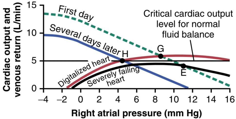
Figure 22-7 Treatment of decompensated heart disease showing the effect of digitalis in elevating the cardiac output curve, with this in turn causing increased urine output, progressive shift of the venous return curve to the left, and decreased right atrial pressure.
شکل 22-7 درمان بیماری قلبی جبران نشده که اثر دیژیتال را در بالا بردن منحنی برون ده قلبی نشان می دهد، که به نوبه خود باعث افزایش برون ده ادرار، تغییر پیشرونده منحنی بازگشت وریدی به چپ و کاهش فشار دهلیز راست می شود.
HEART FAILURE WITH DIASTOLIC DYSFUNCTION AND NORMAL EJECTION FRACTION
Our discussion thus far has focused mainly on cardiac failure due to decreased contractility of the myocardium following a myocardial infarction or impaired coronary blood flow. However, as discussed earlier in the chapter, heart failure can occur from any condition that decreases the heart’s ability to pump enough blood to meet the body’s needs. Heart failure associated with impaired cardiac contractility is often referred to as systolic heart failure or heart failure with reduced ejection fraction (HFrEF). As discussed in Chapter 9, the ejection fraction (EF), often assessed by echocardiography, is the fraction of the end-diastolic volume of the left ventricle that is ejected with each contraction. An EF of 0.6 means that 60% of the end-diastolic volume ventricle is pumped with each heartbeat. Normal values for EF are considered to be from 50% to 70%. When the heart muscle is weakened, as a result of a myocardial infarction or impaired coronary blood flow, the EF is usually reduced, with values below 40% considered to be indicative of HFrEF.
نارسایی قلبی همراه با اختلال عملکرد دیاستولیک و کسر جهشی طبیعی
بحث ما تا کنون عمدتاً بر نارسایی قلبی به دلیل کاهش انقباض میوکارد به دنبال انفارکتوس میوکارد یا اختلال در جریان خون کرونر متمرکز شده است. با این حال، همانطور که قبلا در این فصل مورد بحث قرار گرفت، نارسایی قلبی می تواند در هر شرایطی رخ دهد که توانایی قلب برای پمپاژ خون کافی برای رفع نیازهای بدن را کاهش دهد. نارسایی قلبی مرتبط با اختلال در انقباض قلب اغلب به عنوان نارسایی سیستولیک قلب یا نارسایی قلبی با کسر جهشی کاهش یافته (HFrEF) نامیده می شود. همانطور که در فصل 9 بحث شد، کسر جهشی (EF) که اغلب با اکوکاردیوگرافی ارزیابی می شود، کسری از حجم انتهای دیاستولیک بطن چپ است که با هر انقباض خارج می شود. EF 0.6 به این معنی است که 60٪ از بطن حجم انتهای دیاستولیک با هر ضربان قلب پمپ می شود. مقادیر طبیعی برای EF از 50٪ تا 70٪ در نظر گرفته می شود. هنگامی که عضله قلب در نتیجه انفارکتوس میوکارد یا اختلال در جریان خون کرونر ضعیف می شود، EF معمولاً کاهش می یابد و مقادیر کمتر از 40٪ نشان دهنده HFrEF در نظر گرفته می شود.
Heart failure can also be associated with normal EF if the heart muscle becomes thickened and stiff (concentric hypertrophy), so that filling of the ventricles is impaired, and the ventricles hold a smaller than usual volume of blood. Under these conditions, the total amount of blood pumped by the heart may not be enough to meet the body’s needs, even though it is pumping with a normal or even increased EF. This condition is often referred to as heart failure with preserved ejection fraction (HFPEF).
اگر عضله قلب ضخیم و سفت شود (هیپرتروفی متحدالمرکز)، نارسایی قلبی میتواند با EF طبیعی همراه باشد، به طوری که پر شدن بطنها مختل شود و بطنها حجم خونی کمتر از حد معمول را نگه دارند. در این شرایط، مقدار کل خون پمپ شده توسط قلب ممکن است برای رفع نیازهای بدن کافی نباشد، حتی اگر با یک EF طبیعی یا حتی افزایش یافته پمپاژ می شود. این وضعیت اغلب به عنوان نارسایی قلبی با کسر جهشی حفظ شده (HFPEF) شناخته می شود.
During the last 30 to 40 years, a growing proportion of heart failure patients have presented with HFPEF. Currently, over 50% of patients with heart failure patients have a normal EF. HFPEF occurs more commonly in women and older adults and especially in those who have obesity, diabetes mellitus, and hypertension, a constellation of disorders often called the cardiometabolic syndrome. In these individuals, diastolic dysfunction is characterized by impairment in the rate of ventricular filling, slowed relaxation of cardiomyocytes, increased thickness of the ventricular wall, proliferation of extracellular matrix, and fibrosis, which contribute to a stiffer left ventricle.
در طی 30 تا 40 سال گذشته، نسبت رو به رشدی از بیماران نارسایی قلبی با HFPEF مراجعه کرده اند. در حال حاضر، بیش از 50 درصد از بیماران مبتلا به نارسایی قلبی EF طبیعی دارند. HFPEF بیشتر در زنان و افراد مسن و به ویژه در افرادی که دارای چاقی، دیابت شیرین و فشار خون هستند، رخ می دهد، مجموعه ای از اختلالات که اغلب به آن سندرم متابولیک قلب می گویند. در این افراد، اختلال دیاستولیک با اختلال در سرعت پر شدن بطن، آرام شدن آرام قلب، افزایش ضخامت دیواره بطن، تکثیر ماتریکس خارج سلولی و فیبروز مشخص میشود که به سفتتر شدن بطن چپ کمک میکند.
Although clinicians often classify patients into the categories of HFPEF or HFrEF using the EF threshold of 50%, most patients with advanced heart failure exhibit abnormalities of ventricular filling as well as impaired contractility and impaired systolic function. Most of the neurohumoral changes in heart failure that have been dis- cussed, including activation of the sympathetic and renin- angiotensin system, as well as excessive fluid retention by the kidneys, occur regardless of whether there is normal or reduced EF. Heart failure is a heterogeneous syndrome, rather than a specific disease, and occurs whenever the heart is unable to pump enough blood to meet the needs of the body.
اگرچه پزشکان اغلب بیماران را با استفاده از آستانه EF 50% در دستههای HFPEF یا HFrEF طبقهبندی میکنند، اکثر بیماران مبتلا به نارسایی قلبی پیشرفته، ناهنجاریهای پر شدن بطن و همچنین اختلال در انقباض و اختلال در عملکرد سیستولیک را نشان میدهند. بسیاری از تغییرات عصبی-هومورال در نارسایی قلبی که مورد بحث قرار گرفته است، از جمله فعال شدن سیستم سمپاتیک و رنین آنژیوتانسین، و همچنین احتباس بیش از حد مایع توسط کلیه ها، صرف نظر از وجود EF طبیعی یا کاهش یافته رخ می دهد. نارسایی قلبی به جای یک بیماری خاص، یک سندرم ناهمگن است و زمانی رخ می دهد که قلب نتواند خون کافی برای رفع نیازهای بدن پمپاژ کند.
Thus, measurements of EF, although useful, do not always provide an accurate assessment of cardiac function. A small thick heart with impaired diastolic filling may be unable to pump the appropriate stroke volume and cardiac output to meet the body’s needs but could have a normal or elevated EF. This example illustrates the limitations of left ventricular EF as a marker of left ventricular function and as a means of categorizing patients with heart failure of different causes.
بنابراین، اندازهگیری EF، اگرچه مفید است، اما همیشه ارزیابی دقیقی از عملکرد قلب ارائه نمیکند. یک قلب کوچک ضخیم با پر شدن دیاستولیک مختل ممکن است نتواند حجم ضربه ای مناسب و برون ده قلبی را برای رفع نیازهای بدن پمپ کند، اما ممکن است EF طبیعی یا افزایش یافته داشته باشد. این مثال محدودیت های EF بطن چپ را به عنوان نشانگر عملکرد بطن چپ و به عنوان وسیله ای برای طبقه بندی بیماران مبتلا به نارسایی قلبی به دلایل مختلف نشان می دهد.
HIGH-OUTPUT CARDIAC FAILURE
Figure 22-8 provides an analysis of two types of highoutput cardiac failure. One is caused by an arteriovenous fistula that overloads the heart because of excessive venous return, even though the pumping capability of the heart is not depressed. The other type is caused by beriberi, in which the venous return is greatly increased because of diminished systemic vascular resistance but, at the same time, the pumping capability of the heart is depressed.
نارسایی قلبی با خروجی بالا
شکل 22-8 تجزیه و تحلیل دو نوع نارسایی قلبی با برون ده بالا را ارائه می دهد. یکی از آنها ناشی از فیستول شریانی وریدی است که قلب را به دلیل بازگشت بیش از حد وریدی بارگذاری می کند، حتی اگر توانایی پمپاژ قلب کاهش نیابد. نوع دیگر ناشی از بری بری است که در آن بازگشت وریدی به دلیل کاهش مقاومت عروقی سیستمیک بسیار افزایش مییابد، اما در عین حال، توانایی پمپاژ قلب کاهش مییابد.
Arteriovenous Fistula Increases Venous Return. The normal black curves of Figure 22-8 depict the nor- mal cardiac output and normal venous return curves. These curves equate with each other at point A, which depicts a normal cardiac output of 5 L/min and a normal right atrial pressure of 0 mm Hg.
فیستول شریانی وریدی بازگشت وریدی را افزایش می دهد. منحنی های سیاه نرمال شکل 22-8 منحنی های برون ده قلبی و بازگشت وریدی طبیعی را نشان می دهند. این منحنی ها در نقطه A با یکدیگر برابر می شوند که برون ده قلبی طبیعی 5 لیتر در دقیقه و فشار طبیعی دهلیز راست 0 میلی متر جیوه را نشان می دهد.
Now let us assume that the systemic vascular resistance (the total peripheral vascular resistance) becomes greatly decreased because of the opening of a large arteriovenous fistula (a direct opening between a large artery and large vein). The venous return curve rotates upward to produce the curve labeled “AV fistula” This venous return curve equates with the normal cardiac output curve at point B, with a cardiac output of 12.5 L/min and a right atrial pressure of 3 mm Hg. Thus, the cardiac output has become greatly elevated, the right atrial pressure is slightly elevated, and there are mild signs of peripheral congestion. If the person attempts to exercise, he or she will have little cardiac reserve because the heart is already at near- maximum capacity to pump the extra blood through the arteriovenous fistula. This condition resembles a failure condition and is called high-output failure, but in reality the heart is overloaded by excess venous return.
حال اجازه دهید فرض کنیم که مقاومت عروقی سیستمیک (مقاومت عروق محیطی کل) به دلیل باز شدن یک فیستول شریانی وریدی بزرگ (منفذ مستقیم بین یک شریان بزرگ و ورید بزرگ) بسیار کاهش می یابد. منحنی بازگشت وریدی به سمت بالا می چرخد تا منحنی با عنوان “فیستول AV” ایجاد شود. بنابراین، برون ده قلبی بسیار بالا رفته است، فشار دهلیز راست کمی بالا رفته و علائم خفیفی از احتقان محیطی وجود دارد. اگر فرد سعی کند ورزش کند، ذخیره قلبی کمی خواهد داشت، زیرا قلب در حال حاضر تقریباً حداکثر ظرفیت دارد تا خون اضافی را از طریق فیستول شریانی وریدی پمپ کند. این وضعیت شبیه یک وضعیت نارسایی است و نارسایی با خروجی بالا نامیده می شود، اما در واقع قلب با بازگشت وریدی بیش از حد بارگذاری می شود.
Beriberi Weakens the Heart, Causes Fluid Retention by the Kidneys, and Increases Venous Return. Figure 22-8 shows the approximate changes in the cardiac output and venous return curves caused by beriberi. The decreased level of the cardiac output curve is caused by weakening of the heart because of the avitaminosis (mainly lack of thiamine) that causes the beriberi syndrome. The weakening of the heart has decreased the blood flow to the kidneys. Therefore, the kidneys have retained a large amount of fluid, which in turn has increased the mean systemic filling pressure (represented by the point where the venous return curve now intersects the zero cardiac output level) from the normal value of 7 mm Hg up to 11 mm Hg. This has shifted the venous return curve to the right. Finally, the venous return curve has rotated upward from the normal curve because the avitaminosis has dilated the peripheral blood vessels, as explained in Chapter 17.
بری بری قلب را ضعیف می کند، باعث احتباس مایعات توسط کلیه ها می شود و بازگشت وریدی را افزایش می دهد. شکل 22-8 تغییرات تقریبی در برون ده قلبی و منحنی بازگشت وریدی ناشی از بری بری را نشان می دهد. کاهش سطح منحنی برون ده قلبی ناشی از تضعیف قلب به دلیل آویتامینوز (عمدتاً کمبود تیامین) است که باعث سندرم بری بری می شود. ضعیف شدن قلب باعث کاهش جریان خون به کلیه ها شده است. بنابراین، کلیهها مقدار زیادی مایع را حفظ کردهاند که به نوبه خود باعث افزایش میانگین فشار پرشدگی سیستمی (که نشاندهنده نقطهای است که منحنی بازگشت وریدی اکنون سطح برون ده قلبی صفر را قطع میکند) از مقدار طبیعی 7 میلیمتر جیوه تا 11 میلیمتر جیوه افزایش یافته است. این منحنی بازگشت وریدی را به سمت راست تغییر داده است. در نهایت، همانطور که در فصل 17 توضیح داده شد، منحنی بازگشت وریدی از منحنی طبیعی به سمت بالا چرخیده است، زیرا آویتامینوز رگهای خونی محیطی را گشاد کرده است.
The two blue curves (cardiac output curve and venous return curve) intersect with each other at point C, which describes the circulatory condition in beriberi, with a right atrial pressure in this case of 9 mm Hg and a cardiac output about 65% above normal. This high cardiac output occurs despite the weak heart, as demonstrated by the depressed plateau level of the cardiac output curve.
دو منحنی آبی (منحنی برون ده قلبی و منحنی بازگشت وریدی) در نقطه C که وضعیت گردش خون را در بری بری توصیف میکند، با فشار دهلیز راست در این مورد 9 میلیمتر جیوه و برون ده قلبی حدود 65 درصد بالاتر از حد طبیعی، با یکدیگر تلاقی میکنند. این برون ده قلبی بالا با وجود قلب ضعیف رخ می دهد، همانطور که با سطح فلات پایین منحنی برون ده قلبی نشان داده شده است.

Figure 22-8 Graphic analysis of two types of conditions that can cause high-output cardiac failure-arteriovenous (AV) fistula and beriberi heart disease.
شکل 22-8 تجزیه و تحلیل گرافیکی از دو نوع شرایطی که می توانند باعث نارسایی قلبی با برون ده بالا – فیستول شریانی وریدی (AV) و بیماری قلبی بری بری شوند.
کلیک کنید «Bibliography: فهرست کتب مربوطه»
Bahit MC, Kochar A, Granger CB: Post-myocardial infarction heart failure. JACC Heart Fail 6:179, 2018.
Braunwald E: Cardiomyopathies: an overview. Circ Res 121:711, 2017.
Burke MA, Cook SA, Seidman JG, Seidman CE: Clinical and mechanistic insights into the genetics of cardiomyopathy. J Am Coll Car- diol 68:2871, 2016.
Divakaran S, Loscalzo J: The role of nitroglycerin and other nitrogen oxides in cardiovascular therapeutics. J Am Coll Cardiol 70:2393, 2017.
Ellison DH, Felker GM: Diuretic treatment in heart failure. N Engl J Med 377:1964, 2017.
Guyton AC, Jones CE, Coleman TG: Circulatory Physiology: Cardiac Output and Its Regulation. Philadelphia: WB Saunders, 1973.
Kearns MJ, Walley KR: Tamponade: hemodynamic and echocardio- graphic diagnosis. Chest 153:1266, 2018.
Luo M, Anderson ME: Mechanisms of altered Ca2+ handling in heart failure. Circ Res 113:690, 2013.
Lymperopoulos A, Rengo G, Koch WJ: Adrenergic nervous system in heart failure: pathophysiology and therapy. Circ Res 113:739, 2013.
Marwick TH: Ejection fraction pros and cons. J Am Coll Cardiol 72:2360, 2018.
McHugh K, DeVore AD, Wu J, Matsouaka RA, et al: Heart failure with preserved ejection fraction and diabetes. J Am Coll Cardiol 73:602, 2019.
Mullens W, Verbrugge FH, Nijst P, Tang WHW: Renal sodium avidity in heart failure: from pathophysiology to treatment strategies. Eur Heart J 38:1872, 2017.
Normand C, Kaye DM, Povsic TJ, Dickstein K: Beyond pharmacological treatment: an insight into therapies that target specific aspects of heart failure pathophysiology. Lancet 393:1045, 2019.
Packer M: The conundrum of patients with obesity, exercise intolerance, elevated ventricular filling pressures and a measured ejection fraction in the normal range. Eur J Heart Fail 21:156, 2019.
Packer M: Why is the use of digitalis withering? Another reason that we need medical heart failure specialists. Eur J Heart Fail 20:851, 2018.
Pandey A, Patel KV, Vaduganathan M, Sarma S, et al: Physical activity, fitness, and obesity in heart failure with preserved ejection fraction. JACC Heart Fail 6:975, 2018.
Rossignol P, Hernandez AF, Solomon SD, Zannad F: Heart failure drug treatment. Lancet 393:1034, 2019.
Taqueti VR, Di Carli MF: Coronary microvascular disease pathogenic mechanisms and therapeutic Options. J Am Coll Cardiol 72:2625, 2018.
🚀 با ما همراه شوید!
تازهترین مطالب و آموزشهای مغز و اعصاب را از دست ندهید. با فالو کردن کانال تلگرام آیندهنگاران مغز، از ما حمایت کنید!



