
فیزیولوژی پزشکی گایتون و هال؛ جریان خون عضلانی و برون ده قلبی در طول ورزش. گردش خون کرونر و بیماری ایسکمیک قلب


دعای مطالعه [ نمایش ]
بِسْمِ الله الرَّحْمنِ الرَّحیمِ
اَللّهُمَّ اَخْرِجْنى مِنْ ظُلُماتِ الْوَهْمِ
خدايا مرا بيرون آور از تاريكىهاى وهم،
وَ اَكْرِمْنى بِنُورِ الْفَهْمِ
و به نور فهم گرامى ام بدار،
اَللّهُمَّ افْتَحْ عَلَيْنا اَبْوابَ رَحْمَتِكَ
خدايا درهاى رحمتت را به روى ما بگشا،
وَانْشُرْ عَلَيْنا خَزائِنَ عُلُومِكَ بِرَحْمَتِكَ يا اَرْحَمَ الرّاحِمينَ
و خزانههاى علومت را بر ما باز كن به امید رحمتت اى مهربانترين مهربانان.
کتاب «فیزیولوژی پزشکی گایتون و هال» بهعنوان یکی از جامعترین و معتبرترین منابع در حوزه علوم پزشکی، همچنان مرجع کلیدی برای درک عملکرد پیچیده بدن انسان است. این اثر با تکیه بر تازهترین پژوهشها و توضیحات دقیق از سازوکارهای فیزیولوژیک، پلی میان علوم پایه پزشکی و کاربردهای بالینی ایجاد میکند و نقشی بیبدیل در آموزش، پژوهش و ارتقای دانش سلامت ایفا مینماید.
ترجمه دقیق و علمی این شاهکار توسط برند علمی آیندهنگاران مغز به مدیریت داریوش طاهری، دسترسی فارسیزبانان به مرزهای نوین دانش فیزیولوژی را ممکن ساخته و رسالتی علمی برای ارتقای آموزش پزشکی، فهم عمیقتر سازوکارهای بدن و توسعه روشهای نوین در حوزه سلامت فراهم آورده است.
» کتاب فیزیولوژی پزشکی گایتون و هال
» » فصل ۲۱: جریان خون عضلانی و برون ده قلبی در طول ورزش. گردش خون کرونر و بیماری ایسکمیک قلب
در حال ویرایش
» Guyton and Hall Textbook of Medical Physiology
»» CHAPTER 21: Muscle Blood Flow and Cardiac Output During Exercise; the Coronary Circulation and Ischemic Heart Disease

In this chapter we consider the following: (1) blood flow to the skeletal muscles; and (2) coronary artery blood flow to the heart. Regulation of each of these types of blood flow is achieved mainly by local control of vascular resistance in response to muscle tissue metabolic needs.
در این فصل موارد زیر را در نظر می گیریم: (1) جریان خون به ماهیچه های اسکلتی. و (2) جریان خون شریان کرونر به قلب. تنظیم هر یک از این نوع جریان خون عمدتاً با کنترل موضعی مقاومت عروقی در پاسخ به نیازهای متابولیکی بافت عضلانی به دست می آید.
We also discuss the physiology of related subjects, including the following: (1) cardiac output control during exercise; (2) characteristics of heart attacks; and (3) the pain of angina pectoris.
ما همچنین فیزیولوژی موضوعات مرتبط، از جمله موارد زیر را مورد بحث قرار می دهیم: (1) کنترل برون ده قلبی در طول ورزش. (2) ویژگی های حملات قلبی. و (3) درد آنژین صدری.
BLOOD FLOW REGULATION IN SKELETAL MUSCLE AT REST AND DURING EXERCISE
Strenuous exercise is one of the most stressful conditions that the normal circulatory system faces because there is such a large mass of skeletal muscle in the body, all of it requiring large amounts of blood flow. Also, the cardiac output often must increase to four to five times normal in the nonathlete or to six to seven times normal in the well-trained athlete to satisfy the metabolic needs of the exercising muscles.
تنظیم جریان خون در ماهیچه های اسکلتی در هنگام استراحت و در حین ورزش
ورزش شدید یکی از استرس زاترین شرایطی است که سیستم گردش خون طبیعی با آن مواجه است، زیرا توده بزرگی از ماهیچه های اسکلتی در بدن وجود دارد که همه آنها به مقدار زیادی جریان خون نیاز دارند. همچنین، برون ده قلبی اغلب باید به چهار تا پنج برابر نرمال در غیرورزشکار یا به شش تا هفت برابر نرمال در ورزشکار خوب ورزیده افزایش یابد تا نیازهای متابولیکی عضلات ورزشکار برآورده شود.
SKELETAL MUSCLE BLOOD FLOW RATE
During rest, skeletal muscle blood flow averages 3 to 4 ml/ min/100 g of muscle. During extreme exercise in the wellconditioned athlete, this blood flow can increase 25- to 50-fold, rising to 100 to 200 ml/min/100 g of muscle. Peak blood flows as high as 400 ml/min/100 g of muscle have been reported for thigh muscles of endurance-trained athletes.
نرخ جریان خون ماهیچه های اسکلتی
در طول استراحت، جریان خون ماهیچه های اسکلتی به طور متوسط 3 تا 4 میلی لیتر در دقیقه در 100 گرم عضله است. در طول ورزش شدید در ورزشکاران با شرایط خوب، این جریان خون می تواند 25 تا 50 برابر افزایش یابد و به 100 تا 200 میلی لیتر در دقیقه در 100 گرم عضله برسد. اوج جریان خون به میزان 400 میلی لیتر در دقیقه/100 گرم عضله برای عضلات ران ورزشکاران تمرین شده استقامتی گزارش شده است.
Blood Flow During Muscle Contractions. Figure 21-1 shows a record of blood flow changes in a calf muscle of a leg during strong rhythmic muscular exercise. Note that the flow increases and decreases with each muscle contraction. At the end of the contractions, the blood flow remains high for a few seconds but then returns to normal during the next few minutes.
جریان خون در طول انقباضات عضلانی. شکل 21-1 رکوردی از تغییرات جریان خون در ماهیچه ساق پا در حین تمرین عضلانی ریتمیک قوی را نشان می دهد. توجه داشته باشید که جریان با هر انقباض عضلانی افزایش و کاهش می یابد. در پایان انقباضات، جریان خون برای چند ثانیه بالا باقی میماند اما در چند دقیقه بعد به حالت عادی باز میگردد.
The cause of the lower flow during the muscle contraction phase of exercise is compression of the blood vessels by the contracted muscle. During strong tetanic contraction, which causes sustained compression of the blood vessels, the blood flow can be almost stopped, but this also causes rapid weakening of the contraction.
علت کاهش جریان در مرحله انقباض عضلانی تمرین، فشرده شدن رگ های خونی توسط عضله منقبض شده است. در طول انقباض قوی کزاز، که باعث فشرده شدن مداوم رگهای خونی میشود، جریان خون تقریباً متوقف میشود، اما این امر همچنین باعث ضعیف شدن سریع انقباض میشود.
Increased Blood Flow in Muscle Capillaries During Exercise. During rest, some muscle capillaries have little or no flowing blood, but during strenuous exercise, all the capillaries open. This opening of dormant capillaries diminishes the distance that oxygen and other nutrients must diffuse from the capillaries to the contracting muscle fibers; it sometimes contributes a twofold to threefold increased capillary surface area through which oxygen and nutrients can diffuse from the blood to the tissues.
افزایش جریان خون در مویرگ های عضلانی در طول ورزش. در هنگام استراحت، برخی از مویرگ های عضلانی خون کمی دارند یا اصلاً جریان ندارند، اما در حین ورزش شدید، همه مویرگ ها باز می شوند. این باز شدن مویرگ های خفته فاصله ای را که اکسیژن و سایر مواد مغذی باید از مویرگ ها به فیبرهای عضلانی منقبض پخش شود را کاهش می دهد. گاهی اوقات باعث افزایش دو تا سه برابری سطح مویرگی می شود که از طریق آن اکسیژن و مواد مغذی می توانند از خون به بافت ها پخش شوند.

Figure 21-1. Effects of muscle exercise on blood flow in the calf of a leg during strong rhythmic contraction. The blood flow was much less during contractions than between contractions. (Modified from Barcroft H, Dornhorst AC: The blood flow through the human calf during rhythmic exercise. J Physiol 109:402, 1949.)
شکل 21-1. تأثیر ورزش عضلانی بر جریان خون در ساق پا در حین انقباضات ریتمیک قوی. جریان خون در طول انقباضات بسیار کمتر از بین انقباضات بود. (اصلاح شده از Barcroft H، Dornhorst AC: جریان خون در گوساله انسان در طول تمرینات ریتمیک. J Physiol 109:402، 1949.)
CONTROL OF SKELETAL MUSCLE BLOOD FLOW
کنترل جریان خون ماهیچه های اسکلتی
Decreased Oxygen in Muscle Greatly Enhances Flow. The large increase in muscle blood flow that occurs during skeletal muscle activity is caused mainly by chemicals released locally that act directly on the muscle arterioles to cause dilation. One of the most important chemical effects is reduction of the oxygen level in the muscle tissues. When muscles are active, they use oxygen rapidly, thereby decreasing the oxygen concentration in the tissue fluids. This in turn causes local arteriolar vasodilation because low oxygen levels cause the blood vessels to relax and because oxygen deficiency causes release of vasodilator substances. Adenosine may be an important vasodilator substance, but experiments have shown that even large amounts of adenosine infused directly into a muscle artery cannot increase blood flow to the same extent as during intense exercise, and it cannot sustain vasodilation in skeletal muscle for more than about 2 hours.
کاهش اکسیژن در عضله به شدت جریان را افزایش می دهد. افزایش زیادی در جریان خون عضلانی که در طول فعالیت ماهیچههای اسکلتی اتفاق میافتد عمدتاً ناشی از مواد شیمیایی آزاد شده به صورت موضعی است که مستقیماً بر روی شریانهای عضلانی تأثیر میگذارد و باعث اتساع میشود. یکی از مهمترین اثرات شیمیایی کاهش سطح اکسیژن در بافت های عضلانی است. هنگامی که ماهیچه ها فعال هستند، به سرعت از اکسیژن استفاده می کنند و در نتیجه غلظت اکسیژن در مایعات بافتی کاهش می یابد. این به نوبه خود باعث اتساع موضعی عروق شریانی می شود زیرا سطوح پایین اکسیژن باعث شل شدن رگ های خونی می شود و به دلیل کمبود اکسیژن باعث آزاد شدن مواد گشادکننده عروق می شود. آدنوزین ممکن است یک ماده گشادکننده عروق مهم باشد، اما آزمایشها نشان دادهاند که حتی مقادیر زیادی آدنوزین که مستقیماً به شریان عضلانی تزریق میشود، نمیتواند جریان خون را به همان میزانی که در طول ورزش شدید افزایش میدهد، افزایش دهد و نمیتواند اتساع عروق در ماهیچههای اسکلتی را برای بیش از ۲ ساعت حفظ کند.
Fortunately, even after the muscle blood vessels have become insensitive to the vasodilator effects of adenosine, other vasodilator factors continue to maintain increased capillary blood flow as long as the exercise continues. These factors include the following: (1) potassium ions; (2) adenosine triphosphate (ATP); (3) lactic acid; and (4) carbon dioxide. We still do not know quantitatively how much of a role each of these factors plays in increasing muscle blood flow during muscle activity; this subject was discussed in additional detail in Chapter 17.
خوشبختانه، حتی پس از اینکه رگهای خونی ماهیچهای نسبت به اثرات گشادکننده عروق آدنوزین حساس نشدند، سایر عوامل گشادکننده عروق تا زمانی که تمرین ادامه دارد، افزایش جریان خون مویرگی را حفظ میکنند. این عوامل شامل موارد زیر است: (1) یون های پتاسیم. (2) آدنوزین تری فسفات (ATP). (3) اسید لاکتیک؛ و (4) دی اکسید کربن. ما هنوز از نظر کمی نمی دانیم که هر یک از این عوامل چقدر در افزایش جریان خون عضلانی در طول فعالیت عضلانی نقش دارند. این موضوع با جزئیات بیشتر در فصل 17 مورد بحث قرار گرفت.
Nervous Control of Muscle Blood Flow. In addition to local tissue vasodilator mechanisms, skeletal muscles are provided with sympathetic vasoconstrictor nerves and, in some species of animals, sympathetic vasodilator nerves as well.
کنترل عصبی جریان خون عضلانی. علاوه بر مکانیسمهای گشادکننده عروق بافتی، ماهیچههای اسکلتی دارای اعصاب منقبض کننده عروق سمپاتیک و در برخی از گونههای حیوانات، اعصاب گشادکننده عروق سمپاتیک نیز هستند.
The sympathetic vasoconstrictor nerve fibers secrete norepinephrine at their nerve endings. When maximally activated, this mechanism can decrease blood flow through resting muscles to as little as one-half to one-third normal. This vasoconstriction is of physiologic importance in attenuating decreases of arterial pressure in circulatory shock and during other periods of stress, when it may even be necessary to increase blood pressure.
رشته های عصبی منقبض کننده عروق سمپاتیک در انتهای عصبی خود نوراپی نفرین ترشح می کنند. هنگامی که این مکانیسم به حداکثر فعال میشود، میتواند جریان خون را از طریق عضلات در حال استراحت به نصف تا یک سوم کاهش دهد. این انقباض عروق اهمیت فیزیولوژیکی در کاهش کاهش فشار شریانی در شوک گردش خون و در سایر دورههای استرس دارد، زمانی که حتی ممکن است نیاز به افزایش فشار خون باشد.
In addition to the norepinephrine secreted at the sympathetic vasoconstrictor nerve endings, the medullae of the two adrenal glands also secrete increased amounts of norepinephrine plus even more epinephrine into the circulating blood during strenuous exercise. The circulating norepinephrine acts on the muscle vessels to cause a vasoconstrictor effect similar to that caused by direct sympathetic nerve stimulation. The epinephrine, how- ever, often has a slight vasodilator effect because epinephrine excites more of the beta-adrenergic receptors of the vessels, which are vasodilator receptors, in contrast to the alpha vasoconstrictor receptors excited especially by norepinephrine. These receptors are discussed in Chapter 61.
علاوه بر نوراپی نفرین ترشح شده در انتهای اعصاب منقبض کننده عروق سمپاتیک، بصل النخاع دو غده آدرنال نیز مقادیر بیشتری نوراپی نفرین به علاوه حتی اپی نفرین بیشتری را در جریان ورزش شدید در خون در گردش ترشح می کند. نوراپی نفرین در گردش بر روی رگ های عضلانی اثر می گذارد و باعث ایجاد یک اثر منقبض کننده عروق می شود که شبیه به تحریک مستقیم عصب سمپاتیک است. با این حال، اپی نفرین اغلب یک اثر گشادکننده عروقی خفیف دارد، زیرا اپی نفرین گیرنده های بتا آدرنرژیک عروق را که گیرنده های گشادکننده عروق هستند، بیشتر برانگیخته می کند، برخلاف گیرنده های منقبض کننده عروق آلفا که به ویژه توسط نوراپی نفرین تحریک می شوند. این گیرنده ها در فصل 61 مورد بحث قرار می گیرند.
CIRCULATORY READJUSTMENTS DURING EXERCISE
Three major effects occur during exercise that are essential for the circulatory system to supply the tremendous blood flow required by the muscles: (1) sympathetic nervous system activation in many tissues with consequent stimulatory effects on the circulation; (2) increase in arterial pressure; and (3) increase in cardiac output.
تنظیم مجدد گردش خون در حین ورزش
سه اثر عمده در طول ورزش رخ می دهد که برای سیستم گردش خون ضروری است تا جریان خون فوق العاده مورد نیاز عضلات را تامین کند: (1) فعال شدن سیستم عصبی سمپاتیک در بسیاری از بافت ها با اثرات تحریکی متعاقب آن بر گردش خون. (2) افزایش فشار شریانی؛ و (3) افزایش برون ده قلبی.
Effects of Sympathetic Activation
At the onset of exercise, signals are transmitted not only from the brain to the muscles to cause muscle contraction but also into the vasomotor center to initiate sympathetic discharge in many other tissues. Simultaneously, the parasympathetic signals to the heart are attenuated. Therefore, three major circulatory effects result:
اثرات فعال سازی سمپاتیک
در شروع تمرین، سیگنالها نه تنها از مغز به عضلات برای ایجاد انقباض عضلانی، بلکه به مرکز وازوموتور نیز برای شروع ترشح سمپاتیک در بسیاری از بافتهای دیگر منتقل میشوند. به طور همزمان، سیگنال های پاراسمپاتیک به قلب ضعیف می شوند. بنابراین، سه اثر عمده در گردش خون ایجاد می شود:
1.The heart is stimulated to a greatly increased heart rate and increased pumping strength as a result of the sympathetic drive to the heart plus release of the heart from normal parasympathetic inhibition.
1.قلب به افزایش شدید ضربان قلب و افزایش قدرت پمپاژ در نتیجه حرکت سمپاتیک به قلب به علاوه آزاد شدن قلب از مهار طبیعی پاراسمپاتیک تحریک می شود.
2. Many of the arterioles of the peripheral circulation are strongly contracted, except for the arterioles in the active muscles, which are strongly vasodilated by the local vasodilator effects in the muscles, as noted earlier. Thus, the heart is stimulated to supply the increased blood flow required by the muscles, while at the same time blood flow through most nonmuscular areas of the body is temporarily reduced, thereby “lending” blood supply to the muscles. This process accounts for as much as 2 L/min of extra blood flow to the muscles, which is exceedingly important when considering a person running for his or her life, when even a fractional increase in running speed may make the difference between life and death. Two of the peripheral circulatory systems, the coronary and cerebral systems, are spared this vasoconstrictor effect because both these circulatory areas have poor vasoconstrictor innervation-fortunately so, because both the heart and brain are as essential to exercise as the skeletal muscles.
2. بسیاری از شریان های گردش خون محیطی به شدت منقبض می شوند، به جز شریان های ماهیچه های فعال، که همانطور که قبلاً ذکر شد، توسط اثرات گشادکننده عروق موضعی در عضلات به شدت گشاد می شوند. بنابراین، قلب تحریک می شود تا جریان خون افزایش یافته مورد نیاز عضلات را تامین کند، در حالی که در همان زمان جریان خون در بیشتر نواحی غیر عضلانی بدن به طور موقت کاهش می یابد، در نتیجه خون رسانی به ماهیچه ها “قرض” می شود. این فرآیند به اندازه 2 لیتر در دقیقه جریان خون اضافی به ماهیچه ها را شامل می شود، که زمانی که یک فرد برای زندگی خود می دود بسیار مهم است، زمانی که حتی افزایش جزئی در سرعت دویدن ممکن است بین مرگ و زندگی تفاوت ایجاد کند. دو تا از سیستمهای گردش خون محیطی، سیستمهای کرونری و مغزی، از این اثر منقبض کننده عروق در امان هستند، زیرا هر دو ناحیه گردش خون عصب تنگ کننده عروقی ضعیفی دارند – خوشبختانه، زیرا قلب و مغز هر دو به اندازه عضلات اسکلتی برای ورزش ضروری هستند.
3.The muscle walls of the veins and other capacitative areas of the circulation are contracted powerfully, which greatly increases the mean systemic filling pressure. As we learned in Chapter 20, this effect is one of the most important factors in promoting the increase in venous return of blood to the heart and, therefore, in increasing the cardiac output.
3. دیوارههای ماهیچهای وریدها و سایر نواحی خازنی گردش خون به شدت منقبض میشوند که فشار پرشدگی سیستمیک متوسط را به شدت افزایش میدهد. همانطور که در فصل 20 آموختیم، این اثر یکی از مهمترین عوامل در افزایش بازگشت وریدی خون به قلب و در نتیجه افزایش برون ده قلبی است.
Sympathetic Stimulation May Increase Arterial Pressure During Exercise
An important effect of increased sympathetic stimulation in exercise is to increase the arterial pressure. This increased arterial pressure results from multiple stimulatory effects, including the following: (1) vasoconstriction of the arterioles and small arteries in most tissues of the body except the brain and active muscles, including the heart; (2) increased pumping activity by the heart; and (3) a great increase in mean systemic filling pressure caused mainly by venous contraction. These effects, working together, almost always increase the arterial pressure during exercise. This increase can be as little as 20 mm Hg or as much as 80 mm Hg, depending on the conditions under which the exercise is performed. When a person performs exercise under tense conditions but uses only a few muscles, the sympathetic nervous response still occurs. In the few active muscles, vasodilation occurs, but elsewhere in the body the effect is mainly vasoconstriction, often increasing the mean arterial pressure to as high as 170 mm Hg. Such a condition might occur in a person standing on a ladder and nailing with a hammer on the ceiling above. The tenseness of the situation is obvious.
تحریک سمپاتیک ممکن است فشار شریانی را در حین ورزش افزایش دهد
یک اثر مهم افزایش تحریک سمپاتیک در ورزش، افزایش فشار شریانی است. این افزایش فشار شریانی ناشی از اثرات تحریکی متعدد، از جمله موارد زیر است: (1) انقباض عروق شریانها و شریانهای کوچک در اکثر بافتهای بدن به جز مغز و ماهیچههای فعال، از جمله قلب. (2) افزایش فعالیت پمپاژ توسط قلب. و (3) افزایش زیادی در فشار پرشدگی سیستمیک عمدتاً ناشی از انقباض وریدی است. این اثرات با هم کار می کنند، تقریبا همیشه فشار شریانی را در حین ورزش افزایش می دهند. این افزایش بسته به شرایطی که تمرین در آن انجام می شود می تواند به 20 میلی متر جیوه یا 80 میلی متر جیوه برسد. هنگامی که یک فرد در شرایط تنش، ورزش را انجام می دهد اما فقط از چند ماهیچه استفاده می کند، پاسخ عصبی سمپاتیک همچنان رخ می دهد. در تعداد معدودی از ماهیچههای فعال، اتساع عروق رخ میدهد، اما در جاهای دیگر بدن این اثر عمدتاً انقباض عروق است، که اغلب فشار متوسط شریانی را تا 170 میلیمتر جیوه افزایش میدهد. چنین حالتی ممکن است در فردی رخ دهد که روی نردبان ایستاده و با چکش سقف بالا را میخکوب می کند. تنش شرایط آشکار است.
Conversely, when a person performs massive whole-body exercise, such as running or swimming, the increase in arterial pressure is often only 20 to 40 mm Hg. This lack of a large increase in pressure results from the extreme vasodilation that occurs simultaneously in large masses of active muscle.
برعکس، زمانی که یک فرد ورزش های سنگین کل بدن مانند دویدن یا شنا را انجام می دهد، افزایش فشار شریانی اغلب تنها 20 تا 40 میلی متر جیوه است. این عدم افزایش زیاد فشار ناشی از اتساع شدید عروق است که به طور همزمان در توده های بزرگ عضله فعال رخ می دهد.
Why Is Increased Arterial Pressure During Exercise Important? When muscles are stimulated maximally in a laboratory experiment, but without allowing the arterial pressure to rise, muscle blood flow seldom rises more than about eightfold. Yet, we know from studies of marathon runners that muscle blood flow can increase from as little as 1 L/min for the whole body during rest to more than 20 L/min during maximal activity. Therefore, it is clear that muscle blood flow can increase much more than that which occurs in this simple laboratory experiment. What is the difference? Mainly, the arterial pressure rises during normal exercise. Let us assume, for example, that the arterial pressure rises by 30% during heavy exercise. This 30% increase causes 30% more force to push blood through the muscle tissue vessels. However, this is not the only important effect-the extra pressure also stretches the walls of the vessels, and this effect, along with the locally released vasodilators and higher blood pressure, may increase muscle total flow to more than 20 times normal.
چرا افزایش فشار شریانی در حین ورزش مهم است؟ هنگامی که ماهیچه ها در یک آزمایش آزمایشگاهی حداکثر تحریک می شوند، اما بدون اجازه دادن به فشار شریانی، جریان خون ماهیچه ها به ندرت بیش از حدود هشت برابر افزایش می یابد. با این حال، از مطالعات روی دوندگان ماراتن می دانیم که جریان خون عضلانی می تواند از 1 لیتر در دقیقه برای کل بدن در هنگام استراحت به بیش از 20 لیتر در دقیقه در طول حداکثر فعالیت افزایش یابد. بنابراین، واضح است که جریان خون عضلانی می تواند بسیار بیشتر از آنچه در این آزمایش ساده آزمایشگاهی رخ می دهد افزایش یابد. تفاوت در چیست؟ به طور عمده، فشار شریانی در طول ورزش طبیعی افزایش می یابد. برای مثال فرض کنید فشار شریانی در حین ورزش سنگین 30 درصد افزایش می یابد. این افزایش 30 درصدی باعث می شود 30 درصد نیروی بیشتری برای فشار دادن خون از طریق عروق بافت عضلانی ایجاد کند. با این حال، این تنها اثر مهم نیست – فشار اضافی دیواره عروق را نیز کشیده میشود و این اثر همراه با گشادکنندههای عروقی آزاد شده و فشار خون بالاتر، ممکن است جریان کلی عضله را تا بیش از 20 برابر طبیعی افزایش دهد.
Importance of Increased Cardiac Output During Exercise
Many different physiologic effects occur at the same time during exercise to increase cardiac output approximately in proportion to the degree of exercise. In fact, the ability of the circulatory system to provide increased cardiac output for delivery of oxygen and other nutrients to the muscles during exercise is equally as important as the strength of the muscles themselves in setting the limit for continued muscle work. For example, marathon runners who can increase their cardiac outputs the most are generally the ones who have record-breaking running times.
اهمیت افزایش برون ده قلبی در طول ورزش
بسیاری از اثرات فیزیولوژیکی مختلف به طور همزمان در طول ورزش رخ می دهد تا برون ده قلبی را تقریباً متناسب با میزان ورزش افزایش دهد. در واقع، توانایی سیستم گردش خون برای افزایش برون ده قلبی برای رساندن اکسیژن و سایر مواد مغذی به ماهیچه ها در طول تمرین، به همان اندازه مهم است که قدرت خود عضلات در تعیین محدودیت برای ادامه کار عضلانی. به عنوان مثال، دوندگان ماراتن که می توانند برون ده قلبی خود را بیش از همه افزایش دهند، عموماً کسانی هستند که زمان های دویدن رکوردشکنی دارند.
Graphic Analysis of Changes in Cardiac Output During Heavy Exercise. Figure 21-2 shows a graphic analysis of the large increase in cardiac output that occurs during heavy exercise. The cardiac output and venous return curves crossing at point A represent the normal circulation, and the curves crossing at point B represent heavy exercise. Note that the great increase in cardiac output requires significant changes in both the cardiac output curve and the venous return curve, as follows.
تجزیه و تحلیل گرافیکی تغییرات در برون ده قلبی در طول تمرینات سنگین. شکل 21-2 یک تجزیه و تحلیل گرافیکی از افزایش زیاد برون ده قلبی را نشان می دهد که در طول ورزش سنگین رخ می دهد. منحنی های برون ده قلبی و بازگشت وریدی که در نقطه A تلاقی می کنند، گردش خون طبیعی را نشان می دهند و منحنی هایی که در نقطه B متقاطع می شوند، نشان دهنده ورزش سنگین هستند. توجه داشته باشید که افزایش زیاد برون ده قلبی مستلزم تغییرات قابل توجهی هم در منحنی برون ده قلبی و هم در منحنی بازگشت وریدی است که به شرح زیر است.
The increased level of the cardiac output curve is easy to understand. It results almost entirely from sympathetic stimulation of the heart, which causes the following: (1) increased heart rate, up to as high as 170 to 190 beats/ min; and (2) increased strength of contraction of the heart to as much as twice normal. Without this increased level of cardiac function, the increase in cardiac output would be limited to the plateau level of the normal heart, which would be a maximum increase of cardiac output of only about 2.5-fold rather than the 4-fold increase that can commonly be achieved by the untrained runner and the 7-fold increase that can be achieved in some marathon runners.
سطح افزایش یافته منحنی برون ده قلبی به راحتی قابل درک است. تقریباً به طور کامل ناشی از تحریک سمپاتیک قلب است که باعث موارد زیر می شود: (1) افزایش ضربان قلب تا 170 تا 190 ضربه در دقیقه. و (2) افزایش قدرت انقباض قلب تا دو برابر نرمال. بدون این افزایش سطح عملکرد قلب، افزایش برون ده قلبی به سطح فلات قلب طبیعی محدود می شود، که حداکثر افزایش برون ده قلبی تنها در حدود 2.5 برابر به جای افزایش 4 برابری است که معمولاً توسط دونده تمرین نکرده و افزایش 7 برابری که می تواند در برخی از دوندگان ماراتن به دست آورد.
Now study the venous return curves. If no change occurred from the normal venous return curve, the cardiac output could hardly rise at all in exercise because the upper plateau level of the normal venous return curve is only 6 L/min. Yet, two important changes do occur:
اکنون منحنی های بازگشت وریدی را مطالعه کنید. اگر هیچ تغییری نسبت به منحنی بازگشت وریدی نرمال رخ ندهد، برون ده قلبی به سختی می تواند در ورزش افزایش یابد زیرا سطح فلات بالایی منحنی بازگشت وریدی طبیعی تنها 6 لیتر در دقیقه است. با این حال، دو تغییر مهم رخ می دهد:
1.The mean systemic filling pressure rises at the onset of heavy exercise. This effect results partly from the sympathetic stimulation that contracts the veins and other capacitative parts of the circulation. In addition, tensing of the abdominal and other skeletal muscles of the body compresses many of the internal vessels, thus providing more compression of the entire capacitative vascular system and causing a still greater increase in the mean systemic filling pressure. During maximal exercise, these two effects together can increase the mean systemic filling pressure from a normal level of 7 mm Hg to as high as 30 mm Hg.
1. فشار پرشدگی سیستمیک متوسط در شروع ورزش سنگین افزایش می یابد. این اثر تا حدی ناشی از تحریک سمپاتیک است که وریدها و سایر بخش های خازنی گردش خون را منقبض می کند. علاوه بر این، کشش شکم و سایر ماهیچههای اسکلتی بدن، بسیاری از عروق داخلی را فشرده میکند، بنابراین فشردهسازی بیشتری بر کل سیستم عروقی خازنی ایجاد میکند و باعث افزایش باز هم بیشتر در فشار پرشدگی سیستمیک میشود. در طول تمرین حداکثر، این دو اثر با هم میتوانند میانگین فشار پر شدن سیستمیک را از سطح نرمال 7 میلیمتر جیوه به 30 میلیمتر جیوه افزایش دهند.
2.The slope of the venous return curve rotates upward. This upward rotation is caused by decreased resistance in virtually all the blood vessels in active muscle tissue, which also causes resistance to venous return to decrease, thus increasing the upward slope of the venous return curve.
2. شیب منحنی بازگشت وریدی به سمت بالا می چرخد. این چرخش به سمت بالا ناشی از کاهش مقاومت تقریباً در تمام رگهای خونی در بافت عضلانی فعال است که باعث کاهش مقاومت در برابر بازگشت وریدی میشود و در نتیجه شیب منحنی بازگشت وریدی به سمت بالا افزایش مییابد.
Therefore, the combination of increased mean systemic filling pressure and decreased resistance to venous return raises the entire level of the venous return curve.
بنابراین، ترکیب افزایش میانگین فشار پر شدن سیستمی و کاهش مقاومت در برابر بازگشت وریدی، کل سطح منحنی بازگشت وریدی را افزایش میدهد.
In response to the changes in both the venous return curve and cardiac output curve, the new equilibrium point in Figure 21-2 for cardiac output and right atrial pressure is now point B, in contrast to the normal level at point A. Note especially that the right atrial pressure has hardly changed, having risen only 1.5 mm Hg. In fact, in a person with a strong heart, the right atrial pressure often falls below normal during very heavy exercise because of the greatly increased sympathetic stimulation of the heart. In contrast, even a moderate level of exercise may cause marked increases in right atrial pressure in patients with weakened hearts.
در پاسخ به تغییرات در منحنی بازگشت وریدی و منحنی برون ده قلبی، نقطه تعادل جدید در شکل 21-2 برای برون ده قلبی و فشار دهلیز راست اکنون نقطه B است، برخلاف سطح نرمال در نقطه A. به خصوص توجه داشته باشید که فشار دهلیز راست به سختی تغییر کرده است و تنها 1.5 میلی متر جیوه افزایش یافته است. در واقع، در فردی که قلب قوی دارد، فشار دهلیز راست اغلب در حین ورزش بسیار سنگین به دلیل افزایش شدید تحریک سمپاتیک قلب، کمتر از حد طبیعی می شود. در مقابل، حتی یک سطح متوسط ورزش ممکن است باعث افزایش قابل توجه فشار دهلیز راست در بیماران با قلب ضعیف شود.
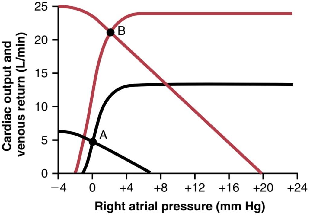
Figure 21-2. Graphic analysis of change in cardiac output, venous return, and right atrial pressure with the onset of strenuous exercise. Black curves, Normal circulation. Red curves, heavy exercise.
شکل 21-2. تجزیه و تحلیل گرافیکی تغییر در برون ده قلبی، بازگشت وریدی و فشار دهلیز راست با شروع ورزش شدید. منحنی های سیاه، گردش طبیعی. منحنی های قرمز، ورزش سنگین.
CORONARY CIRCULATION
About one-third of all deaths in industrialized countries of the Western world result from coronary artery disease, and most older adults have at least some impairment of the coronary artery circulation. For this reason, under- standing normal and pathological physiology of the coronary circulation is one of the most important subjects in medicine.
گردش خون کرونر
حدود یک سوم از کل مرگ و میرها در کشورهای صنعتی جهان غرب ناشی از بیماری عروق کرونر است و اکثر افراد مسن حداقل دارای اختلالاتی در گردش خون عروق کرونر هستند. به همین دلیل، درک فیزیولوژی طبیعی و پاتولوژیک گردش خون کرونر یکی از موضوعات مهم در پزشکی است.
PHYSIOLOGIC ANATOMY OF THE CORONARY BLOOD SUPPLY
Figure 21-3 shows the heart and its coronary blood sup- ply. Note that the main coronary arteries lie on the surface of the heart, and smaller arteries then penetrate from the surface into the cardiac muscle mass. It is almost entirely through these arteries that the heart receives its nutritive blood supply. Only the inner one-tenth millimeter of the endocardial surface can obtain significant nutrition directly from the blood inside the cardiac chambers, so this source of muscle nutrition is minuscule.
آناتومی فیزیولوژیکی تامین خون کرونر
شکل 21-3 قلب و منبع خون کرونری آن را نشان می دهد. توجه داشته باشید که شریان های کرونر اصلی روی سطح قلب قرار دارند و شریان های کوچکتر سپس از سطح به داخل توده عضلانی قلب نفوذ می کنند. تقریباً به طور کامل از طریق این عروق است که قلب منبع خونی مغذی خود را دریافت می کند. فقط یک دهم میلیمتر داخلی سطح اندوکارد میتواند تغذیه قابل توجهی را مستقیماً از خون داخل اتاقکهای قلب دریافت کند، بنابراین این منبع تغذیه ماهیچهها بسیار ناچیز است.
The left coronary artery supplies mainly the anterior and left lateral portions of the left ventricle, whereas the right coronary artery supplies most of the right ventricle, as well as the posterior part of the left ventricle in 80% to 90% of people.
شریان کرونری چپ عمدتاً بخشهای جانبی قدامی و چپ بطن چپ را تامین میکند، در حالی که شریان کرونر راست قسمتهای زیادی از بطن راست و همچنین قسمت خلفی بطن چپ را در 80 تا 90 درصد افراد تامین میکند.
Most of the coronary venous blood flow from the left ventricular muscle returns to the right atrium of the heart by way of the coronary sinus, which is about 75% of the total coronary blood flow. On the other hand,most of the coronary venous blood from the right ventricular muscle returns through small anterior cardiac veins that flow directly into the right atrium, not by way of the coronary sinus. A very small amount of coronary venous blood also flows back into the heart through very minute thebesian veins, which empty directly into all chambers of the heart.
بیشتر جریان خون وریدی کرونری از عضله بطن چپ از طریق سینوس کرونری به دهلیز راست قلب باز می گردد که حدود 75 درصد از کل جریان خون کرونری است. از سوی دیگر، بیشتر خون وریدی کرونری از عضله بطن راست از طریق وریدهای قلبی قدامی کوچک که مستقیماً به دهلیز راست جریان مییابند، نه از طریق سینوس کرونری، باز میگردد. مقدار بسیار کمی از خون وریدی کرونری نیز از طریق سیاهرگهای بسیار ریز تبسی که مستقیماً به تمام حفرههای قلب تخلیه میشوند، به قلب جریان مییابد.
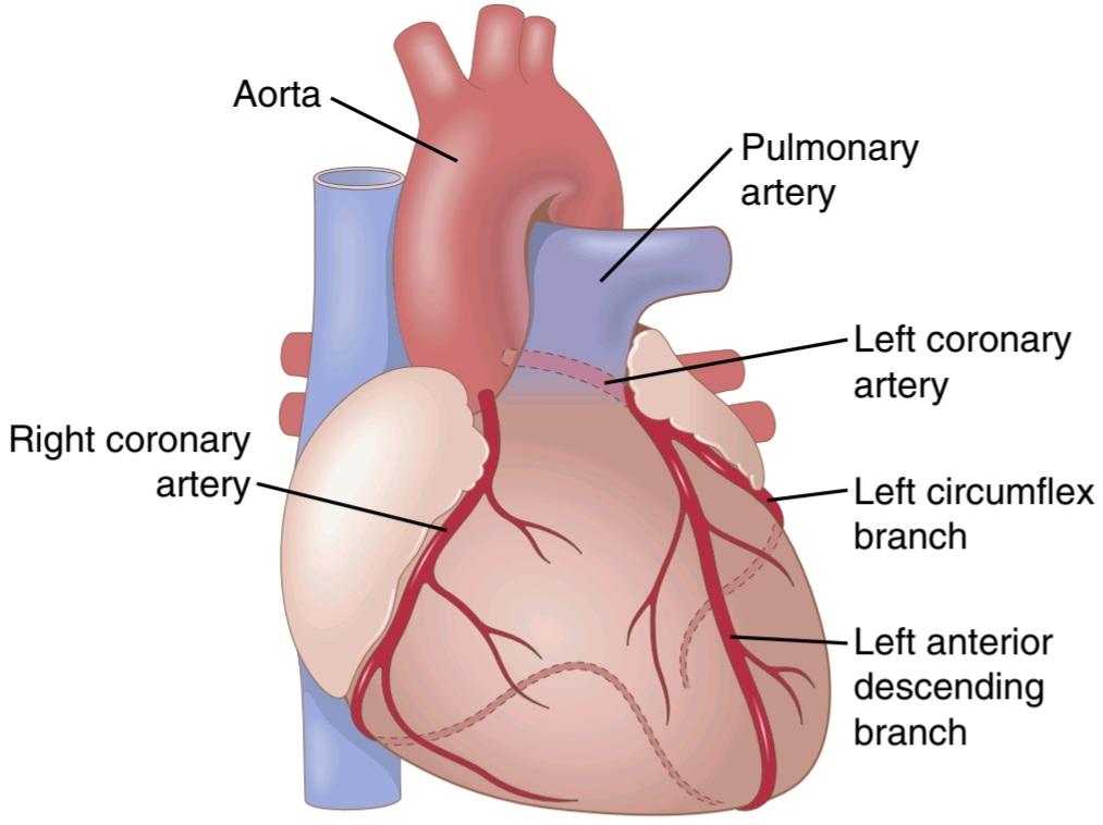
Figure 21-3. Coronary arteries.
شکل 21-3. عروق کرونر.
NORMAL CORONARY BLOOD FLOW AVERAGES 5% OF CARDIAC OUTPUT
The normal coronary blood flow in the resting person averages 70 ml/min/100 g of heart weight, or about 225 ml/min, which is about 4% to 5% of the total cardiac output.
جریان خون کرونر طبیعی به طور متوسط 5% از برون ده قلبی است.
جریان طبیعی خون کرونری در فرد در حال استراحت به طور متوسط 70 میلی لیتر در دقیقه / 100 گرم وزن قلب یا حدود 225 میلی لیتر در دقیقه است که حدود 4 تا 5 درصد کل برون ده قلبی است.
During strenuous exercise, the heart in the young adult increases its cardiac output fourfold to sevenfold, and it pumps this blood against a higher than normal arterial pressure. Consequently, the work output of the heart under severe conditions may increase 6-fold to 9-fold. At the same time, the coronary blood flow increases 3-fold to 4-fold to supply the extra nutrients needed by the heart. This increase is not as much as the increase in workload, which means that the ratio of energy expenditure by the heart to coronary blood flow increases. Thus, the efficiency of cardiac utilization of energy increases to make up for the relative deficiency of coronary blood supply.
در طول ورزش شدید، قلب در بزرگسالان جوان برون ده قلبی خود را چهار برابر تا هفت برابر افزایش می دهد و این خون را در برابر فشار شریانی بالاتر از حد طبیعی پمپ می کند. در نتیجه، برون ده کار قلب در شرایط سخت ممکن است 6 برابر تا 9 برابر افزایش یابد. در همان زمان، جریان خون کرونری 3 تا 4 برابر افزایش می یابد تا مواد مغذی اضافی مورد نیاز قلب تامین شود. این افزایش به اندازه افزایش حجم کار نیست، به این معنی که نسبت انرژی مصرف شده توسط قلب به جریان خون کرونر افزایش می یابد. بنابراین، کارایی استفاده قلبی از انرژی برای جبران کمبود نسبی تامین خون کرونر افزایش مییابد.
Cardiac Muscle Compression Causes Phasic Changes in Coronary Blood Flow During Systole and Diastole. Figure 21-4 shows the changes in blood flow through the nutrient capillaries of the left ventricular coronary system in ml/min in the heart during systole and diastole, as extrapolated from studies in experimental animals. Note from this diagram that the coronary capillary blood flow in the left ventricle muscle falls to a low value during systole, which is opposite to flow in vascular beds elsewhere in the body. The reason for this phenomenon is strong compression of the intramuscular blood vessels by the left ventricular muscle during systolic contraction.
فشرده سازی عضلات قلب باعث تغییرات فازی در جریان خون کرونر در طول سیستول و دیاستول می شود. شکل 21-4 تغییرات جریان خون را از طریق مویرگ های مواد مغذی سیستم کرونری بطن چپ بر حسب میلی لیتر در دقیقه در قلب در طول سیستول و دیاستول نشان می دهد، همانطور که از مطالعات بر روی حیوانات تجربی برون یابی شده است. از این نمودار توجه کنید که جریان خون مویرگی کرونری در عضله بطن چپ در طول سیستول به مقدار کم میرسد، که برخلاف جریان در بسترهای عروقی در سایر نقاط بدن است. دلیل این پدیده فشردگی شدید عروق خونی داخل عضلانی توسط عضله بطن چپ در طی انقباض سیستولیک است.
During diastole, the cardiac muscle relaxes and no longer obstructs blood flow through the left ventricular muscle capillaries, so blood flows rapidly during all of diastole.
در طول دیاستول، عضله قلب شل میشود و دیگر مانع جریان خون از طریق مویرگهای عضلانی بطن چپ نمیشود، بنابراین خون در تمام دیاستول به سرعت جریان مییابد.
Blood flow through the coronary capillaries of the right ventricle also undergoes phasic changes during the cardiac cycle but, because the force of contraction of the right ventricular muscle is far less than that of the left ventricular muscle, the inverse phasic changes are only partial, in contrast to those in the left ventricular muscle.
جریان خون از طریق مویرگ های کرونری بطن راست نیز در طول چرخه قلبی دستخوش تغییرات فازی می شود، اما از آنجایی که نیروی انقباض عضله بطن راست بسیار کمتر از عضله بطن چپ است، تغییرات فازی معکوس تنها جزئی است، برخلاف آنچه در عضله بطن چپ است.
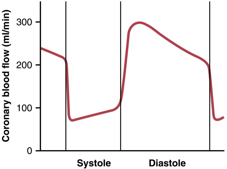
Figure 21-4. Phasic flow of blood through the coronary capillaries of the human left ventricle during cardiac systole and diastole (as extrapolated from measured flows in dogs).
شکل 21-4. جریان فازی خون از طریق مویرگ های کرونری بطن چپ انسان در طول سیستول قلبی و دیاستول (براساس جریان های اندازه گیری شده در سگ ها).
Epicardial Versus Subendocardial Coronary Blood Flow-Effect of Intramyocardial Pressure. Figure 21- 5 demonstrates the special arrangement of the coronary vessels at different depths in the heart muscle, showing on the outer surface epicardial coronary arteries which supply most of the muscle. Smaller intramuscular arteries derived from the epicardial arteries penetrate the muscle, supplying the needed nutrients. Lying immediately beneath the endocardium is a plexus of subendocardial arteries. During systole, blood flow through the subendocardial plexus of the left ventricle, where the intramuscular coronary vessels are compressed greatly by ventricular muscle contraction, tends to be reduced. However, the extra vessels of the subendocardial plexus normally compensate for this reduction. Later in the chapter, we explain how this peculiar difference between blood flow in the epicardial and subendocardial arteries plays an important role in certain types of coronary ischemia.
اپی کاردیال در مقابل جریان خون کرونر سابندوکاردیال – اثر فشار داخل میوکارد. شکل 21-5 آرایش ویژه عروق کرونری را در اعماق مختلف در عضله قلب نشان می دهد، که در سطح بیرونی عروق کرونر اپی کاردیال را نشان می دهد که بیشتر عضله را تامین می کند. شریانهای داخل عضلانی کوچکتر که از شریانهای اپیکاردیال به دست میآیند، به عضله نفوذ میکنند و مواد مغذی مورد نیاز را تامین میکنند. بلافاصله در زیر آندوکارد شبکه ای از شریان های ساب اندوکارد قرار دارد. در طی سیستول، جریان خون از طریق شبکه ساب اندوکاردیال بطن چپ، جایی که عروق کرونری داخل عضلانی توسط انقباض عضله بطنی به شدت فشرده می شوند، کاهش می یابد. با این حال، عروق اضافی شبکه ساب اندوکاردیال معمولاً این کاهش را جبران می کنند. بعداً در این فصل، توضیح میدهیم که چگونه این تفاوت عجیب بین جریان خون در شریانهای اپیکاردیال و ساب اندوکاردیال نقش مهمی در انواع خاصی از ایسکمی کرونری دارد.

Figure 21-5. Diagram of the epicardial, intramuscular, and subendocardial coronary vasculature.
شکل 21-5. نمودار عروق کرونر اپی کاردیال، داخل عضلانی و زیر آندوکاردیال.
CONTROL OF CORONARY BLOOD FLOW
کنترل جریان خون کرونر
Local Muscle Metabolism Is the Primary Controller of Coronary Flow
Blood flow through the coronary system is regulated mostly by local arteriolar vasodilation in response to the nutritional needs of cardiac muscle. That is, whenever the vigor of cardiac contraction is increased, the rate of coronary blood flow also increases. Conversely, decreased heart activity is accompanied by decreased coronary flow. This local regulation of coronary blood flow is similar to that which occurs in many other tissues of the body, especially in the skeletal muscles.
کنترل جریان خون کرونر متابولیسم عضلانی موضعی کنترل کننده اولیه جریان کرونر است
جریان خون از طریق سیستم کرونری عمدتا توسط اتساع عروق شریانی موضعی در پاسخ به نیازهای تغذیه ای عضله قلب تنظیم می شود. یعنی هر گاه قدرت انقباض قلبی افزایش یابد، سرعت جریان خون کرونری نیز افزایش می یابد. برعکس، کاهش فعالیت قلب با کاهش جریان کرونری همراه است. این تنظیم موضعی جریان خون کرونر شبیه به آنچه در بسیاری از بافتهای دیگر بدن، بهویژه در ماهیچههای اسکلتی رخ میدهد، است.
Oxygen Demand Is a Major Factor in Local Coronary Blood Flow Regulation. Blood flow in the coronary arteries usually is regulated almost exactly in proportion to the need of the cardiac musculature for oxygen. Normally, about 70% of the oxygen in the coronary arterial blood is removed as the blood flows through the heart muscle. Because not much oxygen is left, little additional oxygen can be supplied to the heart musculature unless the coronary blood flow increases. Fortunately, the coronary blood flow increases almost in direct proportion to any additional metabolic consumption of oxygen by the heart.
تقاضای اکسیژن یک عامل اصلی در تنظیم جریان خون کرونری موضعی است. جریان خون در عروق کرونر معمولاً تقریباً دقیقاً متناسب با نیاز عضلانی قلب به اکسیژن تنظیم می شود. به طور معمول، حدود 70 درصد از اکسیژن موجود در خون شریانی کرونری با جریان خون در عضله قلب خارج می شود. از آنجایی که اکسیژن زیادی باقی نمانده است، اکسیژن اضافی کمی می تواند به عضله قلب وارد شود، مگر اینکه جریان خون کرونری افزایش یابد. خوشبختانه، جریان خون کرونری تقریباً به نسبت مستقیم با مصرف سوخت و ساز اضافی اکسیژن توسط قلب افزایش می یابد.
The exact means whereby increased oxygen consumption causes coronary dilation has not been determined. Many researchers have speculated that a decrease in oxygen concentration in the heart causes vasodilator substances to be released from the muscle cells and that these substances dilate the arterioles. A substance with great vasodilator propensity is adenosine. In the presence of very low concentrations of oxygen in the muscle cells, a large proportion of the cell’s ATP degrades to adenosine monophosphate (AMP). Small portions of this substance are then further degraded and release adenosine to the tissue fluids of the heart muscle, with a resultant increase in local coronary blood flow. After adenosine causes vasodilation, much of it is reabsorbed into the cardiac cells to be reused for production of ATP.
روش دقیقی که به موجب آن افزایش مصرف اکسیژن باعث اتساع عروق کرونر می شود، مشخص نشده است. بسیاری از محققان حدس می زنند که کاهش غلظت اکسیژن در قلب باعث می شود مواد گشادکننده عروق از سلول های عضلانی آزاد شوند و این مواد باعث گشاد شدن شریان ها می شوند. ماده ای که تمایل زیادی به گشادکننده عروق دارد، آدنوزین است. در حضور غلظت های بسیار کم اکسیژن در سلول های عضلانی، بخش زیادی از ATP سلول به آدنوزین مونوفسفات (AMP) تجزیه می شود. سپس بخشهای کوچکی از این ماده بیشتر تجزیه میشود و آدنوزین را به مایعات بافتی عضله قلب آزاد میکند و در نتیجه جریان خون کرونری موضعی را افزایش میدهد. بعد از اینکه آدنوزین باعث اتساع عروق شد، قسمت زیادی از آن دوباره جذب سلولهای قلبی میشود تا برای تولید ATP مورد استفاده مجدد قرار گیرد.
Adenosine is not the only vasodilator product that has been identified; others include adenosine phosphate compounds, potassium ions, hydrogen ions, carbon dioxide, prostaglandins, and nitric oxide. The mechanisms of coronary vasodilation during increased cardiac activity have not been fully explained by adenosine. Pharmacologic agents that block or partially block the vasodilator effect of adenosine do not completely prevent coronary vasodilation caused by increased heart muscle activity. Studies in skeletal muscle have also shown that the continued infusion of adenosine maintains vascular dilation for only 1 to 3 hours, yet muscle activity still dilates the local blood vessels, even when the adenosine can no longer dilate them. Therefore, the other vasodilator mechanisms listed earlier should be remembered.
آدنوزین تنها محصول گشادکننده عروقی نیست که شناسایی شده است. برخی دیگر شامل ترکیبات آدنوزین فسفات، یون های پتاسیم، یون های هیدروژن، دی اکسید کربن، پروستاگلاندین ها و اکسید نیتریک است. مکانیسم های اتساع عروق کرونر در طول افزایش فعالیت قلبی به طور کامل توسط آدنوزین توضیح داده نشده است. عوامل دارویی که اثر گشادکننده عروق آدنوزین را مسدود یا تا حدی مسدود می کنند، به طور کامل از اتساع عروق کرونر ناشی از افزایش فعالیت عضله قلب جلوگیری نمی کنند. مطالعات روی عضله اسکلتی همچنین نشان داده است که انفوزیون مداوم آدنوزین تنها 1 تا 3 ساعت اتساع عروقی را حفظ می کند، با این حال فعالیت ماهیچه ای همچنان رگ های خونی موضعی را گشاد می کند، حتی زمانی که آدنوزین دیگر نمی تواند آنها را گشاد کند. بنابراین، سایر مکانیسمهای گشادکننده عروقی که قبلاً ذکر شد، باید به خاطر بسپارید.
Nervous Control of Coronary Blood Flow
Stimulation of the autonomic nerves to the heart can affect coronary blood flow directly and indirectly. The direct effects result from action of the nervous transmitter substances acetylcholine from the vagus nerves and norepinephrine from the sympathetic nerves on the coronary vessels. The indirect effects result from secondary changes in coronary blood flow caused by increased or decreased activity of the heart.
کنترل عصبی جریان خون کرونر
تحریک اعصاب اتونوم به قلب می تواند به طور مستقیم و غیرمستقیم بر جریان خون کرونر تأثیر بگذارد. اثرات مستقیم ناشی از عمل مواد انتقال دهنده عصبی استیل کولین از اعصاب واگ و نوراپی نفرین از اعصاب سمپاتیک بر روی عروق کرونر است. اثرات غیر مستقیم ناشی از تغییرات ثانویه در جریان خون کرونر ناشی از افزایش یا کاهش فعالیت قلب است.
The indirect effects, which are mostly opposite to the direct effects, play a far more important role in the normal control of coronary blood flow. Thus, sympathetic stimulation, which releases norepinephrine from the sympathetic nerves and epinephrine, as well as norepinephrine from the adrenal medullae, increases both heart rate and heart contractility and increases the rate of metabolism of the heart. In turn, the increased metabolism of the heart sets off local blood flow regulatory mechanisms for dilating the coronary vessels and blood flow increases approximately in proportion to the metabolic needs of the heart muscle. In contrast, vagal stimulation, with its release of acetylcholine, slows the heart and has a slightly depressive effect on heart contractility. These effects decrease cardiac oxygen consumption and, therefore, indirectly con- strict the coronary arteries.
اثرات غیرمستقیم که عمدتاً مخالف اثرات مستقیم هستند، نقش به مراتب مهمتری در کنترل طبیعی جریان خون کرونر دارند. بنابراین، تحریک سمپاتیک، که نوراپی نفرین را از اعصاب سمپاتیک و اپی نفرین، و همچنین نوراپی نفرین را از بصل الکلیه آدرنال آزاد می کند، ضربان قلب و انقباض قلب را افزایش می دهد و سرعت متابولیسم قلب را افزایش می دهد. به نوبه خود، افزایش متابولیسم قلب مکانیسم های تنظیم کننده جریان خون موضعی را برای گشاد کردن عروق کرونر ایجاد می کند و جریان خون تقریباً متناسب با نیازهای متابولیکی عضله قلب افزایش می یابد. در مقابل، تحریک واگ با آزاد شدن استیل کولین، قلب را کند میکند و تأثیر کمی بر انقباض قلب دارد. این اثرات مصرف اکسیژن قلب را کاهش می دهد و بنابراین به طور غیرمستقیم عروق کرونر را محدود می کند.
Direct Effects of Nervous Stimuli on Coronary Vasculature. The distribution of parasympathetic (vagal) nerve fibers to the ventricular coronary system is not very great. However, the acetylcholine released by parasympathetic stimulation has a direct effect to dilate the coronary arteries.
اثرات مستقیم محرک های عصبی بر عروق کرونر. توزیع رشته های عصبی پاراسمپاتیک (واگ) در سیستم کرونری بطنی خیلی زیاد نیست. با این حال، استیل کولین آزاد شده توسط تحریک پاراسمپاتیک تأثیر مستقیمی در گشاد کردن عروق کرونر دارد.
Much more extensive sympathetic innervation of the coronary vessels occurs. In Chapter 61, we see that the sympathetic transmitter substances norepinephrine and epinephrine can have vascular constrictor or vascular dilator effects, depending on the presence or absence of constrictor or dilator receptors in the blood vessel walls. The constrictor receptors are called alpha receptors, and the dilator receptors are called beta receptors. Both alpha and beta receptors exist in the coronary vessels. In general, the epicardial coronary vessels have a preponderance of alpha receptors, whereas the intramuscular arteries may have a preponderance of beta receptors. Therefore, sympathetic stimulation can, at least theoretically, cause slight overall coronary constriction or dilation, but usu- ally constriction. In some people, the alpha vasoconstrictor effects seem to be disproportionately severe, and these people can have vasospastic myocardial ischemia during periods of excess sympathetic drive, often with resultant anginal pain.
عصب دهی سمپاتیک بسیار گسترده تری در عروق کرونر رخ می دهد. در فصل 61، می بینیم که مواد انتقال دهنده سمپاتیک نوراپی نفرین و اپی نفرین بسته به وجود یا عدم وجود گیرنده های منقبض کننده یا گشادکننده در دیواره رگ های خونی، می توانند اثرات منقبض کننده عروق یا گشادکننده عروق داشته باشند. گیرنده های منقبض کننده گیرنده آلفا و گیرنده های گشاد کننده گیرنده بتا نامیده می شوند. هر دو گیرنده آلفا و بتا در عروق کرونر وجود دارند. به طور کلی، عروق کرونر اپی کاردیال دارای غلبه گیرنده های آلفا هستند، در حالی که شریان های داخل عضلانی ممکن است گیرنده های بتا را داشته باشند. بنابراین، تحریک سمپاتیک، حداقل از نظر تئوری، میتواند باعث انقباض یا اتساع کلی عروق کرونر شود، اما معمولاً انقباض دارد. در برخی از افراد، اثرات منقبض کننده عروق آلفا به طور نامتناسبی شدید به نظر می رسد، و این افراد ممکن است ایسکمی وازواسپاستیک میوکارد را در طول دوره های تحریک بیش از حد سمپاتیک، اغلب همراه با درد آنژین، داشته باشند.
Metabolic factors, especially myocardial oxygen consumption, are the major controllers of myocardial blood flow. Whenever the direct effects of nervous stimulation reduce coronary blood flow, the metabolic control of coronary flow usually overrides the direct coronary nervous effects within seconds.
عوامل متابولیک، به ویژه مصرف اکسیژن میوکارد، کنترل کننده اصلی جریان خون میوکارد هستند. هر گاه اثرات مستقیم تحریک عصبی جریان خون کرونر را کاهش دهد، کنترل متابولیک جریان کرونری معمولاً در عرض چند ثانیه بر اثرات مستقیم عصبی کرونری غلبه می کند.
SPECIAL FEATURES OF CARDIAC MUSCLE METABOLISM
The basic principles of cellular metabolism, discussed in Chapters 68 through 73, apply to cardiac muscle the same as for other tissues, but some quantitative differences exist. Most importantly, under resting conditions, cardiac muscle normally consumes more fatty acids than carbohydrates to supply its energy (≈ 70% of the energy is derived from fatty acids). However, as is also true of other tissues, under anaerobic or ischemic conditions, cardiac metabolism must call on anaerobic glycolysis mechanisms for energy. However, glycolysis consumes large quantities of the blood glucose and, at the same time, forms large amounts of lactic acid in the cardiac tissue. This is prob- ably one of the causes of cardiac pain in cardiac ischemic conditions, as discussed later in this chapter.
ویژگی های خاص متابولیسم ماهیچه های قلبی
اصول اساسی متابولیسم سلولی که در فصلهای 68 تا 73 مورد بحث قرار گرفتهاند، مانند سایر بافتها در مورد عضله قلب نیز صدق میکنند، اما برخی از تفاوتهای کمی وجود دارد. مهمتر از همه، در شرایط استراحت، عضله قلب به طور معمول اسیدهای چرب بیشتری نسبت به کربوهیدرات ها برای تامین انرژی خود مصرف می کند (70٪ انرژی از اسیدهای چرب مشتق می شود). با این حال، همانطور که در مورد سایر بافت ها نیز صادق است، در شرایط بی هوازی یا ایسکمیک، متابولیسم قلبی باید مکانیسم های گلیکولیز بی هوازی را برای انرژی فراخواند. با این حال، گلیکولیز مقادیر زیادی از گلوکز خون را مصرف می کند و در همان زمان، مقادیر زیادی اسید لاکتیک را در بافت قلب تشکیل می دهد. این احتمالاً یکی از علل درد قلبی در شرایط ایسکمیک قلبی است، همانطور که بعداً در این فصل مورد بحث قرار گرفت.
As is true in other tissues, more than 95% of the metabolic energy liberated from foods is used to form ATP in the mitochondria. This ATP in turn acts as the conveyer of energy for cardiac muscular contraction and other cellular functions. In severe coronary ischemia, the ATP degrades first to adenosine diphosphate and then to AMP and adenosine. Because the cardiac muscle cell membrane is slightly permeable to adenosine, much of this agent can diffuse from the muscle cells into the circulating blood.
همانطور که در سایر بافت ها صادق است، بیش از 95 درصد از انرژی متابولیک آزاد شده از غذاها برای تشکیل ATP در میتوکندری استفاده می شود. این ATP به نوبه خود به عنوان انتقال دهنده انرژی برای انقباض عضلانی قلبی و سایر عملکردهای سلولی عمل می کند. در ایسکمی شدید کرونر، ATP ابتدا به آدنوزین دی فسفات و سپس به AMP و آدنوزین تجزیه می شود. از آنجایی که غشای سلولی ماهیچه قلب کمی به آدنوزین نفوذ می کند، بیشتر این عامل می تواند از سلول های عضلانی به خون در گردش پخش شود.
The released adenosine is believed to be one of the substances that causes dilation of the coronary arterioles during coronary hypoxia, as discussed earlier. However, loss of adenosine also has a serious cellular consequence. Within as little as 30 minutes of severe coronary ischemia, as occurs after a myocardial infarct, about half of the adenine base can be lost from the affected cardiac muscle cells. Furthermore, this loss can be replaced by new synthesis of adenine at a rate of only 2%/hour. Therefore, once a serious bout of coronary ischemia has persisted for 30 minutes or longer, relief of the ischemia may be too late to prevent injury and death of the cardiac cells. This is almost certainly one of the major causes of cardiac cellular death during myocardial ischemia.
اعتقاد بر این است که آدنوزین آزاد شده یکی از موادی است که باعث گشاد شدن شریان های کرونر در طول هیپوکسی عروق کرونر می شود، همانطور که قبلاً بحث شد. با این حال، از دست دادن آدنوزین نیز پیامدهای سلولی جدی دارد. در کمتر از 30 دقیقه ایسکمی شدید کرونری، همانطور که پس از انفارکتوس میوکارد اتفاق می افتد، حدود نیمی از پایه آدنین می تواند از سلول های عضلانی قلب آسیب دیده از بین برود. علاوه بر این، این کاهش را می توان با سنتز جدید آدنین با نرخ تنها 2٪ در ساعت جایگزین کرد. بنابراین، هنگامی که یک حمله جدی ایسکمی عروق کرونر به مدت 30 دقیقه یا بیشتر ادامه یابد، ممکن است برای جلوگیری از آسیب و مرگ سلول های قلبی، تسکین ایسکمی خیلی دیر باشد. این تقریباً به طور قطع یکی از علل اصلی مرگ سلولی قلبی در طول ایسکمی میوکارد است.
ISCHEMIC HEART DISEASE
The most common cause of death in the Western countries is ischemic heart disease, which results from insufficient coronary blood flow. About 35% of people in the United States aged 65 years and older die of this cause. Some deaths occur suddenly as a result of acute coronary occlusion or fibrillation of the heart, whereas other deaths occur slowly over a period of weeks to years as a result of progressive weakening of the heart pumping process. In this chapter, we discuss acute coronary ischemia caused by acute coronary occlusion and myocardial infarction. In Chapter 22, we discuss congestive heart failure, which is frequently caused by slowly increasing coronary ischemia and weakening of the cardiac muscle.
بیماری ایسکمیک قلب
شایع ترین علت مرگ و میر در کشورهای غربی بیماری ایسکمیک قلب است که ناشی از جریان ناکافی خون کرونر است. حدود 35 درصد از افراد 65 سال و بالاتر در ایالات متحده به این علت می میرند. برخی از مرگ ها به طور ناگهانی در نتیجه انسداد حاد کرونری یا فیبریلاسیون قلب رخ می دهد، در حالی که سایر مرگ ها به آرامی در طی چند هفته تا سال ها در نتیجه تضعیف تدریجی فرآیند پمپاژ قلب رخ می دهد. در این فصل، ایسکمی حاد کرونر ناشی از انسداد حاد کرونر و انفارکتوس میوکارد را مورد بحث قرار می دهیم. در فصل 22، نارسایی احتقانی قلب را مورد بحث قرار میدهیم که اغلب به دلیل افزایش آهسته ایسکمی کرونری و ضعیف شدن عضله قلب ایجاد میشود.
Atherosclerosis Is a Major Cause of Ischemic Heart Disease. A frequent cause of diminished coronary blood flow is atherosclerosis. The atherosclerotic process is dis- cussed in connection with lipid metabolism in Chapter 69. Briefly, in people who have a genetic predisposition to atherosclerosis, who are overweight or obese and have a sedentary lifestyle, or who have high blood pressure and damage to the endothelial cells of the coronary blood vessels, large quantities of cholesterol gradually become de- posited beneath the endothelium at many points in arteries throughout the body. Gradually, these areas of deposit are invaded by fibrous tissue and frequently become calcified. The net result is the development of atherosclerotic plaques, which actually protrude into the vessel lumens and block or partially block blood flow. A common site for development of atherosclerotic plaques is the first few centimeters of the major coronary arteries.
آترواسکلروز یکی از علل اصلی بیماری ایسکمیک قلبی است. یکی از علل شایع کاهش جریان خون کرونر، آترواسکلروز است. فرآیند آترواسکلروتیک در ارتباط با متابولیسم لیپیدها در فصل 69 مورد بحث قرار گرفته است. به طور خلاصه، در افرادی که دارای استعداد ژنتیکی تصلب شرایین هستند، دارای اضافه وزن یا چاق هستند و سبک زندگی کم تحرکی دارند، یا دارای فشار خون بالا و آسیب به سلول های اندوتلیال رگ های کرونری هستند که به تدریج از بین می روند. اندوتلیوم در بسیاری از نقاط شریان های سراسر بدن. به تدریج، این مناطق رسوبی توسط بافت فیبری مورد تهاجم قرار گرفته و اغلب کلسیفیه می شوند. نتیجه خالص ایجاد پلاک های آترواسکلروتیک است که در واقع به داخل مجرای عروق بیرون زده و جریان خون را مسدود یا تا حدی مسدود می کند. یک مکان رایج برای ایجاد پلاک های آترواسکلروتیک، چند سانتی متر اول از شریان های کرونر اصلی است.
Acute Coronary Artery Occlusion
Acute occlusion of a coronary artery usually occurs in a person who already has underlying atherosclerotic coronary heart disease but almost never occurs in a person with a normal coronary circulation. Acute occlusion can have various causes, two of which are the following:
انسداد حاد عروق کرونر
انسداد حاد شریان کرونری معمولاً در فردی اتفاق میافتد که قبلاً بیماری آترواسکلروتیک زمینهای دارد، اما تقریباً هرگز در فردی با گردش خون طبیعی کرونر رخ نمیدهد. انسداد حاد می تواند علل مختلفی داشته باشد که دو مورد از آنها به شرح زیر است:
1. The atherosclerotic plaque can cause a local blood clot called a thrombus that occludes the artery. The thrombus usually occurs where the arteriosclerotic plaque has broken through the endothelium, thus coming into direct contact with the flowing blood. Because the plaque presents an unsmooth surface, blood platelets adhere to it, fibrin is deposited, and red blood cells become trapped to form a blood clot that grows until it occludes the vessel. Occasionally, the clot breaks away from its attachment on the atherosclerotic plaque and flows to a more peripheral branch of the coronary arterial tree, where it blocks the artery at that point. A thrombus that flows along the artery in this way and occludes the vessel more distally is called a coronary embolus.
1.پلاک آترواسکلروتیک می تواند باعث ایجاد یک لخته خون موضعی به نام ترومبوز شود که شریان را مسدود می کند. ترومبوز معمولاً در جایی اتفاق میافتد که پلاک شریاناسکلروتیک از درون اندوتلیوم شکسته شده است، بنابراین در تماس مستقیم با خون جاری است. از آنجایی که پلاک سطحی ناصاف دارد، پلاکتهای خون به آن میچسبند، فیبرین رسوب میکند و گلبولهای قرمز به دام میافتند تا لختهای را تشکیل دهند که رشد میکند تا رگ را مسدود کند. گاهی اوقات، لخته از اتصال خود به پلاک آترواسکلروتیک جدا می شود و به یک شاخه محیطی تر از درخت شریان کرونر جریان می یابد، جایی که در آن نقطه شریان را مسدود می کند. ترومبی که به این ترتیب در امتداد شریان جریان می یابد و رگ را به صورت دیستال بیشتری مسدود می کند، آمبولی کرونری نامیده می شود.
2. Many clinicians believe that local muscular spasm of a coronary artery also can occur. The spasm might result from direct irritation of the smooth muscle of the arterial wall by the edges of an arteriosclerotic plaque, or it might result from local nervous reflexes that cause excess coronary vascular wall contraction. The spasm may then lead to secondary thrombosis of the vessel.
2.بسیاری از پزشکان معتقدند که اسپاسم عضلانی موضعی شریان کرونر نیز ممکن است رخ دهد. اسپاسم ممکن است ناشی از تحریک مستقیم عضله صاف دیواره شریانی توسط لبههای پلاک شریانی باشد، یا ممکن است ناشی از رفلکسهای عصبی موضعی باشد که باعث انقباض بیش از حد دیواره عروق کرونر میشود. سپس اسپاسم ممکن است منجر به ترومبوز ثانویه رگ شود.
Lifesaving Value of Collateral Circulation in the Heart. The degree of damage to the heart muscle caused by slowly developing atherosclerotic constriction of the coronary arteries or by sudden coronary occlusion is determined to a great extent by the degree of collateral circulation that has already developed or that can open within minutes after the occlusion. In a normal heart, almost no large communications exist among the larger coronary arteries. However, many anastomoses do exist among the smaller arteries of 20 to 250 micrometers in diameter, as shown in Figure 21-6.
ارزش نجات دهنده گردش وثیقه در قلب. میزان آسیب به عضله قلب ناشی از انقباض آترواسکلروتیک عروق کرونر آهسته در حال توسعه یا انسداد ناگهانی کرونری تا حد زیادی با درجه گردش خون جانبی که قبلاً ایجاد شده است یا می تواند در عرض چند دقیقه پس از انسداد باز شود تعیین می شود. در یک قلب طبیعی، تقریبا هیچ ارتباط بزرگی بین شریانهای کرونر بزرگتر وجود ندارد. با این حال، همانطور که در شکل 21-6 نشان داده شده است، بسیاری از آناستوموزها در میان شریان های کوچکتر با قطر 20 تا 250 میکرومتر وجود دارند.
When a sudden occlusion occurs in one of the larger coronary arteries, the small anastomoses begin to dilate within seconds. However, the blood flow through these minute collaterals is usually less than half of that needed to keep most of the cardiac muscle alive that they now supply. The diameters of the collateral vessels do not enlarge much more for the next 8 to 24 hours. Then, however, collateral flow begins to increase, doubling by the second or third day and often reaching normal or almost normal coronary flow within about 1 month. Because of these developing collateral channels, many patients recover almost completely from various degrees of coronary occlusion when the area of muscle involved is not too large.
هنگامی که یک انسداد ناگهانی در یکی از عروق کرونر بزرگتر رخ می دهد، آناستوموزهای کوچک در عرض چند ثانیه شروع به گشاد شدن می کنند. با این حال، جریان خون از طریق این وثیقه های کوچک معمولا کمتر از نصف مقدار مورد نیاز برای زنده نگه داشتن بیشتر عضله قلبی است که در حال حاضر تامین می کنند. قطر رگ های جانبی برای 8 تا 24 ساعت آینده خیلی بیشتر نمی شود. سپس، با این حال، جریان وثیقه شروع به افزایش می کند، در روز دوم یا سوم دو برابر می شود و اغلب در عرض حدود 1 ماه به جریان کرونری طبیعی یا تقریبا طبیعی می رسد. به دلیل این کانال های جانبی در حال توسعه، بسیاری از بیماران تقریباً به طور کامل از درجات مختلف انسداد کرونری بهبود می یابند، زمانی که ناحیه عضله درگیر خیلی بزرگ نباشد.
When atherosclerosis constricts the coronary arteries slowly over a period of many years, rather than suddenly, collateral vessels can develop at the same time, while the atherosclerosis becomes more and more severe. Therefore, the person may never experience an acute episode of cardiac dysfunction. Eventually, however, the sclerotic process develops beyond the limits of even the collateral blood supply to provide the needed blood flow, and sometimes, the collateral blood vessels themselves develop atherosclerosis. When this occurs, the heart muscle becomes severely limited in its work output, and the heart cannot pump even normally required amounts of blood flow. This is one of the most common causes of cardiac failure in older people.
هنگامی که آترواسکلروز عروق کرونر را به آرامی در طی چندین سال منقبض می کند، نه ناگهانی، عروق جانبی می توانند همزمان ایجاد شوند، در حالی که آترواسکلروز شدیدتر و شدیدتر می شود. بنابراین، فرد ممکن است هرگز یک دوره حاد اختلال عملکرد قلبی را تجربه نکند. با این حال، در نهایت، فرآیند اسکلروتیک فراتر از محدودیتهای تامین خون جانبی برای تامین جریان خون مورد نیاز توسعه مییابد و گاهی اوقات، رگهای خونی جانبی خود دچار تصلب شرایین میشوند. هنگامی که این اتفاق می افتد، عضله قلب به شدت در خروجی کار خود محدود می شود و قلب نمی تواند حتی مقادیر عادی جریان خون را پمپ کند. این یکی از شایع ترین علل نارسایی قلبی در افراد مسن است.
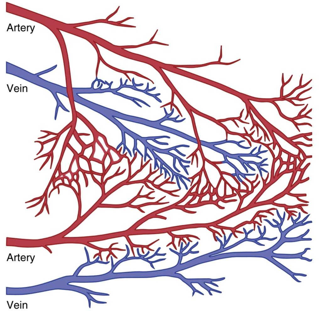
Figure 21-6. Minute anastomoses in the normal coronary arterial system.
شکل 21-6. آناستوموزهای دقیقه ای در سیستم شریان کرونر طبیعی
Myocardial Infarction
Immediately after an acute coronary occlusion, blood flow ceases in the coronary vessels beyond the occlusion, except for small amounts of collateral flow from surrounding vessels. The area of muscle that has zero flow or so little flow that it cannot sustain cardiac muscle function is said to be infarcted. The overall process is called a myocardial infarction.
انفارکتوس میوکارد
بلافاصله پس از انسداد حاد کرونری، جریان خون در عروق کرونر فراتر از انسداد متوقف می شود، به جز مقادیر کمی جریان جانبی از عروق اطراف. گفته میشود ناحیهای از عضله که جریان صفر یا آنقدر کم است که نمیتواند عملکرد عضله قلب را حفظ کند دچار انفارکتوس شده است. روند کلی آن انفارکتوس میوکارد نامیده می شود.
Soon after the onset of the infarction, small amounts of collateral blood begin to infiltrate the infarcted area, which, combined with progressive dilation of local blood vessels, causes the area to become overfilled with stagnant blood. Simultaneously the muscle fibers use the last bits of the oxygen in the blood, causing the hemoglobin to become totally deoxygenated. Therefore, the infarcted area takes on a bluish-brown hue, and the blood vessels of the area appear to be engorged, despite lack of blood flow. In later stages, the vessel walls become highly permeable and leak fluid, the local muscle tissue becomes edematous, and the cardiac muscle cells begin to swell because of diminished cellular metabolism. Within a few hours of almost no blood supply, the cardiac muscle cells die.
بلافاصله پس از شروع انفارکتوس، مقدار کمی خون جانبی شروع به نفوذ به ناحیه انفارکتوس شده می کند، که همراه با اتساع تدریجی رگ های خونی موضعی، باعث پر شدن بیش از حد ناحیه از خون راکد می شود. فیبرهای عضلانی به طور همزمان از آخرین تکههای اکسیژن خون استفاده میکنند که باعث میشود هموگلوبین کاملاً اکسیژنزدایی شود. بنابراین، ناحیه انفارکتوس رنگ آبی مایل به قهوهای به خود میگیرد و رگهای خونی این ناحیه علیرغم عدم جریان خون، به نظر میرسند. در مراحل بعدی، دیوارههای عروق بسیار نفوذپذیر میشوند و مایع نشت میکند، بافت ماهیچهای موضعی ادم میکند و سلولهای ماهیچههای قلبی به دلیل کاهش متابولیسم سلولی شروع به متورم شدن میکنند. در عرض چند ساعت تقریباً بدون خون رسانی، سلول های عضله قلب می میرند.
Cardiac muscle requires about 1.3 ml of oxygen/100 g of muscle tissue/min just to remain alive. In comparison, about 8 ml oxygen/100 g are delivered to the normal resting left ventricle each minute. Therefore, if there is even 15% to 30% of normal resting coronary blood flow, the muscle will not die. In the central portion of a large infarct, however, where there is almost no collateral blood flow, the muscle does die.
عضله قلب فقط برای زنده ماندن به حدود 1.3 میلی لیتر اکسیژن / 100 گرم بافت عضلانی در دقیقه نیاز دارد. در مقایسه، حدود 8 میلی لیتر اکسیژن در 100 گرم در هر دقیقه به بطن چپ طبیعی در حال استراحت تحویل داده می شود. بنابراین، اگر حتی 15 تا 30 درصد جریان خون کرونر در حالت استراحت طبیعی وجود داشته باشد، عضله نمی میرد. با این حال، در بخش مرکزی یک انفارکتوس بزرگ، جایی که تقریباً هیچ جریان خون جانبی وجود ندارد، عضله می میرد.
Subendocardial Infarction. The subendocardial muscle frequently becomes infarcted, even when there is no evidence of infarction in the outer surface portions of the heart. This occurs because the subendocardial muscle has a higher oxygen consumption and extra difficulty obtaining adequate blood flow because the blood vessels in the subendocardium are intensely compressed by systolic contraction of the heart, as explained earlier. Therefore, any condition that compromises blood flow to any area of the heart usually causes damage first in the subendocardi- al regions, and the damage then spreads outward toward the epicardium.
انفارکتوس سابندوکارد. عضله ساب اندوکاردیال اغلب دچار انفارکتوس می شود، حتی زمانی که شواهدی از انفارکتوس در قسمت های سطح بیرونی قلب وجود نداشته باشد. این به این دلیل اتفاق میافتد که عضله ساب اندوکاردیال مصرف اکسیژن بیشتری دارد و در بدست آوردن جریان خون کافی مشکل بیشتری دارد، زیرا عروق خونی در ساب اندوکارد به شدت تحت فشار انقباض سیستولیک قلب قرار میگیرند، همانطور که قبلا توضیح داده شد. بنابراین، هر شرایطی که جریان خون را در هر ناحیه ای از قلب به خطر بیاندازد، معمولاً ابتدا در نواحی ساب اندوکاردیال آسیب ایجاد می کند و سپس آسیب به سمت بیرون به سمت اپی کاردیوم گسترش می یابد.
CAUSES OF DEATH AFTER ACUTE CORONARY OCCLUSION
The most common causes of death after acute myocardial infarction are the following: (1) decreased cardiac output; (2) damming of blood in the pulmonary blood vessels and then death resulting from pulmonary edema; (3) fibrillation of the heart; and, occasionally (4) rupture of the heart.
علل مرگ پس از انسداد حاد کرونر
شایع ترین علل مرگ پس از انفارکتوس حاد میوکارد عبارتند از: (1) کاهش برون ده قلبی. (2) سد کردن خون در رگ های خونی ریوی و سپس مرگ ناشی از ادم ریوی. (3) فیبریلاسیون قلب. و گاهی (4) پارگی قلب.
Decreased Cardiac Output-Systolic Stretch and Cardiac Shock. When some of the cardiac muscle fibers are not functioning and others are too weak to contract with great force, the overall pumping ability of the affected ventricle is proportionately depressed. The overall pumping strength of the infarcted heart is often decreased more than one might expect because of a phenomenon called systolic stretch, shown in Figure 21-7. That is, when the normal portions of the ventricular muscle contract, the ischemic portion of the muscle, whether it is dead or simply nonfunctional, instead of contracting is forced outward by the pressure that develops inside the ventricle. Therefore, much of the pumping force of the ventricle is dissipated by bulging of the area of nonfunctional cardiac muscle.
کاهش برون ده قلبی – کشش سیستولیک و شوک قلبی. هنگامی که برخی از فیبرهای عضلانی قلب کار نمی کنند و برخی دیگر برای انقباض با نیروی زیاد آنقدر ضعیف هستند، توانایی پمپاژ کلی بطن آسیب دیده به طور متناسبی کاهش می یابد. قدرت پمپاژ کلی قلب انفارکتوس شده به دلیل پدیده ای به نام کشش سیستولیک که در شکل 21-7 نشان داده شده است، اغلب بیش از حد انتظار کاهش می یابد. یعنی وقتی بخشهای طبیعی عضله بطنی منقبض میشود، بخش ایسکمی عضله، خواه مرده باشد یا غیرعملکردی، به جای انقباض، تحت فشاری که در داخل بطن ایجاد میشود، به بیرون رانده میشود. بنابراین، بیشتر نیروی پمپاژ بطن با برآمدگی ناحیه عضله قلبی غیرعملکردی از بین می رود.
When the heart becomes incapable of contracting with sufficient force to pump enough blood into the peripheral arterial tree, cardiac failure and death of peripheral tissues ensue as a result of peripheral ischemia. This condition, called coronary shock, cardiogenic shock, cardiac shock, or low cardiac output failure, is discussed more fully in the next chapter. Cardiac shock almost always occurs when more than 40% of the left ventricle is infarcted, and death occurs in more than 70% of patients once cardiac shock develops.
هنگامی که قلب قادر به انقباض با نیروی کافی برای پمپاژ خون کافی به درخت شریان محیطی نباشد، نارسایی قلبی و مرگ بافت های محیطی در نتیجه ایسکمی محیطی رخ می دهد. این وضعیت که شوک کرونری، شوک قلبی، شوک قلبی یا نارسایی برون ده قلبی پایین نامیده می شود، در فصل بعدی به طور کامل مورد بحث قرار می گیرد. شوک قلبی تقریباً همیشه زمانی اتفاق میافتد که بیش از 40 درصد بطن چپ دچار انفارکتوس شده باشد و در بیش از 70 درصد بیماران پس از ایجاد شوک قلبی مرگ اتفاق میافتد.
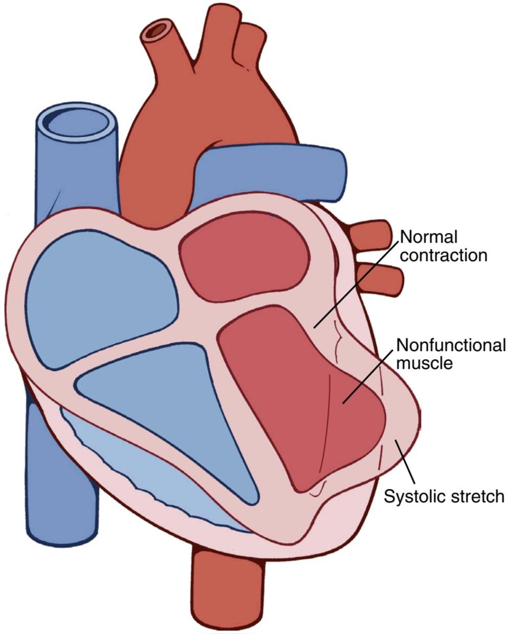
Figure 21-7. Systolic stretch in an area of ischemic cardiac muscle.
شکل 21-7. کشش سیستولیک در ناحیه ای از عضله ایسکمیک قلب.
Damming of Blood in the Body’s Venous System. When the heart is not pumping blood forward, it must be damming blood in the atria and in the blood vessels of the lungs or in the systemic circulation. This leads to increased capillary pressures, particularly in the lungs.
سد کردن خون در سیستم وریدی بدن. وقتی قلب خون را به جلو پمپاژ نمی کند، باید خون را در دهلیزها و رگ های خونی ریه ها یا در گردش خون سیستمیک سد کند. این منجر به افزایش فشار مویرگی، به ویژه در ریه ها می شود.
Damming of blood in the veins often causes little difficulty during the first few hours after a myocardial infarction. Instead, symptoms develop a few days later because the acutely diminished cardiac output leads to diminished blood flow to the kidneys. Then, for reasons discussed in Chapter 22, the kidneys fail to excrete enough urine. This adds progressively to the total blood volume and, therefore, leads to congestive symptoms. Consequently, many patients who seemingly are getting along well during the first few days after the onset of heart failure will suddenly experience acute pulmonary edema and often will die within a few hours after the appearance of the initial pulmonary symptoms.
بند انداختن خون در وریدها اغلب در چند ساعت اول پس از انفارکتوس میوکارد مشکل کمی ایجاد می کند. در عوض، علائم چند روز بعد ظاهر میشوند، زیرا کاهش شدید برون ده قلبی منجر به کاهش جریان خون به کلیهها میشود. سپس، به دلایلی که در فصل 22 بحث شد، کلیه ها قادر به دفع ادرار کافی نیستند. این به تدریج به حجم کل خون می افزاید و در نتیجه منجر به علائم احتقان می شود. در نتیجه، بسیاری از بیمارانی که ظاهراً در چند روز اول پس از شروع نارسایی قلبی با هم خوب هستند، به طور ناگهانی ادم ریوی حاد را تجربه می کنند و اغلب در عرض چند ساعت پس از ظهور علائم اولیه ریوی خواهند مرد.
Fibrillation of the Ventricles After Myocardial Infarction. In many people who die of coronary occlusion, death occurs because of sudden ventricular fibrillation. The tendency for fibrillation to develop is especially great after a large infarction, but fibrillation can sometimes occur after small occlusions as well. Some patients with chronic coronary insufficiency die suddenly of fibrillation without having any acute infarction.
فیبریلاسیون بطن ها پس از انفارکتوس میوکارد. در بسیاری از افرادی که بر اثر انسداد عروق کرونر می میرند، مرگ به دلیل فیبریلاسیون ناگهانی بطنی رخ می دهد. تمایل به ایجاد فیبریلاسیون به ویژه پس از یک انفارکتوس بزرگ بسیار زیاد است، اما فیبریلاسیون گاهی اوقات ممکن است پس از انسدادهای کوچک نیز رخ دهد. برخی از بیماران مبتلا به نارسایی مزمن عروق کرونر به طور ناگهانی در اثر فیبریلاسیون بدون داشتن انفارکتوس حاد می میرند.
Fibrillation is most likely to occur during two especially dangerous periods after coronary infarction. The first period is during the first 10 minutes after the infarction occurs. Then, there is a short period of relative safety, followed by a second period of cardiac irritability beginning 1 hour or so later and lasting for another few hours. Fibrillation can also occur many days after the infarct but is less likely to occur then.
فیبریلاسیون به احتمال زیاد در طی دو دوره خطرناک پس از انفارکتوس کرونر رخ می دهد. اولین دوره در 10 دقیقه اول پس از وقوع انفارکتوس است. سپس، یک دوره کوتاه ایمنی نسبی وجود دارد، و به دنبال آن یک دوره دوم تحریک پذیری قلبی وجود دارد که 1 ساعت یا بیشتر بعد شروع می شود و برای چند ساعت دیگر ادامه می یابد. فیبریلاسیون نیز می تواند چندین روز پس از انفارکتوس رخ دهد اما احتمال وقوع آن در آن زمان کمتر است.
At least four factors are involved in the tendency for the heart to fibrillate, as follows:
حداقل چهار عامل در تمایل قلب به فیبریلاسیون دخیل است که به شرح زیر است:
1. Acute loss of blood supply to the cardiac muscle causes rapid depletion of potassium from the ischemic musculature. This also increases the potassium concentration in the extracellular fluids surrounding the cardiac muscle fibers. Experiments in which potassium has been injected into the coronary system have demonstrated that an elevated extracellular potassium concentration increases the irritability of the cardiac musculature and, therefore, its likelihood of fibrillating.
1. از دست دادن حاد خون رسانی به عضله قلب باعث تخلیه سریع پتاسیم از عضله ایسکمیک می شود. این همچنین غلظت پتاسیم را در مایعات خارج سلولی اطراف فیبرهای عضلانی قلب افزایش می دهد. آزمایشهایی که در آنها پتاسیم به سیستم کرونری تزریق شده است نشان دادهاند که افزایش غلظت پتاسیم خارج سلولی تحریکپذیری ماهیچههای قلب و در نتیجه احتمال فیبریلاسیون آن را افزایش میدهد.
2. Ischemia of the muscle causes an injury current, described in Chapter 12 in relation to electrocardiograms in patients with acute myocardial infarction. That is, the ischemic musculature often cannot completely repolarizeits membranes after a heart- beat, and thus the external surface of this muscle remains negative with respect to normal cardiac muscle membrane potential elsewhere in the heart. Therefore, electric current flows from this ischemic area of the heart to the normal area and can elicit abnormal impulses, which can cause fibrillation.
2. ایسکمی عضله باعث جریان آسیب می شود که در فصل 12 در رابطه با الکتروکاردیوگرام در بیماران مبتلا به انفارکتوس حاد میوکارد توضیح داده شده است. به این معنا که عضله ایسکمیک اغلب نمی تواند غشاهای خود را پس از ضربان قلب به طور کامل رپولاریز کند و بنابراین سطح خارجی این عضله با توجه به پتانسیل طبیعی غشای عضله قلب در جای دیگر قلب منفی می ماند. بنابراین، جریان الکتریکی از این ناحیه ایسکمیک قلب به ناحیه طبیعی میرود و میتواند تکانههای غیرطبیعی را ایجاد کند که میتواند باعث فیبریلاسیون شود.
3. Powerful sympathetic reflexes often develop after massive infarction, principally because the heart does not pump an adequate volume of blood into the arterial tree, which leads to reduced blood pressure. The sympathetic stimulation also increases irritability of the cardiac muscle and thereby predisposes to fibrillation.
3. رفلکس های سمپاتیک قوی اغلب پس از انفارکتوس عظیم ایجاد می شوند، عمدتاً به این دلیل که قلب حجم مناسبی از خون را به درخت شریانی پمپاژ نمی کند، که منجر به کاهش فشار خون می شود. تحریک سمپاتیک همچنین تحریک پذیری عضله قلب را افزایش می دهد و در نتیجه مستعد فیبریلاسیون می شود.
4. Cardiac muscle weakness caused by the myocardial infarction often causes the ventricle to dilate excessively. This excessive dilation increases the pathway length for impulse conduction in the heart and frequently causes abnormal conduction pathways all the way around the infarcted area of the cardiac muscle. Both of these effects predispose to the development of circus movements because, as dis- cussed in Chapter 13, excess prolongation of conduction pathways in the ventricles allows impulses to re-enter muscle that is already recovering from refractoriness, thereby initiating a circus movement cycle of new excitation and resulting in continuation of the process.
4. ضعف عضله قلب ناشی از انفارکتوس میوکارد اغلب باعث گشاد شدن بیش از حد بطن می شود. این اتساع بیش از حد طول مسیر برای هدایت ضربه در قلب را افزایش می دهد و اغلب باعث ایجاد مسیرهای هدایت غیر طبیعی در سراسر ناحیه انفارکتوس شده عضله قلب می شود. هر دوی این اثرات مستعد توسعه حرکات سیرک هستند زیرا همانطور که در فصل 13 بحث شد، طولانی شدن بیش از حد مسیرهای هدایت در بطن ها به تکانه ها اجازه می دهد تا دوباره وارد عضله ای شوند که در حال بهبودی از نسوز هستند و در نتیجه چرخه حرکت سیرک با تحریک جدید آغاز می شود و منجر به ادامه روند می شود.
Rupture of Infarcted Area. During the first day or so after an acute infarct, there is little danger of rupture of the ischemic portion of the heart, but a few days later, the dead muscle fibers begin to degenerate, and the heart wall becomes stretched very thin. When this happens, the dead muscle bulges outward to a severe degree with each heart contraction, and this systolic stretch becomes greater and greater until finally the heart ruptures. One of the methods used to assess the progress of severe myocardial infarction is to record by cardiac imaging, with echocardiography, magnetic resonance imaging (MRI), or computed tomography (CT), whether the degree of systolic stretch is worsening.
پارگی ناحیه انفارکتوس. در طول روز اول یا بیشتر پس از انفارکتوس حاد، خطر پارگی قسمت ایسکمیک قلب کمی وجود دارد، اما چند روز بعد، فیبرهای عضلانی مرده شروع به تحلیل میکنند و دیواره قلب بسیار نازک میشود. هنگامی که این اتفاق می افتد، ماهیچه مرده با هر انقباض قلب به شدت به سمت بیرون برآمده می شود و این کشش سیستولیک بیشتر و بیشتر می شود تا در نهایت قلب پاره شود. یکی از روشهایی که برای ارزیابی پیشرفت سکته قلبی شدید استفاده میشود، ثبت با تصویربرداری قلبی، با اکوکاردیوگرافی، تصویربرداری تشدید مغناطیسی (MRI) یا توموگرافی کامپیوتری (CT) است که آیا میزان کشش سیستولیک در حال بدتر شدن است یا خیر.
When a ventricle does rupture, loss of blood into the pericardial space causes rapid development of cardiac tamponade—that is, compression of the heart from the outside by blood collecting in the pericardial cavity. Because of this compression of the heart, blood cannot flow into the right atrium, and the patient dies of suddenly decreased cardiac output.
هنگامی که یک بطن پاره میشود، از دست دادن خون در فضای پریکارد باعث ایجاد سریع تامپوناد قلبی میشود، یعنی فشرده شدن قلب از بیرون با جمع شدن خون در حفره پریکارد. به دلیل این فشرده سازی قلب، خون نمی تواند به دهلیز راست جریان یابد و بیمار به دلیل کاهش ناگهانی برون ده قلبی جان خود را از دست می دهد.
STAGES OF RECOVERY FROM ACUTE MYOCARDIAL INFARCTION
The upper left part of Figure 21-8 shows the effects of acute coronary occlusion in a patient with a small area of muscle ischemia; to the right is shown a heart with a large area of ischemia. When the area of ischemia is small, little or no death of the muscle cells may occur, but part of the muscle often does become temporarily nonfunctional because of inadequate nutrition to support muscle contraction.
مراحل بهبودی از انفارکتوس حاد میوکارد
قسمت سمت چپ بالای شکل 21-8 اثرات انسداد حاد کرونر را در یک بیمار با ناحیه کوچک ایسکمی عضلانی نشان می دهد. در سمت راست قلب با ناحیه وسیع ایسکمی نشان داده شده است. هنگامی که ناحیه ایسکمی کوچک باشد، ممکن است مرگ کمی یا بدون مرگ سلول های عضلانی رخ دهد، اما بخشی از عضله اغلب به دلیل تغذیه ناکافی برای حمایت از انقباض عضلانی به طور موقت غیرعملکرد می شود.
When the area of ischemia is large, some of the muscle fibers in the center of the area die rapidly, within 1 to 3 hours, where there is total cessation of coronary blood supply. Immediately around the dead area is a nonfunctional area, with failure of contraction and usually failure of impulse conduction. Then, extending circumferentially around the nonfunctional area, is an area that is still contracting but only weakly because of mild ischemia.
هنگامی که ناحیه ایسکمی بزرگ باشد، برخی از فیبرهای عضلانی در مرکز ناحیه به سرعت می میرند، در عرض 1 تا 3 ساعت، که در آن جریان خون کرونر کاملاً متوقف می شود. بلافاصله اطراف ناحیه مرده یک ناحیه غیرعملکردی است، با شکست انقباض و معمولاً نارسایی هدایت ضربه. سپس، گسترش محیطی در اطراف ناحیه غیرعملکردی، ناحیه ای است که هنوز در حال انقباض است اما به دلیل ایسکمی خفیف فقط ضعیف است.
Replacement of Dead Muscle by Scar Tissue. In the lower part of Figure 21-8, the various stages of recovery after a large myocardial infarction are shown. Shortly after the occlusion, the muscle fibers in the center of the ischemic area die. Then, during the ensuing days, this area of dead fibers enlarges because many of the marginal fibers finally succumb to the prolonged ischemia. At the same time, because of the enlargement of collateral arterial channels supplying the outer rim of the infarcted area, much of the nonfunctional muscle recovers. After a few days to 3 weeks, most of the nonfunctional muscle be- comes functional again or dies. In the meantime, fibrous tissue begins developing among the dead fibers because ischemia can stimulate growth of fibroblasts and promote development of greater than normal quantities of fibrous tissue. Therefore, the dead muscle tissue is gradually replaced by fibrous tissue. Then, because it is a general property of fibrous tissue to undergo progressive contraction and dissolution, the fibrous scar may grow smaller over a period of several months to a year.
جایگزینی ماهیچه مرده با بافت اسکار. در قسمت پایین شکل 21-8، مراحل مختلف بهبودی پس از یک انفارکتوس بزرگ میوکارد نشان داده شده است. مدت کوتاهی پس از انسداد، رشته های عضلانی در مرکز ناحیه ایسکمیک می میرند. سپس، در طی روزهای بعد، این ناحیه از الیاف مرده بزرگ می شود زیرا بسیاری از الیاف حاشیه ای در نهایت به ایسکمی طولانی مدت تسلیم می شوند. در عین حال، به دلیل بزرگ شدن کانالهای شریانی جانبی که لبه خارجی ناحیه انفارکتوس را تامین میکنند، بیشتر ماهیچههای غیرعملکردی بهبود مییابند. پس از چند روز تا 3 هفته، بیشتر عضله غیرعملکردی دوباره فعال می شود یا می میرد. در همین حال، بافت فیبری در میان الیاف مرده شروع به رشد می کند زیرا ایسکمی می تواند رشد فیبروبلاست ها را تحریک کرده و باعث رشد مقادیر بیشتر از حد طبیعی بافت فیبری شود. بنابراین بافت ماهیچه ای مرده به تدریج با بافت فیبری جایگزین می شود. سپس، از آنجا که این خاصیت عمومی بافت فیبری است که تحت انقباض و انحلال پیشرونده قرار گیرد، اسکار فیبری ممکن است در طی یک دوره چند ماهه تا یک ساله کوچکتر شود.
Finally, the normal areas of the heart gradually hypertrophy to compensate, at least partially, for the lost dead cardiac musculature. By these means, the heart recovers partially or almost completely within a few months, depending on the severity of the infarction and cardiac tissue death.
در نهایت، نواحی طبیعی قلب به تدریج هیپرتروفی می شوند تا، حداقل تا حدی، عضلات مرده قلبی از دست رفته را جبران کنند. به این ترتیب، بسته به شدت انفارکتوس و مرگ بافت قلبی، قلب تا حدودی یا تقریباً به طور کامل در عرض چند ماه بهبود مییابد.
Value of Rest in Treating Myocardial Infarction. The degree of cardiac cellular death is determined by the degree of ischemia and workload on the heart muscle. When the workload is greatly increased, such as during exercise, in severe emotional strain, or as a result of fatigue, the heart needs increased oxygen and other nutrients for sustaining its life. Furthermore, anastomotic blood vessels that supply blood to ischemic areas of the heart must also still supply the areas of the heart that they normally supply. When the heart becomes excessively active, the vessels of the normal musculature become greatly dilated. This dilation allows most of the blood flowing into the coronary vessels to flow through the normal muscle tissue, thus leaving little blood to flow through the small anastomotic channels into the ischemic area. As a result, the ischemic condition worsens, a condition called the coronary steal syndrome. Consequently, an important factor in the treatment of a patient with myocardial infarction is observance of absolute body rest during the recovery process.
ارزش استراحت در درمان انفارکتوس میوکارد. درجه مرگ سلولی قلبی با درجه ایسکمی و حجم کار روی عضله قلب تعیین می شود. هنگامی که حجم کار به شدت افزایش می یابد، مانند هنگام ورزش، فشار عاطفی شدید یا در نتیجه خستگی، قلب برای حفظ زندگی خود به اکسیژن و سایر مواد مغذی بیشتری نیاز دارد. علاوه بر این، رگ های خونی آناستوموز که خون را به نواحی ایسکمیک قلب می رسانند، همچنان باید مناطقی از قلب را که به طور معمول تامین می کنند، تامین کنند. هنگامی که قلب بیش از حد فعال می شود، رگ های عضلانی طبیعی به شدت گشاد می شوند. این اتساع اجازه می دهد تا بیشتر خونی که در رگ های کرونری جریان دارد از طریق بافت ماهیچه ای طبیعی جریان یابد، بنابراین خون کمی از طریق کانال های آناستوموز کوچک به ناحیه ایسکمیک جریان پیدا می کند. در نتیجه، وضعیت ایسکمیک بدتر می شود، وضعیتی به نام سندرم سرقت عروق کرونر. در نتیجه، یک فاکتور مهم در درمان بیمار مبتلا به انفارکتوس میوکارد، رعایت استراحت مطلق بدن در طول فرآیند بهبودی است.

Figure 21-8. Top, Small and large areas of coronary ischemia. Bottom, Stages of recovery from myocardial infarction.
شکل 21-8. بالا، نواحی کوچک و بزرگ ایسکمی کرونری. پایین، مراحل بهبودی از انفارکتوس میوکارد.
HEART FUNCTION AFTER RECOVERY FROM MYOCARDIAL INFARCTION
Occasionally, a heart that has recovered from a large myocardial infarction returns almost to full functional capability, but more frequently, its pumping capability is permanently decreased below that of a healthy heart. This does not mean that the person is necessarily a cardiac invalid or that the resting cardiac output is depressed below normal because the normal heart is capable of pumping 300% to 400% more blood per minute than the body requires during rest-that is, a normal person has a cardiac reserve of 300% to 400%. Even when the cardiac reserve is reduced to as little as 100%, the person can still perform most normal daily activities but not strenuous exercise, which would overload the heart.
عملکرد قلب پس از بهبودی از انفارکتوس میوکارد
گاهی اوقات، قلبی که پس از یک انفارکتوس بزرگ میوکارد بهبود یافته است تقریباً به توانایی کامل عملکرد خود باز می گردد، اما اغلب، توانایی پمپاژ آن به طور دائم کمتر از قلب سالم کاهش می یابد. این بدان معنا نیست که فرد لزوماً یک ناتوان قلبی است یا برون ده قلبی در حال استراحت کمتر از حد طبیعی است زیرا قلب طبیعی قادر است 300٪ تا 400٪ بیشتر از آنچه بدن در طول استراحت نیاز دارد، خون پمپاژ کند – یعنی یک فرد عادی دارای ذخیره قلبی 300٪ تا 400٪ است. حتی زمانی که ذخیره قلبی تا 100% کاهش می یابد، فرد همچنان می تواند بیشتر فعالیت های عادی روزانه را انجام دهد، اما ورزش های شدید را انجام ندهد، که باعث بارگیری بیش از حد قلب می شود.
PAIN IN CORONARY HEART DISEASE
Normally, a person cannot “feel” the heart, but ischemic cardiac muscle often does cause a pain sensation that is sometimes severe. Exactly what causes this pain is not known, but it is believed that ischemia causes the muscle to release acidic substances such as lactic acid or other pain-promoting products, such as histamine, kinins, or cellular proteolytic enzymes, which are not removed rapidly enough by the slowly moving coronary blood flow. The high concentrations of these abnormal products then stimulate pain nerve endings in the cardiac muscle, sending pain impulses through sensory afferent nerve fibers into the central nervous system.
درد در بیماری عروق کرونر قلب
به طور معمول، یک فرد نمی تواند قلب را “احساس” کند، اما عضله قلب ایسکمیک اغلب باعث ایجاد احساس درد می شود که گاهی اوقات شدید است. دقیقاً علت این درد مشخص نیست، اما اعتقاد بر این است که ایسکمی باعث میشود که عضله مواد اسیدی مانند اسید لاکتیک یا سایر محصولات محرک درد مانند هیستامین، کینینها یا آنزیمهای پروتئولیتیک سلولی را آزاد کند که با جریان خون کرونر به آرامی به سرعت حذف نمیشوند. غلظت بالای این محصولات غیرطبیعی پس از آن پایانه های عصبی درد در عضله قلب را تحریک می کند و تکانه های درد را از طریق رشته های عصبی آوران حسی به سیستم عصبی مرکزی ارسال می کند.
Angina Pectoris (Cardiac Pain). In most people who sustain progressive constriction of their coronary arteries, cardiac pain, called angina pectoris, begins to appear whenever the load on the heart becomes too great in relation to the available coronary blood flow. This pain is usually felt beneath the upper sternum over the heart. In addition, the pain is often referred to distant surface areas of the body, usually to the left arm and left shoulder but also frequently to the neck and even to the side of the face. The reason for this distribution of pain is that during embryonic life, the heart originates in the neck, as do the arms. Therefore, both the heart and these surface areas of the body receive pain nerve fibers from the same spinal cord segments.
آنژین صدری (درد قلبی). در اکثر افرادی که انقباض پیشرونده عروق کرونر خود را تحمل میکنند، درد قلبی که آنژین صدری نامیده میشود، زمانی ظاهر میشود که بار روی قلب نسبت به جریان خون کرونری موجود خیلی زیاد شود. این درد معمولاً در زیر قسمت فوقانی جناغ روی قلب احساس می شود. علاوه بر این، درد اغلب به نواحی دوردست بدن، معمولاً به بازوی چپ و شانه چپ، اما اغلب به گردن و حتی در کنار صورت نیز اشاره دارد. دلیل این توزیع درد این است که در طول زندگی جنینی، قلب و بازوها از گردن منشا می گیرد. بنابراین، هم قلب و هم این نواحی سطحی بدن، رشتههای عصبی درد را از همان بخشهای نخاع دریافت میکنند.
Most people who have chronic angina pectoris feel pain when they exercise or when they experience emotions that increase metabolism of the heart or temporarily constrict the coronary vessels because of sympathetic vasoconstrictor nerve signals. Anginal pain is also exacerbated by cold temperatures or by having a full stomach, both of which increase the workload of the heart. The pain usually lasts for only a few minutes. However, some patients have such severe and lasting ischemia that the pain is present all the time. The pain is frequently described as hot, pressing, and constricting and is of such quality that it usually makes the patient stop all unnecessary body activity.
اکثر افرادی که آنژین صدری مزمن دارند، هنگام ورزش یا زمانی که احساساتی را تجربه می کنند که متابولیسم قلب را افزایش می دهد یا به طور موقت عروق کرونر را به دلیل سیگنال های عصبی منقبض کننده عروق سمپاتیک منقبض می کند، احساس درد می کنند. درد آنژین نیز با سردی هوا یا داشتن معده کامل تشدید می شود که هر دو باعث افزایش بار کار قلب می شوند. درد معمولا فقط چند دقیقه طول می کشد. با این حال، برخی از بیماران به قدری ایسکمی شدید و پایدار دارند که درد همیشه وجود دارد. درد اغلب به صورت گرم، فشار دهنده و منقبض توصیف می شود و از کیفیتی برخوردار است که معمولاً باعث می شود بیمار تمام فعالیت های غیرضروری بدن را متوقف کند.
Drug Treatment. Several vasodilator drugs, when administered during an acute anginal attack, can often provide immediate relief from the pain. Commonly used short-acting vasodilators are nitroglycerin and other nitrate drugs. Other drugs, such as angiotensin-converting enzyme inhibitors, angiotensin receptor blockers, calcium channel blockers, and ranolazine, may be beneficial in treating chronic stable angina pectoris.
درمان دارویی. چندین داروی گشادکننده عروق، زمانی که در طی یک حمله حاد آنژین تجویز میشوند، اغلب میتوانند به تسکین فوری درد کمک کنند. گشادکنندههای عروقی که معمولاً مورد استفاده قرار میگیرند، نیتروگلیسیرین و سایر داروهای نیترات هستند. داروهای دیگر، مانند مهارکنندههای آنزیم تبدیلکننده آنژیوتانسین، مسدودکنندههای گیرنده آنژیوتانسین، مسدودکنندههای کانال کلسیم و رانولازین ممکن است در درمان آنژین صدری مزمن پایدار مفید باشند.
Another class of drugs used for the prolonged treatment of angina pectoris is the beta blockers, such as propranolol. These drugs block sympathetic beta-adrenergic receptors, which prevents sympathetic enhancement of heart rate and cardiac metabolism during exercise or emotional episodes. Therefore, therapy with a beta blocker decreases the need of the heart for extra metabolic oxygen during stressful conditions. For obvious reasons, this therapy can also reduce the number of anginal attacks, as well as their severity.
دسته دیگری از داروهایی که برای درمان طولانی مدت آنژین صدری استفاده می شوند، مسدودکننده های بتا مانند پروپرانولول هستند. این داروها گیرندههای بتا آدرنرژیک سمپاتیک را مسدود میکنند که از افزایش سمپاتیک ضربان قلب و متابولیسم قلبی در طول ورزش یا دورههای هیجانی جلوگیری میکند. بنابراین، درمان با بلوکرهای بتا نیاز قلب به اکسیژن متابولیک اضافی را در شرایط استرس زا کاهش می دهد. به دلایل واضح، این درمان می تواند تعداد حملات آنژین و همچنین شدت آنها را کاهش دهد.
SURGICAL TREATMENT OF CORONARY ARTERY DISEASE
درمان جراحی بیماری عروق کرونر
Aortic-Coronary Bypass Surgery. In many patients with coronary ischemia, the constricted areas of the coronary arteries are located at only a few discrete points blocked by atherosclerotic disease, and the coronary vessels elsewhere are normal or almost normal. In the 1960s, a surgical procedure called aortic-coronary bypass or coronary artery by- pass grafting (CABG) was developed, in which a section of a subcutaneous vein is removed from an arm or leg and then grafted from the root of the aorta to the side of a peripheral coronary artery beyond the atherosclerotic blockage point. One to five such grafts are usually performed, each of which supplies a peripheral coronary artery beyond a block.
جراحی بای پس آئورت-کرونری. در بسیاری از بیماران مبتلا به ایسکمی کرونری، نواحی منقبض شریانهای کرونر تنها در چند نقطه مجزا قرار دارند که توسط بیماری آترواسکلروتیک مسدود شدهاند، و عروق کرونر در جاهای دیگر طبیعی یا تقریباً طبیعی هستند. در دهه 1960، یک روش جراحی به نام بای پس آئورت-کرونری یا پیوند عروق کرونر (CABG) توسعه یافت که در آن بخشی از ورید زیر جلدی از یک بازو یا پا برداشته میشود و سپس از ریشه آئورت به سمت یک شریان کرونری انسدادی محیطی پیوند زده میشود. معمولاً یک تا پنج پیوند از این قبیل انجام میشود که هر یک از آنها یک شریان کرونر محیطی را فراتر از یک بلوک تأمین میکند.
Anginal pain is relieved in most patients after coronary bypass surgery. Also, in patients whose hearts have not become too severely damaged before the operation, the coronary bypass procedure may provide the patient with normal life expectancy. If the heart has already been severely damaged, however, the bypass procedure may be of little value.
درد آنژین در اکثر بیماران پس از جراحی بای پس عروق کرونر تسکین می یابد. همچنین در بیمارانی که قلب آنها قبل از عمل خیلی شدید آسیب ندیده است، روش بای پس عروق کرونر ممکن است امید به زندگی طبیعی را برای بیمار فراهم کند. با این حال، اگر قلب قبلاً به شدت آسیب دیده باشد، روش بای پس ممکن است ارزش کمی داشته باشد.
Coronary Angioplasty. Since the 1980s, a procedure has been used to open partially blocked coronary vessels before they become totally occluded. This procedure, called coronary artery angioplasty, is performed as follows. A small balloon-tipped catheter, about 1 millimeter in diameter, is passed under radiographic guidance into the coronary system and pushed through the partially occluded artery until the balloon portion of the catheter straddles the partially occluded point. The balloon is then inflated with high pressure, which stretches the diseased artery markedly. After this procedure is performed, the blood flow through the vessel often increases 3-fold to 4-fold, and more than 75% of patients who undergo the procedure are relieved of the coronary ischemic symptoms for at least several years, although many of the patients still eventually require coronary bypass surgery.
آنژیوپلاستی عروق کرونر. از دهه 1980، روشی برای باز کردن عروق کرونر تا حدی مسدود شده قبل از مسدود شدن کامل آنها استفاده شده است. این روش که آنژیوپلاستی عروق کرونر نامیده می شود به شرح زیر انجام می شود. یک کاتتر کوچک با نوک بالون، به قطر حدود 1 میلی متر، تحت هدایت رادیوگرافی به داخل سیستم کرونر فرستاده می شود و از طریق شریان تا حدی مسدود شده رانده می شود تا زمانی که بخش بالون کاتتر در نقطه تا حدی مسدود شده قرار گیرد. سپس بالون با فشار بالا باد میشود، که به طور قابل توجهی شریان بیمار را کشیده میشود. پس از انجام این روش، جریان خون در رگ اغلب 3 تا 4 برابر افزایش مییابد و بیش از 75 درصد از بیمارانی که تحت این روش قرار میگیرند، حداقل برای چندین سال از علائم ایسکمیک کرونری خلاص میشوند، اگرچه بسیاری از بیماران همچنان در نهایت به جراحی بای پس کرونر نیاز دارند.
Small stainless steel mesh tubes called stents are sometimes placed inside a coronary artery dilated by angioplasty to hold the artery open, thus preventing its restenosis. Within a few weeks after the stent is placed in the coronary artery, the endothelium usually grows over the metal surface of the stent, allowing blood to flow smoothly through the stent. However, reclosure (restenosis) of the blocked coronary artery occurs in about 25% to 40% of patients treated with angioplasty, often within 6 months of the initial procedure. Restenosis is usually due to the excessive formation of scar tissue that develops underneath the healthy new endothelium that has grown over the stent. Stents that slowly release drugs (drug-eluting stents) may help prevent the excessive growth of scar tissue.
لوله های مشبک فولادی ضد زنگ کوچکی به نام استنت گاهی در داخل یک شریان کرونر گشاد شده توسط آنژیوپلاستی قرار می گیرند تا شریان را باز نگه دارند و در نتیجه از تنگی مجدد آن جلوگیری می کنند. در عرض چند هفته پس از قرار دادن استنت در شریان کرونر، اندوتلیوم معمولاً روی سطح فلزی استنت رشد میکند و اجازه میدهد خون به آرامی در استنت جریان یابد. با این حال، بسته شدن مجدد (تنگی مجدد) شریان کرونر مسدود شده در حدود 25% تا 40% از بیماران تحت درمان با آنژیوپلاستی، اغلب در عرض 6 ماه از عمل اولیه رخ می دهد. تنگی مجدد معمولاً به دلیل تشکیل بیش از حد بافت اسکار است که در زیر اندوتلیوم جدید سالمی که روی استنت رشد کرده است ایجاد می شود. استنت هایی که داروها را به آرامی آزاد می کنند (استنت های دارویی) ممکن است به جلوگیری از رشد بیش از حد بافت اسکار کمک کنند.
Newer procedures for opening atherosclerotic coronary arteries are constantly in experimental development. One of these procedures uses a laser beam from the tip of a coronary artery catheter aimed at the atherosclerotic lesion. The laser literally dissolves the lesion without substantially damaging the remainder of the arterial wall.
روش های جدیدتر برای باز کردن عروق کرونر آترواسکلروتیک به طور مداوم در حال توسعه تجربی هستند. یکی از این روشها با استفاده از پرتو لیزر از نوک کاتتر عروق کرونر به سمت ضایعه آترواسکلروتیک است. لیزر به معنای واقعی کلمه ضایعه را بدون آسیب قابل توجهی به باقی مانده دیواره شریان حل می کند.
کلیک کنید «Bibliography: فهرست کتب مربوطه»
Allaqaband H, Gutterman DD, Kadlec AO: Physiological consequences of coronary arteriolar dysfunction and its influence on cardiovas- cular disease. Physiology (Bethesda) 33:338, 2018. Alexander JH, Smith PK: Coronary-artery bypass grafting. N Engl J Med 374:1954, 2016.
Anderson JL, Morrow DA: Acute myocardial infarction. N Engl J Med 376:2053, 2017.
Casey DP, Joyner MJ: Compensatory vasodilatation during hypoxic exercise: mechanisms responsible for matching oxygen supply to demand. J Physiol 590:6321, 2012.
Crea F, Liuzzo G: Pathogenesis of acute coronary syndromes. J Am Coll Cardiol 61:1, 2013.
Deussen A, Ohanyan V, Jannasch A, et al: Mechanisms of metabolic coronary flow regulation. J Mol Cell Cardiol 52:794, 2012.
Doenst T, Haverich A, Serruys P, Bonow RO, Kappetein P, et al: PCI and CABG for treating stable coronary artery disease. J Am Coll Cardiol 73:964, 2019.
Dora KA: Endothelial-smooth muscle cell interactions in the regulation of vascular tone in skeletal muscle. Microcirculation 23:626, 2016. Drew
RC: Baroreflex and neurovascular responses to skeletal mus- cle mechanoreflex activation in humans: an exercise in integrative physiology. Am J Physiol Regul Integr Comp Physiol 313:R654, 2017.
Duncker DJ, Bache RJ: Regulation of coronary blood flow during ex- ercise. Physiol Rev 88:1009, 2008.
Foreman RD, Garrett KM, Blair RW: Mechanisms of cardiac pain. Compr Physiol 5:929, 2015.
Goodwill AG, Dick GM, Kiel AM, Tune JD: Regulation of coronary blood flow. Compr Physiol 7:321, 2017.
Gori T, Polimeni A, Indolfi C, Räber L, Adriaenssens T, Münzel T: Pre- dictors of stent thrombosis and their implications for clinical prac- tice. Nat Rev Cardiol 16:243, 2019
Guyton AC, Jones CE, Coleman TG: Circulatory Pathology: Cardiac Output and Its Regulation. Philadelphia: WB Saunders, 1973. Joyner MJ, Casey DP: Regulation of increased blood flow (hyperemia) to muscles during exercise: a hierarchy of competing physiological needs. Physiol Rev 95:549, 2015.
Meier P, Schirmer SH, Lansky AJ, et al: The collateral circulation of the heart. BMC Med 11:143, 2013.
Mitchell JH: Abnormal cardiovascular response to exercise in hyper- tension: contribution of neural factors. Am J Physiol Regul Integr Comp Physiol 312:R851, 2017.
Sandoval Y, Jaffe AS: Type 2 Myocardial infarction. J Am Coll Cardiol 73:1846, 2019.
Taqueti VR, Di Carli MF: Coronary microvascular disease pathogenic mechanisms and therapeutic options. J Am Coll Cardiol 72:2625, 2018.
🚀 با ما همراه شوید!
تازهترین مطالب و آموزشهای مغز و اعصاب را از دست ندهید. با فالو کردن کانال تلگرام آیندهنگاران مغز، از ما حمایت کنید!


