
فیزیولوژی پزشکی گایتون و هال؛ فیلتراسیون گلومرولی، جریان خون کلیوی، و کنترل آنها


دعای مطالعه [ نمایش ]
بِسْمِ الله الرَّحْمنِ الرَّحیمِ
اَللّهُمَّ اَخْرِجْنى مِنْ ظُلُماتِ الْوَهْمِ
خدايا مرا بيرون آور از تاريكىهاى وهم،
وَ اَكْرِمْنى بِنُورِ الْفَهْمِ
و به نور فهم گرامى ام بدار،
اَللّهُمَّ افْتَحْ عَلَيْنا اَبْوابَ رَحْمَتِكَ
خدايا درهاى رحمتت را به روى ما بگشا،
وَانْشُرْ عَلَيْنا خَزائِنَ عُلُومِكَ بِرَحْمَتِكَ يا اَرْحَمَ الرّاحِمينَ
و خزانههاى علومت را بر ما باز كن به امید رحمتت اى مهربانترين مهربانان.
کتاب «فیزیولوژی پزشکی گایتون و هال» بهعنوان یکی از جامعترین و معتبرترین منابع در حوزه علوم پزشکی، همچنان مرجع کلیدی برای درک عملکرد پیچیده بدن انسان است. این اثر با تکیه بر تازهترین پژوهشها و توضیحات دقیق از سازوکارهای فیزیولوژیک، پلی میان علوم پایه پزشکی و کاربردهای بالینی ایجاد میکند و نقشی بیبدیل در آموزش، پژوهش و ارتقای دانش سلامت ایفا مینماید.
ترجمه دقیق و علمی این شاهکار توسط برند علمی آیندهنگاران مغز به مدیریت داریوش طاهری، دسترسی فارسیزبانان به مرزهای نوین دانش فیزیولوژی را ممکن ساخته و رسالتی علمی برای ارتقای آموزش پزشکی، فهم عمیقتر سازوکارهای بدن و توسعه روشهای نوین در حوزه سلامت فراهم آورده است.
» کتاب فیزیولوژی پزشکی گایتون و هال
» » فصل ۲۷: فیلتراسیون گلومرولی، جریان خون کلیوی، و کنترل آنها
در حال ویرایش
» Guyton and Hall Textbook of Medical Physiology
»» CHAPTER 27: Glomerular Filtration, Renal Blood Flow, and Their Control

GLOMERULAR FILTRATION-THE FIRST STEP IN URINE FORMATION
The first step in urine formation is filtration of large amounts of fluid through the glomerular capillaries into Bowman’s capsule-almost 180 L/day. Most of this filtrate is reabsorbed, leaving only about 1 liter of fluid to be excreted each day, although the renal fluid excretion rate is highly variable, depending on fluid intake. The high rate of glomerular filtration depends on a high rate of kidney blood flow, as well as the special properties of the glomerular capillary membranes. In this chapter, we discuss the physical forces that determine the glomerular filtration rate (GFR), as well as the physiological mechanisms that regulate GFR and renal blood flow.
فیلتراسیون گلومرولار – اولین مرحله در تشکیل ادرار
اولین مرحله در تشکیل ادرار، فیلتر کردن مقادیر زیادی مایع از طریق مویرگ های گلومرولی به داخل کپسول بومن است – تقریباً 180 لیتر در روز. بیشتر این فیلتر دوباره جذب میشود و تنها حدود 1 لیتر مایع در روز باقی میماند که باید دفع شود، اگرچه میزان دفع مایع کلیوی بسته به مصرف مایعات بسیار متغیر است. سرعت بالای فیلتراسیون گلومرولی به سرعت بالای جریان خون کلیه و همچنین خواص ویژه غشاهای مویرگی گلومرولی بستگی دارد. در این فصل، ما در مورد نیروهای فیزیکی که نرخ فیلتراسیون گلومرولی (GFR) را تعیین میکنند، و همچنین مکانیسمهای فیزیولوژیکی که GFR و جریان خون کلیوی را تنظیم میکنند، بحث میکنیم.
COMPOSITION OF THE GLOMERULAR FILTRATE
Like most capillaries, the glomerular capillaries are relatively impermeable to proteins, so the filtered fluid (called the glomerular filtrate) is essentially protein-free and devoid of cellular elements, including red blood cells. The concentrations of other constituents of the glomerular filtrate, including most salts and organic molecules, are similar to the concentrations in the plasma. Exceptions to this generalization include a few low-molecular-weight substances such as calcium and fatty acids that are not freely filtered because they are partially bound to the plasma proteins. For example, almost half of the plasma calcium and most of the plasma fatty acids are bound to proteins, and these bound portions are not filtered through the glomerular capillaries.
ترکیب فیلتر گلومرولار
مانند بسیاری از مویرگ ها، مویرگ های گلومرولی نسبتاً به پروتئین ها نفوذ ناپذیر هستند، بنابراین مایع فیلتر شده (به نام فیلتر گلومرولی) اساساً فاقد پروتئین و فاقد عناصر سلولی از جمله گلبول های قرمز است. غلظت سایر اجزای تشکیل دهنده فیلتر گلومرولی، از جمله بیشتر نمک ها و مولکول های آلی، مشابه غلظت های موجود در پلاسما است. استثناهای این تعمیم شامل چند ماده با وزن مولکولی کم مانند کلسیم و اسیدهای چرب است که آزادانه فیلتر نمی شوند زیرا تا حدی به پروتئین های پلاسما متصل هستند. برای مثال، تقریباً نیمی از کلسیم پلاسما و بیشتر اسیدهای چرب پلاسما به پروتئینها متصل میشوند و این بخشهای متصل از طریق مویرگهای گلومرولی فیلتر نمیشوند.
GLOMERULAR FILTRATION RATE IS ABOUT 20% OF RENAL PLASMA FLOW
Similar to other capillaries, the glomerular capillaries filter fluid at a rate that is determined by the following: (1) the balance of hydrostatic and colloid osmotic forces acting across the capillary membrane; and (2) the capillary filtration coefficient (K), the product of the permeability and filtering surface area of the capillaries. The glomerular capillaries have a much higher rate of filtration than most other capillaries because of a high glomerular hydrostatic pressure and a large K. In the average adult human, the GFR is about 125 ml/min, or 180 L/day. The fraction of the renal plasma flow that is filtered (the filtration fraction) averages about 0.2, which means that about 20% of the plasma flowing through the kidney is filtered through the glomerular capillaries (Figure 27-1). The filtration fraction is calculated as follows:
میزان فیلتراسیون گلومرولار حدود 20 درصد از جریان پلاسمای کلیوی است.
مشابه مویرگ های دیگر، مویرگ های گلومرولی مایع را با سرعتی فیلتر می کنند که با موارد زیر تعیین می شود: (1) تعادل نیروهای اسمزی هیدرواستاتیک و کلوئیدی که در سراسر غشای مویرگی عمل می کنند. و (2) ضریب فیلتراسیون مویرگی (K)، حاصلضرب نفوذپذیری و سطح صافی مویرگها. مویرگ های گلومرولی به دلیل فشار هیدرواستاتیک گلومرولی بالا و پتاسیم زیاد، سرعت فیلتراسیون بسیار بالاتری نسبت به سایر مویرگ ها دارند. در یک انسان بالغ متوسط، GFR حدود 125 میلی لیتر در دقیقه یا 180 لیتر در روز است. کسری از جریان پلاسمای کلیوی که فیلتر می شود (کسر فیلتراسیون) به طور متوسط حدود 0.2 است، به این معنی که حدود 20٪ از پلاسمایی که از کلیه جریان می یابد از طریق مویرگ های گلومرولی فیلتر می شود (شکل 27-1). کسر فیلتراسیون به صورت زیر محاسبه می شود:

GLOMERULAR CAPILLARY MEMBRANE
The glomerular capillary membrane is similar to that of other capillaries, except that it has three (instead of the usual two) major layers: (1) the endothelium of the capillary; (2) a basement membrane; and (3) a layer of epithelial cells (podocytes) surrounding the outer surface of the capillary basement membrane (Figure 27-2). Together, these layers make up the filtration barrier, which, despite the three layers, filters several hundred times as much water and solutes as the usual capillary membrane. Even with this high rate of filtration, the glomerular capillary membrane normally filters only a small amount of plasma proteins.
غشای مویرگ گلومرولار
غشای مویرگ گلومرولی شبیه به مویرگ های دیگر است، با این تفاوت که دارای سه لایه (به جای دو لایه معمولی) است: (1) اندوتلیوم مویرگ. (2) غشای پایه؛ و (3) لایه ای از سلول های اپیتلیال (پودوسیت ها) که سطح بیرونی غشای پایه مویرگی را احاطه کرده اند (شکل 27-2). این لایه ها با هم سد فیلتراسیون را می سازند که با وجود سه لایه، چندین صد برابر غشای مویرگی معمولی آب و املاح را فیلتر می کند. حتی با این نرخ بالای فیلتراسیون، غشای مویرگی گلومرولی معمولاً فقط مقدار کمی از پروتئین های پلاسما را فیلتر می کند.
The high filtration rate across the glomerular capillary membrane is due partly to its special characteristics. The capillary endothelium is perforated by thousands of small holes called fenestrae, similar to the fenestrated capillaries found in the liver, although smaller than the fenestrae of the liver. Although the fenestrations are relatively large, endothelial cell proteins are richly endowed with fixed negative charges that hinder the passage of plasma proteins.
نرخ بالای فیلتراسیون در سراسر غشای مویرگی گلومرولی تا حدودی به دلیل ویژگی های خاص آن است. اندوتلیوم مویرگی توسط هزاران سوراخ کوچک به نام fenestrae سوراخ می شود، شبیه به مویرگ های فنستره موجود در کبد، اگرچه کوچکتر از fenestrae کبد. اگرچه بالشها نسبتاً بزرگ هستند، اما پروتئینهای سلولهای اندوتلیال دارای بارهای منفی ثابتی هستند که مانع از عبور پروتئینهای پلاسما میشوند.
Surrounding the endothelium is the basement membrane, which consists of a meshwork of collagen and proteoglycan fibrillae that have large spaces through which large amounts of water and small solutes can filter. The basement membrane greatly hinders filtration of plasma proteins, partly because of strong negative electrical charges associated with the proteoglycans.
اطراف اندوتلیوم غشای پایه قرار دارد که از شبکه ای از کلاژن و فیبریل های پروتئوگلیکان تشکیل شده است که دارای فضاهای بزرگی هستند که از طریق آن مقادیر زیادی آب و املاح کوچک می توانند فیلتر شوند. غشای پایه تا حد زیادی به دلیل بارهای الکتریکی منفی قوی مرتبط با پروتئوگلیکان ها از فیلتراسیون پروتئین های پلاسما جلوگیری می کند.
The final part of the glomerular membrane is a layer of epithelial cells (podocytes) that line the outer surface of the glomerulus. These podocytes are not continuous but have long footlike processes (pedicels) that encircle the outer surface of the capillaries (see Figure 27-2). The foot processes are separated by gaps called slit pores through which the glomerular filtrate moves. The epithelial cells, which also have negative charges, provide additional restriction to filtration of plasma proteins. Thus, all layers of the glomerular capillary wall provide a barrier to the filtration of plasma proteins but permit rapid filtration of water and most solutes in the plasma.
قسمت نهایی غشای گلومرولی لایه ای از سلول های اپیتلیال (پودوسیت ها) است که سطح بیرونی گلومرول را می پوشاند. این پادوسیت ها پیوسته نیستند، اما دارای فرآیندهای طولانی مانند پا (پدسل ها) هستند که سطح خارجی مویرگ ها را احاطه می کنند (شکل 27-2 را ببینید). فرآیندهای پا توسط شکاف هایی به نام منافذ شکافی از هم جدا می شوند که فیلتر گلومرولی از طریق آنها حرکت می کند. سلول های اپیتلیال، که دارای بارهای منفی نیز هستند، محدودیت بیشتری برای فیلتراسیون پروتئین های پلاسما ایجاد می کنند. بنابراین، تمام لایههای دیواره مویرگ گلومرولی مانعی برای فیلتراسیون پروتئینهای پلاسما ایجاد میکنند، اما اجازه فیلتر کردن سریع آب و اکثر املاح در پلاسما را میدهند.
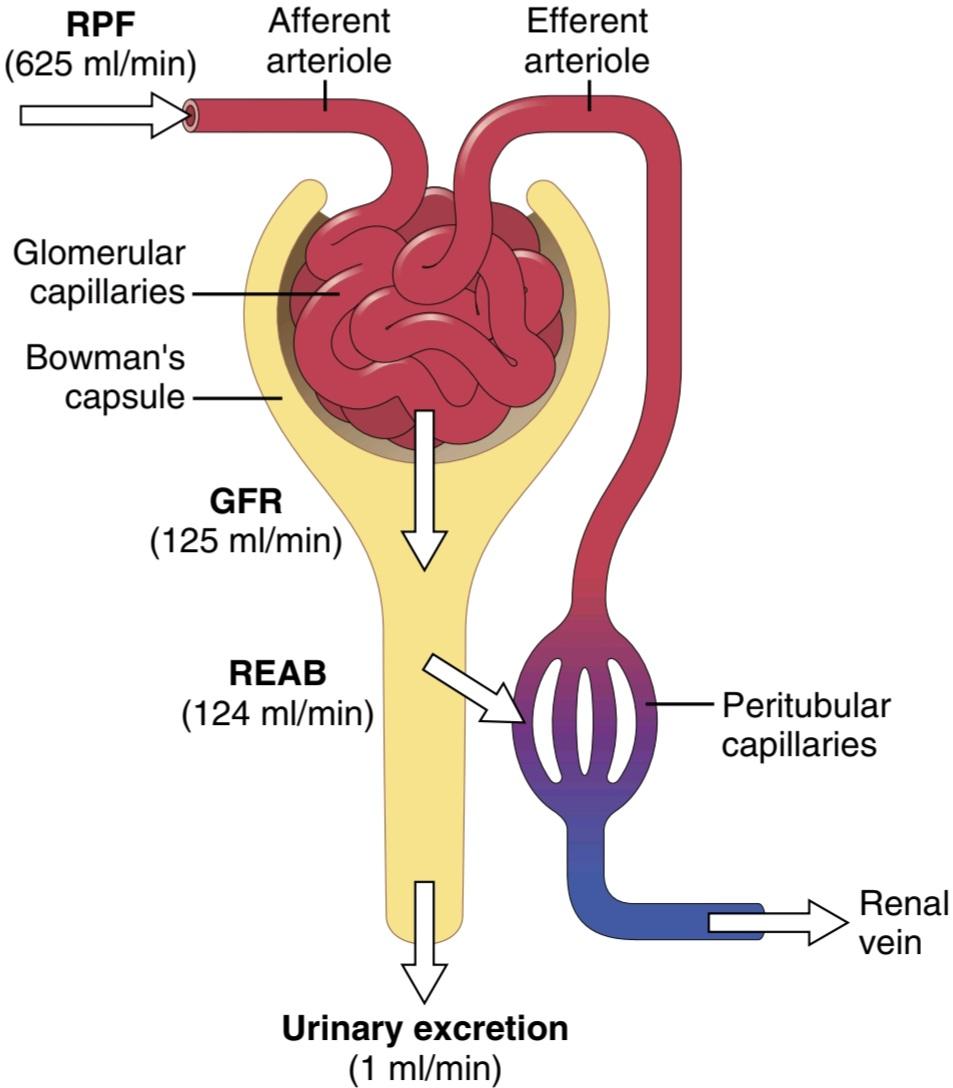
Figure 27-1. Average values for total renal plasma flow (RPF), glomerular filtration rate (GFR), tubular reabsorption (REAB), and urine flow rate. RPF is equal to renal blood flow x (1 – hematocrit). Note that the GFR averages about 20% of the RPF, whereas urine flow rate is less than 1% of the GFR. Therefore, more than 99% of the fluid filtered is normally reabsorbed. The filtration fraction is GFR/RPF.
شکل 27-1. مقادیر متوسط برای جریان کل پلاسمای کلیوی (RPF)، نرخ فیلتراسیون گلومرولی (GFR)، بازجذب لوله ای (REAB) و سرعت جریان ادرار. RPF برابر است با جریان خون کلیوی x (1 – هماتوکریت). توجه داشته باشید که GFR به طور متوسط حدود 20٪ RPF است، در حالی که سرعت جریان ادرار کمتر از 1٪ GFR است. بنابراین، بیش از 99٪ از مایع فیلتر شده به طور معمول دوباره جذب می شود. کسر فیلتراسیون GFR/RPF است.

Figure 27-2. A, Basic ultrastructure of the glomerular capillaries. B, Cross section of the glomerular capillary membrane and its major components: capillary endothelium, basement membrane, and epithelium (podocytes).
شکل 27-2. الف، فراساختار اساسی مویرگهای گلومرولی. ب، مقطع غشای مویرگی گلومرولی و اجزای اصلی آن: اندوتلیوم مویرگی، غشای پایه و اپیتلیوم (پودوسیت ها).
Filterability of Solutes Inversely Related to Their Size. The glomerular capillary membrane is thicker than most other capillaries, but it is also much more porous and therefore filters fluid at a high rate. Despite the high filtration rate, the glomerular filtration barrier is selective in determining which molecules will be filtered, based on their size and electrical charge.
قابلیت فیلترپذیری املاح با اندازه آنها رابطه معکوس دارد. غشای مویرگی گلومرولی ضخیمتر از سایر مویرگها است، اما همچنین بسیار متخلخلتر است و بنابراین مایع را با سرعت بالایی فیلتر میکند. علیرغم نرخ بالای فیلتراسیون، سد فیلتراسیون گلومرولی در تعیین اینکه کدام مولکول ها فیلتر می شوند، بر اساس اندازه و بار الکتریکی آنها انتخابی است.
Table 27-1 lists the effect of molecular size on filterability of different molecules. A filterability of 1.0 means that the substance is filtered as freely as water, whereas a filterability of 0.75 means that the substance is filtered only 75% as rapidly as water. Note that electrolytes such as sodium and small organic compounds such as glucose are freely filtered. As the molecular weight of the molecule approaches that of albumin, the filterability rapidly decreases, approaching zero.
جدول 27-1 اثر اندازه مولکولی را بر قابلیت فیلتر مولکول های مختلف فهرست می کند. فیلترپذیری 1.0 به این معنی است که این ماده به اندازه آب آزادانه فیلتر می شود، در حالی که فیلتر پذیری 0.75 به این معنی است که ماده تنها 75٪ به همان سرعت آب فیلتر می شود. توجه داشته باشید که الکترولیت هایی مانند سدیم و ترکیبات آلی کوچک مانند گلوکز آزادانه فیلتر می شوند. همانطور که وزن مولکولی مولکول به وزن آلبومین نزدیک می شود، فیلترپذیری به سرعت کاهش می یابد و به صفر نزدیک می شود.
Table 27-1 Filterability of Substances by Glomerular Capillaries Based on Molecular Weight.
جدول 27-1 قابلیت فیلتراسیون مواد بر اساس مویرگ های گلومرولی بر اساس وزن مولکولی.

Negatively Charged Large Molecules Are Filtered Less Easily Than Positively Charged Molecules of Equal Molecular Size. The molecular diameter of the plasma protein albumin is only about 6 nanometers, whereas the pores of the glomerular membrane are thought to be about 8 nanometers (80 angstroms [Å]). Albumin is restricted from filtration, however, because of its negative charge and the electrostatic repulsion exerted by negative charges of the glomerular capillary wall proteoglycans.
مولکول های بزرگ با بار منفی کمتر از مولکول های دارای بار مثبت با اندازه مولکولی برابر فیلتر می شوند. قطر مولکولی آلبومین پروتئین پلاسما تنها حدود 6 نانومتر است، در حالی که منافذ غشای گلومرولی حدود 8 نانومتر (80 آنگستروم [Å]) تصور میشود. با این حال، آلبومین به دلیل بار منفی و دافعه الکترواستاتیکی که توسط بارهای منفی پروتئوگلیکان های دیواره مویرگی گلومرولی اعمال می شود، از فیلتراسیون محدود می شود.
Figure 27-3 shows how electrical charge affects the filtration of different molecular weight dextrans by the glomerulus. Dextrans are polysaccharides that can be manufactured as neutral molecules or with negative or positive charges. Note that for any given molecular radius, positively charged molecules are filtered much more readily than negatively charged molecules. Neutral dextrans are also filtered more readily than negatively charged dextrans of equal molecular weight. The reason for these differences in filterability is that the negative charges of the basement membrane and podo- cytes provide an important means for restricting large negatively charged molecules, including the plasma proteins.
شکل 27-3 نشان می دهد که چگونه بار الکتریکی بر فیلتراسیون دکستران با وزن مولکولی مختلف توسط گلومرول تأثیر می گذارد. دکسترانس ها پلی ساکاریدهایی هستند که می توانند به عنوان مولکول های خنثی یا با بارهای منفی یا مثبت تولید شوند. توجه داشته باشید که برای هر شعاع مولکولی معین، مولکول های دارای بار مثبت بسیار راحت تر از مولکول های دارای بار منفی فیلتر می شوند. دکستران های خنثی نیز راحت تر از دکستران های دارای بار منفی با وزن مولکولی برابر فیلتر می شوند. دلیل این تفاوتها در فیلترپذیری این است که بارهای منفی غشای پایه و سلولهای پودوسیت وسیله مهمی برای محدود کردن مولکولهای باردار منفی بزرگ از جمله پروتئینهای پلاسما هستند.

Figure 27-3. Effect of molecular radius and electrical charge of dextran on its filterability by the glomerular capillaries. A value of 1.0 indicates that the substance is filtered as freely as water, whereas a value of O indicates that it is not filtered. Dextrans are polysaccharides that can be manufactured as neutral molecules or with negative or positive charges and with varying molecular weights.
شکل 27-3. تأثیر شعاع مولکولی و بار الکتریکی دکستران بر قابلیت فیلترپذیری آن توسط مویرگ های گلومرولی. مقدار 1.0 نشان می دهد که این ماده به اندازه آب آزادانه فیلتر می شود، در حالی که مقدار O نشان می دهد که فیلتر نشده است. دکسترانس ها پلی ساکاریدهایی هستند که می توانند به عنوان مولکول های خنثی یا با بارهای منفی یا مثبت و با وزن های مولکولی متفاوت تولید شوند.
Minimal-Change Nephropathy and Increased Glomerular Permeability to Plasma Proteins. In minimal- change nephropathy, the glomeruli become more permeable to plasma proteins, even though they may look normal when viewed with a standard light microscope. However, when viewed at high magnification with an electron microscope, the glomeruli usually display flattened podocytes with foot processes that may be detached from the glomerular basement membrane (podocyte effacement).
نفروپاتی با حداقل تغییر و افزایش نفوذپذیری گلومرولی به پروتئین های پلاسما. در نفروپاتی با حداقل تغییر، گلومرولها نسبت به پروتئینهای پلاسما نفوذپذیرتر میشوند، حتی اگر با میکروسکوپ نوری استاندارد مشاهده شوند، ممکن است طبیعی به نظر برسند. با این حال، هنگامی که با بزرگنمایی بالا با میکروسکوپ الکترونی مشاهده میشود، گلومرولها معمولاً پودوسیتهای مسطح با فرآیندهای پا را نشان میدهند که ممکن است از غشای پایه گلومرولی جدا شوند (پودوسیتها).
The causes of minimal change nephropathy are unclear but may be at least partly related to an immunological response and abnormal T-cell secretion of cytokines that injure the podocytes and increase their permeability to some of the lower molecular weight proteins, especially albumin. This increased permeability permits the proteins to be filtered by the glomerular capillaries and excreted in the urine, a condition known as proteinuria or albuminuria. Minimal change nephropathy is most common in young children but can also occur in adults, especially in those who have auto- immune disorders.
علل نفروپاتی با حداقل تغییر نامشخص است، اما ممکن است حداقل تا حدی مربوط به پاسخ ایمنی و ترشح غیرطبیعی سلول های T سیتوکین ها باشد که به پودوسیت ها آسیب می رساند و نفوذپذیری آنها را به برخی از پروتئین های با وزن مولکولی پایین، به ویژه آلبومین افزایش می دهد. این افزایش نفوذپذیری به پروتئین ها اجازه می دهد تا توسط مویرگ های گلومرولی فیلتر شده و از طریق ادرار دفع شوند، وضعیتی که به عنوان پروتئینوری یا آلبومینوری شناخته می شود. نفروپاتی با تغییر حداقلی در کودکان خردسال شایعتر است، اما میتواند در بزرگسالان نیز رخ دهد، بهویژه در افرادی که دارای اختلالات خودایمنی هستند.
DETERMINANTS OF THE GLOMERULAR FILTRATION RATE
The GFR is determined by the following: (1) the sum of the hydrostatic and colloid osmotic forces across the glomerular membrane, which gives the net filtration pressure; and (2) the glomerular K. Expressed mathematically, the GFR equals the product of K and the net filtration pressure:
GFR= KF x Net filtration pressure:
عوامل تعیین کننده نرخ فیلتراسیون گلومرولی
GFR با موارد زیر تعیین می شود: (1) مجموع نیروهای اسمزی هیدرواستاتیک و کلوئیدی در سراسر غشای گلومرولی، که فشار خالص فیلتر را می دهد. و (2) K گلومرولی. با بیان ریاضی، GFR برابر است با حاصلضرب K و فشار خالص فیلتر:

The net filtration pressure represents the sum of the hydrostatic and colloid osmotic forces that favor or oppose filtration across the glomerular capillaries (Figure 27-4). These forces include the following: (1) hydrostatic pressure inside the glomerular capillaries (glomerular hydrostatic pressure, PG), which promotes filtration; (2) the hydrostatic pressure in Bowman’s capsule (PB) outside the capillaries, which opposes filtration; (3) the colloid osmotic pressure of the glomerular capillary plasma proteins (G), which opposes filtration; and (4) the colloid osmotic pressure of the proteins in Bowman’s capsule (B), which promotes filtration. Under normal conditions, the concentration of protein in the glomerular filtrate is so low that the colloid osmotic pressure of the Bowman’s capsule fluid is considered to be zero.
فشار خالص فیلتراسیون مجموع نیروهای اسمزی هیدرواستاتیک و کلوئیدی را نشان می دهد که به نفع یا مخالف فیلتراسیون در سراسر مویرگ های گلومرولی هستند (شکل 27-4). این نیروها شامل موارد زیر است: (1) فشار هیدرواستاتیک در داخل مویرگهای گلومرولی (فشار هیدرواستاتیک گلومرولی، PG)، که باعث افزایش فیلتراسیون می شود. (2) فشار هیدرواستاتیک در کپسول بومن (PB) خارج از مویرگها، که با فیلتراسیون مخالف است. (3) فشار اسمزی کلوئیدی پروتئین های پلاسمای مویرگی گلومرولی (G) که با فیلتراسیون مخالفت می کند. و (4) فشار اسمزی کلوئیدی پروتئین ها در کپسول بومن (B) که باعث افزایش فیلتراسیون می شود. در شرایط عادی، غلظت پروتئین در فیلتر گلومرولی آنقدر کم است که فشار اسمزی کلوئیدی مایع کپسول بومن صفر در نظر گرفته می شود.
The GFR can therefore be expressed as follows:
بنابراین GFR را می توان به صورت زیر بیان کرد:

Although the normal values for the determinants of GFR have not been measured directly in humans, they have been estimated in animals such as dogs and rats. Based on the results in experimental animals, the approximate normal forces favoring and opposing glomerular filtration in humans are believed to be as follows (see Figure 27-4):
اگرچه مقادیر طبیعی برای عوامل تعیین کننده GFR به طور مستقیم در انسان اندازه گیری نشده است، اما در حیواناتی مانند سگ و موش تخمین زده شده است. بر اساس نتایج در حیوانات آزمایشی، نیروهای طبیعی تقریبی که طرفدار و مخالف فیلتراسیون گلومرولی در انسان هستند، به شرح زیر است (شکل 27-4 را ببینید):

Thus, the net filtration pressure = 60-18-32 = +10 mm Hg.
بنابراین، فشار خالص فیلتر = 60-18-32 = +10 میلی متر جیوه.
Some of these values can change markedly under different physiological conditions, whereas others are altered mainly in disease states, as discussed later.
برخی از این مقادیر می توانند تحت شرایط فیزیولوژیکی مختلف به طور قابل توجهی تغییر کنند، در حالی که برخی دیگر عمدتاً در حالت های بیماری تغییر می کنند، همانطور که بعداً بحث شد.
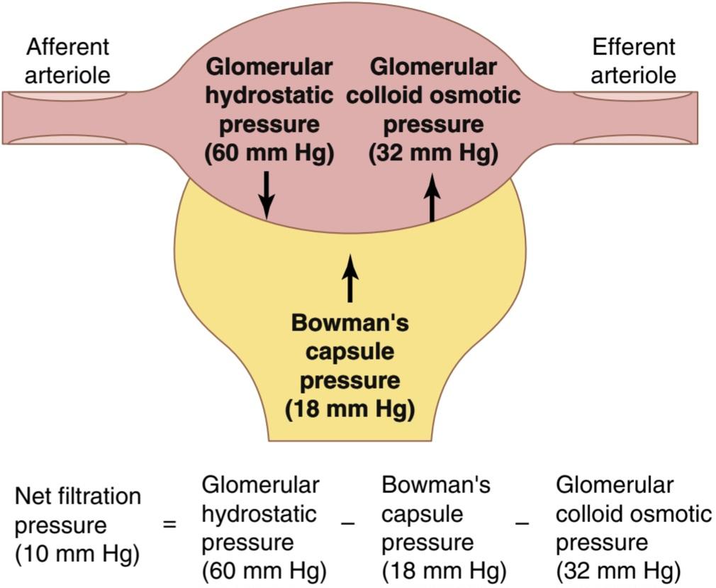
Figure 27-4. Summary of forces causing filtration by the glomerular capillaries. The values shown are estimates for healthy humans.
شکل 27-4. خلاصه نیروهایی که باعث فیلتراسیون توسط مویرگ های گلومرولی می شوند. مقادیر نشان داده شده تخمینی برای انسان های سالم است.
INCREASED GLOMERULAR CAPILLARY FILTRATION COEFFICIENT INCREASES GLOMERULAR FILTRATE RATE
The Kf is a measure of the product of the hydraulic con-ductivity and surface area of the glomerular capillaries. The Kf cannot be measured directly, but can be is estimated experimentally by dividing the GFR by the net filtration pressure:
افزایش ضریب فیلتراسیون مویرگی گلومرولار میزان فیلتراسیون گلومرولی را افزایش می دهد
K معیاری از محصول رسانایی هیدرولیکی و مساحت سطح مویرگ های گلومرولی است. Kf را نمی توان مستقیماً اندازه گیری کرد، اما می توان آن را به صورت تجربی با تقسیم GFR بر فشار خالص تصفیه تخمین زد:

Because the total GFR for both kidneys is about 125 ml/min, and the net filtration pressure is 10 mm Hg, the normal Kf is calculated to be about 12.5 ml/min per mm Hg of filtration pressure. When Kf is expressed per 100 grams of kidney weight, it averages about 4.2 ml/min per mm Hg, a value about 400 times as high as the Kf of most other capillary systems of the body. The average Kf of many other tissues in the body is only about 0.01 ml/min per mm Hg/100 g. This high Kf for the glomerular capillaries contributes to their rapid rate of fluid filtration.
از آنجایی که کل GFR برای هر دو کلیه حدود 125 میلی لیتر در دقیقه و فشار خالص فیلتراسیون 10 میلی متر جیوه است، K طبیعی حدود 12.5 میلی لیتر در دقیقه در هر میلی متر جیوه فشار فیلتراسیون محاسبه می شود. هنگامی که Kf به ازای هر 100 گرم وزن کلیه بیان می شود، به طور متوسط حدود 4.2 میلی لیتر در دقیقه در میلی متر جیوه است، مقداری که حدود 400 برابر بیشتر از Kf در اکثر سیستم های مویرگی بدن است. میانگین پتاسیم بسیاری از بافت های دیگر بدن تنها حدود 0.01 میلی لیتر در دقیقه در هر میلی متر جیوه در 100 گرم است. این Kf بالا برای مویرگ های گلومرولی به سرعت سریع فیلتراسیون مایع کمک می کند.
Although increased Kf raises the GFR and decreased Kf reduces the GFR, changes in Kf probably do not provide a primary mechanism for the normal daily regulation of GFR. Some diseases, however, lower Kf by reducing the number of functional glomerular capillaries (thereby reducing the surface area for filtration) or by increasing the thickness of the glomerular capillary membrane and reducing its hydraulic conductivity. For example, chronic uncontrolled hypertension may gradually reduce Kf by increasing the thickness of the glomerular capillary basement membrane and, eventually, by damaging the capillaries so severely that there is loss of capillary function.
اگرچه افزایش K باعث افزایش GFR و کاهش Kf باعث کاهش GFR می شود، تغییرات در Kf احتمالاً مکانیسم اولیه ای برای تنظیم طبیعی روزانه GFR ارائه نمی دهد. با این حال، برخی بیماری ها با کاهش تعداد مویرگ های گلومرولی عملکردی (در نتیجه کاهش سطح برای فیلتراسیون) یا با افزایش ضخامت غشای مویرگی گلومرولی و کاهش هدایت هیدرولیکی آن، پتاسیم را کاهش می دهند. به عنوان مثال، فشار خون بالا کنترل نشده مزمن ممکن است با افزایش ضخامت غشای پایه مویرگی گلومرولی و در نهایت با آسیب شدید به مویرگ ها که عملکرد مویرگی را از دست می دهد، Kf را به تدریج کاهش دهد.
INCREASED BOWMAN’S CAPSULE HYDROSTATIC PRESSURE DECREASES GLOMERULAR FILTRATION RATE
Direct measurements of hydrostatic pressure in Bow- man’s capsule and at different points in the proximal tubule in experimental animals using micropipettes have suggested that a reasonable estimate for Bowman’s capsule pressure in humans is about 18 mm Hg under normal conditions. Increasing the hydrostatic pressure in Bow- man’s capsule reduces GFR, whereas decreasing this pressure raises GFR. However, changes in Bowman’s capsule pressure normally do not serve as a primary means for regulating GFR.
افزایش فشار هیدرواستاتیک کپسول باومن میزان فیلتراسیون گلومرولار را کاهش می دهد
اندازه گیری مستقیم فشار هیدرواستاتیک در کپسول بومن و در نقاط مختلف لوله پروگزیمال در حیوانات آزمایشگاهی با استفاده از میکروپیپت ها نشان می دهد که یک تخمین معقول برای فشار کپسول بومن در انسان حدود 18 میلی متر جیوه در شرایط عادی است. افزایش فشار هیدرواستاتیک در کپسول Bowman’s GFR را کاهش می دهد، در حالی که کاهش این فشار باعث افزایش GFR می شود. با این حال، تغییرات در فشار کپسول بومن به طور معمول به عنوان یک وسیله اولیه برای تنظیم GFR عمل نمی کند.
In certain pathological states associated with obstruction of the urinary tract, Bowman’s capsule pressure can increase markedly, causing serious reduction of GFR. For example, precipitation of calcium or of uric acid may lead to formation of stones that lodge in the urinary tract, often in the ureter, thereby obstructing outflow of the urinary tract and raising Bowman’s capsule pressure. This situation reduces GFR and eventually can cause hydronephrosis (distention and dilation of the renal pelvis and calyces) and can damage or even destroy the kidney unless the obstruction is relieved.
در برخی از حالات پاتولوژیک مرتبط با انسداد مجاری ادراری، فشار کپسول بومن به طور قابل توجهی افزایش می یابد و باعث کاهش جدی GFR می شود. به عنوان مثال، رسوب کلسیم یا اسید اوریک ممکن است منجر به تشکیل سنگهایی شود که در مجرای ادراری، اغلب در حالب قرار میگیرند، در نتیجه مانع خروج مجرای ادرار و افزایش فشار کپسول بومن میشوند. این وضعیت باعث کاهش GFR می شود و در نهایت می تواند باعث هیدرونفروز (اتساع و گشاد شدن لگن کلیه و کالیس ها) شود و می تواند به کلیه آسیب برساند یا حتی از بین ببرد مگر اینکه انسداد برطرف شود.

Figure 27-5. Increase in colloid osmotic pressure in plasma flowing through the glomerular capillary. Normally, about one-fifth of the fluid in the glomerular capillaries filters into Bowman’s capsule, there- by concentrating the plasma proteins that are not filtered. Increases in the filtration fraction (glomerular filtration rate/renal plasma flow) increase the rate at which the plasma colloid osmotic pressure rises along the glomerular capillary; decreases in the filtration fraction have the opposite effect.
شکل 27-5. افزایش فشار اسمزی کلوئیدی در پلاسمایی که در مویرگ گلومرولی جریان دارد. به طور معمول، حدود یک پنجم مایع موجود در مویرگ های گلومرولی، با تمرکز پروتئین های پلاسما که فیلتر نشده اند، به داخل کپسول بومن فیلتر می شود. افزایش کسر فیلتراسیون (سرعت فیلتراسیون گلومرولی/جریان پلاسمای کلیوی) باعث افزایش سرعت افزایش فشار اسمزی کلوئیدی پلاسما در طول مویرگ گلومرولی می شود. کاهش در کسر فیلتراسیون اثر معکوس دارد.
INCREASED GLOMERULAR CAPILLARY COLLOID OSMOTIC PRESSURE DECREASES GLOMERULAR FILTRATION RATE
As blood passes from the afferent arteriole through the glomerular capillaries to the efferent arterioles, the plasma protein concentration increases about 20% (Figure 27-5). The reason for this increase is that about onefifth of the fluid in the capillaries filters into Bowman’s capsule, thereby concentrating the glomerular plasma proteins that are not filtered. Assuming that the normal colloid osmotic pressure of plasma entering the glomerular capillaries is 28 mm Hg, this value usually rises to about 36 mm Hg by the time the blood reaches the efferent end of the capillaries. Therefore, the aver- age colloid osmotic pressure of the glomerular capillary plasma proteins is midway between 28 and 36 mm Hg, or about 32 mm Hg.
افزایش فشار اسمموتیک کلوئید مویرگی گلومرولار میزان فیلتراسیون گلومرولی را کاهش می دهد
با عبور خون از شریان آوران از طریق مویرگ های گلومرولی به شریان های وابران، غلظت پروتئین پلاسما حدود 20 درصد افزایش می یابد (شکل 27-5). دلیل این افزایش این است که حدود یک پنجم مایع موجود در مویرگ ها به داخل کپسول بومن فیلتر می شود و در نتیجه پروتئین های پلاسمای گلومرولی که فیلتر نشده اند متمرکز می شود. با فرض اینکه فشار اسمزی کلوئیدی طبیعی پلاسمای ورودی به مویرگ های گلومرولی 28 میلی متر جیوه باشد، این مقدار معمولاً تا زمانی که خون به انتهای وابران مویرگ ها می رسد به حدود 36 میلی متر جیوه می رسد. بنابراین، میانگین فشار اسمزی کلوئیدی پروتئین های پلاسمای مویرگی گلومرولی در میانه راه بین 28 تا 36 میلی متر جیوه یا حدود 32 میلی متر جیوه است.
Two factors that influence the glomerular capillary colloid osmotic pressure are the following: (1) the arterial plasma colloid osmotic pressure; and (2) the fraction of plasma filtered by the glomerular capillaries (filtration fraction). Increasing the arterial plasma colloid osmotic pressure raises the glomerular capillary colloid osmotic pressure, which in turn tends to decrease the GFR.
دو عاملی که بر فشار اسمزی کلوئیدی مویرگی گلومرولی تأثیر می گذارند عبارتند از: (1) فشار اسمزی کلوئیدی پلاسمای شریانی. و (2) کسری از پلاسما فیلتر شده توسط مویرگ های گلومرولی (کسری فیلتراسیون). افزایش فشار اسمزی کلوئیدی پلاسمای شریانی باعث افزایش فشار اسمزی کلوئیدی مویرگی گلومرولی می شود که به نوبه خود تمایل به کاهش GFR دارد.
Increasing the filtration fraction also concentrates the plasma proteins and raises the glomerular colloid osmotic pressure (see Figure 27-5). Because the filtration fraction is defined as the GFR divided by the renal plasma flow, the filtration fraction can be increased by raising the GFR or by reducing renal plasma flow. For example, a reduction in renal plasma flow with no initial change in GFR would tend to increase the filtration fraction, which would raise the glomerular capillary colloid osmotic pressure and tend to reduce the GFR. For this reason, changes in renal blood flow can influence GFR independently of changes in glomerular hydrostatic pressure.
افزایش کسر فیلتراسیون پروتئین های پلاسما را نیز متمرکز می کند و فشار اسمزی کلوئیدی گلومرولی را افزایش می دهد (شکل 27-5 را ببینید). از آنجایی که کسر فیلتراسیون به عنوان GFR تقسیم بر جریان پلاسمای کلیوی تعریف می شود، کسر فیلتراسیون را می توان با افزایش GFR یا با کاهش جریان پلاسمای کلیوی افزایش داد. به عنوان مثال، کاهش در جریان پلاسمای کلیوی بدون تغییر اولیه در GFR باعث افزایش کسر فیلتراسیون می شود که فشار اسمزی کلوئیدی مویرگی گلومرولی را افزایش می دهد و تمایل به کاهش GFR دارد. به همین دلیل، تغییرات در جریان خون کلیوی میتواند مستقل از تغییرات فشار هیدرواستاتیک گلومرولی بر GFR تأثیر بگذارد.
With increasing renal blood flow, a lower fraction of the plasma is initially filtered out of the glomerular capillaries, causing a slower rise in the glomerular capillary colloid osmotic pressure and less inhibitory effect on the GFR. Consequently, even with a constant glomerular hydrostatic pressure, a greater rate of blood flow into the glomerulus tends to increase the GFR and a lower rate of blood flow into the glomerulus tends to decrease the GFR.
با افزایش جریان خون کلیوی، بخش پایینتری از پلاسما در ابتدا از مویرگهای گلومرولی فیلتر میشود و باعث افزایش آهستهتر فشار اسمزی کلوئیدی مویرگی گلومرولی و اثر بازدارندگی کمتری بر GFR میشود. در نتیجه، حتی با فشار هیدرواستاتیک گلومرولی ثابت، سرعت بیشتر جریان خون در گلومرول باعث افزایش GFR و سرعت کمتر جریان خون به گلومرول باعث کاهش GFR می شود.
INCREASED GLOMERULAR CAPILLARY HYDROSTATIC PRESSURE INCREASES GLOMERULAR FILTRATION RATE
The glomerular capillary hydrostatic pressure has been estimated to be about 60 mm Hg under normal conditions. Changes in glomerular hydrostatic pressure serve as the primary means for physiological regulation of GFR. Increases in glomerular hydrostatic pressure raise the GFR, whereas decreases in glomerular hydrostatic pressure reduce the GFR.
افزایش فشار هیدرواستاتیک مویرگی گلومرولی نرخ فیلتراسیون گلومرولی را افزایش می دهد
فشار هیدرواستاتیک مویرگی گلومرولی در شرایط عادی حدود 60 میلی متر جیوه برآورد شده است. تغییرات در فشار هیدرواستاتیک گلومرولی به عنوان وسیله اولیه برای تنظیم فیزیولوژیکی GFR عمل می کند. افزایش فشار هیدرواستاتیک گلومرولی باعث افزایش GFR می شود، در حالی که کاهش فشار هیدرواستاتیک گلومرولی باعث کاهش GFR می شود.
Glomerular hydrostatic pressure is determined by three variables, each of which is under physiological control: (1) arterial pressure; (2) afferent arteriolar resistance; and (3) efferent arteriolar resistance.
فشار هیدرواستاتیک گلومرولی توسط سه متغیر تعیین می شود که هر کدام تحت کنترل فیزیولوژیکی هستند: (1) فشار شریانی. (2) مقاومت شریانی آوران؛ و (3) مقاومت شریانی وابران.
Increased arterial pressure tends to raise glomerular hydrostatic pressure and, therefore, to increase the GFR. However, as discussed later, this effect is buffered by auto- regulatory mechanisms that maintain a relatively constant glomerular pressure as arterial pressure fluctuates.
افزایش فشار شریانی تمایل به افزایش فشار هیدرواستاتیک گلومرولی و در نتیجه افزایش GFR دارد. با این حال، همانطور که بعداً بحث شد، این اثر توسط مکانیسمهای خودتنظیمی که فشار گلومرولی نسبتاً ثابتی را با نوسانات فشار شریانی حفظ میکند، بافر میشود.
Increased resistance of afferent arterioles reduces glomerular hydrostatic pressure and decreases the GFR (Figure 27-6). Conversely, dilation of the afferent arterioles increases glomerular hydrostatic pressure and GFR.
افزایش مقاومت شریان های آوران فشار هیدرواستاتیک گلومرولی را کاهش می دهد و GFR را کاهش می دهد (شکل 27-6). برعکس، اتساع شریان های آوران فشار هیدرواستاتیک گلومرولی و GFR را افزایش می دهد.
Constriction of the efferent arterioles increases the resistance to outflow from the glomerular capillaries. This mechanism raises glomerular hydrostatic pressure and, as long as the increase in efferent resistance does not reduce renal blood flow too much, GFR increases slightly (see Figure 27-6). However, because efferent arteriolar constriction also reduces renal blood flow, filtration fraction and glomerular colloid osmotic pressure increase as efferent arteriolar resistance increases. Therefore, if constriction of efferent arterioles is severe (more than about a threefold increase in efferent arteriolar resistance), the rise in colloid osmotic pressure exceeds the increase in glomerular capillary hydrostatic pressure caused by efferent arteriolar constriction. When this situation occurs, the net force for filtration actually decreases, causing a reduction in GFR.
انقباض شریان های وابران مقاومت در برابر خروج از مویرگ های گلومرولی را افزایش می دهد. این مکانیسم فشار هیدرواستاتیک گلومرولی را افزایش می دهد و تا زمانی که افزایش مقاومت وابران باعث کاهش بیش از حد جریان خون کلیوی نشود، GFR اندکی افزایش می یابد (شکل 27-6 را ببینید). با این حال، از آنجا که انقباض شریانی وابران جریان خون کلیوی را نیز کاهش می دهد، با افزایش مقاومت شریان وابران، بخش فیلتراسیون و فشار اسمزی کلوئیدی گلومرولی افزایش می یابد. بنابراین، اگر انقباض شریانهای وابران شدید باشد (بیش از حدود سه برابر افزایش مقاومت شریانی وابران)، افزایش فشار اسمزی کلوئیدی از افزایش فشار هیدرواستاتیک مویرگی گلومرولی ناشی از انقباض شریان وابران بیشتر است. هنگامی که این وضعیت رخ می دهد، نیروی خالص برای فیلتراسیون در واقع کاهش می یابد و باعث کاهش GFR می شود.
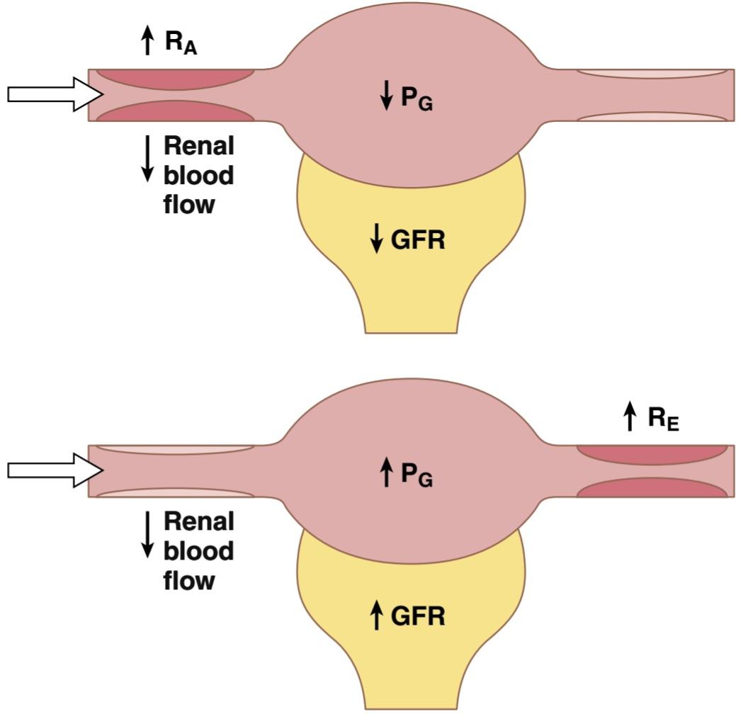
Figure 27-6. Effect of increases in afferent arteriolar resistance (RA, top panel) or efferent arteriolar resistance (RE, bottom panel) on renal blood flow, glomerular hydrostatic pressure (PG), and glomerular filtration rate (GFR).
شکل 27-6. اثر افزایش مقاومت شریانی آوران (RA، پانل بالا) یا مقاومت شریان وابران (RE، پانل پایین) بر جریان خون کلیوی، فشار هیدرواستاتیک گلومرولی (PG) و نرخ فیلتراسیون گلومرولی (GFR).
Thus, efferent arteriolar constriction has a biphasic effect on GFR (Figure 27-7). At moderate levels of constriction, there is a slight increase in GFR but, with severe constriction, there is a decrease in GFR. The primary cause of the eventual decrease in GFR is as follows. As efferent constriction becomes severe, and as plasma protein concentration increases, there is a rapid nonlinear increase in colloid osmotic pressure caused by the Donnan effect; the higher the protein concentration, the more rapidly the colloid osmotic pressure rises because of the interaction of ions bound to the plasma proteins, which also exert an osmotic effect, as discussed in Chapter 16.
بنابراین، انقباض شریان وابران اثر دوفازی بر GFR دارد (شکل 27-7). در سطوح متوسط انقباض، افزایش جزئی در GFR وجود دارد، اما با انقباض شدید، GFR کاهش می یابد. علت اصلی کاهش نهایی GFR به شرح زیر است. همانطور که انقباض وابران شدید می شود، و با افزایش غلظت پروتئین پلاسما، افزایش غیرخطی سریع فشار اسمزی کلوئیدی ناشی از اثر دانان وجود دارد. هر چه غلظت پروتئین بالاتر باشد، فشار اسمزی کلوئیدی به دلیل برهمکنش یونهای متصل به پروتئینهای پلاسما، که در فصل 16 مورد بحث قرار گرفته است، با سرعت بیشتری افزایش مییابد.

Figure 27-7. Effect of change in afferent arteriolar resistance or efferent arteriolar resistance on glomerular filtration rate and renal blood flow.
شکل 27-7. تاثیر تغییر در مقاومت شریانی آوران یا مقاومت شریانی وابران بر میزان فیلتراسیون گلومرولی و جریان خون کلیوی.
To summarize, constriction of afferent arterioles reduces GFR. However, the effect of efferent arteriolar constriction depends on the severity of the constriction; modest efferent constriction raises GFR, but severe efferent constriction (more than a threefold increase in resistance) tends to reduce GFR.
به طور خلاصه، انقباض شریان های آوران باعث کاهش GFR می شود. با این حال، اثر انقباض شریانی وابران به شدت انقباض بستگی دارد. انقباض متوسط وابران GFR را افزایش می دهد، اما انقباض شدید وابران (بیش از سه برابر افزایش مقاومت) تمایل به کاهش GFR دارد.
Table 27-2 summarizes the factors that can decrease the GFR.
جدول 27-2 عواملی را که می توانند GFR را کاهش دهند، خلاصه می کند.
Table 27-2 Factors That Can Decrease the Glomerular Filtration Rate.
جدول 27-2 عواملی که می توانند میزان فیلتراسیون گلومرولی را کاهش دهند.

aOpposite changes in the determinants usually increase the GFR. Ap, Systemic arterial pressure; GFR, glomerular filtration rate; Kf, glomerular filtration coefficient; PB, Bowman’s capsule hydrostatic pressure; G, glomerular capillary colloid osmotic pressure; PG glomerular capillary hydrostatic pressure; RA, afferent arteriolar resistance; RE, efferent arteriolar resistance.
aتغییرات مخالف در تعیین کننده ها معمولا GFR را افزایش می دهد. AP، فشار شریانی سیستمیک؛ GFR، نرخ فیلتراسیون گلومرولی؛ Kf، ضریب فیلتراسیون گلومرولی. PB، فشار هیدرواستاتیک کپسول بومن. G، فشار اسمزی کلوئیدی مویرگی گلومرولی. فشار هیدرواستاتیک مویرگی گلومرولی PG; RA، مقاومت شریانی آوران؛ RE، مقاومت شریانی وابران.
RENAL BLOOD FLOW
In a 70-kg man, the combined blood flow through both kidneys is about 1100 ml/min, or about 22% of the cardiac output. Considering that the two kidneys constitute only about 0.4% of the total body weight, one can readily see that they receive extremely high blood flow compared with other organs.
جریان خون کلیوی
در یک مرد 70 کیلوگرمی، جریان خون ترکیبی از هر دو کلیه حدود 1100 میلی لیتر در دقیقه یا حدود 22 درصد برون ده قلبی است. با توجه به اینکه دو کلیه تنها حدود 0.4 درصد از کل وزن بدن را تشکیل می دهند، به راحتی می توان دریافت که جریان خون بسیار بالایی را در مقایسه با سایر اندام ها دریافت می کنند.
As with other tissues, blood flow supplies the kidneys with nutrients and removes waste products. However, the high blood flow to the kidneys greatly exceeds this need. The purpose of this additional flow is to supply enough plasma for the high rates of glomerular filtration that are necessary for precise regulation of body fluid volumes and solute concentrations. As might be expected, the mechanisms that regulate renal blood flow are closely linked to control of GFR and excretory functions of the kidneys.
مانند سایر بافت ها، جریان خون کلیه ها را با مواد مغذی تامین می کند و مواد زائد را حذف می کند. با این حال، جریان خون بالا به کلیه ها بسیار فراتر از این نیاز است. هدف از این جریان اضافی تامین پلاسما کافی برای سرعت بالای فیلتراسیون گلومرولی است که برای تنظیم دقیق حجم مایعات بدن و غلظت املاح ضروری است. همانطور که انتظار می رود، مکانیسم هایی که جریان خون کلیوی را تنظیم می کنند با کنترل GFR و عملکردهای دفعی کلیه ها ارتباط نزدیکی دارند.
RENAL BLOOD FLOW AND OXYGEN CONSUMPTION
On a per gram-weight basis, the kidneys normally consume oxygen at twice the rate of the brain but have almost seven times the blood flow of the brain. Thus, the oxygen delivered to the kidneys far exceeds their metabolic needs, and the arterial-venous extraction of oxygen is relatively low compared with that of most other tissues.
جریان خون کلیوی و مصرف اکسیژن
بر اساس هر گرم وزن، کلیه ها معمولاً دو برابر مغز اکسیژن مصرف می کنند، اما تقریباً هفت برابر بیشتر از مغز جریان خون دارند. بنابراین، اکسیژن تحویلی به کلیهها بسیار فراتر از نیازهای متابولیکی آنها است و استخراج اکسیژن شریانی-وریدی در مقایسه با اکثر بافتهای دیگر نسبتاً کم است.
A large fraction of the oxygen consumed by the kidneys is related to the high rate of active sodium reabsorption by the renal tubules. If renal blood flow and GFR are reduced, and less sodium is filtered, less sodium is reabsorbed and less oxygen is consumed. Therefore, renal oxygen consumption varies in proportion to renal tubular sodium reabsorption, which in turn is closely related to GFR and the rate of sodium filtered (Figure 27-8). If glomerular filtration ceases completely, renal sodium reabsorption also ceases and oxygen consumption decreases to about one-fourth normal. This residual oxygen consumption reflects the basic metabolic needs of the renal cells.
بخش زیادی از اکسیژن مصرف شده توسط کلیه ها به سرعت بالای بازجذب فعال سدیم توسط لوله های کلیوی مربوط می شود. اگر جریان خون کلیوی و GFR کاهش یابد و سدیم کمتری فیلتر شود، سدیم کمتری بازجذب شده و اکسیژن کمتری مصرف میشود. بنابراین، مصرف اکسیژن کلیوی متناسب با بازجذب سدیم توبولار کلیوی متفاوت است که به نوبه خود با GFR و سرعت فیلتر سدیم مرتبط است (شکل 27-8). اگر فیلتراسیون گلومرولی به طور کامل متوقف شود، بازجذب کلیوی سدیم نیز متوقف می شود و مصرف اکسیژن به حدود یک چهارم نرمال کاهش می یابد. این مصرف اکسیژن باقیمانده منعکس کننده نیازهای متابولیکی اساسی سلول های کلیوی است.
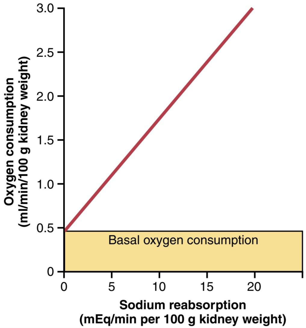
Figure 27-8. Relationship between oxygen consumption and sodium reabsorption in dog kidneys. (From Kramer K, Deetjen P: [Relation of renal oxygen consumption to blood supply and glomerular filtration during variations of blood pressure.] Pflugers Arch Physiol 271:782, 1960.)
شکل 27-8. رابطه بین مصرف اکسیژن و بازجذب سدیم در کلیه سگ. (از Kramer K، Deetjen P: [ارتباط مصرف اکسیژن کلیوی با تامین خون و فیلتراسیون گلومرولی در طول تغییرات فشار خون.] Pflugers Arch Physiol 271:782، 1960.)
DETERMINANTS OF RENAL BLOOD FLOW
Renal blood flow (RBF) is determined by the pressure gradient across the renal vasculature (the difference between renal artery and renal vein hydrostatic pressures), divided by the total renal vascular resistance:
عوامل تعیین کننده جریان خون کلیوی
جریان خون کلیوی (RBF) با گرادیان فشار در سراسر عروق کلیوی (تفاوت بین فشار هیدرواستاتیک شریان کلیوی و ورید کلیوی)، تقسیم بر مقاومت کلی عروق کلیوی تعیین می شود:

Renal artery pressure is about equal to systemic arterial pressure, and renal vein pressure averages about 3 to 4 mm Hg under most conditions. As in other vascular beds, the total vascular resistance through the kidneys is determined by the sum of the resistances in the individual vasculature segments, including the arteries, arterioles, capillaries, and veins (Table 27-3).
فشار شریان کلیوی تقریباً برابر با فشار شریانی سیستمیک است و فشار سیاهرگ کلیوی در اکثر شرایط به طور متوسط حدود 3 تا 4 میلی متر جیوه است. همانند سایر بسترهای عروقی، کل مقاومت عروقی از طریق کلیه ها با مجموع مقاومت ها در بخش های عروقی منفرد، از جمله شریان ها، شریان ها، مویرگ ها و سیاهرگ ها تعیین می شود (جدول 27-3).
Most of the renal vascular resistance resides in three major segments-interlobular arteries, afferent arterioles, and efferent arterioles. Resistance of these vessels is controlled by the sympathetic nervous system, various hormones, and local internal renal control mechanisms, as discussed later. An increase in the resistance of any of the vascular segments of the kidneys tends to reduce the renal blood flow, whereas a decrease in vascular resistance increases renal blood flow if renal artery and renal vein pressures remain constant.
بیشتر مقاومت عروق کلیوی در سه بخش اصلی شریان های بین لوبولی، شریان های آوران و شریان های وابران قرار دارد. مقاومت این عروق توسط سیستم عصبی سمپاتیک، هورمونهای مختلف و مکانیسمهای کنترل داخلی کلیوی محلی کنترل میشود، همانطور که بعداً بحث شد. افزایش مقاومت هر یک از بخش های عروقی کلیه ها باعث کاهش جریان خون کلیوی می شود، در حالی که کاهش مقاومت عروقی باعث افزایش جریان خون کلیوی می شود اگر فشار شریان کلیوی و ورید کلیوی ثابت بماند.
Although changes in arterial pressure have some influence on renal blood flow, the kidneys have effective mechanisms for maintaining renal blood flow and GFR relatively constant over an arterial pressure range between 80 and 170 mm Hg, a process called autoregulation. This capacity for autoregulation occurs through mechanisms that are intrinsic to the kidneys, as discussed later in this chapter.
اگرچه تغییرات فشار شریانی تا حدودی بر جریان خون کلیوی تأثیر می گذارد، کلیه ها مکانیسم های مؤثری برای حفظ جریان خون کلیوی و GFR نسبتاً ثابت در محدوده فشار شریانی بین 80 تا 170 میلی متر جیوه دارند که فرآیندی به نام تنظیم خودکار است. این ظرفیت خودتنظیمی از طریق مکانیسمهایی اتفاق میافتد که ذاتی کلیهها هستند، همانطور که بعداً در این فصل مورد بحث قرار گرفت.
Table 27-3 Approximate Pressures and Vascular Resistances in Circulation of a Normal Kidney
جدول 27-3 فشارهای تقریبی و مقاومت های عروقی در گردش یک کلیه طبیعی

BLOOD FLOW IN VASA RECTA OF RENAL MEDULLA IS LOW COMPARED WITH RENAL CORTEX FLOW
The outer part of the kidney, the renal cortex, receives most of the kidney’s blood flow. Blood flow in the renal medulla accounts for only 1% to 2% of the total renal blood flow. Flow to the renal medulla is supplied by a specialized portion of the peritubular capillary system called the vasa recta. These vessels descend into the medulla in parallel with the loops of Henle and then loop back along with the loops of Henle and return to the cortex before emptying into the venous system. As discussed in Chapter 29, the vasa recta an important role in allowing the kidneys to form concentrated urine.
جریان خون در VASA RECTA مدولای کلیه در مقایسه با جریان قشر کلیه کم است
قسمت بیرونی کلیه، قشر کلیه، بیشتر جریان خون کلیه را دریافت می کند. جریان خون در بصل النخاع کلیه تنها 1 تا 2 درصد از کل جریان خون کلیوی را تشکیل می دهد. جریان به بصل النخاع کلیه توسط بخش ویژه ای از سیستم مویرگ اطراف لوله ای به نام vasa recta تامین می شود. این رگ ها به موازات حلقه های هنله به داخل بصل النخاع فرود می آیند و سپس همراه با حلقه های هنله به سمت بصل می روند و قبل از تخلیه به سیستم وریدی به قشر باز می گردند. همانطور که در فصل 29 بحث شد، وازا رکتا نقش مهمی در اجازه دادن به کلیه ها برای تشکیل ادرار غلیظ دارد.
PHYSIOLOGICAL CONTROL OF GLOMERULAR FILTRATION AND RENAL BLOOD FLOW
The determinant of GFR that is most variable and subject to physiological control is the glomerular hydrostatic pressure. This variable, in turn, is influenced by the sympathetic nervous system, hormones, autacoids (vasoactive substances that are released in the kidneys and act locally), and other feedback controls that are intrinsic to the kidneys.
کنترل فیزیولوژیکی فیلتراسیون گلومرولی و جریان خون کلیوی
تعیین کننده GFR که متغیرترین است و تحت کنترل فیزیولوژیکی است، فشار هیدرواستاتیک گلومرولی است. این متغیر به نوبه خود تحت تأثیر سیستم عصبی سمپاتیک، هورمونها، اتاکوئیدها (مواد وازواکتیو که در کلیهها آزاد میشوند و به صورت موضعی عمل میکنند) و سایر کنترلهای بازخوردی که ذاتی کلیهها هستند، میشود.
STRONG SYMPATHETIC NERVOUS SYSTEM ACTIVATION DECREASES GLOMERULAR FILTRATION RATE
Essentially all the blood vessels of the kidneys, including the afferent and efferent arterioles, are richly innervated by sympathetic nerve fibers. Strong activation of the renal sympathetic nerves can constrict the renal arterioles and decrease renal blood flow and GFR. Moderate or mild sympathetic stimulation has little influence on renal blood flow and GFR. For example, reflex activation of the sympathetic nervous system resulting from moderate decreases in pressure at the carotid sinus baroreceptors or cardiopulmonary receptors has little influence on renal blood flow or GFR. However, as discussed in Chapter 28, even mild increases in renal sympathetic activity can stimulate renin release and increase renal tubular reabsorption, causing decreased sodium and water excretion.
فعال سازی قوی سیستم عصبی سمپاتیک میزان فیلتراسیون گلومرولار را کاهش می دهد
اساساً تمام رگهای خونی کلیهها، از جمله شریانهای آوران و وابران، بهشدت توسط رشتههای عصبی سمپاتیک عصببندی میشوند. فعال شدن قوی اعصاب سمپاتیک کلیه می تواند شریان های کلیوی را منقبض کند و جریان خون کلیوی و GFR را کاهش دهد. تحریک سمپاتیک متوسط یا خفیف تأثیر کمی بر جریان خون کلیوی و GFR دارد. به عنوان مثال، فعال شدن رفلکس سیستم عصبی سمپاتیک ناشی از کاهش متوسط فشار در گیرندههای بارورسینوسی کاروتید یا گیرندههای قلبی ریوی تأثیر کمی بر جریان خون کلیوی یا GFR دارد. با این حال، همانطور که در فصل 28 مورد بحث قرار گرفت، حتی افزایش خفیف در فعالیت سمپاتیک کلیوی می تواند آزادسازی رنین را تحریک کرده و بازجذب لوله های کلیوی را افزایش دهد و باعث کاهش دفع سدیم و آب شود.
The renal sympathetic nerves seem to be the most important in reducing GFR during severe acute disturbances lasting for a few minutes to a few hours, such as those elicited by the defense reaction, brain ischemia, or severe hemorrhage.
به نظر می رسد اعصاب سمپاتیک کلیه در کاهش GFR در طول اختلالات حاد شدید که از چند دقیقه تا چند ساعت طول می کشد، مهم ترین هستند، مانند اختلالاتی که در اثر واکنش دفاعی، ایسکمی مغزی یا خونریزی شدید ایجاد می شود.
HORMONAL AND AUTACOID CONTROL OF RENAL CIRCULATION
Several hormones and autacoids can influence GFR and renal blood flow, as summarized in Table 27-4.
کنترل هورمونی و اتوکوئیدی گردش خون کلیه
همانطور که در جدول 27-4 خلاصه شده است، چندین هورمون و اوتاکوئید می توانند بر GFR و جریان خون کلیوی تأثیر بگذارند.
Table 27-4 Hormones and Autacoids That Influence the Glomerular Filtration Rate (GFR)
جدول 27-4 هورمونها و اوتاکوئیدهایی که بر میزان فیلتراسیون گلومرولی (GFR) تأثیر میگذارند

Norepinephrine, Epinephrine, and Endothelin Constrict Renal Blood Vessels and Decrease Glomerular Filtration Rate. Hormones that constrict afferent and efferent arterioles, causing reductions in GFR and renal blood flow, include norepinephrine and epinephrine released from the adrenal medulla. In general, blood levels of these hormones parallel the activity of the sympathetic nervous system; thus, norepinephrine and epinephrine have little influence on renal hemodynamics except under conditions associated with strong activation of the sympathetic nervous system, such as severe hemorrhage.
نوراپی نفرین، اپی نفرین و اندوتلین عروق خونی کلیوی را منقبض می کنند و میزان فیلتراسیون گلومرولی را کاهش می دهند. هورمونهایی که شریانهای آوران و وابران را منقبض میکنند و باعث کاهش GFR و جریان خون کلیوی میشوند، شامل نوراپی نفرین و اپی نفرین آزاد شده از مدولای آدرنال هستند. به طور کلی، سطح خونی این هورمون ها به موازات فعالیت سیستم عصبی سمپاتیک است. بنابراین، نوراپی نفرین و اپی نفرین تأثیر کمی بر همودینامیک کلیه دارند، مگر در شرایطی که با فعال شدن قوی سیستم عصبی سمپاتیک، مانند خونریزی شدید، همراه است.
Another vasoconstrictor, endothelin, is a peptide that can be released by damaged vascular endothelial cells of the kidneys, as well as by other tissues. The physiological role of this autacoid is not completely understood. However, endothelin may contribute to hemostasis (minimizing blood loss) when a blood vessel is severed, which damages the endothelium and releases this powerful vasoconstrictor. Plasma endothelin levels are also increased in many disease states associated with vascular injury, such as toxemia of pregnancy, acute renal failure, and chronic uremia, and may contribute to renal vasoconstriction and decreased GFR in some of these pathophysiological conditions.
یکی دیگر از منقبض کننده عروق، اندوتلین، پپتیدی است که می تواند توسط سلول های اندوتلیال عروقی آسیب دیده کلیه و همچنین سایر بافت ها آزاد شود. نقش فیزیولوژیکی این اتواکوئید به طور کامل شناخته نشده است. با این حال، اندوتلین ممکن است به هموستاز (به حداقل رساندن از دست دادن خون) در هنگام قطع یک رگ خونی کمک کند، که به اندوتلیوم آسیب می رساند و این تنگ کننده قوی عروق را آزاد می کند. سطح اندوتلین پلاسما نیز در بسیاری از بیماری های مرتبط با آسیب عروقی، مانند سموم بارداری، نارسایی حاد کلیوی، و اورمی مزمن افزایش می یابد و ممکن است در برخی از این شرایط پاتوفیزیولوژیک به انقباض عروق کلیوی و کاهش GFR کمک کند.
Angiotensin II Preferentially Constricts Efferent Arterioles in Most Physiological Conditions. A powerful renal vasoconstrictor, angiotensin II, can be considered to be a circulating hormone and a locally produced autacoid or paracrine hormone because it is formed in the kidneys and in the systemic circulation. Receptors for angiotensin II are present in virtually all blood vessels of the kidneys. However, the preglomerular blood vessels, especially the afferent arterioles, appear to be relatively protected from angiotensin II-mediated constriction in most physiological conditions associated with activation of the renin-angiotensin system, such as during a low- sodium diet or reduced renal perfusion pressure due to renal artery stenosis. This protection is due to release of vasodilators, especially nitric oxide and prostaglandins, which counteract the vasoconstrictor effects of angiotensin II in these blood vessels.
آنژیوتانسین II در اکثر شرایط فیزیولوژیکی شریان های وابران را ترجیحا منقبض می کند. یک تنگ کننده قوی عروق کلیوی، آنژیوتانسین II، می تواند به عنوان یک هورمون در گردش و یک هورمون اوتاکوئید یا پاراکرین تولید محلی در نظر گرفته شود، زیرا در کلیه ها و در گردش خون سیستمیک تشکیل می شود. گیرنده های آنژیوتانسین II تقریباً در تمام رگ های خونی کلیه ها وجود دارد. با این حال، به نظر می رسد عروق خونی پیش گلومرولی، به ویژه شریان های آوران، در اکثر شرایط فیزیولوژیکی مرتبط با فعال شدن سیستم رنین-آنژیوتانسین، مانند رژیم غذایی کم سدیم یا کاهش فشار پرفیوژن کلیوی به دلیل تنگی شریان کلیوی، نسبتاً از انقباض ناشی از آنژیوتانسین II محافظت می شوند. این محافظت به دلیل آزاد شدن گشادکنندههای عروق، به ویژه اکسید نیتریک و پروستاگلاندینها است که با اثرات منقبض کننده عروق آنژیوتانسین II در این رگهای خونی مقابله میکند.
The efferent arterioles, however, are highly sensitive to angiotensin II. Because angiotensin II preferentially constricts efferent arterioles in most physiological conditions, increased angiotensin II levels raise glomerular hydrostatic pressure while reducing renal blood flow. It should be kept in mind that increased angiotensin II formation usually occurs in circumstances associated with decreased arterial pressure or volume depletion, which tend to decrease GFR. In these circumstances, the increased level of angiotensin II, by constricting efferent arterioles, helps prevent decreases in glomerular hydrostatic pressure and GFR. At the same time, though, the reduction in renal blood flow caused by efferent arteriolar constriction contributes to decreased flow through the peritubular capillaries, which in turn increases the reabsorption of sodium and water, as discussed in Chapter 28.
با این حال، شریانهای وابران به آنژیوتانسین II بسیار حساس هستند. از آنجایی که آنژیوتانسین II ترجیحاً شریانهای وابران را در اکثر شرایط فیزیولوژیکی منقبض میکند، افزایش سطح آنژیوتانسین II فشار هیدرواستاتیک گلومرولی را افزایش میدهد و جریان خون کلیوی را کاهش میدهد. باید در نظر داشت که افزایش تشکیل آنژیوتانسین II معمولاً در شرایط مرتبط با کاهش فشار شریانی یا کاهش حجم اتفاق میافتد که تمایل به کاهش GFR دارد. در این شرایط، افزایش سطح آنژیوتانسین II، با انقباض شریانهای وابران، به جلوگیری از کاهش فشار هیدرواستاتیک گلومرولی و GFR کمک میکند. در عین حال، کاهش جریان خون کلیوی ناشی از انقباض شریانی وابران به کاهش جریان از طریق مویرگهای اطراف لولهای کمک میکند که به نوبه خود جذب مجدد سدیم و آب را افزایش میدهد، همانطور که در فصل 28 بحث شد.
Thus, increased angiotensin II levels that occur with a low-sodium diet or volume depletion help maintain GFR and normal excretion of metabolic waste products, such as urea and creatinine, which depend on glomerular filtration for their excretion. At the same time, the angiotensin II-induced constriction of efferent arterioles increases tubular reabsorption of sodium and water, which helps restore blood volume and blood pressure. This effect of angiotensin II in helping autoregulate GFR is discussed in more detail later in this chapter.
بنابراین، افزایش سطح آنژیوتانسین II که با رژیم کم سدیم یا کاهش حجم اتفاق میافتد، به حفظ GFR و دفع طبیعی مواد زائد متابولیک، مانند اوره و کراتینین، که برای دفع آنها به فیلتراسیون گلومرولی بستگی دارد، کمک میکند. در عین حال، انقباض شریانهای وابران ناشی از آنژیوتانسین II باعث افزایش بازجذب لولهای سدیم و آب میشود که به بازیابی حجم خون و فشار خون کمک میکند. این اثر آنژیوتانسین II در کمک به تنظیم خودکار GFR در ادامه این فصل با جزئیات بیشتر مورد بحث قرار می گیرد.
Endothelial-Derived Nitric Oxide Decreases Renal Vascular Resistance and Increases Glomerular Filtration Rate. An autacoid that decreases renal vascular resistance and is released by the vascular endothelium throughout the body is endothelial-derived nitric oxide. A basal level of nitric oxide production appears to be important for maintaining vasodilation of the kidneys and normal excretion of sodium and water. Therefore, administration of drugs that inhibit formation of nitric oxide in- creases renal vascular resistance and decreases GFR and urinary sodium excretion, eventually causing high blood pressure. In some hypertensive patients or in patients with atherosclerosis, damage of the vascular endothelium and impaired nitric oxide production may contribute to increased renal vasoconstriction and elevated blood pressure.
اکسید نیتریک مشتق شده از اندوتلیال مقاومت عروق کلیوی را کاهش می دهد و میزان فیلتراسیون گلومرولی را افزایش می دهد. اوتاکوئیدی که مقاومت عروق کلیوی را کاهش می دهد و توسط اندوتلیوم عروقی در سراسر بدن آزاد می شود، اکسید نیتریک مشتق شده از اندوتلیال است. به نظر می رسد سطح پایه تولید اکسید نیتریک برای حفظ اتساع عروق کلیه ها و دفع طبیعی سدیم و آب مهم باشد. بنابراین، تجویز داروهایی که تشکیل اکسید نیتریک را مهار می کنند، مقاومت عروق کلیوی را افزایش داده و باعث کاهش GFR و دفع سدیم ادرار می شود و در نهایت باعث افزایش فشار خون می شود. در برخی از بیماران مبتلا به فشار خون یا در بیماران مبتلا به تصلب شرایین، آسیب اندوتلیوم عروقی و اختلال در تولید اکسید نیتریک ممکن است به افزایش انقباض عروق کلیوی و افزایش فشار خون کمک کند.
Prostaglandins and Bradykinin Decrease Renal Vascular Resistance and Tend to Increase Glomerular Filtration Rate. Prostaglandins (PGE2 and PGI2) and bradykinin serve as hormones and autacoids that cause vasodilation, increased renal blood flow, and increased GFR. These substances are discussed in Chapter 17. Al- though these vasodilators do not appear to be of major importance in regulating renal blood flow or the GFR in normal conditions, they may dampen the renal vasoconstrictor effects of the sympathetic nerves or angiotensin II, especially their effects to constrict the afferent arterioles.
پروستاگلاندین ها و برادی کینین مقاومت عروق کلیوی را کاهش می دهند و تمایل به افزایش میزان فیلتراسیون گلومرولی دارند. پروستاگلاندین ها (PGE2 و PGI2) و برادی کینین به عنوان هورمون ها و اوتاکوئیدها عمل می کنند که باعث اتساع عروق، افزایش جریان خون کلیوی و افزایش GFR می شوند. این مواد در فصل 17 مورد بحث قرار میگیرند. اگرچه به نظر نمیرسد که این گشادکنندههای عروق اهمیت عمدهای در تنظیم جریان خون کلیوی یا GFR در شرایط عادی داشته باشند، اما ممکن است اثرات منقبض کننده عروق کلیوی اعصاب سمپاتیک یا آنژیوتانسین II، بهویژه اثرات آنها در انقباض شریانهای آوران را کاهش دهند.
By opposing vasoconstriction of afferent arterioles, the prostaglandins help prevent excessive reductions in GFR and renal blood flow. Under stressful conditions, such as volume depletion or after surgery, the administration of nonsteroidal antiinflammatory drugs (NSAIDs), such as aspirin, that inhibit prostaglandin synthesis may cause significant reductions in GFR.
پروستاگلاندین ها با مخالفت با انقباض عروق شریان های آوران، از کاهش بیش از حد GFR و جریان خون کلیوی جلوگیری می کنند. تحت شرایط استرس زا، مانند کاهش حجم یا پس از جراحی، تجویز داروهای ضدالتهابی غیراستروئیدی (NSAIDs) مانند آسپرین که سنتز پروستاگلاندین را مهار می کنند ممکن است باعث کاهش قابل توجه در GFR شود.
AUTOREGULATION OF GLOMERULAR FILTRATION RATE AND RENAL BLOOD FLOW
Feedback mechanisms intrinsic to the kidneys normally keep renal blood flow and GFR relatively constant, despite marked changes in arterial blood pressure. These mechanisms still function in blood-perfused kidneys that have been removed from the body, independent of systemic influences. This relative constancy of GFR and renal blood flow is referred to as autoregulation (Figure 27-9).
تنظیم خودکار میزان فیلتراسیون گلومرولی و جریان خون کلیوی
مکانیسمهای بازخورد ذاتی کلیهها معمولاً جریان خون کلیوی و GFR را نسبتاً ثابت نگه میدارند، علیرغم تغییرات مشخص در فشار خون شریانی. این مکانیسم ها هنوز در کلیه های پرفیوژن خونی که از بدن خارج شده اند، مستقل از تأثیرات سیستمیک عمل می کنند. این ثبات نسبی GFR و جریان خون کلیوی به عنوان تنظیم خودکار نامیده می شود (شکل 27-9).
The primary function of blood flow autoregulation in most tissues, other than the kidneys, is to maintain the delivery of oxygen and nutrients at a normal level and remove the waste products of metabolism, despite changes in the arterial pressure. In the kidneys, the normal blood flow is much higher than that required for these functions. The major function of autoregulation in the kidneys is to maintain a relatively constant the GFR and allow precise control of renal excretion of water and solutes.
عملکرد اصلی خودتنظیمی جریان خون در اکثر بافت ها، به جز کلیه ها، حفظ اکسیژن و مواد مغذی در سطح طبیعی و حذف مواد زائد متابولیسم، علیرغم تغییر در فشار شریانی است. در کلیه ها، جریان خون طبیعی بسیار بیشتر از آن چیزی است که برای این عملکردها لازم است. عملکرد اصلی خودتنظیمی در کلیه ها حفظ یک GFR نسبتاً ثابت و امکان کنترل دقیق دفع کلیوی آب و املاح است.
The GFR normally remains relatively constant, despite considerable arterial pressure fluctuations that occur during a person’s usual activities. For example, a decrease in arterial pressure to as low as 70 to 75 mm Hg or an increase to as high as 160 to 180 mm Hg usually changes the GFR less than 10%. In general, renal blood flow is autoregulated in parallel with GFR, but GFR is more efficiently autoregulated under certain conditions.
GFR بهرغم نوسانات قابلتوجه فشار شریانی که در طول فعالیتهای معمول فرد رخ میدهد، معمولاً نسبتاً ثابت میماند. به عنوان مثال، کاهش فشار شریانی به 70 تا 75 میلیمتر جیوه یا افزایش به 160 تا 180 میلیمتر جیوه معمولاً GFR را کمتر از 10 درصد تغییر میدهد. به طور کلی، جریان خون کلیوی به موازات GFR تنظیم می شود، اما GFR تحت شرایط خاص به طور موثرتری تنظیم می شود.

Figure 27-9. Autoregulation of renal blood flow and glomerular fil- tration rate but lack of autoregulation of urine flow during changes in renal arterial pressure.
شکل 27-9. خودتنظیمی جریان خون کلیوی و نرخ فیلتراسیون گلومرولی اما عدم تنظیم خود تنظیم جریان ادرار در طول تغییرات فشار شریانی کلیوی.
Importance of Glomerular Filtration Rate Autoregulation in Preventing Extreme Changes in Renal Excretion
Although the renal autoregulatory mechanisms are not perfect, they do prevent potentially large changes in GFR and renal excretion of water and solutes that would otherwise occur with changes in blood pressure. One can understand the quantitative importance of autoregulation by considering the relative magnitudes of glomerular filtration, tubular reabsorption, and renal excretion and the changes in renal excretion that would occur without autoregulatory mechanisms.
اهمیت خودتنظیمی میزان فیلتراسیون گلومرولی در جلوگیری از تغییرات شدید در دفع کلیوی
اگرچه مکانیسم های خودتنظیمی کلیه کامل نیستند، اما از تغییرات بالقوه بزرگ در GFR و دفع کلیوی آب و املاح که در غیر این صورت با تغییرات فشار خون رخ می دهد، جلوگیری می کنند. می توان با در نظر گرفتن مقادیر نسبی فیلتراسیون گلومرولی، بازجذب لوله ای، و دفع کلیوی و تغییرات در دفع کلیوی که بدون مکانیسم های خودتنظیمی رخ می دهد، اهمیت کمی خودتنظیمی را درک کرد.
Normally, GFR is about 180 L/day, and tubular reabsorption is 178.5 L/day, leaving 1.5 L/day of fluid to be excreted in the urine. In the absence of autoregulation, a relatively small increase in blood pressure (from 100 to 125 mm Hg) would cause a similar 25% increase in GFR (from about 180 to 225 L/day). If tubular reabsorption remained constant at 178.5 L/day, the urine flow would increase to 46.5 L/day (the difference between GFR and tubular reabsorption)-a total increase in urine of more than 30-fold. Because the total plasma volume is only about 3 liters, such a change would quickly deplete the blood volume.
به طور معمول، GFR حدود 180 لیتر در روز است و بازجذب لوله ای 178.5 لیتر در روز است که 1.5 لیتر در روز مایع باقی می ماند تا از طریق ادرار دفع شود. در غیاب خودتنظیمی، افزایش نسبتاً کمی در فشار خون (از 100 تا 125 میلیمتر جیوه) باعث افزایش مشابه 25 درصدی در GFR (از حدود 180 تا 225 لیتر در روز) میشود. اگر بازجذب لولهای در 178.5 لیتر در روز ثابت بماند، جریان ادرار به 46.5 لیتر در روز افزایش مییابد (تفاوت بین GFR و بازجذب لولهای) – افزایش کل ادرار بیش از 30 برابر. از آنجایی که حجم کل پلاسما تنها حدود 3 لیتر است، چنین تغییری به سرعت حجم خون را کاهش می دهد.
In reality, changes in arterial pressure usually exert much less of an effect on urine volume for two reasons: (1) renal autoregulation prevents large changes in GFR that would otherwise occur; and (2) there are additional adaptive mechanisms in the renal tubules that cause them to increase their reabsorption rate when GFR rises, a phenomenon referred to as glomerulotubular balance (dis- cussed in Chapter 28). Even with these special control mechanisms, changes in arterial pressure still have significant effects on renal excretion of water and sodium; this effect is referred to as pressure diuresis or pressure natriuresis, and it is crucial in the regulation of body fluid volumes and arterial pressure, as discussed in Chapters 19 and 30.
در واقع، تغییرات در فشار شریانی معمولاً به دو دلیل تأثیر بسیار کمتری بر حجم ادرار دارد: (1) خودتنظیمی کلیه از تغییرات بزرگ در GFR که در غیر این صورت رخ میداد جلوگیری میکند. و (2) مکانیسمهای تطبیقی اضافی در لولههای کلیوی وجود دارد که باعث میشود در هنگام افزایش GFR، نرخ بازجذب خود را افزایش دهند، پدیدهای که به آن تعادل گلومرولولوتوبولی گفته میشود (در فصل 28 بحث شد). حتی با وجود این مکانیسمهای کنترلی خاص، تغییرات فشار شریانی هنوز اثرات قابلتوجهی بر دفع کلیوی آب و سدیم دارد. این اثر به عنوان دیورز فشاری یا ناتریورز فشاری نامیده می شود و در تنظیم حجم مایعات بدن و فشار شریانی بسیار مهم است، همانطور که در فصل های 19 و 30 بحث شد.
TUBULOGLOMERULAR FEEDBACK AND AUTOREGULATION OF GLOMERULAR FILTRATION RATE
The kidneys have a special feedback mechanism that links changes in the sodium chloride concentration at the macula densa with the control of renal arteriolar resistance and autoregulation of GFR. This feedback helps ensure a relatively constant delivery of sodium chloride to the distal tubule and helps prevent spurious fluctuations in renal excretion that would otherwise occur. In many circumstances, this feedback autoregulates renal blood flow and GFR in parallel. However, because this mechanism is specifically directed toward stabilizing sodium chloride delivery to the distal tubule, instances occur when GFR is autoregulated at the expense of changes in renal blood flow, as discussed later. In other cases, this mechanism may actually cause changes in GFR in response to primary changes in renal tubular sodium chloride reabsorption.
بازخورد توبولوگلومرولار و تنظیم خودکار میزان فیلتراسیون گلومرولار
کلیه ها مکانیسم بازخورد خاصی دارند که تغییرات در غلظت کلرید سدیم در ماکولا دنسا را با کنترل مقاومت شریانی کلیوی و خودتنظیمی GFR مرتبط می کند. این بازخورد به اطمینان از تحویل نسبتاً ثابت کلرید سدیم به لولههای انتهایی کمک میکند و به جلوگیری از نوسانات کاذب در دفع کلیوی که در غیر این صورت رخ میداد کمک میکند. در بسیاری از شرایط، این بازخورد جریان خون کلیوی و GFR را به طور موازی تنظیم می کند. با این حال، از آنجایی که این مکانیسم به طور خاص به سمت تثبیت انتقال کلرید سدیم به لوله دیستال هدایت می شود، مواردی رخ می دهد که GFR به قیمت تغییرات در جریان خون کلیوی، همانطور که بعداً بحث شد، خودتنظیم می شود. در موارد دیگر، این مکانیسم در واقع ممکن است باعث تغییرات در GFR در پاسخ به تغییرات اولیه در بازجذب کلرید سدیم توبولار کلیوی شود.
The tubuloglomerular feedback mechanism has two components that act together to control GFR: (1) an afferent arteriolar feedback mechanism; and (2) an efferent arteriolar feedback mechanism. These feedback mechanisms depend on special anatomical arrangements of the juxtaglomerular complex (Figure 27-10).
مکانیسم بازخورد توبولوگلومرولی دارای دو جزء است که با هم برای کنترل GFR عمل می کنند: (1) مکانیسم بازخورد شریانی آوران. و (2) مکانیسم فیدبک شریانی وابران. این مکانیسم های بازخورد به ترتیبات تشریحی خاص کمپلکس juxtaglomerular بستگی دارد (شکل 27-10).
The juxtaglomerular complex consists of macula densa cells in the initial portion of the distal tubule and juxtaglomerular cells in the walls of the afferent and efferent arterioles. The macula densa is a specialized group of epithelial cells in the distal tubules that comes in close contact with the afferent and efferent arterioles. The macula densa cells contain the Golgi apparatus, which consists of intracellular secretory organelles directed toward the arterioles, suggesting that these cells may be secreting a substance toward the arterioles.
مجموعه juxtaglomerular شامل سلولهای macula densa در قسمت اولیه توبول دیستال و سلولهای juxtaglomerular در دیواره شریانهای آوران و وابران است. ماکولا دنسا یک گروه تخصصی از سلول های اپیتلیال در لوله های انتهایی است که در تماس نزدیک با شریان های آوران و وابران قرار می گیرد. سلولهای ماکولا دنسا حاوی دستگاه گلژی هستند که از اندامکهای ترشحی درون سلولی که به سمت شریانها هدایت میشوند تشکیل شده است، که نشان میدهد این سلولها ممکن است مادهای را به سمت شریانها ترشح کنند.
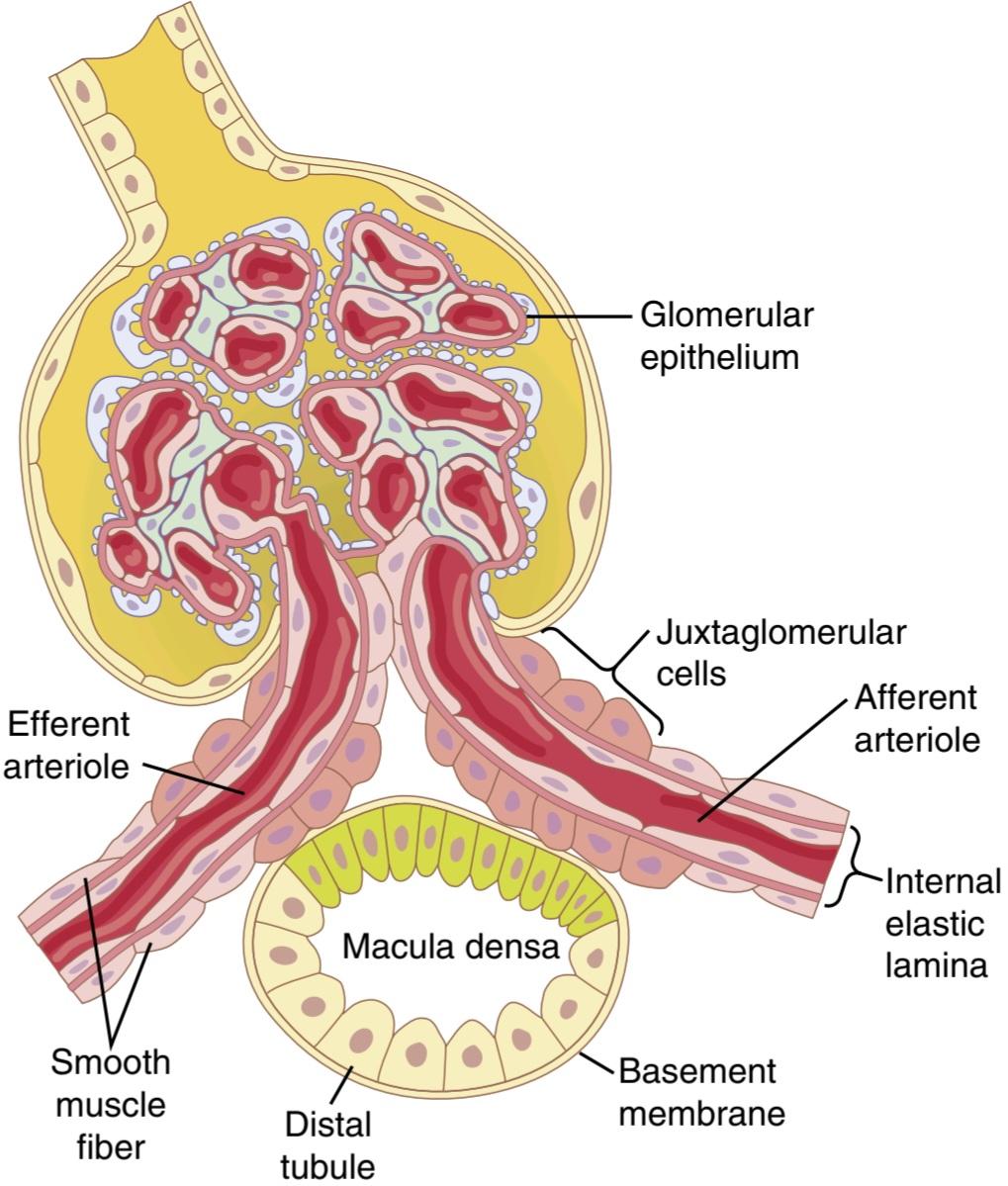
Figure 27-10. Structure of the juxtaglomerular apparatus, demonstrating its possible feedback role in the control of nephron function.
شکل 27-10. ساختار دستگاه juxtaglomerular، نشان دهنده نقش بازخورد احتمالی آن در کنترل عملکرد نفرون است.
Decreased Macula Densa Sodium Chloride Causes Dilation of Afferent Arterioles and Increased Renin Release. The macula densa cells sense changes in sodium chloride delivery to the distal tubule by way of signals that are not completely understood. Experimental studies have suggested that a decreased GFR slows the flow rate in the loop of Henle, causing increased reabsorption of the percentage of sodium and chloride ions delivered to the ascending loop of Henle and thereby reducing the concentration of sodium chloride at the macula densa cells. This decrease in sodium chloride concentration initiates a signal from the macula densa that has two effects (Figure 27-11): (1) it decreases re-sistance to blood flow in the afferent arterioles, which raises glomerular hydrostatic pressure and helps return GFR toward normal; and (2) it increases renin release from the juxtaglomerular cells of the afferent and efferent arterioles, which are the major storage sites for renin. Renin released from these cells then functions as an enzyme to increase the formation of angiotensin I, which is converted to angiotensin II. Finally, angiotensin II constricts the efferent arterioles, thereby increasing glomerular hydrostatic pressure and helping return GFR toward normal.
کاهش کلرید سدیم ماکولا دنسا باعث گشاد شدن شریان های آوران و افزایش ترشح رنین می شود. سلولهای ماکولا دنسا تغییرات انتقال کلرید سدیم به لولههای دیستال را از طریق سیگنالهایی که کاملاً درک نشده است، حس میکنند. مطالعات تجربی نشان داده اند که کاهش GFR سرعت جریان را در حلقه هنله کاهش می دهد و باعث افزایش بازجذب درصد یون های سدیم و کلرید تحویلی به حلقه صعودی هنله می شود و در نتیجه غلظت کلرید سدیم را در سلول های ماکولا دنسا کاهش می دهد. این کاهش غلظت کلرید سدیم سیگنالی را از ماکولا دنسا آغاز میکند که دو اثر دارد (شکل 27-11): (1) مقاومت مجدد در برابر جریان خون در شریانهای آوران را کاهش میدهد که فشار هیدرواستاتیک گلومرولی را افزایش میدهد و به بازگشت GFR به حالت طبیعی کمک میکند. و (2) آزادسازی رنین را از سلولهای مجاورت گلومرولی شریانهای آوران و وابران، که محل ذخیره اصلی رنین هستند، افزایش می دهد. سپس رنین آزاد شده از این سلول ها به عنوان آنزیمی عمل می کند تا تشکیل آنژیوتانسین I را افزایش دهد که به آنژیوتانسین II تبدیل می شود. در نهایت، آنژیوتانسین II شریانهای وابران را منقبض میکند و در نتیجه فشار هیدرواستاتیک گلومرولی را افزایش میدهد و به بازگشت GFR به حالت طبیعی کمک میکند.
These two components of the tubuloglomerular feedback mechanism, operating together by way of the special anatomical structure of the juxtaglomerular apparatus, provide feedback signals to the afferent and efferent arterioles for efficient autoregulation of GFR during changes in arterial pressure. When both of these mechanisms are functioning together, GFR changes by only a few percentage points, even with large fluctuations in arterial pressure between the limits of 75 and 160 mm Hg.
این دو جزء از مکانیسم بازخورد توبولوگلومرولی، که از طریق ساختار آناتومیکی ویژه دستگاه juxtaglomerular با هم کار میکنند، سیگنالهای بازخوردی را به شریانهای آوران و وابران برای تنظیم خودکار GFR در طول تغییرات فشار شریانی ارائه میدهند. هنگامی که هر دوی این مکانیسم ها با هم کار می کنند، GFR تنها با چند درصد تغییر می کند، حتی با نوسانات زیادی در فشار شریانی بین حدود 75 تا 160 میلی متر جیوه.

Figure 27-11. Macula densa feedback mechanism for autoregulation of glomerular hydrostatic pressure and glomerular filtration rate (GFR) during decreased renal arterial pressure.
شکل 27-11. مکانیسم بازخورد ماکولا دنسا برای تنظیم خودکار فشار هیدرواستاتیک گلومرولی و نرخ فیلتراسیون گلومرولی (GFR) در طول کاهش فشار شریانی کلیوی.
Blockade of Angiotensin II Formation Further Reduces Glomerular Filtration Rate During Renal Hypoperfusion. As discussed earlier, a preferential constrictor action of angiotensin II on efferent arterioles helps prevent serious reductions in glomerular hydrostatic pressure and GFR when renal perfusion pressure falls below normal. The ad-ministration of drugs that block the formation of angiotensin II (angiotensin-converting enzyme inhibitors) or that block the action of angiotensin II (angiotensin II receptor antagonists) may cause greater reductions in GFR than usual when the renal arterial pressure falls below normal. Therefore, an important complication of using these drugs to treat patients who have hypertension because of renal artery stenosis (partial blockage of the renal artery) is a severe decrease in GFR that can, in some cases, cause acute renal failure. Nevertheless, angiotensin II-blocking drugs are important therapeutic agents in many patients with hypertension, congestive heart failure, and other conditions, as long as the patients are monitored to ensure that severe decreases in GFR do not occur.
انسداد تشکیل آنژیوتانسین II، میزان فیلتراسیون گلومرولی را در طول هیپوپرفیوژن کلیه کاهش می دهد. همانطور که قبلاً بحث شد، یک اثر منقبض کننده ترجیحی آنژیوتانسین II بر روی شریان های وابران به جلوگیری از کاهش جدی فشار هیدرواستاتیک گلومرولی و GFR در زمانی که فشار پرفیوژن کلیوی کمتر از حد طبیعی می شود، کمک می کند. تجویز داروهایی که مانع از تشکیل آنژیوتانسین II (مهارکنندههای آنزیم تبدیلکننده آنژیوتانسین) میشوند یا از عملکرد آنژیوتانسین II (آنتاگونیستهای گیرنده آنژیوتانسین II) جلوگیری میکنند، ممکن است باعث کاهش بیشتر GFR نسبت به حالت عادی در زمانی که فشار شریانی کلیوی به زیر نرمال میرسد. بنابراین، یکی از عوارض مهم استفاده از این داروها برای درمان بیمارانی که به دلیل تنگی شریان کلیوی (انسداد نسبی شریان کلیوی) دچار فشار خون بالا هستند، کاهش شدید GFR است که در برخی موارد میتواند باعث نارسایی حاد کلیه شود. با این وجود، داروهای مسدودکننده آنژیوتانسین II عوامل درمانی مهمی در بسیاری از بیماران مبتلا به فشار خون بالا، نارسایی احتقانی قلب و سایر شرایط هستند، تا زمانی که بیماران تحت نظر باشند تا از کاهش شدید GFR اطمینان حاصل شود.
MYOGENIC AUTOREGULATION OF RENAL BLOOD FLOW AND GLOMERULAR FILTRATION RATE
Another mechanism that contributes to the maintenance of a relatively constant renal blood flow and GFR is the ability of individual blood vessels to resist stretching during increased arterial pressure, referred to as the myogenic mechanism. Studies of individual blood vessels (especially small arterioles) throughout the body have shown that they respond to increased wall tension or wall stretch by contraction of the vascular smooth muscle. Stretch of the vascular wall allows increased movement of calcium ions from the extracellular fluid into the cells, causing them to contract through the mechanisms discussed in Chapter 8. This contraction prevents excessive stretch of the vessel and, at the same time, by raising vascular resistance, helps prevent excessive increases in renal blood flow and the GFR when arterial pressure increases.
خود تنظیم میوژنیک جریان خون کلیوی و میزان فیلتراسیون گلومرولار
مکانیسم دیگری که به حفظ جریان خون کلیوی و GFR نسبتاً ثابت کمک می کند، توانایی رگ های خونی منفرد برای مقاومت در برابر کشش در طول افزایش فشار شریانی است که به آن مکانیسم میوژنیک گفته می شود. مطالعات روی رگهای خونی منفرد (به ویژه شریانهای کوچک) در سراسر بدن نشان داده است که آنها به افزایش کشش دیواره یا کشش دیواره با انقباض ماهیچه صاف عروق پاسخ میدهند. کشش دیواره عروقی باعث افزایش حرکت یونهای کلسیم از مایع خارج سلولی به داخل سلولها میشود و باعث انقباض آنها از طریق مکانیسمهای مورد بحث در فصل 8 میشود. این انقباض از کشش بیش از حد رگ جلوگیری میکند و در عین حال، با افزایش مقاومت عروقی، از افزایش بیش از حد جریان خون کلیوی و افزایش فشار شریانی در هنگام افزایش GFR کمک میکند.
Although the myogenic mechanism probably operates in most arterioles throughout the body, its importance in renal blood flow and GFR autoregulation has been questioned by some physiologists because this pressure- sensitive mechanism has no means of directly detecting changes in renal blood flow or GFR per se. On the other hand, this mechanism may be more important in protecting the kidney from hypertension-induced injury. In response to sudden increases in blood pressure, the myo- genic constrictor response in afferent arterioles occurs within seconds and therefore attenuates transmission of increased arterial pressure to the glomerular capillaries.
اگرچه مکانیسم میوژنیک احتمالاً در بیشتر شریانهای بدن عمل میکند، اهمیت آن در جریان خون کلیوی و خودتنظیمی GFR توسط برخی فیزیولوژیستها مورد تردید قرار گرفته است، زیرا این مکانیسم حساس به فشار ابزاری برای تشخیص مستقیم تغییرات در جریان خون کلیوی یا GFR فی نفسه ندارد. از سوی دیگر، این مکانیسم ممکن است در محافظت از کلیه در برابر آسیب ناشی از فشار خون بالا اهمیت بیشتری داشته باشد. در پاسخ به افزایش ناگهانی فشار خون، پاسخ منقبض کننده میوژنیک در شریان های آوران در عرض چند ثانیه رخ می دهد و بنابراین انتقال فشار شریانی افزایش یافته به مویرگ های گلومرولی را کاهش می دهد.
High Protein Intake and Hyperglycemia Increase Renal Blood Flow and Glomerular Filtration Rate. Although renal blood flow and GFR are relatively stable under most conditions, there are circumstances in which these variables change significantly. For example, a high protein intake is known to increase renal blood flow and GFR. With a long-term, high-protein diet, such as one that contains large amounts of meat, increases in GFR and renal blood flow are due partly to growth of the kidneys. However, the GFR and renal blood flow also increase 20% to 30% within 1 or 2 hours after a person eats a high-protein meal.
مصرف زیاد پروتئین و هایپرگلیسمی باعث افزایش جریان خون کلیوی و میزان فیلتراسیون گلومرولی می شود. اگرچه جریان خون کلیوی و GFR در اکثر شرایط نسبتاً پایدار هستند، شرایطی وجود دارد که در آن این متغیرها به طور قابل توجهی تغییر می کنند. به عنوان مثال، مصرف زیاد پروتئین باعث افزایش جریان خون کلیوی و GFR می شود. با یک رژیم غذایی طولانی مدت با پروتئین بالا، مانند رژیمی که حاوی مقادیر زیادی گوشت است، افزایش GFR و جریان خون کلیوی تا حدودی به دلیل رشد کلیه ها است. با این حال، GFR و جریان خون کلیوی نیز طی 1 یا 2 ساعت پس از خوردن یک وعده غذایی با پروتئین بالا، 20 تا 30 درصد افزایش می یابد.
One likely explanation for the increased GFR is the following. A high-protein meal releases into the blood amino acids, which are reabsorbed in the proximal tubules. Because amino acids and sodium are reabsorbed together by cotransport in the proximal tubules, increased amino acid reabsorption also stimulates sodium reabsorption. This increased reabsorption of sodium decreases sodium delivery to the macula densa (see Figure 27-12), which elicits a tubuloglomerular feedback-mediated decrease in resistance of the afferent arterioles, as discussed earlier. The decreased afferent arteriolar resistance then raises renal blood flow and GFR, allowing sodium excretion to be maintained at a nearly normal level while increasing excretion of the waste products of protein metabolism, such as urea.
یک توضیح احتمالی برای افزایش GFR موارد زیر است. یک وعده غذایی با پروتئین بالا اسیدهای آمینه خون را آزاد می کند که در لوله های پروگزیمال دوباره جذب می شوند. از آنجایی که اسیدهای آمینه و سدیم با هم توسط انتقال همزمان در لوله های پروگزیمال بازجذب می شوند، افزایش بازجذب اسید آمینه نیز بازجذب سدیم را تحریک می کند. این افزایش بازجذب سدیم، تحویل سدیم به ماکولا دنسا را کاهش میدهد (شکل 27-12 را ببینید)، که باعث کاهش مقاومت شریانهای آوران با واسطه توبولوگلومرولی میشود، همانطور که قبلاً بحث شد. سپس کاهش مقاومت شریانی آوران جریان خون کلیوی و GFR را افزایش میدهد و باعث میشود که دفع سدیم در سطح تقریباً نرمال حفظ شود و در عین حال دفع مواد زائد متابولیسم پروتئین مانند اوره را افزایش دهد.
A similar mechanism may also explain the marked in- creases in renal blood flow and the GFR that occur with large increases in blood glucose levels in persons with uncontrolled diabetes mellitus. Because glucose, like some of the amino acids, is also reabsorbed along with sodium in the proximal tubule, increased glucose delivery to the tubules causes them to reabsorb excess sodium along with glucose. This increased reabsorption of sodium, in turn, decreases the sodium chloride concentration at the macula densa, activating a tubuloglomerular feedback-mediated dilation of the afferent arterioles and subsequent increases in renal blood flow and GFR.
مکانیسم مشابهی ممکن است افزایش قابل توجهی در جریان خون کلیوی و GFR را که با افزایش زیاد سطح گلوکز خون در افراد مبتلا به دیابت قندی کنترل نشده رخ می دهد، توضیح دهد. از آنجایی که گلوکز مانند برخی از اسیدهای آمینه نیز همراه با سدیم در لوله پروگزیمال بازجذب می شود، افزایش تحویل گلوکز به لوله ها باعث می شود که سدیم اضافی همراه با گلوکز دوباره جذب شوند. این افزایش بازجذب سدیم، به نوبه خود، غلظت کلرید سدیم را در ماکولا دنسا کاهش میدهد و اتساع شریانهای آوران با واسطه بازخورد توبولوگلومرولی و افزایش متعاقب آن در جریان خون کلیوی و GFR را فعال میکند.
These examples demonstrate that renal blood flow and GFR per se are not the primary variables controlled by the tubuloglomerular feedback mechanism. The main purpose of this feedback is to ensure a constant delivery of sodium chloride to the distal tubule, where final processing of the urine takes place. Thus, disturbances that tend to increase the reabsorption of sodium chloride at tubular sites before the macula densa tend to elicit increased renal blood flow and GFR, which helps return distal sodium chloride delivery toward normal so that normal rates of sodium and water excretion can be maintained (see Figure 27-12).
این مثالها نشان میدهند که جریان خون کلیوی و GFR به خودی خود متغیرهای اولیه نیستند که توسط مکانیسم بازخورد توبولوگلومرولی کنترل میشوند. هدف اصلی این بازخورد اطمینان از تحویل ثابت کلرید سدیم به لوله دیستال است، جایی که پردازش نهایی ادرار انجام می شود. بنابراین، اختلالاتی که تمایل به افزایش بازجذب کلرید سدیم در محلهای لولهای قبل از ماکولا دنسا دارند، باعث افزایش جریان خون کلیوی و GFR میشوند که به بازگرداندن دیستال کلرید سدیم به حالت طبیعی کمک میکند تا بتوان نرخ طبیعی دفع سدیم و آب را حفظ کرد (شکل 27-12 را ببینید).
An opposite sequence of events occurs when proximal tubular reabsorption is reduced. For example, when the proximal tubules are damaged (which can occur as a result of poisoning by heavy metals, such as mercury, or large doses of drugs, such as tetracyclines), their ability to reabsorb sodium chloride is decreased. As a consequence, large amounts of sodium chloride are delivered to the distal tubule and, without appropriate compensation, would quickly cause excessive volume depletion. One of the important compensatory responses appears to be a tubuloglomerular feedback-mediated renal vasoconstric- tion that occurs in response to the increased sodium chlo- ride delivery to the macula densa in these circumstances. These examples again demonstrate the importance of this feedback mechanism for ensuring that the distal tubules receive the proper rate of delivery of sodium chloride, other tubular fluid solutes, and tubular fluid volume so that appropriate amounts of these substances are excreted in the urine.
وقتی بازجذب لوله پروگزیمال کاهش می یابد، توالی متضادی از رویدادها رخ می دهد. به عنوان مثال، هنگامی که لوله های پروگزیمال آسیب می بینند (که می تواند در نتیجه مسمومیت با فلزات سنگین مانند جیوه یا دوزهای زیادی از داروها مانند تتراسایکلین ها رخ دهد)، توانایی آنها برای بازجذب کلرید سدیم کاهش می یابد. در نتیجه، مقادیر زیادی از کلرید سدیم به لوله دیستال تحویل داده می شود و بدون جبران مناسب، به سرعت باعث کاهش حجم بیش از حد می شود. به نظر می رسد یکی از پاسخ های جبرانی مهم، انقباض عروق کلیوی با واسطه بازخورد توبولوگلومرولی است که در پاسخ به افزایش انتقال کلرید سدیم به ماکولا دنسا در این شرایط رخ می دهد. این مثالها دوباره اهمیت این مکانیسم بازخورد را برای اطمینان از اینکه لولههای دیستال سرعت تحویل مناسب کلرید سدیم، سایر املاح مایع لولهای و حجم مایع لولهای را دریافت میکنند، نشان میدهند تا مقادیر مناسبی از این مواد در ادرار دفع شوند.

Figure 27-12. Possible role of macula densa feedback in mediating increased glomerular filtration rate (GFR) after a high-protein meal.
شکل 27-12. نقش احتمالی بازخورد ماکولا دنسا در میانجیگری افزایش نرخ فیلتراسیون گلومرولی (GFR) پس از یک وعده غذایی با پروتئین بالا.
Other Factors That Influence Renal Blood Flow and Glomerular Filtration Rate. The GFR and renal blood flow are low at birth, approach normal adult levels after about 2 years of life, and in the absence of kidney disease, are maintained relatively constant until the fourth decade. Thereafter, GFR declines by about 5% to 10% per decade, although there is considerable variability among individuals. This decline in GFR coincides with nitric oxide deficiency, increased oxidative stress, and loss of functional nephrons, which may be related partly to increasing blood pressure, metabolic disorders, and other insults that cause cumulative glomerular injury with aging.
عوامل دیگری که بر جریان خون کلیوی و میزان فیلتراسیون گلومرولی تأثیر می گذارد. GFR و جریان خون کلیوی در بدو تولد کم است، پس از حدود 2 سال زندگی به سطوح طبیعی بزرگسالان نزدیک می شود و در غیاب بیماری کلیوی، تا دهه چهارم نسبتاً ثابت می ماند. پس از آن، GFR حدود 5٪ تا 10٪ در هر دهه کاهش می یابد، اگرچه تنوع قابل توجهی در بین افراد وجود دارد. این کاهش در GFR همزمان با کمبود اکسید نیتریک، افزایش استرس اکسیداتیو و از دست دادن نفرون های عملکردی است که ممکن است تا حدی به افزایش فشار خون، اختلالات متابولیک و سایر توهین هایی که باعث آسیب تجمعی گلومرولی با افزایش سن می شود، مرتبط باشد.
Men have higher renal blood flow and GFR than women, even when corrected for body mass. However, men also have a more rapid decline in GFR with aging than premenopausal women. Although the mechanisms responsible for these sex differences are not fully understood, beneficial effects of estrogens and damaging effects of androgens on the kidneys have been suggested as a partial explanation. Table 27-5 summarizes some additional factors that influence renal blood flow and GFR regulation, and should be considered when assessing kidney function.
مردان دارای جریان خون کلیوی و GFR بالاتری نسبت به زنان هستند، حتی در صورت اصلاح توده بدن. با این حال، مردان نیز با افزایش سن نسبت به زنان پیش از یائسگی کاهش سریعتری در GFR دارند. اگرچه مکانیسم های مسئول این تفاوت های جنسی به طور کامل شناخته نشده است، اثرات مفید استروژن ها و اثرات مخرب آندروژن ها بر کلیه ها به عنوان یک توضیح جزئی پیشنهاد شده است. جدول 27-5 برخی از عوامل اضافی را که بر جریان خون کلیوی و تنظیم GFR تأثیر میگذارند، خلاصه میکند و باید هنگام ارزیابی عملکرد کلیه در نظر گرفته شود.
Table 27-5 Some Conditions That Influence Renal Blood Flow (RBF) and the Glomerular Filtration Rate (GFR)
جدول 27-5 برخی از شرایطی که بر جریان خون کلیوی (RBF) و فیلتراسیون گلومرولی تأثیر می گذارد نرخ (GFR)
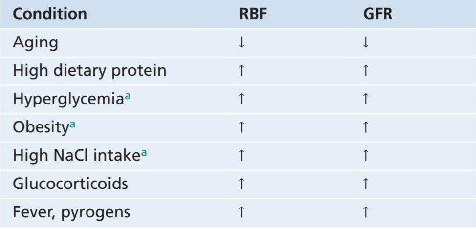
aRefers to early effects, prior to development of glomerular injury that may occur with chronic hyperglycemia, obesity, and high salt intake.
aبه اثرات اولیه قبل از ایجاد آسیب گلومرولی اشاره دارد که ممکن است با هیپرگلیسمی مزمن، چاقی و مصرف زیاد نمک رخ دهد.
کلیک کنید «Bibliography: فهرست کتب مربوطه»
Anders HJ, Huber TB, Isermann B, Schiffer M: CKD in diabetes: diabetic kidney disease versus nondiabetic kidney disease. Nat Rev Nephrol 14:361, 2018.
Baylis C: Sexual dimorphism: the aging kidney, involvement of nitric oxide deficiency, and angiotensin II overactivity. J Gerontol A Biol Sci Med Sci 67:1365, 2012.
Beierwaltes WH, Harrison-Bernard LM, Sullivan JC, Mattson DL: Assessment of renal function; clearance, the renal microcirculation, renal blood flow, and metabolic balance. Compr Physiol 3:165, 2013.
Bidani AK, Polichnowski AJ, Loutzenhiser R, Griffin KA: Renal mi- crovascular dysfunction, hypertension and CKD progression. Curr Opin Nephrol Hypertens 22:1, 2013.
Carlström M, Wilcox CS, Arendshorst WJ: Renal autoregulation in health and disease. Physiol Rev 95:405, 2015.
Cowley AW Jr, Abe M, Mori T, O’Connor PM, et. al: Reactive oxygen species as important determinants of medullary flow, sodium excretion, and hypertension. Am J Physiol Renal Physiol 308:F179, 2013.
de Groat WC, Griffiths D, Yoshimura N: Neural control of the lower urinary tract. Compr Physiol 5:327, 2015.
DiBona GF: Physiology in perspective: the wisdom of the body. Neural control of the kidney. Am J Physiol Regul Integr Comp Physiol 289:R633, 2005.
Gomez RA, Sequeira-Lopez MLS: Renin cells in homeostasis, regener- ation and immune defence mechanisms. Nat Rev Nephrol 14:231, 2018.
Griffin KA: Hypertensive kidney injury and the progression of chronic kidney disease. Hypertension 70:687, 2017.
Guan Z, VanBeusecum JP, Inscho EW: Endothelin and the renal mi- crocirculation. Semin Nephrol 35:145, 2015.
Hall JE, Brands MW: The renin-angiotensin-aldosterone system: renal mechanisms and circulatory homeostasis. In: Seldin DW, Giebisch G (eds): The Kidney-Physiology and Pathophysiology, 3rd ed. New York: Raven Press, 2000, pp 1009-1046.
Hall JE, do Carmo JM, da Silva AA, Wang Z, Hall ME: Obesity, kidney dysfunction and hypertension: mechanistic links. Nature Reviews Nephrology 15: 367, 2019.
Hall JE, do Carmo JM, da Silva AA, Wang Z, Hall ME: Obesity-induced hypertension: interaction of neurohumoral and renal mechanisms. Circ Res 116:991, 2015.
Navar LG, Kobori H, Prieto MC, Gonzalez-Villalobos RA: Intratubular renin-angiotensin system in hypertension. Hypertension 57:355, 2011.
Schell C, Huber TB: The evolving complexity of the podocyte cytoskel- eton. J Am Soc Nephrol 28:3166-, 2017.
Schnermann J, Briggs JP: Tubular control of renin synthesis and secretion. Pflugers Arch 465:39, 2013.
Speed JS, Pollock DM: Endothelin, kidney disease, and hypertension. Hypertension 61:1142, 2013.
Thomson SC, Blantz RC: Biophysics of glomerular filtration. Compr Physiol 2:1671.
Vivarelli M, Massella L, Ruggiero B, Emma F: Minimal change disease. Clin J Am Soc Nephrol 12:332, 2017.
🚀 با ما همراه شوید!
تازهترین مطالب و آموزشهای مغز و اعصاب را از دست ندهید. با فالو کردن کانال تلگرام آیندهنگاران مغز، از ما حمایت کنید!



