
فیزیولوژی پزشکی گایتون و هال؛ بازجذب و ترشح لولهای کلیه

دعای مطالعه [ نمایش ]
بِسْمِ الله الرَّحْمنِ الرَّحیمِ
اَللّهُمَّ اَخْرِجْنى مِنْ ظُلُماتِ الْوَهْمِ
خدايا مرا بيرون آور از تاريكىهاى وهم،
وَ اَكْرِمْنى بِنُورِ الْفَهْمِ
و به نور فهم گرامى ام بدار،
اَللّهُمَّ افْتَحْ عَلَيْنا اَبْوابَ رَحْمَتِكَ
خدايا درهاى رحمتت را به روى ما بگشا،
وَانْشُرْ عَلَيْنا خَزائِنَ عُلُومِكَ بِرَحْمَتِكَ يا اَرْحَمَ الرّاحِمينَ
و خزانههاى علومت را بر ما باز كن به امید رحمتت اى مهربانترين مهربانان.
کتاب «فیزیولوژی پزشکی گایتون و هال» بهعنوان یکی از جامعترین و معتبرترین منابع در حوزه علوم پزشکی، همچنان مرجع کلیدی برای درک عملکرد پیچیده بدن انسان است. این اثر با تکیه بر تازهترین پژوهشها و توضیحات دقیق از سازوکارهای فیزیولوژیک، پلی میان علوم پایه پزشکی و کاربردهای بالینی ایجاد میکند و نقشی بیبدیل در آموزش، پژوهش و ارتقای دانش سلامت ایفا مینماید.
ترجمه دقیق و علمی این شاهکار توسط برند علمی آیندهنگاران مغز به مدیریت داریوش طاهری، دسترسی فارسیزبانان به مرزهای نوین دانش فیزیولوژی را ممکن ساخته و رسالتی علمی برای ارتقای آموزش پزشکی، فهم عمیقتر سازوکارهای بدن و توسعه روشهای نوین در حوزه سلامت فراهم آورده است.
» Guyton and Hall Textbook of Medical Physiology
»» CHAPTER 28: Renal Tubular Reabsorption and Secretion

As the glomerular filtrate enters the renal tubules, it flows sequentially through the successive parts of the tubulethe proximal tubule, loop of Henle, distal tubule, collecting tubule, and collecting duct-before it is excreted as urine. Along this course, some substances are selectively reabsorbed from the tubules back into the blood, whereas others are secreted from the blood into the tubular lumen. Eventually, the urine that is formed and all the substances in the urine represent the sum of three basic renal processes-glomerular filtration, tubular reabsorption, and tubular secretion:
همانطور که فیلتر گلومرولی وارد لوله های کلیوی می شود، به طور متوالی از طریق قسمت های متوالی توبول – لوله پروگزیمال، حلقه هنل، لوله دیستال، لوله جمع کننده و مجرای جمع آوری – قبل از دفع آن به صورت ادرار جریان می یابد. در طول این دوره، برخی از مواد به طور انتخابی از لولهها به داخل خون بازجذب میشوند، در حالی که برخی دیگر از خون به لومن لولهای ترشح میشوند. در نهایت، ادراری که تشکیل میشود و تمام مواد موجود در ادرار، مجموع سه فرآیند کلیوی اصلی را نشان میدهند: فیلتراسیون گلومرولی، بازجذب لولهای و ترشح لولهای:

For many substances, tubular reabsorption plays a much more important role than secretion in determining the final urinary excretion rate. However, tubular secretion accounts for significant amounts of potassium ions, hydrogen ions, and a few other substances that appear in the urine.
برای بسیاری از مواد، بازجذب لولهای نقش بسیار مهمتری نسبت به ترشح در تعیین سرعت دفع نهایی ادرار دارد. با این حال، ترشح لوله ای مقادیر قابل توجهی از یون های پتاسیم، یون های هیدروژن و چند ماده دیگر را که در ادرار ظاهر می شوند، تشکیل می دهد.
TUBULAR REABSORPTION IS QUANTITATIVELY LARGE AND HIGHLY SELECTIVE
Table 28-1 shows the renal handling of several substances that are all freely filtered in the kidneys and reabsorbed at variable rates. The rate at which each of these substances is filtered is calculated as follows:
بازجذب لوله ای از نظر کمی بزرگ است و بسیار انتخابی
جدول 28-1 عملکرد کلیوی چندین ماده را نشان می دهد که همگی آزادانه در کلیه ها فیلتر شده و با سرعت های متغیر بازجذب می شوند. سرعت فیلتر هر یک از این مواد به صورت زیر محاسبه می شود:

This calculation assumes that the substance is freely filtered and not bound to plasma proteins. For example, if plasma glucose concentration is 1 g/L, the amount of glucose filtered each day is about 180 L/day x 1 g/L, or 180 g/day. Because virtually none of the filtered glucose is normally excreted, the rate of glucose reabsorption is also 180 g/day.
این محاسبه فرض می کند که ماده آزادانه فیلتر شده و به پروتئین های پلاسما متصل نیست. به عنوان مثال، اگر غلظت گلوکز پلاسما 1 گرم در لیتر باشد، مقدار گلوکز فیلتر شده در هر روز حدود 180 لیتر در روز x 1 گرم در لیتر یا 180 گرم در روز است. از آنجا که عملاً هیچ یک از گلوکز فیلتر شده به طور معمول دفع نمی شود، سرعت بازجذب گلوکز نیز 180 گرم در روز است.
From Table 28-1, two things are immediately apparent. First, the processes of glomerular filtration and tubular reabsorption are quantitatively large relative to urinary excretion for many substances. Thus, a small change in glomerular filtration or tubular reabsorption can potentially cause a relatively large change in urinary excretion. For example, a 10% decrease in tubular reabsorption, from 178.5 to 160.7 L/day, would increase urine volume from 1.5 to 19.3 L/day (almost a 13-fold increase) if the glomerular filtration rate (GFR) remained constant. In reality, changes in tubular reabsorption and glomerular filtration are closely coordinated so that large fluctuations in urinary excretion are avoided.
از جدول 28-1، دو چیز فوراً آشکار است. اولاً، فرآیندهای فیلتراسیون گلومرولی و بازجذب لولهای نسبت به دفع ادرار برای بسیاری از مواد از نظر کمی بزرگ است. بنابراین، یک تغییر کوچک در فیلتراسیون گلومرولی یا بازجذب لوله ای به طور بالقوه می تواند باعث تغییر نسبتاً بزرگ در دفع ادرار شود. برای مثال، کاهش 10 درصدی بازجذب لولهای، از 5/178 به 7/160 لیتر در روز، اگر میزان فیلتراسیون گلومرولی (GFR) ثابت بماند، حجم ادرار را از 1.5 به 19.3 لیتر در روز افزایش میدهد. در حقیقت، تغییرات در بازجذب لولهای و فیلتراسیون گلومرولی با هم هماهنگ هستند تا از نوسانات زیاد در دفع ادرار جلوگیری شود.
Second, unlike glomerular filtration, which is relatively nonselective (essentially all solutes in the plasma are filtered except the plasma proteins or substances bound to them), tubular reabsorption is highly selective. Some substances, such as glucose and amino acids, are almost completely reabsorbed from the tubules, so the urinary excretion rate is essentially zero. Many ions in the plasma, such as sodium, chloride, and bicarbonate, are also highly reabsorbed, but their rates of reabsorption and urinary excretion are variable, depending on the needs of the body. Waste products, such as urea and creatinine, conversely, are poorly reabsorbed from the tubules and are excreted in relatively large amounts.
دوم، برخلاف فیلتراسیون گلومرولی، که نسبتاً غیرانتخابی است (اساساً تمام املاح موجود در پلاسما به جز پروتئین های پلاسما یا مواد متصل به آنها فیلتر می شوند)، بازجذب لوله ای بسیار انتخابی است. برخی از مواد مانند گلوکز و اسیدهای آمینه تقریباً به طور کامل از لوله ها بازجذب می شوند، بنابراین میزان دفع ادرار اساساً صفر است. بسیاری از یون های پلاسما مانند سدیم، کلرید و بی کربنات نیز به شدت بازجذب می شوند، اما نرخ بازجذب و دفع ادرار آنها بسته به نیاز بدن متغیر است. مواد زائد مانند اوره و کراتینین، برعکس، به خوبی از لوله ها بازجذب می شوند و به مقدار نسبتا زیادی دفع می شوند.
Therefore, by controlling their reabsorption of different substances, the kidneys regulate excretion of solutes independently of one another, a capability that is essential for precise control of the body fluid composition. In this chapter, we discuss the mechanisms that allow the kidneys to selectively reabsorb or secrete different sub- stances at variable rates.
بنابراین، کلیه ها با کنترل بازجذب مواد مختلف، دفع املاح را مستقل از یکدیگر تنظیم می کنند، این قابلیت برای کنترل دقیق ترکیب مایع بدن ضروری است. در این فصل، مکانیسمهایی را مورد بحث قرار میدهیم که به کلیهها اجازه میدهد تا به طور انتخابی مواد مختلف را با سرعتهای متغیر بازجذب یا ترشح کنند.
Table 28-1 Filtration, Reabsorption, and Excretion Rates of Different Substances by the Kidneys
جدول 28-1 فیلتراسیون، بازجذب و میزان دفع مواد مختلف توسط کلیه ها

TUBULAR REABSORPTION INCLUDES PASSIVE AND ACTIVE MECHANISMS
For a substance to be reabsorbed, it must first be transported (1) across the tubular epithelial membranes into the renal interstitial fluid and then (2) through the peritubular capillary membrane back into the blood (Figure 28-1). Thus, reabsorption of water and solutes includes a series of transport steps. Reabsorption across the tubular epithelium into the interstitial fluid includes active or passive transport by the same basic mechanisms discussed in Chapter 4 for transport across other cell membranes of the body. For example, water and solutes can be trans- ported through the cell membranes (transcellular route) or through the spaces between the cell junctions (paracellular route). Then, after absorption across the tubular epithelial cells into the interstitial fluid, water and solutes are transported through the peritubular capillary walls into the blood by ultrafiltration (bulk flow) that is mediated by hydrostatic and colloid osmotic forces. The peritubular capillaries behave like the venous ends of most other capillaries because there is a net reab sorptive force that moves the fluid and solutes from the interstitium into the blood.
بازجذب لوله ای شامل مکانیسم های غیرفعال و فعال
برای اینکه یک ماده دوباره جذب شود، ابتدا باید (1) از طریق غشای اپیتلیال لوله ای به داخل مایع بینابینی کلیوی و سپس (2) از طریق غشای مویرگی اطراف لوله به داخل خون منتقل شود (شکل 28-1). بنابراین، بازجذب آب و املاح شامل یک سری مراحل انتقال است. بازجذب در سراسر اپیتلیوم لولهای به داخل مایع بینابینی شامل انتقال فعال یا غیرفعال توسط همان مکانیسمهای اساسی مورد بحث در فصل 4 برای انتقال از طریق سایر غشای سلولی بدن است. برای مثال، آب و املاح را می توان از طریق غشای سلولی (مسیر بین سلولی) یا از طریق فضاهای بین اتصالات سلولی (مسیر پاراسلولی) منتقل کرد. سپس، پس از جذب از طریق سلولهای اپیتلیال لولهای به داخل مایع بینابینی، آب و املاح از طریق دیوارههای مویرگ اطراف لولهای توسط اولترافیلتراسیون (جریان حجیم) که توسط نیروهای اسمزی هیدرواستاتیک و کلوئیدی انجام میشود، به داخل خون منتقل میشوند. مویرگهای اطراف لوله مانند انتهای وریدی اکثر مویرگهای دیگر رفتار میکنند، زیرا یک نیروی جذبی خالص وجود دارد که مایع و املاح را از بینابینی به داخل خون منتقل میکند.
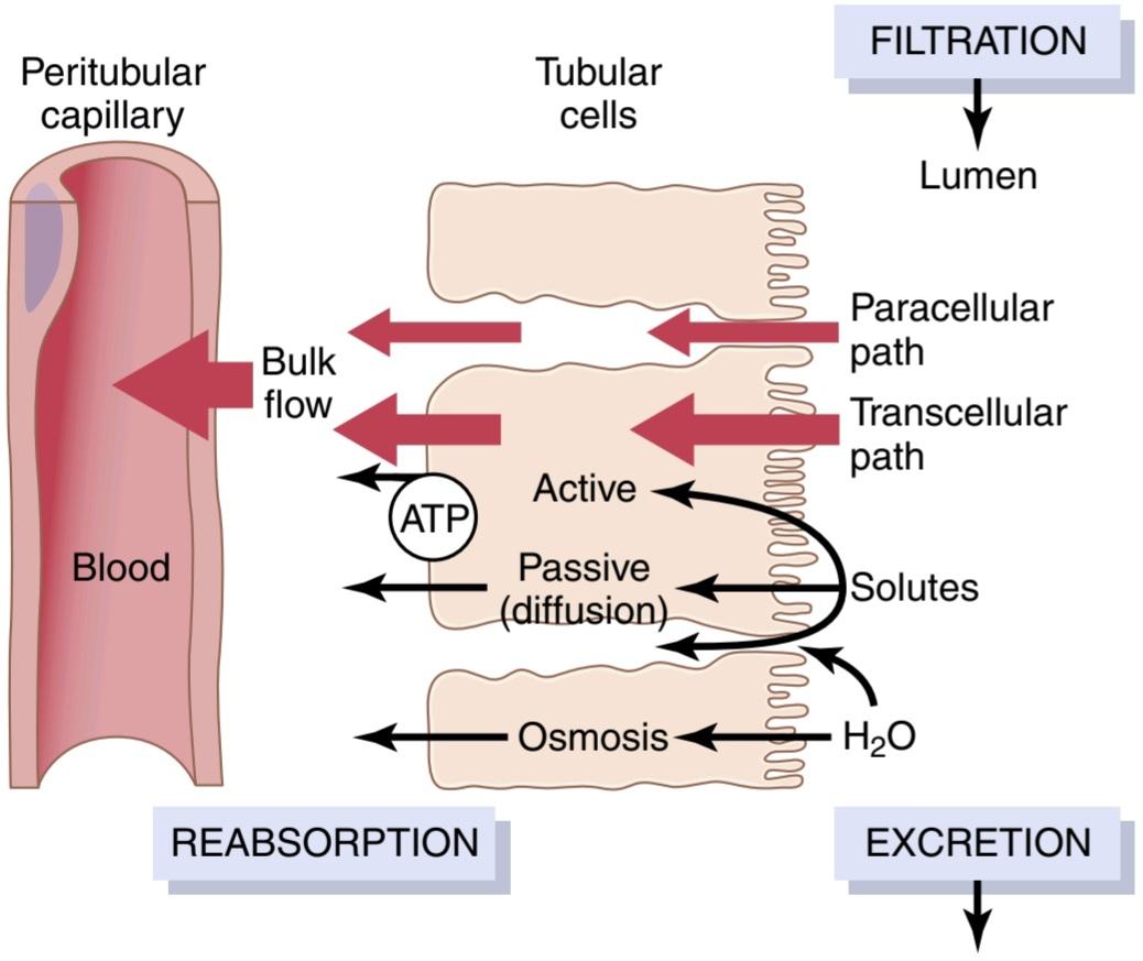
Figure 28-1 Reabsorption of filtered water and solutes from the tubular lumen across the tubular epithelial cells, through the renal interstitium, and back into the blood. Solutes are transported through the cells (transcellular path) by passive diffusion or active transport, or be- tween the cells (paracellular path) by diffusion. Water is transported through the cells and between the tubular cells by osmosis. Transport of water and solutes from the interstitial fluid into the peritubular capillaries occurs by ultrafiltration (bulk flow).
شکل 28-1 بازجذب آب تصفیه شده و املاح از مجرای لولهای در سراسر سلولهای اپیتلیال لولهای، از طریق بینابینی کلیه و بازگشت به خون. املاح از طریق سلول ها (مسیر بین سلولی) توسط انتشار غیرفعال یا انتقال فعال یا بین سلول ها (مسیر پاراسلولی) توسط انتشار منتقل می شوند. آب از طریق سلول ها و بین سلول های لوله ای توسط اسمز منتقل می شود. انتقال آب و املاح از مایع بینابینی به مویرگ های اطراف لوله ای با اولترافیلتراسیون (جریان حجیم) انجام می شود.
ACTIVE TRANSPORT
Active transport can move a solute against an electrochemical gradient; this requires energy derived from metabolism. Transport that is coupled directly to an energy source, such as the hydrolysis of adenosine triphosphate (ATP), is termed primary active transport. An example of this mechanism is the sodiumpotassium adenosine triphosphatase (ATPase) pump (Na+-K+ ATPase pump) that functions throughout most parts of the renal tubule. Transport that is coupled indirectly to an energy source, such as that due to an ion gradient, is referred to as secondary active transport. Reabsorption of glucose by the renal tubule is an example of secondary active transport. Although solutes can be reabsorbed by active and/or passive mechanisms by the tubule, water is always reabsorbed passively across the tubular epithelial membrane by the process of osmosis.
حمل و نقل فعال
انتقال فعال می تواند یک املاح را در برابر یک گرادیان الکتروشیمیایی حرکت دهد. این نیاز به انرژی حاصل از متابولیسم دارد. حمل و نقلی که مستقیماً با یک منبع انرژی مانند هیدرولیز آدنوزین تری فسفات (ATP) همراه است، حمل و نقل فعال اولیه نامیده می شود. نمونه ای از این مکانیسم پمپ سدیم پتاسیم آدنوزین تری فسفاتاز (ATPase) (پمپ Na+-K+ ATPase) است که در اکثر قسمت های توبول کلیوی عمل می کند. حمل و نقلی که به طور غیرمستقیم با یک منبع انرژی جفت می شود، مانند شیب یونی، به عنوان حمل و نقل فعال ثانویه شناخته می شود. بازجذب گلوکز توسط لوله کلیوی نمونه ای از انتقال فعال ثانویه است. اگرچه املاح را می توان با مکانیسم های فعال و/یا غیرفعال توسط لوله بازجذب کرد، آب همیشه به صورت غیرفعال در سراسر غشای اپیتلیال لوله ای توسط فرآیند اسمز جذب می شود.
Solutes Can Be Transported Through Epithelial Cells or Between Cells. Renal tubular cells, like other epithelial cells, are held together by tight junctions. Lateral intercellular spaces lie behind the tight junctions and separate the epithelial cells of the tubule. Solutes can be reabsorbed or secreted across the cells through the transcellular pathway or between the cells by moving across the tight junctions and intercellular spaces via the paracellular pathway. Sodium is a substance that moves through both routes, although most of the sodium is transported through the transcellular pathway. In some nephron segments, especially the proximal tubule, water is also reabsorbed across the paracellular pathway, and substances dissolved in the water, especially potassium, magnesium, and chloride ions, are carried with the reabsorbed fluid between the cells.
املاح را می توان از طریق سلول های اپیتلیال یا بین سلول ها منتقل کرد. سلولهای لولهای کلیوی، مانند سایر سلولهای اپیتلیال، توسط اتصالات محکم به هم متصل میشوند. فضاهای بین سلولی جانبی در پشت اتصالات محکم قرار دارند و سلول های اپیتلیال توبول را از هم جدا می کنند. املاح می توانند از طریق مسیر بین سلولی یا بین سلول ها با حرکت در اتصالات تنگ و فضاهای بین سلولی از طریق مسیر پاراسلولی دوباره جذب یا ترشح شوند. سدیم ماده ای است که از هر دو مسیر حرکت می کند، اگرچه بیشتر سدیم از طریق مسیر بین سلولی منتقل می شود. در برخی از بخشهای نفرون، بهویژه لوله پروگزیمال، آب نیز در مسیر پاراسلولی بازجذب میشود و مواد محلول در آب، بهویژه یونهای پتاسیم، منیزیم و کلرید همراه با مایع بازجذب بین سلولها حمل میشوند.
Primary Active Transport Through the Tubular Membrane Linked to Hydrolysis of Adenosine Triphosphatase. The special importance of primary active trans- port is that it can move solutes against an electrochemical gradient. The energy for this active transport comes from the hydrolysis of ATP by way of membrane-bound ATPase, which is also a component of the carrier mechanism that binds and moves solutes across the cell membranes. The primary active transporters in the kidneys that are known include Na+-K+ ATPase, hydrogen ATPase, hydrogen-potassium ATPase, and calcium ATPase.
انتقال فعال اولیه از طریق غشای لوله ای مرتبط با هیدرولیز آدنوزین تری فسفاتاز. اهمیت ویژه انتقال فعال اولیه این است که می تواند املاح را در برابر یک گرادیان الکتروشیمیایی حرکت دهد. انرژی این انتقال فعال از هیدرولیز ATP از طریق ATPase متصل به غشاء حاصل می شود، که همچنین جزء مکانیسم حامل است که املاح را در غشای سلولی متصل می کند و حرکت می دهد. ناقلین فعال اولیه در کلیه ها که شناخته شده اند عبارتند از Na+-K+ ATPase، هیدروژن ATPase، هیدروژن پتاسیم ATPase و کلسیم ATPase.
A good example of a primary active transport system is the reabsorption of sodium ions across the proximal tubular membrane, as shown in Figure 28-2. On the basolateral sides of the tubular epithelial cell, the cell membrane has an extensive Na+-K+ ATPase system that hydrolyzes ATP and uses the released energy to transport sodium ions out of the cell into the interstitium. At the same time, potassium is transported from the interstitium to the inside of the cell. The operation of this ion pump maintains low intracellular sodium and high intracellular potassium concentrations and creates a net negative charge of about -70 millivolts within the cell. This active pumping of sodium out of the cell across the basolateral membrane of the cell favors passive diffusion of sodium across the luminal membrane of the cell, from the tubular lumen into the cell, for two reasons: (1) there is a concentration gradient favoring sodium diffusion into the cell because the intracellular sodium concentration is low (12 mEq/L) and tubular fluid sodium concentration is high (140 mEq/L); and (2) the negative, -70-millivolt, intracellular potential attracts the positive sodium ions from the tubular lumen into the cell.
همانطور که در شکل 28-2 نشان داده شده است، یک مثال خوب از یک سیستم انتقال فعال اولیه، جذب مجدد یون های سدیم در سراسر غشای لوله پروگزیمال است. در طرفهای قاعدهای سلول اپیتلیال لولهای، غشای سلولی دارای یک سیستم Na+-K+ ATPase گسترده است که ATP را هیدرولیز میکند و از انرژی آزاد شده برای انتقال یونهای سدیم به خارج از سلول به داخل بینابینی استفاده میکند. در همان زمان، پتاسیم از بینابینی به داخل سلول منتقل می شود. عملکرد این پمپ یونی غلظت سدیم درون سلولی پایین و پتاسیم درون سلولی بالا را حفظ می کند و یک بار منفی خالص در حدود 70- میلی ولت در داخل سلول ایجاد می کند. این پمپاژ فعال سدیم به خارج از سلول از طریق غشای قاعده جانبی سلول به نفع انتشار غیرفعال سدیم در سراسر غشای مجرای سلول، از مجرای لولهای به داخل سلول است، به دو دلیل: (1) یک گرادیان غلظت وجود دارد که به انتشار سدیم در سلول کمک میکند، زیرا غلظت سدیم داخل سلولی mq/L کم است. (140 mEq/L)؛ و (2) پتانسیل درون سلولی منفی 70- میلی ولت، یون های سدیم مثبت را از لومن لوله ای به داخل سلول جذب می کند.
Active reabsorption of sodium by Na+-K+ ATPase occurs in most parts of the tubule. In certain parts of the nephron, there are also additional provisions for moving large amounts of sodium into the cell. In the proximal tubule, there is an extensive brush border on the luminal side of the membrane (the side that faces the tubular lumen) that multiplies the surface area by about 20-fold. There are also carrier proteins that bind sodium ions on the luminal surface of the membrane and release them inside the cell, providing facilitated diffusion of sodium through the membrane into the cell. These sodium carrier proteins are also important for secondary active transport of other substances, such as glucose and amino acids, as discussed later.
بازجذب فعال سدیم توسط Na+-K+ ATPase در بیشتر قسمتهای توبول اتفاق می افتد. در بخشهای خاصی از نفرون، مقررات اضافی برای انتقال مقادیر زیادی سدیم به داخل سلول نیز وجود دارد. در لوله پروگزیمال، یک مرز برس گسترده در سمت لومینال غشاء (سمت رو به لومن لوله ای) وجود دارد که مساحت سطح را حدود 20 برابر می کند. همچنین پروتئینهای حاملی وجود دارند که یونهای سدیم را روی سطح مجرای غشاء متصل میکنند و آنها را در داخل سلول آزاد میکنند و انتشار آسان سدیم را از طریق غشاء به داخل سلول فراهم میکنند. این پروتئین های حامل سدیم همچنین برای انتقال فعال ثانویه سایر مواد مانند گلوکز و اسیدهای آمینه، همانطور که بعداً بحث شد، مهم هستند.
Thus, the net reabsorption of sodium ions from the tubular lumen back into the blood involves at least three steps:
بنابراین، بازجذب خالص یونهای سدیم از مجرای لولهای به داخل خون حداقل شامل سه مرحله است:
1. Sodium diffuses across the luminal membrane (also called the apical membrane) into the cell down an electrochemical gradient established by the Na+-K+ ATPase pump on the basolateral side of the membrane.
1. سدیم در سراسر غشای مجرا (که غشای آپیکال نیز نامیده میشود) به داخل سلول و با یک گرادیان الکتروشیمیایی ایجاد شده توسط پمپ Na+-K+ ATPase در سمت قاعدهای جانبی غشاء پخش میشود.
2. Sodium is transported across the basolateral membrane against an electrochemical gradient by the Na+-K+ ATPase pump.
2. سدیم توسط پمپ Na+-K+ ATPase در غشای قاعده جانبی در برابر یک گرادیان الکتروشیمیایی منتقل می شود.
3. Sodium, water, and other substances are reabsorbed from the interstitial fluid into the peritubular capillaries by ultrafiltration, a passive process driven by the hydrostatic and colloid osmotic pressure gradients.
3. سدیم، آب و سایر مواد از مایع بینابینی به مویرگ های اطراف لوله ای توسط اولترافیلتراسیون، یک فرآیند غیرفعال که توسط گرادیان فشار اسمزی هیدرواستاتیک و کلوئیدی هدایت می شود، دوباره جذب می شوند.
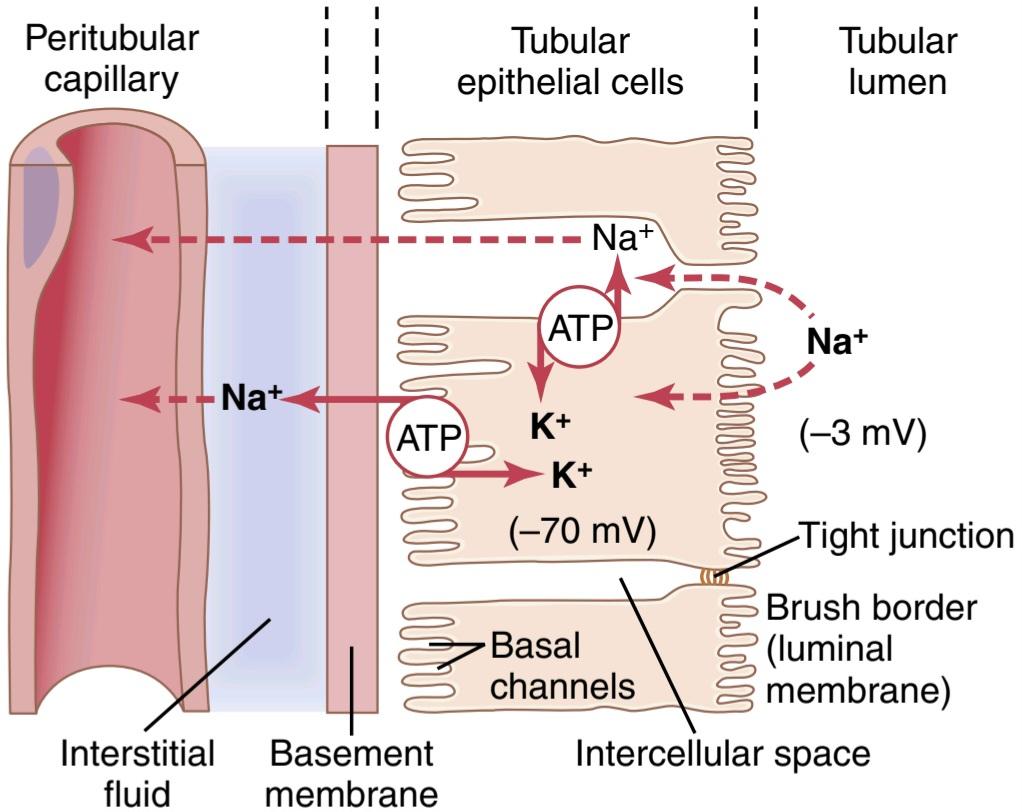
Figure 28-2 Basic mechanism for active transport of sodium through the tubular epithelial cell. The sodium-potassium pump transports sodium from the interior of the cell across the basolateral membrane, creating a low intracellular sodium concentration and a negative intracellular electrical potential. The low intracellular sodium concentration and negative electrical potential cause sodium ions to diffuse from the tubular lumen into the cell through the brush border.
شکل 28-2 مکانیسم اساسی برای انتقال فعال سدیم از طریق سلول اپیتلیال لوله ای. پمپ سدیم-پتاسیم سدیم را از داخل سلول در سراسر غشای قاعده ای منتقل می کند و غلظت سدیم درون سلولی کم و پتانسیل الکتریکی درون سلولی منفی ایجاد می کند. غلظت کم سدیم درون سلولی و پتانسیل الکتریکی منفی باعث میشود یونهای سدیم از مجرای لولهای به داخل سلول از طریق مرز برس منتشر شوند.
Secondary Active Reabsorption Through the Tubular Membrane. In secondary active transport, two or more substances interact with a specific membrane protein (a carrier molecule) and are transported together across the membrane. As one of the substances (e.g., sodium) diffuses down its electrochemical gradient, the energy re- leased is used to drive another substance (e.g., glucose) against its electrochemical gradient. Thus, secondary active transport does not require energy directly from ATP or from other high-energy phosphate sources. Rather, the direct source of the energy is that liberated by the simultaneous facilitated diffusion of another transported sub- stance down its own electrochemical gradient.
بازجذب فعال ثانویه از طریق غشای لوله ای. در انتقال فعال ثانویه، دو یا چند ماده با یک پروتئین غشایی خاص (یک مولکول حامل) برهمکنش میکنند و با هم در سراسر غشاء حمل میشوند. از آنجایی که یکی از مواد (مثلاً سدیم) به سمت گرادیان الکتروشیمیایی خود منتشر می شود، انرژی آزاد شده برای هدایت ماده دیگری (مثلاً گلوکز) در برابر گرادیان الکتروشیمیایی آن استفاده می شود. بنابراین، حمل و نقل فعال ثانویه به انرژی مستقیم از ATP یا سایر منابع فسفات با انرژی بالا نیاز ندارد. در عوض، منبع مستقیم انرژی آن است که با انتشار تسهیل شده همزمان یک ماده انتقال یافته دیگر به پایین گرادیان الکتروشیمیایی خود آزاد می شود.
Figure 28-3 shows secondary active transport of glucose and amino acids in the proximal tubule. In both cases, specific carrier proteins in the brush border combine with a sodium ion and an amino acid or a glucose molecule at the same time. These transport mechanisms are so efficient that they remove virtually all the glucose and amino acids from the tubular lumen. After entry into the cell, glucose and amino acids exit across the basolateral membranes by diffusion, driven by the high glucose and amino acid concentrations in the cell facilitated by specific transport proteins.
شکل 28-3 انتقال فعال ثانویه گلوکز و اسیدهای آمینه را در لوله پروگزیمال نشان می دهد. در هر دو مورد، پروتئین های حامل خاص در مرز قلم مو با یک یون سدیم و یک اسید آمینه یا یک مولکول گلوکز به طور همزمان ترکیب می شوند. این مکانیسمهای انتقال آنقدر کارآمد هستند که تقریباً تمام گلوکز و اسیدهای آمینه را از لومن لولهای حذف میکنند. پس از ورود به سلول، گلوکز و اسیدهای آمینه با انتشار از غشاهای قاعدهای خارج میشوند که توسط غلظت بالای گلوکز و اسید آمینه در سلول که توسط پروتئینهای حملونقل خاص تسهیل میشود، هدایت میشوند.
Sodium glucose co-transporters (SGLT2 and SGLT1) are located on the brush border of proximal tubular cells and carry glucose into the cell cytoplasm against a concentration gradient, as described previously. Approximately 90% of the filtered glucose is reabsorbed by SGLT2 in the early part of the proximal tubule (S1 segment), and the residual 10% is transported by SGLT1 in the latter segments of the proximal tubule. On the basolateral side of the membrane, glucose diffuses out of the cell into the interstitial spaces with the help of glucose transporters-GLUT2 in the S1 segment and GLUT1 in the latter part (S3 segment) of the proximal tubule.
انتقالدهندههای گلوکز سدیم (SGLT2 و SGLT1) در مرز برس سلولهای لولهای پروگزیمال قرار دارند و گلوکز را بر خلاف گرادیان غلظت به سیتوپلاسم سلولی حمل میکنند، همانطور که قبلاً توضیح داده شد. تقریباً 90٪ از گلوکز فیلتر شده توسط SGLT2 در قسمت اولیه لوله پروگزیمال (بخش S1) دوباره جذب می شود و 10٪ باقیمانده توسط SGLT1 در بخش های آخر لوله پروگزیمال منتقل می شود. در سمت قاعده جانبی غشاء، گلوکز با کمک ناقل گلوکز-GLUT2 در بخش S1 و GLUT1 در قسمت آخر (بخش S3) لوله پروگزیمال، از سلول به فضاهای بینابینی منتشر می شود.
Although transport of glucose against a chemical gradient does not directly use ATP, the reabsorption of glucose depends on energy expended by the primary active Na+– K+ ATPase pump in the basolateral membrane. Because of the activity of this pump, an electrochemical gradient for facilitated diffusion of sodium across the luminal membrane is maintained, and it is this downhill diffusion of sodium to the interior of the cell that provides the energy for the simultaneous uphill transport of glucose across the luminal membrane. Thus, this reabsorption of glucose is referred to as secondary active transport because glucose itself is reabsorbed uphill against a chemical gradient, but it is secondary to primary active transport of sodium.
اگرچه انتقال گلوکز در برابر یک گرادیان شیمیایی مستقیماً از ATP استفاده نمی کند، جذب مجدد گلوکز به انرژی مصرف شده توسط پمپ فعال اولیه Na+-K+ ATPase در غشای قاعده ای بستگی دارد. به دلیل فعالیت این پمپ، یک گرادیان الکتروشیمیایی برای انتشار تسهیل شده سدیم در غشای مجرا حفظ می شود و این انتشار سرازیری سدیم به داخل سلول است که انرژی را برای انتقال همزمان گلوکز در سربالایی در سراسر غشای مجرا فراهم می کند. بنابراین، این بازجذب گلوکز به عنوان انتقال فعال ثانویه نامیده می شود زیرا خود گلوکز در سربالایی در برابر یک گرادیان شیمیایی دوباره جذب می شود، اما ثانویه به انتقال فعال اولیه سدیم است.
Another important point is that a substance is said to undergo active transport when at least one of the steps in the reabsorption involves primary or secondary active transport, even though other steps in the reabsorption process may be passive. For glucose reabsorption, secondary active transport occurs at the luminal membrane, but passive facilitated diffusion occurs at the basolateral membrane, and passive uptake by bulk flow occurs at the peritubular capillaries.
نکته مهم دیگر این است که گفته می شود یک ماده زمانی تحت انتقال فعال قرار می گیرد که حداقل یکی از مراحل بازجذب شامل حمل و نقل فعال اولیه یا ثانویه باشد، حتی اگر سایر مراحل در فرآیند بازجذب ممکن است غیرفعال باشند. برای بازجذب گلوکز، انتقال فعال ثانویه در غشای مجرا اتفاق میافتد، اما انتشار تسهیلشده غیرفعال در غشای قاعدهای و جذب غیرفعال توسط جریان تودهای در مویرگهای اطراف لولهای رخ میدهد.
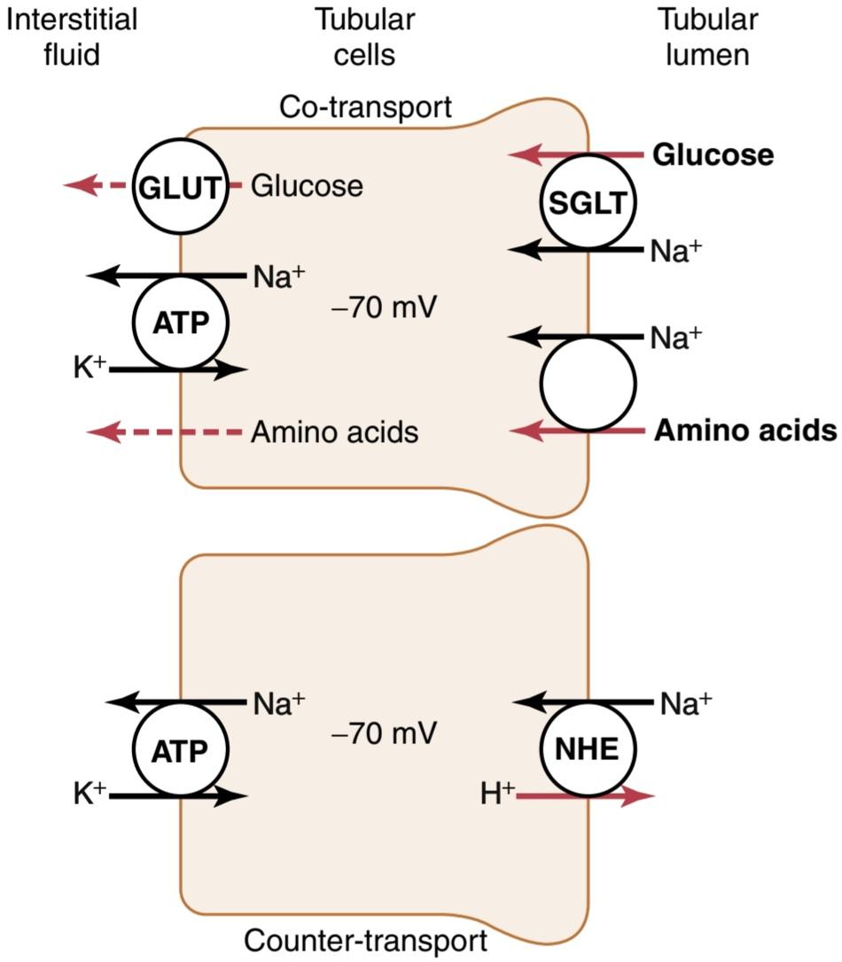
Figure 28-3 Mechanisms of secondary active transport. The upper cell shows the co-transport of glucose and amino acids along with sodium ions through the apical side of the tubular epithelial cells, followed by facilitated diffusion through the basolateral membranes. The lower cell shows the counter-transport of hydrogen ions from the interior of the cell across the apical membrane and into the tubular lumen; movement of sodium ions into the cell, down an electrochemical gradient established by the sodium-potassium pump on the basolateral membrane, provides the energy for transport of the hydrogen ions from inside the cell into the tubular lumen. ATP, Adenosine triphosphate; GLUT, glucose transporter; NHE, sodium-hydrogen exchanger; SGLT, sodiumglucose co-transporter.
شکل 28-3 مکانیسم های حمل و نقل فعال ثانویه. سلول بالایی انتقال همزمان گلوکز و اسیدهای آمینه همراه با یونهای سدیم را از طریق قسمت آپیکال سلولهای اپیتلیال لولهای نشان میدهد و به دنبال آن انتشار تسهیل شده از طریق غشاهای قاعدهای جانبی انجام میشود. سلول پایینی انتقال متقابل یون های هیدروژن را از داخل سلول در سراسر غشای آپیکال و به لومن لوله ای نشان می دهد. حرکت یونهای سدیم به داخل سلول، به سمت پایین یک گرادیان الکتروشیمیایی ایجاد شده توسط پمپ سدیم-پتاسیم بر روی غشای قاعدهای، انرژی را برای انتقال یونهای هیدروژن از داخل سلول به لومن لولهای فراهم میکند. ATP، آدنوزین تری فسفات. GLUT، ناقل گلوکز؛ NHE، مبدل سدیم-هیدروژن؛ SGLT، ناقل سدیم-گلوکز.
Secondary Active Secretion Into the Tubules. Some substances are secreted into the tubules by secondary active transport, which often involves counter-transport of the substance with sodium ions. In counter-transport, the energy liberated from the downhill movement of one of the substances (e.g., sodium ions) enables the uphill movement of a second substance in the opposite direction.
ترشح فعال ثانویه به داخل لوله ها. برخی از مواد با انتقال فعال ثانویه به داخل لوله ها ترشح می شوند که اغلب شامل انتقال متقابل ماده با یون های سدیم می شود. در حمل و نقل متقابل، انرژی آزاد شده از حرکت سرازیری یکی از مواد (مثلاً یونهای سدیم) حرکت سربالایی ماده دوم را در جهت مخالف امکانپذیر میسازد.
One example of counter-transport, shown in Figure 28-3, is the active secretion of hydrogen ions coupled to sodium reabsorption in the luminal membrane of the proximal tubule. In this case, sodium entry into the cell is coupled with hydrogen extrusion from the cell by sodiumhydrogen counter-transport. This transport is mediated by a specific protein (sodium-hydrogen exchanger) in the brush border of the luminal membrane. As sodium is carried to the interior of the cell, hydrogen ions are forced outward in the opposite direction into the tubular lumen. The basic principles of primary and secondary active transport are discussed in Chapter 4.
یکی از نمونههای ضد انتقال، که در شکل 28-3 نشان داده شده است، ترشح فعال یونهای هیدروژن همراه با بازجذب سدیم در غشای مجرای لوله پروگزیمال است. در این حالت، ورود سدیم به سلول با اکستروژن هیدروژن از سلول توسط ضد انتقال سدیم-هیدروژن همراه است. این انتقال توسط یک پروتئین خاص (مبدل سدیم-هیدروژن) در مرز برس غشای مجرا انجام می شود. هنگامی که سدیم به داخل سلول منتقل می شود، یون های هیدروژن در جهت مخالف به سمت بیرون به داخل لومن لوله ای هدایت می شوند. اصول اساسی حمل و نقل فعال اولیه و ثانویه در فصل 4 مورد بحث قرار گرفته است.
Pinocytosis Is an Active Transport Mechanism for Re- absorption of Proteins. Some parts of the tubule, especially the proximal tubule, reabsorb large molecules such as proteins via pinocytosis, a type of endocytosis. In this process, the protein attaches to the brush border of the luminal membrane, and this portion of the membrane then invaginates to the interior of the cell until it is completely pinched off and a vesicle is formed containing the protein. Once inside the cell, the protein is digested into its constituent amino acids, which are reabsorbed through the basolateral membrane into the interstitial fluid. Because pinocytosis requires energy, it is considered a form of active transport.
پینوسیتوز یک مکانیسم انتقال فعال برای بازجذب پروتئین ها است. برخی از قسمت های توبول، به ویژه لوله پروگزیمال، مولکول های بزرگی مانند پروتئین ها را از طریق پینوسیتوز، نوعی اندوسیتوز، دوباره جذب می کنند. در این فرآیند، پروتئین به مرز برس غشای مجرا میچسبد و این قسمت از غشاء سپس به داخل سلول وارد میشود تا کاملاً از بین برود و یک وزیکول حاوی پروتئین تشکیل شود. هنگامی که پروتئین وارد سلول می شود، به اسیدهای آمینه تشکیل دهنده خود هضم می شود که از طریق غشای قاعده جانبی به مایع بین بافتی بازجذب می شوند. از آنجایی که پینوسیتوز به انرژی نیاز دارد، نوعی انتقال فعال در نظر گرفته می شود.
Transport Maximum for Substances That Are Actively Reabsorbed. For most substances that are actively reabsorbed or secreted, there is a limit to the rate at which the solute can be transported, which is often referred to as the transport maximum. This limit is due to saturation of the specific transport systems involved when the amount of solute delivered to the tubule (referred to as the tubular load) exceeds the capacity of the carrier proteins and specific enzymes involved in the transport process.
حداکثر انتقال برای موادی که به طور فعال بازجذب می شوند. برای اکثر موادی که به طور فعال بازجذب یا ترشح می شوند، محدودیتی برای سرعت انتقال املاح وجود دارد که اغلب به عنوان حداکثر انتقال شناخته می شود. این محدودیت به دلیل اشباع شدن سیستم های حمل و نقل خاص درگیر در زمانی است که مقدار املاح تحویل شده به لوله (که به عنوان بار لوله ای نامیده می شود) از ظرفیت پروتئین های حامل و آنزیم های خاص درگیر در فرآیند انتقال فراتر رود.
The glucose transport system in the proximal tubule is a good example. Normally, measurable glucose does not appear in the urine because essentially all the filtered glucose is reabsorbed in the proximal tubule. However, when the filtered load exceeds the capability of the tubules to reabsorb glucose, urinary excretion of glucose does occur.
سیستم انتقال گلوکز در لوله پروگزیمال مثال خوبی است. به طور معمول، گلوکز قابل اندازه گیری در ادرار ظاهر نمی شود زیرا اساساً تمام گلوکز فیلتر شده در لوله پروگزیمال دوباره جذب می شود. با این حال، زمانی که بار فیلتر شده از توانایی لوله ها برای بازجذب گلوکز فراتر رود، دفع ادراری گلوکز اتفاق می افتد.
In the adult human, the transport maximum for glucose averages about 375 mg/min, whereas the filtered load of glucose is only about 125 mg/min (GFR × plasma glucose = 125 ml/min x 1 mg/ml). With large increases in GFR and/or plasma glucose concentration that increase the filtered load of glucose above 375 mg/min, the excess glucose filtered is not reabsorbed and passes into the urine.
در انسان بالغ، حداکثر انتقال گلوکز به طور متوسط حدود 375 میلی گرم در دقیقه است، در حالی که بار فیلتر شده گلوکز تنها حدود 125 میلی گرم در دقیقه است (GFR × گلوکز پلاسما = 125 میلی لیتر در دقیقه x 1 میلی گرم در میلی لیتر). با افزایش زیاد در GFR و/یا غلظت گلوکز پلاسما که بار فیلتر شده گلوکز را بالای 375 میلی گرم در دقیقه افزایش می دهد، گلوکز اضافی فیلتر شده دوباره جذب نمی شود و به ادرار می رود.
Figure 28-4 shows the relationship between plasma concentration of glucose, filtered load of glucose, tubular transport maximum for glucose, and rate of glucose loss in the urine. Note that when the plasma glucose concentration is 100 mg/100 ml and the filtered load is at its normal level (125 mg/min), there is no loss of glucose in the urine. However, when the plasma concentration of glucose rises above about 200 mg/100 ml, increasing the filtered load to about 250 mg/min, a small amount of glucose begins to appear in the urine. This point is termed the threshold for glucose. Note that this appearance of glucose in the urine (at the threshold) occurs before the transport maximum is reached. One reason for the difference between the thresh- old and transport maximum is that not all nephrons have the same transport maximum for glucose, and some of the nephrons therefore begin to excrete glucose before others have reached their transport maximum. The overall trans- port maximum for the kidneys, which is normally about 375 mg/min, is reached when all nephrons have reached their maximal capacity to reabsorb glucose.
شکل 28-4 رابطه بین غلظت گلوکز پلاسما، بار فیلتر شده گلوکز، حداکثر انتقال لوله ای برای گلوکز و سرعت از دست دادن گلوکز در ادرار را نشان می دهد. توجه داشته باشید که وقتی غلظت گلوکز پلاسما 100 میلی گرم در 100 میلی لیتر باشد و بار فیلتر شده در سطح طبیعی خود (125 میلی گرم در دقیقه) باشد، گلوکز در ادرار از دست نمی رود. با این حال، زمانی که غلظت پلاسمایی گلوکز از حدود 200 میلیگرم در 100 میلیلیتر بالاتر میرود و بار فیلتر شده را به حدود 250 میلیگرم در دقیقه افزایش میدهد، مقدار کمی گلوکز در ادرار ظاهر میشود. این نقطه آستانه گلوکز نامیده می شود. توجه داشته باشید که این ظاهر گلوکز در ادرار (در آستانه) قبل از رسیدن به حداکثر انتقال رخ می دهد. یکی از دلایل تفاوت بین آستانه و حداکثر انتقال این است که همه نفرون ها حداکثر انتقال گلوکز را ندارند و بنابراین برخی از نفرون ها قبل از اینکه دیگران به حداکثر انتقال خود برسند شروع به دفع گلوکز می کنند. حداکثر انتقال کلی برای کلیه ها، که به طور معمول حدود 375 میلی گرم در دقیقه است، زمانی حاصل می شود که همه نفرون ها به حداکثر ظرفیت خود برای بازجذب گلوکز رسیده باشند.

Figure 28-4 Relationships among the filtered load of glucose, rate of glucose reabsorption by the renal tubules, and rate of glucose excretion in the urine. The transport maximum is the maximum rate at which glucose can be reabsorbed from the tubules. The threshold for glucose refers to the filtered load of glucose at which glucose first begins to be excreted in the urine.
شکل 28-4 روابط بین بار فیلتر شده گلوکز، سرعت بازجذب گلوکز توسط لوله های کلیوی و سرعت دفع گلوکز در ادرار. حداکثر انتقال حداکثر سرعتی است که در آن گلوکز می تواند از لوله ها بازجذب شود. آستانه گلوکز به بار فیلتر شده گلوکز اشاره دارد که در آن گلوکز ابتدا از طریق ادرار دفع می شود.
The plasma glucose of a healthy person almost never becomes high enough to cause glucose excretion in the urine, even after eating a meal. However, in uncontrolled diabetes mellitus, plasma glucose concentration may rise to high levels, causing the filtered load of glucose to exceed the transport maximum and resulting in urinary glucose excretion. Some of the important transport maximums for substances actively reabsorbed by the tubules are as follows:
گلوکز پلاسمای یک فرد سالم تقریباً هرگز به اندازه ای بالا نمی رود که باعث دفع گلوکز در ادرار شود، حتی پس از خوردن یک وعده غذایی. با این حال، در دیابت قندی کنترل نشده، غلظت گلوکز پلاسما ممکن است به سطوح بالا افزایش یابد، که باعث می شود بار فیلتر شده گلوکز از حداکثر انتقال بیشتر شود و منجر به دفع گلوکز از طریق ادرار شود. برخی از حداکثرهای مهم انتقال برای موادی که به طور فعال توسط لوله ها بازجذب می شوند به شرح زیر است:

Transport Maximums for Actively Secreted Substances. Substances that are actively secreted also exhibit transport maximums, as follows:
حداکثر حمل و نقل برای مواد ترشح شده فعال. موادی که به طور فعال ترشح می شوند نیز حداکثر انتقال را نشان می دهند، به شرح زیر:

Substances That Are Actively Transported but Do Not Exhibit a Transport Maximum. The reason that actively transported solutes often exhibit a transport maximum is that the transport carrier system becomes saturated as the tubular load increases. Some substances that are actively reabsorbed do not demonstrate a transport maximum be- cause their rate of transport is determined by other factors, such as the following: (1) the electrochemical gradient for diffusion of the substance across the membrane; (2) the permeability of the membrane for the substance; and (3) the time that the fluid containing the substance remains within the tubule. Transport of this type is referred to as gradient-time transport because the rate of transport depends on the electrochemical gradient and the time that the substance is in the tubule, which in turn depends on the tubular flow rate.
موادی که به طور فعال حمل می شوند اما حداکثر انتقال را نشان نمی دهند. دلیل اینکه املاح انتقال فعال اغلب حداکثر انتقال را نشان می دهند این است که با افزایش بار لوله ای، سیستم حامل حمل و نقل اشباع می شود. برخی از موادی که به طور فعال بازجذب می شوند حداکثر انتقال را نشان نمی دهند زیرا سرعت انتقال آنها توسط عوامل دیگر تعیین می شود، مانند موارد زیر: (1) گرادیان الکتروشیمیایی برای انتشار ماده در سراسر غشاء. (2) نفوذپذیری غشاء برای ماده؛ و (3) زمانی که مایع حاوی ماده در داخل لوله باقی می ماند. حمل و نقل از این نوع به عنوان انتقال گرادیان-زمان نامیده می شود زیرا سرعت انتقال به گرادیان الکتروشیمیایی و زمانی که ماده در لوله است بستگی دارد که به نوبه خود به سرعت جریان لوله بستگی دارد.
An example of gradient-time transport is sodium reabsorption in the proximal tubule, where the maxi- mum transport capacity of the basolateral Na-K+ ATPase pump is usually far greater than the actual rate of net sodium reabsorption because a significant amount of sodium transported out of the cell leaks back into the tubular lumen through junctions of the epithelial cells. The rate at which this backleak occurs depends on (1) the permeability of the tight junctions; and (2) the interstitial physical forces, which determine the rate of bulk flow reabsorption from the interstitial fluid into the peritubular capillaries. Therefore, sodium transport in the proximal tubules obeys mainly gradient-time trans- port principles rather than tubular maximum transport characteristics. This observation means that the higher the concentration of sodium in the proximal tubules, the higher is its reabsorption rate. Also, the slower the flow rate of tubular fluid, the greater the percentage of sodium that can be reabsorbed from the proximal tubules.
نمونه ای از انتقال شیب زمان، بازجذب سدیم در لوله پروگزیمال است، که در آن حداکثر ظرفیت انتقال پمپ قاعده ای جانبی Na-K+ ATPase معمولاً بسیار بیشتر از نرخ واقعی بازجذب خالص سدیم است، زیرا مقدار قابل توجهی از سدیم که از سلول به بیرون منتقل می شود از طریق سلول های پیوندی به لومن لوله ای سلول های اپیتلیال نشت می کند. سرعت وقوع این نشتی به (1) نفوذپذیری اتصالات محکم بستگی دارد. و (2) نیروهای فیزیکی بینابینی، که نرخ بازجذب جریان عمده از مایع بین بافتی به مویرگ های اطراف لوله را تعیین می کنند. بنابراین، انتقال سدیم در لولههای پروگزیمال بهجای ویژگیهای انتقال حداکثر لولهای، عمدتاً از اصول انتقال گرادیان-زمان پیروی میکند. این مشاهدات بدین معنی است که هر چه غلظت سدیم در لوله های پروگزیمال بیشتر باشد، میزان بازجذب آن نیز بیشتر است. همچنین، هرچه سرعت جریان سیال لوله ای کندتر باشد، درصد سدیمی که می تواند از لوله های پروگزیمال بازجذب شود، بیشتر می شود.
In the more distal parts of the nephron, the epithelial cells have much tighter junctions and transport much smaller amounts of sodium. In these segments, sodium reabsorption exhibits a transport maximum similar to that for other actively transported substances. Furthermore, this transport maximum can be increased by certain hormones, such as aldosterone.
در قسمتهای دورتر نفرون، سلولهای اپیتلیال اتصالات محکمتری دارند و مقادیر بسیار کمتری سدیم را انتقال میدهند. در این بخشها، بازجذب سدیم حداکثر حملونقلی مشابه با سایر مواد انتقالیافته فعال نشان میدهد. علاوه بر این، این حداکثر انتقال را می توان توسط هورمون های خاصی مانند آلدوسترون افزایش داد.
PASSIVE WATER REABSORPTION BY OSMOSIS COUPLED MAINLY TO SODIUM REABSORPTION
When solutes are transported out of the tubule by primary or secondary active transport, their concentrations tend to decrease inside the tubule while increasing in the renal interstitium. This phenomenon creates a concentration difference that causes osmosis of water in the same direction that the solutes are transported, from the tubular lumen to the renal interstitium. Some parts of the renal tubule, especially the proximal tubule, are highly permeable to water, and water reabsorption occurs so rapidly that there is only a small concentration gradient for solutes across the tubular membrane.
بازجذب غیرفعال آب توسط اسمز که عمدتاً با بازجذب سدیم همراه است
هنگامی که املاح توسط حمل و نقل فعال اولیه یا ثانویه به خارج از توبول منتقل می شوند، غلظت آنها در داخل لوله کاهش می یابد در حالی که در بینابینی کلیوی افزایش می یابد. این پدیده یک اختلاف غلظت ایجاد می کند که باعث اسمز آب در همان جهتی می شود که املاح از لومن لوله ای به بینابینی کلیه منتقل می شوند. برخی از قسمتهای لوله کلیوی، بهویژه لوله پروگزیمال، نفوذپذیری بالایی نسبت به آب دارند و بازجذب آب چنان سریع اتفاق میافتد که تنها یک گرادیان غلظت کمی برای املاح در سراسر غشای لولهای وجود دارد.
A large part of the osmotic flow of water in the proximal tubules occurs through water channels (aquaporins) in the cell membranes, as well as through the tight junctions between the epithelial cells. As noted previously, the junctions between the cells are not as tight as their name would imply and permit significant diffusion of water and small ions. This condition is especially true in the proximal tubules, which have a high permeability for water and a smaller but significant permeability to most ions, such as sodium, chloride, potassium, calcium, and magnesium.
بخش بزرگی از جریان اسمزی آب در لوله های پروگزیمال از طریق کانال های آب (آکواپورین ها) در غشای سلولی و همچنین از طریق اتصالات محکم بین سلول های اپیتلیال انجام می شود. همانطور که قبلاً ذکر شد، اتصالات بین سلول ها آنقدر محکم نیستند که از نامشان پیداست و اجازه انتشار قابل توجه آب و یون های کوچک را می دهند. این وضعیت به ویژه در لوله های پروگزیمال که دارای نفوذپذیری بالا برای آب و نفوذپذیری کوچکتر اما قابل توجهی برای اکثر یون ها مانند سدیم، کلرید، پتاسیم، کلسیم و منیزیم هستند، صادق است.
Water moving across the tight junctions by osmosis also carries with it some of the solutes, a process referred to as solvent drag. In addition, because the reabsorption of water, organic solutes, and ions is coupled to sodium reabsorption, changes in sodium reabsorption significantly influence the reabsorption of water and many other solutes.
آب در حال حرکت در میان اتصالات محکم به وسیله اسمز، برخی از املاح را نیز با خود حمل می کند، فرآیندی که به آن کشش حلال می گویند. علاوه بر این، از آنجایی که بازجذب آب، املاح آلی و یون ها با بازجذب سدیم همراه است، تغییرات در بازجذب سدیم به طور قابل توجهی بر جذب مجدد آب و بسیاری از املاح دیگر تأثیر می گذارد.
In the more distal parts of the nephron, beginning in the loop of Henle and extending through the collecting tubule, the tight junctions become far less permeable to water and solutes, and the epithelial cells also have a greatly decreased membrane surface area. Therefore, water cannot move easily across the tight junctions of the tubular membrane by osmosis. However, antidiuretic hormone (ADH) greatly increases the water permeability in the distal and collecting tubules.
در قسمتهای دورتر نفرون، که از حلقه هنل شروع میشود و از طریق لوله جمعآوری امتداد مییابد، اتصالات محکم نسبت به آب و املاح نفوذپذیری کمتری دارند و سلولهای اپیتلیال نیز سطح غشایی بسیار کاهش یافتهاند. بنابراین، آب نمی تواند به راحتی در اتصالات محکم غشای لوله ای توسط اسمز حرکت کند. با این حال، هورمون آنتی دیورتیک (ADH) نفوذپذیری آب در لوله های دیستال و جمع کننده را به شدت افزایش می دهد.
Thus, water movement across the tubular epithelium can occur only if the membrane is permeable to water, no matter how large the osmotic gradient. In the proximal tubule and descending loop of Henle, water permeability is always high, and water is rapidly reabsorbed to reach osmotic equilibrium with the surrounding interstitial fluid. This high permeability is due to abundant expression of the water channel aquaporin-1 (AQP-1) in the luminal and basolateral membranes. In the ascending loop of Henle, water permeability is always low, so almost no water is reabsorbed, despite a large osmotic gradient. Water permeability in the last parts of the tubules-the distal tubules, collecting tubules, and collecting ducts- occurs through aquaporins and can be high or low, depending on the presence or absence of ADH.
بنابراین، حرکت آب در سراسر اپیتلیوم لولهای تنها در صورتی میتواند رخ دهد که غشاء نسبت به آب نفوذپذیر باشد، مهم نیست که گرادیان اسمزی چقدر زیاد باشد. در لوله پروگزیمال و حلقه نزولی هنله، نفوذپذیری آب همیشه بالا است و آب به سرعت جذب می شود تا با مایع بینابینی اطراف به تعادل اسمزی برسد. این نفوذپذیری بالا به دلیل بیان فراوان کانال آب aquaporin-1 (AQP-1) در غشاهای مجرای و قاعدهای است. در حلقه صعودی هنله، نفوذپذیری آب همیشه کم است، بنابراین با وجود شیب اسمزی زیاد، تقریباً هیچ آبی دوباره جذب نمیشود. نفوذپذیری آب در آخرین قسمتهای لولهها – لولههای دیستال، لولههای جمعکننده و مجاری جمعآوری – از طریق آکواپورینها اتفاق میافتد و بسته به وجود یا عدم وجود ADH میتواند زیاد یا کم باشد.
REABSORPTION OF CHLORIDE, UREA, AND OTHER SOLUTES BY PASSIVE DIFFUSION
When sodium is reabsorbed through the tubular epithelial cell, negative ions such as chloride are transported along with sodium because of electrical potentials. That is, transport of positively charged sodium ions out of the lumen leaves the inside of the lumen negatively charged, compared with the interstitial fluid causing chloride ions to diffuse passively through the paracellular pathway. Additional reabsorption of chloride ions occurs because of a chloride concentration gradient that develops when water is reabsorbed from the tubule by osmosis, thereby concentrating the chloride ions in the tubular lumen (Figure 28-5). Thus, active reabsorption of sodium is closely coupled to passive reabsorption of chloride by way of an electrical potential and a chloride concentration gradient.
بازجذب کلرید، اوره و سایر املاح با انتشار غیرفعال
هنگامی که سدیم از طریق سلول اپیتلیال لوله ای بازجذب می شود، یون های منفی مانند کلرید به دلیل پتانسیل الکتریکی همراه با سدیم منتقل می شوند. یعنی انتقال یونهای سدیم با بار مثبت به خارج از لومن، در مقایسه با مایع بینابینی که باعث انتشار غیرفعال یونهای کلرید در مسیر پاراسلولی میشود، داخل لومن را دارای بار منفی میکند. بازجذب اضافی یونهای کلرید به دلیل گرادیان غلظت کلرید رخ میدهد که وقتی آب توسط اسمز از لوله بازجذب میشود، ایجاد میشود و در نتیجه یونهای کلرید در مجرای لولهای متمرکز میشود (شکل 28-5). بنابراین، بازجذب فعال سدیم از طریق پتانسیل الکتریکی و گرادیان غلظت کلرید با بازجذب غیرفعال کلرید پیوند نزدیکی دارد.
Chloride ions can also be reabsorbed by secondary active transport. The most important of the secondary active transport processes for chloride reabsorption involves the co-transport of chloride with sodium across the luminal membrane.
یون های کلرید نیز می توانند با انتقال فعال ثانویه دوباره جذب شوند. مهمترین فرآیند انتقال فعال ثانویه برای بازجذب کلرید شامل انتقال همزمان کلرید با سدیم در سراسر غشای مجرا است.

Figure 28-5 Mechanisms whereby water, chloride, and urea reabsorption are coupled with sodium reabsorption.
شکل 28-5 مکانیسم هایی که به موجب آن بازجذب آب، کلرید و اوره با بازجذب سدیم همراه می شوند.
Urea is also passively reabsorbed from the tubule, but to a much lesser extent than chloride ions. As water is reabsorbed from the tubules (by osmosis coupled to sodium reabsorption), urea concentration in the tubular lumen increases (see Figure 28-5). This increase creates a concentration gradient favoring reabsorption of urea. However, urea does not permeate the tubule as readily as water. In some parts of the nephron, especially the inner medullary collecting duct, passive urea reabsorption is facilitated by specific urea transporters. Yet, only about half of the urea that is filtered by the glomerular capillaries is reabsorbed from the tubules. The remaining urea passes into the urine, allowing the kidneys to excrete large amounts of this waste product of metabolism. In mammals, more than 90% of waste nitrogen, mainly generated in the liver as a product of protein metabolism, is normally excreted by the kidneys as urea.
اوره نیز به طور غیر فعال از لوله بازجذب می شود، اما به میزان بسیار کمتری از یون های کلرید. همانطور که آب از لوله ها بازجذب می شود (توسط اسمز همراه با بازجذب سدیم)، غلظت اوره در لومن لوله افزایش می یابد (شکل 28-5 را ببینید). این افزایش یک گرادیان غلظت ایجاد می کند که به جذب مجدد اوره کمک می کند. با این حال، اوره به راحتی آب در لوله نفوذ نمی کند. در برخی از قسمتهای نفرون، بهویژه مجرای جمعکننده مدولاری داخلی، بازجذب غیرفعال اوره توسط انتقالدهندههای خاص اوره تسهیل میشود. با این حال، تنها حدود نیمی از اوره ای که توسط مویرگ های گلومرولی فیلتر می شود از لوله ها بازجذب می شود. اوره باقی مانده به ادرار می رود و به کلیه ها اجازه می دهد تا مقادیر زیادی از این محصول زائد متابولیسم را دفع کنند. در پستانداران، بیش از 90 درصد نیتروژن زائد، که عمدتاً در کبد به عنوان محصول متابولیسم پروتئین تولید می شود، به طور معمول توسط کلیه ها به صورت اوره دفع می شود.
Another waste product of metabolism, creatinine, is an even larger molecule than urea and is essentially imper- meant to the tubular membrane. Therefore, almost none of the creatinine that is filtered is reabsorbed, so virtually all the creatinine filtered by the glomerulus is excreted in the urine.
یکی دیگر از مواد زائد متابولیسم، کراتینین، مولکولی حتی بزرگتر از اوره است و اساساً به غشای لوله ای نفوذ نمی کند. بنابراین، تقریباً هیچ یک از کراتینین فیلتر شده دوباره جذب نمی شود، بنابراین تقریباً تمام کراتینین فیلتر شده توسط گلومرول از طریق ادرار دفع می شود.
REABSORPTION AND SECRETION ALONG DIFFERENT PARTS OF THE NEPHRON
In the previous sections, we discussed the basic principles whereby water and solutes are transported across the tubular membrane. With these generalizations in mind, we can now discuss the different characteristics of the individual tubular segments that enable them to perform their specific functions. Only the tubular transport functions that are quantitatively most important will be discussed, especially as they relate to the reabsorption of sodium, chloride, and water. In subsequent chapters, we discuss the reabsorption and secretion of other substances in different parts of the tubular system.
بازجذب و ترشح در امتداد بخش های مختلف نفرون
در بخشهای قبلی، اصول اساسی را مورد بحث قرار دادیم که به موجب آن آب و املاح در سراسر غشای لولهای منتقل میشوند. با در نظر گرفتن این تعمیمها، اکنون میتوانیم ویژگیهای مختلف تک تک بخشهای لولهای را مورد بحث قرار دهیم که آنها را قادر میسازد تا عملکردهای خاص خود را انجام دهند. فقط توابع انتقال لولهای که از نظر کمی مهمتر هستند مورد بحث قرار خواهند گرفت، بهویژه که به بازجذب سدیم، کلرید و آب مربوط میشوند. در فصلهای بعدی، بازجذب و ترشح مواد دیگر در بخشهای مختلف سیستم لولهای را مورد بحث قرار میدهیم.
PROXIMAL TUBULAR REABSORPTION
Normally, about 65% of the filtered load of sodium and water and a slightly lower percentage of filtered chloride are reabsorbed by the proximal tubule before the filtrate reaches the loops of Henle. These percentages can be increased or decreased in different physiological conditions, as discussed later.
بازجذب لوله ای پروکسیمال
به طور معمول، حدود 65 درصد از بار فیلتر شده سدیم و آب و درصد کمی کمتر از کلرید فیلتر شده قبل از رسیدن فیلتر به حلقه های هنله توسط لوله پروگزیمال دوباره جذب می شود. این درصدها را می توان در شرایط مختلف فیزیولوژیکی افزایش یا کاهش داد، همانطور که بعداً بحث شد.
Proximal Tubules Have High Capacity for Active and Passive Reabsorption. The high capacity of the proximal tubule for reabsorption results from its special cellular characteristics, as shown in Figure 28-6. The proximal tubule epithelial cells are highly metabolic and have large numbers of mitochondria to support powerful active transport processes. In addition, the proximal tubular cells have an extensive brush border on the luminal (apical) side of the membrane, as well as an extensive labyrinth of intercellular and basal channels, all of which together provide an extensive membrane surface area on the luminal and basolateral sides of the epithelium for rapid transport of sodium ions and other substances.
لوله های پروگزیمال ظرفیت بالایی برای بازجذب فعال و غیرفعال دارند. ظرفیت بالای لوله پروگزیمال برای بازجذب ناشی از ویژگی های سلولی خاص آن است که در شکل 28-6 نشان داده شده است. سلول های اپیتلیال لوله پروگزیمال بسیار متابولیک هستند و دارای تعداد زیادی میتوکندری برای پشتیبانی از فرآیندهای انتقال فعال قدرتمند هستند. علاوه بر این، سلولهای لولهای پروگزیمال دارای یک مرز برس گسترده در سمت مجرای (آپیکال) غشاء، و همچنین هزارتوی وسیعی از کانالهای بین سلولی و پایه هستند، که همگی سطح غشایی گستردهای را در طرف مجرای و قاعدهای جانبی اپیتلیوم برای انتقال سریع مواد سدیم و سایر مواد سدیم ایجاد میکنند.

Figure 28-6 Cellular ultrastructure and primary transport characteristics of the proximal tubule. The proximal tubules reabsorb about 65% of the filtered sodium, chloride, bicarbonate, and potassium and essentially all the filtered glucose and amino acids. The proximal tubules also secrete organic acids, bases, and hydrogen ions into the tubular lumen.
شکل 28-6 فراساختار سلولی و خصوصیات حمل و نقل اولیه لوله پروگزیمال. لوله های پروگزیمال حدود 65 درصد از سدیم، کلرید، بی کربنات و پتاسیم فیلتر شده و اساساً تمام گلوکز و اسیدهای آمینه فیلتر شده را بازجذب می کنند. لوله های پروگزیمال نیز اسیدهای آلی، بازها و یون های هیدروژن را به لومن لوله ترشح می کنند.
The extensive membrane surface of the epithelial brush border is also loaded with protein carrier molecules that transport a large fraction of the sodium ions across the luminal membrane linked via the co-transport mechanism with multiple organic nutrients such as amino acids and glucose. Additional sodium is transported from the tubular lumen into the cell by counter-transport mechanisms that reabsorb sodium while secreting other substances into the tubular lumen, especially hydrogen ions. As discussed in Chapter 31, secretion of hydrogen ions into the tubular lumen is an important step in the removal of bicarbonate ions from the tubule (by combining H+ with the HCO3 to form H2CO3, which then dissociates into H2O and CO2).
سطح غشایی گسترده مرز قلم موی اپیتلیال نیز با مولکول های حامل پروتئین بارگیری می شود که بخش بزرگی از یون های سدیم را در سراسر غشای مجرا حمل می کند که از طریق مکانیسم انتقال همزمان با مواد مغذی آلی متعدد مانند اسیدهای آمینه و گلوکز مرتبط است. سدیم اضافی از مجرای لولهای به داخل سلول توسط مکانیسمهای انتقال متقابلی که سدیم را بازجذب میکند و در عین حال مواد دیگر را به مجرای لولهای ترشح میکند، بهویژه یونهای هیدروژن، منتقل میشود. همانطور که در فصل 31 بحث شد، ترشح یون های هیدروژن در مجرای لوله ای مرحله مهمی در حذف یون های بی کربنات از لوله است (با ترکیب +H با HCO3 برای تشکیل H2CO3، که سپس به H2O و CO2 تجزیه می شود).
Although the Na+-K+ ATPase pump provides the major force for the reabsorption of sodium, chloride, and water throughout proximal tubule, there are some differences in the mechanisms whereby sodium and chloride are transported through the luminal side of the early and late portions of the proximal tubular membrane.
اگرچه پمپ Na+-K+ ATPase نیروی اصلی را برای بازجذب سدیم، کلرید و آب در سرتاسر لوله پروگزیمال فراهم میکند، اما تفاوتهایی در مکانیسمهایی وجود دارد که به موجب آن سدیم و کلرید از سمت مجرای قسمتهای اولیه و انتهایی غشای لولهای پروگزیمال منتقل میشوند.
In the first half of the proximal tubule, sodium is reabsorbed by co-transport along with glucose, amino acids, and other solutes. However, in the second half of the proximal tubule, little glucose and few amino acids remain to be reabsorbed. Instead, sodium is now reabsorbed, mainly with chloride ions. The second half of the proximal tubule has a relatively high concentration of chloride (~140 mEq/L) compared with the early proximal tubule (~105 mEq/L) because when sodium is reabsorbed, it preferentially carries with it glucose, bicarbonate, and organic ions in the early proximal tubule, leaving behind a solution that has a higher concentration of chloride. In the second half of the proximal tubule, the higher chloride concentration favors diffusion of this ion from the tubule lumen through the intercellular junctions into the renal interstitial fluid. Smaller amounts of chloride may also be reabsorbed through specific chloride channels in the proximal tubular cell membrane.
در نیمه اول توبول پروگزیمال، سدیم با انتقال همزمان همراه با گلوکز، اسیدهای آمینه و سایر املاح دوباره جذب می شود. با این حال، در نیمه دوم لوله پروگزیمال، گلوکز کمی و اسیدهای آمینه کمی باقی میماند که باید دوباره جذب شوند. در عوض، سدیم در حال حاضر دوباره جذب میشود، عمدتاً با یونهای کلرید. نیمه دوم لوله پروگزیمال دارای غلظت نسبتاً بالایی از کلرید (~ 140 میلی اکی والان بر لیتر) در مقایسه با لوله پروگزیمال اولیه (~ 105 میلی اکی والان بر لیتر) است، زیرا زمانی که سدیم دوباره جذب می شود، ترجیحاً گلوکز، بی کربنات و یون های آلی را با خود حمل می کند و در اوایل یک محلول پروگزیمال، محلول پروگزیمال بیشتری را پشت سر می گذارد. در نیمه دوم توبول پروگزیمال، غلظت کلرید بالاتر به نفع انتشار این یون از مجرای لوله از طریق اتصالات بین سلولی به مایع بین بافتی کلیه است. مقادیر کمتری از کلرید نیز ممکن است از طریق کانالهای کلریدی خاص در غشای سلولی لولهای پروگزیمال بازجذب شود.
Concentrations of Solutes Along Proximal Tubules. Figure 28-7 summarizes the changes in concentration of various solutes along the proximal tubule. Although the amount of sodium in the tubular fluid decreases markedly along the proximal tubule, sodium concentration (and total osmolarity) remains relatively constant because water permeability of the proximal tubules is so great that water reabsorption keeps pace with sodium reabsorption. Certain organic solutes, such as glucose, amino acids, and bicarbonate, are much more avidly reabsorbed than water, and their concentrations decrease markedly along the length of the proximal tubule. Other organic solutes that are less permeant and not actively reabsorbed, such as creatinine, increase their concentration along the proximal tubule. The total solute concentration, as reflected by osmolarity, remains essentially the same all along the proximal tubule because of the extremely high permeability of this part of the nephron to water.
غلظت املاح در امتداد لوله های پروگزیمال. شکل 28-7 تغییرات غلظت املاح مختلف را در امتداد لوله پروگزیمال خلاصه می کند. اگرچه مقدار سدیم در مایع لولهای در طول لوله پروگزیمال به طور قابل توجهی کاهش مییابد، غلظت سدیم (و اسمولاریته کل) نسبتاً ثابت میماند زیرا نفوذپذیری آب لولههای پروگزیمال آنقدر زیاد است که بازجذب آب همگام با بازجذب سدیم است. برخی از املاح آلی خاص مانند گلوکز، اسیدهای آمینه و بی کربنات، بسیار بیشتر از آب جذب می شوند و غلظت آنها به طور قابل توجهی در طول لوله پروگزیمال کاهش می یابد. سایر املاح آلی که نفوذ کمتری دارند و به طور فعال بازجذب نمی شوند، مانند کراتینین، غلظت خود را در طول لوله پروگزیمال افزایش می دهند. غلظت کل املاح، همانطور که توسط اسمولاریته منعکس میشود، اساساً در تمام طول لوله پروگزیمال یکسان باقی میماند زیرا نفوذپذیری بسیار بالای این قسمت از نفرون به آب است.
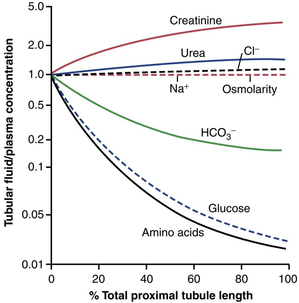
Figure 28-7 Changes in concentrations of different substances in tubular fluid along the proximal convoluted tubule relative to the concentrations of these substances in the plasma and glomerular filtrate. A value of 1.0 indicates that the concentration of the substance in the tubular fluid is the same as the concentration in the plasma. Values below 1.0 indicate that the substance is reabsorbed more avidly than water, whereas values above 1.0 indicate that the substance is reabsorbed to a lesser extent than water or is secreted into the tubules.
شکل 28-7 تغییرات در غلظت مواد مختلف در مایع لولهای در امتداد لوله پیچیده پروگزیمال نسبت به غلظت این مواد در پلاسما و فیلتر گلومرولی. مقدار 1.0 نشان می دهد که غلظت ماده در مایع لوله ای با غلظت پلاسما یکسان است. مقادیر کمتر از 1.0 نشان می دهد که این ماده با اشتیاق بیشتری نسبت به آب جذب می شود، در حالی که مقادیر بالاتر از 1.0 نشان می دهد که این ماده به میزان کمتری نسبت به آب بازجذب می شود یا به داخل لوله ها ترشح می شود.
Secretion of Organic Acids and Bases by Proximal Tubules. The proximal tubule is also an important site for secretion of organic acids and bases such as bile salts, oxalate, urate, and catecholamines. Many of these substances are the end products of metabolism and must be rapidly removed from the body. The secretion of these substances into the proximal tubule plus filtration into the proximal tubule by the glomerular capillaries and almost total lack of reabsorption by the tubules, all combined, contribute to rapid excretion in the urine.
ترشح اسیدها و بازهای آلی توسط لوله های پروگزیمال. لوله پروگزیمال همچنین محل مهمی برای ترشح اسیدها و بازهای آلی مانند نمک های صفراوی، اگزالات، اورات و کاتکول آمین ها است. بسیاری از این مواد محصولات نهایی متابولیسم هستند و باید به سرعت از بدن خارج شوند. ترشح این مواد به داخل لوله پروگزیمال به علاوه فیلتراسیون به داخل لوله پروگزیمال توسط مویرگ های گلومرولی و تقریباً عدم جذب مجدد توسط لوله ها، همگی با هم، به دفع سریع در ادرار کمک می کنند.
In addition to the waste products of metabolism, the kidneys secrete many potentially harmful drugs or toxins into the tubules and rapidly clear these substances from the blood. In the case of certain drugs, such as penicillin and salicylates, the rapid clearance by the kidneys creates a challenge in maintaining a therapeutically effective drug concentration.
علاوه بر مواد زائد متابولیسم، کلیه ها بسیاری از داروها یا سموم بالقوه مضر را به داخل لوله ها ترشح می کنند و به سرعت این مواد را از خون پاک می کنند. در مورد برخی داروها، مانند پنی سیلین و سالیسیلات ها، پاکسازی سریع توسط کلیه ها باعث ایجاد چالش در حفظ غلظت دارو از نظر درمانی موثر می شود.
Another compound that is rapidly secreted by the proximal tubule is paraaminohippuric acid (PAH). PAH is secreted so rapidly that the average person can clear about 90% of the PAH from the plasma flowing through the kidneys and excrete it in the urine. For this reason, the rate of PAH clearance can be used to estimate the renal plasma flow (RPF), as discussed later.
ترکیب دیگری که به سرعت توسط لوله پروگزیمال ترشح می شود، اسید پاراآمینو هیپوریک (PAH) است. PAH به قدری سریع ترشح می شود که یک فرد معمولی می تواند حدود 90 درصد PAH را از پلاسمای جریان یافته از کلیه ها پاک کرده و آن را از طریق ادرار دفع کند. به همین دلیل، نرخ کلیرانس PAH را می توان برای تخمین جریان پلاسمای کلیوی (RPF)، همانطور که بعداً مورد بحث قرار گرفت، استفاده کرد.
SOLUTE AND WATER TRANSPORT IN LOOPS OF HENLE
The loop of Henle consists of three functionally distinct segments the thin descending segment, thin ascending segment, and thick ascending segment. The thin descending and thin ascending segments, as their names imply, have thin epithelial membranes with no brush borders, few mitochondria, and minimal levels of metabolic activity (Figure 28-8).
املاح و حمل و نقل آب در حلقه های HENLE
حلقه هنله از سه بخش عملکردی متمایز تشکیل شده است: بخش نازک نزولی، بخش نازک صعودی، و بخش صعودی ضخیم. بخش های نازک نزولی و نازک صعودی، همانطور که از نام آنها پیداست، دارای غشای اپیتلیال نازک بدون مرزهای قلم مو، تعداد کمی میتوکندری و حداقل سطوح فعالیت متابولیک هستند (شکل 28-8).
The descending part of the thin segment is highly permeable to water and moderately permeable to most solutes, including urea and sodium. The function of this nephron segment is mainly to allow simple diffusion of substances through its walls. About 20% of the filtered water is reabsorbed in the loop of Henle, and almost all of this occurs in the thin descending limb. The ascending limb, including both the thin and thick portions, is virtually impermeable to water, a characteristic that is important for concentrating the urine.
قسمت نزولی بخش نازک نسبت به آب بسیار نفوذپذیر است و نسبت به اکثر املاح از جمله اوره و سدیم نفوذپذیری متوسطی دارد. عملکرد این بخش نفرون عمدتاً اجازه انتشار ساده مواد از طریق دیواره های آن است. حدود 20 درصد از آب تصفیه شده در حلقه هنله دوباره جذب می شود و تقریباً همه اینها در اندام نازک نزولی رخ می دهد. اندام بالارونده، شامل هر دو بخش نازک و ضخیم، عملاً در برابر آب نفوذ ناپذیر است، این ویژگی برای تمرکز ادرار مهم است.
The thick segment of the loop of Henle, which begins about halfway up the ascending limb, has thick epithelial cells that have high metabolic activity and are capable of active reabsorption of sodium, chloride, and potassium (see Figure 28-8). About 25% of the filtered loads of sodium, chloride, and potassium are reabsorbed in the loop of Henle, mostly in the thick ascending limb. Considerable amounts of other ions, such as calcium, bicarbonate, and magnesium, are also reabsorbed in the thick ascending loop of Henle. The thin segment of the ascending limb has a much lower reabsorptive capacity than the thick segment, and the thin descending limb does not reabsorb significant amounts of any of these solutes.
بخش ضخیم حلقه هنله، که تقریباً از نیمه راه اندام صعودی شروع می شود، دارای سلول های اپیتلیال ضخیم است که فعالیت متابولیکی بالایی دارند و قادر به بازجذب فعال سدیم، کلرید و پتاسیم هستند (شکل 28-8 را ببینید). حدود 25 درصد از بارهای فیلتر شده سدیم، کلرید و پتاسیم در حلقه هنله، عمدتاً در اندام صعودی ضخیم، دوباره جذب میشوند. مقادیر قابل توجهی از یون های دیگر مانند کلسیم، بی کربنات و منیزیم نیز در حلقه صعودی ضخیم هنله بازجذب می شوند. بخش نازک اندام صعودی ظرفیت بازجذب بسیار کمتری نسبت به بخش ضخیم دارد و اندام نزولی نازک مقادیر قابل توجهی از هیچ یک از این املاح را بازجذب نمی کند.

Figure 28-8 Cellular ultrastructure and transport characteristics of the thin descending loop of Henle (top) and thick ascending segment of the loop of Henle (bottom). The descending part of the thin segment of the loop of Henle is highly permeable to water and moderately permeable to most solutes but has few mitochondria and little or no active reabsorption. The thick ascending limb of the loop of Henle reabsorbs about 25% of the filtered loads of sodium, chloride, and potassium, as well as large amounts of calcium, bicarbonate, and magnesium. This segment also secretes hydrogen ions into the tubular lumen.
شکل 28-8 فراساختار سلولی و ویژگی های حمل و نقل حلقه نزولی نازک هنل (بالا) و بخش صعودی ضخیم حلقه هنله (پایین). قسمت نزولی بخش نازک حلقه هنله نسبت به آب بسیار نفوذپذیر و نسبت به اکثر املاح نسبتاً نفوذپذیر است اما میتوکندری کمی دارد و بازجذب فعال کم یا اصلاً ندارد. اندام صعودی ضخیم حلقه هنله حدود 25 درصد از بارهای فیلتر شده سدیم، کلرید و پتاسیم و همچنین مقادیر زیادی کلسیم، بی کربنات و منیزیم را بازجذب می کند. این بخش همچنین یون های هیدروژن را به لومن لوله ای ترشح می کند.
An important component of solute reabsorption in the thick ascending limb is the Na+-K+ ATPase pump in the epithelial cell basolateral membranes. As in the proximal tubule, the reabsorption of other solutes in the thick segment of the ascending loop of Henle is closely linked to the reabsorptive capability of the Na+-K+ ATPase pump, which maintains a low intracellular sodium concentration. The low intracellular sodium concentration in turn provides a favorable gradient for movement of sodium from the tubular fluid into the cell. In the thick ascending loop, movement of sodium across the luminal membrane is mediated primarily by a 1-sodium, 2-chloride, 1-potassium cotransporter (NKCC2) (Figure 28-9). This co-transport protein in the luminal membrane uses the potential energy released by downhill diffusion of sodium into the cell to drive the reabsorption of potassium into the cell against a concentration gradient.
یکی از اجزای مهم بازجذب املاح در اندام صعودی ضخیم، پمپ Na+-K+ ATPase در غشای قاعده جانبی سلول های اپیتلیال است. همانند توبول پروگزیمال، بازجذب دیگر املاح در بخش ضخیم حلقه صعودی هنله با قابلیت بازجذب پمپ Na+-K+ ATPase مرتبط است که غلظت سدیم درون سلولی پایینی را حفظ میکند. غلظت کم سدیم داخل سلولی به نوبه خود یک گرادیان مطلوب برای حرکت سدیم از مایع لوله ای به داخل سلول فراهم می کند. در حلقه صعودی ضخیم، حرکت سدیم در سراسر غشای مجرا عمدتاً توسط یک انتقال دهنده 1 سدیم، 2 کلرید، 1 پتاسیم (NKCC2) انجام می شود (شکل 28-9). این پروتئین انتقالی مشترک در غشای مجرا از انرژی پتانسیل آزاد شده توسط انتشار سدیم در سراشیبی به داخل سلول استفاده می کند تا جذب مجدد پتاسیم را به داخل سلول در برابر گرادیان غلظت هدایت کند.
The thick ascending limb of the loop of Henle is the site of action of the powerful loop diuretics furosemide, ethacrynic acid, and bumetanide, all of which inhibit the action of the NKCC2 co-transporter. These diuretics are discussed in Chapter 32. The thick ascending limb also has a sodium-hydrogen counter-transport mechanism in its luminal cell membrane that mediates sodium reabsorption and hydrogen secretion (see Figure 28-9).
اندام صعودی ضخیم حلقه هنله محل اثر دیورتیکهای قوی حلقه فوروزماید، اسید اتاکرینیک و بومتانید است که همگی از عملکرد انتقالدهنده NKCC2 جلوگیری میکنند. این دیورتیک ها در فصل 32 مورد بحث قرار می گیرند. اندام صعودی ضخیم همچنین دارای یک مکانیسم ضد انتقال سدیم-هیدروژن در غشای سلول مجرای خود است که واسطه جذب مجدد سدیم و ترشح هیدروژن است (شکل 28-9 را ببینید).
There is also significant paracellular reabsorption of cations, such as Mg2+, Ca2+, Na+, and K+, in the thick ascending limb as a result of the slight positive charge of the tubular lumen relative to the interstitial fluid. Although the NKCC2 co-transporter moves equal amounts of cat- ions and anions into the cell, there is a slight backleak of potassium ions into the lumen, creating a positive charge of about +8 millivolts in the tubular lumen. This positive charge forces cations such as Mg2+ and Ca2+ to diffuse from the tubular lumen through the paracellular space and into the interstitial fluid.
همچنین بازجذب پاراسلولی قابل توجهی از کاتیونها مانند +Mg2+، Ca2+، Na و +K در اندام صعودی ضخیم در نتیجه بار مثبت خفیف لومن لولهای نسبت به مایع بینابینی وجود دارد. اگرچه انتقال دهنده کمکی NKCC2 مقادیر مساوی از کاتیون ها و آنیون ها را به داخل سلول منتقل می کند، یون های پتاسیم کمی به داخل لومن ریزش می کند و بار مثبتی در حدود 8+ میلی ولت در لومن لوله ایجاد می کند. این بار مثبت کاتیونهایی مانند +Mg2 و +Ca2 را مجبور میکند تا از مجرای لولهای در فضای پاراسلولی و به داخل مایع بینابینی منتشر شوند.
The thick segment of the ascending loop of Henle is virtually impermeable to water. Therefore, most of the water delivered to this segment remains in the tubule, despite reabsorption of large amounts of solute. The tubular fluid in the ascending limb becomes very dilute as it flows toward the distal tubule, a feature that is important in allowing the kidneys to dilute or concentrate the urine under different conditions, as discussed in more detail in Chapter 29.
بخش ضخیم حلقه صعودی هنله عملاً به آب نفوذناپذیر است. بنابراین، با وجود بازجذب مقادیر زیادی از املاح، بیشتر آب تحویلی به این بخش در لوله باقی می ماند. مایع لولهای در اندام صعودی هنگامی که به سمت لوله دیستال جریان مییابد بسیار رقیق میشود، این ویژگی برای اجازه دادن به کلیهها برای رقیق کردن یا تمرکز ادرار در شرایط مختلف مهم است، همانطور که در فصل 29 با جزئیات بیشتر بحث شد.
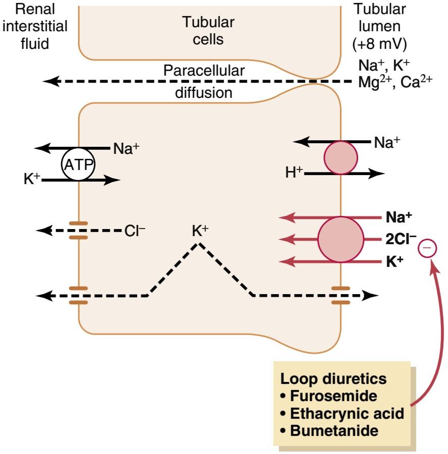
Figure 28-9 Mechanisms of sodium, chloride, and potassium transport in the thick ascending loop of Henle. The Na+-K+ ATPase pump in the basolateral cell membrane maintains a low intracellular sodium concentration and a negative electrical potential in the cell. The 1-sodium, 2-chloride, 1-potassium co-transporter in the luminal membrane transports these three ions from the tubular lumen into the cells, using the potential energy released by the diffusion of sodium down an electrochemical gradient into the cells. Sodium is also transported into the tubular cell by sodium-hydrogen countertransport. The positive charge (+8 mV) of the tubular lumen relative to the interstitial fluid forces cations such as Mg2+ and Ca2+ to diffuse from the lumen to the interstitial fluid via the paracellular pathway.
شکل 28-9 مکانیسمهای انتقال سدیم، کلرید و پتاسیم در حلقه صعودی ضخیم هنله. پمپ Na+-K+ ATPase در غشای سلولی قاعدهای، غلظت سدیم درون سلولی پایین و پتانسیل الکتریکی منفی را در سلول حفظ میکند. انتقال دهنده 1-سدیم، 2-کلرید، 1-پتاسیم در غشای مجرا، این سه یون را از مجرای لوله ای به سلول ها منتقل می کند، با استفاده از انرژی پتانسیل آزاد شده توسط انتشار سدیم به پایین یک گرادیان الکتروشیمیایی به داخل سلول ها. همچنین سدیم با انتقال متقابل سدیم-هیدروژن به داخل سلول لوله ای منتقل می شود. بار مثبت (8+ میلی ولت) لومن لوله ای نسبت به مایع بینابینی کاتیون هایی مانند +Mg2 و +Ca2 را مجبور می کند تا از طریق مسیر پاراسلولی از لومن به مایع بینابینی منتشر شود.
DISTAL TUBULES
The thick segment of the ascending limb of the loop of Henle empties into the distal tubule. The first portion of the distal tubule forms the macula densa, a group of closely packed epithelial cells that is part of the juxtaglomerular complex and provides feedback control of the GFR and blood flow in this same nephron.
لوله های دیستال
بخش ضخیم اندام صعودی حلقه هنله به لوله دیستال تخلیه می شود. اولین بخش از توبول دیستال ماکولا دنسا را تشکیل میدهد، گروهی از سلولهای اپیتلیال به هم فشرده که بخشی از مجموعه juxtaglomerular است و کنترل بازخورد GFR و جریان خون را در همین نفرون فراهم میکند.
The next part of the distal tubule is highly convoluted and has many of the same reabsorb ptive characteristics of the thick segment of the ascending limb of the loop of Henle. That is, it avidly reabsorbs most of the ions, including sodium, potassium, and chloride, but is virtually impermeable to water and urea. For this reason, it is referred to as the diluting segment because it also dilutes the tubular fluid.
قسمت بعدی لوله دیستال به شدت پیچ خورده است و بسیاری از ویژگی های بازجذب یکسان بخش ضخیم اندام صعودی حلقه هنله را دارد. یعنی بیشتر یون ها از جمله سدیم، پتاسیم و کلرید را مشتاقانه بازجذب می کند، اما عملاً در برابر آب و اوره نفوذ ناپذیر است. به همین دلیل از آن به عنوان بخش رقیق کننده یاد می شود زیرا مایع لوله ای را نیز رقیق می کند.
Approximately 5% of the filtered load of sodium chloride is reabsorbed in the early distal tubule. The sodiumchloride co-transporter moves sodium chloride from the tubular lumen into the cell, and the Na+-K+ ATPase pump transports sodium out of the cell across the basolateral membrane (Figure 28-10). Chloride diffuses out of cell into the renal interstitial fluid through chloride channels in the basolateral membrane.
تقریباً 5 درصد از بار فیلتر شده کلرید سدیم در لوله دیستال اولیه بازجذب می شود. انتقال دهنده کمکی سدیم-کلرید کلرید سدیم را از مجرای لوله ای به داخل سلول منتقل می کند و پمپ Na+-K+ ATPase سدیم را به خارج از سلول در سراسر غشای قاعده جانبی منتقل می کند (شکل 28-10). کلرید از طریق کانال های کلرید در غشای قاعده ای جانبی به خارج از سلول به مایع بین بافتی کلیه منتشر می شود.
The thiazide diuretics, which are widely used to treat disorders such as hypertension and heart failure, inhibit the sodium-chloride co-transporter.
دیورتیک های تیازیدی که به طور گسترده برای درمان اختلالاتی مانند فشار خون بالا و نارسایی قلبی استفاده می شود، انتقال دهنده سدیم-کلرید را مهار می کند.
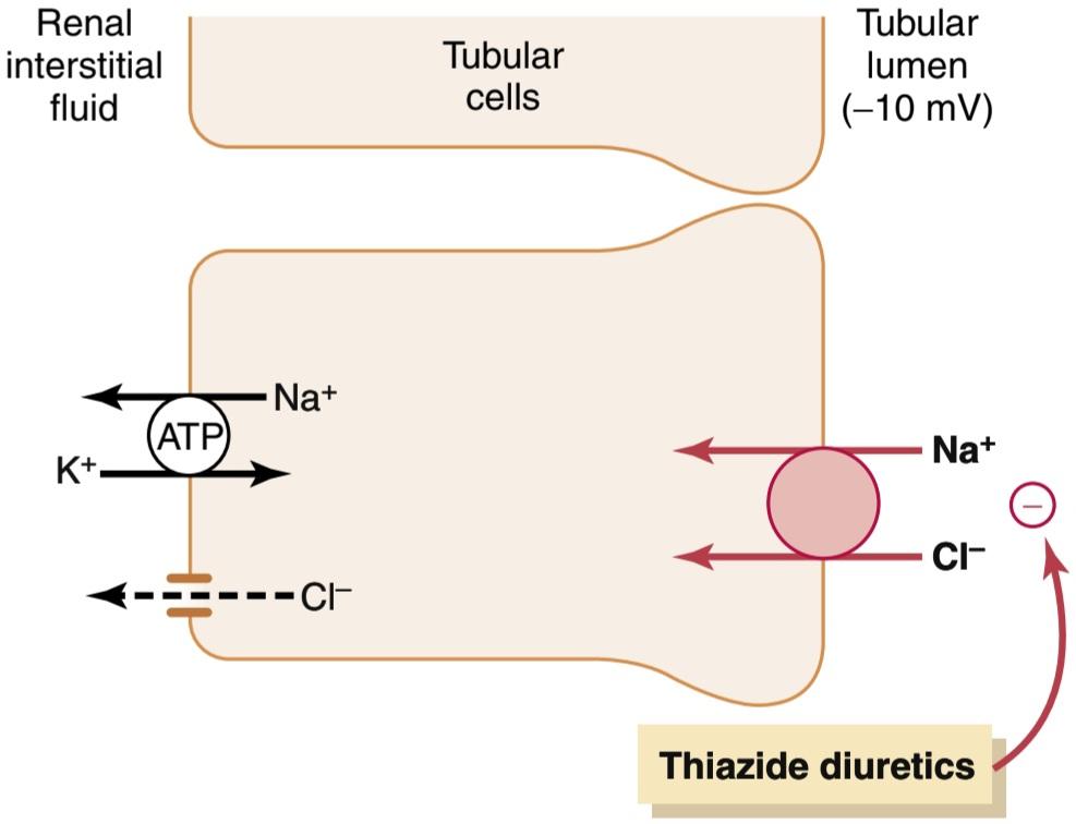
Figure 28-10 Mechanism of sodium chloride transport in the early distal tubule. Sodium and chloride are transported from the tubular lumen into the cell by a co-transporter that is inhibited by thiazide diuretics. Sodium is pumped out of the cell by Na+-K+ ATPase adenosine triphosphatase, and chloride diffuses into the interstitial fluid via chloride channels.
شکل 28-10 مکانیسم انتقال کلرید سدیم در لوله دیستال اولیه سدیم و کلرید از مجرای لولهای به داخل سلول توسط یک انتقالدهنده همراه که توسط دیورتیکهای تیازیدی مهار میشود، منتقل میشوند. سدیم توسط آدنوزین تری فسفاتاز Na+-K+ ATPase از سلول خارج میشود و کلرید از طریق کانالهای کلریدی در مایع بینابینی پخش میشود.
LATE DISTAL TUBULES AND CORTICAL COLLECTING TUBULES
The second half of the distal tubule and subsequent cortical collecting tubule have similar functional characteristics. Anatomically, they are composed of two distinct cell types, the principal cells and intercalated cells (Figure 28-11). The principal cells reabsorb sodium and water from the lumen and secrete potassium ions into the lumen. The type A intercalated cells reabsorb potassium ions and secrete hydrogen ions into the tubular lumen.
توبول های دیستال دیررس و لوله های جمع کننده قشر مغز
نیمه دوم لوله دیستال و لوله جمع کننده قشر بعدی ویژگی های عملکردی مشابهی دارند. از نظر تشریحی، آنها از دو نوع سلول مجزا تشکیل شده اند، سلول های اصلی و سلول های درونی (شکل 28-11). سلول های اصلی سدیم و آب را از لومن بازجذب می کنند و یون های پتاسیم را به لومن ترشح می کنند. سلول های نوع A یون های پتاسیم را دوباره جذب می کنند و یون های هیدروژن را در مجرای لوله ای ترشح می کنند.
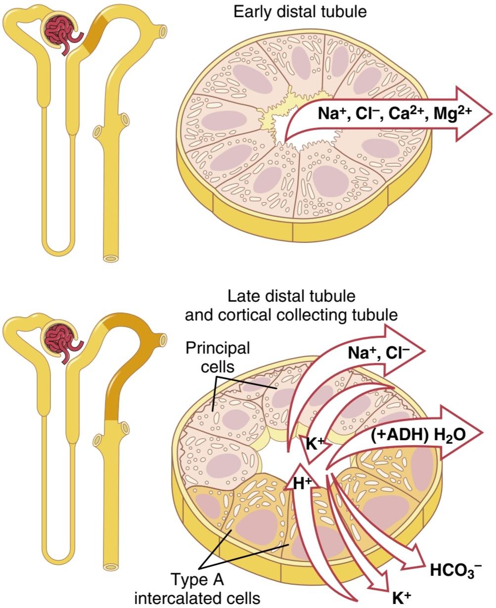
Figure 28-11 Cellular ultrastructure and transport characteristics of the early distal tubule and late distal tubule and collecting tubule. The early distal tubule has many of the same characteristics as the thick ascending loop of Henle and reabsorbs sodium, chloride, calcium, and magnesium but is virtually impermeable to water and urea. The late distal tubules and cortical collecting tubules are composed of two distinct cell types, the principal cells and intercalated cells. The principal cells reabsorb sodium from the lumen and secrete potassium ions into the lumen. Type A intercalated cells reabsorb potassium and bicarbonate ions from the lumen and secrete hydrogen ions into the lumen. The reabsorption of water from this tubular segment is controlled by the concentration of antidiuretic hormone.
شکل 28-11 فراساختار سلولی و ویژگی های انتقال لوله دیستال اولیه و لوله دیستال اواخر و لوله جمع کننده. لوله دیستال اولیه بسیاری از ویژگی های مشابه حلقه صعودی ضخیم هنله را دارد و سدیم، کلرید، کلسیم و منیزیم را بازجذب می کند اما عملاً در برابر آب و اوره نفوذ ناپذیر است. لولههای انتهایی دیستال و لولههای جمعکننده قشر از دو نوع سلول متمایز تشکیل شدهاند، سلولهای اصلی و سلولهای درونی. سلول های اصلی سدیم را از لومن بازجذب می کنند و یون های پتاسیم را به لومن ترشح می کنند. سلول های بینابینی نوع A یون های پتاسیم و بی کربنات را از لومن بازجذب می کنند و یون های هیدروژن را به لومن ترشح می کنند. بازجذب آب از این بخش لولهای توسط غلظت هورمون آنتیدیورتیک کنترل میشود.
Principal Cells Reabsorb Sodium and Secrete Potassium. Sodium reabsorption and potassium secretion by the principal cells depend on the activity of a Na+-K+ ATPase pump in each cell’s basolateral membrane (Figure 28-12). This pump maintains a low sodium concentration inside the cell and, therefore, favors sodium diffusion into the cell through special channels. Secretion of potassium by these cells from the blood into the tubular lumen involves two steps: (1) potassium enters the cell because of the Na+-K+ ATPase pump, which maintains a high intracellular potassium concentration; and (2) once in the cell, potassium diffuses down its concentration gradient across the luminal membrane into the tubular fluid.
سلول های اصلی سدیم را بازجذب و پتاسیم ترشح می کنند. بازجذب سدیم و ترشح پتاسیم توسط سلول های اصلی به فعالیت پمپ Na+-K+ ATPase در غشای قاعده جانبی هر سلول بستگی دارد (شکل 28-12). این پمپ غلظت سدیم پایینی را در داخل سلول حفظ میکند و بنابراین، از طریق کانالهای خاصی به انتشار سدیم در سلول کمک میکند. ترشح پتاسیم توسط این سلول ها از خون به مجرای لوله ای شامل دو مرحله است: (1) پتاسیم به دلیل پمپ Na+-K+ ATPase وارد سلول می شود که غلظت پتاسیم درون سلولی بالایی را حفظ می کند. و (2) هنگامی که پتاسیم در سلول قرار می گیرد، گرادیان غلظت خود را در سراسر غشای مجرا به داخل مایع لوله ای پخش می کند.

Figure 28-12 Mechanism of sodium-chloride reabsorption and potassium secretion in the principal cells of the late distal tubules and cortical collecting tubules. Sodium enters the cell through special channels and is transported out of the cell by the Na+-K+ ATPase pump. Aldosterone antagonists compete with aldosterone for binding sites in the cell and therefore inhibit the effects of aldosterone to stimulate sodium reabsorption and potassium secretion. Sodium channel blockers directly inhibit the entry of sodium into the sodium channels.
شکل 28-12 مکانیسم بازجذب کلرید سدیم و ترشح پتاسیم در سلول های اصلی لوله های انتهایی دیستال و لوله های جمع کننده قشر مغز. سدیم از طریق کانال های مخصوص وارد سلول شده و توسط پمپ Na+-K+ ATPase به خارج از سلول منتقل می شود. آنتاگونیست های آلدوسترون برای مکان های اتصال در سلول با آلدوسترون رقابت می کنند و بنابراین اثرات آلدوسترون را برای تحریک بازجذب سدیم و ترشح پتاسیم مهار می کنند. مسدود کننده های کانال سدیم به طور مستقیم از ورود سدیم به کانال های سدیم جلوگیری می کنند.
The principal cells are the primary sites of action of the potassium-sparing diuretics, including spironolactone, eplerenone, amiloride, and triamterene. Spironolactone and eplerenone are mineralocorticoid receptor antagonists that compete with aldosterone for receptor sites in the principal cells and therefore inhibit the stimulatory effects of aldosterone on sodium reabsorption and potassium secretion. Amiloride and triamterene are sodium channel blockers that directly inhibit the entry of sodium into the sodium channels of the luminal membranes and therefore reduce the amount of sodium that can be transported across the basolateral membranes by the Na+-K+ ATPase pump. This, in turn, decreases transport of potassium into the cells and ultimately reduces potassium secretion into the tubular fluid. For this reason, sodium channel blockers, as well as aldosterone antagonists, decrease urinary excretion of potassium and act as potassium-sparing diuretics.
سلول های اصلی محل اصلی اثر دیورتیک های نگهدارنده پتاسیم، از جمله اسپیرونولاکتون، اپلرنون، آمیلورید و تریامترن هستند. اسپیرونولاکتون و اپلرنون آنتاگونیست های گیرنده مینرالوکورتیکوئیدی هستند که با آلدوسترون برای مکان های گیرنده در سلول های اصلی رقابت می کنند و بنابراین اثرات تحریکی آلدوسترون بر بازجذب سدیم و ترشح پتاسیم را مهار می کنند. آمیلوراید و تریامترن مسدود کننده های کانال سدیم هستند که مستقیماً از ورود سدیم به کانال های سدیم غشاهای مجرا جلوگیری می کنند و بنابراین میزان سدیمی را که می تواند در غشاهای قاعده جانبی توسط پمپ Na+-K+ ATPase منتقل شود کاهش می دهد. این به نوبه خود باعث کاهش انتقال پتاسیم به داخل سلول ها و در نهایت کاهش ترشح پتاسیم به داخل مایع لوله ای می شود. به همین دلیل، مسدود کننده های کانال سدیم و همچنین آنتاگونیست های آلدوسترون، دفع ادراری پتاسیم را کاهش داده و به عنوان دیورتیک های نگهدارنده پتاسیم عمل می کنند.
Intercalated Cells Can Secrete or Reabsorb Hydrogen, Bicarbonate, and Potassium lons. Intercalated cells play a major role in acid-base regulation and constitute 30% to 40% of the cells in the collecting tubules and collecting ducts. There are two types of intercalated cells, type A and type B (Figure 28-13). Type A intercalated cells secrete hydrogen ions by a hydrogen-ATPase transporter and by a hydrogen-potassium-ATPase transporter. Hydrogen is generated in this cell by the action of carbonic anhydrase on water and carbon dioxide to form carbonic acid, which then dissociates into hydrogen ions and bicarbonate ions. The hydrogen ions are then secreted into the tubular lumen and, for each hydrogen ion secreted, a bicarbonate ion becomes available for reabsorption across the basolateral membrane. Type A intercalated cells are especially important in eliminating hydrogen ions while reabsorbing bicarbonate in acidosis.
سلول های درونی می توانند هیدروژن، بی کربنات و یون های پتاسیم را ترشح یا بازجذب کنند. سلول های بینابینی نقش عمده ای در تنظیم اسید-باز دارند و 30 تا 40 درصد سلول های لوله های جمع کننده و مجاری جمع کننده را تشکیل می دهند. دو نوع سلول میانی، نوع A و نوع B وجود دارد (شکل 28-13). سلول های درهم آمیخته نوع A یون های هیدروژن را توسط یک انتقال دهنده هیدروژن-ATPase و یک انتقال دهنده هیدروژن-پتاسیم-ATPase ترشح می کنند. هیدروژن در این سلول از اثر کربنیک انیدراز بر روی آب و دی اکسید کربن برای تشکیل اسید کربنیک تولید می شود که سپس به یون های هیدروژن و یون های بی کربنات تجزیه می شود. سپس یونهای هیدروژن به مجرای لولهای ترشح میشوند و به ازای هر یون هیدروژن ترشح شده، یک یون بی کربنات برای بازجذب در سراسر غشای قاعدهای در دسترس میشود. سلول های نوع A در از بین بردن یون های هیدروژن در حین بازجذب بی کربنات در اسیدوز اهمیت ویژه ای دارند.
Type B intercalated cells have functions opposite to those of type A cells and secrete bicarbonate into the tubular lumen while reabsorbing hydrogen ions in alkalosis. Type B intercalated cells have hydrogen and bicarbonate transporters on opposite sides of the cell membrane compared with type A cells. The chloride-bicarbonate counter-transporter on the apical membrane of type B cells is called pendrin and is different than the chloride- bicarbonate transporter of type A cells. Hydrogen ions are actively transported out of the type B intercalated cell on the basolateral side of the cell membrane by hydrogen- ATPase, and bicarbonate is secreted into the lumen, thus eliminating excess plasma bicarbonate in alkalosis. Induction of chronic metabolic alkalosis increases the number of type B intercalated cells, which contribute to increased excretion of bicarbonate, whereas acidosis increases the number of type A cells.
سلول های نوع B دارای عملکردی بر خلاف عملکرد سلول های نوع A هستند و بی کربنات را در مجرای لوله ای ترشح می کنند در حالی که یون های هیدروژن را در آلکالوز بازجذب می کنند. در مقایسه با سلول های نوع A، سلول های درهم آمیخته نوع B دارای ناقل هیدروژن و بی کربنات در دو طرف غشای سلولی هستند. ضد انتقال دهنده کلرید-بی کربنات روی غشای آپیکال سلول های نوع B پندرین نامیده می شود و با ناقل کلرید-بی کربنات سلول های نوع A متفاوت است. یون های هیدروژن به طور فعال به خارج از سلول های نوع B در سمت قاعده جانبی غشای سلولی توسط هیدروژن ATPase منتقل می شوند و بی کربنات به لومن ترشح می شود، بنابراین بی کربنات اضافی پلاسما در آلکالوز حذف می شود. القای آلکالوز متابولیک مزمن باعث افزایش تعداد سلول های نوع B می شود که به افزایش دفع بی کربنات کمک می کند، در حالی که اسیدوز تعداد سلول های نوع A را افزایش می دهد.
A more detailed discussion of this mechanism is presented in Chapter 31. The intercalated cells can also reabsorb or secrete potassium ions, as shown in Figure 28-13.
همانطور که در شکل 28-13 نشان داده شده است، بحث مفصل تری در مورد این مکانیسم در فصل 31 ارائه شده است. سلول های درهم می توانند یون های پتاسیم را دوباره جذب یا ترشح کنند.
The functional characteristics of the late distal tubule and cortical collecting tubule can be summarized as follows:
ویژگی های عملکردی لوله دیستال دیررس و لوله جمع کننده قشر مغز را می توان به شرح زیر خلاصه کرد:
1. The tubular membranes of both segments are al- most completely impermeable to urea, similar to the diluting segment of the early distal tubule. Thus, almost all the urea that enters these segments pass- es on through and into the collecting duct to be excreted in the urine, although some reabsorption of urea occurs in the medullary collecting ducts.
1. غشاهای لوله ای هر دو بخش تقریباً به طور کامل در برابر اوره غیرقابل نفوذ هستند، مشابه بخش رقیق کننده لوله دیستال اولیه. بنابراین، تقریباً تمام اورهای که وارد این بخشها میشود، از مجرای جمعآوری عبور میکند و به داخل مجرای جمعآوری میرود تا در ادرار دفع شود، اگرچه مقداری بازجذب اوره در مجاری جمعکننده مدولاری اتفاق میافتد.
2. Both the late distal tubule and cortical collecting tubule segments reabsorb sodium ions, and the rate of reabsorption is controlled by hormones, especially aldosterone. At the same time, these segments secrete potassium ions from the peritubular capillary blood into the tubular lumen, a process that is also controlled by aldosterone and other factors, such as the concentration of potassium ions in the body fluids.
2. هر دو بخش انتهایی دیستال و لوله جمع کننده قشر مغز، یون های سدیم را بازجذب می کنند و سرعت بازجذب توسط هورمون ها، به ویژه آلدوسترون، کنترل می شود. در عین حال، این بخشها یونهای پتاسیم را از خون مویرگی اطراف لولهای به مجرای لولهای ترشح میکنند، فرآیندی که توسط آلدوسترون و عوامل دیگر مانند غلظت یونهای پتاسیم در مایعات بدن نیز کنترل میشود.
3. The type A intercalated cells of these nephron segments can avidly secrete hydrogen ions by an active hydrogen-ATPase mechanism in acidosis. This process is different from the secondary active secretion of hydrogen ions by the proximal tubule because it is capable of secreting hydrogen ions against a large concentration gradient, as much as 1000 to 1. This is in contrast to the relatively small gradient (4- to 10-fold) for hydrogen ions that can be achieved by secondary active secretion in the proximal tubule. In alkalosis, the type B intercalated cells secrete bicarbonate and actively reabsorb hydrogen ions. Thus, the intercalated cells play a key role in acidbase regulation of the body fluids.
3. سلول های نوع A این بخش های نفرون می توانند یون های هیدروژن را با مکانیسم فعال هیدروژن-ATPase در اسیدوز ترشح کنند. این فرآیند با ترشح فعال ثانویه یونهای هیدروژن توسط لوله پروگزیمال متفاوت است زیرا قادر است یونهای هیدروژن را در برابر یک گرادیان غلظت زیاد، به اندازه 1000 تا 1 ترشح کند. این برخلاف گرادیان نسبتاً کوچک (4 تا 10 برابر) برای یونهای هیدروژن است که میتوان با ترشح فعال ثانویه در لولههای پروگزیم به دست آورد. در آلکالوز، سلولهای نوع B بیکربنات ترشح میکنند و به طور فعال یونهای هیدروژن را بازجذب میکنند. بنابراین، سلول های درونی نقش کلیدی در تنظیم اسید-باز مایعات بدن دارند.
4. The permeability of the late distal tubule and cortical collecting duct to water is controlled by the concentration of ADH, which is also called vasopressin. With high levels of ADH, these tubular segments are permeable to water but, in the absence of ADH, they are virtually impermeable to water. This special characteristic provides an important mechanism for controlling the degree of dilution or concentration of the urine.
4. نفوذپذیری لوله انتهایی دیستال و مجرای جمع کننده قشر به آب توسط غلظت ADH کنترل می شود که به آن وازوپرسین نیز می گویند. با سطوح بالای ADH، این بخشهای لولهای نسبت به آب نفوذپذیر هستند، اما در غیاب ADH، عملاً به آب غیرقابل نفوذ هستند. این ویژگی خاص مکانیسم مهمی برای کنترل درجه رقت یا غلظت ادرار فراهم می کند.

Figure 28-13 Type A and type B intercalated cells of the collecting tubule. Type A cells contain hydrogen-ATPase and hydrogen- potassium-ATPase in the luminal membrane and secrete hydrogen ions while reabsorbing bicarbonate and potassium ions in acidosis. In type B cells, the hydrogen-ATPase and hydrogen-potassium-ATPase transporters are located in the basolateral membrane and reabsorb hydrogen ions while secreting bicarbonate and potassium ions in alkalosis. The chloride-bicarbonate counter-transporter on the apical membrane of type B cells is called pendrin and is different than the chloride-bicarbonate transporter of type A intercalated cells.
شکل 28-13 نوع A و B سلول های درونی توبول جمع کننده. سلول های نوع A حاوی هیدروژن-ATPase و هیدروژن- پتاسیم-ATPase در غشای مجرا هستند و یون های هیدروژن ترشح می کنند در حالی که یون های بی کربنات و پتاسیم را در اسیدوز بازجذب می کنند. در سلولهای نوع B، انتقالدهندههای هیدروژن-ATPase و هیدروژن-پتاسیم-ATPase در غشای قاعدهای قرار دارند و یونهای هیدروژن را دوباره جذب میکنند در حالی که یونهای بیکربنات و پتاسیم در آلکالوز ترشح میکنند. ضد انتقال دهنده کلرید-بی کربنات روی غشای آپیکال سلول های نوع B پندرین نامیده می شود و با ناقل کلرید-بی کربنات سلول های نوع A متفاوت است.
MEDULLARY COLLECTING DUCTS
Although the medullary collecting ducts usually reabsorb less than 5% of the filtered water and sodium, they are the final site for processing the urine and, therefore, play a critical role in determining the final urine output of water and solutes.
مجاری جمع آوری مدولاری
اگرچه مجاری جمعآوری مدولاری معمولاً کمتر از 5 درصد از آب و سدیم فیلتر شده را بازجذب میکنند، اما مکان نهایی برای پردازش ادرار هستند و بنابراین نقش مهمی در تعیین خروجی ادرار نهایی آب و املاح دارند.
The epithelial cells of the collecting ducts are nearly cuboidal in shape, with smooth surfaces and relatively few mitochondria (Figure 28-14). Special characteristics of this tubular segment are as follows:
سلول های اپیتلیال مجاری جمع کننده تقریباً مکعبی شکل، با سطوح صاف و میتوکندری نسبتاً کمی هستند (شکل 28-14). مشخصات ویژه این بخش لوله ای به شرح زیر است:
1. The permeability of the medullary collecting duct to water is controlled by the level of ADH. With high levels of ADH, water is avidly reabsorbed into the medullary interstitium, thereby reducing the urine volume and concentrating most of the solutes in the urine.
1. نفوذپذیری مجرای جمع کننده مدولاری به آب توسط سطح ADH کنترل می شود. با سطوح بالای ADH، آب مشتاقانه به داخل بینابینی مدولاری بازجذب می شود، در نتیجه حجم ادرار کاهش می یابد و بیشتر املاح در ادرار متمرکز می شود.
2. Unlike the cortical collecting tubule, the medullary collecting duct is permeable to urea, and there are special urea transporters that facilitate urea diffusion across the luminal and basolateral membranes. Therefore, some of the tubular urea is reabsorbed into the medullary interstitium, helping raise the osmolality in this region of the kidneys and contributing to the kidneys’ overall ability to form concentrated urine. This topic is discussed in Chapter 29.
2. بر خلاف لوله جمع کننده کورتیکال، مجرای جمع کننده مدولاری نسبت به اوره نفوذپذیر است و ناقلین اوره ویژه ای وجود دارد که انتشار اوره را در غشاهای مجرای و قاعده جانبی تسهیل می کند. بنابراین، مقداری از اوره لولهای دوباره به داخل بینابینی مدولاری جذب میشود و به افزایش اسمولالیته در این ناحیه از کلیهها کمک میکند و به توانایی کلی کلیهها برای تشکیل ادرار غلیظ کمک میکند. این موضوع در فصل 29 مورد بحث قرار گرفته است.
3. The medullary collecting duct is capable of secreting hydrogen ions against a large concentration gradient, as also occurs in the cortical collecting tubule. Thus, the medullary collecting duct also plays a key role in regulating acid-base balance.
3. مجرای جمع کننده مدولاری قادر به ترشح یون های هیدروژن در برابر یک گرادیان غلظت زیاد است، همانطور که در لوله جمع کننده قشر مغز نیز اتفاق می افتد. بنابراین، مجرای جمع کننده مدولاری نیز نقش کلیدی در تنظیم تعادل اسید و باز ایفا می کند.

Figure 28-14 Cellular ultrastructure and transport characteristics of the medullary collecting duct. The medullary collecting ducts actively reabsorb sodium and secrete hydrogen ions and are permeable to urea, which is reabsorbed in these tubular segments. The reabsorption of water in medullary collecting ducts is controlled by the con- centration of antidiuretic hormone.
شکل 28-14 فراساختار سلولی و ویژگی های حمل و نقل مجرای جمع کننده مدولاری مجاری جمع کننده مدولاری به طور فعال سدیم را بازجذب می کنند و یون های هیدروژن ترشح می کنند و به اوره نفوذ می کنند که در این بخش های لوله ای دوباره جذب می شود. بازجذب آب در مجاری جمع کننده مدولاری توسط غلظت هورمون ضد ادرار کنترل می شود.
SUMMARY OF CONCENTRATIONS OF DIFFERENT SOLUTES IN DIFFERENT TUBULAR SEGMENTS
Whether a solute will become concentrated in the tubular fluid is determined by the relative degree of reabsorption of that solute versus the reabsorption of water. If a greater percentage of water is reabsorbed, the substance becomes more concentrated. If a greater percentage of the solute is reabsorbed, the substance becomes more diluted.
خلاصه غلظت های محلول های مختلف در بخش های لوله ای مختلف
اینکه یک املاح در مایع لولهای متمرکز میشود یا خیر، با درجه نسبی بازجذب آن املاح در مقابل جذب مجدد آب تعیین میشود. اگر درصد بیشتری از آب بازجذب شود، ماده غلیظ تر می شود. اگر درصد بیشتری از املاح دوباره جذب شود، ماده رقیقتر میشود.
Figure 28-15 shows the degree of concentration of several substances in different tubular segments. All the values in this figure represent the tubular fluid concentration divided by the plasma concentration of a substance. If plasma concentration of the substance is assumed to be constant, any change in the tubular fluid/plasma concentration ratio reflects changes in tubular fluid concentration.
شکل 28-15 درجه غلظت چندین ماده را در بخش های لوله ای مختلف نشان می دهد. تمام مقادیر در این شکل نشان دهنده غلظت مایع لوله ای تقسیم بر غلظت پلاسمایی یک ماده است. اگر غلظت پلاسمایی ماده ثابت فرض شود، هر تغییری در نسبت مایع لولهای/غلظت پلاسما نشاندهنده تغییرات غلظت مایع لولهای است.
As the filtrate moves along the tubular system, the concentration rises progressively to higher than 1.0 if more water is reabsorbed than solute, or if there has been a net secretion of the solute into the tubular fluid. If the concentration ratio becomes progressively less than 1.0, this means that relatively more solute has been reabsorbed than water.
همانطور که فیلتر در طول سیستم لوله ای حرکت می کند، اگر آب بیشتری نسبت به املاح بازجذب شود، یا اگر ترشح خالص املاح در مایع لوله ای وجود داشته باشد، غلظت به تدریج به بالاتر از 1.0 افزایش می یابد. اگر نسبت غلظت به تدریج کمتر از 1.0 شود، به این معنی است که نسبتاً بیشتر املاح نسبت به آب بازجذب شده است.
The substances represented at the top of Figure 28-15, such as creatinine, become highly concentrated in the urine. In general, these substances are not needed by the body, and the kidneys have become adapted to reabsorb them only slightly or not at all or even to secrete them into the tubules, thereby excreting large quantities into the urine. Conversely, the substances represented at the bottom of the figure, such as glucose and amino acids, are all strongly reabsorbed. These are all substances that the body needs to conserve, and almost none of them are lost in the urine.
مواد نشان داده شده در بالای شکل 28-15، مانند کراتینین، به شدت در ادرار متمرکز می شوند. به طور کلی، بدن به این مواد نیازی ندارد و کلیهها به گونهای سازگار شدهاند که آنها را فقط کمی یا اصلاً بازجذب نمیکنند یا حتی به داخل لولهها ترشح میکنند و در نتیجه مقادیر زیادی از آنها را به ادرار دفع میکنند. برعکس، مواد نشان داده شده در پایین شکل، مانند گلوکز و اسیدهای آمینه، همگی به شدت بازجذب می شوند. اینها همه موادی هستند که بدن باید آنها را حفظ کند و تقریباً هیچ یک از آنها در ادرار از بین نمی روند.
Tubular Fluid/Plasma Inulin Concentration Ratio Can Be Used to Assess Water Reabsorption by Renal Tubules. Inulin, a polysaccharide used to measure the GFR, is not reabsorbed or secreted by the renal tubules. Changes in inulin concentration at different points along the renal tubule, therefore, reflect changes in the amount of water present in the tubular fluid.
نسبت غلظت مایع توبولار/ پلاسما اینولین را می توان برای ارزیابی بازجذب آب توسط لوله های کلیوی استفاده کرد. اینولین، پلی ساکارید مورد استفاده برای اندازه گیری GFR، توسط لوله های کلیوی جذب یا ترشح نمی شود. بنابراین، تغییرات در غلظت اینولین در نقاط مختلف در امتداد لوله کلیوی، منعکس کننده تغییرات در مقدار آب موجود در مایع لوله است.
For example, the tubular fluid/plasma concentration ratio for inulin rises to about 3.0 at the end of the proximal tubules, indicating that inulin concentration in the tubular fluid is three times greater than in the plasma and glomerular filtrate. Because inulin is not secreted or reabsorbed from the tubules, a tubular fluid/plasma concentration ratio of 3.0 means that only one-third of the water that was filtered remains in the renal tubule and that two-thirds of the filtered water has been reabsorbed as the fluid passes through the proximal tubule. At the end of the collecting ducts, the tubular fluid/plasma inulin concentration ratio rises to about 125 (see Figure 28-15), indicating that only 1/125 of the filtered water remains in the tubule and that more than 99% has been reabsorbed.
به عنوان مثال، نسبت مایع توبولی به غلظت پلاسما برای اینولین به حدود 3.0 در انتهای لوله های پروگزیمال افزایش می یابد، که نشان می دهد غلظت اینولین در مایع لوله ای سه برابر بیشتر از پلاسما و فیلتر گلومرولی است. از آنجایی که اینولین از لولهها ترشح یا جذب مجدد نمیشود، نسبت غلظت مایع/پلاسما 3.0 به این معنی است که تنها یک سوم آبی که فیلتر شده است در لوله کلیوی باقی میماند و دو سوم آب فیلتر شده با عبور مایع از لوله پروگزیمال دوباره جذب شده است. در انتهای مجاری جمع آوری، نسبت غلظت مایع توبولی به اینولین پلاسما به حدود 125 می رسد (شکل 28-15 را ببینید)، که نشان می دهد تنها 1/125 از آب تصفیه شده در لوله باقی می ماند و بیش از 99٪ دوباره جذب شده است.

Figure 28-15 Changes in average concentrations of different substances at different points in the tubular system relative to the concentration of that substance in the plasma and glomerular filtrate. A value of 1.0 indicates that the concentration of the substance in the tubular fluid is the same as the concentration of that substance in the plasma. Values below 1.0 indicate that the substance is reabsorbed more avidly than water, whereas values above 1.0 indicate that the substance is reabsorbed to a lesser extent than water or is secreted into the tubules. PAH, Para amino hippuric acid.
شکل 28-15 تغییرات در غلظت متوسط مواد مختلف در نقاط مختلف سیستم لوله ای نسبت به غلظت آن ماده در پلاسما و فیلتر گلومرولی. مقدار 1.0 نشان می دهد که غلظت ماده در مایع لوله ای با غلظت آن ماده در پلاسما یکسان است. مقادیر کمتر از 1.0 نشان می دهد که این ماده با اشتیاق بیشتری نسبت به آب جذب می شود، در حالی که مقادیر بالاتر از 1.0 نشان می دهد که این ماده به میزان کمتری نسبت به آب بازجذب می شود یا به داخل لوله ها ترشح می شود. PAH، پارا آمینو هیپوریک اسید.
REGULATION OF TUBULAR REABSORPTION
Because it is essential to maintain a precise balance between tubular reabsorption and glomerular filtration, there are multiple nervous, hormonal, and local control mechanisms that regulate tubular reabsorption, just as there are for control of glomerular filtration. An important feature of tubular reabsorption is that reabsorption of some solutes can be regulated independently of others, especially through hormonal control mechanisms.
تنظیم بازجذب لوله ای
از آنجایی که حفظ تعادل دقیق بین بازجذب لولهای و فیلتراسیون گلومرولی ضروری است، مکانیسمهای کنترل عصبی، هورمونی و موضعی متعددی وجود دارد که بازجذب لولهای را تنظیم میکند، همانطور که برای کنترل فیلتراسیون گلومرولی وجود دارد. یکی از ویژگیهای مهم بازجذب لولهای این است که بازجذب برخی از املاح را میتوان بهطور مستقل از سایر املاح تنظیم کرد، مخصوصاً از طریق مکانیسمهای کنترل هورمونی.
GLOMERULOTUBULAR BALANCE- REABSORPTION RATE INCREASES IN RESPONSE TO INCREASED TUBULAR LOAD
One of the most basic mechanisms for controlling tubular reabsorption is the intrinsic ability of the tubules to increase their reabsorption rate in response to increased tubular load (increased tubular inflow). This phenomenon is referred to as glomerulotubular balance. For example, if the GFR increases from 125 to 150 ml/min, the absolute rate of proximal tubular reabsorption also increases from about 81 ml/min (65% of GFR) to about 97.5 ml/min (65% of GFR). Thus, glomerulotubular balance refers to the fact that the total rate of reabsorption increases as the filtered load increases, even though the percentage of GFR reabsorbed in the proximal tubule remains relatively constant, at about 65%.
تعادل گلومرولولولوبولار- نرخ بازجذب در پاسخ به افزایش بار لولهای افزایش مییابد
یکی از اساسیترین مکانیسمها برای کنترل بازجذب لولهای، توانایی ذاتی لولهها برای افزایش نرخ بازجذب در پاسخ به افزایش بار لولهای (افزایش جریان ورودی لوله) است. به این پدیده تعادل گلومرولوتوبولی گفته می شود. به عنوان مثال، اگر GFR از 125 به 150 میلی لیتر در دقیقه افزایش یابد، نرخ مطلق بازجذب لوله پروگزیمال نیز از حدود 81 میلی لیتر در دقیقه (65 درصد GFR) به حدود 97.5 میلی لیتر در دقیقه (65 درصد از GFR) افزایش می یابد. بنابراین، تعادل گلومرولولوتوبولی به این واقعیت اشاره دارد که با افزایش بار فیلتر شده، نرخ کل بازجذب افزایش مییابد، حتی اگر درصد GFR بازجذب شده در لوله پروگزیمال نسبتاً ثابت بماند، در حدود 65%.
Some degree of glomerulotubular balance also occurs in other tubular segments, especially the loop of Henle. The precise mechanisms responsible for this are not fully understood but may be due partly to changes in physical forces in the tubule and surrounding renal interstitium, as discussed later. It is clear that the mechanisms for glomerulotubular balance can occur independently of hormones and can be demonstrated in completely isolated kidneys or even in completely isolated proximal tubular segments.
درجاتی از تعادل گلومرولولوتوبولی نیز در سایر بخش های لوله ای، به ویژه حلقه هنل رخ می دهد. مکانیسم های دقیق مسئول این امر به طور کامل شناخته نشده است، اما ممکن است تا حدی به دلیل تغییرات در نیروهای فیزیکی در توبول و بینابینی کلیوی اطراف آن باشد، همانطور که بعداً بحث شد. واضح است که مکانیسم تعادل گلومرولولوتوبولی می تواند مستقل از هورمون ها رخ دهد و می تواند در کلیه های کاملاً ایزوله یا حتی در بخش های لوله پروگزیمال کاملاً ایزوله نشان داده شود.
Glomerulotubular balance helps prevent overloading of the distal tubular segments when GFR increases. Glomerulotubular balance acts as another line of defense to buffer the effects of spontaneous changes in the GFR on urine output. (The other line of defense, discussed earlier, includes the renal autoregulatory mechanisms, especially tubuloglomerular feedback, which help prevent large changes in GFR.) Working together, the autoregulatory and glomerulotubular balance mechanisms prevent large changes in fluid flow in the distal tubules when the arterial pressure changes or when there are other disturbances that would otherwise upset sodium and volume homeostasis.
تعادل گلومرولوتوبولار به جلوگیری از بارگذاری بیش از حد بخش های لوله ای دیستال در هنگام افزایش GFR کمک می کند. تعادل گلومرولولوتوبولار به عنوان یک خط دفاعی دیگر عمل می کند تا اثرات تغییرات خود به خودی در GFR بر برون ده ادرار را مهار کند. (خط دفاعی دیگر که قبلاً مورد بحث قرار گرفت، شامل مکانیسمهای خودتنظیمی کلیوی، بهویژه بازخورد توبولوگلومرولی است که به جلوگیری از تغییرات بزرگ در GFR کمک میکند.) مکانیسمهای خودتنظیمی و تعادل گلومرولوتوبولی با همکاری یکدیگر از تغییرات بزرگ در جریان مایع در لولههای دیستال در هنگام تغییر فشار شریانی یا زمانی که اختلالات دیگری وجود دارد که باعث افزایش حجم سدیم میشود، جلوگیری میکند.
PERITUBULAR CAPILLARY AND RENAL INTERSTITIAL FLUID PHYSICAL FORCES
Hydrostatic and colloid osmotic forces govern the rate of reabsorption across the peritubular capillaries, just as they control filtration in the glomerular capillaries. Changes in peritubular capillary reabsorption can in turn influence the hydrostatic and colloid osmotic pressures of the renal inters titium and, ultimately, reabsorption of water and solutes from the renal tubules.
نیروهای فیزیکی مویرگی اطراف لوله و مایع بینابینی کلیوی
نیروهای اسمزی هیدرواستاتیک و کلوئیدی بر میزان بازجذب در سرتاسر مویرگ های اطراف لوله نظارت می کنند، همانطور که فیلتراسیون را در مویرگ های گلومرولی کنترل می کنند. تغییرات در بازجذب مویرگی دور لولهای میتواند به نوبه خود بر فشارهای اسمزی هیدرواستاتیک و کلوئیدی تیتیوم میانی کلیوی و در نهایت، بازجذب آب و املاح از لولههای کلیوی تأثیر بگذارد.
Normal Values for Physical Forces and Reabsorption Rate. As the glomerular filtrate passes through the renal tubules, more than 99% of the water and most of the solutes are normally reabsorbed. Fluid and electrolytes are reabsorbed from the tubules into the renal interstitium and from there into the peritubular capillaries. The normal rate of peritubular capillary reabsorption is about 124 ml/min.
مقادیر نرمال برای نیروهای فیزیکی و نرخ بازجذب. هنگامی که فیلتر گلومرولی از لوله های کلیوی عبور می کند، بیش از 99 درصد آب و اکثر املاح به طور معمول دوباره جذب می شوند. مایعات و الکترولیت ها دوباره از لوله ها به داخل بینابینی کلیه و از آنجا به مویرگ های اطراف لوله جذب می شوند. سرعت طبیعی بازجذب مویرگی دور لوله ای حدود 124 میلی لیتر در دقیقه است.
Reabsorption across the peritubular capillaries can be calculated as follows:
بازجذب در مویرگ های اطراف لوله را می توان به صورت زیر محاسبه کرد:

The net reabsorptive force represents the sum of the hydrostatic and colloid osmotic forces that favor or oppose reabsorption across the peritubular capillaries. These forces include the following: (1) hydrostatic pressure inside the peritubular capillaries (peritubular hydrostatic pressure [P]), which opposes reabsorption; (2) hydrostatic pressure in the renal interstitium (P1) outside the capillaries, which favors reabsorption; (3) colloid osmotic pressure of the peritubular capillary plasma proteins (), which favors reabsorption; and (4) colloid osmotic pressure of the proteins in the renal interstitium (), which opposes reabsorption.
نیروی بازجذب خالص مجموع نیروهای اسمزی هیدرواستاتیک و کلوئیدی را نشان می دهد که طرفدار یا مخالف بازجذب در سراسر مویرگ های اطراف لوله هستند. این نیروها شامل موارد زیر است: (1) فشار هیدرواستاتیک در داخل مویرگهای اطراف لوله (فشار هیدرواستاتیک اطراف لوله [P])، که با جذب مجدد مخالف است. (2) فشار هیدرواستاتیک در بینابینی کلیه (P1) خارج از مویرگها، که به نفع بازجذب است. (3) فشار اسمزی کلوئیدی پروتئینهای پلاسمای مویرگی اطراف لولهای ()، که به جذب مجدد کمک میکند. و (4) فشار اسمزی کلوئیدی پروتئین ها در بینابینی کلیوی () که با بازجذب مخالف است.
Figure 28-16 shows the approximate normal forces that favor and oppose peritubular reabsorption. Because the normal peritubular capillary pressure averages about 13 mm Hg and the renal interstitial fluid hydrostatic pressure averages 6 mm Hg, there is a positive hydrostatic pressure gradient from the peritubular capillary to the interstitial fluid of about 7 mm Hg, which opposes fluid reabsorption. This opposition to fluid reabsorption is more than counterbalanced by the colloid osmotic pressures that favor reabsorption. The plasma colloid osmotic pressure, which favors reabsorption, is about 32 mm Hg, and the colloid osmotic pressure of the interstitium, which opposes reabsorption, is 15 mm Hg, causing a net colloid osmotic force of about 17 mm Hg, favoring reabsorption. Therefore, subtracting the net hydrostatic forces that oppose reabsorption (7 mm Hg) from the net colloid osmotic forces that favor reabsorption (17 mm Hg) gives a net reabsorptive force of about 10 mm Hg. This value is high, similar to that found in the glomerular capillaries, but in the opposite direction.
شکل 28-16 نیروهای نرمال تقریبی را نشان می دهد که طرفدار و مخالف بازجذب دور لوله ای هستند. از آنجایی که فشار طبیعی مویرگی اطراف لوله ای به طور متوسط حدود 13 میلی متر جیوه و فشار هیدرواستاتیک مایع بینابینی کلیوی به طور متوسط 6 میلی متر جیوه است، یک گرادیان فشار هیدرواستاتیک مثبت از مویرگ اطراف لوله تا مایع بینابینی حدود 7 میلی متر جیوه وجود دارد که با بازجذب مایع مخالف است. این مخالفت با بازجذب مایع بیشتر از فشارهای اسمزی کلوئیدی است که به نفع بازجذب است. فشار اسمزی کلوئیدی پلاسما که به بازجذب کمک می کند، حدود 32 میلی متر جیوه است و فشار اسمزی کلوئیدی بینابینی که با بازجذب مخالف است، 15 میلی متر جیوه است که باعث ایجاد نیروی اسمزی خالص کلوئیدی حدود 17 میلی متر جیوه می شود که به نفع بازجذب است. بنابراین، با کم کردن نیروهای هیدرواستاتیک خالص مخالف بازجذب (7 میلیمتر جیوه) از نیروهای اسمزی کلوئیدی خالص که به نفع بازجذب (17 میلیمتر جیوه) هستند، نیروی بازجذب خالص حدود 10 میلیمتر جیوه به دست میآید. این مقدار زیاد است، مشابه آنچه در مویرگ های گلومرولی وجود دارد، اما در جهت مخالف است.
The other factor that contributes to the high rate of fluid reabsorption in the peritubular capillaries is a large filtration coefficient (K) because of the high hydraulic conductivity and large surface area of the capillaries. Because the reabsorption rate is normally about 124 ml/ min and net reabsorption pressure is 10 mm Hg, K, normally is about 12.4 ml/min per mm Hg.
عامل دیگری که به سرعت بالای بازجذب مایع در مویرگ های اطراف لوله کمک می کند، ضریب فیلتراسیون (K) زیاد است که به دلیل هدایت هیدرولیکی بالا و سطح بزرگ مویرگ ها است. از آنجایی که سرعت بازجذب به طور معمول حدود 124 میلی لیتر در دقیقه و فشار خالص بازجذب 10 میلی متر جیوه است، پتاسیم معمولاً حدود 4/12 میلی لیتر در دقیقه بر میلی متر جیوه است.
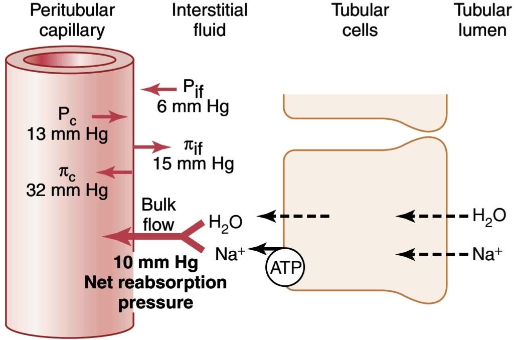
Figure 28-16 Summary of the hydrostatic and colloid osmotic forces that determine fluid reabsorption by the peritubular capillaries. The numerical values shown are estimates of the normal values for humans. The net reabsorptive pressure is normally about 10 mm Hg, causing fluid and solutes to be reabsorbed into the peritubular capillaries as they are transported across the renal tubular cells. ATP, Adenosine triphosphate; P, peritubular capillary hydrostatic pressure; Pit, interstitial fluid hydrostatic pressure; л, peritubular capillary colloid osmotic pressure;, interstitial fluid colloid osmotic pressure.
شکل 28-16 خلاصه ای از نیروهای اسمزی هیدرواستاتیک و کلوئیدی که بازجذب مایع را توسط مویرگ های اطراف لوله تعیین می کنند. مقادیر عددی نشان داده شده تخمینی از مقادیر نرمال برای انسان است. فشار خالص بازجذب معمولاً حدود 10 میلیمتر جیوه است و باعث میشود که مایع و املاح به مویرگهای اطراف لولهای جذب شوند و در سلولهای لولهای کلیوی منتقل شوند. ATP، آدنوزین تری فسفات؛ P، فشار هیدرواستاتیک مویرگی اطراف لوله. گودال، فشار هیدرواستاتیک مایع بینابینی؛ л، فشار اسمزی کلوئیدی مویرگی دور لوله ای؛ فشار اسمزی کلوئیدی مایع بینابینی.
Regulation of Peritubular Capillary Physical Forces. The two determinants of peritubular capillary reabsorption that are directly influenced by renal hemodynamic changes are the hydrostatic and colloid osmotic pressures of the peritubular capillaries. The peritubular capillary hydrostatic pressure is influenced by the arterial pressure and resistances of the afferent and efferent arterioles as follows:
تنظیم نیروهای فیزیکی مویرگی اطراف لوله. دو عامل تعیین کننده بازجذب مویرگی دور لوله ای که مستقیماً تحت تأثیر تغییرات همودینامیک کلیه قرار می گیرند، فشار اسمزی هیدرواستاتیک و کلوئیدی مویرگ های اطراف لوله هستند. فشار هیدرواستاتیک مویرگی اطراف لوله تحت تأثیر فشار شریانی و مقاومت شریان های آوران و وابران به شرح زیر است:
1. Increases in arterial pressure tend to raise peritubular capillary hydrostatic pressure and decrease the reabsorption rate. This effect is buffered to some extent by autoregulatory mechanisms that maintain relatively constant renal blood flow, as well as relatively constant hydrostatic pressures in the renal blood vessels.
1. افزایش فشار شریانی باعث افزایش فشار هیدرواستاتیک مویرگی اطراف لوله و کاهش نرخ بازجذب می شود. این اثر تا حدی توسط مکانیسم های خودتنظیمی که جریان خون کلیوی را نسبتاً ثابت نگه می دارد و همچنین فشار هیدرواستاتیک نسبتاً ثابت در رگ های خونی کلیوی بافر می شود.
2. An increase in resistance of the afferent or efferent arterioles reduces peritubular capillary hydrostatic pressure and tends to increase reabsorption rate. Although constriction of the efferent arterioles in- creases glomerular capillary hydrostatic pressure, it lowers peritubular capillary hydrostatic pressure.
2. افزایش مقاومت شریان های آوران یا وابران فشار هیدرواستاتیک مویرگی دور لوله ای را کاهش می دهد و تمایل به افزایش نرخ بازجذب دارد. اگرچه انقباض شریان های وابران فشار هیدرواستاتیک مویرگی گلومرولی را افزایش می دهد، فشار هیدرواستاتیک مویرگی اطراف لوله را کاهش می دهد.
The second major determinant of peritubular capillary reabsorption is the colloid osmotic pressure of the plasma in these capillaries; raising the colloid osmotic pressure increases peritubular capillary reabsorption. The colloid osmotic pressure of peritubular capillaries is determined by the following: (1) the systemic plasma colloid osmotic pressure (increasing the plasma protein concentration of systemic blood tends to raise peritubular capillary colloid osmotic pressure, thereby increasing reabsorption); and (2) the filtration fraction-the higher the filtration fraction, the greater the fraction of plasma filtered through the glomerulus and, consequently, the more concentrated the protein becomes in the plasma that remains behind. Thus, increasing the filtration fraction also tends to increase the peritubular capillary reabsorption rate. Because filtration fraction is defined as the ratio of GFR/RPF, an increased filtration fraction can occur as a result of increased GFR or decreased RPF. Some renal vasoconstrictors, such as angiotensin II, increase peritubular capillary reabsorption by decreasing RPF and increasing filtration fraction, as discussed later.
دومین عامل تعیین کننده بازجذب مویرگی دور لوله ای فشار اسمزی کلوئیدی پلاسما در این مویرگ ها است. افزایش فشار اسمزی کلوئیدی باعث افزایش بازجذب مویرگی اطراف لوله می شود. فشار اسمزی کلوئیدی مویرگ های اطراف لوله با موارد زیر تعیین می شود: (1) فشار اسمزی کلوئیدی پلاسما سیستمیک (افزایش غلظت پروتئین پلاسمایی خون سیستمیک تمایل به افزایش فشار اسمزی کلوئیدی مویرگی دور لوله ای دارد و در نتیجه بازجذب را افزایش می دهد). و (2) کسر فیلتراسیون – هر چه کسر فیلتراسیون بیشتر باشد، کسری از پلاسمای فیلتر شده از طریق گلومرول بیشتر است و در نتیجه، پروتئین در پلاسمایی که پشت سر باقی می ماند غلیظ تر می شود. بنابراین، افزایش کسر فیلتراسیون نیز تمایل به افزایش نرخ بازجذب مویرگی دور لوله ای دارد. از آنجایی که کسر فیلتراسیون به عنوان نسبت GFR/RPF تعریف می شود، کسر فیلتراسیون افزایش یافته می تواند در نتیجه افزایش GFR یا کاهش RPF رخ دهد. برخی از منقبض کننده های عروق کلیوی، مانند آنژیوتانسین II، با کاهش RPF و افزایش کسر فیلتراسیون، بازجذب مویرگی دور لوله ای را افزایش می دهند، همانطور که بعداً بحث شد.
Changes in the peritubular capillary Kf can also influence the reabsorption rate because K is a measure of the permeability and surface area of the capillaries. Increases in K raise reabsorption, whereas decreases in K lower peritubular capillary reabsorption. Kf remains relatively constant in most physiological conditions. Table 28-2 summarizes the factors that can influence the peritubular capillary reabsorption rate.
تغییرات در Kf مویرگی اطراف لولهای نیز میتواند بر میزان بازجذب تأثیر بگذارد زیرا K معیاری برای نفوذپذیری و سطح مویرگها است. افزایش پتاسیم باعث افزایش بازجذب می شود، در حالی که کاهش پتاسیم باعث کاهش جذب مجدد مویرگی دور لوله ای می شود. Kf در اکثر شرایط فیزیولوژیکی نسبتا ثابت می ماند. جدول 28-2 عواملی را که می توانند بر میزان بازجذب مویرگی دور لوله ای تأثیر بگذارند خلاصه می کند.
Table 28-2 Factors That Can Influence Peritubular Capillary Reabsorption
جدول 28-2 عواملی که می توانند بر بازجذب مویرگی دور لوله ای تأثیر بگذارند
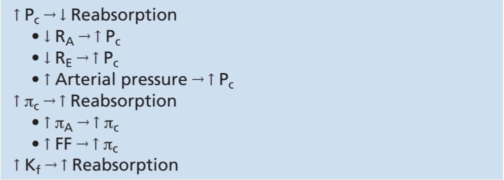
FF, Filtration fraction; Kf, peritubular capillary filtration coefficient; Pc peritubular capillary hydrostatic pressure; A, arterial plasma colloid osmotic pressure; peritubular capillary colloid osmotic pressure; RA, and RE, afferent and efferent arteriolar resistances, respectively.
FF، کسر فیلتر. Kf، ضریب فیلتراسیون مویرگی اطراف لوله. فشار هیدرواستاتیک مویرگی دور لوله ای. A، فشار اسمزی کلوئیدی پلاسمای شریانی. فشار اسمزی کلوئیدی مویرگی اطراف لوله؛ RE، و RA، مقاومت شریانی آوران و وابران به ترتیب.
Renal Interstitial Hydrostatic and Colloid Osmotic Pressures. Ultimately, changes in peritubular capillary physical forces influence tubular reabsorption by changing the physical forces in the renal interstitium surrounding the tubules. For example, a decrease in the reabsorptive force across the peritubular capillary membranes, caused by increased peritubular capillary hydrostatic pressure or decreased peritubular capillary colloid osmotic pressure, reduces the uptake of fluid and solutes from the interstitium into the peritubular capillaries. This action in turn raises renal interstitial fluid hydrostatic pressure and decreases interstitial fluid colloid osmotic pressure because of dilution of the proteins in the renal interstitium. These changes then decrease the net reabsorption of fluid from the renal tubules into the interstitium, especially in the proximal tubules.
فشارهای اسمزی هیدرواستاتیک و کلوئیدی بینابینی کلیوی. در نهایت، تغییرات در نیروهای فیزیکی مویرگی اطراف لوله بر جذب مجدد لولهای با تغییر نیروهای فیزیکی در بینابینی کلیوی اطراف لولهها تأثیر میگذارد. به عنوان مثال، کاهش نیروی بازجذب در سراسر غشاهای مویرگی اطراف لوله، ناشی از افزایش فشار هیدرواستاتیک مویرگی اطراف لوله یا کاهش فشار اسمزی کلوئیدی مویرگی اطراف لوله، باعث کاهش جذب مایع و املاح از بینابینی به مویرگ های اطراف لوله می شود. این عمل به نوبه خود باعث افزایش فشار هیدرواستاتیک مایع بین بافتی کلیه و کاهش فشار اسمزی کلوئیدی مایع بینابینی به دلیل رقیق شدن پروتئین ها در بینابینی کلیه می شود. سپس این تغییرات باعث کاهش جذب مجدد مایع از لوله های کلیوی به داخل بینابینی، به ویژه در لوله های پروگزیمال می شود.
The mechanisms whereby changes in interstitial fluid hydrostatic and colloid osmotic pressures influence tubular reabsorption can be understood by examining the pathways through which solute and water are reabsorbed (Figure 28-17). Once the solutes enter the intercellular channels or renal interstitium by active transport or passive diffusion, water is drawn from the tubular lumen into the interstitium by osmosis. Furthermore, once the water and solutes are in the interstitial spaces, they can be swept up into the peritubular capillaries or diffuse back through the epithelial junctions into the tubular lumen. The so-called tight junctions between the epithelial cells of the proximal tubule are actually leaky, so considerable amounts of sodium can diffuse in both directions through these junctions. With the normal high rate of peritubular capillary reabsorption, the net movement of water and solutes is into the peritubular capillaries, with little back- leak into the lumen of the tubule. However, when peritubular capillary reabsorption is reduced, there is increased interstitial fluid hydrostatic pressure and a tendency for greater amounts of solute and water to leak back into the tubular lumen, thereby reducing the rate of net reabsorption (see Figure 28-17).
مکانیسم هایی که به موجب آن تغییرات در فشارهای اسمزی هیدرواستاتیک و کلوئیدی سیال بینابینی بر جذب مجدد لوله ای تأثیر می گذارد را می توان با بررسی مسیرهایی که املاح و آب از طریق آنها بازجذب می شوند درک کرد (شکل 28-17). هنگامی که املاح با انتقال فعال یا انتشار غیرفعال وارد کانال های بین سلولی یا بینابینی کلیه می شوند، آب از لومن لوله ای به وسیله اسمز به داخل بینابینی کشیده می شود. علاوه بر این، هنگامی که آب و املاح در فضاهای بینابینی قرار می گیرند، می توانند به داخل مویرگ های اطراف لوله منتقل شوند یا از طریق اتصالات اپیتلیال به لومن لوله ای منتشر شوند. به اصطلاح اتصالات محکم بین سلول های اپیتلیال لوله پروگزیمال در واقع نشتی دارند، بنابراین مقادیر قابل توجهی سدیم می تواند در هر دو جهت از طریق این اتصالات پخش شود. با نرخ بالای طبیعی بازجذب مویرگی دور لولهای، حرکت خالص آب و املاح به داخل مویرگهای دور لولهای انجام میشود، با نشت کمی به سمت مجرای لوله. با این حال، هنگامی که بازجذب مویرگی دور لولهای کاهش مییابد، فشار هیدرواستاتیک مایع بینابینی افزایش مییابد و تمایل به نشت مقادیر بیشتری از املاح و آب به مجرای لولهای وجود دارد که در نتیجه سرعت بازجذب خالص کاهش مییابد (شکل 28-17 را ببینید).
The opposite is true when peritubular capillary reabsorption increases above the normal level. An initial increase in reabsorption by the peritubular capillaries tends to reduce interstitial fluid hydrostatic pressure and raise interstitial fluid colloid osmotic pressure. Both these forces favor movement of fluid and solutes out of the tubular lumen and into the interstitium; therefore, backleak of water and solutes into the tubular lumen is reduced, and net tubular reabsorption is increased.
برعکس زمانی که بازجذب مویرگی دور لولهای بیش از سطح طبیعی افزایش مییابد صادق است. افزایش اولیه در بازجذب توسط مویرگ های اطراف لوله باعث کاهش فشار هیدرواستاتیک مایع بینابینی و افزایش فشار اسمزی کلوئیدی مایع بینابینی می شود. هر دوی این نیروها طرفدار حرکت مایع و املاح به خارج از مجرای لولهای و داخل بینابینی هستند. بنابراین، نشت آب و املاح به مجرای لوله ای کاهش می یابد و بازجذب خالص لوله افزایش می یابد.
Thus, through changes in the hydrostatic and colloid osmotic pressures of the renal interstitium, the uptake of water and solutes by the peritubular capillaries is closely matched to the net reabsorption of water and solutes from the tubular lumen into the interstitium. In general, forces that increase peritubular capillary reabsorption also increase reabsorption from the renal tubules. Conversely, hemodynamic changes that inhibit peritubular capillary reabsorption also inhibit tubular reabsorption of water and solutes.
بنابراین، از طریق تغییرات در فشارهای اسمزی هیدرواستاتیک و کلوئیدی بینابینی کلیوی، جذب آب و املاح توسط مویرگ های اطراف لوله با بازجذب خالص آب و املاح از مجرای لوله ای به داخل بینابینی مطابقت دارد. به طور کلی، نیروهایی که باعث افزایش بازجذب مویرگی اطراف لوله می شوند، بازجذب از لوله های کلیوی را نیز افزایش می دهند. برعکس، تغییرات همودینامیک که بازجذب مویرگی دور لوله ای را مهار می کند، بازجذب لوله ای آب و املاح را نیز مهار می کند.

Figure 28-17 Proximal tubular and peritubular capillary reabsorption under normal conditions (top) and during decreased peritubular capillary reabsorption (bottom) caused by increasing peritubular capillary hydrostatic pressure (P) or decreasing peritubular capillary colloid osmotic pressure (7). Reduced peritubular capillary reabsorption, in turn, decreases the net reabsorption of solutes and water by increasing the amounts of solutes and water that leak back into the tubular lumen through the tight junctions of the tubular epithelial cells, especially in the proximal tubule.
شکل 28-17 بازجذب مویرگی پروگزیمال و پرتوبولی در شرایط عادی (بالا) و در طی کاهش بازجذب مویرگی دور لوله ای (پایین) ناشی از افزایش فشار هیدرواستاتیک مویرگی دور لوله ای (P) یا کاهش فشار اسمزی کلوئیدی مویرگی دور لوله ای (7). کاهش بازجذب مویرگی دور لولهای، به نوبه خود، با افزایش مقدار املاح و آبی که از طریق اتصالات محکم سلولهای اپیتلیال لولهای، بهویژه در لوله پروگزیمال، به مجرای لولهای نشت میکند، بازجذب خالص املاح و آب را کاهش میدهد.
EFFECT OF ARTERIAL PRESSURE ON URINE OUTPUT-PRESSURE NATRIURESIS AND
PRESSURE DIURESIS
Even small increases in arterial pressure can cause marked increases in urinary excretion of sodium and water, phenomena referred to as pressure natriuresis and pressure diuresis. Because of the autoregulatory mechanisms described in Chapter 27, increasing the arterial pressure between the limits of 75 and 160 mm Hg usually has only a small effect on renal blood flow and GFR. The slight increase in GFR that does occur contributes in part to the effect of increased arterial pressure on urine output. When GFR autoregulation is impaired, as often occurs in kidney disease, increases in arterial pressure can cause much larger increases in the GFR.
اثر فشار شریانی بر ادرار خروجی-فشار NATRIURESIS و دیورز فشار
حتی افزایش اندک در فشار شریانی می تواند باعث افزایش قابل توجه دفع سدیم و آب از طریق ادرار شود، پدیده ای که به عنوان ناتریورز فشاری و دیورز فشاری شناخته می شود. به دلیل مکانیسم های خودتنظیمی شرح داده شده در فصل 27، افزایش فشار شریانی بین حدود 75 و 160 میلی متر جیوه معمولاً تنها تأثیر کمی بر جریان خون کلیوی و GFR دارد. افزایش جزئی در GFR که اتفاق می افتد تا حدی به تأثیر افزایش فشار شریانی بر خروجی ادرار کمک می کند. هنگامی که خودتنظیمی GFR مختل می شود، همانطور که اغلب در بیماری کلیوی اتفاق می افتد، افزایش فشار شریانی می تواند باعث افزایش بسیار بیشتر در GFR شود.
A second effect of increased renal arterial pressure that raises urine output is that it decreases the percentages of the filtered loads of sodium and water that are reabsorbed by the tubules. Although the mechanisms responsible for this effect are not fully understood, they include a cascade of physical factors, as well as paracrine and hormonal effects. Increased arterial pressure causes a slight increase in peritubular capillary hydrostatic pressure, especially in the vasa recta of the renal medulla, and a subsequent increase in the renal interstitial fluid hydrostatic pressure. As discussed earlier, an increase in the renal interstitial fluid hydrostatic pressure enhances backleak of sodium into the tubular lumen, thereby reducing the net reabsorption of sodium and water and further increasing the rate of urine output when renal arterial pressure rises.
دومین اثر افزایش فشار شریانی کلیوی که برون ده ادرار را افزایش می دهد این است که درصد بارهای فیلتر شده سدیم و آب که توسط لوله ها بازجذب می شوند را کاهش می دهد. اگرچه مکانیسم های مسئول این اثر به طور کامل شناخته نشده اند، اما شامل مجموعه ای از عوامل فیزیکی و همچنین اثرات پاراکرین و هورمونی می شوند. افزایش فشار شریانی باعث افزایش جزئی فشار هیدرواستاتیک مویرگی دور لوله ای، به ویژه در وازا رکتا بصل النخاع کلیه و افزایش متعاقب آن فشار هیدرواستاتیک مایع بین بافتی کلیه می شود. همانطور که قبلاً بحث شد، افزایش فشار هیدرواستاتیک مایع بینابینی کلیوی باعث افزایش نشت سدیم به مجرای لولهای میشود و در نتیجه جذب مجدد خالص سدیم و آب را کاهش میدهد و سرعت خروجی ادرار را هنگام افزایش فشار شریانی کلیوی افزایش میدهد.
A third factor that contributes to pressure natriuresis and pressure diuresis is reduced angiotensin II formation. Angiotensin II itself increases sodium reabsorption by the tubules and stimulates aldosterone secretion, which further increases sodium reabsorption. Therefore, decreased angiotensin II formation contributes to the decreased tubular sodium reabsorption that occurs when arterial pressure is increased.
عامل سومی که به ناتریورز فشاری و دیورز فشاری کمک می کند، کاهش تشکیل آنژیوتانسین II است. خود آنژیوتانسین II بازجذب سدیم توسط لوله ها را افزایش می دهد و ترشح آلدوسترون را تحریک می کند که بازجذب سدیم را بیشتر می کند. بنابراین، کاهش تشکیل آنژیوتانسین II به کاهش بازجذب سدیم لولهای کمک میکند که با افزایش فشار شریانی اتفاق میافتد.
A fourth factor that may contribute to pressure natriuresis is internalization of sodium transporter proteins from the apical membranes to the cytoplasm of the renal tubules, thereby reducing the amount of sodium that can be transported across the cell membranes. This effect of increased arterial pressure may be mediated, in part, by decreased angiotensin II formation and other autacoid or paracrine signals.
چهارمین عاملی که ممکن است به ناتریورز فشار کمک کند، درونی کردن پروتئینهای ناقل سدیم از غشای آپیکال به سیتوپلاسم توبولهای کلیوی است، در نتیجه میزان سدیمی را که میتوان از طریق غشای سلولی منتقل کرد، کاهش داد. این اثر افزایش فشار شریانی ممکن است تا حدی با کاهش تشکیل آنژیوتانسین II و سایر سیگنال های اوتاکوئید یا پاراکرین واسطه شود.
HORMONAL CONTROL OF TUBULAR REABSORPTION
Precise regulation of body fluid volumes and solute concentrations requires the kidneys to excrete different solutes and water at variable rates, sometimes independently of one another. For example, when potassium intake is increased, the kidneys must excrete more potassium while maintaining normal excretion of sodium and other electrolytes. Likewise, when sodium intake is changed, the kidneys must adjust urinary sodium excretion appropriately without major changes in excretion of other electrolytes. Several hormones in the body provide this specificity of tubular reabsorption for different electrolytes and water. Table 28-3 summarizes some of the most important hormones for regulating tubular reabsorption, their principal sites of action on the renal tubule, and their effects on solute and water excretion. Some of these hormones are discussed in more detail in Chapters 29 and 30, but here we briefly review their renal tubular actions.
کنترل هورمونی بازجذب لوله ای
تنظیم دقیق حجم مایعات بدن و غلظت املاح به کلیه ها نیاز دارد که املاح و آب مختلف را با سرعت های متغیر دفع کنند، گاهی اوقات مستقل از یکدیگر. به عنوان مثال، هنگامی که دریافت پتاسیم افزایش می یابد، کلیه ها باید پتاسیم بیشتری دفع کنند و در عین حال دفع طبیعی سدیم و سایر الکترولیت ها را حفظ کنند. به همین ترتیب، وقتی مصرف سدیم تغییر میکند، کلیهها باید دفع سدیم ادراری را بهطور مناسب بدون تغییرات عمده در دفع سایر الکترولیتها تنظیم کنند. چندین هورمون در بدن این ویژگی بازجذب لوله ای را برای الکترولیت ها و آب مختلف فراهم می کنند. جدول 28-3 برخی از مهم ترین هورمون ها را برای تنظیم بازجذب لوله ای، محل های اصلی اثر آنها بر روی لوله کلیوی و اثرات آنها بر دفع املاح و آب خلاصه می کند. برخی از این هورمون ها با جزئیات بیشتری در فصل های 29 و 30 مورد بحث قرار گرفته اند، اما در اینجا به طور خلاصه به بررسی عملکرد لوله های کلیوی آنها می پردازیم.
Table 28-3 Hormones That Regulate Tubular Reabsorption
جدول 28-3 هورمون هایی که بازجذب لوله ای را تنظیم می کنند

Aldosterone Stimulates Renal Sodium Reabsorption and Potassium Secretion. Aldosterone, secreted by the zona glomerulosa cells of the adrenal cortex, is an important regulator of sodium reabsorption and secretion of potassium and hydrogen ions by the renal tubules. A major renal tubular site of aldosterone action is on the principal cells of the cortical collecting tubule. The mechanism whereby aldosterone increases sodium reabsorption and potassium secretion is by stimulating the Na+-K+ ATPase pump on the basolateral side of the cortical collecting tubule membrane. Aldosterone also increases the sodium permeability of the luminal side of the membrane by the insertion of epithelial sodium channels. The cellular mechanisms of aldosterone action are discussed in Chapter 78.
آلدوسترون بازجذب کلیوی سدیم و ترشح پتاسیم را تحریک می کند. آلدوسترون که توسط سلولهای زونا گلومرولوزا قشر آدرنال ترشح میشود، تنظیمکننده مهمی برای بازجذب سدیم و ترشح یونهای پتاسیم و هیدروژن توسط لولههای کلیوی است. محل اصلی عمل آلدوسترون در لولههای کلیوی روی سلولهای اصلی توبول جمعکننده قشر است. مکانیسمی که به موجب آن آلدوسترون باعث افزایش بازجذب سدیم و ترشح پتاسیم می شود، با تحریک پمپ Na+-K+ ATPase در سمت قاعده جانبی غشای لوله جمع کننده قشر مغز است. آلدوسترون همچنین نفوذپذیری سدیم سمت مجرای غشاء را با قرار دادن کانال های سدیم اپیتلیال افزایش می دهد. مکانیسم های سلولی عمل آلدوسترون در فصل 78 مورد بحث قرار گرفته است.
The most important stimuli for aldosterone are the following: (1) increased extracellular potassium concentration; and (2) increased angiotensin II levels, which typically occur in conditions associated with sodium and volume depletion or low blood pressure. Increased secretion of aldosterone associated with these conditions causes renal sodium and water retention, helping restore extracellular fluid volume and blood pressure toward normal.
مهم ترین محرک های آلدوسترون به شرح زیر است: (1) افزایش غلظت پتاسیم خارج سلولی. و (2) افزایش سطح آنژیوتانسین II، که معمولا در شرایط مرتبط با کاهش سدیم و حجم یا فشار خون پایین رخ می دهد. افزایش ترشح آلدوسترون مرتبط با این شرایط باعث احتباس سدیم و آب کلیوی می شود و به بازگرداندن حجم مایع خارج سلولی و فشار خون به حالت طبیعی کمک می کند.
When there is a deficit of aldosterone, as occurs with adrenal destruction or malfunction (Addison disease), there is marked sodium loss from the body and accumulation of potassium. Conversely, excess aldosterone secretion, as occurs in patients with adrenal tumors (Conn syndrome), is associated with sodium retention and decreased plasma potassium concentration due, in part, to excessive potassium secretion by the kidneys. Although daily regulation of sodium balance can be maintained as long as minimal levels of aldosterone are present, the inability to adjust aldosterone secretion appropriately greatly impairs the regulation of renal potassium excretion and potassium concentration of the body fluids. Thus, aldosterone is even more important as a regulator of potassium concentration than for sodium concentration, as discussed in Chapter 30.
هنگامی که کمبود آلدوسترون وجود دارد، مانند تخریب یا عملکرد نادرست آدرنال (بیماری آدیسون)، از دست دادن سدیم مشخص از بدن و تجمع پتاسیم وجود دارد. برعکس، ترشح بیش از حد آلدوسترون، همانطور که در بیماران مبتلا به تومورهای آدرنال (سندرم Conn) رخ می دهد، با احتباس سدیم و کاهش غلظت پتاسیم پلاسما همراه است که تا حدی به دلیل ترشح بیش از حد پتاسیم توسط کلیه ها است. اگرچه تا زمانی که حداقل سطوح آلدوسترون وجود داشته باشد، می توان تنظیم روزانه تعادل سدیم را حفظ کرد، اما ناتوانی در تنظیم ترشح آلدوسترون به طور مناسب، تنظیم دفع کلیوی پتاسیم و غلظت پتاسیم مایعات بدن را به شدت مختل می کند. بنابراین، همانطور که در فصل 30 بحث شد، آلدوسترون به عنوان تنظیم کننده غلظت پتاسیم حتی مهم تر از غلظت سدیم است.
Angiotensin II Increases Sodium and Water Reabsorption. Angiotensin II is perhaps the body’s most powerful sodium-retaining hormone. As discussed in Chapter 19, angiotensin II formation increases in circumstances associated with low blood pressure and/or low extracellular fluid volume, such as during hemorrhage or loss of salt and water from the body fluids by excessive sweating or severe diarrhea. Increased formation of angiotensin II helps return blood pressure and extracellular volume toward normal by increasing sodium and water reabsorption from the renal tubules through three main effects:
آنژیوتانسین II بازجذب سدیم و آب را افزایش می دهد. آنژیوتانسین II شاید قوی ترین هورمون نگهدارنده سدیم بدن باشد. همانطور که در فصل 19 بحث شد، تشکیل آنژیوتانسین II در شرایط مرتبط با فشار خون پایین و/یا حجم مایع خارج سلولی کم، مانند هنگام خونریزی یا از دست دادن نمک و آب از مایعات بدن به دلیل تعریق زیاد یا اسهال شدید، افزایش مییابد. افزایش تشکیل آنژیوتانسین II با افزایش بازجذب سدیم و آب از لولههای کلیوی از طریق سه اثر اصلی به بازگشت فشار خون و حجم خارج سلولی به سمت نرمال کمک میکند:
1. Angiotensin II stimulates aldosterone secretion, which in turn increases sodium reabsorption.
1. آنژیوتانسین II ترشح آلدوسترون را تحریک می کند که به نوبه خود بازجذب سدیم را افزایش می دهد.
2. Angiotensin II constricts the efferent arterioles, which has two effects on peritubular capillary dynamics that increase sodium and water reabsorption. First, efferent arteriolar constriction reduces peritubular capillary hydrostatic pressure, which increases net tubular reabsorption, especially from the proximal tubules. Second, efferent arteriolar constriction, by reducing renal blood flow, raises filtration fraction in the glomerulus and increases the concentration of proteins and colloid osmotic pressure in the peritubular capillaries. This mechanism also increases the reabsorptive force at the peritubular capillaries and raises tubular reabsorption of sodium and water.
2. آنژیوتانسین II شریان های وابران را منقبض می کند، که دو اثر بر دینامیک مویرگی اطراف لوله ای دارد که باعث افزایش بازجذب سدیم و آب می شود. اول، انقباض شریانی وابران، فشار هیدرواستاتیک مویرگی اطراف لوله را کاهش می دهد، که باعث افزایش بازجذب خالص لوله، به ویژه از لوله های پروگزیمال می شود. دوم، انقباض شریان وابران، با کاهش جریان خون کلیوی، کسر فیلتراسیون را در گلومرول بالا می برد و غلظت پروتئین ها و فشار اسمزی کلوئیدی را در مویرگ های اطراف لوله افزایش می دهد. این مکانیسم همچنین نیروی بازجذب را در مویرگ های اطراف لوله افزایش می دهد و بازجذب لوله ای سدیم و آب را افزایش می دهد.
3. Angiotensin II directly stimulates sodium reabsorption in the proximal tubules, the loops of Henle, the distal tubules, and the collecting tubules. One of the direct effects of angiotensin II is to stimulate the Na+-K+ ATPase pump on the tubular epithelial cell basolateral membrane. A second effect is to stimulate sodium-hydrogen exchange in the luminal membrane, especially in the proximal tubule. A third effect of angiotensin II is to stimulate sodium- bicarbonate co-transport in the basolateral membrane (Figure 28-18).
3. آنژیوتانسین II مستقیماً بازجذب سدیم را در لوله های پروگزیمال، حلقه های هنله، لوله های دیستال و لوله های جمع کننده تحریک می کند. یکی از اثرات مستقیم آنژیوتانسین II تحریک پمپ Na+-K+ ATPase بر روی غشای قاعده جانبی سلول های اپیتلیال لوله ای است. اثر دوم تحریک تبادل سدیم-هیدروژن در غشای مجرا، به ویژه در لوله پروگزیمال است. سومین اثر آنژیوتانسین II تحریک انتقال همزمان بی کربنات سدیم در غشای قاعده ای جانبی است (شکل 28-18).
Thus, angiotensin II stimulates sodium transport across both the luminal and basolateral surfaces of the epithelial cell membrane in most renal tubular segments. These multiple actions of angiotensin II cause marked sodium and water retention by the kidneys when angiotensin II levels are increased and play a critical role in permitting the body to adapt to wide variations in sodium intake without large changes in extracellular fluid volume and blood pressure, as discussed in Chapter 30.
بنابراین، آنژیوتانسین II حمل و نقل سدیم را در هر دو سطح مجرای و قاعدهای جانبی غشای سلولهای اپیتلیال در اکثر بخشهای لولهای کلیوی تحریک میکند. این اعمال چندگانه آنژیوتانسین II باعث احتباس شدید سدیم و آب توسط کلیه ها می شود، زمانی که سطح آنژیوتانسین II افزایش می یابد و نقش مهمی در اجازه دادن به بدن برای سازگاری با تغییرات گسترده در مصرف سدیم بدون تغییرات بزرگ در حجم مایع خارج سلولی و فشار خون، همانطور که در فصل 30 بحث شد، ایفا می کند.
At the same time that angiotensin II increases renal tubular sodium reabsorption, its vasoconstrictor effect on efferent arterioles also aids in the maintenance of normal excretion of metabolic waste products such as urea and creatinine that depend mainly on an adequate GFR for their excretion. Thus, increased formation of angiotensin II permits the kidneys to retain sodium and water without causing retention of metabolic waste products.
در همان زمان که آنژیوتانسین II بازجذب سدیم لولهای کلیوی را افزایش میدهد، اثر منقبض کننده عروق آن بر شریانهای وابران نیز به حفظ دفع طبیعی مواد زائد متابولیک مانند اوره و کراتینین کمک میکند که عمدتاً به GFR کافی برای دفع آنها بستگی دارد. بنابراین، افزایش تشکیل آنژیوتانسین II به کلیه ها اجازه می دهد تا سدیم و آب را بدون ایجاد احتباس مواد زائد متابولیک حفظ کنند.

Figure 28-18 Direct effects of angiotensin II (Ang II) to increase proximal tubular sodium reabsorption. Ang II stimulates sodium-hydrogen exchange (NHE) on the luminal membrane and Na+-K+ ATPase transporter as well as sodium-bicarbonate co-transport on the basolateral membrane. These same effects of Ang II likely occur in several other parts of the renal tubule, including the loop of Henle, distal tubule, and collecting tubule. AT1, Angiotensin II type I receptor.
شکل 28-18 اثرات مستقیم آنژیوتانسین II (Ang II) برای افزایش بازجذب سدیم لوله پروگزیمال. Ang II تبادل سدیم-هیدروژن (NHE) را بر روی غشای مجرا و ترانسپورتر Na+-K+ ATPase و همچنین انتقال همزمان بی کربنات سدیم بر روی غشای قاعده ای را تحریک می کند. همین اثرات Ang II احتمالاً در چندین قسمت دیگر از لوله کلیوی، از جمله حلقه هنله، لوله دیستال و لوله جمع کننده رخ می دهد. AT1، گیرنده آنژیوتانسین II نوع I.
Antidiuretic Hormone Increases Water Reabsorption. The most important renal action of ADH is to increase the water permeability of the distal tubule, collecting tubule, and collecting duct epithelia. This effect helps the body conserve water in circumstances such as dehydration. In the absence of ADH, the permeability of the distal tubules and collecting ducts to water is low, causing the kidneys to excrete large amounts of dilute urine, a condition called diabetes insipidus. Thus, the actions of ADH play a key role in controlling the degree of dilution or concentration of the urine, as discussed further in Chapters 29 and 76.
هورمون آنتی دیورتیک بازجذب آب را افزایش می دهد. مهمترین عمل کلیوی ADH افزایش نفوذپذیری آب لوله دیستال، لوله جمع کننده و اپیتلیوم مجرای جمع کننده است. این اثر به بدن در حفظ آب در شرایطی مانند کم آبی کمک می کند. در غیاب ADH، نفوذپذیری لولههای دیستال و مجاری جمعآوری کننده به آب کم است و باعث میشود کلیهها مقادیر زیادی ادرار رقیق را دفع کنند که به آن دیابت بی مزه میگویند. بنابراین، اعمال ADH نقش کلیدی در کنترل درجه رقت یا غلظت ادرار ایفا می کند، همانطور که در فصل های 29 و 76 بیشتر بحث شد.
ADH binds to specific V2 receptors in the late distal tubules, collecting tubules, and collecting ducts, increasing the formation of cyclic adenosine monophosphate and activating protein kinases (Figure 28-19). This action, in turn, stimulates movement of an intracellular protein, called aquaporin-2 (AQP-2), to the luminal side of the cell membranes. The molecules of AQP-2 cluster together and fuse with the cell membrane by exocytosis to form water channels that permit rapid diffusion of water through the cells. There are other aquaporins, AQP-3 and AQP-4, in the basolateral side of the cell membrane that provide a path for water to exit the cells rapidly, although these aquaporins are not regulated by ADH. Chronic increases in ADH levels also increase the formation of AQP-2 protein in the renal tubular cells by stimulating AQP-2 gene transcription. When the concentration of ADH decreases, the molecules of AQP-2 are shuttled back to the cell cytoplasm, thereby removing the water channels from the luminal membrane and reducing water permeability. These actions of ADH are discussed further in Chapters 29 and 76.
ADH به گیرنده های V2 خاص در لوله های انتهایی دیستال متصل می شود، لوله ها را جمع آوری می کند و مجاری جمع آوری می کند، تشکیل آدنوزین مونوفسفات حلقوی را افزایش می دهد و پروتئین کینازها را فعال می کند (شکل 28-19). این عمل به نوبه خود، حرکت یک پروتئین درون سلولی به نام آکواپورین-2 (AQP-2) را به سمت مجرای غشای سلولی تحریک می کند. مولکول های AQP-2 با هم خوشه می شوند و با اگزوسیتوز با غشای سلولی ترکیب می شوند تا کانال های آبی را تشکیل دهند که امکان انتشار سریع آب در سلول ها را فراهم می کند. آکواپورین های دیگری، AQP-3 و AQP-4، در سمت قاعده جانبی غشای سلولی وجود دارند که مسیری را برای خروج سریع آب از سلول ها فراهم می کنند، اگرچه این آکواپورین ها توسط ADH تنظیم نمی شوند. افزایش مزمن در سطوح ADH همچنین با تحریک رونویسی ژن AQP-2 باعث افزایش تشکیل پروتئین AQP-2 در سلول های لوله ای کلیه می شود. هنگامی که غلظت ADH کاهش می یابد، مولکول های AQP-2 به سیتوپلاسم سلولی بازگردانده می شوند، در نتیجه کانال های آب از غشای مجرا حذف می شوند و نفوذپذیری آب کاهش می یابد. این اقدامات ADH بیشتر در فصل های 29 و 76 مورد بحث قرار می گیرد.

Figure 28-19 Mechanism of action of arginine vasopressin (AVP) on the epithelial cells of the late distal tubules, collecting tubules, and collecting ducts. AVP binds to its V2 receptors, which are coupled with stimulatory G proteins (G) that activate adenylate cyclase (AC) and stimulate formation of cyclic adenosine monophosphate (cAMP). This, in turn, activates protein kinase A and phosphorylation of intracellular proteins, causing movement of aquaporin-2 (AQP-2) to the luminal side of the cell membrane. The molecules of AQP-2 fuse together to form water channels. On the basolateral side of the cell membrane are other aquaporins, AQP-3 and AQP-4, that permit water to flow out of the cell, although these aquaporins do not appear to be regulated by AVP.
شکل 28-19 مکانیسم اثر آرژنین وازوپرسین (AVP) بر روی سلول های اپیتلیال لوله های انتهایی انتهایی، لوله های جمع کننده و مجاری جمع کننده. AVP به گیرنده های V2 خود متصل می شود، که با پروتئین های G محرک (G) که آدنیلات سیکلاز (AC) را فعال می کند و تشکیل آدنوزین مونوفسفات حلقوی (cAMP) را تحریک می کند، متصل می شود. این به نوبه خود پروتئین کیناز A و فسفوریلاسیون پروتئین های داخل سلولی را فعال می کند و باعث حرکت آکواپورین-2 (AQP-2) به سمت مجرای غشای سلولی می شود. مولکول های AQP-2 با هم ترکیب می شوند و کانال های آب را تشکیل می دهند. در سمت قاعده جانبی غشای سلولی آکواپورین های دیگر، AQP-3 و AQP-4 وجود دارند که به آب اجازه می دهند از سلول خارج شود، اگرچه به نظر نمی رسد این آکواپورین ها توسط AVP تنظیم شوند.
Atrial Natriuretic Peptide Decreases Sodium and Water Reabsorption. When specific cells of the cardiac atria are stretched because of plasma volume expansion and increased atrial blood pressure, they secrete a peptide called atrial natriuretic peptide (ANP). Increased levels of this peptide in turn directly inhibit reabsorption of sodium and water by the renal tubules, especially in the collecting ducts. ANP also inhibits renin secretion and therefore angiotensin II formation, which in turn reduces renal tubular reabsorption. This decreased sodium and water reabsorption increases urinary excretion, which helps return the blood volume back toward normal.
پپتید ناتریورتیک دهلیزی بازجذب سدیم و آب را کاهش می دهد. هنگامی که سلول های خاص دهلیز قلب به دلیل افزایش حجم پلاسما و افزایش فشار خون دهلیزی کشیده می شوند، پپتیدی به نام پپتید ناتریورتیک دهلیزی (ANP) ترشح می کنند. افزایش سطح این پپتید به نوبه خود مستقیماً از جذب مجدد سدیم و آب توسط لوله های کلیوی به ویژه در مجاری جمع کننده جلوگیری می کند. ANP همچنین ترشح رنین و در نتیجه تشکیل آنژیوتانسین II را مهار می کند که به نوبه خود بازجذب لوله های کلیوی را کاهش می دهد. این کاهش بازجذب سدیم و آب باعث افزایش دفع ادرار می شود که به بازگشت حجم خون به حالت طبیعی کمک می کند.
ANP levels are greatly elevated in congestive heart failure when the cardiac atria are stretched because of impaired pumping of the ventricles. The increased ANP level helps attenuate sodium and water retention in heart failure.
سطوح ANP در نارسایی احتقانی قلب زمانی که دهلیزهای قلب کشیده می شوند به دلیل اختلال در پمپاژ بطن ها به شدت افزایش می یابد. افزایش سطح ANP به کاهش احتباس سدیم و آب در نارسایی قلبی کمک می کند.
Parathyroid Hormone Increases Calcium Reabsorption. Parathyroid hormone is one of the most important calcium-regulating hormones in the body. Its principal action in the kidneys is to increase tubular reabsorption of calcium, especially in the distal tubules and connecting tubules, a tubular segment that connects the distal tubules with the cortical collecting duct. Parathyroid hormone also has other actions, including inhibition of phosphate reabsorption by the proximal tubule and stimulation of magnesium reabsorption by the loop of Henle, as dis- cussed in Chapter 30.
هورمون پاراتیروئید بازجذب کلسیم را افزایش می دهد. هورمون پاراتیروئید یکی از مهم ترین هورمون های تنظیم کننده کلسیم در بدن است. عملکرد اصلی آن در کلیه ها افزایش بازجذب لوله ای کلسیم است، به ویژه در لوله های انتهایی و لوله های اتصال، یک بخش لوله ای که لوله های انتهایی را با مجرای جمع کننده قشر مغز وصل می کند. هورمون پاراتیروئید همچنین اعمال دیگری نیز دارد، از جمله مهار بازجذب فسفات توسط لوله پروگزیمال و تحریک بازجذب منیزیم توسط حلقه هنله، همانطور که در فصل 30 بحث شد.
SYMPATHETIC NERVOUS SYSTEM ACTIVATION INCREASES SODIUM REABSORPTION
Activation of the sympathetic nervous system, if severe, can decrease sodium and water excretion by constricting the renal arterioles, thereby reducing the GFR. Even low levels of sympathetic activation, however, decrease sodium and water excretion by increasing sodium reabsorption in the proximal tubule, the thick ascending limb of the loop of Henle, and perhaps in more distal parts of the renal tubule. This occurs by activation of a-adrenergic receptors on the renal tubular epithelial cells.
فعال سازی سیستم عصبی سمپاتیک باعث افزایش جذب مجدد سدیم می شود
فعال شدن سیستم عصبی سمپاتیک، در صورت شدید بودن، می تواند با انقباض شریان های کلیوی، دفع سدیم و آب را کاهش دهد و در نتیجه GFR را کاهش دهد. با این حال، حتی سطوح پایین فعالسازی سمپاتیک، با افزایش بازجذب سدیم در لوله پروگزیمال، اندام صعودی ضخیم حلقه هنله و شاید در قسمتهای دورتر توبول کلیوی، دفع سدیم و آب را کاهش میدهد. این با فعال شدن گیرنده های آدرنرژیک روی سلول های اپیتلیال لوله های کلیوی رخ می دهد.
Sympathetic nervous system stimulation also increases renin release and angiotensin II formation, which adds to the overall effect to increase tubular reabsorption and decrease renal excretion of sodium.
تحریک سیستم عصبی سمپاتیک همچنین باعث افزایش ترشح رنین و تشکیل آنژیوتانسین II می شود که به اثر کلی افزایش بازجذب لوله ای و کاهش دفع کلیوی سدیم می افزاید.
USE OF CLEARANCE METHODS TO QUANTIFY KIDNEY FUNCTION
The rates at which different substances are cleared from the plasma provide a useful way of quantitating the effectiveness with which the kidneys excrete various substances (Table 28-4). By definition, the renal clearance of a substance is the volume of plasma that is completely cleared of the substance by the kidneys per unit of time.
استفاده از روش های پاکسازی برای کمیت کردن عملکرد کلیه
سرعتهایی که مواد مختلف از پلاسما پاک میشوند، روش مفیدی برای تعیین کمیت اثربخشی دفع مواد مختلف توسط کلیهها فراهم میکند (جدول 28-4). طبق تعریف، کلیرانس کلیوی یک ماده، حجم پلاسمایی است که در واحد زمان توسط کلیه ها به طور کامل از آن ماده پاک می شود.
Table 28-4 Use of Clearance to Quantify Kidney Function
جدول 28-4 استفاده از پاکسازی برای تعیین کمیت عملکرد کلیه

Cs, Clearance rate of substances; EPAH, PAH extraction ratio; ERPF, effective renal plasma flow; GFR, glomerular filtration rate; P, plasma concentration; PAH, para-amino hippuric acid; PPAH, renal arterial PAH concentration; RBF, renal blood flow; RPF, renal plasma flow, S, a substance; U, urine concentration; V, urine flow rate; VPAH, renal venous PAH concentration.
Cs، نرخ پاکسازی مواد؛ EPAH، نسبت استخراج PAH; ERPF، جریان موثر پلاسمای کلیوی؛ GFR، نرخ فیلتراسیون گلومرولی؛ P، غلظت پلاسما. PAH، پارا آمینو هیپوریک اسید؛ PPAH، غلظت PAH شریان کلیه. RBF، جریان خون کلیوی؛ RPF، جریان پلاسمای کلیوی، S، یک ماده. U، غلظت ادرار؛ V، سرعت جریان ادرار. VPAH، غلظت PAH وریدی کلیه.
Although there is no single volume of plasma that is completely cleared of a substance, renal clearance provides a useful way of quantifying excretory function of the kidneys. We can use renal clearance to quantify renal blood flow, GFR, tubular reabsorption, and tubular secretion.
اگرچه هیچ حجمی از پلاسما وجود ندارد که به طور کامل از یک ماده پاک شود، کلیرانس کلیوی راه مفیدی برای تعیین کمیت عملکرد دفعی کلیه ها فراهم می کند. ما می توانیم از کلیرانس کلیوی برای تعیین کمیت جریان خون کلیوی، GFR، بازجذب لوله ای و ترشح لوله ای استفاده کنیم.
To illustrate the clearance principle, consider the following example. If the plasma passing through the kidneys contains 1 milligram of a substance in each milliliter, and if 1 milligram of this substance is also excreted into the urine each minute, then 1 ml/min of the plasma is cleared of the substance. Clearance refers to the volume of plasma that would be necessary to supply the amount of substance excreted in the urine per unit of time. Stated mathematically:
برای نشان دادن اصل ترخیص، مثال زیر را در نظر بگیرید. اگر پلاسمای عبوری از کلیه ها حاوی 1 میلی گرم ماده در هر میلی لیتر باشد و اگر در هر دقیقه 1 میلی گرم از این ماده نیز در ادرار دفع شود، 1 میلی لیتر در دقیقه از پلاسما از این ماده پاک می شود. کلیرنس به حجم پلاسمایی اطلاق می شود که برای تامین مقدار ماده دفع شده در ادرار در واحد زمان لازم است. به صورت ریاضی بیان شد:

where Cs, is the clearance rate of a substance s, Ps, is the plasma concentration of the substance, Us, is the urine concentration of that substance, and V is the urine flow rate. Rearranging this equation, clearance can be expressed as:
که در آن C، نرخ ترخیص کالا از گمرک یک ماده s، P، غلظت پلاسمایی ماده، U، غلظت ادرار آن ماده، و V نرخ جریان ادرار است. با تنظیم مجدد این معادله، فاصله خالی را می توان به صورت زیر بیان کرد:
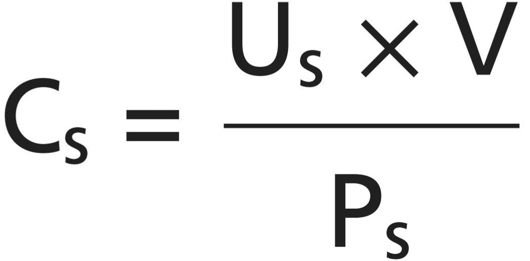
Thus, renal clearance of a substance is calculated from the urinary excretion rate (U, x V) of that substance divided by its plasma concentration.
بنابراین، کلیرانس کلیوی یک ماده از میزان دفع ادراری (U, x V) آن ماده تقسیم بر غلظت پلاسمایی آن محاسبه می شود.
INULIN CLEARANCE CAN BE USED TO ESTIMATE GLOMERULAR FILTRATION RATE
If a substance is freely filtered (filtered as freely as water) and is not reabsorbed or secreted by the renal tubules, then the rate at which that substance is excreted in the urine (Us, × V) is equal to the filtration rate of the substance by the kidneys (GFR x Ps). Thus:
می توان از پاکسازی اینولین برای تخمین میزان فیلتراسیون گلومرولی استفاده کرد
اگر ماده ای آزادانه فیلتر شود (به اندازه آب آزادانه فیلتر شود) و توسط لوله های کلیوی دوباره جذب یا ترشح نشود، سرعت دفع آن ماده در ادرار (U, × V) برابر است با نرخ فیلتراسیون ماده توسط کلیه ها (GFR x P). بدین ترتیب:

The GFR, therefore, can be calculated as the clearance of the substance as follows:
بنابراین، GFR را می توان به عنوان ترخیص کالا از گمرک ماده به صورت زیر محاسبه کرد:
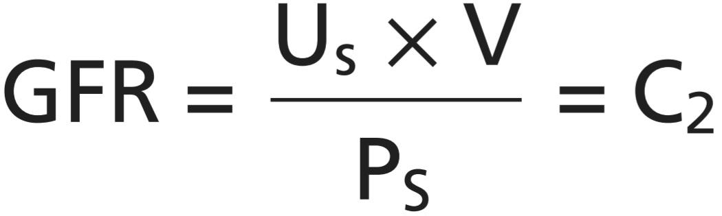
A substance that fits these criteria is inulin, a polysaccharide molecule with a molecular weight of about 5200. Inulin, which is not produced in the body, is found in the roots of certain plants and must be administered intravenously to a patient to measure GFR.
ماده ای که با این معیارها مطابقت دارد، اینولین است، یک مولکول پلی ساکارید با وزن مولکولی حدود 5200. اینولین که در بدن تولید نمی شود، در ریشه گیاهان خاصی یافت می شود و باید به صورت داخل وریدی به بیمار برای اندازه گیری GFR تزریق شود.
Figure 28-20 shows the renal handling of inulin. In this example, the plasma concentration is 1 mg/ml, urine con- centration is 125 mg/ml, and urine flow rate is 1 ml/min. Therefore, 125 mg/min of inulin passes into the urine. Then, inulin clearance is calculated as the urine excretion rate of inulin divided by the plasma concentration, which yields a value of 125 ml/min. Thus, 125 milliliters of plasma flowing through the kidneys must be filtered to deliver the inulin that appears in the urine.
شکل 28-20 عملکرد کلیوی اینولین را نشان می دهد. در این مثال، غلظت پلاسما 1 میلی گرم بر میلی لیتر، غلظت ادرار 125 میلی گرم بر میلی لیتر و سرعت جریان ادرار 1 میلی لیتر در دقیقه است. بنابراین 125 میلی گرم در دقیقه اینولین وارد ادرار می شود. سپس، کلیرانس اینولین به عنوان میزان دفع ادرار اینولین تقسیم بر غلظت پلاسما محاسبه می شود که مقدار 125 میلی لیتر در دقیقه را به دست می دهد. بنابراین، 125 میلی لیتر پلاسمایی که در کلیه ها جریان دارد باید فیلتر شود تا اینولین که در ادرار ظاهر می شود را تحویل دهد.
Inulin is not the only substance that can be used for determining the GFR. Other substances that have been used clinically to estimate the GFR include iothalamate, chromium ethylenediaminetetraacetic acid (EDTA), cystatin C, and creatinine.
اینولین تنها ماده ای نیست که می تواند برای تعیین GFR استفاده شود. سایر موادی که از نظر بالینی برای تخمین GFR استفاده شده اند عبارتند از: یوتالامات، کروم اتیلن دی آمین تترا استیک اسید (EDTA)، سیستاتین C و کراتینین.
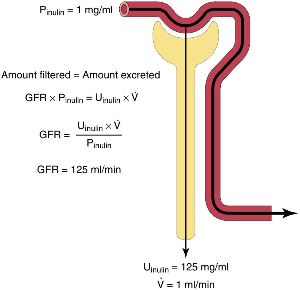
Figure 28-20 Measurement of glomerular filtration rate (GFR) from the renal clearance of inulin. Inulin is freely filtered by the glomerular capillaries but is not reabsorbed by the renal tubules. Pinulin, Plasma inulin concentration; Uinulin, urine inulin concentration; V, urine flow rate.
شکل 28-20 اندازه گیری میزان فیلتراسیون گلومرولی (GFR) از کلیرانس کلیوی اینولین. اینولین آزادانه توسط مویرگ های گلومرولی فیلتر می شود اما توسط لوله های کلیوی دوباره جذب نمی شود. پینولین، غلظت اینولین پلاسما؛ Uinulin، غلظت اینولین ادرار. V، سرعت جریان ادرار.
CREATININE CLEARANCE AND PLASMA CREATININE CONCENTRATION CAN BE USED TO ESTIMATE GLOMERULAR FILTRATION RATE
Creatinine is a by-product of muscle metabolism and is cleared from the body fluids almost entirely by glomerular filtration. Therefore, creatinine clearance can also be used to assess GFR. Because measurement of creatinine clearance does not require intravenous infusion into the patient, this method is much more widely used than inulin clearance for estimating GFR clinically. However, creatinine clearance is not a perfect marker of GFR because a small amount of it is secreted by the tubules, so the amount of creatinine excreted slightly exceeds the amount filtered. There is normally a slight error in measuring plasma creatinine that leads to an overestimation of the plasma creatinine concentration; fortuitously, these two errors tend to cancel each other. Therefore, creatinine clearance provides a reasonable estimate of GFR.
پاکسازی کراتینین و غلظت کراتینین پلاسما را می توان برای تخمین میزان فیلتراسیون گلومرولی استفاده کرد
کراتینین یک محصول جانبی متابولیسم عضلانی است و تقریباً به طور کامل با فیلتراسیون گلومرولی از مایعات بدن پاک می شود. بنابراین، کلیرانس کراتینین نیز می تواند برای ارزیابی GFR استفاده شود. از آنجایی که اندازه گیری کلیرانس کراتینین نیازی به انفوزیون داخل وریدی در بیمار ندارد، این روش بسیار بیشتر از کلیرانس اینولین برای تخمین GFR از نظر بالینی استفاده می شود. با این حال، کلیرانس کراتینین یک نشانگر کامل برای GFR نیست زیرا مقدار کمی از آن توسط لوله ها ترشح می شود، بنابراین مقدار کراتینین دفع شده کمی بیشتر از مقدار فیلتر شده است. معمولاً یک خطای جزئی در اندازه گیری کراتینین پلاسما وجود دارد که منجر به تخمین بیش از حد غلظت کراتینین پلاسما می شود. به طور تصادفی، این دو خطا تمایل به خنثی کردن یکدیگر دارند. بنابراین، کلیرانس کراتینین تخمین معقولی از GFR ارائه می دهد.
In some cases, it may not be practical to collect urine in a patient for measuring creatinine clearance (CC). An approximation of changes in GFR, however, can be obtained by simply measuring the plasma creatinine con- centration (Pcr), which is inversely proportional to the GFR:
در برخی موارد، جمع آوری ادرار در یک بیمار برای اندازه گیری کلیرانس کراتینین (CC) ممکن است عملی نباشد. با این حال، تقریبی از تغییرات در GFR را می توان با اندازه گیری غلظت کراتینین پلاسما (Pcr)، که با GFR نسبت معکوس دارد، بدست آورد:
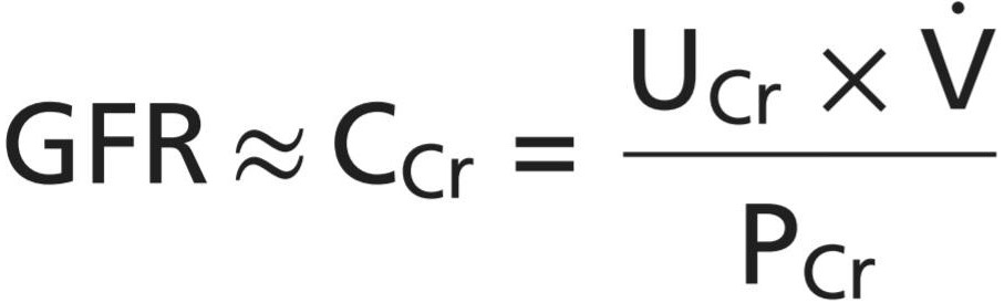
If GFR suddenly decreases by 50%, the kidneys will transiently filter and excrete only half as much creatinine, causing the accumulation of creatinine in the body fluids and raising plasma concentration. The plasma concentration of creatinine will continue to rise until the filtered load of creatinine (Pcr GFR) and creatinine excretion (Ucr x V) return to normal, and a balance between creatinine production and creatinine excretion is reestablished. This response will occur when the plasma creatinine level increases to approximately twice normal, as shown in Figure 28-21.
اگر GFR به طور ناگهانی 50 درصد کاهش یابد، کلیه ها به طور موقت تنها نیمی از کراتینین را فیلتر و دفع می کنند و باعث تجمع کراتینین در مایعات بدن و افزایش غلظت پلاسما می شود. غلظت کراتینین پلاسما تا زمانی که بار فیلتر شده کراتینین (Pcr GFR) و دفع کراتینین (Ucr x V) به حالت عادی برگردد و تعادل بین تولید کراتینین و دفع کراتینین دوباره برقرار شود به افزایش خود ادامه خواهد داد. همانطور که در شکل 28-21 نشان داده شده است، این پاسخ زمانی رخ خواهد داد که سطح کراتینین پلاسما تا تقریباً دو برابر نرمال افزایش یابد.
If GFR falls to one-fourth normal, plasma creatinine level would increase to about four times normal, and a decrease of GFR to one-eighth normal would raise plasma creatinine level to eight times normal. Thus, under steady-state conditions, creatinine excretion rate equals the rate of creatinine production, despite reductions in GFR. However, this normal rate of creatinine excretion occurs at the expense of an elevated plasma creatinine concentration, as shown in Figure 28-22.
اگر GFR به یک چهارم نرمال کاهش یابد، سطح کراتینین پلاسما به حدود چهار برابر نرمال افزایش می یابد و کاهش GFR به یک هشتم نرمال، سطح کراتینین پلاسما را تا هشت برابر طبیعی افزایش می دهد. بنابراین، در شرایط حالت پایدار، نرخ دفع کراتینین با سرعت تولید کراتینین برابر است، علیرغم کاهش GFR. با این حال، این سرعت طبیعی دفع کراتینین به قیمت افزایش غلظت کراتینین پلاسما اتفاق می افتد، همانطور که در شکل 28-22 نشان داده شده است.

Figure 28-21 Effect of reducing glomerular filtration rate (GFR) by 50% on the serum creatinine concentration and on creatinine excretion rate when the production rate of creatinine remains constant. PCreatinine, Plasma creatinine concentration.
شکل 28-21 اثر کاهش نرخ فیلتراسیون گلومرولی (GFR) به میزان 50% بر غلظت کراتینین سرم و بر میزان دفع کراتینین زمانی که میزان تولید کراتینین ثابت بماند. پی کراتینین، غلظت کراتینین پلاسما.
Figure 28-22 Approximate relationship between glomerular filtration rate (GFR) and plasma creatinine concentration under steadystate conditions. Decreasing GFR by 50% will increase plasma creatinine level to twice normal if creatinine production by the body remains constant.
شکل 28-22 رابطه تقریبی بین نرخ فیلتراسیون گلومرولی (GFR) و غلظت کراتینین پلاسما در شرایط حالت پایدار. اگر تولید کراتینین توسط بدن ثابت بماند، کاهش GFR به میزان 50 درصد، سطح کراتینین پلاسما را تا دو برابر طبیعی افزایش می دهد.
PARA-AMINOHIPPURIC ACID CLEARANCE CAN BE USED TO ESTIMATE RENAL PLASMA FLOW
Theoretically, if a substance is completely cleared from the plasma, the clearance rate of that substance is equal to the total RPF. In other words, the amount of the substance delivered to the kidneys in the blood (RPF x P) would be equal to the amount excreted in the urine (U, x V). Thus, RPF could be calculated as follows:
می توان از پاکسازی اسید پارآمینهی پوریک برای تخمین جریان پلاسمای کلیوی استفاده کرد
از نظر تئوری، اگر یک ماده به طور کامل از پلاسما پاک شود، میزان پاکسازی آن ماده برابر با کل RPF است. به عبارت دیگر، مقدار ماده ای که در خون به کلیه ها می رسد (RPF x P) برابر با مقدار دفع شده در ادرار (U, x V) خواهد بود. بنابراین، RPF را می توان به صورت زیر محاسبه کرد:

Because the GFR is only about 20% of the total plasma flow, a substance that is completely cleared from the plasma must be excreted by tubular secretion, as well as by glomerular filtration (Figure 28-23). There is no known substance that is completely cleared by the kidneys. One substance, PAH, is about 90% cleared from the plasma. Therefore, clearance of PAH can be used to approximate RPF. To be more accurate, one can correct for the percentage of PAH that is still in the blood when it leaves the kidneys. The percentage of PAH removed from the blood is known as the extraction ratio of PAH and averages about 90% in normal kidneys. In diseased kidneys, this extraction ratio may be reduced because of the inability of damaged tubules to secrete PAH into the tubular fluid.
از آنجایی که GFR تنها حدود 20 درصد از کل جریان پلاسما است، ماده ای که به طور کامل از پلاسما پاک می شود باید با ترشح لوله ای و همچنین با فیلتراسیون گلومرولی دفع شود (شکل 28-23). هیچ ماده شناخته شده ای وجود ندارد که به طور کامل توسط کلیه ها پاک شود. یک ماده، PAH، حدود 90٪ از پلاسما پاک می شود. بنابراین، پاکسازی PAH می تواند برای تقریبی RPF استفاده شود. برای دقیقتر بودن، میتوان درصد PAH را که هنگام خروج از کلیهها در خون باقی میماند، تصحیح کرد. درصد PAH حذف شده از خون به عنوان نسبت استخراج PAH شناخته می شود و در کلیه های طبیعی به طور متوسط حدود 90٪ است. در کلیههای بیمار، این نسبت استخراج ممکن است به دلیل ناتوانی لولههای آسیبدیده در ترشح PAH به داخل مایع لولهای کاهش یابد.
The calculation of RPF can be demonstrated by the following example. Assume that the plasma concentration of PAH is 0.01 mg/ml, urine concentration is 5.85 mg/ml, and urine flow rate is 1 ml/min. PAH clearance can be calculated from the rate of urinary PAH excretion (5.85 [mg/ml] x 1 [ml/min]) divided by the plasma PAH con- centration (0.01 mg/ml). Thus, the clearance of PAH is calculated to be 585 ml/min.
محاسبه RPF را می توان با مثال زیر نشان داد. فرض کنید که غلظت پلاسمایی PAH 0.01 mg/ml، غلظت ادرار 5.85 mg/ml و سرعت جریان ادرار 1 ml/min باشد. کلیرانس PAH را می توان از میزان دفع PAH ادراری (5.85 [mg/ml] x 1 [ml/min]) تقسیم بر غلظت PAH پلاسما (0.01 mg/ml) محاسبه کرد. بنابراین، کلیرانس PAH 585 میلی لیتر در دقیقه محاسبه می شود.
If the extraction ratio for PAH is 90%, the actual RPF can be calculated by dividing 585 ml/min by 0.9, yielding a value of 650 ml/min. Thus, total RPF can be calculated as follows:
اگر نسبت استخراج برای PAH 90٪ باشد، RPF واقعی را می توان با تقسیم 585 میلی لیتر در دقیقه بر 0.9 محاسبه کرد و مقدار 650 میلی لیتر در دقیقه به دست آمد. بنابراین، کل RPF را می توان به صورت زیر محاسبه کرد:

The excretion ratio EPAH is calculated as the difference between the renal arterial PAH and renal venous PAH cancentrations ,divided by the renal arterial PAH concentration:
نسبت دفع EPAH به عنوان تفاوت بین PAH شریان کلیوی و PAH وریدی کلیوی، تقسیم بر غلظت PAH شریان کلیوی محاسبه می شود:

One can calculate the total blood flow through the kidneys from the total RPF and hematocrit (the per- centage of red blood cells in the blood). If the hematocrit is 0.45 and the total RPF is 650 ml/min, the total blood flow through both kidneys is 650/(1 – 0.45), or 1182 ml/min.
می توان کل جریان خون در کلیه ها را از RPF کل و هماتوکریت (درصد گلبول های قرمز در خون) محاسبه کرد. اگر هماتوکریت 0.45 و کل RPF 650 میلی لیتر در دقیقه باشد، جریان خون کل از هر دو کلیه 650/(1 – 0.45) یا 1182 میلی لیتر در دقیقه است.

Figure 28-23 Measurement of renal plasma flow from the renal clearance of para-aminohippuric acid (PAH). PAH is freely filtered by the glomerular capillaries and is also secreted from the peritubular capillary blood into the tubular lumen. The amount of PAH in the plasma of the renal artery is about equal to the amount of PAH excreted in the urine. Therefore, the renal plasma flow can be calculated from the clearance of PAH. To be more accurate, one can correct for the percentage of PAH that is still in the blood when it leaves the kidneys. PPAH, Arterial plasma PAH concentration; UPAH, urine PAH concentration; V, urine flow rate.
شکل 28-23 اندازه گیری جریان پلاسمای کلیوی از ترخیص کالا از گمرک کلیه اسید پارا آمینو هیپوریک (PAH). PAH آزادانه توسط مویرگ های گلومرولی فیلتر می شود و همچنین از خون مویرگی اطراف لوله به لومن لوله ترشح می شود. مقدار PAH در پلاسمای شریان کلیوی تقریباً برابر با مقدار PAH دفع شده در ادرار است. بنابراین، جریان پلاسمای کلیوی را می توان از پاکسازی PAH محاسبه کرد. برای دقیق تر، می توان درصد PAH را که در هنگام خروج از کلیه ها هنوز در خون وجود دارد، تصحیح کرد. PPAH، غلظت PAH در پلاسمای شریانی. UPAH، غلظت PAH ادرار. V، سرعت جریان ادرار.
FILTRATION FRACTION IS CALCULATED FROM GFR DIVIDED BY RPF
To calculate the filtration fraction, which is the fraction of plasma that filters through the glomerular membrane, one must first know the RPF (PAH clearance) and the GFR (inulin clearance). If the RPF is 650 ml/min and the GFR is 125 ml/min, the filtration fraction (FF) is calculated as follows:
کسر فیلتراسیون از GFR تقسیم بر RPF محاسبه می شود
برای محاسبه کسر فیلتراسیون، که کسری از پلاسما است که از طریق غشای گلومرولی فیلتر میشود، ابتدا باید RPF (تخلیه PAH) و GFR (کلیرانس اینولین) را بشناسید. اگر RPF 650 میلی لیتر در دقیقه و GFR 125 میلی لیتر در دقیقه باشد، کسر فیلتراسیون (FF) به صورت زیر محاسبه می شود:

CALCULATION OF TUBULAR REABSORPTION OR SECRETION FROM RENAL CLEARANCES
If the rates of glomerular filtration and renal excretion of a substance are known, one can calculate whether there is a net reabsorption or net secretion of that substance by the renal tubules. For example, if the rate of excretion of the substance (Us, x V) is less than the filtered load of the substance (GFR × Ps), then some of the substance must have been reabsorbed from the renal tubules. Conversely, if the excretion rate of the substance is greater than its filtered load, then the rate at which it appears in the urine represents the sum of the rate of glomerular filtration plus tubular secretion.
محاسبه بازجذب لوله ای یا ترشح از پاکسازی کلیوی
اگر میزان فیلتراسیون گلومرولی و دفع کلیوی یک ماده مشخص باشد، می توان محاسبه کرد که آیا بازجذب خالص یا ترشح خالص آن ماده توسط لوله های کلیوی وجود دارد یا خیر. به عنوان مثال، اگر سرعت دفع ماده (U, x V) کمتر از بار فیلتر شده ماده (GFR × Ps) باشد، باید مقداری از ماده از لولههای کلیوی بازجذب شده باشد. برعکس، اگر سرعت دفع ماده بیشتر از بار فیلتر شده آن باشد، سرعت ظاهر شدن آن در ادرار نشان دهنده مجموع سرعت فیلتراسیون گلومرولی به اضافه ترشح لوله ای است.
The following example demonstrates the calculation of tubular reabsorption. Assume the following laboratory values for a patient were obtained:
مثال زیر محاسبه بازجذب لوله ای را نشان می دهد. فرض کنید مقادیر آزمایشگاهی زیر برای یک بیمار به دست آمده است:
• Urine flow rate = 1 ml/min
• Urine concentration of sodium (UNA) = 70 mEq/L = 70 μEq/ml
• Plasma sodium concentration = 140 mEq/L = 140 Eq/ml
• GFR (inulin clearance) = 100 ml/min
• سرعت جریان ادرار = 1 میلی لیتر در دقیقه
• غلظت سدیم ادرار (UNA) = 70 mEq/L = 70 μEq/ml
• غلظت سدیم پلاسما = mEq/L 140 = 140 Eq/ml
• GFR (کلیرانس اینولین) = 100 میلی لیتر در دقیقه
In this example, the filtered sodium load is GFR × PNa or 100 ml/min x 140 μEq/ml = 14,000 μEq/min. Urinary sodium excretion (Una x urine flow rate) is 70 μEq/min. Therefore, tubular reabsorption of sodium is the difference between the filtered load and urinary excretion, or 14,000 μEq/min – 70 μEq/min = 13,930 μEq/min.
در این مثال، بار سدیم فیلتر شده GFR × PNa یا 100 ml/min x 140 μEq/ml = 14000 μEq/min است. دفع سدیم ادراری (سرعت جریان ادرار Una x) 70 μEq/min است. بنابراین، بازجذب لولهای سدیم تفاوت بین بار فیلتر شده و دفع ادرار است یا 14000μEq/min – 70μEq/min = 13930μEq/min.
Comparisons of Inulin Clearance with Clearances of Different Solutes. The following generalizations can be made by comparing the clearance of a substance with the clearance of inulin, the gold standard for measuring GFR: (1) if the clearance rate of the substance equals that of inulin, the substance is only filtered and not reabsorbed or secreted; (2) if the clearance rate of a substance is less than inulin clearance, the substance must have been reabsorbed by the nephron tubules; and (3) if the clearance rate of a substance is greater than that of inulin, the substance must be secreted by the nephron tubules. Listed below are the approximate clearance rates for some of the substances normally handled by the kidneys:
مقایسه پاکسازی اینولین با پاکسازی املاح مختلف. تعمیم های زیر را می توان با مقایسه ترخیص کالا از گمرک یک ماده با ترخیص کالا از گمرک اینولین، استاندارد طلایی برای اندازه گیری GFR انجام داد: (1) اگر میزان پاکسازی ماده برابر با اینولین باشد، ماده فقط فیلتر می شود و دوباره جذب یا ترشح نمی شود. (2) اگر میزان ترخیص کالا از گمرک یک ماده کمتر از ترخیص کالا از گمرک اینولین باشد، ماده باید توسط لوله های نفرون دوباره جذب شده باشد. و (3) اگر میزان پاکسازی ماده ای بیشتر از اینولین باشد، ماده باید توسط لوله های نفرون ترشح شود. در زیر نرخ های کلیرانس تقریبی برخی از موادی که به طور معمول توسط کلیه ها استفاده می شوند ذکر شده است:

کلیک کنید «Bibliography: فهرست کتب مربوطه»
Bie P: Natriuretic peptides and normal body fluid regulation. Compr Physiol 8:1211, 2018.
Delpire E, Gagnon KB: Na+ -K+ -2CI- Cotransporter (NKCC) physiological function in nonpolarized cells and transporting epithelia. Compr Physiol 25;8:871, 2018.
Féraille E, Dizin E: Coordinated control of ENaC and Na+,K+-ATPase in renal collecting duct. J Am Soc Nephrol 27:2554, 2016.
Fromm M, Piontek J, Rosenthal R, Günzel D, Krug SM: Tight junctions of the proximal tubule and their channel proteins. Pflugers Arch 469:877, 2018.
Gonzalez-Vicente A, Saez F, Monzon CM, Asirwatham J, Garvin JL: Thick ascending limb sodium transport in the pathogenesis of hy- pertension. Physiol Rev 99:235, 2019.
Hall JE, Brands MW: The renin-angiotensin-aldosterone system: renal mechanisms and circulatory homeostasis. In: Seldin DW, Giebisch G (eds): The Kidney-Physiology and Pathophysiology, 3rd ed. New York: Raven Press, 2000.
Hall JE, do Carmo JM, da Silva AA, Wang Z, Hall ME: Obesity-induced hypertension: interaction of neurohumoral and renal mechanisms. Circ Res 116:991, 2015.
Hall JE, Granger JP, do Carmo JM, et al: Hypertension: physiology and pathophysiology. Compr Physiol 2:2393, 2012.
Klein JD, Sands JM: Urea transport and clinical potential of urearetics. Curr Opin Nephrol Hypertens 25:444, 2016.
Knepper MA, Kwon TH, Nielsen S: Molecular physiology of water bal- ance. N Engl J Med 372:1349, 2015.
Ko B: Parathyroid hormone and the regulation of renal tubular calcium transport. Curr Opin Nephrol Hypertens 26:405, 2017.
Makrides V, Camargo SM, Verrey F: Transport of amino acids in the kidney. Compr Physiol 4:367, 2014.
McCormick JA, Ellison DH: Distal convoluted tubule. Compr Physiol 5:45, 2015.
Moe SM: Calcium homeostasis in health and in kidney disease. Com- pr Physiol 6:1781, 2016.
Mount DB. Thick ascending limb of the loop of Henle. Clin J Am Soc Nephrol 9:1974, 2014.
Nielsen S, Frøkiær J, Marples D, et al: Aquaporins in the kidney: from molecules to medicine. Physiol Rev 82:205, 2002.
Palmer LG, Schnermann J: Integrated control of Na transport along the nephron. Clin J Am Soc Nephrol 10:676, 2015.
Reilly RF, Ellison DH: Mammalian distal tubule: physiology, patho- physiology, and molecular anatomy. Physiol Rev 80:277, 2000.
Rieg T, Vallon V: Development of SGLT1 and SGLT2 inhibitors. Diabetologia 61:2079, 2018.
Rossier BC, Baker ME, Studer RA: Epithelial sodium transport and its control by aldosterone: the story of our internal environment revis- ited. Physiol Rev 95:297, 2015.
Roy A, Al-bataineh MM, Pastor-Soler NM: Collecting duct interca- lated cell function and regulation. Clin J Am Soc Nephrol 10:305, 2015.
Seegmiller JC, Eckfeldt JH, Lieske JC: Challenges in measuring glo- merular filtration rate: a clinical laboratory perspective. Adv Chron- ic Kidney Dis 25:84, 2018.
Staruschenko A: Beneficial effects of high potassium: contribution of renal basolateral K+ channels. Hypertension 71:1015, 2018.
Staruschenko A: Regulation of transport in the connecting tubule and cortical collecting duct. Compr Physiol 2:1541, 2012.
Thomson SC, Blantz RC: Glomerulotubular balance, tubuloglomeru- lar feedback, and salt homeostasis. J Am Soc Nephrol 19:2272, 2008.
Wang K, Kestenbaum B: Proximal tubular secretory clearance: a neglected partner of kidney function. Clin J Am Soc Nephrol 13:1291, 2018.
Wang T, Weinbaum S, Weinstein AM: Regulation of glomerulotubu- lar balance: flow-activated proximal tubule function. Pflugers Arch 469:643, 2017.
Yamazaki O, Ishizawa K, Hirohama D, Fujita T, Shibata S: Electrolyte
transport in the renal collecting duct and its regulation by the renin- angiotensin-aldosterone system. Clin Sci (Lond) 133:75, 2019.
🚀 با ما همراه شوید!
تازهترین مطالب و آموزشهای مغز و اعصاب را از دست ندهید. با فالو کردن کانال تلگرام آیندهنگاران مغز، از ما حمایت کنید!



