
فیزیولوژی پزشکی گایتون و هال؛ غلظت و رقت ادرار؛ تنظیم اسمولاریته مایعات خارج سلولی و غلظت سدیم


دعای مطالعه [ نمایش ]
بِسْمِ الله الرَّحْمنِ الرَّحیمِ
اَللّهُمَّ اَخْرِجْنى مِنْ ظُلُماتِ الْوَهْمِ
خدايا مرا بيرون آور از تاريكىهاى وهم،
وَ اَكْرِمْنى بِنُورِ الْفَهْمِ
و به نور فهم گرامى ام بدار،
اَللّهُمَّ افْتَحْ عَلَيْنا اَبْوابَ رَحْمَتِكَ
خدايا درهاى رحمتت را به روى ما بگشا،
وَانْشُرْ عَلَيْنا خَزائِنَ عُلُومِكَ بِرَحْمَتِكَ يا اَرْحَمَ الرّاحِمينَ
و خزانههاى علومت را بر ما باز كن به امید رحمتت اى مهربانترين مهربانان.
کتاب «فیزیولوژی پزشکی گایتون و هال» بهعنوان یکی از جامعترین و معتبرترین منابع در حوزه علوم پزشکی، همچنان مرجع کلیدی برای درک عملکرد پیچیده بدن انسان است. این اثر با تکیه بر تازهترین پژوهشها و توضیحات دقیق از سازوکارهای فیزیولوژیک، پلی میان علوم پایه پزشکی و کاربردهای بالینی ایجاد میکند و نقشی بیبدیل در آموزش، پژوهش و ارتقای دانش سلامت ایفا مینماید.
ترجمه دقیق و علمی این شاهکار توسط برند علمی آیندهنگاران مغز به مدیریت داریوش طاهری، دسترسی فارسیزبانان به مرزهای نوین دانش فیزیولوژی را ممکن ساخته و رسالتی علمی برای ارتقای آموزش پزشکی، فهم عمیقتر سازوکارهای بدن و توسعه روشهای نوین در حوزه سلامت فراهم آورده است.
» کتاب فیزیولوژی پزشکی گایتون و هال
» » فصل ۲۹: غلظت و رقت ادرار؛ تنظیم اسمولاریته مایعات خارج سلولی و غلظت سدیم
در حال ویرایش
» Guyton and Hall Textbook of Medical Physiology
»» CHAPTER 29: Urine Concentration and Dilution; Regulation of Extracellular Fluid Osmolarity and Sodium Concentration
نوشته

For the cells of the body to function properly, they must be bathed in extracellular fluid with a relatively constant concentration of electrolytes. The total concentration of solutes in the extracellular fluid-and therefore the osmolarity-must also be precisely regulated to prevent the cells from shrinking or swelling. The osmolarity is determined by the amount of solute (mainly sodium chloride) divided by the volume of the extracellular fluid. Thus, to a large extent, extracellular fluid osmolarity and sodium chloride concentration are regulated by the amount of extracellular water. The total body water is controlled by (1) fluid intake, which is regulated by factors that determine thirst; and (2) renal water excretion, which is controlled by multiple factors that influence glomerular filtration and tubular reabsorption.
برای اینکه سلول های بدن به درستی عمل کنند، باید در مایع خارج سلولی با غلظت نسبتاً ثابت الکترولیت ها غوطه ور شوند. غلظت کل املاح در مایع خارج سلولی – و بنابراین اسمولاریته – نیز باید دقیقاً تنظیم شود تا از کوچک شدن یا تورم سلول ها جلوگیری شود. اسمولاریته با مقدار ماده حل شده (عمدتاً کلرید سدیم) تقسیم بر حجم مایع خارج سلولی تعیین می شود. بنابراین، تا حد زیادی اسمولاریته مایع خارج سلولی و غلظت کلرید سدیم توسط مقدار آب خارج سلولی تنظیم می شود. کل آب بدن توسط (1) مصرف مایعات کنترل می شود که توسط عوامل تعیین کننده تشنگی تنظیم می شود. و (2) دفع آب کلیوی، که توسط عوامل متعددی کنترل می شود که بر فیلتراسیون گلومرولی و بازجذب لوله ای تأثیر می گذارد.
In this chapter, we discuss the following: (1) mechanisms that cause the kidneys to eliminate excess water by excreting a dilute urine; (2) mechanisms that cause the kidneys to conserve water by excreting a concentrated urine; (3) renal feedback mechanisms that control the extracellular fluid sodium concentration and osmolarity; and (4) thirst and salt appetite mechanisms that determine the intakes of water and salt, which also help control extracellular fluid volume, osmolarity, and sodium concentration.
در این فصل، ما در مورد موارد زیر بحث می کنیم: (1) مکانیسم هایی که باعث می شود کلیه ها آب اضافی را با دفع ادرار رقیق از بین ببرند. (2) مکانیسم هایی که باعث می شود کلیه ها با دفع ادرار غلیظ آب را ذخیره کنند. (3) مکانیسم های بازخورد کلیوی که غلظت سدیم و اسمولاریته مایع خارج سلولی را کنترل می کند. و (4) مکانیسمهای تشنگی و اشتهای نمک که میزان دریافت آب و نمک را تعیین میکنند، که همچنین به کنترل حجم مایع خارج سلولی، اسمولاریته و غلظت سدیم کمک میکند.
KIDNEYS EXCRETE EXCESS WATER BY FORMING DILUTE URINE
Normal kidneys have a tremendous capability to vary the relative proportions of solutes and water in the urine in response to various challenges. When there is excess water in the body, and body fluid osmolarity is reduced, the kidneys can excrete urine with an osmolarity as low as 50 mOsm/L, a concentration that is only about onesixth the osmolarity of normal extracellular fluid. Conversely, when there is a deficit of water in the body, and extracellular fluid osmolarity is high, the kidneys can excrete highly concentrated urine with an osmolarity of 1200 to 1400 mOsm/L. Equally important, the kidneys can excrete a large volume of dilute urine or a small volume of concentrated urine without major changes in rates of excretion of solutes such as sodium and potassium. This ability to regulate water excretion independently of solute excretion is necessary for survival, especially when fluid intake is limited.
کلیه ها با تشکیل ادرار رقیق، آب اضافی را دفع می کنند.
کلیه های معمولی توانایی فوق العاده ای برای تغییر نسبت نسبی املاح و آب در ادرار در پاسخ به چالش های مختلف دارند. هنگامی که آب اضافی در بدن وجود دارد و اسمولاریته مایع بدن کاهش می یابد، کلیه ها می توانند ادرار را با اسمولاریته کمتر از 50 mOsm/L دفع کنند، غلظتی که تنها حدود یک ششم اسمولاریته مایع خارج سلولی طبیعی است. برعکس، زمانی که کمبود آب در بدن وجود داشته باشد و اسمولاریته مایع خارج سلولی بالا باشد، کلیه ها می توانند ادرار بسیار غلیظی را با اسمولاریته 1200 تا 1400 mOsm/L دفع کنند. به همان اندازه مهم است که کلیه ها می توانند حجم زیادی از ادرار رقیق یا حجم کمی از ادرار غلیظ را بدون تغییرات عمده در میزان دفع املاح مانند سدیم و پتاسیم دفع کنند. این توانایی برای تنظیم دفع آب به طور مستقل از دفع املاح برای بقا ضروری است، به ویژه زمانی که مصرف مایعات محدود است.
ANTIDIURETIC HORMONE CONTROLS URINE CONCENTRATION
The body has a powerful feedback system for regulating plasma osmolarity and sodium concentration that operates by altering renal excretion of water independently of solute excretion rate. A primary effector of this feedback is antidiuretic hormone (ADH), also called vasopressin.
هورمون آنتی دیورتیک غلظت ادرار را کنترل می کند
بدن دارای یک سیستم بازخورد قدرتمند برای تنظیم اسمولاریته پلاسما و غلظت سدیم است که با تغییر دفع کلیوی آب مستقل از میزان دفع املاح عمل می کند. یک عامل اصلی این بازخورد، هورمون ضد ادرار (ADH) است که وازوپرسین نیز نامیده می شود.
When osmolarity of the body fluids increases above normal (i.e., the solutes in the body fluids become too concentrated), the posterior pituitary gland secretes more ADH, which increases the permeability of the distal tubules and collecting ducts to water, as discussed in Chapter 28. This mechanism increases water reabsorption and decreases urine volume but does not markedly alter the rate of renal excretion of the solutes.
هنگامی که اسمولاریته مایعات بدن بیش از حد نرمال افزایش می یابد (یعنی املاح موجود در مایعات بدن بیش از حد متمرکز می شوند)، غده هیپوفیز خلفی ADH بیشتری ترشح می کند که نفوذپذیری لوله های انتهایی و مجاری جمع کننده به آب را افزایش می دهد، همانطور که در فصل 28 بحث شد.این مکانیسم باعث افزایش بازجذب آب و کاهش حجم ادرار می شود، اما به طور قابل توجهی سرعت دفع کلیه املاح را تغییر نمی دهد.
When there is excess water in the body, and extracellular fluid osmolarity is reduced, secretion of ADH by the posterior pituitary decreases, thereby reducing the permeability of the distal tubule and collecting ducts to water, which causes increased amounts of more dilute urine to be excreted. Thus, the rate of ADH secretion determines, to a large extent, whether the kidney excretes dilute or concentrated urine.
هنگامی که آب اضافی در بدن وجود دارد و اسمولاریته مایع خارج سلولی کاهش مییابد، ترشح ADH توسط هیپوفیز خلفی کاهش مییابد و در نتیجه نفوذپذیری لولههای انتهایی و مجاری جمعآوری به آب کاهش مییابد که باعث افزایش مقدار ادرار رقیقتر دفع میشود. بنابراین، سرعت ترشح ADH تا حد زیادی تعیین می کند که آیا کلیه ادرار رقیق یا غلیظ دفع می کند.
RENAL MECHANISMS FOR EXCRETING DILUTE URINE
When there is a large excess of water in the body, the kidney can excrete as much as 20 L/day of dilute urine, with a concentration as low as 50 mOsm/L. The kidney performs this impressive feat by continuing to reabsorb solutes without reabsorbing large amounts of water in the distal parts of the nephron, including the late distal tubule and collecting ducts.
مکانیسم های کلیوی برای دفع ادرار رقیق
هنگامی که مقدار زیادی آب در بدن وجود دارد، کلیه می تواند تا 20 لیتر در روز ادرار رقیق را با غلظت 50 mOsm/L دفع کند. کلیه با ادامه جذب مجدد املاح بدون جذب مجدد مقادیر زیادی آب در قسمت های انتهایی نفرون، از جمله لوله انتهایی انتهایی و مجاری جمع کننده، این کار چشمگیر را انجام می دهد.
Figure 29-1 shows the approximate renal responses in a human after ingestion of 1 liter of water. Note that urine volume increased to about six times normal within 45 minutes after the water had been ingested. However, the total amount of solute excreted remained relatively constant because the urine formed became dilute, and urine osmolarity decreased from 600 to about 100 mOsm/L. Thus, after ingestion of excess water, the kidney rids the body of the excess water but does not excrete excess amounts of solutes.
شکل 29-1 پاسخ های کلیوی تقریبی را در انسان پس از مصرف 1 لیتر آب نشان می دهد. توجه داشته باشید که حجم ادرار در عرض 45 دقیقه پس از مصرف آب به حدود شش برابر طبیعی افزایش یافت. با این حال، مقدار کل املاح دفع شده نسبتا ثابت باقی ماند زیرا ادرار تشکیل شده رقیق شد و اسمولاریته ادرار از 600 به حدود 100 mOsm/L کاهش یافت. بنابراین، کلیه پس از مصرف آب اضافی، بدن را از شر آب اضافی خلاص می کند، اما مقادیر اضافی املاح را دفع نمی کند.
When the glomerular filtrate is initially formed, its osmolarity is about the same as that of plasma (300 mOsm/L). To excrete excess water, the filtrate is diluted as it passes along the tubule by reabsorbing solutes to a greater extent than water, as shown in Figure 29-2. This dilution, however, occurs only in certain segments of the tubular system, as described in the following sections.
هنگامی که فیلتر گلومرولی در ابتدا تشکیل می شود، اسمولاریته آن تقریباً برابر با پلاسما (300 mOsm/L) است. همانطور که در شکل 29-2 نشان داده شده است، برای دفع آب اضافی، فیلتر هنگام عبور از امتداد لوله با بازجذب املاح به میزان بیشتری نسبت به آب رقیق می شود. این رقت، با این حال، تنها در بخش های خاصی از سیستم لوله ای رخ می دهد، همانطور که در بخش های زیر توضیح داده شده است.
Tubular Fluid Remains Isosmotic in Proximal Tubules. As fluid flows through the proximal tubule, solutes and water are reabsorbed in equal proportions, so little change in osmolarity occurs. Thus, the proximal tubule fluid remains isosmotic to the plasma, with an osmolarity of about 300 mOsm/L. As fluid passes down the de- scending loop of Henle, water is reabsorbed by osmosis, and the tubular fluid reaches equilibrium with the sur- rounding interstitial fluid of the renal medulla, which is very hypertonic-about two to four times the osmolarity of the original glomerular filtrate. Therefore, the tubular fluid becomes more concentrated as it flows into the inner medulla.
سیال لوله ای در لوله های پروگزیمال ایزوسموز باقی می ماند. با عبور مایع از لوله پروگزیمال، املاح و آب به نسبت مساوی دوباره جذب می شوند، بنابراین تغییر کمی در اسمولاریته اتفاق می افتد. بنابراین، مایع لوله پروگزیمال نسبت به پلاسما ایزوسموز باقی می ماند، با اسمولاریته حدود 300 mOsm/L. همانطور که مایع از حلقه نزولی هنله عبور می کند، آب توسط اسمز دوباره جذب می شود و مایع لوله ای با مایع بینابینی اطراف بصل النخاع کلیه به تعادل می رسد که بسیار هیپرتونیک است – حدود دو تا چهار برابر اسمولاریته فیلتر گلومرولی اصلی. بنابراین، مایع لوله ای با جریان یافتن به بصل النخاع داخلی، متمرکزتر می شود.
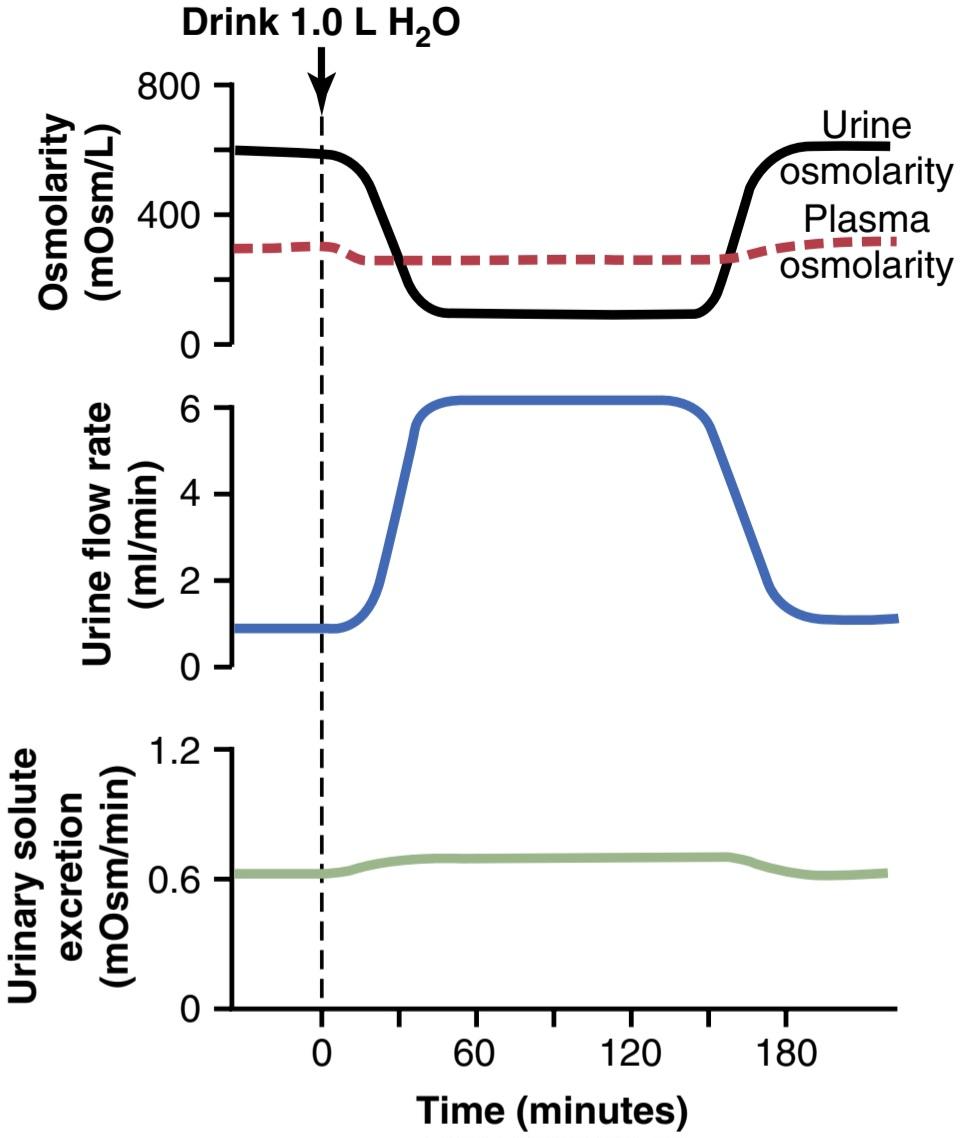
Figure 29-1. Water diuresis in a person after ingestion of 1 liter of water. Note that after water ingestion, urine volume increases and urine osmolarity decreases, causing excretion of a large volume of dilute urine; however, the total amount of solute excreted by the kidneys remains relatively constant. These responses of the kidneys prevent plasma osmolarity from decreasing markedly during excess water ingestion.
شکل 29-1. دیورز آب در فرد پس از مصرف 1 لیتر آب. توجه داشته باشید که پس از مصرف آب، حجم ادرار افزایش می یابد و اسمولاریته ادرار کاهش می یابد و باعث دفع حجم زیادی از ادرار رقیق می شود. با این حال، مقدار کل املاح دفع شده توسط کلیه ها نسبتا ثابت باقی می ماند. این پاسخ های کلیه ها از کاهش قابل توجه اسمولاریته پلاسما در طول مصرف آب اضافی جلوگیری می کند.

Figure 29-2. Formation of dilute urine when antidiuretic hormone (ADH) levels are very low. Note that in the ascending loop of Henle, the tubular fluid becomes very dilute. In the distal tubules and collecting tubules, the tubular fluid is further diluted by the reabsorption of sodium chloride and the failure to reabsorb water when ADH levels are very low. The failure to reabsorb water and continued reabsorption of solutes lead to a large volume of dilute urine. (Numerical values are in milliosmoles per liter.)
شکل 29-2. تشکیل ادرار رقیق زمانی که سطح هورمون آنتی دیورتیک (ADH) بسیار پایین است. توجه داشته باشید که در حلقه صعودی هنل، مایع لوله ای بسیار رقیق می شود. در لولههای انتهایی و لولههای جمعآوری، مایع لولهای با جذب مجدد کلرید سدیم و عدم جذب مجدد آب زمانی که سطح ADH بسیار پایین است، رقیقتر میشود. عدم جذب مجدد آب و ادامه بازجذب املاح منجر به حجم زیادی ادرار رقیق می شود. (مقادیر عددی بر حسب میلی مول در لیتر می باشد.)
Tubular Fluid Is Diluted in the Ascending Loop of Henle. In the ascending limb of the loop of Henle, especially in the thick segment, sodium, potassium, and chloride are avidly reabsorbed. However, this portion of the tubular segment is impermeable to water, even in the presence of large amounts of ADH. Therefore, the tubular fluid becomes more dilute as it flows up the ascending loop of Henle into the early distal tubule, with the osmolarity decreasing progressively to about 100 mOsm/L by the time the fluid enters the early distal tubular segment. Thus, regardless of whether ADH is present or absent, fluid leaving the early distal tubular segment is hypo-osmotic, with an osmolarity of only about one-third the osmolarity of plasma.
مایع لوله ای در حلقه صعودی هنله رقیق می شود. در اندام صعودی حلقه هنله، به ویژه در بخش ضخیم، سدیم، پتاسیم و کلرید مشتاقانه بازجذب می شوند. با این حال، این بخش از بخش لوله ای به آب نفوذناپذیر است، حتی در حضور مقادیر زیادی ADH. بنابراین، مایع لولهای رقیقتر میشود، زیرا از حلقه صعودی هنل به داخل لوله دیستال اولیه جریان مییابد، با زمانی که سیال وارد بخش لوله دیستال اولیه میشود، اسمولاریته به تدریج به حدود 100 mOsm/L کاهش مییابد. بنابراین، صرف نظر از وجود یا عدم وجود ADH، مایع خروجی از بخش لوله دیستال اولیه هیپواسموتیک است و اسمولاریته آن تنها حدود یک سوم اسمولاریته پلاسما است.
Tubular Fluid in Distal and Collecting Tubules Is Further Diluted in Absence of ADH. As the dilute fluid in the early distal tubule passes into the late distal convoluted tubule, cortical collecting duct, and medullary collecting duct, there is additional reabsorption of sodium chloride. In the absence of ADH, this portion of the tubule is also impermeable to water, and the additional re- absorption of solutes causes the tubular fluid to become even more dilute, decreasing its osmolarity to as low as 50 mOsm/L. The failure to reabsorb water and continued re- absorption of solutes lead to a large volume of dilute urine.
مایع لوله ای در لوله های دور و جمع کننده در صورت عدم وجود ADH بیشتر رقیق می شود. همانطور که مایع رقیق در لوله دیستال اولیه به لوله پیچیده انتهایی دیستال، مجرای جمع کننده کورتیکال و مجرای جمع کننده مدولاری می رود، بازجذب اضافی کلرید سدیم وجود دارد. در غیاب ADH، این بخش از لوله در برابر آب نیز نفوذ ناپذیر است و جذب مجدد املاح باعث می شود مایع لوله ای رقیق تر شود و اسمولاریته آن تا 50 mOsm/L کاهش یابد. عدم جذب مجدد آب و ادامه جذب مجدد املاح منجر به حجم زیادی از ادرار رقیق می شود.
To summarize, the mechanism for forming dilute urine is to continue reabsorbing solutes from the distal segments of the tubular system while reducing water reabsorption. In healthy kidneys, fluid leaving the ascending loop of Henle and early distal tubule is always dilute, regardless of the level of ADH. In the absence of ADH, the urine is further diluted in the late distal tubule and collecting ducts, and a large volume of dilute urine is excreted.
به طور خلاصه، مکانیسم تشکیل ادرار رقیق، ادامه بازجذب املاح از بخش های انتهایی سیستم لوله ای و در عین حال کاهش بازجذب آب است. در کلیه های سالم، مایع خروجی از حلقه صعودی هنله و لوله دیستال اولیه، صرف نظر از سطح ADH، همیشه رقیق است. در غیاب ADH، ادرار بیشتر در لوله انتهایی دیستال و مجاری جمع کننده رقیق می شود و حجم زیادی از ادرار رقیق دفع می شود.
KIDNEYS CONSERVE WATER BY EXCRETING CONCENTRATED URINE
The ability of the kidney to form concentrated urine is essential for survival of mammals that live on land, including humans. Water is continuously lost from the body through various routes, including the lungs by evaporation into the expired air, the gastrointestinal tract by way of the feces, the skin through evaporation and perspiration, and the kidneys through excretion of urine. Fluid intake is required to match this loss, but the ability of the kidneys to form a small volume of concentrated urine minimizes the fluid intake required to maintain homeostasis, a function that is especially important when water is in short supply.
کلیه ها با دفع ادرار غلیظ آب را حفظ می کنند
توانایی کلیه برای تشکیل ادرار غلیظ برای بقای پستاندارانی که در خشکی زندگی می کنند، از جمله انسان ضروری است. آب به طور مداوم از طریق راه های مختلف از بدن از دست می رود، از جمله ریه ها از طریق تبخیر به هوای منقضی شده، دستگاه گوارش از طریق مدفوع، پوست از طریق تبخیر و تعریق، و کلیه ها از طریق دفع ادرار. برای تطابق با این کاهش، نیاز به دریافت مایعات است، اما توانایی کلیه ها برای تشکیل حجم کمی از ادرار غلیظ، مصرف مایع مورد نیاز برای حفظ هموستاز را به حداقل می رساند، عملکردی که به ویژه در هنگام کمبود آب مهم است.
When there is a water deficit in the body, the kidneys form concentrated urine by continuing to excrete solutes while increasing water reabsorption and decreasing the urine volume. The human kidney can produce a maximal urine concentration of 1200 to 1400 mOsm/L, four to five times the osmolarity of plasma.
هنگامی که کمبود آب در بدن وجود دارد، کلیه ها با ادامه دفع املاح و در عین حال افزایش بازجذب آب و کاهش حجم ادرار، ادرار غلیظی را تشکیل می دهند. کلیه انسان می تواند حداکثر غلظت ادرار 1200 تا 1400 mOsm/L تولید کند که چهار تا پنج برابر اسمولاریته پلاسما است.
Some desert animals, such as the Australian hop- ping mouse, can concentrate urine to as high as 10,000 mOsm/L. This ability allows the mouse to survive in the desert without drinking water; sufficient water can be obtained through the ingested food and water produced in the body by metabolism of the food. Animals adapted to freshwater environments usually have minimal urine- concentrating ability. Beavers, for example, can concentrate the urine only to about 500 mOsm/L.
برخی از حیوانات بیابانی، مانند موش هوپینگ استرالیایی، می توانند ادرار را تا 10000 mOsm/L غلیظ کنند. این توانایی به موش اجازه می دهد تا بدون نوشیدن آب در بیابان زنده بماند. آب کافی را می توان از طریق غذای خورده شده و آب تولید شده در بدن توسط متابولیسم غذا به دست آورد. حیوانات سازگار با محیط های آب شیرین معمولاً دارای حداقل توانایی تمرکز ادرار هستند. برای مثال، Beavers میتواند ادرار را تنها تا حدود 500 mOsm/L متمرکز کند.
Obligatory Urine Volume
The maximal concentrating ability of the kidney dictates how much urine volume must be excreted each day to rid the body of metabolic waste products and electrolytes that are ingested. A normal 70-kg person must excrete about 600 milliosmoles of solute each day. If the maximal urine concentrating ability is 1200 mOsm/L, the minimal volume of urine that must be excreted, called the obligatory urine volume, can be calculated as follows:
حجم ادرار اجباری
حداکثر توانایی تمرکز کلیه تعیین می کند که چه مقدار حجم ادرار باید در روز دفع شود تا بدن از مواد زائد متابولیک و الکترولیت هایی که بلعیده می شوند خلاص شود. یک فرد معمولی 70 کیلوگرمی باید هر روز حدود 600 میلی مول از املاح دفع کند. اگر حداکثر توانایی تمرکز ادرار 1200 mOsm/L باشد، حداقل حجم ادراری که باید دفع شود، که حجم ادرار اجباری نامیده می شود را می توان به صورت زیر محاسبه کرد:
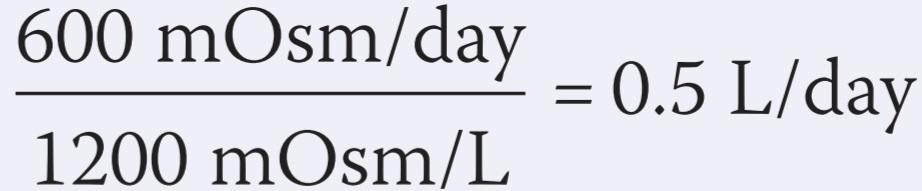
This minimal loss of volume in the urine contributes to dehydration, along with water loss from the skin, respiratory tract, and gastrointestinal tract, when water is not available to drink.
این حداقل کاهش حجم در ادرار به کم آبی بدن، همراه با از دست دادن آب از پوست، دستگاه تنفسی و دستگاه گوارش، زمانی که آب برای نوشیدن در دسترس نیست، کمک می کند.
The limited ability of the human kidney to concentrate the urine to only about 1200 mOsm/L explains why severe dehydration occurs if one attempts to drink seawater. Sodium chloride concentration in the ocean averages about 3.0% to 3.5%, with an osmolarity between about 1000 and 1200 mOsm/L. Drinking 1 liter of seawater with a concentration of 1200 mOsm/L would provide a total sodium chloride intake of 1200 milliosmoles. If the maximal urine concentrating ability is 1200 mOsm/L, the amount of urine volume needed to excrete 1200 milliosmoles would be 1.0 liter. Why then does drinking seawater cause dehydration? The answer is that the kidney must also excrete other solutes, especially urea, which contribute about 600 mOsm/L when the urine is maximally concentrated. Therefore, the maximum concentration of sodium chloride that can be excreted by the kidneys is about 600 mOsm/L. Thus, for every liter of seawater ingested, 1.5 liters of urine volume would be required to rid the body of 1200 milliosmoles of sodium chloride ingested in addition to 600 milliosmoles of other solutes, such as urea. This would result in a net fluid loss of 0.5 liter for every liter of seawater, explaining the rapid dehydration that occurs in shipwreck victims who drink seawater. However, a shipwreck victim’s pet Australian hopping mouse could drink seawater with impunity.
توانایی محدود کلیه انسان برای تمرکز ادرار تا حدود 1200 mOsm/L توضیح می دهد که چرا در صورت تلاش برای نوشیدن آب دریا، کم آبی شدید رخ می دهد. غلظت کلرید سدیم در اقیانوس به طور متوسط حدود 3.0٪ تا 3.5٪، با اسمولاریته بین حدود 1000 تا 1200 mOsm/L است. نوشیدن 1 لیتر آب دریا با غلظت mOsm/L 1200 کلرید سدیم دریافتی 1200 میلی مول را فراهم می کند. اگر حداکثر توانایی تمرکز ادرار 1200 mOsm/L باشد، مقدار حجم ادرار مورد نیاز برای دفع 1200 میلیاسمول 1.0 لیتر خواهد بود. پس چرا نوشیدن آب دریا باعث کم آبی بدن می شود؟ پاسخ این است که کلیه باید سایر املاح، به ویژه اوره را نیز دفع کند، که وقتی ادرار حداکثر غلظت را دارد، حدود 600 mOsm/L کمک می کند. بنابراین حداکثر غلظت کلرید سدیم قابل دفع توسط کلیه ها حدود 600 mOsm/L است. بنابراین، برای هر لیتر آب دریا که مصرف میشود، 1.5 لیتر حجم ادرار لازم است تا بدن از شر 1200 میلیاس مول کلرید سدیم به اضافه 600 میلیاس مول از املاح دیگر مانند اوره خلاص شود. این امر منجر به از دست دادن مایع خالص 0.5 لیتری به ازای هر لیتر آب دریا می شود که توضیح دهنده کم آبی سریعی است که در قربانیان کشتی غرق شده که آب دریا می نوشند رخ می دهد. با این حال، موش استرالیایی حیوان خانگی قربانی کشتی غرق شده می تواند بدون مجازات از آب دریا بنوشد.
Urine Specific Gravity
Urine specific gravity is often used in clinical settings to provide a rapid estimate of urine solute concentration. The more concentrated the urine, the higher the urine specific gravity. In most cases, urine specific gravity increases linearly with increasing urine osmolarity (Figure 29-3). Urine specific gravity, however, is a measure of the weight of solutes in a given volume of urine and is therefore determined by the number and size of the solute molecules. In contrast, osmolarity is determined only by the number of solute molecules in a given volume.
وزن مخصوص ادرار
وزن مخصوص ادرار اغلب در محیط های بالینی برای ارائه یک تخمین سریع از غلظت املاح ادرار استفاده می شود. هرچه غلظت ادرار بیشتر باشد، وزن مخصوص ادرار بیشتر است. در بیشتر موارد، وزن مخصوص ادرار با افزایش اسمولاریته ادرار به صورت خطی افزایش می یابد (شکل 29-3). با این حال، وزن مخصوص ادرار، اندازهگیری وزن املاح در حجم معینی از ادرار است و بنابراین با تعداد و اندازه مولکولهای املاح تعیین میشود. در مقابل، اسمولاریته تنها با تعداد مولکول های املاح در یک حجم معین تعیین می شود.
Urine specific gravity is generally expressed in grams per milliliter (g/ml) and, in humans, normally ranges from 1.002 to 1.028 g/ml, rising by 0.001 for every 35- to 40-mOsm/L increase in urine osmolarity. This relationship between specific gravity and osmolarity is altered when there are significant amounts of large molecules in the urine, such as glucose, radiocontrast media used for diagnostic purposes, or some antibiotics. In these cases, urine specific gravity measurements may falsely suggest a highly concentrated urine, despite a normal urine osmolality.
وزن مخصوص ادرار عموماً بر حسب گرم در میلی لیتر (g/ml) بیان می شود و در انسان معمولاً بین 1.002 تا 1.028 g/ml است که به ازای هر 35 تا 40 mOsm/L افزایش اسمولاریته ادرار 0.001 افزایش می یابد. این رابطه بین وزن مخصوص و اسمولاریته زمانی تغییر می کند که مقادیر قابل توجهی از مولکول های بزرگ در ادرار وجود داشته باشد، مانند گلوکز، مواد کنتراست رادیویی که برای اهداف تشخیصی استفاده می شود، یا برخی از آنتی بیوتیک ها. در این موارد، با وجود اسمولالیته طبیعی ادرار، اندازه گیری وزن مخصوص ادرار ممکن است به اشتباه نشان دهنده ادرار بسیار غلیظ باشد.
Dipsticks are available that measure approximate urine specific gravity, but most laboratories measure specific gravity with a refractometer.
دیپ استیکهایی در دسترس هستند که وزن مخصوص ادرار را اندازهگیری میکنند، اما اکثر آزمایشگاهها وزن مخصوص ادرار را با رفرکتومتر اندازهگیری میکنند.

Figure 29-3. Relationship between specific gravity and osmolarity of the urine.
شکل 29-3. رابطه وزن مخصوص و اسمولاریته ادرار.
EXCRETING CONCENTRATED URINE REQUIRES HIGH ADH LEVELS AND HYPEROSMOTIC RENAL MEDULLA
The basic requirements for forming a concentrated urine are (1) a high level of ADH, which increases the permeability of the distal tubules and collecting ducts to water, thereby allowing these tubular segments to avidly reabsorb water; and (2) a high osmolarity of the renal medullary interstitial fluid, which provides the osmotic gradient necessary for water reabsorption to occur in the presence of high levels of ADH.
دفع ادرار غلیظ به سطوح ADH بالا و مدولای کلیه هایپروسموتیک نیاز دارد
الزامات اساسی برای تشکیل یک ادرار غلیظ (1) سطح بالای ADH است که نفوذپذیری لولههای انتهایی و مجاری جمعآوری را به آب افزایش میدهد و در نتیجه به این بخشهای لولهای اجازه میدهد تا مشتاقانه آب را دوباره جذب کنند. و (2) اسمولاریته بالای مایع بینابینی مدولاری کلیه، که شیب اسمزی لازم برای بازجذب آب را در حضور سطوح بالای ADH فراهم میکند.
The renal medullary interstitium surrounding the collecting ducts is normally hyperosmotic, so when ADH levels are high, water moves through the tubular membrane by osmosis into the renal interstitium; from there it is carried away by the vasa recta back into the blood. Thus, the urine-concentrating ability is limited by the level of ADH and by the degree of hyperosmolarity of the renal medulla. We discuss the factors that control ADH secretion later, but for now, what is the process whereby renal medullary interstitial fluid becomes hyperosmotic? This process involves the operation of the countercurrent multiplier mechanism.
بینابینی مدولاری کلیه که مجاری جمع کننده را احاطه کرده است، به طور معمول هیپراسموتیک است، بنابراین وقتی سطح ADH بالا باشد، آب از طریق غشای لوله ای با اسمز به داخل بینابینی کلیوی حرکت می کند. از آنجا توسط vasa recta دوباره به خون منتقل می شود. بنابراین، توانایی تمرکز ادرار توسط سطح ADH و درجه هیپراسمولاریته بصل الکلیه محدود می شود. ما بعداً درباره عواملی که ترشح ADH را کنترل میکنند صحبت میکنیم، اما در حال حاضر، فرآیندی که در آن مایع بینابینی مدولاری کلیه هیپراسموتیک میشود چیست؟ این فرآیند شامل عملکرد مکانیسم ضربدر جریان مخالف است.
The countercurrent multiplier mechanism depends on the special anatomical arrangement of the loops of Henle and vasa recta, the specialized peritubular capillaries of the renal medulla. In humans, about 25% of the nephrons are juxtamedullary nephrons, with loops of Henle and vasa recta that go deeply into the medulla before returning to the cortex. Some of the loops of Henle dip all the way to the tips of the renal papillae that project from the medulla into the renal pelvis. Paralleling the long loops of Henle are the vasa recta, which also loop down into the medulla before returning to the renal cortex. And finally, the collecting ducts, which carry urine through the hyper- osmotic renal medulla before it is excreted, also play a critical role in the countercurrent mechanism.
مکانیسم ضربکننده جریان مخالف به آرایش آناتومیکی خاص حلقههای هنل و وازا رکتا، مویرگهای تخصصی اطراف لولهای مدولای کلیه بستگی دارد. در انسان، حدود 25 درصد نفرونها نفرونهای کنار هم مدولار هستند، با حلقههای هنله و وازا رکتا که قبل از بازگشت به قشر مغز، عمیقاً به داخل بصل میروند. برخی از حلقههای هنله تا انتهای پاپیلاهای کلیوی فرو میروند که از بصل النخاع به داخل لگن کلیه بیرون میآیند. به موازات حلقه های بلند هنله، وازا رکتا قرار دارد، که قبل از بازگشت به قشر کلیه، به داخل بصل النخاع حلقه می زند. و در نهایت، مجاری جمعآوری که ادرار را قبل از دفع از طریق بصل النخاع کلیوی هیپراسموتیک حمل میکنند، نیز نقش مهمی در مکانیسم جریان متقابل دارند.
COUNTERCURRENT MULTIPLIER MECHANISM PRODUCES HYPEROSMOTIC RENAL MEDULLARY INTERSTITIUM
مکانیسم افزایش جریان مخالف، باعث ایجاد هیپراسموتیک بینابینی مدولاری کلیه میشود
The osmolarity of interstitial fluid in almost all parts of the body is about 300 mOsm/L, which is similar to the plasma osmolarity. (As discussed in Chapter 25, the corrected osmolar activity, which accounts for intermolecular attraction, is about 282 mOsm/L.) The osmolarity of the interstitial fluid in the medulla of the kidney is much higher and may increase progressively to about 1200 to 1400 mOsm/L in the pelvic tip of the medulla. This means that the renal medullary interstitium has accumulated solutes in great excess of water. Once the high solute con- centration in the medulla is achieved, it is maintained by a balanced inflow and outflow of solutes and water in the medulla.
اسمولاریته مایع بینابینی تقریباً در تمام قسمت های بدن حدود 300 mOsm/L است که مشابه اسمولاریته پلاسما است. (همانطور که در فصل 25 بحث شد، فعالیت اسمولی تصحیح شده، که جاذبه بین مولکولی را به حساب می آورد، حدود 282 mOsm/L است.) اسمولاریته مایع بینابینی در بصل النخاع کلیه بسیار بیشتر است و ممکن است به تدریج به حدود 1200 تا 1400 mOsm/L نوک لگن افزایش یابد. این بدان معنی است که بینابینی مدولاری کلیه املاح بیش از حد آب انباشته شده است. هنگامی که غلظت املاح بالا در بصل النخاع به دست آمد، با ورود و خروج متعادل املاح و آب در مدولا حفظ می شود.
The major factors that contribute to the buildup of solute concentration into the renal medulla are as follows:
عوامل اصلی که به تجمع غلظت املاح در بصل النخاع کلیه کمک می کنند به شرح زیر است:
1. Active transport of sodium ions and co-transport of potassium, chloride, and other ions out of the thick portion of the ascending limb of the loop of Henle into the medullary interstitium
1 . انتقال فعال یونهای سدیم و انتقال همزمان پتاسیم، کلرید و سایر یونها به خارج از قسمت ضخیم اندام صعودی حلقه هنله به داخل بینابینی مدولاری
2. Active transport of ions from the collecting ducts into the medullary interstitium
2.انتقال فعال یونها از مجاری جمعآوری به درون بینابینی مدولاری
3. Facilitated diffusion of urea from the inner medullary collecting ducts into the medullary interstitium
3.انتشار آسان اوره از مجاری جمع کننده مدولری داخلی به داخل بینابینی مدولاری
4. Diffusion of only small amounts of water from the medullary tubules into the medullary interstitium— far less than the reabsorption of solutes into the medullary interstitium.
4.انتشار فقط مقادیر کمی آب از لولههای مدولاری به درون میانستیتیوم مدولاری – بسیار کمتر از بازجذب املاح به درون بینابینی مدولاری.
LOOP OF HENLE CHARACTERISTICS THAT CAUSE SOLUTES TO BE TRAPPED IN THE RENAL MEDULLA
The transport characteristics of the loops of Henle are summarized in Table 29-1, along with the properties of the proximal tubules, distal tubules, cortical collecting tubules, and inner medullary collecting ducts.
حلقه از ویژگی های هنله که باعث به دام افتادن املاح در مدولای کلیه می شود
ویژگی های انتقال حلقه های هنله در جدول 29-1 به همراه ویژگی های لوله های پروگزیمال، لوله های دیستال، لوله های جمع کننده قشر و مجاری جمع کننده مدولاری داخلی خلاصه شده است.
A major reason for the high medullary osmolarity is active transport of sodium and co-transport of potassium, chloride, and other ions from the thick ascending loop of Henle into the interstitium. This pump is capable of establishing about a 200-mOsm/L concentration gradient between the tubular lumen and interstitial fluid. Because the thick ascending limb is virtually impermeable to water, the solutes pumped out are not followed by osmotic flow of water into the interstitium. Thus, the active transport of sodium and other ions out of the thick ascending loop adds solutes in excess of water to the renal medullary interstitium. There is some passive reabsorption of sodium chloride from the thin ascending limb of Henle’s loop, which is also essentially impermeable to water, adding further to the high solute concentration of the renal medullary interstitium.
یکی از دلایل اصلی اسمولاریته بالای مدولاری، انتقال فعال سدیم و انتقال همزمان پتاسیم، کلرید و سایر یونها از حلقه صعودی ضخیم هنل به داخل بینابینی است. این پمپ قادر است حدود 200 mOsm/L گرادیان غلظت بین لومن لوله ای و مایع بینابینی ایجاد کند. از آنجایی که اندام صعودی ضخیم عملاً به آب نفوذ ناپذیر است، املاح پمپ شده توسط جریان اسمزی آب به داخل بینابینی دنبال نمی شوند. بنابراین، انتقال فعال سدیم و سایر یون ها به خارج از حلقه صعودی ضخیم، املاح بیش از حد آب را به بینابینی مدولاری کلیه اضافه می کند. مقداری بازجذب غیرفعال کلرید سدیم از اندام صعودی نازک حلقه هنله وجود دارد که اساساً به آب نیز نفوذ ناپذیر است و به غلظت بالای املاح بینابینی مدولاری کلیه می افزاید.
The descending limb of Henle’s loop, in contrast to the ascending limb, is very permeable to water, and the tubular fluid osmolarity quickly becomes equal to the renal medullary osmolarity. Therefore, water diffuses out of the descending limb of Henle’s loop into the interstitium, and the tubular fluid osmolarity gradually rises as it flows toward the tip of the loop of Henle.
اندام نزولی حلقه هنله، بر خلاف اندام صعودی، در برابر آب بسیار نفوذپذیر است و اسمولاریته مایع لوله ای به سرعت برابر با اسمولاریته مدولاری کلیه می شود. بنابراین، آب از اندام نزولی حلقه هنله به داخل بینابینی منتشر می شود و اسمولاریته مایع لوله ای به تدریج با جریان به سمت نوک حلقه هنل افزایش می یابد.
Table 29-1 Summary of Tubule Characteristics Urine Concentration.
جدول 29-1 خلاصه مشخصات توبول غلظت ادرار.
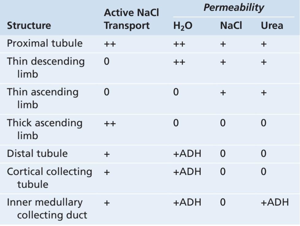
ADH, Antidiuretic hormone; NaCl, sodium chloride; 0, minimal level of active transport or permeability; +, moderate level of active transport or permeability; ++, high level of active transport or permeability; +ADH, permeability to water or urea is increased by ADH.
ADH، هورمون آنتی دیورتیک؛ NaCl، کلرید سدیم؛ 0، حداقل سطح حمل و نقل فعال یا نفوذپذیری؛ +، سطح متوسط انتقال فعال یا نفوذپذیری. ++، سطح بالای حمل و نقل فعال یا نفوذپذیری؛ +ADH، نفوذپذیری به آب یا اوره توسط ADH افزایش می یابد.
Steps Involved in Causing Hyperosmotic Renal Medullary Interstitium. Keeping in mind these characteristics of the loop of Henle, let us now discuss how the renal medulla becomes hyperosmotic (Video 29-1). First, assume that the loop of Henle is filled with fluid having a concentration of 300 mOsm/L, the same as that leaving the proximal tubule (Figure 29-4, step 1). Next, the active ion pump of the thick ascending limb on the loop of Henle reduces the concentration inside the tubule and raises the interstitial concentration; this pump establishes a 200-mOsm/L con- centration gradient between the tubular fluid and interstitial fluid (Figure 29-4, step 2). The limit to the gradient is about 200 mOsm/L because paracellular diffusion of ions back into the tubule eventually counterbalances the trans- port of ions out of the lumen when the 200-mOsm/L con- centration gradient is achieved.
مراحل دخیل در ایجاد اینترستیتیوم مدولاری هیپراسموتیک کلیه. با در نظر گرفتن این ویژگی های حلقه هنله، اجازه دهید اکنون در مورد چگونگی هیپراسموتیک شدن مدولای کلیه بحث کنیم (ویدئو 29-1). ابتدا فرض کنید که حلقه هنله با مایعی با غلظت 300 mOsm/L پر شده است، همان چیزی که از لوله پروگزیمال خارج می شود (شکل 29-4، مرحله 1). سپس، پمپ یونی فعال اندام صعودی ضخیم روی حلقه هنله، غلظت داخل لوله را کاهش می دهد و غلظت بینابینی را افزایش می دهد. این پمپ یک گرادیان غلظت 200 mOsm/L بین مایع لوله ای و مایع بینابینی ایجاد می کند (شکل 29-4، مرحله 2). حد شیب حدود 200 mOsm/L است زیرا انتشار پاراسلولی یونها به داخل لوله در نهایت با رسیدن به گرادیان غلظت 200 mOsm/L، انتقال یونها به خارج از لومن را متعادل میکند.
Step 3 is that the tubular fluid in the descending limb of the loop of Henle and interstitial fluid quickly reaches osmotic equilibrium due to osmosis of water out of the descending limb. The interstitial osmolarity is maintained at 400 mOsm/L because of continued transport of ions out of the thick ascending loop of Henle. Thus, by itself, active transport of sodium chloride out of the thick ascending limb is capable of establishing only a 200-mOsm/L con- centration gradient, which is much less than that achieved by the countercurrent multiplier system.
مرحله 3 این است که مایع لوله ای در اندام نزولی حلقه هنله و مایع بینابینی به دلیل اسمز آب از اندام نزولی به سرعت به تعادل اسمزی می رسد. اسمولاریته بینابینی به دلیل انتقال مداوم یونها به خارج از حلقه صعودی ضخیم هنل در mOsm/L 400 حفظ می شود. بنابراین، به خودی خود، انتقال فعال کلرید سدیم به خارج از اندام صعودی ضخیم قادر به ایجاد یک گرادیان غلظت 200 mOsm/L است که بسیار کمتر از آن چیزی است که توسط سیستم ضرب کننده جریان مخالف به دست می آید.
Step 4 is the additional flow of fluid into the loop of Henle from the proximal tubule, which causes the hyper- osmotic fluid previously formed in the descending limb to flow into the ascending limb. Once this fluid is in the ascending limb, additional ions are pumped into the interstitium and water remains in the tubular fluid until a 200-mOsm/L osmotic gradient is established, and the interstitial fluid osmolarity rises to 500 mOsm/L (step 5). Then, once again, fluid in the descending limb reaches equilibrium with the hyperosmotic medullary interstitial fluid (step 6) and, as the hyperosmotic tubular fluid from the descending limb of the loop of Henle flows into the ascending limb, still more solute is continuously pumped out of the tubules and deposited into the medullary interstitium.
مرحله 4 جریان اضافی مایع به حلقه هنله از لوله پروگزیمال است که باعث می شود مایع هیپراسموتیک که قبلاً در اندام نزولی تشکیل شده بود به اندام صعودی جریان یابد. هنگامی که این مایع در اندام صعودی قرار گرفت، یونهای اضافی به داخل بینابینی پمپ میشوند و آب در مایع لولهای باقی میماند تا زمانی که شیب اسمزی 200 mOsm/L ایجاد شود و اسمولاریته مایع بینابینی به mOsm/L 500 افزایش یابد (مرحله 5). سپس، یک بار دیگر، مایع در اندام نزولی با مایع میان بافتی مدولاری هیپراسموتیک به تعادل می رسد (مرحله 6) و همانطور که مایع لوله ای هیپراسموتیک از اندام نزولی حلقه هنله به سمت اندام صعودی جریان می یابد، همچنان املاح بیشتری به طور مداوم از لوله های بین تیتری به بیرون پمپ می شود.
These steps are repeated over and over, with the net effect of adding more and more solute to the medulla in excess of water. With sufficient time, this process gradually traps solutes in the medulla and multiplies the concentration gradient established by the active pumping of ions out of the thick ascending loop of Henle, eventually raising the interstitial fluid osmolarity to 1200 to 1400 mOsm/L, as shown in step 7.
این مراحل بارها و بارها تکرار می شوند، با اثر خالص افزودن بیشتر و بیشتر املاح به بصل النخاع بیش از حد آب. با گذشت زمان کافی، این فرآیند به تدریج املاح را در بصل النخاع به دام می اندازد و گرادیان غلظت ایجاد شده توسط پمپاژ فعال یون ها به خارج از حلقه صعودی ضخیم هنله را چند برابر می کند و در نهایت اسمولاریته مایع بینابینی را به 1200 تا 1400 mOsm/L افزایش می دهد، همانطور که در مرحله 7 نشان داده شده است.
Thus, the repetitive reabsorption of sodium chloride by the thick ascending loop of Henle and continued inflow of new sodium chloride from the proximal tubule into the loop of Henle is called the countercurrent multiplier. The sodium chloride reabsorbed from the ascending loop of Henle keeps adding to the newly arrived sodium chloride, thus “multiplying” its concentration in the medullary interstitium.
بنابراین، بازجذب مکرر کلرید سدیم توسط حلقه صعودی ضخیم هنله و ادامه ورود کلرید سدیم جدید از لوله پروگزیمال به حلقه هنله را ضربدر جریان مخالف می نامند. کلرید سدیمی که از حلقه صعودی هنله بازجذب میشود، مدام به کلرید سدیم تازه وارد شده اضافه میشود، بنابراین غلظت آن در بینابینی مدولاری افزایش مییابد.

Figure 29-4 Countercurrent multiplier system in loop of Henle for producing a hyperosmotic renal medulla. (Numerical values are in milliosmoles per liter.)
شکل 29-4 سیستم ضربکننده جریان مخالف در حلقه هنله برای ایجاد مدولای کلیوی هیپراسموتیک. (مقادیر عددی بر حسب میلیاسمول در لیتر هستند.)
ROLE OF DISTAL TUBULE AND COLLECTING DUCTS IN EXCRETING CONCENTRATED URINE
When the tubular fluid leaves the loop of Henle and flows into the distal convoluted tubule in the renal cortex, the fluid is dilute, with an osmolarity of only about 100 to 140 mOsm/L (Figure 29-5). The early distal tubule further dilutes the tubular fluid because this segment, like the ascending loop of Henle, actively transports sodium chloride out of the tubule but is relatively impermeable to water.
نقش توبول دیستال و مجراهای جمع کننده در دفع ادرار غلیظ
هنگامی که مایع لولهای از حلقه هنله خارج میشود و به داخل لوله پیچیده دیستال در قشر کلیه جریان مییابد، مایع رقیق میشود و اسمولاریته آن تنها حدود 100 تا 140 mOsm/L است (شکل 29-5). لوله دیستال اولیه مایع لوله ای را بیشتر رقیق می کند زیرا این بخش مانند حلقه صعودی هنله به طور فعال کلرید سدیم را به خارج از لوله منتقل می کند اما نسبت به آب نسبتاً غیرقابل نفوذ است.
As fluid flows into the cortical collecting tubule, the amount of water reabsorbed is critically dependent on the plasma concentration of ADH. In the absence of ADH, this segment is almost impermeable to water and fails to reabsorb water but continues to reabsorb solutes and further dilutes the urine. When there is a high concentration of ADH, the cortical collecting tubule becomes highly permeable to water, so large amounts of water are now reabsorbed from the tubule into the cortex interstitium, where it is swept away by the rapidly flowing peritubular capillaries. Because large amounts of water are reabsorbed into the cortex, rather than into the renal medulla, this helps preserve the high medullary interstitial fluid osmolarity.
همانطور که مایع به داخل لوله جمع کننده قشر مغز جریان می یابد، مقدار آب بازجذب شده به شدت به غلظت ADH پلاسما بستگی دارد. در غیاب ADH، این بخش تقریباً در برابر آب غیرقابل نفوذ است و نمی تواند آب را دوباره جذب کند، اما همچنان به جذب املاح ادامه می دهد و ادرار را بیشتر رقیق می کند. هنگامی که غلظت بالایی از ADH وجود دارد، لوله جمعکننده قشر به آب نفوذپذیری بالایی پیدا میکند، بنابراین مقادیر زیادی آب اکنون از لوله به داخل قشر بینابینی بازجذب میشود، جایی که توسط مویرگهای دور لولهای که به سرعت در حال جریان هستند از بین میرود. از آنجایی که مقادیر زیادی آب به جای داخل بصل النخاع کلیه به قشر مغز جذب می شود، این امر به حفظ اسمولاریته بالای مایع میان بافتی مدولاری کمک می کند.
As the tubular fluid flows along the medullary collecting ducts, there is further water reabsorption from the tubular fluid into the interstitium, but the total amount of water is relatively small compared with that added to the cortex interstitium. The reabsorbed water is carried away by the vasa recta into the venous blood. When high levels of ADH are present, the collecting ducts become permeable to water, so the fluid at the end of the collecting ducts has essentially the same osmolarity as the interstitial fluid of the renal medulla-about 1200 mOsm/L (see Figure 29-4). Thus, by reabsorbing as much water as possible, the kidneys form highly concentrated urine, excreting normal amounts of solutes in the urine while adding water back to the extracellular fluid and compensating for deficits of body water.
از آنجایی که مایع لولهای در امتداد مجاری جمعآوری مدولاری جریان مییابد، جذب مجدد آب بیشتر از مایع لولهای به داخل بینابینی انجام میشود، اما مقدار کل آب در مقایسه با آب اضافه شده به بینابینی کورتکس، نسبتاً کم است. آب بازجذب شده توسط وازا رکتا به خون وریدی منتقل می شود. هنگامی که سطوح بالای ADH وجود داشته باشد، مجاری جمعکننده به آب نفوذپذیر میشوند، بنابراین مایع انتهای مجاری جمعآوری اساساً اسمولاریته مشابه مایع بینابینی بصل النخاع کلیه دارد – حدود mOsm/L 1200 (شکل 29-4 را ببینید). بنابراین، با بازجذب هرچه بیشتر آب، کلیهها ادرار بسیار غلیظی تشکیل میدهند و مقادیر طبیعی املاح را در ادرار دفع میکنند و در عین حال آب را به مایع خارج سلولی برمیگردانند و کمبود آب بدن را جبران میکنند.

Figure 29-5. Formation of a concentrated urine when antidiuretic hormone (ADH) levels are high. Note that the fluid leaving the loop of Henle is dilute but becomes concentrated as water is absorbed from the distal tubules and collecting tubules. With high ADH levels, the osmolarity of the urine is about the same as the osmolarity of the renal medullary interstitial fluid in the papilla, which is about 1200 mOsm/L. (Numerical values are in milliosmoles per liter.)
شکل 29-5. تشکیل ادرار غلیظ زمانی که سطح هورمون آنتی دیورتیک (ADH) بالا باشد. توجه داشته باشید که مایع خروجی از حلقه هنله رقیق است اما با جذب آب از لوله های انتهایی و لوله های جمع کننده غلیظ می شود. با سطوح بالای ADH، اسمولاریته ادرار تقریباً به اندازه اسمولاریته مایع میان بافتی مدولاری کلیه در پاپیلا است که حدود mOsm/L 1200 است. (مقادیر عددی بر حسب میلی مول در لیتر می باشد.)
UREA CONTRIBUTES TO HYPEROSMOTIC RENAL MEDULLARY INTERSTITIUM AND FORMATION OF CONCENTRATED URINE
Urea contributes about 40% to 50% of the osmolarity (500-600 mOsm/L) of the renal medullary interstitium when the kidney is forming a maximally concentrated urine. Unlike sodium chloride, urea is passively reabsorbed from the tubule. When there is a water deficit and blood concentration of ADH is high, large amounts of urea are passively reabsorbed from the inner medullary collecting ducts into the interstitium.
اوره در میان بافتی مدولاری کلیوی هیپراسموتیک و تشکیل ادرار غلیظ نقش دارد
اوره حدود 40% تا 50% اسمولاریته (500-600 mOsm/L) بینابینی مدولاری کلیه را تشکیل میدهد که کلیه در حال تشکیل ادرار با حداکثر غلظت است. برخلاف کلرید سدیم، اوره به طور غیر فعال از لوله بازجذب می شود. هنگامی که کمبود آب وجود دارد و غلظت ADH خون بالا است، مقادیر زیادی اوره به طور غیر فعال از مجاری جمع کننده مدولاری داخلی به داخل بینابینی بازجذب می شود.
The mechanism for reabsorption of urea into the renal medulla is as follows. As water flows up the ascending loop of Henle and into the distal and cortical collecting tubules, little urea is reabsorbed because these segments are impermeable to urea (see Table 29-1). In the presence of high concentrations of ADH, water is reabsorbed rapidly from the cortical collecting tubule, and the urea concentration increases rapidly because urea is not very permeant in this part of the tubule.
مکانیسم بازجذب اوره به بصل النخاع کلیه به شرح زیر است. همانطور که آب از حلقه صعودی هنله و به داخل لوله های جمع کننده انتهایی و قشر مغز جریان می یابد، اوره کمی دوباره جذب می شود زیرا این بخش ها نسبت به اوره نفوذ ناپذیر هستند (جدول 29-1 را ببینید). در حضور غلظت های بالای ADH، آب به سرعت از لوله جمع کننده قشر مغز بازجذب می شود و غلظت اوره به سرعت افزایش می یابد زیرا اوره در این قسمت از لوله بسیار نفوذ نمی کند.
As the tubular fluid flows into the inner medullary collecting ducts, still more water reabsorption takes place, resulting in an even higher concentration of urea in the fluid. This high concentration of urea in the tubular fluid of the inner medullary collecting duct causes urea to diffuse out of the tubule into the renal interstitial fluid. This diffusion is greatly facilitated by specific urea transporters, UT-A1 and UT-A3. These urea transporters are activated by ADH, increasing transport of urea out of the inner medullary collecting duct even more when ADH levels are elevated. The simultaneous movement of water and urea out of the inner medullary collecting ducts maintains a high concentration of urea in the tubular fluid and, eventually, in the urine, even though urea is being reabsorbed.
هنگامی که مایع لولهای به داخل مجرای جمعآوری مدولاری داخلی جریان مییابد، بازجذب مجدد آب بیشتر صورت میگیرد که منجر به غلظت حتی بالاتر اوره در مایع میشود. این غلظت بالای اوره در مایع لولهای مجرای جمعکننده مدولاری داخلی باعث میشود که اوره از لوله به داخل مایع بینابینی کلیوی منتشر شود. این انتشار توسط انتقال دهنده های خاص اوره، UT-A1 و UT-A3 بسیار تسهیل می شود. این انتقال دهنده های اوره توسط ADH فعال می شوند و در صورت افزایش سطح ADH، انتقال اوره به خارج از مجرای جمع کننده مدولاری داخلی را بیشتر می کنند. حرکت همزمان آب و اوره به خارج از مجاری جمعآوری مدولاری داخلی، غلظت بالایی از اوره را در مایع لولهای و در نهایت در ادرار حفظ میکند، حتی اگر اوره دوباره جذب شود.
The fundamental role of urea in contributing to urine- concentrating ability is evidenced by the fact that people who ingest a high-protein diet, yielding large amounts of urea as a nitrogenous waste product, can concentrate their urine much better than people whose protein intake and urea production are low. Malnutrition is associated with a low urea concentration in the medullary interstitium and considerable impairment of urine-concentrating ability.
نقش اساسی اوره در کمک به توانایی تمرکز ادرار با این واقعیت مشهود است که افرادی که رژیم غذایی پر پروتئین مصرف می کنند و مقادیر زیادی اوره به عنوان یک محصول زائد نیتروژن دار تولید می کنند، می توانند ادرار خود را بسیار بهتر از افرادی که دریافت پروتئین و تولید اوره آنها کم است، غلیظ کنند. سوء تغذیه با غلظت کم اوره در بینابینی مدولاری و اختلال قابل توجه در توانایی تمرکز ادرار همراه است.
Recirculation of Urea from Collecting Duct to Loop of Henle Contributes to Hyperosmotic Renal Medulla. A healthy person usually excretes about 20% to 60% of the filtered load of urea, depending on urine flow rate and state of hydration. In general, the rate of urea excretion is determined mainly by the following: (1) concentration of urea in the plasma; (2) glomerular filtration rate (GFR); and (3) renal tubular urea reabsorption. In patients with renal disease who have large reductions in GFR, the plas- ma urea concentration increases markedly, returning the filtered urea load and urea excretion rate to the normal level (equal to the rate of urea production), despite the reduced GFR.
گردش مجدد اوره از مجرای جمع کننده به حلقه هنله به مدولای هیپراسموتیک کلیه کمک می کند. یک فرد سالم معمولاً حدود 20 تا 60 درصد از بار فیلتر شده اوره را، بسته به سرعت جریان ادرار و وضعیت هیدراتاسیون، دفع می کند. به طور کلی، سرعت دفع اوره عمدتاً با موارد زیر تعیین می شود: (1) غلظت اوره در پلاسما. (2) نرخ فیلتراسیون گلومرولی (GFR). و (3) بازجذب اوره لولهای کلیوی. در بیماران مبتلا به بیماری کلیوی که کاهش زیادی در GFR دارند، غلظت اوره پلاسما به طور قابل توجهی افزایش مییابد و با وجود کاهش GFR، بار اوره فیلتر شده و سرعت دفع اوره را به سطح نرمال (برابر سرعت تولید اوره) برمیگرداند.
In the proximal tubule, 40% to 50% of the filtered urea is reabsorbed but, even so, the tubular fluid urea concentration increases because urea is not nearly as permeant as water. The concentration of urea continues to rise as the tubular fluid flows into the thin segments of the loop of Henle, partly because of water reabsorption out of the descending loop of Henle but also because of some secretion of urea into the thin loop of Henle from the medullary interstitium (Figure 29-6). The passive secretion of urea into the thin loops of Henle is facilitated by the urea transporter UT-A2.
در لوله پروگزیمال، 40 تا 50 درصد اوره فیلتر شده دوباره جذب میشود، اما با این وجود، غلظت اوره مایع لولهای افزایش مییابد زیرا اوره تقریباً به اندازه آب نفوذپذیر نیست. با جریان یافتن مایع لولهای به بخشهای نازک حلقه هنل، غلظت اوره همچنان افزایش مییابد، تا حدی به دلیل بازجذب آب از حلقه نزولی هنله و همچنین به دلیل ترشح مقداری اوره به حلقه نازک هنله از میان بافتی مدولاری (شکل 29-6). ترشح غیرفعال اوره در حلقه های نازک هنله توسط ناقل اوره UT-A2 تسهیل می شود.
The thick limb of the loop of Henle, distal tubule, and cortical collecting tubule are all less permeable to urea, and only small amounts of urea reabsorption normally occur in these tubular segments. When the kidney is forming concentrated urine, and high levels of ADH are present, reabsorption of water from the distal tubule and cortical collecting tubule further raises the tubular fluid concentration of urea. As this urea flows into the inner medullary collecting duct, the high tubular fluid concentration of urea and urea transporters UT-A1 and UT-A3 cause urea to diffuse into the medullary interstitium. A moderate share of the urea that moves into the medullary interstitium eventually diffuses into the thin loop of Henle and then passes upward through the ascending loop of Henle, distal tubule, and cortical collecting tubule and back down into the medullary collecting duct again. In this way, urea can recirculate through these terminal parts of the tubular system several times before it is excreted. Each time around the circuit contributes to a higher con- centration of urea.
اندام ضخیم حلقه هنله، لوله دیستال و لوله جمع کننده قشر همگی کمتر به اوره نفوذ می کنند و فقط مقادیر کمی از بازجذب اوره معمولاً در این بخش های لوله ای رخ می دهد. هنگامی که کلیه در حال تشکیل ادرار غلیظ است و سطوح بالایی از ADH وجود دارد، بازجذب آب از لولههای انتهایی و لولههای جمعکننده قشر مغز، غلظت مایع لولهای اوره را بیشتر میکند. هنگامی که این اوره به داخل مجرای جمعآوری مدولاری داخلی جریان مییابد، غلظت بالای مایع لولهای اوره و ناقلهای اوره UT-A1 و UT-A3 باعث انتشار اوره در بینابینی مدولاری میشود. سهم متوسطی از اوره که به داخل بینابینی مدولاری حرکت می کند، در نهایت به حلقه نازک هنله منتشر می شود و سپس از طریق حلقه صعودی هنله، لوله دیستال و لوله جمع کننده قشر مغز به سمت بالا عبور می کند و مجدداً به داخل مجرای جمع کننده مدولاری برمی گردد. به این ترتیب اوره می تواند چندین بار قبل از دفع از طریق این قسمت های انتهایی سیستم لوله ای گردش کند. هر بار دور مدار باعث افزایش غلظت اوره می شود.
This urea recirculation provides an additional mechanism for forming a hyperosmotic renal medulla. Because urea is one of the most abundant waste products that must be excreted by the kidneys, this mechanism for concentrating urea before it is excreted is essential to the economy of the body fluid when water is in short supply.
این گردش مجدد اوره یک مکانیسم اضافی برای تشکیل یک مدولای هیپراسموتیک کلیه فراهم می کند. از آنجایی که اوره یکی از فراوانترین مواد زائد است که باید توسط کلیهها دفع شود، این مکانیسم برای تغلیظ اوره قبل از دفع آن برای صرفهجویی در مایعات بدن در مواقعی که آب کمبود دارد ضروری است.
When there is excess water in the body, urine flow rate increases, and therefore the concentration of urea in the inner medullary collecting ducts decreases, causing less diffusion of urea into the renal medullary interstitium. ADH levels are also reduced when there is excess body water, and this reduction, in turn, decreases the permeability of the inner medullary collecting ducts to both water and urea, and more urea is excreted in the urine.
هنگامی که آب اضافی در بدن وجود دارد، سرعت جریان ادرار افزایش مییابد و در نتیجه غلظت اوره در مجاری جمعآوری مدولاری داخلی کاهش مییابد و باعث انتشار کمتر اوره در بینابینی مدولاری کلیه میشود. سطح ADH نیز در صورت وجود آب اضافی بدن کاهش می یابد و این کاهش به نوبه خود باعث کاهش نفوذپذیری مجاری جمع کننده مدولاری داخلی به آب و اوره می شود و اوره بیشتری از ادرار دفع می شود.
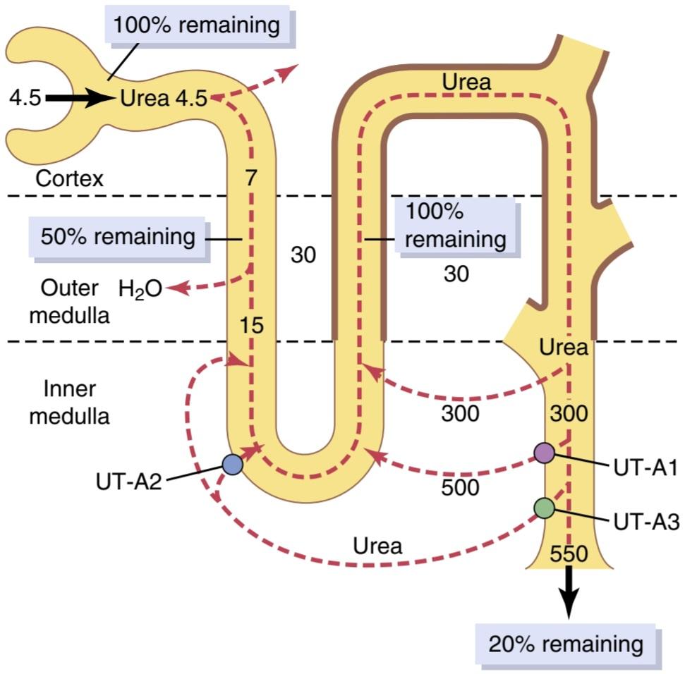
Figure 29-6. Recirculation of urea absorbed from the medullary collecting duct into the interstitial fluid. This urea diffuses into the thin loop of Henle, then passes through the distal tubules, and finally passes back into the collecting duct. The recirculation of urea helps trap urea in the renal medulla and contributes to the hyperosmolarity of the renal medulla. The heavy lines, from the thick ascending loop of Henle to the medullary collecting ducts, indicate that these segments are not very permeable to urea. The urea transporters UT-A1 and UT-A3 facilitate diffusion of urea out of the medullary collecting ducts while UT-A2 facilitates urea diffusion into the thin descending loop of Henle. (Numerical values are in milliosmoles per liter of urea during antidiuresis, when large amounts of antidiuretic hormone are present. Percentages of the filtered load of urea that remain in the tubules are indicated in the boxes.)
شکل 29-6. گردش مجدد اوره جذب شده از مجرای جمع کننده مدولاری در مایع بینابینی. این اوره در حلقه نازک هنله منتشر می شود، سپس از لوله های انتهایی عبور می کند و در نهایت به مجرای جمع کننده باز می گردد. گردش مجدد اوره به به دام انداختن اوره در بصل النخاع کلیه کمک می کند و به هیپراسمولاریته مدولای کلیه کمک می کند. خطوط سنگین، از حلقه صعودی ضخیم هنله تا مجراهای جمع کننده مدولاری، نشان می دهد که این بخش ها نسبت به اوره بسیار نفوذپذیر نیستند. انتقال دهنده های اوره UT-A1 و UT-A3 انتشار اوره را به خارج از مجاری جمع کننده مدولاری تسهیل می کنند در حالی که UT-A2 انتشار اوره را در حلقه نزولی نازک هنله تسهیل می کند. (مقادیر عددی بر حسب میلی مول در لیتر اوره در طول آنتی دیورز است، زمانی که مقادیر زیادی هورمون ضد ادرار وجود دارد. درصد بار فیلتر شده اوره که در لوله ها باقی می ماند در جعبه ها نشان داده شده است.)
COUNTERCURRENT EXCHANGE IN VASA RECTA PRESERVES HYPEROSMOLARITY OF RENAL MEDULLA
Blood flow must be provided to the renal medulla to supply the metabolic needs of the cells in this part of the kidney. Without a special medullary blood flow system, the solutes pumped into the renal medulla by the countercurrent multiplier system would be rapidly dissipated.
تبادل جریان متقابل در VASA RECTA حفظ هیپراسمولاریته مدولای کلیه
جریان خون باید به بصل النخاع کلیه تامین شود تا نیازهای متابولیکی سلول های این قسمت از کلیه را تامین کند. بدون سیستم جریان خون مدولاری خاص، املاح پمپ شده به بصل النخاع کلیه توسط سیستم ضرب کننده جریان مخالف به سرعت دفع می شوند.
Two special features of the renal medullary blood flow contribute to the preservation of the high solute concentrations:
دو ویژگی خاص جریان خون مدولاری کلیه به حفظ غلظت بالای املاح کمک می کند:
1. The medullary blood flow is low, accounting for less than 5% of the total renal blood flow. This sluggish blood flow is sufficient to supply the metabolic needs of the tissues but helps minimize solute loss from the medullary interstitium.
1.جریان خون مدولاری کم است و کمتر از 5 درصد از کل جریان خون کلیوی را تشکیل می دهد. این جریان خون کند برای تامین نیازهای متابولیکی بافت ها کافی است اما به حداقل رساندن از دست دادن املاح از بینابینی مدولاری کمک می کند.
2. The vasa recta serve as countercurrent exchangers, minimizing the washout of solutes from the medullary interstitium.
2. وازا رکتا به عنوان مبدل های جریان مخالف عمل می کند و خروج املاح را از بینابینی مدولاری به حداقل می رساند.
The countercurrent exchange mechanism operates as follows (Figure 29-7). Blood enters and leaves the medulla via the vasa recta at the boundary of the cortex and renal medulla. The vasa recta, like other capillaries, are highly permeable to solutes in the blood, except for the plasma proteins. As blood descends into the medulla toward the papillae, it becomes progressively more concentrated, partly by solute entry from the interstitium and partly by loss of water into the interstitium. By the time the blood reaches the tips of the vasa recta, it has a concentration of about 1200 mOsm/L, the same as that of the medullary interstitium. As blood ascends back toward the cortex, it becomes progressively less concentrated as solutes diffuse back out into the medullary interstitium and as water moves into the vasa recta.
مکانیسم تبادل جریان مخالف به شرح زیر عمل می کند (شکل 29-7). خون از طریق وازا رکتا در مرز قشر و مدولای کلیه وارد بصل النخاع می شود و از آن خارج می شود. وازا رکتا مانند سایر مویرگ ها به املاح خون به جز پروتئین های پلاسما نفوذ پذیری بالایی دارد. همانطور که خون به داخل بصل النخاع به سمت پاپیلاها پایین می آید، تا حدی با ورود املاح از بینابینی و تا حدی با از دست دادن آب به داخل بینابینی، به تدریج متمرکزتر می شود. زمانی که خون به نوک وازا رکتا می رسد، غلظت آن در حدود 1200 mOsm/L است، همان غلظت بینابینی مدولاری. همانطور که خون به سمت قشر مغز بالا می رود، به تدریج غلظت آن کاهش می یابد زیرا املاح به داخل بینابینی مدولاری منتشر می شوند و با حرکت آب به داخل وازا رکتا.
Although large amounts of fluid and solute are exchanged across the vasa recta, there is little net dilution of the concentration of the interstitial fluid at each level of the renal medulla because of the U shape of the vasa recta capillaries, which act as countercurrent exchangers. Thus, the vasa recta do not create the medullary hyperosmolarity, but they do prevent it from being dissipated.
اگرچه مقادیر زیادی مایع و املاح در سرتاسر وازا رکتا رد و بدل میشوند، اما رقت خالص کمی از غلظت مایع بینابینی در هر سطح از بصل النخاع کلیه وجود دارد، زیرا به شکل U مویرگهای وازا رکتا، که به عنوان مبدلهای جریان مخالف عمل میکنند، وجود دارد. بنابراین، وازا رکتا هیپراسمولاریته مدولاری را ایجاد نمی کند، اما مانع از بین رفتن آن می شود.
The U-shaped structure of the vessels minimizes loss of solute from the interstitium but does not prevent bulk flow of fluid and solutes into the blood through the usual colloid osmotic and hydrostatic pressures that favor reabsorption in these capillaries. Under steady-state conditions, the vasa recta carry away only as much solute and water as is absorbed from the medullary tubules, and the high concentration of solutes established by the counter- current mechanism is preserved.
ساختار U شکل رگها از دست دادن املاح از بینابینی را به حداقل میرساند، اما از جریان حجمی مایع و املاح به داخل خون از طریق فشارهای اسمزی و هیدرواستاتیک معمولی کلوئیدی که به جذب مجدد در این مویرگها کمک میکند، جلوگیری نمیکند. تحت شرایط حالت پایدار، وازا رکتا تنها به اندازه ای که از لوله های مدولاری جذب می شود، املاح و آب را با خود می برد و غلظت بالای املاح ایجاد شده توسط مکانیسم جریان مخالف حفظ می شود.
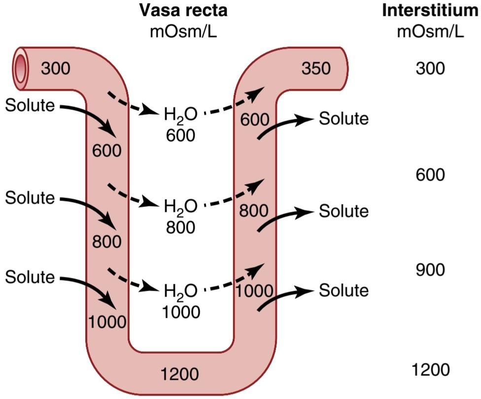
Figure 29-7. Countercurrent exchange in the vasa recta. Plasma flowing down the descending limb of the vasa recta becomes more hyperosmotic because of diffusion of water out of the blood and diffusion of solutes from the renal interstitial fluid into the blood. In the ascending limb of the vasa recta, solutes diffuse back into the interstitial fluid, and water diffuses back into the vasa recta. Large amounts of solutes would be lost from the renal medulla without the U shape of the vasa recta capillaries. (Numerical values are in milliosmoles per liter.)
شکل 29-7. تبادل جریان متقابل در وازا رکتا. پلاسمایی که در اندام نزولی وازا رکتا جریان دارد، به دلیل انتشار آب از خون و انتشار املاح از مایع بینابینی کلیوی به خون، هیپراسموتیک تر می شود. در اندام صعودی وازا رکتا، املاح به داخل مایع بینابینی و آب دوباره به داخل وازا رکتا منتشر می شود. مقادیر زیادی از املاح بدون شکل U مویرگ های وازا رکتا از بصل النخاع کلیه از بین می رود. (مقادیر عددی بر حسب میلی مول در لیتر می باشد.)
Increased Medullary Blood Flow Reduces UrineConcentrating Ability. Certain vasodilators can markedly increase renal medullary blood flow, thereby washing out some of the solutes from the renal medulla and reducing the maximum urine-concentrating ability. Large increases in arterial pressure may also increase the blood flow of the renal medulla to a greater extent than in other regions of the kidney and tend to wash out the hyperosmotic interstitium, thereby reducing urine-concentrating ability. As discussed earlier, maximum concentrating ability of the kidney is determined not only by the level of ADH but also by the osmolarity of the renal medulla interstitial fluid. Even with maximal ADH levels, the urineconcentrating ability will be reduced if medullary blood flow increases enough to reduce the hyperosmolarity in the renal medulla.
افزایش جریان خون مدولاری توانایی تمرکز ادرار را کاهش می دهد. برخی از گشادکنندههای عروقی میتوانند به طور قابل توجهی جریان خون مدولاری کلیه را افزایش دهند و در نتیجه برخی از املاح را از بصل النخاع کلیه شسته و حداکثر توانایی تمرکز ادرار را کاهش دهند. افزایش شدید فشار شریانی نیز ممکن است جریان خون مدولای کلیه را به میزان بیشتری نسبت به سایر نواحی کلیه افزایش دهد و تمایل به شستشوی بینابینی هیپراسموتیک داشته باشد و در نتیجه توانایی تمرکز ادرار را کاهش دهد. همانطور که قبلاً بحث شد، حداکثر توانایی تمرکز کلیه نه تنها با سطح ADH بلکه توسط اسمولاریته مایع بینابینی مدولای کلیه تعیین می شود. حتی با حداکثر سطوح ADH، اگر جریان خون مدولاری به اندازه کافی افزایش یابد تا هیپراسمولاریته در بصل الکلیه کاهش یابد، توانایی تمرکز ادرار کاهش می یابد.
SUMMARY OF URINE-CONCENTRATING MECHANISM AND CHANGES IN OSMOLARITY IN DIFFERENT TUBULAR SEGMENTS
The changes in osmolarity and volume of the tubular fluid as it passes through the different parts of the nephron are shown in Figure 29-8.
خلاصه مکانیسم تغلیظ ادرار و تغییرات در اسمولاریته در بخش های لوله ای مختلف
تغییرات اسمولاریته و حجم مایع لوله ای هنگام عبور از قسمت های مختلف نفرون در شکل 29-8 نشان داده شده است.
Proximal Tubule. About 65% of most filtered electrolytes is reabsorbed in the proximal tubule. However, the proximal tubular membranes are highly permeable to water so, whenever solutes are reabsorbed, water also diffuses through the tubular membrane by osmosis. Water diffusion across the proximal tubular epithelium is aided by the water channel aquaporin 1 (AQP-1). Therefore, the osmolarity of the fluid remains about the same as the glomerular filtrate-300 mOsm/L.
لوله پروگزیمال. حدود 65 درصد بیشتر الکترولیت های فیلتر شده در لوله پروگزیمال بازجذب می شوند. با این حال، غشاهای لولهای پروگزیمال نسبت به آب نفوذپذیری بالایی دارند، بنابراین، هر زمان که املاح دوباره جذب شوند، آب نیز از طریق اسمز از طریق غشای لولهای پخش میشود. انتشار آب در سراسر اپیتلیوم لوله ای پروگزیمال توسط کانال آب آکواپورین 1 (AQP-1) کمک می کند. بنابراین اسمولاریته سیال تقریباً مانند فیلتر گلومرولی 300 mOsm/L باقی می ماند.
Descending Loop of Henle. As fluid flows down the descending loop of Henle, water is absorbed into the medulla. The descending limb also contains AQP-1 and is highly permeable to water but much less permeable to sodium chloride and urea. Therefore, the osmolarity of the fluid flowing through the descending loop gradually increases until it is nearly equal to that of the surrounding interstitial fluid, which is about 1200 mOsm/L when the blood concentration of ADH is high.
حلقه نزولی هنل. همانطور که مایع در حلقه نزولی هنله جریان می یابد، آب به داخل بصل النخاع جذب می شود. اندام نزولی نیز حاوی AQP-1 است و نسبت به آب نفوذپذیری بالایی دارد اما نسبت به کلرید سدیم و اوره بسیار کمتر نفوذپذیر است. بنابراین، اسمولاریته مایعی که در حلقه نزولی جریان دارد، به تدریج افزایش مییابد تا جایی که تقریباً برابر با مایع بینابینی اطراف میشود، که وقتی غلظت ADH خون بالا است، حدود 1200 mOsm/L است.
When dilute urine is being formed, as a result of low ADH concentrations, the medullary interstitial osmolarity is less than 1200 mOsm/L; consequently, the descending loop tubular fluid osmolarity also becomes less concentrated. This decrease in concentration is due partly to the fact that less urea is absorbed into the medullary interstitium from the collecting ducts when ADH levels are low, and the kidney is forming a large volume of dilute urine.
هنگامی که ادرار رقیق تشکیل می شود، در نتیجه غلظت کم ADH، اسمولاریته بینابینی مدولاری کمتر از 1200 mOsm/L است. در نتیجه، اسمولاریته سیال لولهای حلقه نزولی نیز کمتر متمرکز میشود. این کاهش غلظت تا حدی به این دلیل است که وقتی سطح ADH پایین است، اوره کمتری از مجاری جمعآوری به داخل بینابینی مدولاری جذب میشود و کلیه حجم زیادی از ادرار رقیق را تشکیل میدهد.
Thin Ascending Loop of Henle. The thin ascending limb is essentially impermeable to water but reabsorbs some sodium chloride. Because of the high concentration of sodium chloride in the tubular fluid as a result of water removal from the descending loop of Henle, there is some passive diffusion of sodium chloride from the thin ascending limb into the medullary interstitium. Thus, the tubular fluid becomes more dilute as the sodium chloride diffuses out of the tubule and water remains in the tubule.
حلقه صعودی نازک هنله. اندام صعودی نازک اساساً در برابر آب نفوذ ناپذیر است اما مقداری کلرید سدیم را دوباره جذب می کند. به دلیل غلظت بالای کلرید سدیم در مایع لوله ای در نتیجه حذف آب از حلقه نزولی هنله، مقداری انتشار غیرفعال کلرید سدیم از اندام صعودی نازک به داخل بینابینی مدولاری وجود دارد. بنابراین، مایع لولهای رقیقتر میشود، زیرا کلرید سدیم به بیرون از لوله پخش میشود و آب در لوله باقی میماند.
Some of the urea absorbed into the medullary inter stitium from the collecting ducts also diffuses into the ascending limb, thereby returning the urea to the tubular system and helping prevent its washout from the renal medulla. This urea recycling is an additional mechanism that contributes to the hyperosmotic renal medulla.
مقداری از اوره جذب شده از مجاری جمعآوری به درون بافت مدولاری نیز به اندام صعودی منتشر میشود، در نتیجه اوره را به سیستم لولهای باز میگرداند و به جلوگیری از خروج آن از بصل الکلیه کمک میکند. این بازیافت اوره یک مکانیسم اضافی است که به مدولای هیپراسموتیک کلیه کمک می کند.
Thick Ascending Loop of Henle. The thick part of the ascending loop of Henle is also virtually impermeable to water, but large amounts of sodium, chloride, potassium, and other ions are actively transported from the tubule into the medullary interstitium. Therefore, fluid in the thick ascending limb of the loop of Henle becomes very dilute, falling to a concentration of about 140 mOsm/L.
حلقه صعودی ضخیم هنله. قسمت ضخیم حلقه صعودی هنله نیز عملاً در برابر آب غیرقابل نفوذ است، اما مقادیر زیادی سدیم، کلرید، پتاسیم و سایر یون ها به طور فعال از لوله به داخل بینابینی مدولاری منتقل می شوند. بنابراین، مایع در اندام صعودی ضخیم حلقه هنله بسیار رقیق می شود و به غلظت حدود 140 mOsm/L می رسد.
Early Distal Tubule. The early distal tubule has properties similar to those of the thick ascending loop of Henle, so further dilution of the tubular fluid to about 100 mOsm/L occurs as solutes are reabsorbed while water remains in the tubule.
توبول دیستال اولیه. لوله دیستال اولیه خواصی شبیه به حلقه صعودی ضخیم هنله دارد، بنابراین رقیق شدن بیشتر سیال لولهای تا حدود mOsm/L 100 زمانی رخ میدهد که املاح دوباره جذب میشوند در حالی که آب در لوله باقی میماند.
Late Distal Tubule and Cortical Collecting Tubules. In the late distal tubule and cortical collecting tubules, the osmolarity of the fluid depends on the level of ADH. With high ADH levels, these tubules are highly permeable to water, and significant amounts of water are reabsorbed. Urea, however, is not very permeant in this part of the nephron, resulting in increased urea concentration as water is reabsorbed. This process allows most of the urea delivered to the distal tubule and collecting tubule to pass into the inner medullary collecting ducts, from which it is eventually reabsorbed or excreted in the urine. In the absence of ADH, little water is reabsorbed in the late distal tubule and cortical collecting tubule; therefore, osmolarity decreases even further because of continued active reabsorption of ions from these segments.
لوله دیستال دیررس و لوله های جمع کننده قشر. در لوله های انتهایی دیستال و لوله های جمع کننده قشر، اسمولاریته مایع به سطح ADH بستگی دارد. با سطوح بالای ADH، این لوله ها نسبت به آب نفوذپذیری بالایی دارند و مقادیر قابل توجهی آب دوباره جذب می شوند. با این حال، اوره در این قسمت از نفرون بسیار نفوذپذیر نیست و در نتیجه با جذب مجدد آب، غلظت اوره افزایش مییابد. این فرآیند به بیشتر اوره تحویل داده شده به لوله دیستال و لوله جمعآوری اجازه میدهد تا به مجرای جمعکننده مدولاری داخلی منتقل شود، که در نهایت از آن دوباره جذب یا در ادرار دفع میشود. در غیاب ADH، آب کمی در لوله انتهایی دیستال و لوله جمع کننده قشر مغز دوباره جذب می شود. بنابراین، به دلیل ادامه بازجذب فعال یونها از این بخشها، اسمولاریته حتی بیشتر کاهش مییابد.
Inner Medullary Collecting Ducts. The concentration of fluid in the inner medullary collecting ducts also depends on the following: (1) ADH; and (2) the surrounding medullary interstitium osmolarity established by the countercurrent mechanism. In the presence of large amounts of ADH, these ducts are highly permeable to water, and water diffuses from the tubule into the interstitial fluid until osmotic equilibrium is reached, with the tubular fluid having about the same concentration as the renal medullary interstitium (1200-1400 mOsm/L). Thus, a small volume of concentrated urine is produced when ADH levels are high. Because water reabsorption increases urea concentration in the tubular fluid, and because the inner medullary collecting ducts have specific urea transporters that greatly facilitate diffusion, much of the highly con- centrated urea in the ducts diffuses out of the tubular lumen into the medullary interstitium. This absorption of the urea into the renal medulla contributes to the high osmolarity of the medullary interstitium and high concentrating ability of the kidney.
مجراهای جمع کننده مدولاری داخلی. غلظت مایع در مجاری جمع کننده مدولاری داخلی نیز به موارد زیر بستگی دارد: (1) ADH. و (2) اسمولاریته بینابینی مدولاری اطراف ایجاد شده توسط مکانیسم جریان مخالف. در حضور مقادیر زیاد ADH، این مجراها به آب نفوذپذیری بالایی دارند و آب از لوله به داخل مایع بینابینی منتشر میشود تا زمانی که تعادل اسمزی حاصل شود، با مایع لولهای تقریباً همان غلظت بینابینی مدولاری کلیه (1200-1400 OsmL mL). بنابراین، هنگامی که سطح ADH بالا باشد، حجم کمی از ادرار غلیظ تولید می شود. از آنجایی که بازجذب آب غلظت اوره را در مایع لولهای افزایش میدهد و از آنجایی که مجاری جمعآوری مدولاری داخلی دارای ناقلهای اوره خاص است که انتشار را بسیار تسهیل میکند، بسیاری از اوره بسیار غلیظ در مجاری از مجرای لولهای به داخل میان بافتی مدولاری پخش میشود. این جذب اوره در بصل النخاع کلیه به اسمولاریته بالای بینابینی مدولاری و توانایی تمرکز بالای کلیه کمک می کند.
Several important points to consider may not be obvious from this discussion. First, although sodium chloride is one of the principal solutes that contribute to the hyperosmolarity of the medullary interstitium, the kidney can, when needed, excrete a highly concentrated urine that contains little sodium chloride. The hyperosmolarity of the urine in these circumstances is due to high concentrations of other solutes, especially of waste products such as urea. One condition in which this occurs is dehydration accompanied by low sodium intake. As discussed in Chapter 30, a low sodium intake stimulates formation of the hormones angiotensin II and aldosterone, which together cause avid sodium reabsorption from the tubules while leaving the urea and other solutes to maintain the highly concentrated urine.
چند نکته مهم که باید در نظر گرفته شود ممکن است از این بحث آشکار نباشد. اولاً، اگرچه کلرید سدیم یکی از املاح اصلی است که به هیپراسمولاریته بینابینی مدولاری کمک می کند، کلیه می تواند در صورت نیاز، ادرار بسیار غلیظی را که حاوی کلرید سدیم کمی است دفع کند. هیپراسمولاریته ادرار در این شرایط به دلیل غلظت بالای سایر املاح به ویژه مواد زائد مانند اوره است. یکی از شرایطی که در آن رخ می دهد، کم آبی همراه با مصرف کم سدیم است. همانطور که در فصل 30 بحث شد، مصرف کم سدیم تشکیل هورمونهای آنژیوتانسین II و آلدوسترون را تحریک میکند که با هم باعث بازجذب شدید سدیم از لولهها میشوند در حالی که اوره و سایر املاح را برای حفظ ادرار بسیار غلیظ باقی میگذارند.
Second, large quantities of dilute urine can be excreted without increasing sodium excretion. This feat is accom- plished by decreasing ADH secretion, which reduces water reabsorption in the more distal tubular segments without significantly altering sodium reabsorption.
دوم اینکه مقادیر زیادی ادرار رقیق را می توان بدون افزایش دفع سدیم دفع کرد. این کار با کاهش ترشح ADH انجام میشود که جذب مجدد آب را در بخشهای لولهای دیستالتر بدون تغییر قابل توجهی در بازجذب سدیم کاهش میدهد.
Finally, there is an obligatory urine volume dictated by the maximum concentrating ability of the kidney and amount of solute that must be excreted. Therefore, if large amounts of solute must be excreted, they must be accompanied by the minimal amount of water necessary to excrete them. For example, if 600 milliosmoles of solute must be excreted each day, this requires at least 0.5 liter of urine if the maximal urine concentrating ability is 1200 mOsm/L.
در نهایت، یک حجم ادرار اجباری وجود دارد که بر اساس حداکثر توانایی تمرکز کلیه و مقدار املاح که باید دفع شود، وجود دارد. بنابراین، اگر مقدار زیادی از املاح باید دفع شود، باید با حداقل مقدار آب لازم برای دفع آنها همراه باشد. به عنوان مثال، اگر روزانه 600 میلی مول از املاح باید دفع شود، اگر حداکثر توانایی تمرکز ادرار 1200 mOsm/L باشد، حداقل 0.5 لیتر ادرار لازم است.
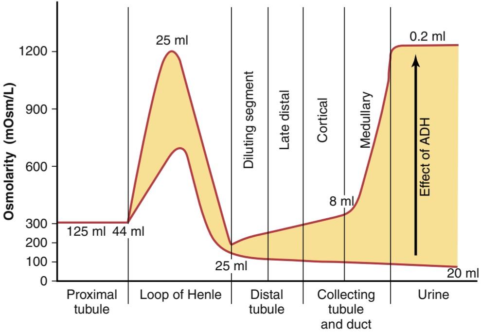
Figure 29-8. Changes in osmolarity of the tubular fluid as it passes through the different tubular segments in the presence of high levels of antidiuretic hormone (ADH) and absence of ADH. (Numerical values indicate the approximate volumes in milliliters per minute or in osmolarities in milliosmoles per liter of fluid flowing along the different tubular segments.)
شکل 29-8. تغییر در اسمولاریته سیال لوله ای هنگام عبور از بخش های مختلف لوله در حضور سطوح بالای هورمون آنتی دیورتیک (ADH) و عدم وجود ADH. (مقادیر عددی حجم تقریبی را بر حسب میلی لیتر در دقیقه یا اسمولاریته بر حسب میلی مول بر لیتر سیال در امتداد بخش های مختلف لوله ای را نشان می دهد.)
Quantifying Renal Urine Concentration and Dilution: Free Water and Osmolar Clearances
The process of concentrating or diluting the urine requires the kidneys to excrete water and solutes somewhat independently. When the urine is dilute, water is excreted in excess of solutes. Conversely, when the urine is concentrated, solutes are excreted in excess of water.
کمی سازی غلظت و رقت ادرار کلیه: پاکسازی های اسمولار و آب رایگان
فرآیند تغلیظ یا رقیق شدن ادرار به کلیه ها نیاز دارد که آب و املاح را تا حدودی مستقل دفع کنند. هنگامی که ادرار رقیق است، آب بیش از حد املاح دفع می شود. برعکس، وقتی ادرار غلیظ است، املاح بیش از حد آب دفع می شود.
The total clearance of solutes from the blood can be expressed as the osmolar clearance (Cosm). This is the volume of plasma cleared of solutes each minute, in the same way that clearance of a single substance is calculated:
کلیرانس کل املاح از خون را می توان به صورت کلیرانس اسمولی (Cosm) بیان کرد. این حجم پلاسمای پاکسازی شده از املاح در هر دقیقه است، به همان روشی که پاکسازی یک ماده واحد محاسبه می شود:
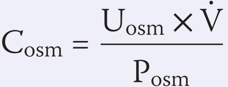
where Uosm is the urine osmolarity, V is the urine flow rate, and Posm is plasma osmolarity. For example, if the plasma osmo- larity is 300 mOsm/L, urine osmolarity is 600 mOsm/L, and urine flow rate is 1 ml/min (0.001 L/min), the rate of osmolar excretion is 0.6 mOsm/min (600 mOsm/L x 0.001 L/min), and osmolar clearance is 0.6 mOsm/min divided by 300 mOsm/L, or 0.002 L/min (2.0 ml/min). This means that 2 milliliters of plasma are being cleared of solute each minute.
که در آن Uosm اسمولاریته ادرار، V سرعت جریان ادرار و Posm اسمولاریته پلاسما است. برای مثال، اگر اسمولاریته پلاسما 300 mOsm/L، اسمولاریته ادرار 600 mOsm/L، و سرعت جریان ادرار 1 میلیلیتر در دقیقه (0.001 L/min) باشد، سرعت دفع اسمولی 0.6 mOsm/min (600 mOsm/min شفافیت) است. 0.6 mOsm/min تقسیم بر 300 mOsm/L یا 0.002 L/min (2.0 ml/min) است. این بدان معناست که در هر دقیقه 2 میلی لیتر پلاسما از املاح پاک می شود.
Free Water Clearance-Relative Rates at Which Solutes and Water Are Excreted
Free water clearance (CH20) is calculated as the difference between water excretion (urine flow rate) and osmolar clearance:
پاکسازی رایگان آب – نرخ های نسبی که در آن املاح و آب دفع می شوند
پاکسازی آب آزاد (CH20) به عنوان تفاوت بین دفع آب (سرعت جریان ادرار) و کلیرانس اسمولی محاسبه می شود:

Thus, the rate of free water clearance represents the rate at which solute-free water is excreted by the kidneys. When free water clearance is positive, excess water is being excreted by the kidneys; when free water clearance is negative, excess solutes are being removed from the blood by the kidneys, and water is being conserved.
بنابراین، میزان پاکسازی آب آزاد نشان دهنده سرعتی است که آب بدون املاح توسط کلیه ها دفع می شود. هنگامی که پاکسازی آب آزاد مثبت باشد، آب اضافی توسط کلیه ها دفع می شود. هنگامی که خلاصی آب آزاد منفی است، املاح اضافی توسط کلیه ها از خون خارج می شود و آب ذخیره می شود.
Using the example discussed earlier, if urine flow rate is 1 ml/min and osmolar clearance is 2 ml/min, free water clearance would be -1 ml/min. This means that instead of water being cleared from the kidneys in excess of solutes, the kidneys are actually returning water to the systemic circulation, as occurs during water deficits. Thus, whenever urine osmolarity is greater than plasma osmolarity, free water clearance is negative, indicating water conservation.
با استفاده از مثالی که قبلا بحث شد، اگر سرعت جریان ادرار 1 میلی لیتر در دقیقه و کلیرانس اسمولی 2 میلی لیتر در دقیقه باشد، خلاصی آب آزاد -1 میلی لیتر در دقیقه خواهد بود. این بدان معنی است که به جای اینکه آب بیش از حد از کلیه ها پاک شود، کلیه ها در واقع آب را به گردش خون سیستمی باز می گرداند، همانطور که در هنگام کمبود آب اتفاق می افتد. بنابراین، هرگاه اسمولاریته ادرار بیشتر از اسمولاریته پلاسما باشد، پاکسازی آب آزاد منفی است که نشان دهنده حفظ آب است.
When the kidneys are forming a dilute urine (i.e., urine osmolarity < plasma osmolarity), free water clearance will be a positive value, denoting that water is being removed from the plasma by the kidneys in excess of solutes. Thus, water free of solutes, called free water, is being lost from the body, and the plasma is being concentrated when free water clearance is positive.
هنگامی که کلیه ها ادرار رقیق تشکیل می دهند (یعنی اسمولاریته ادرار < اسمولاریته پلاسما)، پاکسازی آب آزاد یک مقدار مثبت خواهد بود، به این معنی که آب بیش از حد توسط کلیه ها از پلاسما خارج می شود. بنابراین، آب عاری از املاح، که آب آزاد نامیده میشود، از بدن از بین میرود و وقتی خلاصی از آب آزاد مثبت باشد، پلاسما متمرکز میشود.
Disorders of Urinary Concentrating Ability
Impairment in the ability of the kidneys to concentrate or dilute the urine appropriately can occur with one or more of the following abnormalities:
اختلال در توانایی تمرکز ادرار
اختلال در توانایی کلیه ها برای تمرکز یا رقیق کردن مناسب ادرار می تواند با یک یا چند مورد از ناهنجاری های زیر رخ دهد:
1. Inappropriate secretion of ADH. Either too much or too little ADH secretion results in abnormal water excretion by the kidneys.
1. ترشح نامناسب ADH. ترشح بیش از حد یا خیلی کم ADH منجر به دفع غیر طبیعی آب توسط کلیه ها می شود.
2. Impairment of the countercurrent mechanism. A hyperosmotic medullary interstitium is required for maximal urine-concentrating ability. No matter how much ADH is present, maximal urine concentration is limited by the degree of hyperosmolarity of the medullary interstitium.
2. اختلال در مکانیسم جریان متقابل. یک بینابینی مدولاری هیپراسموتیک برای حداکثر توانایی تمرکز ادرار لازم است. مهم نیست که چقدر ADH وجود دارد، حداکثر غلظت ادرار توسط درجه هیپراسمولاریته بینابینی مدولاری محدود می شود.
3. Inability of the distal tubules, collecting tubules, and collecting ducts to respond to ADH.
3. ناتوانی لوله های دیستال، لوله های جمع کننده و مجاری جمع کننده در پاسخ به ADH.
Failure to Produce ADH: Central Diabetes Insipidus. An inability to produce or release ADH from the posterior pituitary can be caused by head injuries or infections, or it can be congenital. Because the distal tubular segments cannot reabsorb water in the absence of ADH, this condition, called central diabetes insipidus, results in the formation of a large volume of dilute urine, with urine volumes that can exceed 15 L/day. The thirst mechanisms, discussed later in this chapter, are activated when excessive water is lost from the body; therefore, as long as the person drinks enough water, large decreases in body fluid water do not occur. The primary abnormality observed clinically in people with this condition is the large volume of dilute urine. However, if water intake is restricted, as can occur in a hospital setting when fluid intake is restricted or the patient is unconscious (e.g., because of a head injury), severe dehydration can rapidly occur.
عدم تولید ADH: دیابت بی مزه مرکزی. ناتوانی در تولید یا آزادسازی ADH از هیپوفیز خلفی می تواند ناشی از آسیب سر یا عفونت باشد یا مادرزادی باشد. از آنجایی که بخشهای لولهای دیستال در غیاب ADH نمیتوانند آب را بازجذب کنند، این وضعیت که دیابت بیمزه مرکزی نامیده میشود، منجر به تشکیل حجم زیادی از ادرار رقیق میشود که حجم ادرار میتواند بیش از 15 لیتر در روز باشد. مکانیسمهای تشنگی که بعداً در این فصل مورد بحث قرار میگیرند، زمانی فعال میشوند که آب بیش از حد از بدن از بین برود. بنابراین، تا زمانی که فرد به اندازه کافی آب بنوشد، کاهش زیادی در آب مایع بدن رخ نمی دهد. ناهنجاری اولیه که از نظر بالینی در افراد مبتلا به این بیماری مشاهده می شود، حجم زیاد ادرار رقیق است. با این حال، اگر مصرف آب محدود شود، همانطور که ممکن است در یک بیمارستان زمانی که مصرف مایعات محدود شده است یا بیمار بیهوش است (به عنوان مثال، به دلیل آسیب به سر)، کم آبی شدید می تواند به سرعت رخ دهد.
The treatment for central diabetes insipidus is administration of a synthetic analogue of ADH, desmopressin, which acts selectively on V2 receptors to increase water permeability in the late distal and collecting tubules. Desmopressin can be given by injection, as a nasal spray, or orally, and it rapidly restores urine output toward normal.
درمان دیابت بی مزه مرکزی، تجویز آنالوگ مصنوعی ADH، دسموپرسین است که به طور انتخابی بر روی گیرنده های V2 برای افزایش نفوذپذیری آب در لوله های انتهایی دیستال و جمع کننده عمل می کند. دسموپرسین را می توان به صورت تزریقی، به صورت اسپری بینی یا خوراکی تجویز کرد و به سرعت برون ده ادرار را به حالت طبیعی برمی گرداند.
Inability of Kidneys to Respond to ADH: Nephrogenic Diabetes Insipidus. In some circumstances, normal or elevated levels of ADH are present but the renal tubular segments cannot respond appropriately. This condition is referred to as nephrogenic diabetes insipidus because the abnormality resides in the kidneys. This abnormality can be due to failure of the countercurrent mechanism to form a hyperosmotic renal medullary interstitium or failure of the distal and collecting tubules and collecting ducts to respond to ADH. In either case, large volumes of dilute urine are formed, which causes dehydration unless fluid intake is increased by the same amount as urine volume is increased.
ناتوانی کلیه ها در پاسخ به ADH: دیابت بی مزه نفروژنیک. در برخی شرایط، سطوح طبیعی یا افزایش یافته ADH وجود دارد، اما بخشهای لولهای کلیوی نمیتوانند به درستی پاسخ دهند. این بیماری به عنوان دیابت بی مزه نفروژنیک شناخته می شود زیرا ناهنجاری در کلیه ها وجود دارد. این ناهنجاری می تواند به دلیل نارسایی مکانیسم جریان مخالف برای تشکیل یک بینابینی مدولاری کلیوی هیپراسموتیک یا نارسایی توبول های دیستال و جمع کننده و مجاری جمع کننده در پاسخ به ADH باشد. در هر صورت، حجم زیادی از ادرار رقیق تشکیل می شود که باعث کم آبی بدن می شود، مگر اینکه مصرف مایعات به همان میزان افزایش حجم ادرار افزایش یابد.
Many types of renal diseases can impair the concentrating mechanism, especially those that damage the renal medulla (see Chapter 32 for further discussion). Also, impairment of the function of the loop of Henle, as occurs with diuretics that inhibit electrolyte reabsorption by this segment, such as furosemide, can compromise urine- concentrating ability. Furthermore, certain drugs such as lithium (used to treat manic-depressive disorders) and tetracyclines (used as antibiotics) can impair the ability of the distal nephron segments to respond to ADH.
بسیاری از انواع بیماری های کلیوی می توانند مکانیسم تمرکز را مختل کنند، به ویژه آنهایی که به بصل النخاع کلیه آسیب می رسانند (برای بحث بیشتر به فصل 32 مراجعه کنید). همچنین، اختلال در عملکرد حلقه هنله، همانطور که در مورد دیورتیکهایی که بازجذب الکترولیت را توسط این بخش مهار میکنند، مانند فوروزماید، میتواند توانایی تمرکز ادرار را به خطر بیندازد. علاوه بر این، برخی از داروهای خاص مانند لیتیوم (مورد استفاده برای درمان اختلالات شیدایی- افسردگی) و تتراسایکلین ها (مورد استفاده به عنوان آنتی بیوتیک) می توانند توانایی بخش های نفرون دیستال را برای پاسخ به ADH مختل کنند.
Nephrogenic diabetes insipidus can be distinguished from central diabetes insipidus by administration of desmopressin, the synthetic analogue of ADH. Lack of a prompt decrease in urine volume and an increase in urine osmolarity within 2 hours after injection of desmopressin is strongly suggestive of nephrogenic diabetes insipidus. The appropriate treatment for nephrogenic diabetes insipidus is to correct, if possible, the underlying renal disorder. The hypernatremia can also be attenuated by a low-sodium diet and administration of a diuretic that enhances renal sodium excretion, such as a thiazide diuretic.
دیابت بی مزه نفروژنیک را می توان با تجویز دسموپرسین، آنالوگ مصنوعی ADH از دیابت بی مزه مرکزی تشخیص داد. عدم کاهش سریع حجم ادرار و افزایش اسمولاریته ادرار در عرض 2 ساعت پس از تزریق دسموپرسین به شدت نشان دهنده دیابت بی مزه نفروژنیک است. درمان مناسب برای دیابت بی مزه نفروژنیک، اصلاح، در صورت امکان، اختلال کلیوی زمینه ای است. هیپرناترمی همچنین می تواند با رژیم غذایی کم سدیم و تجویز یک دیورتیک که دفع کلیوی سدیم را افزایش می دهد، مانند یک دیورتیک تیازیدی، کاهش یابد.
CONTROL OF EXTRACELLULAR FLUID OSMOLARITY AND SODIUM CONCENTRATION
کنترل اسمولاریته و سدیم مایعات خارج سلولی تمرکز
Regulation of extracellular fluid osmolarity and sodium concentration are closely linked because sodium is the most abundant ion in the extracellular compartment. Plasma sodium concentration is normally regulated within close limits of 140 to 145 mEq/L, with an average concentration of about 142 mEq/L. Osmolarity averages about 300 mOsm/L (~282 mOsm/L when corrected for interionic attraction) and seldom changes more than +2% to 3%. As discussed in Chapter 25, these variables must be precisely controlled because they determine the distribution of fluid between the intracellular and extracellular compartments.
تنظیم اسمولاریته مایع خارج سلولی و غلظت سدیم ارتباط نزدیکی با هم دارند زیرا سدیم فراوان ترین یون در محفظه خارج سلولی است. غلظت سدیم پلاسما به طور معمول در محدوده های نزدیک 140 تا 145 میلی اکی والان در لیتر، با غلظت متوسط حدود 142 میلی اکی والان در لیتر تنظیم می شود. اسمولاریته به طور متوسط حدود 300 mOsm/L (~282 mOsm/L در صورت اصلاح جاذبه بین یونی) است و به ندرت بیش از 2+ تا 3 درصد تغییر می کند. همانطور که در فصل 25 بحث شد، این متغیرها باید دقیقاً کنترل شوند زیرا توزیع مایع را بین بخش های درون سلولی و خارج سلولی تعیین می کنند.
Estimating Plasma Osmolarity From Plasma Sodium Concentration
In most clinical laboratories, plasma osmolarity is not routinely measured. However, because sodium and its associated anions account for about 94% of the solute in the extracellular compartment, plasma osmolarity (Posm) can be roughly estimated from the plasma sodium concentration (PNa+) as follows:
تخمین اسمولاریته پلاسما از غلظت سدیم پلاسما
در اکثر آزمایشگاه های بالینی، اسمولاریته پلاسما به طور معمول اندازه گیری نمی شود. با این حال، از آنجایی که سدیم و آنیونهای مرتبط با آن حدود 94 درصد از املاح موجود در محفظه خارج سلولی را تشکیل میدهند، اسمولاریته پلاسما (Posm) را میتوان تقریباً از غلظت سدیم پلاسما (+PNa) به شرح زیر تخمین زد:

For example, with a plasma sodium concentration of 142 mEq/L, the plasma osmolarity would be estimated from this formula to be about 298 mOsm/L. To be more exact, especially in conditions associated with renal dis- ease, the contribution of the plasma concentrations (in units of mmol/L) of two other solutes, glucose and urea, are usually included:
به عنوان مثال، با غلظت سدیم پلاسما 142 mEq/L، اسمولاریته پلاسما از این فرمول حدود 298 mOsm/L برآورد می شود. به طور دقیق تر، به ویژه در شرایط مرتبط با بیماری کلیوی، سهم غلظت پلاسمایی (بر حسب میلی مول در لیتر) دو محلول دیگر، گلوکز و اوره، معمولاً شامل می شود:

Such estimates of plasma osmolarity are usually accurate within a few percentage points of those measured directly.
چنین تخمینهایی از اسمولاریته پلاسما معمولاً در چند درصد نسبت به اندازهگیری مستقیم دقیق هستند.
Normally, sodium ions and associated anions (primarily bicarbonate and chloride) represent about 94% of the extracellular osmoles, with glucose and urea con- tributing about 3% to 5% of the total osmoles. However, because urea easily permeates most cell membranes, it exerts little effective osmotic pressure under steady-state conditions. Therefore, the sodium ions in the extracellular fluid and associated anions are the principal determinants of fluid movement across the cell membrane. Consequently, we can discuss the control of osmolarity and control of sodium ion concentration at the same time.
به طور معمول، یونهای سدیم و آنیونهای مرتبط (عمدتاً بیکربنات و کلرید) حدود 94 درصد از اسمولهای خارج سلولی را تشکیل میدهند و گلوکز و اوره حدود 3 تا 5 درصد از کل اسمولها را تشکیل میدهند. با این حال، از آنجایی که اوره به راحتی در بیشتر غشای سلولی نفوذ می کند، فشار اسمزی موثر کمی تحت شرایط حالت پایدار اعمال می کند. بنابراین، یونهای سدیم موجود در مایع خارج سلولی و آنیونهای مرتبط، تعیینکنندههای اصلی حرکت مایع در سراسر غشای سلولی هستند. در نتیجه می توان همزمان در مورد کنترل اسمولاریته و کنترل غلظت یون سدیم بحث کرد.
Although multiple mechanisms control the amount of sodium and water excretion by the kidneys, two primary systems are especially involved in regulating the concentration of sodium and osmolarity of extracellular fluid: (1) the osmoreceptor-ADH system; and (2) the thirst mechanism.
اگرچه مکانیسمهای متعددی میزان دفع سدیم و آب توسط کلیهها را کنترل میکنند، دو سیستم اصلی بهویژه در تنظیم غلظت سدیم و اسمولاریته مایع خارج سلولی نقش دارند: (1) سیستم osmoreceptor-ADH. و (2) تشنگی مکانیزم
OSMORECEPTOR-ADH FEEDBACK SYSTEM
Figure 29-9 shows the basic components of the osmoreceptor-ADH feedback system for control of extra-cellular fluid sodium concentration and osmolarity. When osmolarity increases above normal because of water deficit, for example, this feedback system operates as follows:
سیستم بازخورد گیرنده اسمور-ADH
شکل 29-9 اجزای اساسی سیستم بازخورد osmoreceptor-ADH را برای کنترل غلظت سدیم مایع خارج سلولی و اسمولاریته نشان می دهد. به عنوان مثال، هنگامی که اسمولاریته به دلیل کمبود آب بالاتر از حد طبیعی است، این سیستم بازخورد به صورت زیر عمل می کند:
1. An increase in extracellular fluid osmolarity (which in practical terms means an increase in plasma sodium concentration) causes the special nerve cells called osmoreceptor cells, located in the anterior hypothalamus near the supraoptic nuclei, to shrink.
1. افزایش اسمولاریته مایع خارج سلولی (که در عمل به معنای افزایش غلظت سدیم پلاسما است) باعث می شود سلول های عصبی خاصی به نام سلول های اسمورسپتور که در هیپوتالاموس قدامی نزدیک هسته های فوق اپتیک قرار دارند، کوچک شوند.
2. Shrinkage of the osmoreceptor cells causes them to fire, sending nerve signals to additional nerve cells in the supraoptic nuclei, which then relay these signals down the stalk of the pituitary gland to the posterior pituitary.
2. کوچک شدن سلولهای گیرنده اسمزی باعث شلیک آنها میشود و سیگنالهای عصبی را به سلولهای عصبی اضافی در هستههای سوپراپتیک ارسال میکند، که سپس این سیگنالها را از ساقه غده هیپوفیز به هیپوفیز خلفی منتقل میکنند.
3. These action potentials conducted to the posterior pituitary stimulate release of ADH, which is stored in secretory granules (or vesicles) in the nerve endings.
3. این پتانسیلهای عملی که به هیپوفیز خلفی هدایت میشوند، آزادسازی ADH را تحریک میکنند که در گرانولهای ترشحی (یا وزیکولها) در انتهای عصبی ذخیره میشود.
4. ADH enters the blood stream and is transported to the kidneys, where it increases the water permeability of the late distal tubules, cortical collecting tubules, and medullary collecting ducts.
4. ADH وارد جریان خون می شود و به کلیه ها منتقل می شود، جایی که نفوذپذیری آب لوله های انتهایی انتهایی، لوله های جمع کننده قشر و مجاری جمع کننده مدولاری را افزایش می دهد.
5. The increased water permeability in the distal nephron segments causes increased water reabsorption and excretion of a small volume of concentrated urine.
5. افزایش نفوذپذیری آب در بخش های نفرون دیستال باعث افزایش بازجذب آب و دفع حجم کمی از ادرار غلیظ می شود.
Thus, water is conserved while sodium and other solutes continue to be excreted in the urine. This causes dilution of the solutes in the extracellular fluid, thereby correcting the initial excessively concentrated extracellular fluid.
بنابراین، آب حفظ می شود در حالی که سدیم و سایر املاح همچنان از طریق ادرار دفع می شوند. این امر باعث رقیق شدن املاح در مایع خارج سلولی می شود و در نتیجه مایع خارج سلولی بیش از حد غلیظ اولیه را اصلاح می کند.
The opposite sequence of events occurs when the extracellular fluid becomes too dilute (hypo-osmotic). For example, with excess water ingestion and a decrease in extracellular fluid osmolarity, less ADH is formed, the renal tubules decrease their permeability for water, less water is reabsorbed, and a large volume of dilute urine is formed. This in turn concentrates the body fluids and returns plasma osmolarity toward normal.
توالی معکوس رویدادها زمانی رخ می دهد که مایع خارج سلولی بیش از حد رقیق شود (هیپواسموتیک). به عنوان مثال، با مصرف آب اضافی و کاهش اسمولاریته مایع خارج سلولی، ADH کمتری تشکیل می شود، لوله های کلیوی نفوذپذیری خود را برای آب کاهش می دهند، آب کمتری دوباره جذب می شود و حجم زیادی ادرار رقیق تشکیل می شود. این به نوبه خود مایعات بدن را متمرکز می کند و اسمولاریته پلاسما را به حالت عادی برمی گرداند.

Figure 29-9. Osmoreceptor-antidiuretic hormone (ADH) feedback mechanism for regulating extracellular fluid osmolarity in response to a water deficit.
شکل 29-9. مکانیسم بازخورد هورمون گیرنده اسمور-آنتی دیورتیک (ADH) برای تنظیم اسمولاریته مایع خارج سلولی در پاسخ به کمبود آب.
ADH SYNTHESIS IN SUPRAOPTIC AND PARAVENTRICULAR NUCLEI OF HYPOTHALAMUS AND ADH RELEASE FROM POSTERIOR PITUITARY
Figure 29-10 shows the neuroanatomy of the hypothalamus and the pituitary gland, where ADH is synthesized and released. The hypothalamus contains two types of magnocellular (large) neurons that synthesize ADH in the supraoptic and paraventricular nuclei of the hypothalamus, about five-sixths in the supraoptic nuclei and about one-sixth in the paraventricular nuclei. Both of these nuclei have axonal extensions to the posterior pituitary. Once ADH is synthesized, it is transported down the axons of the neurons to their tips, terminating in the posterior pituitary gland. When the supraoptic and paraventricular nuclei are stimulated by increased osmolarity or other factors, nerve impulses pass down these nerve endings, changing their membrane permeability and increasing calcium entry. ADH stored in the secretory granules (also called vesicles) of the nerve endings is released in response to increased calcium entry. The released ADH is then carried away in the capillary blood of the posterior pituitary into the systemic circulation. The secretion of ADH in response to an osmotic stimulus is rapid, so plasma ADH levels can increase several-fold within minutes, thereby providing a rapid means for altering renal excretion of water.
سنتز ADH در هسته های فوقاپتیک و فرا بطنی هیپوتالاموس و رهاسازی ADH از هیپوفیت خلفی
شکل 29-10 نوروآناتومی هیپوتالاموس و غده هیپوفیز را نشان می دهد، جایی که ADH سنتز و آزاد می شود. هیپوتالاموس شامل دو نوع نورون مغناطیسی (بزرگ) است که ADH را در هسته های فوق اپتیک و پارا بطنی هیپوتالاموس، حدود پنج ششم در هسته های فوق اپتیک و حدود یک ششم در هسته های پارا بطنی سنتز می کنند. هر دوی این هسته ها دارای امتداد آکسونی به هیپوفیز خلفی هستند. هنگامی که ADH سنتز شد، از آکسون های نورون ها به سمت نوک آنها منتقل می شود و به غده هیپوفیز خلفی ختم می شود. هنگامی که هسته های فوقاپتیک و پارا بطنی توسط افزایش اسمولاریته یا عوامل دیگر تحریک می شوند، تکانه های عصبی از این انتهای عصبی عبور می کنند و نفوذپذیری غشاء آنها را تغییر می دهند و ورود کلسیم را افزایش می دهند. ADH ذخیره شده در دانه های ترشحی (که به آنها وزیکول نیز گفته می شود) انتهای عصبی در پاسخ به افزایش ورود کلسیم آزاد می شود. سپس ADH آزاد شده در خون مویرگی هیپوفیز خلفی به گردش خون سیستمیک منتقل می شود. ترشح ADH در پاسخ به یک محرک اسمزی سریع است، بنابراین سطوح ADH پلاسما می تواند در عرض چند دقیقه چندین برابر افزایش یابد و در نتیجه وسیله ای سریع برای تغییر دفع کلیوی آب فراهم شود.
A second neuronal area important in controlling osmolarity and ADH secretion is located along the anteroventral region of the third ventricle, called the AV3V region. At the upper part of this region is a structure called the subfornical organ and, at the inferior part, is another structure called the organum vasculosum of the lamina terminalis. Between these two organs is the median preoptic nucleus, which has multiple nerve connections with the two organs, as well as with the supraoptic nuclei and blood pressure control centers in the medulla of the brain. Lesions of the AV3V region cause multiple deficits in the control of ADH secretion, thirst, sodium appetite, and blood pressure. Electrical stimulation of this region or stimulation by angiotensin II can increase ADH secretion, thirst, and sodium appetite.
دومین ناحیه عصبی مهم در کنترل اسمولاریته و ترشح ADH در امتداد ناحیه قدامی بطن سوم به نام ناحیه AV3V قرار دارد. در قسمت فوقانی این ناحیه ساختاری به نام اندام ساب فورنیکال و در قسمت تحتانی ساختار دیگری به نام ارگانوم واسکولوزوم لامینا انتهایی وجود دارد. بین این دو اندام، هسته پره بینایی میانی قرار دارد که دارای اتصالات عصبی متعدد با دو اندام و همچنین با هسته های فوق اپتیک و مراکز کنترل فشار خون در بصل النخاع مغز است. ضایعات ناحیه AV3V باعث نقص های متعدد در کنترل ترشح ADH، تشنگی، اشتهای سدیم و فشار خون می شود. تحریک الکتریکی این ناحیه یا تحریک آنژیوتانسین II می تواند ترشح ADH، تشنگی و اشتهای سدیم را افزایش دهد.
In the vicinity of the AV3V region and supraoptic nuclei are neuronal cells that are excited by small increases in extracellular fluid osmolarity-hence, the term osmoreceptors has been used to describe these neurons. These cells send nerve signals to the supraoptic nuclei to control their firing and secretion of ADH. It is also likely that they induce thirst in response to increased extracellular fluid osmolarity.
در مجاورت ناحیه AV3V و هسته های سوپراپتیک سلول های عصبی قرار دارند که با افزایش اندک اسمولاریته مایع خارج سلولی برانگیخته می شوند، از این رو، اصطلاح osmoreceptors برای توصیف این نورون ها استفاده شده است. این سلولها سیگنالهای عصبی را به هستههای سوپراپتیک ارسال میکنند تا شلیک و ترشح ADH را کنترل کنند. همچنین این احتمال وجود دارد که آنها در پاسخ به افزایش اسمولاریته مایع خارج سلولی، تشنگی را القا کنند.
Both the subfornical organ and organum vasculosum of the lamina terminalis have vascular supplies that lack the typical blood-brain barrier that impedes the diffusion of most ions from the blood into brain tissue. This characteristic makes it possible for ions and other solutes to cross between the blood and local interstitial fluid in this region. As a result, the osmoreceptors rapidly respond to changes in osmolarity of the extracellular fluid, exerting powerful control over the secretion of ADH and over thirst, as discussed later.
هم اندام ساب فورنیکال و هم اندام واسکولوزوم لامینا ترمینالیس دارای منابع عروقی هستند که فاقد سد خونی مغزی معمولی است که مانع از انتشار بیشتر یونها از خون به بافت مغز میشود. این ویژگی باعث می شود که یون ها و سایر املاح بین خون و مایع بینابینی موضعی در این ناحیه عبور کنند. در نتیجه، گیرنده های اسمزی به سرعت به تغییرات اسمولاریته مایع خارج سلولی پاسخ می دهند و کنترل قدرتمندی بر ترشح ADH و تشنگی اعمال می کنند.

Figure 29-10. Neuroanatomy of the hypothalamus, where antidiuretic hormone (ADH) is synthesized, and the posterior pituitary gland, where ADH is released.
شکل 29-10. نوروآناتومی هیپوتالاموس، جایی که هورمون ضد ادرار (ADH) سنتز می شود، و غده هیپوفیز خلفی، جایی که ADH آزاد می شود.
STIMULATION OF ADH RELEASE BY DECREASED ARTERIAL PRESSURE AND/ OR DECREASED BLOOD VOLUME
ADH release is also controlled by cardiovascular reflexes that respond to decreases in blood pressure and/or blood volume, including the following: (1) the arterial baroreceptor reflexes; and (2) the cardiopulmonary reflexes, both of which are discussed in Chapter 18. These reflex pathways originate in high-pressure regions of the circulation, such as the aortic arch and carotid sinus, and in low-pressure regions, especially in the cardiac atria. Afferent stimuli are carried by the vagus and glossopharyngeal nerves, with synapses in the nuclei of the tractus solitarius. Projections from these nuclei relay signals to the hypothalamic nuclei that control ADH synthesis and secretion.
تحریک آزادسازی ADH با کاهش فشار شریانی و/یا کاهش حجم خون
انتشار ADH همچنین توسط رفلکسهای قلبی عروقی که به کاهش فشار خون و/یا حجم خون پاسخ میدهند، کنترل میشود، از جمله موارد زیر: (1) رفلکسهای بارورسپتور شریانی. و (2) رفلکس های قلبی ریوی، که هر دو در فصل 18 مورد بحث قرار گرفته اند. این مسیرهای رفلکس در مناطق پرفشار گردش خون، مانند قوس آئورت و سینوس کاروتید، و در مناطق کم فشار، به ویژه در دهلیز قلب منشأ می گیرند. محرک های آوران توسط اعصاب واگ و گلوسوفارنجئال همراه با سیناپس ها در هسته های tractus solitarius حمل می شوند. برآمدگی از این هسته ها به هسته های هیپوتالاموس که سنتز و ترشح ADH را کنترل می کنند، سیگنال می دهد.
Thus, in addition to increased osmolarity, two other stimuli increase ADH secretion: (1) decreased arterial pressure; and (2) decreased blood volume. Whenever blood pressure and blood volume are reduced, such as during hemorrhage, increased ADH secretion causes increased fluid reabsorption by the kidneys, helping restore blood pressure and blood volume toward normal.
بنابراین، علاوه بر افزایش اسمولاریته، دو محرک دیگر باعث افزایش ترشح ADH می شوند: (1) کاهش فشار شریانی. و (2) کاهش حجم خون. هر زمان که فشار خون و حجم خون کاهش می یابد، مانند هنگام خونریزی، افزایش ترشح ADH باعث افزایش بازجذب مایع توسط کلیه ها می شود و به بازگرداندن فشار خون و حجم خون به حالت طبیعی کمک می کند.
Quantitative Importance of Osmolarity and Cardiovascular Reflexes in Stimulating ADH Secretion
As shown in Figure 29-11, a decrease in effective blood volume or an increase in extracellular fluid osmolarity stimulates ADH secretion. However, ADH is considerably more sensitive to small changes in osmolarity than to simi- lar percentage changes in blood volume. For example, a change in plasma osmolarity of only 1% is sufficient to increase ADH levels. By contrast, after blood loss, plasma ADH levels do not change appreciably until blood volume is reduced by about 10%. With further decreases in blood volume, ADH levels rapidly increase. Thus, with severe decreases in blood volume, the cardiovascular reflexes play a major role in stimulating ADH secretion. The usual daily regulation of ADH secretion during simple dehydration is effected mainly by changes in plasma osmolarity. Decreases in blood volume and blood pressure, however, greatly enhance the ADH response to increased osmolarity.
اهمیت کمی اسمولاریته و رفلکس های قلبی عروقی در تحریک ترشح ADH
همانطور که در شکل 29-11 نشان داده شده است، کاهش حجم موثر خون یا افزایش اسمولاریته مایع خارج سلولی باعث تحریک ترشح ADH می شود. با این حال، ADH به طور قابل توجهی به تغییرات کوچک در اسمولاریته حساس تر از تغییرات درصدی مشابه در حجم خون است. به عنوان مثال، تغییر در اسمولاریته پلاسما تنها 1٪ برای افزایش سطح ADH کافی است. در مقابل، پس از از دست دادن خون، سطح ADH پلاسما تا زمانی که حجم خون تا حدود 10 درصد کاهش نیابد تغییر محسوسی نمی کند. با کاهش بیشتر حجم خون، سطح ADH به سرعت افزایش می یابد. بنابراین، با کاهش شدید حجم خون، رفلکس های قلبی عروقی نقش عمده ای در تحریک ترشح ADH دارند. تنظیم روزانه معمول ترشح ADH در طول کم آبی ساده عمدتاً با تغییر در اسمولاریته پلاسما انجام می شود. با این حال، کاهش حجم خون و فشار خون، پاسخ ADH به افزایش اسمولاریته را تا حد زیادی افزایش می دهد.
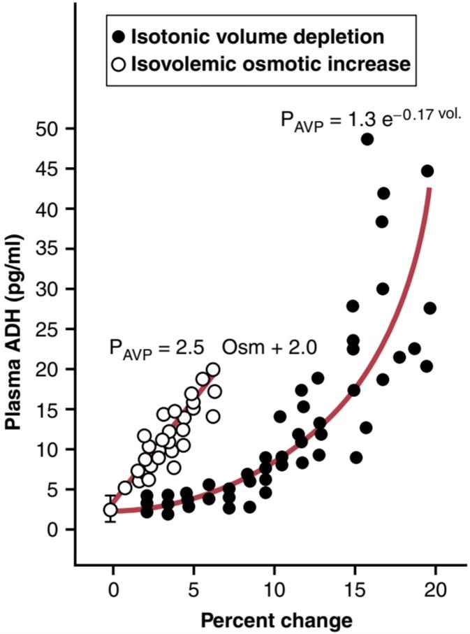
Figure 29-11. Effect of increased plasma osmolarity or decreased blood volume on the level of plasma (P) antidiuretic hormone (ADH), also called arginine vasopressin (AVP). (Modified from Dunn FL, Brennan TJ, Nelson AE, et al: The role of blood osmolality and volume in regulating vasopressin secretion in the rat. J Clin Invest 52[12]:3212, 1973.)
شکل 29-11. اثر افزایش اسمولاریته پلاسما یا کاهش حجم خون بر سطح پلاسما (P) هورمون آنتی دیورتیک (ADH) که آرژنین وازوپرسین (AVP) نیز نامیده می شود. (اصلاح شده از Dunn FL، Brennan TJ، Nelson AE، و همکاران: نقش اسمولالیته خون و حجم در تنظیم ترشح وازوپرسین در موش صحرایی. J Clin Invest 52[12]:3212، 1973.)
OTHER STIMULI FOR ADH SECRETION
ADH secretion can also be increased or decreased by other stimuli to the central nervous system, as well as by various drugs and hormones, as shown in Table 29-2. For example, nausea is a potent stimulus for ADH release, which may increase to as much as 100 times normal after vomiting. Also, drugs such as nicotine and morphine stimulate ADH release, whereas some drugs, such as alcohol, inhibit ADH release. The marked diuresis that occurs after ingestion of alcohol is due in part to the inhibition of ADH release.
محرک های دیگر برای ترشح ADH
همانطور که در جدول 29-2 نشان داده شده است، ترشح ADH همچنین می تواند توسط سایر محرک های سیستم عصبی مرکزی و همچنین توسط داروها و هورمون های مختلف افزایش یا کاهش یابد. به عنوان مثال، حالت تهوع یک محرک قوی برای آزادسازی ADH است که ممکن است پس از استفراغ تا 100 برابر طبیعی افزایش یابد. همچنین، داروهایی مانند نیکوتین و مورفین باعث تحریک ترشح ADH می شوند، در حالی که برخی داروها مانند الکل، آزادسازی ADH را مهار می کنند. دیورز مشخصی که پس از مصرف الکل رخ می دهد تا حدی به دلیل مهار آزاد شدن ADH است.
Table 29-2 Control of Antidiuretic Hormone Secretion
جدول 29-2 کنترل ترشح هورمون آنتی دیورتیک

IMPORTANCE OF THIRST IN CONTROLLING EXTRACELLULAR FLUID OSMOLARITY AND SODIUM CONCENTRATION
اهمیت تشنگی در کنترل اسمولاریته مایعات خارج سلولی و غلظت سدیم
The kidneys minimize fluid loss during water deficits through the osmoreceptor-ADH feedback system. Ade- quate fluid intake, however, is necessary to counterbalance whatever fluid loss does occur through sweating and breathing and through the gastrointestinal tract. Fluid intake is regulated by the thirst mechanism, which, together with the osmoreceptor-ADH mechanism, maintains precise control of extracellular fluid osmolarity and sodium concentration.
کلیه ها از دست دادن مایعات را در هنگام کمبود آب از طریق سیستم بازخورد osmoreceptor-ADH به حداقل می رساند. با این حال، مصرف مایعات کافی برای متعادل کردن هرگونه از دست دادن مایعات از طریق تعریق و تنفس و از طریق دستگاه گوارش ضروری است. مصرف مایعات توسط مکانیسم تشنگی تنظیم می شود، که همراه با مکانیسم osmoreceptor-ADH، کنترل دقیق اسمولاریته مایع خارج سلولی و غلظت سدیم را حفظ می کند.
Many of the same factors that stimulate ADH secretion also increase thirst, which is defined as the conscious desire for water.
بسیاری از عواملی که ترشح ADH را تحریک می کنند، تشنگی را نیز افزایش می دهند که به عنوان میل آگاهانه به آب تعریف می شود.
CENTRAL NERVOUS SYSTEM CENTERS FOR THIRST
Referring again to Figure 29-10, the same area along the anteroventral wall of the third ventricle that promotes ADH release also stimulates thirst. Located anterolaterally in the preoptic nucleus is another small area that when stimulated electrically, causes immediate drinking that continues as long as the stimulation lasts. All these areas together are called the thirst center.
مراکز سیستم عصبی مرکزی برای تشنگی
با اشاره مجدد به شکل 29-10، همان ناحیه در امتداد دیواره قدامی بطن سوم که باعث ترشح ADH می شود، تشنگی را نیز تحریک می کند. ناحیه قدامی در هسته پریاپتیک ناحیه کوچک دیگری است که با تحریک الکتریکی باعث نوشیدن فوری می شود که تا زمانی که تحریک ادامه دارد ادامه می یابد. همه این مناطق با هم کانون تشنگی نامیده می شوند.
The neurons of the thirst center respond to injections of hypertonic salt solutions by stimulating drinking behavior. These cells almost certainly function as osmoreceptors to activate the thirst mechanism in the same way that the osmoreceptors stimulate ADH release.
نورون های مرکز تشنگی با تحریک رفتار نوشیدن به تزریق محلول های نمک هیپرتونیک پاسخ می دهند. این سلول ها تقریباً به طور قطع به عنوان گیرنده های اسمزی عمل می کنند تا مکانیسم تشنگی را به همان روشی که گیرنده های اسمزی باعث تحریک آزاد شدن ADH می شوند، فعال کنند.
Increased osmolarity of the cerebrospinal fluid in the third ventricle has essentially the same effect to promote drinking. It is likely that the organum vasculosum of the lamina terminalis, which lies immediately beneath the ventricular surface at the inferior end of the AV3V region, is intimately involved in mediating this response.
افزایش اسمولاریته مایع مغزی نخاعی در بطن سوم اساساً همان اثر را برای ترویج نوشیدن دارد. این احتمال وجود دارد که اندام عروقی لایه انتهایی، که بلافاصله در زیر سطح بطن در انتهای تحتانی ناحیه AV3V قرار دارد، به طور جدی در میانجیگری این پاسخ دخالت داشته باشد.
STIMULI FOR THIRST
Table 29-3 summarizes some of the known stimuli for thirst.
محرک تشنگی
جدول 29-3 برخی از محرک های شناخته شده برای تشنگی را خلاصه می کند.
Table 29-3 Control of Thirst
جدول 29-3 کنترل تشنگی

1.One of the most important is increased extracellular fluid osmolarity, which causes intracellular dehydration in the thirst centers, thereby stimulating the sensation of thirst.
1. یکی از مهمترین آنها افزایش اسمولاریته مایع خارج سلولی است که باعث کم آبی داخل سلولی در مراکز تشنگی می شود و در نتیجه احساس تشنگی را تحریک می کند.
The value of this response is obvious: it helps dilute extracellular fluids and returns osmolarity toward normal.
ارزش این پاسخ واضح است: به رقیق شدن مایعات خارج سلولی کمک می کند و اسمولاریته را به حالت عادی برمی گرداند.
2. Decreases in extracellular fluid volume and arterial pressure also stimulate thirst by a pathway that is independent of the one stimulated by increased plasma osmolarity.
2. کاهش حجم مایع خارج سلولی و فشار شریانی نیز باعث تحریک تشنگی از طریق مسیری می شود که مستقل از مسیری است که با افزایش اسمولاریته پلاسما تحریک می شود.
Thus, blood volume loss by hemorrhage stimulates thirst, even though there might be no change in plasma osmolarity. This stimulation probably occurs because of neural input from cardiopulmonary and systemic arterial baroreceptors in the circulation.
بنابراین، از دست دادن حجم خون در اثر خونریزی، تشنگی را تحریک می کند، حتی اگر تغییری در اسمولاریته پلاسما وجود نداشته باشد. این تحریک احتمالاً به دلیل ورودی عصبی از بارورسپتورهای شریانی قلبی ریوی و سیستمیک در گردش خون رخ می دهد.
3. A third important stimulus for thirst is angiotensin II.
3. سومین محرک مهم برای تشنگی آنژیوتانسین II است.
Studies in animals have shown that angiotensin II acts on the subfornical organ and on the organum vasculosum of the lamina terminalis. These regions are outside the blood-brain barrier, and peptides such as angiotensin II diffuse into the tissues. Because angiotensin II is also stimulated by factors associated with hypovolemia and low blood pressure, its effect on thirst helps restore blood volume and blood pressure toward normal along with the other actions of angiotensin II on the kidneys to decrease fluid excretion
مطالعات بر روی حیوانات نشان داده است که آنژیوتانسین II بر روی اندام ساب فورنیکال و روی اندام عروقی لایه انتهایی (lamina terminalis) اثر میگذارد. این نواحی خارج از سد خونی مغزی هستند و پپتیدهایی مانند آنژیوتانسین II در بافت ها پخش می شوند. از آنجایی که آنژیوتانسین II نیز توسط عوامل مرتبط با هیپوولمی و فشار خون پایین تحریک می شود، اثر آن بر تشنگی به بازگرداندن حجم خون و فشار خون به حالت نرمال همراه با سایر اعمال آنژیوتانسین II روی کلیه ها برای کاهش دفع مایعات کمک می کند.
4. Dryness of the mouth and mucous membranes of the esophagus can elicit the sensation of thirst.
4. خشکی دهان و غشاهای مخاطی مری می تواند احساس تشنگی را برانگیزد.
As a result, a thirsty person may receive relief from thirst almost immediately after drinking water, even though the water has not been absorbed from the gastro- intestinal tract and has not yet had an effect on extracellular fluid osmolarity.
در نتیجه، یک فرد تشنه ممکن است تقریباً بلافاصله پس از نوشیدن آب از تشنگی تسکین یابد، حتی اگر آب از دستگاه گوارش جذب نشده باشد و هنوز تأثیری بر اسمولاریته مایع خارج سلولی نداشته باشد.
5. Gastrointestinal and pharyngeal stimuli influence thirst.
5. محرک های گوارشی و حلقی بر تشنگی تأثیر می گذارد.
In animals that have an esophageal opening to the exterior so that water is never absorbed into the blood, partial relief of thirst occurs after drinking, although the relief is only temporary. Also, gastrointestinal distention may partially alleviate thirst; For example, simple inflation of a balloon in the stomach can relieve thirst. However, relief of thirst sensations through gastrointestinal or pha- ryngeal mechanisms is short-lived; the desire to drink is completely satisfied only when plasma osmolarity and/or blood volume returns to normal.
در حیواناتی که دهانه مری به بیرون دارند به طوری که آب هرگز جذب خون نمی شود، رفع تشنگی نسبی پس از نوشیدن اتفاق می افتد، هرچند که تسکین موقتی است. همچنین، اتساع دستگاه گوارش ممکن است تا حدی تشنگی را کاهش دهد. به عنوان مثال، باد کردن ساده یک بالون در معده می تواند تشنگی را برطرف کند. با این حال، تسکین احساس تشنگی از طریق مکانیسم های گوارشی یا حلقی کوتاه مدت است. میل به نوشیدن تنها زمانی کاملاً ارضا می شود که اسمولاریته پلاسما و/یا حجم خون به حالت عادی برگردد.
The ability of animals and humans to “meter” fluid intake is important because it prevents overhydration. After a person drinks water, 30 to 60 minutes may be required for the water to be reabsorbed and distributed throughout the body. If the thirst sensation were not temporarily relieved after drinking water, the person would continue to drink more and more, eventually leading to overhydration and excess dilution of the body fluids. Experimental studies have repeatedly shown that ani- mals drink almost exactly the amount necessary to return plasma osmolarity and volume to normal.
توانایی حیوانات و انسان در اندازه گیری مایعات دریافتی مهم است زیرا از هیدراتاسیون بیش از حد جلوگیری می کند. پس از نوشیدن آب، 30 تا 60 دقیقه ممکن است لازم باشد تا آب دوباره جذب شود و در سراسر بدن توزیع شود. اگر احساس تشنگی پس از نوشیدن آب به طور موقت برطرف نمی شد، فرد به نوشیدن بیشتر و بیشتر ادامه می داد و در نهایت منجر به هیدراتاسیون بیش از حد و رقیق شدن بیش از حد مایعات بدن می شد. مطالعات تجربی مکرراً نشان داده است که حیوانات تقریباً دقیقاً به مقدار لازم برای بازگرداندن اسمولاریته و حجم پلاسما به حالت عادی می نوشند.
THRESHOLD FOR OSMOLAR STIMULUS OF DRINKING
The kidneys must continually excrete an obligatory amount of water, even in a dehydrated person, to rid the body of excess solutes that are ingested or produced by metabolism. Water is also lost by evaporation from the lungs and the gastrointestinal tract and by evaporation and sweating from the skin. Therefore, there is always a tendency for dehydration, with resultant increased extra- cellular fluid sodium concentration and osmolarity.
آستانه برای تحریک اسمولار نوشیدن
کلیه ها باید به طور مداوم مقدار اجباری آب را حتی در یک فرد کم آب دفع کنند تا بدن از املاح اضافی که توسط متابولیسم بلعیده شده یا تولید می شود خلاص شود. آب نیز با تبخیر از ریه ها و دستگاه گوارش و با تبخیر و تعریق از پوست از بین می رود. بنابراین، همیشه تمایل به کم آبی وجود دارد و در نتیجه غلظت سدیم مایع خارج سلولی و اسمولاریته افزایش می یابد.
When the sodium concentration increases only about 2 mEq/L above normal, the thirst mechanism is activated, causing a desire to drink water. This is called the thresh- old for drinking. Thus, even small increases in plasma osmolarity are normally followed by water intake, which restores extracellular fluid osmolarity and volume toward normal. In this way, the extracellular fluid osmolarity and sodium concentration are precisely controlled.
هنگامی که غلظت سدیم تنها حدود 2 میلی اکی والان در لیتر بالاتر از حد طبیعی افزایش می یابد، مکانیسم تشنگی فعال می شود و باعث میل به نوشیدن آب می شود. به این آستانه نوشیدن می گویند. بنابراین، حتی افزایش اندک در اسمولاریته پلاسما معمولاً با مصرف آب همراه است که اسمولاریته و حجم مایع خارج سلولی را به حالت طبیعی باز می گرداند. به این ترتیب اسمولاریته مایع خارج سلولی و غلظت سدیم دقیقاً کنترل می شود.
Disorders of Thirst and Water Intake. As discussed previously, increased thirst occurs in various medical disorders associated with increased urine volume and reductions in extracellular fluid volume, such as poorly controlled diabetes mellitus or diabetes insipidus. In these cases, increased water intake serves as a compensatory response for increased plasma osmolarity and/or extracellular fluid volume depletion. Polydipsia, or excessive thirst, occasionally occurs in the absence of known physiological stimuli for thirst. Psychogenic polydipsia, for example, may be caused by mental illnesses such as schizophrenia or obsessive-compulsive disorders and can lead to significant hyponatremia. In contrast, adipsia, the absence of thirst even in the presence of hypernatremia or volume depletion, is rare but usually results from lesions to the hypothalamic thirst centers caused by trauma, infection, or surgery. Partial deficiency of the thirst mechanism, causing inadequate water intake (hypodipsia), or an inability to access fluid may occur in patients who have suffered stroke, in older patients with dementia, or in critically ill patients. In the absence of adequate water intake, dehydration and hypernatremia occur, despite large increases in ADH levels.
اختلالات تشنگی و مصرف آب. همانطور که قبلاً بحث شد، افزایش تشنگی در اختلالات پزشکی مختلف همراه با افزایش حجم ادرار و کاهش حجم مایع خارج سلولی، مانند دیابت شیرین کنترل نشده یا دیابت بی مزه رخ می دهد. در این موارد، افزایش مصرف آب به عنوان یک پاسخ جبرانی برای افزایش اسمولاریته پلاسما و/یا کاهش حجم مایع خارج سلولی عمل می کند. Polydipsia یا تشنگی بیش از حد، گاهی اوقات در غیاب محرک های فیزیولوژیکی شناخته شده برای تشنگی رخ می دهد. به عنوان مثال، پلی دیپسی روانزا ممکن است ناشی از بیماریهای روانی مانند اسکیزوفرنی یا اختلالات وسواس فکری-اجباری باشد و میتواند منجر به هیپوناترمی قابل توجهی شود. در مقابل، آدیپسی، فقدان تشنگی حتی در صورت هایپرناترمی یا کاهش حجم، نادر است اما معمولاً از ضایعات مراکز تشنگی هیپوتالاموس ناشی از ضربه، عفونت یا جراحی ناشی میشود. کمبود جزئی مکانیسم تشنگی، باعث مصرف ناکافی آب (هیپودیپسی)، یا ناتوانی در دسترسی به مایعات ممکن است در بیمارانی که دچار سکته مغزی شدهاند، در بیماران مسنتر مبتلا به زوال عقل یا در بیماران بدحال رخ دهد. در غیاب مصرف آب کافی، با وجود افزایش زیاد سطوح ADH، کم آبی و هیپرناترمی رخ می دهد.
INTEGRATED RESPONSES OF OSMORECEPTOR-ADH AND THIRST MECHANISMS
In a healthy person, the osmoreceptor-ADH and thirst mechanisms work in parallel to regulate extracellular fluid osmolarity and sodium concentration precisely, despite the constant challenges of dehydration. Even with additional challenges, such as high salt intake, these feedback sys- tems are usually able to keep plasma osmolarity reasonably constant. Figure 29-12 shows that an increase in sodium intake to as high as six times normal has only a small effect on plasma sodium concentration as long as the ADH and thirst mechanisms are both functioning normally.
پاسخ های یکپارچه گیرنده های اسموری-ADH و مکانیسم تشنگی
در یک فرد سالم، گیرنده اسمزی-ADH و مکانیسم تشنگی به طور موازی کار می کنند تا اسمولاریته مایع خارج سلولی و غلظت سدیم را دقیقاً تنظیم کنند، علی رغم چالش های مداوم کم آبی. حتی با چالشهای اضافی، مانند مصرف نمک زیاد، این سیستمهای بازخورد معمولاً قادرند اسمولاریته پلاسما را تا حد معقولی ثابت نگه دارند. شکل 29-12 نشان می دهد که افزایش دریافت سدیم تا شش برابر نرمال تنها تأثیر کمی بر غلظت سدیم پلاسما دارد تا زمانی که مکانیسم های ADH و تشنگی هر دو به طور طبیعی کار کنند.
When the ADH or thirst mechanism fails, ordinarily the other can still control extracellular osmolarity and sodium concentration with reasonable effectiveness as long as there is enough fluid intake to balance the daily obligatory urine volume and water losses caused by respiration, sweating, or gastrointestinal losses. However, if both the ADH and thirst mechanisms fail simultaneously, plasma sodium concentration and osmolarity are poorly controlled. Thus, when sodium intake is increased after blocking the total ADH-thirst system, relatively large changes in plasma sodium concentration occur. In the absence of the ADH-thirst mechanisms, no other feedback mechanism is capable of adequately regulating plasma sodium concentration and osmolarity.
هنگامی که مکانیسم ADH یا تشنگی از کار میافتد، معمولاً دیگری هنوز میتواند اسمولاریته خارج سلولی و غلظت سدیم را با اثربخشی معقول کنترل کند تا زمانی که مایعات کافی برای متعادل کردن حجم ادرار اجباری روزانه و تلفات آب ناشی از تنفس، تعریق یا تلفات دستگاه گوارش وجود داشته باشد. با این حال، اگر هر دو مکانیسم ADH و تشنگی به طور همزمان از کار بیفتند، غلظت سدیم پلاسما و اسمولاریته به خوبی کنترل نمی شود. بنابراین، هنگامی که مصرف سدیم پس از مسدود کردن کل سیستم تشنگی ADH افزایش می یابد، تغییرات نسبتاً زیادی در غلظت سدیم پلاسما رخ می دهد. در غیاب مکانیسمهای تشنگی ADH، هیچ مکانیسم فیدبک دیگری قادر به تنظیم کافی غلظت سدیم پلاسما و اسمولاریته نیست.

Figure 29-12. Effect of large changes in sodium intake on extracellular fluid sodium concentration in dogs under normal conditions (red line) and after the antidiuretic hormone (ADH) and thirst feedback systems were blocked (blue line). Note that control of extracellular fluid sodium concentration was poor in the absence of these feed- back systems. (Courtesy Dr. David B. Young.)
شکل 29-12. تأثیر تغییرات زیاد در دریافت سدیم بر غلظت سدیم مایع خارج سلولی در سگها در شرایط عادی (خط قرمز) و پس از مسدود شدن هورمون ضد ادرار (ADH) و سیستمهای بازخورد تشنگی (خط آبی). توجه داشته باشید که کنترل غلظت سدیم مایع خارج سلولی در غیاب این سیستمهای بازخورد ضعیف بود. (با احترام از دکتر دیوید بی یانگ.)
Role of Angiotensin II and Aldosterone in Controlling Extracellular Fluid Osmolarity and Sodium Concentration
As discussed in Chapter 28, both angiotensin II and aldosterone play an important role in regulating sodium reabsorption by the renal tubules. When sodium intake is low, increased levels of these hormones stimulate sodium reabsorption by the kidneys and prevent large sodium losses, even though sodium intake may be reduced to as low as 10% of normal. Conversely, with high sodium intake, decreased formation of these hormones permits the kidneys to excrete large amounts of sodium.
نقش آنژیوتانسین II و آلدوسترون در کنترل اسمولاریته مایعات خارج سلولی و غلظت سدیم
همانطور که در فصل 28 بحث شد، هر دو آنژیوتانسین II و آلدوسترون نقش مهمی در تنظیم بازجذب سدیم توسط لوله های کلیوی ایفا می کنند. هنگامی که مصرف سدیم کم باشد، افزایش سطح این هورمونها باعث تحریک بازجذب سدیم توسط کلیهها میشود و از تلفات سدیم زیاد جلوگیری میکند، حتی اگر مصرف سدیم ممکن است تا 10 درصد از حد طبیعی کاهش یابد. برعکس، با مصرف زیاد سدیم، کاهش تشکیل این هورمون ها به کلیه ها اجازه می دهد تا مقادیر زیادی سدیم را دفع کنند.
Because of the importance of angiotensin II and aldosterone in regulating sodium excretion by the kidneys, one might mistakenly infer that they also play a major role in regulating extracellular fluid sodium concentration. Al- though these hormones increase the amount of sodium in the extracellular fluid, they also increase the extracellular fluid volume by increasing reabsorption of water along with the sodium. Therefore, angiotensin II and aldosterone have little effect on sodium concentration, except under extreme conditions.
به دلیل اهمیت آنژیوتانسین II و آلدوسترون در تنظیم دفع سدیم توسط کلیه ها، ممکن است به اشتباه چنین استنباط شود که آنها همچنین نقش عمده ای در تنظیم غلظت سدیم مایع خارج سلولی دارند. اگرچه این هورمون ها باعث افزایش میزان سدیم در مایع خارج سلولی می شوند، اما با افزایش بازجذب آب همراه با سدیم، حجم مایع خارج سلولی را نیز افزایش می دهند. بنابراین، آنژیوتانسین II و آلدوسترون تأثیر کمی بر غلظت سدیم دارند، مگر در شرایط شدید.
This relative unimportance of aldosterone in regulating extracellular fluid sodium concentration is shown by the experiment depicted in Figure 29-13. This figure shows the effect on plasma sodium concentration of changing sodium intake more than sixfold under two conditions: (1) under normal conditions; and (2) after the aldosterone feedback system was blocked by removing the adrenal glands and infusing the animals with aldosterone at a constant rate so that plasma levels could not increase or decrease. Note that when sodium intake was increased sixfold, plasma concentration changed only about 1% to 2% in either case. This finding indicates that even without a functional aldosterone feedback system, plasma sodium concentration can be well regulated. The same type of experiment has been conducted after blocking angiotensin II formation, with the same result.
این بی اهمیت بودن نسبی آلدوسترون در تنظیم غلظت سدیم مایع خارج سلولی توسط آزمایش نشان داده شده در شکل 29-13 نشان داده شده است. این شکل تاثیر تغییر مصرف سدیم بیش از شش برابر را بر غلظت سدیم پلاسما در دو شرایط نشان میدهد: (1) در شرایط عادی. و (2) پس از مسدود شدن سیستم بازخورد آلدوسترون با برداشتن غدد فوق کلیوی و تزریق آلدوسترون به حیوانات با سرعت ثابت به طوری که سطوح پلاسما نتواند افزایش یا کاهش یابد. توجه داشته باشید که وقتی مصرف سدیم شش برابر شد، غلظت پلاسما در هر دو مورد تنها حدود 1% تا 2% تغییر کرد. این یافته نشان می دهد که حتی بدون یک سیستم بازخورد آلدوسترون عملکردی، غلظت سدیم پلاسما می تواند به خوبی تنظیم شود. همین نوع آزمایش پس از مسدود کردن تشکیل آنژیوتانسین II با نتیجه مشابه انجام شده است.
There are two primary reasons why changes in angiotensin II and aldosterone do not have a major effect on plasma sodium concentration. First, as discussed. earlier, angiotensin II and aldosterone increase both sodium and water reabsorption by the renal tubules, leading to increases in extracellular fluid volume and sodium quantity but little change in sodium concentration. Second, as long as the ADH-thirst mechanism is functional, any tendency toward increased plasma sodium concentration is compensated for by increased water intake or increased plasma ADH secretion, which tends to dilute the extracellular fluid back toward normal. The ADH-thirst system far overshadows the angiotensin II and aldosterone systems for regulating sodium concentration under normal conditions. Even in patients with primary aldosteronism, who have extremely high levels of aldosterone, the plasma sodium concentration usually increases only about 3 to 5 mEq/L above normal.
دو دلیل اصلی وجود دارد که چرا تغییرات آنژیوتانسین II و آلدوسترون تأثیر عمده ای بر غلظت سدیم پلاسما ندارد. اول، همانطور که بحث شد. قبلاً، آنژیوتانسین II و آلدوسترون باعث افزایش بازجذب سدیم و آب توسط لولههای کلیوی میشوند که منجر به افزایش حجم مایع خارج سلولی و مقدار سدیم میشود اما تغییر کمی در غلظت سدیم ایجاد میشود. ثانیاً، تا زمانی که مکانیسم تشنگی ADH فعال است، هرگونه تمایل به افزایش غلظت سدیم پلاسما با افزایش مصرف آب یا افزایش ترشح ADH پلاسما جبران میشود، که تمایل دارد مایع خارج سلولی را به حالت طبیعی رقیق کند. سیستم تشنگی ADH سیستم های آنژیوتانسین II و آلدوسترون را برای تنظیم غلظت سدیم در شرایط عادی تحت الشعاع قرار می دهد. حتی در بیماران مبتلا به آلدوسترونیسم اولیه که سطوح بسیار بالایی از آلدوسترون دارند، غلظت سدیم پلاسما معمولاً تنها حدود 3 تا 5 میلی اکی والان در لیتر بالاتر از حد طبیعی افزایش می یابد.
Under extreme conditions caused by complete loss of aldosterone secretion as a result of adrenalectomy or in patients with Addison disease (severely impaired secretion or total lack of aldosterone), there is tremendous loss of sodium by the kidneys, which can lead to a marked reduction in plasma sodium concentration. One reason for this is that large losses of sodium eventually cause severe volume depletion and decreased blood pressure, which can activate the thirst mechanism through the cardio- vascular reflexes. This activation leads to a further dilution of the plasma sodium concentration, even though the increased water intake helps minimize the decrease in body fluid volumes under these conditions.
تحت شرایط شدید ناشی از از دست دادن کامل ترشح آلدوسترون در نتیجه آدرنالکتومی یا در بیماران مبتلا به بیماری آدیسون (تخلخل شدید ترشح یا کمبود کامل آلدوسترون)، از دست دادن شدید سدیم توسط کلیه ها وجود دارد که می تواند منجر به کاهش قابل توجه غلظت سدیم پلاسما شود. یکی از دلایل این امر این است که از دست دادن زیاد سدیم در نهایت باعث کاهش شدید حجم و کاهش فشار خون می شود که می تواند مکانیسم تشنگی را از طریق رفلکس های قلبی عروقی فعال کند. این فعال سازی منجر به رقیق شدن بیشتر غلظت سدیم پلاسما می شود، حتی اگر افزایش مصرف آب به حداقل رساندن کاهش حجم مایعات بدن در این شرایط کمک کند.
Thus, there are extreme situations in which the plasma sodium concentration may change significantly, even with a functional ADH-thirst mechanism. Even so, the ADH- thirst mechanism is the most powerful feedback system in the body for controlling extracellular fluid osmolarity and sodium concentration.
بنابراین، شرایط شدیدی وجود دارد که در آن غلظت سدیم پلاسما ممکن است به طور قابل توجهی تغییر کند، حتی با مکانیسم عملکردی تشنگی ADH. با این حال، مکانیسم تشنگی ADH قدرتمندترین سیستم بازخوردی در بدن برای کنترل اسمولاریته مایع خارج سلولی و غلظت سدیم است.

Figure 29-13. Effect of large changes in sodium intake on extracellular fluid sodium concentration in dogs under normal conditions (red line) and after the aldosterone feedback system was blocked (blue line). Note that the sodium concentration was maintained relatively constant over this wide range of sodium intakes, with or without aldosterone feedback control. (Courtesy Dr. David B. Young.)
شکل 29-13. تأثیر تغییرات زیاد در دریافت سدیم بر غلظت سدیم مایع خارج سلولی سگ در شرایط عادی (خط قرمز) و پس از مسدود شدن سیستم بازخورد آلدوسترون (خط آبی). توجه داشته باشید که غلظت سدیم در این محدوده وسیع از مصرف سدیم، با یا بدون کنترل بازخورد آلدوسترون، نسبتاً ثابت نگه داشته شد. (با احترام از دکتر دیوید بی یانگ.)
Salt-Appetite Mechanism for Controlling Extracellular Fluid Sodium Concentration and Volume
The maintenance of a normal extracellular fluid volume and sodium concentration requires a balance between sodium excretion and sodium intake. Currently, sodium intake is almost always greater than necessary for homeostasis. In fact, the average sodium intake for persons in industrialized cultures who eat processed foods usually ranges between 100 and 200 mEq/day, even though humans can survive and function normally while ingesting only 10 to 20 mEq/day. Thus, most people eat far more sodium than is necessary for homeostasis, and evidence indicates that our usual high sodium intake may contribute to cardiovascular disorders such as hypertension.
مکانیسم نمک-اشتها برای کنترل غلظت و حجم مایعات خارج سلولی سدیم
حفظ حجم طبیعی مایع خارج سلولی و غلظت سدیم نیاز به تعادل بین دفع سدیم و دریافت سدیم دارد. در حال حاضر، مصرف سدیم تقریباً همیشه بیشتر از مقدار لازم برای هموستاز است. در واقع، متوسط دریافت سدیم برای افراد در فرهنگ های صنعتی که غذاهای فرآوری شده می خورند معمولاً بین 100 تا 200 میلی اکی والان در روز است، حتی اگر انسان ها می توانند زنده بمانند و عملکرد طبیعی خود را داشته باشند در حالی که فقط 10 تا 20 میلی اکی والان در روز مصرف می کنند. بنابراین، اکثر مردم بسیار بیشتر از مقدار مورد نیاز برای هموستاز، سدیم می خورند و شواهد نشان می دهد که مصرف زیاد سدیم ما ممکن است به اختلالات قلبی عروقی مانند فشار خون بالا کمک کند.
Salt appetite is due in part to the fact that animals and humans like salt and eat it, regardless of whether they are salt-deficient. Salt appetite also has a regulatory component in which there is a behavioral drive to obtain salt when a sodium deficiency exists in the body. This behavioral drive is particularly important in herbivores, which naturally eat a low-sodium diet, but salt craving may also be important in humans, especially in those who have an extreme deficiency of sodium, such as occurs in Addison disease. In this case, there is a deficiency of aldosterone secretion, which causes excessive loss of sodium in the urine and leads to decreased extracellular fluid volume and decreased sodium concentration; both of these changes elicit the desire for salt.
اشتهای نمک تا حدی به این دلیل است که حیوانات و انسان ها بدون توجه به کمبود نمک، نمک را دوست دارند و می خورند. اشتهای نمک همچنین دارای یک جزء تنظیمی است که در آن یک انگیزه رفتاری برای بدست آوردن نمک زمانی که کمبود سدیم در بدن وجود دارد وجود دارد. این انگیزه رفتاری به ویژه در گیاهخواران که به طور طبیعی از رژیم غذایی کم سدیم استفاده می کنند، مهم است، اما ولع نمک نیز ممکن است در انسان ها مهم باشد، به ویژه در کسانی که کمبود شدید سدیم دارند، مانند بیماری آدیسون. در این حالت، کمبود ترشح آلدوسترون وجود دارد که باعث از دست رفتن بیش از حد سدیم در ادرار و کاهش حجم مایع خارج سلولی و کاهش غلظت سدیم می شود. هر دوی این تغییرات میل به نمک را برمی انگیزند.
In general, the primary stimuli that increase salt appetite are those associated with sodium deficits and decreased blood volume or decreased blood pressure associated with circulatory insufficiency.
به طور کلی، محرک های اولیه که اشتهای نمک را افزایش می دهند، آنهایی هستند که با کمبود سدیم و کاهش حجم خون یا کاهش فشار خون مرتبط با نارسایی گردش خون مرتبط هستند.
The neuronal mechanism for salt appetite is analogous to that of the thirst mechanism. Some of the same neuronal centers in the AV3V region of the brain seem to be involved because lesions in this region frequently affect both thirst and salt appetite simultaneously in animals. Also, circulatory reflexes elicited by low blood pressure or decreased blood volume affect both thirst and salt appetite at the same time.
مکانیسم عصبی برای اشتهای نمک مشابه مکانیسم تشنگی است. برخی از مراکز عصبی مشابه در ناحیه AV3V مغز درگیر به نظر می رسد زیرا ضایعات در این ناحیه اغلب بر تشنگی و اشتهای نمک به طور همزمان در حیوانات تأثیر می گذارد. همچنین، رفلکس های گردش خون ناشی از فشار خون پایین یا کاهش حجم خون بر تشنگی و اشتهای نمک به طور همزمان تأثیر می گذارد.
کلیک کنید «Bibliography: فهرست کتب مربوطه»
Agre P: The aquaporin water channels. Proc Am Thorac Soc 3:5, 2006.
Begg DP: Disturbances of thirst and fluid balance associated with ag- ing. Physiol Behav 178:28, 2017.
Berl T: Vasopressin antagonists. N Engl J Med 372:2207, 2015.
Bourque CW: Central mechanisms of osmosensation and systemic osmoregulation. Nat Rev Neurosci 9:519, 2008.
Brown D: The discovery of water channels (aquaporins). Ann Nutr Metab 70 (Suppl 1):37, 2017.
Dantzler WH, Layton AT, Layton HE, Pannabecker TL: Urine- concentrating mechanism in the inner medulla: function of the thin limbs of the loops of Henle. Clin J Am Soc Nephrol 9:1781, 2014.
Geerling JC, Loewy AD: Central regulation of sodium appetite. Exp Physiol 93:177, 2008.
Gizowski C, Bourque CW: The neural basis of homeostatic and antici- patory thirst. Nat Rev Nephrol 14:11, 2018.
Harrois A, Anstey JR: Diabetes insipidus and syndrome of inappro- priate antidiuretic hormone in critically ill patients. Crit Care Clin 35:187, 2019.
Klein JD, Sands JM: Urea transport and clinical potential of urearetics. Curr Opin Nephrol Hypertens 25:444, 2016.
Knepper MA, Kwon TH, Nielsen S: Molecular physiology of water bal- ance. N Engl J Med 372:1349, 2015.
Koshimizu TA, Nakamura K, Egashira N, et al: Vasopressin V1a and V1b receptors: from molecules to physiological systems. Physiol
Rev 92:1813, 2012.
Nawata CM, Pannabecker TL: Mammalian urine concentration: a re- view of renal medullary architecture and membrane transporters. J Comp Physiol B 188:899, 2018.
Nielsen S, Frøkiær J, Marples D, et al: Aquaporins in the kidney: from molecules to medicine. Physiol Rev 82:205, 2002.
Olesen ET, Fenton RA: Aquaporin-2 membrane targeting: still a co- nundrum. Am J Physiol Pallone TL, Edwards A, Mattson DL: Renal medullary circulation. Compr Physiol 2:97, 2012.
Pannabecker TL: Structure and function of the thin limbs of the loop of Henle. Compr Physiol 2:2063, 2012.
Chapter 29 Urine Concentration and Dilution
Pannabecker TL, Layton AT: Targeted delivery of solutes and oxygen in the renal medulla: role of microvessel architecture. Am J Physiol Renal Physiol 307:F649, 2014.
Sands JM, Klein JD: Physiological insights into novel therapies for nephrogenic diabetes insipidus. Am J Physiol Renal Physiol 311:F1149, 2016.
Sands JM, Layton HE: The physiology of urinary concentration: an update. Semin Nephrol 29:178, 2009.
Weiner ID, Mitch WE, Sands JM: Urea and ammonia metabolism and the control of renal nitrogen excretion. Clin J Am Soc Nephrol 10:1444, 2015.
Zimmerman CA, Huey EL, Ahn JS et al: A gut-to-brain signal of fluid osmolarity controls thirst satiation. Nature 568: 98, 2019. Zimmerman
CA, Leib DE, Knight ZA: Neural circuits underlying thirst and fluid homeostasis. Nat Rev Neurosci 18:459, 2017.
🚀 با ما همراه شوید!
تازهترین مطالب و آموزشهای مغز و اعصاب را از دست ندهید. با فالو کردن کانال تلگرام آیندهنگاران مغز، از ما حمایت کنید!



