
فیزیولوژی پزشکی گایتون و هال؛ تنظیم کلیوی پتاسیم، کلسیم، فسفات و منیزیم


دعای مطالعه [ نمایش ]
بِسْمِ الله الرَّحْمنِ الرَّحیمِ
اَللّهُمَّ اَخْرِجْنى مِنْ ظُلُماتِ الْوَهْمِ
خدايا مرا بيرون آور از تاريكىهاى وهم،
وَ اَكْرِمْنى بِنُورِ الْفَهْمِ
و به نور فهم گرامى ام بدار،
اَللّهُمَّ افْتَحْ عَلَيْنا اَبْوابَ رَحْمَتِكَ
خدايا درهاى رحمتت را به روى ما بگشا،
وَانْشُرْ عَلَيْنا خَزائِنَ عُلُومِكَ بِرَحْمَتِكَ يا اَرْحَمَ الرّاحِمينَ
و خزانههاى علومت را بر ما باز كن به امید رحمتت اى مهربانترين مهربانان.
کتاب «فیزیولوژی پزشکی گایتون و هال» بهعنوان یکی از جامعترین و معتبرترین منابع در حوزه علوم پزشکی، همچنان مرجع کلیدی برای درک عملکرد پیچیده بدن انسان است. این اثر با تکیه بر تازهترین پژوهشها و توضیحات دقیق از سازوکارهای فیزیولوژیک، پلی میان علوم پایه پزشکی و کاربردهای بالینی ایجاد میکند و نقشی بیبدیل در آموزش، پژوهش و ارتقای دانش سلامت ایفا مینماید.
ترجمه دقیق و علمی این شاهکار توسط برند علمی آیندهنگاران مغز به مدیریت داریوش طاهری، دسترسی فارسیزبانان به مرزهای نوین دانش فیزیولوژی را ممکن ساخته و رسالتی علمی برای ارتقای آموزش پزشکی، فهم عمیقتر سازوکارهای بدن و توسعه روشهای نوین در حوزه سلامت فراهم آورده است.
» کتاب فیزیولوژی پزشکی گایتون و هال
» » فصل ۳۰: تنظیم کلیوی پتاسیم، کلسیم، فسفات و منیزیم؛ ادغام مکانیسم های کلیوی برای کنترل حجم خون و حجم مایع خارج سلولی
در حال ویرایش
» Guyton and Hall Textbook of Medical Physiology
»» CHAPTER 30: Renal Regulation of Potassium, Calcium, Phosphate, and Magnesium; Integration of Renal Mechanisms for Control of Blood Volume and Extracellular Fluid Volume

REGULATION OF EXTRACELLULAR FLUID POTASSIUM CONCENTRATION AND POTASSIUM EXCRETION
The extracellular fluid potassium concentration normally is regulated at about 4.2 mEq/L, seldom rising or falling more than ±0.3 mEq/L. This precise control is necessary because many cell functions are sensitive to changes in extracellular fluid potassium concentration. For example, an increase in plasma potassium concentration of only 3 to 4 mEq/L can cause cardiac arrhythmias, and higher concentrations can lead to cardiac arrest or fibrillation.
تنظیم غلظت پتاسیم مایعات خارج سلولی و دفع پتاسیم
غلظت پتاسیم مایع خارج سلولی به طور معمول در حدود 4.2 میلی اکی والان در لیتر تنظیم می شود که به ندرت بیش از 0.3 ± میلی اکی والان در لیتر افزایش یا کاهش می یابد. این کنترل دقیق ضروری است زیرا بسیاری از عملکردهای سلولی به تغییرات غلظت پتاسیم مایع خارج سلولی حساس هستند. به عنوان مثال، افزایش غلظت پتاسیم پلاسما تنها 3 تا 4 میلی اکی والان در لیتر می تواند باعث آریتمی قلبی شود و غلظت های بالاتر می تواند منجر به ایست قلبی یا فیبریلاسیون شود.
A special difficulty in regulating extracellular potassium concentration is the fact that more than 98% of the total body potassium is contained in the cells, and only 2% is contained in the extracellular fluid (Figure 30-1). For a 70-kg adult, who has about 28 liters of intracellular fluid (40% of body weight) and 14 liters of extracellular fluid (20% of body weight), about 3920 mEq of potassium are inside the cells, and only about 59 mEq are in the extracellular fluid. Also, the potassium contained in a single meal may be as high as 50 mEq, and the daily intake usually ranges between 50 and 200 mEq/day; therefore, failure to rapidly rid the extracellular fluid of the ingested potassium could cause life-threatening hyperkalemia (increased plasma potassium concentration). Likewise, a small loss of potassium from the extracellular fluid could cause severe hypokalemia (low plasma potassium con- centration) in the absence of rapid and appropriate compensatory responses.
یک مشکل خاص در تنظیم غلظت پتاسیم خارج سلولی این واقعیت است که بیش از 98٪ از کل پتاسیم بدن در سلول ها وجود دارد و تنها 2٪ در مایع خارج سلولی موجود است (شکل 30-1). برای یک بزرگسال 70 کیلوگرمی که حدود 28 لیتر مایع درون سلولی (40 درصد وزن بدن) و 14 لیتر مایع خارج سلولی (20 درصد وزن بدن) دارد، حدود 3920 میلی اکی والان پتاسیم در داخل سلول ها و تنها حدود 59 میلی اکی والان در مایع خارج سلولی است. همچنین، پتاسیم موجود در یک وعده غذایی ممکن است تا 50 میلی اکی والان باشد و مصرف روزانه آن معمولا بین 50 تا 200 میلی اکی والان در روز است. بنابراین، عدم دفع سریع مایع خارج سلولی از پتاسیم بلعیده شده می تواند باعث هیپرکالمی تهدید کننده زندگی (افزایش غلظت پتاسیم پلاسما) شود. به همین ترتیب، از دست دادن اندک پتاسیم از مایع خارج سلولی میتواند باعث هیپوکالمی شدید (غلظت پایین پتاسیم پلاسما) در غیاب پاسخهای جبرانی سریع و مناسب شود.
Maintenance of a balance between intake and output of potassium depends primarily on excretion by the kidneys because the amount excreted in the feces is only about 5% to 10% of the potassium intake. Thus, the maintenance of a normal potassium balance requires the kidneys to adjust their potassium excretion rapidly and precisely in response to wide variations in intake, as is also true for most other electrolytes.
حفظ تعادل بین دریافت و خروجی پتاسیم در درجه اول به دفع توسط کلیه ها بستگی دارد زیرا مقدار دفع شده از طریق مدفوع تنها حدود 5 تا 10 درصد پتاسیم دریافتی است. بنابراین، حفظ تعادل طبیعی پتاسیم به کلیهها نیاز دارد که دفع پتاسیم خود را به سرعت و دقیقاً در پاسخ به تغییرات گسترده در مصرف تنظیم کنند، همانطور که برای اکثر الکترولیتهای دیگر نیز صادق است.
Control of potassium distribution between the extracellular and intracellular compartments also plays an important role in potassium homeostasis. Because more than 98% of the total body potassium is contained in the cells, they can serve as an overflow site for excess extracellular fluid potassium during hyperkalemia or as a source of potassium during hypokalemia. Thus, redistribution of potassium between the intracellular and extracellular fluid compartments provides a first line of defense against changes in extracellular fluid potassium concentration.
کنترل توزیع پتاسیم بین بخشهای خارج سلولی و داخل سلولی نیز نقش مهمی در هموستاز پتاسیم ایفا میکند. از آنجا که بیش از ۹۸٪ از کل پتاسیم بدن در سلولها قرار دارد، آنها میتوانند به عنوان محل سرریز پتاسیم اضافی مایع خارج سلولی در طول هیپرکالمی یا به عنوان منبع پتاسیم در طول هیپوکالمی عمل کنند. بنابراین، توزیع مجدد پتاسیم بین بخشهای مایع داخل سلولی و خارج سلولی، اولین خط دفاعی در برابر تغییرات غلظت پتاسیم مایع خارج سلولی را فراهم میکند.
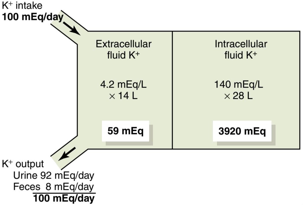
Figure 30-1. Normal potassium intake, distribution of potassium in the body fluids, and potassium output from the body.
شکل 30-1. مصرف معمولی پتاسیم، توزیع پتاسیم در مایعات بدن و خروجی پتاسیم از بدن.
REGULATION OF INTERNAL POTASSIUM DISTRIBUTION
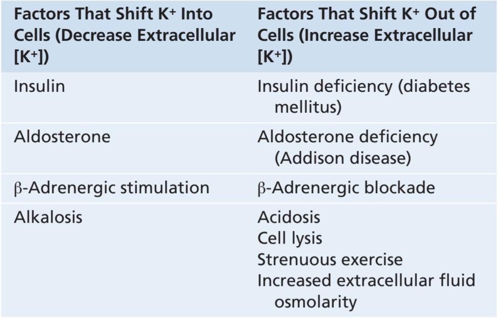
After ingestion of a potassium-rich meal, extracellular fluid potassium concentration would rise to a dangerous level if the ingested potassium did not move into the cells rapidly. For example, absorption of 40 mEq of potassium (the amount contained in a meal rich in vegetables and fruit) into an extracellular fluid volume of 14 liters would raise plasma potassium concentration by about 2.9 mEq/L if all the potassium remained in the extracellular compartment. Fortunately, most of the ingested potassium rapidly moves into the cells until the kidneys can eliminate the excess. Between meals, plasma potassium concentration also remains nearly constant as potassium is released by the cells to balance the extracellular fluid potassium excreted by the kidneys. Table 30-1 summarizes some of the factors that can influence the distribution of potassium between the intracellular and extracellular compartments.
مقررات توزیع داخلی پتاسیم
پس از مصرف یک وعده غذایی غنی از پتاسیم، اگر پتاسیم مصرفی به سرعت وارد سلول ها نشود، غلظت پتاسیم مایع خارج سلولی به سطح خطرناکی می رسد. به عنوان مثال، جذب 40 میلی اکی والان پتاسیم (مقدار موجود در یک وعده غذایی غنی از سبزیجات و میوه ها) در حجم مایع خارج سلولی 14 لیتری، اگر تمام پتاسیم در محفظه خارج سلولی باقی بماند، غلظت پتاسیم پلاسما را حدود 2.9 میلی اکی والان در لیتر افزایش می دهد. خوشبختانه، بیشتر پتاسیم مصرفی به سرعت به داخل سلولها میرود تا زمانی که کلیهها قادر به از بین بردن مقدار اضافی آن باشند. بین وعده های غذایی، غلظت پتاسیم پلاسما نیز تقریبا ثابت می ماند زیرا پتاسیم توسط سلول ها آزاد می شود تا پتاسیم مایع خارج سلولی دفع شده توسط کلیه ها را متعادل کند. جدول 30-1 برخی از عواملی را که می توانند بر توزیع پتاسیم بین بخش های درون سلولی و خارج سلولی تأثیر بگذارند، خلاصه می کند.
Table 30-1 Factors That Can Alter Potassium
Distribution Between Intracellular and Extracellular Fluids1
جدول 30-1 عواملی که میتوانند پتاسیم را تغییر دهند
توزیع بین مایعات داخل سلولی و خارج سلولی1
Insulin Stimulates Potassium Uptake Into Cells. Insulin stimulates sodium-potassium adenosine triphosphatase (ATPase) activity in many tissues, including skeletal muscle, which in turn transports potassium into the cells. Insulin is important for increasing cell potassium uptake after a meal. In people who have insulin-deficient diabetes mellitus, the rise in plasma potassium concentration after eating a meal is much greater than normal. Injections of insulin, however, can help correct the hyperkalemia.
انسولین جذب پتاسیم را به داخل سلول ها تحریک می کند. انسولین فعالیت سدیم پتاسیم آدنوزین تری فسفاتاز (ATPase) را در بسیاری از بافت ها از جمله ماهیچه های اسکلتی تحریک می کند که به نوبه خود پتاسیم را به سلول ها منتقل می کند. انسولین برای افزایش جذب پتاسیم سلولی بعد از غذا مهم است. در افرادی که مبتلا به دیابت قندی کمبود انسولین هستند، افزایش غلظت پتاسیم پلاسما پس از خوردن یک وعده غذایی بسیار بیشتر از حد طبیعی است. با این حال، تزریق انسولین می تواند به اصلاح هیپرکالمی کمک کند.
Aldosterone Increases Potassium Uptake Into Cells. Increased potassium intake also stimulates secretion of aldosterone, which increases cell potassium uptake. Excess aldosterone secretion (Conn syndrome) is almost invariably associated with hypokalemia, due in part to movement of extracellular potassium into the cells. Conversely, patients with deficient aldosterone production (Addison disease) often have clinically significant hyperkalemia due to accumulation of potassium in the extracellular space, as well as renal retention of potassium.
آلدوسترون جذب پتاسیم را به داخل سلول ها افزایش می دهد. افزایش دریافت پتاسیم همچنین ترشح آلدوسترون را تحریک می کند که جذب پتاسیم سلولی را افزایش می دهد. ترشح بیش از حد آلدوسترون (سندرم کان) تقریباً همیشه با هیپوکالمی همراه است که بخشی از آن به دلیل حرکت پتاسیم خارج سلولی به داخل سلول است. برعکس، بیماران مبتلا به کمبود تولید آلدوسترون (بیماری آدیسون) اغلب به دلیل تجمع پتاسیم در فضای خارج سلولی و همچنین احتباس کلیوی پتاسیم، هیپرکالمی قابل توجهی دارند.
B-Adrenergic Stimulation Increases Cellular Uptake of Potassium. Increased secretion of catecholamines, especially epinephrine, can cause movement of potassium from the extracellular to the intracellular fluid, mainly by activation of ẞ2-adrenergic receptors. Conversely, treatment of hypertension with ẞ-adrenergic receptor blockers, such as propranolol, causes potassium to move out of the cells and creates a tendency toward hyperkalemia.
تحریک B-آدرنرژیک جذب سلولی پتاسیم را افزایش می دهد. افزایش ترشح کاتکول آمین ها، به ویژه اپی نفرین، می تواند باعث حرکت پتاسیم از مایع خارج سلولی به داخل سلولی، عمدتاً با فعال شدن گیرنده های 2- آدرنرژیک شود. برعکس، درمان فشار خون بالا با مسدودکنندههای گیرنده آدرنرژیک، مانند پروپرانولول، باعث خروج پتاسیم از سلولها و ایجاد تمایل به هیپرکالمی میشود.
Acid-Base Abnormalities Can Cause Changes in Potassium Distribution. Metabolic acidosis increases extracellular potassium concentration, in part by causing loss of potassium from the cells, whereas metabolic alkalosis decreases extracellular fluid potassium concentration. Although the mechanisms responsible for the effect of hydrogen ion concentration on internal distribution of potassium are not completely understood, one effect of increased hydrogen ion concentration is to reduce activity of the Na+-K+ ATPase pump. This reduction, in turn, decreases cellular uptake of potassium and raises extracellular potassium concentration. Alkalosis has the opposite effect, shifting potassium from the extracellular fluid into the cells and tending to cause hypokalemia.
ناهنجاری های اسید و باز می تواند باعث تغییر در توزیع پتاسیم شود. اسیدوز متابولیک غلظت پتاسیم خارج سلولی را تا حدی با از دست دادن پتاسیم از سلول ها افزایش می دهد، در حالی که آلکالوز متابولیک غلظت پتاسیم مایع خارج سلولی را کاهش می دهد. اگرچه مکانیسم های مسئول اثر غلظت یون هیدروژن بر توزیع داخلی پتاسیم به طور کامل شناخته نشده است، یکی از اثرات افزایش غلظت یون هیدروژن کاهش فعالیت پمپ Na+-K+ ATPase است. این کاهش به نوبه خود جذب سلولی پتاسیم را کاهش می دهد و غلظت پتاسیم خارج سلولی را افزایش می دهد. آلکالوز اثر معکوس دارد، پتاسیم را از مایع خارج سلولی به سلول منتقل می کند و تمایل به ایجاد هیپوکالمی دارد.
Cell Lysis Causes Increased Extracellular Potassium Concentration. As cells are destroyed, the large amounts of potassium contained in the cells are released into the extracellular fluid. This release of potassium can cause significant hyperkalemia if large amounts of tissue are destroyed, as occurs with severe muscle injury or with red blood cell lysis.
لیز سلولی باعث افزایش غلظت پتاسیم خارج سلولی می شود. با از بین رفتن سلول ها، مقادیر زیادی پتاسیم موجود در سلول ها در مایع خارج سلولی آزاد می شود. این آزاد شدن پتاسیم می تواند باعث هیپرکالمی قابل توجهی در صورت تخریب مقادیر زیادی از بافت شود، همانطور که با آسیب شدید عضلانی یا لیز گلبول های قرمز اتفاق می افتد.
Strenuous Exercise Can Cause Hyperkalemia by Releasing Potassium From Skeletal Muscle. During prolonged exercise, potassium is released from skeletal muscle into the extracellular fluid. Usually the hyperkalemia is mild, but it may be clinically significant after heavy exercise, especially in patients treated with ẞ-adrenergic blockers or in individuals with insulin deficiency. In rare cases, hyperkalemia after exercise may be severe enough to cause cardiac toxicity.
ورزش شدید با آزادسازی پتاسیم از ماهیچه های اسکلتی می تواند باعث هیپرکالمی شود. در طول ورزش طولانی مدت، پتاسیم از ماهیچه های اسکلتی به مایع خارج سلولی آزاد می شود. معمولا هیپرکالمی خفیف است، اما ممکن است پس از ورزش سنگین از نظر بالینی قابل توجه باشد، به خصوص در بیمارانی که با مسدود کننده های آدرنرژیک درمان می شوند یا در افراد مبتلا به کمبود انسولین. در موارد نادر، هیپرکالمی پس از ورزش ممکن است آنقدر شدید باشد که باعث مسمومیت قلبی شود.
Increased Extracellular Fluid Osmolarity Causes Redistribution of Potassium From Cells to Extracellular Fluid. Increased extracellular fluid osmolarity causes osmotic flow of water out of the cells. The cellular dehydration increases intracellular potassium concentration, thereby promoting diffusion of potassium out of the cells and increasing extracellular fluid potassium concentration. Decreased extracellular fluid osmolarity has the opposite effect.
افزایش اسمولاریته سیال خارج سلولی باعث توزیع مجدد پتاسیم از سلول به مایع خارج سلولی می شود. افزایش اسمولاریته مایع خارج سلولی باعث جریان اسمزی آب از سلول ها می شود. کم آبی سلولی باعث افزایش غلظت پتاسیم درون سلولی می شود و در نتیجه باعث افزایش انتشار پتاسیم از سلول ها و افزایش غلظت پتاسیم مایع خارج سلولی می شود. کاهش اسمولاریته مایع خارج سلولی اثر معکوس دارد.
OVERVIEW OF RENAL POTASSIUM EXCRETION
Renal potassium excretion is determined by the sum of three processes: (1) the rate of potassium filtration (glomerular filtration rate [GFR] multiplied by the plasma potassium concentration); (2) the rate of potassium reabsorption by the tubules; and (3) the rate of potassium secretion by the tubules. The normal rate of potassium filtration by the glomerular capillaries is about 756 mEq/day (GFR, 180 L/day, multiplied by plasma potassium concentration, 4.2 mEq/L). This rate of filtration is relatively constant in healthy persons because of the autoregulatory mechanisms for GFR discussed previously and the precision with which plasma potassium concentration is regulated. Severe decreases in GFR in certain renal diseases, however, can cause serious potassium accumulation and hyperkalemia.
بررسی اجمالی دفع پتاسیم کلیوی
دفع کلیوی پتاسیم توسط مجموع سه فرآیند تعیین می شود: (1) سرعت فیلتراسیون پتاسیم (نرخ فیلتراسیون گلومرولی [GFR] ضرب در غلظت پتاسیم پلاسما). (2) سرعت بازجذب پتاسیم توسط لوله ها. و (3) میزان ترشح پتاسیم توسط لوله ها. سرعت طبیعی فیلتراسیون پتاسیم توسط مویرگ های گلومرولی حدود 756 میلی اکی والان در روز است (GFR، 180 لیتر در روز، ضرب در غلظت پتاسیم پلاسما، 2/4 میلی اکی والان در لیتر). این نرخ فیلتراسیون در افراد سالم به دلیل مکانیسم های خودتنظیمی برای GFR که قبلاً مورد بحث قرار گرفت و دقتی که با آن غلظت پتاسیم پلاسما تنظیم می شود نسبتاً ثابت است. کاهش شدید GFR در برخی بیماری های کلیوی، با این حال، می تواند باعث تجمع پتاسیم جدی و هیپرکالمی شود.
Figure 30-2 summarizes the tubular handling of potassium under normal conditions. About 65% of the filtered potassium is reabsorbed in the proximal tubule. Another 25% to 30% of the filtered potassium is reabsorbed in the loop of Henle, especially in the thick ascending part, where potassium is actively co-transported along with sodium and chloride. In the proximal tubule and loop of Henle, a relatively constant fraction of the filtered potassium load is reabsorbed. Changes in potassium reabsorption in these segments can influence potassium excretion, but most of the daily variation of potassium excretion is not due to changes in reabsorption in the proximal tubule or loop of Henle. The collecting tubules and collecting ducts reabsorb potassium at variable rates, depending on the potassium intake.
شکل 30-2 نحوه انتقال پتاسیم لوله ای را در شرایط عادی خلاصه می کند. حدود 65 درصد از پتاسیم فیلتر شده در لوله پروگزیمال بازجذب می شود. 25 تا 30 درصد دیگر از پتاسیم فیلتر شده در حلقه هنله، به ویژه در قسمت بالارونده ضخیم، که در آن پتاسیم به طور فعال همراه با سدیم و کلرید همراه با هم حمل می شود، دوباره جذب می شود. در لوله و حلقه پروگزیمال هنله، کسری نسبتاً ثابت از بار پتاسیم فیلتر شده دوباره جذب می شود. تغییرات در بازجذب پتاسیم در این بخشها میتواند بر دفع پتاسیم تأثیر بگذارد، اما بیشتر تغییرات روزانه دفع پتاسیم به دلیل تغییر در بازجذب در لوله پروگزیمال یا حلقه هنله نیست. لولههای جمعآوری و مجاری جمعآوری، بسته به میزان دریافتی پتاسیم، پتاسیم را با نرخهای متغیری بازجذب میکنند.
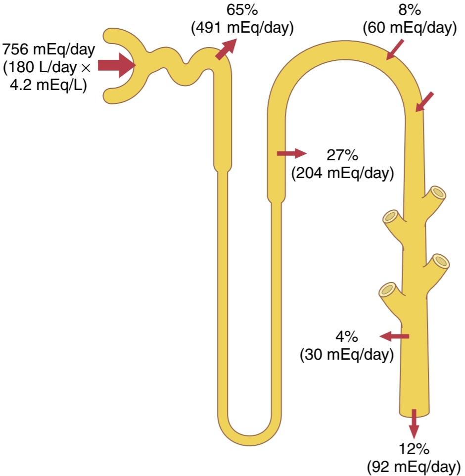
Figure 30-2. Renal tubular sites of potassium reabsorption and secretion. Potassium is reabsorbed in the proximal tubule and ascending loop of Henle, so only about 8% of the filtered load is delivered to the distal tubule. Secretion of potassium by the principal cells of the late distal tubules and collecting ducts adds to the amount delivered, but there is some additional reabsorption by the intercalated cells; therefore, the daily excretion is about 12% of the potassium filtered at the glomerular capillaries. The percentages indicate how much of the filtered load is reabsorbed or secreted into the different tubular segments.
شکل 30-2. محل های لوله های کلیوی بازجذب و ترشح پتاسیم. پتاسیم در لوله پروگزیمال و حلقه صعودی هنله دوباره جذب می شود، بنابراین تنها حدود 8 درصد از بار فیلتر شده به لوله دیستال تحویل داده می شود. ترشح پتاسیم توسط سلولهای اصلی لولههای انتهایی دیستال و مجاری جمعآوری به مقدار تحویل میافزاید، اما مقداری بازجذب اضافی توسط سلولهای بینقلابی وجود دارد. بنابراین، دفع روزانه حدود 12 درصد از پتاسیم فیلتر شده در مویرگ های گلومرولی است. درصدها نشان می دهد که چه مقدار از بار فیلتر شده دوباره جذب یا ترشح می شود به بخش های مختلف لوله.
Variable Potassium Secretion in Distal and Collecting Tubules Mediates Most Daily Changes in Potassium Excretion. The most important sites for regulating potassium excretion are the principal cells of the late distal tubules and cortical collecting tubules. In these tubular segments, potassium can at times be reabsorbed or at other times can be secreted, depending on the needs of the body. With a normal potassium intake of 100 mEq/ day, the kidneys must excrete about 92 mEq/day (the remaining 8 mEq are lost in the feces). About 60 mEq/day of potassium are secreted into the distal and collecting tubules, accounting for most of the excreted potassium.
ترشح متغیر پتاسیم در لولههای دیستال و جمعکننده، واسطه اکثر تغییرات روزانه در دفع پتاسیم است. مهم ترین مکان برای تنظیم دفع پتاسیم، سلول های اصلی لوله های انتهایی انتهایی و لوله های جمع کننده قشر مغز هستند. در این بخشهای لولهای، بسته به نیاز بدن، پتاسیم میتواند در مواقعی بازجذب شود یا در زمانهای دیگر ترشح شود. با دریافت معمولی 100 میلی اکی والان پتاسیم در روز، کلیه ها باید حدود 92 میلی اکی والان در روز دفع کنند (8 میلی اکی والان باقی مانده در مدفوع از بین می رود). حدود 60 میلی اکی والان در روز پتاسیم به داخل لوله های دیستال و جمع کننده ترشح می شود که بیشتر پتاسیم دفع شده را تشکیل می دهد.
With a high potassium intake, the required extra excretion of potassium is achieved almost entirely by increasing the secretion of potassium into the distal and collecting tubules. In fact, in those who consume extremely highpotassium diets, the rate of potassium excretion can exceed the amount of potassium in the glomerular filtrate, indicating a powerful mechanism for secreting potassium.
با مصرف زیاد پتاسیم، دفع اضافی مورد نیاز پتاسیم تقریباً به طور کامل با افزایش ترشح پتاسیم به داخل لولههای دیستال و جمعآوری کننده حاصل میشود. در واقع، در افرادی که رژیمهای غذایی با پتاسیم بسیار بالا مصرف میکنند، سرعت دفع پتاسیم میتواند از مقدار پتاسیم موجود در فیلتر گلومرولی بیشتر شود، که نشاندهنده مکانیسم قدرتمندی برای ترشح پتاسیم است.
When potassium intake is low, secretion of potassium in the distal and collecting tubules decreases, causing a reduction in urinary potassium excretion. There is also increased reabsorption of potassium by the intercalated cells in the distal segments of the nephron, and potassium excretion can fall to less than 1% of the potassium in the glomerular filtrate (to <10 mEq/day). With potassium intakes below this level, severe hypokalemia can develop.
هنگامی که دریافت پتاسیم کم باشد، ترشح پتاسیم در لوله های دیستال و جمع کننده کاهش می یابد و باعث کاهش دفع پتاسیم از طریق ادرار می شود. همچنین افزایش بازجذب پتاسیم توسط سلولهای درونقلابی در بخشهای دیستال نفرون وجود دارد و دفع پتاسیم میتواند به کمتر از 1 درصد پتاسیم موجود در فیلتر گلومرولی کاهش یابد (به کمتر از 10 میلیاکیوالان در روز). با دریافت پتاسیم کمتر از این سطح، هیپوکالمی شدید می تواند ایجاد شود.
Thus, most of the daily regulation of potassium excretion occurs in the late distal and cortical collecting tubules, where potassium can be reabsorbed or secreted, depending on the needs of the body. In the next section, we consider the basic mechanisms of potassium secretion and the factors that regulate this process.
بنابراین، بیشتر تنظیم روزانه دفع پتاسیم در لولههای جمعآوری دیستال و قشر مغز اتفاق میافتد، جایی که بسته به نیاز بدن، پتاسیم میتواند بازجذب یا ترشح شود. در بخش بعدی مکانیسم های اساسی ترشح پتاسیم و عوامل تنظیم کننده این فرآیند را بررسی می کنیم.
PRINCIPAL CELLS OF LATE DISTAL AND CORTICAL COLLECTING TUBULES SECRETE POTASSIUM
The cells in the late distal and cortical collecting tubules that secrete potassium are called principal cells and make up most of the epithelial cells in these regions. Figure 30-3 shows the basic cellular mechanisms of potassium secretion by the principal cells.
سلول های اصلی لوله های جمع آوری کننده انتهایی دیستال و قشر مخ، پتاسیم را ترشح می کنند
سلولهای موجود در لولههای جمعکننده انتهایی و قشری که پتاسیم ترشح میکنند، سلولهای اصلی نامیده میشوند و بیشتر سلولهای اپیتلیال این نواحی را تشکیل میدهند. شکل 30-3 مکانیسم های سلولی پایه ترشح پتاسیم توسط سلول های اصلی را نشان می دهد.
Secretion of potassium from the blood into the tubular lumen is a two-step process, beginning with uptake from the interstitium into the cell by the Na+-K+ ATPase pump in the basolateral cell membrane. This pump moves sodium out of the cell into the interstitium and, at the same time, moves potassium to the interior of the cell.
ترشح پتاسیم از خون به مجرای لولهای یک فرآیند دو مرحلهای است که با جذب از بینابینی به داخل سلول توسط پمپ Na+-K+ ATPase در غشای سلولی قاعدهای آغاز میشود. این پمپ سدیم را از سلول به داخل بینابینی منتقل می کند و در عین حال پتاسیم را به داخل سلول منتقل می کند.
The second step of the process is passive diffusion of potassium from the interior of the cell into the tubular fluid. The Na+-K+ ATPase pump creates a high intracellular potassium concentration, which provides the driving force for passive diffusion of potassium from the cell into the tubular lumen. The luminal membrane of the principal cells is highly permeable to potassium because there are two types of special channels that allow potassium ions to diffuse across the membrane rapidly: (1) the renal outer medullary potassium (ROMK) channels, and (2) high-conductance, “big” potassium (BK) channels. Both types of potassium channels are required for efficient renal potassium excretion, and their abundance in the luminal membrane is increased during high potassium intake.
مرحله دوم این فرآیند، انتشار غیرفعال پتاسیم از داخل سلول به داخل مایع لولهای است. پمپ Na+-K+ ATPase غلظت پتاسیم درون سلولی بالایی ایجاد می کند که نیروی محرکه برای انتشار غیرفعال پتاسیم از سلول به لومن لوله ای را فراهم می کند. غشای مجرای سلول های اصلی به پتاسیم بسیار نفوذپذیر است زیرا دو نوع کانال ویژه وجود دارد که به یون های پتاسیم اجازه می دهد تا به سرعت در غشا پخش شوند: (1) کانال های پتاسیم مدولاری بیرونی کلیوی (ROMK) و (2) کانال های رسانایی بالا (بزرگ پتاسیم). هر دو نوع کانال پتاسیم برای دفع موثر پتاسیم کلیوی مورد نیاز هستند و فراوانی آنها در غشای مجرا در هنگام مصرف زیاد پتاسیم افزایش می یابد.
Control of Potassium Secretion by Principal Cells. The primary factors that control potassium secretion by the principal cells of the late distal and cortical collecting tubules are the following: (1) the activity of the Na+-K+ ATPase pump; (2) the electrochemical gradient for potassium secretion from the blood to the tubular lumen; and (3) the permeability of the luminal membrane for potassium. These three determinants of potassium secretion are, in turn, regulated by several factors, discussed later.
کنترل ترشح پتاسیم توسط سلول های اصلی. عوامل اولیه ای که ترشح پتاسیم توسط سلول های اصلی لوله های جمع کننده انتهایی دیستال و قشر مغز را کنترل می کنند به شرح زیر است: (1) فعالیت پمپ Na+-K+ ATPase. (2) گرادیان الکتروشیمیایی برای ترشح پتاسیم از خون به لومن لولهای. و (3) نفوذ پذیری غشای مجرا برای پتاسیم. این سه عامل تعیین کننده ترشح پتاسیم به نوبه خود توسط عوامل متعددی تنظیم می شوند که بعداً مورد بحث قرار خواهند گرفت.
Intercalated Cells Can Reabsorb or Secrete Potassium. In circumstances associated with severe potassium depletion, there is a cessation of potassium secretion and a net reabsorption of potassium in the late distal and collecting tubules. This reabsorption occurs through the type A intercalated cells. Although this reabsorptive process is not completely understood, one mechanism believed to contribute is a hydrogen-potassium ATPase transport mechanism located in the luminal membrane (see Chapter 28, Figure 28-13). This transporter reabsorbs potassium in exchange for hydrogen ions secreted into the tubular lumen, and the potassium then diffuses through basolateral membrane potassium channels into the interstitial fluid. This abundance of intercalated cell hydrogen-potassium ATPase transporters is enhanced with potassium depletion and hypokalemia, causing increased hydrogen ion secretion and alkalosis. Under normal conditions, however, potassium reabsorption by intercalated cells plays only a small role in controlling potassium excretion.
سلول های درونی می توانند پتاسیم را بازجذب یا ترشح کنند. در شرایط همراه با کاهش شدید پتاسیم، توقف ترشح پتاسیم و بازجذب خالص پتاسیم در لوله های انتهایی دیستال و جمع کننده وجود دارد. این بازجذب از طریق سلول های نوع A رخ می دهد. اگرچه این فرآیند بازجذب به طور کامل درک نشده است، یک مکانیسمی که اعتقاد بر این است که به کمک یک مکانیسم انتقال هیدروژن پتاسیم ATPase است که در غشای مجرا قرار دارد (به بخش 28، شکل 28-13 مراجعه کنید). این ناقل پتاسیم را در ازای یونهای هیدروژن ترشح شده در مجرای لولهای بازجذب میکند و پتاسیم سپس از طریق کانالهای پتاسیم غشای قاعدهای جانبی به داخل مایع بینابینی منتشر میشود. این فراوانی انتقالدهندههای ATPase هیدروژن پتاسیم سلولی با کاهش پتاسیم و هیپوکالمی افزایش مییابد و باعث افزایش ترشح یون هیدروژن و آلکالوز میشود. با این حال، در شرایط عادی، بازجذب پتاسیم توسط سلولهای درونقلابی تنها نقش کوچکی در کنترل دفع پتاسیم دارد.
When there is excess potassium in the body fluids, type B intercalated cells in the late distal tubules and collecting tubules actively secrete potassium into the tubular lumen and have functions that are opposite to those of type A cells (see Chapter 28, Figure 28-13). Potassium is pumped into the type B intercalated cell by a hydrogenpotassium ATPase transporter on the basolateral membrane, and it then diffuses into the tubular lumen through potassium channels.
هنگامی که پتاسیم اضافی در مایعات بدن وجود دارد، سلولهای بینقلبی نوع B در لولههای انتهایی دیستال و لولههای جمعکننده به طور فعال پتاسیم را به داخل مجرای لولهای ترشح میکنند و عملکردهایی بر خلاف عملکرد سلولهای نوع A دارند (به فصل 28، شکل 28-13 مراجعه کنید). پتاسیم توسط یک ناقل ATPase هیدروژن-پتاسیم بر روی غشای قاعدهای به داخل سلول بینالمللی نوع B پمپ میشود و سپس از طریق کانالهای پتاسیم به داخل مجرای لولهای منتشر میشود.
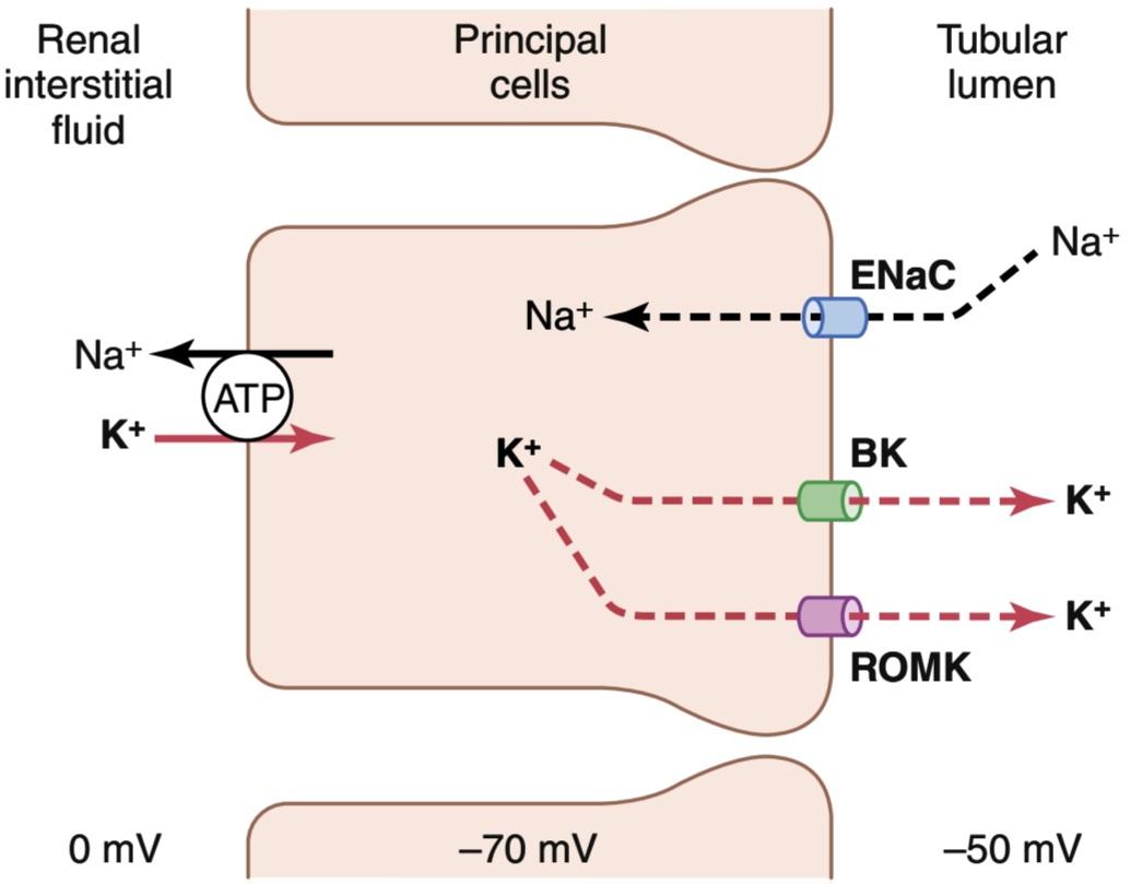
Figure 30-3. Mechanisms of potassium secretion and sodium reabsorption by the principal cells of the late distal and collecting tubules. BK, “big” potassium channel; ENaC, epithelial sodium channel; ROMK, renal outer medullary potassium channel.
شکل 30-3. مکانیسم های ترشح پتاسیم و بازجذب سدیم توسط سلول های اصلی لوله های انتهایی و جمع کننده. BK، کانال پتاسیم “بزرگ”؛ ENaC، کانال سدیم اپیتلیال؛ ROMK، کانال پتاسیم مدولاری بیرونی کلیه.
SUMMARY OF MAJOR FACTORS THAT REGULATE POTASSIUM SECRETION
Because the normal regulation of potassium excretion occurs mainly as a result of changes in potassium secretion by the principal cells of the late distal and collecting tubules, in this chapter we discuss the primary factors that influence secretion by these cells. The most important factors that stimulate potassium secretion by the principal cells include the following: (1) increased extracellular fluid potassium concentration; (2) increased aldosterone; and (3) increased tubular flow rate.
خلاصه ای از عوامل اصلی تنظیم کننده ترشح پتاسیم
از آنجایی که تنظیم طبیعی دفع پتاسیم عمدتاً در نتیجه تغییرات در ترشح پتاسیم توسط سلولهای اصلی لولههای انتهایی و جمعآوری کننده رخ میدهد، در این فصل به عوامل اولیهای که بر ترشح این سلولها تأثیر میگذارند، بحث میکنیم. مهم ترین عواملی که باعث تحریک ترشح پتاسیم توسط سلول های اصلی می شوند عبارتند از: (1) افزایش غلظت پتاسیم مایع خارج سلولی. (2) افزایش آلدوسترون. و (3) افزایش سرعت جریان لوله ای.
One factor that decreases potassium secretion is an increased hydrogen ion concentration (acidosis).
یکی از عواملی که باعث کاهش ترشح پتاسیم می شود افزایش غلظت یون هیدروژن (اسیدوز) است.
Increased Extracellular Fluid Potassium Concentration Stimulates Potassium Secretion. The rate of potassium secretion in the late distal and cortical collecting tubules is directly stimulated by increased extracellular fluid potassium concentration, leading to increases in potassium excretion, as shown in Figure 30-4. This effect is especially pronounced when extracellular fluid potassium concentration rises above about 4.1 mEq/L, slightly less than the normal concentration.Increased plasma potassium concentration, therefore, serves as one of the most important mechanisms for increasing potassium secretion and regulating extracellular fluid potassium ion concentration.
افزایش غلظت پتاسیم مایع خارج سلولی ترشح پتاسیم را تحریک می کند. میزان ترشح پتاسیم در لولههای جمعآوری انتهایی دیستال و قشر مغز مستقیماً با افزایش غلظت پتاسیم مایع خارج سلولی تحریک میشود که منجر به افزایش دفع پتاسیم میشود، همانطور که در شکل 30-4 نشان داده شده است. این اثر به ویژه هنگامی که غلظت پتاسیم مایع خارج سلولی از حدود 4.1 میلی اکی والان در لیتر، کمی کمتر از غلظت طبیعی افزایش می یابد، آشکار می شود. بنابراین، افزایش غلظت پتاسیم پلاسما به عنوان یکی از مهم ترین مکانیسم ها برای افزایش ترشح پتاسیم و تنظیم غلظت یون پتاسیم مایع خارج سلولی عمل می کند.
Increased dietary potassium intake and increased extracellular fluid potassium concentration stimulate potassium secretion by at least four mechanisms:
افزایش دریافت پتاسیم در رژیم غذایی و افزایش غلظت پتاسیم مایع خارج سلولی باعث تحریک ترشح پتاسیم با حداقل چهار مکانیسم می شود:
1. Increased extracellular fluid potassium concentration stimulates the Na+-K+ ATPase pump, thereby increasing potassium uptake across the basolateral membrane. This increased potassium uptake in turn increases intracellular potassium ion concentration, causing potassium to diffuse across the luminal membrane into the tubule.
1. افزایش غلظت پتاسیم مایع خارج سلولی پمپ Na+-K+ ATPase را تحریک می کند و در نتیجه جذب پتاسیم را در غشای قاعده جانبی افزایش می دهد. این افزایش جذب پتاسیم به نوبه خود غلظت یون پتاسیم درون سلولی را افزایش می دهد و باعث می شود پتاسیم در سراسر غشای مجرا به داخل لوله پخش شود.
2. Increased extracellular potassium concentration increases the potassium gradient from the renal interstitial fluid to the interior of the epithelial cell, which reduces backleakage of potassium ions from inside the cells through the basolateral membrane.
2. افزایش غلظت پتاسیم خارج سلولی، گرادیان پتاسیم را از مایع بین بافتی کلیه به داخل سلول اپیتلیال افزایش می دهد، که باعث کاهش نشت یون های پتاسیم از داخل سلول ها از طریق غشای قاعده جانبی می شود.
3. Increased potassium intake stimulates synthesis of potassium channels and their translocation from the cytosol to the luminal membrane, which, in turn, increases the ease of potassium diffusion through the membrane.
3. افزایش دریافت پتاسیم باعث تحریک سنتز کانال های پتاسیم و انتقال آنها از سیتوزول به غشای مجرا می شود که به نوبه خود باعث افزایش سهولت انتشار پتاسیم از طریق غشاء می شود.
4. Increased potassium concentration stimulates aldosterone secretion by the adrenal cortex, which further stimulates potassium secretion, as discussed next.
4. افزایش غلظت پتاسیم باعث تحریک ترشح آلدوسترون توسط قشر آدرنال می شود که باعث تحریک بیشتر ترشح پتاسیم می شود، همانطور که در ادامه بحث شد.
Aldosterone Stimulates Potassium Secretion. Aldosterone stimulates active reabsorption of sodium ions by the principal cells of the late distal tubules and collecting ducts (see Chapter 28). This effect is mediated through a Na+-K+ ATPase pump that transports sodium outward through the basolateral membrane of the cell and into the renal interstitial fluid at the same time that it pumps potassium into the cell. Thus, aldosterone also has a powerful effect on controlling the rate at which the principal cells secrete potassium and reabsorb sodium.
آلدوسترون ترشح پتاسیم را تحریک می کند. آلدوسترون بازجذب فعال یون های سدیم را توسط سلول های اصلی لوله های انتهایی انتهایی و مجاری جمع کننده تحریک می کند (به فصل 28 مراجعه کنید). این اثر از طریق پمپ Na+-K+ ATPase انجام میشود که همزمان با پمپاژ پتاسیم به داخل سلول، سدیم را از طریق غشای قاعدهای جانبی سلول به بیرون و به داخل مایع بینابینی کلیوی منتقل میکند. بنابراین، آلدوسترون همچنین تأثیر قدرتمندی در کنترل سرعت ترشح پتاسیم و بازجذب سدیم توسط سلول های اصلی دارد.
A second effect of aldosterone is to increase the number of potassium channels in the luminal membrane and therefore its permeability for potassium, further adding to the effectiveness of aldosterone in stimulating potassium secretion. Therefore, aldosterone has a powerful effect to increase potassium excretion, as shown in Figure 30-4.
اثر دوم آلدوسترون افزایش تعداد کانالهای پتاسیم در غشای مجرا و در نتیجه نفوذپذیری آن برای پتاسیم است که بر اثربخشی آلدوسترون در تحریک ترشح پتاسیم میافزاید. بنابراین، همانطور که در شکل 30-4 نشان داده شده است، آلدوسترون اثر قدرتمندی برای افزایش دفع پتاسیم دارد.
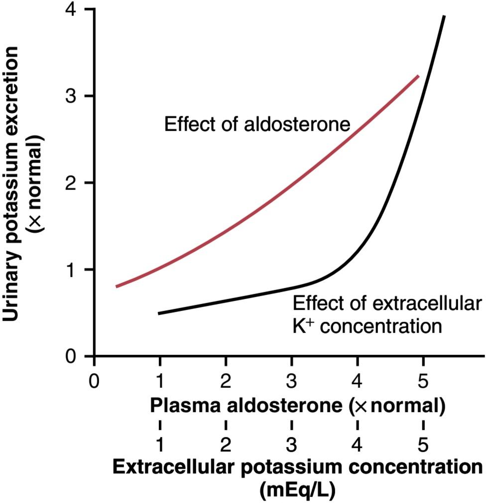
Figure 30-4. Effect of plasma aldosterone concentration (red line) and extracellular potassium ion concentration (black line) on the rate of urinary potassium excretion. These factors stimulate potassium secretion by the principal cells of the cortical collecting tubules. (Data from Young DB, Paulsen AW: Interrelated effects of aldosterone and plasma potassium on potassium excretion. Am J Physiol 244:F28, 1983.)
شکل 30-4. تأثیر غلظت آلدوسترون پلاسما (خط قرمز) و غلظت یون پتاسیم خارج سلولی (خط سیاه) بر سرعت دفع پتاسیم از طریق ادرار. این عوامل باعث تحریک ترشح پتاسیم توسط سلول های اصلی لوله های جمع کننده قشر می شود. (داده های Young DB، Paulsen AW: اثرات مرتبط آلدوسترون و پتاسیم پلاسما بر دفع پتاسیم. Am J Physiol 244:F28، 1983.)
Increased Extracellular Potassium lon Concentration Stimulates Aldosterone Secretion. In negative feed- back control systems, the factor that is controlled usually has a feedback effect on the controller. In the case of the aldosterone-potassium control system, the rate of aldosterone secretion by the adrenal gland is controlled strongly by extracellular fluid potassium ion concentration. Figure 30-5 shows that an increase in plasma potassium concentration of about 3 mEq/L can increase the plasma aldosterone concentration from nearly 0 to as high as 60 ng/100 ml, a concentration almost 10 times normal.
افزایش غلظت پتاسیم خارج سلولی ترشح آلدوسترون را تحریک می کند. در سیستم های کنترل بازخورد منفی، عاملی که کنترل می شود معمولاً بر کنترل کننده اثر بازخوردی دارد. در مورد سیستم کنترل آلدوسترون-پتاسیم، میزان ترشح آلدوسترون توسط غده فوق کلیوی به شدت توسط غلظت یون پتاسیم مایع خارج سلولی کنترل می شود. شکل 30-5 نشان می دهد که افزایش غلظت پتاسیم پلاسما در حدود 3 میلی اکی والان در لیتر می تواند غلظت آلدوسترون پلاسما را از نزدیک به 0 تا 60 نانوگرم در 100 میلی لیتر افزایش دهد که غلظتی تقریباً 10 برابر طبیعی است.
The effect of potassium ion concentration to stimulate aldosterone secretion is part of a powerful feedback system for regulating potassium excretion, as shown in Figure 30-6. In this feedback system, an increase in plasma potassium concentration stimulates aldosterone secretion and, therefore, increases plasma aldosterone concentration (block 1). The increase in plasma aldosterone then causes a marked increase in potassium excretion by the kidneys (block 2). The increased potassium excretion then reduces the extracellular fluid potassium concentration back toward normal (circle 3 and block 4). Thus, this feedback mechanism acts synergistically, with the direct effect of increased extracellular potassium concentration to elevate potassium excretion when potassium intake is raised (Figure 30-7).
اثر غلظت یون پتاسیم برای تحریک ترشح آلدوسترون بخشی از یک سیستم بازخورد قدرتمند برای تنظیم دفع پتاسیم است که در شکل 30-6 نشان داده شده است. در این سیستم بازخورد، افزایش غلظت پتاسیم پلاسما باعث تحریک ترشح آلدوسترون و در نتیجه افزایش غلظت آلدوسترون پلاسما می شود (بلوک 1). سپس افزایش آلدوسترون پلاسما باعث افزایش قابل توجه دفع پتاسیم توسط کلیه ها می شود (بلوک 2). افزایش دفع پتاسیم سپس غلظت پتاسیم مایع خارج سلولی را به حالت طبیعی کاهش می دهد (دایره 3 و بلوک 4). بنابراین، این مکانیسم بازخورد به طور هم افزایی با اثر مستقیم افزایش غلظت پتاسیم خارج سلولی برای افزایش دفع پتاسیم در هنگام افزایش دریافت پتاسیم عمل می کند (شکل 30-7).
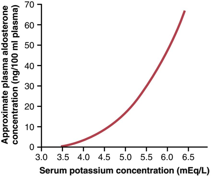
Figure 30-5. Effect of extracellular fluid potassium ion concentration on plasma aldosterone concentration. Note that small changes in potassium concentration cause large changes in aldosterone concentration.
شکل 30-5. اثر غلظت یون پتاسیم مایع خارج سلولی بر غلظت آلدوسترون پلاسما. توجه داشته باشید که تغییرات کوچک در غلظت پتاسیم باعث تغییرات بزرگ در غلظت آلدوسترون می شود.
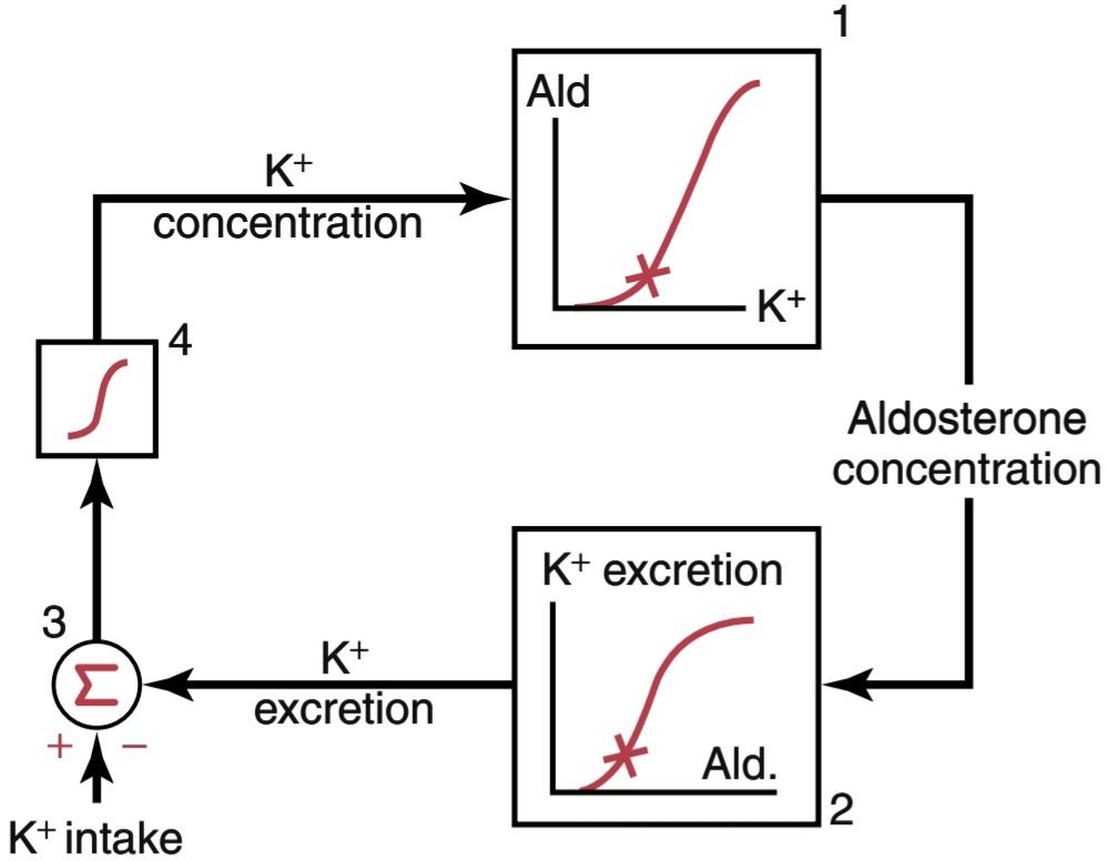
Figure 30-6. Basic feedback mechanism for control of extracellular fluid potassium concentration by aldosterone (A/d).
شکل 30-6. مکانیسم بازخورد اساسی برای کنترل غلظت پتاسیم مایع خارج سلولی توسط آلدوسترون (A/d).
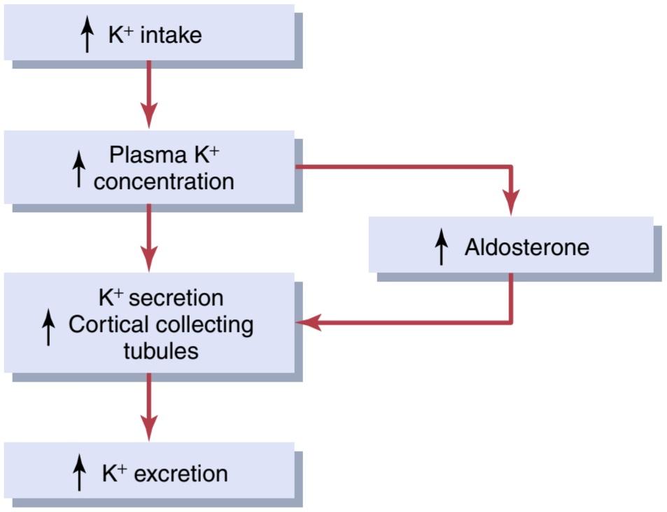
Figure 30-7. Primary mechanisms whereby high potassium intake raises potassium excretion. Note that increased plasma potassium concentration directly raises potassium secretion by the cortical collecting tubules and indirectly increases potassium secretion by raising plasma aldosterone concentration.
شکل 30-7. مکانیسم های اولیه که به موجب آن مصرف زیاد پتاسیم باعث افزایش دفع پتاسیم می شود. توجه داشته باشید که افزایش غلظت پتاسیم پلاسما به طور مستقیم باعث افزایش ترشح پتاسیم توسط لوله های جمع کننده قشر مغز و به طور غیرمستقیم با افزایش غلظت آلدوسترون پلاسما باعث افزایش ترشح پتاسیم می شود.
Blockade of Aldosterone Feedback System Greatly Impairs Potassium Regulation. In the absence of aldosterone secretion, as occurs in patients with Addison disease, renal secretion of potassium is impaired, thus causing the extracellular fluid potassium concentration to rise to dangerously high levels. Conversely, with excess aldosterone secretion (primary aldosteronism), potassium secretion becomes greatly increased, causing potassium loss by the kidneys, thus leading to hypokalemia.
مسدود کردن سیستم بازخورد آلدوسترون به شدت تنظیم پتاسیم را مختل می کند. در غیاب ترشح آلدوسترون، همانطور که در بیماران مبتلا به بیماری آدیسون اتفاق می افتد، ترشح کلیوی پتاسیم مختل می شود و در نتیجه باعث می شود که غلظت پتاسیم مایع خارج سلولی به سطوح خطرناکی بالا برود. برعکس، با ترشح بیش از حد آلدوسترون (آلدوسترونیسم اولیه)، ترشح پتاسیم به شدت افزایش می یابد و باعث از دست دادن پتاسیم توسط کلیه ها می شود و در نتیجه منجر به هیپوکالمی می شود.
In addition to its stimulatory effect on renal secretion of potassium, aldosterone also increases cellular uptake of potassium. This contributes to the powerful aldosterone- potassium feedback system, as discussed previously.
آلدوسترون علاوه بر اثر تحریکی بر ترشح کلیوی پتاسیم، جذب سلولی پتاسیم را نیز افزایش می دهد. این به سیستم بازخورد قدرتمند آلدوسترون- پتاسیم کمک می کند، همانطور که قبلاً بحث شد.
The special quantitative importance of the aldosterone feedback system in controlling potassium concentration is shown in Figure 30-8. In this experiment, potassium intake was increased almost sevenfold in dogs under two conditions: (1) under normal conditions; and (2) after the aldosterone feedback system had been blocked by removing the adrenal glands and placing the animals on a fixed rate of aldosterone infusion, so that plasma aldosterone concentration was maintained at a normal level but could neither increase nor decrease as potassium intake was altered.
اهمیت کمی ویژه سیستم بازخورد آلدوسترون در کنترل غلظت پتاسیم در شکل 30-8 نشان داده شده است. در این آزمایش، دریافت پتاسیم در سگ ها در دو شرایط تقریباً هفت برابر افزایش یافت: (1) در شرایط عادی. و (2) پس از مسدود شدن سیستم بازخورد آلدوسترون با برداشتن غدد فوق کلیوی و قرار دادن حیوانات بر روی یک نرخ ثابت انفوزیون آلدوسترون، به طوری که غلظت آلدوسترون پلاسما در سطح نرمال حفظ شد اما با تغییر دریافت پتاسیم، نه افزایش یافت و نه کاهش یافت.
Note that in the normal animals, a sevenfold increase in potassium intake caused only a slight increase in plasma potassium concentration, from 4.2 to 4.3 mEq/L. Thus, when the aldosterone feedback system is functioning normally, potassium concentration is precisely con- trolled, despite large changes in potassium intake.
توجه داشته باشید که در حیوانات عادی، افزایش هفت برابری دریافت پتاسیم باعث افزایش جزئی غلظت پتاسیم پلاسما، از 4.2 به 4.3 mEq/L شد. بنابراین، هنگامی که سیستم بازخورد آلدوسترون به طور طبیعی کار می کند، با وجود تغییرات زیاد در دریافت پتاسیم، غلظت پتاسیم دقیقاً کنترل می شود.
When the aldosterone feedback system was blocked, the same increases in potassium intake caused a much larger increase in plasma potassium concentration, from 3.8 to almost 4.7 mEq/L. Thus, control of potassium concentration is greatly impaired when the aldosterone feedback system is blocked. A similar impairment of potassium regulation is observed in people with poorly functioning aldosterone feedback systems, such as occurs in patients with primary aldosteronism (too much aldosterone) or Addison disease (too little aldosterone).
هنگامی که سیستم بازخورد آلدوسترون مسدود شد، همان افزایش در دریافت پتاسیم باعث افزایش بسیار بیشتر در غلظت پتاسیم پلاسما شد، از 3.8 به تقریبا 4.7 mEq/L. بنابراین، هنگامی که سیستم بازخورد آلدوسترون مسدود می شود، کنترل غلظت پتاسیم به شدت مختل می شود. اختلال مشابهی در تنظیم پتاسیم در افراد با سیستم بازخورد آلدوسترون ضعیف مشاهده می شود، مانند این که در بیماران مبتلا به آلدوسترونیسم اولیه (آلدوسترون بیش از حد) یا بیماری آدیسون (آلدوسترون خیلی کم) رخ می دهد.
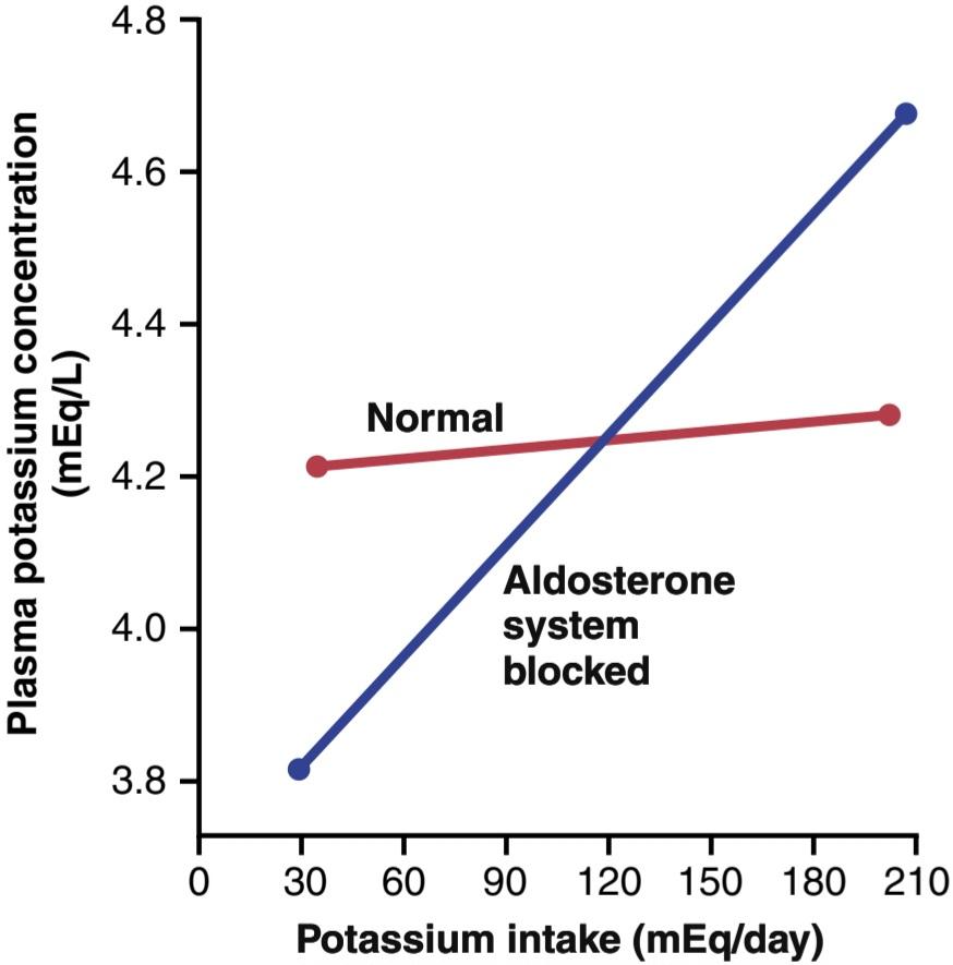
Figure 30-8. Effect of large changes in potassium intake on plasma potassium concentration under normal conditions (red line) and after the aldosterone feedback was blocked (blue line). Note that after blockade of the aldosterone system, regulation of potassium concentration was greatly impaired. (Courtesy Dr. David B. Young.)
شکل 30-8. تأثیر تغییرات زیاد در دریافت پتاسیم بر غلظت پتاسیم پلاسما در شرایط عادی (خط قرمز) و پس از مسدود شدن بازخورد آلدوسترون (خط آبی). توجه داشته باشید که پس از مسدود شدن سیستم آلدوسترون، تنظیم غلظت پتاسیم تا حد زیادی مختل شد. (با احترام از دکتر دیوید بی یانگ.)
Increased Distal Tubular Flow Rate Stimulates Potassium Secretion. A rise in distal tubular flow rate, as occurs with volume expansion, high sodium intake, or treatment with some diuretics, stimulates potassium secretion (Figure 30-9). Conversely, a decrease in distal tubular flow rate, as caused by sodium depletion, reduces potassium secretion.
افزایش سرعت جریان لوله ای دیستال ترشح پتاسیم را تحریک می کند. افزایش سرعت جریان لوله ای دیستال، همانطور که با افزایش حجم، مصرف زیاد سدیم یا درمان با برخی دیورتیک ها اتفاق می افتد، ترشح پتاسیم را تحریک می کند (شکل 30-9). برعکس، کاهش سرعت جریان لولهای دیستال، که ناشی از کاهش سدیم است، ترشح پتاسیم را کاهش میدهد.
The effect of the tubular flow rate on potassium secretion in the distal and collecting tubules is strongly influenced by potassium intake. When potassium intake is high, increased tubular flow rate has a much greater effect to stimulate potassium secretion than when potassium intake is low (see Figure 30-9).
تأثیر سرعت جریان لولهای بر ترشح پتاسیم در لولههای دیستال و جمعکننده به شدت تحت تأثیر مصرف پتاسیم است. هنگامی که مصرف پتاسیم زیاد است، افزایش سرعت جریان لولهای تأثیر بسیار بیشتری برای تحریک ترشح پتاسیم نسبت به زمانی که دریافت پتاسیم کم است دارد (شکل 30-9 را ببینید).
There are two main effects of a high-volume flow rate that increase potassium secretion:
دو اثر اصلی جریان با حجم بالا وجود دارد که باعث افزایش ترشح پتاسیم می شود:
1. When potassium is secreted into the tubular fluid, the luminal concentration of potassium increases,thereby reducing the driving force for potassium diffusion across the luminal membrane. With increased tubular flow rate, the secreted potassium is continuously flushed down the tubule, minimizing the rise in tubular potassium concentration and in- creasing net potassium secretion.
1. هنگامی که پتاسیم در مایع لوله ای ترشح می شود، غلظت پتاسیم در مجرا افزایش می یابد و در نتیجه نیروی محرکه انتشار پتاسیم در سراسر غشای مجرا کاهش می یابد. با افزایش سرعت جریان لوله ای، پتاسیم ترشح شده به طور مداوم در لوله تخلیه می شود و افزایش غلظت پتاسیم لوله ای را به حداقل می رساند و ترشح خالص پتاسیم را افزایش می دهد.
2. A high tubular flow rate also increases the number of high-conductance BK channels in the luminal membrane. Although the BK channels are normally quiescent, they become active in response to increases in flow rate, thereby greatly increasing potassium conductance across the luminal membrane.
2. سرعت جریان لوله ای بالا همچنین تعداد کانال های BK با رسانایی بالا را در غشای مجرا افزایش می دهد. اگرچه کانالهای BK معمولاً ساکن هستند، اما در پاسخ به افزایش سرعت جریان فعال میشوند و در نتیجه رسانایی پتاسیم را در سراسر غشای مجرا افزایش میدهند.
The effect of increased tubular flow rate is especially important in helping preserve normal potassium excretion during changes in sodium intake. For example, with a high sodium intake, there is decreased aldosterone secretion, which by itself would tend to decrease the rate of potassium secretion and, therefore, reduce urinary excretion of potassium. However, the high distal tubular flow rate that occurs with a high sodium intake tends to increase potassium secretion (Figure 30-10). Therefore, the two effects of a high sodium intake-decreased aldosterone secretion and high tubular flow ratecounterbalance each other, so there is little change in potassium excretion. Likewise, with a low sodium intake, there is little change in potassium excretion because of the counterbalancing effects of increased aldosterone secretion and decreased tubular flow rate on potassium secretion.
اثر افزایش سرعت جریان لوله ای به ویژه در کمک به حفظ دفع طبیعی پتاسیم در طول تغییرات در مصرف سدیم مهم است. به عنوان مثال، با مصرف زیاد سدیم، ترشح آلدوسترون کاهش می یابد که به خودی خود باعث کاهش میزان ترشح پتاسیم و در نتیجه کاهش دفع ادراری پتاسیم می شود. با این حال، سرعت بالای جریان لولهای دیستال که با مصرف زیاد سدیم اتفاق میافتد باعث افزایش ترشح پتاسیم میشود (شکل 30-10). بنابراین، دو اثر مصرف زیاد سدیم- کاهش ترشح آلدوسترون و سرعت جریان لوله ای بالا- یکدیگر را متعادل می کنند، بنابراین تغییر کمی در دفع پتاسیم وجود دارد. به همین ترتیب، با مصرف کم سدیم، به دلیل اثرات متعادل کننده افزایش ترشح آلدوسترون و کاهش سرعت جریان لوله ای بر ترشح پتاسیم، تغییر کمی در دفع پتاسیم ایجاد می شود.

Figure 30-9. Relationship between flow rate in the cortical collecting tubules and potassium secretion and the effect of changes in potassium intake. Note that a high dietary potassium intake greatly enhances the effect of increased tubular flow rate to increase potassium secretion.The shaded bar shows the approximate normal tubular flow rate under most physiological conditions. (Data from Malnic G, Berliner RW, Giebisch G: Flow dependence of K+ secretion in cortical distal tubes of the rat. Am J Physiol 256:F932, 1989.)
شکل 30-9. ارتباط بین سرعت جریان در لوله های جمع کننده قشر مغز و ترشح پتاسیم و تأثیر تغییرات در دریافت پتاسیم. توجه داشته باشید که مصرف زیاد پتاسیم در رژیم غذایی تأثیر افزایش سرعت جریان لولهای را برای افزایش ترشح پتاسیم بسیار افزایش میدهد. نوار سایهدار، سرعت جریان لولهای طبیعی تقریبی را در اکثر شرایط فیزیولوژیکی نشان میدهد. (دادههای Malnic G، Berliner RW، Giebisch G: وابستگی به جریان ترشح +K در لولههای انتهایی قشری موش صحرایی. Am J Physiol 256:F932، 1989.)

Figure 30-10. Effect of high sodium intake on renal excretion of potassium. Note that a high-sodium diet decreases plasma aldosterone, which tends to decrease potassium secretion by the cortical collecting tubules. However, the high-sodium diet simultaneously increases fluid delivery to the cortical collecting duct, which tends to increase potassium secretion. The opposing effects of a high-sodium diet counterbalance each other, so there is little change in potassium excretion. GFR, Glomerular filtration rate.
شکل 30-10. تأثیر مصرف زیاد سدیم بر دفع کلیوی پتاسیم. توجه داشته باشید که یک رژیم غذایی با سدیم بالا باعث کاهش آلدوسترون پلاسما می شود که تمایل به کاهش ترشح پتاسیم توسط لوله های جمع کننده قشر مغز دارد. با این حال، رژیم غذایی با سدیم بالا به طور همزمان باعث افزایش مایعات به مجرای جمع کننده قشر مغز می شود که تمایل به افزایش ترشح پتاسیم دارد. اثرات متضاد یک رژیم غذایی پر سدیم یکدیگر را متعادل می کند، بنابراین تغییر کمی در دفع پتاسیم وجود دارد. GFR، نرخ فیلتراسیون گلومرولی.
Acute Acidosis Decreases Potassium Secretion. Acute increases in extracellular fluid hydrogen ion concentration (acidosis) reduce potassium secretion, whereas decreased hydrogen ion concentration (alkalosis) increases potassium secretion. The primary mechanism whereby increased hydrogen ion concentration inhibits potassium secretion is by reducing activity of the Na+-K+ ATPase pump. This reduction in turn decreases intracellular potassium concentration and subsequent passive diffusion of potassium across the luminal membrane into the tubule. Acidosis may also reduce the number of potassium channels in the luminal membrane.
اسیدوز حاد ترشح پتاسیم را کاهش می دهد. افزایش شدید غلظت یون هیدروژن مایع خارج سلولی (اسیدوز) باعث کاهش ترشح پتاسیم می شود، در حالی که کاهش غلظت یون هیدروژن (آلکالوز) باعث افزایش ترشح پتاسیم می شود. مکانیسم اولیه که به موجب آن افزایش غلظت یون هیدروژن ترشح پتاسیم را مهار میکند، با کاهش فعالیت پمپ Na+-K+ ATPase است. این کاهش به نوبه خود باعث کاهش غلظت پتاسیم درون سلولی و متعاقب آن انتشار غیرفعال پتاسیم در سراسر غشای مجرا به داخل لوله می شود. اسیدوز همچنین ممکن است تعداد کانال های پتاسیم را در غشای مجرا کاهش دهد.
With more prolonged acidosis, lasting over a period of several days, there is an increase in urinary potassium excretion. The mechanism for this effect is due in part to an effect of chronic acidosis to inhibit proximal tubular sodium chloride and water reabsorption, which increases distal volume delivery, thereby stimulating potassium secretion. This effect overrides the inhibitory effect of hydrogen ions on the Na+-K+ ATPase pump. Thus, chronic acidosis leads to a loss of potassium, whereas acute acidosis leads to decreased potassium excretion.
با اسیدوز طولانی تر، که طی یک دوره چند روزه طول می کشد، دفع پتاسیم از طریق ادرار افزایش می یابد. مکانیسم این اثر تا حدی به دلیل اثر اسیدوز مزمن برای مهار کلرید سدیم لوله پروگزیمال و بازجذب آب است که باعث افزایش حجم دیستال میشود و در نتیجه ترشح پتاسیم را تحریک میکند. این اثر اثر بازدارندگی یون های هیدروژن بر روی پمپ Na+-K+ ATPase را نادیده می گیرد. بنابراین، اسیدوز مزمن منجر به از دست دادن پتاسیم می شود، در حالی که اسیدوز حاد منجر به کاهش دفع پتاسیم می شود.
Beneficial Effects of Diet High in Potassium and Low in Sodium Content
For most of human history, the typical diet has been one that is low in sodium and high in potassium content, compared with the typical modern diet. In isolated populations that have not experienced industrialization, such as the Yanomamo tribe living in the Amazon region of Northern Brazil, sodium intake may be as low as 10 to 20 mmol/day, and potassium intake may be as high as 200 mmol/day. This intake is due to their consumption of a diet containing large amounts of fruits and vegetables and no processed foods. Populations consuming this type of diet typically do not experience age-related in- creases in blood pressure and cardiovascular diseases.
اثرات مفید رژیم غذایی سرشار از پتاسیم و محتوای کم سدیم
در بیشتر تاریخ بشر، رژیم غذایی معمولی رژیمی بوده است که در مقایسه با رژیم معمولی مدرن، دارای مقدار کمی سدیم و پتاسیم بالا است. در جمعیت های جدا شده ای که صنعتی شدن را تجربه نکرده اند، مانند قبیله یانومامو که در منطقه آمازون در شمال برزیل زندگی می کنند، مصرف سدیم ممکن است بین 10 تا 20 میلی مول در روز و دریافت پتاسیم ممکن است به 200 میلی مول در روز برسد. این دریافت به دلیل مصرف رژیم غذایی حاوی مقادیر زیادی میوه و سبزیجات و بدون غذاهای فرآوری شده است. جمعیت هایی که از این نوع رژیم غذایی استفاده می کنند معمولاً افزایش فشار خون و بیماری های قلبی عروقی مرتبط با افزایش سن را تجربه نمی کنند.
With industrialization and increased consumption of processed foods, which often have high sodium and low potassium content, there have been dramatic increases in sodium intake and decreases in potassium intake. In most industrialized countries, potassium consumption averages only 30 to 70 mmol/day, and sodium intake averages 140 to 180 mmol/day.
با صنعتی شدن و افزایش مصرف غذاهای فرآوری شده، که اغلب دارای سدیم بالا و پتاسیم کم هستند، افزایش چشمگیری در مصرف سدیم و کاهش دریافت پتاسیم مشاهده شده است. در اکثر کشورهای صنعتی، مصرف پتاسیم به طور متوسط تنها 30 تا 70 میلی مول در روز و مصرف سدیم به طور متوسط 140 تا 180 میلی مول در روز است.
Experimental and clinical studies have shown that the combination of a high-sodium and low-potassium diet increases the risk for hypertension and associated cardiovascular and kidney diseases. A diet rich in potassium, however, seems to protect against the adverse effects of a high-sodium diet, reducing blood pressure and the risk for stroke, coronary artery disease, and kidney disease. The beneficial effects of increasing potassium intake are especially apparent when combined with a low-sodium diet.
مطالعات تجربی و بالینی نشان داده است که ترکیب رژیم غذایی پر سدیم و کم پتاسیم خطر ابتلا به فشار خون بالا و بیماری های قلبی عروقی و کلیوی مرتبط را افزایش می دهد. با این حال، به نظر می رسد رژیم غذایی غنی از پتاسیم در برابر اثرات نامطلوب رژیم غذایی پر سدیم، کاهش فشار خون و خطر سکته مغزی، بیماری عروق کرونر و بیماری کلیوی محافظت می کند. اثرات مفید افزایش دریافت پتاسیم به ویژه هنگامی که با رژیم غذایی کم سدیم ترکیب شود آشکار می شود.
Dietary guidelines published by various organizations have recommended reducing the dietary intake of sodium chloride to about 65 to 100 mmol/day (corresponding to 1.5-2.3 g/day of sodium or 3.8-5.8 g/day sodium chloride) while increasing potassium intake to 120 mmol/day (4.7 g/ day) for healthy adults.
دستورالعمل های غذایی منتشر شده توسط سازمان های مختلف توصیه می کند که مصرف رژیم غذایی کلرید سدیم را به حدود 65 تا 100 میلی مول در روز کاهش دهید (مطابق با 1.5-2.3 گرم در روز سدیم یا 3.8-5.8 گرم در روز کلرید سدیم) در حالی که مصرف پتاسیم را به 120.7 میلی مول در روز برای بزرگسالان سالم افزایش دهید.
REGULATION OF RENAL CALCIUM EXCRETION AND EXTRACELLULAR CALCIUM ION CONCENTRATION
The mechanisms for regulating calcium ion concentration are discussed in detail in Chapter 80, along with the endocrinology of the calcium-regulating hormones, parathyroid hormone (PTH), and calcitonin. Therefore, calcium ion regulation is discussed only briefly in this chapter.
تنظیم دفع کلسیم کلیوی و غلظت یون کلسیم خارج سلولی
مکانیسم های تنظیم غلظت یون کلسیم به طور مفصل در فصل 80، همراه با غدد درون ریز هورمون های تنظیم کننده کلسیم، هورمون پاراتیروئید (PTH) و کلسی تونین مورد بحث قرار گرفته است. بنابراین، تنظیم یون کلسیم تنها به طور خلاصه در این فصل مورد بحث قرار گرفته است.
The extracellular fluid calcium ion concentration normally remains tightly controlled within a few percentage points of its normal level, 2.4 mEq/L. When the calcium ion concentration falls to low levels (hypocalcemia), the excitability of nerve and muscle cells increases markedly and can, in extreme cases, result in hypocalcemic tetany. This condition is characterized by spastic skeletal muscle contractions. Hypercalcemia (increased calcium concentration) depresses neuromuscular excitability and can lead to cardiac arrhythmias.
غلظت یون کلسیم مایع خارج سلولی معمولاً در چند درصد از سطح طبیعی خود یعنی mEq/L 2.4 به شدت کنترل می شود. هنگامی که غلظت یون کلسیم به سطوح پایین کاهش می یابد (هیپوکلسمی)، تحریک پذیری سلول های عصبی و ماهیچه ای به طور قابل توجهی افزایش می یابد و در موارد شدید می تواند منجر به تتانی هیپوکلسمیک شود. این وضعیت با انقباضات اسپاستیک عضلات اسکلتی مشخص می شود. هیپرکلسمی (افزایش غلظت کلسیم) تحریک پذیری عصبی عضلانی را کاهش می دهد و می تواند منجر به آریتمی قلبی شود.
About 50% of the total calcium in the plasma (5 mEq/L) exists in the ionized form, which is the form that has bio- logical activity at cell membranes. The remainder is bound to plasma proteins (~40%) or complexed in the nonionized form with anions such as phosphate and citrate (~10%).
حدود 50 درصد از کل کلسیم پلاسما (5 میلی اکی والان در لیتر) به شکل یونیزه شده وجود دارد که فعالیت بیولوژیکی در غشای سلولی دارد. باقیمانده به پروتئین های پلاسما (~40٪) متصل می شود یا به شکل غیریونیزه شده با آنیون هایی مانند فسفات و سیترات (~10٪) کمپلکس می شود.
Changes in plasma hydrogen ion concentration influence calcium binding to plasma proteins. With acidosis, less calcium is bound to the plasma proteins. Conversely, with alkalosis, a greater amount of calcium is bound to the plasma proteins. Therefore, patients with alkalosis are more susceptible to hypocalcemic tetany.
تغییرات در غلظت یون هیدروژن پلاسما بر اتصال کلسیم به پروتئین های پلاسما تأثیر می گذارد. با اسیدوز، کلسیم کمتری به پروتئین های پلاسما متصل می شود. برعکس، با آلکالوز، مقدار بیشتری از کلسیم به پروتئین های پلاسما متصل می شود. بنابراین، بیماران مبتلا به آلکالوز بیشتر مستعد ابتلا به کزاز هیپوکلسمیک هستند.
As with other substances in the body, calcium intake must be balanced with the net loss of calcium over the long term. Unlike ions such as sodium and chloride, however, much of the calcium excretion occurs in the feces. The usual rate of dietary calcium intake is about 1000 mg/ day, with about 900 mg/day of calcium excreted in the feces. Under certain conditions, fecal calcium excretion can exceed calcium ingestion because calcium can also be secreted into the intestinal lumen. Therefore, the gastrointestinal tract and regulatory mechanisms that influence intestinal calcium absorption and secretion play a major role in calcium homeostasis, as discussed in Chapter 80.
مانند سایر مواد در بدن، دریافت کلسیم باید با از دست دادن خالص کلسیم در دراز مدت متعادل شود. برخلاف یونهایی مانند سدیم و کلرید، بیشتر دفع کلسیم از طریق مدفوع انجام میشود. میزان معمول دریافت کلسیم از طریق رژیم غذایی حدود 1000 میلی گرم در روز است که حدود 900 میلی گرم در روز کلسیم از طریق مدفوع دفع می شود. تحت شرایط خاص، دفع کلسیم از طریق مدفوع می تواند از مصرف کلسیم بیشتر شود، زیرا کلسیم می تواند به مجرای روده نیز ترشح شود. بنابراین، دستگاه گوارش و مکانیسمهای تنظیمی که بر جذب و ترشح کلسیم روده تأثیر میگذارند، نقش عمدهای در هموستاز کلسیم دارند، همانطور که در فصل 80 بحث شد.
Almost all the calcium in the body (99%) is stored in the bone, with only about 0.1% in the extracellular fluid and 1.0% in the intracellular fluid and cell organelles. The bone, therefore, acts as a large reservoir for calcium and as a source of calcium when extracellular fluid calcium concentration tends to decrease.
تقریباً تمام کلسیم بدن (99٪) در استخوان ذخیره می شود که تنها حدود 0.1٪ در مایع خارج سلولی و 1.0٪ در مایع داخل سلولی و اندامک های سلولی ذخیره می شود. بنابراین، زمانی که غلظت کلسیم مایع خارج سلولی تمایل به کاهش دارد، استخوان به عنوان یک مخزن بزرگ برای کلسیم و به عنوان منبع کلسیم عمل می کند.
One of the most important regulators of bone uptake and release of calcium is PTH. When extracellular fluid calcium concentration falls below normal, activity of calcium-sensing receptors (CSRs) on the cell membrane of the parathyroid glands is reduced, promoting increased secretion of PTH. This hormone then acts directly on the bones to increase resorption of bone salts (release of salts from the bones) and release large amounts of calcium into the extracellular fluid, thereby returning calcium levels back toward normal. When the calcium ion con- centration is elevated, CSR activity of the parathyroid cells is stimulated, causing a reduction in PTH secretion, so almost no bone resorption occurs; instead, excess calcium is deposited in the bones. Thus, the daily regulation of calcium ion concentration is mediated in large part by the effects of PTH on bone resorption.
یکی از مهم ترین تنظیم کننده های جذب استخوان و آزادسازی کلسیم PTH است. هنگامی که غلظت کلسیم مایع خارج سلولی به زیر نرمال میرسد، فعالیت گیرندههای حسگر کلسیم (CSRs) بر روی غشای سلولی غدد پاراتیروئید کاهش مییابد و باعث افزایش ترشح PTH میشود. سپس این هورمون مستقیماً روی استخوانها عمل میکند تا جذب نمکهای استخوانی را افزایش دهد (آزاد شدن نمک از استخوانها) و مقدار زیادی کلسیم را در مایع خارج سلولی آزاد میکند و در نتیجه سطح کلسیم را به حالت طبیعی برمیگرداند. هنگامی که غلظت یون کلسیم بالا می رود، فعالیت CSR سلول های پاراتیروئید تحریک می شود و باعث کاهش ترشح PTH می شود، بنابراین تقریباً هیچ تحلیل استخوانی رخ نمی دهد. در عوض، کلسیم اضافی در استخوان ها رسوب می کند. بنابراین، تنظیم روزانه غلظت یون کلسیم تا حد زیادی توسط اثرات PTH بر تحلیل استخوان انجام می شود.
The bones, however, do not have an inexhaustible sup- ply of calcium. Therefore, over the long term, intake of calcium must be balanced with calcium excretion by the gastrointestinal tract and kidneys. The most important regulator of calcium reabsorption at both these sites is PTH, which regulates the plasma calcium concentration through three main effects: (1) by stimulating bone resorption; (2) by stimulating activation of vitamin D, which then increases intestinal reabsorption of calcium; and (3) by increasing renal tubular calcium reabsorption (Figure 30- 11). The control of gastrointestinal calcium reabsorption and calcium exchange in the bones is discussed elsewhere; the remainder of this section focuses on the mechanisms that control renal calcium excretion.
با این حال، استخوانها منبع کلسیم پایانناپذیری ندارند. بنابراین، در طولانی مدت، دریافت کلسیم باید با دفع کلسیم توسط دستگاه گوارش و کلیه ها متعادل شود. مهم ترین تنظیم کننده بازجذب کلسیم در هر دو محل PTH است که غلظت کلسیم پلاسما را از طریق سه اثر اصلی تنظیم می کند: (1) با تحریک تحلیل استخوان. (2) با تحریک فعال شدن ویتامین D، که سپس بازجذب روده ای کلسیم را افزایش می دهد. و (3) با افزایش بازجذب کلسیم توبولار کلیوی (شکل 30-11). کنترل بازجذب کلسیم گوارشی و تبادل کلسیم در استخوان ها در جای دیگری مورد بحث قرار گرفته است. بقیه این بخش بر مکانیسم هایی تمرکز دارد که دفع کلسیم را از طریق کلیوی کنترل می کند.
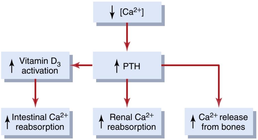
Figure 30-11. Compensatory responses to decreased plasma ionized calcium concentration mediated by parathyroid hormone (PTH) and vitamin D.
شکل 30-11. پاسخ های جبرانی به کاهش غلظت کلسیم یونیزه شده پلاسما با واسطه هورمون پاراتیروئید (PTH) و ویتامین D.
CONTROL OF CALCIUM EXCRETION BY THE KIDNEYS
Calcium is filtered and reabsorbed in the kidneys but is not secreted. Therefore, the rate of renal calcium excretion is calculated as follows:
کنترل دفع کلسیم توسط کلیه ها
کلسیم در کلیه ها فیلتر شده و دوباره جذب می شود اما ترشح نمی شود. بنابراین، میزان دفع کلسیم از کلیه به صورت زیر محاسبه می شود:

Only about 60% of the plasma calcium is ionized, with 40% being bound to the plasma proteins and 10% complexed with anions such as phosphate. Therefore, only about 60% of the plasma calcium is filtered at the glomerulus. Normally, about 99% of the filtered calcium is reabsorbed by the tubules, with only about 1% of the filtered calcium being excreted. About 65% of the filtered calcium is reabsorbed in the proximal tubule, 25% to 30% is reabsorbed in the loop of Henle, and 4% to 9% is reabsorbed in the distal and collecting tubules. This pattern of reabsorption is similar to that for sodium.
تنها حدود 60 درصد از کلسیم پلاسما یونیزه می شود که 40 درصد آن به پروتئین های پلاسما و 10 درصد با آنیون هایی مانند فسفات کمپلکس می شود. بنابراین، تنها حدود 60 درصد از کلسیم پلاسما در گلومرول فیلتر می شود. به طور معمول، حدود 99٪ از کلسیم فیلتر شده توسط لوله ها دوباره جذب می شود و تنها حدود 1٪ از کلسیم فیلتر شده دفع می شود. حدود 65 درصد از کلسیم فیلتر شده در لوله پروگزیمال، 25 تا 30 درصد در حلقه هنله و 4 تا 9 درصد در لوله های دیستال و جمع کننده بازجذب می شود. این الگوی بازجذب مشابه الگوی سدیم است.
As is true with the other ions, calcium excretion is adjusted to meet the body’s needs. With an increase in calcium intake, there is also increased renal calcium excretion, although much of the increase of calcium intake is eliminated in the feces. With calcium depletion, calcium excretion by the kidneys decreases as a result of enhanced tubular reabsorption.
همانطور که در مورد سایر یون ها صادق است، دفع کلسیم برای رفع نیازهای بدن تنظیم می شود. با افزایش دریافت کلسیم، دفع کلسیم کلیوی نیز افزایش می یابد، اگرچه بیشتر افزایش دریافت کلسیم از طریق مدفوع دفع می شود. با کاهش کلسیم، دفع کلسیم توسط کلیه ها در نتیجه افزایش بازجذب لوله ای کاهش می یابد.
Proximal Tubular Calcium Reabsorption. Most of the calcium reabsorption in the proximal tubule occurs through the paracellular pathway; it is dissolved in water and carried with the reabsorbed fluid as it flows between the cells. Only about 20% of proximal tubular calcium re- absorption occurs through the transcellular pathway in two steps;
بازجذب کلسیم لوله ای پروگزیمال. بیشتر بازجذب کلسیم در لوله پروگزیمال از طریق مسیر پاراسلولی انجام می شود. در آب حل می شود و در جریان جریان بین سلول ها با مایع جذب شده مجدد حمل می شود. فقط حدود 20% از بازجذب کلسیم لوله پروگزیمال از طریق مسیر بین سلولی در دو مرحله انجام می شود.
1. Calcium diffuses from the tubular lumen into the cell down an electrochemical gradient due to the much higher concentration of calcium in the tubular lumen, compared with the epithelial cell cytoplasm, and because the cell interior has a negative charge relative to the tubular lumen.
1. به دلیل غلظت بسیار بیشتر کلسیم در لومن لولهای در مقایسه با سیتوپلاسم سلولهای اپیتلیال، و به این دلیل که درون سلول نسبت به لومن لولهای بار منفی دارد، کلسیم از مجرای لولهای به داخل سلول به سمت پایین گرادیان الکتروشیمیایی منتشر میشود.
2. Calcium exits the cell across the basolateral mem- brane by a calcium-ATPase pump and by the sodium- calcium counter-transporter (Figure 30-12).
2. کلسیم از طریق غشای قاعده جانبی توسط یک پمپ کلسیم-ATPase و توسط ضد انتقال دهنده سدیم-کلسیم از سلول خارج می شود (شکل 30-12).

Figure 30-12. Mechanisms of calcium reabsorption by paracellular and transcellular pathways in the proximal tubular cells.
شکل 30-12. مکانیسم های بازجذب کلسیم توسط مسیرهای پاراسلولی و بین سلولی در سلول های لوله پروگزیمال
Loop of Henle and Distal Tubule Calcium Reabsorption. In the loop of Henle, calcium reabsorption is restricted to the thick ascending limb. Approximately 50% of calcium reabsorption in the thick ascending limb occurs through the paracellular route by passive diffusion due to the slight positive charge of the tubular lumen relative to the interstitial fluid. The remaining 50% of calcium reabsorption in the thick ascending limb occurs through the transcellular pathway, a process that is stimulated by PTH.
حلقه هنله و بازجذب کلسیم توبول دیستال. در حلقه هنله، بازجذب کلسیم به اندام ضخیم صعودی محدود می شود. تقریباً 50٪ از بازجذب کلسیم در اندام صعودی ضخیم از طریق مسیر پاراسلولی با انتشار غیرفعال به دلیل بار مثبت خفیف لومن لوله ای نسبت به مایع بینابینی رخ می دهد. 50 درصد باقیمانده بازجذب کلسیم در اندام صعودی ضخیم از طریق مسیر بین سلولی اتفاق می افتد، فرآیندی که توسط PTH تحریک می شود.
In the distal tubule, calcium reabsorption occurs almost entirely by active transport through the cell membrane. The mechanism for this active transport is similar to that in the proximal tubule and thick ascending limb. It involves diffusion across the luminal membrane through calcium channels and exit across the basolateral membrane by a calcium-ATPase pump, as well as a sodium-calcium counter-transport mechanism. In this segment, as well as in the loops of Henle, PTH stimulates calcium reabsorption. Vitamin D (calcitriol) and calcitonin also stimulate calcium reabsorption in the thick ascending limb of Henle’s loop and in the distal tubule, although these hormones are not as important quantitatively as PTH in reducing renal calcium excretion.
در لوله دیستال، بازجذب کلسیم تقریباً به طور کامل با انتقال فعال از طریق غشای سلولی انجام می شود. مکانیسم این انتقال فعال شبیه به مکانیزم توبول پروگزیمال و اندام صعودی ضخیم است. این شامل انتشار در غشای مجرا از طریق کانال های کلسیم و خروج از غشای قاعده جانبی توسط یک پمپ کلسیم-ATPase، و همچنین یک مکانیسم ضد انتقال سدیم-کلسیم است. در این بخش و همچنین در حلقه های هنله، PTH بازجذب کلسیم را تحریک می کند. ویتامین D (کلسیتریول) و کلسی تونین نیز بازجذب کلسیم را در اندام صعودی ضخیم حلقه هنله و در لوله دیستال تحریک می کنند، اگرچه این هورمون ها از نظر کمی به اندازه PTH در کاهش دفع کلسیم از کلیه مهم نیستند.
Regulation of Tubular Calcium Reabsorption. One of the primary controllers of renal tubular calcium reabsorption is PTH. Increased levels of PTH stimulate calcium reabsorption in the thick ascending loops of Henle and distal tubules, which reduces urinary excretion of calcium. Conversely, reduction of PTH promotes calcium excretion by decreasing reabsorption in the loops of Henle and distal tubules.
تنظیم بازجذب کلسیم لوله ای. یکی از کنترلکنندههای اصلی بازجذب کلسیم در لولههای کلیوی، PTH است. افزایش سطح PTH باعث تحریک بازجذب کلسیم در حلقه های صعودی ضخیم هنله و لوله های دیستال می شود که باعث کاهش دفع ادراری کلسیم می شود. برعکس، کاهش PTH با کاهش بازجذب در حلقههای هنله و لولههای دیستال، باعث افزایش دفع کلسیم میشود.
Increased extracellular fluid calcium ion concentration also directly stimulates CSRs, which inhibit calcium reabsorption in the thick ascending loops of Henle. Conversely, reductions in calcium concentration decrease CSR activity and increase calcium reabsorption in the thick ascending loop of Henle.
افزایش غلظت یون کلسیم مایع خارج سلولی مستقیماً CSR ها را تحریک می کند که بازجذب کلسیم را در حلقه های صعودی ضخیم هنله مهار می کند. برعکس، کاهش غلظت کلسیم باعث کاهش فعالیت CSR و افزایش بازجذب کلسیم در حلقه صعودی ضخیم هنله می شود.
In the proximal tubule, calcium reabsorption usually parallels sodium and water reabsorption and is independent of PTH. Therefore, in cases of extracellular volume expansion or increased arterial pressure—both of which decrease proximal sodium and water reabsorption-there is also reduction in calcium reabsorption and, consequently, increased urinary calcium excretion. Conversely, with reduced extracellular volume or decreased blood pressure, calcium excretion decreases primarily because of increased proximal tubular reabsorption.
در لوله پروگزیمال، بازجذب کلسیم معمولاً به موازات بازجذب سدیم و آب است و مستقل از PTH است. بنابراین، در موارد افزایش حجم خارج سلولی یا افزایش فشار شریانی – که هر دو باعث کاهش بازجذب پروگزیمال سدیم و آب می شوند – کاهش بازجذب کلسیم و در نتیجه افزایش دفع کلسیم از طریق ادرار وجود دارد. برعکس، با کاهش حجم خارج سلولی یا کاهش فشار خون، دفع کلسیم در درجه اول به دلیل افزایش بازجذب لوله پروگزیمال کاهش می یابد.
Another factor that influences calcium reabsorption is the plasma concentration of phosphate. Increased plasma phosphate stimulates PTH, which increases calcium reabsorption by the renal tubules, thereby reducing calcium excretion. The opposite occurs with a reduction in plasma phosphate concentration.
عامل دیگری که بر جذب مجدد کلسیم تأثیر می گذارد غلظت فسفات پلاسما است. افزایش فسفات پلاسما باعث تحریک PTH می شود که باعث افزایش بازجذب کلسیم توسط لوله های کلیوی می شود و در نتیجه دفع کلسیم را کاهش می دهد. برعکس با کاهش غلظت فسفات پلاسما اتفاق می افتد.
Calcium reabsorption is also stimulated by metabolic alkalosis and inhibited by metabolic acidosis. Thus, acidosis tends to increase calcium excretion, whereas alkalosis tends to reduce calcium excretion. Most of the effect of hydrogen ion concentration on calcium excretion results from changes in calcium reabsorption in the distal tubule.
بازجذب کلسیم نیز توسط آلکالوز متابولیک تحریک شده و توسط اسیدوز متابولیک مهار می شود. بنابراین، اسیدوز تمایل به افزایش دفع کلسیم دارد، در حالی که آلکالوز تمایل به کاهش دفع کلسیم دارد. بیشتر تأثیر غلظت یون هیدروژن بر دفع کلسیم ناشی از تغییر در بازجذب کلسیم در لوله دیستال است.
A summary of the factors known to influence calcium excretion is shown in Table 30-2.
خلاصه ای از عوامل موثر بر دفع کلسیم در جدول 30-2 نشان داده شده است.
Table 30-2 Factors That Alter Renal Calcium Excretion
جدول 30-2 عواملی که دفع کلسیم از کلیه را تغییر می دهند

REGULATION OF RENAL PHOSPHATE EXCRETION
Phosphate excretion by the kidneys is controlled primarily by an overflow mechanism that can be explained as follows. The renal tubules have a normal transport maximum for reabsorbing phosphate of about 0.1 mmol/min. When less than this amount of phosphate is present in the glomerular filtrate, essentially all the filtered phosphate is reabsorbed. When more than this amount is present, the excess is excreted. Therefore, phosphate normally begins to spill into the urine when its concentration in the extracellular fluid rises above a threshold of about 0.8 mM/L, which gives a tubular load of phosphate of about 0.1 mmol/min, assuming a GFR of 125 ml/min. Because most people ingest large quantities of phosphate in milk products and meat, the concentration of phosphate is usually maintained above 1 mM/L; at this level, there is continual excretion of phosphate into the urine.
تنظیم دفع فسفات کلیوی
دفع فسفات توسط کلیه ها در درجه اول توسط یک مکانیسم سرریز کنترل می شود که می تواند به شرح زیر توضیح داده شود. لوله های کلیوی حداکثر انتقال طبیعی برای بازجذب فسفات در حدود 0.1 میلی مول در دقیقه دارند. وقتی کمتر از این مقدار فسفات در فیلتر گلومرولی وجود داشته باشد، اساساً تمام فسفات فیلتر شده دوباره جذب می شود. هنگامی که بیش از این مقدار وجود داشته باشد، مازاد آن دفع می شود. بنابراین، فسفات معمولاً زمانی شروع به ریختن به داخل ادرار میکند که غلظت آن در مایع خارج سلولی از آستانه حدود 0.8 میلیمولار در لیتر بالاتر میرود، که با فرض GFR 125 میلیلیتر در دقیقه، بار فسفات حدود 0.1 میلیمول در دقیقه را ایجاد میکند. از آنجایی که بیشتر مردم مقادیر زیادی فسفات را در محصولات شیر و گوشت مصرف می کنند، غلظت فسفات معمولاً بالای 1 میلی مولار در لیتر حفظ می شود. در این سطح، فسفات به طور مداوم در ادرار دفع می شود.
The proximal tubule normally reabsorbs 75% to 80% of the filtered phosphate. The distal tubule reabsorbs about 10% of the filtered load, and only small amounts are reabsorbed in the loop of Henle, collecting tubules, and collecting ducts. Approximately 10% of the filtered phosphate is excreted in the urine.
لوله پروگزیمال معمولاً 75 تا 80 درصد فسفات فیلتر شده را بازجذب می کند. لوله دیستال حدود 10 درصد از بار فیلتر شده را بازجذب می کند و فقط مقادیر کمی در حلقه هنله، لوله ها و مجاری جمع آوری مجدد جذب می شود. تقریباً 10٪ از فسفات فیلتر شده از طریق ادرار دفع می شود.
In the proximal tubule, phosphate reabsorption occurs mainly through the transcellular pathway. Phosphate enters the cell from the lumen by a sodium-phosphate co- transporter and exits the cell across the basolateral membrane by a process that is not well understood but may involve a counter-transport mechanism in which phosphate is exchanged for an anion.
در لوله پروگزیمال، بازجذب فسفات عمدتاً از طریق مسیر بین سلولی اتفاق میافتد. فسفات از لومن توسط یک انتقال دهنده کمکی سدیم-فسفات وارد سلول می شود و با فرآیندی که به خوبی شناخته نشده است، از سلول خارج می شود و از طریق غشای قاعده ای خارج می شود، اما ممکن است شامل یک مکانیسم ضد انتقال باشد که در آن فسفات با یک آنیون مبادله می شود.
Changes in tubular phosphate reabsorptive capability can also occur in different conditions and influence phosphate excretion. For example, a diet low in phosphate can, over time, increase the reabsorptive transport maximum for phosphate, thereby reducing the tendency for phosphate to spill over into the urine.
تغییرات در قابلیت بازجذب فسفات لوله ای نیز می تواند در شرایط مختلف رخ دهد و بر دفع فسفات تأثیر بگذارد. به عنوان مثال، یک رژیم غذایی کم فسفات می تواند به مرور زمان حداکثر انتقال مجدد فسفات را افزایش دهد و در نتیجه تمایل به ریختن فسفات در ادرار را کاهش دهد.
PTH can play a significant role in regulating phosphate concentration through two effects: (1) PTH promotes bone resorption, thereby releasing large amounts of phosphate into the extracellular fluid from the bone salts; and (2) PTH decreases the abundance of sodium-phosphate co-transporters in the apical membrane, which in turn reduces phosphate reabsorption by the renal tubules. Thus, whenever plasma PTH is increased, tubular phosphate reabsorption is decreased, and more phosphate is excreted. These interrelationships among phosphate, PTH, and calcium are discussed in more detail in Chapter 80.
PTH میتواند نقش مهمی در تنظیم غلظت فسفات از طریق دو اثر داشته باشد: (1) PTH باعث جذب استخوان میشود و در نتیجه مقادیر زیادی فسفات را از نمکهای استخوان به داخل مایع خارج سلولی آزاد میکند. و (2) PTH فراوانی هم انتقال دهنده های سدیم-فسفات را در غشای آپیکال کاهش می دهد که به نوبه خود جذب مجدد فسفات توسط لوله های کلیوی را کاهش می دهد. بنابراین، هرگاه PTH پلاسما افزایش یابد، بازجذب فسفات لوله ای کاهش یافته و فسفات بیشتری دفع می شود. این روابط متقابل بین فسفات، PTH و کلسیم با جزئیات بیشتر در فصل 80 مورد بحث قرار گرفته است.
Table 30-3 summarizes some of the factors that influence renal phosphate excretion.
جدول 30-3 برخی از عوامل موثر بر دفع فسفات کلیوی را خلاصه می کند.
Table 30-3 Factors That Alter Renal Phosphate Excretion
جدول 30-3 عواملی که دفع فسفات کلیه را تغییر می دهند

REGULATION OF RENAL MAGNESIUM EXCRETION AND EXTRACELLULAR MAGNESIUM ION CONCENTRATION
More than half of the body’s magnesium is stored in the bones. Most of the rest is within the cells, with less than 1% located in the extracellular fluid. Although the total plasma magnesium concentration is about 1.8 mEq/L, more than half of this is bound to plasma proteins. There- fore, the free ionized concentration of magnesium is only about 0.8 mEq/L.
تنظیم دفع کلیوی منیزیم و غلظت یون منیزیم خارج سلولی
بیش از نیمی از منیزیم بدن در استخوان ها ذخیره می شود. بیشتر بقیه در داخل سلول ها قرار دارد و کمتر از 1٪ در مایع خارج سلولی قرار دارد. اگر چه غلظت کل منیزیم پلاسما حدود mEq/L 1.8 است، بیش از نیمی از آن به پروتئین های پلاسما متصل می شود. بنابراین، غلظت یونیزه آزاد منیزیم تنها حدود mEq/L 0.8 است.
The normal daily intake of magnesium is about 250 to 300 mg/day, but only about half of this intake is absorbed by the gastrointestinal tract. To maintain magnesium balance, the kidneys must excrete this absorbed magnesium, about half the daily intake of magnesium, or 125 to 150 mg/day. The kidneys normally excrete about 10% to 15% of the magnesium in the glomerular filtrate.
مصرف طبیعی منیزیم روزانه حدود 250 تا 300 میلی گرم در روز است، اما تنها حدود نیمی از این دریافتی توسط دستگاه گوارش جذب می شود. برای حفظ تعادل منیزیم، کلیه ها باید این منیزیم جذب شده را دفع کنند، تقریباً نیمی از مصرف روزانه منیزیم یا 125 تا 150 میلی گرم در روز. کلیه ها به طور معمول حدود 10 تا 15 درصد منیزیم موجود در فیلتر گلومرولی را دفع می کنند.
Renal excretion of magnesium can increase markedly during magnesium excess or decrease to almost zero during magnesium depletion. Because magnesium is involved in many biochemical processes in the body, including activation of many enzymes, its concentration must be closely regulated.
دفع کلیوی منیزیم می تواند به طور قابل توجهی در طول منیزیم اضافی افزایش یابد یا در طول کاهش منیزیم تقریباً به صفر کاهش یابد. از آنجایی که منیزیم در بسیاری از فرآیندهای بیوشیمیایی در بدن، از جمله فعال شدن بسیاری از آنزیمها نقش دارد، غلظت آن باید به دقت تنظیم شود.
Regulation of magnesium excretion is achieved mainly by changing tubular reabsorption. The proximal tubule usually reabsorbs only about 25% of the filtered magnesium. The primary site of reabsorption is the loop of Henle, where about 65% of the filtered load of magnesium is reabsorbed. Only a small amount (usually <5%) of filtered magnesium is reabsorbed in the distal and collecting tubules.
تنظیم دفع منیزیم عمدتاً با تغییر بازجذب لوله ای حاصل می شود. لوله پروگزیمال معمولاً تنها حدود 25 درصد از منیزیم فیلتر شده را بازجذب می کند. محل اولیه بازجذب، حلقه هنله است، جایی که حدود 65 درصد از بار فیلتر شده منیزیم دوباره جذب می شود. فقط مقدار کمی (معمولا <5٪) منیزیم فیلتر شده در لوله های دیستال و جمع کننده دوباره جذب می شود.
The mechanisms that regulate magnesium reabsorption are not well understood, but Table 30-4 summarizes some of the factors that influence renal magnesium excretion. Note that several factors that alter renal calcium excretion have similar effects on magnesium excretion.
مکانیسمهایی که بازجذب منیزیم را تنظیم میکنند به خوبی شناخته نشدهاند، اما جدول 30-4 برخی از عواملی را که بر دفع کلیوی منیزیم تأثیر میگذارند خلاصه میکند. توجه داشته باشید که عوامل متعددی که دفع کلسیم را تغییر می دهند اثرات مشابهی بر دفع منیزیم دارند.
Table 30-4 Factors That Alter Renal Magnesium
جدول 30-4 عواملی که منیزیم کلیوی را تغییر می دهند

INTEGRATION OF RENAL MECHANISMS FOR CONTROL OF EXTRACELLULAR FLUID
Extracellular fluid volume is determined mainly by the balance between intake and output of water and salt. In many cases, salt and fluid intakes are dictated by a person’s habits rather than by physiological control mechanisms. Therefore, the burden of extracellular volume regulation is often placed on the kidneys, which must adapt their excretion of salt and water to match intake under steadystate conditions.
ادغام مکانیسم های کلیوی برای کنترل مایعات خارج سلولی
حجم مایع خارج سلولی به طور عمده توسط تعادل بین دریافت و خروجی آب و نمک. در بسیاری از موارد، مصرف نمک و مایعات به جای مکانیسمهای کنترل فیزیولوژیکی، توسط عادتهای فرد تعیین میشود. بنابراین، بار تنظیم حجم خارج سلولی اغلب بر روی کلیه ها قرار می گیرد، که باید دفع نمک و آب خود را با مصرف در شرایط پایدار تطبیق دهند.
In discussing the regulation of extracellular fluid volume, we consider the factors that control the amount of sodium chloride in the extracellular fluid because changes in extracellular fluid sodium chloride content usually cause parallel changes in extracellular fluid volume, provided the antidiuretic hormone (ADH)-thirst mechanisms are operative. When the ADH-thirst mechanisms are functioning normally, a change in extracellular fluid sodium chloride content is usually matched by a similar change in the amount of extracellular water, and thus maintenance of osmolality and sodium concentration is relatively constant.
در بحث تنظیم حجم مایع خارج سلولی، ما عواملی را در نظر می گیریم که میزان کلرید سدیم در مایع خارج سلولی را کنترل می کنند زیرا تغییرات در محتوای کلرید سدیم مایع خارج سلولی معمولاً باعث تغییرات موازی در حجم مایع خارج سلولی می شود، مشروط بر اینکه مکانیسم های تشنگی هورمون ضد ادرار (ADH) عمل کند. هنگامی که مکانیسمهای تشنگی ADH به طور طبیعی کار میکنند، تغییر در محتوای کلرید سدیم مایع خارج سلولی معمولاً با تغییر مشابهی در مقدار آب خارج سلولی مطابقت دارد و بنابراین حفظ اسمولالیته و غلظت سدیم نسبتاً ثابت است.
SODIUM INTAKE AND EXCRETION ARE BALANCED UNDER STEADY-STATE CONDITIONS
An important consideration in overall control of sodium excretion or excretion of most electrolytes, for that matter-is that under steady-state conditions, excretion by the kidneys is determined by intake. To maintain life, a person must, over the long term, excrete almost precisely the amount of sodium ingested. Therefore, even with disturbances that cause major changes in kidney function, balance between intake and output of sodium usually is restored within a few days.
دریافت و دفع سدیم در شرایط پایدار متعادل است
یک نکته مهم در کنترل کلی دفع سدیم یا دفع بیشتر الکترولیت ها، برای آن ماده این است که در شرایط پایدار، دفع توسط کلیه ها با مصرف تعیین می شود. برای حفظ زندگی، فرد باید در دراز مدت تقریباً مقدار سدیم مصرفی را دفع کند. بنابراین، حتی با وجود اختلالاتی که باعث تغییرات عمده در عملکرد کلیه می شود، تعادل بین دریافت و خروجی سدیم معمولاً در عرض چند روز برقرار می شود.
If disturbances of kidney function are not too severe, sodium balance may be achieved mainly by intrarenal adjustments, with minimal changes in the extracellular fluid volume or other systemic adjustments. However, when perturbations to the kidneys are severe, and intrarenal compensations are exhausted, systemic adjustments are often invoked, such as changes in blood pressure, circulating hormones, and sympathetic nervous system activity.
اگر اختلالات عملکرد کلیه خیلی شدید نباشد، تعادل سدیم ممکن است عمدتاً با تنظیمات داخل کلیوی، با حداقل تغییرات در حجم مایع خارج سلولی یا سایر تنظیمات سیستمیک به دست آید. با این حال، هنگامی که اختلال در کلیه ها شدید است، و جبران های درون کلیوی تمام می شود، تنظیمات سیستمیک اغلب مورد استناد قرار می گیرند، مانند تغییرات در فشار خون، هورمون های گردش خون، و فعالیت سیستم عصبی سمپاتیک.
These adjustments can be costly in terms of overall homeostasis because they cause other changes through- out the body that may, in the long run, be damaging. For example, impaired kidney function may lead to increased blood pressure that, in turn, helps maintain normal sodium excretion. Over the long term, the high blood pressure may injure the blood vessels, heart, and other organs. These compensations, however, are necessary because a sustained imbalance between fluid and electrolyte intake and excretion would quickly lead to accumulation or loss of electrolytes and fluid, causing severe cardiovascular consequences within a few days. Thus, one can view the systemic adjustments that occur in response to abnormalities of kidney function as a necessary trade- off that brings electrolyte and fluid excretion back into balance with intake.
این تنظیمات از نظر هموستاز کلی می تواند پرهزینه باشد زیرا تغییرات دیگری را در بدن ایجاد می کند که ممکن است در دراز مدت آسیب زا باشد. به عنوان مثال، اختلال در عملکرد کلیه ممکن است منجر به افزایش فشار خون شود که به نوبه خود به حفظ دفع طبیعی سدیم کمک می کند. در دراز مدت، فشار خون بالا ممکن است به عروق خونی، قلب و سایر اندامها آسیب برساند. با این حال، این جبرانها ضروری هستند، زیرا عدم تعادل پایدار بین دریافت و دفع مایعات و الکترولیتها به سرعت منجر به تجمع یا از دست دادن الکترولیتها و مایعات میشود و در عرض چند روز باعث عواقب شدید قلبی عروقی میشود. بنابراین، میتوان تنظیمات سیستمیک را که در پاسخ به ناهنجاریهای عملکرد کلیه رخ میدهد، بهعنوان یک معاوضه ضروری در نظر گرفت که دفع الکترولیت و مایعات را با مصرف به تعادل میرساند.
SODIUM EXCRETION IS CONTROLLED BY ALTERING GLOMERULAR FILTRATION OR TUBULAR SODIUM REABSORPTION RATES
The two variables that influence sodium and water excretion are the rates of glomerular filtration and tubular reabsorption:
دفع سدیم با تغییر فیلتراسیون گلومرولی یا سرعت جذب مجدد سدیم لوله ای کنترل می شود
دو متغیری که بر دفع سدیم و آب تأثیر میگذارند، میزان فیلتراسیون گلومرولی و بازجذب لولهای هستند:
The GFR normally is about 180 L/day, tubular reabsorption is 178.5 L/day, and urine excretion is 1.5 L/ day. Thus, small changes in GFR or tubular reabsorption potentially can cause large changes in renal excretion. For example, a 5% increase in GFR (to 189 L/day) would cause a 9-L/day increase in urine volume if tubular compensations did not occur; this increase would quickly cause catastrophic changes in body fluid volumes. Similarly, small changes in tubular reabsorption, in the absence of compensatory adjustments of GFR, would also lead to dramatic changes in urine volume and sodium excretion. Tubular reabsorption and the GFR usually are regulated precisely in parallel, so that excretion by the kidneys can be exactly matched to the intake of water and electrolytes.
GFR به طور معمول حدود 180 لیتر در روز، بازجذب لوله ای 178.5 لیتر در روز و دفع ادرار 1.5 لیتر در روز است. بنابراین، تغییرات کوچک در GFR یا بازجذب لوله ای به طور بالقوه می تواند باعث تغییرات بزرگ در دفع کلیوی شود. به عنوان مثال، افزایش 5 درصدی GFR (به 189 لیتر در روز) باعث افزایش 9 لیتری در روز در حجم ادرار در صورتی که جبران های لوله ای رخ ندهد، می شود. این افزایش به سرعت باعث تغییرات فاجعه بار در حجم مایعات بدن می شود. به طور مشابه، تغییرات کوچک در بازجذب لولهای، در غیاب تنظیمات جبرانی GFR، منجر به تغییرات چشمگیری در حجم ادرار و دفع سدیم میشود. بازجذب لوله ای و GFR معمولاً دقیقاً به صورت موازی تنظیم می شوند، به طوری که دفع توسط کلیه ها می تواند دقیقاً با دریافت آب و الکترولیت ها مطابقت داشته باشد.
Even with disturbances that alter GFR or tubular reabsorption, changes in urinary excretion are minimized by various buffering mechanisms. For example, if the kidneys become greatly vasodilated, and GFR increases (as can occur with certain drugs or high fever), this condition raises sodium chloride delivery to the tubules, which in turn leads to at least two intrarenal compensations: (1) increased tubular reabsorption of much of the extra sodium chloride filtered, called glomerulotubular balance; and (2) macula densa feedback, in which increased sodium chloride delivery to the distal tubule causes afferent arteriolar constriction and return of GFR toward nor- mal. Likewise, abnormalities of tubular reabsorption in the proximal tubule or loop of Henle are partially compensated for by these same intrarenal feedbacks, as dis- cussed in Chapter 27.
حتی با اختلالاتی که GFR یا بازجذب لوله ای را تغییر می دهد، تغییرات در دفع ادرار با مکانیسم های مختلف بافر به حداقل می رسد. به عنوان مثال، اگر کلیه ها به شدت گشاد شوند و GFR افزایش یابد (همانطور که ممکن است با برخی داروها یا تب بالا اتفاق بیفتد)، این وضعیت باعث افزایش انتقال کلرید سدیم به لوله ها می شود که به نوبه خود منجر به حداقل دو جبران داخل کلیوی می شود: (1) افزایش بازجذب لوله ای قسمت اعظم تعادل کلرید سدیم اضافی فیلتر شده به نام گلوبولوتوبولار. و (2) فیدبک ماکولا دنسا، که در آن افزایش انتقال کلرید سدیم به لوله دیستال باعث انقباض شریانی آوران و بازگشت GFR به سمت طبیعی می شود. به همین ترتیب، ناهنجاریهای بازجذب لولهای در لوله پروگزیمال یا حلقه هنله تا حدی با همین بازخوردهای درون کلیوی، همانطور که در فصل 27 بحث شد، جبران میشود.
Because neither of these two mechanisms operates perfectly to restore distal sodium chloride delivery all the way back to normal, changes in GFR or tubular reabsorption can lead to significant changes in urine sodium and water excretion. When this occurs, other feedback mechanisms may come into play, such as changes in blood pressure and various hormones; these mechanisms eventually return sodium excretion to equal sodium intake. In the next few sections, we review how these mechanisms operate together to control sodium and water balance and, in so doing, also act to control extracellular fluid volume. All these feedback mechanisms control renal excretion of sodium and water by altering GFR or tubular reabsorption.
از آنجایی که هیچ یک از این دو مکانیسم برای بازگرداندن دیستال کلرید سدیم به حالت عادی عمل نمی کند، تغییر در GFR یا بازجذب لوله ای می تواند منجر به تغییرات قابل توجهی در دفع سدیم و آب در ادرار شود. هنگامی که این اتفاق می افتد، مکانیسم های بازخورد دیگری ممکن است وارد بازی شوند، مانند تغییرات در فشار خون و هورمون های مختلف. این مکانیسم ها در نهایت دفع سدیم را به میزان دریافتی برابر سدیم برمی گرداند. در چند بخش بعدی، نحوه عملکرد این مکانیسمها با هم برای کنترل تعادل سدیم و آب و با انجام این کار، برای کنترل حجم مایع خارج سلولی را بررسی میکنیم. همه این مکانیسمهای بازخورد، دفع کلیوی سدیم و آب را با تغییر GFR یا بازجذب لولهای کنترل میکنند.
IMPORTANCE OF PRESSURE NATRIURESIS AND PRESSURE DIURESIS IN MAINTAINING BODY SODIUM AND FLUID BALANCE
One of the most basic and powerful mechanisms for maintaining sodium and fluid balance, as well as for controlling blood volume and extracellular fluid volume, is the effect of blood pressure on sodium and water excretion-the pressure natriuresis and pressure diuresis mechanisms, respectively. As discussed in Chapter 19, this feedback also plays a dominant role in long-term blood pressure regulation.
اهمیت فشار دیورز ناتریورز و دیورز فشار در حفظ تعادل سدیم و مایعات بدن
یکی از اساسیترین و قویترین مکانیسمها برای حفظ تعادل سدیم و مایعات و همچنین برای کنترل حجم خون و حجم مایع خارج سلولی، تأثیر فشار خون بر دفع سدیم و آب است که به ترتیب مکانیسمهای ناتریورز فشاری و دیورز فشاری است. همانطور که در فصل 19 بحث شد، این بازخورد همچنین نقش غالبی در تنظیم طولانی مدت فشار خون دارد.
Figure 30-13 shows the effect of arterial pressure on urinary sodium output. Note that acute increases in blood pressure of 30 to 50 mm Hg cause a twofold to threefold increase in urinary sodium output. This effect is independent of changes in activity of the sympathetic nervous system or of various hormones, such as angiotensin II (Ang II), ADH, or aldosterone, because pressure natriuresis can be demonstrated in an isolated kidney that has been removed from the influence of these factors. With chronic increases in blood pressure, the effectiveness of pressure natriuresis is greatly enhanced because the increased blood pressure also, after a short time delay, suppresses renin release and, therefore, decreases the formation of Ang II and aldosterone. As discussed previously, decreased levels of Ang II and aldosterone inhibit renal tubular reabsorption of sodium, thereby amplifying the direct effects of increased blood pressure to increase sodium and water excretion.
شکل 30-13 اثر فشار شریانی را بر خروجی سدیم ادرار نشان می دهد. توجه داشته باشید که افزایش حاد فشار خون 30 تا 50 میلی متر جیوه باعث افزایش 2 تا 3 برابری برون ده سدیم ادرار می شود. این اثر مستقل از تغییرات در فعالیت سیستم عصبی سمپاتیک یا هورمون های مختلف مانند آنژیوتانسین II (Ang II)، ADH یا آلدوسترون است، زیرا ناتریورز فشاری را می توان در کلیه ایزوله ای که تحت تأثیر این عوامل حذف شده است نشان داد. با افزایش مزمن فشار خون، اثر ناتریورز فشاری به شدت افزایش مییابد، زیرا افزایش فشار خون نیز پس از تأخیر کوتاه مدت، آزادسازی رنین را سرکوب میکند و در نتیجه تشکیل آنگ II و آلدوسترون را کاهش میدهد. همانطور که قبلاً بحث شد، کاهش سطوح Ang II و آلدوسترون، بازجذب لولهای کلیوی سدیم را مهار میکند و در نتیجه اثرات مستقیم افزایش فشار خون برای افزایش دفع سدیم و آب را تقویت میکند.

Figure 30-13. Acute and chronic effects of arterial pressure on sodium output by the kidneys (pressure natriuresis). Note that chronic increases in arterial pressure cause much greater increases in sodium output than those measured during acute increases in arterial pressure.
شکل 30-13. اثرات حاد و مزمن فشار شریانی بر خروجی سدیم توسط کلیه ها (ناتریورزیس فشار). توجه داشته باشید که افزایش مزمن فشار شریانی باعث افزایش بسیار بیشتر در خروجی سدیم نسبت به افزایشهای حاد فشار شریانی میشود.
PRESSURE NATRIURESIS AND DIURESIS: KEY COMPONENTS OF A RENAL-BODY FLUID FEEDBACK FOR REGULATING BODY FLUID VOLUMES AND ARTERIAL PRESSURE
The effect of increased blood pressure to raise urine out- put is part of a powerful feedback system that operates to maintain balance between fluid intake and output, as shown in Figure 30-14. This mechanism is the same mechanism discussed in Chapter 19 for arterial pressure control. The extracellular fluid volume, blood volume, cardiac output, arterial pressure, and urine output are all controlled at the same time as separate parts of this basic feedback mechanism.
دیورز و دیورز فشاری: اجزای کلیدی یک فیدبک مایع کلیوی-بدنی برای تنظیم حجم مایعات بدن و فشار شریانی
اثر افزایش فشار خون برای افزایش خروجی ادرار بخشی از یک سیستم بازخورد قدرتمند است که برای حفظ تعادل بین دریافتی و خروجی مایع عمل می کند، همانطور که در شکل 30-14 نشان داده شده است. این مکانیسم همان مکانیزمی است که در فصل 19 برای کنترل فشار شریانی مورد بحث قرار گرفت. حجم مایع خارج سلولی، حجم خون، برون ده قلبی، فشار شریانی و برون ده ادرار همگی همزمان به عنوان بخش های جداگانه این مکانیسم بازخورد اساسی کنترل می شوند.
During changes in sodium and fluid intake, this feedback mechanism helps maintain fluid balance and minimizes changes in blood volume, extracellular fluid volume, and arterial pressure as follows:
در طول تغییرات در مصرف سدیم و مایعات، این مکانیسم بازخورد به حفظ تعادل مایعات کمک می کند و تغییرات در حجم خون، حجم مایع خارج سلولی و فشار شریانی را به حداقل می رساند:
1. An increase in fluid intake (assuming that sodium accompanies the fluid intake) above the level of urine output causes a temporary accumulation of fluid in the body.
1. افزایش مصرف مایعات (با فرض اینکه سدیم همراه با مصرف مایعات باشد) بالاتر از سطح خروجی ادرار باعث تجمع موقت مایع در بدن می شود.
2. As long as fluid intake exceeds urine output, fluid accumulates in the blood and interstitial spaces, causing parallel increases in blood volume and extracellular fluid volume. As discussed later, the actual increases in these variables are usually small because of the effectiveness of this feedback.
2. تا زمانی که مایعات دریافتی بیش از خروجی ادرار باشد، مایع در خون و فضاهای بینابینی تجمع می یابد و باعث افزایش موازی حجم خون و حجم مایع خارج سلولی می شود. همانطور که بعداً بحث شد، افزایش واقعی در این متغیرها به دلیل اثربخشی این بازخورد معمولاً اندک است.
3. An increase in blood volume raises mean circulatory filling pressure.
3. افزایش حجم خون باعث افزایش میانگین فشار پر شدن گردش خون می شود.
4. An increase in mean circulatory filling pressure raises the pressure gradient for venous return.
4. افزایش متوسط فشار پر شدن گردش خون، گرادیان فشار را برای بازگشت وریدی افزایش می دهد.
5. An increased pressure gradient for venous return elevates cardiac output.
5. افزایش گرادیان فشار برای بازگشت وریدی، برون ده قلبی را افزایش می دهد.
6. An increased cardiac output raises arterial pressure.
6. افزایش برون ده قلبی فشار شریانی را افزایش می دهد.
7. An increased arterial pressure increases urine output by way of pressure diuresis. The steepness of the normal pressure natriuresis relationship indicates that only a slight increase in blood pressure is required to raise urinary excretion severalfold.
7. افزایش فشار شریانی باعث افزایش خروج ادرار از طریق دیورز فشاری می شود. تند بودن رابطه ناتریورز فشار طبیعی نشان می دهد که برای افزایش چندین برابری دفع ادرار تنها به افزایش جزئی فشار خون نیاز است.
8. The increased fluid excretion balances the increased intake, and further accumulation of fluid is prevented.
8. افزایش دفع مایع افزایش مصرف را متعادل می کند و از تجمع بیشتر مایع جلوگیری می شود.
Thus, the renal-body fluid feedback mechanism operates to prevent continuous accumulation of salt and water in the body during increased salt and water intake. As long as the kidneys and various nervous and hormonal mechanisms are operating effectively, large changes in salt and water intake can be accommodated with minimal changes in blood volume, extracellular fluid volume, cardiac output, and arterial pressure.
بنابراین، مکانیسم بازخورد مایع کلیه-بدن برای جلوگیری از تجمع مداوم نمک و آب در بدن در هنگام افزایش مصرف نمک و آب عمل می کند. تا زمانی که کلیه ها و مکانیسم های عصبی و هورمونی مختلف به طور موثر عمل می کنند، تغییرات بزرگ در مصرف نمک و آب را می توان با حداقل تغییرات در حجم خون، حجم مایع خارج سلولی، برون ده قلبی و فشار شریانی سازگار کرد.
The opposite sequence of events occurs when fluid intake falls below normal. In this case, there is a tendency toward decreased blood volume and extracellular fluid volume, as well as reduced arterial pressure. Even a small decrease in blood pressure causes a large decrease in urine output, thereby allowing fluid balance to be maintained, with minimal changes in blood pressure, blood volume, or extracellular fluid volume. The effectiveness of this mechanism in preventing large changes in blood volume is demonstrated in Figure 30-15, which shows that changes in blood volume are almost imperceptible, despite large variations in daily intake of water and electrolytes, except when intake becomes so low that it is not sufficient to make up for fluid losses caused by evaporation or other inescapable losses.
توالی معکوس رویدادها زمانی رخ می دهد که مصرف مایعات از حد طبیعی کمتر شود. در این حالت تمایل به کاهش حجم خون و حجم مایع خارج سلولی و همچنین کاهش فشار شریانی وجود دارد. حتی یک کاهش کوچک در فشار خون باعث کاهش شدید در خروجی ادرار می شود و در نتیجه باعث می شود تعادل مایعات با حداقل تغییرات در فشار خون، حجم خون یا حجم مایع خارج سلولی حفظ شود. اثربخشی این مکانیسم در جلوگیری از تغییرات بزرگ در حجم خون در شکل 30-15 نشان داده شده است، که نشان میدهد علیرغم تغییرات زیاد در مصرف روزانه آب و الکترولیتها، تغییرات در حجم خون تقریبا غیرقابل محسوس است، مگر زمانی که مصرف آنقدر کم شود که برای جبران تلفات مایعات ناشی از تبخیر یا سایر تلفات غیرقابل اجتناب کافی نباشد.
As discussed later, there are nervous and hormonal systems, in addition to intrarenal mechanisms, that can raise sodium excretion to match increased sodium intake, even without measurable increases in cardiac output or arterial pressure in many persons. Other individuals who are more “salt-sensitive” have significant increases in arterial pressure with even moderate increases in sodium intake. With prolonged high-sodium intake, lasting over several years, high blood pressure may occur, even in persons who are not initially salt-sensitive. When blood pressure does rise, pressure natriuresis provides a critical means of maintaining balance between sodium intake and urinary sodium excretion.
همانطور که بعداً بحث شد، علاوه بر مکانیسمهای داخل کلیوی، سیستمهای عصبی و هورمونی وجود دارند که میتوانند دفع سدیم را برای مطابقت با افزایش مصرف سدیم افزایش دهند، حتی بدون افزایش قابل اندازهگیری برون ده قلبی یا فشار شریانی در بسیاری از افراد. سایر افرادی که «حساس به نمک» هستند، افزایش قابل توجهی در فشار شریانی با افزایش متوسط در مصرف سدیم دارند. با مصرف طولانی مدت سدیم بالا، که بیش از چندین سال طول می کشد، فشار خون بالا ممکن است حتی در افرادی که در ابتدا به نمک حساس نیستند، رخ دهد. هنگامی که فشار خون بالا می رود، ناتریورز فشار وسیله ای حیاتی برای حفظ تعادل بین مصرف سدیم و دفع سدیم ادراری فراهم می کند.

Figure 30-14. The basic renal-body fluid feedback mechanism for control of blood volume, extracellular fluid volume, and arterial pressure. Solid lines indicate positive effects; dashed lines indicate negative effects.
شکل 30-14. مکانیسم اصلی بازخورد مایع کلیه-بدن برای کنترل حجم خون، حجم مایع خارج سلولی و فشار شریانی. خطوط یکدست نشان دهنده اثرات مثبت است. خطوط چین نشان دهنده اثرات منفی است.

Figure 30-15. Approximate effect of changes in daily fluid intake on blood volume. Note that blood volume remains relatively constant in the normal range of daily fluid intakes.
شکل 30-15. تاثیر تقریبی تغییرات در مصرف مایعات روزانه بر حجم خون. توجه داشته باشید که حجم خون در محدوده طبیعی دریافت روزانه مایعات نسبتا ثابت می ماند.
EFFECTIVENESS OF BLOOD VOLUME AND EXTRACELLULAR FLUID VOLUME REGULATION
By studying Figure 30-14, one can see the main reasons that blood volume remains almost exactly constant, despite extreme changes in daily fluid intake: (1) a slight change in blood volume causes a marked change in cardiac output; (2) a slight change in cardiac output causes a large change in blood pressure; and (3) a slight change in blood pressure causes a large change in urine output. These factors work together to provide effective feedback control of blood volume.
اثربخشی تنظیم حجم خون و مایعات خارج سلولی
با مطالعه شکل 30-14، می توان دلایل اصلی ثابت ماندن حجم خون را با وجود تغییرات شدید در مصرف روزانه مایعات مشاهده کرد: (1) تغییر جزئی در حجم خون باعث تغییر قابل توجهی در برون ده قلبی می شود. (2) تغییر جزئی در برون ده قلبی باعث تغییر زیادی در فشار خون می شود. و (3) تغییر جزئی در فشار خون باعث تغییر زیادی در خروجی ادرار می شود. این عوامل با هم کار می کنند تا کنترل بازخورد موثر حجم خون را فراهم کنند.
The same control mechanisms operate whenever there is blood loss because of hemorrhage. In this case, a fall in blood pressure along with nervous and hormonal factors (discussed later) cause fluid retention by the kidneys. Other parallel processes occur to reconstitute the red blood cells and plasma proteins in the blood. If abnormalities of red blood cell volume remain, such as when there is deficiency of erythropoietin or other factors needed to stimulate red blood cell production, the plasma volume will usually make up the difference, and the overall blood volume will return essentially to normal, despite the low red blood cell mass.
مکانیسمهای کنترلی مشابه هر زمان که به دلیل خونریزی از دست میرود، عمل میکنند. در این حالت، افت فشار خون همراه با عوامل عصبی و هورمونی (که در ادامه بحث خواهد شد) باعث احتباس مایع توسط کلیه ها می شود. سایر فرآیندهای موازی برای بازسازی گلبول های قرمز و پروتئین های پلاسما در خون رخ می دهد. اگر ناهنجاریهایی در حجم گلبولهای قرمز باقی بماند، مانند زمانی که کمبود اریتروپویتین یا سایر عوامل مورد نیاز برای تحریک تولید گلبولهای قرمز خون وجود داشته باشد، حجم پلاسما معمولاً تفاوت را جبران میکند و حجم کلی خون اساساً به حالت عادی باز میگردد، علیرغم توده کم گلبولهای قرمز.
DISTRIBUTION OF EXTRACELLULAR FLUID BETWEEN INTERSTITIAL SPACES AND VASCULAR SYSTEM
From Figure 30-14 it is apparent that blood volume and extracellular fluid volume are usually controlled in parallel with each other. Ingested fluid initially goes into the blood, but it rapidly becomes distributed between the interstitial spaces and the plasma. Therefore, blood volume and extracellular fluid volume usually are controlled simultaneously.
توزیع مایع خارج سلولی بین فضاهای بینابینی و سیستم عروقی
از شکل 30-14 آشکار است که حجم خون و حجم مایع خارج سلولی معمولاً به موازات یکدیگر کنترل می شوند. مایع بلعیده شده ابتدا وارد خون می شود، اما به سرعت بین فضاهای بینابینی و پلاسما توزیع می شود. بنابراین حجم خون و حجم مایع خارج سلولی معمولاً به طور همزمان کنترل می شوند.
There are circumstances, however, in which the distribution of extracellular fluid between the interstitial spaces and blood can vary greatly. As discussed in Chapter 25, the principal factors that can cause accumulation of fluid in the interstitial spaces include the following: (1) increased capillary hydrostatic pressure; (2) decreased plasma colloid osmotic pressure; (3) increased permeability of the capillaries; and (4) obstruction of lymphatic vessels. In all these conditions, an unusually high proportion of the extracellular fluid becomes distributed to the interstitial spaces.
با این حال، شرایطی وجود دارد که در آن توزیع مایع خارج سلولی بین فضاهای بینابینی و خون می تواند بسیار متفاوت باشد. همانطور که در فصل 25 بحث شد، عوامل اصلی که می توانند باعث تجمع مایع در فضاهای بینابینی شوند عبارتند از: (1) افزایش فشار هیدرواستاتیک مویرگی. (2) کاهش فشار اسمزی کلوئیدی پلاسما. (3) افزایش نفوذپذیری مویرگها. و (4) انسداد عروق لنفاوی. در تمام این شرایط، نسبت غیرمعمول بالایی از مایع خارج سلولی در فضاهای بینابینی توزیع می شود.
Figure 30-16 shows the normal distribution of fluid between the interstitial spaces and vascular system and the distribution that occurs in edema states. When small amounts of fluid accumulate in the blood as a result of too much fluid intake or a decrease in renal output of fluid, about 20% to 30% of it stays in the blood and increases the blood volume. The remainder is distributed to the interstitial spaces. When the extracellular fluid volume rises more than 30% to 50% above normal, almost all the additional fluid goes into the interstitial spaces, and little remains in the blood. Once the interstitial fluid pressure rises from its normally negative value to become positive the tissue interstitial spaces become compliant, and large amounts of fluid then pour into the tissues without the interstitial fluid pressure rising much more. In other words, the safety factor against edema, owing to a rising interstitial fluid pressure that counteracts fluid accumulation in the tissues, is lost once the tissues become highly compliant.
شکل 30-16 توزیع نرمال مایع را بین فضاهای بینابینی و سیستم عروقی و توزیعی که در حالت های ادم رخ می دهد نشان می دهد. هنگامی که در نتیجه مصرف بیش از حد مایعات یا کاهش برون ده کلیوی، مقادیر کمی مایع در خون جمع می شود، حدود 20 تا 30 درصد آن در خون باقی می ماند و حجم خون را افزایش می دهد. باقی مانده به فضاهای بینابینی توزیع می شود. هنگامی که حجم مایع خارج سلولی بیش از 30 تا 50 درصد بالاتر از حد طبیعی باشد، تقریباً تمام مایع اضافی به فضاهای بینابینی می رود و مقدار کمی در خون باقی می ماند. هنگامی که فشار مایع بینابینی از مقدار طبیعی منفی خود افزایش می یابد تا مثبت شود، فضاهای بین بافتی سازگار می شوند و سپس مقادیر زیادی مایع بدون افزایش فشار مایع بین بافتی به داخل بافت ها ریخته می شود. به عبارت دیگر، فاکتور ایمنی در برابر ادم، به دلیل افزایش فشار مایع میان بافتی که با تجمع مایع در بافت ها مقابله می کند، زمانی که بافت ها به شدت سازگار شدند، از بین می رود.
Thus, under normal conditions, the interstitial spaces act as an overflow reservoir for excess fluid, sometimes increasing in volume 10 to 30 liters. This situation causes edema, as explained in Chapter 25, but it also acts as an important overflow release valve for the circulation, protecting the cardiovascular system against a dangerous overload that could lead to pulmonary edema and cardiac failure.
بنابراین، در شرایط عادی، فضاهای بینابینی به عنوان یک مخزن سرریز برای مایع اضافی عمل می کنند و گاهی اوقات حجم آنها بین 10 تا 30 لیتر افزایش می یابد. همانطور که در فصل 25 توضیح داده شد، این وضعیت باعث ادم می شود، اما همچنین به عنوان یک دریچه آزادسازی سرریز مهم برای گردش خون عمل می کند و از سیستم قلبی عروقی در برابر بار اضافی خطرناک که می تواند منجر به ادم ریوی و نارسایی قلبی شود، محافظت می کند.
To summarize, extracellular fluid volume and blood volume are often controlled simultaneously, but the quantitative amounts of fluid distribution between the interstitium and the blood depend on the physical properties of the circulation and the interstitial spaces, as well as on the dynamics of fluid exchange through the capillary membranes.
به طور خلاصه، حجم مایع خارج سلولی و حجم خون اغلب به طور همزمان کنترل می شوند، اما مقادیر کمی توزیع مایع بین بینابینی و خون به خواص فیزیکی گردش خون و فضاهای بین بافتی و همچنین به پویایی تبادل مایع از طریق غشای مویرگی بستگی دارد.

Figure 30-16. Approximate relationship between extracellular fluid volume and blood volume, showing a nearly linear relationship in the normal range but also showing the failure of blood volume to continue to increase when the extracellular fluid volume becomes excessive. When this condition occurs, the additional extracellular fluid volume resides in the interstitial spaces, and edema results.
شکل 30-16. رابطه تقریبی بین حجم مایع خارج سلولی و حجم خون، نشاندهنده یک رابطه تقریبا خطی در محدوده طبیعی است، اما همچنین نشاندهنده عدم موفقیت حجم خون در ادامه افزایش زمانی که حجم مایع خارج سلولی بیش از حد میشود. هنگامی که این وضعیت رخ می دهد، حجم اضافی مایع خارج سلولی در فضاهای بینابینی قرار می گیرد و ادم ایجاد می شود.
NERVOUS AND HORMONAL FACTORS INCREASE EFFECTIVENESS OF RENALBODY FLUID FEEDBACK CONTROL
In Chapters 27 and 28, we discuss the nervous and hormonal factors that influence GFR and tubular reabsorption and, therefore, renal excretion of salt and water. These nervous and hormonal mechanisms usually act together with the pressure natriuresis and pressure diuresis mechanisms, making them more effective in minimizing the changes in blood volume, extracellular fluid volume, and arterial pressure that occur in response to daily challenges. In many cases, nervous and hormonal mechanisms can regulate renal sodium and water excretion and maintain balance between intake and output without significant changes in blood pressure. However, abnormalities of kidney function or of the various nervous and hormonal factors that influence the kidneys can lead to serious changes in blood pressure and body fluid volumes, as discussed later.
عوامل عصبی و هورمونی باعث افزایش اثربخشی کنترل بازخورد کلیه – مایعات بدن می شود
در فصلهای 27 و 28، ما در مورد عوامل عصبی و هورمونی که بر GFR و بازجذب لولهای و بنابراین دفع کلیوی نمک و آب تأثیر میگذارند، بحث میکنیم. این مکانیسمهای عصبی و هورمونی معمولاً همراه با مکانیسمهای ناتریورز فشاری و دیورز فشاری عمل میکنند و آنها را در به حداقل رساندن تغییرات حجم خون، حجم مایع خارج سلولی و فشار شریانی که در پاسخ به چالشهای روزانه رخ میدهند مؤثرتر میسازند. در بسیاری از موارد، مکانیسمهای عصبی و هورمونی میتوانند دفع کلیوی سدیم و آب را تنظیم کنند و تعادل بین دریافت و خروجی را بدون تغییرات قابل توجه در فشار خون حفظ کنند. با این حال، ناهنجاریهای عملکرد کلیه یا عوامل عصبی و هورمونی مختلف که بر کلیهها تأثیر میگذارند، میتوانند منجر به تغییرات جدی در فشار خون و حجم مایعات بدن شوند، همانطور که بعداً بحث شد.
SYMPATHETIC NERVOUS SYSTEM CONTROL OF RENAL EXCRETION: ARTERIAL BARORECEPTOR AND LOW- PRESSURE STRETCH RECEPTOR REFLEXES
Because the kidneys receive extensive sympathetic innervation, changes in sympathetic activity can alter renal sodium and water excretion, as well as regulation of extracellular fluid volume under some conditions. For example, when blood volume is reduced by hemorrhage, pressures in the pulmonary blood vessels and other low-pressure regions of the thorax decrease, causing reflex activation of the sympathetic nervous system. This, in turn, increases renal sympathetic nerve activity, which has several effects to decrease sodium and water excretion: (1) constriction of the renal arterioles, which decreases GFR if the sympathetic activation is severe; (2) increased tubular reabsorption of salt and water; and (3) stimulation of renin release and increased Ang II and aldosterone formation, both of which further increase tubular reabsorption. If the reduction in blood volume is great enough to lower systemic arterial pressure, further activation of the sympathetic nervous system occurs because of decreased stretch of the arterial baroreceptors located in the carotid sinus and aortic arch. All these reflexes together play an important role in the rapid restitution of blood volume that occurs in acute conditions such as hemorrhage. Also, reflex inhibition of renal sympathetic activity may contribute to the rapid elimination of excess fluid in the circulation that occurs after eating a meal that contains large amounts of salt and water.
سیستم عصبی سمپاتیک کنترل دفع کلیه: گیرنده فشاری شریانی و رفلکس های گیرنده کششی فشار پایین
از آنجایی که کلیه ها عصب سمپاتیک گسترده ای دریافت می کنند، تغییرات در فعالیت سمپاتیک می تواند دفع سدیم و آب کلیوی و همچنین تنظیم حجم مایع خارج سلولی را تحت برخی شرایط تغییر دهد. به عنوان مثال، هنگامی که حجم خون در اثر خونریزی کاهش می یابد، فشار در رگ های خونی ریوی و سایر مناطق کم فشار قفسه سینه کاهش می یابد و باعث فعال شدن رفلکس سیستم عصبی سمپاتیک می شود. این، به نوبه خود، فعالیت عصب سمپاتیک کلیه را افزایش میدهد، که اثرات متعددی برای کاهش دفع سدیم و آب دارد: (1) انقباض شریانهای کلیوی، که اگر فعالسازی سمپاتیک شدید باشد، GFR را کاهش میدهد. (2) افزایش بازجذب لوله ای نمک و آب. و (3) تحریک آزادسازی رنین و افزایش آنگ II و تشکیل آلدوسترون، که هر دو باعث افزایش بیشتر بازجذب لولهای میشوند. اگر کاهش حجم خون به اندازه کافی زیاد باشد که فشار شریانی سیستمیک را کاهش دهد، فعال شدن بیشتر سیستم عصبی سمپاتیک به دلیل کاهش کشش بارورسپتورهای شریانی واقع در سینوس کاروتید و قوس آئورت اتفاق میافتد. همه این رفلکس ها با هم نقش مهمی در بازگرداندن سریع حجم خون دارند که در شرایط حاد مانند خونریزی رخ می دهد. همچنین، مهار رفلکس فعالیت سمپاتیک کلیه ممکن است به دفع سریع مایع اضافی در گردش خون کمک کند که پس از خوردن یک وعده غذایی حاوی مقادیر زیادی نمک و آب اتفاق می افتد.
Excessive and inappropriate activation of the sympathetic nervous system, however, can lead to a cascade of effects, including increases in renin secretion, Ang II formation, and renal sodium reabsorption that elevate blood pressure. In fact, ablation of the renal sympathetic nerves often lowers arterial pressure in hypertension, especially when associated with obesity.
با این حال، فعال شدن بیش از حد و نامناسب سیستم عصبی سمپاتیک می تواند منجر به مجموعه ای از اثرات، از جمله افزایش ترشح رنین، تشکیل Ang II و بازجذب کلیوی سدیم شود که باعث افزایش فشار خون می شود. در واقع، فرسایش اعصاب سمپاتیک کلیوی اغلب فشار شریانی را در پرفشاری خون کاهش می دهد، به ویژه هنگامی که با چاقی همراه باشد.
ROLE OF ANGIOTENSIN II IN CONTROLLING RENAL EXCRETION
One of the body’s most powerful controllers of sodium excretion is Ang II. Changes in sodium intake are associated with reciprocal changes in Ang II formation, and this in turn contributes greatly to the maintenance of body sodium balances. When sodium intake is elevated above normal, renin secretion and Ang II formation decrease. Because Ang II has several important effects to increase tubular reabsorption of sodium, as explained in Chapter 28, a reduced level of Ang II decreases tubular reabsorption of sodium and water, thus increasing renal excretion of sodium and water. The net result is to minimize the rise in extracellular fluid volume and arterial pressure that would otherwise occur when sodium intake increases.
نقش آنژیوتانسین II در کنترل دفع کلیوی
یکی از قوی ترین کنترل کننده های دفع سدیم بدن، Ang II است. تغییرات در مصرف سدیم با تغییرات متقابل در تشکیل Ang II مرتبط است و این به نوبه خود به حفظ تعادل سدیم بدن کمک زیادی می کند. هنگامی که مصرف سدیم بالاتر از حد طبیعی باشد، ترشح رنین و تشکیل آنگ II کاهش می یابد. از آنجایی که Ang II چندین اثر مهم برای افزایش بازجذب لولهای سدیم دارد، همانطور که در فصل 28 توضیح داده شد، کاهش سطح Ang II باعث کاهش بازجذب لولهای سدیم و آب میشود و در نتیجه دفع کلیوی سدیم و آب را افزایش میدهد. نتیجه خالص به حداقل رساندن افزایش حجم مایع خارج سلولی و فشار شریانی است که در غیر این صورت با افزایش مصرف سدیم اتفاق می افتد.
Conversely, when sodium intake decreases below normal, increased levels of Ang II cause sodium and water retention and oppose reductions in arterial blood pressure that would otherwise occur. Thus, changes in activity of the renin-angiotensin system act as a powerful regulator of sodium excretion and as an amplifier of the pressure natriuresis mechanism for maintaining stable blood pressures and body fluid volumes.
برعکس، زمانی که مصرف سدیم کمتر از حد نرمال کاهش مییابد، افزایش سطح Ang II باعث احتباس سدیم و آب میشود و با کاهش فشار خون شریانی مخالفت میکند. بنابراین، تغییرات در فعالیت سیستم رنین-آنژیوتانسین به عنوان یک تنظیم کننده قدرتمند دفع سدیم و به عنوان یک تقویت کننده مکانیسم ناتریورز فشار برای حفظ فشار خون و حجم مایعات بدن پایدار عمل می کند.
Importance of Changes in Angiotensin II in Reg-ulating Sodium Balance and Altering Pressure Natriuresis. The importance of Ang II in regulating sodium balance and making the pressure natriuresis mechanism more effective is shown in Figure 19-13 and Figure 30-17. Note that when the angiotensin control of natriuresis is fully functional, the pressure natriuresis curve is steep (normal curve), indicating that increases in sodium excretion can be achieved when the sodium intake is raised with minimal changes in arterial pressure.
اهمیت تغییرات آنژیوتانسین II در تنظیم تعادل سدیم و تغییر ناتریورز فشار. اهمیت Ang II در تنظیم تعادل سدیم و موثرتر کردن مکانیسم ناتریورز فشار در شکل 19-13 و شکل 30-17 نشان داده شده است. توجه داشته باشید که وقتی کنترل آنژیوتانسین ناتریورز کاملاً کاربردی است، منحنی ناتریورز فشار شیب دار است (منحنی نرمال) که نشان می دهد وقتی سدیم دریافتی با حداقل تغییرات در فشار شریانی افزایش می یابد می توان به افزایش دفع سدیم دست یافت.
In contrast, when Ang II levels cannot be sup- pressed in response to increased sodium intake (high angiotensin II curve), as occurs in some hypertensive patients who have an impaired ability to decrease renin secretion and Ang II formation, the pressure natriuresis curve is not nearly as steep. Therefore, when sodium intake is raised, much greater increases in arterial pressure are necessary to increase sodium excretion and maintain sodium balance. For example, in most people, a 10-fold increase in sodium intake causes an increase of only a few mm of Hg in arterial pressure, whereas in subjects who cannot suppress Ang II formation appropriately in response to excess sodium, the same rise in sodium intake causes blood pressure to rise by as much as 50 mm Hg. Thus, the inability to suppress Ang II formation when there is excess sodium reduces the slope of pressure natriuresis and makes arterial pressure very salt-sensitive, as discussed in Chapter 19.
در مقابل، هنگامی که سطوح Ang II را نمی توان در پاسخ به افزایش مصرف سدیم سرکوب کرد (منحنی آنژیوتانسین II بالا)، همانطور که در برخی از بیماران مبتلا به فشار خون بالا که توانایی کاهش ترشح رنین و تشکیل آنگ II را مختل می کنند، منحنی ناتریورز فشار تقریباً تند نیست. بنابراین، هنگامی که مصرف سدیم افزایش مییابد، افزایش بسیار بیشتری در فشار شریانی برای افزایش دفع سدیم و حفظ تعادل سدیم ضروری است. به عنوان مثال، در اکثر افراد، افزایش 10 برابری در مصرف سدیم باعث افزایش تنها چند میلی متر جیوه در فشار شریانی می شود، در حالی که در افرادی که نمی توانند تشکیل Ang II را به طور مناسب در پاسخ به سدیم اضافی سرکوب کنند، همین افزایش در مصرف سدیم باعث افزایش فشار خون تا 50 میلی متر جیوه می شود. بنابراین، ناتوانی در سرکوب تشکیل Ang II در هنگام وجود سدیم اضافی، شیب ناتریورز فشار را کاهش می دهد و فشار شریانی را بسیار حساس به نمک می کند، همانطور که در فصل 19 بحث شد.
The use of drugs to block the effects of Ang II has proved to be important clinically for improving the kidneys’ ability to excrete salt and water. When Ang II formation is blocked with an angiotensin-converting enzyme (ACE) inhibitor (see Figures 19-13 and 30- 17) or an Ang II receptor antagonist, the renal-pressure natriuresis curve is shifted to lower pressures, which indicates an enhanced ability of the kidneys to excrete sodium because normal levels of sodium excretion can now be maintained at reduced arterial pressures. This shift of pressure natriuresis provides the basis for the chronic blood pressure-lowering effects of the ACE inhibitors and Ang II receptor antagonists in hyperten- sive patients.
استفاده از داروها برای جلوگیری از اثرات Ang II از نظر بالینی برای بهبود توانایی کلیه ها در دفع نمک و آب مهم است. هنگامی که تشکیل Ang II با یک مهارکننده آنزیم مبدل آنژیوتانسین (ACE) مسدود می شود (شکل های 19-13 و 30-17 را ببینید) یا یک آنتاگونیست گیرنده Ang II، منحنی ناتریورز فشار کلیوی به فشارهای پایین تر تغییر می کند، که نشان دهنده افزایش توانایی دفع در کلیه ها در حال حاضر طبیعی است. کاهش فشارهای شریانی این تغییر ناتریورز فشار، مبنایی برای اثرات مزمن کاهش فشار خون مهارکنندههای ACE و آنتاگونیستهای گیرنده Ang II در بیماران مبتلا به فشار خون بالا فراهم میکند.

Figure 30-17. Effects of excessive angiotensin II (Ang II) formation or blocking Ang II formation on the renal-pressure natriuresis curve. Note that high levels of Ang II formation decrease the slope of pres-sure natriuresis, making blood pressure very sensitive to changes in sodium intake. Blockade of Ang II formation shifts pressure natriuresis to lower blood pressures.
شکل 30-17. اثرات تشکیل بیش از حد آنژیوتانسین II (Ang II) یا مسدود کردن تشکیل Ang II بر روی منحنی ناتریورز فشار کلیه. توجه داشته باشید که سطوح بالای تشکیل Ang II شیب ناتریورز تحت فشار را کاهش می دهد و فشار خون را به تغییرات در مصرف سدیم بسیار حساس می کند. محاصره سازند Ang II، ناتریورز فشار را به کاهش فشار خون تغییر می دهد.
Excessive Angiotensin II Does Not Usually Cause Large Increases in Extracellular Fluid Volume Because Increased Arterial Pressure .Counterbalances Angiotensin II-Mediated Sodium Retention. Although Ang II is one of the most powerful sodium- and water-retaining hormones in the body, neither a decrease nor increase in circulating Ang II has a large effect on extracellular fluid volume or blood volume as long as heart failure or kidney failure does not occur. The reason for this phenomenon is that with large increases in Ang II levels, as occurs with a renin-secreting tumor of the kidney, the high Ang II levels initially cause sodium and water retention by the kidneys and a small increase in extracellular fluid volume. This also initiates a rise in arterial pressure that quickly increases kidney output of sodium and water, thereby overcoming the sodiumand water-retaining effects of the Ang II and re-establishing a balance between the intake and output of sodium at a higher blood pressure. Conversely, after blockade of Ang II formation with an ACE inhibitor or an Ang II receptor antagonist, there is initial loss of sodium and water, but the fall in blood pressure rapidly offsets this effect, and sodium excretion is once again restored to normal.
آنژیوتانسین II بیش از حد معمولاً باعث افزایش زیادی در حجم مایع خارج سلولی نمی شود زیرا افزایش فشار شریانی احتباس سدیم با واسطه آنژیوتانسین II را متعادل می کند. اگرچه Ang II یکی از قویترین هورمونهای نگهدارنده سدیم و آب در بدن است، اما تا زمانی که نارسایی قلبی یا نارسایی کلیه اتفاق نیفتد، نه کاهش و نه افزایش Ang II در گردش تأثیر زیادی بر حجم مایع خارج سلولی یا حجم خون ندارد. دلیل این پدیده این است که با افزایش زیاد سطح Ang II، همانطور که در مورد تومور ترشح کننده رنین کلیه اتفاق می افتد، سطوح بالای Ang II در ابتدا باعث احتباس سدیم و آب توسط کلیه ها و افزایش جزئی در حجم مایع خارج سلولی می شود. این همچنین باعث افزایش فشار شریانی می شود که به سرعت برون ده سدیم و آب کلیه را افزایش می دهد و در نتیجه بر اثرات حفظ سدیم و آب Ang II غلبه می کند و تعادلی بین دریافت و خروجی سدیم در فشار خون بالاتر برقرار می کند. برعکس، پس از مسدود شدن تشکیل Ang II با یک مهارکننده ACE یا یک آنتاگونیست گیرنده Ang II، از دست دادن اولیه سدیم و آب رخ می دهد، اما کاهش فشار خون به سرعت این اثر را خنثی می کند و دفع سدیم یک بار دیگر به حالت عادی باز می گردد.
If the heart is weakened, or there is underlying heart disease, cardiac pumping ability may not be great enough to raise arterial pressure enough to overcome the sodiumretaining effects of high levels of Ang II; in these cases, Ang II may cause large amounts of sodium and water retention that may progress to congestive heart failure. Blockade of Ang II formation may, in these cases, relieve some of the sodium and water retention and attenuate the large expansion of extracellular fluid volume associated with heart failure.
اگر قلب ضعیف شده باشد، یا بیماری قلبی زمینهای وجود داشته باشد، توانایی پمپاژ قلب ممکن است به اندازه کافی برای بالا بردن فشار شریانی برای غلبه بر اثرات حفظ سدیم ناشی از سطوح بالای Ang II کافی نباشد. در این موارد، Ang II ممکن است باعث احتباس سدیم و آب شود که ممکن است منجر به نارسایی احتقانی قلب شود. انسداد تشکیل Ang II ممکن است در این موارد، مقداری از احتباس سدیم و آب را کاهش دهد و انبساط زیاد حجم مایع خارج سلولی مرتبط با نارسایی قلبی را کاهش دهد.
ROLE OF ALDOSTERONE IN CONTROLLING RENAL EXCRETION
Aldosterone increases sodium reabsorption, especially in the collecting tubules and collecting ducts. The increased sodium reabsorption is also associated with increased water reabsorption and potassium secretion. Therefore, the net effect of aldosterone is to make the kidneys retain sodium and water and increase potassium excretion in the urine.
نقش آلدوسترون در کنترل دفع کلیوی
آلدوسترون باعث افزایش بازجذب سدیم به خصوص در لوله های جمع کننده و مجاری جمع آوری می شود. افزایش بازجذب سدیم نیز با افزایش بازجذب آب و ترشح پتاسیم همراه است. بنابراین، اثر خالص آلدوسترون این است که کلیه ها سدیم و آب را نگه می دارند و دفع پتاسیم را از طریق ادرار افزایش می دهند.
The function of aldosterone in regulating sodium balance is closely related to that described for Ang II. That is, with a reduction in sodium intake, increased Ang II levels stimulate secretion of aldosterone, which in turn contributes to the reduction in urinary sodium excretion and, therefore, to maintenance of sodium balance. Conversely, with high sodium intake, sup- pression of aldosterone formation decreases tubular reabsorption, allowing the kidneys to excrete larger amounts of sodium. Thus, changes in aldosterone formation also aid the pressure natriuresis mechanism in maintaining sodium balance during variations in salt intake.
عملکرد آلدوسترون در تنظیم تعادل سدیم ارتباط نزدیکی با آنچه برای Ang II شرح داده شده است. یعنی با کاهش مصرف سدیم، افزایش سطح Ang II باعث تحریک ترشح آلدوسترون می شود که به نوبه خود به کاهش دفع سدیم ادراری و در نتیجه حفظ تعادل سدیم کمک می کند. برعکس، با مصرف زیاد سدیم، سرکوب تشکیل آلدوسترون، بازجذب لولهای را کاهش میدهد و به کلیهها اجازه میدهد تا مقادیر بیشتری سدیم را دفع کنند. بنابراین، تغییرات در تشکیل آلدوسترون همچنین به مکانیسم ناتریورز فشار در حفظ تعادل سدیم در طول تغییرات در مصرف نمک کمک می کند.
During Chronic Oversecretion of Aldosterone, the Kidneys Escape From Sodium Retention as Arterial Pressure Rises. Although aldosterone has powerful effects on sodium reabsorption, when there is excessive formation of aldosterone, as occurs in patients with tumors of the adrenal gland (Conn syndrome), the increased sodium reabsorption and decreased sodium excretion by the kidneys are transient. After 1 to 3 days of sodium and water retention, the extracellular fluid volume rises by about 10% to 15%, and there is a simultaneous increase in arterial blood pressure. When the arterial pressure rises sufficiently, the kidneys “escape” from the sodium and water retention and thereafter excrete amounts of sodium equal to the daily in- take, despite the continued presence of high levels of aldosterone. The primary reason for this escape is the pressure natriuresis and diuresis that occur when the arterial pressure rises (see Chapter 78, Figure 78-3).
در طول ترشح بیش از حد مزمن آلدوسترون، با افزایش فشار شریانی، کلیه ها از احتباس سدیم فرار می کنند. اگرچه آلدوسترون اثرات قدرتمندی بر بازجذب سدیم دارد، اما در صورت تشکیل بیش از حد آلدوسترون، همانطور که در بیماران مبتلا به تومورهای غده آدرنال (سندرم کان) اتفاق می افتد، افزایش بازجذب سدیم و کاهش دفع سدیم توسط کلیه ها گذرا است. پس از 1 تا 3 روز احتباس سدیم و آب، حجم مایع خارج سلولی حدود 10 تا 15 درصد افزایش می یابد و همزمان افزایش فشار خون شریانی وجود دارد. هنگامی که فشار شریانی به اندازه کافی افزایش می یابد، کلیه ها از احتباس سدیم و آب “فرار” می کنند و پس از آن مقادیری سدیم برابر با مصرف روزانه، علیرغم وجود مداوم سطوح بالای آلدوسترون، دفع می کنند. دلیل اصلی این فرار، ناتریورز فشار و دیورز است که هنگام افزایش فشار شریانی رخ می دهد (به فصل 78، شکل 78-3 مراجعه کنید).
In patients with adrenal insufficiency who do not secrete enough aldosterone (Addison disease), there is increased excretion of sodium and water, reduction in extracellular fluid volume, and a tendency toward low blood pressure. In the complete absence of aldosterone, the volume depletion may be severe unless the person is allowed to eat large amounts of salt and drink large amounts of water to balance the increased urine output of salt and water.
در بیماران مبتلا به نارسایی آدرنال که به اندازه کافی آلدوسترون ترشح نمی کنند (بیماری آدیسون)، افزایش دفع سدیم و آب، کاهش حجم مایع خارج سلولی و تمایل به فشار خون پایین وجود دارد. در غیاب کامل آلدوسترون، کاهش حجم ممکن است شدید باشد، مگر اینکه به فرد اجازه داده شود مقادیر زیادی نمک بخورد و مقادیر زیادی آب بنوشد تا افزایش خروجی نمک و آب ادرار را متعادل کند.
ROLE OF ANTIDIURETIC HORMONE IN CONTROLLING RENAL WATER EXCRETION
As discussed in Chapter 29, ADH plays an important role in allowing the kidneys to form a small volume of concentrated urine while excreting normal amounts of salt. This effect is especially important during water deprivation, which strongly elevates plasma levels of ADH that, in turn, increase water reabsorption by the kidneys and help minimize the decreases in extracellular fluid volume and arterial pressure that would otherwise occur. Water deprivation for 24 to 48 hours normally causes only a small decrease in extracellular fluid volume and arterial pressure. However, if the effects of ADH are blocked with a drug that antagonizes the action of ADH to promote water reabsorption in the distal and collecting tubules, the same period of water deprivation causes a substantial fall in extracellular fluid volume and arterial pressure. Conversely, when there is excess extracellular volume, decreased ADH levels reduce the reabsorption of water by the kidneys, thus helping rid the body of the excess volume.
نقش هورمون آنتی دیورتیک در کنترل دفع آب کلیوی
همانطور که در فصل 29 بحث شد، ADH نقش مهمی در اجازه دادن به کلیه ها برای تشکیل حجم کمی از ادرار غلیظ در حین دفع مقادیر طبیعی نمک ایفا می کند. این اثر به ویژه در زمان محرومیت از آب مهم است، که به شدت سطوح پلاسمایی ADH را افزایش میدهد و به نوبه خود، جذب مجدد آب توسط کلیهها را افزایش میدهد و به به حداقل رساندن کاهش حجم مایع خارج سلولی و فشار شریانی که در غیر این صورت اتفاق میافتد، کمک میکند. محرومیت از آب برای 24 تا 48 ساعت به طور معمول تنها باعث کاهش جزئی حجم مایع خارج سلولی و فشار شریانی می شود. با این حال، اگر اثرات ADH با دارویی مسدود شود که با عملکرد ADH مخالفت می کند تا جذب مجدد آب را در لوله های دیستال و جمع کننده تقویت کند، همان دوره محرومیت از آب باعث کاهش قابل توجه حجم مایع خارج سلولی و فشار شریانی می شود. برعکس، زمانی که حجم خارج سلولی اضافی وجود دارد، کاهش سطح ADH باعث کاهش جذب مجدد آب توسط کلیه ها می شود و در نتیجه به خلاص شدن بدن از حجم اضافی کمک می کند.
Excess Antidiuretic Hormone Secretion Usually Causes Only Small Increases in Extracellular fluid Volume but Large Decreases in Sodium Concentration. Although ADH is important in regulating extracellular fluid volume, excessive levels of ADH seldom cause large increases in arterial pressure or extracellular fluid volume. Infusion of large amounts of ADH into animals initially causes renal retention of water and a 10% to 15% increase in extracellular fluid volume. As the arterial pressure rises in response to this increased volume, much of the excess volume is excreted because of the pressure diuresis mechanism. Also, the rise in blood pressure causes pressure natriuresis and loss of sodium from the extracellular fluid. After several days of ADH infusion, the blood volume and extracellular fluid volume are elevated by no more than 5% to 10%, and the arterial pressure is also elevated by less than 10 mm Hg. The same is true for patients with inappropriate ADH syndrome, in which ADH levels may be elevated several- fold.
ترشح بیش از حد هورمون آنتی دیورتیک معمولا فقط باعث افزایش جزئی در حجم مایع خارج سلولی می شود اما باعث کاهش شدید غلظت سدیم می شود. اگرچه ADH در تنظیم حجم مایع خارج سلولی مهم است، سطوح بیش از حد ADH به ندرت باعث افزایش شدید فشار شریانی یا حجم مایع خارج سلولی می شود. تزریق مقادیر زیادی ADH به حیوانات در ابتدا باعث احتباس کلیوی آب و افزایش 10 تا 15 درصدی حجم مایع خارج سلولی می شود. همانطور که فشار شریانی در پاسخ به این حجم افزایش یافته افزایش می یابد، بیشتر حجم اضافی به دلیل مکانیسم دیورز فشار دفع می شود. همچنین افزایش فشار خون باعث ناتریورز فشار و از دست دادن سدیم از مایع خارج سلولی می شود. پس از چند روز انفوزیون ADH، حجم خون و حجم مایع خارج سلولی بیش از 5٪ تا 10٪ افزایش نمی یابد و فشار شریانی نیز کمتر از 10 میلی متر جیوه افزایش می یابد. همین امر در مورد بیماران مبتلا به سندرم نامناسب ADH که در آن سطوح ADH ممکن است چندین برابر افزایش یابد صادق است.
Thus, high levels of ADH do not cause major increases of body fluid volume or arterial pressure, although high ADH levels can cause severe reductions in extracellular sodium ion concentration. The reason for this is that increased water reabsorption by the kidneys dilutes the extracellular sodium and, at the same time, the small increase in blood pressure that does occur causes loss of sodium from the extracellular fluid in the urine through pressure natriuresis.
بنابراین، سطوح بالای ADH باعث افزایش عمده حجم مایع بدن یا فشار شریانی نمی شود، اگرچه سطوح بالای ADH می تواند باعث کاهش شدید غلظت یون سدیم خارج سلولی شود. دلیل این امر این است که افزایش بازجذب آب توسط کلیه ها، سدیم خارج سلولی را رقیق می کند و در عین حال، افزایش اندک فشار خون که اتفاق می افتد باعث از دست رفتن سدیم از مایع خارج سلولی در ادرار از طریق ناتریورز فشاری می شود.
In patients who have lost their ability to secrete ADH because of destruction of the supraoptic nuclei, the urine volume may become 5 to 10 times normal. This increase in volume is almost always compensated for by ingestion of enough water to maintain fluid balance. If free access to water is prevented, the inability to secrete ADH may lead to marked reductions in blood volume and arterial pressure.
در بیمارانی که توانایی خود را برای ترشح ADH به دلیل تخریب هسته های فوق اپتیک از دست داده اند، حجم ادرار ممکن است 5 تا 10 برابر طبیعی شود. این افزایش حجم تقریباً همیشه با مصرف آب کافی برای حفظ تعادل مایعات جبران می شود. اگر از دسترسی آزاد به آب جلوگیری شود، ناتوانی در ترشح ADH ممکن است به کاهش قابل توجه حجم خون و فشار شریانی منجر شود.
ROLE OF ATRIAL NATRIURETIC PEPTIDE IN CONTROLLING RENAL EXCRETION
Thus far, we have discussed mainly the role of sodiumand water-retaining hormones in controlling extracellular fluid volume. However, several different natriuretic hormones may also contribute to volume regulation. One of the most important of these is a peptide referred to as atrial natriuretic peptide (ANP), released by the cardiac atrial muscle fibers. A major stimulus for release of this peptide is increased stretch of the atria, which can result from excess blood volume. Once released by the cardiac atria, ANP enters the circulation and acts on the kidneys to cause small increases in GFR, decreases in renin secretion and Ang II formation, and reductions in sodium reabsorption by the collecting ducts. These combined actions of ANP lead to increased excretion of salt and water, which helps compensate for the excess blood volume.
نقش پپتید ناتریورتیک دهلیزی در کنترل دفع کلیوی
تا کنون، ما عمدتاً نقش هورمون های نگهدارنده سدیم و آب را در کنترل حجم مایع خارج سلولی مورد بحث قرار داده ایم. با این حال، چندین هورمون ناتریورتیک مختلف نیز ممکن است به تنظیم حجم کمک کنند. یکی از مهمترین آنها پپتیدی است که به عنوان پپتید ناتریورتیک دهلیزی (ANP) شناخته می شود که توسط فیبرهای عضلانی دهلیزی قلب آزاد می شود. یک محرک اصلی برای آزادسازی این پپتید، افزایش کشش دهلیزها است که می تواند از حجم خون اضافی ناشی شود. پس از آزاد شدن توسط دهلیزهای قلب، ANP وارد گردش خون می شود و بر روی کلیه ها عمل می کند تا باعث افزایش اندک در GFR، کاهش ترشح رنین و تشکیل Ang II و کاهش بازجذب سدیم توسط مجاری جمع کننده شود. این اقدامات ترکیبی ANP منجر به افزایش دفع نمک و آب می شود که به جبران حجم اضافی خون کمک می کند.
Changes in ANP levels help minimize changes in blood volume during various disturbances, such as increased salt and water intake. However, excessive production of ANP or even complete lack of ANP does not cause major changes in blood volume because these effects can be overcome by small changes in blood pressure, acting through pressure natriuresis. For example, infusions of large amounts of ANP initially raise urine output of salt and water and cause slight decreases in blood volume. In less than 24 hours, this effect is overcome by a slight decrease in blood pressure that returns urine output toward normal, despite continued excess of ANP.
تغییرات در سطح ANP کمک می کند تا تغییرات حجم خون در طول اختلالات مختلف مانند افزایش مصرف نمک و آب به حداقل برسد. با این حال، تولید بیش از حد ANP یا حتی فقدان کامل ANP باعث تغییرات عمده در حجم خون نمی شود زیرا این اثرات را می توان با تغییرات کوچک در فشار خون که از طریق ناتریورز فشاری عمل می کند غلبه کرد. به عنوان مثال، تزریق مقادیر زیادی از ANP در ابتدا باعث افزایش خروجی نمک و آب ادرار و کاهش جزئی در حجم خون می شود. در کمتر از 24 ساعت، این اثر با کاهش جزئی فشار خون غلبه میکند که با وجود افزایش مداوم ANP، خروجی ادرار را به حالت عادی برمیگرداند.
INTEGRATED RESPONSES TO CHANGES IN SODIUM INTAKE
The integration of different control systems that regulate sodium and fluid excretion under normal conditions can be summarized by examining the homeostatic responses to progressive increases in dietary sodium intake. As noted previously, the kidneys have an amazing capability to match their excretion of salt and water to intakes, which can range from as low as one tenth of normal to as high as 10 times normal.
پاسخ های یکپارچه به تغییرات در دریافت سدیم
ادغام سیستمهای کنترل مختلف که دفع سدیم و مایعات را در شرایط عادی تنظیم میکنند را میتوان با بررسی پاسخهای هموستاتیک به افزایش تدریجی دریافت سدیم در رژیم غذایی خلاصه کرد. همانطور که قبلاً ذکر شد، کلیه ها توانایی شگفت انگیزی برای تطبیق دفع نمک و آب خود با میزان دریافتی دارند که می تواند از یک دهم میزان طبیعی تا 10 برابر طبیعی متغیر باشد.
High Sodium Intake Suppresses Antinatriuretic Systems and Activates Natriuretic Systems. As sodium intake is increased, sodium output initially lags slightly behind intake. The time delay results in a small increase in the cumulative sodium balance, which causes a slight increase in extracellular fluid volume. It is mainly this small increase in extracellular fluid volume that triggers various mechanisms in the body to increase sodium excretion. These mechanisms include the following:
مصرف زیاد سدیم سیستم های ضد ناتریورتیک را سرکوب می کند و سیستم های ناتریورتیک را فعال می کند. با افزایش مصرف سدیم، خروجی سدیم در ابتدا کمی از میزان دریافتی عقب میافتد. تأخیر زمانی منجر به افزایش کمی در تعادل تجمعی سدیم می شود که باعث افزایش جزئی در حجم مایع خارج سلولی می شود. عمدتاً همین افزایش اندک در حجم مایع خارج سلولی است که مکانیسم های مختلفی را در بدن برای افزایش دفع سدیم تحریک می کند. این مکانیسم ها شامل موارد زیر است:
1. Activation of low-pressure receptor reflexes that originate from the stretch receptors of the right atrium and the pulmonary blood vessels. Signals from the stretch receptors go to the brain stem and there inhibit sympathetic nerve activity to the kidneys to decrease tubular sodium reabsorption. This mechanism is most important in the first few hours-or perhaps the first day-after a large increase in salt and water intake.
1. فعال شدن رفلکس های گیرنده کم فشار که از گیرنده های کششی دهلیز راست و رگ های خونی ریوی منشا می گیرند. سیگنال های گیرنده های کششی به ساقه مغز می روند و در آنجا فعالیت اعصاب سمپاتیک را به کلیه ها مهار می کنند تا بازجذب سدیم لوله ای را کاهش دهند. این مکانیسم در چند ساعت اول – یا شاید اولین روز – پس از افزایش زیاد نمک و آب مصرفی بسیار مهم است.
2. Suppression of Ang II and aldosterone formation, caused by increased arterial pressure and extracellular fluid volume expansion, decreases tubular sodium reabsorption by eliminating the normal effect of Ang II and aldosterone to increase sodium reabsorption.
2. سرکوب تشکیل Ang II و آلدوسترون، ناشی از افزایش فشار شریانی و انبساط حجم مایع خارج سلولی، با از بین بردن اثر طبیعی Ang II و آلدوسترون برای افزایش بازجذب سدیم، بازجذب سدیم لوله ای را کاهش می دهد.
3. Stimulation of natriuretic systems, especially ANP, contributes further to increased sodium excretion. Thus, the combined activation of natriuretic systems and suppression of sodiumand waterretaining systems leads to an increase in sodium excretion when sodium intake is increased. The opposite changes take place when sodium intake is reduced below normal levels.
3. تحریک سیستم های ناتریورتیک، به ویژه ANP، به افزایش دفع سدیم کمک می کند. بنابراین، فعال سازی ترکیبی سیستم های ناتریورتیک و سرکوب سیستم های نگهدارنده سدیم و آب منجر به افزایش دفع سدیم در هنگام افزایش مصرف سدیم می شود. زمانی که مصرف سدیم به زیر سطح نرمال کاهش می یابد، تغییرات معکوس روی می دهد.
4. Small increases in arterial pressure, caused by volume expansion, may occur with large increases in sodium intake, especially in salt-sensitive individuals; this mechanism raises sodium excretion through pressure natriuresis. As discussed previously, if the nervous, hormonal, and intrarenal mechanisms are operating effectively, measurable increases in blood pressure may not occur, even with large increases in sodium intake over several days. However, when high sodium intake is sustained for months or years, the kidneys may become damaged and less effective in excreting sodium, necessitating increased blood pressure to maintain sodium balance through the pressure natriuresis mechanism.
4. افزایش اندک در فشار شریانی، ناشی از افزایش حجم، ممکن است با افزایش زیاد در مصرف سدیم، به ویژه در افراد حساس به نمک، رخ دهد. این مکانیسم باعث افزایش دفع سدیم از طریق ناتریورز فشاری می شود. همانطور که قبلاً بحث شد، اگر مکانیسمهای عصبی، هورمونی و داخل کلیوی به طور مؤثر عمل کنند، افزایش قابل اندازهگیری فشار خون ممکن است رخ ندهد، حتی با افزایش زیاد در مصرف سدیم طی چند روز. با این حال، زمانی که مصرف زیاد سدیم برای ماهها یا سالها ادامه یابد، کلیهها ممکن است آسیب ببینند و در دفع سدیم کمتر موثر باشند، که نیاز به افزایش فشار خون برای حفظ تعادل سدیم از طریق مکانیسم ناتریورز فشار دارد.
CONDITIONS THAT CAUSE LARGE INCREASES IN BLOOD VOLUME AND EXTRACELLULAR FLUID VOLUME
Despite the powerful regulatory mechanisms that maintain blood volume and extracellular fluid volume reason- ably constant, there are abnormal conditions that can cause large increases in both these variables. Almost all these conditions result from circulatory abnormalities.
شرایطی که باعث افزایش زیاد در حجم خون و حجم مایعات خارج سلولی می شود
علیرغم مکانیسمهای تنظیمی قدرتمندی که حجم خون و حجم مایع خارج سلولی را بطور منطقی ثابت نگه میدارد، شرایط غیر طبیعی وجود دارد که میتواند باعث افزایش زیادی در هر دو متغیر شود. تقریباً تمام این شرایط ناشی از ناهنجاری های گردش خون است.
INCREASED BLOOD VOLUME AND EXTRACELLULAR FLUID VOLUME CAUSED BY HEART DISEASES
In persons with congestive heart failure, blood volume may increase by 15% to 20%, and the extracellular fluid volume sometimes increases by 200% or more. The reason for these increases can be understood by reexamination of Figure 30-14. Initially, heart failure reduces cardiac output and, consequently, decreases arterial pressure. This effect in turn activates multiple sodium-retaining systems, especially the renin-angiotensin-aldosterone system (RAAS) and sympathetic nervous systems. In addition, the low blood pressure itself causes the kidneys to retain salt and water. Therefore, the kidneys retain volume in an attempt to return the arterial pressure and cardiac output toward normal.
افزایش حجم خون و حجم مایعات خارج سلولی ناشی از بیماری های قلبی
در افراد مبتلا به نارسایی احتقانی قلب، حجم خون ممکن است بین 15 تا 20 درصد افزایش یابد و حجم مایع خارج سلولی گاهی تا 200 درصد یا بیشتر افزایش می یابد. دلیل این افزایش ها را می توان با بررسی مجدد شکل 30-14 درک کرد. نارسایی قلبی در ابتدا باعث کاهش برون ده قلبی و در نتیجه کاهش فشار شریانی می شود. این اثر به نوبه خود سیستم های نگهدارنده سدیم متعدد، به ویژه سیستم رنین-آنژیوتانسین-آلدوسترون (RAAS) و سیستم های عصبی سمپاتیک را فعال می کند. علاوه بر این، فشار خون پایین خود باعث حفظ نمک و آب در کلیه ها می شود. بنابراین، کلیه ها حجم خود را حفظ می کنند تا فشار شریانی و برون ده قلبی را به حالت عادی برگردانند.
If the heart failure is not too severe, the rise in blood volume can often return cardiac output and arterial pressure virtually all the way to normal, and sodium excretion will eventually increase back to normal, although increased extracellular fluid volume and blood volume will remain to keep the weakened heart pumping adequately. However, if the heart is greatly weakened, arterial pressure may not be able to increase enough to restore urine output to normal. When this situation occurs, the kidneys continue to retain volume until severe circulatory congestion develops, and the person may eventually die of pulmonary edema unless corrective measures are taken.
اگر نارسایی قلبی خیلی شدید نباشد، افزایش حجم خون اغلب میتواند برون ده قلبی و فشار شریانی را تقریباً به حالت عادی بازگرداند، و دفع سدیم در نهایت به حالت طبیعی باز میگردد، اگرچه افزایش حجم مایع خارج سلولی و حجم خون باقی میماند تا قلب ضعیف شده به اندازه کافی پمپاژ کند. با این حال، اگر قلب بسیار ضعیف شده باشد، فشار شریانی ممکن است نتواند به اندازه کافی افزایش یابد تا برون ده ادرار را به حالت عادی بازگرداند. هنگامی که این وضعیت رخ می دهد، کلیه ها به حفظ حجم خود ادامه می دهند تا زمانی که احتقان شدید گردش خون ایجاد شود و فرد ممکن است در نهایت بر اثر ادم ریوی بمیرد مگر اینکه اقدامات اصلاحی انجام شود.
In myocardial failure, heart valvular disease, and congenital abnormalities of the heart, increased blood volume serves as an important circulatory compensation that helps return cardiac output and blood pressure toward normal. This compensation allows even the weakened heart to maintain a life-sustaining level of cardiac output.
در نارسایی میوکارد، بیماری دریچهای قلب و ناهنجاریهای مادرزادی قلب، افزایش حجم خون به عنوان یک جبران مهم گردش خون عمل میکند که به برون ده قلبی و فشار خون به حالت طبیعی باز میگردد. این جبران حتی به قلب ضعیف اجازه می دهد تا سطح برون ده قلبی را حفظ کند.
INCREASED BLOOD VOLUME CAUSED BY INCREASED VASCULAR CAPACITY
Any condition that increases vascular capacity will also cause the blood volume to increase to fill this extra capacity. An increase in vascular capacity initially reduces mean circulatory filling pressure (see Figure 30-14), which leads to decreased cardiac output and decreased arterial pressure. The fall in pressure causes salt and water retention by the kidneys until blood volume increases sufficiently to fill the extra capacity.
افزایش حجم خون ناشی از افزایش ظرفیت عروقی
هر شرایطی که ظرفیت عروقی را افزایش دهد باعث افزایش حجم خون نیز می شود تا این ظرفیت اضافی پر شود. افزایش ظرفیت عروقی در ابتدا فشار متوسط پر شدن گردش خون را کاهش می دهد (شکل 30-14 را ببینید)، که منجر به کاهش برون ده قلبی و کاهش فشار شریانی می شود. کاهش فشار باعث احتباس نمک و آب توسط کلیه ها می شود تا زمانی که حجم خون به اندازه ای افزایش یابد که ظرفیت اضافی را پر کند.
During pregnancy, increased vascular capacity of the uterus, placenta, and other enlarged organs of the woman’s body generally increases blood volume by 15% to 25%. Similarly, in patients who have large varicose veins of the legs, which in rare cases may hold up to an extra liter of blood, the blood volume increases to fill the extra vascular capacity. In these cases, salt and water are retained by the kidneys until the total vascular bed is filled enough to raise blood pressure to the level required to balance renal output of fluid with daily intake of fluid.
در دوران بارداری، افزایش ظرفیت عروقی رحم، جفت و سایر اندام های بزرگ شده بدن زن، به طور کلی حجم خون را بین 15 تا 25 درصد افزایش می دهد. به طور مشابه، در بیمارانی که رگهای واریسی بزرگ پا دارند، که در موارد نادر ممکن است تا یک لیتر خون اضافی را نگه دارد، حجم خون افزایش مییابد تا ظرفیت عروق اضافی پر شود. در این موارد، نمک و آب توسط کلیه ها حفظ می شود تا زمانی که کل بستر عروقی به اندازه کافی پر شود تا فشار خون به سطح مورد نیاز برای متعادل کردن خروجی کلیوی مایع با مصرف روزانه مایعات افزایش یابد.
CONDITIONS THAT CAUSE LARGE INCREASES IN EXTRACELLULAR FLUID VOLUME WITH NORMAL OR REDUCED BLOOD VOLUME
In several conditions, extracellular fluid volume becomes markedly increased, but blood volume remains normal or even slightly reduced. These conditions are usually initiated by leakage of fluid and protein into the interstitium, which tends to decrease the blood volume. The kidneys’ response to these conditions is similar to the response after hemorrhage-that is, the kidneys retain salt and water in an attempt to restore blood volume toward normal. Much of the extra fluid, however, leaks into the interstitium, causing further edema.
شرایطی که باعث افزایش زیادی در حجم مایع خارج سلولی با حجم خون طبیعی یا کاهش یافته می شود
در شرایط مختلف، حجم مایع خارج سلولی به طور قابل توجهی افزایش می یابد، اما حجم خون طبیعی یا حتی اندکی کاهش می یابد. این شرایط معمولاً با نشت مایع و پروتئین به داخل بینابینی شروع می شود که تمایل به کاهش حجم خون دارد. پاسخ کلیه ها به این شرایط مشابه پاسخ پس از خونریزی است، یعنی کلیه ها نمک و آب را در تلاش برای بازگرداندن حجم خون به حالت طبیعی حفظ می کنند. با این حال، بیشتر مایع اضافی به داخل بینابینی نشت می کند و باعث ادم بیشتر می شود.
NEPHROTIC SYNDROME-LOSS OF PLASMA PROTEINS IN URINE AND SODIUM RETENTION BY THE KIDNEYS
The general mechanisms that lead to extracellular edema are reviewed in Chapter 25. An important clinical cause of edema is nephrotic syndrome. In nephrotic syndrome, the glomerular capillaries leak large amounts of protein into the filtrate and urine because of increased glomerular capillary permeability. Thirty to 50 grams of plasma protein may be lost in the urine each day, sometimes causing the plasma protein concentration to fall to less than one-third normal and reducing plasma colloid osmotic pressure to low levels. This effect causes the capillaries all over the body to filter large amounts of fluid into the various tissues, which in turn causes edema and decreases the plasma volume.
سندرم نفروتیک-از دست دادن پروتئین های پلاسما در ادرار و احتباس سدیم توسط کلیه ها
مکانیسم های کلی که منجر به ادم خارج سلولی می شود در فصل 25 بررسی می شود. یکی از علل بالینی مهم ادم، سندرم نفروتیک است. در سندرم نفروتیک، مویرگ های گلومرولی به دلیل افزایش نفوذپذیری مویرگی گلومرولی، مقادیر زیادی پروتئین را به داخل فیلتر و ادرار نشت می کنند. ممکن است روزانه 30 تا 50 گرم پروتئین پلاسما در ادرار از بین برود که گاهی باعث کاهش غلظت پروتئین پلاسما به کمتر از یک سوم نرمال و کاهش فشار اسمزی کلوئیدی پلاسما به سطوح پایین می شود. این اثر باعث می شود مویرگ های سراسر بدن مقادیر زیادی مایع را به بافت های مختلف فیلتر کنند که به نوبه خود باعث ادم و کاهش حجم پلاسما می شود.
Renal sodium retention in nephrotic syndrome occurs through multiple mechanisms activated by leakage of protein and fluid from the plasma into the interstitial fluid, including stimulation of various sodium-retaining systems, such as the RAAS and sympathetic nervous system. The kidneys continue to retain sodium and water until plasma volume is restored nearly to normal. However, because of the large amount of sodium and water retention, the plasma protein concentration becomes fur-ther diluted, causing still more fluid to leak into the tissues of the body. The net result is massive fluid retention by the kidneys until tremendous extracellular edema occurs, unless treatment is instituted to restore the plasma proteins.
احتباس سدیم کلیوی در سندرم نفروتیک از طریق مکانیسم های متعددی که با نشت پروتئین و مایع از پلاسما به داخل مایع بینابینی فعال می شود، از جمله تحریک سیستم های مختلف نگهدارنده سدیم، مانند RAAS و سیستم عصبی سمپاتیک، رخ می دهد. کلیه ها به حفظ سدیم و آب ادامه می دهند تا زمانی که حجم پلاسما تقریباً به حالت عادی بازگردد. با این حال، به دلیل مقدار زیادی سدیم و احتباس آب، غلظت پروتئین پلاسما بیشتر رقیق می شود و باعث می شود مایعات بیشتری به بافت های بدن نشت کند. نتیجه خالص احتباس عظیم مایع توسط کلیه ها تا زمانی که ادم خارج سلولی فوق العاده رخ دهد، است، مگر اینکه درمان برای بازیابی پروتئین های پلاسما انجام شود.
LIVER CIRRHOSIS-DECREASED SYNTHESIS OF PLASMA PROTEINS BY THE LIVER AND SODIUM RETENTION BY THE KIDNEYS
In liver cirrhosis, a reduction in plasma protein concentration results from destruction of liver cells, thus reducing the ability of the liver to synthesize enough plasma proteins. Cirrhosis is also associated with large amounts of fibrous tissue in the liver structure, which greatly impedes the flow of portal blood through the liver. This impedance in turn raises capillary pressure throughout the portal vascular bed, which also contributes to the leakage of fluid and proteins into the peritoneal cavity, a condition called ascites.
سیروز کبدی-کاهش سنتز پروتئین های پلاسما توسط کبد و احتباس سدیم توسط کلیه ها
در سیروز کبدی، کاهش غلظت پروتئین پلاسما ناشی از تخریب سلول های کبدی است، بنابراین توانایی کبد برای سنتز پروتئین های پلاسما به اندازه کافی کاهش می یابد. سیروز همچنین با مقادیر زیادی بافت فیبری در ساختار کبد همراه است که جریان خون پورتال از طریق کبد را به شدت مختل می کند. این امپدانس به نوبه خود فشار مویرگی را در سرتاسر بستر عروق پورتال افزایش می دهد، که همچنین به نشت مایع و پروتئین به داخل حفره صفاقی کمک می کند، وضعیتی که آسیت نامیده می شود.
Once fluid and protein are lost from the circulation, the renal responses are similar to those observed in other conditions associated with decreased plasma volume. That is, the kidneys continue to retain salt and water until plasma volume and arterial pressure are restored to normal. In some cases, plasma volume may actually increase above normal because of increased vascular capacity in cirrhosis; the high pressures in the portal circulation can greatly distend veins and therefore increase vascular capacity.
هنگامی که مایع و پروتئین از گردش خون از دست میرود، پاسخهای کلیوی مشابه آنهایی است که در سایر شرایط مرتبط با کاهش حجم پلاسما مشاهده میشود. یعنی کلیه ها به حفظ نمک و آب ادامه می دهند تا حجم پلاسما و فشار شریانی به حالت عادی بازگردد. در برخی موارد، به دلیل افزایش ظرفیت عروقی در سیروز، حجم پلاسما ممکن است بالاتر از حد طبیعی باشد. فشارهای بالا در گردش خون پورتال می تواند تا حد زیادی وریدها را متسع کند و در نتیجه ظرفیت عروقی را افزایش دهد.
کلیک کنید «Bibliography: فهرست کتب مربوطه»
Alexander RT, Cordat E, Chambrey R, Dimke H, Eladari D: Acidosis and urinary calcium. J Am Soc Nephrol 27:3511, 2016.
Aronson PS, Giebisch G: Effects of pH on potassium: new explana- tions for old observations. J Am Soc Nephrol 22:1981, 2011.
Biber J, Murer H, Mohebbi N, Wagner CA: Renal handling of phos- phate and sulfate Compr Physiol 4:771, 2014.
Bie P: Natriuretic peptides and normal body fluid regulation. Compr Physiol 8:1211, 2018.
Blaine J, Chonchol M, Levi M: Renal control of calcium, phosphate, and magnesium homeostasis. Clin J Am Soc Nephrol 10:1257, 2015.
Cowley AW Jr: Long-term control of arterial pressure. Physiol Rev 72:231, 1992.
Curry JN, Yu ASL: Magnesium handling in the kidney. Adv Chronic Kidney Dis 25:236, 2018.
de Baaij JH, Hoenderop JG, Bindels RJ. Magnesium in man: implica- tions for health and disease. Physiol Rev 95:1, 2015.
DuBose TD Jr: Regulation of potassium homeostasis in CKD. Adv Chronic Kidney Dis 24:305, 2017.
Ellison DH, Felker GM: Diuretic treatment in heart failure. N Engl J Med 377:1964, 2017.
Ferrè S, Hoenderop JG, Bindels RJ: Sensing mechanisms involved in Ca2+ and Mg2+ homeostasis. Kidney Int 82:1157, 2012.
Guyton AC: Blood pressure control-special role of the kidneys and body fluids. Science 252:1813, 1991.
Hall JE: The kidney, hypertension, and obesity. Hypertension 41:625, 2003.
Hall JE, do Carmo JM, da Silva AA, Wang Z, Hall ME: Obesity, kidney dysfunction and hypertension: mechanistic links. Nature Reviews Nephrology 15: 367, 2019.
Hall JE, Granger JP, do Carmo JM, et al: Hypertension: physiology and pathophysiology. Compr Physiol 2:2393, 2012.
Hebert SC, Desir G, Giebisch G, Wang W: Molecular diversity and regulation of renal potassium channels. Physiol Rev 85:319, 2005.
Kamel KS, Schreiber M, Halperin ML: Renal potassium physiology: integration of the renal response to dietary potassium depletion. Kidney Int 93:41, 2018.
McDonough AA, Youn JH: Potassium homeostasis: the knowns, the un-knowns, and the health benefits. Physiology (Bethesda) 32:100, 2017.
Moe SM: Calcium homeostasis in health and in kidney disease. Compr Physiol 6:1781, 2016.
Mullens W, Verbrugge FH, Nijst P, Tang WHW: Renal sodium avidity in heart failure: from pathophysiology to treatment strategies. Eur Heart J 38:1872, 2017.
Palmer BF: Regulation of potassium homeostasis. Clin J Am Soc Nephrol 10:1050, 2015.
Rossier BC, Baker ME, Studer RA: Epithelial sodium transport and its control by aldosterone: the story of our internal environment revis- ited. Physiol Rev 95:297, 2015
Staruschenko A: Beneficial effects of high potassium: contribution of renal basolateral K+ channels. Hypertension 71:1015, 2018. Whelton
PK, Appel U, Sacco RL, et al: Sodium, blood pressure, and cardiovascular disease: further evidence supporting the American Heart Association sodium reduction recommendations. Circulation 126:2880, 2012.
Young DB: Quantitative analysis of aldosterone’s role in potassium regulation. Am J Physiol 255:F811, 1988.
🚀 با ما همراه شوید!
تازهترین مطالب و آموزشهای مغز و اعصاب را از دست ندهید. با فالو کردن کانال تلگرام آیندهنگاران مغز، از ما حمایت کنید!



