فصل پیوست کتاب نوروآناتومی بالینی اسنل: هماتومها، سوراخ مته گیجگاهی و تکنیکهای ونتریکولوستومی


دعای مطالعه [ نمایش ]
بِسْمِ الله الرَّحْمنِ الرَّحیمِ
اَللّهُمَّ اَخْرِجْنى مِنْ ظُلُماتِ الْوَهْمِ
خدايا مرا بيرون آور از تاريكىهاى وهم،
وَ اَكْرِمْنى بِنُورِ الْفَهْمِ
و به نور فهم گرامى ام بدار،
اَللّهُمَّ افْتَحْ عَلَيْنا اَبْوابَ رَحْمَتِكَ
خدايا درهاى رحمتت را به روى ما بگشا،
وَانْشُرْ عَلَيْنا خَزائِنَ عُلُومِكَ بِرَحْمَتِكَ يا اَرْحَمَ الرّاحِمينَ
و خزانههاى علومت را بر ما باز كن به امید رحمتت اى مهربانترين مهربانان.
آیندهنگاران مغز: «ما مغز را میشناسیم، تا آینده را بسازیم.»
کتاب «نوروآناتومی بالینی اسنل» اثر دکتر ریچارد اس. اسنل، با ویرایش جدید دکتر رایان اسپلیتگربر، همچنان بهعنوان مرجع طلایی این حوزه شناخته میشود. این اثر با تلفیق تازهترین پژوهشها و کاربردهای بالینی، پلی استوار میان دانش پایه و عمل پزشکی بنا میکند.
تلاشهای علمی و سالها تدریس ارزشمند استاد گرانقدر آقای دکتر محمدتقی جغتایی الهامبخش ترجمه و گسترش این دانش در ایران بوده است.
ترجمه دقیق این کتاب توسط برند علمی آیندهنگاران مغز به مدیریت داریوش طاهری، امکان دسترسی فارسیزبانان به مرزهای نوین علوم اعصاب را فراهم کرده و رسالتی علمی در جهت شناخت عمیقتر مغز و ترسیم آیندهای روشنتر بر عهده گرفته است.
»» فصل: پیوست
»» Chapter: Appendix

NEUROANATOMICAL DATA OF CLINICAL SIGNIFICANCE AND CLINICAL NEUROANATOMY TECHNIQUES
Baseline of the Skull
The baseline of the skull extends from the lower margin of the orbit backward through the upper margin of the external auditory meatus. The cerebrum lies entirely above the line, and the cerebellum lies in the posterior cranial fossa below the posterior third of the line (Fig. A-1).
دادههای نوروآناتومیکی اهمیت بالینی و تکنیکهای نوروآناتومی بالینی
خط پایه جمجمه
خط پایه جمجمه از حاشیه پایینی حدقه چشم به سمت عقب و از طریق حاشیه بالایی مجرای شنوایی خارجی امتداد مییابد. مخ کاملاً بالای این خط قرار دارد و مخچه در حفره جمجمهای خلفی زیر یک سوم خلفی خط قرار دارد (شکل A-1).
 Figure A-1 Surface landmarks on the right side of the head. The relation of the middle meningeal artery and the brain to the surface of the skull is shown.
شکل A-۱ نشانههای سطحی در سمت راست سر. ارتباط شریان مننژیال میانی و مغز با سطح جمجمه نشان داده شده است.
Falx Cerebri, Superior Sagittal Sinus, and the Longitudinal Cerebral Fissure Between the Cerebral Hemispheres
The position of the falx cerebri, superior sagittal sinus, and the longitudinal cerebral fissure between the cerebral hemispheres can be indicated by passing a line over the vertex of the skull in the sagittal plane that joins the root of the nose to the external occipital protuberance.
داس مغزی، سینوس ساژیتال فوقانی و شکاف مغزی طولی بین نیمکرههای مغزی
موقعیت داس مغزی، سینوس ساژیتال فوقانی و شکاف مغزی طولی بین نیمکرههای مغزی را میتوان با عبور خطی از بالای رأس جمجمه در صفحه ساژیتال که ریشه بینی را به برآمدگی اکسیپیتال خارجی متصل میکند، نشان داد.
Parietal Eminence
The parietal eminence is a raised area on the lateral surface of the parietal bone that can be felt about 2 in (5 cm) above the auricle. It lies close to the lower end of the central cerebral sulcus of the brain.
برجستگی آهیانهای
برجستگی آهیانهای یک ناحیه برجسته در سطح جانبی استخوان آهیانهای است که میتوان آن را حدود ۵ سانتیمتر بالاتر از لاله گوش لمس کرد. این برجستگی نزدیک به انتهای پایینی شیار مغزی مرکزی مغز قرار دارد.
Pterion
The pterion is the point where the greater wing of the sphenoid bone meets the anteroinferior angle of the parietal bone. Lying 1% in (4 cm) above the midpoint of the zygomatic arch, it is not marked by an eminence or a depression, but it is important since the anterior branches of the middle meningeal artery and vein lie beneath it.
تریون
تریون نقطهای است که بال بزرگ استخوان اسفنوئید به زاویه قدامی-تحتانی استخوان آهیانه میرسد. این نقطه که ۱٪ اینچ (۴ سانتیمتر) بالاتر از نقطه میانی قوس گونه قرار دارد، هیچ برجستگی یا فرورفتگی ندارد، اما از آنجایی که شاخههای قدامی شریان و ورید مننژیال میانی در زیر آن قرار دارند، مهم است.
Intracranial Hematoma Treatment
Cranial decompression is performed in a patient with a history of progressive neurologic deterioration and signs of brain herniation, despite adequate medical treatment. The presence of a hematoma should be confirmed by a computed tomography scan, if possible.
درمان هماتوم داخل جمجمه
رفع فشار جمجمه در بیماری با سابقه وخامت پیشرونده عصبی و علائم فتق مغز، علیرغم درمان پزشکی کافی، انجام میشود. در صورت امکان، وجود هماتوم باید با اسکن توموگرافی کامپیوتری تأیید شود.
Temporal Burr Hole
1. The patient is placed in a supine position with the head rotated so that the side for the burr hole is uppermost. For example, in a patient with a rightsided fixed and dilated pupil, indicating herniation of the right uncus with pressure on the right oculomotor nerve, a hematoma on the right side must be presumed, and a burr hole is placed on the right side.
سوراخ مته گیجگاهی
۱. بیمار در وضعیت خوابیده به پشت قرار میگیرد و سر چرخانده میشود تا سمت سوراخ مته در بالاترین قسمت قرار گیرد. به عنوان مثال، در بیماری که مردمک چشم راست ثابت و گشاد شده است، که نشان دهنده فتق استخوان گیجگاهی راست با فشار بر عصب اوکولوموتور راست است، باید هماتوم در سمت راست فرض شود و سوراخ مته در سمت راست قرار داده میشود.
2. The temporal skin is shaved and prepared for surgery in the usual way.
۲. پوست گیجگاهی به روش معمول تراشیده شده و برای جراحی آماده میشود.
3. A 3-cm vertical skin incision is made two fingerbreadths anterior to the tragus of the ear and three fingerbreadths above this level (Fig. A-2A).
۳. یک برش عمودی ۳ سانتیمتری روی پوست، به اندازه دو بند انگشت در قدام تراگوس گوش و سه بند انگشت بالاتر از این سطح ایجاد میشود (شکل A-2A).
4. The following structures are then incised:
۴. سپس ساختارهای زیر برش داده میشوند:
a. Skin.
الف. پوست.
b. Superficial fascia containing small branches of the superficial temporal artery.
ب. فاسیای سطحی حاوی شاخههای کوچک شریان تمپورال سطحی.
c. Deep fascia covering the outer surface of the temporalis muscle.
ج. فاسیای عمقی که سطح خارجی عضله تمپورالیس را میپوشاند.
d. The temporalis muscle is then incised vertically down to the periosteum of the squamous part of the temporal bone.
د. سپس عضله تمپورالیس به صورت عمودی تا پریوست قسمت اسکواموس استخوان تمپورال برش داده میشود.
e. The temporalis muscle is elevated from its attachment to the skull, and a retractor is positioned (some muscular bleeding will be encountered).
ه. عضله تمپورالیس از محل اتصالش به جمجمه بالا آورده میشود و یک منقبضکننده قرار میگیرد (مقداری خونریزی عضلانی مشاهده خواهد شد).
f. A small hole is then drilled through the outer and inner tables of the skull at right angles to the skull surface, and the hole is enlarged with a burr (unless a blood clot is present between the inner table and the endosteal layer of dura).
و. سپس یک سوراخ کوچک در صفحات بیرونی و داخلی جمجمه با زاویه قائمه نسبت به سطح جمجمه ایجاد میشود و سوراخ با یک مته بزرگ میشود (مگر اینکه لخته خون بین صفحه داخلی و لایه اندوستئال سختشامه وجود داشته باشد).
g. The white meningeal layer of dura is flexible and slightly compresses with gentle pressure.
ز. لایه مننژیال سفید سختشامه انعطافپذیر است و با فشار ملایم کمی فشرده میشود.
h. The hole may be enlarged with a curette, and bleeding from the diploe may be controlled with bone wax.
ح. سوراخ را میتوان با کورت بزرگ کرد و خونریزی از دیپلو را میتوان با موم استخوان کنترل کرد.
The surgical wound is closed in layers with interrupted sutures placed in the temporalis muscle, the deep fascia covering the temporalis muscle, and the scalp.
زخم جراحی به صورت لایه لایه با بخیههای منقطع در عضله گیجگاهی، فاسیای عمقی پوشاننده عضله گیجگاهی و پوست سر بسته میشود.
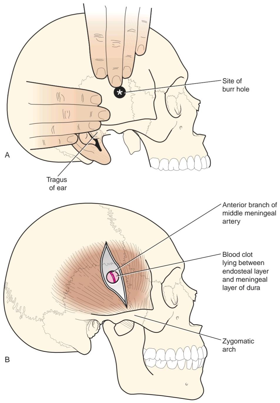 Figure A-2 A: Surface landmarks for a temporal burr hole. B: The vertical incision passes through the temporalis muscle down to bone. The middle meningeal artery lies between the endosteal and meningeal layers of dura and is embedded in the endosteal layer of dura or lies in a bony tunnel.
شکل A-۲ الف: نشانههای سطحی برای سوراخ مته گیجگاهی. ب: برش عمودی از طریق عضله گیجگاهی به سمت استخوان میرود. شریان مننژیال میانی بین لایههای اندوستئال و مننژیال سختشامه قرار دارد و در لایه اندوستئال سختشامه فرو رفته یا در یک تونل استخوانی قرار دارد.
Epidural Hematoma Burr Hole
Once the inner table of the squamous part of the temporal bone (or the anterior inferior angle of the parietal bone) is pierced with a small bit and enlarged with a burr, the dark red clotted blood beneath the endosteal layer of dura is usually easily recognized. However, bright red liquid blood means that the middle meningeal artery or one of its branches is bleeding. The meningeal artery is located deep to the clot and between the endosteal layer of dura and the meningeal layer of dura or in the substance of the endosteal layer of dura; or it may lie in a tunnel of bone.
سوراخ هماتوم اپیدورال با مته
هنگامی که سطح داخلی قسمت سنگفرشی استخوان گیجگاهی (یا زاویه قدامی تحتانی استخوان آهیانه) با یک مته کوچک سوراخ شده و با مته بزرگ میشود، خون لخته شده قرمز تیره در زیر لایه اندوستیال سختشامه معمولاً به راحتی قابل تشخیص است. با این حال، خون مایع قرمز روشن به این معنی است که شریان مننژیال میانی یا یکی از شاخههای آن در حال خونریزی است. شریان مننژیال در عمق لخته و بین لایه اندوستیال سختشامه و لایه مننژیال سختشامه یا در ماده لایه اندوستیال سختشامه قرار دارد؛ یا ممکن است در یک تونل استخوانی قرار داشته باشد.
Subdural Hematoma Burr Hole
When the squamous part of the temporal bone is penetrated, as described earlier, the endosteal layer of dura will be exposed. In this case, there is no blood clot between the endosteal layer of dura and the meningeal layer of dura, but both fused layers of dura will be dark bluish. The dura (endosteal and meningeal layers) is gently incised to enter the space between the meningeal layer of dura and the arachnoid mater. The subdural blood usually gushes out, leaving the unprotected brain covered only by arachnoid and pia mater in the depths of the hole.
هماتوم ساب دورال، سوراخ سوراخ
هنگامی که قسمت سنگفرشی استخوان گیجگاهی، همانطور که قبلاً توضیح داده شد، سوراخ شود، لایه اندوستیال سختشامه نمایان میشود. در این حالت، هیچ لخته خونی بین لایه اندوستیال سختشامه و لایه مننژیال سختشامه وجود ندارد، اما هر دو لایه به هم جوش خورده سختشامه به رنگ آبی تیره خواهند بود. سختشامه (لایههای اندوستیال و مننژیال) به آرامی برش داده میشود تا وارد فضای بین لایه مننژیال سختشامه و عنکبوتیه شود. خون زیر سختشامه معمولاً فوران میکند و مغز بدون محافظ را که فقط توسط عنکبوتیه و نرمشامه پوشیده شده است، در اعماق سوراخ باقی میگذارد.
Ventriculostomy
Ventriculostomy is indicated in acute hydrocephalus, in which cerebrospinal fluid flow is suddenly obstructed.
ونتریکولوستومی
ونتریکولوستومی در هیدروسفالی حاد، که در آن جریان مایع مغزی نخاعی به طور ناگهانی مسدود میشود، اندیکاسیون دارد.
Anatomy of the Technique
To perform a ventriculostomy, the needle is inserted into the lateral ventricle through either a frontal or parietal burr hole. The anatomy of these burr holes has been described previously. The needle is inserted through the burr hole using the following anatomical landmarks.
آناتومی تکنیک
برای انجام ونتریکولوستومی، سوزن از طریق سوراخ فرونتال یا آهیانهای وارد بطن جانبی میشود. آناتومی این سوراخهای فرونتال قبلاً شرح داده شده است. سوزن با استفاده از نشانههای آناتومیکی زیر از طریق سوراخ فرونتال وارد میشود.
1. Frontal Approach. The needle is inserted through the frontal burr hole and is directed downward and forward in the direction of the inner canthus of the ipsilateral eye (Fig. A-3).
۱. رویکرد پیشانی. سوزن از طریق سوراخ فرونتال وارد میشود و به سمت پایین و جلو در جهت کانتوس داخلی چشم همان طرف هدایت میشود (شکل A-3).
2. Parietal Approach. The needle is inserted through the parietal burr hole and is directed downward and for- ward in the direction of the pupil of the ipsilateral eye.
۲. رویکرد آهیانهای. سوزن از طریق سوراخ آهیانهای وارد میشود و به سمت پایین و جلو در جهت مردمک چشم همان طرف هدایت میشود.
The needle is inserted to a depth of about 2 in (5.5 cm) from the skull opening; in cases of chronic hydrocephalus with gross dilatation of the ventricles, the depth of penetration to the ventricular cavity may be much less.
سوزن تا عمق حدود ۵.۵ سانتیمتر (۲ اینچ) از دهانه جمجمه وارد میشود؛ در موارد هیدروسفالی مزمن با اتساع آشکار بطنها، عمق نفوذ به حفره بطنی ممکن است بسیار کمتر باشد.
 Figure A-3 Ventriculostomy. Needles passing through frontal or parietal burr holes to enter the lateral ventricle area are shown. The needle is inserted to a depth of about 2 in (5.5 cm) from the skull opening in order to enter the lateral ventricle.
شکل A-۳ ونتریکولوستومی. سوزنهایی که از طریق سوراخهای پیشانی یا آهیانهای وارد ناحیه بطن جانبی میشوند، نشان داده شده است. سوزن تا عمق حدود 5.5 سانتیمتر (2 اینچ) از دهانه جمجمه وارد میشود تا وارد بطن جانبی شود.
Vertebral Numbers and Spinal Cord Segments
Table A-1 relates which vertebral body is related to a particular spinal cord segment.
تعداد مهرهها و بخشهای نخاع
جدول A-1 نشان میدهد که کدام جسم مهره به یک بخش خاص نخاع مربوط میشود.
Table A-1 Correlation of Vertebral Body With Spinal Cord Segment
جدول A-۱ همبستگی جسم مهره با قطعه نخاع
 Segmental Innervation of Muscles

Test for the integrity of the segmental innervation of muscles by performing the following simple muscle reflexes.
عصبدهی سگمنتال عضلات
با انجام رفلکسهای عضلانی ساده زیر، یکپارچگی عصبدهی سگمنتال عضلات را بررسی کنید.
Biceps brachii tendon reflex C5-C6 (flexion of the elbow joint by tapping the biceps tendon).
رفلکس تاندون عضله دوسر بازویی C5-C6 (خم شدن مفصل آرنج با ضربه زدن به تاندون عضله دوسر).
Triceps tendon reflex C6-C7 and C8 (extension of the elbow joint by tapping the triceps tendon).
رفلکس تاندون عضله سه سر بازویی C6-C7 و C8 (باز شدن مفصل آرنج با ضربه زدن به تاندون عضله سه سر).
Brachioradialis tendon reflex C5-C6 and C7 (supination of the radioulnar joints by tapping the insertion of the brachioradialis tendon).
رفلکس تاندون عضله براکیورادیالیس C5-C6 و C7 (چرخش مفاصل رادیواولنار با ضربه زدن به محل اتصال تاندون عضله براکیورادیالیس).
Abdominal superficial reflexes (contraction of under- lying abdominal muscles by stroking the skin). Upper abdominal skin T6-T7; middle abdominal skin T8-T9; lower abdominal skin T10-T12.
رفلکسهای سطحی شکم (انقباض عضلات زیرین شکم با لمس پوست). پوست بالای شکم T6-T7؛ پوست وسط شکم T8-T9؛ پوست پایین شکم T10-T12.
Patellar tendon reflex (knee jerk) L2, L3, and L4 (extension of knee joint on tapping the patellar tendon).
رفلکس تاندون کشکک (حرکت ناگهانی زانو) L2، L3 و L4 (باز شدن مفصل زانو با ضربه زدن به تاندون کشکک).
Achilles tendon reflex (ankle jerk) S1 and S2 (plantar flexion of ankle joint on tapping the Achilles tendotendo calcaneus).
رفلکس تاندون آشیل (حرکت ناگهانی مچ پا) S1 و S2 (خم شدن کف پا با ضربه زدن به تاندون پاشنه آشیل).
Relationship Between Possible Intervertebral Disc Herniations and Spinal Nerve Roots
These are shown for the cervical and lumbar regions in Figure A-4.
رابطه بین فتق دیسک بین مهرهای احتمالی و ریشههای عصبی نخاعی
این موارد برای نواحی گردنی و کمری در شکل A-4 نشان داده شده است.
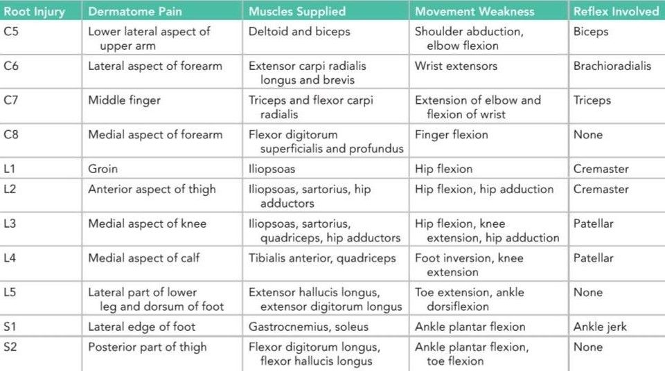
A correlation between the nerve roots involved, the pain dermatome, the muscle weakness, and the missing or diminished reflex is shown in Table A-2.
همبستگی بین ریشههای عصبی درگیر، درماتوم درد، ضعف عضلانی و فقدان یا کاهش رفلکس در جدول A-2 نشان داده شده است.
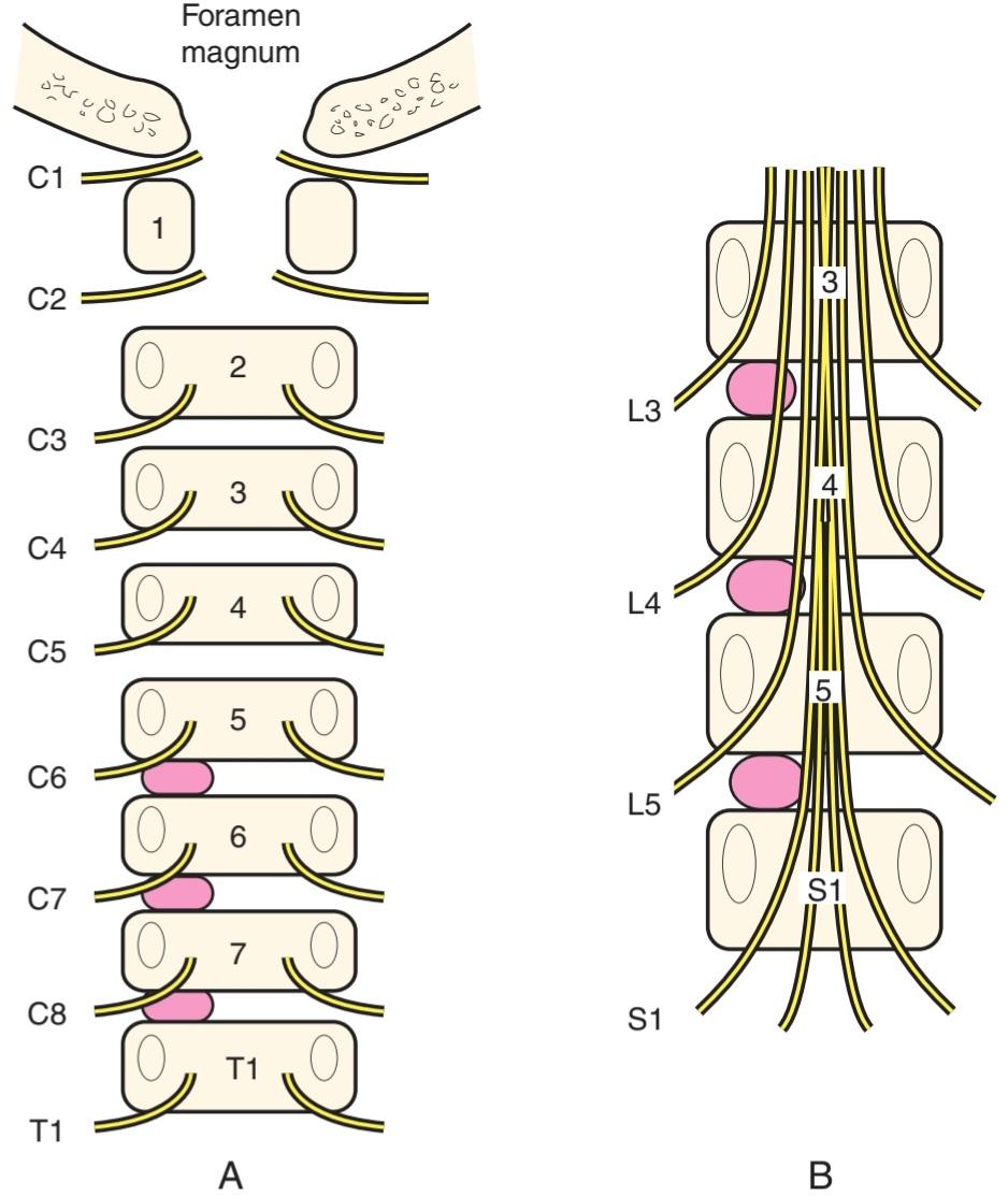
Figure A-4 A,B: Posterior views of vertebral bodies in the cervical and lumbar regions showing the relationship that might exist between herniated nucleus pulposus (pink) and spinal nerve roots. Note that there are eight cervical nerves and only seven cervical vertebrae. In the lumbar region, for example, the emerging L4 nerve roots pass out laterally close to the pedicles of the fourth lumbar vertebra and are not related to the intervertebral disc between the fourth and the fifth lumbar vertebrae. Pressure on the L5 motor nerve root produces weakness of plantar flexion of the ankle joint.
شکل A,B A-۴: نماهای خلفی از جسم مهرهها در نواحی گردنی و کمری که رابطهای را که ممکن است بین فتق هسته پالپوزوس (صورتی) و ریشههای عصبی نخاعی وجود داشته باشد، نشان میدهد. توجه داشته باشید که هشت عصب گردنی و تنها هفت مهره گردنی وجود دارد. به عنوان مثال، در ناحیه کمری، ریشههای عصبی L4 که در حال ظهور هستند، به صورت جانبی نزدیک به پایههای مهره چهارم کمری خارج میشوند و به دیسک بین مهرهای بین مهرههای چهارم و پنجم کمری مربوط نیستند. فشار بر ریشه عصب حرکتی L5 باعث ضعف خم شدن کف پا در مفصل مچ پا میشود.
Table A-2 Correlation Between Nerve Roots, Pain Dermatome, Muscle Weakness, and Missing or Diminished Reflex.
جدول A-۲ همبستگی بین ریشههای عصبی، درماتوم درد، ضعف عضلانی و فقدان یا کاهش رفلکس.
 Surface Landmarks for Performing a Lumbar Puncture
To perform a lumbar puncture, the patient is placed in the lateral prone position or in the upright sitting position. The trunk is then bent well forward to open up to the maximum, the space between adjoining laminae in the lumbar region. A groove runs down the middle of the back over the tips of the spines of the thoracic and the upper four lumbar vertebrae. The spines are made more prominent when the vertebral column is flexed. An imaginary line joining the highest points on the iliac crests passes over the fourth lumbar spine. With a careful aseptic technique and under local anesthesia, the spinal tap needle-fitted with a stylet-is passed into the vertebral canal above or below the fourth lumbar spine.
نشانههای سطحی برای انجام پونکسیون کمری
برای انجام پونکسیون کمری، بیمار در وضعیت خوابیده به پهلو یا در وضعیت نشسته عمودی قرار میگیرد. سپس تنه به خوبی به جلو خم میشود تا فضای بین لایههای مجاور در ناحیه کمر تا حداکثر باز شود. یک شیار در وسط پشت روی نوک خارهای مهرههای سینهای و چهار مهره بالایی کمری امتداد دارد. خارها هنگام خم شدن ستون مهرهها برجستهتر میشوند. یک خط فرضی که بالاترین نقاط روی ستیغهای ایلیاک را به هم متصل میکند، از روی چهارمین مهره کمری عبور میکند. با یک تکنیک آسپتیک دقیق و تحت بیحسی موضعی، سوزن نخاعی – که به یک استایلت مجهز است – به داخل کانال مهرهای بالا یا پایین چهارمین مهره کمری وارد میشود.
The following structures are pierced by the needle before it enters the subarachnoid space (Fig. A-5):
1. Skin
2. Superficial fascia
3. Supraspinous ligament
4. Interspinous ligament
5. Ligamentum flavum
6. Areolar tissue containing the internal vertebral venous plexus in the epidural space
7. Dura mater
8. Arachnoid mater
ساختارهای زیر قبل از ورود سوزن به فضای زیر عنکبوتیه توسط سوزن سوراخ میشوند (شکل A-5):
1. پوست
2. فاسیای سطحی
3. رباط فوق خاری
4. رباط بین خاری
5. رباط زرد
6. بافت آرئولار حاوی شبکه وریدی مهرهای داخلی در فضای اپیدورال
7. سختشامه
8. ماده عنکبوتیه
The depth to which the needle will have to pass will vary from an inch or less in children to as much as 4 in (10 cm) in obese adults.
عمقی که سوزن باید از آن عبور کند از یک اینچ یا کمتر در کودکان تا 4 اینچ (10 سانتیمتر) در بزرگسالان چاق متغیر است.
The pressure of the cerebrospinal fluid in the lateral recumbent position is normally about 60 to 150 mm of water.
فشار مایع مغزی نخاعی در حالت خوابیده به پهلو معمولاً حدود 60 تا 150 میلیمتر آب است.
See Table 16-1 for physical characteristics and composition of the cerebrospinal fluid.
برای مشخصات فیزیکی و ترکیب مایع مغزی نخاعی به جدول ۱۶-۱ مراجعه کنید.

Figure A-5 A: Structures penetrated by the spinal tap needle before it reaches the dura mater. B: Important anatomic landmarks when performing a spinal tap. Although this is usually performed with the patient in a lateral recumbent position with the vertebral column well flexed, the patient may be placed in the sitting position and bent well forward.
شکل A-۵ الف: ساختارهایی که سوزن پونکسیون نخاعی قبل از رسیدن به سختشامه به آنها نفوذ میکند. ب: نشانههای آناتومیک مهم هنگام انجام پونکسیون نخاعی. اگرچه این کار معمولاً در حالی انجام میشود که بیمار در وضعیت خوابیده به پهلو و ستون فقرات به خوبی خم شده باشد، میتوان بیمار را در وضعیت نشسته قرار داد و به خوبی به جلو خم کرد.
«فصل پیوست کتاب نورواناتومی بالینی اسنل»
🚀 با ما همراه شوید!
تازهترین مطالب و آموزشهای مغز و اعصاب را از دست ندهید. با فالو کردن کانال تلگرام آیندهنگاران مغز، از ما حمایت کنید!