نشانگرهای سلولی، پیوند سلول و جایگزینی ژن


دعای مطالعه [ نمایش ]
بِسْمِ الله الرَّحْمنِ الرَّحیمِ
اَللّهُمَّ اَخْرِجْنى مِنْ ظُلُماتِ الْوَهْمِ
خدايا مرا بيرون آور از تاريكىهاى وهم،
وَ اَكْرِمْنى بِنُورِ الْفَهْمِ
و به نور فهم گرامى ام بدار،
اَللّهُمَّ افْتَحْ عَلَيْنا اَبْوابَ رَحْمَتِكَ
خدايا درهاى رحمتت را به روى ما بگشا،
وَانْشُرْ عَلَيْنا خَزائِنَ عُلُومِكَ بِرَحْمَتِكَ يا اَرْحَمَ الرّاحِمينَ
و خزانههاى علومت را بر ما باز كن به امید رحمتت اى مهربانترين مهربانان.
آیندهنگاران مغز: «ما مغز را میشناسیم، تا آینده را بسازیم.»
کتاب «حقایق مغز» (Brain Facts) که به همت انجمن علوم اعصاب (Society for Neuroscience) تدوین و منتشر شده است، یکی از معتبرترین منابع عمومی آموزش علوم اعصاب در جهان به شمار میرود. این اثر با زبانی روان و مبتنی بر تازهترین دستاوردهای پژوهشی، مفاهیم بنیادین علوم اعصاب را بهگونهای دقیق و قابلفهم برای طیف گستردهای از خوانندگان ارائه میکند.
تلاشهای علمی و سالها فعالیت آموزشی اعضای انجمن علوم اعصاب و صدها پژوهشگر برجسته این حوزه، الهامبخش تدوین این اثر ارزشمند و گامی مؤثر در گسترش دانش مغز و علوم اعصاب در سراسر جهان بوده است.
ترجمه دقیق این کتاب توسط برند علمی آیندهنگاران مغز به مدیریت داریوش طاهری، امکان دسترسی فارسیزبانان به مرزهای نوین علوم اعصاب را فراهم کرده و رسالتی علمی در جهت شناخت عمیقتر مغز و ترسیم آیندهای روشنتر بر عهده گرفته است.
»» فصل ۱۷: حل مشکلات انسان؛ قسمت چهارم
»» CHAPTER 17: Solving Human Problems

BRAIN-MACHINE INTERFACE
If you know someone with severe neurological damage perhaps cerebral palsy, trauma from a car or motorcycle accident, a military or sports injury, or a stroke you’ve seen, firsthand, how communicating with the outside world and performing daily tasks can be a challenge. But during the past few decades, scientists have made impressive progress in developing technologies that can bypass such damage. Now brain-machine interfaces can read the activity of millions of neurons – through electroencephalographic (EEG) activity from the brain’s surface or from implanted electrodes — and predict the behavioral intentions of research participants. These advances give human and animal subjects neural control of parts of their surroundings: from computer cursors to video games to robotic limbs.
رابط مغز و ماشین
اگر کسی را میشناسید که آسیب عصبی شدید دارد، مثلاً فلج مغزی، ترومای ناشی از تصادف ماشین یا موتورسیکلت، آسیب نظامی یا ورزشی یا سکته مغزی، از نزدیک دیدهاید که چگونه برقراری ارتباط با دنیای خارج و انجام کارهای روزانه میتواند یک چالش باشد. اما در طول چند دهه گذشته، دانشمندان پیشرفت چشمگیری در توسعه فناوریهایی داشتهاند که میتوانند از چنین آسیبهایی جلوگیری کنند. اکنون رابطهای مغز و ماشین میتوانند فعالیت میلیونها نورون را – از طریق فعالیت الکتروانسفالوگرافی (EEG) از سطح مغز یا از الکترودهای کاشته شده – بخوانند و نیات رفتاری شرکتکنندگان در تحقیق را پیشبینی کنند. این پیشرفتها به افراد انسانی و حیوانی کنترل عصبی بخشهایی از محیط اطرافشان را میدهد: از مکاننماهای رایانهای گرفته تا بازیهای ویدیویی و اندامهای رباتیک.
Despite the scifi sizzle, most of the work on electronic brain implants is derived from basic research on how animals and people plan and control various types of movement. Using hair-thin wires inserted into the brains of monkeys and rats, scientists first recorded the firing patterns of cells located in the premotor, primary motor, and posterior parietal cortical areas of the brain. As the animals performed repetitive tasks like pressing a lever to receive a reward, researchers found specific firing patterns associated with the motion. Eventually, these patterns were translated into computer algorithms that allowed animals to complete a task via a robotic arm or prosthetic device simply by thinking about it.
با وجود هیاهوی علمی تخیلی، بیشتر کار روی ایمپلنتهای الکترونیکی مغز از تحقیقات پایه در مورد چگونگی برنامهریزی و کنترل انواع مختلف حرکات توسط حیوانات و انسانها گرفته شده است. دانشمندان ابتدا با استفاده از سیمهای نازک مو که در مغز میمونها و موشها قرار داده شده بودند، الگوهای شلیک سلولهای واقع در نواحی پیشحرکتی، حرکتی اولیه و قشر جداری خلفی مغز را ثبت کردند. همانطور که حیوانات کارهای تکراری مانند فشار دادن یک اهرم برای دریافت پاداش انجام میدادند، محققان الگوهای شلیک خاصی مرتبط با حرکت را یافتند. در نهایت، این الگوها به الگوریتمهای کامپیوتری ترجمه شدند که به حیوانات اجازه میدادند تا یک کار را از طریق یک بازوی رباتیک یا دستگاه پروتز، صرفاً با فکر کردن در مورد آن، انجام دهند.
The clinical applications of brain-machine interfaces quickly became clear. Neuroscientists and surgeons implanted electrode arrays in the brains of patients with epilepsy, paralysis, stroke, or Lou Gehrig’s disease (ALS), in hopes of enabling them to communicate and, someday, move independently. In early experiments, patients were only able to gain rudimentary control of a computer cursor. But a breakthrough occurred in 2011 when, after months of extensive training, quadriplegic patients learned to control movements of a third (robotic) arm-enabling them to grasp a drink of water or reach out to a loved one.
کاربردهای بالینی رابطهای مغز و ماشین به سرعت آشکار شد. دانشمندان علوم اعصاب و جراحان، آرایههای الکترودی را در مغز بیماران مبتلا به صرع، فلج، سکته مغزی یا بیماری لو گریگ (ALS) کاشتند، به این امید که آنها را قادر به برقراری ارتباط و روزی حرکت مستقل کنند. در آزمایشهای اولیه، بیماران فقط میتوانستند کنترل اولیه مکاننمای کامپیوتر را به دست آورند. اما در سال ۲۰۱۱، پس از ماهها آموزش گسترده، پیشرفت چشمگیری رخ داد؛ زمانی که بیماران فلج چهار اندام یاد گرفتند حرکات بازوی سوم (رباتیک) خود را کنترل کنند و این امر آنها را قادر ساخت تا یک لیوان آب را بگیرند یا به عزیزان خود کمک کنند.
Honing this technology is allowing patients to control their own paralyzed limbs. Electrode chips implanted in their brains are connected to sleeves or gloves worn over the injured limbs. Sending tiny blasts of electricity into the patient’s nerves, located under the sleeve or glove, can reanimate paralyzed muscles. But brain-machine interfaces won’t become part of clinical medicine until they’re simplified, miniaturized, and made more reliable. Devices that wire-lessly transmit commands from brain implants are a step in that direction.
پیشرفت این فناوری به بیماران اجازه میدهد تا اندامهای فلج خود را کنترل کنند. تراشههای الکترودی کاشته شده در مغز آنها به آستینها یا دستکشهایی که روی اندامهای آسیبدیده پوشیده شدهاند، متصل میشوند. ارسال جریانهای کوچک الکتریسیته به اعصاب بیمار، که در زیر آستین یا دستکش قرار دارند، میتواند عضلات فلج شده را دوباره زنده کند. اما رابطهای مغز و ماشین تا زمانی که ساده، کوچک و قابل اعتمادتر نشوند، بخشی از پزشکی بالینی نخواهند شد. دستگاههایی که به صورت بیسیم دستورات را از ایمپلنتهای مغزی منتقل میکنند، گامی در این جهت هستند.
A parallel line of research has explored applying this technology in the broader field of neuroprostheses. Neuro- prosthetic devices not only receive out- put commands from a patient’s nervous system, but can also provide input — as occurs in retinal implants and prosthetic limbs. Prosthetic arms, for example, have remained frustratingly low-tech, but some brain-guided prostheses have integrated nerves and muscles at several different levels, allowing users to perform more precise and natural movements, and even enabling some to “feel” again. Still, even the most sophisticated neuroprostheses (such as brain implants) are limited by their number of electrodes and the lifespan of the implanted electrodes. Current arrays can only connect to 100 or so neurons, so a more complex and useful bionic future is still far away. Yet scientists and entrepreneurs are already thinking of new uses for the technology: restoring memory; enhancing cognition; and treating diseases such as depression, Alzheimer’s, and epilepsy.
یک خط تحقیقاتی موازی، کاربرد این فناوری را در حوزه وسیعتر پروتزهای عصبی بررسی کرده است. دستگاههای پروتز عصبی نه تنها دستورات خروجی را از سیستم عصبی بیمار دریافت میکنند، بلکه میتوانند ورودی نیز ارائه دهند – همانطور که در ایمپلنتهای شبکیه و اندامهای مصنوعی اتفاق میافتد. به عنوان مثال، بازوهای مصنوعی به طرز ناامیدکنندهای از فناوری پایینی برخوردار بودهاند، اما برخی از پروتزهای هدایتشده توسط مغز، اعصاب و عضلات را در سطوح مختلف ادغام کردهاند و به کاربران امکان میدهند حرکات دقیقتر و طبیعیتری انجام دهند و حتی برخی را قادر میسازند دوباره “احساس” کنند. با این حال، حتی پیچیدهترین پروتزهای عصبی (مانند ایمپلنتهای مغزی) به دلیل تعداد الکترودها و طول عمر الکترودهای کاشته شده محدود هستند. آرایههای فعلی فقط میتوانند به حدود ۱۰۰ نورون متصل شوند، بنابراین آیندهای پیچیدهتر و مفیدتر در حوزه بیونیک هنوز دور از دسترس است. با این حال، دانشمندان و کارآفرینان در حال حاضر به کاربردهای جدیدی برای این فناوری فکر میکنند: بازیابی حافظه؛ افزایش شناخت؛ و درمان بیماریهایی مانند افسردگی، آلزایمر و صرع.
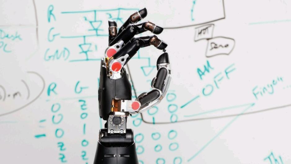
Advances in brain-machine interfaces are leading to incredible treatments, like this prosthetic arm that is controlled through thought.
پیشرفتها در رابطهای مغز و ماشین منجر به درمانهای باورنکردنی میشود، مانند این بازوی مصنوعی که از طریق فکر کنترل میشود.
DEEP BRAIN STIMULATION
Insights into the pathophysiology of movement disorders have rekindled interest in the use of focused electrical stimulation as a form of treatment. The most advanced and precise method — deep brain stimulation (DBS) — was inspired by pacemakers engineered for the heart. Instead of electrodes implanted in the heart, the electrodes of the DBS device are surgically embedded in specific brain regions. Depending on where the electrodes are placed, DBS devices can help alleviate the symptoms of some brain disorders.
تحریک عمیق مغز
بینشها در مورد پاتوفیزیولوژی اختلالات حرکتی، علاقه به استفاده از تحریک الکتریکی متمرکز به عنوان نوعی درمان را دوباره برانگیخته است. پیشرفتهترین و دقیقترین روش – تحریک عمیق مغز (DBS) – از ضربانسازهای قلبی الهام گرفته شده است. به جای الکترودهای کاشته شده در قلب، الکترودهای دستگاه DBS با جراحی در مناطق خاصی از مغز جاسازی میشوند. بسته به محل قرارگیری الکترودها، دستگاههای DBS میتوانند به کاهش علائم برخی از اختلالات مغزی کمک کنند.
During most of these implantation surgeries, patients remain awake so that a neurologist can talk to them and ensure that the electrodes are stimulating the correct locations. While the patient’s head is held in place with a stereotactic frame, the surgeon drills a dime-sized hole (or smaller) in the skull. Then, a thin insulated wire with electrodes at the tip or along the shaft is inserted deep into the brain; if both sides of the brain are to receive implants, a wire is inserted into each side. In a separate surgery, a battery-operated pulse generator is implanted in the upper chest and connected to the electrodes. When the device is turned on, it starts sending electrical currents that alter the activity of the targeted brain cells.
در طول بیشتر این جراحیهای کاشت، بیماران بیدار میمانند تا متخصص مغز و اعصاب بتواند با آنها صحبت کند و مطمئن شود که الکترودها مکانهای صحیح را تحریک میکنند. در حالی که سر بیمار با یک قاب استریوتاکتیک در جای خود نگه داشته میشود، جراح سوراخی به اندازه یک سکه (یا کوچکتر) در جمجمه ایجاد میکند. سپس، یک سیم عایق نازک با الکترودهایی در نوک یا در امتداد محور، در اعماق مغز قرار داده میشود. اگر قرار باشد هر دو طرف مغز ایمپلنت دریافت کنند، یک سیم در هر طرف قرار داده میشود. در یک جراحی جداگانه، یک مولد پالس باتریدار در قسمت بالای سینه کاشته شده و به الکترودها متصل میشود. وقتی دستگاه روشن میشود، شروع به ارسال جریانهای الکتریکی میکند که فعالیت سلولهای مغزی هدف را تغییر میدهد.
The implanted device relies on the fact that neuronal communication uses electrical signals. In many movement disorders, an abnormal signal or pulse can gain control of a circuit and can easily become magnified. Like someone shouting in a crowded room, this aberrant signal can drown out other activity. DBS interrupts the shouting, so that normal communication can continue.
دستگاه کاشته شده بر این واقعیت متکی است که ارتباط عصبی از سیگنالهای الکتریکی استفاده میکند. در بسیاری از اختلالات حرکتی، یک سیگنال یا پالس غیرطبیعی میتواند کنترل یک مدار را به دست گیرد و به راحتی بزرگنمایی شود. مانند کسی که در یک اتاق شلوغ فریاد میزند، این سیگنال نابجا میتواند فعالیتهای دیگر را خفه کند. DBS فریاد را قطع میکند، به طوری که ارتباط عادی میتواند ادامه یابد.
To determine where brain activity needs to be silenced or induced, neurosurgeons must identify the locations of the problems. The brain areas first targeted for tremors and Parkinson’s disease were chosen after years of pains- taking neuroimaging, neuroanatomy, and fundamental research, especially in nonhuman primate models. Since then, deep brain stimulation has been used to treat epilepsy, dystonia, Tourette’s syndrome and, more recently, obsessive-compulsive disorder. Now researchers are investigating whether the DBS technique can potentially be extended to mood disorders such as treatment-resistant depression, as well as other complex mental disorders.
برای تعیین اینکه فعالیت مغز در کجا باید خاموش یا القا شود، جراحان مغز و اعصاب باید محل مشکلات را شناسایی کنند. نواحی مغزی که ابتدا برای لرزش و بیماری پارکینسون هدف قرار گرفتند، پس از سالها تصویربرداری عصبی، کالبدشناسی عصبی و تحقیقات بنیادی طاقتفرسا، به ویژه در مدلهای غیرانسانی نخستیها، انتخاب شدند. از آن زمان، تحریک عمیق مغز برای درمان صرع، دیستونی، سندرم تورت و اخیراً اختلال وسواس فکری-عملی استفاده شده است. اکنون محققان در حال بررسی این موضوع هستند که آیا تکنیک DBS میتواند به طور بالقوه به اختلالات خلقی مانند افسردگی مقاوم به درمان و همچنین سایر اختلالات روانی پیچیده گسترش یابد یا خیر.
Yet DBS, like any surgical procedure, is not without some risks. It is highly invasive, and potential complications include infection, stroke, and bleeding in the brain. It also requires regular neurological follow-up and battery changes every 3 to 4 years.
با این حال، DBS، مانند هر عمل جراحی، بدون خطر نیست. این روش بسیار تهاجمی است و عوارض احتمالی آن شامل عفونت، سکته مغزی و خونریزی در مغز است. همچنین نیاز به پیگیری منظم عصبی و تعویض باتری هر 3 تا 4 سال دارد.

Deep brain stimulation uses electrodes implanted deep in the brain, which carry electric impulses to specific brain regions. The power packs that provide the electricity are implanted in the patient’s back, as seen in this X-ray.
تحریک عمیق مغز از الکترودهایی استفاده میکند که در اعماق مغز کاشته میشوند و تکانههای الکتریکی را به مناطق خاصی از مغز منتقل میکنند. همانطور که در این عکس رادیولوژی دیده میشود، بستههای قدرتی که برق را تأمین میکنند، در پشت بیمار کاشته میشوند.
PSYCHOACTIVE THERAPIES
Transcranial Stimulation
A few noninvasive treatments can stimulate cells near the surface of the brain: Transcranial magnetic stimulation (TMS), transcranial direct current stimulation (tDCS), and transcranial alternating current stimulation (tACS) all use magnetic fields or low electrical currents to alter neural activity in a specific region of the human cortex and, indirectly, deeper brain structures to which it connects.
درمانهای روانفعال
تحریک فراجمجمهای
چند درمان غیرتهاجمی میتوانند سلولهای نزدیک سطح مغز را تحریک کنند: تحریک مغناطیسی فراجمجمهای (TMS)، تحریک جریان مستقیم فراجمجمهای (tDCS) و تحریک جریان متناوب فراجمجمهای (tACS) همگی از میدانهای مغناطیسی یا جریانهای الکتریکی ضعیف برای تغییر فعالیت عصبی در ناحیه خاصی از قشر مغز انسان و به طور غیرمستقیم، ساختارهای عمیقتر مغز که به آن متصل هستند، استفاده میکنند.
During TMS therapy, patients sit in a chair while a nurse or technician places a magnetic stimulator against their head. The device painlessly delivers brief magnetic pulses to the brain, similar in strength to those generated by magnetic resonance imaging (MRI) devices, but highly targeted. For patients with depression, pulses are focused over their left prefrontal cortex. Here, they generate electrical currents among neurons which, over time, help lift the patient’s mood.
در طول درمان TMS، بیماران روی صندلی مینشینند در حالی که یک پرستار یا تکنسین یک محرک مغناطیسی را روی سر آنها قرار میدهد. این دستگاه بدون درد، پالسهای مغناطیسی کوتاهی را به مغز ارسال میکند که از نظر قدرت مشابه پالسهای تولید شده توسط دستگاههای تصویربرداری رزونانس مغناطیسی (MRI) هستند، اما بسیار هدفمند. برای بیماران مبتلا به افسردگی، پالسها روی قشر جلوی مغز چپ آنها متمرکز میشوند. در اینجا، آنها جریانهای الکتریکی را در بین نورونها ایجاد میکنند که به مرور زمان به بهبود خلق و خوی بیمار کمک میکنند.
Similarly, tDCS uses one or two milliamperes of direct current to tune the brain. Although research into tDCS and its close cousin, tACS, is in its early stages, these techniques offer clear advantages over deep brain stimulation and even over TMS. Generally, patients report only a slight tingling or tapping feeling on their head as the therapy is administered. The devices used to administer these therapies are also cheaper, more portable, and lower -tech compared to TMS and DBS.
به طور مشابه، tDCS از یک یا دو میلیآمپر جریان مستقیم برای تنظیم مغز استفاده میکند. اگرچه تحقیقات در مورد tDCS و پسرعموی نزدیک آن، tACS، در مراحل اولیه خود است، اما این تکنیکها مزایای آشکاری نسبت به تحریک عمیق مغز و حتی نسبت به TMS دارند. به طور کلی، بیماران هنگام انجام درمان فقط احساس سوزن سوزن شدن یا ضربه زدن جزئی روی سر خود گزارش میدهند. دستگاههایی که برای انجام این درمانها استفاده میشوند نیز در مقایسه با TMS و DBS ارزانتر، قابل حملتر و با فناوری پایینتر هستند.
A number of studies have suggested that tDCS and tACS might be used to improve working memory as well as to relieve chronic pain and the symptoms of depression, fibromyalgia, schizophrenia, and other disorders. However, despite the substantial literature, meta-analyses have failed to conclusively prove any effects of transcranial electrical stimulation. Currently, there is no consensus among scientists on how these treatments might work, or even on the best way to position the stimulation devices.
تعدادی از مطالعات نشان دادهاند که tDCS و tACS ممکن است برای بهبود حافظه کاری و همچنین تسکین درد مزمن و علائم افسردگی، فیبرومیالژیا، اسکیزوفرنی و سایر اختلالات استفاده شوند. با این حال، علیرغم وجود مقالات فراوان، متاآنالیزها نتوانستهاند به طور قطعی هیچ گونه اثری از تحریک الکتریکی ترانس کرانیال را اثبات کنند. در حال حاضر، هیچ اجماعی بین دانشمندان در مورد نحوه عملکرد این درمانها یا حتی بهترین روش برای قرار دادن دستگاههای تحریک وجود ندارد.
New Types of Drugs
Most doctors still recommend medication as the first line of treatment for neurological and psychiatric disorders. The antidepressants, antipsychotics, and other mind-altering medications used today have been tested in extensive clinical trials and remain largely unchanged from their prototypes developed in the 1950s. Each of these classes of drugs is now filled with subsequent generations of closely related drugs designed to interact more selectively with their targets, producing better therapeutic effects and fewer side effects, in some cases. Nevertheless, these are just slightly improved copies of one another. Truly novel drug candidates are rare, and hard to develop.
انواع جدید داروها
اکثر پزشکان هنوز دارو را به عنوان خط اول درمان اختلالات عصبی و روانی توصیه میکنند. داروهای ضد افسردگی، ضد روان پریشی و سایر داروهای تغییر دهنده ذهن که امروزه مورد استفاده قرار میگیرند، در آزمایشات بالینی گسترده آزمایش شدهاند و تا حد زیادی نسبت به نمونههای اولیه خود که در دهه 1950 توسعه یافتهاند، بدون تغییر باقی ماندهاند. هر یک از این دستههای دارویی اکنون مملو از نسلهای بعدی داروهای نزدیک به هم هستند که برای تعامل انتخابیتر با اهداف خود طراحی شدهاند و در برخی موارد اثرات درمانی بهتر و عوارض جانبی کمتری ایجاد میکنند. با این وجود، اینها فقط کپیهای کمی بهبود یافته از یکدیگر هستند. نامزدهای دارویی واقعاً جدید نادر هستند و توسعه آنها دشوار است.

MRI scans can provide detailed images of brain tissue. Here, it displays tissue high in water and fat content in white.
اسکنهای MRI میتوانند تصاویر دقیقی از بافت مغز ارائه دهند. در اینجا، بافتی با محتوای آب و چربی بالا به رنگ سفید نمایش داده شده است.
One of the biggest challenges in developing new drugs to treat neurological or psychiatric problems is finding molecules that can cross the protective blood-brain barrier-tightly packed endothelial cells lining blood vessels restrict the kinds of molecules that can enter the brain. While this barrier’s fortress-like quality is good for normal function, it prevents most drugs delivered by typical means— including pills, patches, injections, or enemas – from having any useful therapeutic effects within the brain. Scientists have had to design extremely tiny molecules or adopt ingenious strategies such as nanoparticles that shuttle in drugs, enzymes that activate molecules after they’ve “snuck through” the barrier, and antibodies that were specifically engineered for the brain.
یکی از بزرگترین چالشها در توسعه داروهای جدید برای درمان مشکلات عصبی یا روانی، یافتن مولکولهایی است که بتوانند از سد خونی-مغزی محافظ عبور کنند. سلولهای اندوتلیال محکم بستهبندی شده که رگهای خونی را میپوشانند، انواع مولکولهایی را که میتوانند وارد مغز شوند، محدود میکنند. در حالی که کیفیت قلعهمانند این سد برای عملکرد طبیعی خوب است، اما مانع از آن میشود که اکثر داروهایی که به روشهای معمول – از جمله قرصها، چسبها، تزریقها یا تنقیه – تحویل داده میشوند، اثرات درمانی مفیدی در مغز داشته باشند. دانشمندان مجبور بودهاند مولکولهای بسیار کوچکی را طراحی کنند یا استراتژیهای هوشمندانهای مانند نانوذراتی که داروها را در خود جای میدهند، آنزیمهایی که مولکولها را پس از “عبور” از سد فعال میکنند، و آنتیبادیهایی که به طور خاص برای مغز مهندسی شدهاند، اتخاذ کنند.
Preliminary trials of drugs that use the body’s own immune system to confront and clear unwanted proteins from the brain have sparked a great deal of interest, particularly in the Alzheimer’s disease community. When mice and monkeys receive a vaccine that contains a major component of amyloid plaques, their immune systems develop antibodies capable of traveling to the brain and “tagging” the amyloid-beta plaques that are the hall- mark of Alzheimer’s disease. This tagging seems to alert microglial cells in the brain, which head for the plaques and try to remove them. In some experiments, mice bred to develop an Alzheimer’s-like disease remembered how to navigate through a Morris water maze, after being vaccinated- indicating that such vaccines might also relieve symptoms of the disease.
آزمایشهای اولیه داروهایی که از سیستم ایمنی بدن خود برای مقابله و پاکسازی پروتئینهای ناخواسته از مغز استفاده میکنند، توجه زیادی را به ویژه در جامعه بیماری آلزایمر برانگیخته است. وقتی موشها و میمونها واکسنی دریافت میکنند که حاوی جزء اصلی پلاکهای آمیلوئید است، سیستم ایمنی آنها آنتیبادیهایی تولید میکند که قادر به سفر به مغز و “برچسبگذاری” پلاکهای آمیلوئید بتا هستند که مشخصه بیماری آلزایمر هستند. به نظر میرسد این برچسبگذاری، سلولهای میکروگلیال در مغز را هوشیار میکند که به سمت پلاکها میروند و سعی میکنند آنها را از بین ببرند. در برخی آزمایشها، موشهایی که برای ابتلا به بیماری شبیه آلزایمر پرورش داده شده بودند، پس از واکسیناسیون، نحوه حرکت در هزارتوی آبی موریس را به خاطر آوردند – که نشان میدهد چنین واکسنهایی ممکن است علائم بیماری را نیز تسکین دهند.
In the past, however, many Alzheimer’s vaccines have failed in later-stage clinical trials. One reason for these failures was the development of harmful side effects. Several human participants experienced severe inflammation when their brains reacted to the antibodies against its proteins. Since then, newer approaches have engineered antibodies or antibody fragments that bind to their specific targets without triggering an autoimmune response. Other re- searchers have engineered double-duty antibodies. These use one end to sneak into the brain by binding to a receptor on the blood-brain barrier. Once inside, the antibody’s other end can cut off production of harmful amyloid- beta proteins even before plaques form.
با این حال، در گذشته، بسیاری از واکسنهای آلزایمر در آزمایشهای بالینی مراحل بعدی شکست خوردهاند. یکی از دلایل این شکستها، ایجاد عوارض جانبی مضر بود. چندین شرکتکننده انسانی هنگامی که مغزشان به آنتیبادیهای علیه پروتئینهای آن واکنش نشان داد، التهاب شدیدی را تجربه کردند. از آن زمان، رویکردهای جدیدتر، آنتیبادیها یا قطعات آنتیبادی را مهندسی کردهاند که بدون ایجاد پاسخ خودایمنی به اهداف خاص خود متصل میشوند. سایر محققان، آنتیبادیهای دو منظوره را مهندسی کردهاند. این آنتیبادیها از یک سر برای ورود مخفیانه به مغز با اتصال به گیرندهای روی سد خونی-مغزی استفاده میکنند. پس از ورود، سر دیگر آنتیبادی میتواند تولید پروتئینهای مضر آمیلوئید بتا را حتی قبل از تشکیل پلاکها قطع کند.
By contrast, some therapies aim to boost helpful peptides and proteins called trophic factors, which are native to the brain. Neurotrophic factors support the growth and survival of specific groups of neurons. Scientists hope to modify these factors to reduce the amount of cell death in various neurodegenerative diseases.
در مقابل، برخی از درمانها با هدف تقویت پپتیدها و پروتئینهای مفید به نام فاکتورهای تروفیک که بومی مغز هستند، انجام میشوند. فاکتورهای نوروتروفیک از رشد و بقای گروههای خاصی از نورونها پشتیبانی میکنند. دانشمندان امیدوارند این عوامل را اصلاح کنند تا میزان مرگ سلولی در بیماریهای مختلف عصبی کاهش یابد.
The possible value of at least one trophic factor — nerve growth factor (NGF) has already been demonstrated in several preclinical and early stage clinical trials. NGF slows the destruction of cholinergic neurons that plays a role in the cognitive decline of Alzheimer’s disease. Injecting NGF into patients’ brains stimulated the regeneration of these neurons and induced sprouting of new nerve fibers around the injection site. In some cases, evidence of this sprouting lasted up to 10 years after the initial therapy.
ارزش احتمالی حداقل یک فاکتور تروفیک – فاکتور رشد عصبی (NGF) – در چندین آزمایش بالینی پیشبالینی و مراحل اولیه نشان داده شده است. NGF تخریب نورونهای کولینرژیک را که در زوال شناختی بیماری آلزایمر نقش دارند، کند میکند. تزریق NGF به مغز بیماران، بازسازی این نورونها را تحریک کرده و باعث جوانه زدن فیبرهای عصبی جدید در اطراف محل تزریق میشود. در برخی موارد، شواهد این جوانه زدن تا 10 سال پس از درمان اولیه ادامه داشت.
Brain-derived neurotrophic factor (BDNF) is showing potential for treating Alzheimer’s disease, as well as Huntington’s, Parkinson’s, ALS, and Rett syndrome. Moreover, the effects of boosting BDNF could even be stronger than those of NGF. But, in an interesting twist, the inhibition of some neurotrophic factors such as the neurite outgrowth inhibitor might also benefit patients. Studies have found that neurite outgrowth inhibitor is upregulated in the early stages of motor disease, and having too much of it around could prevent nerve regeneration. Scientists are now conducting clinical trials in which patients with ALS and spinal cord injuries receive custom-made antibodies to disable the neurite outgrowth inhibitor protein.
فاکتور نوروتروفیک مشتق از مغز (BDNF) پتانسیل درمان بیماری آلزایمر و همچنین هانتینگتون، پارکینسون، ALS و سندرم رت را نشان میدهد. علاوه بر این، اثرات تقویت BDNF حتی میتواند قویتر از NGF باشد. اما، در یک چرخش جالب، مهار برخی از عوامل نوروتروفیک مانند مهارکننده رشد نوریت نیز ممکن است برای بیماران مفید باشد. مطالعات نشان دادهاند که مهارکننده رشد نوریت در مراحل اولیه بیماری حرکتی افزایش مییابد و وجود بیش از حد آن میتواند از بازسازی عصب جلوگیری کند. دانشمندان اکنون در حال انجام آزمایشهای بالینی هستند که در آن بیماران مبتلا به ALS و آسیبهای نخاعی آنتیبادیهای سفارشی برای غیرفعال کردن پروتئین مهارکننده رشد نوریت دریافت میکنند.
Ultimately, the ever-increasing global demand for therapies for neurological and mental diseases is a strong motivator for scientists and doctors in this field.
در نهایت، تقاضای جهانی روزافزون برای درمان بیماریهای عصبی و روانی، انگیزهای قوی برای دانشمندان و پزشکان در این زمینه است.
PREDICTIVE NEUROIMAGING AND PERSONALIZED MEDICINE
As we gain understanding of the anatomical and functional changes underlying neurological illnesses, it becomes increasingly clear that these changes provide clues for earlier detection – even before symptoms appear. Many disorders, such as Alzheimer’s disease, are accompanied by specific brain activity and structural changes that can be tracked over time using MRI. By comparing this information with a baseline model of a healthy brain, researchers hope to predict which patients might one day develop neurological problems.
تصویربرداری عصبی پیشبینیکننده و پزشکی شخصیسازیشده
با افزایش درک ما از تغییرات آناتومیکی و عملکردی زمینهساز بیماریهای عصبی، بهطور فزایندهای آشکار میشود که این تغییرات سرنخهایی برای تشخیص زودهنگام – حتی قبل از بروز علائم – ارائه میدهند. بسیاری از اختلالات، مانند بیماری آلزایمر، با فعالیت خاص مغز و تغییرات ساختاری همراه هستند که میتوان آنها را در طول زمان با استفاده از MRI ردیابی کرد. محققان امیدوارند با مقایسه این اطلاعات با یک مدل پایه از یک مغز سالم، پیشبینی کنند که کدام بیماران ممکن است روزی دچار مشکلات عصبی شوند.
Although it is still too early for these “markers” to be used as clinical reference points, they could pave the way for objective diagnoses of brain disorders, much as electrocardiograms and laboratory tests are currently used to reveal heart problems. The first step in this process is to produce a generic brain template by averaging the images from hundreds of randomly selected MRI scans. Scientists can then use machine-learning software to characterize the sets of healthy brain scans and the sets of scans known to show disease-associated changes.
اگرچه هنوز برای استفاده از این «نشانگرها» به عنوان نقاط مرجع بالینی خیلی زود است، اما میتوانند راه را برای تشخیص عینی اختلالات مغزی هموار کنند، همانطور که در حال حاضر از الکتروکاردیوگرام و آزمایشهای آزمایشگاهی برای تشخیص مشکلات قلبی استفاده میشود. اولین قدم در این فرآیند، تولید یک الگوی کلی مغز با میانگینگیری از تصاویر صدها اسکن MRI تصادفی انتخاب شده است. سپس دانشمندان میتوانند از نرمافزار یادگیری ماشینی برای توصیف مجموعه اسکنهای مغز سالم و مجموعه اسکنهایی که تغییرات مرتبط با بیماری را نشان میدهند، استفاده کنند.
Data from predictive neuroimaging can also be useful for guiding personalized treatment options and assessing a treatment’s clinical effectiveness. In studies of major depression, for example, patients whose brain scans showed an overactive amygdala (a brain region involved in emotional processing) were more likely to respond to psychotherapy. However, patients who exhibited higher activity in the anterior insula (another brain region involved in emotions) tended to improve with medication, but not with psychotherapy. In the future, psychiatrists could offer patients the best possible course of treatment based on their own biological characteristics, rather than relying only on symptoms or treatment preferences.
دادههای حاصل از تصویربرداری عصبی پیشبینیکننده همچنین میتواند برای هدایت گزینههای درمانی شخصیسازی شده و ارزیابی اثربخشی بالینی یک درمان مفید باشد. به عنوان مثال، در مطالعات افسردگی اساسی، بیمارانی که اسکن مغز آنها آمیگدال بیشفعال (ناحیهای از مغز که در پردازش عاطفی دخیل است) را نشان میداد، بیشتر به رواندرمانی پاسخ میدادند. با این حال، بیمارانی که فعالیت بالاتری در اینسولای قدامی (ناحیه دیگری از مغز که در احساسات دخیل است) نشان میدادند، با دارو بهبود مییافتند، اما با رواندرمانی بهبود نمییافتند. در آینده، روانپزشکان میتوانند به جای تکیه صرف بر علائم یا ترجیحات درمانی، بهترین دوره درمانی ممکن را بر اساس ویژگیهای بیولوژیکی خود بیماران ارائه دهند.
CELLULAR MARKERS
In the past few years, a growing number of clinicians and scientists have rejected the boundaries of conventional DSM (Diagnostic and Statistical Manual of Mental Disorders)-defined diagnostic protocols that mental health professionals usually rely on. Rather than analyzing symptoms such as sadness, fatigue, or lack of sleep, the focus has shifted to finding biological markers that provide objective indices of those symptoms.
نشانگرهای سلولی
در چند سال گذشته، تعداد فزایندهای از پزشکان و دانشمندان، مرزهای پروتکلهای تشخیصی مرسوم تعریفشده توسط DSM (راهنمای تشخیصی و آماری اختلالات روانی) را که متخصصان سلامت روان معمولاً به آنها تکیه میکنند، رد کردهاند. به جای تجزیه و تحلیل علائمی مانند غم، خستگی یا کمبود خواب، تمرکز به یافتن نشانگرهای بیولوژیکی که شاخصهای عینی این علائم را ارائه میدهند، تغییر یافته است.
Much like neuroimaging, cellular markers could be used to predict a patient’s risk and diagnosis before disease symptoms become obvious, as well as indicate how a patient may respond to certain treatments. The markers may be proteins, lipids, hormones, nucleic acids, or other compounds that can be detected in samples of blood, urine, saliva, or cerebrospinal fluid.
درست مانند تصویربرداری عصبی، میتوان از نشانگرهای سلولی برای پیشبینی خطر ابتلا به بیماری و تشخیص آن قبل از آشکار شدن علائم بیماری و همچنین نشان دادن چگونگی واکنش بیمار به درمانهای خاص استفاده کرد. این نشانگرها ممکن است پروتئینها، لیپیدها، هورمونها، اسیدهای نوکلئیک یا سایر ترکیباتی باشند که میتوانند در نمونههای خون، ادرار، بزاق یا مایع مغزی نخاعی تشخیص داده شوند.
Although neuropsychiatric research on biomarkers still lags behind other fields such as oncology, researchers are investigating associations between genetic and cellular mechanisms and various mental disorders. A single biological cause for a mental disorder is hard to pin down in fact, many skeptics say it is impossible to understand mental illness solely by understanding the brain. Caus es of mental disorders are very complex and not easy to decipher. And yet, recent technological advances are enabling scientists to decipher more of the brain’s mysteries. Researchers can look deeper into the brain with imaging technology, map the circuits underlying specific mental states, and study how chemical levels change in individual neurons. Bio- markers reflect these physiological conditions, and studying them could lead to better targets for treatments. If chosen carefully, biomarkers might even provide useful ways to compare the effectiveness of treatments between patients, as well as in future clinical trials.
اگرچه تحقیقات روانپزشکی عصبی در مورد نشانگرهای زیستی هنوز از سایر زمینهها مانند سرطانشناسی عقب مانده است، محققان در حال بررسی ارتباط بین مکانیسمهای ژنتیکی و سلولی و اختلالات روانی مختلف هستند. در واقع، تعیین یک علت بیولوژیکی واحد برای یک اختلال روانی دشوار است، بسیاری از افراد بدبین میگویند که درک بیماری روانی صرفاً با درک مغز غیرممکن است. علل اختلالات روانی بسیار پیچیده هستند و رمزگشایی آنها آسان نیست. با این حال، پیشرفتهای اخیر فناوری دانشمندان را قادر میسازد تا اسرار مغز را بیشتر رمزگشایی کنند. محققان میتوانند با فناوری تصویربرداری عمیقتر به مغز نگاه کنند، مدارهای زیربنایی حالتهای ذهنی خاص را نقشهبرداری کنند و نحوه تغییر سطح مواد شیمیایی در نورونهای فردی را مطالعه کنند. نشانگرهای زیستی این شرایط فیزیولوژیکی را منعکس میکنند و مطالعه آنها میتواند به اهداف بهتری برای درمانها منجر شود. اگر نشانگرهای زیستی با دقت انتخاب شوند، حتی میتوانند روشهای مفیدی برای مقایسه اثربخشی درمانها بین بیماران و همچنین در آزمایشهای بالینی آینده ارائه دهند.
CELL TRANSPLANT
To find new treatments for schizophrenia, stroke, Parkinson’s disease and other debilitating diseases, researchers around the world are turning to stem cells to study the biology of the diseases and disorders. These undifferentiated cells – from embryos or from certain adult tissues – have the remarkable potential to develop into any of the three major cell types of the brain: neurons; astrocytes, which nourish and protect neurons; and oligodendrocytes, which surround axons and enable them to conduct signals efficiently. Scientists hope that stem cells transplanted into the brain might be able to replace and repair neural cells that were lost due to disease or injury.
پیوند سلول
برای یافتن درمانهای جدید برای اسکیزوفرنی، سکته مغزی، بیماری پارکینسون و سایر بیماریهای ناتوانکننده، محققان در سراسر جهان به سلولهای بنیادی روی آوردهاند تا زیستشناسی بیماریها و اختلالات را مطالعه کنند. این سلولهای تمایز نیافته – از جنین یا از برخی بافتهای بالغ – پتانسیل قابل توجهی برای تبدیل شدن به هر یک از سه نوع سلول اصلی مغز دارند: نورونها؛ آستروسیتها، که نورونها را تغذیه و محافظت میکنند؛ و الیگودندروسیتها، که آکسونها را احاطه کرده و آنها را قادر به هدایت موثر سیگنالها میکنند. دانشمندان امیدوارند که سلولهای بنیادی پیوند شده به مغز بتوانند سلولهای عصبی از دست رفته به دلیل بیماری یا آسیب را جایگزین و ترمیم کنند.
In mice, stem cell therapy has re- versed the signs of serious spinal cord injury. Within weeks of treatment, researchers observed that previously paralyzed mice could walk again. So far, only a few small trials of fetal and stem cell grafts have been conducted in humans. Some of the patients treated showed meaningful recovery from otherwise hard-to-treat disorders like stroke and Parkinson’s. Other trials were not successful, with replacement cells starting to produce excessive amounts of dopamine.
در موشها، درمان با سلولهای بنیادی علائم آسیب جدی نخاعی را معکوس کرده است. در عرض چند هفته پس از درمان، محققان مشاهده کردند که موشهای قبلاً فلج شده میتوانند دوباره راه بروند. تاکنون، تنها چند آزمایش کوچک از پیوند سلولهای بنیادی و جنینی در انسان انجام شده است. برخی از بیماران تحت درمان، بهبودی قابل توجهی از اختلالات صعبالعلاج مانند سکته مغزی و پارکینسون نشان دادند. آزمایشهای دیگر موفقیتآمیز نبودند و سلولهای جایگزین شروع به تولید مقادیر زیادی دوپامین کردند.
Thus, there are several challenges to overcome before successful use of neural stem cell transplant therapy. Embryonic cells and adult stem cells are difficult to harness and transplant into the brain. Controlling where and how stem cells differentiate into the necessary replacement cells is also tricky. Furthermore, stem cells carry a risk of being rejected by the recipient’s immune system. Scientists have recently discovered how to convert a patient’s own brain cells directly into dopamine neurons, which eliminates many risks, but the procedures are far from standard. None of them has yet been approved by the U.S. Food and Drug Administration.
بنابراین، قبل از استفاده موفقیتآمیز از درمان پیوند سلولهای بنیادی عصبی، چالشهای متعددی برای غلبه بر آنها وجود دارد. مهار و پیوند سلولهای جنینی و سلولهای بنیادی بالغ به مغز دشوار است. کنترل محل و چگونگی تمایز سلولهای بنیادی به سلولهای جایگزین لازم نیز دشوار است. علاوه بر این، سلولهای بنیادی خطر رد شدن توسط سیستم ایمنی گیرنده را به همراه دارند. دانشمندان اخیراً کشف کردهاند که چگونه سلولهای مغزی خود بیمار را مستقیماً به نورونهای دوپامین تبدیل کنند، که بسیاری از خطرات را از بین میبرد، اما این روشها به دور از استاندارد هستند. هیچ یک از آنها هنوز توسط سازمان غذا و داروی ایالات متحده تأیید نشدهاند.
GENE REPLACEMENT
As researchers work to improve the safety and efficacy of genetic and cellular treatments, neuroscientists are finding new ways to deliver therapeutic genes into cells that need them. Designing therapies able to breach the blood-brain barrier is a challenge. Recent research has shown that small viruses with healthy genes tucked inside are able to cross the blood-brain barrier and replace faulty genes. Currently, adeno-associated virus and lentivirus seem to be the safest and most efficient vectors for gene therapy. These vectors are being used in clinical trials in patients with Parkinson’s and for some rare genetic diseases. Herpes simplex virus and adenovirus vectors have also been evaluated in early-stage human trials for treating brain tumors.
جایگزینی ژن
همزمان با تلاش محققان برای بهبود ایمنی و اثربخشی درمانهای ژنتیکی و سلولی، دانشمندان علوم اعصاب در حال یافتن راههای جدیدی برای رساندن ژنهای درمانی به سلولهایی هستند که به آنها نیاز دارند. طراحی درمانهایی که بتوانند از سد خونی-مغزی عبور کنند، یک چالش است. تحقیقات اخیر نشان داده است که ویروسهای کوچک با ژنهای سالم که درون آنها قرار دارند، قادر به عبور از سد خونی-مغزی و جایگزینی ژنهای معیوب هستند. در حال حاضر، به نظر میرسد ویروسهای مرتبط با آدنو و لنتی ویروسها ایمنترین و کارآمدترین ناقلها برای ژندرمانی هستند. این ناقلها در آزمایشهای بالینی در بیماران مبتلا به پارکینسون و برای برخی از بیماریهای ژنتیکی نادر استفاده میشوند. ناقلهای ویروس هرپس سیمپلکس و آدنوویروس نیز در آزمایشهای انسانی در مراحل اولیه برای درمان تومورهای مغزی ارزیابی شدهاند.

Stem cell and gene therapies hold huge potential for treating brain diseases. Therapeutic genetic material can be introduced in the brain though engineered viruses, while stem cells can be used to replace damaged or diseased cells in the brain.
درمانهای مبتنی بر سلولهای بنیادی و ژندرمانی پتانسیل عظیمی برای درمان بیماریهای مغزی دارند. مواد ژنتیکی درمانی را میتوان از طریق ویروسهای مهندسیشده به مغز وارد کرد، در حالی که سلولهای بنیادی میتوانند برای جایگزینی سلولهای آسیبدیده یا بیمار در مغز استفاده شوند.
In recent years, a new gene- editing method, CRISPR (Clustered Regularly Interspaced Short Palindromic Repeats), has begun to rewrite “conventional” gene therapy. The new technique uses RNA-guided enzymes to snip out or add DNA segments to a cell, allowing researchers to make extremely precise changes in a cell’s genome. Neuroscientists have already used CRISPR to repair part of a gene that produces toxic protein aggregates in the brains of mouse models of Huntington’s disease. When scientists looked at the mouse brains a few weeks after the procedure, the aggregated proteins typical of Huntington’s were almost gone, and the animals’ motor abilities had amazingly improved.
در سالهای اخیر، یک روش جدید ویرایش ژن، CRISPR (تکرارهای کوتاه پالیندرومیک فاصلهدار منظم خوشهای)، شروع به بازنویسی ژندرمانی «متعارف» کرده است. این تکنیک جدید از آنزیمهای هدایتشده توسط RNA برای برش یا اضافه کردن بخشهای DNA به یک سلول استفاده میکند و به محققان اجازه میدهد تغییرات بسیار دقیقی در ژنوم سلول ایجاد کنند. دانشمندان علوم اعصاب پیش از این از CRISPR برای ترمیم بخشی از ژنی که تجمعات پروتئینی سمی را در مغز مدلهای موشی بیماری هانتینگتون تولید میکند، استفاده کردهاند. وقتی دانشمندان چند هفته پس از این عمل به مغز موشها نگاه کردند، پروتئینهای تجمعیافتهی معمول بیماری هانتینگتون تقریباً از بین رفته بودند و تواناییهای حرکتی حیوانات به طرز شگفتانگیزی بهبود یافته بود.
Of course, the usefulness of gene-editing technologies goes far beyond direct therapeutic applications. With CRISPR, dozens of mouse (and other animal) models can be made much more efficiently, facilitating studies of the brain and mental illness. But the technology is still relatively new, and it’s not perfect. The CRISPR system can make unintended cuts in the DNA if sequences are similar enough, so that unintended mutations could arise that affect the health of the animal being studied. In addition, this technology is not yet useful for treating complex conditions like schizophrenia and autism, which are thought to involve multiple genes. As with all new technologies, the ethical issues of using CRISPR as a gene therapy in humans are being hotly contested. Only time will tell whether CRISPR can be added to the expanding list of technologies that solve problems of the human brain.
البته، سودمندی فناوریهای ویرایش ژن فراتر از کاربردهای درمانی مستقیم است. با CRISPR، میتوان دهها مدل موش (و سایر حیوانات) را بسیار کارآمدتر ساخت و مطالعات مغز و بیماریهای روانی را تسهیل کرد. اما این فناوری هنوز نسبتاً جدید است و کامل نیست. سیستم CRISPR میتواند در صورت شباهت کافی توالیها، برشهای ناخواستهای در DNA ایجاد کند، به طوری که جهشهای ناخواستهای ایجاد شود که بر سلامت حیوان مورد مطالعه تأثیر بگذارد. علاوه بر این، این فناوری هنوز برای درمان بیماریهای پیچیدهای مانند اسکیزوفرنی و اوتیسم که تصور میشود چندین ژن را درگیر میکنند، مفید نیست. مانند همه فناوریهای جدید، مسائل اخلاقی استفاده از CRISPR به عنوان ژن درمانی در انسان به شدت مورد بحث است. تنها زمان مشخص خواهد کرد که آیا CRISPR میتواند به فهرست رو به گسترش فناوریهایی که مشکلات مغز انسان را حل میکنند، اضافه شود یا خیر.
»» فصل قبل: انواع پژوهش
»» تمامی کتاب
🚀 با ما همراه شوید!
تازهترین مطالب و آموزشهای مغز و اعصاب را از دست ندهید. با فالو کردن کانال تلگرام آیندهنگاران مغز، از ما حمایت کنید!