
فیزیولوژی پزشکی گانونگ؛ عملکرد غدد درون ریز پانکراس و تنظیم متابولیسم کربوهیدرات


دعای مطالعه [ نمایش ]
بِسْمِ الله الرَّحْمنِ الرَّحیمِ
اَللّهُمَّ اَخْرِجْنى مِنْ ظُلُماتِ الْوَهْمِ
خدايا مرا بيرون آور از تاريكىهاى وهم،
وَ اَكْرِمْنى بِنُورِ الْفَهْمِ
و به نور فهم گرامى ام بدار،
اَللّهُمَّ افْتَحْ عَلَيْنا اَبْوابَ رَحْمَتِكَ
خدايا درهاى رحمتت را به روى ما بگشا،
وَانْشُرْ عَلَيْنا خَزائِنَ عُلُومِكَ بِرَحْمَتِكَ يا اَرْحَمَ الرّاحِمينَ
و خزانههاى علومت را بر ما باز كن به امید رحمتت اى مهربانترين مهربانان.
کتاب «فیزیولوژی پزشکی گانونگ» بهعنوان یکی از جامعترین و معتبرترین منابع در حوزه علوم پزشکی، همچنان مرجع کلیدی برای درک عملکرد پیچیده بدن انسان است. این اثر با تکیه بر تازهترین پژوهشها و توضیحات دقیق از سازوکارهای فیزیولوژیک، پلی میان علوم پایه پزشکی و کاربردهای بالینی ایجاد میکند و نقشی بیبدیل در آموزش، پژوهش و ارتقای دانش سلامت ایفا مینماید.
ترجمه دقیق و علمی این شاهکار توسط برند علمی آیندهنگاران مغز به مدیریت داریوش طاهری، دسترسی فارسیزبانان به مرزهای نوین دانش فیزیولوژی را ممکن ساخته و رسالتی علمی برای ارتقای آموزش پزشکی، فهم عمیقتر سازوکارهای بدن و توسعه روشهای نوین در حوزه سلامت فراهم آورده است.
» کتاب فیزیولوژی پزشکی گانونگ
» » فصل ۲۴: عملکرد غدد درون ریز پانکراس و تنظیم متابولیسم کربوهیدرات
در حال ویرایش
» Ganong’s Review of Medical Physiology
»» CHAPTER 24: Endocrine Functions of the Pancreas & Regulation of Carbohydrate Metabolism
OBJECTIVES
After studying his chapter you should be able to:
• List the hormones that affect the plasma glucose concentration and briefly describe the action of each.
• Describe the structure of the pancreatic islets and name the hormones secreted by each of the cell types in the islets.
• Describe the structure of insulin and outline the steps involved in its biosynthesis and release into the bloodstream.
• List the consequences of insulin deficiency and explain how each of these abnormalities is produced.
• Describe insulin receptors, the way they mediate the effects of insulin, and the way they are regulated.
• Describe the types of glucose transporters found in the body and the function of each.
• List the major factors that affect the secretion of insulin.
• Describe the structure of glucagon and other physiologically active peptides produced from its precursor.
• List the physiologically significant effects of glucagon and the factors that regulate glucagon secretion.
• Describe the physiologic effects of somatostatin in the pancreas.
• Outline the mechanisms by which thyroid hormones, adrenal glucocorticoids, catecholamines, and growth hormone affect carbohydrate metabolism.
• Understand the major differences between type 1 and type 2 diabetes.
اهداف
پس از مطالعه فصل، باید بتوانید:
• هورمون هایی را که بر غلظت گلوکز پلاسما تأثیر می گذارند فهرست کنید و عملکرد هر کدام را به اختصار شرح دهید.
• ساختار جزایر پانکراس را شرح دهید و هورمون های ترشح شده توسط هر یک از انواع سلول های جزایر را نام ببرید.
• ساختار انسولین را شرح دهید و مراحل مربوط به بیوسنتز و رهاسازی آن در جریان خون را مشخص کنید.
• عواقب کمبود انسولین را فهرست کنید و توضیح دهید که هر یک از این ناهنجاری ها چگونه تولید می شوند.
• گیرنده های انسولین، روشی که آنها تأثیرات انسولین را واسطه می کنند و نحوه تنظیم آنها را توضیح دهید.
• انواع ناقل گلوکز موجود در بدن و عملکرد هر کدام را شرح دهید.
• عوامل عمده ای که بر ترشح انسولین تأثیر می گذارند را فهرست کنید.
• ساختار گلوکاگون و سایر پپتیدهای فعال فیزیولوژیکی تولید شده از پیش ساز آن را شرح دهید.
• اثرات فیزیولوژیکی مهم گلوکاگون و عوامل تنظیم کننده ترشح گلوکاگون را فهرست کنید.
• اثرات فیزیولوژیکی سوماتوستاتین در پانکراس را شرح دهید.
• مکانیسم هایی را که توسط آن هورمون های تیروئید، گلوکوکورتیکوئیدهای آدرنال، کاتکول آمین ها و هورمون رشد بر متابولیسم کربوهیدرات ها تأثیر می گذارند، بیان کنید.
• تفاوت های عمده بین دیابت نوع 1 و نوع 2 را درک کنید.
INTRODUCTION
The pancreas is a glandular organ that contributes to the physiological regulation of the endocrine and digestive system. It is located in the abdominal cavity, below the liver and behind the stomach (Figure 25-11, Chapter 25). The islets of Langerhans are the endocrine cells within the pancreas that secrete four polypeptides with regulatory activity. Two of these, insulin and glucagon, are hormones and have important functions in the regulation of the intermediary metabolism of carbohydrates, proteins, and fats. The third polypeptide, somatostatin, plays a role in the regulation of islet cell secretion, and the fourth, pancreatic polypeptide, is probably concerned primarily with the regulation of ion transport in the intestine. Glucagon, somatostatin, and possibly pancreatic polypeptide are also secreted by cells in the mucosa of the gastrointestinal tract (Chapter 25).
مقدمه
پانکراس یک اندام غده ای است که به تنظیم فیزیولوژیکی غدد درون ریز و دستگاه گوارش کمک می کند. در حفره شکمی، زیر کبد و پشت معده قرار دارد (شکل 25-11، فصل 25). جزایر لانگرهانس سلول های غدد درون ریز در پانکراس هستند که چهار پلی پپتید با فعالیت تنظیمی ترشح می کنند. دو مورد از اینها، انسولین و گلوکاگون، هورمون هستند و عملکردهای مهمی در تنظیم متابولیسم واسطه کربوهیدرات ها، پروتئین ها و چربی ها دارند. پلی پپتید سوم، سوماتوستاتین، نقشی در تنظیم ترشح سلول جزایر ایفا می کند و چهارمین پلی پپتید پانکراس، احتمالاً در درجه اول مربوط به تنظیم انتقال یون در روده است. گلوکاگون، سوماتوستاتین و احتمالاً پلی پپتید پانکراس نیز توسط سلول های مخاطی دستگاه گوارش ترشح می شوند (فصل 25).
Insulin is anabolic, increasing the storage of glucose, fatty acids, and amino acids. Glucagon is catabolic, mobilizing glucose, fatty acids, and the amino acids from stores into the bloodstream. The two hormones are thus reciprocal in their overall action and are reciprocally secreted in most circumstances. Insulin excess causes hypoglycemia, which leads to convulsions and coma. Insulin deficiency, either absolute or relative, causes diabetes mellitus (chronic elevated blood glucose), a complex and debilitating disease that if untreated is eventually fatal. Glucagon deficiency can cause hypoglycemia, and glucagon excess makes diabetes worse. Excess pancreatic production of somatostatin causes hyperglycemia and other manifestations of diabetes.
انسولین آنابولیک است و ذخیره گلوکز، اسیدهای چرب و اسیدهای آمینه را افزایش می دهد. گلوکاگون کاتابولیک است و گلوکز، اسیدهای چرب و اسیدهای آمینه را از ذخایر به جریان خون بسیج می کند. بنابراین، این دو هورمون در عملکرد کلی خود متقابل هستند و در اکثر شرایط به طور متقابل ترشح می شوند. انسولین اضافی باعث هیپوگلیسمی می شود که منجر به تشنج و کما می شود. کمبود انسولین، اعم از مطلق یا نسبی، باعث دیابت شیرین (افزایش مزمن گلوکز خون) می شود، بیماری پیچیده و ناتوان کننده ای که اگر درمان نشود در نهایت کشنده است. کمبود گلوکاگون می تواند باعث هیپوگلیسمی شود و گلوکاگون بیش از حد باعث بدتر شدن دیابت می شود. تولید بیش از حد سوماتوستاتین از پانکراس باعث افزایش قند خون و سایر تظاهرات دیابت می شود.
A variety of other hormones also have important roles in the regulation of carbohydrate metabolism.
انواع هورمون های دیگر نیز نقش مهمی در تنظیم متابولیسم کربوهیدرات دارند.
ISLET CELL STRUCTURE
The islets of Langerhans (Figure 24-1) are ovoid, 76- × 175-μm collections of cells. The islets are scattered throughout the pancreas, although they are more plentiful in the tail than in the body and head. ẞ-Islets make up about 2% of the volume of the gland, whereas the exocrine portion of the pancreas (see Chapter 25) makes up 80%, and ducts and blood vessels make up the remainder. Humans have 1-2 million islets. Each has a copious blood supply; blood from the islets, like that from the gastrointestinal tract (but unlike that from any other endocrine organs) drains into the hepatic portal vein.
ساختار سلول جزایر
جزایر لانگرهانس (شکل 24-1) مجموعه ای از سلول های تخم مرغی به ابعاد 76 × 175 میکرومتر هستند. این جزایر در سراسر پانکراس پراکنده هستند، اگرچه در دم بیشتر از بدن و سر وجود دارند. جزایر حدود 2٪ از حجم غده را تشکیل می دهند، در حالی که بخش برون ریز پانکراس (به فصل 25 مراجعه کنید) 80٪ و مجاری و عروق خونی بقیه را تشکیل می دهند. انسان ها 1-2 میلیون جزیره دارند. هرکدام دارای منبع خون فراوان هستند. خون از جزایر، مانند خون از دستگاه گوارش (اما بر خلاف سایر اندام های غدد درون ریز) به داخل ورید پورتال کبدی تخلیه می شود.
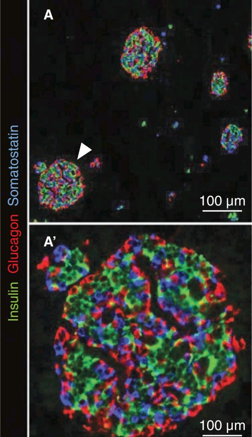
FIGURE 24-1 Islet of Langerhans in the human pancreas. Green fluorescent labeling are insulin containing B cells within the islets, red fluorescent labeling are glucagon containing A cells and blue fluorescent labeling are somatostatin containing D cells. Surrounding dark area is pancreatic acinar tissue. (Used with permission from N. Hart).
شکل 24-1 جزیره لانگرهانس در پانکراس انسان. برچسب های فلورسنت سبز سلول های B حاوی انسولین در جزایر، برچسب های فلورسنت قرمز گلوکاگون حاوی سلول های A و برچسب های فلورسنت آبی سلول های D حاوی سوماتوستاتین هستند. اطراف ناحیه تاریک بافت آسینار پانکراس است. (با اجازه N. Hart استفاده می شود).
The cells in the islets can be divided into types on the basis of their staining properties and morphology. Humans have at least four distinct cell types: A, B, D, and F cells. A, B, and D cells are also called a, ẞ, and 8 cells. However, this leads to confusion in view of the use of Greek letters to refer to other structures in the body, particularly adrenergic receptors (see Chapter 7). The A cells secrete glucagon, the B cells secrete insulin, the D cells secrete somatostatin, and the F cells secrete pancreatic polypeptide. The B cells, which are the most common and account for 60-75% of the cells in the islets, are generally located in the center of each islet. They tend to be surrounded by the A cells, which make up 20% of the total, and the less common D and F cells. The islets in the tail, the body, and the anterior and superior part of the head of the human pancreas have many A cells and few if any F cells in the outer rim, whereas in rats and probably in humans, the islets in the posterior part of the head of the pancreas have a relatively large number of F cells and few A cells. The A-cell-rich (glucagon-rich) islets arise embryologically from the dorsal pancreatic bud, and the F-cell-rich (pancreatic polypeptide-rich) islets arise from the ventral pancreatic bud. These buds arise separately from the duodenum.
سلول های موجود در جزایر را می توان بر اساس خواص رنگ آمیزی و مورفولوژی آنها به انواع تقسیم کرد. انسان حداقل چهار نوع سلول متمایز دارد: سلول های A، B، D و F. سلول های A، B و D نیز سلول های a، ẞ و 8 نامیده می شوند. با این حال، با توجه به استفاده از حروف یونانی برای اشاره به سایر ساختارهای بدن، به ویژه گیرنده های آدرنرژیک، این امر منجر به سردرگمی می شود (به فصل 7 مراجعه کنید). سلول های A گلوکاگون، سلول های B انسولین، سلول های D سوماتوستاتین و سلول های F پلی پپتید پانکراس ترشح می کنند. سلول های B که رایج ترین هستند و 60 تا 75 درصد از سلول های جزایر را تشکیل می دهند، عموماً در مرکز هر جزایر قرار دارند. آنها تمایل دارند توسط سلول های A احاطه شوند، که 20٪ از کل را تشکیل می دهند، و سلول های D و F کمتر رایج هستند. جزایر دم، بدن و قسمت قدامی و فوقانی سر پانکراس انسان دارای تعداد زیادی سلول A و تعداد کمی سلول F در لبه بیرونی هستند، در حالی که در موشها و احتمالاً در انسان، جزایر در قسمت خلفی سر پانکراس دارای تعداد نسبتاً زیادی سلول F و تعداد کمی سلول A هستند. جزایر غنی از سلول A (غنی از گلوکاگون) از نظر جنینی از جوانه پشتی پانکراس و جزایر غنی از سلول F (غنی از پلی پپتید پانکراس) از جوانه پانکراس شکمی به وجود می آیند. این جوانه ها جدا از اثنی عشر ایجاد می شوند.
The B cell granules are packets of insulin in the cell cytoplasm. The shape of the packets varies from species to species; in humans, some are round whereas others are rectangular (Figure 24-2). In the B cells, the insulin molecule forms polymers and also complexes with zinc. The differences in the shape of the packets are probably due to differences in the size of polymers or zinc aggregates of insulin. The A granules, which contain glucagon, are relatively uniform from species to species (Figure 24-3). The D cells also contain large numbers of relatively homogeneous granules.
گرانول های سلول B بسته هایی از انسولین در سیتوپلاسم سلولی هستند. شکل بسته ها از گونه ای به گونه دیگر متفاوت است. در انسان، برخی گرد و برخی دیگر مستطیلی هستند (شکل 24-2). در سلول های B، مولکول انسولین پلیمرها را تشکیل می دهد و همچنین با روی کمپلکس می شود. تفاوت در شکل بسته ها احتمالاً به دلیل تفاوت در اندازه پلیمرها یا دانه های روی انسولین است. گرانول های A که حاوی گلوکاگون هستند، از گونه ای به گونه دیگر نسبتاً یکنواخت هستند (شکل 24-3). سلول های D همچنین حاوی تعداد زیادی گرانول نسبتا همگن هستند.
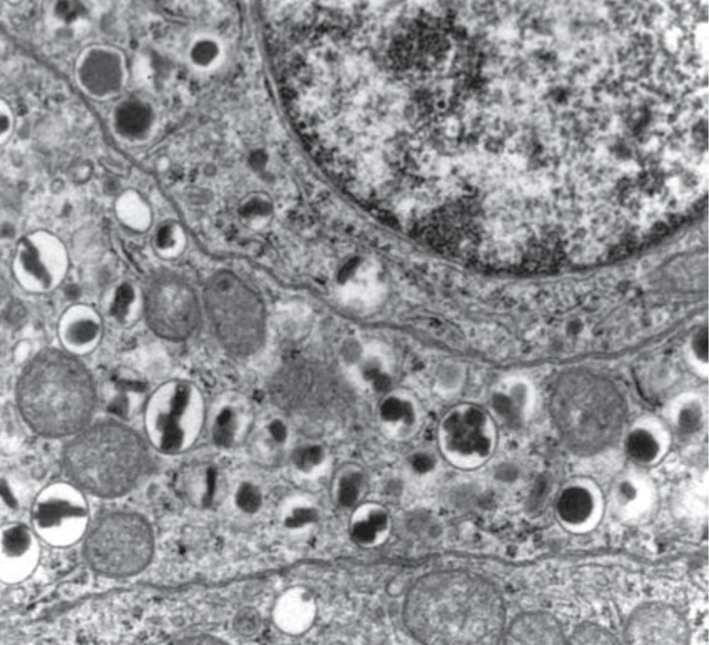
FIGURE 24-2 Electronmicrograph of two adjoining B cells in a human pancreatic islet. The B granules are the crystals in the membrane-lined vesicles. They vary in shape from rhombic to round (× 26,000). (Reproduced with permission from Fawcett DW: Bloom and Fawcett A Textbook of Histology, 11th ed. St. Louis, MO: Saunders; 1986.)
شکل 24-2 میکروگراف الکترونی دو سلول B مجاور در یک جزیره پانکراس انسانی. گرانول های B کریستال های موجود در وزیکول های پوشیده از غشاء هستند. شکل آنها از لوزی تا گرد (× 26000) متفاوت است. (تکثیر شده با اجازه Fawcett DW: Bloom and Fawcett A Textbook of Histology، ویرایش یازدهم سنت لوئیس، MO: ساندرز؛ 1986.)
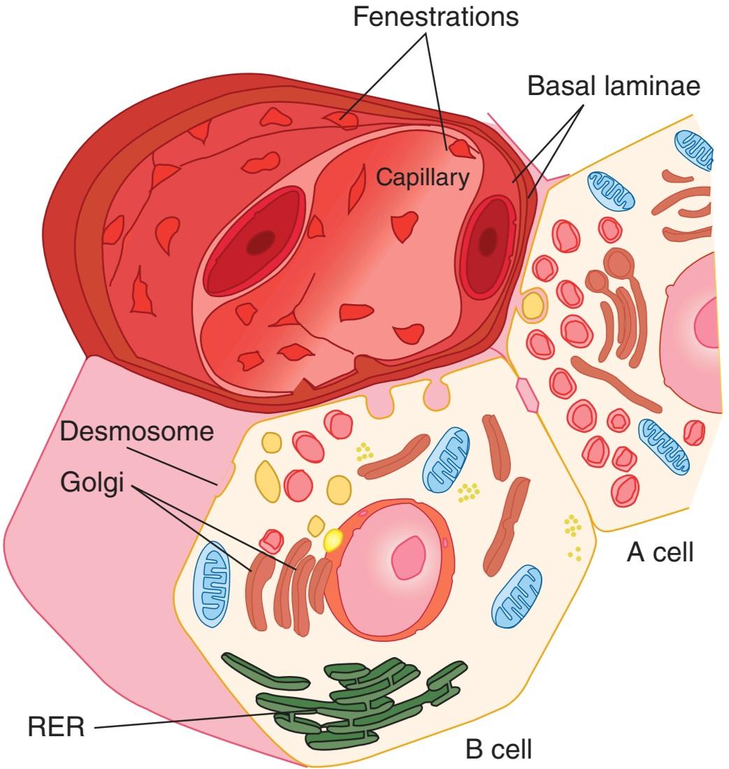
FIGURE 24-3 A and B cells, showing their relation to a blood vessel. Insulin from the B cell and glucagon from the A cell are secreted by exocytosis and cross the basal lamina of the cell and the basal lamina of the capillary before entering the lumen of the fenestrated capillary. RER, rough endoplasmic reticulum. (Reproduced with permission from Junqueira IC, Carneiro J: Basic Histology: Text and Atlas, 10th ed. New York, NY: McGraw-Hill; 2003.)
شکل 24-3 سلول های A و B، ارتباط آنها را با یک رگ خونی نشان می دهد. انسولین از سلول B و گلوکاگون از سلول A توسط اگزوسیتوز ترشح میشوند و قبل از ورود به لومن مویرگهای فنستره، از لایه بازال سلول و لایه بازال مویرگ عبور میکنند. RER، شبکه آندوپلاسمی خشن. (تکثیر شده با اجازه Junqueira IC، Carneiro J: Basic Histology: Text and Atlas، ویرایش 10. نیویورک، نیویورک: McGraw-Hill؛ 2003.)
STRUCTURE, BIOSYNTHESIS, & SECRETION OF INSULIN
STRUCTURE & SPECIES SPECIFICITY
Insulin is a polypeptide containing two chains of amino acids linked by disulfide bridges. Minor differences occur in the amino acid composition of the molecule from species to species. The differences are generally not sufficient to affect the biologic activity of a particular insulin in heterologous species but are sufficient to make the insulin antigenic. If insulin of one species is injected for a prolonged period into another species, the anti-insulin antibodies formed inhibit the injected insulin. Almost all humans who have received commercial bovine insulin for more than 2 months have antibodies against bovine insulin, but the titer is usually low. Porcine insulin differs from human insulin by only one amino acid residue and has low antigenicity. Human insulin produced in bacteria by recombinant DNA technology is now widely used to avoid antibody formation.
ساختار، بیوسنتز، و ترشح انسولین
ساختار و ویژگی های گونه
انسولین یک پلی پپتید حاوی دو زنجیره اسید آمینه است که توسط پل های دی سولفیدی به هم متصل شده اند. تفاوت های جزئی در ترکیب اسید آمینه مولکول از گونه ای به گونه دیگر رخ می دهد. این تفاوت ها معمولاً برای تأثیرگذاری بر فعالیت بیولوژیکی یک انسولین خاص در گونه های هترولوگ کافی نیستند، اما برای ایجاد آنتی ژن انسولین کافی هستند. اگر انسولین یک گونه برای مدت طولانی به گونه دیگر تزریق شود، آنتی بادی های ضد انسولین تشکیل شده انسولین تزریق شده را مهار می کنند. تقریباً تمام افرادی که انسولین تجاری گاوی را برای بیش از 2 ماه دریافت کرده اند، دارای آنتی بادی علیه انسولین گاوی هستند، اما تیتر آن معمولاً پایین است. انسولین خوک با انسولین انسانی تنها با یک اسید آمینه تفاوت دارد و آنتی ژنی پایینی دارد. انسولین انسانی تولید شده در باکتری ها با فناوری DNA نوترکیب، اکنون به طور گسترده برای جلوگیری از تشکیل آنتی بادی استفاده می شود.
BIOSYNTHESIS & SECRETION
Insulin is synthesized in the rough endoplasmic reticulum of the B cells (Figure 24-3). It is then transported to the Golgi apparatus, where it is packaged into membrane-bound granules. These granules move to the plasma membrane by a process involving microtubules, and their contents are expelled by exocytosis (see Chapters 2 and 16). The insulin then crosses the basal lamina of the B cell and a neighboring capillary and the fenestrated endothelium of the capillary to reach the bloodstream. The fenestrations are discussed in detail in Chapter 31.
بیوسنتز و ترشح
انسولین در شبکه آندوپلاسمی خشن سلول های B سنتز می شود (شکل 24-3). سپس به دستگاه گلژی منتقل می شود و در آنجا به گرانول های متصل به غشاء بسته بندی می شود. این گرانول ها با فرآیندی شامل میکروتوبول ها به سمت غشای پلاسمایی حرکت می کنند و محتویات آنها توسط اگزوسیتوز خارج می شود (به فصل های 2 و 16 مراجعه کنید). سپس انسولین از لایه بازال سلول B و یک مویرگ مجاور و اندوتلیوم پره مویرگ عبور می کند تا به جریان خون برسد. فنسترها در فصل 31 به تفصیل مورد بحث قرار گرفته اند.
Like other polypeptide hormones and related proteins that enter the endoplasmic reticulum, insulin is synthesized as part of a larger preprohormone (see Chapter 1). The gene for insulin is located on the short arm of chromosome 11 in humans. It has two introns and three exons. Preproinsulin originates from the endoplasmic reticulum. The remainder of the molecule is then folded, and the disulfide bonds are formed to make proinsulin. The peptide segment connecting the A and B chains, the connecting peptide (C peptide), facilitates the folding and then is detached in the granules before secretion. Two proteases are involved in processing the proinsulin. Normally, 90-97% of the product released from the B cells is insulin along with equimolar amounts of C peptide. The rest is mostly proinsulin. C peptide can be measured by radioimmunoassay, and its level in blood provides an index of B cell function in patients receiving exogenous insulin.
مانند سایر هورمون های پلی پپتیدی و پروتئین های مرتبط که وارد شبکه آندوپلاسمی می شوند، انسولین به عنوان بخشی از یک پیش پروهورمون بزرگتر سنتز می شود (به فصل 1 مراجعه کنید). ژن انسولین در بازوی کوتاه کروموزوم 11 در انسان قرار دارد. دارای دو اینترون و سه اگزون است. پری پروانسولین از شبکه آندوپلاسمی منشا می گیرد. سپس بقیه مولکول تا می شود و پیوندهای دی سولفید برای ساخت پروانسولین تشکیل می شود. بخش پپتیدی که زنجیرههای A و B را به هم متصل میکند، پپتید اتصال (پپتید C)، تا شدن را تسهیل میکند و سپس قبل از ترشح در دانهها جدا میشود. دو پروتئاز در پردازش پروانسولین نقش دارند. به طور معمول، 90-97٪ از محصول آزاد شده از سلول های B انسولین به همراه مقادیر هم مولار پپتید C است. بقیه بیشتر پروانسولین است. پپتید C را می توان با رادیوامونواسی اندازه گیری کرد و سطح آن در خون شاخصی از عملکرد سلول B را در بیماران دریافت کننده انسولین اگزوژن ارائه می کند.
FATE OF SECRETED INSULIN
INSULIN & INSULIN-LIKE ACTIVITY IN BLOOD
Plasma contains a number of substances with insulin-like activity in addition to insulin. The activity that is not suppressed by anti-insulin antibodies has been called nonsuppressible insulin-like activity (NSILA). Most, if not all, of this activity persists after pancreatectomy and is due to the insulin-like growth factors IGF-I and IGF-II (see Chapter 18). These IGFs are polypeptides. Small amounts are free in the plasma (low-molecular-weight fraction), but large amounts are bound to proteins (high-molecular-weight fraction).
سرنوشت انسولین ترشحی
انسولین و فعالیت انسولین مانند در خون
پلاسما علاوه بر انسولین حاوی تعدادی مواد با فعالیت شبه انسولین است. فعالیتی که توسط آنتی بادی های ضد انسولین سرکوب نمی شود، فعالیت شبه انسولین غیر قابل سرکوب (NSILA) نامیده می شود. بیشتر، اگر نه همه، این فعالیت بعد از پانکراتکتومی ادامه می یابد و به دلیل فاکتورهای رشد شبه انسولین IGF-I و IGF-II است (به فصل 18 مراجعه کنید). این IGF ها پلی پپتید هستند. مقادیر کمی در پلاسما آزاد هستند (کسری با وزن مولکولی کم)، اما مقادیر زیاد به پروتئین ها متصل می شوند (کسری با وزن مولکولی بالا).
One may well ask why pancreatectomy causes diabetes mellitus when NSILA persists in the plasma. However, the insulin-like activities of IGF-I and IGF-II are weak compared to that of insulin and likely subserve other specific functions.
ممکن است بپرسید که چرا پانکراتکتومی باعث دیابت می شود وقتی NSILA در پلاسما باقی می ماند. با این حال، فعالیت های شبه انسولین IGF-I و IGF-II در مقایسه با انسولین ضعیف است و احتمالاً عملکردهای خاص دیگری را انجام می دهد.
METABOLISM
The half-life of insulin in the circulation in humans is about 5 min. Insulin binds to insulin receptors, and some is internalized. It is destroyed by proteases in the endosomes formed by the endocytotic process.
متابولیسم
نیمه عمر انسولین در گردش خون در انسان حدود 5 دقیقه است. انسولین به گیرنده های انسولین متصل می شود و مقداری درونی می شود. توسط پروتئازهای موجود در اندوزوم های تشکیل شده توسط فرآیند اندوسیتوز تخریب می شود.
EFFECTS OF INSULIN
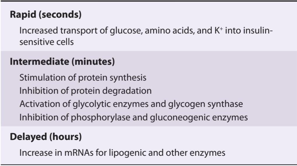
The physiologic effects of insulin are far-reaching and complex. They are conveniently divided into rapid, intermediate, and delayed actions (Table 24-1). The best known is the hypoglycemic effect, but there are additional effects on amino acid and electrolyte transport, many enzymes, and growth. The net effect of the hormone is storage of carbohydrate, protein, and fat. Therefore, insulin is appropriately called the “hormone of abundance.”
اثرات انسولین
اثرات فیزیولوژیکی انسولین گسترده و پیچیده است. آنها به راحتی به اقدامات سریع، میانی و تاخیری تقسیم می شوند (جدول 24-1). شناخته شده ترین اثر هیپوگلیسمی است، اما اثرات اضافی بر انتقال اسید آمینه و الکترولیت، بسیاری از آنزیم ها و رشد وجود دارد. اثر خالص این هورمون ذخیره کربوهیدرات، پروتئین و چربی است. بنابراین، انسولین به طور مناسب “هورمون فراوانی” نامیده می شود.
TABLE 24-1 Principal actions of insulin.
جدول 24-1 اقدامات اصلی انسولین.
Courtesy of ID Goldfine.
با حسن نیت از ID Goldfine.
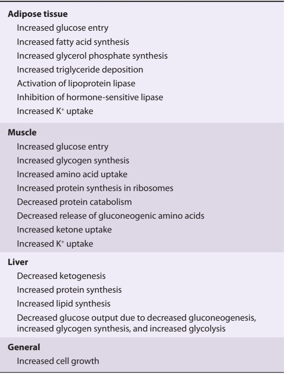
The actions of insulin on adipose tissue; skeletal, cardiac, and smooth muscle; and the liver are summarized in Table 24-2.
اثرات انسولین بر روی بافت چربی؛ عضلات اسکلتی، قلبی و صاف؛ و کبد در جدول 24-2 خلاصه شده است.
TABLE 24-2 Effects of insulin on various tissues.
جدول 24-2 اثرات انسولین بر بافت های مختلف.
GLUCOSE TRANSPORTERS
Glucose enters cells by facilitated diffusion (see Chapter 1) or, in the intestine and kidneys, by secondary active transport with Nat. In muscle, adipose, and some other tissues, insulin stimulates glucose entry into cells by increasing the number of glucose transporters (GLUTS) in the cell membranes.
انتقال دهنده های گلوکز
گلوکز با انتشار تسهیل شده (به فصل 1 مراجعه کنید) یا در روده و کلیه ها با انتقال فعال ثانویه با Nat وارد سلول ها می شود. در عضله، چربی و برخی بافت های دیگر، انسولین با افزایش تعداد ناقل گلوکز (GLUTS) در غشای سلولی، ورود گلوکز به سلول ها را تحریک می کند.
The GLUTs that are responsible for facilitated diffusion of glucose across cell membranes are a family of closely related proteins that span the cell membrane 12 times and have their amino and carboxyl terminals inside the cell. They differ from and have no homology with the Sodium-dependent glucose cotransporters (SGLT-1 and SGLT-2), which are responsible for the secondary active transport of glucose in the intestine (see Chapter 26) and renal tubules (see Chapter 38), although the SGLTs also have 12 transmembrane domains.
GLUT هایی که مسئول انتشار آسان گلوکز در غشای سلولی هستند، خانواده ای از پروتئین های نزدیک به هم هستند که 12 بار روی غشای سلولی قرار دارند و پایانه های آمینه و کربوکسیل خود را در داخل سلول دارند. آنها با همترانسپورترهای گلوکز وابسته به سدیم (SGLT-1 و SGLT-2) که مسئول انتقال فعال ثانویه گلوکز در روده (به فصل 26) و لوله های کلیوی (به فصل 38 مراجعه کنید) متفاوت هستند، اگرچه SGLT ها همچنین دارای 12 دامنه هستند.
Seven different GLUTS, named GLUT 1-7 in order of discovery, have been characterized (Table 24-3). They contain 492-524 amino acid residues and their affinity for glucose varies. Each transporter appears to have evolved for special tasks. GLUT-4 is the transporter in muscle and adipose tissue that is stimulated by insulin. A pool of GLUT-4 molecules is maintained within vesicles in the cytoplasm of insulin-sensitive cells. When the insulin receptors of these cells are activated, the vesicles move rapidly to the cell membrane and fuse with it, inserting the transporters into the cell membrane (Figure 24-4). When insulin action ceases, the transporter-containing patches of membrane are endocytosed and the vesicles are ready for the next exposure to insulin. Activation of the insulin receptor brings about the movement of the vesicles to the cell membrane by activating phosphatidylinositol 3-kinase (Figure 24-4). Most of the other GLUT transporters that are not insulin-sensitive appear to be constitutively expressed in the cell membrane.
هفت GLUTS مختلف که به ترتیب کشف GLUT 1-7 نامیده می شوند، مشخص شده اند (جدول 24-3). آنها حاوی 492-524 باقی مانده اسید آمینه هستند و میل ترکیبی آنها با گلوکز متفاوت است. به نظر می رسد که هر حمل و نقل برای وظایف خاصی تکامل یافته است. GLUT-4 ناقل عضله و بافت چربی است که توسط انسولین تحریک می شود. مجموعه ای از مولکول های GLUT-4 در داخل وزیکول ها در سیتوپلاسم سلول های حساس به انسولین نگهداری می شود. هنگامی که گیرنده های انسولین این سلول ها فعال می شوند، وزیکول ها به سرعت به سمت غشای سلولی حرکت می کنند و با آن ترکیب می شوند و ناقل ها را وارد غشای سلولی می کنند (شکل 24-4). هنگامی که عملکرد انسولین متوقف می شود، تکه های حاوی ناقل غشاء اندوسیتوز می شوند و وزیکول ها برای مواجهه بعدی با انسولین آماده می شوند. فعال شدن گیرنده انسولین باعث حرکت وزیکول ها به غشای سلولی با فعال کردن فسفاتیدیل 3-کیناز می شود (شکل 24-4). به نظر می رسد که اکثر ناقلین GLUT دیگر که به انسولین حساس نیستند، در غشای سلولی بیان می شوند.
TABLE 24-3 Glucose transporters in mammals.
جدول 24-3 ناقل گلوکز در پستانداران.
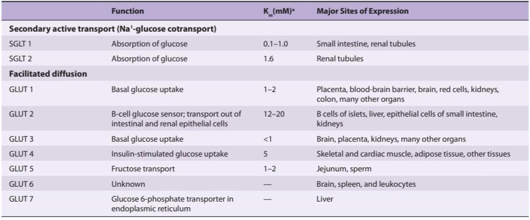
aThe Kis the glucose concentration at which transport is half-maximal.
Data from Stephens JM, Pilch PF: The metabolic regulation and vesicular transport of GLUT 4, the major insulin-responsive glucose transporter. Endocr Rev 1995;16:529.
Kisa غلظت گلوکز است که در آن انتقال نیمه حداکثر است.
داده ها از Stephens JM، Pilch PF: تنظیم متابولیک و انتقال تاولی GLUT 4، اصلی ترین انتقال دهنده گلوکز پاسخگو به انسولین. Endocr Rev 1995؛ 16:529.
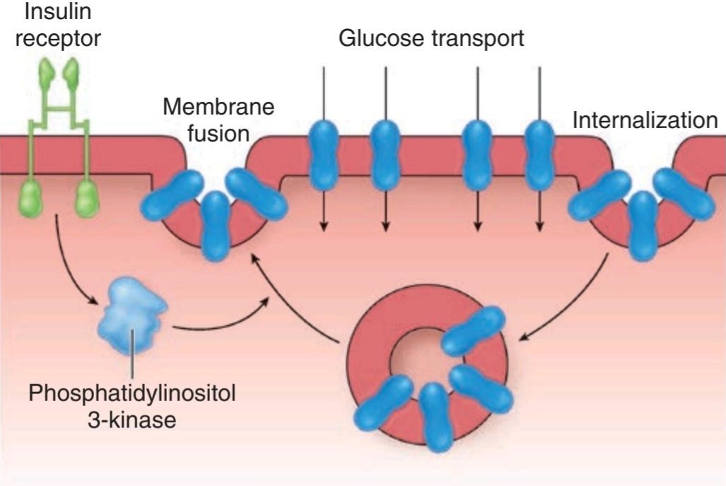
FIGURE 24-4 Cycling of GLUT-4 transporters through endosomes in insulin-sensitive tissues. Activation of the insulin receptor causes activation of phosphatidylinositol 3-kinase, which speeds translocation of the GLUT-4– containing endosomes into the cell membrane. The GLUT-4 transporters then mediate glucose transport into the cell.
شکل 24-4 چرخش ناقل های GLUT-4 از طریق اندوزوم ها در بافت های حساس به انسولین. فعال شدن گیرنده انسولین باعث فعال شدن فسفاتیدیل 3- کیناز می شود که انتقال اندوزوم های حاوی GLUT-4 را به غشای سلولی سرعت می بخشد. سپس ناقلین GLUT-4 انتقال گلوکز را به داخل سلول واسطه می کنند.
In the tissues in which insulin increases the number of GLUTs in cell membranes, the rate of phosphorylation of the glucose, once it has entered the cells, is regulated by other hormones. Growth hormone and cortisol both inhibit phosphorylation in certain tissues. Transport is normally so rapid that it is not a rate-limiting step in glucose metabolism. However, it is rate-limiting in B cells.
در بافت هایی که انسولین تعداد GLUT ها را در غشای سلولی افزایش می دهد، سرعت فسفوریلاسیون گلوکز پس از ورود به سلول ها توسط هورمون های دیگر تنظیم می شود. هورمون رشد و کورتیزول هر دو فسفوریلاسیون را در بافت های خاص مهار می کنند. انتقال معمولاً آنقدر سریع است که یک مرحله محدودکننده سرعت در متابولیسم گلوکز نیست. با این حال، در سلول های B محدود کننده سرعت است.
Insulin also increases the entry of glucose into liver cells, but it does not exert this effect by increasing the number of GLUT-4 transporters in the cell membranes. Instead, it induces glucokinase, and this increases the phosphorylation of glucose, so that the intracellular free glucose concentration stays low, facilitating the entry of glucose into the cell.
انسولین همچنین ورود گلوکز را به سلولهای کبدی افزایش میدهد، اما این اثر را با افزایش تعداد ناقلهای GLUT-4 در غشای سلولی اعمال نمیکند. در عوض، گلوکوکیناز را القا می کند و این باعث افزایش فسفوریلاسیون گلوکز می شود، به طوری که غلظت گلوکز آزاد داخل سلولی پایین می ماند و ورود گلوکز به سلول را تسهیل می کند.
Insulin-sensitive tissues also contain a population of GLUT-4 vesicles that move into the cell membrane in response to exercise, a process that occurs independent of the action of insulin. This is why exercise lowers blood sugar. A 5′-adenosine monophosphate (AMP)-activated kinase may trigger the insertion of these vesicles into the cell membrane.
بافتهای حساس به انسولین همچنین حاوی جمعیتی از وزیکولهای GLUT-4 هستند که در پاسخ به ورزش به غشای سلولی حرکت میکنند، فرآیندی که مستقل از عملکرد انسولین رخ میدهد. به همین دلیل ورزش قند خون را کاهش می دهد. یک کیناز فعال شده با 5′-آدنوزین مونوفسفات (AMP) ممکن است باعث وارد شدن این وزیکول ها به غشای سلولی شود.
INSULIN PREPARATIONS
The maximal decline in plasma glucose occurs 30 min after intravenous injection of insulin. After subcutaneous administration, the maximal fall occurs in 2-3 h. A wide variety of insulin preparations are now available commercially. These include insulins that have been complexed with protamine and other polypeptides to delay absorption and degradation, and synthetic insulins in which there have been changes in amino acid residues. In general, they fall into three categories: rapid, intermediate-acting, and long-acting (24-36 h).
آماده سازی انسولین
حداکثر کاهش گلوکز پلاسما 30 دقیقه پس از تزریق داخل وریدی انسولین رخ می دهد. پس از تزریق زیر جلدی، حداکثر سقوط در 2-3 ساعت رخ می دهد. طیف گسترده ای از آماده سازی انسولین در حال حاضر به صورت تجاری در دسترس است. اینها شامل انسولین هایی هستند که با پروتامین و سایر پلی پپتیدها کمپلکس شده اند تا جذب و تجزیه را به تعویق بیندازند و انسولین های مصنوعی که در آنها تغییراتی در بقایای اسید آمینه ایجاد شده است. به طور کلی، آنها به سه دسته تقسیم می شوند: سریع، متوسط، و طولانی اثر (24-36 ساعت).
Relation To Potassium
Insulin causes K+ to enter cells, with a resultant lowering of the extracellular K+ concentration. Infusions of insulin and glucose significantly lower the plasma K* level in normal individuals and are very effective for the temporary relief of hyperkalemia in patients with renal failure. Hypokalemia often develops when patients with diabetic acidosis are treated with insulin. The reason for the intracellular migration of K+ is still uncertain. However, insulin increases the activity of Na, K ATPase in cell membranes, so that more K+ is pumped into cells.
ارتباط با پتاسیم
انسولین باعث می شود +K وارد سلول ها شود و در نتیجه غلظت +K خارج سلولی کاهش می یابد. تزریق انسولین و گلوکز به طور قابل توجهی سطح +K پلاسما را در افراد عادی کاهش می دهد و برای تسکین موقت هیپرکالمی در بیماران مبتلا به نارسایی کلیوی بسیار موثر است. هیپوکالمی اغلب زمانی ایجاد می شود که بیماران مبتلا به اسیدوز دیابتی با انسولین درمان می شوند. دلیل مهاجرت درون سلولی +K هنوز نامشخص است. با این حال، انسولین فعالیت Na, K ATPase را در غشای سلولی افزایش می دهد، به طوری که +K بیشتری به سلول ها پمپ می شود.
OTHER ACTIONS
The hypoglycemic and other effects of insulin are summarized in temporal terms in Table 24-1, and the net effects on various tissues are summarized in Table 24- 2. The action on glycogen synthase fosters glycogen storage, and the actions on glycolytic enzymes favor glucose metabolism to two carbon fragments (see Chapter 1), with resulting promotion of lipogenesis. Stimulation of protein synthesis from amino acids entering the cells and inhibition of protein degradation foster growth.
سایر اقدامات
اثرات هیپوگلیسمی و سایر اثرات انسولین در شرایط زمانی در جدول 24-1 خلاصه شده است، و اثرات خالص بر روی بافت های مختلف در جدول 24-2 خلاصه شده است. اثر روی گلیکوژن سنتاز ذخیره گلیکوژن را تقویت می کند، و اعمال روی آنزیم های گلیکولیتیک به نفع متابولیسم گلوکز است. تحریک سنتز پروتئین از ورود اسیدهای آمینه به سلول ها و مهار تخریب پروتئین رشد را تقویت می کند.
The anabolic effect of insulin is aided by the protein-sparing action of adequate intracellular glucose supplies. Failure to grow is a symptom of diabetes in children, and insulin stimulates the growth of immature hypophysectomized rats to almost the same degree as growth hormone.
اثر آنابولیک انسولین با عملکرد ذخیره پروتئین منابع کافی گلوکز درون سلولی کمک می کند. عدم رشد یکی از علائم دیابت در کودکان است و انسولین رشد موشهای نابالغ هیپوفیسکتومی شده را تقریباً به همان میزان هورمون رشد تحریک میکند.
MECHANISM OF ACTION
INSULIN RECEPTORS
Insulin receptors are found on many different cells in the body, including cells in which insulin does not increase glucose uptake.
مکانیسم عمل
گیرنده های انسولین
گیرنده های انسولین در بسیاری از سلول های مختلف بدن یافت می شوند، از جمله سلول هایی که انسولین در آنها جذب گلوکز را افزایش نمی دهد.
The insulin receptor, which has a molecular weight of approximately 340,000, is a tetramer made up of two a and two ẞ glycoprotein subunits (Figure 24-5). All these are synthesized on a single mRNA and then proteolytically separated and bound to each other by disulfide bonds. The gene for the insulin receptor has 22 exons and in humans is located on chromosome 19. The a subunits bind insulin and are extracellular, whereas the ẞ subunits span the membrane. The intracellular portions of the ẞ subunits have tyrosine kinase activity. The a and ẞ subunits are both glycosylated, with sugar residues extending into the interstitial fluid.
گیرنده انسولین که وزن مولکولی آن تقریباً 340000 است، یک تترامر است که از دو زیرواحد گلیکوپروتئین a و دو ẞ تشکیل شده است (شکل 24-5). همه اینها بر روی یک mRNA منفرد سنتز می شوند و سپس به صورت پروتئولیتی جدا شده و توسط پیوندهای دی سولفیدی به یکدیگر متصل می شوند. ژن گیرنده انسولین دارای 22 اگزون است و در انسان روی کروموزوم 19 قرار دارد. زیر واحدهای a به انسولین متصل می شوند و خارج سلولی هستند، در حالی که زیر واحدهای ẞ غشاء را می پوشانند. بخش های درون سلولی زیر واحدهای ẞ دارای فعالیت تیروزین کیناز هستند. زیرواحدهای a و ẞ هر دو گلیکوزیله هستند و بقایای قند در مایع بینابینی گسترش می یابد.
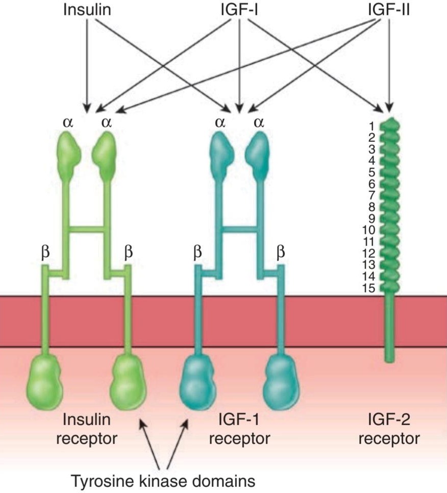
FIGURE 24-5 Insulin, IGF-I, and IGF-II receptors. Each hormone binds primarily to its own receptor, but insulin also binds to the IGF-I receptor, and IGF-I and IGF-II bind to all three. The purple boxes are intracellular tyrosine kinase domains. Note the marked similarity between the insulin receptor and the IGF-I receptor; also note the 15 repeat sequences in the extracellular portion of the IGF-II receptor.
شکل 24-5 گیرنده های انسولین، IGF-I و IGF-II. هر هورمون در درجه اول به گیرنده خود متصل می شود، اما انسولین همچنین به گیرنده IGF-I و IGF-I و IGF-II به هر سه متصل می شوند. جعبه های بنفش دامنه های تیروزین کیناز داخل سلولی هستند. به شباهت مشخص بین گیرنده انسولین و گیرنده IGF-I توجه کنید. همچنین به 15 توالی تکرار در بخش خارج سلولی گیرنده IGF-II توجه کنید.
Binding of insulin triggers the tyrosine kinase activity of the ẞ subunits, producing autophosphorylation of the ẞ subunits on tyrosine residues. The autophosphorylation, which is necessary for insulin to exert its biologic effects, triggers phosphorylation of some cytoplasmic proteins and dephosphorylation of others, mostly on serine and threonine residues. Insulin receptor substrate (IRS- 1) mediates some of the effects in humans but there are other effector systems as well (Figure 24-6). For example, mice in which the insulin receptor gene is knocked out show marked growth retardation in utero, have abnormalities of the central nervous system (CNS) and skin, and die at birth of respiratory failure, whereas IRS-1 knockouts show only moderate growth retardation in utero, survive, and are insulin-resistant but otherwise nearly normal.
اتصال انسولین فعالیت تیروزین کیناز زیرواحدهای ẞ را تحریک می کند و باعث اتوفسفوریلاسیون زیر واحدهای ẞ بر روی باقیمانده های تیروزین می شود. اتوفسفوریلاسیون، که برای اعمال اثرات بیولوژیکی انسولین ضروری است، باعث فسفوریلاسیون برخی از پروتئین های سیتوپلاسمی و دفسفوریلاسیون برخی دیگر، عمدتاً بر روی باقیمانده های سرین و ترئونین می شود. بستر گیرنده انسولین (IRS-1) برخی از اثرات را در انسان واسطه می کند، اما سیستم های موثر دیگری نیز وجود دارد (شکل 24-6). به عنوان مثال، موش هایی که در آنها ژن گیرنده انسولین از بین رفته است، تاخیر رشد قابل توجهی را در رحم نشان می دهند، ناهنجاری های سیستم عصبی مرکزی (CNS) و پوست دارند و در بدو تولد به دلیل نارسایی تنفسی می میرند، در حالی که ناک اوت های IRS-1 فقط تاخیر رشد متوسطی را در رحم نشان می دهند، در غیر این صورت سولین تقریباً سالم است، و در حالت عادی زنده می مانند.
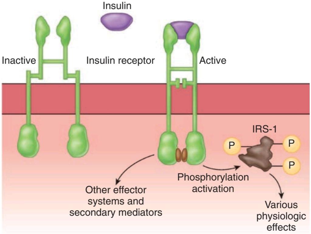
FIGURE 24-6 Intracellular responses triggered by insulin binding to the insulin receptor. Circles labeled P represent phosphate groups. IRS-1, insulin receptor substrate-1.
شکل 24-6 پاسخ های درون سلولی که با اتصال انسولین به گیرنده انسولین ایجاد می شوند. دایره هایی با برچسب P نشان دهنده گروه های فسفات هستند. IRS-1، بستر گیرنده انسولین-1.
The growth-promoting protein anabolic effects of insulin are mediated via phosphatidylinositol 3-kinase (PI3K), and evidence indicates that in invertebrates, this pathway is involved in the growth of nerve cells and axon guidance in the visual system.
اثرات آنابولیک پروتئین محرک رشد انسولین از طریق فسفاتیدیل 3-کیناز (PI3K) انجام می شود و شواهد نشان می دهد که در بی مهرگان، این مسیر در رشد سلول های عصبی و هدایت آکسون در سیستم بینایی نقش دارد.
It is interesting to compare the insulin receptor with other related receptors. The insulin receptor is very similar to the receptor for IGF-I but different from the receptor for IGF-II (Figure 24-5). Other receptors for growth factors and receptors for various oncogenes also are tyrosine kinases. However, the amino acid composition of these receptors is quite different.
مقایسه گیرنده انسولین با سایر گیرنده های مرتبط جالب است. گیرنده انسولین بسیار شبیه به گیرنده IGF-I است اما با گیرنده IGF-II متفاوت است (شکل 24-5). سایر گیرنده های فاکتورهای رشد و گیرنده های انکوژن های مختلف نیز تیروزین کیناز هستند. با این حال، ترکیب اسید آمینه این گیرنده ها کاملاً متفاوت است.
When insulin binds to its receptors, they aggregate in patches and are taken up into the cell by receptor-mediated endocytosis (see Chapter 2). Eventually, the insulin-receptor complexes enter lysosomes, where the receptors are broken down or recycled. The half-life of the insulin receptor is about 7 h.
هنگامی که انسولین به گیرنده های خود متصل می شود، آنها به صورت تکه ای جمع می شوند و توسط اندوسیتوز با واسطه گیرنده به داخل سلول می روند (به فصل 2 مراجعه کنید). در نهایت، کمپلکسهای گیرنده انسولین وارد لیزوزوم میشوند، جایی که گیرندهها شکسته یا بازیافت میشوند. نیمه عمر گیرنده انسولین حدود 7 ساعت است.
CONSEQUENCES OF INSULIN DEFICIENCY
The far-reaching physiologic effects of insulin are highlighted by a consideration of the extensive and serious consequences of insulin deficiency (Clinical Box 24-1).
پیامدهای کمبود انسولین
اثرات فیزیولوژیکی گسترده انسولین با در نظر گرفتن پیامدهای گسترده و جدی کمبود انسولین برجسته می شود (کادر بالینی 24-1).
In humans, insulin deficiency is a common pathologic condition. In animals, it can be produced by pancreatectomy; by administration of alloxan, streptozocin, or other toxins that in appropriate doses cause selective destruction of the B cells of the pancreatic islets; by administration of drugs that inhibit insulin secretion; and by administration of anti-insulin antibodies. Strains of mice, rats, hamsters, guinea pigs, miniature swine, and monkeys that have a high incidence of spontaneous diabetes mellitus have also been described.
در انسان، کمبود انسولین یک وضعیت پاتولوژیک رایج است. در حیوانات، می توان آن را با پانکراتکتومی تولید کرد. با تجویز آلوکسان، استرپتوزوسین یا سایر سموم که در دوزهای مناسب باعث تخریب انتخابی سلول های B جزایر پانکراس می شوند. با تجویز داروهایی که ترشح انسولین را مهار می کنند. و با تجویز آنتی بادی های ضد انسولین. گونههایی از موشها، موشها، همسترها، خوکچههای هندی، خوکهای مینیاتوری و میمونها که شیوع بالایی از دیابت خودبهخودی دارند نیز توصیف شدهاند.
CLINICAL BOX 24 -1
Diabetes Mellitus
The constellation of abnormalities caused by insulin deficiency is called diabetes mellitus. Greek and Roman physicians used the term “diabetes” to refer to conditions in which the cardinal finding was a large urine volume, and two types were distinguished: “diabetes mellitus,” in which the urine tasted sweet; and “diabetes insipidus,” in which the urine had little taste. Today, the term “diabetes insipidus” is reserved for conditions in which there is a deficiency of the production or action of vasopressin (see Chapter 38), and the unmodified word “diabetes” is generally used as a synonym for diabetes mellitus.
جعبه بالینی 24 -1
دیابت ملیتوس
مجموعه ای از ناهنجاری های ناشی از کمبود انسولین دیابت شیرین نامیده می شود. پزشکان یونانی و رومی از اصطلاح «دیابت» برای اشاره به شرایطی استفاده میکردند که در آن یافته اصلی حجم ادرار زیاد بود، و دو نوع آن متمایز بود: «دیابت شیرین» که در آن ادرار طعم شیرینی داشت. و «دیابت بی مزه» که در آن ادرار طعم کمی داشت. امروزه، اصطلاح “دیابت بی مزه” برای شرایطی که در آن کمبود تولید یا عملکرد وازوپرسین وجود دارد (به فصل 38 مراجعه کنید) اختصاص دارد و کلمه اصلاح نشده “دیابت” به طور کلی به عنوان مترادف دیابت شیرین استفاده می شود.
The cause of clinical diabetes is always a deficiency of the effects of insulin at the tissue level. Type 1 diabetes, or insulin-dependent diabetes mellitus (IDDM), is due to insulin deficiency caused by autoimmune destruction of the B cells in the pancreatic islets, and it accounts for 3-5% of cases and usually presents in children. Type 2 diabetes, or non-insulin- dependent diabetes mellitus (NIDDM), is characterized by the dysregulation of insulin release from the B cells, along with insulin resistance in peripheral tissues such as skeletal muscle, brain, and liver. Type 2 diabetes historically presented in overweight or obese adults, although it is increasingly being diagnosed in children as childhood obesity increases.
علت دیابت بالینی همیشه کمبود اثرات انسولین در سطح بافت است. دیابت نوع 1 یا دیابت ملیتوس وابسته به انسولین (IDDM) به دلیل کمبود انسولین ناشی از تخریب خودایمنی سلول های B در جزایر پانکراس است و 3 تا 5 درصد موارد را تشکیل می دهد و معمولاً در کودکان ظاهر می شود. دیابت نوع 2 یا دیابت شیرین غیر وابسته به انسولین (NIDDM) با اختلال در تنظیم ترشح انسولین از سلول های B همراه با مقاومت به انسولین در بافت های محیطی مانند ماهیچه های اسکلتی، مغز و کبد مشخص می شود. دیابت نوع 2 به طور تاریخی در بزرگسالان دارای اضافه وزن یا چاق دیده می شود، اگرچه با افزایش چاقی در دوران کودکی به طور فزاینده ای در کودکان تشخیص داده می شود.
Diabetes is characterized by polyuria (passage of large volumes of urine), polydipsia (excessive drinking), weight loss in spite of polyphagia (increased appetite), hyperglycemia, glycosuria, ketosis, acidosis, and coma. Widespread biochemical abnormalities are present, but the fundamental defects to which most of the abnormalities can be traced are (1) reduced entry of glucose into various “peripheral” tissues and (2) increased liberation of glucose into the circulation from the liver. Therefore, there is an extracellular glucose excess and, in many cells, an intracellular glucose deficiency-a situation that has been called “starvation in the midst of plenty.” Also, the entry of amino acids into muscle is decreased and lipolysis is increased.
دیابت با پلی اوری (دفع حجم زیاد ادرار)، پلی دیپسی (نوشیدن زیاد)، کاهش وزن با وجود پلی فاژی (افزایش اشتها)، هیپرگلیسمی، گلیکوزوری، کتوز، اسیدوز و کما مشخص می شود. ناهنجاریهای بیوشیمیایی گسترده وجود دارد، اما نقایص اساسی که بیشتر ناهنجاریها را میتوان ردیابی کرد عبارتند از (1) کاهش ورود گلوکز به بافتهای مختلف “محیطی” و (2) افزایش آزادسازی گلوکز به گردش خون از کبد. بنابراین، گلوکز اضافی خارج سلولی و در بسیاری از سلول ها، کمبود گلوکز درون سلولی وجود دارد – وضعیتی که به آن “گرسنگی در میان فراوانی” می گویند. همچنین ورود اسیدهای آمینه به ماهیچه کاهش یافته و لیپولیز افزایش می یابد.
THERAPEUTIQ HIGHLIGHTS
In type 1 diabetes, the mainstay of therapy is provision of exogenous insulin, carefully titrated to dietary intake of glucose. In type 2 diabetes, lifestyle changes such as alterations in the diet or increased exercise can often delay symptoms in early disease, but these are difficult to secure. Insulin-sensitizing drugs represent second-line agents (see Chapter 16).
نکات برجسته درمانی
در دیابت نوع 1، درمان اصلی، ارائه انسولین اگزوژن است که به دقت با دریافت گلوکز از رژیم غذایی تیتر شده است. در دیابت نوع 2، تغییرات سبک زندگی مانند تغییر در رژیم غذایی یا افزایش ورزش اغلب می تواند علائم را در مراحل اولیه بیماری به تعویق بیندازد، اما تضمین این موارد دشوار است. داروهای حساس کننده به انسولین نشان دهنده عوامل خط دوم هستند (به فصل 16 مراجعه کنید).
GLUCOSE TOLERANCE
In diabetes, glucose piles up in the bloodstream, especially after meals. If a glucose load is given to a diabetic, the plasma glucose rises higher and returns to the baseline more slowly than it does in normal individuals. The response to a standard oral test dose of glucose, the oral glucose tolerance test, is used in the clinical diagnosis of diabetes (Figure 24-7).
تحمل گلوکز
در دیابت، گلوکز در جریان خون انباشته می شود، به خصوص بعد از غذا. اگر بار گلوکز به یک دیابتی داده شود، گلوکز پلاسما بالاتر می رود و کندتر از افراد عادی به سطح اولیه باز می گردد. پاسخ به دوز تست خوراکی استاندارد گلوکز، تست تحمل گلوکز خوراکی، در تشخیص بالینی دیابت استفاده می شود (شکل 24-7).

FIGURE 24-7 Oral glucose tolerance test. Adults are given 75 g of glucose in 300 mL of water. In normal individuals, the fasting venous plasma glucose is less than 115 mg/dL, the 2-hour value is less than 140 mg/dL, and no value is greater than 200 mg/dL. Diabetes mellitus is present if the 2-hour value and one other value are greater than 200 mg/dL. Impaired glucose tolerance is diagnosed when the values are above the upper limits of normal but below the values diagnostic of diabetes.
شکل 24-7 تست تحمل گلوکز خوراکی. به بزرگسالان 75 گرم گلوکز در 300 میلی لیتر آب داده می شود. در افراد عادی، گلوکز پلاسمای وریدی ناشتا کمتر از 115 میلی گرم در دسی لیتر، مقدار 2 ساعته آن کمتر از 140 میلی گرم در دسی لیتر است و هیچ مقداری بیشتر از 200 میلی گرم در دسی لیتر نیست. اگر مقدار 2 ساعت و یک مقدار دیگر بیشتر از 200 میلی گرم در دسی لیتر باشد، دیابت وجود دارد. اختلال تحمل گلوکز زمانی تشخیص داده می شود که مقادیر بالاتر از حد طبیعی باشد اما کمتر از مقادیر تشخیصی دیابت باشد.
Impaired glucose tolerance in diabetes is due in part to reduced entry of glucose into cells (decreased peripheral utilization). In the absence of insulin, the entry of glucose into skeletal, cardiac, and smooth muscle and other tissues is decreased (Figure 24-8). Glucose uptake by the liver is also reduced, but the effect is indirect. Intestinal absorption of glucose is unaffected, as is its reabsorption from the urine by the cells of the proximal tubules of the kidneys. Glucose uptake by most of the brain and the red blood cells is also normal.
اختلال تحمل گلوکز در دیابت تا حدی به دلیل کاهش ورود گلوکز به سلول ها (کاهش استفاده محیطی) است. در غیاب انسولین، ورود گلوکز به ماهیچه های اسکلتی، قلبی و صاف و سایر بافت ها کاهش می یابد (شکل 24-8). جذب گلوکز توسط کبد نیز کاهش می یابد، اما اثر غیر مستقیم است. جذب روده ای گلوکز و همچنین جذب مجدد آن از ادرار توسط سلول های لوله های پروگزیمال کلیه ها بی تاثیر است. جذب گلوکز توسط بیشتر مغز و گلبول های قرمز خون نیز طبیعی است.
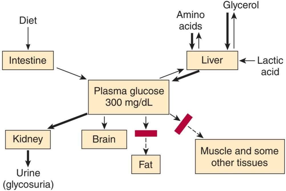
FIGURE 24-8 Disordered plasma glucose homeostasis in insulin deficiency. The heavy arrows indicate reactions that are accentuated. The rectangles across arrows indicate reactions that are blocked.
شکل 24-8 اختلال در هموستاز گلوکز پلاسما در کمبود انسولین. فلش های سنگین نشان دهنده واکنش هایی است که تاکید دارند. مستطیل های روی فلش ها نشان دهنده واکنش هایی هستند که مسدود شده اند.
The second and the major cause of hyperglycemia in diabetes is derangement of the glucostatic function of the liver (see Chapter 28). The liver takes up glucose from the bloodstream and stores it as glycogen, but because the liver contains glucose-6-phosphatase it also discharges glucose into the bloodstream. Insulin facilitates glycogen synthesis and inhibits hepatic glucose output. When the plasma glucose is high, insulin secretion is normally increased and hepatic glucogenesis is decreased. This response does not occur in type 1 diabetes mellitus (as insulin is absent) or in type 2 diabetes mellitus (as tissues are insulin-resistant). Glucagon can contribute to hyperglycemia as it stimulates gluconeogenesis. Glucose output by the liver can be stimulated by catecholamines, cortisol, and growth hormone (ie, during a stress response).
دومین و مهمترین علت هیپرگلیسمی در دیابت، اختلال در عملکرد گلوکوستاتیک کبد است (به فصل 28 مراجعه کنید). کبد گلوکز را از جریان خون می گیرد و آن را به عنوان گلیکوژن ذخیره می کند، اما از آنجایی که کبد حاوی گلوکز-6-فسفاتاز است، گلوکز را نیز به جریان خون تخلیه می کند. انسولین سنتز گلیکوژن را تسهیل می کند و خروجی گلوکز کبدی را مهار می کند. هنگامی که گلوکز پلاسما بالا باشد، ترشح انسولین به طور معمول افزایش یافته و گلوکوژنز کبدی کاهش می یابد. این پاسخ در دیابت نوع 1 (به دلیل عدم وجود انسولین) یا در دیابت نوع 2 (از آنجایی که بافت ها به انسولین مقاوم هستند) رخ نمی دهد. گلوکاگون می تواند به هیپرگلیسمی کمک کند زیرا گلوکونئوژنز را تحریک می کند. خروجی گلوکز توسط کبد می تواند توسط کاتکول آمین ها، کورتیزول و هورمون رشد تحریک شود (یعنی در طی یک پاسخ استرس).
EFFECTS OF HYPERGLYCEMIA
Hyperglycemia by itself can cause symptoms resulting from the hyperosmolality of the blood. In addition, there is glycosuria because the renal capacity for glucose reabsorption is exceeded. Excretion of the osmotically active glucose molecules entails the loss of large amounts of water (osmotic diuresis; see Chapter 38). The resultant dehydration activates the mechanisms regulating water intake, leading to polydipsia. There is an appreciable urinary loss of Na+ and K+ as well. For every gram of glucose excreted, 4.1 kcal is lost from the body. Increasing the oral caloric intake to cover this loss simply raises the plasma glucose further and increases the glycosuria, so mobilization of endogenous protein and fat stores and weight loss are not prevented.
اثرات هیپرگلیسمی
هایپرگلیسمی به خودی خود می تواند علائم ناشی از هیپراسمولالیته خون را ایجاد کند. علاوه بر این، گلیکوزوری وجود دارد زیرا ظرفیت کلیوی برای بازجذب گلوکز بیش از حد است. دفع مولکولهای گلوکز فعال اسمزی مستلزم از دست دادن مقادیر زیادی آب است (ادرار اسمزی؛ به فصل 38 مراجعه کنید). کم آبی حاصل، مکانیسم های تنظیم کننده مصرف آب را فعال می کند و منجر به پلی دیپسی می شود. همچنین کاهش قابل توجهی از +Na و +K در ادرار وجود دارد. به ازای هر گرم گلوکز دفع شده، 1/4 کیلوکالری از بدن دفع می شود. افزایش دریافت کالری خوراکی برای پوشش این کاهش به سادگی باعث افزایش بیشتر گلوکز پلاسما و افزایش گلیکوزوری می شود، بنابراین از تحرک ذخایر پروتئین و چربی درون زا و کاهش وزن جلوگیری نمی شود.
When plasma glucose is episodically elevated over time, small amounts of hemoglobin A are nonenzymatically glycated to form HbA1c (see Chapter 31). Careful control of the diabetes with insulin reduces the amount formed and consequently HbA1c concentration is measured clinically as an integrated index of diabetic control for the 4- to 6-week period before the measurement.
هنگامی که گلوکز پلاسما به طور دوره ای در طول زمان افزایش می یابد، مقادیر کمی از هموگلوبین A به صورت غیر آنزیمی گلیکوزه می شود و HbA1c را تشکیل می دهد (به فصل 31 مراجعه کنید). کنترل دقیق دیابت با انسولین مقدار تشکیل شده را کاهش می دهد و در نتیجه غلظت HbA1c به عنوان یک شاخص تلفیقی از کنترل دیابت برای دوره 4 تا 6 هفته قبل از اندازه گیری اندازه گیری می شود.
The role of chronic hyperglycemia in production of the long-term complications of diabetes is discussed below.
نقش هیپرگلیسمی مزمن در تولید عوارض طولانی مدت دیابت در زیر مورد بحث قرار گرفته است.
EFFECTS OF INTRACELLULAR GLUCOSE DEFICIENCY
The abundance of glucose outside the cells in diabetes contrasts with the intracellular deficit. Glucose catabolism is normally a major source of energy for cellular processes, and in diabetes energy requirements can be met only by drawing on protein and fat reserves. Mechanisms are activated that greatly increase the catabolism of protein and fat, and one of the consequences of increased fat catabolism is ketosis.
اثرات کمبود گلوکز داخل سلولی
فراوانی گلوکز در خارج از سلول ها در دیابت با کمبود داخل سلولی در تضاد است. کاتابولیسم گلوکز به طور معمول منبع اصلی انرژی برای فرآیندهای سلولی است و در دیابت نیاز به انرژی تنها با استفاده از ذخایر پروتئین و چربی تامین می شود. مکانیسم هایی فعال می شوند که کاتابولیسم پروتئین و چربی را به شدت افزایش می دهند و یکی از پیامدهای افزایش کاتابولیسم چربی، کتوز است.
Deficient glucose utilization and deficient hormone sensing (insulin, leptin, CCK) in the cells of the hypothalamus that regulate satiety are the probable causes of hyperphagia in diabetes. The feeding area of the hypothalamus is not inhibited and thus satiety is not sensed so food intake is increased.
کمبود استفاده از گلوکز و کمبود حس هورمونی (انسولین، لپتین، CCK) در سلولهای هیپوتالاموس که سیری را تنظیم میکنند، از علل احتمالی هیپرفاژی در دیابت هستند. ناحیه تغذیه هیپوتالاموس مهار نمی شود و در نتیجه سیری احساس نمی شود بنابراین مصرف غذا افزایش می یابد.
Glycogen depletion is a common consequence of intracellular glucose deficit, and the glycogen content of liver and skeletal muscle in diabetic animals is usually reduced.
کاهش گلیکوژن پیامد شایع کمبود گلوکز درون سلولی است و میزان گلیکوژن کبد و ماهیچه های اسکلتی در حیوانات دیابتی معمولاً کاهش می یابد.
CHANGES IN PROTEIN METABOLISM
In diabetes, the rate at which amino acids are catabolized to CO2 and H2O is increased. In addition, more amino acids are converted to glucose in the liver. The increased gluconeogenesis has many causes. Glucagon stimulates gluconeogenesis, and hyperglucagonemia is generally present in diabetes. Adrenal glucocorticoids also contribute to increased gluconeogenesis when they are elevated in severely ill diabetics. The supply of amino acids is increased for gluconeogenesis because, in the absence of insulin, less protein synthesis occurs in muscle and hence blood amino acid levels rise. Alanine is particularly easily converted to glucose. In addition, the activity of the enzymes that catalyze the conversion of pyruvate and other two-carbon metabolic fragments to glucose is increased. These include phosphoenolpyruvate carboxykinase, which facilitates the conversion of oxaloacetate to phosphoenolpyruvate (see Chapter 1). They also include fructose 1,6-diphosphatase, which catalyzes the conversion of fructose diphosphate to fructose 6-phosphate, and glucose 6-phosphatase, which controls the entry of glucose into the circulation from the liver. Increased acetyl- CoA increases pyruvate carboxylase activity, and insulin deficiency increases the supply of acetyl-CoA because lipogenesis is decreased. Pyruvate carboxylase catalyzes the conversion of pyruvate to oxaloacetate (see Figure 1-22).
تغییرات در متابولیسم پروتئین
در دیابت، سرعت کاتابولیزاسیون اسیدهای آمینه به CO2 و H2O افزایش می یابد. علاوه بر این، اسیدهای آمینه بیشتری در کبد به گلوکز تبدیل می شوند. افزایش گلوکونئوژنز دلایل زیادی دارد. گلوکاگون گلوکونئوژنز را تحریک می کند و هیپرگلوکاگونمی به طور کلی در دیابت وجود دارد. گلوکوکورتیکوئیدهای آدرنال همچنین در افزایش گلوکونئوژنز در بیماران دیابتی شدید نقش دارند. عرضه اسیدهای آمینه برای گلوکونئوژنز افزایش می یابد، زیرا در غیاب انسولین، سنتز پروتئین کمتری در ماهیچه رخ می دهد و در نتیجه سطح اسید آمینه خون افزایش می یابد. آلانین به ویژه به راحتی به گلوکز تبدیل می شود. علاوه بر این، فعالیت آنزیم هایی که تبدیل پیرووات و سایر قطعات متابولیک دو کربنی را به گلوکز کاتالیز می کنند افزایش می یابد. اینها شامل فسفونول پیروات کربوکسی کیناز است که تبدیل اگزالواستات به فسفونول پیروات را تسهیل می کند (به فصل 1 مراجعه کنید). آنها همچنین شامل فروکتوز 1،6-دی فسفاتاز، که تبدیل فروکتوز دی فسفات به فروکتوز 6-فسفات را کاتالیز می کند، و گلوکز 6-فسفاتاز، که ورود گلوکز به گردش خون از کبد را کنترل می کند. افزایش استیل کوآ باعث افزایش فعالیت پیرووات کربوکسیلاز می شود و کمبود انسولین عرضه استیل کوآ را افزایش می دهد زیرا لیپوژنز کاهش می یابد. پیروات کربوکسیلاز تبدیل پیرووات به اگزالواستات را کاتالیز می کند (شکل 1-22 را ببینید).
In diabetes, the net effect of accelerated protein conversion to CO2, H2O, and glucose, plus diminished protein synthesis, is protein depletion and wasting. Protein depletion from any cause is associated with poor “resistance” to infections.
در دیابت، اثر خالص تبدیل سریع پروتئین به CO2، H2O و گلوکز، به علاوه کاهش سنتز پروتئین، کاهش و هدر رفتن پروتئین است. کاهش پروتئین به هر دلیلی با “مقاومت” ضعیف در برابر عفونت ها همراه است.
FAT METABOLISM IN DIABETES
The principal abnormalities of fat metabolism in diabetes are accelerated lipid catabolism, with increased formation of ketone bodies, and decreased synthesis of fatty acids and triglycerides. The manifestations of the disordered lipid metabolism are so prominent that diabetes has been called “more a disease of lipid than of carbohydrate metabolism.”
متابولیسم چربی در دیابت
اختلالات اصلی متابولیسم چربی در دیابت، تسریع کاتابولیسم چربی، با افزایش تشکیل اجسام کتون و کاهش سنتز اسیدهای چرب و تری گلیسیرید است. تظاهرات اختلال در متابولیسم لیپیدها به قدری برجسته است که دیابت را “بیشتر بیماری لیپیدها تا متابولیسم کربوهیدرات ها” می نامند.
Fifty percent of an ingested glucose load is normally burned to CO2 and H2O; 5% is converted to glycogen; and 30-40% is converted to fat in the fat depots. In diabetes, less than 5% of ingested glucose is converted to fat, despite a decrease in the amount burned to CO2 and H2O, and no change in the amount converted to glycogen. Therefore, glucose accumulates in the bloodstream and spills over into the urine.
50 درصد از بار گلوکز خورده شده معمولاً به CO2 و H2O می سوزد. 5٪ به گلیکوژن تبدیل می شود. و 30-40 درصد در انبارهای چربی به چربی تبدیل می شود. در دیابت، کمتر از 5 درصد از گلوکز مصرفی به چربی تبدیل میشود، علیرغم کاهش میزان سوختن به CO2 و H2O، و تغییری در مقدار تبدیل به گلیکوژن وجود ندارد. بنابراین، گلوکز در جریان خون جمع می شود و به داخل ادرار می ریزد.
The role of lipoprotein lipase and hormone-sensitive lipase in the regulation of the metabolism of fat depots is discussed in Chapter 1. In diabetes, conversion of glucose to fatty acids in the depots is decreased because of the intracellular glucose deficiency. Insulin inhibits the hormone-sensitive lipase in adipose tissue, and, in the absence of this hormone, the plasma level of free fatty acids (NEFA, UFA, FFA) is more than doubled. The increased glucagon also contributes to the mobilization of FFA. Thus, the FFA level parallels the plasma glucose level in diabetes and in some ways is a better indicator of the severity of the diabetic state. In the liver and other tissues, the fatty acids are catabolized to acetyl-CoA. Some of the acetyl-CoA is burned along with amino acid residues to yield CO2 and H2O in the citric acid cycle. However, the supply exceeds the capacity of the tissues to catabolize the acetyl-CoA.
نقش لیپوپروتئین لیپاز و لیپاز حساس به هورمون در تنظیم متابولیسم ذخایر چربی در فصل 1 مورد بحث قرار گرفته است. در دیابت، تبدیل گلوکز به اسیدهای چرب در انبارها به دلیل کمبود گلوکز درون سلولی کاهش می یابد. انسولین لیپاز حساس به هورمون را در بافت چربی مهار می کند و در غیاب این هورمون، سطح اسیدهای چرب آزاد پلاسما (NEFA، UFA، FFA) بیش از دو برابر می شود. افزایش گلوکاگون نیز به بسیج FFA کمک می کند. بنابراین، سطح FFA به موازات سطح گلوکز پلاسما در دیابت است و از برخی جهات نشانگر بهتری از شدت وضعیت دیابت است. در کبد و سایر بافت ها، اسیدهای چرب به استیل کوآ کاتابولیز می شوند. مقداری از استیل-CoA همراه با باقی مانده های اسید آمینه برای تولید CO2 و H2O در چرخه اسید سیتریک سوزانده می شود. با این حال، عرضه بیش از ظرفیت بافت ها برای کاتابولیزاسیون استیل-CoA است.
In addition to the previously mentioned increase in gluconeogenesis and marked outpouring of glucose into the circulation, the conversion of acetyl-CoA to malonyl-CoA and thence to fatty acids is markedly impaired. This is due to a deficiency of acetyl-CoA carboxylase, the enzyme that catalyzes the conversion. The excess acetyl-CoA is converted to ketone bodies (Clinical Box 24-2).
علاوه بر افزایش ذکر شده قبلی در گلوکونئوژنز و ریزش مشخص گلوکز به گردش خون، تبدیل استیل-CoA به مالونیل-CoA و در نتیجه به اسیدهای چرب به طور قابل توجهی مختل می شود. این به دلیل کمبود استیل کوآ کربوکسیلاز است، آنزیمی که تبدیل را کاتالیز می کند. استیل-CoA اضافی به اجسام کتون تبدیل می شود (کادر بالینی 24-2).
In uncontrolled diabetes, the plasma concentration of triglycerides and chylomicrons as well as FFA is increased, and the plasma is often lipemic. The rise in these constituents is mainly due to decreased removal of triglycerides into the fat depots. The decreased activity of lipoprotein lipase contributes to this decreased removal.
در دیابت کنترل نشده، غلظت پلاسمایی تری گلیسیرید و شیلومیکرون و همچنین FFA افزایش می یابد و پلاسما اغلب لیپمیک است. افزایش این ترکیبات عمدتاً به دلیل کاهش حذف تری گلیسیرید در انبارهای چربی است. کاهش فعالیت لیپوپروتئین لیپاز به این کاهش حذف کمک می کند.
CLINICAL BOX 24-2
Ketosis
When excess acetyl-CoA is present in the body, some of it is converted to acetoacetyl-CoA and then, in the liver, to acetoacetate. Acetoacetate and its derivatives, acetone and ẞ-hydroxybutyrate, enter the circulation in large quantities (see Chapter 1).
جعبه بالینی 24-2
کتوز
هنگامی که استیل کوآ اضافی در بدن وجود دارد، مقداری از آن به استواستیل کوآ و سپس در کبد به استواستات تبدیل می شود. استواستات و مشتقات آن، استون و ẞ-هیدروکسی بوتیرات، در مقادیر زیاد وارد گردش خون می شوند (به فصل 1 مراجعه کنید).
These circulating ketone bodies are an important source of energy in fasting. Half of the metabolic rate in fasted normal dogs is said to be due to metabolism of ketones. The rate of ketone utilization in diabetics is also appreciable. It has been calculated that the maximal rate at which fat can be catabolized without significant ketosis is 2.5 g/kg body weight/d in diabetic humans. In untreated diabetes, production is much greater than this, and ketone bodies pile up in the bloodstream.
این اجسام کتونی در گردش منبع مهم انرژی در روزه داری هستند. گفته می شود که نیمی از میزان متابولیسم در سگ های عادی ناشتا به دلیل متابولیسم کتون ها است. میزان استفاده از کتون در بیماران دیابتی نیز قابل توجه است. محاسبه شده است که حداکثر سرعت کاتابولیزاسیون چربی بدون کتوز قابل توجه 2.5 گرم بر کیلوگرم وزن بدن در روز در انسان دیابتی است. در دیابت درمان نشده، تولید بسیار بیشتر از این است و اجسام کتون در جریان خون انباشته می شوند.
ACIDOSIS
As noted in Chapter 1, acetoacetate and ẞ-hydroxybutyrate are anions of the fairly strong acids acetoacetic acid and ẞ-hydroxybutyric acids. The hydrogen ions from these acids are buffered, but the buffering capacity is soon exceeded if production is increased. The resulting acidosis stimulates respiration, producing the rapid, deep respiration described by Kussmaul as “air hunger” and named (for him) Kussmaul breathing. The urine becomes acidic. However, when the ability of the kidneys to replace the plasma cations accompanying the organic anions with H+ and NH4* is exceeded, Na* and K* are lost in the urine. The electrolyte and water losses lead to dehydration, hypovolemia, and hypotension. Finally, the acidosis and dehydration depress consciousness to the point of coma. Diabetic acidosis is a medical emergency. Now that the infections that used to complicate the disease can be controlled with antibiotics, acidosis is the most common cause of early death in persons with clinical diabetes.
اسیدوز
همانطور که در فصل 1 اشاره شد، استواستات و ẞ-هیدروکسی بوتیرات آنیونهای اسیدهای نسبتاً قوی اسید استواستیک و اسیدهای هیدروکسی بوتیریک هستند. یون های هیدروژن از این اسیدها بافر می شوند، اما در صورت افزایش تولید، به زودی از ظرفیت بافری فراتر می رود. اسیدوز ایجاد شده تنفس را تحریک می کند و باعث ایجاد تنفس سریع و عمیق می شود که توسط کوسمال به عنوان “گرسنگی هوا” توصیف شده است و (برای او) تنفس کوسمال نامیده می شود. ادرار اسیدی می شود. با این حال، زمانی که توانایی کلیه ها برای جایگزینی کاتیون های پلاسما همراه با آنیون های آلی با +H و +NH4 فراتر رود، +Na و +K در ادرار از بین می رود. از دست دادن الکترولیت و آب منجر به کم آبی، هیپوولمی و افت فشار خون می شود. در نهایت، اسیدوز و کم آبی باعث کاهش هوشیاری تا حد کما می شود. اسیدوز دیابتی یک اورژانس پزشکی است. اکنون که عفونتهایی که قبلاً بیماری را پیچیده میکردند میتوان با آنتیبیوتیکها کنترل کرد، اسیدوز شایعترین علت مرگ زودهنگام در افراد مبتلا به دیابت بالینی است.
In severe acidosis, total body Na* is markedly depleted, and when Na* loss exceeds water loss, plasma Na* may also be low. Total body K+ is also low, but the plasma K+ is usually normal, partly because extracellular fluid (ECF) volume is reduced and partly because K+ moves from cells to ECF when the ECF H+ concentration is high. Another factor tending to maintain the plasma K* is the lack of insulin-induced entry of K* into cells.
در اسیدوز شدید، +Na کل بدن به طور قابل توجهی کاهش مییابد، و زمانی که تلفات +Na از تلفات آب بیشتر شود، +Na پلاسما نیز ممکن است کم باشد. +K کل بدن نیز کم است، اما +K پلاسما معمولاً طبیعی است، تا حدی به این دلیل که حجم مایع خارج سلولی (ECF) کاهش مییابد و تا حدودی به این دلیل که +K از سلولها به ECF حرکت میکند که غلظت +ECF H بالا باشد. عامل دیگری که تمایل به حفظ +K پلاسما دارد، عدم ورود +K ناشی از انسولین به سلولها است.
COMA
Coma in diabetes can be due to acidosis and dehydration. However, the plasma glucose can be elevated to such a degree that independent of plasma pH, the hyperosmolarity of the plasma causes unconsciousness (hyperosmolar coma). Accumulation of lactate in the blood (lactic acidosis) may also complicate diabetic ketoacidosis if the tissues become hypoxic, and lactic acidosis may itself cause coma. Brain edema occurs in about 1% of children with ketoacidosis, and it can cause coma. Its cause is unsettled, but it is a serious complication, with a mortality rate of about 25%.
کما
کما در دیابت می تواند به دلیل اسیدوز و کم آبی بدن باشد. با این حال، گلوکز پلاسما را می توان به حدی افزایش داد که مستقل از pH پلاسما، هیپراسمولاریته پلاسما باعث بیهوشی (کما هیپراسمولار) شود. تجمع لاکتات در خون (اسیدوز لاکتیک) همچنین ممکن است در صورت هیپوکسی شدن بافت ها کتواسیدوز دیابتی را پیچیده کند و اسیدوز لاکتیک خود ممکن است باعث کما شود. ادم مغزی در حدود 1 درصد از کودکان مبتلا به کتواسیدوز رخ می دهد و می تواند باعث کما شود. علت آن مشخص نیست، اما یک عارضه جدی است و میزان مرگ و میر آن حدود 25٪ است.
CHOLESTEROL METABOLISM
In diabetes, the plasma cholesterol level is usually elevated and this plays a role in the accelerated development of the atherosclerotic vascular disease that is a major long-term complication of diabetes in humans. The rise in plasma cholesterol level is due to an increase in the plasma concentration of very low- density lipoprotein (VLDL) and low-density lipoprotein (LDL) (see Chapter 1). These in turn may be due to increased hepatic production of VLDL or decreased removal of VLDL and LDL from the circulation.
متابولیسم کلسترول
در دیابت، سطح کلسترول پلاسما معمولاً افزایش مییابد و این در پیشرفت سریع بیماری عروقی آترواسکلروتیک که یکی از عوارض طولانی مدت دیابت در انسان است، نقش دارد. افزایش سطح کلسترول پلاسما به دلیل افزایش غلظت لیپوپروتئین با چگالی بسیار کم (VLDL) و لیپوپروتئین با چگالی کم (LDL) در پلاسما است (به فصل 1 مراجعه کنید). این به نوبه خود ممکن است به دلیل افزایش تولید کبدی VLDL یا کاهش حذف VLDL و LDL از گردش خون باشد.
SUMMARY
Because of the complexities of the metabolic abnormalities in diabetes, a summary is in order. One of the key features of insulin deficiency (Figure 24-9) is decreased entry of glucose into many tissues (decreased peripheral utilization). Also, the net release of glucose from the liver is increased (increased production), due in part to glucagon excess. The resultant hyperglycemia leads to glycosuria and a dehydrating osmotic diuresis. Dehydration leads to polydipsia. In the face of intracellular glucose deficiency, appetite is stimulated, glucose is formed from protein (gluconeogenesis), and energy supplies are maintained by metabolism of proteins and fats. Weight loss, debilitating protein deficiency, and inanition are the result.
خلاصه
به دلیل پیچیدگیهای ناهنجاریهای متابولیک در دیابت، یک خلاصه لازم است. یکی از ویژگی های کلیدی کمبود انسولین (شکل 24-9) کاهش ورود گلوکز به بسیاری از بافت ها (کاهش استفاده محیطی) است. همچنین، آزادسازی خالص گلوکز از کبد افزایش مییابد (تولید افزایش مییابد)، که تا حدی به دلیل گلوکاگون اضافی است. هیپرگلیسمی حاصل منجر به گلیکوزوری و دیورز اسمزی کم آب می شود. کم آبی منجر به پلی دیپسی می شود. در مواجهه با کمبود گلوکز درون سلولی، اشتها تحریک می شود، گلوکز از پروتئین تشکیل می شود (گلوکونئوژنز)، و منابع انرژی با متابولیسم پروتئین ها و چربی ها حفظ می شود. کاهش وزن، کمبود پروتئین تضعیف کننده و بی حرکتی نتیجه آن است.
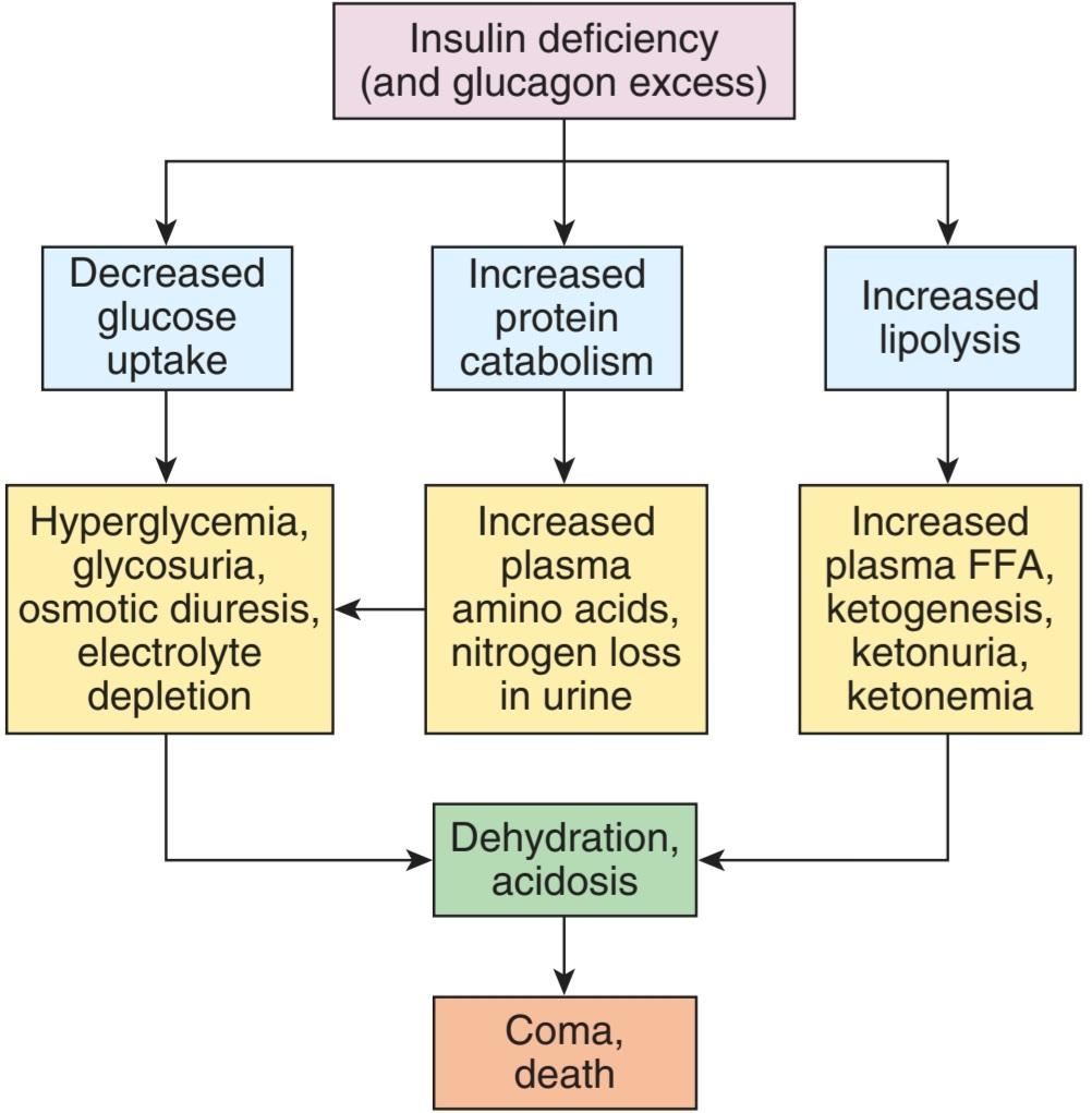
FIGURE 24-9 Effects of insulin deficiency. FFA, free fatty acids. (Used with permission of RJ Havel.)
شکل 24-9 اثرات کمبود انسولین. FFA، اسیدهای چرب آزاد. (با اجازه RJ Havel استفاده می شود.)
Fat catabolism is increased and the system is flooded with triglycerides and FFA. Fat synthesis is inhibited and the overloaded catabolic pathways cannot handle the excess acetyl-CoA that is formed. In the liver, the acetyl-CoA is converted to ketone bodies. Two of these are organic acids, and metabolic acidosis develops as ketones accumulate. Na* and K+ depletion is added to the acidosis because these plasma cations are excreted with the organic anions not covered by the H+ and NH4+ secreted by the kidneys. Finally, the acidotic, hypovolemic, hypotensive, depleted animal or patient becomes comatose because of the toxic effects of acidosis, dehydration, and hyperosmolarity on the nervous system and dies if treatment is not instituted.
کاتابولیسم چربی افزایش یافته و سیستم پر از تری گلیسیرید و FFA می شود. سنتز چربی مهار می شود و مسیرهای کاتابولیک بیش از حد نمی توانند استیل-CoA اضافی تشکیل شده را کنترل کنند. در کبد، استیل کوآ به اجسام کتون تبدیل می شود. دو مورد از آنها اسیدهای آلی هستند و اسیدوز متابولیک با تجمع کتون ها ایجاد می شود. کاهش +Na و +K به اسیدوز اضافه میشود، زیرا این کاتیونهای پلاسما با آنیونهای آلی که توسط +H و +NH4 ترشح شده از کلیهها پوشیده نشدهاند دفع میشوند. در نهایت، حیوان یا بیمار دارای اسیدوز، هیپوولمیک، کاهش فشار خون، ضعیف شده به دلیل اثرات سمی اسیدوز، کم آبی و هیپراسمولاریته روی سیستم عصبی به کما می رود و در صورت عدم شروع درمان می میرد.
All of these abnormalities are corrected by administration of insulin. Although emergency treatment of acidosis also includes administration of alkali to combat the acidosis as well as parenteral water, Na+, and K* to replenish body stores, only insulin repairs the fundamental defects in a way that permits a return to normal.
تمام این ناهنجاری ها با تجویز انسولین اصلاح می شوند. اگرچه درمان اورژانسی اسیدوز شامل تجویز قلیایی برای مبارزه با اسیدوز و همچنین آب تزریقی، +Na و +K برای پر کردن ذخایر بدن میشود، فقط انسولین نقصهای اساسی را به گونهای ترمیم میکند که امکان بازگشت به حالت عادی را فراهم کند.
INSULIN EXCESS
SYMPTOMS
All the known consequences of insulin excess are manifestations, directly or indirectly, of the effects of hypoglycemia on the nervous system. Except in individuals who have been fasting for some time, glucose is the only fuel used in appreciable quantities by the brain. The carbohydrate reserves in neural tissue are very limited and normal function depends on a continuous glucose supply. As the plasma glucose level falls, the first symptoms are palpitations, sweating, and nervousness due to autonomic discharge. These appear at plasma glucose values slightly lower than the value at which autonomic activation first begins, because the threshold for symptoms is slightly above the threshold for initial activation. At lower plasma glucose levels, so-called neuroglycopenic symptoms begin to appear. These include hunger as well as confusion and the other cognitive abnormalities. At even lower plasma glucose levels, lethargy, coma, convulsions, and eventually death occur. Obviously, the onset of hypoglycemic symptoms calls for prompt treatment with glucose or glucose- containing drinks such as orange juice. Although a dramatic disappearance of symptoms is the usual response, abnormalities ranging from intellectual dulling to coma may persist if the hypoglycemia was severe or prolonged.
انسولین اضافی
علائم
تمام عواقب شناخته شده بیش از حد انسولین، تظاهرات مستقیم یا غیرمستقیم اثرات هیپوگلیسمی بر روی سیستم عصبی است. به جز در افرادی که مدتی ناشتا بوده اند، گلوکز تنها سوختی است که به مقدار قابل توجهی توسط مغز استفاده می شود. ذخایر کربوهیدرات در بافت عصبی بسیار محدود است و عملکرد طبیعی به عرضه مداوم گلوکز بستگی دارد. با کاهش سطح گلوکز پلاسما، اولین علائم تپش قلب، تعریق و عصبی بودن ناشی از ترشحات اتونومیک است. اینها در مقادیر گلوکز پلاسما کمی کمتر از مقداری که برای اولین بار فعال سازی اتونومیک شروع می شود ظاهر می شوند، زیرا آستانه علائم کمی بالاتر از آستانه فعال سازی اولیه است. در سطوح پایینتر گلوکز پلاسما، علائم به اصطلاح نوروگلیکوپنیک ظاهر میشوند. اینها شامل گرسنگی و همچنین سردرگمی و سایر ناهنجاری های شناختی است. حتی در سطوح پایین تر گلوکز پلاسما، بی حالی، کما، تشنج و در نهایت مرگ رخ می دهد. بدیهی است که شروع علائم هیپوگلیسمی نیاز به درمان سریع با گلوکز یا نوشیدنی های حاوی گلوکز مانند آب پرتقال دارد. اگرچه ناپدید شدن چشمگیر علائم پاسخ معمول است، اما در صورتی که هیپوگلیسمی شدید یا طولانی مدت باشد، ناهنجاریهایی از تیرگی ذهنی تا کما ممکن است باقی بماند.
COMPENSATORY MECHANISMS
One important compensation for hypoglycemia is cessation of the secretion of endogenous insulin. Inhibition of insulin secretion is complete at a plasma glucose level of about 80 mg/dL (Figure 24–10). In addition, hypoglycemia triggers increased secretion of at least four counterregulatory hormones: glucagon, epinephrine, growth hormone, and cortisol. The epinephrine response is reduced during sleep. Glucagon and epinephrine increase the hepatic output of glucose by increasing glycogenolysis. Growth hormone decreases the utilization of glucose in various peripheral tissues, and cortisol has a similar action. The keys to counterregulation appear to be epinephrine and glucagon: if the plasma concentration of either increases, the decline in the plasma glucose level is reversed; but if both fail to increase, there is little if any compensatory rise in the plasma glucose level. The actions of the other hormones are supplementary.
مکانیسم های جبرانی
یکی از جبران مهم هیپوگلیسمی، توقف ترشح انسولین درون زا است. مهار ترشح انسولین در سطح گلوکز پلاسما حدود 80 میلی گرم در دسی لیتر کامل می شود (شکل 24-10). علاوه بر این، هیپوگلیسمی باعث افزایش ترشح حداقل چهار هورمون ضد تنظیمی می شود: گلوکاگون، اپی نفرین، هورمون رشد و کورتیزول. پاسخ اپی نفرین در طول خواب کاهش می یابد. گلوکاگون و اپی نفرین با افزایش گلیکوژنولیز برون ده گلوکز کبدی را افزایش می دهند. هورمون رشد استفاده از گلوکز را در بافت های مختلف محیطی کاهش می دهد و کورتیزول نیز عملکرد مشابهی دارد. به نظر می رسد کلیدهای ضد تنظیم اپی نفرین و گلوکاگون هستند: اگر غلظت هر یک از آنها در پلاسما افزایش یابد، کاهش سطح گلوکز پلاسما معکوس می شود. اما اگر هر دو نتوانند افزایش پیدا کنند، افزایش اندکی و یا جبرانی در سطح گلوکز پلاسما وجود دارد. اعمال سایر هورمون ها مکمل هستند.
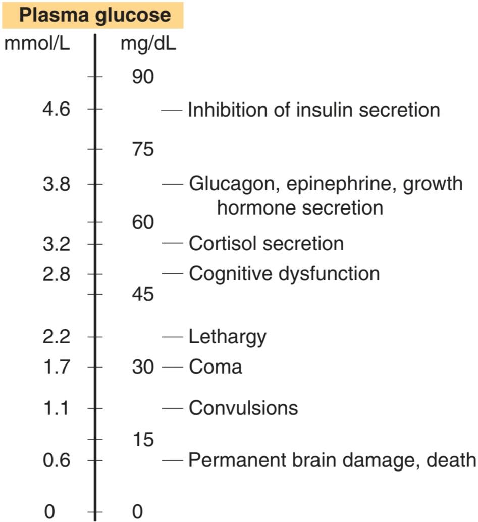
FIGURE 24-10 Plasma glucose levels at which various effects of hypoglycemia appear.
شکل 24-10 سطوح گلوکز پلاسما که در آن اثرات مختلف هیپوگلیسمی ظاهر می شود.
Note that the autonomic discharge and release of counterregulatory hormones normally occur at a higher plasma glucose level than the cognitive deficits and other more serious CNS changes (Figure 24–10). For diabetics treated with insulin, the symptoms caused by the autonomic discharge serve as a warning to seek glucose replacement. However, particularly in long-term diabetics who have been tightly regulated, the autonomic symptoms may not occur, and the resulting hypoglycemia unawareness can be a clinical problem of some magnitude.
توجه داشته باشید که ترشحات خودمختار و آزادسازی هورمون های ضد تنظیمی معمولاً در سطح گلوکز پلاسما بالاتر از نقص های شناختی و سایر تغییرات جدی تر CNS رخ می دهد (شکل 24-10). برای بیماران دیابتی که با انسولین درمان می شوند، علائم ناشی از ترشحات خودکار به عنوان یک هشدار برای جستجوی جایگزینی گلوکز است. با این حال، بهویژه در دیابتیهای طولانیمدت که به شدت تحت کنترل قرار گرفتهاند، علائم خودمختار ممکن است رخ ندهد و ناآگاهی ناشی از هیپوگلیسمی میتواند یک مشکل بالینی بزرگ باشد.
REGULATION OF INSULIN SECRETION
The normal concentration of insulin measured by radioimmunoassay in the peripheral venous plasma of fasting normal humans is 0-70 μU/mL (0-502 pmol/L). The amount of insulin secreted in the basal state is about 1 unit/h, with a 5-fold to 10-fold increase following ingestion of food. Therefore, the average amount secreted per day in a normal human is about 40 units (287 nmol).
تنظیم ترشح انسولین
غلظت طبیعی انسولین اندازهگیری شده توسط رادیوامونواسی در پلاسمای وریدی محیطی انسانهای عادی ناشتا 70-0 میکروU/mL (0-502 pmol/L) است. مقدار انسولین ترشح شده در حالت پایه حدود 1 واحد در ساعت است که پس از مصرف غذا 5 تا 10 برابر افزایش می یابد. بنابراین، مقدار متوسط ترشح شده در روز در یک انسان عادی حدود 40 واحد (287 نانومول) است.
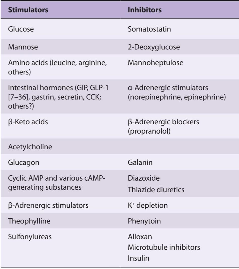
Factors that stimulate and inhibit insulin secretion are summarized in Table 24-4.
عواملی که باعث تحریک و مهار ترشح انسولین می شوند در جدول 24-4 خلاصه شده است.
TABLE 24-4 Factors affecting insulin secretion.
جدول 24-4 عوامل مؤثر بر ترشح انسولین.
EFFECTS OF THE PLASMA GLUCOSE LEVEL
It has been known for many years that glucose acts directly on pancreatic B cells to increase insulin secretion. The response to glucose is biphasic; there is a rapid but short-lived increase in secretion followed by a more slowly developing prolonged increase.
اثرات سطح گلوکز پلاسما
سال هاست که مشخص شده است که گلوکز مستقیماً روی سلول های B پانکراس برای افزایش ترشح انسولین اثر می گذارد. پاسخ به گلوکز دو فازی است. یک افزایش سریع اما کوتاه مدت در ترشح و به دنبال آن افزایش طولانی مدت آهسته تر وجود دارد.
Glucose enters the B cells via GLUT-2 transporters and is phosphorylated by glucokinase then metabolized to pyruvate in the cytoplasm (Figure 24-11). The pyruvate enters the mitochondria and is metabolized to CO2 and H2O via the citric acid cycle with the formation of ATP by oxidative phosphorylation. The ATP enters the cytoplasm, where it inhibits ATP-sensitive K+ channels, reducing K* efflux. This depolarizes the B cell, and Ca2+ enters the cell via voltage-gated Ca2+ channels. The Ca2+ influx causes exocytosis of a readily releasable pool of insulin-containing secretory granules, producing the initial spike of insulin secretion.
گلوکز از طریق انتقال دهنده های GLUT-2 وارد سلول های B می شود و توسط گلوکوکیناز فسفریله می شود و سپس به پیرووات در سیتوپلاسم متابولیزه می شود (شکل 24-11). پیروات وارد میتوکندری شده و از طریق چرخه اسید سیتریک با تشکیل ATP توسط فسفوریلاسیون اکسیداتیو به CO2 و H2O متابولیزه می شود. ATP وارد سیتوپلاسم می شود، جایی که کانال های +K حساس به ATP را مهار می کند و جریان +K را کاهش می دهد. این باعث دپلاریزه شدن سلول B می شود و +Ca2 از طریق کانال های +Ca2 دارای ولتاژ وارد سلول می شود. هجوم +Ca2 باعث اگزوسیتوز حوضچههای ترشحی حاوی انسولین میشود که به آسانی آزاد میشوند و باعث تولید نوک اولیه ترشح انسولین میشوند.
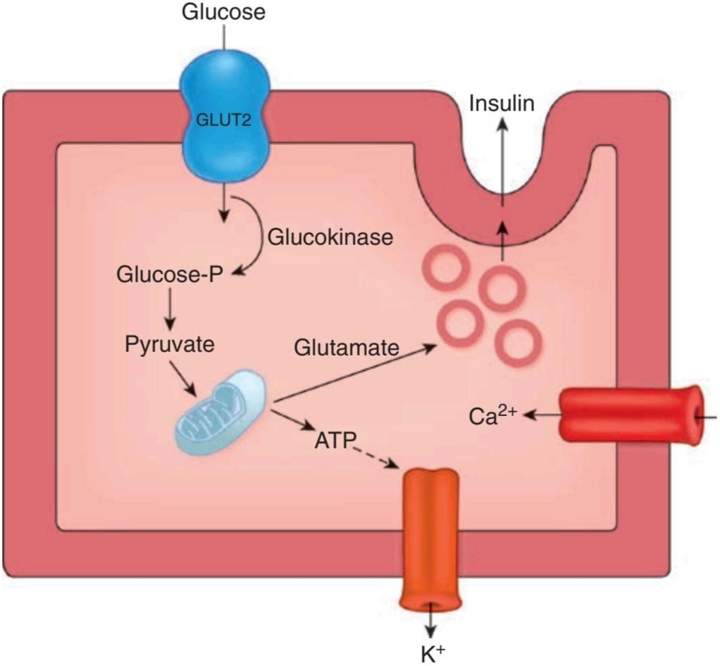
FIGURE 24-11 Insulin secretion. Glucose enters B cells by GLUT-2 transporters. It is phosphorylated and metabolized to pyruvate (Pyr) in the cytoplasm. The Pyr enters the mitochondria and is metabolized via the citric acid cycle. The ATP formed by oxidative phosphorylation inhibits ATP-sensitive K+ channels, reducing K+ efflux. This depolarizes the B cell, and Ca2+ influx is increased. The Ca2+ stimulates release of insulin by exocytosis. Glutamate (Glu) is also formed, and this primes secretory granules, preparing them for exocytosis.
شکل 24-11 ترشح انسولین. گلوکز توسط انتقال دهنده های GLUT-2 وارد سلول های B می شود. فسفریله شده و در سیتوپلاسم به پیرووات (Pyr) متابولیزه می شود. Pyr وارد میتوکندری می شود و از طریق چرخه اسید سیتریک متابولیزه می شود. ATP تشکیل شده توسط فسفوریلاسیون اکسیداتیو کانال های +K حساس به ATP را مهار می کند و جریان +K را کاهش می دهد. این امر سلول B را دپولاریزه می کند و هجوم +Ca2 افزایش می یابد. +Ca2 آزادسازی انسولین را با اگزوسیتوز تحریک می کند. گلوتامات (Glu) نیز تشکیل میشود و گرانولهای ترشحی را آغاز میکند و آنها را برای اگزوسیتوز آماده میکند.
Metabolism of pyruvate via the citric acid cycle also causes an increase in intracellular glutamate. The glutamate appears to act on a second pool of secretory granules, committing them to the releasable form. The action of glutamate may be to decrease the pH in the secretory granules, a necessary step in their maturation. The release of these granules then produces the prolonged second phase of the insulin response to glucose. Thus, glutamate appears to act as an intracellular second messenger that primes secretory granules for secretion.
متابولیسم پیروات از طریق چرخه اسید سیتریک نیز باعث افزایش گلوتامات داخل سلولی می شود. به نظر می رسد گلوتامات روی دومین حوضچه گرانول های ترشحی عمل می کند و آنها را به شکل آزاد شدنی متعهد می کند. عمل گلوتامات ممکن است کاهش pH در گرانول های ترشحی باشد که یک مرحله ضروری در بلوغ آنهاست. سپس آزاد شدن این گرانول ها فاز دوم طولانی پاسخ انسولین به گلوکز را ایجاد می کند. بنابراین، به نظر می رسد گلوتامات به عنوان یک پیام رسان دوم درون سلولی عمل می کند که دانه های ترشحی را برای ترشح آماده می کند.
The feedback control of plasma glucose on insulin secretion normally operates with great precision so that plasma glucose and insulin levels parallel each other with remarkable consistency.
کنترل بازخورد گلوکز پلاسما بر ترشح انسولین معمولاً با دقت زیادی عمل می کند به طوری که سطح گلوکز و انسولین پلاسما با یک ثبات قابل توجه با یکدیگر موازی می شوند.
PROTEIN & FAT DERIVATIVES
Insulin stimulates the incorporation of amino acids into proteins and combats the fat catabolism that produces the B-keto acids. Therefore, it is not surprising that arginine, leucine, and certain other amino acids stimulate insulin secretion, as do B-keto acids such as acetoacetate. Like glucose, these compounds generate ATP when metabolized, and this closes ATP-sensitive K+ channels in the B cells. In addition, L-arginine is the precursor of NO, and NO stimulates insulin secretion.
پروتئین و مشتقات چربی
انسولین ترکیب اسیدهای آمینه را در پروتئین ها تحریک می کند و با کاتابولیسم چربی که اسیدهای B-keto را تولید می کند، مبارزه می کند. بنابراین، جای تعجب نیست که آرژنین، لوسین و برخی اسیدهای آمینه دیگر مانند اسیدهای B-کتو مانند استواستات ترشح انسولین را تحریک می کنند. مانند گلوکز، این ترکیبات هنگام متابولیزه شدن، ATP تولید می کنند و کانال های +K حساس به ATP را در سلول های B می بندند. علاوه بر این، ال آرژنین پیش ساز NO است و NO ترشح انسولین را تحریک می کند.
ORAL HYPOGLYCEMIC AGENTS
Tolbutamide and other sulfonylurea derivatives such as acetohexamide, tolazamide, glipizide, and glyburide are orally active hypoglycemic agents that lower blood glucose by increasing the secretion of insulin. They only work in patients with some remaining B cells and are ineffective after pancreatectomy or in type 1 diabetes. They bind to the ATP-inhibited K+ channels in the B cell membranes and inhibit channel activity, depolarizing the B cell membrane and increasing Ca2+ influx and hence insulin release, independent of increases in plasma glucose.
عوامل هیپوگلیسمی خوراکی
تولبوتامید و سایر مشتقات سولفونیل اوره مانند استوهگزامید، تولازامید، گلیپیزید و گلیبورید از عوامل کاهش دهنده قند خون خوراکی هستند که با افزایش ترشح انسولین، گلوکز خون را کاهش می دهند. آنها فقط در بیمارانی که تعدادی سلول B باقی مانده دارند کار می کنند و بعد از پانکراتکتومی یا در دیابت نوع 1 بی اثر هستند. آنها به کانال های +K مهار شده با ATP در غشای سلول B متصل می شوند و فعالیت کانال را مهار می کنند، غشای سلول B را دپولاریزه می کنند و هجوم +Ca2 و در نتیجه آزاد شدن انسولین را، مستقل از افزایش گلوکز پلاسما، افزایش می دهند.
Persistent hyperinsulinemic hypoglycemia of infancy is a condition in which plasma insulin is elevated despite the hypoglycemia. The condition is caused by mutations in the genes for various enzymes in B cells that decrease K+ efflux via the ATP-sensitive K+ channels. Treatment consists of administration of diazoxide, a drug that increases the activity of the K+ channels or, in more severe cases, subtotal pancreatectomy.
هیپوگلیسمی هیپرانسولینمی مداوم در دوران نوزادی وضعیتی است که در آن انسولین پلاسما با وجود هیپوگلیسمی افزایش می یابد. این وضعیت به دلیل جهش در ژن های آنزیم های مختلف در سلول های B ایجاد می شود که جریان +K را از طریق کانال های +K حساس به ATP کاهش می دهد. درمان شامل تجویز دیازوکساید، دارویی است که فعالیت کانال های +K را افزایش می دهد یا در موارد شدیدتر، پانکراتکتومی ساب توتال.
The biguanide metformin is an oral hypoglycemic agent that acts in the absence of insulin. Metformin acts primarily by reducing gluconeogenesis and therefore decreasing hepatic glucose output. It is sometimes combined with a sulfonylurea in the treatment of type 2 diabetes. Metformin can cause lactic acidosis, but the incidence is usually low.
بیگوانید متفورمین یک عامل کاهش دهنده قند خون خوراکی است که در غیاب انسولین عمل می کند. متفورمین در درجه اول با کاهش گلوکونئوژنز و در نتیجه کاهش برون ده گلوکز کبدی عمل می کند. گاهی اوقات در درمان دیابت نوع 2 با سولفونیل اوره ترکیب می شود. متفورمین می تواند باعث اسیدوز لاکتیک شود، اما بروز آن معمولا کم است.
Troglitazone (Rezulin) and related thiazolidinediones are also used in the treatment of diabetes because they increase insulin-mediated peripheral glucose disposal, thus reducing insulin resistance. They bind to and activate peroxisome proliferator-activated receptor y (PPARY) in the nucleus of cells. Activation of this receptor, which is a member of the superfamily of hormone-sensitive nuclear transcription factors, has a unique ability to normalize a variety of metabolic functions.
تروگلیتازون (رزولین) و تیازولیدین دیونهای مرتبط نیز در درمان دیابت استفاده میشوند، زیرا باعث افزایش دفع گلوکز محیطی با واسطه انسولین میشوند و در نتیجه مقاومت به انسولین را کاهش میدهند. آنها به گیرنده y فعال شده با تکثیر کننده پراکسی زوم (PPARY) در هسته سلول ها متصل می شوند و آنها را فعال می کنند. فعال شدن این گیرنده که عضوی از خانواده فاکتورهای رونویسی هسته ای حساس به هورمون است، توانایی منحصر به فردی در عادی سازی انواع عملکردهای متابولیک دارد.
CYCLIC AMP & INSULIN SECRETION
Stimuli that increase cAMP levels in B cells increase insulin secretion, including B-adrenergic agonists, glucagon, and phosphodiesterase inhibitors such as theophylline.
AMP چرخه ای و ترشح انسولین
محرک هایی که سطح cAMP را در سلول های B افزایش می دهند، ترشح انسولین را افزایش می دهند، از جمله آگونیست های آدرنرژیک B، گلوکاگون و مهارکننده های فسفودی استراز مانند تئوفیلین.
Catecholamines have a dual effect on insulin secretion; they inhibit insulin secretion via α2-adrenergic receptors and stimulate insulin secretion via ẞ- adrenergic receptors. The net effect of epinephrine and norepinephrine is usually inhibition. However, if catecholamines are infused after administration of a- adrenergic blocking drugs, the inhibition is converted to stimulation.
کاتکولامین ها اثر دوگانه ای بر ترشح انسولین دارند. آنها ترشح انسولین را از طریق گیرنده های α2-آدرنرژیک مهار می کنند و ترشح انسولین را از طریق گیرنده های آدرنرژیک تحریک می کنند. اثر خالص اپی نفرین و نوراپی نفرین معمولاً مهار است. با این حال، اگر کاتکول آمین ها پس از تجویز داروهای مسدودکننده آدرنرژیک تزریق شوند، مهار به تحریک تبدیل می شود.
EFFECT OF AUTONOMIC NERVES
Branches of the right vagus nerve innervate the pancreatic islets, and stimulation of this parasympathetic pathway causes increased insulin secretion via M3 receptors (see Table 7-2). Atropine blocks the response and acetylcholine stimulates insulin secretion. The effect of acetylcholine, like that of glucose, is due to increased cytoplasmic Ca2+, but acetylcholine activates phospholipase C, with the released IP3 releasing the Ca2+ from the endoplasmic reticulum.
اثر اعصاب خودگردان
شاخه های عصب واگ راست جزایر پانکراس را عصب دهی می کنند و تحریک این مسیر پاراسمپاتیک باعث افزایش ترشح انسولین از طریق گیرنده های M3 می شود (جدول 7-2 را ببینید). آتروپین پاسخ را مسدود می کند و استیل کولین ترشح انسولین را تحریک می کند. اثر استیل کولین، مانند گلوکز، به دلیل افزایش +Ca2 سیتوپلاسمی است، اما استیل کولین فسفولیپاز C را فعال می کند، با IP3 آزاد شده، +Ca2 را از شبکه آندوپلاسمی آزاد می کند.
Stimulation of the sympathetic nerves to the pancreas inhibits insulin secretion. The inhibition is produced by released norepinephrine acting on α2- adrenergic receptors. However, if a-adrenergic receptors are blocked, stimulation of the sympathetic nerves causes increased insulin secretion mediated by ẞ2- adrenergic receptors. The polypeptide galanin is found in some of the autonomic nerves innervating the islets, and galanin inhibits insulin secretion by activating the K+ channels that are inhibited by ATP. Thus, although the denervated pancreas responds to glucose, the autonomic innervation of the pancreas is involved in the overall regulation of insulin secretion (Clinical Box 24-3).
تحریک اعصاب سمپاتیک به پانکراس باعث مهار ترشح انسولین می شود. این مهار توسط نوراپی نفرین آزاد شده ایجاد می شود که بر روی گیرنده های α2- آدرنرژیک اثر می کند. با این حال، اگر گیرنده های a-آدرنرژیک مسدود شوند، تحریک اعصاب سمپاتیک باعث افزایش ترشح انسولین با واسطه گیرنده های آدرنرژیک 2 می شود. پلی پپتید گالانین در برخی از اعصاب خودمختار که جزایر را عصب میکنند یافت میشود و گالانین با فعال کردن کانالهای +K که توسط ATP مهار میشوند، ترشح انسولین را مهار میکند. بنابراین، اگرچه پانکراس عصبکشی شده به گلوکز پاسخ میدهد، عصب خودکار پانکراس در تنظیم کلی ترشح انسولین نقش دارد (کادر بالینی 24-3).
INTESTINAL HORMONES
Orally administered glucose exerts a greater insulin-stimulating effect than intravenously administered glucose, and orally administered amino acids also produce a greater insulin response than intravenous amino acids. These observations led to exploration of the possibility that a substance secreted by the gastrointestinal mucosa stimulated insulin secretion. Glucagon, glucagon derivatives, secretin, cholecystokinin (CCK), gastrin, and gastric inhibitory peptide (GIP) all have such an action (see Chapter 25), and CCK potentiates the insulin-stimulating effects of amino acids. However, GIP is the only one of these peptides that produces stimulation when administered in doses that reflect blood GIP levels produced by an oral glucose load.
هورمون های روده ای
گلوکز خوراکی نسبت به گلوکز داخل وریدی اثر تحریک انسولین بیشتری دارد و آمینو اسیدهای خوراکی نیز پاسخ انسولین بیشتری نسبت به اسیدهای آمینه داخل وریدی ایجاد می کنند. این مشاهدات منجر به بررسی این احتمال شد که ماده ای که از مخاط دستگاه گوارش ترشح می شود باعث تحریک ترشح انسولین شود. گلوکاگون، مشتقات گلوکاگون، سکرتین، کوله سیستوکینین (CCK)، گاسترین و پپتید بازدارنده معده (GIP) همگی چنین عملکردی دارند (به فصل 25 مراجعه کنید)، و CCK اثرات تحریک کننده انسولین اسیدهای آمینه را تقویت می کند. با این حال، GIP تنها یکی از این پپتیدها است که در صورت تجویز در دوزهایی که منعکس کننده سطوح GIP خون تولید شده توسط بار گلوکز خوراکی است، تحریک ایجاد می کند.
CLINICAL BOX 24-3
Effects of K+ Depletion
K+ depletion decreases insulin secretion, and K+-depleted patients, for example, patients with primary hyperaldoster-onism (see Chapter 20), develop diabetic glucose tolerance curves. These curves are restored to normal by K+ repletion.
جعبه بالینی 24-3
اثرات کاهش +K
کاهش +K ترشح انسولین را کاهش می دهد و بیماران با کمبود +K، برای مثال، بیماران مبتلا به هیپرآلدوستر-آنیسم اولیه (به فصل 20 مراجعه کنید)، منحنی های تحمل گلوکز دیابتی را ایجاد می کنند. این منحنی ها با بازیابی +K به حالت عادی باز می گردند.
THERAPEUTIQ HIGHLIGHTS
The thiazide diuretics, which cause loss of K* as well as Na+ in the urine (see Chapter 37), decrease glucose tolerance and make diabetes worse. They apparently exert this effect primarily because of their K*-depleting effects, although some of them also cause pancreatic islet cell damage. Potassium- sparing diuretics, such as amiloride, should be substituted in the diabetic patient who needs such treatment.
نکات برجسته درمانی
دیورتیک های تیازیدی که باعث از دست دادن +K و همچنین +Na در ادرار می شوند (به فصل 37 مراجعه کنید)، تحمل گلوکز را کاهش داده و دیابت را بدتر می کنند. ظاهراً آنها این اثر را عمدتاً به دلیل اثرات کاهش دهنده K * اعمال می کنند، اگرچه برخی از آنها باعث آسیب سلول های جزایر پانکراس نیز می شوند. دیورتیک های نگهدارنده پتاسیم، مانند آمیلوراید، باید در بیماران دیابتی که نیاز به چنین درمانی دارند جایگزین شوند.
Recently, attention has focused on glucagon-like polypeptide 1 (7–36) (GLP- 1 [7-36]) as an additional gut factor that stimulates insulin secretion. This polypeptide is a product of preproglucagon. B cells have GLP-1 (7–36) receptors as well as GIP receptors, and GLP-1 (7-36) is a more potent insulinotropic hormone than GIP. GIP and GLP-1 (7-36) both appear to act by increasing Ca2+ influx through voltage-gated Ca2+ channels.
اخیراً، توجه به پلی پپتید 1 شبه گلوکاگون (7-36) (GLP-1 [7-36]) به عنوان یک عامل روده اضافی که ترشح انسولین را تحریک می کند، متمرکز شده است. این پلی پپتید محصول پیش پروگلوکاگون است. سلول های B دارای گیرنده های GLP-1 (7-36) و همچنین گیرنده های GIP هستند و GLP-1 (7-36) یک هورمون انسولینوتروپیک قوی تر از GIP است. به نظر می رسد GIP و GLP-1 (7-36) هر دو با افزایش هجوم +Ca2 از طریق کانال های +Ca2 دارای ولتاژ عمل می کنند.
The possible roles of pancreatic somatostatin and glucagon in the regulation of insulin secretion are discussed below.
نقش احتمالی سوماتوستاتین پانکراس و گلوکاگون در تنظیم ترشح انسولین در زیر مورد بحث قرار گرفته است.
LONG-TERM CHANGES IN B CELL RESPONSES
The magnitude of the insulin response to a given stimulus is determined in part by the secretory history of the B cells. Individuals fed a high-carbohydrate diet for several weeks not only have higher fasting plasma insulin levels but also show a greater secretory response to a glucose load than individuals fed an isocaloric low-carbohydrate diet.
تغییرات دراز مدت در پاسخ های سلول B
میزان پاسخ انسولین به یک محرک مشخص تا حدی توسط تاریخچه ترشحی سلول های B تعیین می شود. افرادی که به مدت چندین هفته از رژیم غذایی پر کربوهیدرات تغذیه میشوند، نه تنها سطح انسولین ناشتا پلاسمایی بالاتری دارند، بلکه نسبت به افرادی که از رژیم کم کربوهیدرات ایزوکالری تغذیه میکنند، پاسخ ترشحی بیشتری به بار گلوکز نشان میدهند.
Although B cells respond to stimulation with hypertrophy like other endocrine cells, they become exhausted and stop secreting (B cell exhaustion) when the stimulation is marked or prolonged. The pancreatic reserve is large and it is difficult to produce B cell exhaustion in normal animals, but if the pancreatic reserve is reduced by partial pancreatectomy, exhaustion of the remaining B cells can be initiated by any procedure that chronically raises the plasma glucose level. For example, diabetes can be produced in animals with limited pancreatic reserves by anterior pituitary extracts, growth hormone, thyroid hormones, or the prolonged continuous infusion of glucose alone. The diabetes precipitated by hormones in animals is at first reversible, but with prolonged treatment it becomes permanent. The transient diabetes is usually named for the agent producing it, for example, “hypophysial diabetes” or “thyroid diabetes.” Permanent diabetes persisting after treatment has been discontinued is indicated by the prefix meta-, for example, “metahypophysial diabetes” or “metathyroid diabetes.” When insulin is administered along with the diabetogenic hormones, the B cells are protected, probably because the plasma glucose is lowered, and diabetes does not develop.
اگرچه سلولهای B مانند سایر سلولهای غدد درون ریز به تحریک با هیپرتروفی پاسخ میدهند، اما وقتی تحریک مشخص یا طولانی شد، خسته میشوند و ترشحشان متوقف میشود (خستگی سلول B). ذخیره لوزالمعده زیاد است و ایجاد فرسودگی سلول های B در حیوانات عادی دشوار است، اما اگر ذخیره پانکراس با پانکراتکتومی نسبی کاهش یابد، فرسودگی سلول های B باقی مانده را می توان با هر روشی که به طور مزمن سطح گلوکز پلاسما را بالا می برد، آغاز کرد. به عنوان مثال، دیابت می تواند در حیوانات با ذخایر محدود لوزالمعده توسط عصاره هیپوفیز قدامی، هورمون رشد، هورمون های تیروئید یا تزریق مداوم گلوکز به تنهایی تولید شود. دیابت ایجاد شده توسط هورمون ها در حیوانات ابتدا قابل برگشت است، اما با درمان طولانی مدت دائمی می شود. دیابت گذرا معمولاً برای عامل تولید کننده آن نامگذاری می شود، به عنوان مثال، “دیابت هیپوفیزیال” یا “دیابت تیروئید”. دیابت دائمی که پس از قطع درمان ادامه می یابد با پیشوند متا نشان داده می شود، به عنوان مثال، “دیابت متا هیپوفیزیال” یا “دیابت متاتیروئید”. هنگامی که انسولین همراه با هورمون های دیابت زا تجویز می شود، سلول های B محافظت می شوند، احتمالاً به این دلیل که گلوکز پلاسما کاهش می یابد و دیابت ایجاد نمی شود.
It is interesting in this regard that genetic factors may be involved in the control of B cell reserve. In mice in which the gene for IRS-1 has been knocked out (see above), a robust compensatory B cell response occurs. However, in IRS- 2 knockouts, the compensation is reduced and a more severe diabetic phenotype is produced.
از این نظر جالب است که عوامل ژنتیکی ممکن است در کنترل ذخیره سلول B نقش داشته باشند. در موش هایی که ژن IRS-1 در آنها از بین رفته است (به بالا مراجعه کنید)، یک پاسخ جبرانی قوی سلول B رخ می دهد. با این حال، در ناک اوت IRS-2، جبران کاهش می یابد و یک فنوتیپ دیابتی شدیدتر تولید می شود.
GLUCAGON
CHEMISTRY
Human glucagon, a linear polypeptide with a molecular weight of 3485, is produced by the A cells of the pancreatic islets and the upper gastrointestinal tract. It contains 29 amino acid residues. All mammalian glucagons appear to have the same structure. Human preproglucagon (Figure 24-12) is a 179-amino- acid protein that is found in pancreatic A cells, in L cells in the lower gastrointestinal tract, and in the brain. It is the product of a single mRNA, but it is processed differently in different tissues. In A cells, it is processed primarily to glucagon and major proglucagon fragment (MPGF). In L cells, it is processed primarily to glicentin, a polypeptide that consists of glucagon extended by additional amino acid residues at either end, plus glucagon-like polypeptides 1 and 2 (GLP-1 and GLP-2). Some oxyntomodulin is also formed, and in both A and L cells, residual glicentin-related polypeptide (GRPP) is left. Glicentin has some glucagon activity. GLP-1 and GLP-2 have no definite biologic activity by themselves. However, GLP-1 is processed further by removal of its amino- terminal amino acid residues and the product, GLP-1 (7-36), is a potent stimulator of insulin secretion that also increases glucose utilization (see above). GLP-1 and GLP-2 are also produced in the brain. The function of GLP-1 in this location is uncertain, but GLP-2 appears to be the mediator in a pathway from the nucleus tractus solitarius (NTS) to the dorsomedial nuclei of the hypothalamus, and injection of GLP-2 lowers food intake. Oxyntomodulin inhibits gastric acid secretion, though its physiologic role is unsettled, and GRPP does not have any established physiologic effects.
گلوکاگون
شیمی
گلوکاگون انسانی، یک پلی پپتید خطی با وزن مولکولی 3485، توسط سلول های A جزایر پانکراس و دستگاه گوارش فوقانی تولید می شود. حاوی 29 اسید آمینه باقی مانده است. به نظر می رسد که تمام گلوکاگون های پستانداران ساختار یکسانی دارند. پری پروگلوکاگون انسانی (شکل 24-12) یک پروتئین 179 اسید آمینه است که در سلول های A پانکراس، در سلول های L در دستگاه گوارش تحتانی و در مغز یافت می شود. این محصول یک mRNA واحد است، اما در بافت های مختلف به طور متفاوتی پردازش می شود. در سلول های A، عمدتاً به گلوکاگون و قطعه اصلی پروگلوکاگون (MPGF) پردازش می شود. در سلول های L، عمدتاً به گلیسنتین، پلی پپتیدی که متشکل از گلوکاگون است که توسط بقایای اسید آمینه اضافی در هر دو انتها گسترش می یابد، به علاوه پلی پپتیدهای 1 و 2 شبه گلوکاگون (GLP-1 و GLP-2) پردازش می شود. مقداری اکسینتومودولین نیز تشکیل می شود و در هر دو سلول A و L، پلی پپتید مرتبط با گلیسنتین (GRPP) باقی مانده است. گلیسنتین مقداری فعالیت گلوکاگون دارد. GLP-1 و GLP-2 به خودی خود هیچ فعالیت بیولوژیکی مشخصی ندارند. با این حال، GLP-1 با حذف بقایای اسید آمینه ترمینال آن بیشتر پردازش می شود و محصول GLP-1 (7-36)، یک محرک قوی ترشح انسولین است که استفاده از گلوکز را نیز افزایش می دهد (به بالا مراجعه کنید). GLP-1 و GLP-2 نیز در مغز تولید می شوند. عملکرد GLP-1 در این مکان نامشخص است، اما به نظر می رسد GLP-2 واسطه مسیری از هسته تراکتوس سولیتریوس (NTS) به هسته های پشتی میانی هیپوتالاموس باشد و تزریق GLP-2 مصرف غذا را کاهش می دهد. Oxyntomodulin ترشح اسید معده را مهار می کند، اگرچه نقش فیزیولوژیکی آن ثابت نشده است، و GRPP هیچ اثر فیزیولوژیکی ثابتی ندارد.

FIGURE 24-12 Posttranslational processing of preproglucagon in A and L cells. S, signal peptide; GRPP, glicentin-related polypeptide; GLP, glucagon-like polypeptide; Oxy, oxyntomodulin; MPGF, major proglucagon fragment. (Modified with permission from Drucker DJ: Glucagon and glucagon-like peptides. Pancreas 1990; July; 5(4):484–488.)
شکل 24-12 پردازش پس از ترجمه پیش پروگلوکاگون در سلولهای A و L. S، پپتید سیگنال. GRPP، پلی پپتید مرتبط با گلیسنتین؛ GLP، پلی پپتید شبه گلوکاگون؛ اکسی، اکسینتومودولین؛ MPGF، قطعه اصلی پروگلوکاگون. (اصلاح شده با اجازه Drucker DJ: Glucagon and glucagon-like peptides. Pancreas 1990; July; 5(4):484-488.)
ACTION
Glucagon is glycogenolytic, gluconeogenic, lipolytic, and ketogenic. It acts on G-protein-coupled receptors with a molecular weight of about 190,000. In the liver, it acts via G, to activate adenylyl cyclase and increase intracellular cAMP. This leads via protein kinase A to activation of phosphorylase and therefore to increased breakdown of glycogen and an increase in plasma glucose. However, glucagon acts on different glucagon receptors located on the same hepatic cells to activate phospholipase C, and the resulting increase in cytoplasmic Ca2+ also stimulates glycogenolysis. Protein kinase A also decreases the metabolism of glucose-6-phosphate (Figure 24-13) by inhibiting the conversion of phosphoenolpyruvate to pyruvate. It also decreases the concentration of fructose 2,6-diphosphate and this in turn inhibits the conversion of fructose 6-phosphate to fructose 1,6-diphosphate. The resultant buildup of glucose-6-phosphate leads to increased glucose synthesis and release.
ACTION
گلوکاگون گلیکوژنولیتیک، گلوکونئوژنیک، لیپولیتیک و کتوژنیک است. روی گیرنده های جفت شده با پروتئین G با وزن مولکولی حدود 190000 عمل می کند. در کبد، از طریق G برای فعال کردن آدنیلیل سیکلاز و افزایش cAMP داخل سلولی عمل می کند. این امر از طریق پروتئین کیناز A منجر به فعال شدن فسفوریلاز و در نتیجه افزایش تجزیه گلیکوژن و افزایش گلوکز پلاسما می شود. با این حال، گلوکاگون روی گیرندههای گلوکاگون مختلف واقع در سلولهای کبدی یکسان عمل میکند تا فسفولیپاز C را فعال کند و افزایش در نتیجه افزایش +Ca2 سیتوپلاسمی نیز باعث تحریک گلیکوژنولیز میشود. پروتئین کیناز A همچنین متابولیسم گلوکز-6-فسفات (شکل 24-13) را با مهار تبدیل فسفونول پیروات به پیرووات کاهش می دهد. همچنین غلظت فروکتوز 2،6-دی فسفات را کاهش می دهد و این به نوبه خود از تبدیل فروکتوز 6-فسفات به فروکتوز 1،6-دی فسفات جلوگیری می کند. تجمع گلوکز-6-فسفات منجر به افزایش سنتز و آزادسازی گلوکز می شود.

FIGURE 24-13 Mechanisms by which glucagon increases glucose output from the liver. Solid arrows indicate facilitation; dashed arrows indicate inhibition.
شکل 24-13 مکانیسم هایی که توسط آن گلوکاگون برون ده گلوکز را از کبد افزایش می دهد. فلش های ثابت نشان دهنده تسهیل است. فلش های چین نشان دهنده مهار است.
Glucagon does not cause glycogenolysis in muscle. It increases gluconeogenesis from available amino acids in the liver and elevates the metabolic rate. It increases ketone body formation by decreasing malonyl-CoA levels in the liver. Its lipolytic activity, which leads in turn to increased ketogenesis, is discussed in Chapter 1. The calorigenic action of glucagon is not due to the hyperglycemia per se but probably to the increased hepatic deamination of amino acids.
گلوکاگون باعث گلیکوژنولیز در عضلات نمی شود. گلوکونئوژنز از اسیدهای آمینه موجود در کبد را افزایش می دهد و سرعت متابولیسم را افزایش می دهد. با کاهش سطح مالونیل-CoA در کبد، تشکیل بدن کتون را افزایش می دهد. فعالیت لیپولیتیک آن، که به نوبه خود منجر به افزایش کتوژنز می شود، در فصل 1 مورد بحث قرار می گیرد. اثر کالری زایی گلوکاگون به خودی خود به دلیل هیپرگلیسمی نیست بلکه احتمالاً به دلیل افزایش دآمیناسیون کبدی اسیدهای آمینه است.
Large doses of exogenous glucagon exert a positive inotropic effect on the heart (see Chapter 30) without producing increased myocardial excitability, presumably because they increase myocardial cAMP. Use of this hormone in the treatment of heart disease has been advocated, but there is no evidence for a physiologic role of glucagon in the regulation of cardiac function. Glucagon also stimulates the secretion of growth hormone, insulin, and pancreatic somatostatin.
دوزهای زیاد گلوکاگون اگزوژن یک اثر اینوتروپیک مثبت بر قلب اعمال می کند (به فصل 30 مراجعه کنید) بدون ایجاد تحریک پذیری میوکارد، احتمالاً به این دلیل که cAMP میوکارد را افزایش می دهد. استفاده از این هورمون در درمان بیماری قلبی توصیه شده است، اما هیچ مدرکی دال بر نقش فیزیولوژیکی گلوکاگون در تنظیم عملکرد قلب وجود ندارد. گلوکاگون همچنین ترشح هورمون رشد، انسولین و سوماتوستاتین پانکراس را تحریک می کند.
METABOLISM
Glucagon has a half-life in the circulation of 5-10 min. It is degraded by many tissues but particularly by the liver. Because glucagon is secreted into the portal vein and reaches the liver before it reaches the peripheral circulation, peripheral blood levels are relatively low. The rise in peripheral blood glucagon levels produced by excitatory stimuli is exaggerated in patients with cirrhosis, presumably because of decreased hepatic degradation of the hormone.
متابولیسم
نیمه عمر گلوکاگون در گردش خون 10-5 دقیقه است. توسط بسیاری از بافت ها، به ویژه توسط کبد، تجزیه می شود. از آنجایی که گلوکاگون به داخل سیاهرگ باب ترشح می شود و قبل از رسیدن به گردش خون محیطی به کبد می رسد، سطح خون محیطی نسبتاً پایین است. افزایش سطح گلوکاگون خون محیطی تولید شده توسط محرک های تحریکی در بیماران مبتلا به سیروز، احتمالاً به دلیل کاهش تخریب کبدی هورمون، اغراق آمیز است.
REGULATION OF SECRETION
The principal factors known to affect glucagon secretion are summarized in Table 24-5. Secretion is increased by hypoglycemia and decreased by a rise in plasma glucose. Pancreatic B cells contain GABA, and evidence suggests that coincident with the increased insulin secretion produced by hyperglycemia, GABA is released and acts on the A cells to inhibit glucagon secretion by activating GABAA receptors. The GABAA receptors are Cl channels, and the resulting Cl-influx hyperpolarizes the A cells.
مقررات ترشح
عوامل اصلی موثر بر ترشح گلوکاگون در جدول 24-5 خلاصه شده است. ترشح با هیپوگلیسمی افزایش و با افزایش گلوکز پلاسما کاهش می یابد. سلول های B پانکراس حاوی GABA هستند و شواهد نشان می دهد که همزمان با افزایش ترشح انسولین تولید شده توسط هیپرگلیسمی، GABA آزاد می شود و بر روی سلول های A عمل می کند تا با فعال کردن گیرنده های گاباآ، ترشح گلوکاگون را مهار کند. گیرندههای گاباآ کانالهای کلر هستند و هجوم کلر حاصل، سلولهای A را هیپرپلاریزه میکند.
TABLE 24-5 Factors affecting glucagon secretion.
جدول 24-5 عوامل مؤثر بر ترشح گلوکاگون.
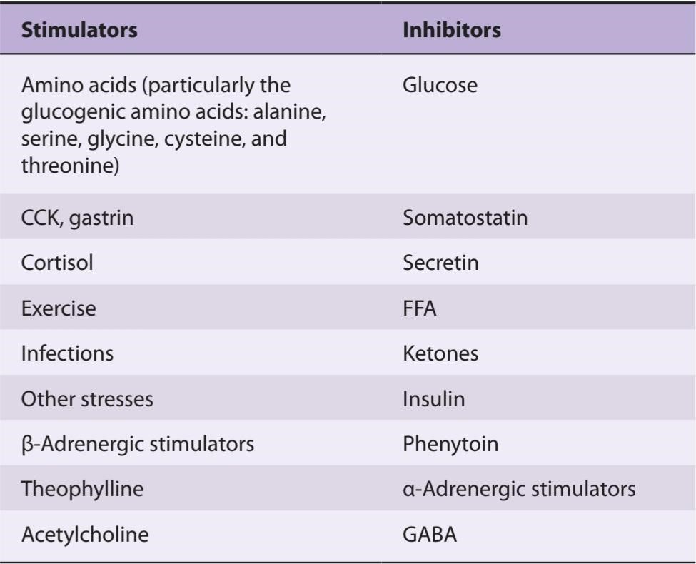
Secretion is also increased by stimulation of the sympathetic nerves to the pancreas, and this sympathetic effect is mediated via ẞ-adrenergic receptors and CAMP. It appears that the A cells are like the B cells in that stimulation of B- adrenergic receptors increases secretion and stimulation of a-adrenergic receptors inhibits secretion. However, the pancreatic response to sympathetic stimulation in the absence of blocking drugs is increased secretion of glucagon, so the effect of B-receptors predominates in the glucagon-secreting cells. The stimulatory effects of various stresses and possibly of exercise and infection are mediated at least in part via the sympathetic nervous system. Vagal stimulation also increases glucagon secretion.
ترشح همچنین با تحریک اعصاب سمپاتیک به لوزالمعده افزایش می یابد و این اثر سمپاتیک از طریق گیرنده های آدرنرژیک و CAMP انجام می شود. به نظر می رسد که سلول های A مانند سلول های B هستند که تحریک گیرنده های آدرنرژیک B ترشح را افزایش می دهد و تحریک گیرنده های a-آدرنرژیک ترشح را مهار می کند. با این حال، پاسخ پانکراس به تحریک سمپاتیک در غیاب داروهای مسدودکننده افزایش ترشح گلوکاگون است، بنابراین تأثیر گیرنده های B در سلول های ترشح کننده گلوکاگون غالب است. اثرات تحریکی استرس های مختلف و احتمالاً ورزش و عفونت حداقل تا حدودی از طریق سیستم عصبی سمپاتیک انجام می شود. تحریک واگ باعث افزایش ترشح گلوکاگون نیز می شود.
A protein meal and infusion of various amino acids increase glucagon secretion. It seems appropriate that the glucogenic amino acids are particularly potent in this regard, since these are the amino acids that are converted to glucose in the liver under the influence of glucagon. The increase in glucagon secretion following a protein meal is also valuable, since the amino acids stimulate insulin secretion and the secreted glucagon prevents the development of hypoglycemia while the insulin promotes storage of the absorbed carbohydrates and lipids. Glucagon secretion increases during starvation. It reaches a peak on the third day of a fast, at the time of maximal gluconeogenesis. Thereafter, the plasma glucagon level declines as fatty acids and ketones become the major sources of energy.
یک وعده غذایی پروتئینی و تزریق اسیدهای آمینه مختلف باعث افزایش ترشح گلوکاگون می شود. به نظر می رسد که آمینو اسیدهای گلوکوژنیک در این زمینه بسیار قوی هستند، زیرا این آمینو اسیدهایی هستند که تحت تأثیر گلوکاگون در کبد به گلوکز تبدیل می شوند. افزایش ترشح گلوکاگون پس از یک وعده غذایی پروتئین نیز ارزشمند است، زیرا اسیدهای آمینه ترشح انسولین را تحریک می کنند و گلوکاگون ترشح شده از ایجاد هیپوگلیسمی جلوگیری می کند در حالی که انسولین ذخیره کربوهیدرات ها و لیپیدهای جذب شده را افزایش می دهد. ترشح گلوکاگون در زمان گرسنگی افزایش می یابد. در روز سوم روزه داری، در زمان حداکثر گلوکونئوژنز، به اوج خود می رسد. پس از آن، سطح گلوکاگون پلاسما کاهش می یابد زیرا اسیدهای چرب و کتون ها به منابع اصلی انرژی تبدیل می شوند.
During exercise, there is an increase in glucose utilization that is balanced by an increase in glucose production caused by an increase in circulating glucagon levels.
در طول ورزش، افزایش مصرف گلوکز وجود دارد که با افزایش تولید گلوکز ناشی از افزایش سطح گلوکاگون در گردش متعادل می شود.
The glucagon response to oral administration of amino acids is greater than the response to intravenous infusion of amino acids, suggesting that a glucagon- stimulating factor is secreted from the gastrointestinal mucosa. CCK and gastrin increase glucagon secretion, whereas secretin inhibits it. Because CCK and gastrin secretion are both increased by a protein meal, either hormone could be the gastrointestinal mediator of the glucagon response. The inhibition produced by somatostatin is discussed below.
پاسخ گلوکاگون به تجویز خوراکی اسیدهای آمینه بیشتر از پاسخ به انفوزیون داخل وریدی اسیدهای آمینه است، که نشان می دهد یک عامل تحریک کننده گلوکاگون از مخاط دستگاه گوارش ترشح می شود. CCK و گاسترین ترشح گلوکاگون را افزایش می دهند، در حالی که سکرتین آن را مهار می کند. از آنجایی که ترشح CCK و گاسترین هر دو با یک وعده غذایی پروتئین افزایش مییابند، هر کدام از این هورمونها میتوانند واسطه گوارشی پاسخ گلوکاگون باشند. مهار تولید شده توسط سوماتوستاتین در زیر مورد بحث قرار گرفته است.
Glucagon secretion is also inhibited by FFA and ketones. However, this inhibition can be overridden, since plasma glucagon levels are high in diabetic ketoacidosis.
ترشح گلوکاگون نیز توسط FFA و کتون ها مهار می شود. با این حال، این مهار را می توان نادیده گرفت، زیرا سطح گلوکاگون پلاسما در کتواسیدوز دیابتی بالا است.
INSULIN-GLUCAGON MOLAR RATIOS
As noted previously, insulin is glycogenic, antigluconeogenetic, antilipolytic, and antiketotic in its actions. It thus favors storage of absorbed nutrients and is a”hormone of energy storage.” Glucagon, on the other hand, is glycogenolytic, gluconeogenetic, lipolytic, and ketogenic. It mobilizes energy stores and is a “hormone of energy release.” Because of their opposite effects, the blood levels of both hormones must be considered in any given situation. It is convenient to think in terms of the molar ratios of these hormones.
نسبت مولی انسولین-گلوکاگون
همانطور که قبلاً اشاره شد، انسولین دارای اثرات گلیکوژنیک، ضد گلوکونئوژنتیک، ضد لیپولیتیک و ضد عفونی کننده است. بنابراین به ذخیره مواد مغذی جذب شده کمک می کند و “هورمون ذخیره انرژی” است. از طرف دیگر گلوکاگون گلیکوژنولیتیک، گلوکونئوژنتیک، لیپولیتیک و کتوژنیک است. ذخایر انرژی را بسیج می کند و “هورمون آزاد کننده انرژی” است. به دلیل اثرات متضاد آنها، سطح خونی هر دو هورمون باید در هر موقعیتی در نظر گرفته شود. فکر کردن به نسبت مولی این هورمون ها راحت است.
The insulin-glucagon molar ratios fluctuate markedly because the secretion of glucagon and insulin are both modified by the conditions that preceded the application of any given stimulus (Table 24-6). Thus, for example, the insulin- glucagon molar ratio on a balanced diet is approximately 2.3. An infusion of arginine increases the secretion of both hormones and raises the ratio to 3.0. After 3 days of starvation, the ratio falls to 0.4, and an infusion of arginine in this state lowers the ratio to 0.3. Conversely, the ratio is 25 in individuals receiving a constant infusion of glucose and rises to 170 on ingestion of a protein meal during the infusion (Table 24-6). The rise occurs because insulin secretion rises sharply, while the usual glucagon response to a protein meal is abolished. Thus, when energy is needed during starvation, the insulin-glucagon molar ratio is low, favoring glycogen breakdown and gluconeogenesis; conversely, when the need for energy mobilization is low, the ratio is high, favoring the deposition of glycogen, protein, and fat (Clinical Box 24-4).
نسبت مولی انسولین به گلوکاگون به طور قابل توجهی در نوسان است، زیرا ترشح گلوکاگون و انسولین هر دو توسط شرایطی که قبل از اعمال هر محرکی وجود دارد تغییر مییابند (جدول 24-6). بنابراین، برای مثال، نسبت مولی انسولین به گلوکاگون در یک رژیم غذایی متعادل تقریباً 2.3 است. تزریق آرژنین ترشح هر دو هورمون را افزایش می دهد و نسبت را به 3.0 می رساند. پس از 3 روز گرسنگی، این نسبت به 0.4 کاهش می یابد و تزریق آرژنین در این حالت نسبت را به 0.3 کاهش می دهد. برعکس، این نسبت در افرادی که انفوزیون ثابت گلوکز دریافت می کنند 25 است و با مصرف وعده غذایی پروتئین در طول تزریق به 170 افزایش می یابد (جدول 24-6). این افزایش به این دلیل رخ می دهد که ترشح انسولین به شدت افزایش می یابد، در حالی که پاسخ معمول گلوکاگون به یک وعده غذایی پروتئین لغو می شود. بنابراین، زمانی که انرژی در طول گرسنگی مورد نیاز است، نسبت مولی انسولین به گلوکاگون کم است و به تجزیه گلیکوژن و گلوکونئوژنز کمک می کند. برعکس، زمانی که نیاز به بسیج انرژی کم است، نسبت بالا است و به نفع رسوب گلیکوژن، پروتئین و چربی است (کادر بالینی 24-4).
TABLE 24-6 Insulin-glucagon molar ratios (I/G) in blood in various conditions.
جدول 24-6 نسبت مولی انسولین-گلوکاگون (I/G) در خون در شرایط مختلف.
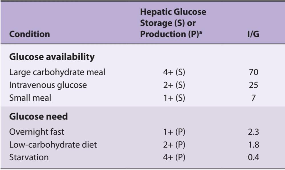
a1+ to 4+ indicate relative magnitude.
Courtesy of RH Unger.
1a+ تا 4+ نشان دهنده قدر نسبی است.
با حسن نیت از RH Unger.
CLINICAL BOX 24-4
Macrosomia & GLUT-1 Deficiency
Infants born to diabetic mothers often have high birth weights and large organs (macrosomia). This condition is caused by excess circulating insulin in the fetus, which in turn is caused in part by stimulation of the fetal pancreas by high blood glucose and amino acids from the diabetic mother.
جعبه بالینی 24-4
ماکروزومی و کمبود GLUT-1
نوزادانی که از مادران دیابتی متولد می شوند اغلب دارای وزن بالا و اندام های بزرگ هستند (ماکروزومی). این عارضه ناشی از گردش بیش از حد انسولین در جنین است که به نوبه خود تا حدودی به دلیل تحریک پانکراس جنین توسط قند خون بالا و اسیدهای آمینه از مادر دیابتی ایجاد می شود.
Free insulin in maternal blood is destroyed by proteases in the placenta, but antibody-bound insulin is protected, so it reaches the fetus.
انسولین آزاد در خون مادر توسط پروتئازهای جفت از بین می رود، اما انسولین متصل به آنتی بادی محافظت می شود، بنابراین به جنین می رسد.
Infants with GLUT-1 deficiency have defective transport of glucose across the blood-brain barrier. They have low cerebrospinal fluid glucose in the presence of normal plasma glucose, seizures, and developmental delay.
نوزادان مبتلا به کمبود GLUT-1 دارای انتقال ناقص گلوکز از طریق سد خونی مغزی هستند. گلوکز مایع مغزی نخاعی آنها در حضور گلوکز طبیعی پلاسما، تشنج و تاخیر رشدی پایین است.
OTHER ISLET CELL HORMONES
In addition to insulin and glucagon, the pancreatic islets secrete somatostatin and pancreatic polypeptide into the bloodstream. In addition, somatostatin may be involved in regulatory processes within the islets that adjust the pattern of hormones secreted in response to various stimuli.
سایر هورمون های سلول جزایر
علاوه بر انسولین و گلوکاگون، جزایر پانکراس سوماتوستاتین و پلی پپتید پانکراس را به جریان خون ترشح می کنند. علاوه بر این، سوماتوستاتین ممکن است در فرآیندهای تنظیمی درون جزایر که الگوی هورمونهای ترشح شده در پاسخ به محرکهای مختلف را تنظیم میکنند، دخالت داشته باشد.
SOMATOSTATIN
Somatostatin and its receptors are discussed in Chapter 7. Somatostatin 14 (SS 14) and its amino terminal-extended form somatostatin 28 (SS 28) are found in the D cells of pancreatic islets. Both forms inhibit the secretion of insulin, glucagon, and pancreatic polypeptide and act locally within the pancreatic islets in a paracrine fashion. SS 28 is more active than SS 14 in inhibiting insulin secretion, and it apparently acts via the SSTR5 receptor (see Chapter 7). Patients with somatostatin-secreting pancreatic tumors (somatostatinomas) develop hyperglycemia and other manifestations of diabetes that disappear when the tumor is removed. Dyspepsia also develops as a result of slow gastric emptying and decreased gastric acid secretion, and gallstones, which are precipitated by decreased gallbladder contraction due to inhibition of CCK secretion. The secretion of pancreatic somatostatin is increased by several of the same stimuli that increase insulin secretion, that is, glucose and amino acids, particularly arginine and leucine. It is also increased by CCK. Somatostatin is released from the pancreas and the gastrointestinal tract into the peripheral blood.
سوماتوستاتین
سوماتواستاتین و گیرنده های آن در فصل 7 مورد بحث قرار می گیرند. سوماتوستاتین 14 (SS 14) و آمینو ترمینال گسترش یافته آن سوماتوستاتین 28 (SS 28) در سلول های D جزایر پانکراس یافت می شوند. هر دو شکل ترشح انسولین، گلوکاگون و پلی پپتید پانکراس را مهار می کنند و به صورت موضعی در جزایر پانکراس به صورت پاراکرین عمل می کنند. SS 28 در مهار ترشح انسولین فعال تر از SS 14 است و ظاهراً از طریق گیرنده SSTR5 عمل می کند (به فصل 7 مراجعه کنید). بیماران مبتلا به تومورهای پانکراس ترشح کننده سوماتوستاتین (سوماتوستاتینوما) دچار هیپرگلیسمی و سایر تظاهرات دیابت می شوند که با برداشتن تومور ناپدید می شوند. سوء هاضمه همچنین در نتیجه تخلیه آهسته معده و کاهش ترشح اسید معده و سنگ های صفراوی ایجاد می شود که با کاهش انقباض کیسه صفرا به دلیل مهار ترشح CCK ایجاد می شود. ترشح سوماتوستاتین لوزالمعده توسط چندین محرک که ترشح انسولین را افزایش می دهد، یعنی گلوکز و اسیدهای آمینه، به ویژه آرژنین و لوسین، افزایش می یابد. همچنین توسط CCK افزایش می یابد. سوماتواستاتین از پانکراس و دستگاه گوارش به خون محیطی آزاد می شود.
PANCREATIC POLYPEPTIDE
Human pancreatic polypeptide is a linear polypeptide that contains 36 amino acid residues and is produced by F cells in the islets. It is closely related to two other 36-amino acid polypeptides, polypeptide YY, a gastrointestinal peptide (see Chapter 25), and neuropeptide Y, which is found in the brain and the autonomic nervous system (see Chapter 7). All end in tyrosine and are amidated at their carboxyl terminal. At least in part, pancreatic polypeptide secretion is under cholinergic control; plasma levels fall after administration of atropine. Its secretion is increased by a meal containing protein and by fasting, exercise, and acute hypoglycemia. Secretion is decreased by somatostatin and intravenous glucose. Infusions of leucine, arginine, and alanine do not affect it, so the stimulatory effect of a protein meal may be mediated indirectly. Pancreatic polypeptide slows the absorption of food in humans, and it may smooth out the peaks and valleys of absorption. However, its exact physiologic function is still uncertain.
پلی پپتید پانکراس
پلی پپتید پانکراس انسانی یک پلی پپتید خطی است که حاوی 36 اسید آمینه باقی مانده است و توسط سلول های F در جزایر تولید می شود. ارتباط نزدیکی با دو پلی پپتید 36 اسید آمینه دیگر دارد، پلی پپتید YY، یک پپتید گوارشی (به فصل 25 مراجعه کنید)، و نوروپپتید Y که در مغز و سیستم عصبی خودمختار یافت می شود (به فصل 7 مراجعه کنید). همه آنها به تیروزین ختم می شوند و در پایانه کربوکسیل خود آمید می شوند. حداقل تا حدی، ترشح پلی پپتید پانکراس تحت کنترل کولینرژیک است. سطح پلاسما پس از تجویز آتروپین کاهش می یابد. ترشح آن با یک وعده غذایی حاوی پروتئین و با ناشتا، ورزش و هیپوگلیسمی حاد افزایش می یابد. ترشح توسط سوماتوستاتین و گلوکز وریدی کاهش می یابد. تزریق لوسین، آرژنین و آلانین بر آن تأثیر نمی گذارد، بنابراین اثر تحریکی یک وعده غذایی پروتئینی ممکن است به طور غیر مستقیم واسطه شود. پلی پپتید پانکراس جذب غذا را در انسان کند می کند و ممکن است قله ها و دره های جذب را صاف کند. با این حال، عملکرد فیزیولوژیکی دقیق آن هنوز نامشخص است.
ORGANIZATION OF THE PANCREATIC ISLETS
The presence in the pancreatic islets of hormones that affect the secretion of other islet hormones suggests that the islets function as secretory units in the regulation of nutrient homeostasis. Somatostatin inhibits the secretion of insulin, glucagon, and pancreatic polypeptide (Figure 24-14); insulin inhibits the secretion of glucagon; and glucagon stimulates the secretion of insulin and somatostatin. As noted above, A and D cells and pancreatic polypeptide- secreting cells are generally located around the periphery of the islets, with the B cells in the center. There are clearly two types of islets, glucagon-rich islets and pancreatic polypeptide-rich islets, but the functional significance of this separation is not known. The islet cell hormones released into the ECF probably diffuse to other islet cells and influence their function (paracrine communication; see Chapter 25). It has been demonstrated that gap junctions are present between A, B, and D cells and that these permit the passage of ions and other small molecules from one cell to another, which could coordinate their secretory functions.
سازمان جزایر پانکراس
وجود هورمونهایی در جزایر پانکراس که بر ترشح هورمونهای جزایر دیگر تأثیر میگذارند، نشان میدهد که جزایر به عنوان واحدهای ترشحی در تنظیم هموستاز مواد مغذی عمل میکنند. سوماتوستاتین ترشح انسولین، گلوکاگون و پلی پپتید پانکراس را مهار می کند (شکل 24-14). انسولین ترشح گلوکاگون را مهار می کند. و گلوکاگون باعث تحریک ترشح انسولین و سوماتوستاتین می شود. همانطور که در بالا ذکر شد، سلول های A و D و سلول های ترشح کننده پلی پپتید پانکراس به طور کلی در اطراف محیط جزایر قرار دارند و سلول های B در مرکز قرار دارند. به وضوح دو نوع جزایر وجود دارد، جزایر غنی از گلوکاگون و جزایر غنی از پلی پپتید پانکراس، اما اهمیت عملکردی این جداسازی مشخص نیست. هورمونهای سلول جزایری که در ECF آزاد میشوند احتمالاً به سایر سلولهای جزایر منتشر میشوند و بر عملکرد آنها تأثیر میگذارند (ارتباط پاراکرین؛ فصل 25 را ببینید). نشان داده شده است که اتصالات شکافی بین سلولهای A، B و D وجود دارد و این اجازه عبور یونها و مولکولهای کوچک دیگر را از یک سلول به سلول دیگر میدهد که میتواند عملکردهای ترشحی آنها را هماهنگ کند.
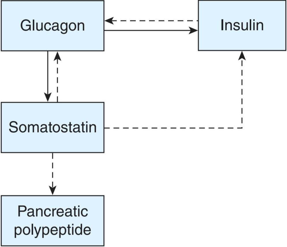
FIGURE 24-14 Effects of islet cell hormones on the secretion of other islet cell hormones. Solid arrows indicate stimulation; dashed arrows indicate inhibition.
شکل 24-14 اثرات هورمون های سلول جزایر بر ترشح سایر هورمون های سلول جزایر. فلش های جامد نشان دهنده تحریک است. فلش های چین نشان دهنده مهار است.
EFFECTS OF OTHER HORMONES & EXERCISE ON CARBOHYDRATE METABOLISM
Exercise has direct effects on carbohydrate metabolism. Many hormones in addition to insulin, IGF-I, IGF-II, glucagon, and somatostatin also have important roles in the regulation of carbohydrate metabolism. They include epinephrine, thyroid hormones, glucocorticoids, and growth hormone. The other functions of these hormones are considered elsewhere, but it seems wise to summarize their effects on carbohydrate metabolism in the context of the present chapter.
اثرات سایر هورمون ها و ورزش بر متابولیسم کربوهیدرات ها
ورزش تأثیر مستقیمی بر متابولیسم کربوهیدرات دارد. بسیاری از هورمون ها علاوه بر انسولین، IGF-I، IGF-II، گلوکاگون و سوماتوستاتین نیز نقش مهمی در تنظیم متابولیسم کربوهیدرات دارند. آنها شامل اپی نفرین، هورمون های تیروئید، گلوکوکورتیکوئیدها و هورمون رشد هستند. عملکردهای دیگر این هورمونها در جای دیگری مورد توجه قرار گرفتهاند، اما به نظر میرسد که عاقلانه است که اثرات آنها بر متابولیسم کربوهیدرات را در چارچوب فصل حاضر خلاصه کنیم.
EXERCISE
The entry of glucose into skeletal muscle is increased during exercise in the absence of insulin by causing an insulin- independent increase in the number of GLUT-4 transporters in muscle cell membranes (see above). This increase in glucose entry persists for several hours after exercise, and regular exercise training can also produce prolonged increases in insulin sensitivity. Exercise can precipitate hypoglycemia in diabetics not only because of the increase in muscle uptake of glucose but also because absorption of injected insulin is more rapid during exercise. Patients with diabetes should take in extra calories or reduce their insulin dosage when they exercise.
ورزش کنید
ورود گلوکز به عضله اسکلتی در حین ورزش در غیاب انسولین با ایجاد افزایش مستقل از انسولین در تعداد ناقلین GLUT-4 در غشای سلولی عضلانی افزایش می یابد (به بالا مراجعه کنید). این افزایش در ورود گلوکز تا چند ساعت پس از ورزش ادامه می یابد و تمرینات ورزشی منظم نیز می تواند باعث افزایش طولانی مدت حساسیت به انسولین شود. ورزش می تواند هیپوگلیسمی را در دیابتی ها تسریع کند نه تنها به دلیل افزایش جذب گلوکز عضلانی، بلکه به این دلیل که جذب انسولین تزریقی در حین ورزش سریعتر است. بیماران دیابتی هنگام ورزش باید کالری اضافی دریافت کنند یا دوز انسولین خود را کاهش دهند.
CATECHOLAMINES
The activation of phosphorylase in liver by catecholamines is discussed in Chapter 1. Activation occurs via ẞ-adrenergic receptors, which increase intracellular CAMP, and a-adrenergic receptors, which increase intracellular Ca2+. Hepatic glucose output is increased, producing hyperglycemia. In muscle, the phosphorylase is also activated via cAMP and presumably via Ca2+, but the glucose-6-phosphate formed can be catabolized only to pyruvate because of the absence of glucose-6-phosphatase. For reasons that are not entirely clear, large amounts of pyruvate are converted to lactate, which diffuses from the muscle into the circulation (Figure 24-15). The lactate is oxidized in the liver to pyruvate and converted to glycogen. Therefore, the response to an injection of epinephrine is an initial glycogenolysis followed by a rise in hepatic glycogen content. Lactate oxidation may be responsible for the calorigenic effect of epinephrine (see Chapter 19). Epinephrine and norepinephrine also liberate FFA into the circulation, and epinephrine decreases peripheral utilization of glucose.
کاتکولامین ها
فعال شدن فسفوریلاز در کبد توسط کاتکول آمین ها در فصل 1 مورد بحث قرار گرفته است. فعال سازی از طریق گیرنده های ẞ-آدرنرژیک، که CAMP داخل سلولی را افزایش می دهد، و گیرنده های a-آدرنرژیک، که +Ca2 داخل سلولی را افزایش می دهد، رخ می دهد. برون ده گلوکز کبدی افزایش یافته و هیپرگلیسمی ایجاد می کند. در عضله، فسفوریلاز نیز از طریق cAMP و احتمالاً از طریق +Ca2 فعال می شود، اما گلوکز-6-فسفات تشکیل شده به دلیل عدم وجود گلوکز-6-فسفاتاز، فقط به پیروات کاتابولیز می شود. به دلایلی که کاملاً مشخص نیست، مقادیر زیادی پیروات به لاکتات تبدیل میشود که از عضله به گردش خون منتشر میشود (شکل 24-15). لاکتات در کبد به پیرووات اکسید شده و به گلیکوژن تبدیل می شود. بنابراین، پاسخ به تزریق اپی نفرین یک گلیکوژنولیز اولیه است که با افزایش محتوای گلیکوژن کبدی همراه است. اکسیداسیون لاکتات ممکن است مسئول اثر کالری زایی اپی نفرین باشد (به فصل 19 مراجعه کنید). اپی نفرین و نوراپی نفرین همچنین FFA را در گردش خون آزاد می کنند و اپی نفرین استفاده محیطی از گلوکز را کاهش می دهد.
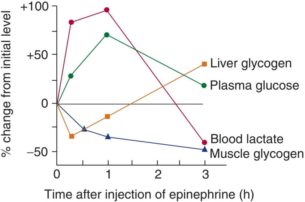
FIGURE 24-15 Effect of epinephrine on tissue glycogen, plasma glucose, and blood lactate levels in fed rats. (Reproduced with permission from Ruch TC, Patton HD [editors]: Physiology and Biophysics, 20th ed, St. Louis, MO: Saunders; 1973.)
شکل 24-15 اثر اپی نفرین بر سطح گلیکوژن بافت، گلوکز پلاسما و لاکتات خون در موش های تغذیه شده. (تکثیر شده با اجازه از Ruch TC، Patton HD [ویراستاران]: Physiology and Biophysics, 20th ed, St. Louis, MO: Saunders, 1973.)
THYROID HORMONES
Thyroid hormones make experimental diabetes worse; thyrotoxicosis aggravates clinical diabetes; and metathyroid diabetes can be produced in animals with decreased pancreatic reserve. The principal diabetogenic effect of thyroid hormones is to increase absorption of glucose from the intestine, but the hormones also cause (probably by potentiating the effects of catecholamines) some degree of hepatic glycogen depletion. Glycogen-depleted liver cells are easily damaged. When the liver is damaged, the glucose tolerance curve is diabetic because the liver takes up less of the absorbed glucose. Thyroid hormones may also accelerate the degradation of insulin. All these actions have a hyperglycemic effect and, if the pancreatic reserve is low, may lead to B cell exhaustion.
ورمون های تیروئید
هورمون های تیروئید دیابت تجربی را بدتر می کنند. تیروتوکسیکوز دیابت بالینی را تشدید می کند. و دیابت متاتیروئیدی را می توان در حیوانات با کاهش ذخیره پانکراس ایجاد کرد. اثر اصلی دیابت زایی هورمون های تیروئید افزایش جذب گلوکز از روده است، اما هورمون ها همچنین باعث (احتمالاً با تقویت اثرات کاتکول آمین ها) درجاتی از کاهش گلیکوژن کبدی می شوند. سلول های کبدی فاقد گلیکوژن به راحتی آسیب می بینند. هنگامی که کبد آسیب می بیند، منحنی تحمل گلوکز دیابتی است زیرا کبد گلوکز جذب شده کمتری را می گیرد. هورمون های تیروئید نیز ممکن است تخریب انسولین را تسریع کنند. همه این اعمال اثر هیپرگلیسمی دارند و اگر ذخیره پانکراس کم باشد ممکن است منجر به فرسودگی سلول های B شود.
ADRENAL GLUCOCORTICOIDS
Glucocorticoids from the adrenal cortex (see Chapter 19) elevate blood glucose and produce a diabetic type of glucose tolerance curve. In humans, this effect may occur only in individuals with a genetic predisposition to diabetes. Glucose tolerance is reduced in 80% of patients with Cushing syndrome (see Chapter 19), and 20% of these patients have frank diabetes. The glucocorticoids are necessary for glucagon to exert its gluconeogenic action during fasting. They are gluconeogenic themselves, but their role is mainly permissive. In adrenal insufficiency, the blood glucose is normal as long as food intake is maintained, but fasting precipitates hypoglycemia and collapse. The plasma-glucose- lowering effect of insulin is greatly enhanced in patients with adrenal insufficiency. In animals with experimental diabetes, adrenalectomy markedly ameliorates the diabetes. The major diabetogenic effects are an increase in protein catabolism with increased gluconeogenesis in the liver; increased hepatic glycogenesis and ketogenesis; and a decrease in peripheral glucose utilization relative to the blood insulin level that may be due to inhibition of glucose phosphorylation.
گلوکوکورتیکوئیدهای آدرنال
گلوکوکورتیکوئیدها از قشر آدرنال (به فصل 19 مراجعه کنید) گلوکز خون را بالا می برند و یک نوع دیابتی از منحنی تحمل گلوکز تولید می کنند. در انسان، این اثر ممکن است فقط در افراد دارای استعداد ژنتیکی دیابت رخ دهد. تحمل گلوکز در 80 درصد از بیماران مبتلا به سندرم کوشینگ کاهش می یابد (به فصل 19 مراجعه کنید)، و 20 درصد از این بیماران دیابت آشکار دارند. گلوکوکورتیکوئیدها برای گلوکاگون برای اعمال گلوکونئوژنیک خود در طول روزه داری ضروری هستند. آنها خودشان گلوکونئوژن هستند، اما نقش آنها عمدتاً مجاز است. در نارسایی آدرنال، تا زمانی که مصرف غذا حفظ شود، گلوکز خون طبیعی است، اما ناشتا باعث کاهش قند خون و فروپاشی می شود. اثر کاهش دهنده گلوکز پلاسما انسولین در بیماران مبتلا به نارسایی آدرنال بسیار افزایش می یابد. در حیوانات مبتلا به دیابت تجربی، آدرنالکتومی به طور قابل توجهی دیابت را بهبود می بخشد. اثرات عمده دیابت زایی افزایش کاتابولیسم پروتئین با افزایش گلوکونئوژنز در کبد است. افزایش گلیکوژنز و کتوژنز کبدی؛ و کاهش مصرف محیطی گلوکز نسبت به سطح انسولین خون که ممکن است به دلیل مهار فسفوریلاسیون گلوکز باشد.
GROWTH HORMONE
Human growth hormone makes clinical diabetes worse, and 25% of patients with growth hormone-secreting tumors of the anterior pituitary have diabetes. Hypophysectomy ameliorates diabetes and decreases insulin resistance even more than adrenalectomy, whereas growth hormone treatment increases insulin resistance.
هورمون رشد
هورمون رشد انسانی باعث بدتر شدن دیابت بالینی می شود و 25 درصد از بیماران مبتلا به تومورهای ترشح کننده هورمون رشد هیپوفیز قدامی دیابت دارند. هیپوفیزکتومی دیابت را بهبود می بخشد و مقاومت به انسولین را حتی بیشتر از آدرنالکتومی کاهش می دهد، در حالی که درمان با هورمون رشد مقاومت به انسولین را افزایش می دهد.
The effects of growth hormone are partly direct and partly mediated via IGF-I (see Chapter 18). Growth hormone mobilizes FFA from adipose tissue, thus favoring ketogenesis. It decreases glucose uptake into some tissues (“anti-insulin action”), increases hepatic glucose output, and may decrease tissue binding of insulin. Indeed, it has been suggested that the ketosis and decreased glucose tolerance produced by starvation are due to hypersecretion of growth hormone. Growth hormone does not stimulate insulin secretion directly, but the hyperglycemia it produces secondarily stimulates the pancreas and may eventually exhaust the B cells.
اثرات هورمون رشد تا حدی مستقیم و تا حدی از طریق IGF-I انجام می شود (به فصل 18 مراجعه کنید). هورمون رشد FFA را از بافت چربی بسیج می کند، بنابراین کتوژنز را مورد حمایت قرار می دهد. جذب گلوکز را در برخی بافت ها کاهش می دهد (“عمل ضد انسولین”)، برون ده گلوکز کبدی را افزایش می دهد و ممکن است اتصال بافتی انسولین را کاهش دهد. در واقع، پیشنهاد شده است که کتوز و کاهش تحمل گلوکز ناشی از گرسنگی ناشی از ترشح بیش از حد هورمون رشد است. هورمون رشد مستقیماً ترشح انسولین را تحریک نمی کند، اما هیپرگلیسمی که تولید می کند به طور ثانویه پانکراس را تحریک می کند و ممکن است در نهایت سلول های B را خسته کند.
HYPOGLYCEMIA & DIABETES MELLITUS IN HUMANS
HYPOGLYCEMIA
“Insulin reactions” are common in type 1 diabetics and occasional hypoglycemic episodes are the price of good diabetic control in most diabetics. Glucose uptake by skeletal muscle and absorption of injected insulin both increase during exercise (see above).
هیپوگلیسمی و دیابت در انسان
هیپوگلیسمی
“واکنش های انسولین” در بیماران دیابتی نوع 1 رایج است و دوره های گاه به گاه هیپوگلیسمی بهای کنترل خوب دیابت در اکثر بیماران دیابتی است. جذب گلوکز توسط ماهیچه های اسکلتی و جذب انسولین تزریقی هر دو در طول تمرین افزایش می یابد (به بالا مراجعه کنید).
Symptomatic hypoglycemia also occurs in nondiabetics, and a review of some of the more important causes serves to emphasize the variables affecting plasma glucose homeostasis. Chronic mild hypoglycemia can cause incoordination and slurred speech, and the condition can be mistaken for drunkenness. Mental aberrations and convulsions in the absence of frank coma also occur. When the level of insulin secretion is chronically elevated by an insulinoma, a rare, insulin-secreting tumor of the pancreas, symptoms are most common in the morning. This is because a night of fasting has depleted hepatic glycogen reserves. However, symptoms can develop at any time, and in such patients, the diagnosis may be missed. Some cases of insulinoma have been erroneously diagnosed as epilepsy or psychosis. Hypoglycemia also occurs in some patients with large malignant tumors that do not involve the pancreatic islets, and the hypoglycemia in these cases is apparently due to excess secretion of IGF-II.
هیپوگلیسمی علامت دار در افراد غیر دیابتی نیز رخ می دهد، و بررسی برخی از علل مهم تر بر متغیرهای مؤثر بر هموستاز گلوکز پلاسما تأکید می کند. هیپوگلیسمی مزمن خفیف می تواند باعث ناهماهنگی و اختلال در گفتار شود و این وضعیت می تواند با مستی اشتباه گرفته شود. انحرافات ذهنی و تشنج در غیاب کمای آشکار نیز رخ می دهد. هنگامی که سطح ترشح انسولین به طور مزمن توسط یک انسولینوما، یک تومور نادر و ترشح کننده انسولین در پانکراس، افزایش می یابد، علائم در صبح شایع ترین هستند. این به این دلیل است که یک شب روزه داری ذخایر گلیکوژن کبدی را تخلیه کرده است. با این حال، علائم ممکن است در هر زمانی ایجاد شود و در چنین بیمارانی ممکن است تشخیص داده نشود. برخی از موارد انسولینوما به اشتباه صرع یا روان پریشی تشخیص داده شده است. هیپوگلیسمی در برخی از بیماران مبتلا به تومورهای بدخیم بزرگ که جزایر پانکراس را درگیر نمی کنند نیز رخ می دهد و ظاهراً افت قند خون در این موارد به دلیل ترشح بیش از حد IGF-II است.
As noted above, the autonomic discharge caused by lowered blood glucose that produces shakiness, sweating, anxiety, and hunger normally occurs at plasma glucose levels that are higher than the glucose levels that cause cognitive dysfunction, thereby serving as a warning to ingest sugar. However, in some individuals, these warning symptoms fail to occur before the cognitive symptoms, due to cerebral dysfunction (desensitization), and this hypoglycemia unawareness is potentially dangerous. The condition is prone to develop in patients with insulinomas and in diabetics receiving intensive insulin therapy, so it appears that repeated bouts of hypoglycemia cause the eventual development of hypoglycemia unawareness. If blood sugar rises again for some time, the warning symptoms again appear at a higher plasma glucose level than cognitive abnormalities and coma. The reason why prolonged hypoglycemia causes loss of the warning symptoms is unsettled.
همانطور که در بالا ذکر شد، ترشحات خودمختار ناشی از کاهش گلوکز خون که باعث ایجاد لرزش، تعریق، اضطراب و گرسنگی میشود، معمولاً در سطوح گلوکز پلاسما بالاتر از سطوح گلوکز که باعث اختلال عملکرد شناختی میشود، رخ میدهد و در نتیجه به عنوان یک هشدار برای مصرف قند عمل میکند. با این حال، در برخی از افراد، این علائم هشداردهنده قبل از علائم شناختی، به دلیل اختلال عملکرد مغزی (حساسیت زدایی) رخ نمی دهد و این ناآگاهی از هیپوگلیسمی بالقوه خطرناک است. این بیماری در بیماران مبتلا به انسولینوم و در بیماران دیابتی که انسولین درمانی فشرده دریافت می کنند مستعد ایجاد می شود، بنابراین به نظر می رسد که حملات مکرر هیپوگلیسمی باعث ایجاد ناآگاهی نهایی از هیپوگلیسمی می شود. اگر قند خون برای مدتی دوباره افزایش یابد، علائم هشدار دهنده دوباره در سطح گلوکز پلاسما بالاتر از ناهنجاری های شناختی و کما ظاهر می شوند. دلیل اینکه هیپوگلیسمی طولانی مدت باعث از بین رفتن علائم هشداردهنده می شود مشخص نیست.
In liver disease, the glucose tolerance curve is diabetic but the fasting plasma glucose level is low (Figure 24-16). In functional hypoglycemia, the plasma glucose rise is normal after a test dose of glucose, but the subsequent fall overshoots to hypoglycemic levels, producing symptoms 3-4 h after meals. This pattern is sometimes seen in individuals in whom diabetes develops later. Patients with this syndrome should be distinguished from the more numerous patients with similar symptoms due to psychological or other problems who do not have hypoglycemia when blood is drawn during the symptomatic episode. It has been postulated that the overshoot of the plasma glucose is due to insulin secretion stimulated by impulses in the right vagus, but cholinergic blocking agents do not routinely correct the abnormality. In some thyrotoxic patients and in patients who have had gastrectomies or other operations that speed the passage of food into the intestine, glucose absorption is abnormally rapid. The plasma glucose rises to a high, early peak, but it then falls rapidly to hypoglycemic levels because the wave of hyperglycemia evokes a greater than normal rise in insulin secretion. Symptoms characteristically occur about 2 h after meals.
در بیماری کبد، منحنی تحمل گلوکز دیابتی است اما سطح گلوکز پلاسما ناشتا پایین است (شکل 24-16). در هیپوگلیسمی عملکردی، افزایش گلوکز پلاسما پس از دوز آزمایشی گلوکز طبیعی است، اما سقوط بعدی به سطوح هیپوگلیسمی بیش از حد می رسد و علائمی را 3-4 ساعت پس از غذا ایجاد می کند. این الگو گاهی اوقات در افرادی که دیابت در آنها دیرتر ایجاد می شود، دیده می شود. بیماران مبتلا به این سندرم را باید از بیماران متعددی که علائم مشابه دارند به دلیل مشکلات روانی یا سایر مشکلاتی که هنگام خونگیری در طول دوره علامت دار هیپوگلیسمی ندارند، متمایز شوند. فرض بر این است که افزایش بیش از حد گلوکز پلاسما به دلیل ترشح انسولین است که توسط تکانههایی در واگ راست تحریک میشود، اما عوامل مسدودکننده کولینرژیک به طور معمول این ناهنجاری را اصلاح نمیکنند. در برخی از بیماران تیروتوکسیک و در بیمارانی که گاسترکتومی یا سایر عملهایی که عبور غذا به روده را تسریع میکند انجام دادهاند، جذب گلوکز بهطور غیر طبیعی سریع است. گلوکز پلاسما تا یک اوج بالا و زودرس افزایش می یابد، اما سپس به سرعت به سطوح هیپوگلیسمی کاهش می یابد زیرا موج هیپرگلیسمی باعث افزایش بیشتر از حد طبیعی در ترشح انسولین می شود. علائم به طور مشخص حدود 2 ساعت پس از غذا رخ می دهد.
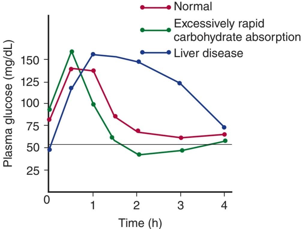
FIGURE 24–16 Typical glucose tolerance curves after an oral glucose load in liver disease and in conditions causing excessively rapid absorption of glucose from the intestine. The horizontal line is the approximate plasma glucose level at which hypoglycemic symptoms may appear.
شکل 24-16 منحنی های معمول تحمل گلوکز پس از بار گلوکز خوراکی در بیماری کبد و شرایطی که باعث جذب بیش از حد سریع گلوکز از روده می شود. خط افقی سطح تقریبی گلوکز پلاسما است که در آن علائم هیپوگلیسمی ممکن است ظاهر شود.
DIABETES MELLITUS
The incidence of diabetes mellitus in the human population has reached epidemic proportions worldwide and it is increasing at a rapid rate. In 2010 an estimated 285 million people worldwide had diabetes, according to the International Diabetes Federation. The federation predicts as many as 438 million will have diabetes by 2030. Ninety percent of the present cases are type 2 diabetes, and most of the increase will be in type 2, paralleling the increase in the incidence of obesity.
دیابت قندی
بروز دیابت در جمعیت انسانی به ابعاد اپیدمی در سرتاسر جهان رسیده است و به سرعت در حال افزایش است. بر اساس گزارش فدراسیون بین المللی دیابت، در سال 2010 حدود 285 میلیون نفر در سراسر جهان به دیابت مبتلا بودند. فدراسیون پیشبینی میکند تا سال 2030، 438 میلیون نفر به دیابت مبتلا شوند. 90 درصد از موارد کنونی دیابت نوع 2 هستند و بیشترین افزایش در نوع 2 خواهد بود که به موازات افزایش شیوع چاقی است.
Diabetes is sometimes complicated by acidosis and coma, and in long- standing diabetes, additional complications occur. These include microvascular, macrovascular, and neuropathic disease. The microvascular abnormalities are proliferative scarring of the retina (diabetic retinopathy) leading to blindness and renal disease (diabetic nephropathy) leading to chronic kidney disease. The macrovascular abnormalities are due to accelerated atherosclerosis, which is secondary to increased plasma LDL. The result is an increased incidence of stroke and myocardial infarction. The neuropathic abnormalities (diabetic neuropathy) involve the autonomic nervous system and peripheral nerves. The neuropathy plus the atherosclerotic circulatory insufficiency in the extremities and reduced resistance to infection can lead to chronic ulceration and gangrene, particularly in the feet.
دیابت گاهی اوقات با اسیدوز و کما پیچیده می شود و در دیابت طولانی مدت عوارض دیگری نیز رخ می دهد. این بیماریها شامل بیماریهای میکروواسکولار، ماکروواسکولار و نوروپاتیک است. ناهنجاری های میکروواسکولار عبارتند از: اسکار تکثیری شبکیه (رتینوپاتی دیابتی) که منجر به کوری و بیماری کلیوی (نفروپاتی دیابتی) می شود که منجر به بیماری مزمن کلیوی می شود. ناهنجاری های ماکرو عروقی به دلیل تصلب شرایین تسریع شده است که ثانویه به افزایش LDL پلاسما است. نتیجه افزایش بروز سکته مغزی و انفارکتوس میوکارد است. ناهنجاری های نوروپاتیک (نوروپاتی دیابتی) سیستم عصبی خودمختار و اعصاب محیطی را درگیر می کند. نوروپاتی به علاوه نارسایی گردش خون آترواسکلروتیک در اندام ها و کاهش مقاومت در برابر عفونت می تواند منجر به زخم مزمن و قانقاریا، به ویژه در پاها شود.
The ultimate cause of the microvascular and neuropathic complications is chronic hyperglycemia, and tight control of the diabetes reduces their incidence. Intracellular hyperglycemia activates the enzyme aldose reductase. This increases the formation of sorbitol in cells, which in turn reduces cellular Na, K ATPase. In addition, intracellular glucose can be converted to so-called Amadori products, and these in turn can form advanced glycosylation end products (AGES), which cross-link matrix proteins. This damages blood vessels. The AGEs also interfere with leukocyte responses to infection.
علت نهایی عوارض میکروواسکولار و نوروپاتیک هیپرگلیسمی مزمن است و کنترل دقیق دیابت باعث کاهش بروز آنها می شود. هیپرگلیسمی داخل سلولی آنزیم آلدوز ردوکتاز را فعال می کند. این باعث افزایش تشکیل سوربیتول در سلول ها می شود که به نوبه خود باعث کاهش Na و K ATPase سلولی می شود. علاوه بر این، گلوکز درون سلولی می تواند به محصولات آمادوری تبدیل شود و این محصولات به نوبه خود می توانند محصولات نهایی گلیکوزیلاسیون پیشرفته (AGES) را تشکیل دهند که پروتئین های ماتریکس را به هم متصل می کنند. این به رگ های خونی آسیب می رساند. AGE ها همچنین با پاسخ لکوسیت ها به عفونت تداخل می کنند.
TYPES OF DIABETES
The cause of clinical diabetes is always a deficiency of the effects of insulin at the tissue level, but the deficiency may be relative. One of the common forms, type 1, or insulin-dependent diabetes mellitus (IDDM), is due to insulin deficiency caused by autoimmune destruction of the B cells in the pancreatic islets; the A, D, and F cells remain intact. The second common form, type 2, or noninsulin-dependent diabetes mellitus (NIDDM), is characterized by insulin resistance.
انواع دیابت
علت دیابت بالینی همیشه کمبود اثرات انسولین در سطح بافت است، اما ممکن است کمبود آن نسبی باشد. یکی از اشکال رایج، دیابت نوع 1 یا دیابت وابسته به انسولین (IDDM)، به دلیل کمبود انسولین ناشی از تخریب خودایمنی سلول های B در جزایر پانکراس است. سلول های A، D و F دست نخورده باقی می مانند. دومین شکل رایج، دیابت شیرین نوع 2 یا غیر وابسته به انسولین (NIDDM) با مقاومت به انسولین مشخص می شود.
In addition, some cases of diabetes are due to other diseases or conditions such as chronic pancreatitis, total pancreatectomy, Cushing syndrome (see Chapter 19), and acromegaly (see Chapter 18). These make up 5% of the total cases and are sometimes classified as secondary diabetes.
علاوه بر این، برخی از موارد دیابت به دلیل بیماری ها یا شرایط دیگری مانند پانکراتیت مزمن، پانکراتکتومی کامل، سندرم کوشینگ (به فصل 19 مراجعه کنید)، و آکرومگالی (به فصل 18 مراجعه کنید) ایجاد می شود. این موارد 5 درصد از کل موارد را تشکیل می دهند و گاهی اوقات به عنوان دیابت ثانویه طبقه بندی می شوند.
Type 1 diabetes usually develops before the age of 40 and hence is called juvenile diabetes. Patients with this disease are not obese and they have a high incidence of ketosis and acidosis. Various anti-B cell antibodies are present in plasma, but the current thinking is that type 1 diabetes is primarily a T lymphocyte-mediated disease. Definite genetic susceptibility is present as well; if the disease develops in one identical twin, the chances are 1 in 3 that it will also develop in the other twin. In other words, the concordance rate is about 33%. The main genetic abnormality is in the major histocompatibility complex on chromosome 6, making individuals with certain types of histocompatibility antigens (see Chapter 3) much more prone to the disease. Other genes are also involved.
دیابت نوع 1 معمولاً قبل از 40 سالگی ایجاد می شود و از این رو دیابت نوجوانان نامیده می شود. بیماران مبتلا به این بیماری چاق نیستند و میزان کتوز و اسیدوز در آنها زیاد است. آنتی بادی های مختلف ضد سلول B در پلاسما وجود دارد، اما تفکر فعلی این است که دیابت نوع 1 در درجه اول یک بیماری با واسطه لنفوسیت T است. حساسیت ژنتیکی قطعی نیز وجود دارد. اگر بیماری در یکی از دوقلوهای همسان ایجاد شود، احتمال اینکه در دوقلو دیگر نیز ایجاد شود، 1 در 3 است. به عبارت دیگر میزان تطابق حدود 33 درصد است. ناهنجاری ژنتیکی اصلی در کمپلکس اصلی سازگاری بافتی در کروموزوم 6 است که باعث می شود افراد دارای انواع خاصی از آنتی ژن های سازگاری بافتی (به فصل 3 مراجعه کنید) بسیار مستعد ابتلا به این بیماری هستند. ژن های دیگر نیز در این امر دخیل هستند.
Immunosuppression with drugs such as cyclosporine ameliorate type 1 diabetes if given early in the disease before all islet B cells are lost. Attempts have been made to treat type 1 diabetes by transplanting pancreatic tissue or isolated islet cells, but results to date have been poor, largely because B cells are easily damaged and it is difficult to transplant enough of them to normalize glucose responses.
سرکوب سیستم ایمنی با داروهایی مانند سیکلوسپورین، دیابت نوع 1 را بهبود می بخشد، اگر در اوایل بیماری قبل از از بین رفتن تمام سلول های جزایر B داده شود. تلاش هایی برای درمان دیابت نوع 1 با پیوند بافت پانکراس یا سلول های جزایر جدا شده انجام شده است، اما نتایج تا به امروز ضعیف بوده است، عمدتاً به این دلیل که سلول های B به راحتی آسیب می بینند و پیوند کافی از آنها برای عادی سازی پاسخ های گلوکز دشوار است.
As mentioned above, type 2 is the most common type of diabetes and is usually associated with obesity. It usually develops after age 40 and is not associated with total loss of the ability to secrete insulin. It has an insidious onset, is rarely associated with ketosis, and is usually associated with normal B cell morphology and insulin content if the B cells have not become exhausted. The genetic component in type 2 diabetes is actually stronger than the genetic component in type 1 diabetes; in identical twins, the concordance rate is higher, ranging in some studies to nearly 100%.
همانطور که در بالا ذکر شد، نوع 2 شایع ترین نوع دیابت است و معمولاً با چاقی همراه است. معمولاً بعد از 40 سالگی ایجاد می شود و با از دست دادن کامل توانایی ترشح انسولین همراه نیست. شروعی موذی دارد، به ندرت با کتوز همراه است و معمولاً در صورتی که سلول های B خسته نشده باشند، با مورفولوژی طبیعی سلول های B و محتوای انسولین همراه است. مؤلفه ژنتیکی در دیابت نوع 2 در واقع قوی تر از مؤلفه ژنتیکی در دیابت نوع 1 است. در دوقلوهای همسان، میزان تطابق بیشتر است و در برخی مطالعات تا نزدیک به 100٪ متغیر است.
In some patients, type 2 diabetes is due to defects in identified genes. Over 60 of these defects have been described. They include defects in glucokinase (about 1% of the cases), the insulin molecule itself (about 0.5% of the cases), the insulin receptor (about 1% of the cases), GLUT-4 (about 1% of the cases), or IRS-1 (about 15% of the cases). In maturity-onset diabetes occurring in young individuals (MODY), which accounts for about 1% of the cases of type 2 diabetes, loss-of-function mutations have been described in six different genes. Five of the genes code for transcription factors affecting the production of enzymes involved in glucose metabolism. The sixth is the gene for glucokinase (Figure 24-11), the enzyme that controls the rate of glucose phosphorylation and hence its metabolism in the B cells. However, the vast majority of cases of type 2 diabetes are almost certainly polygenic in origin, and the actual genes involved are still unknown.
در برخی از بیماران، دیابت نوع 2 به دلیل نقص در ژن های شناسایی شده است. بیش از 60 مورد از این نقص ها شرح داده شده است. آنها شامل نقص در گلوکوکیناز (حدود 1٪ موارد)، خود مولکول انسولین (حدود 0.5٪ موارد)، گیرنده انسولین (حدود 1٪ موارد)، GLUT-4 (حدود 1٪ موارد)، یا IRS-1 (حدود 15٪ موارد) هستند. در دیابت با شروع بلوغ که در افراد جوان رخ می دهد (MODY)، که حدود 1٪ از موارد دیابت نوع 2 را تشکیل می دهد، جهش های از دست دادن عملکرد در شش ژن مختلف توصیف شده است. پنج مورد از ژن ها برای فاکتورهای رونویسی موثر بر تولید آنزیم های دخیل در متابولیسم گلوکز کد می کنند. ششمین ژن گلوکوکیناز است (شکل 24-11)، آنزیمی که میزان فسفوریلاسیون گلوکز و در نتیجه متابولیسم آن را در سلول های B کنترل می کند. با این حال، اکثریت قریب به اتفاق موارد دیابت نوع 2 تقریباً به طور قطع منشأ چند ژنی دارند و ژنهای واقعی درگیر هنوز ناشناخته هستند.
OBESITY, THE METABOLIC SYNDROME, & TYPE 2 DIABETES
Obesity is increasing in incidence and relates to the regulation of food intake and energy balance and overall nutrition. It deserves additional consideration in this chapter because of its special relation to disordered carbohydrate metabolism and diabetes. As body weight increases, insulin resistance increases, that is, there is a decreased ability of insulin to move glucose into fat and muscle and to shut off glucose release from the liver. Weight reduction decreases insulin resistance. Associated with obesity there is hyperinsulinemia, dyslipidemia (characterized by high circulating triglycerides and low high-density lipoprotein [HDL]), and accelerated development of atherosclerosis. This combination of findings is commonly called the metabolic syndrome, or syndrome X. Some of the patients with the syndrome are prediabetic, whereas others have frank type 2 diabetes. It has not been proved but it is logical to assume that the hyperinsulinemia is a compensatory response to the increased insulin resistance and that frank diabetes develops in individuals with reduced B cell reserves.
چاقی، سندرم متابولیک و دیابت نوع 2
بروز چاقی در حال افزایش است و به تنظیم میزان مصرف غذا و تعادل انرژی و تغذیه کلی مربوط می شود. در این فصل به دلیل ارتباط ویژه آن با اختلال متابولیسم کربوهیدرات و دیابت، سزاوار توجه بیشتری است. با افزایش وزن بدن، مقاومت به انسولین افزایش مییابد، یعنی توانایی انسولین برای انتقال گلوکز به چربی و ماهیچه و جلوگیری از انتشار گلوکز از کبد کاهش مییابد. کاهش وزن مقاومت به انسولین را کاهش می دهد. همراه با چاقی، هیپرانسولینمی، دیس لیپیدمی (که با تری گلیسیرید در گردش بالا و لیپوپروتئین با چگالی بالا (HDL) مشخص می شود) و تسریع پیشرفت آترواسکلروز وجود دارد. این ترکیب از یافتهها معمولاً سندرم متابولیک یا سندرم X نامیده میشود. برخی از بیماران مبتلا به این سندرم پیش دیابتی هستند، در حالی که برخی دیگر به دیابت نوع 2 آشکار مبتلا هستند. ثابت نشده است، اما منطقی است که فرض کنیم هیپرانسولینمی یک پاسخ جبرانی به افزایش مقاومت به انسولین است و دیابت آشکار در افرادی با ذخایر سلول B کاهش یافته است.
These observations and other data strongly suggest that fat produces a chemical signal or signals that act on muscles and the liver to increase insulin resistance. Evidence for this includes the recent observation that when GLUTS are selectively knocked out in adipose tissue, there is an associated decrease in glucose transport in muscle in vivo, but when the muscles of those animals are tested in vitro their transport is normal.
این مشاهدات و سایر دادهها قویاً نشان میدهند که چربی یک سیگنال شیمیایی یا سیگنالهایی تولید میکند که روی عضلات و کبد برای افزایش مقاومت به انسولین عمل میکند. شواهد برای این امر شامل مشاهدات اخیر است که وقتی GLUTS به طور انتخابی در بافت چربی حذف می شود، کاهش همراهی در انتقال گلوکز در عضله در داخل بدن وجود دارد، اما هنگامی که عضلات آن حیوانات در شرایط آزمایشگاهی آزمایش می شوند، انتقال آنها طبیعی است.
One possible signal is the circulating level of FFAs, which is elevated in many insulin-resistant states. Other possibilities are peptides and proteins secreted by fat cells. It is now clear that white fat depots are not inert lumps but are actually endocrine tissues that secrete not only leptin but also other hormones that affect fat metabolism. These adipose-derived hormones are commonly termed adipokines as they are cytokines secreted by adipose tissue. Known adipokines are leptin, adiponectin, and resistin.
یکی از سیگنال های احتمالی سطح گردش FFA ها است که در بسیاری از حالت های مقاوم به انسولین افزایش می یابد. احتمالات دیگر پپتیدها و پروتئین های ترشح شده توسط سلول های چربی است. اکنون مشخص شده است که ذخایر چربی سفید توده های بی اثر نیستند، بلکه در واقع بافت های غدد درون ریز هستند که نه تنها لپتین بلکه هورمون های دیگری را نیز ترشح می کنند که بر متابولیسم چربی تأثیر می گذارند. این هورمون های مشتق از چربی معمولاً آدیپوکین نامیده می شوند زیرا سیتوکین هایی هستند که توسط بافت چربی ترشح می شوند. آدیپوکین های شناخته شده لپتین، آدیپونکتین و رزیستین هستند.
Some adipokines decrease, rather than increase, insulin resistance. Leptin and adiponectin, for example, decrease insulin resistance, whereas resistin increases insulin resistance. Further complicating the situation, marked insulin resistance is present in the rare metabolic disease congenital lipodystrophy, in which fat depots fail to develop. This resistance is reduced by leptin and adiponectin. Finally, a variety of knockouts of intracellular second messengers have been reported to increase insulin resistance. It is unclear how, or indeed if, these findings fit together to provide an explanation of the relation of obesity to insulin tolerance, but the topic is obviously an important one and it is under intensive investigation.
برخی از آدیپوکین ها به جای افزایش، مقاومت به انسولین را کاهش می دهند. برای مثال لپتین و آدیپونکتین مقاومت به انسولین را کاهش می دهند، در حالی که رزیستین مقاومت به انسولین را افزایش می دهد. علاوه بر پیچیدهتر کردن وضعیت، مقاومت مشخص به انسولین در بیماری متابولیک نادر لیپودیستروفی مادرزادی وجود دارد، که در آن ذخایر چربی رشد نمیکنند. این مقاومت توسط لپتین و آدیپونکتین کاهش می یابد. در نهایت، انواع ناک اوت های پیام رسان دوم درون سلولی برای افزایش مقاومت به انسولین گزارش شده است. مشخص نیست که چگونه، یا در واقع، این یافته ها برای ارائه توضیحی در مورد رابطه چاقی با تحمل انسولین با هم همخوانی دارند، اما موضوع بدیهی است که موضوع مهمی است و تحت بررسی دقیق است.
CHAPTER SUMMARY
خلاصه فصل
• Four polypeptides with hormonal activity are secreted by the pancreas: insulin, glucagon, somatostatin, and pancreatic polypeptide.
• چهار پلی پپتید با فعالیت هورمونی توسط پانکراس ترشح می شود: انسولین، گلوکاگون، سوماتوستاتین و پلی پپتید پانکراس.
• Insulin increases the entry of glucose into cells. In skeletal muscle cell it increases the number of GLUT-4 transporters in the cell membranes. In liver it induces glucokinase, which increases the phosphorylation of glucose, facilitating the entry of glucose into the cell.
• انسولین باعث افزایش ورود گلوکز به سلول ها می شود. در سلول عضله اسکلتی تعداد ناقل GLUT-4 را در غشای سلولی افزایش می دهد. در کبد، گلوکوکیناز را القا می کند، که فسفوریلاسیون گلوکز را افزایش می دهد و ورود گلوکز به سلول را تسهیل می کند.
• Insulin causes K* to enter cells, with a resultant lowering of the extracellular K+ concentration. Insulin increases the activity of Na, K ATPase in cell membranes, so that more K+ is pumped into cells. Hypokalemia often develops when patients with diabetic acidosis are treated with insulin.
• انسولین باعث ورود +K به سلول ها می شود و در نتیجه غلظت +K خارج سلولی کاهش می یابد. انسولین فعالیت Na، K ATPase را در غشای سلولی افزایش می دهد، به طوری که پتاسیم بیشتری به سلول ها پمپ می شود. هیپوکالمی اغلب زمانی ایجاد می شود که بیماران مبتلا به اسیدوز دیابتی با انسولین درمان می شوند.
• Insulin receptors are found on many different cells in the body and have two subunits, a and ẞ. Binding of insulin to its receptor triggers a signaling pathway that involves autophosphorylation of the ẞ subunits on tyrosine residues. This triggers phosphorylation of some cytoplasmic proteins and dephosphorylation of others, mostly on serine and threonine residues.
• گیرنده های انسولین بر روی سلول های مختلف بدن یافت می شوند و دارای دو زیر واحد a و ẞ هستند. اتصال انسولین به گیرنده آن باعث ایجاد یک مسیر سیگنالینگ می شود که شامل اتوفسفوریلاسیون زیر واحدهای ẞ بر روی باقی مانده های تیروزین می شود. این باعث فسفوریلاسیون برخی از پروتئین های سیتوپلاسمی و دفسفوریلاسیون برخی دیگر، عمدتاً بر روی باقی مانده های سرین و ترئونین می شود.
• The constellation of abnormalities caused by insulin deficiency is called diabetes mellitus. Type 1 diabetes is due to insulin deficiency caused by autoimmune destruction of the B cells in the pancreatic islets. Type 2 diabetes is characterized by the dysregulation of insulin release from the B cells, along with insulin resistance in peripheral tissues such as skeletal muscle, brain, and liver.
• مجموعه ناهنجاری های ناشی از کمبود انسولین دیابت شیرین نامیده می شود. دیابت نوع 1 به دلیل کمبود انسولین ناشی از تخریب خودایمنی سلول های B در جزایر پانکراس است. دیابت نوع 2 با اختلال در ترشح انسولین از سلول های B همراه با مقاومت به انسولین در بافت های محیطی مانند ماهیچه های اسکلتی، مغز و کبد مشخص می شود.
MULTIPLE-CHOICE QUESTIONS
For all questions, select the single best answer unless otherwise directed.
سوالات چند گزینه ای
برای همه سؤالات، بهترین پاسخ را انتخاب کنید، مگر اینکه دستور دیگری داده شود.
1. Which of the following are incorrectly paired?
A. B cells: insulin
B. D cells: somatostatin
C. A cells: glucagons
D. Pancreatic exocrine cells: chymotrypsinogen
E. F cells: gastrin
1. کدام یک از موارد زیر به اشتباه جفت شده اند؟
الف. سلول های B: انسولین
ب. سلول های D: سوماتوستاتین
ج. سلول های A: گلوکاگون
د. سلول های برون ریز پانکراس: کیموتریپسینوژن
ی. سلول های F: گاسترین
2. Which of the following are incorrectly paired?
A. Epinephrine: increased glycogenolysis in skeletal muscle
B. Insulin: increased protein synthesis
C. Glucagon: increased gluconeogenesis
D. Progesterone: increased plasma glucose level
E. Growth hormone: increased plasma glucose level
2- کدام یک از موارد زیر به اشتباه جفت شده اند؟
الف. اپی نفرین: افزایش گلیکوژنولیز در عضلات اسکلتی
ب. انسولین: افزایش سنتز پروتئین
ج. گلوکاگون: افزایش گلوکونئوژنز
د. پروژسترون: افزایش سطح گلوکز پلاسما
ی. هورمون رشد: افزایش سطح گلوکز پلاسما
3. Which of the following would be least likely to be seen 14 days after a rat is injected with a drug that kills all of its pancreatic B cells?
A. A rise in the plasma H* concentration
B. A rise in the plasma glucagon concentration
C. A fall in the plasma HCO3 concentration
D. A fall in the plasma amino acid concentration
E. A rise in plasma osmolality
3. 14 روز پس از تزریق دارویی که تمام سلول های B پانکراس را از بین می برد، کدام یک از موارد زیر کمتر دیده می شود
الف. افزایش غلظت +H پلاسما
ب. افزایش غلظت گلوکاگون پلاسما
ج. کاهش غلظت HCO3 پلاسما
د. کاهش غلظت اسید آمینه پلاسما
ی. افزایش اسمولالیته پلاسما
4. When the plasma glucose concentration falls to low levels, a number of different hormones help combat the hypoglycemia. After intravenous administration of a large dose of insulin, the return of a low blood sugar level to normal is delayed in
A. adrenal medullary insufficiency.
B. glucagon deficiency.
C. combined adrenal medullary insufficiency and glucagon deficiency.
D. thyrotoxicosis.
E. acromegaly.
4. هنگامی که غلظت گلوکز پلاسما به سطوح پایین کاهش می یابد، تعدادی از هورمون های مختلف به مبارزه با هیپوگلیسمی کمک می کنند. پس از تجویز داخل وریدی دوز زیاد انسولین، بازگشت سطح قند خون پایین به حالت عادی به تأخیر می افتد.
الف. نارسایی مدولاری آدرنال.
ب. کمبود گلوکاگون.
ج. نارسایی ترکیبی مدولاری آدرنال و کمبود گلوکاگون.
د. تیروتوکسیکوز.
ی. آکرومگالی.
5. Insulin increases the entry of glucose into
A. all tissues.
B. renal tubular cells.
C. the mucosa of the small intestine.
D. most neurons in the cerebral cortex.
E. skeletal muscle.
5. انسولین باعث افزایش ورود گلوکز به داخل می شود
الف. تمام بافت ها.
ب. سلول های لوله ای کلیه.
ج. مخاط روده کوچک.
د. اکثر نورون ها در قشر مغز.
ی. عضله اسکلتی.
6. Glucagon increases glycogenolysis in liver cells but ACTH does not because
A. cortisol increases the plasma glucose level.
B. liver cells have an adenylyl cyclase different from that in adrenocortical cells.
C. ACTH cannot enter the nucleus of liver cells.
D. the membranes of liver cells contain receptors different from those in adrenocortical cells.
E. liver cells contain a protein that inhibits the action of ACTH.
6. گلوکاگون باعث افزایش گلیکوژنولیز در سلول های کبدی می شود اما ACTH به این دلیل نیست
الف. کورتیزول سطح گلوکز پلاسما را افزایش می دهد.
ب. سلول های کبدی یک آدنیلیل سیکلاز متفاوت از سلول های قشر آدرنال دارند.
ج. ACTH نمی تواند وارد هسته سلول های کبدی شود.
د. غشای سلول های کبدی حاوی گیرنده های متفاوت از گیرنده های سلول های قشر آدرنال است.
ی. سلول های کبدی حاوی پروتئینی هستند که از عمل ACTH جلوگیری می کند.
7. A meal rich in proteins containing the amino acids that stimulate insulin secretion but low in carbohydrates does not cause hypoglycemia because
A. the meal causes a compensatory increase in T4 secretion.
B. cortisol in the circulation prevents glucose from entering muscle.
C. glucagon secretion is also stimulated by the meal.
D. the amino acids in the meal are promptly converted to glucose.
E. insulin does not bind to insulin receptors if the plasma concentration of amino acids is elevated.
7. یک وعده غذایی غنی از پروتئین های حاوی اسیدهای آمینه که ترشح انسولین را تحریک می کنند اما کربوهیدرات کمی دارند باعث افت قند خون نمی شود زیرا
الف. وعده غذایی باعث افزایش جبرانی در ترشح T4 می شود.
ب. کورتیزول در گردش خون از ورود گلوکز به عضلات جلوگیری می کند.
ج. ترشح گلوکاگون نیز توسط وعده غذایی تحریک می شود.
د. اسیدهای آمینه موجود در وعده غذایی به سرعت به گلوکز تبدیل می شوند.
ی. اگر غلظت آمینو اسیدها در پلاسما افزایش یابد، انسولین به گیرنده های انسولین متصل نمی شود.
کلیک کنید «منابع»
🚀 با ما همراه شوید!
تازهترین مطالب و آموزشهای مغز و اعصاب را از دست ندهید. با فالو کردن کانال تلگرام آیندهنگاران مغز، از ما حمایت کنید!



