
فیزیولوژی پزشکی گانونگ؛ عملکرد دستگاه تناسلی مردانه


دعای مطالعه [ نمایش ]
بِسْمِ الله الرَّحْمنِ الرَّحیمِ
اَللّهُمَّ اَخْرِجْنى مِنْ ظُلُماتِ الْوَهْمِ
خدايا مرا بيرون آور از تاريكىهاى وهم،
وَ اَكْرِمْنى بِنُورِ الْفَهْمِ
و به نور فهم گرامى ام بدار،
اَللّهُمَّ افْتَحْ عَلَيْنا اَبْوابَ رَحْمَتِكَ
خدايا درهاى رحمتت را به روى ما بگشا،
وَانْشُرْ عَلَيْنا خَزائِنَ عُلُومِكَ بِرَحْمَتِكَ يا اَرْحَمَ الرّاحِمينَ
و خزانههاى علومت را بر ما باز كن به امید رحمتت اى مهربانترين مهربانان.
کتاب «فیزیولوژی پزشکی گانونگ» بهعنوان یکی از جامعترین و معتبرترین منابع در حوزه علوم پزشکی، همچنان مرجع کلیدی برای درک عملکرد پیچیده بدن انسان است. این اثر با تکیه بر تازهترین پژوهشها و توضیحات دقیق از سازوکارهای فیزیولوژیک، پلی میان علوم پایه پزشکی و کاربردهای بالینی ایجاد میکند و نقشی بیبدیل در آموزش، پژوهش و ارتقای دانش سلامت ایفا مینماید.
ترجمه دقیق و علمی این شاهکار توسط برند علمی آیندهنگاران مغز به مدیریت داریوش طاهری، دسترسی فارسیزبانان به مرزهای نوین دانش فیزیولوژی را ممکن ساخته و رسالتی علمی برای ارتقای آموزش پزشکی، فهم عمیقتر سازوکارهای بدن و توسعه روشهای نوین در حوزه سلامت فراهم آورده است.
» کتاب فیزیولوژی پزشکی گانونگ
» » فصل ۲۳: عملکرد دستگاه تناسلی مردانه
در حال ویرایش
» Ganong’s Review of Medical Physiology
»» CHAPTER 23: Function of the Male Reproductive System
اهداف
OBJECTIVES
After studying his chapter, you should be able to:
• Name the key hormones secreted by Leydig cells and Sertoli cells of the testes.
• Outline the steps involved in spermatogenesis.
• Outline the mechanisms that produce erection and ejaculation.
• Know the general structure of testosterone and describe its biosynthesis, transport, metabolism, and actions.
• Describe the processes involved in regulation of testosterone secretion.
اهداف
پس از مطالعه فصل، باید بتوانید:
• هورمون های کلیدی ترشح شده توسط سلول های لیدیگ و سلول های سرتولی بیضه ها را نام ببرید.
• مراحل دخیل در اسپرم زایی را مشخص کنید.
• مکانیسم هایی که باعث ایجاد نعوظ و انزال می شوند را مشخص کنید.
• ساختار کلی تستوسترون را بشناسید و بیوسنتز، انتقال، متابولیسم و اعمال آن را شرح دهید.
• فرآیندهای دخیل در تنظیم ترشح تستوسترون را شرح دهید.
فصل 23

INTRODUCTION
The role for a functional, secreting testis in the formation of male genitalia, the action of male hormones on the brain in early development, and development of the male reproductive system through adolescence and into adulthood were discussed in the previous chapter. As observed in the female, male gonads have a dual function: the production of germ cells (gametogenesis) and the secretion of sex hormones. The androgens are the steroid sex hormones that are masculinizing in their action. The testes secrete large amounts of androgens, principally testosterone, but they also secrete small amounts of estrogens. Unlike that observed in females, male gonadotropin secretion is noncyclical, and once mature, male gonadal function slowly declines with advancing age, but the ability to produce viable gametes persists. In this chapter, the discussion will be focused on the structure and physiology of the mature male reproductive system.
مقدمه
نقش یک بیضه عملکردی و ترشح کننده در تشکیل اندام تناسلی مردانه، اثر هورمون های مردانه بر روی مغز در مراحل اولیه رشد و توسعه دستگاه تناسلی مردانه در دوران نوجوانی و بزرگسالی در فصل قبل مورد بحث قرار گرفت. همانطور که در زنان مشاهده شد، غدد جنسی مردانه عملکرد دوگانه ای دارند: تولید سلول های زایا (گامتوژنز) و ترشح هورمون های جنسی. آندروژن ها هورمون های جنسی استروئیدی هستند که در عملکرد خود مردانه می شوند. بیضه ها مقادیر زیادی آندروژن، عمدتاً تستوسترون، ترشح می کنند، اما مقدار کمی استروژن نیز ترشح می کنند. برخلاف آنچه در زنان مشاهده می شود، ترشح گنادوتروپین نر غیر چرخه ای است و هنگامی که بالغ شد، عملکرد غدد جنسی مردانه به آرامی با افزایش سن کاهش می یابد، اما توانایی تولید گامت های زنده باقی می ماند. در این فصل، بحث بر روی ساختار و فیزیولوژی دستگاه تناسلی مردان بالغ متمرکز خواهد شد.
THE MALE REPRODUCTIVE SYSTEM
STRUCTURE
The testes are made up of loops of convoluted seminiferous tubules, in the walls of which the spermatozoa are formed from the primitive germ cells (spermatogenesis). Both ends of each loop drain into a network of ducts in the head of the epididymis. From there, spermatozoa pass through the tail of the epididymis into the vas deferens. They enter through the ejaculatory ducts into the urethra in the body of the prostate at the time of ejaculation (Figure 23-1). Between the tubules in the testes are nests of cells containing lipid granules, the interstitial cells of Leydig (Leydig cells; Figures 23-2 and 23-3), which secrete testosterone into the bloodstream. The spermatic arteries to the testes are tortuous, and blood in them runs parallel but in the opposite direction to blood in the pampiniform plexus of spermatic veins. This anatomic arrangement may permit countercurrent exchange of heat and testosterone. The principles of countercurrent exchange are considered in detail in relation to the kidney in Chapter 37.
سیستم تولید مثل نر
ساختار
بیضه ها از حلقه هایی از لوله های اسپرم ساز پیچیده تشکیل شده اند که در دیواره های آن اسپرم ها از سلول های زایای اولیه (اسپرماتوژنز) تشکیل شده اند. هر دو انتهای هر حلقه به شبکه ای از مجاری در سر اپیدیدیم تخلیه می شود. از آنجا، اسپرم از طریق دم اپیدیدیم به مجرای دفران عبور می کند. آنها در زمان انزال از طریق مجاری انزال وارد مجرای ادرار در بدن پروستات می شوند (شکل 23-1). بین لولههای بیضه لانههایی از سلولهای حاوی گرانولهای لیپیدی، سلولهای بینابینی لیدیگ (سلولهای لیدیگ؛ شکلهای 23-2 و 23-3) وجود دارد که تستوسترون را در جریان خون ترشح میکنند. شریانهای اسپرماتیک به بیضهها پرپیچوخم هستند و خون در آنها موازی اما در خلاف جهت خون در شبکه پامپینیفرم وریدهای اسپرماتیک جریان دارد. این آرایش آناتومیک ممکن است اجازه تبادل متقابل گرما و تستوسترون را بدهد. اصول تبادل جریان متقابل به طور مفصل در رابطه با کلیه در فصل 37 بررسی شده است.

FIGURE 23-1 Male reproductive system. Anatomic features of the male reproductive system in relation to the bladder are shown at left. A cross section of the duct system within the testis is shown at right.
شکل 23-1 دستگاه تناسلی مرد. ویژگی های آناتومیک دستگاه تناسلی مرد در رابطه با مثانه در سمت چپ نشان داده شده است. مقطعی از سیستم مجرای داخل بیضه در سمت راست نشان داده شده است.

FIGURE 23-2 Section of the human testis. Cross section shows the diversity of cellular anatomy within the testis.
شکل 23-2 بخش بیضه انسان. مقطع عرضی تنوع آناتومی سلولی داخل بیضه را نشان می دهد.
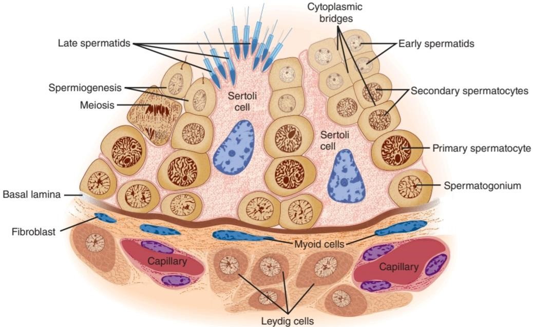
FIGURE 23-3 Seminiferous epithelium. Note that maturing germ cells remain connected by cytoplasmic bridges through the early spermatid stage and that these cells are closely invested by Sertoli cell cytoplasm as they move from the basal lamina to the lumen. (Reproduced with permission from Junqueira LC, Carneiro J: Basic Histology: Text & Atlas, 10th ed. New York, NY: McGraw- Hill; 2003.)
شکل 23-3 اپیتلیوم منی ساز. توجه داشته باشید که سلولهای زایای بالغ از طریق پلهای سیتوپلاسمی در مراحل اولیه اسپرماتید به هم متصل میمانند و این سلولها از نزدیک توسط سیتوپلاسم سلول سرتولی در حین حرکت از لایه بازال به سمت لومن سرمایهگذاری میشوند. (تکثیر شده با مجوز Junqueira LC، Carneiro J: Basic Histology: Text & Atlas، ویرایش 10 نیویورک، نیویورک: McGraw- Hill؛ 2003.)
GAMETOGENESIS & EJACULATION
Blood-Testis Barrier
The walls of the seminiferous tubules are lined by primitive germ cells and Sertoli cells, large, complex glycogen-containing cells that stretch from the basal lamina of the tubule to the lumen (Figure 23-3). Germ cells must stay in contact with Sertoli cells to survive; this contact is maintained by cytoplasmic bridges. Tight junctions between adjacent Sertoli cells near the basal lamina form a blood-testis barrier that prevents many large molecules from passing from the interstitial tissue and the part of the tubule near the basal lamina (basal compartment) to the region near the tubular lumen (adluminal compartment) and the lumen. However, steroids penetrate this barrier with ease, and evidence suggests that some proteins also pass from the Sertoli cells to the Leydig cells, and vice versa, to function in a paracrine manner. In addition, maturing germ cells must pass through the barrier as they move to the lumen. This appears to occur without disruption of the barrier by coordinated breakdown of the tight junctions above the germ cells and formation of new tight junctions below them.
GAMETOGENESIS و انزال
سد خونی بیضه
دیوارههای لولههای منیساز توسط سلولهای زایای اولیه و سلولهای سرتولی، سلولهای بزرگ و پیچیده حاوی گلیکوژن پوشانده شدهاند که از لایه بازال توبول تا لومن امتداد دارند (شکل 23-3). سلول های زاینده برای زنده ماندن باید با سلول های سرتولی در تماس باشند. این تماس توسط پل های سیتوپلاسمی حفظ می شود. اتصالات محکم بین سلول های مجاور سرتولی در نزدیکی لایه بازال یک سد خونی بیضه را تشکیل می دهد که از عبور بسیاری از مولکول های بزرگ از بافت بینابینی و بخشی از لوله در نزدیکی لایه بازال (محفظه پایه) به ناحیه نزدیک به لومن توبولار (محفظه آدرومینال) و لومن جلوگیری می کند. با این حال، استروئیدها به راحتی به این سد نفوذ می کنند، و شواهد نشان می دهد که برخی از پروتئین ها نیز از سلول های سرتولی به سلول های لیدیگ عبور می کنند و بالعکس، تا به شیوه ای پاراکرین عمل کنند. علاوه بر این، سلول های زایای بالغ باید در حین حرکت به سمت لومن از سد عبور کنند. به نظر می رسد که این بدون ایجاد اختلال در سد با شکست هماهنگ اتصالات محکم بالای سلول های زاینده و تشکیل اتصالات محکم جدید در زیر آنها رخ می دهد.
The fluid in the lumen of the seminiferous tubules is quite different from plasma; it contains very little protein and glucose but is rich in androgens, estrogens, K+, inositol, and glutamic and aspartic acids. Maintenance of its composition depends on the blood-testis barrier. The barrier also protects the germ cells from bloodborne noxious agents, prevents antigenic products of germ cell division and maturation from entering the circulation and generating an autoimmune response, and may help establish an osmotic gradient that facilitates movement of fluid into the tubular lumen.
مایع موجود در لومن لوله های منی ساز کاملاً متفاوت از پلاسما است. حاوی پروتئین و گلوکز بسیار کمی است اما سرشار از آندروژن، استروژن، +K، اینوزیتول و اسیدهای گلوتامیک و آسپارتیک است. حفظ ترکیب آن به سد خونی بیضه بستگی دارد. این سد همچنین از سلولهای زایا در برابر عوامل مضر خون محافظت میکند، از ورود محصولات آنتی ژنی تقسیم و بلوغ سلولهای زاینده به گردش خون و ایجاد پاسخ خود ایمنی جلوگیری میکند و ممکن است به ایجاد یک گرادیان اسمزی کمک کند که حرکت مایع را به داخل لومن لولهای تسهیل میکند.
Spermatogenesis
Spermatogonia, the primitive germ cells next to the basal lamina of the seminiferous tubules, mature into primary spermatocytes (Figure 23–3). This process begins during adolescence. The primary spermatocytes undergo meiotic division, reducing the number of chromosomes. In this two-stage process, they divide into secondary spermatocytes and then into spermatids, which contain the haploid number of 23 chromosomes. The spermatids mature into spermatozoa (sperm). As a single spermatogonium divides and matures, its descendants remain tied together by cytoplasmic bridges until the late spermatid stage. This arrangement helps ensure synchrony of the differentiation of each clone of germ cells. The estimated number of spermatids formed from a single spermatogonium is 512. The formation of a mature sperm from a primitive germ cell by spermatogenesis in humans spans approximately 74 days.
اسپرماتوژنز
اسپرماتوگونیا، سلول های زایای اولیه در کنار لایه بازال لوله های اسپرم ساز، به اسپرماتوسیت های اولیه بالغ می شوند (شکل 23-3). این روند در دوران نوجوانی شروع می شود. اسپرماتوسیت های اولیه تحت تقسیم میوز قرار می گیرند و تعداد کروموزوم ها را کاهش می دهند. در این فرآیند دو مرحله ای، آنها به اسپرماتوسیت های ثانویه و سپس به اسپرماتیدها تقسیم می شوند که دارای عدد هاپلوئید 23 کروموزوم هستند. اسپرماتیدها به اسپرماتوزوا (اسپرم) بالغ می شوند. هنگامی که یک اسپرماتوگونیوم منفرد تقسیم می شود و بالغ می شود، فرزندان آن توسط پل های سیتوپلاسمی تا اواخر مرحله اسپرماتید به هم متصل می مانند. این آرایش به اطمینان از هماهنگی تمایز هر کلون سلول های زاینده کمک می کند. تعداد تخمینی اسپرماتیدهای تشکیل شده از یک اسپرماتوگونیوم منفرد 512 است. تشکیل یک اسپرم بالغ از یک سلول زایای اولیه توسط اسپرماتوژنز در انسان تقریباً 74 روز طول می کشد.
Each sperm is an intricate motile cell, rich in DNA, with a head that is made up mostly of chromosomal material (Figure 23-4). Covering the head like a cap is the acrosome, a lysosome-like organelle rich in enzymes involved in sperm penetration of the ovum and other events associated with fertilization. The motile tail of the sperm is wrapped in its proximal portion by a sheath holding numerous mitochondria. The membranes of late spermatids and spermatozoa contain a special small form of angiotensin-converting enzyme (ACE) called germinal angiotensin-converting enzyme (ACE). Germinal ACE is transcribed from the same gene as the somatic ACE (SACE); however, gACE displays tissue-specific expression based on alternative transcription initiation sites and alternate splicing patterns. The full function of gACE has yet to be elucidated, although gACE-specific knockout mouse models are sterile.
هر اسپرم یک سلول متحرک پیچیده، غنی از DNA، با سر است که بیشتر از مواد کروموزومی ساخته شده است (شکل 23-4). سر را مانند کلاهک می پوشاند، آکروزوم، اندامک لیزوزوم مانند غنی از آنزیم های دخیل در نفوذ اسپرم به تخمک و سایر رویدادهای مرتبط با لقاح. دم متحرک اسپرم توسط غلافی که میتوکندری های متعددی را در خود جای داده است در قسمت نزدیک آن پیچیده شده است. غشاهای اسپرماتیدهای دیررس و اسپرماتوزواها حاوی شکل کوچک خاصی از آنزیم مبدل آنژیوتانسین (ACE) به نام آنزیم مبدل آنژیوتانسین ژرمینال (ACE) است. ACE ژرمینال از همان ژن ACE سوماتیک (SACE) رونویسی می شود. با این حال، gACE بیان بافت خاص را بر اساس مکانهای شروع رونویسی جایگزین و الگوهای پیوند متناوب نشان میدهد. عملکرد کامل gACE هنوز مشخص نشده است، اگرچه مدلهای حذفی موش مخصوص gACE استریل هستند.

FIGURE 23-4 Human spermatozoon, profile view. Note the acrosome, an organelle that covers half the sperm head inside the plasma membrane of the sperm. (Reproduced with permission from Junqueira LC, Carneiro J: Basic Histology: Text & Atlas, 11th ed. New York, NY: McGraw-Hill; 2005.)
شکل 23-4 اسپرم انسان، نمای نیم رخ. به آکروزوم، اندامکی که نیمی از سر اسپرم را در داخل غشای پلاسمایی اسپرم می پوشاند، توجه کنید. (تکثیر شده با اجازه Junqueira LC، Carneiro J: Basic Histology: Text & Atlas, ed. 11 New York, NY: McGraw-Hill; 2005.)
Spermatids mature into spermatozoa in deep folds of the cytoplasm of the Sertoli cells (Figure 23-3). Mature spermatozoa are released from the Sertoli cells and become free in the lumens of the tubules. The Sertoli cells secrete androgen-binding protein (ABP), inhibin, and Mullerian inhibiting substance (MIS). They do not synthesize androgens, but they contain aromatase (CYP19), the enzyme responsible for conversion of androgens to estrogens, and they can produce estrogens. ABP probably functions to maintain a high, stable supply of androgen in the tubular fluid. Inhibin inhibits follicle- stimulating hormone (FSH) secretion.
اسپرماتیدها در چین های عمیق سیتوپلاسم سلول های سرتولی به اسپرم تبدیل می شوند (شکل 23-3). اسپرم های بالغ از سلول های سرتولی آزاد می شوند و در مجرای لوله ها آزاد می شوند. سلول های سرتولی پروتئین متصل شونده به آندروژن (ABP)، اینهیبین و ماده بازدارنده مولرین (MIS) ترشح می کنند. آنها آندروژن را سنتز نمی کنند، اما حاوی آروماتاز (CYP19)، آنزیمی که مسئول تبدیل آندروژن ها به استروژن است، هستند و می توانند استروژن تولید کنند. ABP احتمالاً برای حفظ یک منبع بالا و پایدار آندروژن در مایع لوله ای عمل می کند. اینهیبین ترشح هورمون محرک فولیکول (FSH) را مهار می کند.
FSH and androgens maintain the gametogenic function of the testis. After hypophysectomy, injection of luteinizing hormone (LH) produces a high local concentration of androgen in the testes, and this maintains spermatogenesis. The stages from spermatogonia to spermatids appear to be androgen-independent. However, the maturation from spermatids to spermatozoa depends on androgen acting on the Sertoli cells in which the developing spermatozoa are embedded. FSH acts on the Sertoli cells to facilitate the last stages of spermatid maturation. In addition, it promotes the production of ABP.
FSH و آندروژن ها عملکرد گامتوژنیک بیضه را حفظ می کنند. پس از هیپوفیزکتومی، تزریق هورمون لوتئینیزه کننده (LH) غلظت محلی بالایی از آندروژن در بیضه ها تولید می کند و این امر باعث حفظ اسپرماتوژنز می شود. به نظر می رسد مراحل از اسپرماتوگونی تا اسپرماتیدها مستقل از آندروژن باشد. با این حال، بلوغ از اسپرماتیدها به اسپرماتوزوا بستگی به آندروژنی دارد که بر روی سلولهای سرتولی که اسپرمهای در حال رشد در آن جاسازی شدهاند، عمل میکند. FSH بر روی سلول های سرتولی عمل می کند تا آخرین مراحل بلوغ اسپرماتید را تسهیل کند. علاوه بر این، تولید ABP را ترویج می کند.
An interesting observation is that the estrogen content of the fluid in the rete testis (Figure 23-1) is high, and the walls of the rete testis contain numerous a estrogen receptors (ERa). In this region, fluid is reabsorbed and the spermatozoa are concentrated. If this does not occur, the sperm entering the epididymis are diluted in a large volume of fluid, resulting in reduced fertility.
یک مشاهدات جالب این است که محتوای استروژن مایع در بیضه ریت (شکل 23-1) زیاد است و دیوارههای بیضه حاوی گیرندههای استروژن متعدد (ERa) هستند. در این ناحیه مایع دوباره جذب می شود و اسپرم ها متمرکز می شوند. اگر این اتفاق نیفتد، اسپرم وارد شده به اپیدیدیم در حجم زیادی از مایع رقیق می شود و در نتیجه باروری کاهش می یابد.
Further Development of Spermatozoa
Spermatozoa leaving the testes are not fully mobile. They continue their maturation and acquire motility during their passage through the epididymis. Motility is obviously important in vivo, but fertilization occurs in vitro if an immotile spermatozoon from the head of the epididymis is microinjected directly into an ovum. The ability to move forward (progressive motility), which is acquired in the epididymis, involves activation of a unique set of cation channel proteins from the CatSper family, which are localized to the principal piece of the sperm tail. CatSpers form an alkaline-sensitive Ca2+ channel that becomes more active as the sperm go from the acidic vagina (pH ~5) to the cervical mucus (pH ~8). Sperm from knockout mice that do not express CatSper1-4 have altered motility and are infertile, emphasizing the important role of these channel proteins. In addition, spermatozoa express olfactory receptors, and ovaries produce odorant-like molecules. Recent evidence indicates that these molecules and their receptors interact, fostering movement of the spermatozoa toward the ovary (chemotaxis).
رشد بیشتر اسپرم
اسپرم هایی که از بیضه ها خارج می شوند کاملاً متحرک نیستند. آنها در طی عبور از اپیدیدیم به بلوغ خود ادامه می دهند و تحرک پیدا می کنند. تحرک بدیهی است در داخل بدن مهم است، اما لقاح در شرایط آزمایشگاهی اتفاق می افتد اگر یک اسپرم غیر متحرک از سر اپیدیدیم به طور مستقیم به تخمک تزریق شود. توانایی حرکت رو به جلو (حرکت پیشرونده)، که در اپیدیدیم به دست میآید، شامل فعالسازی مجموعهای از پروتئینهای کانال کاتیونی از خانواده CatSper است که در قسمت اصلی دم اسپرم قرار دارند. CatSpers یک کانال +Ca2 حساس به قلیایی تشکیل می دهد که با رفتن اسپرم از واژن اسیدی (pH ~ 5) به مخاط دهانه رحم (pH ~ 8) فعال تر می شود. اسپرم موشهای ناک اوت که CatSper1-4 را بیان نمیکنند، تحرک تغییر یافته و نابارور هستند، که بر نقش مهم این پروتئینهای کانال تأکید دارد. علاوه بر این، اسپرم گیرنده های بویایی را بیان می کند و تخمدان ها مولکول های بویایی مانند تولید می کنند. شواهد اخیر نشان میدهد که این مولکولها و گیرندههای آنها با هم تعامل دارند و حرکت اسپرمها را به سمت تخمدان تقویت میکنند (کموتاکسی).
Ejaculation of the spermatozoon involves contractions of the vas deferens mediated in part by P2X purinoreceptors, ligand-gated cation channels that open in response to ATP (see Chapter 7), and fertility is reduced in mice in which these receptors are knocked out.
انزال اسپرم شامل انقباضات مجرای دفران است که تا حدی توسط گیرنده های پورینی P2X، کانال های کاتیونی دردار لیگاند که در پاسخ به ATP باز می شوند (به فصل 7 مراجعه کنید) و باروری در موش هایی که این گیرنده ها از بین رفته اند کاهش می یابد.
Once ejaculated into the female, the spermatozoa move up the uterus to the isthmus of the uterine tubes, where they slow down and undergo capacitation. This further maturation process involves two components: increasing the motility of the spermatozoa and facilitating their preparation for the acrosome reaction. However, the role of capacitation appears to be facilitatory rather than obligatory, because fertilization is readily produced in vitro. From the isthmuses, the capacitated spermatozoa move rapidly to the tubal ampullas, where fertilization takes place.
پس از انزال در ماده، اسپرم ها از رحم به سمت تنگه لوله های رحمی حرکت می کنند، جایی که سرعت آنها کاهش می یابد و تحت ظرفیت قرار می گیرند. این فرآیند بلوغ بیشتر شامل دو جزء است: افزایش تحرک اسپرم ها و تسهیل آماده سازی آنها برای واکنش آکروزوم. با این حال، به نظر می رسد نقش ظرفیت سازی به جای اجباری، تسهیل کننده است، زیرا لقاح به راحتی در شرایط آزمایشگاهی تولید می شود. از تنگه ها، اسپرم های دارای ظرفیت به سرعت به آمپول های لوله ای حرکت می کنند، جایی که لقاح انجام می شود.
Effect of Temperature
Spermatogenesis requires a temperature considerably lower than that of the interior of the body. The testes are normally maintained at a temperature of about 32°C. They are kept cool by air circulating around the scrotum and probably by heat exchange in a countercurrent manner between the spermatic arteries and veins. When the testes are retained in the abdomen or when, in experimental animals, they are held close to the body, degeneration of the tubular walls and sterility result. Situations that increase heat around the testes in humans (e.g., hot baths [43-45°C for 30 min/d] and insulated athletic supporters) can reduce sperm counts, in some cases by 90%. However, the reductions produced in this manner are not consistent enough to make the procedures reliable forms of male contraception. In addition, evidence suggests a seasonal effect in men, with sperm counts being greater in the winter regardless of the temperature to which the scrotum is exposed.
اثر دما
اسپرماتوژنز به دمایی بسیار کمتر از دمای داخلی بدن نیاز دارد. بیضه ها معمولاً در دمای حدود 32 درجه سانتیگراد نگهداری می شوند. آنها با گردش هوا در اطراف کیسه بیضه و احتمالاً با تبادل گرما به روشی خلاف جریان بین شریان ها و وریدهای اسپرماتیک خنک نگه داشته می شوند. وقتی بیضه ها در شکم نگه داشته می شوند یا در حیوانات آزمایشی نزدیک بدن نگه داشته می شوند، انحطاط دیواره های لوله ای و عقیمی ایجاد می شود. موقعیتهایی که گرمای اطراف بیضهها را در انسان افزایش میدهند (مانند حمام آب گرم [43-45 درجه سانتیگراد برای 30 دقیقه در روز] و حامیان ورزشی عایقشده) میتوانند تعداد اسپرمها را در برخی موارد تا 90 درصد کاهش دهند. با این حال، کاهش های ایجاد شده به این روش به اندازه کافی سازگار نیست که روش ها را به اشکال قابل اعتمادی از پیشگیری از بارداری مردانه تبدیل کند. علاوه بر این، شواهد حاکی از تأثیر فصلی در مردان است، به طوری که تعداد اسپرم ها در زمستان بدون توجه به دمایی که کیسه بیضه در معرض آن قرار دارد، بیشتر است.
Semen
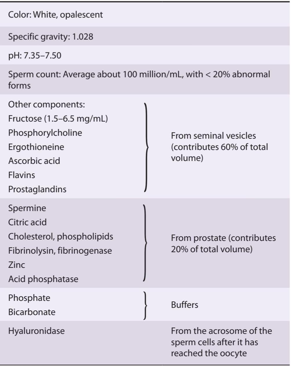
The fluid that is ejaculated at the time of orgasm, the semen, contains sperm and the secretions of the seminal vesicles, prostate, Cowper glands, and, probably, the urethral glands (Table 23-1). An average volume per ejaculate is 2.5-3.5 mL after several days of abstinence from sexual activity. The volume of semen and the sperm count decrease rapidly with repeated ejaculation. Even though it takes only one sperm to fertilize the ovum, each milliliter of semen normally contains about 100 million sperm. Reduction in sperm production is associated with infertility: 50% of men with counts of 20-40 million/mL and essentially all of those with counts under 20 million/mL are sterile. The presence of many morphologically abnormal or immotile spermatozoa also correlates with infertility. The prostaglandins in semen, which come from the seminal vesicles, are at high concentrations, but their function in semen is unknown. The causes of male infertility, as well as the underlying mechanisms of sperm in fertilization, are used as clues in developing male contraception (Clinical Box 23-1).
منی
مایعی که در زمان ارگاسم انزال میشود، مایع منی، حاوی اسپرم و ترشحات وزیکولهای منی، پروستات، غدد Cowper و احتمالاً غدد مجرای ادرار است (جدول 23-1). میانگین حجم هر انزال 2.5-3.5 میلی لیتر پس از چند روز پرهیز از فعالیت جنسی است. حجم منی و تعداد اسپرم با انزال مکرر به سرعت کاهش می یابد. حتی اگر برای بارور کردن تخمک فقط یک اسپرم لازم است، هر میلی لیتر منی به طور معمول حدود 100 میلیون اسپرم دارد. کاهش تولید اسپرم با ناباروری همراه است: 50 درصد مردان با تعداد 20 تا 40 میلیون در میلی لیتر و اساساً همه مردان با تعداد کمتر از 20 میلیون در میلی لیتر عقیم هستند. وجود بسیاری از اسپرم های غیر طبیعی یا غیر متحرک از نظر مورفولوژیکی نیز با ناباروری ارتباط دارد. پروستاگلاندین های موجود در مایع منی، که از وزیکول های منی می آیند، در غلظت های بالایی هستند، اما عملکرد آنها در مایع منی ناشناخته است. علل ناباروری مردان، و همچنین مکانیسم های زمینه ای اسپرم در لقاح، به عنوان سرنخ در توسعه پیشگیری از بارداری مردانه استفاده می شود (کادر بالینی 23-1).
Human sperm move at a speed of about 3 mm/min through the female genital tract. Sperm reach the uterine tubes 30-60 min after copulation. Contractions of the female organs may facilitate the transport of the sperm to the uterine tubes.
اسپرم انسان با سرعتی در حدود 3 میلی متر در دقیقه از طریق دستگاه تناسلی زنانه حرکت می کند. اسپرم 30-60 دقیقه پس از جفت شدن به لوله های رحمی می رسد. انقباضات اندام های زنانه ممکن است انتقال اسپرم به لوله های رحمی را تسهیل کند.
TABLE 23-1 Composition of human semen.
جدول 23-1 ترکیب مایع منی انسان.
CLINICAL BOX 23-1
Male Contraception
Several methods independent of physical intervention (such as hormonal control of sperm development, targeting of cation channel proteins important in fertilization [e.g., CatSpers], and the use of natural compounds that limit sperm function) have been explored as male contraceptives. However, considering the number of sperm and their ability to regenerate, methods that sufficiently reduce sperm production or limit function with an absence of side effects have been difficult to obtain. In lieu of pharmacological control, the most common male contraception continues to be bilateral ligation of the vas deferens (vasectomy), a relatively safe and convenient contraceptive procedure. Interestingly, antibodies against spermatozoa develop in ~50% of the men who have undergone vasectomy; in monkeys, the presence of such antibodies is associated with a higher incidence of infertility after restoration of the patency of the vas. However, the anti-sperm antibodies do not appear to have any other adverse effects. Alternatives to ligation are vas occlusion methods, such as silicone plugs, that aim to block the vas deferens while leaving the tube intact, making reversal easier if desired. Not surprisingly, such methods are not as effective as traditional vasectomy.
جعبه بالینی 23-1
پیشگیری از بارداری مردانه
چندین روش مستقل از مداخله فیزیکی (مانند کنترل هورمونی رشد اسپرم، هدف قرار دادن پروتئین های کانال کاتیونی مهم در لقاح [به عنوان مثال، CatSpers]، و استفاده از ترکیبات طبیعی که عملکرد اسپرم را محدود می کنند) به عنوان ضد بارداری مردانه مورد بررسی قرار گرفته اند. با این حال، با توجه به تعداد اسپرم ها و توانایی آنها برای بازسازی، روش هایی که به اندازه کافی تولید اسپرم را کاهش می دهند یا عملکرد را با عدم وجود عوارض جانبی محدود می کنند، دشوار بوده است. به جای کنترل دارویی، رایج ترین روش پیشگیری از بارداری مردانه بستن دو طرفه مجرای دفران (وازکتومی)، یک روش پیشگیری از بارداری نسبتاً ایمن و راحت است. جالب توجه است که در 50 درصد از مردانی که تحت وازکتومی قرار گرفته اند، آنتی بادی علیه اسپرم ایجاد می شود. در میمونها، وجود چنین آنتیبادیهایی با بروز بالاتر ناباروری پس از بازیابی عروق همراه است. با این حال، به نظر نمی رسد که آنتی بادی های ضد اسپرم عوارض جانبی دیگری داشته باشند. روشهای جایگزین برای بستن، روشهای انسداد عروق هستند، مانند پلاگهای سیلیکونی، که هدف آن مسدود کردن مجرای دفران در حالی که لوله دست نخورده باقی میماند، در صورت تمایل، برگشت را آسانتر میکند. جای تعجب نیست که چنین روش هایی به اندازه وازکتومی سنتی موثر نیستند.
THERAPEUTIC HIGHLIGHTS
Vasectomy reversal: Whereas it was once quite difficult to restore the patency of the vas in those wishing to restore fertility, the success rate for such operations has improved steadily. Successful reversal of vasectomy usually results in meaningful sperm counts within months, although delays of a year or more are not unusual. The ultimate success, as measured by pregnancy, is recorded in 50% of reversals within 2 years.
نکات برجسته درمانی
معکوس کردن وازکتومی: در حالی که زمانی بازگرداندن باز بودن مجرا در افرادی که مایل به بازگرداندن باروری بودند بسیار دشوار بود، میزان موفقیت برای چنین عمل هایی به طور پیوسته بهبود یافته است. معکوس کردن موفقیت آمیز وازکتومی معمولاً منجر به تعداد معنی دار اسپرم در طی چند ماه می شود، اگرچه تأخیر یک سال یا بیشتر غیرعادی نیست. موفقیت نهایی، همانطور که با بارداری اندازه گیری می شود، در 50٪ از برگشت ها در عرض 2 سال ثبت می شود.
Erection
Erection is initiated by dilation of the arterioles of the penis. As the erectile tissue of the penis fills with blood, the veins are compressed, blocking outflow and adding to the turgor of the organ. The integrating centers in the lumbar segments of the spinal cord are activated by impulses in afferents from the genitalia and descending tracts that mediate erection in response to erotic psychological stimuli. The efferent parasympathetic fibers are in the pelvic splanchnic nerves (nervi erigentes). The fibers presumably release acetylcholine and the vasodilator vasoactive intestinal polypeptide (VIP) as cotransmitters (see Chapter 7).
نعوظ
نعوظ با گشاد شدن شریان های آلت تناسلی شروع می شود. همانطور که بافت نعوظ آلت تناسلی با خون پر می شود، وریدها فشرده می شوند و مانع از خروج آن می شوند و به تورگور اندام می افزایند. مراکز یکپارچه در بخشهای کمری نخاع توسط تکانههایی در آورانهای دستگاه تناسلی و دستگاههای نزولی فعال میشوند که نعوظ را در پاسخ به محرکهای روانشناختی وابسته به عشق شهوانی انجام میدهند. فیبرهای پاراسمپاتیک وابران در اعصاب لگنی (nervi erigentes) قرار دارند. فیبرها احتمالاً استیل کولین و پلی پپتید فعال عروقی روده ای گشادکننده عروق (VIP) را به عنوان انتقال دهنده همزمان آزاد می کنند (به فصل 7 مراجعه کنید).
Nonadrenergic noncholinergic fibers are also present in the nervi erigentes, and these contain large amounts of nitric oxide synthase (NOS), the enzyme that catalyzes the formation of nitric oxide (NO; see Chapter 32). NO activates soluble guanylyl cyclase, resulting in increased production of cyclic guanosine monophosphate (cGMP), and cGMP is a potent vasodilator. Injection of NOS inhibitors prevents the erection normally produced by stimulation of the pelvic nerve in experimental animals. Thus, it seems clear that NO plays a prominent role in the production of erection. The drugs sildenafil (Viagra), tadalafil (Cialis), and vardenafil (Levitra) all inhibit the breakdown of cGMP by phosphodiesterases and have gained worldwide fame for the treatment of erectile dysfunction. The multiple phosphodiesterases (PDEs) in the body have been divided into 11 isoenzyme families, and these drugs are all most active against PDE-5, the type of phosphodiesterase found in the corpora cavernosa (Clinical Box 5-7). It is worth noting, however, that these drugs can also produce significant inhibition of PDE-6 (and others, if taken at high doses). PDE-6 is found in the retina, and one of the side effects of these drugs is a transient loss of the ability to discriminate between blue and green (see Chapter 9).
فیبرهای غیر کولینرژیک غیر آدرنرژیک نیز در اریژنت های عصبی وجود دارند و این فیبرها حاوی مقادیر زیادی از نیتریک اکسید سنتاز (NOS) هستند، آنزیمی که تشکیل اکسید نیتریک را کاتالیز می کند (NO؛ به فصل 32 مراجعه کنید). NO گوانیلیل سیکلاز محلول را فعال می کند و در نتیجه تولید گوانوزین مونوفسفات حلقوی (cGMP) را افزایش می دهد و cGMP یک گشادکننده عروق قوی است. تزریق مهارکننده های NOS از نعوظی که معمولاً با تحریک عصب لگن در حیوانات آزمایشگاهی ایجاد می شود، جلوگیری می کند. بنابراین واضح است که NO نقش برجسته ای در تولید نعوظ دارد. داروهای سیلدنافیل (ویاگرا)، تادالافیل (سیالیس) و واردنافیل (لویترا) همگی از تجزیه cGMP توسط فسفودی استرازها جلوگیری می کنند و برای درمان اختلال نعوظ شهرت جهانی پیدا کرده اند. فسفودی استرازهای متعدد (PDEs) در بدن به 11 خانواده ایزوآنزیمی تقسیم شده اند، و این داروها همگی در برابر PDE-5، نوع فسفودی استراز موجود در اجسام غارنوردی فعال هستند (کادربالینی 5-7). البته شایان ذکر است که این داروها همچنین می توانند مهار قابل توجهی از PDE-6 (و سایر داروها در صورت مصرف با دوزهای بالا) ایجاد کنند. PDE-6 در شبکیه چشم یافت می شود و یکی از عوارض جانبی این داروها از دست دادن گذرا توانایی تمایز بین آبی و سبز است (به فصل 9 مراجعه کنید).
Normally, erection is terminated by sympathetic vasoconstrictor impulses to the penile arterioles.
به طور معمول، نعوظ با تکانه های تنگ کننده عروق سمپاتیک به شریان های آلت تناسلی خاتمه می یابد.
Ejaculation
Ejaculation is a two-part spinal reflex that involves emission, the movement of the semen into the urethra; and ejaculation proper, the propulsion of the semen out of the urethra at the time of orgasm. The afferent pathways are mostly fibers from touch receptors in the glans penis that reach the spinal cord through the internal pudendal nerves. Emission is a sympathetic response, integrated in the upper lumbar segments of the spinal cord and effected by contraction of the smooth muscle of the vasa deferentia and seminal vesicles in response to stimuli in the hypogastric nerves. The semen is propelled out of the urethra by contraction of the bulbocavernosus muscle, a skeletal muscle. The spinal reflex centers for this part of the reflex are in the upper sacral and lowest lumbar segments of the spinal cord, and the motor pathways traverse the first to third sacral roots and the internal pudendal nerves.
انزال
انزال یک رفلکس نخاعی دو قسمتی است که شامل انتشار، حرکت مایع منی به داخل مجرای ادرار است. و انزال مناسب، بیرون راندن منی از مجرای ادرار در زمان ارگاسم. مسیرهای آوران عمدتاً فیبرهایی از گیرنده های لمسی در آلت تناسلی هستند که از طریق اعصاب پودندال داخلی به نخاع می رسند. انتشار یک پاسخ سمپاتیک است که در بخشهای کمری فوقانی نخاع ادغام میشود و با انقباض عضله صاف وازا دفرنتیا و وزیکولهای منی در پاسخ به محرکهای اعصاب هیپوگاستریک ایجاد میشود. منی با انقباض عضله بولبوکاورنوس که یک ماهیچه اسکلتی است از مجرای ادرار خارج می شود. مراکز رفلکس نخاعی برای این بخش از رفلکس در قسمت های خاجی فوقانی و پایین ترین بخش های کمری نخاع قرار دارند و مسیرهای حرکتی از ریشه های اول تا سوم خاجی و اعصاب پودندال داخلی عبور می کنند.
Prostate-Specific Antigen
The prostate produces and secretes into the semen and the bloodstream a 30-kDa serine protease generally called prostate-specific antigen (PSA). The gene for PSA has two androgen response elements. PSA hydrolyzes the sperm motility inhibitor semenogelin in semen, and it has several substrates in plasma, but its precise function in the circulation is unknown. An elevated plasma PSA occurs in prostate cancer and has been widely used as a screening test for this disease. However, PSA is also elevated in benign prostatic hyperplasia and prostatitis, and the effectiveness of PSA screening as a sole tool in diagnosis of prostate cancer has been called into question.
آنتی ژن اختصاصی پروستات
پروستات یک سرین پروتئاز 30 کیلو دالتون را تولید کرده و در منی و جریان خون ترشح می کند که به طور کلی آنتی ژن اختصاصی پروستات (PSA) نامیده می شود. ژن PSA دارای دو عنصر پاسخ آندروژن است. PSA سمنوژلین بازدارنده حرکت اسپرم را در مایع منی هیدرولیز می کند و چندین بستر در پلاسما دارد، اما عملکرد دقیق آن در گردش خون ناشناخته است. افزایش PSA پلاسما در سرطان پروستات رخ می دهد و به طور گسترده ای به عنوان یک آزمایش غربالگری برای این بیماری استفاده می شود. با این حال، PSA در هیپرپلازی خوش خیم پروستات و پروستاتیت نیز افزایش می یابد و اثربخشی غربالگری PSA به عنوان تنها ابزار در تشخیص سرطان پروستات زیر سوال رفته است.
ENDOCRINE FUNCTION OF THE TESTES
Chemistry & Biosynthesis of Testosterone
Testosterone, the principal hormone of the testes, is a C19 steroid with a hydroxyl group in the 17 position (Figure 23-5). It is synthesized from cholesterol in the Leydig cells and is also formed from androstenedione secreted by the adrenal cortex. The biosynthetic pathways in all endocrine organs that form steroid hormones are similar, the organs differing only in the enzyme systems they contain. In the Leydig cells, the 11- and 21-hydroxylases found in the adrenal cortex (see Figure 20-7) are absent, but 17a-hydroxylase is present. Pregnenolone is therefore hydroxylated in the 17 position and then subjected to side chain cleavage to form dehydroepiandrosterone. Androstenedione is also formed via progesterone and 17-hydroxyprogesterone, but this pathway is less prominent in humans. Dehydroepiandrosterone and androstenedione are then converted to testosterone.
عملکرد غدد درون ریز بیضه ها
شیمی و بیوسنتز تستوسترون
تستوسترون، هورمون اصلی بیضه ها، یک استروئید C19 با گروه هیدروکسیل در موقعیت 17 است (شکل 23-5). از کلسترول موجود در سلول های لیدیگ سنتز می شود و همچنین از آندروستندیون ترشح شده توسط قشر آدرنال تشکیل می شود. مسیرهای بیوسنتزی در تمام اندامهای غدد درون ریز که هورمونهای استروئیدی را تشکیل میدهند مشابه هستند، اندامها فقط در سیستم آنزیمی که دارند متفاوت هستند. در سلول های لیدیگ، هیدروکسیلازهای 11 و 21 موجود در قشر آدرنال (شکل 20-7 را ببینید) وجود ندارند، اما 17a-هیدروکسیلاز وجود دارد. بنابراین پرگننولون در موقعیت 17 هیدروکسیله می شود و سپس در معرض شکاف زنجیره جانبی برای تشکیل دهیدرواپی آندروسترون قرار می گیرد. آندروستندیون نیز از طریق پروژسترون و 17 هیدروکسی پروژسترون تشکیل می شود، اما این مسیر در انسان کمتر برجسته است. سپس دهیدرواپی آندروسترون و آندروستندیون به تستوسترون تبدیل می شوند.
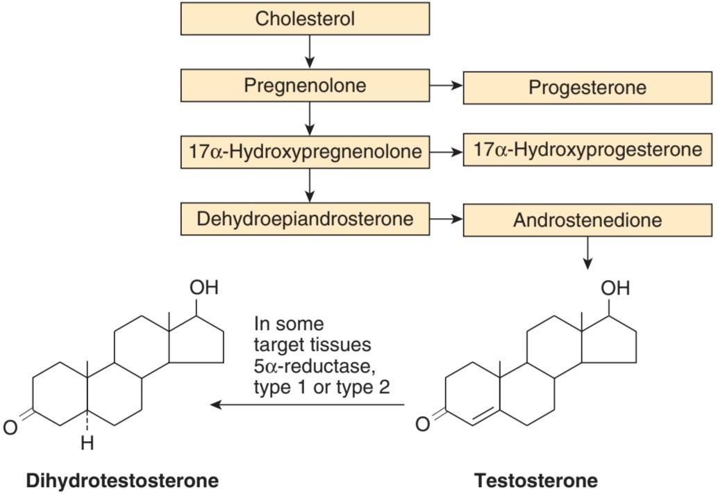
FIGURE 23-5 Biosynthesis of testosterone. The formulas of the precursor steroids are shown in Figure 20-7. Although the main secretory product of the Leydig cells is testosterone, some of the precursors also enter the circulation.
شکل 23-5 بیوسنتز تستوسترون. فرمول استروئیدهای پیش ساز در شکل 20-7 نشان داده شده است. اگرچه محصول ترشحی اصلی سلول های لیدیگ تستوسترون است، برخی از پیش سازها نیز وارد گردش خون می شوند.
The secretion of testosterone is under the control of luteinizing hormone (LH), and the mechanism by which LH stimulates Leydig cells involves increased formation of cyclic adenosine monophosphate (cAMP) via the G- protein-coupled LH receptor and G. Cyclic AMP increases the formation of cholesterol from cholesterol esters and the conversion of cholesterol to pregnenolone via the activation of protein kinase A.
ترشح تستوسترون تحت کنترل هورمون لوتئینیزه کننده (LH) است و مکانیسمی که توسط آن LH سلول های لیدیگ را تحریک می کند شامل افزایش تشکیل آدنوزین مونوفسفات حلقوی (cAMP) از طریق گیرنده LH جفت شده با پروتئین G و G. AMP حلقوی تشکیل کلسترول کلسترول از کلسترول و کلسترول را افزایش می دهد. پرگننولون از طریق فعال سازی پروتئین کیناز A.
Secretion
The testosterone secretion rate is 4-9 mg/d (13.9-31.33 μmol/d) in normal adult males. Small amounts of testosterone are also secreted in females, with the major source being the ovary, but possibly from the adrenal as well.
ترشح
میزان ترشح تستوسترون 4-9 میلی گرم در روز (13.9-31.33 میکرومول در روز) در مردان بالغ طبیعی است. مقادیر کمی تستوسترون نیز در زنان ترشح می شود که منبع اصلی آن تخمدان است، اما احتمالاً از غده فوق کلیوی نیز می باشد.
Transport & Metabolism
Ninety-eight percent of the testosterone in plasma is bound to protein: 65% is bound to a B-globulin called gonadal steroid-binding globulin (GBG) or sex steroid-binding globulin, and 33% to albumin (Table 23-2). GBG also binds estradiol. The plasma testosterone level (free and bound) is 300-1000 ng/dL (10.4-34.7 nmol/L) in adult men (Figure 22-8), compared with 30-70 ng/dL (1.04-2.43 nmol/L) in adult women. It declines somewhat with age in men.
حمل و نقل و متابولیسم
نود و هشت درصد تستوسترون پلاسما به پروتئین متصل می شود: 65 درصد به یک B-گلوبولین به نام گلوبولین متصل کننده استروئید جنسی (GBG) یا گلوبولین اتصال به استروئید جنسی و 33 درصد به آلبومین متصل می شود (جدول 23-2). GBG همچنین به استرادیول متصل می شود. سطح تستوسترون پلاسما (آزاد و محدود) 300-1000 ng/dL (10.4-34.7 نانومول در لیتر) در مردان بالغ است (شکل 22-8)، در مقایسه با 30-70 نانوگرم در دسی لیتر (1.04-2.43 نانومول در لیتر) در زنان بالغ. با افزایش سن در مردان تا حدودی کاهش می یابد.
TABLE 23-2 Distribution of gonadal steroids and cortisol in plasma.
جدول 23-2 توزیع استروئیدهای گناد و کورتیزول در پلاسما.
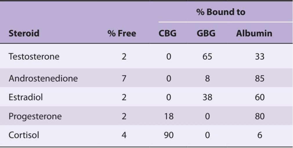
CBG, corticosteroid-binding globulin; GBG, gonadal steroid-binding globulin. (Used with permission of S Munroe.)
CBG، گلوبولین اتصال به کورتیکواستروئید؛ GBG، گلوبولین متصل شونده به استروئید گناد. (با اجازه S Munroe استفاده می شود.)
A small amount of circulating testosterone is converted to estradiol, but most of the testosterone is converted to 17-ketosteroids, principally androsterone and its isomer etiocholanolone (Figure 23-6), and excreted in the urine. About two- thirds of the urinary 17-ketosteroids are of adrenal origin, and one-third are of testicular origin. Although most of the 17-ketosteroids are weak androgens (they have 20% or less the potency of testosterone), it is worth emphasizing that not all 17-ketosteroids are androgens and not all androgens are 17-ketosteroids. Etiocholanolone, for example, has no androgenic activity, and testosterone itself is not a 17-ketosteroid.
مقدار کمی از تستوسترون در گردش به استرادیول تبدیل می شود، اما بیشتر تستوسترون به 17-کتوستروئید، عمدتاً آندروسترون و ایزومر آن اتیوکولانولون (شکل 23-6) تبدیل می شود و از طریق ادرار دفع می شود. حدود دو سوم 17 کتوستروئیدهای ادراری منشأ آدرنال و یک سوم منشاء بیضه دارند. اگرچه اکثر 17 کتوستروئیدها آندروژن ضعیف هستند (آنها 20٪ یا کمتر از تستوسترون قدرت دارند)، شایان ذکر است که همه 17 کتوستروئیدها آندروژن نیستند و همه آندروژن ها 17 کتوستروئید نیستند. برای مثال اتیوکولانولون هیچ فعالیت آندروژنی ندارد و تستوسترون خود یک 17 کتوستروئید نیست.

FIGURE 23-6 Testosterone metabolites. Androsterone and etiocholanolone are two 17-ketosteroid metabolites of testosterone that are secreted in the urine.
شکل 23-6 متابولیت های تستوسترون. آندروسترون و اتیوکولانولون دو متابولیت 17 کتوستروئیدی تستوسترون هستند که در ادرار ترشح می شوند.
Actions
In addition to their actions during development, testosterone and other androgens exert an inhibitory feedback effect on pituitary LH secretion; develop and maintain the male secondary sex characteristics; exert an important protein- anabolic, growth-promoting effect; and, along with FSH, maintain spermatogenesis.
اقدامات
تستوسترون و سایر آندروژنها علاوه بر اعمال خود در طول تکامل، اثر بازخوردی مهاری بر ترشح LH هیپوفیز دارند. ایجاد و حفظ ویژگی های جنسی ثانویه مرد؛ یک اثر مهم پروتئین-آنابولیک و محرک رشد داشته باشد. و همراه با FSH باعث حفظ اسپرماتوژنز می شود.
Secondary Sex Characteristics
The widespread changes in hair distribution, body configuration, and genital size that develop in boys at puberty-the male secondary sex characteristics are summarized in Table 23-3. The prostate and seminal vesicles enlarge, and the seminal vesicles begin to secrete fructose.This sugar appears to function as the main nutritional supply for the spermatozoa. The psychological effects of testosterone are difficult to define in humans, but in experimental animals, androgens provoke boisterous and aggressive play. The effects of androgens and estrogens on sexual behavior are considered in detail in Chapter 22. Although body hair is increased by androgens, scalp hair is decreased (Figure 23-7). Hereditary baldness often fails to develop unless dihydrotestosterone (DHT) is present.
ویژگی های جنسی ثانویه
تغییرات گسترده در توزیع مو، پیکربندی بدن و اندازه اندام تناسلی که در پسران در سن بلوغ ایجاد می شود – ویژگی های جنسی ثانویه مرد در جدول 23-3 خلاصه شده است. پروستات و وزیکولهای منی بزرگ میشوند و وزیکولهای منی شروع به ترشح فروکتوز میکنند. تعریف اثرات روانشناختی تستوسترون در انسان دشوار است، اما در حیوانات آزمایشگاهی، آندروژن ها باعث ایجاد بازی های شدید و تهاجمی می شوند. اثرات آندروژن ها و استروژن ها بر رفتار جنسی در فصل 22 به تفصیل در نظر گرفته شده است. اگرچه موهای بدن توسط آندروژن ها افزایش می یابد، موهای سر کاهش می یابد (شکل 23-7). طاسی ارثی اغلب ایجاد نمی شود مگر اینکه دی هیدروتستوسترون (DHT) وجود داشته باشد.
TABLE 23-3 Changes at puberty in boys (male secondary sex characteristics).
جدول 23-3 تغییرات در دوران بلوغ در پسران (ویژگی های جنسی ثانویه مرد).

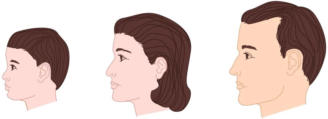
FIGURE 23-7 Hairline in children and adults. The hairline of the woman is like that of the child, whereas that of the man is indented in the lateral frontal region.
شکل 23-7 خط مو در کودکان و بزرگسالان. خط موی زن مانند کودک است، در حالی که خط موی مرد در ناحیه جلوی جانبی فرو رفته است.
Anabolic Effects
Androgens increase the synthesis and decrease the breakdown of protein, leading to an increase in the rate of growth. It used to be argued that they cause the epiphyses to fuse to the long bones, thus eventually stopping growth, but it now appears that epiphysial closure is due to estrogens (see Chapter 21). Secondary to their anabolic effects, androgens cause moderate Na+, K+, H2O, Ca2+, SO42, and PO43 retention, and they also increase the size of the kidneys. Doses of exogenous testosterone that exert significant anabolic effects are also masculinizing and increase libido, which limits the usefulness of the hormone as an anabolic agent in patients with wasting diseases. Attempts to develop synthetic steroids in which the anabolic action is separated from the androgenic action have not been successful.
اثرات آنابولیک
آندروژن ها باعث افزایش سنتز و کاهش تجزیه پروتئین می شوند که منجر به افزایش سرعت رشد می شود. قبلاً استدلال میشد که آنها باعث میشوند اپیفیزها به استخوانهای دراز جوش بخورند، بنابراین در نهایت رشد را متوقف میکنند، اما اکنون به نظر میرسد که بسته شدن اپی فیزیال به دلیل استروژنها است (به فصل 21 مراجعه کنید). ثانویه به اثرات آنابولیک، آندروژن ها باعث احتباس متوسط Na+، K+، H2O، Ca2+، SO42 و PO43 می شوند و همچنین اندازه کلیه ها را افزایش می دهند. دوزهای تستوسترون اگزوژن که اثرات آنابولیک قابل توجهی دارند نیز مردانه می شوند و میل جنسی را افزایش می دهند، که مفید بودن این هورمون را به عنوان یک عامل آنابولیک در بیماران مبتلا به بیماری های ضایع کننده محدود می کند. تلاش برای توسعه استروئیدهای مصنوعی که در آن عمل آنابولیک از عمل آندروژنیک جدا شده است موفقیت آمیز نبوده است.
Mechanism of Action
Like other steroids, testosterone binds to an intracellular receptor, for example, androgen receptor (or NR3C4), and the receptor/steroid complex then binds to DNA in the nucleus, facilitating transcription of various genes. In addition, testosterone is converted to DHT by 5a-reductase in some target cells (Figure 23-5 and Figure 23–8), and DHT binds to the same intracellular receptor as testosterone. DHT also circulates, with a plasma level that is about 10% of the testosterone level. Testosterone-receptor complexes are less stable than DHT- receptor complexes in target cells, and they conform less well to the DNA- binding state. Thus, DHT formation is a way of amplifying the action of testosterone in target tissues. Humans have two 5a-reductases that are encoded by different genes. Type 1 5a-reductase is present in skin throughout the body and is the dominant enzyme in the scalp. Type 2 5α-reductase is present in genital skin, the prostate, and other genital tissues.
مکانیسم عمل
مانند سایر استروئیدها، تستوسترون به یک گیرنده درون سلولی، به عنوان مثال، گیرنده آندروژن (یا NR3C4) متصل می شود و مجتمع گیرنده/استروئید سپس به DNA در هسته متصل می شود و رونویسی ژن های مختلف را تسهیل می کند. علاوه بر این، تستوسترون توسط 5a-reductase در برخی از سلول های هدف به DHT تبدیل می شود (شکل 23-5 و شکل 23-8)، و DHT به همان گیرنده داخل سلولی مانند تستوسترون متصل می شود. DHT همچنین با سطح پلاسمایی که حدود 10 درصد سطح تستوسترون است، در گردش است. کمپلکس های گیرنده تستوسترون نسبت به کمپلکس های گیرنده DHT در سلول های هدف پایداری کمتری دارند و با حالت اتصال به DNA سازگاری کمتری دارند. بنابراین، تشکیل DHT راهی برای تقویت عملکرد تستوسترون در بافت های هدف است. انسان ها دارای دو 5a-reductases هستند که توسط ژن های مختلف کدگذاری می شوند. 5a ردوکتاز نوع 1 در پوست سرتاسر بدن وجود دارد و آنزیم غالب در پوست سر است. 5α ردوکتاز نوع 2 در پوست اندام تناسلی، پروستات و سایر بافت های تناسلی وجود دارد.

FIGURE 23-8 Schematic diagram of the actions of testosterone (solid arrows) and dihydrotestosterone (dashed arrows). Note that they both bind to the same receptor, but DHT binds more effectively. (Reproduced with permission of Wilson JD, Griffin JE, Russell DW: Steroid 5 alpha-reductase 2 deficiency. Endocr Rev 1993 Oct; 14(5):577-593.)
شکل 23-8 نمودار شماتیکی از اعمال تستوسترون (فلش های جامد) و دی هیدروتستوسترون (فلش های چین دار). توجه داشته باشید که هر دو به یک گیرنده متصل می شوند، اما DHT به طور موثرتری متصل می شود. (تکثیر شده با اجازه Wilson JD، Griffin JE، Russell DW: Steroid 5 alpha-reductase 2 deficiency. Endocr Rev 1993 Oct; 14(5):577-593.)
Testosterone-receptor complexes are responsible for the maturation of Wolffian duct structures and consequently for the formation of male internal genitalia during development, but DHT-receptor complexes are needed to form male external genitalia (Figure 23-8). DHT-receptor complexes are also primarily responsible for enlargement of the prostate and probably of the penis at the time of puberty, as well as for the facial hair, the acne, and the temporal recession of the hairline. On the other hand, the increase in muscle mass and the development of male sex drive and libido depend primarily on testosterone rather than DHT (see Clinical Box 23-2).
کمپلکس های گیرنده تستوسترون مسئول بلوغ ساختارهای مجرای ولفیان و در نتیجه تشکیل اندام تناسلی داخلی مرد در طول تکامل هستند، اما کمپلکس های گیرنده DHT برای تشکیل اندام تناسلی خارجی مرد مورد نیاز است (شکل 23-8). کمپلکس های گیرنده DHT همچنین مسئول اصلی بزرگ شدن پروستات و احتمالاً آلت تناسلی در زمان بلوغ و همچنین برای موهای صورت، آکنه و تحلیل موقت خط مو هستند. از سوی دیگر، افزایش توده عضلانی و توسعه میل جنسی و میل جنسی مردان در درجه اول به تستوسترون بستگی دارد تا DHT (به کادر بالینی 23-2 مراجعه کنید).
CLINICAL BOX 23-2
Congenital 5a-Reductase Deficiency
Congenital 5a-reductase deficiency, in which the gene for type 2 5α- reductase is mutated, is common in certain parts of the Dominican Republic. It produces an interesting form of male pseudohermaphroditism. Individuals with this syndrome are born with male internal genitalia including testes, but they have female external genitalia and are usually raised as girls. However, when they reach puberty, LH secretion and circulating testosterone levels are increased. Consequently, they develop male body contours and male libido. At this point, they usually change their gender identities and “become boys.” The clitoris enlarges (“penis-at-12 syndrome”) to the point that some of the individuals can have intercourse with women. This enlargement probably occurs because, with the high LH, enough testosterone is produced to overcome the need for DHT amplification in the genitalia.
جعبه بالینی 23-2
کمبود مادرزادی ردوکتاز 5a
کمبود مادرزادی 5a-ردوکتاز، که در آن ژن 5α- ردوکتاز نوع 2 جهش یافته است، در مناطق خاصی از جمهوری دومینیکن رایج است. شکل جالبی از شبه هرمافرودیتیسم مردانه ایجاد می کند. افراد مبتلا به این سندرم با اندام تناسلی داخلی مردانه از جمله بیضه ها متولد می شوند، اما دارای اندام تناسلی خارجی زنانه هستند و معمولاً به عنوان دختر بزرگ می شوند. با این حال، هنگامی که آنها به بلوغ می رسند، ترشح LH و سطح تستوسترون در گردش افزایش می یابد. در نتیجه، آنها خطوط بدن مردانه و میل جنسی مردانه را ایجاد می کنند. در این مرحله معمولاً هویت جنسی خود را تغییر می دهند و «پسر می شوند». کلیتوریس بزرگ می شود (“سندرم آلت تناسلی در 12”) به حدی که برخی از افراد می توانند با زنان رابطه جنسی داشته باشند. این بزرگ شدن احتمالاً به این دلیل رخ می دهد که با LH بالا، تستوسترون کافی برای غلبه بر نیاز به تقویت DHT در اندام تناسلی تولید می شود.
THERAPEUTIC HIGHLIGHTS
5a-Reductase-inhibiting drugs are being used clinically to treat benign prostatic hyperplasia, and finasteride, the most extensively used drug, has its greatest effect on type 2 5a-reductase.
نکات برجسته درمانی
داروهای مهارکننده ردوکتاز 5a به صورت بالینی برای درمان هیپرپلازی خوش خیم پروستات استفاده می شود و فیناستراید، پرمصرف ترین دارو، بیشترین تأثیر خود را بر روی 5a- ردوکتاز نوع 2 دارد.
Testicular Production of Estrogens
Over 80% of the estradiol and 95% of the estrone in the plasma of adult men are formed by extragonadal and extra-adrenal aromatization of circulating testosterone and androstenedione. The remainder comes from the testes. Some of the estradiol in testicular venous blood comes from the Leydig cells, but some is also produced by aromatization of androgens in Sertoli cells. In men, the plasma estradiol level is 20-50 pg/mL (73-184 pmol/L) and the total production rate is approximately 50 μg/d (184 nmol/d). In contrast to the situation in women, estrogen production moderately increases with advancing age in men.
تولید استروژن از بیضه
بیش از 80 درصد استرادیول و 95 درصد استرون در پلاسمای مردان بالغ با آروماتیزاسیون خارج غدد جنسی و خارج آدرنال تستوسترون و آندروستندیون در گردش تشکیل می شود. باقی مانده از بیضه ها می آید. مقداری از استرادیول موجود در خون وریدی بیضه از سلولهای لیدیگ میآید، اما مقداری نیز از طریق آروماتیزاسیون آندروژنها در سلولهای سرتولی تولید میشود. در مردان، سطح استرادیول پلاسما 20-50 pg/mL (73-184 pmol/L) و نرخ کل تولید تقریباً 50 میکروگرم در روز (184 نانومول در روز) است. برخلاف وضعیت در زنان، تولید استروژن در مردان با افزایش سن به طور متوسط افزایش می یابد.
CONTROL OF TESTICULAR FUNCTION
FSH is tropic for Sertoli cells, and FSH and androgens maintain the gametogenic function of the testes. FSH also stimulates the secretion of ABP and inhibin. Inhibin feeds back to inhibit FSH secretion. LH is tropic for Leydig cells and stimulates the secretion of testosterone, which in turn feeds back to inhibit LH secretion. Hypothalamic lesions in animals and hypothalamic disease in humans lead to atrophy of the testes and loss of their function.
کنترل عملکرد بیضه
FSH برای سلول های سرتولی استوایی است و FSH و آندروژن ها عملکرد گامتوژنیک بیضه ها را حفظ می کنند. FSH همچنین باعث تحریک ترشح ABP و اینهیبین می شود. اینهیبین برای مهار ترشح FSH بازخورد می کند. LH برای سلول های لیدیگ گرمسیری است و ترشح تستوسترون را تحریک می کند که به نوبه خود برای مهار ترشح LH بازخورد می کند. ضایعات هیپوتالاموس در حیوانات و بیماری هیپوتالاموس در انسان منجر به آتروفی بیضه ها و از بین رفتن عملکرد آنها می شود.
Inhibins
Testosterone reduces plasma LH, but, except in large doses, it has no effect on plasma FSH. Plasma FSH is elevated in patients who have atrophy of the seminiferous tubules but normal levels of testosterone and LH secretion. These observations led to the search for inhibin, a factor of testicular origin that inhibits FSH secretion. There are two inhibins in extracts of testes in men and in antral fluid from ovarian follicles in women. They are formed from three polypeptide subunits: a glycosylated a subunit with a molecular weight of 18,000, and two nonglycosylated ẞ subunits, ẞ and ẞ, each with a molecular weight of 14,000. The subunits are formed from precursor proteins (Figure 23- 9). The a subunit combines with ẞA to form a heterodimer and with ẞg to form another heterodimer, with the subunits linked by disulfide bonds. Both aẞA (inhibin A) and aẞp (inhibin B) inhibit FSH secretion by a direct action on the pituitary, although it appears that it is inhibin B that is the FSH-regulating inhibin in adult men and women. Inhibins are produced by Sertoli cells in males and granulosa cells in females.
مهار کننده ها
تستوسترون LH پلاسما را کاهش می دهد، اما، به جز در دوزهای زیاد، هیچ تاثیری بر FSH پلاسما ندارد. FSH پلاسما در بیمارانی که آتروفی لولههای منیساز را دارند، اما سطوح طبیعی تستوسترون و ترشح LH دارند، افزایش مییابد. این مشاهدات منجر به جستجوی اینهیبین شد، عاملی با منشا بیضه که ترشح FSH را مهار می کند. دو مهارکننده در عصاره بیضه در مردان و در مایع آنترال از فولیکول های تخمدان در زنان وجود دارد. آنها از سه زیر واحد پلی پپتیدی تشکیل می شوند: یک زیر واحد گلیکوزیله با وزن مولکولی 18000، و دو زیر واحد غیرگلیکوزیله ẞ، ẞ و ẞ، هر کدام با وزن مولکولی 14000. زیر واحدها از پروتئین های پیش ساز تشکیل می شوند (شکل 23-9). زیرواحد a با ẞA ترکیب می شود تا یک هترودایمر و با ẞg برای تشکیل هترودایمر دیگری، با زیرواحدها با پیوندهای دی سولفیدی به هم متصل شوند. هر دو aẞA (اینهیبین A) و aẞp (اینهیبین B) با یک اثر مستقیم روی هیپوفیز ترشح FSH را مهار می کنند، اگرچه به نظر می رسد که اینهیبین B است که مهارکننده تنظیم کننده FSH در مردان و زنان بالغ است. اینهیبین ها توسط سلول های سرتولی در مردان و سلول های گرانولوزا در زنان تولید می شوند.

FIGURE 23-9 Inhibin and activin production. From three precursor proteins (α, BA, and BB), five separate inhibins and activins are produced. SS, disulfide bonds.
شکل 23-9 تولید اینهیبین و اکتیوین. از سه پروتئین پیش ساز (α، BA و BB)، پنج مهارکننده و اکتیوین مجزا تولید می شود. SS، پیوندهای دی سولفیدی.
The heterodimer BAẞB and the homodimers BABA and BÅẞ are also formed. They stimulate rather than inhibit FSH secretion and consequently are called activins. Their function in reproduction is unsettled. However, the inhibins and activins are members of the transforming growth factor beta (TGF-ẞ) superfamily of dimeric growth factors that also includes MIS. Activin receptors have been identified and belong to the serine/threonine kinase receptor family. Inhibins and activins are found not only in the gonads but also in the brain and many other tissues. In the bone marrow, activins are involved in the development of white blood cells. In embryonic life, activins are involved in the formation of mesoderm. All mice with a targeted deletion of the a-inhibin subunit gene initially exhibit normal growth but then develop gonadal stromal tumors, so the gene is a tumor suppressor gene.
هترودایمر BAẞB و همودایمرهای BABA و BÅẞ نیز تشکیل می شوند. آنها به جای مهار ترشح FSH را تحریک می کنند و در نتیجه اکتیوین نامیده می شوند. کارکرد آنها در تولید مثل ثابت نشده است. با این حال، اینهیبین ها و اکتیوین ها اعضای فاکتور رشد تبدیل کننده بتا (TGF-ẞ) فاکتورهای رشد دیمری هستند که شامل MIS نیز می شود. گیرنده های اکتیوین شناسایی شده اند و به خانواده گیرنده های سرین/ترئونین کیناز تعلق دارند. اینهیبین ها و اکتیوین ها نه تنها در غدد جنسی، بلکه در مغز و بسیاری از بافت های دیگر نیز یافت می شوند. در مغز استخوان، اکتیوین ها در توسعه گلبول های سفید خون نقش دارند. در زندگی جنینی، اکتیوین ها در تشکیل مزودرم نقش دارند. همه موشهای با حذف هدفمند ژن زیر واحد a-inhibin در ابتدا رشد طبیعی را نشان میدهند اما سپس تومورهای استرومایی غدد جنسی ایجاد میکنند، بنابراین ژن یک ژن سرکوبکننده تومور است.
In plasma, α2-macroglobulin binds activins and inhibins. In tissues, activins bind to a family of four glycoproteins called follistatins. Binding of the activins inactivates their biological activity, but the relation of follistatins to inhibin and their physiological function remain unsettled.
در پلاسما، α2-ماکروگلوبولین به اکتیوین ها و اینهیبین ها متصل می شود. در بافت ها، اکتیوین ها به یک خانواده چهار گلیکوپروتئینی به نام فولیستاتین ها متصل می شوند. اتصال اکتیوین ها باعث غیرفعال شدن فعالیت بیولوژیکی آنها می شود، اما رابطه فولیستاتین ها با اینهیبین و عملکرد فیزیولوژیکی آنها نابسامان باقی می ماند.
Steroid Feedback
A current “working hypothesis” of the way the functions of the testes are regulated by steroids is shown in Figure 23-10. Castration is followed by a rise in the pituitary content and secretion of FSH and LH, and hypothalamic lesions prevent this rise. Testosterone inhibits LH secretion by acting directly on the anterior pituitary and by inhibiting the secretion of gonadotropin-releasing hormone (GnRH) from the hypothalamus. Inhibin acts directly on the anterior pituitary to inhibit FSH secretion.
بازخورد استروئیدی
یک “فرضیه کاری” فعلی از نحوه تنظیم عملکرد بیضه ها توسط استروئیدها در شکل 23-10 نشان داده شده است. اخته شدن با افزایش محتوای هیپوفیز و ترشح FSH و LH همراه است و ضایعات هیپوتالاموس از این افزایش جلوگیری می کند. تستوسترون با اثر مستقیم بر هیپوفیز قدامی و با مهار ترشح هورمون آزاد کننده گنادوتروپین (GnRH) از هیپوتالاموس، ترشح LH را مهار می کند. اینهیبین مستقیماً بر روی هیپوفیز قدامی تأثیر می گذارد و ترشح FSH را مهار می کند.

FIGURE 23-10 The hypothalamus, pituitary, and testes interact via signaling molecules. Postulated signaling pathways between the hypothalamus, anterior pituitary, and testis are shown. Solid arrows indicate excitatory effects, whereas dashed arrows indicate inhibitory effects.
شکل 23-10 هیپوتالاموس، هیپوفیز و بیضه ها از طریق مولکول های سیگنال دهنده برهم کنش دارند. مسیرهای سیگنالینگ فرضی بین هیپوتالاموس، هیپوفیز قدامی و بیضه نشان داده شده است. فلش های توپر اثرات تحریکی را نشان می دهند، در حالی که فلش های چین دار اثرات بازدارندگی را نشان می دهند.
In response to LH, some of the testosterone secreted from the Leydig cells bathes the seminiferous epithelium and provides the high local concentration of androgen to the Sertoli cells that is necessary for normal spermatogenesis. Systemically administered testosterone does not raise the androgen level in the testes to as great a degree, and it inhibits LH secretion. Consequently, the net effect of systemically administered testosterone is generally a decrease in sperm count. Testosterone therapy has been suggested as a means of male contraception. However, the dose of testosterone needed to suppress spermatogenesis causes sodium and water retention. The possible use of inhibins as male contraceptives is being explored.
در پاسخ به LH، مقداری از تستوسترون ترشح شده از سلول های لیدیگ، اپیتلیوم منی ساز را حمام می کند و غلظت محلی بالایی از آندروژن را برای سلول های سرتولی فراهم می کند که برای اسپرم زایی طبیعی لازم است. تستوسترون که به صورت سیستمیک تجویز می شود، سطح آندروژن را در بیضه ها تا حد زیادی افزایش نمی دهد و ترشح LH را مهار می کند. در نتیجه، اثر خالص تستوسترون سیستمیک به طور کلی کاهش تعداد اسپرم است. تستوسترون درمانی به عنوان وسیله ای برای پیشگیری از بارداری مردان پیشنهاد شده است. با این حال، دوز تستوسترون مورد نیاز برای سرکوب اسپرماتوژنز باعث احتباس سدیم و آب می شود. استفاده احتمالی از اینهیبین ها به عنوان داروهای ضد بارداری مردانه در حال بررسی است.
ABNORMALITIES OF TESTICULAR FUNCTION
Cryptorchidism
The testes develop in the abdominal cavity and normally migrate to the scrotum during fetal development. Testicular descent to the inguinal region depends on MIS, and descent from the inguinal region to the scrotum depends on other factors. Descent is incomplete on one or, less commonly, both sides in 10% of newborn males, with the testes remaining in the abdominal cavity or inguinal canal. Gonadotropic hormone treatment speeds descent in some cases, or the defect can be corrected surgically. Spontaneous descent of the testes is the rule, and the proportion of boys with undescended testes (cryptorchidism) falls to 2% at age 1 year and 0.3% after puberty. However, early treatment is now recommended despite these figures because the incidence of malignant tumors is higher in undescended than in scrotal testes and because after puberty the higher temperature in the abdomen eventually causes irreversible damage to the spermatogenic epithelium.
ناهنجاری های عملکرد بیضه
کریپتورکیدیسم
بیضه ها در حفره شکمی رشد می کنند و به طور معمول در طول رشد جنین به کیسه بیضه مهاجرت می کنند. نزول بیضه به ناحیه اینگوینال به MIS بستگی دارد و نزول از ناحیه اینگوینال به کیسه بیضه به عوامل دیگری بستگی دارد. نزول در 10 درصد از نوزادان پسر از یک یا کمتر در هر دو طرف ناقص است و بیضه ها در حفره شکمی یا کانال مغبنی باقی می مانند. درمان با هورمون گنادوتروپیک در برخی موارد سرعت نزول را افزایش میدهد یا این نقص را میتوان با جراحی اصلاح کرد. نزول خود به خودی بیضه ها یک قانون است و نسبت پسرانی که بیضه های نزول نکرده دارند (کریپتورکیدیسم) در سن یک سالگی به 2 درصد و پس از بلوغ 0.3 درصد کاهش می یابد. با این حال، درمان زودهنگام با وجود این ارقام در حال حاضر توصیه می شود زیرا بروز تومورهای بدخیم در بیضه های نزول نکرده بیشتر از بیضه های بیضه است و به این دلیل که پس از بلوغ دمای بالاتر در شکم در نهایت باعث آسیب غیر قابل برگشت به اپیتلیوم اسپرماتوژن می شود.
Male Hypogonadism
The clinical picture of male hypogonadism depends on whether testicular deficiency develops before or after puberty. In adults, if it is due to testicular disease, circulating gonadotropin levels are elevated (hypergonadotropic hypogonadism); if it is secondary to disorders of the pituitary or the hypothalamus (eg, Kallmann syndrome), circulating gonadotropin levels are decreased (hypogonadotropic hypogonadism). If the endocrine function of the testes is lost in adulthood, the secondary sex characteristics regress slowly because it takes very little androgen to maintain them once they are established. The growth of the larynx during adolescence is permanent, and the voice remains deep. Men castrated in adulthood suffer some loss of libido, although the ability to copulate persists for some time. They occasionally have hot flushes and are generally more irritable, passive, and depressed than men with intact testes. When the Leydig cell deficiency dates from childhood, the clinical picture is that of eunuchoidism. Eunuchoid individuals over the age of 20 are characteristically tall, although not as tall as hyperpituitary giants, because their epiphyses remain open and some growth continues past the normal age of puberty. They have narrow shoulders and small muscles, a body configuration resembling that of the adult female. The genitalia are small and the voice high- pitched. Pubic hair and axillary hair are present because of adrenocortical androgen secretion. However, the hair is sparse, and the pubic hair has the female “triangle with the base up” distribution rather than the “triangle with the base down” pattern (male escutcheon) seen in normal males.
هیپوگنادیسم مردانه
تصویر بالینی هیپوگنادیسم مردان بستگی به این دارد که آیا کمبود بیضه قبل یا بعد از بلوغ ایجاد می شود. در بزرگسالان، اگر به دلیل بیماری بیضه باشد، سطح گنادوتروپین در گردش افزایش می یابد (هیپوگنادیسم هیپرگنادوتروپیک). اگر ثانویه به اختلالات هیپوفیز یا هیپوتالاموس (به عنوان مثال، سندرم کالمن) باشد، سطح گنادوتروپین در گردش کاهش می یابد (هیپوگنادیسم هیپوگنادوتروپیک). اگر عملکرد غدد درونریز بیضهها در بزرگسالی از بین برود، ویژگیهای جنسی ثانویه به آرامی کاهش مییابد، زیرا پس از استقرار، حفظ آندروژن بسیار کمی نیاز دارد. رشد حنجره در دوران نوجوانی دائمی است و صدا عمیق می ماند. مردانی که در بزرگسالی اخته میشوند، تا حدودی میل جنسی را از دست میدهند، اگرچه توانایی جفتگیری برای مدتی ادامه دارد. آنها گاهی اوقات گرگرفتگی دارند و عموماً نسبت به مردانی که بیضه های سالمی دارند تحریک پذیرتر، منفعل و افسرده تر هستند. هنگامی که کمبود سلول لیدیگ از دوران کودکی شروع می شود، تصویر بالینی مربوط به eunuchoidism است. افراد Eunuchoid بالای 20 سال به طور مشخص قد بلندی دارند، اگرچه نه به اندازه غول های هیپوفیز، زیرا اپی فیز آنها باز می ماند و مقداری رشد پس از سن طبیعی بلوغ ادامه می یابد. آنها دارای شانه های باریک و ماهیچه های کوچک هستند، پیکربندی بدنی شبیه بدن زن بالغ. اندام تناسلی کوچک و صدای بلند است. موهای ناحیه تناسلی و زیر بغل به دلیل ترشح آندروژن قشر آدرنال وجود دارند. با این حال، مو کم است و موهای ناحیه تناسلی دارای توزیع «مثلث زنانه با قاعده رو به بالا» به جای الگوی «مثلث با پایه رو به پایین» (اسکاج نر) است که در مردان عادی دیده میشود.
Androgen-Secreting Tumors
“Hyperfunction” of the testes in the absence of tumor formation is not a recognized entity. Androgen-secreting Leydig cell tumors are rare and cause detectable endocrine symptoms only in prepubertal boys, in whom precocious pseudopuberty develops (Table 22-2).
تومورهای ترشح کننده آندروژن
“عملکرد بیش از حد” بیضه ها در غیاب تشکیل تومور یک موجود شناخته شده نیست. تومورهای سلول لیدیگ ترشح کننده آندروژن نادر هستند و علائم غدد درون ریز قابل تشخیص را فقط در پسران قبل از بلوغ، که در آنها بلوغ کاذب زودرس در آنها ایجاد می شود، ایجاد می کنند (جدول 22-2).
Hormones & Cancer
Some carcinomas of the prostate are androgen-dependent and regress temporarily after the removal of the testes or treatment with GnRH agonists in doses that are sufficient to produce downregulation of the GnRH receptors on gonadotropes and decrease LH secretion.
هورمون ها و سرطان
برخی از کارسینومهای پروستات وابسته به آندروژن هستند و پس از برداشتن بیضهها یا درمان با آگونیستهای GnRH در دوزهایی که برای کاهش گیرندههای GnRH روی گنادوتروپها و کاهش ترشح LH کافی است، موقتاً پسرفت میکنند.
CHAPTER SUMMARY
خلاصه فصل
• The gonads have a dual function: the production of germ (gametogenesis) and the secretion of sex hormones. The testes secrete large amounts of androgens, principally testosterone, but they also secrete small amounts of estrogens.
• غدد جنسی عملکرد دوگانه ای دارند: تولید میکروب (گامتوژنز) و ترشح هورمون های جنسی. بیضه ها مقادیر زیادی آندروژن، عمدتاً تستوسترون، ترشح می کنند، اما مقدار کمی استروژن نیز ترشح می کنند.
• Spermatogonia develop into mature spermatozoa in the seminiferous tubules via a process called spermatogenesis. This is a multistep process that includes maturation of spermatogonia into primary spermatocytes, which undergo meiotic division, resulting in haploid secondary spermatocytes. Several further divisions result in spermatids. Each cell division from a spermatogonium to a spermatid is incomplete, with cells remaining connected via cytoplasmic bridges. Spermatids eventually mature into motile spermatozoa to complete spermatogenesis; this last part of maturation is called spermiogenesis.
• اسپرماتوگونی از طریق فرآیندی به نام اسپرماتوژنز به اسپرماتوزوئید بالغ در لوله های اسپرم ساز تبدیل می شود. این یک فرآیند چند مرحلهای است که شامل بلوغ اسپرماتوگونی به اسپرماتوسیتهای اولیه میشود که تحت تقسیم میوز قرار میگیرند و در نتیجه اسپرماتوسیتهای ثانویه هاپلوئید ایجاد میشوند. چندین تقسیم بیشتر منجر به اسپرماتید می شود. هر تقسیم سلولی از یک اسپرماتوگونیوم به یک اسپرماتید ناقص است و سلول ها از طریق پل های سیتوپلاسمی به هم متصل می شوند. اسپرماتیدها در نهایت به اسپرماتوزواهای متحرک بالغ میشوند تا اسپرمزایی کامل شوند. این قسمت آخر بلوغ، اسپرموژنز نامیده می شود.
• Testosterone is the principal hormone of the testis. It is synthesized from cholesterol in Leydig cells. The secretion of testosterone from Leydig cells is under control of luteinizing hormone at a rate of 4-9 mg/d in adult males. Most testosterone is bound to albumin or to gonadal steroid-binding globulin in the plasma. Testosterone plays an important role in the development and maintenance of male secondary sex characteristics, as well as other defined functions.
• تستوسترون هورمون اصلی بیضه است. این از کلسترول در سلول های لیدیگ سنتز می شود. ترشح تستوسترون از سلول های لیدیگ تحت کنترل هورمون لوتئینیزه کننده با سرعت 9-4 میلی گرم در روز در مردان بالغ است. بیشتر تستوسترون به آلبومین یا گلوبولین متصل به استروئید گناد در پلاسما متصل می شود. تستوسترون نقش مهمی در ایجاد و حفظ خصوصیات جنسی ثانویه مردانه و همچنین سایر عملکردهای تعریف شده دارد.
MULTIPLE-CHOICE QUESTIONS
For all questions, select the single best answer unless otherwise directed.
سوالات چند گزینه ای
برای همه سؤالات، بهترین پاسخ را انتخاب کنید، مگر اینکه دستور دیگری داده شود.
1. Full development and function of the seminiferous tubules require
A. somatostatin.
B. LH.
C. oxytocin.
D. FSH.
E. androgens and FSH.
1. توسعه کامل و عملکرد لوله های منی ساز نیاز دارد
الف. سوماتوستاتین.
ب. LH.
ج. اکسی توسین.
د. FSH.
ی. آندروژن و FSH.
2. In human males, testosterone is produced mainly by the
A. Leydig cells.
B. Sertoli cells.
C. seminiferous tubules.
D. epididymis.
E. vas deferens.
2. در مردان انسان، تستوسترون به طور عمده توسط
الف. سلول های لیدیگ.
ب. سلول های سرتولی.
ج. لوله های اسپرم ساز.
د. اپیدیدیم.
ی. vas deferens.
3. NOS contributes to erection by:
A. raising cAMP levels that relax smooth muscles and increase blood flow.
B. blocking phosphodiesterases to increase cGMP levels that release smooth muscle and increase blood flow.
C. activating soluble guanylyl cyclases to increase cGMP levels that relax smooth muscle and increase blood flow.
D. raising intracellular Ca2+ concentrations that relax smooth muscles and increase blood flow.
3. NOS از طریق موارد زیر به نعوظ کمک می کند:
الف. افزایش سطح cAMP که عضلات صاف را شل می کند و جریان خون را افزایش می دهد.
ب. مسدود کردن فسفودی استرازها برای افزایش سطح cGMP که عضله صاف را آزاد می کند و جریان خون را افزایش می دهد.
ج. فعال کردن گوانیلیل سیکلازهای محلول برای افزایش سطح cGMP که عضلات صاف را شل می کند و جریان خون را افزایش می دهد.
د. افزایش غلظت های Ca2 داخل سلولی که باعث شل شدن عضلات صاف و افزایش جریان خون می شود.
4. Testosterone is produced
A. in the testes after reduction of dihydrotestosterone.
B. in Leydig cells from cholesterol and pregnenolone precursors.
C. by LH in Leydig cells.
D. as a precursor for several membrane lipids.
4. تستوسترون تولید می شود
الف. در بیضه پس از کاهش دی هیدروتستوسترون.
ب. در سلول های لیدیگ از پیش سازهای کلسترول و پرگننولون.
ج. توسط LH در سلول های لیدیگ.
د. به عنوان پیش ساز برای چندین لیپید غشایی.
کلیک کنید «منابع»
🚀 با ما همراه شوید!
تازهترین مطالب و آموزشهای مغز و اعصاب را از دست ندهید. با فالو کردن کانال تلگرام آیندهنگاران مغز، از ما حمایت کنید!



