
فیزیولوژی پزشکی گانونگ؛ رشد تولیدمثلی و عملکرد دستگاه تناسلی زن


دعای مطالعه [ نمایش ]
بِسْمِ الله الرَّحْمنِ الرَّحیمِ
اَللّهُمَّ اَخْرِجْنى مِنْ ظُلُماتِ الْوَهْمِ
خدايا مرا بيرون آور از تاريكىهاى وهم،
وَ اَكْرِمْنى بِنُورِ الْفَهْمِ
و به نور فهم گرامى ام بدار،
اَللّهُمَّ افْتَحْ عَلَيْنا اَبْوابَ رَحْمَتِكَ
خدايا درهاى رحمتت را به روى ما بگشا،
وَانْشُرْ عَلَيْنا خَزائِنَ عُلُومِكَ بِرَحْمَتِكَ يا اَرْحَمَ الرّاحِمينَ
و خزانههاى علومت را بر ما باز كن به امید رحمتت اى مهربانترين مهربانان.
کتاب «فیزیولوژی پزشکی گانونگ» بهعنوان یکی از جامعترین و معتبرترین منابع در حوزه علوم پزشکی، همچنان مرجع کلیدی برای درک عملکرد پیچیده بدن انسان است. این اثر با تکیه بر تازهترین پژوهشها و توضیحات دقیق از سازوکارهای فیزیولوژیک، پلی میان علوم پایه پزشکی و کاربردهای بالینی ایجاد میکند و نقشی بیبدیل در آموزش، پژوهش و ارتقای دانش سلامت ایفا مینماید.
ترجمه دقیق و علمی این شاهکار توسط برند علمی آیندهنگاران مغز به مدیریت داریوش طاهری، دسترسی فارسیزبانان به مرزهای نوین دانش فیزیولوژی را ممکن ساخته و رسالتی علمی برای ارتقای آموزش پزشکی، فهم عمیقتر سازوکارهای بدن و توسعه روشهای نوین در حوزه سلامت فراهم آورده است.
» کتاب فیزیولوژی پزشکی گانونگ
» » فصل ۲۲: رشد تولیدمثلی و عملکرد دستگاه تناسلی زن
در حال ویرایش
» Ganong’s Review of Medical Physiology
»» CHAPTER 22: Reproductive Development & Function of the Female Reproductive System
OBJECTIVES
After studying his chapter you should be able to:
• Outline the role of chromosomes, hormones, and related factors in sex determination and development.
• Summarize the hormonal changes that occur at puberty in females.
• Describe oogenesis within the ovarian follicle. Explain the roles of FSH, LH and inhibin in oogenesis and maturation of follicles.
• Describe ovulation and the role of the corpus luteum in the physiologic changes that occur in the female reproductive organs during the menstrual cycle.
• Outline the hormonal changes and their physiologic effects during perimenopause and menopause.
• Describe the hormonal regulation of estrogen and progesterone biosynthesis, transport, metabolism, targets, and actions.
• Describe the roles of the pituitary and the hypothalamus in the regulation of ovarian function, and the role of feedback loops in this process.
• Describe the process of fertilization, implantation and the development of the placenta.
• Discuss the hormonal changes that accompany pregnancy and parturition.
• Outline the processes involved in lactation including neuroendocrine regulation of milk secretion and ejection.
اهداف
پس از مطالعه فصل، باید بتوانید:
• نقش کروموزوم ها، هورمون ها و عوامل مرتبط در تعیین جنسیت و رشد را مشخص کنید.
• تغییرات هورمونی که در دوران بلوغ در زنان رخ می دهد را خلاصه کنید.
اووژنز در فولیکول تخمدان را شرح دهید. نقش FSH، LH و Inhibin در اووژنز و بلوغ فولیکول ها را توضیح دهید.
• تخمک گذاری و نقش جسم زرد در تغییرات فیزیولوژیکی که در اندام های تناسلی زنانه در طول چرخه قاعدگی رخ می دهد را شرح دهید.
• تغییرات هورمونی و اثرات فیزیولوژیکی آنها را در دوران پیش از یائسگی و یائسگی بیان کنید.
• تنظیم هورمونی بیوسنتز استروژن و پروژسترون، انتقال، متابولیسم، اهداف و اقدامات را شرح دهید.
• نقش هیپوفیز و هیپوتالاموس در تنظیم عملکرد تخمدان و نقش حلقه های بازخورد در این فرآیند را شرح دهید.
• فرآیند لقاح، لانه گزینی و رشد جفت را شرح دهید.
تغییرات هورمونی همراه با بارداری و زایمان را مورد بحث قرار دهید.
• فرآیندهای دخیل در شیردهی از جمله تنظیم نورواندوکرین ترشح و دفع شیر را شرح دهید.

INTRODUCTION
Modern genetics and experimental embryology make it clear that, in most species of mammals, the multiple differences between the male and the female depend primarily on a single chromosome (the Y chromosome) and a single pair of endocrine structures, namely the testes in the male and the ovaries in the female. The differentiation of the primitive gonads into testes or ovaries in utero is genetically determined in humans, but the formation of male genitalia depends on the presence of a functional, secreting testis; in the absence of testicular tissue, development is female. Evidence indicates that male sexual behavior and, in some species, the male pattern of gonadotropin secretion are due to the action of male hormones on the brain in early development. After birth, the gonads remain quiescent until adolescence, when they are activated by gonadotropins from the anterior pituitary. Hormones secreted by the gonads at this time cause the appearance of features typical of the adult male or female and the onset of the sexual cycle in the female. In human females, ovarian function regresses after a number of years and sexual cycles cease (the menopause). In males, gonadal function slowly declines with advancing age, but the ability to produce viable gametes persists.
مقدمه
ژنتیک مدرن و جنین شناسی تجربی روشن می کند که در اکثر گونه های پستانداران، تفاوت های متعدد بین نر و ماده در درجه اول به یک کروموزوم منفرد (کروموزوم Y) و یک جفت ساختار غدد درون ریز، یعنی بیضه ها در نر و تخمدان ها در ماده بستگی دارد. تمایز غدد جنسی اولیه به بیضه ها یا تخمدان ها در رحم به طور ژنتیکی در انسان تعیین می شود، اما تشکیل اندام تناسلی مردانه به وجود یک بیضه عملکردی و ترشح کننده بستگی دارد. در غیاب بافت بیضه، رشد زن است. شواهد نشان می دهد که رفتار جنسی مردانه و در برخی گونه ها، الگوی مردانه ترشح گنادوتروپین به دلیل عملکرد هورمون های مردانه بر روی مغز در مراحل اولیه رشد است. پس از تولد، غدد جنسی تا سنین نوجوانی ساکن می مانند، زمانی که توسط گنادوتروپین های هیپوفیز قدامی فعال می شوند. هورمونهایی که در این زمان از غدد جنسی ترشح میشوند، باعث پیدایش ویژگیهای معمولی نر یا ماده بالغ و شروع چرخه جنسی در زن میشوند. در زنان انسان، عملکرد تخمدان پس از چند سال کاهش می یابد و چرخه های جنسی متوقف می شود (یائسگی). در نرها، عملکرد غدد جنسی با افزایش سن به آرامی کاهش می یابد، اما توانایی تولید گامت های زنده باقی می ماند.
In both sexes, the gonads have a dual function: the production of germ cells (gametogenesis) and the secretion of sex hormones. The androgens are steroid sex hormones that are masculinizing in their action; the estrogens are those that are feminizing. Both types of hormones are normally secreted in both sexes. The ovaries secrete large amounts of estrogens and small amounts of androgens, a pattern that is reversed in males. Androgens are secreted from the adrenal cortex in both sexes, and some of the androgens are converted to estrogens in fat and other extragonadal and extra-adrenal tissues. The ovaries also secrete progesterone, a steroid that has special functions in preparing the uterus for pregnancy.
در هر دو جنس، غدد جنسی عملکرد دوگانه ای دارند: تولید سلول های زایا (گامتوژنز) و ترشح هورمون های جنسی. آندروژن ها هورمون های جنسی استروئیدی هستند که در عمل مردانه می شوند. استروژن ها آنهایی هستند که زنانه می شوند. هر دو نوع هورمون معمولاً در هر دو جنس ترشح می شوند. تخمدانها مقادیر زیادی استروژن و مقادیر کمی آندروژن ترشح میکنند که این الگو در مردان برعکس است. آندروژن ها از قشر آدرنال در هر دو جنس ترشح می شوند و برخی از آندروژن ها در چربی و سایر بافت های خارج غدد گناد و خارج آدرنال به استروژن تبدیل می شوند. تخمدان ها همچنین پروژسترون ترشح می کنند، استروئیدی که عملکرد ویژه ای در آماده سازی رحم برای بارداری دارد.
Particularly during pregnancy, the ovaries secrete the polypeptide hormone relaxin, which loosens the ligaments of the pubic symphysis and softens the cervix, facilitating delivery of the fetus. In both sexes, the gonads secrete other polypeptides, including inhibin B, a polypeptide that inhibits follicle-stimulating hormone (FSH) secretion.
به ویژه در دوران بارداری، تخمدان ها هورمون پلی پپتیدی ریلکسین را ترشح می کنند که رباط های سمفیز شرمگاهی را شل می کند و دهانه رحم را نرم می کند و زایمان جنین را تسهیل می کند. در هر دو جنس، غدد جنسی پلی پپتیدهای دیگری از جمله اینهیبین B ترشح می کنند، پلی پپتیدی که ترشح هورمون محرک فولیکول (FSH) را مهار می کند.
The secretory and gametogenic functions of the gonads are both dependent on the secretion of the anterior pituitary gonadotropins, FSH, and luteinizing hormone (LH). The sex hormones and inhibin B feed back to inhibit gonadotropin secretion. In males, gonadotropin secretion is noncyclic; but in postpubertal females an orderly, sequential secretion of gonadotropins is necessary for the occurrence of menstruation, pregnancy, and lactation.
عملکرد ترشحی و گامتوژنیک غدد جنسی هر دو به ترشح گنادوتروپین های هیپوفیز قدامی، FSH و هورمون لوتئینیزه کننده (LH) وابسته هستند. هورمونهای جنسی و اینهیبین B برای مهار ترشح گنادوتروپین بازخورد میکنند. در مردان، ترشح گنادوتروپین غیر حلقوی است. اما در زنان پس از بلوغ، ترشح منظم و متوالی گنادوتروپین برای وقوع قاعدگی، بارداری و شیردهی ضروری است.
SEX DIFFERENTIATION & DEVELOPMENT
CHROMOSOMAL SEX
The Sex Chromosomes
Sex is determined genetically by two chromosomes, called the sex chromosomes, to distinguish them from the somatic chromosomes (autosomes). In humans and many other mammals, the sex chromosomes are called X and Y. The Y chromosome is necessary and sufficient for the production of testes, and the testis-determining gene product is called SRY (for sex-determining region of the Y chromosome). SRY is a DNA-binding regulatory protein. It bends the DNA and acts as a transcription factor that initiates transcription of a cascade of genes necessary for testicular differentiation, including the gene for müllerian-inhibiting substance (MIS; see below). The gene for SRY is located near the tip of the short arm of the human Y chromosome. Diploid male cells contain an X and a Y chromosome (XY pattern), whereas female cells contain two X chromosomes (XX pattern). As a consequence of meiosis during gametogenesis, each normal ovum contains a single X chromosome, but half of the normal sperm contain an X chromosome and half contain a Y chromosome (Figure 22-1). When a sperm containing a Y chromosome fertilizes an ovum, an XY pattern results and the zygote develops into a genetic male. When fertilization occurs with an X-containing sperm, an XX pattern and a genetic female results. Cell division and the chemical nature of chromosomes are discussed in Chapter 1.
تمایز و توسعه جنسی
جنس کروموزومی
کروموزوم های جنسی
جنسیت به طور ژنتیکی توسط دو کروموزوم به نام کروموزوم جنسی تعیین می شود تا آنها را از کروموزوم های سوماتیک (اتوزوم) متمایز کند. در انسان و بسیاری از پستانداران دیگر، کروموزوم های جنسی X و Y نامیده می شوند. کروموزوم Y برای تولید بیضه ها لازم و کافی است و محصول ژن تعیین کننده بیضه SRY (برای ناحیه تعیین کننده جنسیت کروموزوم Y) نامیده می شود. SRY یک پروتئین تنظیم کننده اتصال به DNA است. DNA را خم می کند و به عنوان یک فاکتور رونویسی عمل می کند که رونویسی یک آبشار از ژن های لازم برای تمایز بیضه را آغاز می کند، از جمله ژن ماده بازدارنده مولرین (MIS؛ زیر را ببینید). ژن SRY در نزدیکی نوک بازوی کوتاه کروموزوم Y انسان قرار دارد. سلول های مذکر دیپلوئید حاوی یک کروموزوم X و Y (الگوی XY) هستند، در حالی که سلول های ماده حاوی دو کروموزوم X (الگوی XX) هستند. در نتیجه میوز در طول گامتوژنز، هر تخمک طبیعی حاوی یک کروموزوم X منفرد است، اما نیمی از اسپرم طبیعی حاوی کروموزوم X و نیمی حاوی کروموزوم Y است (شکل 22-1). هنگامی که یک اسپرم حاوی کروموزوم Y تخمک را بارور می کند، یک الگوی XY ایجاد می شود و زیگوت به یک مرد ژنتیکی تبدیل می شود. هنگامی که لقاح با یک اسپرم حاوی X اتفاق می افتد، الگوی XX و یک زن ژنتیکی به دست می آید. تقسیم سلولی و ماهیت شیمیایی کروموزوم ها در فصل 1 مورد بحث قرار گرفته است.

FIGURE 22-1 Basis of genetic sex determination. In the two-stage meiotic division in the female, only one cell survives as the mature ovum. In the male, the meiotic division results in the formation of four sperms, two containing the X and two the Y chromosome. Fertilization thus produces a male zygote with 22 pairs of autosomes plus an X and a Y or a female zygote with 22 pairs of autosomes and two X chromosomes. Note that for clarity, this figure and Figure 22-6 and 22-7 differ from the current international nomenclature for karyotypes, which lists the total number of chromosomes followed by the sex chromosome pattern. Thus, XO is 45, X; XY is 46, XY; XXY is 47, XXY, and so on.
شکل 22-1 مبنای تعیین جنسیت ژنتیکی. در تقسیم میوز دو مرحله ای در ماده، تنها یک سلول به عنوان تخمک بالغ زنده می ماند. در مردان، تقسیم میوز منجر به تشکیل چهار اسپرم می شود که دو اسپرم حاوی کروموزوم X و دو اسپرم Y هستند. بنابراین لقاح یک زیگوت نر با 22 جفت اتوزوم به اضافه یک X و یک Y یا یک زیگوت ماده با 22 جفت اتوزوم و دو کروموزوم X تولید می کند. توجه داشته باشید که برای وضوح، این شکل و شکل 22-6 و 22-7 با نامگذاری بین المللی فعلی برای کاریوتیپ ها متفاوت است، که تعداد کل کروموزوم ها را به دنبال الگوی کروموزوم جنسی فهرست می کند. بنابراین، XO 45، X است. XY 46 است، XY; XXY 47، XXY و غیره است.
Human Chromosomes
Human chromosomes can be studied in detail. Human cells are grown in tissue culture; treated with the drug colchicine, which arrests mitosis at the metaphase; exposed to a hypotonic solution that makes the chromosomes swell and disperse; and then “squashed” onto slides. Staining techniques make it possible to identify the individual chromosomes (Figure 22–2). There are 46 chromosomes: in males, 22 pairs of autosomes plus an X chromosome and a Y chromosome; in females, 22 pairs of autosomes plus two X chromosomes. The individual chromosomes are usually arranged in an arbitrary pattern (karyotype). The individual autosome pairs are identified by the numbers 1-22 on the basis of their morphologic characteristics.
کروموزوم های انسانی
کروموزوم های انسانی را می توان با جزئیات مطالعه کرد. سلول های انسانی در کشت بافت رشد می کنند. درمان با داروی کلشی سین، که میتوز را در متافاز متوقف می کند. قرار گرفتن در معرض یک محلول هیپوتونیک که باعث متورم شدن و پراکندگی کروموزوم ها می شود. و سپس روی اسلایدها “له شد”. تکنیکهای رنگآمیزی شناسایی کروموزومهای فردی را ممکن میسازد (شکل 22-2). 46 کروموزوم وجود دارد: در مردان، 22 جفت اتوزوم به اضافه یک کروموزوم X و یک کروموزوم Y. در زنان، 22 جفت اتوزوم به علاوه دو کروموزوم X. کروموزوم های منفرد معمولاً در یک الگوی دلخواه (کاریوتیپ) مرتب می شوند. جفت های اتوزوم منفرد با اعداد 1-22 بر اساس ویژگی های مورفولوژیکی آنها شناسایی می شوند.

FIGURE 22.2 Karyotype of chromosomes from a normal male. The chromosomes have been stained with Giemsa stain, which produces a characteristic banding pattern. (Reproduced with permission from Lingappa VJ, Farey K: Physiological Medicine. New York, NY: McGraw-Hill; 2000.)
شکل 22.2 کاریوتیپ کروموزوم از یک مرد نرمال. کروموزوم ها با رنگ گیمسا رنگ آمیزی شده اند که یک الگوی نواری مشخص را ایجاد می کند. (تکثیر شده با اجازه Lingappa VJ، Farey K: Physiological Medicine. نیویورک، نیویورک: McGraw-Hill؛ 2000.)
Sex Chromatin
Soon after cell division has started during embryonic development, one of the two X chromosomes of the somatic cells in normal females becomes functionally inactive. In abnormal individuals with more than two X chromosomes, only one remains active. The process that is normally responsible for inactivation is initiated in an X-inactivation center in the chromosome, probably via the transactivating factor CTCF (for CCCTC-binding factor), which is also induced during gene imprinting. However, the details of the inactivation process are still incompletely understood. The choice of which X chromosome remains active is random, so normally one X chromosome remains active in approximately half of the cells and the other X chromosome is active in the other half. The selection persists through subsequent divisions of these cells, and consequently some of the somatic cells in adult females contain an active X chromosome of paternal origin and some contain an active X chromosome of maternal origin.
کروماتین جنسی
به زودی پس از شروع تقسیم سلولی در طول رشد جنینی، یکی از دو کروموزوم X سلول های جسمی در زنان طبیعی از نظر عملکردی غیرفعال می شود. در افراد غیر طبیعی با بیش از دو کروموزوم X، تنها یک کروموزوم فعال باقی می ماند. فرآیندی که معمولاً مسئول غیرفعالسازی است، در یک مرکز غیرفعالسازی X در کروموزوم آغاز میشود، احتمالاً از طریق فاکتور transactivating CTCF (برای فاکتور CCCTC-binding)، که در طول چاپ ژن نیز القا میشود. با این حال، جزئیات فرآیند غیرفعال سازی هنوز به طور کامل درک نشده است. انتخاب اینکه کدام کروموزوم X فعال باقی می ماند تصادفی است، بنابراین به طور معمول یک کروموزوم X تقریباً در نیمی از سلول ها فعال می ماند و کروموزوم X دیگر در نیمه دیگر فعال است. انتخاب از طریق تقسیمات بعدی این سلول ها ادامه می یابد و در نتیجه برخی از سلول های سوماتیک در زنان بالغ حاوی یک کروموزوم X فعال با منشاء پدری و برخی حاوی یک کروموزوم X فعال با منشاء مادری هستند.
In normal cells, the inactive X chromosome condenses and can be seen in various types of cells, usually near the nuclear membrane, as the Barr body, also called sex chromatin (Figure 22–3). Thus, there is a Barr body for each X chromosome in excess of one in the cell. The inactive X chromosome is also visible as a small “drumstick” of chromatin projecting from the nuclei of 1–15% of the polymorphonuclear leukocytes in females but not in males (Figure 22–3).
در سلولهای طبیعی، کروموزوم X غیرفعال متراکم میشود و میتواند در انواع مختلف سلولها، معمولاً نزدیک غشای هستهای، بهعنوان بدن بار، که کروماتین جنسی نیز نامیده میشود، دیده شود (شکل 22-3). بنابراین، یک جسم بار برای هر کروموزوم X بیش از یک در سلول وجود دارد. کروموزوم X غیرفعال نیز بهعنوان یک «طبل» کوچک کروماتین که از هستههای 15-1 درصد لکوسیتهای پلیمورفونوکلئر در زنان بیرون میآید، اما در مردان مشاهده نمیشود (شکل 22-3).
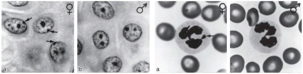
FIGURE 22-3 Left: Barr body (arrows) in the epidermal spinous cell layer. Right: Nuclear appendage (“drumstick”) identified by arrow in white blood cells. (Reproduced with permission from Grumbach MM, Barr ML: Cytologic tests of chromosomal sex in relation to sex anomalies in man. Recent Prog Horm Res 1958;14:255-324.)
شکل 22-3 سمت چپ: بدنه بار (فلش) در لایه سلولی خاردار اپیدرمی. سمت راست: زائده هسته ای (“طبل”) که با فلش در گلبول های سفید مشخص می شود. (تکثیر شده با اجازه Grumbach MM، Barr ML: تست های سیتولوژیک جنسیت کروموزومی در رابطه با ناهنجاری های جنسی در انسان. Recent Prog Horm Res 1958; 14:255-324.)
EMBRYOLOGY OF THE HUMAN REPRODUCTIVE SYSTEM
Development of the Gonads
On each side of the embryo, a primitive gonad arises from the genital ridge, a condensation of tissue near the adrenal gland. The gonad develops a cortex and a medulla. Until the sixth week of development, these structures are identical in both sexes. In genetic males, the medulla develops during the seventh and eighth weeks into a testis, and the cortex regresses. Leydig and Sertoli cells appear, and testosterone and MIS are secreted. In genetic females, the cortex develops into an ovary and the medulla regresses. The embryonic ovary does not secrete hormones. Hormonal treatment of the mother has no effect on gonadal (as opposed to ductal and genital) differentiation in humans, although it does in some experimental animals.
جنین شناسی سیستم تولید مثل انسان
توسعه غدد جنسی
در هر طرف جنین، یک غدد جنسی اولیه از برجستگی دستگاه تناسلی، متراکم شدن بافت نزدیک غده فوق کلیوی، ایجاد می شود. گناد یک قشر و یک مدولا ایجاد می کند. تا هفته ششم رشد، این ساختارها در هر دو جنس یکسان هستند. در مردان ژنتیکی، مدولا در هفته های هفتم و هشتم به بیضه تبدیل می شود و قشر آن پسرفت می کند. سلول های لیدیگ و سرتولی ظاهر می شوند و تستوسترون و MIS ترشح می شوند. در زنان ژنتیکی، قشر به تخمدان تبدیل می شود و بصل النخاع به عقب می رود. تخمدان جنینی هورمون ترشح نمی کند. درمان هورمونی مادر هیچ تاثیری بر تمایز غدد جنسی (برخلاف مجرای و تناسلی) در انسان ندارد، اگرچه در برخی از حیوانات آزمایشگاهی تاثیری دارد.
Embryology of the Genitalia
The embryology of the gonads is summarized in Figures 22-4 and 22-5. In the seventh week of gestation, the embryo has both male and female primordial genital ducts (Figure 22–4). In a normal female fetus, the müllerian duct system then develops into uterine tubes (oviducts) and a uterus. In the normal male fetus, the wolffian duct system on each side develops into the epididymis and vas deferens. The external genitalia are similarly bipotential until the eighth week (Figure 22–5). Thereafter, the urogenital slit disappears and male genitalia form, or, alternatively, it remains open and female genitalia form.
جنین شناسی دستگاه تناسلی
جنین شناسی غدد جنسی در شکل های 22-4 و 22-5 خلاصه شده است. در هفته هفتم بارداری، جنین دارای مجرای تناسلی اولیه نر و ماده است (شکل 22-4). در یک جنین دختر طبیعی، سیستم مجرای مولر به لوله های رحمی (مجرای تخمدان) و رحم تبدیل می شود. در جنین پسر طبیعی، سیستم مجرای ولفیان در هر طرف به اپیدیدیم و مجرای دفران تبدیل می شود. اندام تناسلی خارجی تا هفته هشتم به طور مشابه دو پتانسیل هستند (شکل 22-5). پس از آن، شکاف ادراری تناسلی ناپدید می شود و اندام تناسلی مردان تشکیل می شود، یا، به طور متناوب، باز باقی می ماند و اندام تناسلی زنان تشکیل می شود.

FIGURE 22-4 Embryonic differentiation of male and female internal genitalia (genital ducts) from wolffian (male) and müllerian (female) primordia. (Reproduced with permission from Wilson JD, Foster DW [editors]: Williams Textbook of Endocrinology, 7th ed. Saunders; 1985.)
شکل 22-4 تمایز جنینی اندام تناسلی داخلی نر و ماده (مجرای تناسلی) از پریموردیا ولفیان (نر) و مولرین (مونث). (تکثیر شده با اجازه Wilson JD، Foster DW [ویراستاران]: کتاب درسی غدد درون ریز ویلیامز، ویرایش هفتم ساندرز؛ 1985.)

FIGURE 22-5 Differentiation of male and female external genitalia from indifferent primordial structures in the embryo.
شکل 22-5 تمایز دستگاه تناسلی خارجی نر و ماده از ساختارهای اولیه بی تفاوت در جنین.
When the embryo has functional testes, male internal and external genitalia develop. The Leydig cells of the fetal testis secrete testosterone, and the Sertoli cells secrete MIS (also called müllerian regression factor, or MRF). MIS is a 536-amino-acid homodimer that is a member of the transforming growth factor B (TGF-B) super-family of growth factors, which includes inhibins and activins.
هنگامی که جنین دارای بیضه های عملکردی باشد، اندام تناسلی داخلی و خارجی مرد رشد می کند. سلولهای لیدیگ بیضه جنینی تستوسترون ترشح میکنند و سلولهای سرتولی MIS (که فاکتور رگرسیون مولریا یا MRF نیز نامیده میشود) ترشح میکنند. MIS یک همودایمر 536 آمینو اسیدی است که عضوی از فاکتور رشد تبدیل کننده B (TGF-B) از خانواده فاکتورهای رشد است که شامل مهارکننده ها و اکتیوین ها می شود.
In their effects on the internal as opposed to the external genitalia, MIS and testosterone act unilaterally. MIS causes regression of the müllerian ducts by apoptosis on the side on which it is secreted, and testosterone fosters the development of the vas deferens and related structures from the wolffian ducts. The testosterone metabolite dihydrotestosterone induces the formation of male external genitalia and male secondary sex characteristics (Figure 22-6).
MIS و تستوسترون در تأثیرات خود بر روی اندام تناسلی داخلی به جای دستگاه تناسلی خارجی به صورت یک طرفه عمل می کنند. MIS با آپوپتوز در سمتی که در آن ترشح می شود باعث پسرفت مجاری مولرین می شود و تستوسترون باعث رشد مجرای دفران و ساختارهای مرتبط از مجاری ولفیان می شود. متابولیت تستوسترون دی هیدروتستوسترون باعث ایجاد اندام تناسلی خارجی و خصوصیات جنسی ثانویه مرد می شود (شکل 22-6).

FIGURE 22-6 Diagrammatic summary of normal sex determination,differentiation, and development in humans. DHT, dihydrotestosterone, MIS, müllerian-inhibiting substance; T, testosterone.
شکل 22-6 خلاصه نموداری از تعیین جنسیت طبیعی، تمایز، و رشد در انسان. DHT، دی هیدروتستوسترون، MIS، ماده مهارکننده مولرین؛ تی، تستوسترون
MIS continues to be secreted by the Sertoli cells, and it reaches mean values of 48 ng/mL in plasma in 1- to 2-year-old boys. Thereafter, it declines to low levels by the time of puberty and persists at low but detectable levels throughout life. In girls, MIS is produced by granulosa cells in small follicles in the ovaries, but plasma levels are very low or undetectable until puberty. Thereafter, plasma MIS is about the same as in adult men, that is, about 2 ng/mL. The functions of MIS after early embryonic life are unsettled, but it is probably involved in germ cell maturation in both sexes and in control of testicular descent in boys.
MIS همچنان توسط سلول های سرتولی ترشح می شود و در پسران 1 تا 2 ساله به مقادیر متوسط 48 نانوگرم در میلی لیتر در پلاسما می رسد. پس از آن، تا زمان بلوغ به سطوح پایین کاهش می یابد و در سطوح پایین اما قابل تشخیص در طول زندگی باقی می ماند. در دختران، MIS توسط سلولهای گرانولوزا در فولیکولهای کوچک تخمدانها تولید میشود، اما سطوح پلاسما تا زمان بلوغ بسیار کم یا غیرقابل تشخیص است. پس از آن، MIS پلاسما تقریباً مشابه مردان بالغ است، یعنی حدود 2 نانوگرم در میلی لیتر. عملکرد MIS پس از زندگی اولیه جنینی ناموفق است، اما احتمالاً در بلوغ سلول های زایای هر دو جنس و در کنترل نزول بیضه در پسران نقش دارد.
Development of the Brain
At least in some species, the development of the brain as well as the external genitalia is affected by androgens early in life. In rats, a brief exposure to androgens during the first few days of life causes the male pattern of sexual behavior and the male pattern of hypothalamic control of gonadotropin secretion to develop after puberty. In the absence of androgens, female patterns develop (see Chapter 17). In monkeys, similar effects on sexual behavior are produced by exposure to androgens in utero, but the pattern of gonadotropin secretion remains cyclical. Early exposure of female human fetuses to androgens also appears to cause subtle but significant masculinizing effects on behavior. However, women with adrenogenital syndrome due to congenital adrenocortical enzyme deficiency (see Chapter 20) develop normal menstrual cycles when treated with cortisol. Thus, the human, like the monkey, appears to retain the cyclical pattern of gonadotropin secretion despite exposure to androgens in utero.
رشد مغز
حداقل در برخی از گونه ها، رشد مغز و همچنین اندام تناسلی خارجی در اوایل زندگی تحت تأثیر آندروژن ها قرار می گیرد. در موشها، قرار گرفتن کوتاه مدت در معرض آندروژنها در چند روز اول زندگی باعث میشود که الگوی رفتار جنسی مردانه و الگوی مردانه کنترل هیپوتالاموس ترشح گنادوتروپین پس از بلوغ ایجاد شود. در غیاب آندروژن ها، الگوهای زنانه رشد می کنند (به فصل 17 مراجعه کنید). در میمونها، اثرات مشابهی بر رفتار جنسی با قرار گرفتن در معرض آندروژنها در رحم ایجاد میشود، اما الگوی ترشح گنادوتروپین چرخهای باقی میماند. قرار گرفتن زودهنگام جنینهای دختر در معرض آندروژنها نیز به نظر میرسد که اثرات مردانهسازی ظریف اما قابلتوجهی بر رفتار دارد. با این حال، زنان مبتلا به سندرم آدرنوژنیتال به دلیل کمبود مادرزادی آنزیم قشر آدرنال (به فصل 20 مراجعه کنید) هنگامی که تحت درمان با کورتیزول قرار می گیرند، چرخه های قاعدگی طبیعی دارند. بنابراین، به نظر می رسد که انسان، مانند میمون، الگوی چرخه ای ترشح گنادوتروپین را با وجود قرار گرفتن در معرض آندروژن ها در رحم حفظ می کند.
ABERRANT SEXUAL DIFFERENTIATION
Chromosomal Abnormalities
From the preceding discussion, it might be expected that abnormalities of sexual development could be caused by genetic or hormonal abnormalities as well as by other nonspecific teratogenic influences, and this is indeed the case. The major classes of abnormalities are listed in Table 22-1.
تمایز نابجای جنسی
ناهنجاری های کروموزومی
از بحث قبلی، میتوان انتظار داشت که ناهنجاریهای رشد جنسی میتواند ناشی از ناهنجاریهای ژنتیکی یا هورمونی و همچنین سایر تأثیرات تراتوژنیک غیراختصاصی باشد، و این در واقع همینطور است. کلاس های اصلی ناهنجاری ها در جدول 22-1 فهرست شده اند.
TABLE 22-1 Classification of the major disorders of sex differentiation in humans.
جدول 22-1 طبقه بندی اختلالات عمده تمایز جنسی در انسان.

aMany of these syndromes can have great variation in degree and, consequently, in manifestations.
aبسیاری از این سندرمها میتوانند از نظر درجه و در نتیجه تظاهرات تنوع زیادی داشته باشند.
Nondisjunction of sex chromosomes during the first division in meiosis results in distinct defects (see Clinical Box 22-1; Figure 22-7). Meiosis is at two-stage process, and although nondisjunction usually occurs during the first meiotic division, it can occur in the second, producing more complex chromosomal abnormalities. In addition, nondisjunction or simple loss of a sex chromosome can occur during the early mitotic divisions after fertilization. The consequence of faulty mitoses in the early zygote is mosaicism, in which two or more populations of cells have different chromosome complements. True hermaphroditism, the condition in which the individual has both ovaries and testes, is probably due to XX/XY mosaicism and related mosaic patterns, although other genetic aberrations are possible.
عدم اتصال کروموزوم های جنسی در طول اولین تقسیم در میوز منجر به نقایص مشخص می شود (به کادر بالینی 22-1 مراجعه کنید؛ شکل 22-7). میوز در فرآیند دو مرحله ای است، و اگرچه عدم تفکیک معمولاً در طول اولین تقسیم میوز رخ می دهد، می تواند در مرحله دوم رخ دهد و ناهنجاری های کروموزومی پیچیده تری ایجاد کند. علاوه بر این، عدم تفکیک یا از دست دادن ساده یک کروموزوم جنسی می تواند در طول تقسیمات اولیه میتوزی پس از لقاح رخ دهد. پیامد میتوزهای معیوب در زیگوت اولیه موزائیسم است که در آن دو یا چند جمعیت از سلول ها دارای مکمل های کروموزوم متفاوتی هستند. هرمافرودیتیسم واقعی، وضعیتی که در آن فرد هم تخمدان و هم بیضه دارد، احتمالاً به دلیل موزائیسم XX/XY و الگوهای موزاییکی مرتبط است، اگرچه سایر انحرافات ژنتیکی ممکن است.

FIGURE 22–7 Summary of four possible defects produced by maternal nondisjunction of the sex chromosomes at the time of meiosis. The YO combination is believed to be lethal, and the fetus dies in utero.
شکل 22-7 خلاصه ای از چهار نقص احتمالی ناشی از عدم تفکیک کروموزوم های جنسی توسط مادر در زمان میوز. اعتقاد بر این است که ترکیب YO کشنده است و جنین در رحم می میرد.
CLINICAL BOX 22-1
Chromosomal Abnormalities
An established defect in gametogenesis is nondisjunction, a phenomenon in which a pair of chromosomes fail to separate, so that both go to one of the daughter cells during meiosis. Four of the abnormal zygotes that can form as a result of nondisjunction of one of the X chromosomes during oogenesis are shown in Figure 25-7. In individuals with the XO chromosomal pattern, the gonads are rudimentary or absent, so that female external genitalia develop, stature is short, other congenital abnormalities are often present, and no sexual maturation occurs at puberty. This syndrome is called gonadal dysgenesis or, alternatively, ovarian agenesis or Turner syndrome. Individuals with the XXY pattern, the most common sex chromosome disorder, have the genitalia of a normal male. Testosterone secretion at puberty is often great enough for the development of male characteristics; however, the seminiferous tubules are abnormal, and the incidence of mental retardation is higher than normal. This syndrome is known as seminiferous tubule dysgenesis or Klinefelter syndrome. The XXX (“superfemale”) pattern is second in frequency only to the XXY pattern and may be even more common in the general population, since it does not seem to be associated with any characteristic abnormalities. The YO combination is probably lethal.
جعبه بالینی 22-1
ناهنجاری های کروموزومی
یک نقص ثابت شده در گامتوژنز، عدم تفکیک است، پدیده ای که در آن یک جفت کروموزوم از هم جدا نمی شوند، به طوری که هر دو در طول میوز به یکی از سلول های دختر می روند. چهار تا از زیگوت های غیر طبیعی که می توانند در نتیجه عدم تفکیک یکی از کروموزوم های X در طی اووژنز تشکیل شوند در شکل 25-7 نشان داده شده است. در افراد با الگوی کروموزومی XO، غدد جنسی ابتدایی هستند یا وجود ندارند، به طوری که اندام تناسلی خارجی زنان رشد می کند، قد کوتاه است، سایر ناهنجاری های مادرزادی اغلب وجود دارد و بلوغ جنسی در بلوغ رخ نمی دهد. این سندروم دیسژنزیس گناد یا به عبارت دیگر آژنزی تخمدان یا سندرم ترنر نامیده می شود. افراد با الگوی XXY، شایع ترین اختلال کروموزوم جنسی، اندام تناسلی یک مرد طبیعی دارند. ترشح تستوسترون در دوران بلوغ اغلب به اندازه کافی برای رشد ویژگی های مردانه است. با این حال، لوله های منی ساز غیر طبیعی هستند و بروز عقب ماندگی ذهنی بالاتر از حد طبیعی است. این سندرم به نام دیسژنزیس لوله منی ساز یا سندرم کلاین فلتر شناخته می شود. الگوی XXX (“فوق زن”) از نظر فراوانی تنها پس از الگوی XXY در رتبه دوم قرار دارد و ممکن است حتی در جمعیت عمومی رایج تر باشد، زیرا به نظر نمی رسد با هیچ گونه ناهنجاری مشخصه ای مرتبط باشد. ترکیب YO احتمالا کشنده است.
Nondisjunction of chromosome 21 produces trisomy 21, the chromosomal abnormality associated with Down syndrome. The additional chromosome 21 is normal, so Down syndrome is a pure case of gene excess causing abnormalities.
عدم اتصال کروموزوم 21 باعث ایجاد تریزومی 21 می شود، ناهنجاری کروموزومی مرتبط با سندرم داون. کروموزوم 21 اضافی طبیعی است، بنابراین سندرم داون یک مورد خالص از بیش از حد ژن است که باعث ناهنجاری می شود.
Many other chromosomal abnormalities occur as well as numerous diseases due to defects in single genes. These conditions are generally diagnosed in utero by analysis of fetal cells in a sample of amniotic fluid collected by inserting a needle through the abdominal wall (amniocentesis) or, earlier in pregnancy, by examining fetal cells obtained by a needle biopsy of chorionic villi (chorionic villus sampling).
بسیاری از ناهنجاری های کروموزومی دیگر و همچنین بیماری های متعدد به دلیل نقص در ژن های منفرد رخ می دهد. این شرایط عموماً در داخل رحم با تجزیه و تحلیل سلولهای جنینی در نمونهای از مایع آمنیوتیک که با وارد کردن سوزن از دیواره شکم (آمنیوسنتز) جمعآوری شده است، یا در اوایل بارداری، با بررسی سلولهای جنینی بهدستآمده از بیوپسی سوزنی پرزهای کوریونی (نمونهگیری از پرزهای کوریونی) تشخیص داده میشوند.
THERAPEUTIQ HIGHLIGHTS
Many of the syndromes mentioned have effects on multiple organ systems, and patients must be carefully monitored with a multidisciplinary approach to avert the consequences of cardiovascular defects, infections secondary to urinary tract and renal malformations, and the psychological impact of reproductive implications. Girls with Turner syndrome and evidence of gonadal failure are also treated with low-dose estrogen to evoke puberty, followed by gradual replacement of mature estrogen levels to permit feminization. Conversely, patients with Klinefelter syndrome are often supplemented with androgens to improve virilization and libido.
نکات برجسته درمانی
بسیاری از سندرمهای ذکر شده بر روی سیستمهای اندام متعددی تأثیر میگذارند، و بیماران باید با رویکردی چند رشتهای به دقت تحت نظر قرار گیرند تا از عواقب نقایص قلبی عروقی، عفونتهای ثانویه ناهنجاریهای مجاری ادراری و کلیوی و تأثیر روانی پیامدهای تولید مثل جلوگیری شود. دختران مبتلا به سندرم ترنر و شواهد نارسایی غدد جنسی نیز با دوز کم استروژن برای برانگیختن بلوغ درمان میشوند و به دنبال آن سطوح استروژن بالغ جایگزین تدریجی میشوند تا زنانه شدن را مجاز کنند. برعکس، بیماران مبتلا به سندرم کلاین فلتر اغلب با آندروژن برای بهبود ویریلیزاسیون و میل جنسی مکمل دریافت می کنند.
Chromosomal abnormalities also include transposition of parts of chromosomes to other chromosomes. Rarely, genetic males are found to have the XX karyotype because the short arm of their father’s Y chromosome was transposed to their father’s X chromosome during meiosis and they received that X chromosome along with their mother’s. Similarly, deletion of the small portion of the Y chromosome containing SRY produces females with the XY karyotype.
ناهنجاری های کروموزومی همچنین شامل جابجایی قسمت هایی از کروموزوم ها به کروموزوم های دیگر است. به ندرت، مردان ژنتیکی دارای کاریوتایپ XX هستند زیرا بازوی کوتاه کروموزوم Y پدرشان در طول میوز به کروموزوم X پدرشان منتقل شد و آنها آن کروموزوم X را همراه با کروموزوم مادر دریافت کردند. به طور مشابه، حذف بخش کوچکی از کروموزوم Y حاوی SRY باعث تولید مادههایی با کاریوتیپ XY میشود.
Hormonal Abnormalities
Development of the male external genitalia occurs normally in genetic males in response to androgen secreted by the embryonic testes, but male genital development may also occur in genetic females exposed to androgens from some other source during the 8th to the 13th weeks of gestation. The syndrome that results is female pseudohermaphroditism. A pseudohermaphrodite is an individual with the genetic constitution and gonads of one sex and the genitalia of the other. After the 13th week, the genitalia are fully formed, but exposure to androgens can cause hypertrophy of the clitoris. Female pseudohermaphroditism may be due to congenital virilizing adrenal hyperplasia (see Chapter 19), or it may be caused by androgens administered to the mother. Conversely, one cause of the development of female external genitalia in genetic males (male pseudohermaphroditism) is defective testicular development. Because the testes also secrete MIS, genetic males with defective testes have female internal genitalia.
ناهنجاری های هورمونی
رشد اندام تناسلی خارجی مرد به طور طبیعی در مردان ژنتیکی در پاسخ به آندروژن ترشح شده توسط بیضه های جنینی اتفاق می افتد، اما رشد دستگاه تناسلی مردانه ممکن است در زنان ژنتیکی که در معرض آندروژن ها از منبع دیگری در طول هفته های 8 تا 13 بارداری قرار دارند نیز رخ دهد. سندرمی که حاصل می شود، هرمافرودیتیسم کاذب زنانه است. هرمافرودیت کاذب فردی است با ساختار ژنتیکی و غدد جنسی یک جنس و اندام تناسلی جنس دیگر. پس از هفته سیزدهم، اندام تناسلی به طور کامل تشکیل می شود، اما قرار گرفتن در معرض آندروژن ها می تواند باعث هیپرتروفی کلیتوریس شود. هرمافرودیتیسم کاذب زنان ممکن است به دلیل هیپرپلازی مادرزادی آدرنال ویریل کننده باشد (به فصل 19 مراجعه کنید)، یا ممکن است ناشی از آندروژن های تجویز شده به مادر باشد. برعکس، یکی از علل ایجاد اندام تناسلی خارجی زنان در مردان ژنتیکی (کاذب هرمافرودیتیسم مردانه) رشد ناقص بیضه است. از آنجا که بیضه ها نیز MIS ترشح می کنند، مردان ژنتیکی با بیضه های معیوب دارای اندام تناسلی داخلی زنانه هستند.
Another cause of male pseudohermaphroditism is androgen resistance, in which, as a result of various congenital abnormalities, male hormones cannot exert their full effects on the tissues. One form of androgen resistance is a 5α- reductase deficiency, in which the enzyme responsible for the formation of dihydrotestosterone, the active form of testosterone, is decreased (Figure 22–8). The consequences of this deficiency are discussed in Chapter 23. Other forms of androgen resistance are due to various mutations in the androgen receptor gene, and the resulting defects in receptor function range from minor to severe. Mild defects cause infertility with or without gynecomastia. When the loss of receptor function is complete, the testicular feminizing syndrome, now known as complete androgen resistance syndrome, results. In this condition, MIS is present and testosterone is secreted at normal or even elevated rates. The external genitalia are female, but the vagina ends blindly because there are no female internal genitalia. At puberty, enlarged breasts develop in persons with this syndrome. The syndrome usually remains undiagnosed until women seek medical advice because of lack of menstruation.
یکی دیگر از علل کاذب هرمافرودیتیسم مردانه مقاومت آندروژنی است که در نتیجه ناهنجاری های مادرزادی مختلف، هورمون های مردانه نمی توانند اثرات کامل خود را بر روی بافت ها اعمال کنند. یکی از اشکال مقاومت به آندروژن، کمبود 5α ردوکتاز است که در آن آنزیم مسئول تشکیل دی هیدروتستوسترون، شکل فعال تستوسترون، کاهش می یابد (شکل 22-8). عواقب این کمبود در فصل 23 مورد بحث قرار می گیرد. اشکال دیگر مقاومت به آندروژن به دلیل جهش های مختلف در ژن گیرنده آندروژن است و نقایص ناشی از عملکرد گیرنده از جزئی تا شدید متغیر است. نقایص خفیف باعث ناباروری با ژنیکوماستی یا بدون آن می شود. هنگامی که از دست دادن عملکرد گیرنده کامل می شود، سندرم زنانه شدن بیضه، که اکنون به عنوان سندرم مقاومت کامل آندروژن شناخته می شود، ایجاد می شود. در این شرایط، MIS وجود دارد و تستوسترون با سرعت طبیعی یا حتی بالا ترشح می شود. اندام تناسلی خارجی زنانه است، اما واژن کورکورانه خاتمه می یابد زیرا دستگاه تناسلی داخلی زنان وجود ندارد. در سن بلوغ، سینه های بزرگ در افراد مبتلا به این سندرم ایجاد می شود. این سندرم معمولاً تا زمانی که زنان به دلیل عدم قاعدگی به دنبال مشاوره پزشکی نباشند، تشخیص داده نمیشود.
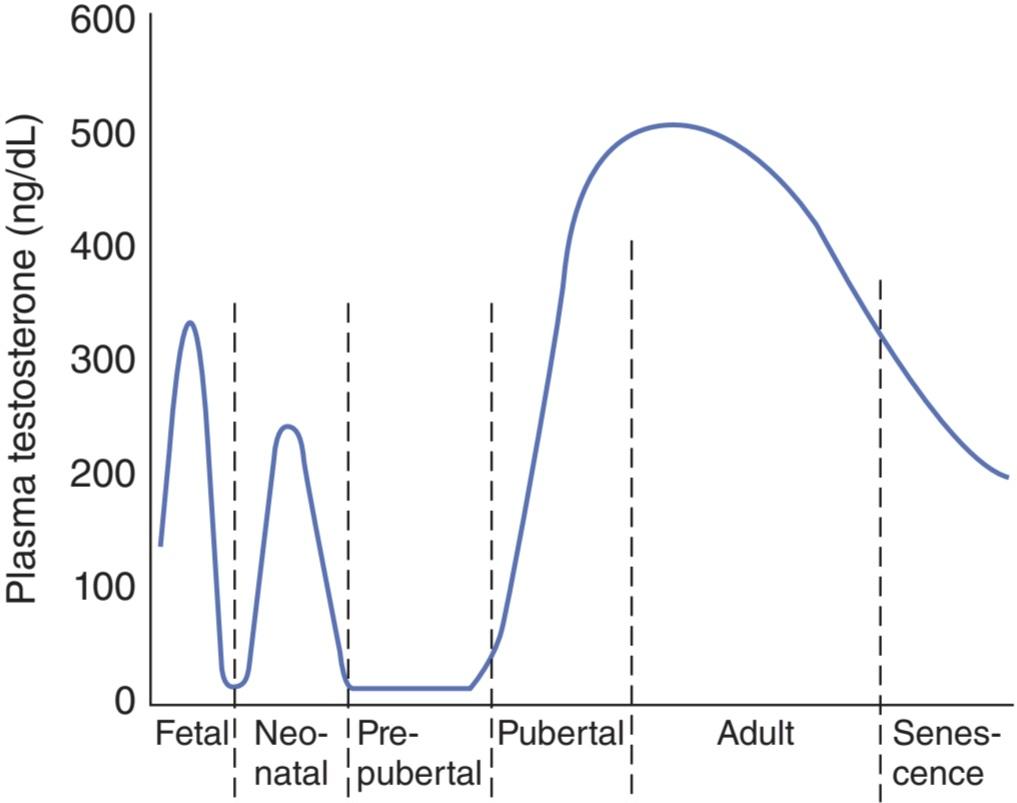
FIGURE 22-8 Plasma testosterone levels at various ages in human males.
شکل 22-8 سطوح تستوسترون پلاسما در سنین مختلف در مردان انسان.
It is worth noting that genetic males with congenital blockage of the formation of pregnenolone are pseudohermaphrodites because testicular as well as adrenal androgens are normally formed from pregnenolone. Male pseudohermaphroditism also occurs when there is a congenital deficiency of 17α-hydroxylase (see Chapter 19).
شایان ذکر است که مردان ژنتیکی با انسداد مادرزادی تشکیل پرگننولون شبه هرمافرودیت هستند زیرا آندروژنهای بیضه و همچنین آدرنال معمولاً از پرگننولون تشکیل میشوند. هرمافرودیتیسم کاذب مردانه نیز زمانی رخ می دهد که کمبود مادرزادی 17α هیدروکسیلاز وجود داشته باشد (به فصل 19 مراجعه کنید).
PUBERTY
As noted above, a burst of testosterone secretion occurs in male fetuses before birth (see Chapter 23). In the neonatal period there is another burst, with unknown function, but thereafter the Leydig cells become quiescent. There follows in all mammals a period in which the gonads of both sexes are quiescent until they are activated by gonadotropins from the pituitary to bring about the final maturation of the reproductive system. This period of final maturation is known as adolescence. It is often also called puberty, although puberty, strictly defined, is the period when the endocrine and gametogenic functions of the gonads have first developed to the point where reproduction is possible. In girls, the first event is thelarche, the development of breasts, followed by pubarche, the development of axillary and pubic hair, and then by menarche, the first menstrual period. Initial menstrual periods are generally anovulatory, and regular ovulation appears about a year later. In contrast to the situation in adulthood, removal of the gonads during the period from soon after birth to puberty causes only a small increase in gonadotropin secretion, so gonadotropin secretion is not being held in check by the gonadal hormones. In children between the ages of 7 and 10, a slow increase in estrogen and androgen secretion precedes the more rapid rise in the early teens (Figure 22–9).
بلوغ
همانطور که در بالا ذکر شد، ترشح تستوسترون در جنین های پسر قبل از تولد اتفاق می افتد (به فصل 23 مراجعه کنید). در دوره نوزادی انفجار دیگری با عملکرد ناشناخته وجود دارد، اما پس از آن سلول های لیدیگ ساکن می شوند. در تمام پستانداران دوره ای وجود دارد که در آن غدد جنسی هر دو جنس ساکن هستند تا زمانی که توسط گنادوتروپین های هیپوفیز فعال شوند تا بلوغ نهایی سیستم تولید مثل را به همراه داشته باشند. این دوره بلوغ نهایی به عنوان نوجوانی شناخته می شود. اغلب به آن بلوغ نیز می گویند، اگرچه بلوغ، با تعریف دقیق، دوره ای است که عملکردهای غدد درون ریز و گامتوژنیک غدد جنسی برای اولین بار تا جایی که امکان تولید مثل وجود دارد، توسعه یافته است. در دختران اولین اتفاق، تلارش، رشد سینهها و به دنبال آن پوبارک، رشد موهای زیر بغل و شرمگاهی و بعد از آن قاعدگی، اولین قاعدگی است. دوره های اولیه قاعدگی معمولاً بدون تخمک گذاری هستند و تخمک گذاری منظم حدود یک سال بعد ظاهر می شود. بر خلاف وضعیت بزرگسالی، برداشتن غدد جنسی در طول دوره از بلافاصله پس از تولد تا بلوغ تنها باعث افزایش اندکی در ترشح گنادوتروپین می شود، بنابراین ترشح گنادوتروپین توسط هورمون های گناد کنترل نمی شود. در کودکان بین 7 تا 10 سال، افزایش آهسته در ترشح استروژن و آندروژن قبل از افزایش سریعتر در اوایل نوجوانی است (شکل 22-9).
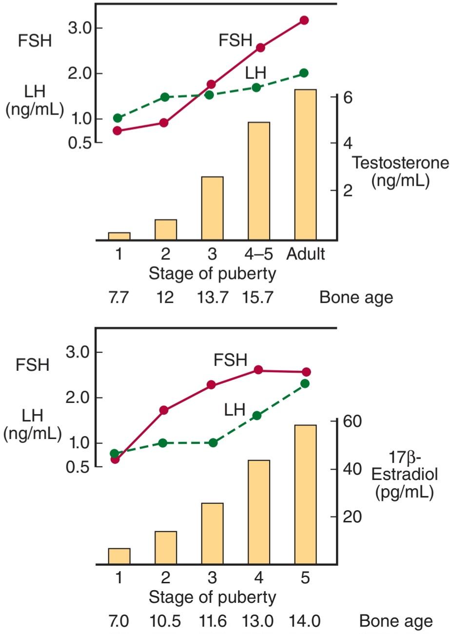
FIGURE 22-9 Changes in plasma hormone concentrations during puberty in boys (top) and girls (bottom). Stage 1 of puberty is preadolescence in both sexes. In boys, stage 2 is characterized by beginning enlargement of the testes, stage 3 by penile enlargement, stage 4 by growth of the glans penis, and stage 5 by adult genitalia. In girls, stage 2 is characterized by breast buds, stage 3 by elevation and enlargement of the breasts, stage 4 by projection of the areolas, and stage 5 by adult breasts. FSH, follicle-stimulating hormone; LH, luteinizing hormone. (Modified and reproduced with permission from Berenberg SR [ed]: Puberty: Biologic and Psychosocial Components. HE Stenfoert Kroese BV; 1975.)
شکل 22-9 تغییرات در غلظت هورمون های پلاسما در دوران بلوغ در پسران (بالا) و دختران (پایین). مرحله 1 بلوغ در هر دو جنس پیش از بلوغ است. در پسران، مرحله 2 با شروع بزرگ شدن بیضه ها، مرحله 3 با بزرگ شدن آلت تناسلی، مرحله 4 با رشد آلت تناسلی و مرحله 5 توسط دستگاه تناسلی بزرگسالان مشخص می شود. در دختران مرحله 2 با جوانه های سینه، مرحله 3 با بلند شدن و بزرگ شدن سینه ها، مرحله 4 با برآمدگی آرئول ها و مرحله 5 با سینه های بالغ مشخص می شود. FSH، هورمون محرک فولیکول؛ LH، هورمون لوتئینه کننده. (اصلاح و تکثیر شده با اجازه Berenberg SR [ed]: بلوغ: اجزای زیستی و روانی اجتماعی. HE Stenfoert Kroese BV; 1975.)
The age at the time of puberty is variable. In Europe and the United States, it has been declining at the rate of 1-3 months per decade for more than 175 years. In the United States in recent years, puberty generally occurs between the ages of 8 and 13 in girls and 9 and 14 in boys.
سن در زمان بلوغ متغیر است. در اروپا و ایالات متحده بیش از 175 سال است که به میزان 1-3 ماه در دهه کاهش یافته است. در ایالات متحده در سال های اخیر، بلوغ عموماً بین 8 تا 13 سالگی در دختران و 9 تا 14 سالگی در پسران رخ می دهد.
Another event that occurs in humans at the time of puberty is an increase in the secretion of adrenal androgens (see Figure 19-12). The onset of this increase is called adrenarche. It occurs at age 8-10 years in girls and age 10-12 years in boys. Dehydroepiandrosterone (DHEA) values peak at about age 25 in women and slightly later than that in men. They then decline slowly to low values in old age. The rise appears to be due to an increase in the activity of 17α-hydroxylase.
رویداد دیگری که در انسان در زمان بلوغ رخ می دهد، افزایش ترشح آندروژن های آدرنال است (شکل 19-12 را ببینید). شروع این افزایش آدرنارک نامیده می شود. در سنین 10-8 سالگی در دختران و 10-12 سالگی در پسران رخ می دهد. مقدار دهیدرواپی آندروسترون (DHEA) در حدود 25 سالگی در زنان و کمی دیرتر از مردان به اوج خود می رسد. سپس در سنین بالا به آرامی کاهش می یابد و به مقادیر پایین می رسد. به نظر می رسد افزایش به دلیل افزایش فعالیت 17α هیدروکسیلاز باشد.
Control of the Onset of Puberty
The gonads of children can be stimulated by gonadotropins; their pituitaries contain gonadotropins and their hypothalami contain gonadotropin-releasing hormone (GnRH) (see Chapter 17). However, their gonadotropins are not secreted. In immature monkeys, normal menstrual cycles can be brought on by pulsatile injection of GnRH, and they persist as long as the pulsatile injection is continued. Thus, it seems clear that pulsatile secretion of GnRH brings on puberty. During the period from birth to puberty, a neural mechanism is operating to prevent the normal pulsatile release of GnRH. The nature of the mechanism inhibiting the GnRH pulse generator is unknown. However, one or more genes produce products that stimulate secretion of GnRH, and inhibition of these genes before puberty is an interesting possibility (Clinical Box 22-2).
کنترل شروع بلوغ
غدد جنسی کودکان را می توان توسط گنادوتروپین ها تحریک کرد. هیپوفیز آنها حاوی گنادوتروپین و هیپوتالامی آنها حاوی هورمون آزاد کننده گنادوتروپین (GnRH) است (به فصل 17 مراجعه کنید). با این حال، گنادوتروپین های آنها ترشح نمی شود. در میمونهای نابالغ، چرخههای قاعدگی طبیعی را میتوان با تزریق ضربانی GnRH ایجاد کرد و تا زمانی که تزریق ضرباندار ادامه یابد، ادامه مییابد. بنابراین واضح است که ترشح ضربانی GnRH باعث بلوغ می شود. در طول دوره از تولد تا بلوغ، یک مکانیسم عصبی برای جلوگیری از انتشار ضربانی طبیعی GnRH عمل می کند. ماهیت مکانیسم مهار مولد پالس GnRH ناشناخته است. با این حال، یک یا چند ژن محصولاتی تولید میکنند که ترشح GnRH را تحریک میکنند و مهار این ژنها قبل از بلوغ امکان جالبی است (کادر بالینی 22-2).
CLINICAL BOX 22-2
Leptin
It has been argued for some time that a critical body weight must normally be reached for puberty to occur. Thus, for example, young women who engage in strenuous athletics lose weight and stop menstruating, as do girls with anorexia nervosa. If these girls start to eat and gain weight, they menstruate again, that is, they “go back through puberty.” It now appears that leptin, the satiety-producing hormone secreted by fat cells, may be the link between body weight and puberty. Obese ob/ob mice that cannot make leptin are infertile, and their fertility is restored by injections of leptin. Leptin treatment also induces precocious puberty in immature female mice. However, the way that leptin fits into the overall control of puberty remains to be determined.
جعبه بالینی 22-2
لپتین
مدتی است که استدلال شده است که برای رسیدن به بلوغ به طور معمول باید به وزن بحرانی بدن رسید. بنابراین، به عنوان مثال، زنان جوانی که به ورزش های شدید می پردازند، وزن خود را کاهش می دهند و قاعدگی را متوقف می کنند، مانند دختران مبتلا به بی اشتهایی عصبی. اگر این دختران شروع به خوردن و افزایش وزن کنند، دوباره پریود می شوند، یعنی «از دوران بلوغ برمی گردند». اکنون به نظر می رسد که لپتین، هورمون تولید کننده سیری که توسط سلول های چربی ترشح می شود، ممکن است ارتباط بین وزن بدن و بلوغ باشد. موش های چاق ob/ob که نمی توانند لپتین بسازند نابارور هستند و باروری آنها با تزریق لپتین بازیابی می شود. درمان با لپتین همچنین باعث بلوغ زودرس در موش های ماده نابالغ می شود. با این حال، روشی که لپتین در کنترل کلی بلوغ قرار می گیرد هنوز مشخص نیست.
PRECOCIOUS & DELAYED PUBERTY
Sexual Precocity
The major causes of precocious sexual development in humans are listed in Table 22-2. Early development of secondary sexual characteristics without gametogenesis is caused by abnormal exposure of immature males to androgen or females to estrogen. This syndrome should be called precocious pseudopuberty to distinguish it from true precocious puberty due to an early but otherwise normal pubertal pattern of gonadotropin secretion from the pituitary.
بلوغ زودرس و تاخیری
زودرس بودن جنسی
علل عمده رشد جنسی زودرس در انسان در جدول 22-2 ذکر شده است. رشد اولیه خصوصیات جنسی ثانویه بدون گامتوژنز در اثر مواجهه غیرطبیعی مردان نابالغ با آندروژن یا زنان با استروژن ایجاد می شود. این سندرم را باید بلوغ کاذب زودرس نامید تا آن را از بلوغ زودرس واقعی به دلیل الگوی بلوغ زودرس، اما در غیر این صورت طبیعی ترشح گنادوتروپین از هیپوفیز، متمایز کند.
TABLE 22-2 Classification of the causes of precocious sexual development in humans.
جدول 22-2 طبقه بندی علل رشد جنسی زودرس در انسان.

Constitutional precocious puberty, that is, precocious puberty in which no cause can be determined, is more common in girls than in boys. In both sexes, tumors or infections involving the hypothalamus cause precocious puberty.
بلوغ زودرس مشروط، یعنی بلوغ زودرس که هیچ علتی در آن مشخص نیست، در دختران بیشتر از پسران است. در هر دو جنس، تومورها یا عفونت های هیپوتالاموس باعث بلوغ زودرس می شود.
Indeed, in one large series of cases, precocious puberty was the most common endocrine symptom of hypothalamic disease. In experimental animals, precocious puberty can be produced by hypothalamic lesions. Apparently the lesions interrupt a pathway that normally holds pulsatile GnRH secretion in check. Pineal tumors are sometimes associated with precocious puberty, but evidence indicates that these tumors are associated with precocity only when there is secondary damage to the hypothalamus.
در واقع، در یک سری از موارد، بلوغ زودرس شایع ترین علامت غدد درون ریز بیماری هیپوتالاموس بود. در حیوانات آزمایشگاهی، بلوغ زودرس می تواند توسط ضایعات هیپوتالاموس ایجاد شود. ظاهراً ضایعات مسیری را که معمولاً ترشح ضربانی GnRH را کنترل میکند، قطع میکنند. تومورهای پینه آل گاهی با بلوغ زودرس همراه هستند، اما شواهد نشان می دهد که این تومورها تنها زمانی با زودرس شدن همراه هستند که آسیب ثانویه به هیپوتالاموس وجود داشته باشد.
Precocious gametogenesis and steroidogenesis can occur without the pubertal pattern of gonadotropin secretion (gonadotropin-independent precocity). At least in some cases of this condition, the sensitivity of LH receptors to gonadotropins is increased because of an activating mutation in the G-protein that couples the receptors to adenylyl cyclase.
گامتوژنز و استروئیدوژنز زودرس می تواند بدون الگوی بلوغ ترشح گنادوتروپین (پیش سنی مستقل از گنادوتروپین) رخ دهد. حداقل در برخی موارد این وضعیت، حساسیت گیرنده های LH به گنادوتروپین ها به دلیل جهش فعال کننده در پروتئین G که گیرنده ها را با آدنیلیل سیکلاز جفت می کند، افزایش می یابد.
Delayed or Absent Puberty
The normal variation in the age at which adolescent changes occur is so wide that puberty cannot be considered to be pathologically delayed until the menarche has failed to occur by the age of 17 or testicular development by the age of 20. Failure of maturation due to panhypopituitarism is associated with dwarfing and evidence of other endocrine abnormalities. Patients with the XO chromosomal pattern and gonadal dysgenesis are also dwarfed. In some individuals, puberty is delayed even though the gonads are present and other endocrine functions are normal. In males, this clinical picture is called eunuchoidism. In females, it is called primary amenorrhea (Clinical Box 22- 3).
بلوغ تاخیری یا غایب
تغییرات طبیعی در سنی که در آن تغییرات نوجوانان اتفاق میافتد به قدری گسترده است که بلوغ را نمیتوان از نظر پاتولوژیک به تأخیر انداخت تا زمانی که منارک تا سن 17 سالگی یا رشد بیضه در سن 20 سالگی رخ ندهد. بیماران با الگوی کروموزومی XO و دیسژنزی گناد نیز کوتوله هستند. در برخی از افراد، بلوغ به تاخیر می افتد، حتی اگر غدد جنسی وجود داشته باشند و سایر عملکردهای غدد درون ریز طبیعی هستند. در مردان، این تصویر بالینی به نام eunuchoidism نامیده می شود. در زنان به آن آمنوره اولیه می گویند (کادر بالینی 22-3).
CLINICAL BOX 22-3
Hyperprolactinemia
Up to 70% of the patients with chromophobe adenomas of the anterior pituitary have elevated plasma prolactin levels. In some instances, the elevation may be due to damage to the pituitary stalk, but in most cases, the tumor cells are actually secreting the hormone. The hyperprolactinemia may cause galactorrhea, but in many individuals no demonstrable endocrine abnormalities are present. Conversely, most women with galactorrhea have normal prolactin levels; definite elevations are found in less than one-third of patients with this condition.
جعبه بالینی 22-3
هیپرپرولاکتینمی
تا 70 درصد از بیماران مبتلا به آدنوم کروموفوب هیپوفیز قدامی سطح پرولاکتین پلاسما افزایش یافته است. در برخی موارد، ارتفاع ممکن است به دلیل آسیب به ساقه هیپوفیز باشد، اما در بیشتر موارد، سلول های تومور در واقع هورمون را ترشح می کنند. هیپرپرولاکتینمی ممکن است باعث گالاکتوره شود، اما در بسیاری از افراد هیچ ناهنجاری غدد درون ریز قابل اثباتی وجود ندارد. برعکس، بیشتر زنان مبتلا به گالاکتوره سطح پرولاکتین طبیعی دارند. افزایش قطعی در کمتر از یک سوم بیماران مبتلا به این بیماری مشاهده می شود.
Another interesting observation is that 15-20% of women with secondary amenorrhea have elevated prolactin levels, and when prolactin secretion is reduced, normal menstrual cycles and fertility return. Prolactin may produce amenorrhea by blocking the action of gonadotropins on the ovaries. The hypogonadism produced by prolactinomas is associated with osteoporosis due to estrogen deficiency.
مشاهدات جالب دیگر این است که 15 تا 20 درصد از زنان مبتلا به آمنوره ثانویه سطح پرولاکتین را افزایش می دهند و هنگامی که ترشح پرولاکتین کاهش می یابد، چرخه های قاعدگی طبیعی و باروری باز می گردند. پرولاکتین ممکن است با مسدود کردن عملکرد گنادوتروپین ها بر روی تخمدان ها باعث آمنوره شود. هیپوگنادیسم تولید شده توسط پرولاکتینوما با پوکی استخوان به دلیل کمبود استروژن همراه است.
As noted previously, hyperprolactinemia in men is associated with erectile dysfunction and hypogonadism that disappear when prolactin secretion is reduced.
همانطور که قبلا ذکر شد، هیپرپرولاکتینمی در مردان با اختلال نعوظ و هیپوگنادیسم همراه است که با کاهش ترشح پرولاکتین ناپدید می شود.
THERAPEUTIC HIGHLIGHTS
Prescription drug use is a common cause of hyperprolactinemia. Prolactin secretion in the pituitary is suppressed by the brain chemical dopamine. Use of drugs that block the effects of dopamine can cause the pituitary to secrete prolactin. Examples of some prescription drugs that can cause hyperprolactinemia include haloperidol and phenothiazines; most antipsychotic medications; and cisapride, which is used to treat nausea and gastroesophageal reflux in cancer patients. If possible, the drug suspected as causing hyperprolactinemia should be withdrawn, or the dose titrated. Regardless of the etiology, treatment should endeavor to restore normal prolactin levels to avoid suppressive effects on the ovaries and preserve bone density. Dopamine agonists also provide benefit in many cases, including prolactinoma, and can be used in patients for whom an inciting pharmaceutical agent cannot be withdrawn.
نکات برجسته درمانی
مصرف داروهای تجویزی یکی از علل شایع هیپرپرولاکتینمی است. ترشح پرولاکتین در هیپوفیز توسط ماده شیمیایی مغز دوپامین سرکوب می شود. استفاده از داروهایی که اثرات دوپامین را مسدود می کنند می تواند باعث ترشح پرولاکتین از هیپوفیز شود. نمونه هایی از برخی داروهای تجویزی که می توانند باعث هیپرپرولاکتینمی شوند عبارتند از: هالوپریدول و فنوتیازین ها. اکثر داروهای ضد روان پریشی؛ و سیزاپراید که برای درمان تهوع و رفلاکس معده به مری در بیماران سرطانی استفاده می شود. در صورت امکان، دارویی که مشکوک به ایجاد هیپرپرولاکتینمی است باید قطع شود، یا دوز آن تیتر شود. صرف نظر از علت، درمان باید سعی کند سطوح طبیعی پرولاکتین را بازگرداند تا از اثرات سرکوبگر بر تخمدان ها جلوگیری کند و تراکم استخوان را حفظ کند. آگونیستهای دوپامین نیز در بسیاری از موارد از جمله پرولاکتینوما سودمند هستند و میتوانند در بیمارانی که نمیتوان آنها را از داروی محرک استفاده کرد استفاده کرد.
THE FEMALE REPRODUCTIVE SYSTEM
THE MENSTRUAL CYCLE
The reproductive system of women (Figure 22–10), unlike that of men, shows regular cyclic changes that teleologically may be regarded as periodic preparations for fertilization and pregnancy. In humans and other primates, the cycle is a menstrual cycle, and its most conspicuous feature is the periodic vaginal bleeding that occurs with the shedding of the uterine mucosa (menstruation). The length of the cycle is notoriously variable in women, but an average figure is 28 days from the start of one menstrual period to the start of the next. By common usage, the days of the cycle are identified by number, starting with the first day of menstruation.
سیستم تولید مثل زنان
چرخه قاعدگی
سیستم تناسلی زنان (شکل 22-10)، برخلاف مردان، تغییرات چرخه ای منظمی را نشان می دهد که از نظر غایت شناختی ممکن است به عنوان آماده سازی دوره ای برای لقاح و بارداری در نظر گرفته شود. در انسان و سایر پستانداران، این چرخه یک چرخه قاعدگی است و بارزترین ویژگی آن خونریزی دوره ای واژینال است که با ریزش مخاط رحم (قاعدگی) اتفاق می افتد. طول چرخه در زنان بسیار متغیر است، اما به طور متوسط از شروع یک دوره قاعدگی تا شروع دوره بعدی 28 روز است. با استفاده رایج، روزهای چرخه با شماره مشخص می شوند، که از اولین روز قاعدگی شروع می شود.
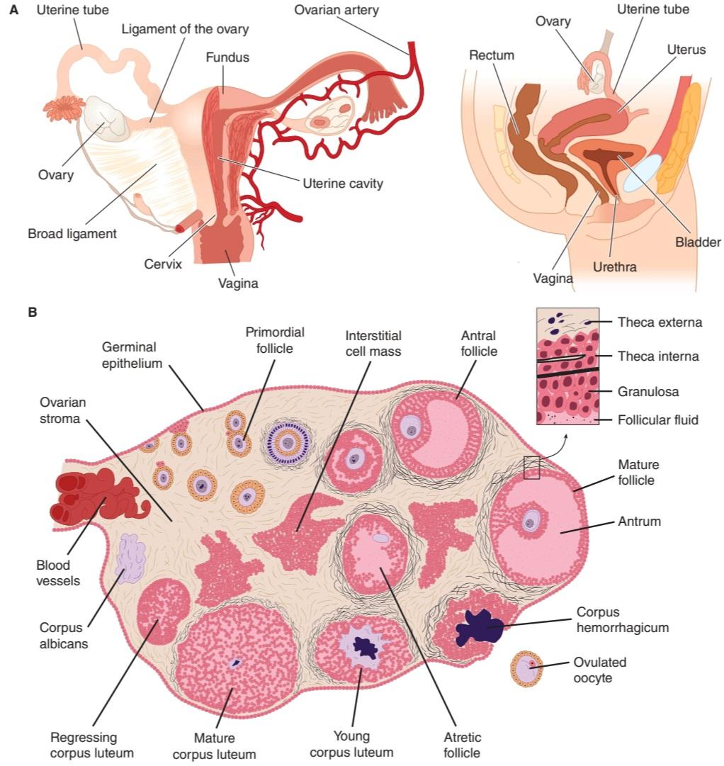
FIGURE 22-10 Functional anatomy of the female reproductive tract. The female reproductive organs include the ovaries, the uterus and the fallopian tubes, and the breast/mammary glands. The sequential development of a follicle, the formation of a corpus luteum and follicular atresia are shown.
شکل 22-10 آناتومی عملکردی دستگاه تناسلی زن. اندامهای تناسلی زنانه شامل تخمدانها، رحم و لولههای فالوپ و غدد پستانی/پستانی است. رشد متوالی یک فولیکول، تشکیل جسم زرد و آترزی فولیکولی نشان داده شده است.
Ovarian Cycle
From the time of birth, there are many primordial follicles under the ovarian capsule. Each contains an immature ovum (Figure 22-10). At the start of each cycle, several of these follicles enlarge, and a cavity forms around the ovum (antrum formation). This cavity is filled with follicular fluid. In humans, usually one of the follicles in one ovary starts to grow rapidly on about the sixth day and becomes the dominant follicle, while the others regress, forming atretic follicles. The atretic process involves apoptosis. It is uncertain how one follicle is selected to be the dominant follicle in this follicular phase of the menstrual cycle, but it seems to be related to the ability of the follicle to secrete the estrogen inside it that is needed for final maturation. When women are given human pituitary gonadotropin preparations by injection, many follicles develop simultaneously.
چرخه تخمدان
از زمان تولد، فولیکول های اولیه زیادی در زیر کپسول تخمدان وجود دارد. هر کدام حاوی یک تخمک نابالغ است (شکل 22-10). در شروع هر چرخه، چندین مورد از این فولیکول ها بزرگ می شوند و حفره ای در اطراف تخمک ایجاد می شود (تشکیل آنتروم). این حفره با مایع فولیکولی پر شده است. در انسان، معمولاً یکی از فولیکولهای یک تخمدان تقریباً در روز ششم شروع به رشد سریع میکند و به فولیکول غالب تبدیل میشود، در حالی که بقیه فولیکولها پسرفت میکنند و فولیکولهای آترتیک را تشکیل میدهند. فرآیند آترتیک شامل آپوپتوز است. مشخص نیست که چگونه یک فولیکول به عنوان فولیکول غالب در این مرحله فولیکولی از چرخه قاعدگی انتخاب می شود، اما به نظر می رسد که به توانایی فولیکول برای ترشح استروژنی که برای بلوغ نهایی لازم است در داخل آن مربوط می شود. هنگامی که به زنان داروهای گنادوتروپین هیپوفیز انسانی با تزریق داده می شود، بسیاری از فولیکول ها به طور همزمان رشد می کنند.
The structure of a maturing ovarian (graafian) follicle is shown in Figure 22– 10. The primary source of circulating estrogen is the granulosa cells of the ovaries; however, the cells of the theca interna of the follicle are necessary for the production of estrogen as they secrete androgens that are aromatized to estrogen by the granulosa cells.
ساختار یک فولیکول تخمدان در حال بلوغ (گرافین) در شکل 10-22 نشان داده شده است. منبع اصلی استروژن در گردش، سلول های گرانولوزای تخمدان ها هستند. با این حال، سلولهای theca interna فولیکول برای تولید استروژن ضروری هستند، زیرا آندروژنهایی ترشح میکنند که توسط سلولهای گرانولوزا به استروژن معطر میشوند.
At about the 14th day of the cycle, the distended follicle ruptures, and the ovum is extruded into the abdominal cavity. This is the process of ovulation. The ovum is picked up by the fimbriated ends of the uterine tubes (oviducts). It is transported to the uterus and, unless fertilization occurs, out through the vagina.
تقریباً در روز چهاردهم چرخه، فولیکول متسع شده پاره می شود و تخمک به داخل حفره شکمی اکسترود می شود. این فرآیند تخمک گذاری است. تخمک توسط انتهای فیمبریایی لوله های رحمی (مجاری تخمک) برداشته می شود. به رحم منتقل می شود و از طریق واژن، مگر اینکه لقاح اتفاق بیفتد.
The follicle that ruptures at the time of ovulation promptly fills with blood, forming what is sometimes called a corpus hemorrhagicum. Minor bleeding from the follicle into the abdominal cavity may cause peritoneal irritation and fleeting lower abdominal pain (“mittelschmerz”). The granulosa and theca cells of the follicle lining promptly begin to proliferate, and the clotted blood is rapidly replaced with yellowish, lipid-rich luteal cells, forming the corpus luteum. This initiates the luteal phase of the menstrual cycle, during which the luteal cells secrete estrogen and progesterone. Growth of the corpus luteum depends on its developing an adequate blood supply, and there is evidence that vascular endothelial growth factor (VEGF) (see Chapter 31) is essential for this process.
فولیکولی که در زمان تخمکگذاری پاره میشود، بلافاصله با خون پر میشود و چیزی را تشکیل میدهد که گاهی به آن کورپوس هموراژیکوم میگویند. خونریزی جزئی از فولیکول به داخل حفره شکمی ممکن است باعث تحریک صفاق و درد زودگذر پایین شکم (“mittelschmerz”) شود. سلول های گرانولوزا و تکا پوشش فولیکول به سرعت شروع به تکثیر می کنند و خون لخته شده به سرعت با سلول های لوتئال زردرنگ و غنی از چربی جایگزین می شود و جسم زرد را تشکیل می دهد. این مرحله لوتئال چرخه قاعدگی را آغاز می کند که طی آن سلول های لوتئال استروژن و پروژسترون ترشح می کنند. رشد جسم زرد به تولید خون کافی بستگی دارد و شواهدی وجود دارد که فاکتور رشد اندوتلیال عروقی (VEGF) (به فصل 31 مراجعه کنید) برای این فرآیند ضروری است.
If pregnancy occurs, the corpus luteum persists and usually there are no more periods until after delivery. If pregnancy does not occur, the corpus luteum begins to degenerate about 4 days before the next menses (24th day of the cycle)and is eventually replaced by scar tissue, forming a corpus albicans.
اگر حاملگی اتفاق بیفتد، جسم زرد باقی می ماند و معمولاً تا بعد از زایمان دیگر پریود نمی شود. اگر حاملگی اتفاق نیفتد، جسم زرد حدود 4 روز قبل از قاعدگی بعدی (روز بیست و چهارم سیکل) شروع به انحطاط می کند و در نهایت با بافت اسکار جایگزین می شود و جسم آلبیکنس را تشکیل می دهد.
The ovarian cycle in other mammals is similar, except that in many species more than one follicle ovulates and multiple births are the rule. Corpora lutea form in some submammalian species but not in others.
چرخه تخمدان در سایر پستانداران مشابه است، با این تفاوت که در بسیاری از گونه ها بیش از یک فولیکول تخمک گذاری می کند و زایمان های متعدد قانون است. Corpora lutea در برخی از گونه های زیر پستانداران تشکیل می شود اما در برخی دیگر نه.
In humans, no new ova are formed after birth. During fetal development, the ovaries contain over 7 million primordial follicles. However, many undergo atresia (involution) before birth and others are lost after birth. At the time of birth, there are 2 million ova, but 50% of these are atretic. The million that are normal undergo the first part of the first meiotic division at about this time and enter a stage of arrest in prophase in which those that survive persist until adulthood. Atresia continues during development, and the number of ova in both of the ovaries at the time of puberty is less than 300,000 (Figure 22–14). Only one of these ova per cycle (or about 500 in the course of a normal reproductive life) normally reaches maturity; the remainder degenerate. Just before ovulation, the first meiotic division is completed. One of the daughter cells, the secondary oocyte, receives most of the cytoplasm, while the other, the first polar body, fragments and disappears. The secondary oocyte immediately begins the second meiotic division, but this division stops at metaphase and is completed only when a sperm penetrates the oocyte. At that time, the second polar body is cast off and the fertilized ovum proceeds to form a new individual. The arrest in metaphase is due, at least in some species, to formation in the ovum of the protein pp39mos, which is encoded by the cmos protooncogene. When fertilization occurs, the pp39mos is destroyed within 30 min by calpain, a calcium-dependent cysteine protease.
در انسان بعد از تولد تخمک جدیدی تشکیل نمی شود. در طول رشد جنین، تخمدان ها حاوی بیش از 7 میلیون فولیکول اولیه هستند. با این حال، بسیاری از آنها قبل از تولد دچار آترزی (انفولشن) می شوند و برخی دیگر پس از تولد از بین می روند. در زمان تولد، 2 میلیون تخمک وجود دارد، اما 50 درصد آنها آترتیک هستند. میلیون نفری که طبیعی هستند تقریباً در این زمان تحت اولین بخش تقسیم میوز قرار می گیرند و وارد مرحله ای از بازداشت در پروفاز می شوند که در آن کسانی که زنده می مانند تا بزرگسالی ادامه می یابند. آترزی در طول تکامل ادامه می یابد و تعداد تخمک ها در هر دو تخمدان در زمان بلوغ کمتر از 300000 است (شکل 22-14). فقط یکی از این تخمک ها در هر چرخه (یا حدود 500 تخمک در طول یک زندگی باروری طبیعی) به طور معمول به بلوغ می رسد. بقیه منحط درست قبل از تخمک گذاری، اولین تقسیم میوز کامل می شود. یکی از سلول های دختر، تخمک ثانویه، بیشتر سیتوپلاسم را دریافت می کند، در حالی که دیگری، اولین جسم قطبی، تکه تکه شده و ناپدید می شود. تخمک ثانویه بلافاصله تقسیم میوز دوم را آغاز می کند، اما این تقسیم در متافاز متوقف می شود و تنها زمانی تکمیل می شود که یک اسپرم به داخل تخمک نفوذ کند. در آن زمان، جسم قطبی دوم بیرون ریخته می شود و تخمک بارور شده برای تشکیل یک فرد جدید ادامه می یابد. توقف در متافاز، حداقل در برخی گونه ها، به دلیل تشکیل پروتئین pp39mos در تخمک است که توسط پروتونکوژن cmos کدگذاری می شود. هنگامی که لقاح اتفاق می افتد، pp39mos در عرض 30 دقیقه توسط کالپین، یک سیستئین پروتئاز وابسته به کلسیم، از بین می رود.
Uterine Cycle
At the end of menstruation, all but the deep layers of the endometrium have sloughed. A new endometrium then regrows under the influence of estrogens from the developing follicle. The endometrium increases rapidly in thickness from the 5th to the 14th days of the menstrual cycle. As the thickness increases, the uterine glands are drawn out so that they lengthen (Figure 22-11), but they do not become convoluted or secrete to any degree. These endometrial changes are called proliferative, and this part of the menstrual cycle is sometimes called the proliferative phase. It is also called the preovulatory or follicular phase of the cycle. After ovulation, the endometrium becomes more highly vascularized and slightly edematous under the influence of estrogen and progesterone from the corpus luteum. The glands become coiled and tortuous and they begin to secrete a clear fluid. Consequently, this phase of the cycle is called the secretory or luteal phase. Late in the luteal phase, the endometrium, like the anterior pituitary, produces prolactin, but the function of this endometrial prolactin is unknown.
چرخه رحم
در پایان قاعدگی، به جز لایه های عمیق آندومتر، همه لایه های آندومتر از بین رفته است. سپس یک آندومتر جدید تحت تأثیر استروژن های فولیکول در حال رشد دوباره رشد می کند. ضخامت آندومتر از روز پنجم تا چهاردهم سیکل قاعدگی به سرعت افزایش می یابد. با افزایش ضخامت، غدد رحم بهگونهای کشیده میشوند که دراز شوند (شکل 22-11)، اما به هیچ درجهای پیچیده نمیشوند و ترشح نمیکنند. این تغییرات آندومتر پرولیفراتیو نامیده می شود و این قسمت از چرخه قاعدگی گاهی اوقات فاز پرولیفراتیو نامیده می شود. به آن مرحله پیش از تخمک گذاری یا فولیکولی چرخه نیز می گویند. پس از تخمک گذاری، آندومتر تحت تأثیر استروژن و پروژسترون از جسم زرد بسیار عروقی و کمی ادماتوز می شود. غدد پیچیده و پرپیچ و خم می شوند و شروع به ترشح مایع شفاف می کنند. در نتیجه، این مرحله از چرخه، فاز ترشحی یا لوتئال نامیده می شود. در اواخر فاز لوتئال، آندومتر، مانند هیپوفیز قدامی، پرولاکتین تولید می کند، اما عملکرد این پرولاکتین آندومتر ناشناخته است.
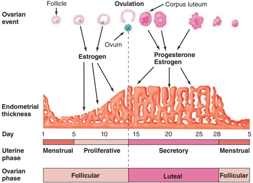
FIGURE 22–11 Relationship between ovarian and uterine changes during the menstrual cycle. (Reproduced with permission from Windmaier EP, Raff H, Strang KT: Vander’s Human Physiology: The Mechanisms of Body Function, 11th ed. New York, NY: McGraw-Hill; 2008.)
شکل 22-11 رابطه بین تغییرات تخمدان و رحم در طول چرخه قاعدگی. (تکثیر شده با اجازه Windmaier EP, Raff H, Strang KT: Vander’s Human Physiology: The Mechanisms of Body Function, ed. 11 New York, NY: McGraw-Hill; 2008.)
The endometrium is supplied by two types of arteries. The superficial two- thirds of the endometrium that is shed during menstruation, the stratum functionale, is supplied by long, coiled spiral arteries, whereas the deep layer that is not shed, the stratum basale, is supplied by short, straight basilar arteries.
آندومتر توسط دو نوع شریان تامین می شود. دو سوم سطحی آندومتر که در طول قاعدگی ریزش میکند، یعنی لایه عملکردی، توسط شریانهای مارپیچی بلند و پیچدار تامین میشود، در حالی که لایه عمیقی که ریزش نمیکند، لایه بازال، توسط شریانهای قاعدهای کوتاه و مستقیم تامین میشود.
When the corpus luteum regresses, hormonal support for the endometrium is withdrawn. The endometrium becomes thinner, which adds to the coiling of the spiral arteries. Foci of necrosis appear in the endometrium, and these coalesce.
هنگامی که جسم زرد پسرفت می کند، حمایت هورمونی از آندومتر برداشته می شود. آندومتر نازکتر میشود که به پیچیدگی شریانهای مارپیچی میافزاید. کانونهای نکروز در آندومتر ظاهر میشوند و اینها با هم ترکیب میشوند.
In addition, spasm and degeneration of the walls of the spiral arteries take place, leading to spotty hemorrhages that become confluent and produce the menstrual flow.
علاوه بر این، اسپاسم و انحطاط دیوارههای شریانهای مارپیچی رخ میدهد که منجر به خونریزیهای لکهای میشود که به هم پیوسته میشوند و جریان قاعدگی را تولید میکنند.
The vasospasm is probably produced by locally released prostaglandins. Large quantities of prostaglandins are present in the secretory endometrium and in menstrual blood, and infusions of prostagladin F2a(PGF2a) produce endometrial necrosis and bleeding.
وازواسپاسم احتمالاً توسط پروستاگلاندین های آزاد شده محلی ایجاد می شود. مقادیر زیادی پروستاگلاندین در آندومتر ترشحی و خون قاعدگی وجود دارد و تزریق پروستاگلادین F2a (PGF2a) باعث نکروز و خونریزی آندومتر می شود.
From the point of view of endometrial function, the proliferative phase of the menstrual cycle represents restoration of the epithelium from the preceding menstruation, and the secretory phase represents preparation of the uterus for implantation of the fertilized ovum. The length of the secretory phase is remarkably constant at about 14 days, and the variations seen in the length of the menstrual cycle are due for the most part to variations in the length of the proliferative phase. When fertilization fails to occur during the secretory phase, the endometrium is shed and a new cycle starts.
از نقطه نظر عملکرد آندومتر، فاز پرولیفراتیو چرخه قاعدگی نشان دهنده بازیابی اپیتلیوم از قاعدگی قبلی است و فاز ترشحی نشان دهنده آماده سازی رحم برای لانه گزینی تخمک بارور شده است. طول فاز ترشحی به طور قابل توجهی در حدود 14 روز ثابت است و تغییرات مشاهده شده در طول چرخه قاعدگی بیشتر به دلیل تغییرات در طول فاز پرولیفراتیو است. هنگامی که لقاح در مرحله ترشحی انجام نمی شود، آندومتر ریخته می شود و یک چرخه جدید شروع می شود.
Normal Menstruation
Menstrual blood is predominantly arterial, with only 25% of the blood being of venous origin. It contains tissue debris, prostaglandins, and relatively large amounts of fibrinolysin from endometrial tissue. The fibrinolysin lyses clots, so that menstrual blood does not normally contain clots unless the flow is excessive.
قاعدگی طبیعی
خون قاعدگی عمدتاً شریانی است و تنها 25 درصد خون منشا وریدی دارد. حاوی بقایای بافتی، پروستاگلاندین ها و مقادیر نسبتاً زیادی فیبرینولیزین از بافت آندومتر است. فیبرینولیزین لخته ها را لیز می کند، به طوری که خون قاعدگی به طور معمول حاوی لخته نیست، مگر اینکه جریان بیش از حد باشد.
The usual duration of the menstrual flow is 3-5 days, but flows as short as 1 day and as long as 8 days can occur in normal women. The amount of blood lost may range normally from slight spotting to 80 mL; the average amount lost is 30 mL. Loss of more than 80 mL is abnormal. Obviously, the amount of flow can be affected by various factors, including the thickness of the endometrium, medication, and diseases that affect the clotting mechanism.
مدت زمان معمول قاعدگی 3 تا 5 روز است، اما این جریان به کوتاهی 1 روز و تا 8 روز می تواند در زنان عادی رخ دهد. مقدار خون از دست رفته ممکن است به طور معمول از لکه بینی خفیف تا 80 میلی لیتر متغیر باشد. مقدار متوسط از دست رفته 30 میلی لیتر است. از دست دادن بیش از 80 میلی لیتر غیر طبیعی است. بدیهی است که میزان جریان می تواند تحت تاثیر عوامل مختلفی از جمله ضخامت اندومتر، داروها و بیماری هایی که بر مکانیسم لخته شدن اثر می گذارند، باشد.
Anovulatory Cycles
In some instances, ovulation fails to occur during the menstrual cycle. Such anovulatory cycles are common for the first 12-18 months after menarche and again before the onset of the menopause. When ovulation does not occur, no corpus luteum is formed and the effects of progesterone on the endometrium are absent. Estrogens continue to cause growth, however, and the proliferative endometrium becomes thick enough to break down and begins to slough. The time it takes for bleeding to occur is variable, but it usually occurs in less than 28 days from the last menstrual period. The flow is also variable and ranges from scanty to relatively profuse.
چرخه های عدم تخمک گذاری
در برخی موارد، تخمک گذاری در طول چرخه قاعدگی انجام نمی شود. چنین چرخه های عدم تخمک گذاری برای 12-18 ماه اول پس از قاعدگی و دوباره قبل از شروع یائسگی رایج است. هنگامی که تخمک گذاری اتفاق نمی افتد، جسم زرد تشکیل نمی شود و اثرات پروژسترون بر روی آندومتر وجود ندارد. با این حال، استروژن ها به رشد خود ادامه می دهند و آندومتر تکثیر شده به اندازه کافی ضخیم می شود که تجزیه شود و شروع به ریزش می کند. مدت زمانی که طول می کشد تا خونریزی رخ دهد متغیر است، اما معمولا در کمتر از 28 روز از آخرین دوره قاعدگی رخ می دهد. جریان نیز متغیر است و از اندک تا نسبتاً زیاد متغیر است.
Cyclical Changes in the Uterine Cervix
Although it is continuous with the body of the uterus, the cervix of the uterus is different in a number of ways. The mucosa of the uterine cervix does not undergo cyclical desquamation, but there are regular changes in the cervical mucus. Estrogen makes the mucus thinner and more alkaline, changes that promote the survival and transport of sperm. Progesterone makes it thick, tenacious, and cellular. The mucus is thinnest at the time of ovulation, and its elasticity, or spinnbarkeit, increases so that by midcycle, a drop can be stretched into a long, thin thread that may be 8-12 cm or more in length. In addition, it dries in an arborizing, fern-like pattern (Figure 22–12) when a thin layer is spread on a slide. After ovulation and during pregnancy, it becomes thick and fails to form the fern pattern.
تغییرات چرخه ای در دهانه رحم
اگرچه با بدنه رحم پیوسته است، اما دهانه رحم از جهات مختلفی متفاوت است. مخاط دهانه رحم تحت پوسته پوسته شدن چرخه ای قرار نمی گیرد، اما تغییرات منظمی در مخاط دهانه رحم وجود دارد. استروژن مخاط را نازک تر و قلیایی تر می کند، تغییراتی که باعث بقا و انتقال اسپرم می شود. پروژسترون آن را غلیظ، مقاوم و سلولی می کند. مخاط در زمان تخمکگذاری نازکترین است و خاصیت ارتجاعی آن یا spinnbarkeit افزایش مییابد به طوری که در اواسط چرخه، میتوان قطرهای را به صورت نخ بلند و نازکی که ممکن است 8 تا 12 سانتیمتر یا بیشتر طول داشته باشد کشیده شود. علاوه بر این، هنگامی که یک لایه نازک روی یک اسلاید پخش می شود، به شکل درختکاری و سرخس مانند خشک می شود (شکل 22-12). پس از تخمک گذاری و در دوران بارداری، غلیظ می شود و الگوی سرخس را نمی سازد.

FIGURE 22-12 Patterns formed when cervical mucus is smeared on a slide, permitted to dry, and examined under the microscope. Progesterone makes the mucus thick and cellular. In the smear from a patient who did not ovulate (bottom), no progesterone is present to inhibit the estrogen-induced fern pattern.
شکل ۲۲-۱۲ الگوهایی که وقتی مخاط دهانه رحم روی اسلاید آغشته میشود، خشک میشود و زیر میکروسکوپ بررسی میشود، شکل میگیرند. پروژسترون مخاط را ضخیم و سلولی میکند. در اسمیر گرفته شده از بیماری که تخمکگذاری نداشته است (پایین)، هیچ پروژسترونی برای مهار الگوی سرخسی ناشی از استروژن وجود ندارد.
Vaginal Cycle
Under the influence of estrogens, the vaginal epithelium becomes cornified, and cornified epithelial cells can be identified in the vaginal smear. Under the influence of progesterone, a thick mucus is secreted, and the epithelium proliferates and becomes infiltrated with leukocytes. The cyclical changes in the vaginal smear in rats are relatively marked. The changes in humans and other species are similar but not so clear-cut.
چرخه واژن
تحت تأثیر استروژن ها، اپیتلیوم واژن به شکل قرنیه در می آید و سلول های اپیتلیال کورنیفیه شده را می توان در اسمیر واژن شناسایی کرد. تحت تأثیر پروژسترون، مخاط غلیظی ترشح می شود و اپیتلیوم تکثیر می شود و با لکوسیت ها نفوذ می کند. تغییرات چرخهای در اسمیر واژینال در موشها نسبتاً مشخص است. تغییرات در انسان و سایر گونه ها مشابه است اما چندان واضح نیست.
Cyclical Changes in the Breasts
Although lactation normally does not occur until the end of pregnancy, cyclical changes take place in the breasts during the menstrual cycle. Estrogens cause proliferation of mammary ducts, whereas progesterone causes growth of lobules and alveoli. The breast swelling, tenderness, and pain experienced by many women during the 10 days preceding menstruation are probably due to distension of the ducts, hyperemia, and edema of the interstitial tissue of the breast. All these changes regress, along with the symptoms, during menstruation.
تغییرات چرخه ای در سینه ها
اگرچه شیردهی به طور معمول تا پایان بارداری رخ نمی دهد، تغییرات چرخه ای در سینه ها در طول چرخه قاعدگی رخ می دهد. استروژن ها باعث تکثیر مجاری پستانی می شوند، در حالی که پروژسترون باعث رشد لوبول ها و آلوئول ها می شود. تورم، حساسیت و درد سینهها که در طول 10 روز قبل از قاعدگی توسط بسیاری از زنان تجربه میشود، احتمالاً به دلیل اتساع مجاری، پرخونی و ادم بافت بینابینی پستان است. همه این تغییرات همراه با علائم در طول قاعدگی پسرفت می کنند.
Changes During Intercourse
During sexual excitement in women, fluid is secreted onto the vaginal walls, probably because of release of vasoactive intestinal polypeptide (VIP) from vaginal nerves. A lubricating mucus is also secreted by the vestibular glands. The upper part of the vagina is sensitive to stretch, while tactile stimulation from the labia minora and clitoris adds to the sexual excitement. These stimuli are reinforced by tactile stimuli from the breasts and, as in men, by visual, auditory, and olfactory stimuli, which may build to the crescendo known as orgasm. During orgasm, autonomically mediated rhythmic contractions occur in the vaginal walls. Impulses also travel via the pudendal nerves and produce rhythmic contraction of the bulbocavernosus and ischiocavernosus muscles. The vaginal contractions may aid sperm transport but are not essential for it, since fertilization of the ovum is not dependent on female orgasm.
تغییرات در طول رابطه جنسی
در طول هیجان جنسی در زنان، احتمالاً به دلیل آزاد شدن پلی پپتید وازواکتیو روده ای (VIP) از اعصاب واژن، مایع روی دیواره های واژن ترشح می شود. یک مخاط روان کننده نیز توسط غدد دهلیزی ترشح می شود. قسمت بالایی واژن به کشش حساس است، در حالی که تحریک لمسی از لابیا مینور و کلیتوریس به هیجان جنسی می افزاید. این محرکها توسط محرکهای لمسی سینهها و مانند مردان با محرکهای بینایی، شنیداری و بویایی تقویت میشوند که ممکن است به اوج برسد که به ارگاسم معروف است. در طول ارگاسم، انقباضات ریتمیک با واسطه خودمختار در دیواره های واژن رخ می دهد. ایمپالس ها همچنین از طریق اعصاب پودندال حرکت می کنند و باعث انقباض ریتمیک ماهیچه های bulbocavernosus و ischiocavernosus می شوند. انقباضات واژن ممکن است به انتقال اسپرم کمک کند، اما برای آن ضروری نیست، زیرا لقاح تخمک به ارگاسم زن بستگی ندارد.
Indicators of Ovulation
Knowing when during the menstrual cycle ovulation occurs is important in increasing fertility or, conversely, in family planning. A convenient and reasonably reliable indicator of the time of ovulation is a change usually a rise -in the basal body temperature (Figure 22-13). The rise starts 1-2 days after ovulation. Women interested in obtaining an accurate temperature chart should use a digital thermometer and take their temperatures (oral or rectal) in the morning before getting out of bed. The cause of the temperature change at the time of ovulation is probably the increase in progesterone secretion, since progesterone is thermogenic.
شاخص های تخمک گذاری
دانستن زمان تخمک گذاری در چرخه قاعدگی در افزایش باروری یا برعکس در تنظیم خانواده مهم است. یک شاخص مناسب و قابل اعتماد برای زمان تخمک گذاری، تغییر معمولاً افزایش در دمای پایه بدن است (شکل 22-13). افزایش 1-2 روز پس از تخمک گذاری شروع می شود. خانمهایی که علاقهمند به تهیه نمودار دقیق دما هستند، باید از دماسنج دیجیتالی استفاده کنند و صبحها قبل از بلند شدن از رختخواب، دمای خود را (دهانی یا مقعدی) اندازهگیری کنند. علت تغییر دما در زمان تخمک گذاری احتمالاً افزایش ترشح پروژسترون است، زیرا پروژسترون ترموژنیک است.

FIGURE 22-13 Basal body temperature and plasma hormone concentrations (mean ± standard error) during the normal human menstrual cycle. Values are aligned with respect to the day of the midcycle LH peak. FSH, follicle-stimulating hormone; LH, luteinizing hormone; M, menses.
شکل 22-13 دمای پایه بدن و غلظت هورمون پلاسما (میانگین ± خطای استاندارد) در طول چرخه قاعدگی طبیعی انسان. مقادیر با توجه به روز اوج LH در چرخه میانی تراز هستند. FSH، هورمون محرک فولیکول؛ LH، هورمون لوتئینه کننده؛ م، قاعدگی
A surge in LH secretion triggers ovulation, and ovulation normally occurs about 9 h after the peak of the LH surge at midcycle (Figure 22–13). The ovum lives for approximately 72 h after it is extruded from the follicle, but it is fertilizable for a much shorter time than this. In a study of the relation of isolated intercourse to pregnancy, 36% of women had a detected pregnancy following intercourse on the day of ovulation, but with intercourse on days after ovulation, the percentage was zero. Isolated intercourse on the first and second day before ovulation also led to pregnancy in about 36% of women. A few pregnancies resulted from isolated intercourse on day 3, 4, or 5 before ovulation, although the percentage was much lower, for example, 8% on day 5 before ovulation. Thus, some sperms can survive in the female genital tract and fertilize the ovum for up to 120 h before ovulation, but the most fertile period is clearly the 48 h before ovulation. However, for those interested in the “rhythm method” of contraception, it should be noted that there are rare but documented cases in the literature of pregnancy resulting from isolated coitus on every day of the cycle.
افزایش ترشح LH باعث تخمک گذاری می شود و تخمک گذاری معمولاً حدود 9 ساعت پس از اوج افزایش LH در اواسط چرخه اتفاق می افتد (شکل 22-13). تخمک پس از اکسترود شدن از فولیکول تقریباً 72 ساعت زندگی می کند، اما برای مدت زمان بسیار کمتری قابل بارور شدن است. در مطالعه ای که در مورد ارتباط رابطه جنسی ایزوله با بارداری انجام شد، 36 درصد از زنان به دنبال آمیزش در روز تخمک گذاری حاملگی تشخیص داده بودند، اما با آمیزش در روزهای بعد از تخمک گذاری، این درصد صفر بود. مقاربت جدا شده در روز اول و دوم قبل از تخمک گذاری نیز در حدود 36 درصد از زنان منجر به بارداری شد. تعداد کمی از بارداریها به دلیل مقاربت منفرد در روز 3، 4 یا 5 قبل از تخمکگذاری به وجود آمدند، اگرچه این درصد بسیار کمتر بود، برای مثال، 8٪ در روز 5 قبل از تخمکگذاری. بنابراین، برخی از اسپرم ها می توانند در دستگاه تناسلی زن زنده بمانند و تخمک را تا 120 ساعت قبل از تخمک گذاری بارور کنند، اما بارورترین دوره به وضوح 48 ساعت قبل از تخمک گذاری است. با این حال، برای کسانی که به “روش ریتم” پیشگیری از بارداری علاقه مند هستند، باید توجه داشت که موارد نادر اما مستندی در ادبیات بارداری وجود دارد که ناشی از کویتیشن ایزوله در هر روز از سیکل است.
The Estrous Cycle
Mammals other than primates do not menstruate, and their sexual cycle is called an estrous cycle. It is named for the conspicuous period of “heat” (estrus) at the time of ovulation, normally the only time during which the sexual interest of the female is aroused. In spontaneously ovulating species with estrous cycles, such as the rat, no episodic vaginal bleeding occurs but the underlying endocrine events are essentially the same as those in the menstrual cycle. In other species, ovulation is induced by copulation (reflex ovulation).
چرخه فحلی
پستانداران غیر از پستانداران پریود نمی شوند و چرخه جنسی آنها را چرخه فحلی می نامند. این نام برای دوره آشکار “گرما” (فحلی) در زمان تخمک گذاری است، که معمولاً تنها زمانی است که در طی آن علاقه جنسی زن برانگیخته می شود. در گونه هایی که به طور خود به خود تخمک گذاری می کنند و دارای چرخه فحلی هستند، مانند موش صحرایی، خونریزی اپیزودیک واژینال اتفاق نمی افتد، اما رویدادهای غدد درون ریز اساساً مشابه رویدادهای چرخه قاعدگی است. در گونه های دیگر، تخمک گذاری با جفت شدن (تخمک گذاری رفلکس) القا می شود.
MENOPAUSE
The human ovaries become unresponsive to gonadotropins with advancing age, and their function declines, so that sexual cycles disappear (menopause). This unresponsiveness is associated with and probably caused by a decline in the number of primordial follicles, which becomes precipitous at the time of menopause (Figure 22–14). The ovaries no longer secrete progesterone and 17ẞ- estradiol in appreciable quantities, and estrogen is formed only in small amounts by aromatization of androstenedione in peripheral tissues (see Chapter 20). The uterus and the vagina gradually become atrophic. As the negative feedback effect of estrogens and progesterone is reduced, secretion of FSH is increased, and plasma FSH increases to high levels, LH levels are moderately high. Old female mice and rats have long periods of diestrus and increased levels of gonadotropin secretion. In women, a period called perimenopause precedes menopause and can last up to 10 years. During perimenopause FSH levels will increase before an increase in LH is observed due to a decrease in estrogen, progesterone, and inhibins and the menses become irregular. This usually occurs between the ages of 45 and 55. The average age at onset of the menopause is 51 years.
یائسگی
تخمدان های انسان با افزایش سن به گنادوتروپین ها پاسخ نمی دهند و عملکرد آنها کاهش می یابد، به طوری که چرخه های جنسی ناپدید می شوند (یائسگی). این عدم پاسخگویی با کاهش تعداد فولیکولهای اولیه که در زمان یائسگی شدید میشود، مرتبط است و احتمالاً ناشی از آن است (شکل 22-14). تخمدانها دیگر پروژسترون و 17-استرادیول را در مقادیر قابلتوجهی ترشح نمیکنند، و استروژن تنها در مقادیر کم با معطر شدن آندروستندیون در بافتهای محیطی تشکیل میشود (به فصل 20 مراجعه کنید). رحم و واژن به تدریج آتروفیک می شوند. با کاهش اثر بازخورد منفی استروژن ها و پروژسترون، ترشح FSH افزایش می یابد و FSH پلاسما به سطوح بالا افزایش می یابد، سطح LH نسبتاً بالا است. موشهای ماده پیر و موشهای صحرایی دارای دورههای طولانی دی استروس و افزایش ترشح گنادوتروپین هستند. در زنان، دوره ای به نام پیش یائسگی قبل از یائسگی است و می تواند تا 10 سال طول بکشد. در دوران قبل از یائسگی، سطح FSH قبل از مشاهده افزایش LH به دلیل کاهش استروژن، پروژسترون و اینهیبین ها افزایش می یابد و قاعدگی نامنظم می شود. این معمولا در سنین 45 تا 55 سالگی رخ می دهد. میانگین سن شروع یائسگی 51 سال است.

FIGURE 22-14 Number of primordial follicles per ovary in women at various ages. Blue squares, premenopausal women (regular menses); red squares, perimenopausal women (irregular menses for at least 1 year); red triangles, postmenopausal women (no menses for at least 1 year). Note that the vertical scale is a log scale and that the values are from one rather than two ovaries. (Redrawn by PM Wise and reproduced with permission from Richardson SJ, Senikas V, Nelson JF: Follicular depletion during the menopausal transition: Evidence for accelerated loss and ultimate exhaustion. J Clin Endocrinol Metab 1987;65:1231.)
شکل 22-14 تعداد فولیکول های اولیه در هر تخمدان در زنان در سنین مختلف. مربع های آبی، زنان قبل از یائسگی (قاعدگی منظم)؛ مربع قرمز، زنان یائسه (قاعدگی نامنظم برای حداقل 1 سال)؛ مثلث قرمز، زنان یائسه (حداقل 1 سال بدون قاعدگی). توجه داشته باشید که مقیاس عمودی یک مقیاس ورود به سیستم است و مقادیر از یک تخمدان است نه دو تخمدان. (بازطراحی شده توسط PM Wise و تکثیر با اجازه ریچاردسون SJ، Senikas V، Nelson JF: تخلیه فولیکولی در طول انتقال یائسگی: شواهدی برای از دست دادن سریع و خستگی نهایی. J Clin Endocrinol Metab 1987؛ 65:1231.)
The loss of ovarian function causes many symptoms such as sensations of warmth spreading from the trunk to the face (hot flushes; also called hot flashes) and night sweats. In addition, the onset of menopause increases the risk of many diseases such as osteoporosis, ischemic heart disease, and renal disease.
از دست دادن عملکرد تخمدان باعث علائم بسیاری مانند انتشار احساس گرما از تنه به صورت (گرگرفتگی که گرگرفتگی نیز نامیده می شود) و تعریق شبانه می شود. علاوه بر این، شروع یائسگی خطر ابتلا به بسیاری از بیماری ها مانند پوکی استخوان، بیماری ایسکمیک قلبی و بیماری کلیوی را افزایش می دهد.
Hot flushes are said to occur in 75% of menopausal women and may continue intermittently for as long as 40 years. They also occur when early menopause is produced by bilateral ovariectomy, and they are prevented by estrogen treatment. In addition, they occur after castration in men. Their cause is unknown. However, they coincide with surges of LH secretion. LH is secreted in episodic bursts at intervals of 30-60 min or more (circhoral secretion), and in the absence of gonadal hormones these bursts are large. Each hot flush begins with the start of a burst. However, LH itself is not responsible for the symptoms, because they can continue after removal of the pituitary. Instead, it appears that some estrogen-sensitive event in the hypothalamus initiates both the release of LH and the episode of flushing.
گفته می شود که گرگرفتگی در 75 درصد از زنان یائسه رخ می دهد و ممکن است به طور متناوب تا 40 سال ادامه یابد. آنها همچنین زمانی رخ می دهند که یائسگی زودرس توسط تخمدان دو طرفه ایجاد می شود و با درمان استروژن از آنها جلوگیری می شود. علاوه بر این، پس از اخته شدن در مردان رخ می دهد. علت آنها ناشناخته است. با این حال، آنها با افزایش ترشح LH همزمان هستند. LH در انفجارهای اپیزودیک در فواصل 30-60 دقیقه یا بیشتر ترشح می شود (ترشح سیرکورال) و در غیاب هورمون های غدد جنسی این انفجارها بزرگ هستند. هر گرگرفتگی با شروع یک انفجار شروع می شود. با این حال، LH خود مسئول علائم نیست، زیرا ممکن است پس از برداشتن هیپوفیز ادامه یابد. درعوض، به نظر می رسد که برخی از رویدادهای حساس به استروژن در هیپوتالاموس هم باعث آزاد شدن LH و هم دوره گرگرفتگی می شود.
Although the function of the testes tends to decline slowly with advancing age, the evidence is unclear whether there is a “male menopause” (andropause) similar to that occurring in women.
اگرچه عملکرد بیضه ها با افزایش سن به آرامی کاهش می یابد، شواهد مشخص نیست که آیا “یائسگی مردانه” (آندروپوز) مشابه آنچه در زنان اتفاق می افتد وجود دارد یا خیر.
OVARIAN HORMONES
Chemistry, Biosynthesis, & Metabolism of Estrogens
The naturally occurring estrogens are 17ẞ-estradiol, estrone, and estriol (Figure 22-15). They are C18 steroids that do not have an angular methyl group attached to the 10 position or a A4-3-keto configuration in the A ring. They are secreted primarily by the granulosa cells of the ovarian follicles, the corpus luteum, and the placenta. Their biosynthesis depends on the enzyme aromatase (CYP19), which converts testosterone to estradiol and androstenedione to estrone (Figure 22–15). The latter reaction also occurs in fat, liver, muscle, and the brain.
هورمون های تخمدان
شیمی، بیوسنتز، و متابولیسم استروژن
استروژن های طبیعی عبارتند از 17-استرادیول، استرون و استریول (شکل 22-15). آنها استروئیدهای C18 هستند که یک گروه متیل زاویه ای متصل به موقعیت 10 یا یک پیکربندی A4-3-keto در حلقه A ندارند. آنها عمدتاً توسط سلول های گرانولوزای فولیکول های تخمدان، جسم زرد و جفت ترشح می شوند. بیوسنتز آنها به آنزیم آروماتاز (CYP19) بستگی دارد که تستوسترون را به استرادیول و آندروستندیون را به استرون تبدیل می کند (شکل 22-15). واکنش دوم در چربی، کبد، ماهیچه و مغز نیز رخ می دهد.
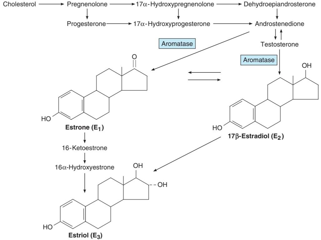
FIGURE 22-15 Biosynthesis and metabolism of estrogens. The formulas of the precursor steroids are shown in Figure 19-7.
شکل 22-15 بیوسنتز و متابولیسم استروژن. فرمول استروئیدهای پیش ساز در شکل 19-7 نشان داده شده است.
Theca interna cells have many LH receptors, and LH acts via cAMP to increase conversion of cholesterol to androstenedione. The theca interna cells supply androstenedione to the granulosa cells. The granulosa cells make estradiol when provided with androgens (Figure 22-16), and it appears that the estradiol they form in primates is secreted into the follicular fluid. Granulosa cells have many FSH receptors, and FSH facilitates their secretion of estradiol by acting via cAMP to increase their aromatase activity. Mature granulosa cells also acquire LH receptors, and LH also stimulates estradiol production.
سلول های Theca interna گیرنده های LH زیادی دارند و LH از طریق cAMP برای افزایش تبدیل کلسترول به آندروستندیون عمل می کند. سلول های theca interna آندروستندیون را به سلول های گرانولوزا می رسانند. سلول های گرانولوزا زمانی استرادیول می سازند که با آندروژن تامین شود (شکل 22-16)، و به نظر می رسد که استرادیولی که در پستانداران تشکیل می دهند در مایع فولیکولی ترشح می شود. سلول های گرانولوزا گیرنده های FSH زیادی دارند و FSH ترشح استرادیول آنها را با عمل از طریق cAMP برای افزایش فعالیت آروماتاز آنها تسهیل می کند. سلول های گرانولوزای بالغ نیز گیرنده های LH را بدست می آورند و LH نیز تولید استرادیول را تحریک می کند.

FIGURE 22-16 Interactions between theca and granulosa cells in estradiol synthesis and secretion.
شکل 22-16 تعاملات بین سلولهای تکا و گرانولوزا در سنتز و ترشح استرادیول.
Two percent of the circulating estradiol is free, and the remainder is bound to protein: 60% to albumin and 38% to the same gonadal steroid-binding globulin (GBG) that binds testosterone.
دو درصد از استرادیول در گردش آزاد است و باقیمانده به پروتئین متصل است: 60 درصد به آلبومین و 38 درصد به همان گلوبولین اتصال دهنده استروئیدی غدد جنسی (GBG) که به تستوسترون متصل می شود.
In the liver, estradiol, estrone, and estriol are converted to glucuronide and sulfate conjugates. All these compounds, along with other metabolites, are excreted in the urine. Appreciable amounts are secreted in the bile and reabsorbed into the bloodstream (enterohepatic circulation).
در کبد، استرادیول، استرون و استریول به ترکیبات گلوکورونید و سولفات تبدیل می شوند. تمام این ترکیبات همراه با سایر متابولیت ها از طریق ادرار دفع می شوند. مقادیر قابل توجهی در صفرا ترشح می شود و دوباره جذب جریان خون می شود (گردش کبدی).
Secretion
The concentration of estradiol in the plasma during the menstrual cycle is shown in Figure 22–14. Almost all of this estrogen comes from the ovary, and two peaks of secretion occur: one just before ovulation and one during the midluteal phase. The estradiol secretion rate is 36 μg/day (133 nmol/day) in the early follicular phase, 380 μg/day just before ovulation, and 250 μg/day during the midluteal phase (Table 22-3). After menopause, estrogen secretion declines to low levels.
ترشح
غلظت استرادیول در پلاسما در طول چرخه قاعدگی در شکل 22-14 نشان داده شده است. تقریباً تمام این استروژن از تخمدان می آید و دو اوج ترشح رخ می دهد: یکی درست قبل از تخمک گذاری و دیگری در مرحله میانی لوتئال. میزان ترشح استرادیول 36 میکروگرم در روز (133 نانومول در روز) در فاز فولیکولی اولیه، 380 میکروگرم در روز درست قبل از تخمک گذاری و 250 میکروگرم در روز در طول فاز میانی لوتئال است (جدول 22-3). پس از یائسگی، ترشح استروژن به سطوح پایین کاهش می یابد.
TABLE 22-3 Twenty-four-hour production rates of sex steroids in women at different stages of the menstrual cycle.
جدول 22-3 میزان تولید 24 ساعته استروئیدهای جنسی در زنان در مراحل مختلف چرخه قاعدگی.

Data from Baird DT, Fraser IS: Blood production and ovarian secretion rates of estradiol-17 beta and estrone in women throughout the menstrual cycle. J Clin Endocrinal Metab 1974 Jun; 38(6):1009-1017.
داده های Baird DT، Fraser IS: تولید خون و میزان ترشح تخمدانی استرادیول-17 بتا و استرون در زنان در طول چرخه قاعدگی. J Clin غدد درون ریز متاب 1974 ژوئن; 38 (6): 1009-1017.
As noted previously, the estradiol production rate in men is about 50 μg/day (184 nmol/day).
همانطور که قبلا ذکر شد، میزان تولید استرادیول در مردان حدود 50 میکروگرم در روز (184 نانومول در روز) است.
Effects on the Female Genitalia
Estrogens facilitate the growth of the ovarian follicles and increase the motility of the uterine tubes. Their role in the cyclic changes in the endometrium, cervix, and vagina has been discussed previously. They increase uterine blood flow and have important effects on the smooth muscle of the uterus. In immature and castrated females, the uterus is small and the myometrium atrophic and inactive. Estrogens increase the amount of uterine muscle and its content of contractile proteins. Under the influence of estrogens, the muscle becomes more active and excitable, and action potentials in the individual fibers become more frequent. The “estrogen-dominated” uterus is also more sensitive to oxytocin.
اثرات بر دستگاه تناسلی زنان
استروژن ها رشد فولیکول های تخمدان را تسهیل کرده و تحرک لوله های رحمی را افزایش می دهند. نقش آنها در تغییرات چرخه ای در آندومتر، دهانه رحم و واژن قبلاً مورد بحث قرار گرفته است. آنها جریان خون رحم را افزایش می دهند و اثرات مهمی روی ماهیچه صاف رحم دارند. در زنان نابالغ و اخته، رحم کوچک و میومتر آتروفیک و غیر فعال است. استروژن ها میزان ماهیچه های رحم و محتوای پروتئین های انقباضی آن را افزایش می دهند. تحت تأثیر استروژنها، ماهیچهها فعالتر و تحریکپذیرتر میشوند و پتانسیلهای عمل در فیبرهای فردی بیشتر میشوند. رحم تحت تسلط استروژن نیز به اکسی توسین حساس تر است.
Chronic treatment with estrogens causes the endometrium to hypertrophy. When estrogen therapy is discontinued, sloughing takes place with withdrawal bleeding. Some “breakthrough” bleeding may occur during treatment when estrogens are given for long periods. Prolonged exposure to estrogen alone (unopposed by progesterone) has been indicated as a risk factor in the development of endometrial cancer.
درمان مزمن با استروژن باعث هیپرتروفی آندومتر می شود. هنگامی که درمان با استروژن قطع می شود، تخلیه با خونریزی قطع می شود. هنگامی که استروژن برای دوره های طولانی تجویز می شود، ممکن است مقداری خونریزی “سریع” در طول درمان رخ دهد. قرار گرفتن طولانی مدت در معرض استروژن به تنهایی (بدون مخالفت با پروژسترون) به عنوان یک عامل خطر در ایجاد سرطان آندومتر نشان داده شده است.
Effects on Endocrine Organs
Estrogens decrease FSH secretion. Under some circumstances, they inhibit LH secretion (negative feedback); in other circumstances, they increase LH secretion (positive feedback). Women are sometimes given large doses of estrogens for 4- 6 days to prevent conception after coitus during the fertile period (postcoital or “morning-after” contraception). However, in this instance, pregnancy is probably prevented by interference with implantation of the ovum rather than changes in gonadotropin secretion.
اثرات بر اندام های غدد درون ریز
استروژن باعث کاهش ترشح FSH می شود. تحت برخی شرایط، آنها ترشح LH را مهار می کنند (بازخورد منفی). در شرایط دیگر، ترشح LH را افزایش می دهند (بازخورد مثبت). گاهی اوقات به زنان دوزهای زیادی از استروژن به مدت 4 تا 6 روز داده می شود تا از بارداری پس از مقاربت در طول دوره باروری جلوگیری شود (پیشگیری از بارداری یا صبح و بعد از بارداری). با این حال، در این مورد، احتمالاً با تداخل در لانه گزینی تخمک به جای تغییر در ترشح گنادوتروپین، از بارداری جلوگیری می شود.
Estrogens cause increased secretion of angiotensinogen and thyroid-binding globulin. They exert an important protein anabolic effect in chickens and cattle, possibly by stimulating the secretion of androgens from the adrenal, and estrogen treatment has been used commercially to increase the weight of domestic animals. They cause epiphysial closure in humans (see Chapter 21).
استروژن باعث افزایش ترشح آنژیوتانسینوژن و گلوبولین متصل به تیروئید می شود. آنها یک اثر آنابولیک پروتئین مهم در جوجه ها و گاوها، احتمالا با تحریک ترشح آندروژن از غده فوق کلیوی، اعمال می کنند، و درمان استروژن به صورت تجاری برای افزایش وزن حیوانات اهلی استفاده می شود. آنها باعث بسته شدن اپی فیزیال در انسان می شوند (به فصل 21 مراجعه کنید).
Effects on the Central Nervous System
The estrogens are responsible for estrous behavior in animals, and they increase libido in humans. They apparently exert this action by a direct effect on certain neurons in the hypothalamus. Estrogens also increase the proliferation of dendrites on neurons and the number of synaptic knobs in rats.
اثرات بر روی سیستم عصبی مرکزی
استروژن ها مسئول رفتار فحلی در حیوانات هستند و میل جنسی را در انسان افزایش می دهند. آنها ظاهراً این عمل را با تأثیر مستقیم بر نورون های خاصی در هیپوتالاموس انجام می دهند. استروژن ها همچنین تکثیر دندریت ها روی نورون ها و تعداد دستگیره های سیناپسی را در موش ها افزایش می دهند.
Effects on the Breasts
Estrogens produce duct growth in the breasts and are largely responsible for breast enlargement at puberty in girls; they have been called the growth hormones of the breast. They are responsible for the pigmentation of the areolas, although pigmentation usually becomes more intense during the first pregnancy than it does at puberty. The role of estrogens in the overall control of breast growth and lactation is discussed below.
اثرات بر روی سینه ها
استروژن ها باعث رشد مجرای سینه ها می شوند و تا حد زیادی مسئول بزرگ شدن سینه در دوران بلوغ در دختران هستند. آنها را هورمون رشد سینه می نامند. آنها مسئول رنگدانه شدن آرئول ها هستند، اگرچه رنگدانه ها معمولاً در بارداری اول شدیدتر از دوران بلوغ می شوند. نقش استروژن ها در کنترل کلی رشد سینه و شیردهی در زیر مورد بحث قرار گرفته است.
Female Secondary Sex Characteristics
The body changes that develop in girls at puberty—in addition to enlargement of breasts, uterus, and vagina—are due in part to estrogens, which are the “feminizing hormones,” and in part simply to the absence of testicular androgens. Women have narrow shoulders and broad hips, thighs that converge, and arms that diverge (wide carrying angle). This body configuration, plus the female distribution of fat in the breasts and buttocks, is seen also in castrate males. In women, the larynx retains its prepubertal proportions and the voice stays high-pitched. Women have less body hair and more scalp hair, and the pubic hair generally has a characteristic flat-topped pattern (female escutcheon). However, growth of pubic and axillary hair in both sexes is due primarily to androgens rather than estrogens.
ویژگی های جنسی ثانویه زن
تغییرات بدنی که در دختران در سن بلوغ ایجاد میشود -علاوه بر بزرگ شدن سینهها، رحم و واژن- تا حدی به دلیل استروژنها است که “هورمونهای زنانهساز” هستند و تا حدی به دلیل عدم وجود آندروژنهای بیضه است. زنان شانههای باریک و باسن پهن، رانهایی که به هم نزدیک میشوند و بازوهایی از هم جدا میشوند (زاویه حمل گسترده). این پیکربندی بدن، به علاوه توزیع چربی زنانه در سینه ها و باسن، در مردان اخته نیز دیده می شود. در زنان، حنجره نسبت های قبل از بلوغ خود را حفظ می کند و صدا بلند می ماند. زنان موهای بدن کمتر و موهای سر بیشتری دارند، و موهای ناحیه تناسلی به طور کلی دارای یک الگوی مشخص با بالای صاف (پرده زنانه) هستند. با این حال، رشد موهای ناحیه تناسلی و زیر بغل در هر دو جنس بیشتر به دلیل آندروژن ها است تا استروژن ها.
Other Actions
Normal women retain salt and water and gain weight just before menstruation. Estrogens cause some degree of salt and water retention. However, aldosterone secretion is slightly elevated in the luteal phase, and this also contributes to the premenstrual fluid retention.
سایر اقدامات
زنان عادی نمک و آب را حفظ می کنند و درست قبل از قاعدگی وزن اضافه می کنند. استروژن باعث درجاتی از احتباس نمک و آب می شود. با این حال، ترشح آلدوسترون در فاز لوتئال کمی افزایش می یابد و این نیز به احتباس مایعات قبل از قاعدگی کمک می کند.
Estrogens are said to make sebaceous gland secretions more fluid and thus to counter the effect of testosterone and inhibit formation of comedones (“black- heads”) and acne. The liver palms, spider angiomas, and slight breast enlargement seen in advanced liver disease are due to increased circulating estrogens. The increase appears to be due to decreased hepatic metabolism of androstenedione, making more of this androgen available for conversion to estrogens.
گفته میشود استروژنها ترشحات غدد چربی را مایعتر میکنند و در نتیجه با اثر تستوسترون مقابله میکنند و از تشکیل کومدونها (“سر سیاه”) و آکنه جلوگیری میکنند. کف دست کبد، آنژیوم عنکبوتی و بزرگ شدن جزئی سینه که در بیماری پیشرفته کبدی مشاهده می شود به دلیل افزایش استروژن در گردش است. به نظر می رسد که این افزایش به دلیل کاهش متابولیسم کبدی آندروستندیون باشد، که باعث می شود مقدار بیشتری از این آندروژن برای تبدیل به استروژن در دسترس باشد.
Estrogens have a significant plasma cholesterol-lowering action, and they rapidly produce vasodilation by increasing the local production of NO. Their action on bone is discussed in Chapter 21.
استروژن ها اثر قابل توجهی در کاهش کلسترول پلاسما دارند و با افزایش تولید محلی NO به سرعت باعث اتساع عروق می شوند. عمل آنها بر روی استخوان در فصل 21 مورد بحث قرار گرفته است.
Mechanism of Action
There are two principal types of nuclear estrogen receptors: estrogen receptor a (ERA) encoded by a gene on chromosome 6; and estrogen receptor ẞ (ERB), encoded by a gene on chromosome 14. Both are members of the nuclear receptor super-family (see Chapter 2). After binding estrogen, they form homodimers and bind to DNA, altering its transcription. Some tissues contain one type or the other, but overlap also occurs, with some tissues containing both ERa and ERB. ERa is found primarily in the uterus, kidneys, liver, and heart, whereas ERẞ is found primarily in the ovaries, prostate, lungs, gastrointestinal tract, hemopoietic system, and central nervous system (CNS). ERa and ERẞ can also form heterodimers. Male and female mice in which the gene for ERa has been knocked out are sterile, develop osteoporosis, and continue to grow because their epiphyses do not close. ERẞ female knockouts are infertile, but ERẞ male knockouts are fertile even though they have hyperplastic prostates and loss of fat. Both receptors exist in isoforms and, like thyroid receptors, can bind to various activating and stimulating factors. In some situations, ERẞ can inhibit ERa transcription. Thus, their actions are complex, multiple, and varied.
مکانیسم عمل
دو نوع اصلی از گیرنده های استروژن هسته ای وجود دارد: گیرنده استروژن a (ERA) که توسط یک ژن در کروموزوم 6 کدگذاری می شود. و گیرنده استروژن ẞ (ERB)، که توسط یک ژن در کروموزوم 14 رمزگذاری شده است. پس از اتصال استروژن، آنها همودایمرها را تشکیل می دهند و به DNA متصل می شوند و رونویسی آن را تغییر می دهند. برخی از بافت ها حاوی یک نوع یا نوع دیگر هستند، اما همپوشانی نیز رخ می دهد، با برخی از بافت ها حاوی ERa و ERB. ERa عمدتاً در رحم، کلیه ها، کبد و قلب یافت می شود، در حالی که ERẞ عمدتاً در تخمدان ها، پروستات، ریه ها، دستگاه گوارش، سیستم خون ساز و سیستم عصبی مرکزی (CNS) یافت می شود. ERa و ERẞ همچنین می توانند هترودیمر تشکیل دهند. موشهای نر و ماده که ژن ERa در آنها از بین رفته است، عقیم هستند، به پوکی استخوان مبتلا میشوند و به دلیل بسته نشدن اپیفیزشان به رشد خود ادامه میدهند. ناک اوت های زنانه ERẞ نابارور هستند، اما ناک اوت های مردانه ERẞ بارور هستند حتی اگر پروستات هیپرپلاستیک و کاهش چربی داشته باشند. هر دو گیرنده در ایزوفرم ها وجود دارند و مانند گیرنده های تیروئید می توانند به عوامل فعال کننده و محرک مختلفی متصل شوند. در برخی شرایط، ERẞ می تواند رونویسی ERa را مهار کند. بنابراین، اقدامات آنها پیچیده، متعدد و متنوع است.
Most of the effects of estrogens are genomic, that is, due to actions on the nucleus, but some are so rapid that it is difficult to believe they are mediated via production of mRNAs. These include effects on neuronal discharge in the brain and, possibly, feedback effects on gonadotropin secretion. Evidence is accumulating that these effects are mediated by cell membrane receptors that appear to be structurally related to the nuclear receptors and produce their effects by intracellular mitogen-activated protein kinase pathways. Similar rapid effects of progesterone, testosterone, glucocorticoids, aldosterone, and 1,25- dihydroxycholecalciferol may also be produced by membrane receptors (see Chapter 16).
بیشتر اثرات استروژن ها ژنومی هستند، یعنی به دلیل اعمال روی هسته، اما برخی از آنها آنقدر سریع هستند که باور کردن آنها از طریق تولید mRNA ها دشوار است. اینها شامل اثرات بر ترشحات عصبی در مغز و احتمالاً اثرات بازخوردی بر ترشح گنادوتروپین است. شواهدی در حال انباشته شدن است مبنی بر اینکه این اثرات توسط گیرنده های غشای سلولی که به نظر می رسد از نظر ساختاری با گیرنده های هسته ای مرتبط هستند و اثرات خود را از طریق مسیرهای پروتئین کیناز فعال شده با میتوژن درون سلولی ایجاد می کنند، انجام می شود. اثرات سریع مشابه پروژسترون، تستوسترون، گلوکوکورتیکوئیدها، آلدوسترون و 1،25- دی هیدروکسی کوله کلسیفرول نیز ممکن است توسط گیرنده های غشایی ایجاد شود (به فصل 16 مراجعه کنید).
Synthetic & Environmental Estrogens
The ethinyl derivative of estradiol is a potent estrogen and, unlike the naturally occurring estrogens, is relatively active when given by mouth because it is resistant to hepatic metabolism. The activity of the naturally occurring hormones is low when they are administered by mouth because the portal venous drainage of the intestine carries them to the liver, where they are inactivated before they can reach the general circulation. Some nonsteroidal substances and a few compounds found in plants also have estrogenic activity. The plant estrogens are rarely a problem in human nutrition, but they may cause undesirable effects in farm animals. Dioxins, which are found in the environment and are produced by a variety of industrial processes, can activate estrogen response elements on genes. However, they have been reported to have antiestrogenic as well as estrogenic effects, and their role, if any, in the production of human disease remains a matter of disagreement and debate.
استروژن های مصنوعی و محیطی
مشتق اتینیل استرادیول یک استروژن قوی است و بر خلاف استروژنهای طبیعی، وقتی از راه خوراکی داده میشود، نسبتاً فعال است زیرا در برابر متابولیسم کبدی مقاوم است. فعالیت هورمونهای طبیعی زمانی که از طریق دهان تجویز میشوند کم است، زیرا تخلیه ورید پورتال روده آنها را به کبد میبرد، جایی که قبل از رسیدن به گردش خون عمومی غیرفعال میشوند. برخی از مواد غیر استروئیدی و چند ترکیب موجود در گیاهان نیز دارای فعالیت استروژنی هستند. استروژن های گیاهی به ندرت در تغذیه انسان مشکل ساز هستند، اما ممکن است اثرات نامطلوبی در حیوانات مزرعه ایجاد کنند. دیوکسینها که در محیط یافت میشوند و توسط فرآیندهای صنعتی مختلف تولید میشوند، میتوانند عناصر پاسخ استروژن را روی ژنها فعال کنند. با این حال، گزارش شده است که آنها دارای اثرات ضد استروژنی و همچنین استروژنی هستند و نقش آنها، در صورت وجود، در تولید بیماری های انسانی همچنان محل اختلاف و بحث است.
Because natural estrogens have undesirable as well as desirable effects (eg, they preserve bone in osteoporosis but can cause uterine and breast cancer), there has been an active search for “tailor-made” estrogens that have selective effects in humans. Two compounds, tamoxifen and raloxifene, show promise in this regard. Neither combats the symptoms of menopause, but both have the bone-preserving effects of estradiol. In addition, tamoxifen does not stimulate the breast, and raloxifene does not stimulate the breast or uterus. The way the effects of these selective estrogen receptor modulators (SERMs) are brought about is related to the complexity of the estrogen receptors and hence to differences in the way that the receptor-ligand complexes they form bind to DNA.
از آنجایی که استروژنهای طبیعی دارای اثرات نامطلوب و همچنین مطلوب هستند (به عنوان مثال، استخوان را در پوکی استخوان حفظ میکنند اما میتوانند باعث سرطان رحم و سینه شوند)، جستجوی فعالی برای استروژنهای «ساختشده» که اثرات انتخابی در انسان دارند، انجام شده است. دو ترکیب تاموکسیفن و رالوکسیفن در این زمینه امیدوار کننده هستند. هیچ کدام با علائم یائسگی مقابله نمی کنند، اما هر دو دارای اثرات حفظ استخوان استرادیول هستند. علاوه بر این، تاموکسیفن سینه را تحریک نمی کند و رالوکسیفن سینه یا رحم را تحریک نمی کند. نحوه ایجاد اثرات این تعدیلکنندههای انتخابی گیرنده استروژن (SERMs) به پیچیدگی گیرندههای استروژن و از این رو به تفاوتهایی در نحوه اتصال کمپلکسهای گیرنده-لیگاند آنها به DNA مربوط میشود.
Chemistry, Biosynthesis, & Metabolism of Progesterone
Progesterone is a C21 steroid (Figure 22–17) secreted by the corpus luteum, the placenta, and (in small amounts) the follicle. It is an important intermediate in steroid biosynthesis in all tissues that secrete steroid hormones, and small amounts apparently enter the circulation from the testes and adrenal cortex. About 2% of the circulating progesterone is free whereas 80% is bound to albumin and 18% is bound to corticosteroid-binding globulin. Progesterone has a short half-life and is converted in the liver to pregnanediol, which is conjugated to glucuronic acid and excreted in the urine.
شیمی، بیوسنتز و متابولیسم پروژسترون
پروژسترون یک استروئید C21 است (شکل 22-17) که توسط جسم زرد، جفت و (در مقادیر کم) فولیکول ترشح می شود. این یک واسطه مهم در بیوسنتز استروئیدها در تمام بافت هایی است که هورمون های استروئیدی ترشح می کنند و ظاهراً مقادیر کمی از بیضه ها و قشر آدرنال وارد گردش خون می شود. حدود 2٪ از پروژسترون در گردش آزاد است در حالی که 80٪ به آلبومین و 18٪ به گلوبولین متصل به کورتیکواستروئید متصل است. پروژسترون نیمه عمر کوتاهی دارد و در کبد به پرگناندیول تبدیل می شود که به اسید گلوکورونیک کونژوگه شده و از طریق ادرار دفع می شود.

FIGURE 22–17 Biosynthesis of progesterone and major pathway for its metabolism. Other metabolites are also formed.
شکل 22-17 بیوسنتز پروژسترون و مسیر اصلی متابولیسم آن. متابولیت های دیگری نیز تشکیل می شوند.
Secretion
In men, the plasma progesterone level is approximately 0.3 ng/mL (1 nmol/L). In women, the level is approximately 0.9 ng/mL (3 nmol/L) during the follicular phase of the menstrual cycle (Figure 22–14). The difference is due to secretion of small amounts of progesterone by cells in the ovarian follicles; theca cells provide pregnenolone to the granulosa cells, which convert it to progesterone. Late in the follicular phase, progesterone secretion begins to increase. During the luteal phase, the corpus luteum produces large quantities of progesterone (Table 22-3) and plasma progesterone is markedly increased to a peak value of approximately 18 ng/mL (60 nmol/L).
ترشح
در مردان، سطح پروژسترون پلاسما تقریباً 0.3 نانوگرم در میلی لیتر (1 نانومول در لیتر) است. در زنان، این سطح در طول فاز فولیکولی چرخه قاعدگی تقریباً 0.9 نانوگرم در میلی لیتر (3 نانومول در لیتر) است (شکل 22-14). این تفاوت به دلیل ترشح مقادیر کمی پروژسترون توسط سلول های فولیکول های تخمدان است. سلول های theca پرگننولون را به سلول های گرانولوزا می دهند که آن را به پروژسترون تبدیل می کند. در اواخر فاز فولیکولی، ترشح پروژسترون شروع به افزایش می کند. در طول فاز لوتئال، جسم زرد مقادیر زیادی پروژسترون تولید می کند (جدول 22-3) و پروژسترون پلاسما به طور قابل توجهی به حداکثر مقدار تقریباً 18 نانوگرم در میلی لیتر (60 نانومول در لیتر) افزایش می یابد.
The stimulating effect of LH on progesterone secretion by the corpus luteum is due to activation of adenylyl cyclase and involves a subsequent step that is dependent on protein synthesis.
اثر تحریکی LH بر ترشح پروژسترون توسط جسم زرد به دلیل فعال شدن آدنیلیل سیکلاز است و شامل مرحله بعدی است که وابسته به سنتز پروتئین است.
Actions
The principal target organs of progesterone are the uterus, the breasts, and the brain. Progesterone is responsible for the progestational changes in the endometrium and the cyclical changes in the cervix and vagina described above. It has an antiestrogenic effect on the myometrial cells, decreasing their excitability, their sensitivity to oxytocin, and their spontaneous electrical activity while increasing their membrane potential. It also decreases the number of estrogen receptors in the endometrium and increases the rate of conversion of 17ẞ-estradiol to less active estrogens.
اقدامات
اندام های هدف اصلی پروژسترون رحم، سینه ها و مغز هستند. پروژسترون مسئول تغییرات پروژستاتیک در آندومتر و تغییرات چرخه ای در دهانه رحم و واژن است که در بالا توضیح داده شد. این یک اثر ضد استروژنی بر روی سلولهای میومتر دارد و باعث کاهش تحریکپذیری، حساسیت آنها به اکسیتوسین و فعالیت الکتریکی خود به خودی آنها و افزایش پتانسیل غشایی آنها میشود. همچنین تعداد گیرنده های استروژن را در آندومتر کاهش می دهد و سرعت تبدیل 17-استرادیول به استروژن های کمتر فعال را افزایش می دهد.
In the breast, progesterone stimulates the development of lobules and alveoli. It induces differentiation of estrogen-prepared ductal tissue and supports the secretory function of the breast during lactation.
در پستان، پروژسترون رشد لوبول ها و آلوئول ها را تحریک می کند. این باعث تمایز بافت مجرای آماده شده با استروژن می شود و از عملکرد ترشحی پستان در دوران شیردهی پشتیبانی می کند.
The feedback effects of progesterone are complex and are exerted at both the hypothalamic and pituitary levels. Large doses of progesterone inhibit LH secretion and potentiate the inhibitory effect of estrogens, preventing ovulation.
اثرات بازخورد پروژسترون پیچیده است و در هر دو سطح هیپوتالاموس و هیپوفیز اعمال می شود. دوزهای زیاد پروژسترون ترشح LH را مهار می کند و اثر مهاری استروژن ها را تقویت می کند و از تخمک گذاری جلوگیری می کند.
Progesterone is thermogenic and is probably responsible for the rise in basal body temperature at the time of ovulation. It stimulates respiration, and the alveolar PCO2 (see Chapter 34) in women during the luteal phase of the menstrual cycle is lower than that in men. In pregnancy, the PCO2 falls as progesterone secretion rises. However, the physiologic significance of this respiratory response is unknown.
پروژسترون ترموژنیک است و احتمالاً مسئول افزایش دمای پایه بدن در زمان تخمک گذاری است. تنفس را تحریک می کند و PCO2 آلوئولی (به فصل 34 مراجعه کنید) در زنان در طول فاز لوتئال چرخه قاعدگی کمتر از مردان است. در بارداری، PCO2 با افزایش ترشح پروژسترون کاهش می یابد. با این حال، اهمیت فیزیولوژیکی این پاسخ تنفسی ناشناخته است.
Large doses of progesterone produce natriuresis, probably by blocking the action of aldosterone on the kidney. The hormone does not have a significant anabolic effect.
دوزهای زیاد پروژسترون احتمالاً با مسدود کردن عمل آلدوسترون روی کلیه، ناتریورز ایجاد می کند. این هورمون اثر آنابولیک قابل توجهی ندارد.
Mechanism of Action
The effects of progesterone, like those of other steroids, are brought about by an action on DNA to initiate synthesis of new mRNA. The progesterone receptor is bound to a heat shock protein in the absence of the steroid, and progesterone binding releases the heat shock protein, exposing the DNA-binding domain of the receptor. The synthetic steroid mifepristone (RU 486) binds to the receptor but does not release the heat shock protein, and it blocks the binding of progesterone. Because the maintenance of early pregnancy depends on the stimulatory effect of progesterone on endometrial growth and its inhibition of uterine contractility, mifepristone combined with a prostaglandin can be used to produce elective abortions.
مکانیسم عمل
اثرات پروژسترون، مانند سایر استروئیدها، با عمل روی DNA برای شروع سنتز mRNA جدید ایجاد می شود. گیرنده پروژسترون در غیاب استروئید به پروتئین شوک حرارتی متصل می شود و اتصال پروژسترون پروتئین شوک حرارتی را آزاد می کند و دامنه اتصال گیرنده به DNA را آشکار می کند. استروئید مصنوعی میفپریستون (RU 486) به گیرنده متصل می شود اما پروتئین شوک حرارتی را آزاد نمی کند و اتصال پروژسترون را مسدود می کند. از آنجایی که حفظ اوایل بارداری به اثر تحریکی پروژسترون بر رشد آندومتر و مهار آن از انقباض رحم بستگی دارد، میفپریستون همراه با یک پروستاگلاندین می تواند برای ایجاد سقط های انتخابی استفاده شود.
There are two isoforms of the progesterone receptor-PR and PR—that are produced by differential processing from a single gene. PRA is a truncated form, but it is likely that both isoforms mediate unique subsets of progesterone action.
دو ایزوفرم گیرنده پروژسترون PR و PR وجود دارد که از طریق پردازش افتراقی از یک ژن تولید می شوند. PRA یک شکل کوتاه است، اما این احتمال وجود دارد که هر دو ایزوفرم واسطه زیرمجموعه های منحصر به فرد عمل پروژسترون باشند.
Substances that mimic the action of progesterone are sometimes called progestational agents, gestagens, or progestins. They are used along with synthetic estrogens as oral contraceptive agents.
موادی که عملکرد پروژسترون را تقلید می کنند، گاهی اوقات عوامل پروژستاتیک، ژستاژن ها یا پروژستین نامیده می شوند. آنها همراه با استروژن های مصنوعی به عنوان عوامل ضد بارداری خوراکی استفاده می شوند.
Relaxin
Relaxin is a polypeptide hormone that is produced in the corpus luteum, uterus, placenta, and mammary glands in women and in the prostate gland in men. During pregnancy, it relaxes the pubic symphysis and other pelvic joints and softens and dilates the uterine cervix. Thus, it facilitates delivery. It also inhibits uterine contractions and may play a role in the development of the mammary glands. In nonpregnant women, relaxin is found in the corpus luteum and the endometrium during the secretory but not the proliferative phase of the menstrual cycle. Its function in nonpregnant women is unknown. In men, it is found in semen, where it may help maintain sperm motility and aid in sperm penetration of the ovum.
ریلکسین
رلاکسین یک هورمون پلی پپتیدی است که در جسم زرد، رحم، جفت و غدد پستانی در زنان و در غده پروستات در مردان تولید میشود. در دوران بارداری، سمفیز شرمگاهی و سایر مفاصل لگن را شل می کند و دهانه رحم را نرم و گشاد می کند. بنابراین، تحویل را تسهیل می کند. همچنین انقباضات رحمی را مهار می کند و ممکن است در رشد غدد پستانی نقش داشته باشد. در زنان غیرباردار، ریلکسین در جسم زرد و آندومتر در طول ترشح، اما نه در مرحله تکثیر چرخه قاعدگی یافت میشود. عملکرد آن در زنان غیر باردار ناشناخته است. در مردان، در مایع منی یافت می شود، جایی که ممکن است به حفظ تحرک اسپرم کمک کند و به نفوذ اسپرم به تخمک کمک کند.
In most species there is only one relaxin gene, but in humans there are two genes on chromosome 9 that code for two structurally different polypeptides that both have relaxin activity. However, only one of these genes is active in the ovary and the prostate.
در بیشتر گونه ها فقط یک ژن ریلکسین وجود دارد، اما در انسان دو ژن روی کروموزوم 9 وجود دارد که کد دو پلی پپتید ساختاری متفاوت را دارند که هر دو دارای فعالیت ریلکسین هستند. با این حال، تنها یکی از این ژن ها در تخمدان و پروستات فعال است.
CONTROL OF OVARIAN FUNCTION
FSH from the pituitary is responsible for the early maturation of the ovarian follicles, and FSH and LH together are responsible for their final maturation. A burst of LH secretion (Figure 22-14) is responsible for ovulation and the initial formation of the corpus luteum. A smaller midcycle burst of FSH secretion also occurs, the significance of which is uncertain. LH stimulates the secretion of estrogen and progesterone from the corpus luteum.
کنترل عملکرد تخمدان
FSH از هیپوفیز مسئول بلوغ زودرس فولیکول های تخمدان است و FSH و LH با هم مسئول بلوغ نهایی آنها هستند. انفجار ترشح LH (شکل 22-14) مسئول تخمک گذاری و تشکیل اولیه جسم زرد است. یک انفجار میانی سیکل کوچکتر ترشح FSH نیز رخ می دهد که اهمیت آن نامشخص است. LH ترشح استروژن و پروژسترون از جسم زرد را تحریک می کند.
Hypothalamic Components
The hypothalamus occupies a key position in the control of gonadotropin secretion. Hypothalamic control is exerted by GnRH secreted into the portal hypophysial vessels. GnRH stimulates the secretion of FSH as well as LH.
اجزای هیپوتالاموس
هیپوتالاموس یک موقعیت کلیدی در کنترل ترشح گنادوتروپین دارد. کنترل هیپوتالاموس توسط GnRH ترشح شده در عروق هیپوفیزیال پورتال اعمال می شود. GnRH ترشح FSH و LH را تحریک می کند.
GnRH is normally secreted in episodic bursts, and these bursts produce the circhoral peaks of LH secretion. They are essential for normal secretion of gonadotropins. If GnRH is administered by constant infusion, the GnRH receptors in the anterior pituitary downregulate and LH secretion declines to zero. However, if GnRH is administered episodically at a rate of one pulse per hour, LH secretion is stimulated. This is true even when endogenous GnRH secretion has been prevented by a lesion of the ventral hypothalamus.
GnRH به طور معمول در انفجارهای اپیزودیک ترشح می شود و این انفجارها پیک های سیرکورال ترشح LH را ایجاد می کنند. آنها برای ترشح طبیعی گنادوتروپین ها ضروری هستند. اگر GnRH با انفوزیون مداوم تجویز شود، گیرنده های GnRH در هیپوفیز قدامی کاهش می یابد و ترشح LH به صفر می رسد. با این حال، اگر GnRH به صورت اپیزودیک با سرعت یک پالس در ساعت تجویز شود، ترشح LH تحریک می شود. این امر حتی زمانی صادق است که از ترشح درون زا GnRH توسط ضایعه هیپوتالاموس شکمی جلوگیری شود.
It is now clear not only that episodic secretion of GnRH is a general phenomenon but also that fluctuations in the frequency and amplitude of the GnRH bursts are important in generating the other hormonal changes that are responsible for the menstrual cycle. Frequency is increased by estrogens and decreased by progesterone and testosterone. The frequency increases late in the follicular phase of the cycle, culminating in the LH surge. During the secretory phase, the frequency decreases as a result of the action of progesterone (Figure 22-18), but when estrogen and progesterone secretion decrease at the end of the cycle, the frequency once again increases.
اکنون مشخص شده است که نه تنها ترشح اپیزودیک GnRH یک پدیده عمومی است، بلکه همچنین نوسانات در فرکانس و دامنه انفجارهای GnRH در ایجاد سایر تغییرات هورمونی که مسئول چرخه قاعدگی هستند، مهم است. فرکانس توسط استروژن افزایش و توسط پروژسترون و تستوسترون کاهش می یابد. فرکانس در اواخر فاز فولیکولی چرخه افزایش مییابد و در افزایش LH به اوج خود میرسد. در مرحله ترشحی، فرکانس در نتیجه عمل پروژسترون کاهش می یابد (شکل 22-18)، اما زمانی که ترشح استروژن و پروژسترون در پایان چرخه کاهش می یابد، فرکانس یک بار دیگر افزایش می یابد.

FIGURE 22–18 Episodic secretion of LH (s) and FSH (d) during the follicular stage (top) and the luteal stage (bottom) of the menstrual cycle. The numbers above each graph indicate the numbers of LH pulses per 12 hours and the plasma estradiol (E2) and progesterone (P) concentrations at these two times of the cycle. (Reproduced with permission from Marshall JC, Kelch RO: Gonadotropin-releasing hormone: Role of pulsatile secretion in the regulation of reproduction. N Engl J Med 1986; Dec 4; 315(23):1459–1468.)
شکل 22-18 ترشح اپیزودیک LH (s) و FSH (d) در طول مرحله فولیکولی (بالا) و مرحله لوتئال (پایین) چرخه قاعدگی. اعداد بالای هر نمودار تعداد پالس های LH در هر 12 ساعت و غلظت استرادیول (E2) و پروژسترون (P) پلاسما را در این دو زمان از چرخه نشان می دهد. (تکثیر شده با اجازه Marshall JC، Kelch RO: هورمون آزاد کننده گنادوتروپین: نقش ترشح ضربانی در تنظیم تولید مثل. N Engl J Med 1986؛ Dec 4؛ 315(23):1459-1468.)
At the time of the midcycle LH surge, the sensitivity of the gonadotropes to GnRH is greatly increased because of their exposure to GnRH pulses at a specific frequency. This self-priming effect of GnRH is important in producing a maximum LH response.
در زمان افزایش LH در چرخه میانی، حساسیت گنادوتروپ ها به GnRH به دلیل قرار گرفتن در معرض پالس های GnRH در یک فرکانس خاص به شدت افزایش می یابد. این اثر خود پرایمینگ GnRH در تولید حداکثر پاسخ LH مهم است.
The nature and the exact location of the GnRH pulse generator in the hypothalamus are still unsettled. However, it is known in a general way that norepinephrine and possibly epinephrine in the hypothalamus increase GnRH pulse frequencies. Conversely, opioid peptides such as the enkephalins and B- endorphin reduce the frequency of GnRH pulses.
ماهیت و مکان دقیق مولد پالس GnRH در هیپوتالاموس هنوز مشخص نیست. با این حال، به طور کلی مشخص است که نوراپی نفرین و احتمالاً اپی نفرین در هیپوتالاموس، فرکانس پالس GnRH را افزایش می دهد. برعکس، پپتیدهای اپیوئیدی مانند انکفالین ها و B-اندورفین، فرکانس پالس های GnRH را کاهش می دهند.
The downregulation of pituitary receptors and the consequent decrease in LH secretion produced by constantly elevated levels of GnRH has led to the use of long-acting GnRH analogs to inhibit LH secretion in precocious puberty and in cancer of the prostate.
تنظیم پایین گیرنده های هیپوفیز و در نتیجه کاهش ترشح LH تولید شده توسط سطوح دائمی افزایش GnRH منجر به استفاده از آنالوگ های طولانی اثر GnRH برای مهار ترشح LH در بلوغ زودرس و در سرطان پروستات شده است.
Feedback Effects
Changes in plasma LH, FSH, sex steroids, and inhibin during the menstrual cycle are shown in Figure 22-13, and their feedback relations are diagrammed in Figure 22–19. During the early part of the follicular phase, inhibin B is low and FSH is modestly elevated, fostering follicular growth. LH secretion is held in check by the negative feedback effect of the rising plasma estrogen level. At 36– 48 h before ovulation, the estrogen feedback effect becomes positive, and this initiates the burst of LH secretion (LH surge) that produces ovulation. Ovulation occurs about 9 h after the LH peak. FSH secretion also peaks, despite a small rise in inhibin, probably because of the strong stimulation of gonadotropes by GnRH. During the luteal phase, the secretion of LH and FSH is low because of the elevated levels of estrogen, progesterone, and inhibin.
اثرات بازخورد
تغییرات در LH، FSH، استروئیدهای جنسی و اینهیبین پلاسما در طول چرخه قاعدگی در شکل 22-13 نشان داده شده است، و روابط بازخورد آنها در شکل 22-19 نشان داده شده است. در اوایل فاز فولیکولی، اینهیبین B کم است و FSH نسبتاً بالا می رود و رشد فولیکولی را تقویت می کند. ترشح LH توسط اثر بازخورد منفی افزایش سطح استروژن پلاسما کنترل می شود. در 36 تا 48 ساعت قبل از تخمک گذاری، اثر بازخورد استروژن مثبت می شود و این باعث انفجار ترشح LH (سرگرم LH) می شود که باعث تخمک گذاری می شود. تخمک گذاری حدود 9 ساعت پس از پیک LH اتفاق می افتد. ترشح FSH نیز علیرغم افزایش اندک اینهیبین، احتمالاً به دلیل تحریک قوی گنادوتروپ ها توسط GnRH به اوج خود می رسد. در طول فاز لوتئال، ترشح LH و FSH به دلیل سطوح بالای استروژن، پروژسترون و اینهیبین کم است.

FIGURE 22-19 Feedback regulation of ovarian function. The cells of the theca interna provide androgens to the granulosa cells, and theca cells also produce the circulating estrogens that inhibit the secretion of GnRH, LH, and FSH. Inhibin from the granulosa cells inhibits FSH secretion. LH regulates the thecal cells, whereas the granulosa cells are regulated by both LH and FSH. The dashed arrows indicate inhibitory effects and the solid arrows stimulatory effects. FSH, follicle-stimulating hormone; GnRH, gonadotropin-releasing hormone; LH, luteinizing hormone.
شکل 22-19 تنظیم بازخورد عملکرد تخمدان. سلولهای theca interna آندروژنها را برای سلولهای گرانولوزا فراهم میکنند و سلولهای theca نیز استروژنهای در گردشی را تولید میکنند که ترشح GnRH، LH و FSH را مهار میکنند. اینهیبین از سلول های گرانولوزا ترشح FSH را مهار می کند. LH سلول های پوستی را تنظیم می کند، در حالی که سلول های گرانولوزا توسط LH و FSH تنظیم می شوند. فلش های چین دار اثرات بازدارندگی و فلش های جامد اثرات محرک را نشان می دهند. FSH، هورمون محرک فولیکول؛ GnRH، هورمون آزاد کننده گنادوتروپین؛ LH، هورمون لوتئینه کننده.
It should be emphasized that a moderate, constant level of circulating estrogen exerts a negative feedback effect on LH secretion, whereas during the cycle, an elevated estrogen level exerts a positive feedback effect and stimulates LH secretion. In monkeys, it has been demonstrated that estrogens must also be elevated for a minimum time to produce positive feedback. When circulating estrogen was increased about 300% for 24 h, only negative feedback was seen; but when it was increased about 300% for 36 h or more, a brief decline in secretion was followed by a burst of LH secretion that resembled the midcycle surge. When circulating levels of progesterone were high, the positive feedback effect of estrogen was inhibited. In primates, there is evidence that both the negative and the positive feedback effects of estrogen are exerted in the hypothalamus and recent studies have identified a role for the peptide kisspeptin, which is expressed in neurons in the hypothalamus (in humans, kisspeptin positive neurons are concentrated in the infundibular nucleus). Estrogen and progesterone modulate kisspeptin activity though receptors expressed on kisspeptin neurons.
باید تاکید کرد که سطح متوسط و ثابت استروژن در گردش اثر بازخورد منفی بر ترشح LH دارد، در حالی که در طول چرخه، سطح استروژن بالا اثر بازخورد مثبتی دارد و ترشح LH را تحریک میکند. در میمونها، ثابت شده است که استروژن باید برای حداقل زمان افزایش یابد تا بازخورد مثبت ایجاد شود. هنگامی که استروژن در گردش به مدت 24 ساعت حدود 300٪ افزایش یافت، تنها بازخورد منفی مشاهده شد. اما هنگامی که حدود 300٪ برای 36 ساعت یا بیشتر افزایش یافت، کاهش مختصری در ترشح با یک انفجار ترشح LH که شبیه موج میانی چرخه بود، دنبال شد. هنگامی که سطوح پروژسترون در گردش بالا بود، اثر بازخورد مثبت استروژن مهار شد. در پستانداران، شواهدی وجود دارد که هم اثرات بازخورد منفی و هم مثبت استروژن در هیپوتالاموس اعمال میشود و مطالعات اخیر نقش پپتید کیسپپتین را شناسایی کردهاند که در نورونهای هیپوتالاموس بیان میشود (در انسان، نورونهای مثبت کیسپپتین در هسته زیرین متمرکز شدهاند). استروژن و پروژسترون فعالیت کیسسپپتین را تعدیل میکنند، اگرچه گیرندههایی که روی نورونهای کیسپپتین بیان میشوند.
Control of the Cycle
In an important sense, regression of the corpus luteum (luteolysis) starting 3-4 days before menses is the key to the menstrual cycle. PGF2a appears to be a physiologic luteolysin, but this prostaglandin is only active when endothelial cells producing ET-1 (see Chapter 32) are present. Therefore, it appears that at least in some species luteolysis is produced by the combined action of PGF2a and ET-1. In some domestic animals, oxytocin secreted by the corpus luteum appears to exert a local luteolytic effect, possibly by causing the release of prostaglandins. Once luteolysis begins, the estrogen and progesterone levels fall and the secretion of FSH and LH increases. A new crop of follicles develops, and then a single dominant follicle matures as a result of the action of FSH and LH. Near midcycle, estrogen secretion from the follicle rises. This rise augments the responsiveness of the pituitary to GnRH and triggers a burst of LH secretion. The resulting ovulation is followed by formation of a corpus luteum. Estrogen secretion drops, but progesterone and estrogen levels then rise together, along with inhibin B. The elevated levels inhibit FSH and LH secretion for a while, but luteolysis again occurs and a new cycle starts.
کنترل چرخه
به معنای مهم، پسرفت جسم زرد (لوتئولیز) که 3-4 روز قبل از قاعدگی شروع می شود، کلید چرخه قاعدگی است. به نظر می رسد PGF2a یک لوتئولیسین فیزیولوژیک است، اما این پروستاگلاندین تنها زمانی فعال است که سلول های اندوتلیال تولید کننده ET-1 وجود داشته باشد (به فصل 32 مراجعه کنید). بنابراین، به نظر می رسد که حداقل در برخی از گونه ها، لوتئولیز با عمل ترکیبی PGF2a و ET-1 ایجاد می شود. در برخی از حیوانات اهلی، به نظر می رسد اکسی توسین ترشح شده توسط جسم زرد، یک اثر لوتئولیتیک موضعی، احتمالاً با ایجاد ترشح پروستاگلاندین ها، اعمال می کند. هنگامی که لوتئولیز شروع می شود، سطح استروژن و پروژسترون کاهش می یابد و ترشح FSH و LH افزایش می یابد. یک محصول جدید از فولیکول ها ایجاد می شود و سپس یک فولیکول غالب در نتیجه عمل FSH و LH بالغ می شود. نزدیک به چرخه میانی، ترشح استروژن از فولیکول افزایش می یابد. این افزایش پاسخ غده هیپوفیز به GnRH را افزایش می دهد و باعث انفجار ترشح LH می شود. تخمک گذاری حاصل با تشکیل جسم زرد دنبال می شود. ترشح استروژن کاهش مییابد، اما سطح پروژسترون و استروژن همراه با اینهیبین B افزایش مییابد. سطوح بالا برای مدتی ترشح FSH و LH را مهار میکند، اما لوتئولیز دوباره رخ میدهد و چرخه جدیدی شروع میشود.
Reflex Ovulation
Female cats, rabbits, mink, and some other animals have long periods of estrus, during which they ovulate only after copulation. Such reflex ovulation is brought about by afferent impulses from the genitalia and the eyes, ears, and nose that converge on the ventral hypothalamus and provoke an ovulation- inducing release of LH from the pituitary. In species such as rats, monkeys, and humans, ovulation is a spontaneous periodic phenomenon, but neural mechanisms are also involved. Ovulation can be delayed 24 h in rats by administering pentobarbital or various other neurally active drugs 12 h before the expected time of follicle rupture.
تخمک گذاری رفلکس
گربههای ماده، خرگوشها، راسو و برخی دیگر از حیوانات دورههای طولانی فحلی دارند که در طی آن فقط پس از جفت شدن تخمکگذاری میکنند. چنین تخمک گذاری رفلکسی توسط تکانه های آوران از اندام تناسلی و چشم ها، گوش ها و بینی ایجاد می شود که روی هیپوتالاموس شکمی همگرا می شوند و باعث ترشح LH از هیپوفیز می شوند. در گونه هایی مانند موش ها، میمون ها و انسان ها، تخمک گذاری یک پدیده دوره ای خودبخودی است، اما مکانیسم های عصبی نیز در آن دخیل هستند. تخمک گذاری را می توان با تجویز پنتوباربیتال یا سایر داروهای فعال عصبی 12 ساعت قبل از زمان مورد انتظار پارگی فولیکول، 24 ساعت در موش ها به تاخیر انداخت.
Contraception
Methods commonly used to prevent conception are listed in Table 22-4, along with their failure rates. Once conception has occurred, abortion can be produced by progesterone antagonists such as mifepristone.
پیشگیری از بارداری
روشهایی که معمولاً برای جلوگیری از لقاح استفاده میشوند در جدول 22-4 به همراه میزان شکست آنها فهرست شدهاند. هنگامی که لقاح اتفاق افتاد، سقط جنین می تواند توسط آنتاگونیست های پروژسترون مانند میفپریستون ایجاد شود.
TABLE 22-4 Relative effectiveness of frequently used contraceptive methods.
جدول 22-4 اثربخشی نسبی روش های مکرر پیشگیری از بارداری.
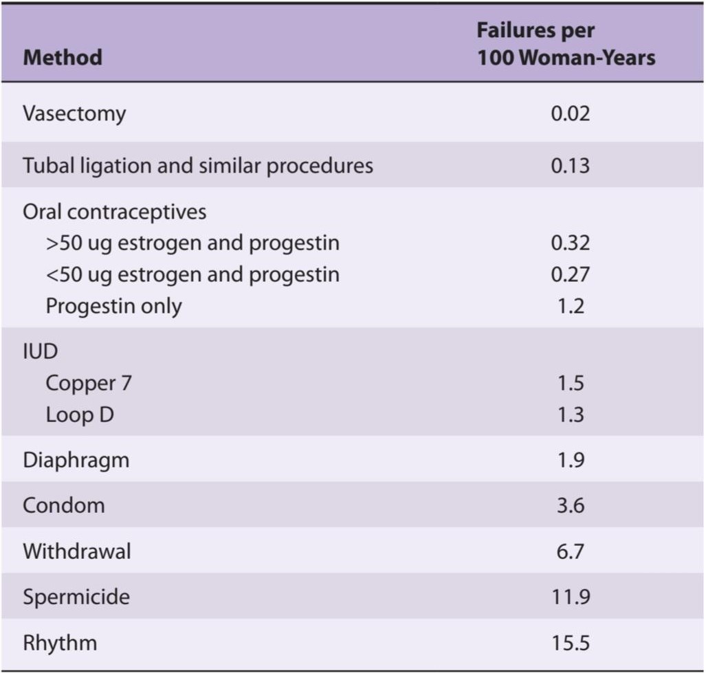
Data from Vessey M, Lawless M, Yeates D: Efficacy of different contraceptive methods. Lancet 1982;1:841. Reproduced with permission.
داده ها از Vessey M، Lawless M، Yeates D: اثربخشی روش های مختلف پیشگیری از بارداری. Lancet 1982؛ 1:841. با اجازه تکثیر شد.
Implantation of foreign bodies in the uterus causes changes in the duration of the sexual cycle in a number of mammalian species. In humans, such foreign bodies do not alter the menstrual cycle, but they act as effective contraceptive devices. Intrauterine implantation of pieces of metal or plastic (intrauterine devices, IUDs) has been used in programs aimed at controlling population growth. Although the mechanism of action of IUDs is still unsettled, they seem in general to prevent sperm from fertilizing ova. Those containing copper appear to exert a spermatocidal effect. IUDs that slowly release progesterone or synthetic progestins have the additional effect of thickening cervical mucus so that entry of sperm into the uterus is impeded. IUDs can cause intrauterine infections, but these usually occur in the first month after insertion and in women exposed to sexually transmitted diseases.
لانه گزینی اجسام خارجی در رحم باعث تغییراتی در طول چرخه جنسی در تعدادی از گونه های پستانداران می شود. در انسان، چنین اجسام خارجی چرخه قاعدگی را تغییر نمی دهند، اما به عنوان یک ابزار پیشگیری از بارداری موثر عمل می کنند. کاشت داخل رحمی قطعات فلزی یا پلاستیکی (دستگاه های داخل رحمی، آی یو دی) در برنامه هایی با هدف کنترل رشد جمعیت استفاده شده است. اگرچه مکانیسم عملکرد آی یو دی ها هنوز مشخص نیست، اما به نظر می رسد که به طور کلی از بارور شدن تخمک ها توسط اسپرم جلوگیری می کند. به نظر می رسد آنهایی که حاوی مس هستند اثر اسپرم کشی دارند. آی یو دی هایی که به آهستگی پروژسترون یا پروژستین های مصنوعی را آزاد می کنند، اثر اضافی ضخیم شدن موکوس دهانه رحم را دارند به طوری که ورود اسپرم به رحم مانع می شود. IUD می تواند باعث عفونت های داخل رحمی شود، اما این عفونت ها معمولا در ماه اول پس از قرار دادن و در زنانی که در معرض بیماری های مقاربتی قرار دارند رخ می دهد.
Women undergoing long-term treatment with relatively large doses of estrogen do not ovulate, probably because they have depressed FSH levels and multiple irregular bursts of LH secretion rather than a single midcycle peak. Women treated with similar doses of estrogen plus a progestational agent do not ovulate because the secretion of both gonadotropins is suppressed. In addition, the progestin makes the cervical mucus thick and unfavorable to sperm migration, and it may also interfere with implantation. For contraception, an orally active estrogen such as ethinyl estradiol is often combined with a synthetic progestin such as norethindrone. The pills are administered for 21 days, then withdrawn for 5-7 days to permit menstrual flow, and started again. Like ethinyl estradiol, norethindrone has an ethinyl group on position 17 of the steroid nucleus, so it is resistant to hepatic metabolism and consequently is effective by mouth. In addition to being a progestin, it is partly metabolized to ethinyl estradiol, and for this reason it also has estrogenic activity. Small as well as large doses of estrogen are effective (Table 22-4).
زنانی که تحت درمان طولانی مدت با دوزهای نسبتاً زیاد استروژن قرار میگیرند، تخمکگذاری نمیکنند، احتمالاً به این دلیل که سطح FSH افسرده شده و چندین انفجار نامنظم ترشح LH به جای یک پیک منفرد در چرخه میانی وجود دارد. زنانی که با دوزهای مشابه استروژن به همراه یک عامل پروژستاتیو درمان می شوند، تخمک گذاری نمی کنند زیرا ترشح هر دو گنادوتروپین سرکوب می شود. علاوه بر این، پروژسترون باعث غلیظ شدن مخاط دهانه رحم می شود و برای مهاجرت اسپرم نامطلوب می شود و همچنین ممکن است در لانه گزینی اختلال ایجاد کند. برای پیشگیری از بارداری، یک استروژن فعال خوراکی مانند اتینیل استرادیول اغلب با یک پروژستین مصنوعی مانند نورتیندرون ترکیب می شود. قرص ها به مدت 21 روز تجویز می شوند، سپس برای 5-7 روز قطع می شوند تا جریان قاعدگی فراهم شود و دوباره شروع می شود. مانند اتینیل استرادیول، نورتیندرون دارای یک گروه اتینیل در موقعیت 17 هسته استروئیدی است، بنابراین در برابر متابولیسم کبدی مقاوم است و در نتیجه از طریق خوراکی موثر است. علاوه بر پروژسترون بودن، تا حدی به اتینیل استرادیول متابولیزه می شود و به همین دلیل دارای فعالیت استروژنی نیز می باشد. دوزهای کوچک و همچنین زیاد استروژن مؤثر هستند (جدول 22-4).
Implants made up primarily of progestins such as levonorgestrel are now seeing increased use in some parts of the world. These are inserted under the skin and can prevent pregnancy for up to 5 years. They often produce amenorrhea, but otherwise they appear to be effective and well tolerated.
ایمپلنتهایی که عمدتاً از پروژستینهایی مانند لوونورژسترل تشکیل شدهاند، اکنون در برخی از نقاط جهان استفاده فزایندهای دارند. اینها زیر پوست قرار می گیرند و می توانند تا 5 سال از بارداری جلوگیری کنند. آنها اغلب باعث آمنوره می شوند، اما در غیر این صورت به نظر می رسد موثر و به خوبی تحمل شوند.
ABNORMALITIES OF OVARIAN FUNCTION
Menstrual Abnormalities
Some women who are infertile have anovulatory cycles; they fail to ovulate but have menstrual periods at fairly regular intervals. As noted above, anovulatory cycles are the rule for the first 1-2 years after menarche and again before the menopause. Amenorrhea is the absence of menstrual periods. If menstrual bleeding has never occurred, the condition is called primary amenorrhea. Some women with primary amenorrhea have small breasts and other signs of failure to mature sexually. Cessation of cycles in a woman with previously normal periods is called secondary amenorrhea. The most common cause of secondary amenorrhea is pregnancy, and the old clinical maxim that “secondary amenorrhea should be considered to be due to pregnancy until proved otherwise”has considerable merit. Other causes of amenorrhea include emotional stimuli and changes in the environment, hypothalamic diseases, pituitary disorders, primary ovarian disorders, and various systemic diseases. Evidence suggests that in some women with hypothalamic amenorrhea, the frequency of GnRH pulses is slowed as a result of excess opioid activity in the hypothalamus. In encouraging preliminary studies, the frequency of GnRH pulses was increased by administration of the orally active opioid blocker naltrexone.
ناهنجاری های عملکرد تخمدان
ناهنجاری های قاعدگی
برخی از زنان نابارور سیکل های تخمک گذاری دارند. آنها قادر به تخمک گذاری نیستند اما دوره های قاعدگی در فواصل نسبتاً منظم دارند. همانطور که در بالا ذکر شد، چرخه های عدم تخمک گذاری برای 1-2 سال اول پس از قاعدگی و دوباره قبل از یائسگی یک قانون است. آمنوره عدم وجود دوره های قاعدگی است. اگر خونریزی قاعدگی هرگز رخ نداده باشد، این بیماری آمنوره اولیه نامیده می شود. برخی از زنان مبتلا به آمنوره اولیه دارای سینه های کوچک و سایر علائم عدم بلوغ جنسی هستند. قطع قاعدگی در زنانی که قبلاً قاعدگی طبیعی دارند، آمنوره ثانویه نامیده می شود. شایع ترین علت آمنوره ثانویه حاملگی است و این اصل بالینی قدیمی که “آمنوره ثانویه را باید به دلیل حاملگی در نظر گرفت تا زمانی که خلاف آن ثابت شود” ارزش قابل توجهی دارد. از دیگر علل آمنوره می توان به محرک های هیجانی و تغییرات محیطی، بیماری های هیپوتالاموس، اختلالات هیپوفیز، اختلالات اولیه تخمدان و بیماری های مختلف سیستمیک اشاره کرد. شواهد حاکی از آن است که در برخی از زنان مبتلا به آمنوره هیپوتالاموس، تعداد نبض های GnRH در نتیجه فعالیت بیش از حد مواد افیونی در هیپوتالاموس کاهش می یابد. در تشویق مطالعات مقدماتی، فرکانس پالس های GnRH با تجویز نالترکسون مسدود کننده اپیوئیدی خوراکی افزایش یافت.
The terms hypomenorrhea and menorrhagia refer to scanty and abnormally profuse flow, respectively, during regular periods. Metrorrhagia is bleeding from the uterus between periods, and oligomenorrhea is reduced frequency of periods. Dysmenorrhea is painful menstruation. The severe menstrual cramps that are common in young women quite often disappear after the first pregnancy. Most of the symptoms of dysmenorrhea are due to accumulation of prostaglandins in the uterus, and symptomatic relief has been obtained by treatment with inhibitors of prostaglandin synthesis.
اصطلاحات هیپومنوره و منوراژی به ترتیب به جریان کم و غیرطبیعی فراوان در طول دوره های منظم اشاره دارد. متروراژی خونریزی از رحم بین پریودها و الیگومنوره کاهش تعداد پریودها است. دیسمنوره یک قاعدگی دردناک است. دردهای شدید قاعدگی که در زنان جوان رایج است اغلب پس از اولین بارداری ناپدید می شوند. بیشتر علائم دیسمنوره به دلیل تجمع پروستاگلاندین ها در رحم است و با درمان با مهارکننده های سنتز پروستاگلاندین ها تسکین علائم حاصل شده است.
Some women develop symptoms such as irritability, bloating, edema, decreased ability to concentrate, depression, headache, and constipation during the last 7-10 days of their menstrual cycles. These symptoms of the premenstrual syndrome (PMS) have been attributed to salt and water retention. However, it seems unlikely that this or any of the other hormonal alterations that occur in the late luteal phase are responsible because the time course and severity of the symptoms are not modified if the luteal phase is terminated early and menstruation produced by administration of mifepristone. The antidepressant fluoxetine, which is a serotonin reuptake inhibitor, and the benzodiazepine alprazolam produce symptomatic relief, and so do GnRH- releasing agonists in doses that suppress the pituitary-ovarian axis. How these diverse clinical observations fit together to produce a picture of the pathophysiology of PMS is still unknown (Clinical Box 22-4).
برخی از زنان علائمی مانند تحریک پذیری، نفخ، ادم، کاهش توانایی تمرکز، افسردگی، سردرد و یبوست را در طول 7 تا 10 روز آخر دوره قاعدگی خود نشان می دهند. این علائم سندرم پیش از قاعدگی (PMS) به احتباس نمک و آب نسبت داده شده است. با این حال، بعید به نظر میرسد که این یا هر یک از تغییرات هورمونی دیگری که در اواخر فاز لوتئال رخ میدهد، مسئول باشد، زیرا اگر فاز لوتئال زودتر خاتمه یابد و قاعدگی با تجویز میفپریستون ایجاد شود، دوره زمانی و شدت علائم تغییر نمیکند. فلوکستین ضد افسردگی، که یک مهارکننده بازجذب سروتونین است، و بنزودیازپین آلپرازولام باعث تسکین علائم میشوند، و همچنین آگونیستهای آزادکننده GnRH در دوزهایی که محور هیپوفیز-تخمدان را سرکوب میکنند، ایجاد میکنند. اینکه چگونه این مشاهدات بالینی متنوع برای ایجاد تصویری از پاتوفیزیولوژی سندرم پیش از قاعدگی با هم تطابق می یابند هنوز ناشناخته است (کادر بالینی 22-4).
CLINICAL BOX 22-4
Genetic Defects Causing Reproductive Abnormalities
A number of single-gene mutations cause reproductive abnormalities when they occur in women. Examples include (1) Kallmann syndrome, which causes hypogonadotropic hypogonadism; (2) GnRH resistance, FSH resistance, and LH resistance, which are due to defects in the GnRH, FSH, or LH receptors, respectively; and (3) aromatase deficiency, which prevents the formation of estrogens. These are all caused by loss-of-function mutations. An interesting gain-of-function mutation causes the McCune-Albright syndrome, in which Gsa becomes constitutively active in certain cells but not others (mosaicism) because a somatic mutation after initial cell division has occurred in the embryo. It is associated with multiple endocrine abnormalities, including precocious puberty and amenorrhea with galactorrhea.
جعبه بالینی 22-4
نقص ژنتیکی که باعث ناهنجاری های تولید مثل می شود
تعدادی از جهشهای تک ژنی زمانی که در زنان رخ میدهند باعث ناهنجاریهای تولید مثل میشوند. مثالها عبارتند از (1) سندرم کالمن، که باعث هیپوگنادیسم هیپوگنادوتروپیک میشود. (2) مقاومت به GnRH، مقاومت به FSH و مقاومت LH، که به ترتیب به دلیل نقص در گیرنده های GnRH، FSH یا LH است. و (3) کمبود آروماتاز، که از تشکیل استروژن جلوگیری می کند. اینها همه ناشی از جهش های از دست دادن عملکرد هستند. یک جهش جالب با افزایش عملکرد باعث سندرم مککون-آلبرایت میشود، که در آن Gsa در سلولهای خاصی فعال میشود اما در برخی دیگر فعال نمیشود (موزاییکیسم) زیرا یک جهش سوماتیک پس از تقسیم سلولی اولیه در جنین رخ داده است. این بیماری با چندین ناهنجاری غدد درون ریز، از جمله بلوغ زودرس و آمنوره همراه با گالاکتوره همراه است.
PREGNANCY
Fertilization & Implantation
In humans, fertilization of the ovum by the sperm (see Chapter 23) usually occurs in the ampulla of the uterine tube. Fertilization involves (1) chemoattraction of the sperm to the ovum by substances produced by the ovum; (2) adherence to the zona pellucida, the membranous structure surrounding the ovum; (3) penetration of the zona pellucida and the acrosome reaction; and (4) adherence of the sperm head to the cell membrane of the ovum, with breakdown of the area of fusion and release of the sperm nucleus into the cytoplasm of the ovum (Figure 22–20). Millions of sperm are deposited in the vagina during intercourse. Eventually, 50-100 sperm reach the ovum, and many of them contact the zona pellucida. Sperm bind to a receptor in the zona, and this is followed by the acrosomal reaction, that is, the breakdown of the acrosome, the lysosome-like organelle on the head of the sperm (Figure 23–4). Various enzymes are released, including the trypsin-like protease acrosin. Acrosin facilitates but is not required for the penetration of the sperm through the zona pellucida. When one sperm reaches the membrane of the ovum, fusion to the ovum membrane is mediated by fertilin, a protein on the surface of the sperm head that resembles the viral fusion proteins that permit some viruses to attack cells. The fusion provides the signal that initiates development. In addition, the fusion sets off a reduction in the membrane potential of the ovum that prevents polyspermy, the fertilization of the ovum by more than one sperm. This transient potential change is followed by a structural change in the zona pellucida that provides protection against polyspermy on a more long-term basis.
بارداری
لقاح و کاشت
در انسان، لقاح تخمک توسط اسپرم (به فصل 23 مراجعه کنید) معمولاً در آمپول لوله رحمی رخ می دهد. لقاح شامل (1) جذب شیمیایی اسپرم به تخمک توسط مواد تولید شده توسط تخمک است. (2) پایبندی به زونا پلوسیدا، ساختار غشایی اطراف تخمک. (3) نفوذ زونا پلوسیدا و واکنش آکروزوم. و (4) چسبیدن سر اسپرم به غشای سلولی تخمک، با تجزیه ناحیه همجوشی و آزاد شدن هسته اسپرم در سیتوپلاسم تخمک (شکل 22-20). میلیون ها اسپرم در حین مقاربت در واژن رسوب می کند. در نهایت 50 تا 100 اسپرم به تخمک می رسد و بسیاری از آنها با زونا پلوسیدا تماس می گیرند. اسپرم به گیرنده ای در زونا متصل می شود و به دنبال آن واکنش آکروزومی، یعنی تجزیه آکروزوم، اندامک لیزوزوم مانند روی سر اسپرم دنبال می شود (شکل 23-4). آنزیم های مختلفی از جمله پروتئاز آکروزین شبیه تریپسین آزاد می شوند. آکروزین نفوذ اسپرم را از طریق زونا پلوسیدا تسهیل می کند اما لازم نیست. هنگامی که یک اسپرم به غشای تخمک می رسد، پیوند به غشای تخمک توسط fertilin انجام می شود، پروتئینی در سطح سر اسپرم که شبیه پروتئین های همجوشی ویروسی است که به برخی از ویروس ها اجازه حمله به سلول ها را می دهد. فیوژن سیگنالی را ارائه می دهد که توسعه را آغاز می کند. علاوه بر این، همجوشی باعث کاهش پتانسیل غشایی تخمک می شود که از پلی اسپرمی، لقاح تخمک توسط بیش از یک اسپرم جلوگیری می کند. این تغییر پتانسیل گذرا با یک تغییر ساختاری در زونا شفاف دنبال میشود که محافظت طولانیمدت در برابر پلی اسپرمی را فراهم میکند.

FIGURE 22-20 Sequential events in fertilization in mammals. Sperm are attracted to the ovum, bind to the zona pellucida, release acrosomal enzymes, penetrate the zona pellucida, and fuse with the membrane of the ovum, releasing the sperm nucleus into its cytoplasm. Current evidence indicates that the side, rather than the tip, of the sperm head fuses with the egg cell membrane. (Modified with permission from Vacquier VD: Evolution of gamete recognition proteins. Science 1998; Sep 25; 281(5385):1995–1998.)
شکل 22-20 رویدادهای متوالی در لقاح در پستانداران. اسپرم ها به سمت تخمک جذب می شوند، به زونا پلوسیدا متصل می شوند، آنزیم های آکروزومی آزاد می کنند، به زونا پلوسیدا نفوذ می کنند و با غشای تخمک ترکیب می شوند و هسته اسپرم را در سیتوپلاسم خود آزاد می کنند. شواهد کنونی نشان می دهد که طرف سر اسپرم به جای نوک آن با غشای سلول تخمک ترکیب می شود. (با مجوز Vacquier VD: Evolution of gamete recognition proteins. Science 1998؛ 25 سپتامبر؛ 281 (5385): 1995-1998 اصلاح شده است.)
The developing embryo, now called a blastocyst, moves down the tube into the uterus. This journey takes about 3 days, during which the blastocyst reaches the 8- or 16-cell stage. Once in contact with the endometrium, the blastocyst becomes surrounded by an outer layer of syncytiotrophoblast, a multi-nucleate mass with no discernible cell boundaries, and an inner layer of cytotrophoblast made up of individual cells. The syncytiotrophoblast erodes the endometrium, and the blastocyst burrows into it (implantation). The implantation site is usually on the dorsal wall of the uterus. A placenta then develops, and the trophoblast remains associated with it.
جنین در حال رشد که اکنون بلاستوسیست نامیده می شود، از لوله به داخل رحم حرکت می کند. این سفر حدود 3 روز طول می کشد و در طی آن بلاستوسیست به مرحله 8 یا 16 سلولی می رسد. هنگامی که بلاستوسیست با آندومتر تماس پیدا می کند، توسط یک لایه بیرونی سینسیتیوتروفوبلاست، یک توده چند هسته ای بدون مرز سلولی قابل تشخیص، و یک لایه داخلی سیتوتروفوبلاست که از سلول های جداگانه تشکیل شده است، احاطه می شود. سینسیتیوتروفوبلاست آندومتر را فرسایش می دهد و بلاستوسیست در آن سوراخ می شود (لانه گزینی). محل لانه گزینی معمولاً روی دیواره پشتی رحم است. سپس جفت ایجاد می شود و تروفوبلاست با آن همراه می ماند.
Failure to Reject the “Fetal Graft”
It should be noted that the fetus and the mother are two genetically distinct individuals, and the fetus is in effect a transplant of foreign tissue in the mother. However, the transplant is tolerated, and the rejection reaction that is characteristically produced when other foreign tissues are transplanted (see Chapter 3) fails to occur. The way the “fetal graft” is protected is unknown. However, one explanation may be that the placental trophoblast, which separates maternal and fetal tissues, does not express the polymorphic class I and class II major histocompatibility complex (MHC) genes and instead expresses HLA-G, a nonpolymorphic gene. Therefore, antibodies against the fetal proteins do not develop. In addition, there is Fas ligand on the surface of the placenta, and this binds to T cells, causing them to undergo apoptosis (see Chapter 3).
عدم رد “پیوند جنین”
لازم به ذکر است که جنین و مادر دو فرد از نظر ژنتیکی متمایز هستند و جنین در واقع پیوند بافت خارجی در مادر است. با این حال، پیوند قابل تحمل است، و واکنش رد که مشخصاً هنگام پیوند سایر بافتهای خارجی ایجاد میشود (به فصل 3 مراجعه کنید) انجام نمیشود. نحوه محافظت از “گرافت جنینی” ناشناخته است. با این حال، یک توضیح ممکن است این باشد که تروفوبلاست جفت، که بافتهای مادر و جنین را جدا میکند، ژنهای کمپلکس اصلی سازگاری بافتی کلاس I و کلاس II (MHC) را بیان نمیکند و در عوض HLA-G، یک ژن غیرپلیمورف را بیان میکند. بنابراین، آنتی بادی علیه پروتئین های جنینی ایجاد نمی شود. علاوه بر این، لیگاند Fas در سطح جفت وجود دارد، و این به سلول های T متصل می شود و باعث آپوپتوز آنها می شود (به فصل 3 مراجعه کنید).
Infertility
The vexing clinical problem of infertility often requires extensive investigation before a cause is found. In 30% of cases, the problem is in the man; in 45%, the problem is in the woman; in 20%, both partners have a problem; and in 5%, no cause can be found. In vitro fertilization, that is, removing mature ova, fertilizing them with sperm, and implanting one or more of them in the uterus at the four-cell stage is of some value in these cases. It has a 5-10% chance of producing a live birth.
ناباروری
مشکل آزاردهنده بالینی ناباروری اغلب نیاز به بررسی گسترده قبل از یافتن علت دارد. در 30 درصد موارد، مشکل در مرد است. در 45% مشکل در زن است. در 20٪، هر دو شریک مشکل دارند. و در 5% هیچ علتی پیدا نمی شود. لقاح آزمایشگاهی، یعنی برداشتن تخمک های بالغ، لقاح آنها با اسپرم و کاشت یک یا چند عدد از آنها در رحم در مرحله چهار سلولی، در این موارد ارزش دارد. 5-10 درصد احتمال تولد زنده را دارد.
Endocrine Changes
In all mammals, the corpus luteum in the ovary at the time of fertilization fails to regress and instead enlarges in response to stimulation by gonadotropic hormones secreted by the placenta. The placental gonadotropin in humans is called human chorionic gonadotropin (hCG). The enlarged corpus luteum of pregnancy secretes estrogens, progesterone, and relaxin. Progesterone and relaxin help maintain pregnancy by inhibiting myometrial contractions; progesterone prevents prostaglandin production by the uterus, which stops contractions from occurring. In humans, the placenta produces sufficient estrogen and progesterone from maternal and fetal precursors to take over the function of the corpus luteum after the sixth week of pregnancy. Ovariectomy before the sixth week thus leads to abortion, but ovariectomy thereafter has no effect on the pregnancy. The function of the corpus luteum begins to decline after 8 weeks of pregnancy, but it persists throughout pregnancy. hCG secretion decreases after an initial marked rise, but estrogen and progesterone secretion increase until just before parturition (Table 22-5).
تغییرات غدد درون ریز
در تمام پستانداران، جسم زرد در تخمدان در زمان لقاح، پسرفت نمی کند و در عوض در پاسخ به تحریک هورمون های گنادوتروپیک ترشح شده توسط جفت، بزرگ می شود. گنادوتروپین جفتی در انسان گنادوتروپین جفتی انسان (hCG) نامیده می شود. بزرگ شدن جسم زرد بارداری استروژن، پروژسترون و ریلکسین ترشح می کند. پروژسترون و ریلکسین با مهار انقباضات میومتر به حفظ بارداری کمک می کنند. پروژسترون از تولید پروستاگلاندین توسط رحم جلوگیری می کند که از بروز انقباضات جلوگیری می کند. در انسان، جفت به اندازه کافی استروژن و پروژسترون از پیش سازهای مادر و جنین تولید می کند تا پس از هفته ششم بارداری، عملکرد جسم زرد را به عهده بگیرد. بنابراین اواریکتومی قبل از هفته ششم منجر به سقط جنین می شود، اما تخمدان پس از آن هیچ تاثیری بر بارداری ندارد. عملکرد جسم زرد پس از 8 هفته بارداری شروع به کاهش می کند، اما در طول بارداری ادامه می یابد. ترشح hCG پس از افزایش مشخص اولیه کاهش می یابد، اما ترشح استروژن و پروژسترون تا قبل از زایمان افزایش می یابد (جدول 22-5).
TABLE 22-5 Hormone levels in human maternal blood during normal pregnancy.
جدول 22-5 سطح هورمون در خون مادر انسان در دوران بارداری طبیعی.

Human Chorionic Gonadotropin
hCG is a glycoprotein that contains galactose and hexosamine. It is produced by the syncytiotrophoblast. Like the pituitary glycoprotein hormones, it is made up of a and ẞ subunits. hCG-a is identical to the a subunit of LH, FSH, and TSH. The molecular weight of hCG-a is 18,000, and that of hCG-ẞ is 28,000. hCG is primarily luteinizing and luteotropic and has little FSH activity. It can be measured by radioimmunoassay and detected in the blood as early as 6 days after conception. Its presence in the urine in early pregnancy is the basis of the various laboratory tests for pregnancy, and it can sometimes be detected in the urine as early as 14 days after conception. It appears to act on the same receptor as LH. hCG is not absolutely specific for pregnancy. Small amounts are secreted by a variety of gastrointestinal and other tumors in both sexes, and hCG has been measured in individuals with suspected tumors as a “tumor marker.” It also appears that the fetal liver and kidney normally produce small amounts of hCG.
گنادوتروپین کوریونی انسانی
hCG یک گلیکوپروتئین است که حاوی گالاکتوز و هگزوزامین است. این توسط سینسیتیوتروفوبلاست تولید می شود. مانند هورمون های گلیکوپروتئین هیپوفیز، از زیر واحدهای a و ẞ تشکیل شده است. hCG-a با زیر واحد LH، FSH و TSH یکسان است. وزن مولکولی hCG-a 18000 و hCG-ẞ 28000 است. hCG در درجه اول لوتئینه کننده و لوتئوتروپیک است و فعالیت FSH کمی دارد. می توان آن را با روش رادیو ایمونواسی اندازه گیری کرد و 6 روز پس از لقاح در خون تشخیص داد. وجود آن در ادرار در اوایل بارداری اساس آزمایش های مختلف آزمایشگاهی برای بارداری است و گاهی اوقات می توان آن را در اوایل 14 روز پس از لقاح در ادرار تشخیص داد. به نظر می رسد که روی همان گیرنده LH عمل می کند. hCG برای بارداری کاملا اختصاصی نیست. مقادیر کمی توسط انواع تومورهای گوارشی و سایر تومورها در هر دو جنس ترشح می شود و hCG در افراد مشکوک به تومور به عنوان “تومور مارکر” اندازه گیری شده است. همچنین به نظر می رسد که کبد و کلیه جنین به طور معمول مقادیر کمی hCG تولید می کنند.
Human Chorionic Somatomammotropin
The syncytiotrophoblast also secretes large amounts of a protein hormone that is lactogenic and has a small amount of growth-stimulating activity. This hormone has been called chorionic growth hormone-prolactin (CGP) and human placental lactogen (hPL), but it is now generally called human chorionic somatomammotropin (hCS). The structure of hCS is very similar to that of human growth hormone (see Chapter 18), and it appears that these two hormones and prolactin evolved from a common progenitor hormone. Large quantities of hCS are found in maternal blood, but very little reaches the fetus. Secretion of growth hormone from the maternal pituitary is not increased during pregnancy and may actually be decreased by hCS. However, hCS has most of the actions of growth hormone and apparently functions as a “maternal growth hormone of pregnancy” to bring about the nitrogen, potassium, and calcium retention, lipolysis, and decreased glucose utilization seen in this state. These latter two actions divert glucose to the fetus. The amount of hCS secreted is proportional to the size of the placenta, which normally weighs about one-sixth as much as the fetus. Low hCS levels are a sign of placental insufficiency.
سوماتوماموتروپین کوریونی انسان
سینسیتیوتروفوبلاست همچنین مقادیر زیادی هورمون پروتئینی ترشح می کند که لاکتوژنیک است و مقدار کمی فعالیت محرک رشد دارد. این هورمون هورمون رشد جفتی-پرولاکتین (CGP) و لاکتوژن جفت انسانی (hPL) نامیده می شود، اما اکنون به طور کلی به آن سوماتوماموتروپین جفتی انسانی (hCS) می گویند. ساختار hCS بسیار شبیه به هورمون رشد انسانی است (به فصل 18 مراجعه کنید)، و به نظر می رسد که این دو هورمون و پرولاکتین از یک هورمون پیش ساز مشترک تکامل یافته اند. مقادیر زیادی hCS در خون مادر یافت می شود، اما مقدار بسیار کمی از آن به جنین می رسد. ترشح هورمون رشد از هیپوفیز مادر در دوران بارداری افزایش نمی یابد و در واقع ممکن است توسط hCS کاهش یابد. با این حال، hCS بیشتر اعمال هورمون رشد را دارد و ظاهراً به عنوان یک “هورمون رشد مادر بارداری” عمل می کند تا نیتروژن، پتاسیم و کلسیم را حفظ کند، لیپولیز و کاهش مصرف گلوکز را که در این حالت مشاهده می شود، ایجاد کند. این دو عمل اخیر گلوکز را به سمت جنین هدایت می کند. مقدار hCS ترشح شده متناسب با اندازه جفت است که به طور معمول حدود یک ششم وزن جنین دارد. سطوح پایین hCS نشانه نارسایی جفت است.
Other Placental Hormones
In addition to hCG, hCS, progesterone, and estrogens, the placenta secretes other hormones. Human placental fragments probably produce proopiomelanocortin (POMC). In culture, they release corticotropin-releasing hormone (CRH), B- endorphin, a-melanocyte-stimulating hormone (MSH), and dynorphin A, all of which appear to be identical to their hypothalamic counterparts. They also secrete GnRH and inhibin, and since GnRH stimulates and inhibin inhibits hCG secretion, locally produced GnRH and inhibin may act in a paracrine fashion to regulate hCG secretion. The trophoblast cells and amnion cells also secrete leptin, and moderate amounts of this satiety hormone enter the maternal circulation. Some also enters the amniotic fluid. Its function in pregnancy is unknown. The placenta also secretes prolactin in a number of forms.
سایر هورمون های جفتی
جفت علاوه بر hCG، hCS، پروژسترون و استروژن، هورمون های دیگری نیز ترشح می کند. قطعات جفت انسان احتمالا پروپیوملانوکورتین (POMC) تولید می کنند. در فرهنگ، آنها هورمون آزاد کننده کورتیکوتروپین (CRH)، B-اندورفین، هورمون محرک a-melanocyte (MSH) و دینورفین A را آزاد می کنند، که به نظر می رسد همه آنها با همتایان هیپوتالاموس خود یکسان هستند. آنها همچنین GnRH و اینهیبین ترشح می کنند و از آنجایی که GnRH باعث تحریک و مهار ترشح hCG می شود، GnRH و اینهیبین تولید شده محلی ممکن است به روش پاراکرین برای تنظیم ترشح hCG عمل کنند. سلول های تروفوبلاست و سلول های آمنیون نیز لپتین ترشح می کنند و مقادیر متوسطی از این هورمون سیری وارد گردش خون مادر می شود. مقداری نیز وارد مایع آمنیوتیک می شود. عملکرد آن در بارداری ناشناخته است. جفت همچنین پرولاکتین را به اشکال مختلف ترشح می کند.
Finally, the placenta secretes the a subunits of hCG, and the plasma concentration of free a subunits rises throughout pregnancy. These a subunits acquire a carbohydrate composition that makes them unable to combine with B subunits, and their prominence suggests that they have a function of their own. It is interesting in this regard that the secretion of the prolactin produced by the endometrium also appears to increase throughout pregnancy, and it may be that the circulating a subunits stimulate endometrial prolactin secretion. The cytotrophoblast of the human chorion contains prorenin (see Chapter 38). A large amount of prorenin is also present in amniotic fluid, but its function in this location is unknown.
در نهایت جفت زیرواحدهای a hCG ترشح می کند و غلظت زیرواحدهای آزاد a پلاسما در طول بارداری افزایش می یابد. این زیرواحدهای a دارای ترکیب کربوهیدراتی هستند که باعث می شود نتوانند با زیر واحدهای B ترکیب شوند و برجستگی آنها نشان می دهد که عملکرد خود را دارند. از این نظر جالب است که ترشح پرولاکتین تولید شده توسط آندومتر نیز در طول بارداری افزایش می یابد و ممکن است زیر واحدهای در گردش ترشح پرولاکتین آندومتر را تحریک کنند. سیتوتروفوبلاست کوریون انسانی حاوی پرورنین است (به فصل 38 مراجعه کنید). مقدار زیادی پرورنین نیز در مایع آمنیوتیک وجود دارد، اما عملکرد آن در این محل ناشناخته است.
Fetoplacental Unit
The fetus and the placenta interact in the formation of steroid hormones. The placenta synthesizes pregnenolone and progesterone from cholesterol. Some of the progesterone enters the fetal circulation and provides the substrate for the formation of cortisol and corticosterone in the fetal adrenal glands (Figure 22- 21). Some of the pregnenolone enters the fetus and, along with pregnenolone synthesized in the fetal liver, is the substrate for the formation of dehydroepiandrosterone sulfate (DHEAS) and 16- hydroxydehydroepiandrosterone sulfate (16 OHDHEAS) in the fetal adrenal. Some 16-hydroxylation also occurs in the fetal liver. DHEAS and 16- OHDHEAS are transported back to the placenta, where DHEAS forms estradiol and 16-OHDHEAS forms estriol. The principal estrogen formed is estriol, and since fetal 16-OHDHEAS is the principal substrate for the estrogens, the urinary estriol excretion of the mother can be monitored as an index of the state of the fetus.
واحد جنین جفتی
جنین و جفت در تشکیل هورمونهای استروئیدی با هم تعامل دارند. جفت پرگننولون و پروژسترون را از کلسترول سنتز می کند. مقداری از پروژسترون وارد گردش خون جنین می شود و بستری را برای تشکیل کورتیزول و کورتیکوسترون در غدد فوق کلیوی جنین فراهم می کند (شکل 22-21). مقداری از پرگننولون وارد جنین می شود و همراه با پرگننولون سنتز شده در کبد جنین، بستری برای تشکیل دهیدرواپی آندروسترون سولفات (DHEAS) و 16 هیدروکسید هیدرو اپی آندروسترون سولفات (16 OHDHEAS) در آدرنال جنین است. مقداری 16 هیدروکسیلاسیون نیز در کبد جنین رخ می دهد. DHEAS و 16- OHDHEAS به جفت منتقل می شوند، جایی که DHEAS استرادیول و 16-OHDHEAS استریول را تشکیل می دهد. استروژن اصلی تشکیل شده استریول است و از آنجایی که 16-OHDHEAS جنینی بستر اصلی استروژن ها است، دفع استریول ادراری مادر را می توان به عنوان شاخصی از وضعیت جنین بررسی کرد.
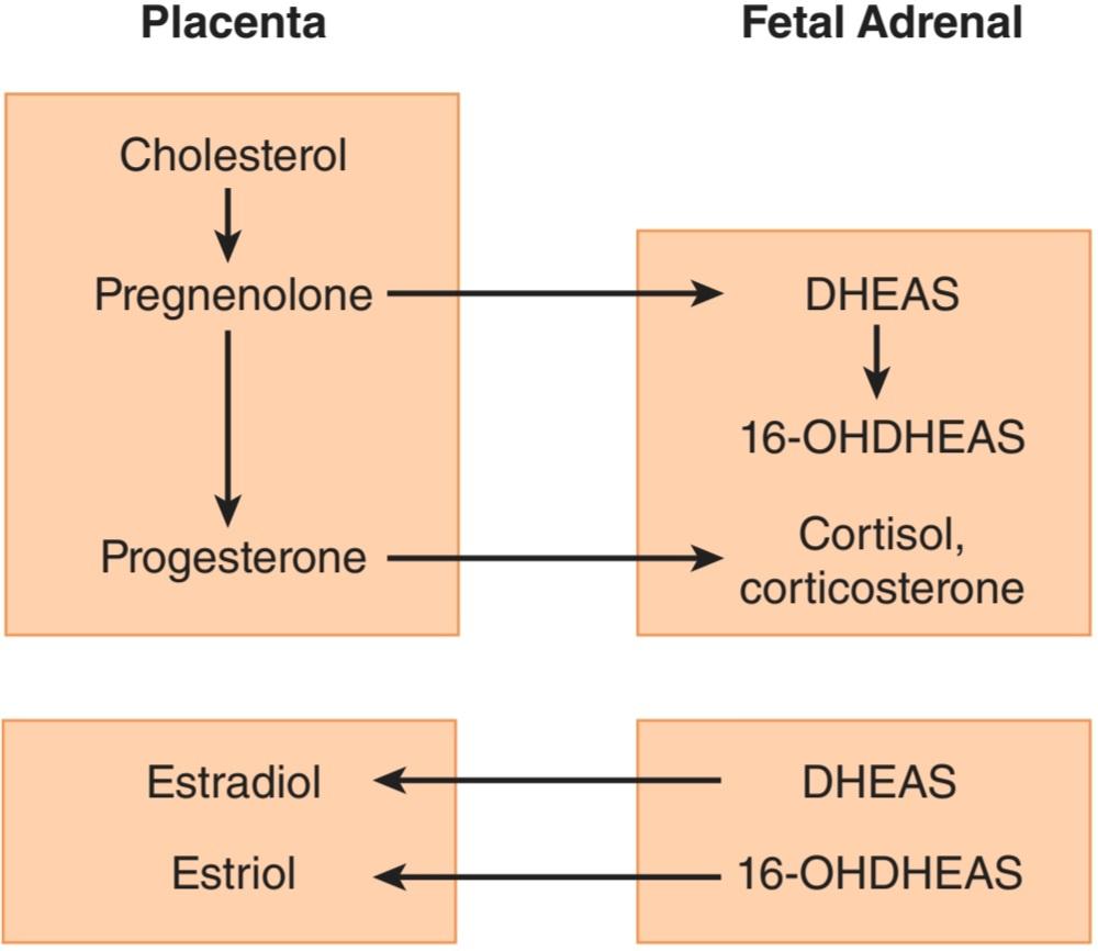
FIGURE 22–21 Interactions between the placenta and the fetal adrenal cortex in the production of steroids.
شکل 22-21 تعامل بین جفت و قشر آدرنال جنین در تولید استروئیدها.
Parturition
The duration of pregnancy in humans averages 270 days from fertilization (284 days from the first day of the menstrual period preceding conception). Irregular uterine contractions increase in frequency in the last month of pregnancy.
زایمان
طول مدت بارداری در انسان به طور متوسط 270 روز از زمان لقاح است (284 روز از اولین روز قاعدگی قبل از لقاح). انقباضات نامنظم رحم در ماه آخر بارداری افزایش می یابد.
The difference between the body of the uterus and the cervix becomes evident at the time of delivery. The cervix, which is firm in the nonpregnant state and throughout pregnancy until near the time of delivery, softens and dilates, while the body of the uterus contracts and expels the fetus.
تفاوت بین بدن رحم و دهانه رحم در زمان زایمان آشکار می شود. دهانه رحم که در حالت غیر حاملگی و در تمام دوران بارداری تا نزدیک به زمان زایمان سفت است، نرم و گشاد می شود، در حالی که بدن رحم منقبض می شود و جنین را خارج می کند.
There is still considerable uncertainty about the mechanisms responsible for the onset of labor. One factor is the increase in circulating estrogens produced by increased circulating DHEAS. This makes the uterus more excitable, increases the number of gap junctions between myometrial cells, and causes production of more prostaglandins, which in turn cause uterine contractions. In humans, CRH secretion by the fetal hypothalamus increases and is supplemented by increased placental production of CRH. This increases circulating adrenocorticotropic hormone (ACTH) in the fetus, and the resulting increase in cortisol hastens the maturation of the respiratory system. Thus, in a sense, the fetus picks the time to be born by increasing CRH secretion.
هنوز عدم قطعیت قابل توجهی در مورد مکانیسم های مسئول شروع زایمان وجود دارد. یکی از عوامل افزایش استروژن در گردش تولید شده توسط افزایش DHEAS در گردش است. این باعث تحریک پذیری رحم می شود، تعداد اتصالات شکاف بین سلول های میومتر را افزایش می دهد و باعث تولید بیشتر پروستاگلاندین ها می شود که به نوبه خود باعث انقباضات رحمی می شود. در انسان، ترشح CRH توسط هیپوتالاموس جنین افزایش می یابد و با افزایش تولید جفتی CRH تکمیل می شود. این باعث افزایش هورمون آدرنوکورتیکوتروپیک (ACTH) در جنین می شود و افزایش کورتیزول در نتیجه بلوغ سیستم تنفسی را تسریع می کند. بنابراین، به یک معنا، جنین با افزایش ترشح CRH زمان تولد را انتخاب می کند.
The number of oxytocin receptors in the myometrium and the decidua (the endometrium of pregnancy) increases more than 100-fold during pregnancy and reaches a peak during early labor. Estrogens increase the number of oxytocin receptors, and uterine distension late in pregnancy may also increase their formation. In early labor, the oxytocin concentration in maternal plasma is not elevated from the prelabor value of about 25 pg/mL. It is possible that the marked increase in oxytocin receptors causes the uterus to respond to normal plasma oxytocin concentrations. However, at least in rats, the amount of oxytocin mRNA in the uterus increases, reaching a peak at term; this suggests that locally produced oxytocin also participates in the process.
تعداد گیرنده های اکسی توسین در میومتر و دسیدو (آندومتر بارداری) در دوران بارداری بیش از 100 برابر افزایش می یابد و در اوایل زایمان به اوج خود می رسد. استروژن ها تعداد گیرنده های اکسی توسین را افزایش می دهند و اتساع رحم در اواخر بارداری نیز ممکن است تشکیل آنها را افزایش دهد. در اوایل زایمان، غلظت اکسی توسین در پلاسمای مادر از مقدار قبل از زایمان حدود 25 pg/mL افزایش نمی یابد. این امکان وجود دارد که افزایش قابل توجه در گیرنده های اکسی توسین باعث شود که رحم به غلظت طبیعی اکسی توسین پلاسما پاسخ دهد. با این حال، حداقل در موشها، مقدار mRNA اکسی توسین در رحم افزایش مییابد و در ترم به اوج خود میرسد. این نشان می دهد که اکسی توسین تولید شده محلی نیز در این فرآیند شرکت می کند.
Premature onset of labor is a problem because premature infants have a high mortality rate and often require intensive, expensive care. Intramuscular 17α- hydroxyprogesterone causes a significant decrease in the incidence of premature labor. The mechanism by which it exerts its effect is uncertain, but it may be that the steroid provides a stable level of circulating progesterone. Progesterone relaxes uterine smooth muscle, inhibits the action of oxytocin on the muscle, and reduces the formation of gap junctions between the muscle fibers. All these actions would be expected to inhibit the onset of labor.
شروع زودرس زایمان یک مشکل است زیرا نوزادان نارس میزان مرگ و میر بالایی دارند و اغلب به مراقبت های فشرده و پرهزینه نیاز دارند. 17α-هیدروکسی پروژسترون عضلانی باعث کاهش قابل توجهی در بروز زایمان زودرس می شود. مکانیسمی که توسط آن تأثیر آن را اعمال می کند نامشخص است، اما ممکن است این استروئید سطح پایداری از پروژسترون در گردش را فراهم کند. پروژسترون عضله صاف رحم را شل می کند، از عملکرد اکسی توسین بر روی ماهیچه جلوگیری می کند و ایجاد اتصالات شکاف بین رشته های عضلانی را کاهش می دهد. انتظار می رود همه این اقدامات مانع از شروع زایمان شوند.
Once labor is started, the uterine contractions dilate the cervix, and this dilation in turn sets up signals in afferent nerves that increase oxytocin secretion (Figure 22–22). The plasma oxytocin level rises and more oxytocin becomes available to act on the uterus. Thus, a positive feedback loop is established that aids delivery and terminates on expulsion of the products of conception. Oxytocin increases uterine contractions in two ways: (1) It acts directly on uterine smooth muscle cells to make them contract and (2) it stimulates the formation of prostaglandins in the decidua. The prostaglandins enhance the oxytocin-induced contractions.
هنگامی که زایمان شروع می شود، انقباضات رحمی دهانه رحم را گشاد می کند و این اتساع به نوبه خود سیگنال هایی را در اعصاب آوران ایجاد می کند که ترشح اکسی توسین را افزایش می دهد (شکل 22-22). سطح اکسی توسین پلاسما افزایش می یابد و اکسی توسین بیشتری برای اثر بر روی رحم در دسترس می شود. بنابراین، یک حلقه بازخورد مثبت ایجاد میشود که به تحویل کمک میکند و با دفع محصولات بارداری خاتمه مییابد. اکسی توسین به دو صورت انقباضات رحمی را افزایش می دهد: (1) مستقیماً روی سلول های ماهیچه صاف رحم تأثیر می گذارد تا آنها را منقبض کند و (2) تشکیل پروستاگلاندین ها را در دسیدوا تحریک می کند. پروستاگلاندین ها انقباضات ناشی از اکسی توسین را افزایش می دهند.
During labor, spinal reflexes and voluntary contractions of the abdominal muscles (“bearing down”) also aid in delivery. However, delivery can occur without bearing down and without a reflex increase in secretion of oxytocin from the posterior pituitary gland, since paraplegic women can go into labor and deliver.
در طول زایمان، رفلکس های نخاعی و انقباضات ارادی عضلات شکم (“پایین کشیدن”) نیز به زایمان کمک می کنند. با این حال، زایمان می تواند بدون کاهش و بدون افزایش رفلکس در ترشح اکسی توسین از غده هیپوفیز خلفی انجام شود، زیرا زنان پاراپلژیک می توانند زایمان کنند و زایمان کنند.

FIGURE 22-22 Role of oxytocin in parturition.
شکل 22-22 نقش اکسی توسین در زایمان.
LACTATION
Development of the Breasts
Many hormones are necessary for full mammary development. In general, estrogens are primarily responsible for proliferation of the mammary ducts and progesterone for the development of the lobules. In rats, some prolactin is also needed for development of the glands at puberty, but it is not known if prolactin is necessary in humans. During pregnancy, prolactin levels increase steadily until term, and levels of estrogens and progesterone are elevated as well, producing full lobuloalveolar development.
شیردهی
رشد سینه ها
بسیاری از هورمون ها برای رشد کامل پستان ضروری هستند. به طور کلی، استروژن ها مسئول اصلی تکثیر مجاری پستانی و پروژسترون برای رشد لوبول ها هستند. در موشها، مقداری پرولاکتین نیز برای رشد غدد در دوران بلوغ مورد نیاز است، اما مشخص نیست که آیا پرولاکتین در انسان ضروری است یا خیر. در طول بارداری، سطح پرولاکتین به طور پیوسته تا پایان ترم افزایش مییابد و سطح استروژن و پروژسترون نیز افزایش مییابد و باعث رشد کامل لوبولوآلوئولار میشود.
Secretion & Ejection of Milk
The composition of human and cows’ milk is shown in Table 22-6. In estrogen- and progesterone-primed rodents, injections of prolactin cause the formation of milk droplets and their secretion into the ducts. Oxytocin causes contraction of the myoepithelial cells lining the duct walls, with consequent ejection of the milk through the nipple.
ترشح و دفع شیر
ترکیب شیر انسان و گاو در جدول 22-6 نشان داده شده است. در جوندگان با استروژن و پروژسترون، تزریق پرولاکتین باعث تشکیل قطرات شیر و ترشح آنها در مجاری می شود. اکسی توسین باعث انقباض سلول های میواپیتلیال پوشاننده دیواره مجرا و در نتیجه خروج شیر از طریق نوک پستان می شود.
TABLE 22-6 Composition of colostrum and milk.
جدول 22-6 ترکیب آغوز و شیر.

Reproduced with permission from Findlay ALR: Lactation. Res Reprod (Nov) 1974;6(6).
با مجوز Findlay ALR: Lactation تکثیر شده است. Res Reprod (نوامبر) 1974؛ 6 (6).
Initiation of Lactation after Delivery
The breasts enlarge during pregnancy in response to high circulating levels of estrogens, progesterone, prolactin, and possibly hCG. Some milk is secreted into the ducts as early as the fifth month, but the amounts are small compared with the surge of milk secretion that follows delivery. In most animals, milk is secreted within an hour after delivery, but in women it takes 1-3 days for the milk to “come in.”
شروع شیردهی پس از زایمان
سینه ها در دوران بارداری در پاسخ به سطوح بالای استروژن، پروژسترون، پرولاکتین و احتمالا hCG در گردش بزرگ می شوند. مقداری شیر در اوایل ماه پنجم به داخل مجاری ترشح می شود، اما مقدار آن در مقایسه با افزایش ترشح شیر پس از زایمان ناچیز است. در بیشتر حیوانات، شیر ظرف یک ساعت پس از زایمان ترشح میشود، اما در زنان 1 تا 3 روز طول میکشد تا شیر وارد شود.
After expulsion of the placenta at parturition, the levels of circulating estrogens and progesterone abruptly decline. The drop in circulating estrogen initiates lactation. Prolactin and estrogen synergize in producing breast growth, but estrogen antagonizes the milk-producing effect of prolactin on the breast. Indeed, in women who do not wish to breastfeed their babies, estrogens may be administered to stop lactation.
پس از دفع جفت در هنگام زایمان، سطح استروژن و پروژسترون در گردش به طور ناگهانی کاهش می یابد. کاهش استروژن در گردش باعث شروع شیردهی می شود. پرولاکتین و استروژن در تولید رشد سینه با هم همکاری می کنند، اما استروژن با اثر تولید شیر پرولاکتین روی سینه مخالفت می کند. در واقع، در زنانی که تمایلی به شیر دادن به نوزادان خود ندارند، ممکن است استروژن برای توقف شیردهی تجویز شود.
Suckling not only evokes reflex oxytocin release and milk ejection, it also maintains and augments the secretion of milk because of the stimulation of prolactin secretion it produces.
مکیدن نه تنها باعث ترشح رفلکس اکسی توسین و دفع شیر می شود، بلکه ترشح شیر را نیز به دلیل تحریک ترشح پرولاکتینی که تولید می کند حفظ و افزایش می دهد.
Effect of Lactation on Menstrual Cycles
Women who do not breastfeed their infants usually have their first menstrual period 6 weeks after delivery. However, women who breastfeed regularly have amenorrhea for 25-30 weeks. Breastfeeding stimulates prolactin secretion, and evidence suggests that prolactin inhibits GnRH secretion, inhibits the action of GnRH on the pituitary, and antagonizes the action of gonadotropins on the ovaries. Ovulation is inhibited, and the ovaries are inactive, so estrogen and progesterone output falls to low levels. Consequently, only 5-10% of women become pregnant again during the suckling period, and breastfeeding has long been known to be an important, if only partly effective, method of birth control. Furthermore, almost 50% of the cycles in the first 6 months after resumption of menses are anovulatory (Clinical Box 22-5).
تاثیر شیردهی بر سیکل های قاعدگی
زنانی که به نوزاد خود شیر نمی دهند معمولاً 6 هفته پس از زایمان اولین قاعدگی خود را تجربه می کنند. با این حال، زنانی که به طور منظم شیر می دهند، به مدت 25 تا 30 هفته دچار آمنوره می شوند. شیردهی ترشح پرولاکتین را تحریک می کند و شواهد نشان می دهد که پرولاکتین ترشح GnRH را مهار می کند، از عملکرد GnRH بر هیپوفیز جلوگیری می کند و با عملکرد گنادوتروپین ها بر تخمدان ها مخالفت می کند. تخمک گذاری مهار می شود و تخمدان ها غیرفعال هستند، بنابراین تولید استروژن و پروژسترون به سطوح پایین کاهش می یابد. در نتیجه، تنها 5 تا 10 درصد از زنان در طول دوره شیردهی دوباره باردار میشوند و مدتهاست که شیردهی به عنوان یک روش مهم، اگر تا حدی موثر، برای کنترل بارداری شناخته شده است. علاوه بر این، تقریباً 50 درصد از سیکل ها در 6 ماه اول پس از شروع مجدد قاعدگی، بدون تخمک گذاری هستند (کادر بالینی 22-5).
CLINICAL BOX 22-5
Chiari-Frommel Syndrome
An interesting, although rare, condition is persistence of lactation (galactorrhea) and amenorrhea in women who do not breastfeed after delivery. This condition, called the Chiari-Frommel syndrome, may be associated with some genital atrophy and is due to persistent prolactin secretion without the secretion of the FSH and LH necessary to produce maturation of new follicles and ovulation. A similar pattern of galactorrhea and amenorrhea with high circulating prolactin levels is seen in nonpregnant women with chromophobe pituitary tumors and in women in whom the pituitary stalk has been sectioned during treatment of cancer.
جعبه بالینی 22-5
سندرم کیاری فرومل
یک وضعیت جالب، اگرچه نادر، تداوم شیردهی (گالاکتوره) و آمنوره در زنانی است که پس از زایمان شیر نمیدهند. این بیماری که سندرم کیاری-فرومل نامیده می شود ممکن است با مقداری آتروفی دستگاه تناسلی همراه باشد و به دلیل ترشح مداوم پرولاکتین بدون ترشح FSH و LH لازم برای تولید فولیکول های جدید و تخمک گذاری است. الگوی مشابهی از گالاکتوره و آمنوره با سطوح بالای پرولاکتین در گردش خون در زنان غیرباردار مبتلا به تومورهای کروموفوب هیپوفیز و در زنانی که ساقه هیپوفیز در طول درمان سرطان برش داده شده است، دیده می شود.
Gynecomastia
Breast development in the male is called gynecomastia. It may be unilateral but is more commonly bilateral. It is common in newborns because of transplacental passage of maternal estrogens (occurring in about 75%). It also occurs in a mild, transient form in 70% of normal boys at the time of puberty and in many men over the age of 50. It occurs in androgen resistance. It is a complication of estrogen therapy and is seen in patients with estrogen-secreting tumors. It is found in a wide variety of seemingly unrelated conditions, including eunuchoidism, hyperthyroidism, and cirrhosis of the liver. Digitalis can produce it, apparently because cardiac glycosides are weakly estrogenic. It can also be caused by many other drugs. It has been seen in malnourished prisoners of war, but only after they were liberated and eating an adequate diet. A feature common to many and perhaps all cases of gynecomastia is an increase in the plasma estrogen:androgen ratio due to either increased circulating estrogens or decreased circulating androgens.
ژنیکوماستی
رشد سینه در مردان ژنیکوماستی نامیده می شود. ممکن است یک طرفه باشد اما بیشتر دو طرفه است. در نوزادان به دلیل عبور استروژن از طریق جفت (در حدود 75٪) شایع است. همچنین به شکل خفیف و گذرا در 70 درصد پسران عادی در زمان بلوغ و در بسیاری از مردان بالای 50 سال رخ می دهد. در مقاومت آندروژنی رخ می دهد. از عوارض استروژن درمانی است و در بیماران مبتلا به تومورهای ترشح کننده استروژن دیده می شود. این بیماری در طیف گسترده ای از بیماری های به ظاهر نامرتبط، از جمله یونوکوئیدیسم، پرکاری تیروئید، و سیروز کبدی یافت می شود. دیژیتال می تواند آن را تولید کند، ظاهراً به این دلیل که گلیکوزیدهای قلبی ضعیف استروژن هستند. همچنین می تواند توسط بسیاری از داروهای دیگر ایجاد شود. این بیماری در اسیران جنگی دچار سوءتغذیه دیده شده است، اما تنها پس از آزادی و رژیم غذایی مناسب. یک ویژگی مشترک در بسیاری از و شاید همه موارد ژنیکوماستی، افزایش نسبت استروژن به آندروژن پلاسما به دلیل افزایش استروژن در گردش یا کاهش آندروژن در گردش است.
HORMONES & CANCER
About 35% of carcinomas of the breast in women of childbearing age are estrogen-dependent; their continued growth depends on the presence of estrogens in the circulation. The tumors are not cured by decreasing estrogen secretion, but symptoms are dramatically relieved, and the tumor regresses for months or years before recurring (see Chapter 16). Women with estrogen- dependent tumors often have a remission when their ovaries are removed. Inhibition of the action of estrogens with tamoxifen also produces remissions, and inhibition of estrogen formation with drugs that inhibit aromatase (Figure 22-15) is even more effective.
هورمون ها و سرطان
حدود 35 درصد از سرطان پستان در زنان در سنین باروری وابسته به استروژن است. رشد مداوم آنها به حضور استروژن در گردش خون بستگی دارد. تومورها با کاهش ترشح استروژن درمان نمی شوند، اما علائم به طور چشمگیری کاهش می یابد و تومور برای ماه ها یا سال ها قبل از عود مجدد پسرفت می کند (به فصل 16 مراجعه کنید). زنان مبتلا به تومورهای وابسته به استروژن اغلب با برداشتن تخمدانهایشان بهبود مییابند. مهار عمل استروژن با تاموکسیفن نیز باعث بهبودی می شود و مهار تشکیل استروژن با داروهایی که آروماتاز را مهار می کنند (شکل 22-15) حتی موثرتر است.
CHAPTER SUMMARY
خلاصه فصل
• Differences between males and females depend primarily on a single chromosome (the Y chromosome) and a single pair of endocrine structures (the gonads); testes in the male and ovaries in the female.
• تفاوت بین مردان و زنان در درجه اول به یک کروموزوم منفرد (کروموزوم Y) و یک جفت ساختار غدد درون ریز (گنادها) بستگی دارد. بیضه ها در مردان و تخمدان ها در زنان.
• The gonads have a dual function: the production of germ cells (gametogenesis) and the secretion of sex hormones. The testes secrete large amounts of androgens, principally testosterone, but they also secrete small amounts of estrogens. The ovaries secrete large amounts of estrogens and small amounts of androgens.
• غدد جنسی عملکرد دوگانه ای دارند: تولید سلول های زایا (گامتوژنز) و ترشح هورمون های جنسی. بیضه ها مقادیر زیادی آندروژن، عمدتاً تستوسترون، ترشح می کنند، اما مقدار کمی استروژن نیز ترشح می کنند. تخمدان ها مقدار زیادی استروژن و مقدار کمی آندروژن ترشح می کنند.
• The reproductive system of women has regular cyclical changes that can be thought of as periodic preparations for fertilization and pregnancy. In humans and other primates, the cycle is a menstrual cycle, and features the periodic vaginal bleeding that occurs with the shedding of the uterine mucosa (menstruation).
• سیستم تناسلی زنان دارای تغییرات چرخه ای منظمی است که می توان آن را آماده سازی دوره ای برای لقاح و بارداری در نظر گرفت. در انسان و دیگر پستانداران، این چرخه یک چرخه قاعدگی است و خونریزی دوره ای واژینال را که با ریزش مخاط رحم (قاعدگی) رخ می دهد، نشان می دهد.
• Ovaries also secrete progesterone, a steroid that has special functions in preparing the uterus for pregnancy. During pregnancy, the ovaries secrete relaxin, which facilitates the delivery of the fetus. In both sexes, the gonads secrete other polypeptides, including inhibin B, a polypeptide that inhibits FSH secretion.
• تخمدان ها همچنین پروژسترون ترشح می کنند، استروئیدی که عملکرد خاصی در آماده سازی رحم برای بارداری دارد. در دوران بارداری، تخمدان ها ریلکسین ترشح می کنند که زایمان جنین را تسهیل می کند. در هر دو جنس، غدد جنسی پلی پپتیدهای دیگری از جمله اینهیبین B ترشح می کنند، پلی پپتیدی که ترشح FSH را مهار می کند.
• In women, a period called perimenopause precedes menopause, and can last up to 10 years; during this time the menstrual cycles become irregular and the level of inhibins decrease.
• در زنان، دوره ای به نام پیش یائسگی قبل از یائسگی است و می تواند تا 10 سال طول بکشد. در این مدت سیکل های قاعدگی نامنظم می شوند و سطح اینهیبین ها کاهش می یابد.
• Once in menopause, the ovaries no longer secrete progesterone and 17ẞ- estradiol and estrogen is formed only in small amounts by aromatization of androstenedione in peripheral tissues.
• پس از یائسگی، تخمدانها دیگر پروژسترون ترشح نمیکنند و 17- استرادیول و استروژن تنها در مقادیر کم با معطر شدن آندروستندیون در بافتهای محیطی تشکیل میشوند.
• The naturally occurring estrogens are 17ẞ-estradiol, estrone, and estriol. They are secreted primarily by the granulosa cells of the ovarian follicles, the corpus luteum, and the placenta. Their biosynthesis depends on the enzyme aromatase (CYP19), which converts testosterone to estradiol and androstenedione to estrone. The latter reaction also occurs in fat, liver, muscle, and the brain.
• استروژنهایی که به طور طبیعی وجود دارند عبارتند از 17-استرادیول، استرون و استریول. آنها عمدتاً توسط سلول های گرانولوزای فولیکول های تخمدان، جسم زرد و جفت ترشح می شوند. بیوسنتز آنها به آنزیم آروماتاز (CYP19) بستگی دارد که تستوسترون را به استرادیول و آندروستندیون را به استرون تبدیل می کند. واکنش دوم در چربی، کبد، ماهیچه و مغز نیز رخ می دهد.
MULTIPLE-CHOICE QUESTIONS
For all questions, select the single best answer unless otherwise directed.
سوالات چند گزینه ای
برای همه سؤالات، بهترین پاسخ را انتخاب کنید، مگر اینکه دستور دیگری داده شود.
1. If a young woman has high plasma levels of T3, cortisol, and renin activity but
her blood pressure is only slightly elevated and she has no symptoms or signs of thyrotoxicosis or Cushing syndrome, the most likely explanation is that
A. she has been treated with TSH and ACTH.
B. she has been treated with T3 and cortisol.
C. she is in the third trimester of pregnancy.
D. she has an adrenocortical tumor.
E. she has been subjected to chronic stress.
1. اگر یک زن جوان دارای سطوح بالای T3، کورتیزول و فعالیت رنین پلاسما باشد اما فشار خون او فقط کمی افزایش یافته است و هیچ علامت یا نشانه ای از تیروتوکسیکوز یا سندرم کوشینگ ندارد، محتمل ترین توضیح این است که
الف. او با TSH و ACTH درمان شده است.
ب. او با T3 و کورتیزول درمان شده است.
ج. او در سه ماهه سوم بارداری است.
د. او تومور قشر آدرنال دارد.
ی. او تحت استرس مزمن قرار گرفته است.
2. In humans, fertilization usually occurs in the
A. vagina.
B. cervix.
C. uterine cavity.
D. uterine tubes.
E. abdominal cavity.
2. در انسان، لقاح معمولا در
الف. واژن.
ب. دهانه رحم.
ج. حفره رحم.
د. لوله های رحمی.
ی. حفره شکمی.
3. Which of the following is not a steroid?
A. 17α-hydroxyprogesterone
B. Estrone
C. Relaxin
D. Pregnenolone
E. Etiocholanolone
3. کدام یک از موارد زیر استروئید نیست؟
الف. 17α-هیدروکسی پروژسترون
ب. استرون
ج. ریلکسین
د. پرگننولون
ی. اتیوکولانولون
4. Which of the following probably triggers the onset of labor?
A. ACTH in the fetus
B. ACTH in the mother
C. Prostaglandins
D. Oxytocin
E. Placental renin
4. کدام یک از موارد زیر احتمالاً باعث شروع زایمان می شود؟
الف. ACTH در جنین
ب. ACTH در مادر
ج. پروستاگلاندین ها
د. اکسی توسین
ی. رنین جفت
کلیک کنید «منابع»
🚀 با ما همراه شوید!
تازهترین مطالب و آموزشهای مغز و اعصاب را از دست ندهید. با فالو کردن کانال تلگرام آیندهنگاران مغز، از ما حمایت کنید!



