
فیزیولوژی پزشکی گانونگ؛ کنترل هورمونی متابولیسم کلسیم و فسفات و فیزیولوژی استخوان


دعای مطالعه [ نمایش ]
بِسْمِ الله الرَّحْمنِ الرَّحیمِ
اَللّهُمَّ اَخْرِجْنى مِنْ ظُلُماتِ الْوَهْمِ
خدايا مرا بيرون آور از تاريكىهاى وهم،
وَ اَكْرِمْنى بِنُورِ الْفَهْمِ
و به نور فهم گرامى ام بدار،
اَللّهُمَّ افْتَحْ عَلَيْنا اَبْوابَ رَحْمَتِكَ
خدايا درهاى رحمتت را به روى ما بگشا،
وَانْشُرْ عَلَيْنا خَزائِنَ عُلُومِكَ بِرَحْمَتِكَ يا اَرْحَمَ الرّاحِمينَ
و خزانههاى علومت را بر ما باز كن به امید رحمتت اى مهربانترين مهربانان.
کتاب «فیزیولوژی پزشکی گانونگ» بهعنوان یکی از جامعترین و معتبرترین منابع در حوزه علوم پزشکی، همچنان مرجع کلیدی برای درک عملکرد پیچیده بدن انسان است. این اثر با تکیه بر تازهترین پژوهشها و توضیحات دقیق از سازوکارهای فیزیولوژیک، پلی میان علوم پایه پزشکی و کاربردهای بالینی ایجاد میکند و نقشی بیبدیل در آموزش، پژوهش و ارتقای دانش سلامت ایفا مینماید.
ترجمه دقیق و علمی این شاهکار توسط برند علمی آیندهنگاران مغز به مدیریت داریوش طاهری، دسترسی فارسیزبانان به مرزهای نوین دانش فیزیولوژی را ممکن ساخته و رسالتی علمی برای ارتقای آموزش پزشکی، فهم عمیقتر سازوکارهای بدن و توسعه روشهای نوین در حوزه سلامت فراهم آورده است.
» کتاب فیزیولوژی پزشکی گانونگ
» » فصل ۲۱: کنترل هورمونی متابولیسم کلسیم و فسفات و فیزیولوژی استخوان
در حال ویرایش
» Ganong’s Review of Medical Physiology
»» CHAPTER 21: Hormonal Control of Calcium & Phosphate Metabolism & the Physiology of Bone
اهداف
OBJECTIVES
After studying his chapter you should be able to:
• Understand the importance of maintaining homeostasis of body calcium and phosphate concentrations.
• Describe the body pools of calcium, their rates of turnover, and the organs that play central roles in regulating movement of calcium between stores.
• Delineate the mechanisms of calcium and phosphate absorption and excretion.
• Identify the major hormones-vitamin D, parathyroid hormone, and calcitonin and other factors that regulate calcium and phosphate homeostasis, their sites of synthesis, targets of their action, and consequences of dysfunction.
• Define the basic anatomy of bone and understand how linear bone growth is arrested after puberty.
• Delineate the cell types that regulate bone formation and resorption and their mechanism of action, and discuss diseases that result from abnormalities in bone homeostasis.
اهداف
پس از مطالعه فصل، باید بتوانید:
• اهمیت حفظ هموستاز غلظت کلسیم و فسفات بدن را درک کنید.
حوضچه های کلسیم بدن، میزان گردش آنها و اندام هایی که نقش اصلی را در تنظیم حرکت کلسیم بین ذخایر ایفا می کنند را توصیف کنید.
• مکانیسم های جذب و دفع کلسیم و فسفات را مشخص کنید.
• هورمون های اصلی ویتامین D، هورمون پاراتیروئید، و کلسی تونین و سایر عواملی که هموستاز کلسیم و فسفات را تنظیم می کنند، محل های سنتز آنها، اهداف عمل آنها و پیامدهای اختلال را شناسایی کنید.
• آناتومی اساسی استخوان را تعریف کنید و بفهمید که چگونه رشد خطی استخوان پس از بلوغ متوقف می شود.
• انواع سلول هایی که تشکیل و جذب استخوان را تنظیم می کنند و مکانیسم عمل آنها را مشخص کنید و در مورد بیماری هایی که از ناهنجاری های هموستاز استخوان ناشی می شوند صحبت کنید.
INTRODUCTION
Calcium is an essential intracellular signaling molecule and also plays a variety of extracellular functions, thus the control of body calcium concentrations is vitally important. The components of the system that maintains calcium homeostasis include cell types that sense changes in extracellular calcium and release calcium-regulating hormones, and the targets of these hormones, including the kidneys, bones, and intestine, that respond with changes in calcium mobilization, excretion, or uptake. Three hormones are primarily concerned with the regulation of calcium homeostasis. 1,25-Dihydroxycholecalciferol is a steroid hormone formed from vitamin D by successive hydroxylations in the liver and kidneys. Its primary action is to increase calcium absorption from the intestine. Parathyroid hormone (PTH) is secreted by the parathyroid glands. Its main action is to mobilize calcium from bone and increase urinary phosphate excretion. Calcitonin, a calcium-lowering hormone that in mammals is secreted primarily by cells in the thyroid gland, inhibits bone resorption. Although the role of calcitonin seems to be relatively minor, all three hormones probably operate in concert to maintain the constancy of the calcium level in the body fluids. Phosphate homeostasis is likewise critical to normal body function, particularly given its inclusion in adenosine triphosphate (ATP), its role as a biologic buffer, and its role as a modifier of proteins, thereby altering their functions. Many of the systems that regulate calcium homeostasis also contribute to that of phosphate, albeit sometimes in a reciprocal manner, and thus will also be discussed in this chapter.
مقدمه
کلسیم یک مولکول پیام رسانی درون سلولی ضروری است و همچنین انواع مختلفی از عملکردهای خارج سلولی را ایفا می کند، بنابراین کنترل غلظت کلسیم بدن بسیار مهم است. اجزای سیستمی که هموستاز کلسیم را حفظ میکند شامل انواع سلولهایی است که تغییرات کلسیم خارج سلولی را حس میکنند و هورمونهای تنظیمکننده کلسیم را آزاد میکنند، و اهداف این هورمونها، از جمله کلیهها، استخوانها و رودهها که با تغییر در تحرک، دفع یا جذب کلسیم پاسخ میدهند. سه هورمون در درجه اول با تنظیم هموستاز کلسیم مرتبط هستند. 1,25-Dihydroxycholecalciferol یک هورمون استروئیدی است که از ویتامین D توسط هیدروکسیلاسیون های متوالی در کبد و کلیه ها تشکیل می شود. عملکرد اولیه آن افزایش جذب کلسیم از روده است. هورمون پاراتیروئید (PTH) توسط غدد پاراتیروئید ترشح می شود. عمل اصلی آن به حرکت درآوردن کلسیم از استخوان و افزایش دفع فسفات در ادرار است. کلسی تونین، یک هورمون کاهش دهنده کلسیم است که در پستانداران عمدتاً توسط سلول های غده تیروئید ترشح می شود و از جذب استخوان جلوگیری می کند. اگرچه نقش کلسی تونین نسبتاً کم به نظر می رسد، اما هر سه هورمون احتمالاً برای حفظ ثبات سطح کلسیم در مایعات بدن هماهنگ عمل می کنند. هموستاز فسفات نیز برای عملکرد طبیعی بدن بسیار مهم است، به ویژه با توجه به گنجاندن آن در آدنوزین تری فسفات (ATP)، نقش آن به عنوان یک بافر بیولوژیک، و نقش آن به عنوان یک اصلاح کننده پروتئین ها، در نتیجه عملکرد آنها را تغییر می دهد. بسیاری از سیستمهایی که هموستاز کلسیم را تنظیم میکنند، به فسفات کمک میکنند، البته گاهی اوقات به صورت متقابل، و بنابراین در این فصل نیز مورد بحث قرار خواهند گرفت.
CALCIUM & PHOSPHORUS BALANCE IN THE BODY
CALCIUM
The body of a young adult human contains about 1100 g (27.5 moles) of calcium. Ninety-nine percent of the calcium is in the skeleton. Plasma calcium, normally at a concentration of around 10 mg/dL (5 mEq/L, 2.5 mmol/L), is partly bound to protein and partly diffusible (Table 21-1). The distribution of calcium inside the cells and the role of Ca2+ as a second messenger molecule are discussed in Chapter 2.
تعادل کلسیم و فسفر در بدن
کلسیم
بدن یک انسان بالغ جوان حاوی حدود 1100 گرم (27.5 مول) کلسیم است. نود و نه درصد کلسیم در اسکلت است. کلسیم پلاسما، معمولاً در غلظتی در حدود 10 میلی گرم در دسی لیتر (5 میلی اکی والان در لیتر، 2.5 میلی مول در لیتر)، تا حدی به پروتئین متصل و تا حدی قابل انتشار است (جدول 21-1). توزیع کلسیم در داخل سلول ها و نقش Ca2+ به عنوان مولکول پیام رسان دوم در فصل 2 مورد بحث قرار گرفته است.
TABLE 21-1 Distribution (mg/dL) of calcium in normal human plasma.
جدول 21-1 توزیع (mg/dL) کلسیم در پلاسمای طبیعی انسان.

It is the free, ionized calcium (Ca2+) in the body fluids that is a vital second messenger and is necessary for blood coagulation, muscle contraction, and nerve function. A decrease in extracellular Ca2+ exerts a net excitatory effect on nerve and muscle cells in vivo (see Chapters 4 and 5). The result is hypocalcemic tetany, which is characterized by extensive spasms of skeletal muscle, involving especially the muscles of the extremities and the larynx. Laryngospasm can become so severe that the airway is obstructed and fatal asphyxia is produced. Ca2+ also plays an important role in blood clotting (see Chapter 31), but in vivo, fatal tetany would occur before the clotting reaction would be compromised.
این کلسیم آزاد و یونیزه شده (+Ca2) در مایعات بدن است که پیام آور دوم حیاتی است و برای انعقاد خون، انقباض عضلانی و عملکرد اعصاب ضروری است. کاهش +Ca2 خارج سلولی یک اثر تحریکی خالص بر روی سلول های عصبی و عضلانی در داخل بدن اعمال می کند (به فصل های 4 و 5 مراجعه کنید). نتیجه آن کزاز هیپوکلسمیک است که با اسپاسم گسترده ماهیچه های اسکلتی مشخص می شود که به ویژه عضلات اندام ها و حنجره را درگیر می کند. اسپاسم حنجره می تواند آنقدر شدید شود که راه هوایی مسدود شده و خفگی کشنده ایجاد شود. +Ca2 همچنین نقش مهمی در لخته شدن خون بازی می کند (به فصل 31 مراجعه کنید)، اما در داخل بدن، تتانی کشنده قبل از به خطر افتادن واکنش لخته شدن رخ می دهد.
Because the extent of Ca2+ binding by plasma proteins is proportional to the plasma protein level, it is important to know the plasma protein level when evaluating the total plasma calcium. Other electrolytes and pH also affect the free Ca2+ level. Thus, for example, symptoms of tetany can occur at higher total calcium levels if the patient hyperventilates, thereby increasing plasma pH. Plasma proteins are more ionized when the pH is high, providing more protein anions to bind with and sequester free Ca2+.
از آنجایی که میزان اتصال +Ca2 توسط پروتئین های پلاسما متناسب با سطح پروتئین پلاسما است، دانستن سطح پروتئین پلاسما هنگام ارزیابی کلسیم کل پلاسما مهم است. سایر الکترولیت ها و pH نیز بر سطح +Ca2 آزاد تأثیر می گذارند. بنابراین، به عنوان مثال، علائم کزاز می تواند در سطوح بالاتر کلسیم کلسیم رخ دهد، اگر بیمار تهویه بیش از حد داشته باشد، در نتیجه pH پلاسما افزایش می یابد. پروتئینهای پلاسما وقتی PH بالا باشد یونیزهتر میشوند و آنیونهای پروتئین بیشتری را برای اتصال و جداسازی +Ca2 آزاد فراهم میکنند.
The calcium in bone is of two types: a readily exchangeable reservoir and a much larger pool of stable calcium that is only slowly exchangeable. Two independent but interacting homeostatic systems affect the calcium in bone. One is the system that regulates plasma Ca2+, providing for the movement of about 500 mmol of Ca2+ per day into and out of the readily exchangeable pool in the bone (Figure 21-1). The other system involves bone remodeling by the constant interplay of bone resorption and deposition (see following text). However, the Ca2+ interchange between plasma and this stable pool of bone calcium is only about 7.5 mmol/d.
کلسیم موجود در استخوان دو نوع است: یک مخزن به راحتی قابل تعویض و یک مخزن بسیار بزرگتر از کلسیم پایدار که فقط به آرامی قابل تعویض است. دو سیستم هموستاتیک مستقل اما متقابل بر کلسیم استخوان تأثیر میگذارند. یکی سیستمی است که +Ca2 پلاسما را تنظیم می کند و حرکت حدود 500 میلی مول +Ca2 را در روز به داخل و خارج از حوضچه به راحتی قابل تعویض در استخوان فراهم می کند (شکل 21-1). سیستم دیگر شامل بازسازی استخوان توسط فعل و انفعال مداوم جذب و رسوب استخوان است (به متن زیر مراجعه کنید). با این حال، تبادل +Ca2 بین پلاسما و این مخزن پایدار کلسیم استخوان تنها حدود 7.5 میلی مول در روز است.

FIGURE 21-1 Calcium metabolism in an adult human. A typical daily dietary intake of 25 mmol Ca2+ (1000 mg) moves through many body compartments. Note that the majority of body calcium is in bones, in a pool that is only slowly exchangeable with the extracellular fluid (ECF).
شکل 21-1 متابولیسم کلسیم در یک انسان بالغ. یک رژیم غذایی معمولی روزانه 25 میلی مول Ca2+ (1000 میلی گرم) در بسیاری از بخش های بدن حرکت می کند. توجه داشته باشید که اکثر کلسیم بدن در استخوان ها است، در استخری که فقط به آرامی با مایع خارج سلولی (ECF) قابل تعویض است.
Ca2+ is taken up into the body across the brush border of intestinal epithelial cells via channels known as transient receptor potential vanilloid type 6 (TRPV6) and binds to an intracellular protein known as calbindin-D9k. Calbindin-D9k sequesters the absorbed calcium so that it does not disturb epithelial signaling processes that involve calcium. The absorbed Ca2+ is thereby delivered to the basolateral membrane of the epithelial cell, from where it can be transported into the bloodstream by either a Na+/Ca2+ exchanger (NCX1) or the Ca2+-Mg2+ ATPase, PMCA16 (Figure 21-2). Nevertheless, it should be noted that recent studies indicate that some intestinal Ca2+ uptake persists even in the absence of TRPV6 and calbindin-D9, suggesting that additional pathways are likely also involved in this critical process. The overall transport process is regulated by 1,25-dihydroxycholecalciferol (see below). As Ca2+ uptake rises, moreover, 1,25-dihydroxycholecalciferol levels fall in response to increased plasma Ca2+.
+Ca2 در سراسر مرز سلولهای اپیتلیال روده از طریق کانال هایی به نام وانیلوئید بالقوه گیرنده گذرا نوع 6 (TRPV6) وارد بدن می شود و به پروتئین داخل سلولی به نام calbindin-D9k متصل می شود. Calbindin-D9k کلسیم جذب شده را جدا می کند تا در فرآیندهای سیگنال دهی اپیتلیال که کلسیم را در بر می گیرند، اختلالی ایجاد نکند. +Ca2 جذب شده به غشای قاعده جانبی سلول اپیتلیال تحویل داده میشود، از آنجا میتواند توسط مبدل Na+/Ca2+ (NCX1) یا Ca2+-Mg2+ ATPase، PMCA16 به جریان خون منتقل شود (شکل 21-2). با این وجود، باید توجه داشت که مطالعات اخیر نشان میدهد که برخی از جذب +Ca2 رودهای حتی در غیاب TRPV6 و calbindin-D9 ادامه مییابد، که نشان میدهد احتمالاً مسیرهای اضافی نیز در این فرآیند حیاتی دخیل هستند. فرآیند انتقال کلی توسط 1،25-دی هیدروکسی کوله کلسیفرول تنظیم می شود (به زیر مراجعه کنید). با افزایش جذب +Ca2، علاوه بر این، سطح 1،25-dihydroxycholecalciferol در پاسخ به افزایش +Ca2 پلاسما کاهش می یابد.

FIGURE 21-2 Intestinal absorption of calcium. Calcium is taken up across the enterocyte apical membrane via TRPV6 calcium channels. It is sequestered in the cytosol by calbindin-D and trafficked to the basolateral membrane, from which it is exported to the bloodstream by either NCX1 (taking advantage of the low intracellular sodium concentration to drive calcium efflux) or PMCA1b, with expenditure of cellular energy. The expression levels of TRPV6, calbindin-D9k and PMCA 16 are all upregulated by 1,25-dihydroxycholecalciferol.
شکل 21-2 جذب روده ای کلسیم. کلسیم از طریق کانال های کلسیمی TRPV6 در غشای آپیکال انتروسیت جذب می شود. توسط calbindin-D در سیتوزول جدا می شود و به غشای قاعده جانبی منتقل می شود، که از آن توسط NCX1 (با استفاده از غلظت کم سدیم درون سلولی برای تحریک جریان کلسیم) یا PMCA1b با صرف انرژی سلولی به جریان خون صادر می شود. سطح بیان TRPV6، calbindin-D9k و PMCA 16 همگی توسط 1،25-dihydroxycholecalciferol تنظیم می شوند.
Plasma Ca2+ is filtered in the kidneys, but 98-99% of the filtered Ca2+ is reabsorbed. About 60% of the reabsorption occurs in the proximal tubules and the remainder in the ascending limb of the loop of Henle and the distal tubule. Distal tubular reabsorption depends on the TRPV5 channel, whose expression is regulated by PTH.
پلاسما +Ca2 در کلیه ها فیلتر می شود، اما 98-99٪ از +Ca2 فیلتر شده دوباره جذب می شود. حدود 60 درصد از بازجذب در لوله های پروگزیمال و بقیه در اندام صعودی حلقه هنله و لوله دیستال رخ می دهد. بازجذب لوله ای دیستال به کانال TRPV5 بستگی دارد که بیان آن توسط PTH تنظیم می شود.
PHOSPHORUS
Phosphate is found in ATP, cyclic adenosine monophosphate (cAMP), 2,3- diphosphoglycerate, many proteins, and other vital compounds in the body. Phosphorylation and dephosphorylation of proteins are involved in the regulation of cell function (see Chapter 2). Therefore, it is not surprising that, like calcium, phosphate metabolism is closely regulated. Total body phosphorus is 500-800 g (16.1-25.8 moles), 85-90% of which is in the skeleton. Total plasma phosphorus is about 12 mg/dL, with two-thirds of this total in organic compounds and the remainder as inorganic phosphorus (P;) (mostly in PO3, HPO42, and H2PO4 ̄). The amount of phosphorus normally entering bone is about 3 mg (97 μmol)/kg/d, with an equal amount leaving via resorption.
فسفر
فسفات در ATP، آدنوزین مونوفسفات حلقوی (cAMP)، 2،3- دی فسفوگلیسرات، بسیاری از پروتئین ها و سایر ترکیبات حیاتی در بدن یافت می شود. فسفوریلاسیون و دفسفوریلاسیون پروتئین ها در تنظیم عملکرد سلول نقش دارند (به فصل 2 مراجعه کنید). بنابراین، جای تعجب نیست که مانند کلسیم، متابولیسم فسفات از نزدیک تنظیم می شود. کل فسفر بدن 500-800 گرم (16.1-25.8 مول) است که 90-85 درصد آن در اسکلت است. کل فسفر پلاسما حدود 12 میلی گرم در دسی لیتر است که دو سوم این کل در ترکیبات آلی و بقیه آن به صورت فسفر معدنی (P;) است (بیشتر در PO3، HPO42 و H2PO4 ̄). مقدار فسفری که به طور معمول وارد استخوان میشود حدود 3 میلیگرم (97 میکرومول) بر کیلوگرم در روز است که به همان نسبت از طریق جذب خارج میشود.
P; in the plasma is filtered in the glomeruli, and 85-90% of the filtered P; is reabsorbed. Active transport in the proximal tubule accounts for most of the reabsorption and involves two related sodium-dependent P; cotransporters, NaPi- Ila and NaPi-IIc. NaPi-IIa is powerfully inhibited by PTH, which causes its internalization and degradation and thus a reduction in renal P¡ reabsorption (see below).
P; در پلاسما در گلومرول ها فیلتر می شود و 85-90٪ از P فیلتر شده. دوباره جذب می شود. حمل و نقل فعال در لوله پروگزیمال بیشتر جذب مجدد را تشکیل می دهد و شامل دو فسفر وابسته به سدیم مرتبط است. انتقال دهنده های مشترک، NaPi-Ila و NaPi-IIc. NaPi-IIa به شدت توسط PTH مهار می شود که باعث درونی سازی و تخریب آن و در نتیجه کاهش بازجذب کلیوی P¡ می شود (به زیر مراجعه کنید).
P; is absorbed in the duodenum and small intestine. Uptake occurs by a transporter related to those in the kidney, NaPi-IIb, that takes advantage of the low intracellular Na* concentration established by the Na, K ATPase on the basolateral membrane of intestinal epithelial cells to load P; against its concentration gradient. However, the pathway by which P; exits into the bloodstream is not known. Many stimuli that increase Ca2+ absorption, including 1,25-dihydroxycholecalciferol, also increase P; absorption via increased NaPi-IIb expression and/or its insertion into the enterocyte apical membrane.
P; در دوازدهه و روده کوچک جذب می شود. جذب توسط یک ناقل مربوط به کلیه، NaPi-IIb، که از غلظت کم +Na درون سلولی ایجاد شده توسط Na, K ATPase بر روی غشای قاعدهای جانبی سلولهای اپیتلیال روده برای بارگذاری P بهره میبرد، رخ میدهد. در برابر گرادیان غلظت آن با این حال، مسیری که P; خروجی به جریان خون مشخص نیست. بسیاری از محرکهایی که جذب +Ca2 را افزایش میدهند، از جمله 1،25-dihydroxycholecalciferol، P را نیز افزایش میدهند. جذب از طریق افزایش بیان NaPi-IIb و/یا درج آن در غشای آپیکال انتروسیت.
VITAMIN D & THE HYDROXYCHOLECALCIFEROLS
CHEMISTRY
The active transport of Ca2+ and PO43- from the intestine is increased by a metabolite of vitamin D. The term “vitamin D” is used to refer to a group of closely related sterols produced by the action of ultraviolet light on certain provitamins (Figure 21-3). Vitamin D3, which is also called cholecalciferol, is produced in the skin of mammals from 7-dehydrocholesterol by the action of sunlight. The reaction involves the rapid formation of previtamin D3, which is then converted more slowly to vitamin D3. Vitamin D3 and its hydroxylated derivatives are transported in the plasma bound to vitamin D-binding protein (DBP). Vitamin D3 is also ingested in the diet.
ویتامین D و هیدروکسی کوله کلسیفرول ها
شیمی
انتقال فعال +Ca2 و PO43- از روده توسط متابولیت ویتامین D افزایش مییابد. اصطلاح «ویتامین D» برای اشاره به گروهی از استرولهای مرتبط نزدیک تولید شده توسط پرتو فرابنفش بر روی برخی از پروویتامینها استفاده میشود (شکل 21-3). ویتامین D3 که کوله کلسیفرول نیز نامیده می شود، در پوست پستانداران از 7-دهیدروکلسترول در اثر نور خورشید تولید می شود. این واکنش شامل تشکیل سریع پروویتامین D3 است که سپس کندتر به ویتامین D3 تبدیل می شود. ویتامین D3 و مشتقات هیدروکسیله آن در پلاسما به پروتئین متصل شونده به ویتامین D (DBP) منتقل می شوند. ویتامین D3 نیز در رژیم غذایی مصرف می شود.
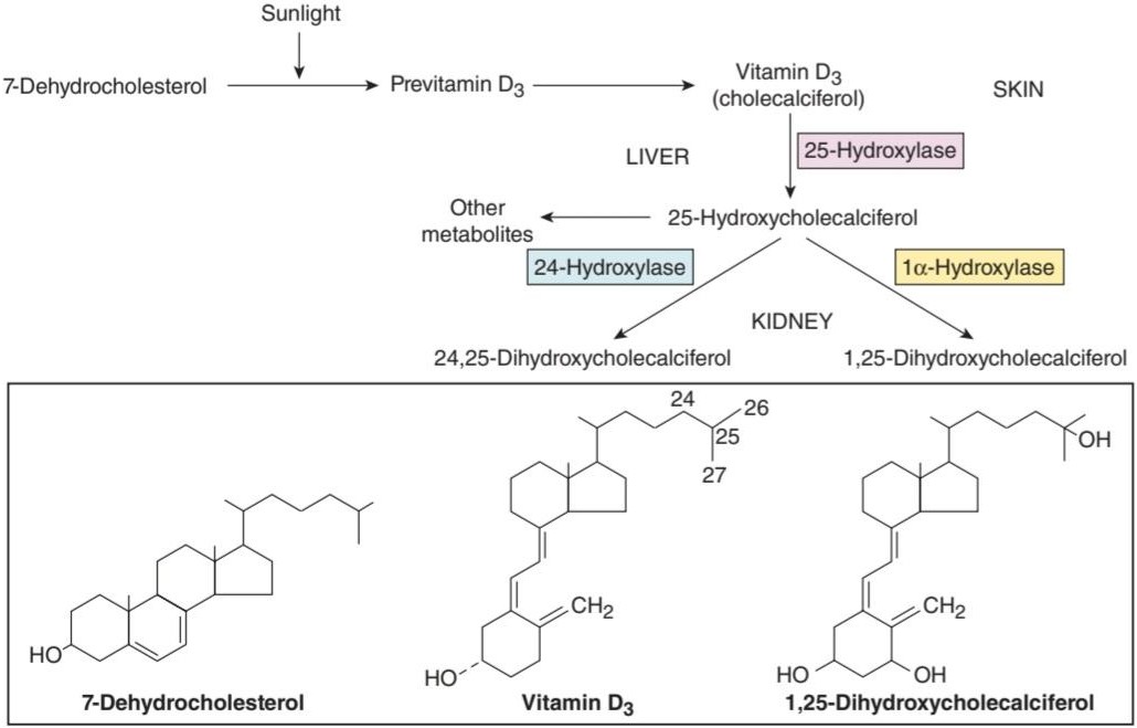
FIGURE 21-3 Formation and hydroxylation of vitamin D3. 25-Hydroxylation takes place in the liver, and the other hydroxylations occur primarily in the kidneys. The structures of 7-dehydrocholesterol, vitamin D3, and 1,25-dihydroxycholecalciferol are also shown in the boxed area.
شکل 21-3 تشکیل و هیدروکسیلاسیون ویتامین D3 . 25- هیدروکسیلاسیون در کبد صورت می گیرد و سایر هیدروکسیلاسیون ها عمدتاً در کلیه ها اتفاق می افتد. ساختارهای 7-دهیدروکلسترول، ویتامین D3 و 1،25-دی هیدروکسی کوله کلسیفرول نیز در قسمت جعبه نشان داده شده است.
Vitamin D3 is metabolized by enzymes that are members of the cytochrome P450 (CYP) superfamily (see Chapters 1 and 28). In the liver, vitamin D3 is converted to 25-hydroxycholecalciferol (calcidiol, 25-(OH)D3). The 25-hydroxycholecalciferol is further converted in the cells of the proximal tubules of the kidneys to the more active metabolite 1,25-dihydroxycholecalciferol, which is also called calcitriol or 1,25-(OH)2D3. 1,25-Dihydroxycholecalciferol is also made in the placenta, in keratinocytes in the skin, and in macrophages. The normal plasma level of 25-hydroxycholecalciferol is about 30 ng/mL, and that of 1,25-dihydroxycholecalciferol is about 0.03 ng/mL (approximately 100 pmol/L). The less active metabolite 24,25-dihydroxycholecalciferol is also formed in the kidneys (Figure 21-3).
ویتامین D3 توسط آنزیم هایی متابولیزه می شود که اعضای بالای خانواده سیتوکروم P450 (CYP) هستند (به فصل های 1 و 28 مراجعه کنید). در کبد، ویتامین D3 به 25-هیدروکسی کوله کلسیفرول (کلسیدیول، 25-(OH)D3) تبدیل می شود. 25-هیدروکسی کوله کلسیفرول بیشتر در سلول های لوله های پروگزیمال کلیه ها به متابولیت فعال تر 1,25-دی هیدروکسی کوله کلسیفرول تبدیل می شود که کلسیتریول یا 1،25-(OH)2D3 نیز نامیده می شود. 1,25-دی هیدروکسی کوله کلسیفرول نیز در جفت، در کراتینوسیت های پوست و در ماکروفاژها ساخته می شود. سطح طبیعی 25-هیدروکسی کوله کلسیفرول در پلاسما حدود 30 نانوگرم در میلی لیتر و 1،25-دی هیدروکسی کوله کلسیفرول حدود 0.03 نانوگرم در میلی لیتر (تقریباً 100 pmol/L) است. متابولیت کمتر فعال 24،25-دی هیدروکسی کوله کلسیفرول نیز در کلیه ها تشکیل می شود (شکل 21-3).
MECHANISM OF ACTION
1,25-Dihydroxycholecalciferol stimulates the expression of a number of gene products involved in Ca2+ transport and handling via its receptor (VDR), which acts as a transcriptional regulator in its ligand-bound form. One target is the family of calbindin-D proteins. These are members of the troponin C superfamily of Ca2+-binding proteins that also includes calmodulin (see Chapter 2). Calbindin-Ds are found in human intestine, brain, and kidneys. In the intestinal epithelium and many other tissues, two calbindins are induced: calbindin-D9K and calbindin-D28K, with molecular weights of 9000 and 28,000, respectively. 1,25-Dihydroxycholecalciferol also increases the number of Ca2+- ATPase and TRPV6 molecules in the intestinal cells, and thus, the overall capacity for absorption of dietary calcium.
مکانیسم عمل
1،25-دی هیدروکسی کوله کلسیفرول بیان تعدادی از محصولات ژنی را تحریک می کند که در انتقال و جابجایی +Ca2 از طریق گیرنده خود (VDR)، که به عنوان یک تنظیم کننده رونویسی در شکل متصل به لیگاند عمل می کند، نقش دارند. یک هدف خانواده پروتئین های calbindin-D است. اینها اعضای ابرخانواده تروپونین C پروتئین های متصل شونده به +Ca2 هستند که شامل کالمودولین نیز می شود (به فصل 2 مراجعه کنید). Calbindin-Ds در روده، مغز و کلیه انسان یافت می شود. در اپیتلیوم روده و بسیاری از بافتهای دیگر، دو کالبیندین القا میشوند: calbindin-D9K و calbindin-D28K، با وزنهای مولکولی به ترتیب 9000 و 28000. 1,25-دی هیدروکسی کوله کلسیفرول همچنین تعداد مولکول های Ca2+–ATPase و TRPV6 را در سلول های روده افزایش می دهد و در نتیجه ظرفیت کلی برای جذب کلسیم غذایی را افزایش می دهد.
In addition to increasing Ca2+ absorption from the intestine, 1,25- dihydroxycholecalciferol facilitates Ca2+ reabsorption in the kidneys via increased TRPV5 expression in the proximal tubules, increases the synthetic activity of osteoblasts, and is necessary for normal calcification of matrix (Clinical Box 21-1). The stimulation of osteoblasts brings about a secondary increase in the activity of osteoclasts (see below).
علاوه بر افزایش جذب +Ca2 از روده، 1،25-دی هیدروکسی کوله کلسیفرول از طریق افزایش بیان TRPV5 در لولههای پروگزیمال، بازجذب +Ca2 را در کلیهها تسهیل میکند، فعالیت سنتزی استئوبلاستها را افزایش میدهد و برای کلسیفیکاسیون طبیعی ماتریکس ضروری است (کادر بالینی 21-1). تحریک استئوبلاستها باعث افزایش ثانویه فعالیت استئوکلاستها میشود (به زیر مراجعه کنید).
CLINICAL BOX 21- 1
Rickets & Osteomalacia
Vitamin D deficiency causes defective calcification of bone matrix and the disease called rickets in children and osteomalacia in adults. Even though 1,25-dihydroxycholecalciferol is necessary for normal mineralization of bone matrix, the main defect in this condition is failure to deliver adequate amounts of Ca2+ and PO3 to the sites of mineralization. The full-blown condition in children is characterized by weakness and bowing of weight-bearing bones, dental defects, and hypocalcemia. In adults, the condition is less obvious. It used to be most commonly due to inadequate exposure to the sun in smoggy cities, but now it is more commonly due to inadequate intake of the provitamins on which the sun acts in the skin. These cases respond to administration of vitamin D. The condition can also be caused by inactivating mutations of the gene for renal 1a-hydroxylase, or in severe kidney or liver diseases, in which case there is no response to vitamin D but a normal response to 1,25-dihydroxycholecalciferol (type I vitamin D-resistant rickets). In rare instances, it can be due to inactivating mutations of the gene for the VDR (type II vitamin D-resistant rickets), in which case there is a deficient response to both vitamin D and 1,25-dihydroxycholecalciferol.
جعبه بالینی 21- 1
راشیتیسم و استئومالاسی
کمبود ویتامین D باعث کلسیفیکاسیون معیوب ماتریکس استخوان و بیماری به نام راشیتیسم در کودکان و استئومالاسی در بزرگسالان می شود. حتی اگر 1,25-دی هیدروکسی کوله کلسیفرول برای کانی سازی طبیعی ماتریکس استخوان ضروری است، نقص اصلی در این شرایط عدم تحویل مقادیر کافی از +Ca2 و PO3 به محل های معدنی شدن است. وضعیت کامل در کودکان با ضعف و خمیدگی استخوان های تحمل کننده وزن، نقص های دندانی و هیپوکلسمی مشخص می شود. در بزرگسالان، این وضعیت کمتر آشکار است. این بیماری معمولاً به دلیل قرار گرفتن ناکافی در معرض نور خورشید در شهرهای مه آلود بود، اما اکنون بیشتر به دلیل دریافت ناکافی پروویتامین هایی است که خورشید روی پوست بر روی آن تأثیر می گذارد. این موارد به تجویز ویتامین D پاسخ میدهند. این وضعیت همچنین میتواند ناشی از غیرفعال کردن جهشهای ژن 1a-هیدروکسیلاز کلیوی، یا در بیماریهای شدید کلیوی یا کبدی باشد، در این صورت هیچ پاسخی به ویتامین D وجود ندارد اما پاسخ طبیعی به 1,25-دی هیدروکسی کوله کلسیفرول (راشیتیسم نوع I مقاوم به ویتامین D) وجود دارد. در موارد نادر، میتواند به دلیل غیرفعال کردن جهشهای ژن VDR (راشیتیسم مقاوم به ویتامین D نوع II) باشد، که در این صورت پاسخ کمبودی به ویتامین D و 1,25-dihydroxycholecalciferol وجود دارد.
THERAPEUTIC HIGHLIGHTS
Treatment of these conditions depends on the underlying biochemical basis, as indicated above. Routine supplementation of milk with vitamin D has greatly reduced the occurrence of rickets in Western countries, but the condition remains among the most common childhood diseases in developing countries. Orthopedic surgery may be necessary in severely affected children.
نکات برجسته درمانی
درمان این شرایط به اساس بیوشیمیایی اساسی، همانطور که در بالا نشان داده شد، بستگی دارد. مکمل شیر با ویتامین D تا حد زیادی وقوع راشیتیسم را در کشورهای غربی کاهش داده است، اما این بیماری در میان شایع ترین بیماری های دوران کودکی در کشورهای در حال توسعه باقی مانده است. جراحی ارتوپدی ممکن است در کودکانی که به شدت مبتلا هستند ضروری باشد.
REGULATION OF SYNTHESIS
The formation of 25-hydroxycholecalciferol does not appear to be stringently regulated. However, the formation of 1,25 dihydroxycholecalciferol in the kidneys, which is catalyzed by the renal 1a-hydroxylase, is regulated in a feedback manner by plasma Ca2+ and PO43+ (Figure 21-4). When the plasma Ca2+ level is high, little 1,25-dihydroxycholecalciferol is produced, and the kidneys produce the relatively inactive metabolite 24,25- dihydroxycholecalciferol instead. This effect of Ca2+ on production of 1,25- dihydroxycholecalciferol thereby reduces the signal for Ca2+ absorption from the intestine (see previous text). Conversely, when the plasma Ca2+ level is low, PTH secretion is increased, and expression of 1a-hydroxylase is stimulated by PTH. The production of 1,25-dihydroxycholecalciferol is also increased by low plasma PO3 levels and inhibited by high plasma PO43 levels, by a direct inhibitory effect of PO43 on the 1a-hydroxylase. Additional control of 1,25- dihydroxycholecalciferol formation results from its direct negative feedback effect on 1a-hydroxylase activity, a positive feedback action on the formation of 24,25-dihydroxycholecalciferol, and a direct action on the parathyroid gland to inhibit PTH expression.
تنظیم سنتز
به نظر نمی رسد تشکیل 25-هیدروکسی کوله کلسیفرول به شدت تنظیم شود. با این حال، تشکیل 1،25 دی هیدروکسی کوله کلسیفرول در کلیه ها، که توسط 1a-هیدروکسیلاز کلیه کاتالیز می شود، به شیوه ای بازخورد توسط +Ca2 و PO43 پلاسما تنظیم می شود (شکل 21-4). هنگامی که سطح +Ca2 پلاسما بالا است، 1،25-دی هیدروکسی کوله کلسیفرول کمی تولید می شود و کلیه ها به جای آن، متابولیت نسبتاً غیر فعال 24،25- دی هیدروکسی کوله کلسیفرول را تولید می کنند. این اثر +Ca2 بر تولید 1,25- دی هیدروکسی کوله کلسیفرول در نتیجه سیگنال جذب +Ca2 از روده را کاهش می دهد (متن قبلی را ببینید). برعکس، وقتی سطح +Ca2 پلاسما کم است، ترشح PTH افزایش مییابد و بیان 1a-hydroxylase توسط PTH تحریک میشود. تولید 1,25-دی هیدروکسی کوله کلسیفرول نیز با سطوح پایین PO3 پلاسما افزایش می یابد و توسط سطوح بالای PO43 پلاسما، با اثر مهاری مستقیم PO43 بر روی 1a-هیدروکسیلاز، مهار می شود. کنترل اضافی تشکیل 1،25- دی هیدروکسی کوله کلسیفرول ناشی از اثر بازخورد منفی مستقیم آن بر فعالیت 1a-هیدروکسیلاز، یک اثر بازخورد مثبت در تشکیل 24،25-dihydroxycholecalciferol، و یک اثر مستقیم بر روی غده پاراتیروئید برای مهار بیان PTH است.

FIGURE 21-4 Effects of PTH and 1,25-dihydroxycholecalciferol on whole body calcium homeostasis. A reduction in plasma calcium stimulates parathyroid hormone secretion. PTH in turn causes calcium conservation and production of 1,25-dihydroxycholecalciferol in the kidneys, the latter of which increases calcium uptake in the intestine. PTH also releases calcium from the readily exchangeable pool in the bone. All of these actions act to restore normal plasma calcium. (Reproduced with permission from Widmaier EP, Raff H, Strang KT: Vander’s Human Physiology, 10th ed. New York, NY: McGraw-Hill; 2006.)
شکل 21-4 اثرات PTH و 1،25-دی هیدروکسی کوله کلسیفرول بر هموستاز کلسیم کل بدن. کاهش کلسیم پلاسما باعث تحریک ترشح هورمون پاراتیروئید می شود. PTH به نوبه خود باعث حفظ کلسیم و تولید 1,25-dihydroxycholecalciferol در کلیه ها می شود که دومی باعث افزایش جذب کلسیم در روده می شود. PTH همچنین کلسیم را از حوضچه به راحتی قابل تعویض در استخوان آزاد می کند. همه این اقدامات برای بازگرداندن کلسیم طبیعی پلاسما عمل می کنند. (تکثیر شده با اجازه Widmaier EP, Raff H, Strang KT: Vander’s Human Physiology, ed. 10th New York, NY: McGraw-Hill; 2006.)
An “anti-aging” protein called a-Klotho has also recently been discovered to play important roles in calcium and phosphate homeostasis, in part by reciprocal effects on 1,25-dihydroxycholecalciferol levels. Mice deficient in a-Klotho displayed accelerated aging, decreased bone mineral density, calcifications, and hypercalcemia and hyperphosphatemia. a-Klotho stabilizes the membrane localization of proteins important in calcium and phosphate (re)absorption, such as TRPV5 and Na, K ATPase. Likewise, it enhances the activity of another factor, fibroblast growth factor 23 (FGF23), at its receptor. FGF23 thereby decreases renal NaPi-IIa and NaPi-IIc expression and inhibits the production of 1a-hydroxylase, reducing levels of 1,25-dihydroxycholecalciferol.
همچنین اخیراً یک پروتئین “ضد پیری” به نام a-Klotho کشف شده است که نقش مهمی در هموستاز کلسیم و فسفات دارد، تا حدی با اثرات متقابل روی سطوح 1,25-dihydroxycholecalciferol. موشهای فاقد a-Klotho پیری سریع، کاهش تراکم استخوان، کلسیفیکاسیون و هیپرکلسمی و هیپرفسفاتمی را نشان دادند. a-Klotho محلی سازی غشایی پروتئین های مهم در جذب (باز) کلسیم و فسفات مانند TRPV5 و Na, K ATPase را تثبیت می کند. به همین ترتیب، فعالیت یک عامل دیگر، فاکتور رشد فیبروبلاست 23 (FGF23) را در گیرنده خود افزایش می دهد. FGF23 در نتیجه بیان NaPi-IIa و NaPi-IIc کلیوی را کاهش می دهد و تولید 1a-hydroxylase را مهار می کند و سطح 1,25-dihydroxycholecalciferol را کاهش می دهد.
THE PARATHYROID GLANDS
ANATOMY
Humans usually have four parathyroid glands: two embedded in the superior poles of the posterior thyroid and two in its inferior poles (Figure 21-5). Each parathyroid gland is a richly vascularized disk, about 3 × 6 × 2 mm, containing two distinct types of cells (Figure 21-6). The abundant chief cells, which contain a prominent Golgi apparatus plus endoplasmic reticulum and secretory granules, synthesize and secrete PTH. The less abundant and larger oxyphil cells contain oxyphil granules and large numbers of mitochondria in their cytoplasm. In humans, few oxyphil cells are seen before puberty, and thereafter they increase in number with age. Their function is unknown. Consequences of loss of the parathyroid glands are discussed in Clinical Box 21-2.
غدد پاراتیروئید
آناتومی
انسان ها معمولاً چهار غده پاراتیروئید دارند: دو غده در قطب های فوقانی تیروئید خلفی و دو غده در قطب های تحتانی آن تعبیه شده است (شکل 21-5). هر غده پاراتیروئید یک دیسک بسیار عروقی به ابعاد 3 × 6 × 2 میلی متر است که شامل دو نوع متمایز سلول است (شکل 21-6). سلولهای اصلی فراوان که حاوی دستگاه گلژی برجسته بهعلاوه شبکه آندوپلاسمی و گرانولهای ترشحی هستند، PTH را سنتز و ترشح میکنند. سلول های اکسیفیل کمتر و بزرگتر حاوی گرانول های اکسیفیل و تعداد زیادی میتوکندری در سیتوپلاسم خود هستند. در انسان، سلول های اکسیفیل کمی قبل از بلوغ دیده می شود و پس از آن با افزایش سن بر تعداد آنها افزوده می شود. عملکرد آنها ناشناخته است. پیامدهای از دست دادن غدد پاراتیروئید در کادر بالینی 21-2 مورد بحث قرار گرفته است.

FIGURE 21-5 The human parathyroid glands, viewed from behind. The glands are small structures adherent to the posterior surface of the thyroid gland.
شکل 21-5 غدد پاراتیروئید انسان، از پشت مشاهده می شود. غدد ساختارهای کوچکی هستند که به سطح خلفی غده تیروئید چسبیده اند.

FIGURE 21-6 Section of human parathyroid. (Reduced 50% from ×960.) Small cells are chief cells; large stippled cells (especially prominent in the lower left of picture) are oxyphil cells. (Reproduced with permission from Fawcett DW: Bloom and Fawcett, A Textbook of Histology, 11th ed. Saunders, 1986.)
شکل 21-6 بخش پاراتیروئید انسانی. (50% از 960 × کاهش یافته است.) سلول های کوچک سلول های اصلی هستند. سلول های برجسته بزرگ (به ویژه در سمت چپ پایین تصویر برجسته) سلول های اکسیفیل هستند. (تکثیر شده با اجازه Fawcett DW: Bloom and Fawcett, A Textbook of Histology, ed. 11th. Saunders, 1986.)
CLINICAL BOX 21- 2
Effects of Parathyroidectomy
Occasionally, inadvertent parathyroidectomy occurs in humans during thyroid surgery. This can have serious consequences as PTH is essential for life. After parathyroidectomy, there is a steady decline in the plasma Ca2+ level. Signs of neuromuscular hyperexcitability appear, followed by full-blown hypocalcemic tetany (see text). Plasma phosphate levels usually rise as the plasma Ca2+ level falls. Symptoms usually develop 2-3 days postoperatively but may not appear for several weeks or more. The signs of tetany in humans include Chvostek sign, a quick contraction of the ipsilateral facial muscles elicited by tapping over the facial nerve at the angle of the jaw, and Trousseau sign, a spasm of the muscles of the upper extremity that causes flexion of the wrist and thumb with extension of the fingers. In individuals with mild tetany in whom spasm is not yet evident, Trousseau sign can sometimes be produced by occluding the circulation for a few minutes with a blood pressure cuff.
جعبه بالینی 21-2
اثرات پاراتیروئیدکتومی
گاهی اوقات، پاراتیروئیدکتومی غیرعمدی در انسان در طی جراحی تیروئید رخ می دهد. این می تواند عواقب جدی داشته باشد زیرا PTH برای زندگی ضروری است. پس از پاراتیروئیدکتومی، کاهش مداوم سطح Ca2 پلاسما وجود دارد. علائم تحریک پذیری عصبی عضلانی و به دنبال آن تتانی هیپوکلسمیک کامل ظاهر می شود (به متن مراجعه کنید). سطح فسفات پلاسما معمولاً با کاهش سطح Ca2 پلاسما افزایش می یابد. علائم معمولاً 2-3 روز پس از عمل ظاهر می شوند، اما ممکن است تا چند هفته یا بیشتر ظاهر نشوند. علائم تتانی در انسان عبارتند از علامت Chvostek، انقباض سریع عضلات صورت که با ضربه زدن روی عصب صورت در زاویه فک ایجاد می شود، و علامت Trousseau، اسپاسم عضلات اندام فوقانی که باعث خم شدن مچ دست و شست با امتداد انگشتان می شود. در افراد مبتلا به کزاز خفیف که اسپاسم در آنها هنوز مشهود نیست، علامت Trousseau گاهی اوقات می تواند با مسدود کردن گردش خون برای چند دقیقه با کاف فشار خون ایجاد شود.
THERAPEUTIQ HIGHLIGHTS
Treatment centers around replacing the PTH that would normally be produced by the missing glands. Injections of PTH can be given to correct the chemical abnormalities, and the symptoms then disappear. Injections of Ca2+ salts can also give temporary relief.
نکات برجسته درمانی
تمرکز درمان بر روی جایگزینی PTH است که معمولاً توسط غدد از دست رفته تولید می شود. تزریق PTH را می توان برای اصلاح ناهنجاری های شیمیایی انجام داد و سپس علائم ناپدید می شوند. تزریق نمک های +Ca2 نیز می تواند باعث تسکین موقت شود.
SYNTHESIS & METABOLISM OF PTH
Human PTH is a linear polypeptide with a molecular weight of 9500 that contains 84 amino acid residues (Figure 21-7). It is synthesized as part of a larger molecule containing 115 amino acid residues (preproPTH). On entry of preproPTH into the endoplasmic reticulum, a leader sequence is removed from the amino terminal to form the 90-amino-acid polypeptide proPTH. Six additional amino acid residues are removed from the amino terminal of proPTH in the Golgi apparatus, and the 84-amino-acid polypeptide PTH is packaged in secretory granules and released as the main secretory product of the chief cells.
سنتز و متابولیسم PTH
PTH انسانی یک پلی پپتید خطی با وزن مولکولی 9500 است که حاوی 84 باقیمانده اسید آمینه است (شکل 21-7). این به عنوان بخشی از یک مولکول بزرگتر حاوی 115 باقی مانده اسید آمینه (preproPTH) سنتز می شود. با ورود preproPTH به شبکه آندوپلاسمی، یک توالی رهبر از ترمینال آمینه حذف می شود تا پلی پپتید 90 اسید آمینه proPTH را تشکیل دهد. شش باقی مانده اسید آمینه اضافی از پایانه آمینه proPTH در دستگاه گلژی حذف میشود و پلی پپتید 84 آمینو اسید PTH در گرانولهای ترشحی بستهبندی میشود و به عنوان محصول ترشحی اصلی سلولهای اصلی آزاد میشود.

FIGURE 21–7 Parathyroid hormone. The solid boxes show residues that are identical to, or similar to, those at the N-terminus of parathyroid hormone- related peptide (PTHrP). The dashed box indicates that the first 34 amino acids of PTH are sufficient for full biologic activity. Interestingly, this is also the case for PTHrP, although after the first 13 amino acids, its sequence is divergent from that of PTH.
شکل 21-7 هورمون پاراتیروئید. جعبههای جامد باقیماندههایی را نشان میدهند که مشابه یا مشابه آنهایی هستند که در انتهای N پپتید مرتبط با هورمون پاراتیروئید (PTHrP) قرار دارند. جعبه چین نشان می دهد که 34 اسید آمینه اول PTH برای فعالیت بیولوژیکی کامل کافی است. جالب اینجاست که این مورد برای PTHrP نیز صادق است، اگرچه بعد از 13 اسید آمینه اول، توالی آن از PTH متفاوت است.
The normal plasma level of intact PTH is 10-55 pg/mL. The half-life of PTH is approximately 10 min, and the secreted polypeptide is rapidly cleaved by Kupffer cells in the liver into inactive fragments. PTH and these fragments are then cleared by the kidneys. Currently used immunoassays for PTH are designed only to measure mature PTH (ie, 84 amino acids) and not its fragments in order to obtain an accurate measure of “active” PTH.
سطح طبیعی پلاسمایی PTH دست نخورده 10-55 pg/mL است. نیمه عمر PTH تقریباً 10 دقیقه است و پلی پپتید ترشح شده به سرعت توسط سلول های کوپفر در کبد به قطعات غیرفعال تبدیل می شود. سپس PTH و این قطعات توسط کلیه ها پاک می شوند. آزمایشهای ایمنی مورد استفاده در حال حاضر برای PTH فقط برای اندازهگیری PTH بالغ (یعنی 84 اسید آمینه) طراحی شدهاند و نه قطعات آن به منظور اندازهگیری دقیق PTH «فعال».
ACTIONS
PTH acts directly on bone to increase bone resorption and mobilize Ca2+. In addition to increasing plasma Ca2+, PTH increases phosphate excretion in the urine and thereby depresses plasma phosphate levels. This phosphaturic action is due to a decrease in reabsorption of phosphate via effects on NaPi-IIa in the proximal tubules, as discussed previously. PTH also increases reabsorption of Ca2+ in the distal tubules, although Ca2+ excretion in the urine is often increased in hyperparathyroidism because the increase in the load of filtered calcium overwhelms the effect on reabsorption (Clinical Box 21-3). PTH also increases the formation of 1,25-dihydroxycholecalciferol, and this increases Ca2+ absorption from the intestine. On a longer time scale, PTH stimulates both osteoblasts and osteoclasts.
اقدامات
PTH مستقیماً روی استخوان عمل می کند تا جذب استخوان را افزایش دهد و +Ca2 را بسیج کند. علاوه بر افزایش Ca2 پلاسما، PTH باعث افزایش دفع فسفات در ادرار و در نتیجه کاهش سطح فسفات پلاسما می شود. این عمل فسفاتوری به دلیل کاهش بازجذب فسفات از طریق اثرات روی NaPi-IIa در لوله های پروگزیمال است، همانطور که قبلاً بحث شد. PTH همچنین بازجذب +Ca2 را در لولههای دیستال افزایش میدهد، اگرچه دفع +Ca2 در ادرار اغلب در هیپرپاراتیروئیدیسم افزایش مییابد زیرا افزایش بار کلسیم فیلتر شده تأثیر بر جذب مجدد را تحت تأثیر قرار میدهد (کادر بالینی 21-3). PTH همچنین تشکیل 1,25-دی هیدروکسی کوله کلسیفرول را افزایش می دهد و این باعث افزایش جذب +Ca2 از روده می شود. در مقیاس زمانی طولانی تر، PTH هم استئوبلاست ها و هم استئوکلاست ها را تحریک می کند.
CLINICAL BOX 21- 3
Diseases of Parathyroid Excess
Hyperparathyroidism due to hypersecretion from a functioning parathyroid tumor in humans is characterized by hypercalcemia and hypophosphatemia. Humans with PTH-secreting adenomas are usually asymptomatic, with the condition detected when plasma Ca2+ is measured in conjunction with a routine physical examination. However, there may be minor changes in personality, and calcium-containing kidney stones occasionally form. In conditions such as chronic kidney disease and rickets, in which the plasma Ca2+ level is chronically low, the associated stimulation of the parathyroid glands causes compensatory parathyroid hypertrophy and secondary hyperparathyroidism. The plasma Ca2+ level is low in chronic kidney disease primarily because the diseased kidneys lose the ability to form 1,25- dihydroxycholecalciferol. Finally, mutations in the Ca2+-sensing receptor gene CASR cause predictable long-term changes in plasma Ca2+. Individuals heterozygous for inactivating mutations have familial benign hypocalciuric hypercalcemia, a condition in which there is a chronic moderate elevation in plasma Ca2+ because the feedback inhibition of PTH secretion by Ca2+ is reduced. Plasma PTH levels are normal or even elevated. However, children who are homozygous for inactivating mutations develop neonatal severe primary hyperparathyroidism. Conversely, individuals with gain-of-function mutations in the CASR gene develop familial hypercalciuric hypocalcemia due to increased sensitivity of the parathyroid glands to plasma Ca2+.
جعبه بالینی 21- 3
بیماری های پاراتیروئید بیش از حد
هیپرپاراتیروئیدیسم ناشی از ترشح بیش از حد از تومور پاراتیروئید فعال در انسان با هیپرکلسمی و هیپوفسفاتمی مشخص می شود. انسانهای مبتلا به آدنومهای ترشحکننده PTH معمولاً بدون علامت هستند و زمانی که +Ca2 پلاسما همراه با معاینه فیزیکی معمول اندازهگیری میشود، این وضعیت مشخص میشود. با این حال، ممکن است تغییرات جزئی در شخصیت وجود داشته باشد و گاهی اوقات سنگ کلیه حاوی کلسیم تشکیل شود. در شرایطی مانند بیماری مزمن کلیوی و راشیتیسم، که در آن سطح +Ca2 پلاسما به طور مزمن پایین است، تحریک غدد پاراتیروئید باعث هیپرتروفی جبرانی پاراتیروئید و هیپرپاراتیروئیدیسم ثانویه می شود. سطح کلسیم پلاسما در بیماری مزمن کلیوی پایین است، عمدتاً به این دلیل که کلیه های بیمار توانایی تشکیل 1،25- دی هیدروکسی کوله کلسیفرول را از دست می دهند. در نهایت، جهش در CASR ژن گیرنده +Ca2 حسگر باعث تغییرات طولانی مدت قابل پیش بینی در +Ca2 پلاسما می شود. افراد هتروزیگوت برای جهش های غیرفعال، هیپرکلسمی هیپوکلسیوری خوش خیم خانوادگی دارند، وضعیتی که در آن افزایش مزمن متوسط در +Ca2 پلاسما وجود دارد، زیرا مهار بازخورد ترشح PTH توسط +Ca2 کاهش می یابد. سطح PTH پلاسما طبیعی یا حتی افزایش یافته است. با این حال، کودکانی که هموزیگوت هستند برای جهشهای غیرفعال، دچار هیپرپاراتیروئیدیسم اولیه شدید نوزادی میشوند. برعکس، افرادی که دارای جهش های افزایش عملکرد در ژن CASR هستند، به دلیل افزایش حساسیت غدد پاراتیروئید به پلاسما +Ca2، هیپوکلسمی هیپرکلسیوری خانوادگی ایجاد می کنند.
THERAPEUTIQ HIGHLIGHTS
Subtotal parathyroidectomy is sometimes necessary in patients in whom parathyroid adenoma or hyperplasia with associated hypercalcemia and resulting symptoms develops. However, because parathyroid disease is often benign or only slowly progressing, surgery remains controversial in most patients and is typically reserved for those who have experienced life- threatening complications of hypercalcemia.
نکات برجسته درمانی
پاراتیروئیدکتومی ساب توتال گاهی اوقات در بیمارانی که آدنوم یا هیپرپلازی پاراتیروئید همراه با هیپرکلسمی همراه و علائم ناشی از آن ایجاد می شود، ضروری است. با این حال، از آنجایی که بیماری پاراتیروئید اغلب خوشخیم است یا به آهستگی پیشرفت میکند، جراحی در اکثر بیماران بحثبرانگیز است و معمولاً برای کسانی که عوارض تهدیدکننده زندگی هیپرکلسمی را تجربه کردهاند، اختصاص دارد.
MECHANISM OF ACTION
It now appears that there are at least three different PTH receptors. One also binds parathyroid hormone-related protein (PTHrP; see below) and is known as the hPTH/PTHrP receptor. A second receptor, PTH2 (hPTH2-R), does not bind PTHrP and is found in the brain, placenta, and pancreas. In addition, there is evidence for a third receptor, CPTH, which reacts with the carboxyl terminal rather than the amino terminal of PTH. The first two receptors are coupled to G,, and via this heterotrimeric G-protein they activate adenylyl cyclase, increasing intracellular CAMP. The hPTH/PTHrP receptor also activates PLC via G, increasing intracellular Ca2+ concentrations and activating protein kinase C (Figure 21-8). However, the way these second messengers affect Ca2+ in bone is unsettled.
مکانیسم عمل
اکنون به نظر می رسد که حداقل سه گیرنده مختلف PTH وجود دارد. یکی همچنین پروتئین مرتبط با هورمون پاراتیروئید (PTHrP؛ به زیر مراجعه کنید) متصل می شود و به عنوان گیرنده hPTH/PTHrP شناخته می شود. گیرنده دوم، PTH2 (hPTH2-R)، به PTHrP متصل نمی شود و در مغز، جفت و پانکراس یافت می شود. علاوه بر این، شواهدی برای گیرنده سوم، CPTH، وجود دارد که با پایانه کربوکسیل به جای پایانه آمینه PTH واکنش نشان می دهد. دو گیرنده اول با G جفت می شوند و از طریق این پروتئین G هتروتریمری آدنیلیل سیکلاز را فعال می کنند و CAMP داخل سلولی را افزایش می دهند. گیرنده hPTH/PTHrP همچنین PLC را از طریق G فعال می کند و غلظت +Ca2 داخل سلولی را افزایش می دهد و پروتئین کیناز C را فعال می کند (شکل 21-8). با این حال، روشی که این پیامرسانهای دوم روی +Ca2 در استخوان تأثیر میگذارند، ثابت نیست.
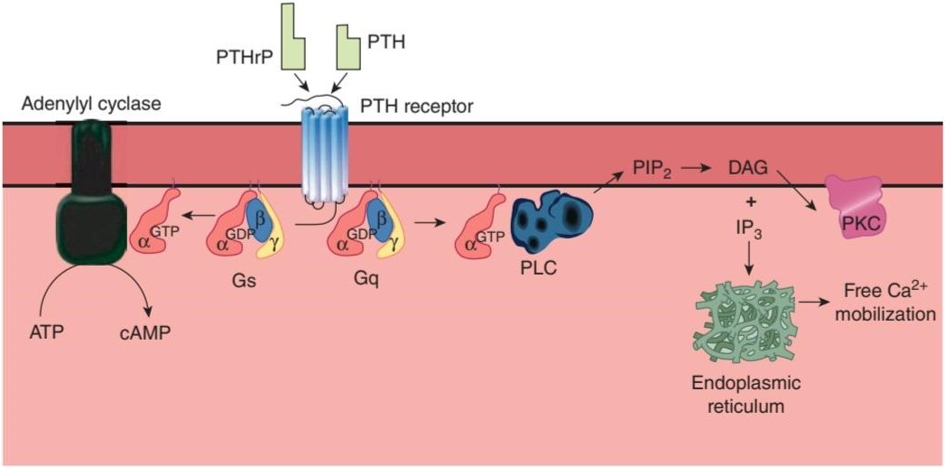
FIGURE 21-8 Signal transduction pathways activated by PTH or PTHrP binding to the hPTH/PTHrP receptor. Intracellular cAMP is increased via G and adenylyl cyclase (AC). Diacylglycerol (DAG) and inositol 1,4,5- trisphosphate (IP3) are increased via G, and phospholipase C (PLC). DAG and IP3 activate protein kinase C (PKC) and mobilization of calcium from the endoplasmic reticulum.
شکل 21-8 مسیرهای انتقال سیگنال فعال شده با اتصال PTH یا PTHrP به گیرنده hPTH/PTHrP. cAMP داخل سلولی از طریق G و آدنیلیل سیکلاز (AC) افزایش می یابد. دی آسیل گلیسرول (DAG) و اینوزیتول 1،4،5- تری فسفات (IP3) از طریق G و فسفولیپاز C (PLC) افزایش می یابد. DAG و IP3 پروتئین کیناز C (PKC) را فعال می کنند و کلسیم را از شبکه آندوپلاسمی بسیج می کنند.
In the disease called pseudohypoparathyroidism, the signs and symptoms of hypoparathyroidism develop but the circulating level of PTH is normal or even elevated. Because tissues fail to respond to the hormone, this is a receptor disease. There are two forms. In the more common form, a congenital 50% reduction of the activity of G, occurs and PTH fails to produce a normal increase in cAMP concentration. In a different, less common form, the cAMP response is normal but the phosphaturic action of the hormone is defective.
در بیماری کاذب هیپوپاراتیروئیدیسم، علائم و نشانه های هیپوپاراتیروئیدیسم ایجاد می شود اما سطح گردش خون PTH طبیعی یا حتی افزایش یافته است. از آنجایی که بافت ها به هورمون پاسخ نمی دهند، این یک بیماری گیرنده است. دو شکل وجود دارد. در شکل رایج تر، کاهش 50 درصدی مادرزادی فعالیت G رخ می دهد و PTH نمی تواند افزایش طبیعی در غلظت cAMP ایجاد کند. در شکل متفاوت و کمتر رایج، پاسخ cAMP طبیعی است اما عملکرد فسفاتوری هورمون معیوب است.
REGULATION OF SECRETION
Circulating Ca2+ acts directly on the parathyroid glands in a negative feedback manner to regulate the secretion of PTH. The key to this regulation is a cell membrane Ca2+ sensing receptor, CaSR. Activation of this G-protein-coupled receptor leads to phosphoinositide turnover in many tissues. In the parathyroid, its activation inhibits PTH secretion. In this way, when the plasma Ca2+ level is high, PTH secretion is inhibited and Ca2+ is deposited in the bones. When it is low, secretion is increased and Ca2+ is mobilized from the bones.
مقررات ترشح
+Ca2 در گردش به طور مستقیم بر روی غدد پاراتیروئید به شیوه ای بازخورد منفی برای تنظیم ترشح PTH عمل می کند. کلید این تنظیم یک گیرنده سنجش +Ca2 غشای سلولی، CaSR است. فعال شدن این گیرنده جفت شده با پروتئین G منجر به گردش فسفوئینوزیتید در بسیاری از بافت ها می شود. در پاراتیروئید، فعال شدن آن باعث مهار ترشح PTH می شود. به این ترتیب زمانی که سطح +Ca2 پلاسما بالا باشد، ترشح PTH مهار شده و +Ca2 در استخوان ها رسوب می کند. وقتی کم باشد، ترشح افزایش می یابد و +Ca2 از استخوان ها بسیج می شود.
1,25-Dihydroxycholecalciferol acts directly on the parathyroid glands to decrease preproPTH mRNA. Increased plasma P; stimulates PTH secretion by lowering plasma levels of free Ca2+ and inhibiting the formation of 1,25- dihydroxycholecalciferol. Magnesium is required to maintain normal parathyroid secretory responses. Impaired PTH release along with diminished target organ responses to PTH account for the hypocalcemia that occasionally occurs in magnesium deficiency (Clinical Boxes 21-2 and 21-3).
1،25-دی هیدروکسی کوله کلسیفرول به طور مستقیم بر روی غدد پاراتیروئید برای کاهش mRNA preproPTH عمل می کند. افزایش P پلاسما؛ ترشح PTH را با کاهش سطح پلاسمایی +Ca2 آزاد و مهار تشکیل 1,25- دی هیدروکسی کوله کلسیفرول تحریک می کند. منیزیم برای حفظ پاسخ های ترشحی طبیعی پاراتیروئید مورد نیاز است. اختلال در انتشار PTH همراه با کاهش پاسخ اندام هدف به PTH دلیل هیپوکلسمی است که گاهی در کمبود منیزیم رخ می دهد (جعبه های بالینی 21-2 و 21-3).
PTHrP
PTHrP, another protein with PTH activity, is produced by many different tissues in the body. PTHrP and PTH have marked homology at their amino terminal ends and they both bind to the hPTH/PTHrP receptor, yet their physiologic effects are very different. How is this possible when they bind to the same receptor? For one thing, PTHrP is primarily a paracrine factor, acting close to where it is produced. It may be that circulating PTH cannot reach at least some of these sites. Second, subtle conformational differences may be produced by binding of PTH versus PTHrP to their receptor, despite their structural similarities. Another possibility is action of one or the other hormone on additional, more selective receptors.
PTHrP
PTHrP، پروتئین دیگری با فعالیت PTH، توسط بسیاری از بافت های مختلف بدن تولید می شود. PTHrP و PTH همولوژی مشخصی در انتهای آمینو انتهایی خود دارند و هر دو به گیرنده hPTH/PTHrP متصل میشوند، اما اثرات فیزیولوژیکی آنها بسیار متفاوت است. وقتی آنها به یک گیرنده متصل می شوند چگونه ممکن است؟ برای یک چیز، PTHrP در درجه اول یک عامل پاراکرین است که نزدیک به محل تولید آن عمل می کند. ممکن است PTH در گردش نتواند حداقل به برخی از این مکان ها برسد. دوم، تفاوتهای ساختاری ظریف ممکن است با اتصال PTH در مقابل PTHrP به گیرنده آنها، علیرغم شباهتهای ساختاری آنها، ایجاد شود. احتمال دیگر، اثر یک یا آن هورمون بر روی گیرنده های انتخابی بیشتر است.
PTHrP has a marked effect on the growth and development of cartilage in utero. Mice in which both alleles of the PTHrP gene are knocked out have severe skeletal deformities and die soon after birth. In normal animals, on the other hand, PTHrP-stimulated cartilage cells proliferate and their terminal differentiation is inhibited. PTHrP is also expressed in the brain, where evidence indicates that it inhibits excitotoxic damage to developing neurons. In addition, there is evidence that it is involved in Ca2+ transport in the placenta. PTHrP is also found in keratinocytes in the skin, in smooth muscle, and in the teeth, where it is present in the enamel epithelium that caps each tooth. In the absence of PTHrP, teeth cannot erupt.
PTHrP تأثیر قابل توجهی بر رشد و توسعه غضروف در رحم دارد. موش هایی که در آنها هر دو آلل ژن PTHrP حذف شده اند، بدشکلی های اسکلتی شدید دارند و بلافاصله پس از تولد می میرند. از طرف دیگر در حیوانات عادی سلول های غضروفی تحریک شده با PTHrP تکثیر می شوند و تمایز نهایی آنها مهار می شود. PTHrP همچنین در مغز بیان میشود، جایی که شواهد نشان میدهد که آسیب تحریککننده به نورونهای در حال رشد را مهار میکند. علاوه بر این، شواهدی وجود دارد که نشان میدهد در انتقال +Ca2 در جفت نقش دارد. PTHrP همچنین در کراتینوسیتهای پوست، ماهیچههای صاف و دندانها، جایی که در اپیتلیوم مینای دندان وجود دارد، یافت میشود. در غیاب PTHrP، دندان ها نمی توانند رویش کنند.
HYPERCALCEMIA OF MALIGNANCY
Hypercalcemia is a common metabolic complication of cancer. About 20% of hypercalcemic patients have bone metastases that produce hypercalcemia secondary to bone erosion (local osteolytic hypercalcemia). Evidence suggests that this erosion is produced by prostaglandins such as prostaglandin E2 arising from the tumor. Hypercalcemia in the remaining 80% of the patients is due to elevated circulating levels of PTHrP (humoral hypercalcemia of malignancy). The tumors responsible for this hypersecretion include cancers of the breast, kidney, ovary, and skin.
هیپرکلسمی بدخیم
هیپرکلسمی یکی از عوارض متابولیکی شایع سرطان است. حدود 20 درصد از بیماران هیپرکلسمیک متاستازهای استخوانی دارند که باعث ایجاد هیپرکلسمی ثانویه به فرسایش استخوان (هیپرکلسمی استئولیتیک موضعی) می شود. شواهد نشان می دهد که این فرسایش توسط پروستاگلاندین هایی مانند پروستاگلاندین E2 ناشی از تومور ایجاد می شود. هیپرکلسمی در 80 درصد بقیه بیماران به دلیل افزایش سطح گردش خون PTHrP (هیپرکلسمی هومورال بدخیمی) است. تومورهای مسئول این ترشح بیش از حد عبارتند از سرطان سینه، کلیه، تخمدان و پوست.
CALCITONIN
ORIGIN
In dogs, perfusion of the thyroparathyroid region with solutions containing high concentrations of Ca2+ leads to a fall in peripheral plasma Ca2+, and after damage to this region, Ca2+ infusions cause a greater increase in plasma Ca2+ than they do in control animals. These and other observations led to the discovery that a Ca2+-lowering as well as a Ca2+-elevating peptide hormone was secreted by structures in the neck. This Ca2+-lowering hormone has been named calcitonin. In mammals, calcitonin is produced by the parafollicular cells of the thyroid gland, which are also known as the clear or C cells.
کلسیتونین
مبدا
در سگها، پرفیوژن ناحیه تیروپاراتیروئید با محلولهای حاوی غلظتهای بالای +Ca2 منجر به سقوط +Ca2 پلاسمای محیطی میشود و پس از آسیب به این ناحیه، تزریق +Ca2 باعث افزایش بیشتر +Ca2 پلاسما نسبت به حیوانات کنترل میشود. این مشاهدات و مشاهدات دیگر منجر به کشف این شد که یک هورمون پپتیدی کاهشدهنده +Ca2 و همچنین یک هورمون پپتیدی افزایشدهنده +Ca2 توسط ساختارهای گردن ترشح میشود. این هورمون کاهش دهنده +Ca2 کلسیتونین نامگذاری شده است. در پستانداران، کلسی تونین توسط سلول های پارافولیکولی غده تیروئید تولید می شود که به سلول های شفاف یا C نیز معروف هستند.
SECRETION & METABOLISM
Calcitonin secretion is increased when the thyroid gland is exposed to a plasma calcium level of approximately 9.5 mg/dL. Above this level, plasma calcitonin is directly proportional to plasma calcium. ẞ-Adrenergic agonists, dopamine, and estrogens also stimulate calcitonin secretion. Gastrin, cholecystokinin (CCK), glucagon, and secretin have also been reported to stimulate calcitonin secretion, with gastrin being the most potent stimulus (see Chapter 25). Thus, the plasma calcitonin level is elevated in Zollinger-Ellison syndrome and in pernicious anemia (see Chapter 25). However, the dose of gastrin needed to stimulate calcitonin secretion is supraphysiologic and not seen after eating in normal individuals, so dietary calcium in the intestine probably does not induce secretion of a calcium-lowering hormone prior to the calcium being absorbed. In any event, the actions of calcitonin are short-lived because it has a half-life of less than 10 min in humans.
ترشح و متابولیسم
هنگامی که غده تیروئید در معرض سطح کلسیم پلاسما حدود 9.5 میلی گرم در دسی لیتر قرار می گیرد، ترشح کلسی تونین افزایش می یابد. بالاتر از این سطح، کلسی تونین پلاسما با کلسیم پلاسما نسبت مستقیم دارد. آگونیستهای آدرنرژیک، دوپامین و استروژنها نیز ترشح کلسیتونین را تحریک میکنند. همچنین گزارش شده است که گاسترین، کوله سیستوکینین (CCK)، گلوکاگون و سکرتین ترشح کلسی تونین را تحریک می کنند و گاسترین قوی ترین محرک است (به فصل 25 مراجعه کنید). بنابراین، سطح کلسی تونین پلاسما در سندرم زولینگر-الیسون و در کم خونی پرنیشیوز افزایش می یابد (به فصل 25 مراجعه کنید). با این حال، دوز گاسترین مورد نیاز برای تحریک ترشح کلسی تونین فوق فیزیولوژیک است و پس از خوردن غذا در افراد عادی دیده نمی شود، بنابراین کلسیم رژیم غذایی در روده احتمالاً قبل از جذب کلسیم باعث ترشح هورمون کاهش دهنده کلسیم نمی شود. در هر صورت، عملکرد کلسی تونین کوتاه مدت است زیرا نیمه عمر آن در انسان کمتر از 10 دقیقه است.
ACTIONS
Receptors for calcitonin are found in bones and the kidneys. Calcitonin lowers circulating calcium and phosphate levels. It exerts its calcium-lowering effect by inhibiting bone resorption. This action is direct, and calcitonin also inhibits the activity of osteoclasts in vitro. It also increases Ca2+ excretion in the urine.
اقدامات
گیرنده های کلسی تونین در استخوان ها و کلیه ها یافت می شوند. کلسی تونین سطح کلسیم و فسفات در گردش را کاهش می دهد. این اثر کاهش دهنده کلسیم خود را با مهار تحلیل استخوان اعمال می کند. این عمل مستقیم است و کلسی تونین همچنین فعالیت استئوکلاست ها را در شرایط آزمایشگاهی مهار می کند. همچنین باعث افزایش دفع +Ca2 در ادرار می شود.
The exact physiologic role of calcitonin is uncertain. The calcitonin content of the human thyroid is low, and after thyroidectomy, bone density and plasma Ca2+ level are normal as long as the parathyroid glands are intact. In addition, after thyroidectomy, there are only transient abnormalities of Ca2+ homeostasis when a Ca2+ load is injected. This may be explained in part by secretion of calcitonin from tissues other than the thyroid. However, there is general agreement that the hormone has little long-term effect on the plasma Ca2+ level in adult animals and humans. Further, unlike PTH and 1,25 dihydroxycholecalciferol, calcitonin does not appear to be involved in phosphate homeostasis. Moreover, patients with medullary carcinoma of the thyroid have a very high circulating calcitonin level but no symptoms directly attributable to the hormone, and their bones are essentially normal. No syndrome due to calcitonin deficiency has been described. More hormone is secreted in young individuals, and it may play a role in skeletal development. In addition, it may protect the bones of the mother from excess calcium loss during pregnancy. Bone formation in the infant and lactation are major drains on Ca2+ stores, and plasma concentrations of 1,25-dihydroxycholecalciferol are elevated in pregnancy. They would cause bone loss in the mother if bone resorption were not simultaneously inhibited by an increase in the plasma calcitonin level.
نقش فیزیولوژیکی دقیق کلسی تونین نامشخص است. محتوای کلسی تونین تیروئید انسان کم است و پس از تیروئیدکتومی، تا زمانی که غدد پاراتیروئید دست نخورده باشند، تراکم استخوان و سطح +Ca2 پلاسما طبیعی است. علاوه بر این، پس از تیروئیدکتومی، تنها ناهنجاری های گذرا در هموستاز +Ca2 زمانی که یک بار +Ca2 تزریق می شود وجود دارد. این ممکن است تا حدی با ترشح کلسی تونین از بافت هایی غیر از تیروئید توضیح داده شود. با این حال، توافق کلی وجود دارد که این هورمون در درازمدت اثر کمی بر سطح +Ca2 پلاسما در حیوانات بالغ و انسان دارد. علاوه بر این، بر خلاف PTH و 1.25 دی هیدروکسی کوله کلسیفرول، به نظر نمی رسد که کلسی تونین در هموستاز فسفات نقش داشته باشد. علاوه بر این، بیماران مبتلا به کارسینوم مدولاری تیروئید سطح کلسی تونین در گردش خون بسیار بالایی دارند، اما هیچ علامتی مستقیماً به این هورمون نسبت داده نمیشوند و استخوانهای آنها اساساً طبیعی هستند. هیچ سندرم ناشی از کمبود کلسیتونین شرح داده نشده است. هورمون بیشتری در افراد جوان ترشح می شود و ممکن است در رشد اسکلتی نقش داشته باشد. علاوه بر این، ممکن است از استخوان های مادر در برابر از دست دادن بیش از حد کلسیم در دوران بارداری محافظت کند. تشکیل استخوان در نوزاد و شیردهی تخلیه اصلی ذخایر +Ca2 است و غلظت 1,25-دی هیدروکسی کوله کلسیفرول پلاسما در بارداری افزایش می یابد. اگر همزمان با افزایش سطح کلسی تونین پلاسما، تحلیل استخوان مهار نشود، باعث از دست دادن استخوان در مادر می شود.
SUMMARY OF CALCIUM HOMEOSTATIC MECHANISMS
The actions of the three principal hormones that regulate the plasma concentration of Ca2+ can now be summarized. PTH increases plasma Ca2+ by mobilizing this ion from bone. It increases Ca2+ reabsorption in the kidney, but this may be offset by the increase in filtered Ca2+. It also increases the formation of 1,25-dihydroxycholecalciferol. 1,25-Dihydroxycholecalciferol increases Ca2+ absorption from the intestine and increases Ca2+ reabsorption in the kidneys. Calcitonin inhibits bone resorption and increases the amount of Ca2+ in the urine.
خلاصه مکانیسم های هموستاتیک کلسیم
اکنون می توان فعالیت های سه هورمون اصلی را که غلظت پلاسمایی +Ca2 را تنظیم می کنند، خلاصه کرد. PTH با به حرکت درآوردن این یون از استخوان، +Ca2 پلاسما را افزایش می دهد. بازجذب +Ca2 را در کلیه افزایش می دهد، اما ممکن است با افزایش +Ca2 فیلتر شده جبران شود. همچنین تشکیل 1,25-دی هیدروکسی کوله کلسیفرول را افزایش می دهد. 1,25-دی هیدروکسی کوله کلسیفرول باعث افزایش جذب +Ca2 از روده و افزایش جذب مجدد +Ca2 در کلیه ها می شود. کلسی تونین جذب استخوان را مهار کرده و مقدار +Ca2 را در ادرار افزایش می دهد.
EFFECTS OF OTHER HORMONES & HUMORAL AGENTS ON CALCIUM METABOLISM
Calcium metabolism is affected by various hormones in addition to 1,25- dihydroxycholecalciferol, PTH, and calcitonin. Glucocorticoids lower plasma Ca2+ levels by inhibiting osteoclast formation and activity acutely, but over long periods they cause osteoporosis by decreasing bone formation and increasing bone resorption. They decrease bone formation by inhibiting protein synthesis in osteoblasts. They also decrease the absorption of Ca2+ and PO43- from the intestine and increase the renal excretion of these ions. The decrease in plasma Ca2+ concentration also increases the secretion of PTH, and bone resorption is facilitated. Growth hormone increases Ca2+ excretion in the urine, but it also increases intestinal absorption of Ca2+, and this effect may be greater than the effect on excretion, with a resultant positive calcium balance. Insulin-like growth factor I (IGF-I) generated by the action of growth hormone stimulates protein synthesis in bone. As noted previously, thyroid hormones may cause hypercalcemia, hypercalciuria, and, in some instances, osteoporosis. Estrogens prevent osteoporosis by inhibiting the stimulatory effects of certain cytokines on osteoclasts. Insulin increases bone formation, and there is significant bone loss in untreated diabetes.
تأثیر سایر هورمون ها و عوامل شوخ طبعی بر متابولیسم کلسیم
متابولیسم کلسیم علاوه بر 1،25- دی هیدروکسی کوله کلسیفرول، PTH و کلسی تونین تحت تأثیر هورمون های مختلف قرار می گیرد. گلوکوکورتیکوئیدها با مهار تشکیل و فعالیت استئوکلاست ها به طور حاد سطح +Ca2 پلاسما را کاهش می دهند، اما در دوره های طولانی با کاهش تشکیل استخوان و افزایش تحلیل استخوان باعث پوکی استخوان می شوند. آنها با مهار سنتز پروتئین در استئوبلاست ها باعث کاهش تشکیل استخوان می شوند. همچنین جذب +Ca2 و PO43- از روده را کاهش داده و دفع کلیوی این یون ها را افزایش می دهند. کاهش غلظت +Ca2 پلاسما نیز باعث افزایش ترشح PTH می شود و تحلیل استخوان تسهیل می شود. هورمون رشد باعث افزایش دفع +Ca2 در ادرار میشود، اما جذب رودهای +Ca2 را نیز افزایش میدهد و این تأثیر ممکن است بیشتر از تأثیر بر دفع باشد و در نتیجه تعادل کلسیم مثبت حاصل شود. فاکتور رشد شبه انسولین I (IGF-I) که توسط عملکرد هورمون رشد تولید می شود، سنتز پروتئین را در استخوان تحریک می کند. همانطور که قبلا ذکر شد، هورمون های تیروئید ممکن است باعث هیپرکلسمی، هیپرکلسیوری و در برخی موارد پوکی استخوان شوند. استروژن ها با مهار اثرات تحریکی سیتوکین های خاص روی استئوکلاست ها از پوکی استخوان جلوگیری می کنند. انسولین تشکیل استخوان را افزایش می دهد و در دیابت درمان نشده از دست دادن استخوان قابل توجهی وجود دارد.
BONE PHYSIOLOGY
Bone is a special form of connective tissue with a collagen framework impregnated with Ca2+ and PO43 salts, particularly hydroxyapatites, which have the general formula Ca10(PO4)6(OH)2. Bone is also involved in overall Ca2+ and PO43-homeostasis. It protects vital organs, and the rigidity it provides permits locomotion and the support of loads against gravity. Old bone is constantly being resorbed and new bone formed, permitting remodeling that allows it to respond to the stresses and strains that are put upon it. It is a living tissue that is well vascularized and has a total blood flow of 200-400 mL/min in adult humans.
فیزیولوژی استخوان
استخوان شکل خاصی از بافت همبند با چارچوب کلاژن آغشته به نمک های +Ca2 و PO43، به ویژه هیدروکسی آپاتیت ها، که دارای فرمول کلی Ca10(PO4)6(OH)2 هستند، است. استخوان همچنین در هموستاز کلی +Ca2 و PO43 دخیل است. از اندامهای حیاتی محافظت میکند و استحکامی که ایجاد میکند اجازه حرکت و حمایت بارها در برابر گرانش را میدهد. استخوان قدیمی دائماً در حال جذب است و استخوان جدید تشکیل میشود و امکان بازسازی را فراهم میکند که به آن اجازه میدهد به تنشها و فشارهایی که بر آن وارد میشود پاسخ دهد. این بافت زنده ای است که به خوبی عروقی شده است و در انسان بالغ دارای جریان خون 200-400 میلی لیتر در دقیقه است.
STRUCTURE
There are two types of bone: compact or cortical bone, which makes up the outer layer of most bones (Figure 21-9) and accounts for 80% of the bone in the body; and trabecular or spongy bone inside the cortical bone, which makes up the remaining 20% of bone in the body. In compact bone, the surface-to-volume ratio is low, and bone cells lie in lacunae. They receive nutrients by way of canaliculi that ramify throughout the compact bone (Figure 21-9). Trabecular bone is made up of spicules or plates, with a high surface to volume ratio and many cells sitting on the surface of the plates. Nutrients diffuse from bone extracellular fluid (ECF) into the trabeculae, but in compact bone, nutrients are provided via haversian canals (Figure 21-9), which contain blood vessels. Around each haversian canal, collagen is arranged in concentric layers, forming cylinders called osteons or haversian systems.
ساختار
دو نوع استخوان وجود دارد: استخوان فشرده یا کورتیکال، که لایه بیرونی اکثر استخوان ها را تشکیل می دهد (شکل 21-9) و 80 درصد از استخوان بدن را تشکیل می دهد. و استخوان ترابکولار یا اسفنجی داخل استخوان قشر مغز که 20 درصد باقیمانده استخوان بدن را تشکیل می دهد. در استخوان فشرده، نسبت سطح به حجم کم است و سلول های استخوانی در لاکونا قرار دارند. آنها مواد مغذی را از طریق کانالی دریافت می کنند که در سرتاسر استخوان فشرده منشعب می شود (شکل 21-9). استخوان ترابکولار از اسپیکول ها یا صفحات با نسبت سطح به حجم بالا و تعداد زیادی سلول روی سطح صفحات تشکیل شده است. مواد مغذی از مایع خارج سلولی استخوان (ECF) به داخل ترابکولها منتشر میشوند، اما در استخوان فشرده، مواد مغذی از طریق کانالهای هارسین (شکل 21-9)، که حاوی رگهای خونی هستند، تامین میشوند. در اطراف هر کانال هارسین، کلاژن در لایه های متحدالمرکز قرار گرفته و استوانه هایی به نام استئون ها یا سیستم های هارسین را تشکیل می دهد.

FIGURE 21-9 Structure of compact and trabecular bone. The compact bone is shown in horizontal section (top) and vertical section (bottom). (Reproduced with permission from Williams PL et al (editors): Gray’s Anatomy, 37th ed. Churchill Livingstone; 1989.)
شکل 21-9 ساختار استخوان فشرده و ترابکولار. استخوان فشرده در بخش افقی (بالا) و بخش عمودی (پایین) نشان داده شده است. (تکثیر شده با اجازه ویلیامز PL و همکاران (ویراستاران): Gray’s Anatomy, 37th ed. Churchill Livingstone, 1989.)
The protein in bone matrix is over 90% type I collagen, which is also the major structural protein in tendons and skin. This collagen, which weight for weight is as strong as steel, is made up of a triple helix of three polypeptides bound tightly together. Two of these are identical a1 polypeptides encoded by one gene, and one is an a2 polypeptide encoded by a different gene. Collagens make up a family of structurally related proteins that maintain the integrity of many different organs.
پروتئین موجود در ماتریکس استخوان بیش از 90 درصد از کلاژن نوع I است که همچنین پروتئین ساختاری اصلی در تاندون ها و پوست است. این کلاژن، که وزن آن به اندازه فولاد قوی است، از یک مارپیچ سه گانه از سه پلی پپتید تشکیل شده است که به طور محکم به هم متصل شده اند. دو مورد از اینها پلی پپتیدهای a1 یکسان هستند که توسط یک ژن کدگذاری می شوند و یکی پلی پپتید a2 است که توسط یک ژن متفاوت کدگذاری می شود. کلاژن ها خانواده ای از پروتئین های مرتبط با ساختار را تشکیل می دهند که یکپارچگی بسیاری از اندام های مختلف را حفظ می کنند.
BONE GROWTH
During fetal development, most bones are modeled in cartilage and then transformed into bone by ossification (enchondral bone formation). The exceptions are the clavicles, the mandibles, and certain bones of the skull, in which mesenchymal cells form bone directly (intramembranous bone formation).
رشد استخوان
در طول رشد جنین، بیشتر استخوان ها در غضروف مدل می شوند و سپس با استخوان سازی (تشکیل استخوان انکندرال) به استخوان تبدیل می شوند. استثناء ترقوه، فک پایین و برخی از استخوان های جمجمه است که در آن سلول های مزانشیمی به طور مستقیم استخوان را تشکیل می دهند (تشکیل استخوان داخل غشایی).
During growth, specialized areas at the ends of each long bone (epiphyses) are separated from the shaft of the bone by a plate of actively proliferating cartilage, the epiphysial plate (Figure 21-10). The bone increases in length as this plate lays down new bone on the end of the shaft. The width of the epiphysial plate is proportional to the rate of growth. The width is affected by a number of hormones, but most markedly by growth hormone and IGF-I (see Chapter 18).
در طول رشد، نواحی تخصصی در انتهای هر استخوان بلند (اپی فیز) توسط صفحه ای از غضروف در حال تکثیر فعال، صفحه اپی فیزیال از شفت استخوان جدا می شود (شکل 21-10). با قرار دادن استخوان جدید در انتهای شفت، طول استخوان افزایش می یابد. عرض صفحه اپی فیزیال متناسب با سرعت رشد است. عرض تحت تأثیر تعدادی از هورمون ها قرار می گیرد، اما بیشتر از همه تحت تأثیر هورمون رشد و IGF-I قرار می گیرد (به فصل 18 مراجعه کنید).

FIGURE 21-10 Structure of a typical long bone before (left) and after (right) epiphysial closure. Note the rearrangement of cells and growth of the bone as the epiphysial plate closes (see text for details).
شکل 21-10 ساختار یک استخوان بلند معمولی قبل از (چپ) و بعد از (راست) بسته شدن اپی فیزیال. به ترتیب مجدد سلول ها و رشد استخوان با بسته شدن صفحه اپی فیزیال توجه کنید (برای جزئیات به متن مراجعه کنید).
Linear bone growth can occur as long as the epiphyses are separated from the shaft of the bone, but such growth ceases after the epiphyses unite with the shaft (epiphysial closure). The cartilage cells stop proliferating, become hypertrophic, and secrete vascular endothelial growth factor (VEGF), leading to vascularization and ossification. The epiphyses of the various bones close in an orderly temporal sequence, the last epiphyses closing after puberty. The normal age at which each of the epiphyses closes is known, and the “bone age” of a young individual can be determined by radiographing the skeleton and noting which epiphyses are open and which are closed.
رشد خطی استخوان تا زمانی که اپی فیزها از شفت استخوان جدا شده باشند می تواند رخ دهد، اما این رشد پس از اتحاد اپی فیزها با شفت (بسته شدن اپی فیزیال) متوقف می شود. تکثیر سلول های غضروف متوقف می شود، هیپرتروفیک می شوند و فاکتور رشد اندوتلیال عروقی (VEGF) ترشح می کنند که منجر به عروقی شدن و استخوانی شدن می شود. اپی فیز استخوان های مختلف در یک توالی زمانی منظم بسته می شود، آخرین اپی فیز بعد از بلوغ بسته می شود. سن طبیعی بسته شدن هر یک از اپیفیزها مشخص است و «سن استخوانی» یک فرد جوان را میتوان با رادیوگرافی از اسکلت و مشخص کردن اینکه کدام اپیفیز باز و کدام بسته است، تعیین کرد.
The periosteum is a dense fibrous, vascular, and innervated membrane that covers the surface of bones. This layer consists of an outer layer of collagenous tissue and an inner layer of fine elastic fibers containing cells that contribute to bone growth. The periosteum covers all surfaces of the bone except for those capped with cartilage (eg, at the joints) and serves as a site of attachment of ligaments and tendons. As one ages, the periosteum becomes thinner and loses some of its vasculature. This renders bones more susceptible to injury and disease.
پریوستوم غشایی متراکم فیبری، عروقی و عصب دار است که سطح استخوان ها را می پوشاند. این لایه از یک لایه بیرونی بافت کلاژنی و یک لایه داخلی از الیاف الاستیک ظریف حاوی سلولهایی است که به رشد استخوان کمک میکنند. پریوستوم تمام سطوح استخوان را به جز سطوحی که با غضروف پوشیده شده اند (مثلاً در مفاصل) می پوشاند و به عنوان محل اتصال رباط ها و تاندون ها عمل می کند. با افزایش سن، پریوستوم نازکتر میشود و بخشی از عروق خود را از دست میدهد. این امر استخوان ها را مستعد آسیب و بیماری می کند.
BONE FORMATION & RESORPTION
The cells responsible for bone formation are osteoblasts and the cells responsible for bone resorption are osteoclasts.
تشکیل و جذب استخوان
سلول های مسئول تشکیل استخوان استئوبلاست ها و سلول های مسئول تحلیل استخوان استئوکلاست ها هستند.
Osteoblasts are modified fibroblasts. Their initial development from the mesenchyme is the same as that of fibroblasts. Later, ossification-specific transcription factors, such as runt-related transcription factor 2 (Runx2; also known as core binding factor subunit alpha-1), contribute to their differentiation. The importance of this transcription factor in bone development is underscored in knockout mice deficient for the RUNX2 gene. These mice develop to term with their skeletons made exclusively of cartilage; no ossification occurs. Normal osteoblasts, on the other hand, are able to lay down type 1 collagen and form new bone.
استئوبلاست ها فیبروبلاست های اصلاح شده هستند. رشد اولیه آنها از مزانشیم مانند فیبروبلاست ها است. بعداً، عوامل رونویسی خاص استخوانبندی، مانند فاکتور رونویسی 2 مربوط به runt (Runx2؛ همچنین به عنوان زیرواحد فاکتور اتصال هسته آلفا-1 شناخته میشود)، در تمایز آنها کمک میکند. اهمیت این فاکتور رونویسی در رشد استخوان در موش های ناک اوت فاقد ژن RUNX2 تاکید شده است. این موشها با اسکلتهایشان که منحصراً از غضروف ساخته شده است، رشد میکنند. هیچ استخوانی سازی رخ نمی دهد. از طرف دیگر، استئوبلاستهای معمولی قادر به ریختن کلاژن نوع 1 و تشکیل استخوان جدید هستند.
Osteoclasts are members of the monocyte family. Stromal cells in the bone marrow, osteoblasts, and T lymphocytes all express receptor activator for nuclear factor kappa beta ligand (RANKL) on their surface. When these cells come in contact with appropriate monocytes expressing RANK (ie, the RANKL receptor) two distinct signaling pathways are initiated: (1) There is a RANKL/RANK interaction between the cell pairs and (2) mononuclear phagocyte colony stimulating factor (M-CSF) is secreted by the nonmonocytic cells and it binds to its corresponding receptor on the monocytes (colony stimulating factor 1 receptor, CSF1R). The combination of these two signaling events leads to differentiation of the monocytes into osteoclasts. The precursor cells also secrete osteoprotegerin (OPG), which limits differentiation of the monocytes by competing with RANK for binding of RANKL.
استئوکلاست ها از خانواده مونوسیت ها هستند. سلولهای استرومایی در مغز استخوان، استئوبلاستها و لنفوسیتهای T همگی فعالکننده گیرنده لیگاند کاپا بتا فاکتور هستهای (RANKL) را بر روی سطح خود بیان میکنند. هنگامی که این سلولها با مونوسیتهای مناسب بیانکننده RANK (یعنی گیرنده RANKL) تماس پیدا میکنند، دو مسیر سیگنالی متمایز آغاز میشود: (1) یک تعامل RANKL/RANK بین جفتهای سلولی وجود دارد و (2) عامل محرک کلنی فاگوسیت تک هستهای (M-CSF) به سلولهای مربوطه ترشح میشود. مونوسیت ها (گیرنده فاکتور 1 محرک کلنی، CSF1R). ترکیب این دو رویداد سیگنالینگ منجر به تمایز مونوسیت ها به استئوکلاست می شود. سلول های پیش ساز همچنین استئوپروتجرین (OPG) ترشح می کنند که تمایز مونوسیت ها را با رقابت با RANK برای اتصال RANKL محدود می کند.
Osteoclasts erode and absorb previously formed bone. They become attached to bone via integrins in a membrane extension called the sealing zone. This creates an isolated area (bone-resorbing compartment) between the bone and a portion of the osteoclast. Proton pumps (ie, H*-dependent ATPases) then move from endosomes into the cell membrane apposed to the isolated area, and they acidify the area to approximately pH 4.0. Similar proton pumps are found in the endosomes and lysosomes of all eukaryotic cells, but in only a few other instances do they move into the cell membrane. Thus, the sealed-off space formed by the osteoclast resembles a large lysosome. The acidic pH dissolves hydroxyapatite, and acid proteases secreted by the cell break down collagen, forming a shallow depression in the bone (Figure 21-11). The products of bone digestion are then endocytosed and move across the osteoclast by transcytosis (see Chapter 2), with release into the interstitial fluid. The collagen breakdown products have pyridinoline structures, and pyridinolines can be measured in the urine as an index of the rate of bone resorption.
استئوکلاست ها باعث فرسایش و جذب استخوان های قبلی می شوند. آنها از طریق اینتگرین ها در یک غشاء به نام ناحیه مهر و موم به استخوان متصل می شوند. این یک ناحیه جدا شده (محفظه جذب کننده استخوان) بین استخوان و بخشی از استئوکلاست ایجاد می کند. پمپهای پروتون (یعنی ATPaseهای وابسته به +H) سپس از آندوزومها به غشای سلولی مربوط به ناحیه جدا شده حرکت میکنند و ناحیه را تا حدود pH 4.0 اسیدی میکنند. پمپهای پروتون مشابهی در اندوزومها و لیزوزومهای همه سلولهای یوکاریوتی یافت میشوند، اما تنها در چند نمونه دیگر به غشای سلولی حرکت میکنند. بنابراین، فضای مهر و موم شده تشکیل شده توسط استئوکلاست شبیه یک لیزوزوم بزرگ است. PH اسیدی هیدروکسی آپاتیت را حل می کند و پروتئازهای اسیدی ترشح شده توسط سلول، کلاژن را تجزیه می کنند و یک فرورفتگی کم عمق در استخوان ایجاد می کنند (شکل 21-11). محصولات هضم استخوان سپس اندوسیتوز می شوند و با ترانس سیتوز در سراسر استئوکلاست حرکت می کنند (به فصل 2 مراجعه کنید)، با آزاد شدن در مایع بینابینی. محصولات تجزیه کلاژن ساختار پیریدینولین دارند و پیریدینولین را می توان در ادرار به عنوان شاخصی از میزان تحلیل استخوان اندازه گیری کرد.

FIGURE 21-11 Osteoclast resorbing bone. The edges of the cell are tightly sealed to bone, permitting secretion of acid from the ruffled apical membrane and consequent erosion of the bone underneath the cell. Note the multiple nuclei (n) and mitochondria (mi). (Used with permission of R. Baron.)
شکل 21-11 استخوان جذب کننده استئوکلاست. لبههای سلول محکم به استخوان بسته شدهاند، که اجازه ترشح اسید از غشای اپیکال ناهموار و در نتیجه فرسایش استخوان زیر سلول را میدهد. به هسته های متعدد (n) و میتوکندری (mi) توجه کنید. (با اجازه R. Baron استفاده می شود.)
Throughout life, bone is being constantly resorbed and new bone is being formed. The calcium in bone turns over at a rate of 100% per year in infants and 18% per year in adults. Bone remodeling is mainly a local process carried out in small areas by populations of cells called bone-remodeling units. First, osteoclasts resorb bone, and then osteoblasts lay down new bone in the same general area. This cycle takes about 100 days. The shape of bones may also change as bone is resorbed in one location but added in another. Osteoclasts tunnel into cortical bone followed by osteoblasts, whereas trabecular bone remodeling occurs on the surface of the trabeculae. About 5% of the bone mass is being remodeled by about 2 million bone-remodeling units in the human skeleton at any one time. The renewal rate for bone is about 4% per year for compact bone and 20% per year for trabecular bone. The remodeling is related in part to the stresses and strains imposed on the skeleton by gravity.
در طول زندگی، استخوان به طور مداوم در حال جذب است و استخوان جدید در حال تشکیل است. کلسیم استخوان به میزان 100 درصد در سال در نوزادان و 18 درصد در سال در بزرگسالان تغییر می کند. بازسازی استخوان عمدتاً یک فرآیند محلی است که در مناطق کوچک توسط جمعیتهایی از سلولها به نام واحدهای بازسازی استخوان انجام میشود. ابتدا استئوکلاست ها استخوان را جذب می کنند و سپس استئوبلاست ها استخوان جدید را در همان ناحیه عمومی می گذارند. این چرخه حدود 100 روز طول می کشد. شکل استخوانها نیز ممکن است تغییر کند، زیرا استخوان در یک مکان جذب میشود اما در مکان دیگری اضافه میشود. استئوکلاست ها به داخل استخوان کورتیکال و به دنبال آن استئوبلاست ها تونل می کنند، در حالی که بازسازی استخوان ترابکولار در سطح ترابکول ها اتفاق می افتد. حدود 5 درصد از توده استخوانی توسط حدود 2 میلیون واحد بازسازی استخوان در اسکلت انسان در هر زمان بازسازی می شود. میزان نوسازی برای استخوان حدود 4 درصد در سال برای استخوان فشرده و 20 درصد در سال برای استخوان ترابکولار است. بازسازی تا حدی به تنش ها و کرنش های تحمیل شده بر اسکلت توسط گرانش مربوط می شود.
At the cellular level, there is some regulation of osteoclast formation by osteoblasts via the RANKL-RANK and the M-CSF-CSF1R mechanism; however, specific feedback mechanisms of osteoclasts on osteoblasts are not well defined. In a broader sense, the bone-remodeling process is primarily under endocrine control. Not surprisingly, hormonal control of bone metabolism can be quite complex, and this can be illustrated by examining effects of the weight- associated hormone leptin on bone metabolism. When administered intracerebroventricularly, leptin decreases bone formation. This is thought to occur via release of various substances from the hypothalamus that can reduce osteoblast function. However, circulating leptin can increase bone mass through osteoblast and pre-osteoblast cell signaling pathways. More generally, PTH accelerates bone resorption, and estrogens slow bone resorption by inhibiting the production of bone-eroding cytokines.
در سطح سلولی، تنظیم تشکیل استئوکلاست توسط استئوبلاست ها از طریق مکانیسم RANKL-RANK و M-CSF-CSF1R وجود دارد. با این حال، مکانیسمهای بازخورد خاص استئوکلاستها روی استئوبلاستها به خوبی تعریف نشدهاند. در یک مفهوم گسترده تر، فرآیند بازسازی استخوان در درجه اول تحت کنترل غدد درون ریز است. جای تعجب نیست که کنترل هورمونی متابولیسم استخوان می تواند کاملاً پیچیده باشد و این را می توان با بررسی اثرات هورمون مرتبط با وزن لپتین بر متابولیسم استخوان نشان داد. هنگامی که لپتین به صورت داخل بطنی مغز تجویز می شود، تشکیل استخوان را کاهش می دهد. تصور می شود که این امر از طریق آزاد شدن مواد مختلف از هیپوتالاموس رخ می دهد که می تواند عملکرد استئوبلاست را کاهش دهد. با این حال، لپتین در گردش می تواند توده استخوانی را از طریق مسیرهای سیگنالینگ سلولی استئوبلاست و پیش از استئوبلاست افزایش دهد. به طور کلی تر، PTH جذب استخوان را تسریع می کند و استروژن ها با مهار تولید سیتوکین های فرساینده استخوان، تحلیل استخوان را کاهش می دهند.
BONE DISEASE
The diseases produced by selective abnormalities of the cells and processes discussed above illustrate the interplay of factors that maintain normal bone function.
بیماری استخوان
بیماریهای تولید شده توسط ناهنجاریهای انتخابی سلولها و فرآیندهای مورد بحث در بالا، تأثیر متقابل عواملی را نشان میدهند که عملکرد طبیعی استخوان را حفظ میکنند.
In osteopetrosis, a rare and often severe disease, the osteoclasts are defective and are unable to resorb bone in their usual manner so the osteoblasts operate unopposed. The result is a steady increase in bone density, neurologic defects due to narrowing and distortion of foramina through which nerves normally pass, and hematologic abnormalities due to crowding out of the marrow cavities. Mice lacking the protein encoded by the immediate-early gene c-fos develop osteopetrosis; osteopetrosis also occurs in mice lacking the PU.1 transcription factor. This suggests that all these factors are involved in normal osteoclast development and function.
در استئوپتروز، یک بیماری نادر و اغلب شدید، استئوکلاستها معیوب هستند و قادر به جذب استخوان به روش معمول خود نیستند، بنابراین استئوبلاستها بدون مخالفت عمل میکنند. نتیجه افزایش مستمر تراکم استخوان، نقصهای عصبی ناشی از باریک شدن و اعوجاج سوراخهایی است که معمولاً اعصاب از آن عبور میکنند و ناهنجاریهای خونی به دلیل ازدحام حفرههای مغزی. موشهایی که فاقد پروتئین کدگذاری شده توسط ژن c-fos هستند، دچار استئوپتروز میشوند. پوکی استخوان همچنین در موش هایی که فاکتور رونویسی PU.1 را ندارند رخ می دهد. این نشان می دهد که همه این عوامل در رشد و عملکرد طبیعی استئوکلاست ها نقش دارند.
On the other hand, osteoporosis is caused by a relative excess of osteoclastic function. Loss of bone matrix in this condition (Figure 21-12) is marked, and the incidence of fractures is increased. Fractures are particularly common in the distal forearm (Colles fracture), vertebral body, and hip. All of these areas have a high content of trabecular bone, and because trabecular bone is more active metabolically, it is lost more rapidly. Fractures of the vertebrae with compression cause kyphosis, with the production of a typical “widow’s hump” that is common in elderly women with osteoporosis. Fractures of the hip in elderly individuals are associated with a mortality rate of 12-20%, and half of those who survive require prolonged expensive care.
از سوی دیگر، پوکی استخوان به دلیل بیش از حد نسبی عملکرد استئوکلاستیک ایجاد می شود. از دست دادن ماتریکس استخوان در این وضعیت (شکل 21-12) مشخص است و بروز شکستگی افزایش می یابد. شکستگی ها به ویژه در دیستال ساعد (شکستگی Colles)، بدن مهره ها و لگن شایع هستند. همه این نواحی دارای محتوای بالایی از استخوان ترابکولار هستند و از آنجایی که استخوان ترابکولار از نظر متابولیکی فعال تر است، با سرعت بیشتری از بین می رود. شکستگی مهره ها با فشار باعث کیفوز می شود، با تولید یک “قوز بیوه” معمولی که در زنان مسن مبتلا به پوکی استخوان رایج است. شکستگی لگن در افراد مسن با نرخ مرگ و میر 12 تا 20 درصد همراه است و نیمی از کسانی که زنده می مانند نیاز به مراقبت های طولانی مدت و گران قیمت دارند.

FIGURE 21-12 Normal trabecular bone (left) compared with trabecular bone from a patient with osteoporosis (right). The loss of mass in osteoporosis leaves bones more susceptible to breakage.
شکل 21-12 استخوان ترابکولار طبیعی (چپ) در مقایسه با استخوان ترابکولار از یک بیمار مبتلا به پوکی استخوان (راست). از دست دادن توده در پوکی استخوان، استخوان ها را مستعد شکستگی می کند.
Osteoporosis has multiple causes, but by far the most common form is involutional osteoporosis. Humans normally gain bone during growth early in life. After a plateau, they begin to lose bone as they grow older (Figure 21–13). When this loss is accelerated or exaggerated, it leads to osteoporosis (Clinical Box 21-4). Increased intake of calcium, particularly from natural sources such as milk, and moderate exercise may help prevent or slow the progress of osteoporosis, although their effects are not very large. Bisphosphonates such as etidronate, which inhibit osteoclastic activity, increase the mineral content of bone and decrease the rate of new vertebral fractures when administered in a cyclical manner. Fluoride stimulates osteoblasts, making bone more dense, but it has proven to be of little value in the treatment of the disease.
پوکی استخوان دلایل متعددی دارد، اما شایعترین شکل آن پوکی استخوان است. انسان ها به طور معمول در اوایل زندگی در طول رشد استخوان به دست می آورند. پس از یک پلاتو، با افزایش سن شروع به از دست دادن استخوان می کنند (شکل 21-13). هنگامی که این از دست دادن تسریع یا اغراق آمیز باشد، منجر به پوکی استخوان می شود (کادر بالینی 21-4). افزایش دریافت کلسیم، به ویژه از منابع طبیعی مانند شیر، و ورزش متوسط ممکن است به پیشگیری یا کند کردن پیشرفت پوکی استخوان کمک کند، اگرچه اثرات آنها خیلی زیاد نیست. بیس فسفوناتها مانند اتیدرونات که فعالیت استئوکلاستیک را مهار میکنند، محتوای مواد معدنی استخوان را افزایش میدهند و سرعت شکستگیهای مهرههای جدید را در صورت تجویز به صورت دورهای کاهش میدهند. فلوراید استئوبلاست ها را تحریک می کند و استخوان را متراکم تر می کند، اما ثابت شده است که ارزش کمی در درمان بیماری دارد.

FIGURE 21-13 Total body calcium, an index of bone mass, at various ages in men and women. Note the rapid increase to young adult levels (phase I) followed by the steady loss of bone with advancing age in both sexes (phase III) and the superimposed rapid loss in women after menopause (phase II). (Reproduced with permission from Evans TG, Williams TF (editors): Oxford Textbook of Geriatric Medicine. Oxford University Press; 1992.)
شکل 21-13 کلسیم کل بدن، شاخص توده استخوانی، در سنین مختلف در مردان و زنان. به افزایش سریع سطوح بزرگسالان جوان (فاز I) و به دنبال از دست دادن مداوم استخوان با افزایش سن در هر دو جنس (فاز III) و از دست دادن سریع در زنان پس از یائسگی (فاز II) توجه کنید. (تکثیر شده با اجازه از Evans TG، Williams TF (ویراستاران): Oxford Textbook of Geriatric Medicine. انتشارات دانشگاه آکسفورد؛ 1992.)
CLINICAL BOX 21-4
Osteoporosis
Adult women have less bone mass than adult men, and after menopause they initially lose it more rapidly than men of comparable age. Consequently, they are more prone to development of serious osteoporosis. The cause of bone loss after menopause is primarily estrogen deficiency, and estrogen treatment arrests the progress of the disease. Estrogens inhibit secretion of cytokines such as interleukin-1 (IL-1), IL-6, and tumor necrosis factor (TNF-α), cytokines that otherwise foster the development of osteoclasts. Estrogen also stimulates production of transforming growth factor (TGF-ẞ), and this cytokine increases apoptosis of osteoclasts.
جعبه بالینی 21-4
پوکی استخوان
زنان بالغ نسبت به مردان بالغ توده استخوانی کمتری دارند و پس از یائسگی در ابتدا با سرعت بیشتری نسبت به مردان در سنین مشابه آن را از دست می دهند. در نتیجه، آنها بیشتر در معرض ابتلا به پوکی استخوان جدی هستند. علت از دست دادن استخوان پس از یائسگی در درجه اول کمبود استروژن است و درمان با استروژن از پیشرفت بیماری جلوگیری می کند. استروژن ها ترشح سیتوکین هایی مانند اینترلوکین-1 (IL-1)، IL-6 و فاکتور نکروز تومور (TNF-α) را مهار می کنند، سیتوکین هایی که در غیر این صورت باعث رشد استئوکلاست ها می شوند. استروژن همچنین تولید فاکتور رشد تبدیل کننده (TGF-ẞ) را تحریک می کند و این سیتوکین آپوپتوز استئوکلاست ها را افزایش می دهد.
Bone loss can also occur in both men and women as a result of inactivity. In patients who are immobilized for any reason, and during space flight, bone resorption exceeds bone formation and disuse osteoporosis develops. The plasma calcium level is not markedly elevated, but plasma concentrations of PTH and 1,25-dihydroxycholecalciferol fall and large amounts of calcium are lost in the urine.
از دست دادن استخوان نیز می تواند در مردان و زنان در نتیجه عدم تحرک رخ دهد. در بیمارانی که به هر دلیلی بیحرکت میشوند و در طول پرواز فضایی، تحلیل استخوان از تشکیل استخوان بیشتر میشود و پوکی استخوان ایجاد میشود. سطح کلسیم پلاسما به طور قابل توجهی افزایش نمی یابد، اما غلظت پلاسمایی PTH و 1,25-dihydroxycholecalciferol کاهش می یابد و مقادیر زیادی کلسیم در ادرار از بین می رود.
THERAPEUTIQ HIGHLIGHTS
Hormone therapy has traditionally been used to offset osteoporosis. Estrogen replacement therapy begun shortly after menopause can help maintain bone density. However, it appears that even small doses of estrogens may increase the incidence of uterine and breast cancer, and in carefully controlled studies, estrogens do not protect against cardiovascular disease. Therefore, treatment of a postmenopausal woman with estrogens is no longer used as a primary option. Raloxifene is a selective estrogen receptor modulator that can mimic the beneficial effects of estrogen on bone density in postmenopausal women without some of the risks associated with estrogen. However, this too carries risk of side effects (eg, blood clots). Other hormone treatments include the use of calcitonin and the PTH analogue teriparatide. An alternative to hormone treatments is the bisphosphonates. These drugs can inhibit bone breakdown, preserve bone mass, and even increase bone density in the spine and hip to reduce the risk of fractures. Unfortunately, these drugs also can cause mild to serious side effects and require monitoring for patient suitability. In addition to hormones and medications listed above, physical therapy to increase appropriate mechanical load and improve balance and muscle strength can significantly improve quality of life.
نکات برجسته درمانی
هورمون درمانی به طور سنتی برای جبران پوکی استخوان استفاده می شود. درمان جایگزینی استروژن که مدت کوتاهی پس از یائسگی آغاز می شود می تواند به حفظ تراکم استخوان کمک کند. با این حال، به نظر می رسد که حتی دوزهای کوچک استروژن ممکن است بروز سرطان رحم و پستان را افزایش دهد و در مطالعات به دقت کنترل شده، استروژن ها در برابر بیماری های قلبی عروقی محافظت نمی کنند. بنابراین، درمان یک زن یائسه با استروژن دیگر به عنوان یک گزینه اولیه استفاده نمی شود. رالوکسیفن یک تعدیل کننده انتخابی گیرنده استروژن است که می تواند اثرات مفید استروژن بر تراکم استخوان در زنان یائسه را بدون برخی از خطرات مرتبط با استروژن تقلید کند. با این حال، این نیز خطر عوارض جانبی (به عنوان مثال، لخته شدن خون) را به همراه دارد. سایر درمان های هورمونی شامل استفاده از کلسی تونین و تری پاراتید آنالوگ PTH است. یک جایگزین برای درمان های هورمونی بیس فسفونات ها است. این داروها می توانند از تجزیه استخوان جلوگیری کنند، توده استخوانی را حفظ کنند و حتی تراکم استخوان را در ستون فقرات و لگن افزایش دهند تا خطر شکستگی را کاهش دهند. متأسفانه، این داروها همچنین می توانند عوارض جانبی خفیف تا جدی ایجاد کنند و برای مناسب بودن بیمار نیاز به نظارت دارند. علاوه بر هورمون ها و داروهای ذکر شده در بالا، فیزیوتراپی برای افزایش بار مکانیکی مناسب و بهبود تعادل و قدرت عضلانی می تواند به طور قابل توجهی کیفیت زندگی را بهبود بخشد.
CHAPTER SUMMARY
خلاصه فصل
• Calcium and inorganic phosphate ions fulfill physiologic roles in cell signaling, nerve function, muscle contraction, and blood coagulation, among others. The majority of the calcium in the body is stored in the bones but it is the free, ionized calcium in the cells and extracellular fluids that is physiologically relevant. Phosphate is likewise predominantly stored in the bones.
• یونهای کلسیم و فسفات معدنی نقشهای فیزیولوژیکی را در سیگنالدهی سلولی، عملکرد عصبی، انقباض عضلانی و انعقاد خون و غیره ایفا میکنند. اکثر کلسیم بدن در استخوان ها ذخیره می شود، اما این کلسیم آزاد و یونیزه در سلول ها و مایعات خارج سلولی است که از نظر فیزیولوژیکی مرتبط است. فسفات نیز به طور عمده در استخوان ها ذخیره می شود.
• Calcium and phosphate are absorbed from the diet in a regulated fashion, enter the extracellular fluid, and move into and out of the bones. Circulating calcium and phosphate are filtered by the kidneys, and typically largely reabsorbed unless renal wasting is called for when circulating levels of the minerals are elevated.
• کلسیم و فسفات به صورت منظم از رژیم غذایی جذب می شوند، وارد مایع خارج سلولی می شوند و به داخل و خارج استخوان ها حرکت می کنند. کلسیم و فسفات در گردش توسط کلیهها فیلتر میشوند و معمولاً تا حد زیادی دوباره جذب میشوند، مگر اینکه زمانی که سطح مواد معدنی در گردش خون بالا میرود، اتلاف کلیوی مورد نیاز باشد.
• Circulating levels of calcium and phosphate ions are controlled by cells that sense the levels of these electrolytes in the blood and are thereby stimulated to release hormones, and these hormones mobilize the minerals from the bones, increase intestinal absorption, and/or modulate renal wasting.
• سطوح گردشی یون های کلسیم و فسفات توسط سلول هایی کنترل می شود که سطح این الکترولیت ها را در خون حس می کنند و در نتیجه تحریک می شوند تا هورمون ها را آزاد کنند و این هورمون ها مواد معدنی را از استخوان ها به حرکت در می آورند، جذب روده را افزایش می دهند و/یا هدر رفتن کلیه را تعدیل می کنند.
• The major hormones regulating calcium and phosphate homeostasis are 1,25-dihydroxycholecalciferol (a derivative of vitamin D that is produced cooperatively by the skin, liver and kidneys) and parathyroid hormone, secreted by the parathyroid glands. Calcitonin is also capable of regulating levels of these ions, but its full physiologic contribution is unclear.
• هورمون های اصلی تنظیم کننده هموستاز کلسیم و فسفات عبارتند از: 1،25-دی هیدروکسی کوله کلسیفرول (مشتقات ویتامین D که با همکاری پوست، کبد و کلیه ها تولید می شود) و هورمون پاراتیروئید که توسط غدد پاراتیروئید ترشح می شود. کلسی تونین همچنین قادر به تنظیم سطوح این یون ها است، اما سهم فیزیولوژیکی کامل آن نامشخص است.
• 1,25-Dihydroxycholecalciferol acts to elevate plasma calcium and phosphate by predominantly transcriptional mechanisms, whereas parathyroid hormone elevates calcium but decreases phosphate by increasing the latter’s renal excretion. Calcitonin lowers both calcium and phosphate levels. Deficiencies of 1,25-dihydroxycholecalciferol, or mutations in its receptor, also lead to decreases in circulating calcium, defective calcification of the bones, and bone weakness. Disease states also result from either deficiencies or overproduction of parathyroid hormone, with reciprocal effects on calcium and phosphate.
• 1،25-دی هیدروکسی کوله کلسیفرول برای افزایش کلسیم و فسفات پلاسما با مکانیسم های عمدتا رونویسی عمل می کند، در حالی که هورمون پاراتیروئید کلسیم را افزایش می دهد اما با افزایش دفع کلیوی دومی، فسفات را کاهش می دهد. کلسی تونین سطح کلسیم و فسفات را کاهش می دهد. کمبود 1،25-دی هیدروکسی کوله کلسیفرول یا جهش در گیرنده آن نیز منجر به کاهش کلسیم در گردش، کلسیفیکاسیون معیوب استخوان ها و ضعف استخوان می شود. حالات بیماری همچنین ناشی از کمبود یا تولید بیش از حد هورمون پاراتیروئید است که اثرات متقابلی بر کلسیم و فسفات دارد.
• Bone is a highly structured mass with outer cortical and inner trabecular layers. Regulated bone growth through puberty occurs through epiphysial plates. These plates are located near the end of the bone shaft and fuse with the shaft of the bone to cease linear bone growth.
• استخوان توده ای بسیار ساختار یافته با لایه های بیرونی قشر و ترابکولار داخلی است. رشد منظم استخوان از طریق بلوغ از طریق صفحات اپی فیزیال اتفاق می افتد. این صفحات در نزدیکی انتهای شفت استخوان قرار دارند و با شفت استخوان ترکیب می شوند تا رشد خطی استخوان متوقف شود.
• Bone is constantly remodeled by osteoclasts, which erode and absorb bone, and osteoblasts, which lay down new bone. An imbalance between the activities of these two cell types can lead to a steady increase in bone density when osteoblasts function unopposed (osteopetrosis) or a loss of bone mass when there is a relative excess of osteoclast activity (osteoporosis).
• استخوان دائماً توسط استئوکلاستها که استخوان را فرسایش میدهند و جذب میکنند و استئوبلاستها که استخوان جدید میگذارند بازسازی میشود. عدم تعادل بین فعالیتهای این دو نوع سلول میتواند منجر به افزایش مداوم در تراکم استخوان زمانی که استئوبلاستها بدون تقابل عمل میکنند (استئوپتروز) یا از دست دادن توده استخوانی در صورت وجود بیش از حد نسبی فعالیت استئوکلاست (پوکی استخوان) منجر شود.
MULTIPLE-CHOICE QUESTIONS
For all questions, select the single best answer unless otherwise directed.
سوالات چند گزینه ای
برای همه سؤالات، بهترین پاسخ را انتخاب کنید، مگر اینکه دستور دیگری داده شود.
1. A patient with parathyroid deficiency developing 10 days after inadvertent damage to the parathyroid glands during thyroid surgery would probably have
A. low plasma phosphate and Ca2+ levels and tetany.
B. low plasma phosphate and Ca2+ levels and tetanus.
C. a low plasma Ca2+ level, increased muscular excitability, and spasm of the muscles of the upper extremity (Trousseau sign).
D. high plasma phosphate and Ca2+ levels and bone demineralization.
E. increased muscular excitability, a high plasma Ca2+ level, and bone demineralization.
1. بیمار مبتلا به کمبود پاراتیروئید که 10 روز پس از آسیب ناخواسته به غدد پاراتیروئید در طی جراحی تیروئید ایجاد میشود، احتمالاً دچار آن میشود.
الف. سطوح پایین فسفات پلاسما و +Ca2 و کزاز.
ب. سطوح پایین فسفات پلاسما و +Ca2 و کزاز.
ج. سطح پایین +Ca2 پلاسما، افزایش تحریک پذیری عضلانی و اسپاسم عضلات اندام فوقانی (علامت تروسو).
د. سطوح بالای فسفات پلاسما و +Ca2 و دمینرالیزاسیون استخوان.
ی. افزایش تحریک پذیری عضلانی، سطح بالای +Ca2 پلاسما، و دمینرالیزاسیون استخوان.
2. In an experiment, a rat is infused with a small volume of a calcium chloride solution, or sodium chloride as a control. Compared to the control condition, which of the following would result from the calcium load?
A. Bone demineralization
B. Increased formation of 1,25-dihydroxycholecalciferol
C. Decreased secretion of calcitonin
D. Decreased blood coagulability
E. Increased formation of 24,25-dihydroxycholecalciferol
2. در یک آزمایش، یک موش با حجم کمی از محلول کلرید کلسیم یا کلرید سدیم به عنوان شاهد تزریق می شود. در مقایسه با شرایط کنترل، کدام یک از موارد زیر ناشی از بار کلسیم است؟
الف. دمینرالیزاسیون استخوان
ب. افزایش تشکیل 1،25-دی هیدروکسی کوله کلسیفرول
ج. کاهش ترشح کلسی تونین
د. کاهش انعقاد خون
ی. افزایش تشکیل 24،25-dihydroxycholecalciferol
3. In the same rat, which of the following would be increased?
A. Intestinal absorption of calcium
B. Expression of TRPV5 in the renal proximal tubule
C. NaPi-IIb abundance in the enterocyte apical membrane
D. Urinary excretion of calcium
E. Expression of calbindins
3. در همان موش، کدام یک از موارد زیر افزایش می یابد؟
الف. جذب روده ای کلسیم
ب. بیان TRPV5 در لوله پروگزیمال کلیه
ج. فراوانی NaPi-IIb در غشای آپیکال انتروسیت
د. دفع ادرار کلسیم
ی. بیان calbindins
4. Which of the following is not involved in regulating plasma Ca2+ levels?
A. Kidneys
B. Skin
C. Liver
D. Lungs
E. Intestine
4. کدام یک از موارد زیر در تنظیم سطح +Ca2 پلاسما نقشی ندارد؟
الف. کلیه ها
ب. پوست
ج. کبد
د. ریه ها
ی. روده
5. 1,25-Dihydroxycholecalciferol affects intestinal Ca2+ absorption through a mechanism that
A. includes alterations in the activity of genes.
B. activates adenylyl cyclase.
C. decreases cell turnover.
D. changes gastric acid secretion.
E. involves degradation of apical calcium channels.
5. 1،25-دی هیدروکسی کوله کلسیفرول از طریق مکانیسمی بر جذب +Ca2 روده ای تأثیر می گذارد.
الف. شامل تغییرات در فعالیت ژن ها می شود.
ب. آدنیلیل سیکلاز را فعال می کند.
ج. گردش سلولی را کاهش می دهد.
د. ترشح اسید معده را تغییر می دهد.
ی. شامل تخریب کانال های کلسیم آپیکال است.
6. Which of the following would you expect to find in a patient whose diet has been low in calcium for 2 month
A. Increased formation of 24,25-dihydroxycholecalciferol
B. Decreased expression of TRPV6 in intestinal epithelial cells
C. Increased parathyroid hormone secretion
D. A high plasma calcitonin concentration
E. Increased plasma phosphate
6. انتظار دارید کدام یک از موارد زیر را در بیمارانی که رژیم غذایی او به مدت 2 ماه دارای کلسیم کم بوده است پیدا کنید؟
الف. افزایش تشکیل 24،25-دی هیدروکسی کوله کلسیفرول
ب. کاهش بیان TRPV6 در سلول های اپیتلیال روده
ج. افزایش ترشح هورمون پاراتیروئید
د. غلظت بالای کلسی تونین پلاسما
ی. افزایش فسفات پلاسما
7. The skeleton of a normal male college student would be expected to display which of the following features, relative to that of his 7-year-old brother?
A. Merging of cortical bone and trabecular bone.
B. Differentiation of osteoclasts and osteoblasts.
C. An extended amount of proliferating cartilage that contributes to bone elongation.
D. A meeting of the lacunae with the trabecular bone.
E. Ephyses that are united with the bone shaft.
7. انتظار می رود اسکلت یک دانشجوی پسر عادی نسبت به اسکلت برادر 7 ساله اش، کدام یک از ویژگی های زیر را نشان دهد؟
الف. ادغام استخوان قشر مغز و استخوان ترابکولار.
ب. تمایز استئوکلاست ها و استئوبلاست ها.
ج. مقدار زیاد غضروف در حال تکثیر که به طویل شدن استخوان کمک می کند.
د. ملاقات لکون ها با استخوان ترابکولار.
ی. افیزهایی که با ساقه استخوانی متحد شده اند.
8. A mouse is engineered to lack a transcription factor necessary for the normal development of osteoclasts. Compared to normal littermate mice, which of the following would be reduced in the knock-out animals?
A. Phosphate deposition in trabecular bone
B. Hydroxyapatite levels in bone
C. Osteoblast proliferation
D. Secretion of acid proteases
E. Bone collagen
8. موش طوری طراحی شده است که فاکتور رونویسی لازم برای رشد طبیعی استئوکلاست ها را نداشته باشد. در مقایسه با موش های معمولی بچه خوار، کدام یک از موارد زیر در حیوانات ناک اوت کاهش می یابد؟
الف. رسوب فسفات در استخوان ترابکولار
ب. سطح هیدروکسی آپاتیت در استخوان
ج. تکثیر استئوبلاست
د. ترشح پروتئازهای اسیدی
ی. کلاژن استخوان
کلیک کنید «منابع»
🚀 با ما همراه شوید!
تازهترین مطالب و آموزشهای مغز و اعصاب را از دست ندهید. با فالو کردن کانال تلگرام آیندهنگاران مغز، از ما حمایت کنید!



