
فیزیولوژی پزشکی گانونگ؛ غده تیروئید


دعای مطالعه [ نمایش ]
بِسْمِ الله الرَّحْمنِ الرَّحیمِ
اَللّهُمَّ اَخْرِجْنى مِنْ ظُلُماتِ الْوَهْمِ
خدايا مرا بيرون آور از تاريكىهاى وهم،
وَ اَكْرِمْنى بِنُورِ الْفَهْمِ
و به نور فهم گرامى ام بدار،
اَللّهُمَّ افْتَحْ عَلَيْنا اَبْوابَ رَحْمَتِكَ
خدايا درهاى رحمتت را به روى ما بگشا،
وَانْشُرْ عَلَيْنا خَزائِنَ عُلُومِكَ بِرَحْمَتِكَ يا اَرْحَمَ الرّاحِمينَ
و خزانههاى علومت را بر ما باز كن به امید رحمتت اى مهربانترين مهربانان.
کتاب «فیزیولوژی پزشکی گانونگ» بهعنوان یکی از جامعترین و معتبرترین منابع در حوزه علوم پزشکی، همچنان مرجع کلیدی برای درک عملکرد پیچیده بدن انسان است. این اثر با تکیه بر تازهترین پژوهشها و توضیحات دقیق از سازوکارهای فیزیولوژیک، پلی میان علوم پایه پزشکی و کاربردهای بالینی ایجاد میکند و نقشی بیبدیل در آموزش، پژوهش و ارتقای دانش سلامت ایفا مینماید.
ترجمه دقیق و علمی این شاهکار توسط برند علمی آیندهنگاران مغز به مدیریت داریوش طاهری، دسترسی فارسیزبانان به مرزهای نوین دانش فیزیولوژی را ممکن ساخته و رسالتی علمی برای ارتقای آموزش پزشکی، فهم عمیقتر سازوکارهای بدن و توسعه روشهای نوین در حوزه سلامت فراهم آورده است.
» کتاب فیزیولوژی پزشکی گانونگ
» » فصل ۲۰: غده تیروئید
در حال ویرایش
» Ganong’s Review of Medical Physiology
»» CHAPTER 20: The Thyroid Gland
OBJECTIVES
After studying this chapter you should be able to:
• Describe the development, anatomy, and histology of the thyroid gland, and how these relate to its function, as well as the changes in histology that accompany the activation of secretion with reabsorption of thyroglobulin from the colloid.
• Define the chemical nature of the thyroid hormones and how they are synthesized. Understand the critical role of iodine in the thyroid gland and how its transport is controlled, as well as how it is incorporated into the tyrosine residues of thyroglobulin at the interface between thyrocytes and colloid via the process of organification.
• Describe the relative roles of binding to albumin, transthyretin, and thyroxine-binding globulin in the transport of thyroid hormones, the availability of free hormones, and peripheral metabolism.
• Identify the role of the hypothalamus and pituitary in regulating thyroid function and the feedback loops that establish the level of secretion of thyroid hormones.
• Define the effects of the thyroid hormones in homeostasis, metabolism, and growth.
• Understand the basis of conditions where thyroid function is abnormal and how they can be treated.
اهداف
پس از مطالعه این فصل باید بتوانید:
• رشد، آناتومی و بافت شناسی غده تیروئید و چگونگی ارتباط این غده با عملکرد آن و همچنین تغییرات بافت شناسی که با فعال شدن ترشح همراه با بازجذب تیروگلوبولین از کلوئید همراه است را شرح دهید.
• ماهیت شیمیایی هورمون های تیروئید و نحوه سنتز آنها را تعریف کنید. نقش حیاتی ید در غده تیروئید و نحوه کنترل انتقال آن و همچنین نحوه ادغام آن در باقی مانده های تیروزین تیروگلوبولین در حد فاصل بین تیروسیت ها و کلوئید از طریق فرآیند سازماندهی را درک کنید.
• نقش نسبی اتصال به آلبومین، ترانس تیرتین و گلوبولین متصل شونده به تیروکسین را در انتقال هورمون های تیروئید، در دسترس بودن هورمون های آزاد و متابولیسم محیطی شرح دهید.
• نقش هیپوتالاموس و هیپوفیز را در تنظیم عملکرد تیروئید و حلقه های بازخوردی که سطح ترشح هورمون های تیروئید را تعیین می کند، شناسایی کنید.
• اثرات هورمون های تیروئید را در هموستاز، متابولیسم و رشد تعریف کنید.
• اساس شرایطی که عملکرد تیروئید غیر طبیعی است و چگونه می توان آنها را درمان کرد را درک کنید.

INTRODUCTION
The thyroid gland is one of the larger endocrine glands of the body. The gland has two primary functions. The first is to secrete the thyroid hormones, which maintain the level of metabolism in the tissues that is optimal for their normal function. Thyroid hormones stimulate O2 consumption by most of the cells in the body, help regulate lipid and carbohydrate metabolism, and thereby influence body mass and mentation. Consequences of thyroid gland dysfunction depend on the life stage at which they occur. The thyroid is not essential for life, but its absence or hypofunction during fetal and neonatal life results in severe mental retardation and dwarfism. In adults, hypothyroidism is accompanied by mental and physical slowing and poor resistance to cold. Conversely, excess thyroid secretion leads to body wasting, nervousness, tachycardia, tremor, and excess heat production. Thyroid function is controlled by the thyroid-stimulating hormone (TSH, thyrotropin) that is secreted by the anterior lobe of the pituitary. The secretion of this hormone is in turn increased by thyrotropin-releasing hormone (TRH) from the hypothalamus and is also subject to negative feedback control by high circulating levels of thyroid hormones acting on the anterior pituitary and the hypothalamus.
مقدمه
غده تیروئید یکی از غدد درون ریز بزرگ بدن است. غده دو وظیفه اصلی دارد. اولین مورد ترشح هورمون های تیروئید است که سطح متابولیسم را در بافت ها حفظ می کند که برای عملکرد طبیعی آنها بهینه است. هورمون های تیروئید مصرف O2 را توسط اکثر سلول های بدن تحریک می کنند، به تنظیم متابولیسم لیپید و کربوهیدرات کمک می کنند و در نتیجه بر توده بدن و ذهنیت تأثیر می گذارند. پیامدهای اختلال عملکرد غده تیروئید به مرحله زندگی که در آن رخ می دهد بستگی دارد. تیروئید برای زندگی ضروری نیست، اما فقدان یا کم کاری آن در طول زندگی جنینی و نوزادی منجر به عقب ماندگی ذهنی شدید و کوتولگی می شود. در بزرگسالان کم کاری تیروئید با کندی ذهنی و جسمی و مقاومت ضعیف در برابر سرما همراه است. برعکس، ترشح بیش از حد تیروئید منجر به تحلیل رفتن بدن، عصبی شدن، تاکی کاردی، لرزش و تولید گرمای بیش از حد می شود. عملکرد تیروئید توسط هورمون محرک تیروئید (TSH، تیروتروپین) که از لوب قدامی هیپوفیز ترشح می شود، کنترل می شود. ترشح این هورمون به نوبه خود توسط هورمون آزادکننده تیروتروپین (TRH) از هیپوتالاموس افزایش مییابد و همچنین تحت کنترل بازخورد منفی با سطوح بالای هورمونهای تیروئیدی در گردش است که بر روی هیپوفیز قدامی و هیپوتالاموس اثر میکنند.
The second function of the thyroid gland is to secrete calcitonin, a hormone that regulates circulating levels of calcium. This function of the thyroid gland is discussed in Chapter 21 in the broader context of whole body calcium homeostasis.
دومین عملکرد غده تیروئید ترشح کلسی تونین است، هورمونی که سطح کلسیم در گردش خون را تنظیم می کند. این عملکرد غده تیروئید در فصل 21 در زمینه گسترده تر هموستاز کلسیم کل بدن مورد بحث قرار گرفته است.
DEVELOPMENT, STRUCTURE, AND CELL TYPES OF THE THYROID GLAND
GROSS ANATOMY
The thyroid is a butterfly-shaped gland that straddles the trachea in the front of the neck. It develops from an evagination of the floor of the pharynx, and a thyroglossal duct marking the path of the thyroid from the tongue to the neck sometimes persists in the adult. The two lobes of the human thyroid are connected by a bridge of tissue, the thyroid isthmus, and there is sometimes a pyramidal lobe arising from the isthmus in front of the larynx (Figure 20-1). The gland is well vascularized, and the thyroid has one of the highest rates of blood flow per gram of tissue of any organ in the body.
رشد، ساختار و انواع سلول های غده تیروئید
آناتومی ناخالص
تیروئید غده ای پروانه ای شکل است که نای را در جلوی گردن قرار می دهد. این بیماری از تخلیه کف حلق ایجاد می شود و یک مجرای تیروگلوسال که مسیر تیروئید را از زبان تا گردن مشخص می کند، گاهی در بزرگسالان باقی می ماند. دو لوب تیروئید انسان توسط پلی از بافت به نام تنگه تیروئید به هم متصل می شوند و گاهی اوقات یک لوب هرمی شکل از تنگه جلوی حنجره ایجاد می شود (شکل 20-1). غده به خوبی عروقی است و تیروئید یکی از بالاترین نرخهای جریان خون در هر گرم بافت را در میان اعضای بدن دارد.

FIGURE 20-1 The human thyroid.
شکل 20-1 تیروئید انسان.
HISTOLOGY
The portion of the thyroid concerned with the production of thyroid hormone consists of multiple follicles. Each spherical follicle is surrounded by a single layer of polarized epithelial cells and filled with proteinaceous material calledcolloid. Colloid consists predominantly of the glycoprotein, thyroglobulin. When the gland is inactive, the colloid is abundant, the follicles are large, and the cells lining them are flat. When the gland is active, the follicles are small, the cells are cuboid or columnar, and areas where the colloid is being actively reabsorbed into the thyrocytes are visible on histology as “reabsorption lacunae” (Figure 20-2).
بافت شناسی
بخشی از تیروئید که مربوط به تولید هورمون تیروئید است از چندین فولیکول تشکیل شده است. هر فولیکول کروی توسط یک لایه سلول های اپیتلیال پلاریزه احاطه شده و با مواد پروتئینی به نام کلوئید پر شده است. کلوئید عمدتاً از گلیکوپروتئین، تیروگلوبولین تشکیل شده است. هنگامی که غده غیرفعال است، کلوئید فراوان است، فولیکول ها بزرگ هستند و سلول های پوشاننده آنها صاف هستند. هنگامی که غده فعال است، فولیکولها کوچک هستند، سلولها مکعبی یا ستونی هستند و مناطقی که کلوئید به طور فعال در تیروسیتها بازجذب میشود، در بافتشناسی بهعنوان “شکاف بازجذب” قابل مشاهده است (شکل 20-2).

FIGURE 20-2 Thyroid histology. The appearance of the gland when it is inactive (left) and actively secreting (right) is shown. Note the small, punched- out “reabsorption lacunae” in the colloid next to the cells in the active gland.
شکل 20-2 بافت شناسی تیروئید. ظاهر غده زمانی که غیرفعال است (سمت چپ) و به طور فعال ترشح می کند (راست) نشان داده می شود. به “خالهای بازجذب” کوچک و سوراخ شده در کلوئید در کنار سلولهای غده فعال توجه کنید.
Microvilli project into the colloid from the apexes of the thyrocytes and canaliculi extend into them. The endoplasmic reticulum is prominent, a feature common to most glandular cells, and secretory granules containing thyroglobulin are seen (Figure 20-3). The individual thyroid cells rest on a basal lamina that separates them from the adjacent capillaries. The capillaries are fenestrated, like those of other endocrine glands (see Chapter 31).
میکروویلی ها از رأس تیروسیت ها به داخل کلوئید و کانالیکول ها به داخل آنها گسترش می یابند. شبکه آندوپلاسمی برجسته است، ویژگی مشترک اکثر سلول های غده ای است و گرانول های ترشحی حاوی تیروگلوبولین دیده می شود (شکل 20-3). تک تک سلول های تیروئید بر روی یک لایه بازال قرار دارند که آنها را از مویرگ های مجاور جدا می کند. مویرگ ها مانند سایر غدد درون ریز دارای فنس هستند (به فصل 31 مراجعه کنید).

FIGURE 20–3 Thyrocyte. Left: Normal pattern. Right: After TSH stimulation. The arrows on the right show the secretion of thyroglobulin into the colloid. On the right, endocytosis of the colloid and merging of a colloid-containing vacuole with a lysosome are also shown. The cell rests on a capillary with gaps (fenestrations) in the endothelial wall.
شکل 20-3 تیروسیت. سمت چپ: الگوی معمولی. سمت راست: پس از تحریک TSH. فلش های سمت راست ترشح تیروگلوبولین را در کلوئید نشان می دهد. در سمت راست، اندوسیتوز کلوئید و ادغام یک واکوئل حاوی کلوئید با یک لیزوزوم نیز نشان داده شده است. سلول بر روی یک مویرگ با شکاف (فنستراسیون) در دیواره اندوتلیال قرار دارد.
FORMATION & SECRETION OF THYROID HORMONES
CHEMISTRY
The primary hormone secreted by the thyroid is thyroxine (T4), along with much lesser amounts of triiodothyronine (T3). T3 has much greater biologic activity than T4 and is also specifically generated at its site of action in peripheral tissues by deiodination of T4 (see below). Both hormones are iodine- containing amino acids (Figure 20-4). Small amounts of reverse triiodothyronine (3,3′,5′-triiodothyronine, RT3) and other compounds are also found in thyroid venous blood. Whether RT3 is biologically active remains unclear.
تشکیل و ترشح هورمون های تیروئیدی
شیمی
هورمون اولیه ترشح شده توسط تیروئید تیروکسین (T4) همراه با مقادیر بسیار کمتر تری یدوتیرونین (T3) است. T3 فعالیت بیولوژیکی بسیار بیشتری نسبت به T4 دارد و همچنین به طور خاص در محل اثر خود در بافت های محیطی با ید زدایی T4 ایجاد می شود (به زیر مراجعه کنید). هر دو هورمون اسیدهای آمینه حاوی ید هستند (شکل 20-4). مقادیر کمی از تری یدوتیرونین معکوس (3،3’،5′-تری یدوتیرونین، RT3) و سایر ترکیبات نیز در خون وریدی تیروئید یافت می شود. اینکه آیا RT3 از نظر بیولوژیکی فعال است، نامشخص است.

FIGURE 20-4 Thyroid hormones. The numbers in the rings in the T4 formula indicate the numbers of positions in the molecule. RT3, reverse triiodothyronine.
شکل 20-4 هورمون های تیروئید. اعداد موجود در حلقه ها در فرمول T4 تعداد موقعیت های مولکول را نشان می دهد. RT3، تری یدوتیرونین معکوس.
IODINE HOMEOSTASIS
Iodine is an essential raw material for thyroid hormone synthesis. Dietary iodide is absorbed by the intestine and enters the circulation; its subsequent fate is summarized in Figure 20-5. The minimum daily iodine intake that will maintain normal thyroid function is 150 μg in adults. In most developed countries, supplementation of table salt means that the average dietary intake is approximately 500 μg/day. The principal organs that take up circulating I are the thyroid, which uses it to make thyroid hormones, and the kidneys, which excrete it in the urine. About 120 μg/day enter the thyroid at normal rates of thyroid hormone synthesis and secretion. The thyroid secretes 80 μg/day in the form of T3 and T4, while 40 μg/day diffuses back into the extracellular fluid (ECF). Circulating T3 and T4 are metabolized in the liver and other tissues, with the release of a further 60 μg of I per day into the ECF. Some thyroid hormone derivatives are excreted in the bile, and some of the iodine in them is reabsorbed (enterohepatic circulation), but there is a net loss of I in the stool of approximately 20 μg/day. The total amount of I ̄ entering the ECF is thus 500 + 40+ 60, or 600 μg/day; 20% of this I enters the thyroid, whereas 80% is excreted in the urine.
هموستاز ید
ید یک ماده خام ضروری برای سنتز هورمون تیروئید است. یدید رژیم غذایی توسط روده جذب شده و وارد گردش خون می شود. سرنوشت بعدی آن در شکل 20-5 خلاصه شده است. حداقل مصرف روزانه ید که عملکرد طبیعی تیروئید را حفظ می کند، 150 میکروگرم در بزرگسالان است. در اکثر کشورهای توسعه یافته، مکمل نمک خوراکی به این معنی است که میانگین مصرف رژیم غذایی تقریباً 500 میکروگرم در روز است. اندامهای اصلی که گردش خون I را اشغال میکنند تیروئید هستند که از آن برای تولید هورمونهای تیروئید استفاده میکنند و کلیهها که آن را از طریق ادرار دفع میکنند. حدود 120 میکروگرم در روز با سرعت طبیعی سنتز و ترشح هورمون تیروئید وارد تیروئید می شود. تیروئید 80 میکروگرم در روز به شکل T3 و T4 ترشح می کند، در حالی که 40 میکروگرم در روز به مایع خارج سلولی (ECF) منتشر می شود. T3 و T4 در گردش در کبد و سایر بافت ها متابولیزه می شوند و 60 میکروگرم I در روز به ECF آزاد می شوند. برخی از مشتقات هورمون تیروئید در صفرا دفع می شوند و مقداری از ید موجود در آنها دوباره جذب می شود (گردش خون داخل کبدی)، اما کاهش خالص I در مدفوع تقریباً 20 میکروگرم در روز وجود دارد. بنابراین مقدار کل I ̄ وارد شده به ECF 500 + 40 + 60 یا 600 میکروگرم در روز است. 20٪ از این I وارد تیروئید می شود، در حالی که 80٪ از طریق ادرار دفع می شود.

FIGURE 20-5 Iodide metabolism. The figure shows the movement of iodide among various body compartments on a daily basis.
شکل 20-5 متابولیسم ید. شکل حرکت یدید را در بین محفظه های مختلف بدن به صورت روزانه نشان می دهد.
IODIDE TRANSPORT ACROSS THYROCYTES
The basolateral membranes of thyrocytes facing the capillaries contain a symporter that transports two Na+ ions and one I ion into the cell with each cycle, against the electrochemical gradient for I-. This Na+/I- symporter (NIS) is capable of producing intracellular I concentrations that are 20–40 times as great as the concentration in plasma (Figure 20-6). The process involved is secondary active transport (see Chapter 2), with the energy provided by active transport of Na* out of thyroid cells by Na, K ATPase. NIS is regulated both by transcriptional means and by active trafficking into and out of the thyrocyte basolateral membrane; in particular, TSH (see below) induces both NIS expression and the retention of NIS in the basolateral membrane, where it can mediate sustained iodide uptake.
انتقال ید از طریق تیروسیت ها
غشاهای قاعدهای جانبی تیروسیتها که رو به مویرگها قرار دارند، حاوی یک سمپورتر هستند که در هر چرخه، دو یون Na+ و یک یون I را به داخل سلول منتقل میکند، در مقابل گرادیان الکتروشیمیایی برای I-. این سمپورتر Na+/I- (NIS) قادر به تولید غلظت I درون سلولی است که 20 تا 40 برابر غلظت پلاسما است (شکل 20-6). فرآیند درگیر، انتقال فعال ثانویه است (به فصل 2 مراجعه کنید)، با انرژی حاصل از انتقال فعال Na* به خارج از سلول های تیروئید توسط Na، K ATPase. NIS هم از طریق رونویسی و هم با قاچاق فعال به داخل و خارج از غشای بازولترال تیروسیت تنظیم می شود. به طور خاص، TSH (به پایین مراجعه کنید) هم بیان NIS و هم حفظ NIS را در غشای قاعدهای جانبی القا میکند، جایی که میتواند باعث جذب پایدار یدید شود.
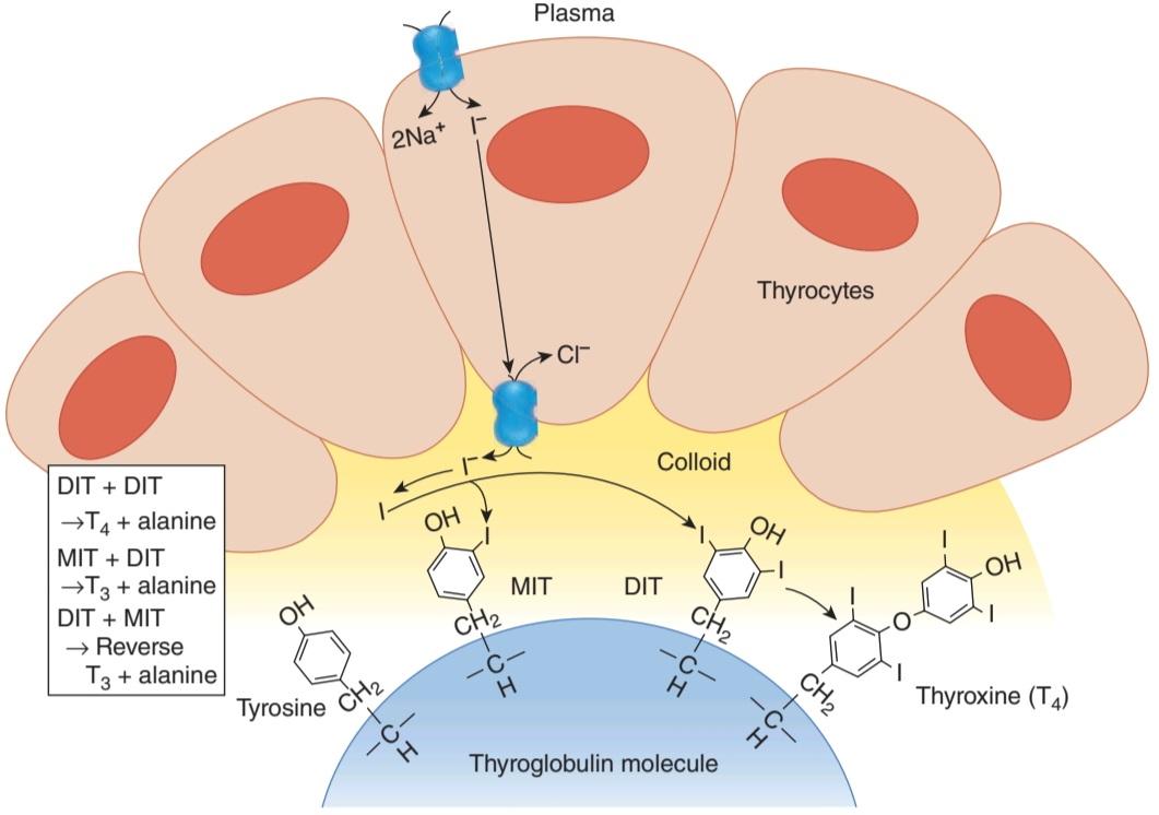
FIGURE 20-6 Outline of thyroid hormone biosynthesis. Iodide (I) is transported vectorially from the plasma across the epithelial cells of the thyroid gland by specific transporters. The iodide is converted to iodine, which reacts with tyrosine residues exposed on the surface of thyroglobulin molecules resident in the colloid. Iodination of tyrosine takes place at the apical border of the thyroid cells while the tyrosine moieties remain bound to thyroglobulin via peptide linkages.
شکل 20-6 طرح کلی بیوسنتز هورمون تیروئید. یدید (I) به صورت برداری از پلاسما از طریق سلول های اپیتلیال غده تیروئید توسط ناقل های خاص منتقل می شود. یدید به ید تبدیل می شود که با باقی مانده های تیروزین در معرض سطح مولکول های تیروگلوبولین ساکن در کلوئید واکنش می دهد. ید زایی تیروزین در مرز آپیکال سلول های تیروئید صورت می گیرد در حالی که قسمت های تیروزین از طریق پیوندهای پپتیدی به تیروگلوبولین متصل می مانند.
Iodide must also exit the thyrocyte across the apical membrane to access the colloid, where the initial steps of thyroid hormone synthesis occur. This transport step is believed to be mediated, at least in part, by a CI/I exchanger known as pendrin (Figure 20-6). This protein was first identified as the product of the gene responsible for the Pendred syndrome, which causes thyroid dysfunction and deafness. Pendrin (SLC26A4) is one member of the larger family of SLC26 anion exchangers.
یدید همچنین باید از تیروسیت در سراسر غشای آپیکال خارج شود تا به کلوئید دسترسی پیدا کند، جایی که مراحل اولیه سنتز هورمون تیروئید رخ می دهد. اعتقاد بر این است که این مرحله انتقال، حداقل تا حدی، توسط یک مبدل CI/I معروف به پندرین انجام می شود (شکل 20-6). این پروتئین برای اولین بار به عنوان محصول ژن مسئول سندرم پندر شناسایی شد که باعث اختلال در عملکرد تیروئید و ناشنوایی می شود. پندرین (SLC26A4) یکی از اعضای خانواده بزرگتر مبدل های آنیونی SLC26 است.
The relationship of thyroid function to iodide is unique. Iodide is essential for normal thyroid function, but iodide deficiency and iodide excess both inhibit thyroid function.
رابطه عملکرد تیروئید با یدید منحصر به فرد است. یدید برای عملکرد طبیعی تیروئید ضروری است، اما کمبود ید و بیش از حد یدید هر دو عملکرد تیروئید را مهار می کنند.
The salivary glands, the gastric mucosa, the placenta, the ciliary body of the eye, the choroid plexus, the mammary glands, and certain cancers derived from these tissues also express NIS and can transport iodide against a concentration gradient, but the transporter in these tissues is not affected by TSH. The physiologic significance of all these extrathyroidal iodide-concentrating mechanisms is obscure, but they may provide pathways for radioablation of NIS- expressing cancer cells using iodide radioisotopes. This approach is also useful for the ablation of thyroid cancers.
غدد بزاقی، مخاط معده، جفت، جسم مژگانی چشم، شبکه مشیمیه، غدد پستانی و سرطان های خاصی که از این بافت ها به دست می آیند نیز NIS را بیان می کنند و می توانند یدید را بر خلاف گرادیان غلظت انتقال دهند، اما ناقل در این بافت ها تحت تأثیر TSH قرار نمی گیرد. اهمیت فیزیولوژیکی همه این مکانیسمهای متمرکز بر یدید خارج تیروئید مبهم است، اما ممکن است مسیرهایی را برای رادیوآبیلیشن سلولهای سرطانی بیانکننده NIS با استفاده از ایزوتوپهای رادیویی یدید فراهم کنند. این روش برای رفع سرطان تیروئید نیز مفید است.
THYROID HORMONE SYNTHESIS & SECRETION
At the interface between the thyrocyte and the colloid, iodide undergoes a process referred to as organification. First, it is oxidized to iodine, and then incorporated into the carbon 3 position of tyrosine residues that are part of the thyroglobulin molecule in the colloid (Figure 20–6). Thyroglobulin is a glycoprotein made up of two subunits. It contains 123 tyrosine residues, but only 4-8 of these are normally incorporated into thyroid hormones. Thyroglobulin is synthesized in the thyrocytes and secreted into the colloid by exocytosis of granules. The oxidation and reaction of iodide with the secreted thyroglobulin is mediated by thyroid peroxidase, a membrane-bound enzyme found in the thyrocyte apical membrane. The thyroid hormones so produced remain part of the thyroglobulin molecule until needed. As such, colloid represents a reservoir of thyroid hormones, and humans can ingest a diet completely devoid of iodide for up to 2 months before a decline in circulating thyroid hormone levels is seen. When there is a need for thyroid hormone secretion, colloid is internalized by the thyrocytes by endocytosis, and directed toward lysosomal degradation. Thus, the peptide bonds of thyroglobulin are hydrolyzed, and free T4 and T3 are discharged into cytosol and thence to the capillaries (see below). Thyrocytes thus have four functions: They collect and transport iodine, they synthesize thyroglobulin and secrete it into the colloid, they fix iodine to the thyroglobulin to generate thyroid hormones, and they remove the thyroid hormones from thyroglobulin and secrete them into the circulation.
سنتز و ترشح هورمون تیروئید
در سطح مشترک بین تیروسیت و کلوئید، یدید تحت فرآیندی قرار می گیرد که به آن سازمان دهی می گویند. ابتدا به ید اکسید می شود و سپس در موقعیت کربن 3 باقی مانده های تیروزین که بخشی از مولکول تیروگلوبولین در کلوئید هستند قرار می گیرد (شکل 20-6). تیروگلوبولین یک گلیکوپروتئین است که از دو زیر واحد تشکیل شده است. حاوی 123 باقیمانده تیروزین است، اما تنها 4 تا 8 مورد از آنها به طور معمول در هورمون های تیروئید ترکیب می شوند. تیروگلوبولین در تیروسیت ها سنتز می شود و با اگزوسیتوز گرانول ها به کلوئید ترشح می شود. اکسیداسیون و واکنش یدید با تیروگلوبولین ترشح شده توسط تیروئید پراکسیداز، یک آنزیم متصل به غشاء که در غشای آپیکال تیروسیت یافت می شود، انجام می شود. هورمون های تیروئیدی که به این ترتیب تولید می شوند، بخشی از مولکول تیروگلوبولین باقی می مانند تا زمانی که مورد نیاز باشد. به این ترتیب، کلوئید مخزنی از هورمون های تیروئید است و انسان می تواند یک رژیم غذایی کاملاً بدون ید را تا 2 ماه قبل از کاهش سطح هورمون تیروئید در گردش مصرف کند. هنگامی که نیاز به ترشح هورمون تیروئید وجود دارد، کلوئید توسط تیروسیت ها درونی می شود و به سمت تخریب لیزوزومی هدایت می شود. بنابراین، پیوندهای پپتیدی تیروگلوبولین هیدرولیز می شوند و T4 و T3 آزاد به سیتوزول و از آنجا به مویرگ ها تخلیه می شوند (به زیر مراجعه کنید). بنابراین، تیروسیت ها چهار عملکرد دارند: ید را جمع آوری و انتقال می دهند، تیروگلوبولین را سنتز کرده و به کلوئید ترشح می کنند، ید را به تیروگلوبولین برای تولید هورمون های تیروئید تثبیت می کنند و هورمون های تیروئید را از تیروگلوبولین خارج کرده و به گردش خون ترشح می کنند.
Thyroid hormone synthesis is a multistep process. Thyroid peroxidase generates reactive iodine species that can attack thyroglobulin. The first product is monoiodotyrosine (MIT). MIT is next iodinated on the carbon 5 position to form diiodotyrosine (DIT). Two DIT molecules then undergo an oxidative condensation to form T4 with the elimination of the alanine side chain from the molecule that forms the outer ring. There are two theories of how this coupling reaction occurs. One holds that the coupling occurs with both DIT molecules attached to thyroglobulin (intramolecular coupling). The other holds that the DIT that forms the outer ring is first detached from thyroglobulin (intermolecular coupling). In either case, thyroid peroxidase is involved in coupling as well as iodination. T3 is formed by condensation of MIT with DIT. RT3 is likely formed by condensation of DIT with MIT. In the normal human thyroid, the average distribution of iodinated compounds is 3% MIT, 33% DIT, 35% T4, and 7% T3. Only traces of RT3 and other components are present.
سنتز هورمون تیروئید یک فرآیند چند مرحله ای است. پراکسیداز تیروئید گونه های ید فعالی تولید می کند که می توانند به تیروگلوبولین حمله کنند. اولین محصول مونو یدوتیروزین (MIT) است. MIT سپس در موقعیت کربن 5 یددار می شود تا دی یدوتیروزین (DIT) را تشکیل دهد. سپس دو مولکول DIT تحت یک تراکم اکسیداتیو قرار میگیرند تا با حذف زنجیره جانبی آلانین از مولکولی که حلقه بیرونی را تشکیل میدهد، T4 را تشکیل دهند. دو نظریه در مورد چگونگی این واکنش جفتی وجود دارد. یکی معتقد است که جفت شدن با هر دو مولکول DIT متصل به تیروگلوبولین (جفت درون مولکولی) اتفاق می افتد. دیگری معتقد است که DIT که حلقه بیرونی را تشکیل می دهد، ابتدا از تیروگلوبولین (کوپلینگ بین مولکولی) جدا می شود. در هر صورت، پراکسیداز تیروئید در جفت شدن و همچنین یدسازی نقش دارد. T3 از تراکم MIT با DIT تشکیل می شود. RT3 احتمالاً از تراکم DIT با MIT تشکیل می شود. در تیروئید طبیعی انسان، میانگین توزیع ترکیبات یددار 3% MIT، 33% DIT، 35% T4 و 7% T3 است. فقط آثاری از RT3 و سایر اجزا وجود دارد.
The human thyroid secretes about 80 μg (103 nmol) of T4, 4 μg (7 nmol) of T3, and 2 μg (3.5 nmol) of RT3 per day (Figure 20-7). MIT and DIT are not secreted. These iodinated tyrosines can be deiodinated by a microsomal iodotyrosine deiodinase. This represents a mechanism to recover iodine and bound tyrosines and recycle them for additional rounds of hormone synthesis. The iodine liberated by deiodination of MIT and DIT is reutilized in the gland and normally provides about twice as much iodide for hormone synthesis as NIS does. In patients with congenital absence of the iodotyrosine deiodinase, MIT and DIT appear in the urine and there are symptoms of iodine deficiency (see below). Iodinated thyronines are resistant to the activity of iodotyrosine deiodinase, thus allowing T4 and T3 to pass into the circulation.
تیروئید انسان حدود 80 میکروگرم (103 نانومول) T4، 4 میکروگرم (7 نانومول) T3 و 2 میکروگرم (3.5 نانومول) RT3 در روز ترشح می کند (شکل 20-7). MIT و DIT ترشح نمی شوند. این تیروزین های یددار را می توان توسط یک میکروزومی یدوتیروزین دیودیناز ید زدایی کرد. این نشان دهنده مکانیسمی برای بازیابی ید و تیروزین های متصل و بازیافت آنها برای دورهای اضافی سنتز هورمون است. ید آزاد شده توسط ید زدایی MIT و DIT مجدداً در غده مورد استفاده قرار می گیرد و به طور معمول حدود دو برابر بیشتر از NIS یدید برای سنتز هورمون فراهم می کند. در بیماران با عدم وجود مادرزادی یدوتیروزین دیودیناز، MIT و DIT در ادرار ظاهر می شوند و علائم کمبود ید وجود دارد (به زیر مراجعه کنید). تیرونین های یددار در برابر فعالیت یدوتیروزین دیودیناز مقاوم هستند، بنابراین به T4 و T3 اجازه می دهند وارد گردش خون شوند.

FIGURE 20-7 Secretion and interconversion of thyroid hormones in normal adult humans. Figures are in micrograms per day. Note that most of the T3 and RT3 are formed from T4 deiodination in the tissues and only small amounts are secreted by the thyroid. T4 is also conjugated for subsequent excretion from the body.
شکل 20-7 ترشح و تبدیل هورمون های تیروئید در انسان بالغ طبیعی. ارقام بر حسب میکروگرم در روز است. توجه داشته باشید که بیشتر T3 و RT3 از ید زدایی T4 در بافت ها تشکیل می شود و فقط مقادیر کمی از تیروئید ترشح می شود. T4 همچنین برای دفع بعدی از بدن کونژوگه می شود.
TRANSPORT & METABOLISM OF THYROID HORMONES
PROTEIN BINDING
The normal total plasma T4 level in adults is approximately 8 μg/dL (103 nmol/L), and the plasma T3 level is approximately 0.15 μg/dL (2.3 nmol/L). T4 and T3 are relatively lipophilic; thus, their free forms in plasma are in equilibrium with a much larger pool of protein-bound thyroid hormones in plasma and in tissues. Free thyroid hormones are added to the circulating pool by the thyroid. It is the free thyroid hormones in plasma that are physiologically active and that feed back to inhibit pituitary secretion of TSH (Figure 20-8). The function of protein-binding appears to be the maintenance of a large pool of hormone that can readily be mobilized as needed. In addition, at least for T3, hormone-binding prevents excess uptake by the first cells encountered and promotes uniform tissue distribution. Total T4 and T3 can both be measured by radioimmunoassay. There are also direct assays that specifically measure only the free forms of the hormones. The latter are the more clinically relevant measures given that these are the active forms, and also due to both acquired and congenital variations in the concentrations of binding proteins between individuals.
انتقال و متابولیسم هورمون های تیروئید
اتصال پروتئین
سطح طبیعی کل T4 پلاسما در بزرگسالان تقریباً 8 میکروگرم در دسی لیتر (103 نانومول در لیتر) و سطح T3 پلاسما تقریباً 0.15 میکروگرم در دسی لیتر (2.3 نانومول در لیتر) است. T4 و T3 نسبتاً چربی دوست هستند. بنابراین، اشکال آزاد آنها در پلاسما در تعادل با مخزن بسیار بزرگتری از هورمون های تیروئیدی متصل به پروتئین در پلاسما و در بافت ها هستند. هورمون های تیروئید آزاد توسط تیروئید به حوضچه در گردش اضافه می شوند. این هورمون های تیروئید آزاد در پلاسما هستند که از نظر فیزیولوژیکی فعال هستند و برای مهار ترشح هیپوفیز TSH بازخورد می کنند (شکل 20-8). به نظر می رسد که عملکرد اتصال به پروتئین، حفظ یک مجموعه بزرگ از هورمون است که می تواند به راحتی در صورت نیاز بسیج شود. علاوه بر این، حداقل برای T3، اتصال به هورمون از جذب بیش از حد توسط اولین سلولهایی که با آن مواجه میشوند جلوگیری میکند و توزیع یکنواخت بافت را افزایش میدهد. مجموع T4 و T3 هر دو را می توان با رادیو ایمونواسی اندازه گیری کرد. همچنین سنجش های مستقیمی وجود دارد که به طور خاص فقط اشکال آزاد هورمون ها را اندازه گیری می کند. با توجه به اینکه اینها اشکال فعال هستند، و همچنین به دلیل تغییرات اکتسابی و مادرزادی در غلظت پروتئین های اتصال بین افراد، معیارهای بالینی مرتبط تر هستند.
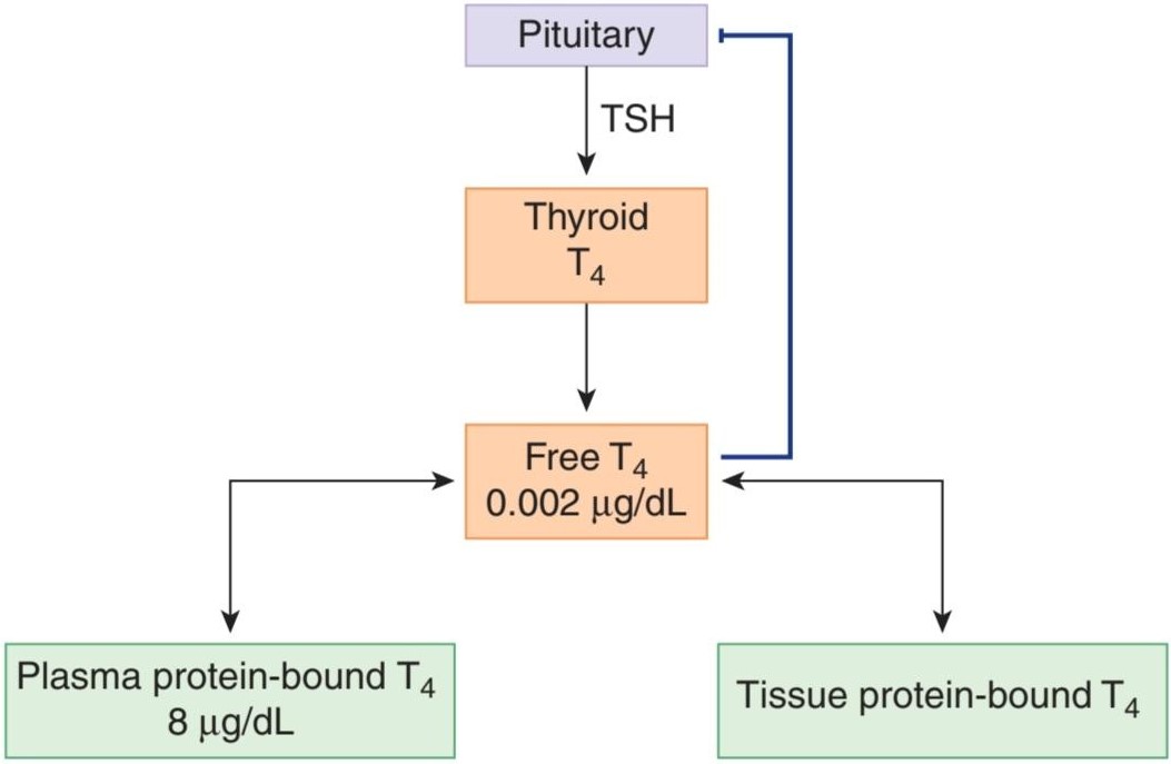
FIGURE 20-8 Regulation of thyroid hormone synthesis. T4 is secreted by the thyroid in response to TSH. Free T4 secreted by the thyroid into the circulation is in equilibrium with T4 bound to both plasma and tissue proteins. Free T4 also feeds back to inhibit TSH secretion by the pituitary. Not shown, T3 is also secreted (in small amounts) by the thyroid and produced (in larger amounts) from T4 in the periphery by deiodination. T3 also feeds back to reduce secretion of TSH from the pituitary.
شکل 20-8 تنظیم سنتز هورمون تیروئید. T4 در پاسخ به TSH توسط تیروئید ترشح می شود. T4 آزاد ترشح شده توسط تیروئید به گردش خون در تعادل با T4 به هر دو پروتئین پلاسما و بافت متصل است. T4 آزاد همچنین برای مهار ترشح TSH توسط هیپوفیز بازخورد می کند. نشان داده نشده، T3 نیز (در مقادیر کم) توسط تیروئید ترشح می شود و (در مقادیر بیشتر) از T4 در محیط با ید زدایی تولید می شود. T3 همچنین برای کاهش ترشح TSH از هیپوفیز بازخورد می کند.
The plasma proteins that bind thyroid hormones are albumin, transthyretin (formerly called thyroxine-binding prealbumin), and thyroxine-binding globulin (TBG). Of the three proteins, albumin has the largest capacity to bind T4 (ie, it can bind the most T4 before becoming saturated) and TBG has the smallest capacity. However, the affinities of the proteins for T4 (ie, the avidity with which they bind T4 under physiologic conditions) are such that most of the circulating T4 is bound to TBG (Table 20-1). Smaller amounts of T4 are bound to transthyretin and albumin.
پروتئینهای پلاسما که به هورمونهای تیروئید متصل میشوند، آلبومین، ترانس تیرتین (که قبلاً پرآلبومین اتصال به تیروکسین نامیده میشد) و گلوبولین اتصال به تیروکسین (TBG) هستند. از بین این سه پروتئین، آلبومین بیشترین ظرفیت را برای اتصال T4 دارد (یعنی می تواند بیشتر T4 را قبل از اشباع شدن متصل کند) و TBG کمترین ظرفیت را دارد. با این حال، میل پروتئین ها به T4 (یعنی علاقه ای که با آن در شرایط فیزیولوژیکی به T4 متصل می شوند) به گونه ای است که بیشتر T4 در گردش به TBG متصل می شود (جدول 20-1). مقادیر کمتری از T4 به ترانس تیرتین و آلبومین متصل می شود.
TABLE 20-1 Binding of thyroid hormones to plasma proteins in normal adult humans.
جدول 20-1 اتصال هورمون های تیروئید به پروتئین های پلاسما در انسان بالغ طبیعی.

Normally, 99.98% of the T4 in plasma is bound; the free T4 level is only about 2 ng/dL. There is very little T4 in the urine. Its biologic half-life is long (about 6-7 days), and its volume of distribution is less than that of ECF (10 L, or about 15% of body weight). All of these properties are characteristic of a substance that is strongly bound to protein.
به طور معمول، 99.98٪ از T4 در پلاسما متصل است. سطح T4 آزاد تنها حدود 2 نانوگرم در دسی لیتر است. T4 بسیار کمی در ادرار وجود دارد. نیمه عمر بیولوژیکی آن طولانی است (حدود 6-7 روز) و حجم توزیع آن کمتر از ECF (10 لیتر یا حدود 15 درصد وزن بدن) است. همه این خواص مشخصه ماده ای است که به شدت به پروتئین متصل است.
T3 is not bound to quite as great an extent; of the 0.15 μg/dL normally found in plasma, 0.2% (0.3 ng/dL) is free. The remaining 99.8% is protein-bound, 46% to TBG and most of the remainder to albumin, with very little binding to transthyretin (Table 20-1). The lesser binding of T3 correlates with the facts that T3 has a shorter half-life than T4 and that its action on the tissues is much more rapid. RT3 also binds to TBG.
T3 به این اندازه محدود نیست. از 0.15 میکروگرم در دسی لیتر که به طور معمول در پلاسما یافت می شود، 0.2٪ (0.3 نانوگرم در دسی لیتر) آزاد است. 99.8 درصد باقیمانده به پروتئین، 46 درصد به TBG و بیشتر بقیه به آلبومین متصل است، با اتصال بسیار کمی به ترانس تیرتین (جدول 20-1). اتصال کمتر T3 با این واقعیت مرتبط است که T3 نیمه عمر کوتاه تری نسبت به T4 دارد و عملکرد آن بر روی بافت ها بسیار سریعتر است. RT3 همچنین به TBG متصل می شود.
FLUCTUATIONS IN BINDING
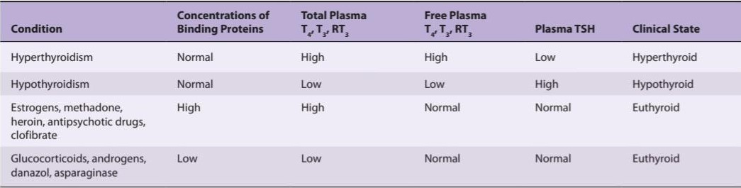
When a sudden, sustained increase in the concentration of thyroid-binding proteins in the plasma takes place, the concentration of free thyroid hormones falls. This change is temporary, however, because the decrease in the concentration of free thyroid hormones in the circulation stimulates TSH secretion, which in turn causes an increase in the production of free thyroid hormones. A new equilibrium is eventually reached at which the total quantity of thyroid hormones in the blood is elevated but the concentration of free hormones, the rate of their metabolism, and the rate of TSH secretion are normal. Corresponding changes in the opposite direction occur when the concentration of thyroid-binding protein is reduced. Consequently, patients with elevated or decreased concentrations of binding proteins, particularly TBG, are typically neither hyperthyroid nor hypothyroid; that is, they are euthyroid.
نوسانات در صحافی
هنگامی که یک افزایش مداوم و ناگهانی در غلظت پروتئین های متصل شونده به تیروئید در پلاسما رخ می دهد، غلظت هورمون های تیروئید آزاد کاهش می یابد. اما این تغییر موقتی است، زیرا کاهش غلظت هورمون های تیروئید آزاد در گردش خون باعث تحریک ترشح TSH می شود که به نوبه خود باعث افزایش تولید هورمون های تیروئید آزاد می شود. در نهایت تعادل جدیدی حاصل می شود که در آن مقدار کل هورمون های تیروئید در خون افزایش می یابد اما غلظت هورمون های آزاد، سرعت متابولیسم آنها و میزان ترشح TSH طبیعی است. تغییرات متناظر در جهت مخالف زمانی رخ می دهد که غلظت پروتئین متصل شونده به تیروئید کاهش یابد. در نتیجه، بیماران با افزایش یا کاهش غلظت پروتئین های اتصال دهنده، به ویژه TBG، به طور معمول نه هیپرتیروئید هستند و نه هیپوتیروئید. یعنی یوتیروئید هستند.
TBG levels are elevated in estrogen-treated patients and during pregnancy, as well as after treatment with various drugs (Table 20-2). They are depressed by glucocorticoids, androgens, the weak androgen danazol, and the cancer chemotherapeutic agent L-asparaginase. A number of other drugs, including salicylates, the anticonvulsant phenytoin, and the cancer chemotherapeutic agents mitotane (o, p’-DDD) and 5-fluorouracil, inhibit binding of T4 and T3 to TBG and consequently produce changes similar to those produced by a decrease in TBG concentration. Changes in total plasma T4 and T3 can also be produced by changes in plasma concentrations of albumin and prealbumin.
سطح TBG در بیماران تحت درمان با استروژن و در دوران بارداری و همچنین پس از درمان با داروهای مختلف افزایش می یابد (جدول 20-2). آنها توسط گلوکوکورتیکوئیدها، آندروژن ها، آندروژن ضعیف دانازول و عامل شیمی درمانی سرطان ال-آسپارژیناز افسرده می شوند. تعدادی از داروهای دیگر، از جمله سالیسیلات ها، فنی توئین ضد تشنج، و عوامل شیمی درمانی سرطان میتوتان (o, p’-DDD) و 5- فلورواوراسیل، اتصال T4 و T3 به TBG را مهار می کنند و در نتیجه تغییراتی مشابه آنچه که با کاهش غلظت TBG ایجاد می شود، ایجاد می کنند. تغییرات در کل T4 و T3 پلاسما نیز می تواند با تغییر در غلظت پلاسمایی آلبومین و پری آلبومین ایجاد شود.
TABLE 20-2 Effect of variations in the concentrations of thyroid hormone-binding proteins in the plasma on various parameters of thyroid function after equilibrium has been reached.
جدول 20-2 اثر تغییرات در غلظت پروتئین های متصل شونده به هورمون تیروئید در پلاسما بر پارامترهای مختلف عملکرد تیروئید پس از رسیدن به تعادل.
METABOLISM OF THYROID HORMONES
T4 and T3 are deiodinated in the liver, the kidneys, and many other tissues. These deiodination reactions serve not only to catabolize the hormones, but also to provide a local supply specifically of T3, which is believed to be the primary mediator of the physiologic effects of thyroid secretion. One-third of the circulating T4 is normally converted to T3 in adult humans, and 45% is converted to RT3. As shown in Figure 20–7, only about 13% of the circulating T3 is secreted by the thyroid while 87% is formed by deiodination of T4; similarly, only 5% of the circulating RT3 is secreted by the thyroid and 95% is formed by deiodination of T4. It should be noted as well that marked differences in the ratio of T3 to T4 occur in various tissues. Two tissues that have very high T3/T4 ratios are the pituitary and the cerebral cortex, due to the expression of specific deiodinases, as discussed below. In the brain, in particular, high levels of deiodinase activity ensure an ample supply of active T3.
متابولیسم هورمون های تیروئید
T4 و T3 در کبد، کلیه ها و بسیاری از بافت های دیگر دی یدین می شوند. این واکنشهای یدزدایی نه تنها برای کاتابولیزاسیون هورمونها عمل میکنند، بلکه یک منبع محلی به طور خاص از T3 را نیز فراهم میکنند، که گمان میرود واسطه اولیه اثرات فیزیولوژیک ترشح تیروئید باشد. یک سوم T4 در گردش به طور معمول در انسان بالغ به T3 و 45٪ به RT3 تبدیل می شود. همانطور که در شکل 20-7 نشان داده شده است، تنها حدود 13% از T3 در گردش توسط تیروئید ترشح می شود در حالی که 87% از ید زدایی T4 تشکیل می شود. به طور مشابه، تنها 5٪ از RT3 در گردش توسط تیروئید و 95٪ از ید زدایی T4 تشکیل می شود. همچنین لازم به ذکر است که تفاوت های قابل توجهی در نسبت T3 به T4 در بافت های مختلف رخ می دهد. دو بافتی که نسبت T3/T4 بسیار بالایی دارند، هیپوفیز و قشر مغز هستند، به دلیل بیان دیودینازهای خاص، همانطور که در زیر بحث می شود. به ویژه در مغز، سطوح بالای فعالیت دیودیناز، تامین کافی T3 فعال را تضمین می کند.
Three different deiodinases act on thyroid hormones: D1, D2, and D3. All are unique in that they contain the rare amino acid selenocysteine, with selenium in place of sulfur, which is essential for their enzymatic activity. D1 is present in high concentrations in the liver, kidneys, thyroid, and pituitary. It appears primarily to be responsible for maintaining the formation of T3 from T4 in the periphery. D2 is present in the brain, pituitary, and brown fat. It also contributes to the formation of T3. In the brain, it is located in astroglia and provides a supply of T3 to neurons. D3 is also present in the brain and in reproductive tissues. It acts only on the 5 position of T4 and T3 and is probably the main source of RT3 in the blood and tissues.
سه دیودیناز مختلف بر روی هورمون های تیروئید اثر می کنند: D1، D2 و D3. همه از این نظر منحصر به فرد هستند که حاوی اسید آمینه کمیاب سلنوسیستئین هستند که به جای گوگرد، سلنیوم برای فعالیت آنزیمی آنها ضروری است. D1 در غلظت های بالایی در کبد، کلیه ها، تیروئید و هیپوفیز وجود دارد. به نظر می رسد در درجه اول مسئول حفظ تشکیل T3 از T4 در محیط باشد. D2 در مغز، هیپوفیز و چربی قهوه ای وجود دارد. همچنین به تشکیل T3 کمک می کند. در مغز، در اخترگلیا قرار دارد و منبع T3 را برای نورون ها فراهم می کند. D3 همچنین در مغز و در بافت های تولید مثلی وجود دارد. این تنها در موقعیت 5 T4 و T3 عمل می کند و احتمالا منبع اصلی RT3 در خون و بافت ها است.
Some of the T4 and T3 are further converted to deiodotyrosines by deiodinases. T4 and T3 are also conjugated in the liver to form sulfates and glucuronides. These conjugates enter the bile and pass into the intestine. The thyroid conjugates are hydrolyzed, and some are thereafter reabsorbed (enterohepatic circulation), but others are excreted in the stool. In addition, some T4 and T3 pass directly from the circulation to the intestinal lumen. The iodide lost by these routes amounts to about 4% of the total daily iodide loss.
برخی از T4 و T3 بیشتر توسط دیودینازها به دیودوتیروزین تبدیل می شوند. T4 و T3 نیز در کبد کونژوگه می شوند و سولفات ها و گلوکورونیدها را تشکیل می دهند. این مواد مزدوج وارد صفرا شده و وارد روده می شوند. کونژوگه های تیروئید هیدرولیز می شوند و برخی پس از آن دوباره جذب می شوند (گردش کبدی)، اما برخی دیگر از طریق مدفوع دفع می شوند. علاوه بر این، مقداری T4 و T3 مستقیماً از گردش خون به مجرای روده عبور می کنند. یدید از دست رفته توسط این مسیرها به حدود 4 درصد از کل از دست رفتن یدید روزانه می رسد.
FLUCTUATIONS IN DEIODINATION
Much more RT3 and much less T3 are formed during fetal life, and the ratio shifts to that of adults about 6 weeks after birth. Various drugs inhibit deiodinases, producing a fall in plasma T3 levels and a reciprocal rise in RT3. Selenium deficiency has the same effect. A wide variety of nonthyroidal illnesses also suppress deiodinases. These include burns, trauma, advanced cancer, cirrhosis, chronic kidney disease, myocardial infarction, and febrile states. The low-T3 state produced by these conditions disappears with recovery.
نوسانات در دیودیناسیون
RT3 بسیار بیشتر و T3 بسیار کمتر در طول زندگی جنینی تشکیل می شود و حدود 6 هفته پس از تولد نسبت به بزرگسالان تغییر می کند. داروهای مختلف دیودینازها را مهار می کنند و باعث کاهش سطح T3 پلاسما و افزایش متقابل RT3 می شوند. کمبود سلنیوم نیز همین اثر را دارد. طیف گسترده ای از بیماری های غیر تیروئیدی نیز دیودینازها را سرکوب می کنند. اینها شامل سوختگی، تروما، سرطان پیشرفته، سیروز، بیماری مزمن کلیوی، انفارکتوس میوکارد و حالت های تب دار است. حالت T3 پایین ایجاد شده توسط این شرایط با بهبودی از بین می رود.
Diet also has a clear-cut effect on conversion of T4 to T3. In fasted individuals, plasma T3 is reduced by 10-20% within 24 h and by about 50% in 3-7 days, with a corresponding rise in RT3 (Figure 20-9). Free and bound T4 levels remain essentially normal. During more prolonged starvation, RT3 returns to normal but T3 remains depressed. At the same time, the basal metabolic rate (BMR) falls and urinary nitrogen excretion, an index of protein breakdown, is decreased. Thus, the decline in T3 conserves calories and protein. Conversely, overfeeding increases T3 and reduces RT3.
رژیم غذایی همچنین تأثیر واضحی بر تبدیل T4 به T3 دارد. در افراد روزه دار، T3 پلاسما در عرض 24 ساعت 10-20٪ و در 3-7 روز حدود 50٪ کاهش می یابد، با افزایش متناظر در RT3 (شکل 20-9). سطح آزاد و محدود T4 اساساً نرمال باقی می ماند. در طول گرسنگی طولانی تر، RT3 به حالت عادی باز می گردد اما T3 افسرده باقی می ماند. در همان زمان، نرخ متابولیسم پایه (BMR) کاهش مییابد و دفع نیتروژن ادرار، که شاخصی از تجزیه پروتئین است، کاهش مییابد. بنابراین، کاهش T3 باعث حفظ کالری و پروتئین می شود. برعکس، تغذیه بیش از حد باعث افزایش T3 و کاهش RT3 می شود.

FIGURE 20-9 Effect of starvation on plasma levels of T4, T3, and RT3 in humans. The scale for T3 and RT3 is on the left and the scale for T4 is on the right. The most pronounced effect is a reduction in T3 levels with a reciprocal rise in RT3. The changes, which conserve calories by reducing tissue metabolism, are reversed promptly by refeeding. Similar changes occur in wasting diseases. (Reproduced with permission from Burger AG: New aspects of the peripheral action of thyroid hormones. Triangle 1983;22:175. Copyright © 1983 Sandoz Ltd., Basel, Switzerland.)
شکل 20-9 اثر گرسنگی بر سطوح پلاسمایی T4، T3 و RT3 در انسان. ترازو T3 و RT3 در سمت چپ و ترازو T4 در سمت راست است. بارزترین اثر کاهش سطح T3 با افزایش متقابل RT3 است. تغییراتی که با کاهش متابولیسم بافت باعث حفظ کالری می شود، با تغذیه مجدد سریعاً معکوس می شود. تغییرات مشابهی در بیماری های هدر می رود. (تکثیر شده با مجوز از Burger AG: جنبه های جدید عملکرد محیطی هورمون های تیروئید. Triangle 1983; 22:175. حق چاپ © 1983 Sandoz Ltd., Basel, Switzerland.)
REGULATION OF THYROID SECRETION
Thyroid function is regulated primarily by variations in the circulating level of pituitary TSH (Figure 20-8). TSH secretion is increased by the hypothalamic hormone TRH (see Chapter 17) and inhibited in a negative feedback manner by circulating free T4 and T3. The effect of T4 is enhanced by production of T3 in the cytoplasm of the pituitary cells by the D2 they contain. TSH secretion is also inhibited by stress, and in experimental animals it is increased by cold and decreased by warmth.
تنظیم ترشح تیروئید
عملکرد تیروئید در درجه اول با تغییرات در سطح گردش خون TSH هیپوفیز تنظیم می شود (شکل 20-8). ترشح TSH توسط هورمون هیپوتالاموس TRH افزایش می یابد (به فصل 17 مراجعه کنید) و به شیوه ای بازخورد منفی با گردش T4 و T3 آزاد مهار می شود. اثر T4 با تولید T3 در سیتوپلاسم سلول های هیپوفیز توسط D2 موجود در آنها افزایش می یابد. ترشح TSH نیز با استرس مهار می شود و در حیوانات آزمایشی با سرما افزایش و با گرما کاهش می یابد.
CHEMISTRY & METABOLISM OF TSH
Human TSH is a glycoprotein made up of a and ẞ subunits that become noncovalently linked in the pituitary thyrotropes. TSH-a is identical to the a subunit of luteinizing hormone, follicle-stimulating hormone, and human chorionic gonadotropin (hCG) (see Chapters 18 and 22). The functional specificity of TSH is therefore conferred by the ẞ subunit.
شیمی و متابولیسم TSH
TSH انسانی یک گلیکوپروتئین است که از زیر واحدهای a و ẞ تشکیل شده است که به صورت غیرکووالانسی در تیروتروپ های هیپوفیز به هم متصل می شوند. TSH-a مشابه زیر واحد هورمون لوتئینیزه کننده، هورمون محرک فولیکول و گنادوتروپین جفتی انسانی (hCG) است (فصل 18 و 22 را ببینید). بنابراین ویژگی عملکردی TSH توسط زیرواحد ẞ اعطا می شود.
The biologic half-life of human TSH is about 60 min. TSH is degraded for the most part in the kidneys and to a lesser extent in the liver. Secretion is pulsatile, and mean output starts to rise at about 9:00 PM, peaks at midnight, and then declines thereafter. The normal secretion rate is about 110 μg/day. The average plasma level is about 2 μg/mL.
نیمه عمر بیولوژیکی TSH انسان حدود 60 دقیقه است. TSH بیشتر در کلیه ها و به میزان کمتری در کبد تجزیه می شود. ترشح ضربان دار است و میانگین خروجی در حدود ساعت 9:00 بعد از ظهر شروع به افزایش می کند، در نیمه شب به اوج خود می رسد و پس از آن کاهش می یابد. میزان طبیعی ترشح حدود 110 میکروگرم در روز است. متوسط سطح پلاسما حدود 2 میکروگرم در میلی لیتر است.
Because the a subunit in hCG is the same as that in TSH, large amounts of hCG can activate TSH receptors nonspecifically. In some patients with benign or malignant tumors of placental origin, plasma hCG levels can rise so high that they produce mild hyperthyroidism.
از آنجایی که زیرواحد a در hCG همانند TSH است، مقادیر زیاد hCG می تواند گیرنده های TSH را به طور غیر اختصاصی فعال کند. در برخی از بیماران مبتلا به تومورهای خوش خیم یا بدخیم با منشاء جفت، سطح hCG پلاسما می تواند به حدی افزایش یابد که باعث پرکاری تیروئید خفیف شود.
EFFECTS OF TSH ON THE THYROID
When the pituitary is removed, thyroid function is depressed and the gland atrophies; when TSH is administered, thyroid function is stimulated. TSH acts via its receptor, a typical G-protein-coupled, seven-transmembrane receptor that activates adenylyl cyclase through G ̧. It also activates phospholipase C (PLC). Within a few minutes after the injection of TSH, there are increases in iodide binding, synthesis of T3, T4, and iodotyrosines, secretion of thyroglobulin into the colloid, and endocytosis of colloid. Iodide trapping is increased in a few hours; blood flow increases; and, with long-term TSH treatment, the thyrocytes hypertrophy and the weight of the gland increases.
اثرات TSH بر تیروئید
هنگامی که هیپوفیز برداشته می شود، عملکرد تیروئید کاهش می یابد و غده آتروفی می شود. هنگامی که TSH تجویز می شود، عملکرد تیروئید تحریک می شود. TSH از طریق گیرنده خود، یک گیرنده معمولی جفت شده با پروتئین G، گیرنده هفت گذرنده که آدنیلیل سیکلاز را از طریق G ̧ فعال می کند، عمل می کند. همچنین فسفولیپاز C (PLC) را فعال می کند. در عرض چند دقیقه پس از تزریق TSH، افزایش اتصال یدید، سنتز T3، T4 و یدوتیروزین ها، ترشح تیروگلوبولین در کلوئید و اندوسیتوز کلوئید مشاهده می شود. به دام انداختن یدید در چند ساعت افزایش می یابد. جریان خون افزایش می یابد؛ و با درمان طولانی مدت TSH، هیپرتروفی تیروسیت ها و وزن غده افزایش می یابد.
Whenever TSH stimulation is prolonged, the thyroid becomes detectably enlarged. Enlargement of the thyroid is called a goiter.
هر زمان که تحریک TSH طولانی شود، تیروئید به طور قابل تشخیصی بزرگ می شود. بزرگ شدن تیروئید را گواتر می گویند.
OTHER FACTORS AFFECTING THYROID GROWTH
In addition to TSH receptors, thyrocytes express receptors for insulin-like growth factor I (IGF-I) and epidermal growth factor (EGF). IGF-I and EGF promote growth, whereas interferon-y and tumor necrosis factor-a inhibit growth. The effect of the cytokines implies that thyroid function might be inhibited in the setting of chronic inflammation, which could contribute to cachexia, or weight loss.
سایر عوامل مؤثر بر رشد تیروئید
علاوه بر گیرنده های TSH، تیروسیت ها گیرنده های فاکتور رشد شبه انسولین I (IGF-I) و فاکتور رشد اپیدرمی (EGF) را بیان می کنند. IGF-I و EGF رشد را افزایش می دهند، در حالی که اینترفرون-y و فاکتور نکروز تومور-a رشد را مهار می کنند. اثر سیتوکین ها نشان می دهد که عملکرد تیروئید ممکن است در شرایط التهاب مزمن مهار شود، که می تواند به کاشکسی یا کاهش وزن کمک کند.
CONTROL MECHANISMS
The mechanisms regulating thyroid secretion are summarized in Figure 20-8. The negative feedback effect of thyroid hormones on TSH secretion is exerted in part at the hypothalamic level, but it is also due in large part to an action on the pituitary, since T4 and T3 block the increase in TSH secretion produced by TRH. Infusion of either T4 or T3 reduces circulating TSH, which declines measurably within 1 h. The day-to-day maintenance of thyroid secretion depends on the feedback interplay of thyroid hormones with TSH and TRH (Figure 20-8). The adjustments that appear to be mediated via TRH include the increased secretion of thyroid hormones produced by cold and, presumably, the decrease produced by heat. It is worth noting that although cold produces clear-cut increases in circulating TSH in human infants, the rise produced by cold in adults is negligible. Consequently, in adults, increased heat production due to increased thyroid hormone secretion (thyroid hormone thermogenesis) plays little if any role in the response to cold. Stress has an inhibitory effect on TRH secretion. Glucocorticoids also inhibit TSH secretion.
مکانیسم های کنترل
مکانیسم های تنظیم کننده ترشح تیروئید در شکل 20-8 خلاصه شده است. اثر بازخورد منفی هورمون های تیروئید بر ترشح TSH تا حدی در سطح هیپوتالاموس اعمال می شود، اما تا حد زیادی به دلیل اثر بر هیپوفیز است، زیرا T4 و T3 افزایش ترشح TSH تولید شده توسط TRH را مسدود می کنند. تزریق T4 یا T3 باعث کاهش TSH در گردش می شود که در عرض 1 ساعت به میزان قابل توجهی کاهش می یابد. حفظ روزانه ترشح تیروئید به تعامل بازخورد هورمون های تیروئید با TSH و TRH بستگی دارد (شکل 20-8). تنظیماتی که به نظر می رسد از طریق TRH انجام می شود شامل افزایش ترشح هورمون های تیروئید تولید شده توسط سرما و احتمالاً کاهش تولید شده توسط گرما است. شایان ذکر است که اگرچه سرما باعث افزایش واضح TSH در گردش خون در نوزادان انسان می شود، افزایش تولید شده توسط سرما در بزرگسالان ناچیز است. در نتیجه، در بزرگسالان، افزایش تولید گرما به دلیل افزایش ترشح هورمون تیروئید (ترموژنز هورمون تیروئید) نقش کمی در پاسخ به سرما دارد. استرس اثر مهاری بر ترشح TRH دارد. گلوکوکورتیکوئیدها نیز ترشح TSH را مهار می کنند.
EFFECTS OF THYROID HORMONES
Some of the widespread effects of thyroid hormones in the body are secondary to stimulation of O2 consumption (calorigenic action), although the hormones also affect growth and development in mammals, help regulate lipid metabolism, and increase the absorption of carbohydrates from the intestine (Table 20-3). They also increase the dissociation of oxygen from hemoglobin by increasing red cell 2,3-diphosphoglycerate (DPG) (see Chapter 35).
اثرات هورمون های تیروئید
برخی از اثرات گسترده هورمون های تیروئید در بدن ثانویه به تحریک مصرف O2 است (عمل کالری زا)، اگرچه این هورمون ها بر رشد و نمو در پستانداران نیز تأثیر می گذارد، به تنظیم متابولیسم لیپیدها و افزایش جذب کربوهیدرات ها از روده کمک می کند (جدول 20-3). آنها همچنین تفکیک اکسیژن از هموگلوبین را با افزایش 2،3-دی فسفوگلیسرات گلبول قرمز (DPG) افزایش می دهند (به فصل 35 مراجعه کنید).
TABLE 20-3 Physiologic effects of thyroid hormones.
جدول 20-3 اثرات فیزیولوژیکی هورمون های تیروئید.

Modified with permission from McPhee SJ, Lingarra VR, Ganong WF (editors): Pathophysiology of Disease, 6th ed. New York, NY: McGraw-Hill; 2010.
اصلاح شده با اجازه McPhee SJ, Lingarra VR, Ganong WF (ویراستاران): Pathophysiology of Disease, 6th ed. نیویورک، نیویورک: McGraw-Hill; 2010.
MECHANISM OF ACTION
Thyroid hormones enter cells and T3 binds to thyroid hormone receptors (TRS), members of the superfamily of hormone-sensitive nuclear transcription factors. T4 can also bind, but not as avidly. The hormone-receptor complex thenmigrates to the nucleus and binds to DNA via zinc fingers and increases (or in some cases, decreases) the expression of a variety of different genes that code for proteins regulating cell function (see Chapters 1 and 16).
مکانیسم عمل
هورمون های تیروئید وارد سلول ها می شوند و T3 به گیرنده های هورمون تیروئید (TRS)، اعضای خانواده فاکتورهای رونویسی هسته ای حساس به هورمون، متصل می شود. T4 همچنین می تواند متصل شود، اما نه آنقدر مشتاقانه. سپس مجموعه هورمون – گیرنده به هسته مهاجرت می کند و از طریق انگشتان روی به DNA متصل می شود و بیان انواع ژن های مختلف را افزایش می دهد (یا در برخی موارد کاهش می دهد) که پروتئین های تنظیم کننده عملکرد سلول را کد می کنند (به فصل های 1 و 16 مراجعه کنید).
There are two human TR genes: an a receptor gene on chromosome 17 and a ẞ receptor gene on chromosome 3. By alternative splicing, each forms at least two different mRNAs and therefore two different receptor proteins. TRẞ2 is found only in the brain, but TRα1, TRα2, and TRẞ1 are widely distributed. TRa2 differs from the other three in that it does not bind thyroid hormones due to a unique C-terminus, and may actually antagonize the activity of the other TRS. TRs bind to DNA as monomers, homodimers, and heterodimers with other nuclear receptors, particularly the retinoid X receptor (RXR). The TR/RXR heterodimer does not bind to 9-cis retinoic acid, the usual ligand for RXR, but TR binding to DNA is greatly enhanced in response to thyroid hormones when the receptor is in the form of this heterodimer. There are also coactivator and corepressor proteins that affect the actions of TRS. Presumably, this complexity underlies the ability of thyroid hormones to produce many different effects in the body.
دو ژن TR انسانی وجود دارد: یک ژن گیرنده در کروموزوم 17 و یک ژن گیرنده ẞ در کروموزوم 3. با پیوند جایگزین، هر کدام حداقل دو mRNA مختلف و بنابراین دو پروتئین گیرنده متفاوت را تشکیل می دهند. TRẞ2 فقط در مغز یافت می شود، اما TRA1، TRA2 و TRẞ1 به طور گسترده توزیع می شوند. تفاوت TRA2 با سه مورد دیگر در این است که به دلیل یک پایانه C منحصر به فرد به هورمون های تیروئید متصل نمی شود و ممکن است در واقع با فعالیت TRS دیگر مخالفت کند. TR ها به عنوان مونومر، همودایمر و هترودایمر با سایر گیرنده های هسته ای، به ویژه گیرنده رتینوئید X (RXR) به DNA متصل می شوند. هترودایمر TR/RXR به 9-cis رتینوئیک اسید، لیگاند معمول برای RXR، متصل نمی شود، اما اتصال TR به DNA در پاسخ به هورمون های تیروئید زمانی که گیرنده به شکل این هترودایمر باشد، بسیار افزایش می یابد. همچنین پروتئینهای کواکتیواتور و مهارکننده وجود دارند که بر عملکرد TRS تأثیر میگذارند. احتمالاً، این پیچیدگی زمینه ساز توانایی هورمون های تیروئید برای ایجاد اثرات مختلف در بدن است.
In most of its actions, T3 acts more rapidly and is three to five times more potent than T4 (Figure 20-10). This is because T3 is less tightly bound to plasma proteins than is T4, but binds more avidly to thyroid hormone receptors. As previously noted, RT3 is inert.
در اکثر اقدامات خود، T3 سریعتر عمل می کند و سه تا پنج برابر قوی تر از T4 است (شکل 20-10). این به این دلیل است که T3 نسبت به T4 کمتر به پروتئین های پلاسما متصل است، اما مشتاقانه تر به گیرنده های هورمون تیروئید متصل می شود. همانطور که قبلا ذکر شد، RT3 بی اثر است.
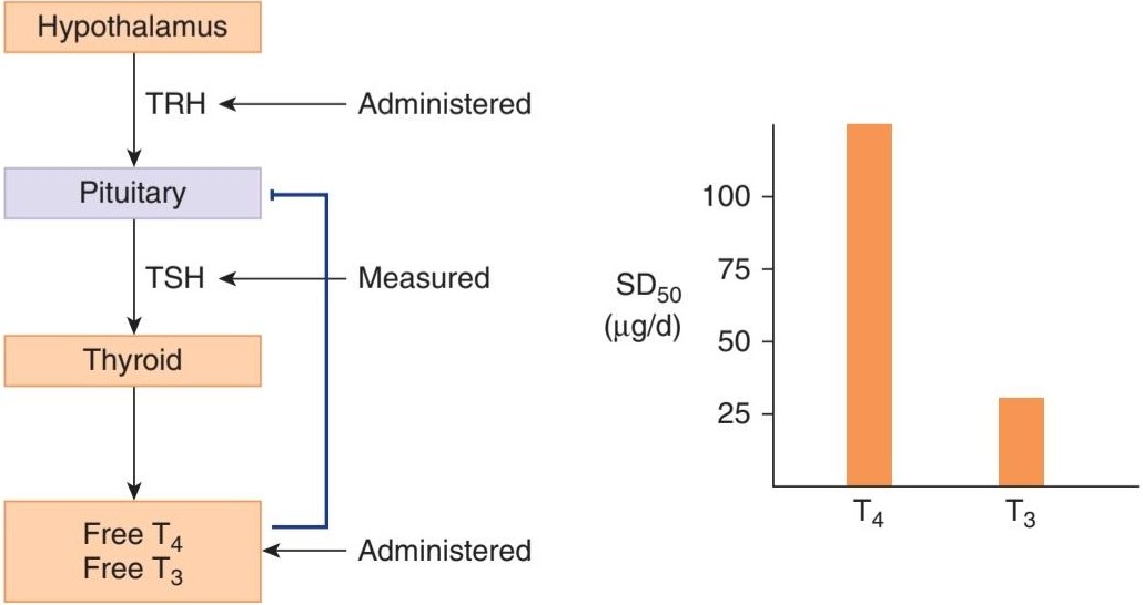
FIGURE 20-10 Experiment demonstrating the relative potency of T4 and T3. Healthy male volunteers were given TRH to stimulate TSH production in the absence or presence of various doses of T3 or T4. The left hand portion of the figure shows the relationships between the hormones that were administered or measured. On the right, the calculated doses of T3 and T4 needed to reduce the TSH response induced by TRH by 50% (SD50) are shown. Note the substantially greater potency of T3. (Data from Sawin CT, Hershman JM, Chopra IJ: The comparative effect of T4 and T3 on the TSH response to TRH in young adult men. J Clin Endo Metab 1977; 44:273–278.)
شکل 20-10 آزمایشی که قدرت نسبی T4 و T3 را نشان می دهد. به داوطلبان مرد سالم برای تحریک تولید TSH در غیاب یا حضور دوزهای مختلف T3 یا T4 TRH داده شد. قسمت سمت چپ شکل، روابط بین هورمون هایی را که تجویز یا اندازه گیری شده اند نشان می دهد. در سمت راست، دوزهای محاسبه شده T3 و T4 مورد نیاز برای کاهش پاسخ TSH ناشی از TRH به میزان 50٪ (SD50) نشان داده شده است. به قدرت قابل توجهی بیشتر T3 توجه کنید. (داده های Sawin CT، Hershman JM، Chopra IJ: اثر مقایسه ای T4 و T3 بر پاسخ TSH به TRH در مردان بالغ جوان. J Clin Endo Metab 1977؛ 44:273-278.)
CALORIGENIC ACTION
T4 and T3 increase O2 consumption by almost all metabolically active tissues. The exceptions are the adult brain, testes, uterus, lymph nodes, spleen, and anterior pituitary. T4 actually depresses the O2 consumption of the anterior pituitary, presumably because it inhibits TSH secretion. The increase in metabolic rate produced by a single dose of T4 becomes measurable after a latent period of several hours and lasts 6 days or more.
عمل کالری
T4 و T3 مصرف O2 را تقریباً در تمام بافت های فعال متابولیکی افزایش می دهند. استثناها مغز بالغ، بیضه ها، رحم، غدد لنفاوی، طحال و هیپوفیز قدامی هستند. T4 در واقع مصرف O2 هیپوفیز قدامی را کاهش می دهد، احتمالاً به این دلیل که ترشح TSH را مهار می کند. افزایش سرعت متابولیک تولید شده توسط یک دوز T4 پس از یک دوره نهفته چند ساعته قابل اندازه گیری است و 6 روز یا بیشتر طول می کشد.
Some of the calorigenic effect of thyroid hormones is due to metabolism of the fatty acids they mobilize. In addition, thyroid hormones increase the activity of the membrane-bound Na, K ATPase in many tissues.
برخی از تأثیرات کالری زایی هورمون های تیروئید به دلیل متابولیسم اسیدهای چرب است که آنها را بسیج می کنند. علاوه بر این، هورمون های تیروئیدی فعالیت Na, K ATPase متصل به غشاء را در بسیاری از بافت ها افزایش می دهند.
EFFECTS SECONDARY TO CALORIGENESIS
When the metabolic rate is increased by T4 and T3 in adults, nitrogen excretion is increased; if food intake is not increased, endogenous protein and fat stores are catabolized and weight is lost. In hypothyroid children, small doses of thyroid hormones cause a positive nitrogen balance because they stimulate growth, but large doses cause protein catabolism similar to that produced in the adult. The potassium liberated during protein catabolism appears in the urine, and there is also an increase in urinary hexosamine and uric acid excretion.
اثرات ثانویه نسبت به کالری زایی
هنگامی که میزان متابولیسم توسط T4 و T3 در بزرگسالان افزایش می یابد، دفع نیتروژن افزایش می یابد. اگر مصرف غذا افزایش نیابد، ذخایر پروتئین و چربی درون زا کاتابولیز شده و وزن کاهش می یابد. در کودکان کمکاری تیروئید، دوزهای کوچک هورمونهای تیروئیدی باعث تعادل مثبت نیتروژن میشوند، زیرا رشد را تحریک میکنند، اما دوزهای زیاد باعث کاتابولیسم پروتئینی مشابه آنچه در بزرگسالان تولید میشود، میشود. پتاسیم آزاد شده در طی کاتابولیسم پروتئین در ادرار ظاهر می شود و همچنین افزایش دفع هگزوزامین و اسید اوریک از طریق ادرار وجود دارد.
When the metabolic rate is increased, the need for all vitamins is increased and vitamin deficiency syndromes may be precipitated. Thyroid hormones are necessary for hepatic conversion of carotene to vitamin A, and the accumulation of carotene in the bloodstream (carotenemia) in hypothyroidism is responsible for the yellowish tint of the skin. Carotenemia can be distinguished from jaundice because in the former condition the sclera are not yellow.
هنگامی که میزان متابولیسم افزایش می یابد، نیاز به همه ویتامین ها افزایش می یابد و ممکن است سندرم های کمبود ویتامین ایجاد شود. هورمون های تیروئید برای تبدیل کبدی کاروتن به ویتامین A ضروری هستند و تجمع کاروتن در جریان خون (کاروتنمی) در کم کاری تیروئید مسئول رنگ زرد پوست است. کاروتنمی را می توان از یرقان تشخیص داد زیرا در حالت قبلی صلبیه زرد رنگ نیست.
The skin normally contains a variety of proteins combined with polysaccharides, hyaluronic acid, and chondroitin sulfuric acid. In hypothyroidism, these complexes accumulate, promoting water retention and the characteristic puffiness of the skin (myxedema). When thyroid hormones are administered, the proteins are metabolized, and diuresis continues until the myxedema is cleared.
پوست به طور معمول حاوی انواع پروتئین های ترکیب شده با پلی ساکاریدها، اسید هیالورونیک و اسید سولفوریک کندرویتین است. در کمکاری تیروئید، این کمپلکسها تجمع مییابند و باعث احتباس آب و پفکردگی مشخصه پوست (میکسدم) میشوند. هنگامی که هورمون های تیروئید تجویز می شود، پروتئین ها متابولیزه می شوند و دیورز تا زمانی که میکسدم از بین برود ادامه می یابد.
Milk secretion is decreased in hypothyroidism and stimulated by thyroid hormones, a fact sometimes put to practical use in the dairy industry. Thyroid hormones do not stimulate the metabolism of the uterus but are essential for normal menstrual cycles and fertility.
ترشح شیر در کمکاری تیروئید کاهش مییابد و توسط هورمونهای تیروئید تحریک میشود، این واقعیت گاهی اوقات در صنایع لبنی مورد استفاده عملی قرار میگیرد. هورمون های تیروئید متابولیسم رحم را تحریک نمی کنند، اما برای چرخه های قاعدگی طبیعی و باروری ضروری هستند.
EFFECTS ON THE CARDIOVASCULAR SYSTEM
Large doses of thyroid hormones cause enough extra heat production to lead to a slight rise in body temperature (Chapter 17), which in turn activates heat- dissipating mechanisms. Peripheral resistance decreases because of cutaneous vasodilation, and this increases levels of renal Na* and water absorption, expanding blood volume. Cardiac output is increased by the direct action of thyroid hormones, as well as that of catecholamines, on the heart, so that pulse pressure and cardiac rate are increased and circulation time is shortened.
اثرات بر سیستم قلبی عروقی
دوزهای زیاد هورمون های تیروئید باعث تولید گرمای اضافی کافی می شود که منجر به افزایش جزئی دمای بدن می شود (فصل 17) که به نوبه خود مکانیسم های دفع گرما را فعال می کند. مقاومت محیطی به دلیل اتساع عروق پوستی کاهش مییابد و این باعث افزایش سطح سدیم* و جذب آب در کلیه میشود و حجم خون را افزایش میدهد. برون ده قلبی با اثر مستقیم هورمون های تیروئید و همچنین کاتکول آمین ها بر روی قلب افزایش می یابد، به طوری که فشار نبض و ضربان قلب افزایش می یابد و زمان گردش خون کوتاه می شود.
T3 is not formed from T4 in cardiac myocytes, but circulating T3 enters myocytes, combines with its receptors, and enters the nucleus, where the complex promotes the expression of some genes and inhibits the expression of others. Those that are enhanced include the genes for a-myosin heavy chain, sarcoplasmic reticulum Ca2+ ATPase, ẞ-adrenergic receptors, G-proteins, Na, K ATPase, and certain K+ channels. Those that are inhibited include the genes for B-myosin heavy chain, phospholamban, two types of adenylyl cyclase, TRs, and NCX, the Na+-Ca2+ exchanger. The net result is increased heart rate and force of contraction.
T3 از T4 در میوسیتهای قلبی تشکیل نمیشود، اما T3 در گردش وارد میوسیتها میشود، با گیرندههای آن ترکیب میشود و وارد هسته میشود، جایی که کمپلکس بیان برخی از ژنها را تقویت میکند و بیان برخی دیگر را مهار میکند. آنهایی که تقویت می شوند عبارتند از ژن های زنجیره سنگین a-myosin، شبکه سارکوپلاسمی Ca2+ ATPase، گیرنده های آدرنرژیک، پروتئین های G، Na، K ATPase و کانال های +K خاص. آنهایی که مهار می شوند عبارتند از ژن های زنجیره سنگین B-myosin، فسفولامبان، دو نوع آدنیلیل سیکلاز، TRs و NCX، مبدل +Na+-Ca2. نتیجه خالص افزایش ضربان قلب و نیروی انقباض است.
The two myosin heavy chain (MHC) isoforms, a-MHC and ẞ-MHC, produced by the heart are encoded by two highly homologous genes located on the short arm of chromosome 17. Each myosin molecule consists of two heavy chains and two pairs of light chains (see Chapter 5). The myosin containing B- MHC has less ATPase activity than the myosin containing a-MHC. α-MHC predominates in the atria in adults, and its level is increased by treatment with thyroid hormone. This increases the speed of cardiac contraction. Conversely, expression of the a-MHC gene is depressed and that of the B-MHC gene is enhanced in hypothyroidism.
دو ایزوفرم زنجیره سنگین میوزین (MHC)، a-MHC و ẞ-MHC که توسط قلب تولید می شوند توسط دو ژن بسیار همولوگ که روی بازوی کوتاه کروموزوم 17 قرار دارند کدگذاری می شوند. هر مولکول میوزین از دو زنجیره سنگین و دو جفت زنجیره سبک تشکیل شده است (به فصل 5 مراجعه کنید). میوزین حاوی B-MHC فعالیت ATPase کمتری نسبت به میوزین حاوی a-MHC دارد. α-MHC در بزرگسالان در دهلیزها غالب است و سطح آن با درمان با هورمون تیروئید افزایش می یابد. این باعث افزایش سرعت انقباض قلب می شود. برعکس، بیان ژن a-MHC کاهش می یابد و بیان ژن B-MHC در کم کاری تیروئید افزایش می یابد.
EFFECTS ON THE NERVOUS SYSTEM
In hypothyroidism, mentation is slow and the cerebrospinal fluid (CSF) protein level is elevated. Thyroid hormones reverse these changes, and large doses cause rapid mentation, irritability, and restlessness. Overall, cerebral blood flow and glucose and O2 consumption by the brain are normal in adult hypothyroidism and hyperthyroidism. However, thyroid hormones enter the brain in adults and are found in gray matter in numerous different locations. In addition, astrocytes in the brain convert T4 to T3, and there is a sharp increase in brain D2 activity after thyroidectomy that is reversed within 4 h by a single intravenous dose of T3. Some of the effects of thyroid hormones on the brain are probably secondary to increased responsiveness to catecholamines, with consequent increased activation of the reticular activating system (see Chapter 14). In addition, thyroid hormones have marked effects on brain development. The parts of the central nervous system (CNS) most affected are the cerebral cortex and the basal ganglia. In addition, the cochlea is also affected. Consequently, thyroid hormone deficiency during development causes mental retardation, motor rigidity, and deaf-mutism. Deficiencies in thyroid hormone synthesis secondary to a failure of thyrocytes to transport iodide presumably also contribute to deafness in Pendred syndrome, discussed above.
اثرات بر روی سیستم عصبی
در کم کاری تیروئید، ذهن آهسته است و سطح پروتئین مایع مغزی نخاعی (CSF) بالا می رود. هورمون های تیروئید این تغییرات را معکوس می کنند و دوزهای زیاد باعث ذهنیت سریع، تحریک پذیری و بی قراری می شود. به طور کلی، جریان خون مغزی و مصرف گلوکز و O2 توسط مغز در کم کاری تیروئید و پرکاری تیروئید بزرگسالان طبیعی است. با این حال، هورمون های تیروئید در بزرگسالان وارد مغز می شوند و در ماده خاکستری در مکان های مختلف متعددی یافت می شوند. علاوه بر این، آستروسیتهای مغز T4 را به T3 تبدیل میکنند و پس از تیروئیدکتومی افزایش شدیدی در فعالیت D2 مغز وجود دارد که در عرض 4 ساعت با یک دوز داخل وریدی T3 معکوس میشود. برخی از اثرات هورمون های تیروئید بر مغز احتمالاً ثانویه به دلیل افزایش پاسخگویی به کاتکول آمین ها و در نتیجه افزایش فعال شدن سیستم فعال کننده شبکه است (به فصل 14 مراجعه کنید). علاوه بر این، هورمون های تیروئید تأثیرات مشخصی بر رشد مغز دارند. بخش هایی از سیستم عصبی مرکزی (CNS) که بیشتر تحت تأثیر قرار می گیرند، قشر مغز و عقده های پایه هستند. علاوه بر این، حلزون گوش نیز تحت تأثیر قرار می گیرد. در نتیجه، کمبود هورمون تیروئید در طول رشد باعث عقب ماندگی ذهنی، سفتی حرکتی و ناشنوایی می شود. نقص در سنتز هورمون تیروئید ثانویه به نارسایی تیروسیت ها در انتقال یدید احتمالاً به ناشنوایی در سندرم پندرد نیز کمک می کند.
Thyroid hormones also exert effects on reflexes. The reaction time of stretch reflexes (see Chapter 12) is shortened in hyperthyroidism and prolonged in hypothyroidism. Measurement of the reaction time of the ankle jerk (Achilles reflex) once attracted attention as a clinical test for evaluating thyroid function, but this reaction time is also affected by other diseases and thus is not a specific assessment of thyroid activity.
هورمون های تیروئید نیز بر رفلکس ها تأثیر می گذارند. زمان واکنش رفلکس های کششی (به فصل 12 مراجعه کنید) در پرکاری تیروئید کوتاه و در کم کاری تیروئید طولانی می شود. اندازهگیری زمان واکنش تکانهای مچ پا (رفلکس آشیل) زمانی به عنوان یک آزمایش بالینی برای ارزیابی عملکرد تیروئید مورد توجه قرار گرفت، اما این زمان واکنش تحت تأثیر سایر بیماریها قرار میگیرد و بنابراین ارزیابی خاصی از فعالیت تیروئید نیست.
RELATION TO CATECHOLAMINES
The actions of thyroid hormones and the catecholamines norepinephrine and epinephrine are intimately interrelated. Epinephrine increases the metabolic rate, stimulates the nervous system, and produces cardiovascular effects similar to those of thyroid hormones, although the duration of these actions is brief.Norepinephrine has generally similar actions. The toxicity of the catecholamines is markedly increased in rats treated with T4. Although plasma catecholamine levels are normal in hyperthyroidism, the cardiovascular effects, tremulousness, and sweating that are seen in the setting of excess thyroid hormones can be reduced or abolished by sympathectomy. They can also be reduced by drugs such as propranolol that block ẞ-adrenergic receptors. Indeed, propranolol and other B-blockers are used extensively in the treatment of thyrotoxicosis and in the treatment of the severe exacerbations of hyperthyroidism called thyroid storms. However, even though ẞ-blockers can weakly inhibit extrathyroidal conversion of T4 to T3, and consequently may produce a small fall in plasma T3, they have little effect on the other actions of thyroid hormones. Presumably, the functional synergism observed between catecholamines and thyroid hormones, particularly in pathologic settings, arises from their overlapping biologic functions as well as the ability of thyroid hormones to increase expression of catecholamine receptors and the signaling effectors to which they are linked.
ارتباط با کاتکولامین ها
عملکرد هورمون های تیروئید و کاتکول آمین های نوراپی نفرین و اپی نفرین ارتباط تنگاتنگی با یکدیگر دارند. اپی نفرین سرعت متابولیسم را افزایش می دهد، سیستم عصبی را تحریک می کند و اثرات قلبی عروقی مشابه هورمون های تیروئید ایجاد می کند، اگرچه مدت زمان این اعمال کوتاه است. نوراپی نفرین به طور کلی اثرات مشابهی دارد. سمیت کاتکول آمین ها به طور قابل توجهی در موش های تحت درمان با T4 افزایش یافته است. اگرچه سطح کاتکول آمین پلاسما در پرکاری تیروئید طبیعی است، اما اثرات قلبی عروقی، لرزش و تعریق که در شرایط هورمون های تیروئید اضافی دیده می شود را می توان با سمپاتکتومی کاهش داد یا از بین برد. آنها همچنین می توانند با داروهایی مانند پروپرانولول که گیرنده های آدرنرژیک را مسدود می کنند کاهش یابد. در واقع، پروپرانولول و سایر بلوکرهای B به طور گسترده در درمان تیروتوکسیکوز و در درمان تشدیدهای شدید پرکاری تیروئید به نام طوفان تیروئید استفاده می شود. با این حال، حتی اگر مسدودکننده های ẞ می توانند تبدیل خارج تیروئیدی T4 به T3 را به طور ضعیف مهار کنند و در نتیجه ممکن است کاهش کمی در T3 پلاسما ایجاد کنند، آنها تأثیر کمی بر سایر اعمال هورمون های تیروئید دارند. احتمالاً، هم افزایی عملکردی مشاهده شده بین کاتکول آمین ها و هورمون های تیروئید، به ویژه در محیط های پاتولوژیک، از همپوشانی عملکردهای بیولوژیکی آنها و همچنین توانایی هورمون های تیروئید برای افزایش بیان گیرنده های کاتکول آمین و عوامل سیگنال دهی که آنها به آنها مرتبط هستند ناشی می شود.
EFFECTS ON SKELETAL MUSCLE
Muscle weakness occurs in most patients with hyperthyroidism (thyrotoxic myopathy), and when the hyperthyroidism is severe and prolonged, the myopathy may be severe. The muscle weakness may be due in part to increased protein catabolism. Thyroid hormones affect the expression of the MHC genes in skeletal as well as cardiac muscle (see Chapter 5). However, the effects produced are complex and their relation to the myopathy is not established. Hypothyroidism is also associated with muscle weakness, cramps, and stiffness.
اثرات بر روی ماهیچه های اسکلتی
ضعف عضلانی در بیشتر بیماران مبتلا به پرکاری تیروئید (میوپاتی تیروتوکسیک) رخ می دهد و زمانی که پرکاری تیروئید شدید و طولانی باشد، میوپاتی ممکن است شدید باشد. ضعف عضلانی ممکن است تا حدی به دلیل افزایش کاتابولیسم پروتئین باشد. هورمونهای تیروئید بر بیان ژنهای MHC در ماهیچههای اسکلتی و قلبی تأثیر میگذارند (به فصل 5 مراجعه کنید). با این حال، اثرات ایجاد شده پیچیده هستند و ارتباط آنها با میوپاتی ثابت نشده است. کم کاری تیروئید همچنین با ضعف عضلانی، گرفتگی و سفتی همراه است.
EFFECTS ON CARBOHYDRATE METABOLISM
Thyroid hormones increase the rate of absorption of carbohydrates from the gastrointestinal tract, an action that is probably independent of their calorigenic action. In hyperthyroidism, therefore, the plasma glucose level rises rapidly after a carbohydrate meal, sometimes exceeding the renal threshold. However, it falls again at a rapid rate.
اثرات بر متابولیسم کربوهیدرات
هورمونهای تیروئید سرعت جذب کربوهیدراتها را از دستگاه گوارش افزایش میدهند، عملی که احتمالاً مستقل از اثر کالریزای آنها است. بنابراین، در پرکاری تیروئید، سطح گلوکز پلاسما پس از یک وعده غذایی کربوهیدرات به سرعت افزایش می یابد، و گاهی اوقات از آستانه کلیوی فراتر می رود. با این حال، دوباره با سرعت سریع سقوط می کند.
EFFECTS ON CHOLESTEROL METABOLISM
Thyroid hormones lower circulating cholesterol levels. The plasma cholesterol level drops before the metabolic rate rises, which indicates that this action is independent of the stimulation of O2 consumption. The decrease in plasma cholesterol concentration is due to increased formation of low-density lipoprotein (LDL) receptors in the liver, resulting in increased hepatic removal of cholesterol from the circulation. Despite considerable effort, however, it has not been possible to produce a clinically useful thyroid hormone analog that lowers plasma cholesterol without increasing metabolism.
اثرات بر متابولیسم کلسترول
هورمون های تیروئید سطح کلسترول در گردش را کاهش می دهند. سطح کلسترول پلاسما قبل از افزایش سرعت متابولیسم کاهش می یابد، که نشان می دهد این عمل مستقل از تحریک مصرف O2 است. کاهش غلظت کلسترول پلاسما به دلیل افزایش تشکیل گیرنده های لیپوپروتئین با چگالی کم (LDL) در کبد است که منجر به افزایش حذف کبدی کلسترول از گردش خون می شود. با وجود تلاش های قابل توجه، تولید آنالوگ هورمون تیروئید مفید بالینی که کلسترول پلاسما را بدون افزایش متابولیسم کاهش دهد، ممکن نبوده است.
EFFECTS ON GROWTH
Thyroid hormones are essential for normal growth and skeletal maturation (see Chapter 21). In hypothyroid children, bone growth is slowed and epiphysial closure delayed. In the absence of thyroid hormones, growth hormone secretion is also depressed. This further impairs growth and development, since thyroid hormones normally potentiate the effect of growth hormone on tissues.
اثرات بر رشد
هورمون های تیروئید برای رشد طبیعی و بلوغ اسکلتی ضروری هستند (به فصل 21 مراجعه کنید). در کودکان کم کاری تیروئید رشد استخوان کند شده و بسته شدن اپی فیزیال به تاخیر می افتد. در غیاب هورمون های تیروئید، ترشح هورمون رشد نیز کاهش می یابد. این امر باعث اختلال بیشتر در رشد و نمو می شود، زیرا هورمون های تیروئید به طور معمول اثر هورمون رشد را بر بافت ها تقویت می کنند.
DISORDERS OF THYROID FUNCTION
Based on the foregoing, some of the causes and effects of either under- or overactive thyroid function (hypo- and hyperthyroid conditions, respectively) should be predictable (Clinical Boxes 20-1 and 20-2). Altered regulation of thyroid-responsive physiological systems can also be seen in the setting of thyroid hormone resistance, most commonly attributed to mutations in TRẞ (Clinical Box 20-3).
اختلالات عملکرد تیروئید
بر اساس موارد فوق، برخی از علل و اثرات عملکرد کمکاری یا پرکاری تیروئید (به ترتیب شرایط کمکاری و پرکاری تیروئید) باید قابل پیشبینی باشند (کادرهای بالینی 20-1 و 20-2). تغییر تنظیم سیستمهای فیزیولوژیکی پاسخگو به تیروئید نیز میتواند در زمینه مقاومت به هورمون تیروئید مشاهده شود که بیشتر به جهش در TRA نسبت داده میشود (کادر بالینی 20-3).
The amount of thyroid hormone required to maintain normal function in thyroidectomized individuals is defined as the amount necessary to return plasma TSH to normal. Indeed, measurement of TSH is regarded as one of the best tests of thyroid function. The amount of T4 that normalizes plasma TSH in individuals lacking a functional thyroid gland (athyreotic) averages 112 μg of T4 by mouth per day in adults. About 80% of this dose is absorbed from the gastrointestinal tract. It produces a slightly greater than normal level of free T4 but a normal level of free T3, indicating that circulating T3 may be the principal feedback regulator of TSH secretion in humans.
مقدار هورمون تیروئید مورد نیاز برای حفظ عملکرد طبیعی در افراد تیروئیدکتومی شده به عنوان مقدار لازم برای بازگرداندن TSH پلاسما به حالت طبیعی تعریف می شود. در واقع، اندازه گیری TSH به عنوان یکی از بهترین تست های عملکرد تیروئید در نظر گرفته می شود. مقدار T4 که TSH پلاسما را در افراد فاقد غده تیروئید عملکردی (اتیرئوتیک) عادی می کند، به طور متوسط 112 میکروگرم T4 خوراکی در روز در بزرگسالان است. حدود 80 درصد از این دوز از دستگاه گوارش جذب می شود. این مقدار کمی بیشتر از سطح طبیعی T4 آزاد اما سطح طبیعی T3 آزاد تولید می کند، که نشان می دهد T3 در گردش ممکن است تنظیم کننده اصلی بازخورد ترشح TSH در انسان باشد.
CLINICAL BOX 20-1
Reduced Thyroid Function
The syndrome of adult hypothyroidism is generally called myxedema, although this term is also used to refer specifically to the skin changes in the syndrome. Hypothyroidism may be the end result of a number of diseases of the thyroid gland, or it may be secondary to pituitary or hypothalamic failure. In the latter two conditions, the thyroid remains able to respond to TSH. Thyroid function may be reduced by a number of conditions (Table 20-4). For example, when the dietary iodine intake falls below 50 μg/day, thyroid hormone synthesis is inadequate and secretion declines. As a result of increased TSH secretion, the thyroid hypertrophies, producing an iodine deficiency goiter that may become very large. Such “endemic goiters” have been substantially reduced by the practice of adding iodide to table salt. Drugs may also inhibit thyroid function. Most do so either by interfering with the iodide-trapping mechanism or by blocking the organic binding of iodine. In either case, TSH secretion is stimulated by the decline in circulating thyroid hormones, and a goiter is produced. Paradoxically, another substance that inhibits thyroid function under certain conditions is iodide itself. In normal individuals, large doses of iodide act directly on the thyroid to produce a mild and transient inhibition of organic binding of iodide and hence of hormone synthesis. This inhibition is known as the Wolff-Chaikoff effect.
جعبه بالینی 20-1
کاهش عملکرد تیروئید
سندرم کم کاری تیروئید بزرگسالان عموما میکسدم نامیده می شود، اگرچه این اصطلاح به طور خاص برای اشاره به تغییرات پوستی در سندرم نیز استفاده می شود. کم کاری تیروئید ممکن است نتیجه نهایی تعدادی از بیماری های غده تیروئید باشد یا ممکن است ثانویه به نارسایی هیپوفیز یا هیپوتالاموس باشد. در دو حالت اخیر، تیروئید همچنان قادر به پاسخگویی به TSH است. عملکرد تیروئید ممکن است با تعدادی از شرایط کاهش یابد (جدول 20-4). به عنوان مثال، زمانی که مصرف ید در رژیم غذایی به کمتر از 50 میکروگرم در روز می رسد، سنتز هورمون تیروئید کافی نیست و ترشح آن کاهش می یابد. در نتیجه افزایش ترشح TSH، تیروئید هیپرتروفی می کند و گواتر کمبود ید ایجاد می کند که ممکن است بسیار بزرگ شود. چنین “گواترهای بومی” با تمرین افزودن یدید به نمک خوراکی به میزان قابل توجهی کاهش یافته است. داروها همچنین ممکن است عملکرد تیروئید را مهار کنند. اکثر آنها این کار را یا با تداخل در مکانیسم به دام انداختن ید یا با مسدود کردن اتصال آلی ید انجام می دهند. در هر صورت، ترشح TSH با کاهش هورمون های تیروئیدی در گردش تحریک می شود و گواتر تولید می شود. به طور متناقض، ماده دیگری که عملکرد تیروئید را تحت شرایط خاص مهار می کند، خود یدید است. در افراد عادی، دوزهای زیادی از یدید مستقیماً بر روی تیروئید اثر میگذارد تا یک مهار خفیف و گذرا از اتصال آلی یدید و در نتیجه سنتز هورمون ایجاد کند. این مهار به عنوان اثر Wolff-Chaikoff شناخته می شود.
In completely athyreotic adults, the BMR falls to about 40%. The hair is coarse and sparse, the skin is dry and yellowish (carotenemia), and cold is poorly tolerated. Mentation is slow, memory is poor, and in some patients there are severe mental symptoms (“myxedema madness”). Plasma cholesterol is elevated. Children who are hypothyroid from birth or before are called cretins. They are dwarfed and mentally retarded. Worldwide, congenital hypothyroidism is one of the most common causes of preventable mental retardation. The main causes are included in Table 20-4. They include not only maternal iodine deficiency and various congenital abnormalities of the fetal hypothalamo-pituitary-thyroid axis, but also maternal antithyroid antibodies that cross the placenta and damage the fetal thyroid. T4 also crosses the placenta, and unless the mother is hypothyroid, growth and development are normal until birth. If treatment is started at birth, the prognosis for normal growth and development is good, and mental retardation can generally be avoided; for this reason, screening tests for congenital hypothyroidism are becoming routine. When the mother is hypothyroid as well, as in the case of iodine deficiency, the mental deficiency is more severe and less responsive to treatment after birth. It has been estimated that 20 million people in the world now have various degrees of brain damage caused by iodine deficiency in utero.
در بزرگسالان کاملاً آتیرئوتیک، BMR به حدود 40٪ کاهش می یابد. مو ضخیم و کم پشت، پوست خشک و مایل به زرد (کاروتنمی) و سرما ضعیف است. ذهنیت کند است، حافظه ضعیف است، و در برخی بیماران علائم ذهنی شدید (“جنون میکسدم”) وجود دارد. کلسترول پلاسما افزایش می یابد. کودکانی که از بدو تولد یا قبل از آن کم کاری تیروئید دارند، کرتین نامیده می شوند. آنها کوتوله و عقب مانده ذهنی هستند. در سراسر جهان، کم کاری مادرزادی تیروئید یکی از شایع ترین علل عقب ماندگی ذهنی قابل پیشگیری است. علل اصلی در جدول 20-4 آمده است. آنها نه تنها کمبود ید مادر و ناهنجاری های مادرزادی مختلف محور هیپوتالاموس-هیپوفیز-تیروئید جنین را شامل می شوند، بلکه آنتی بادی های ضد تیروئید مادر را نیز شامل می شوند که از جفت عبور کرده و به تیروئید جنین آسیب می رسانند. T4 همچنین از جفت عبور می کند و اگر مادر کم کاری تیروئید نباشد، رشد و نمو تا زمان تولد طبیعی است. اگر درمان از بدو تولد شروع شود، پیش آگهی رشد و نمو طبیعی خوب است و به طور کلی می توان از عقب ماندگی ذهنی جلوگیری کرد. به همین دلیل، آزمایش های غربالگری برای کم کاری مادرزادی تیروئید در حال تبدیل شدن به یک امر عادی است. هنگامی که مادر کم کاری تیروئید نیز باشد، مانند کمبود ید، کمبود ذهنی شدیدتر است و کمتر به درمان پس از تولد پاسخ می دهد. تخمین زده شده است که در حال حاضر 20 میلیون نفر در جهان درجات مختلفی از آسیب مغزی ناشی از کمبود ید در رحم دارند.
Uptake of tracer doses of radioactive iodine can be used to assess thyroid function (contrast this with the use of large doses to ablate thyroid tissue in cases of hyperthyroidism (Clinical Box 20-2)).
جذب دوزهای ردیاب ید رادیواکتیو را می توان برای ارزیابی عملکرد تیروئید استفاده کرد (در مقایسه با استفاده از دوزهای زیاد برای از بین بردن بافت تیروئید در موارد پرکاری تیروئید (باکس بالینی 20-2)).
THERAPEUTIC HIGHLIGHTS
The treatment of hypothyroidism depends on the underlying mechanisms. Iodide deficiency can be addressed by adding it to the diet, as is done routinely in developed countries. In congenital hypothyroidism, levothyroxine—a synthetic form of the thyroid hormone T4-can be given. It is important that this take place as soon as possible after birth, with levels regularly monitored, to minimize long-term adverse effects.
نکات برجسته درمانی
درمان کم کاری تیروئید به مکانیسم های زمینه ای بستگی دارد. کمبود یدید را می توان با افزودن آن به رژیم غذایی برطرف کرد، همانطور که به طور معمول در کشورهای توسعه یافته انجام می شود. در کم کاری مادرزادی تیروئید، لووتیروکسین – یک شکل مصنوعی از هورمون تیروئید T4 – می تواند تجویز شود. مهم است که این امر در اسرع وقت پس از تولد، با نظارت منظم سطوح، انجام شود تا اثرات نامطلوب طولانی مدت به حداقل برسد.
TABLE 20-4 Causes of congenital hypothyroidism.
جدول 20-4 علل کم کاری مادرزادی تیروئید.

CLINICAL BOX 20-2
Hyperthyroidism
The symptoms of an overactive thyroid gland follow logically from the actions of thyroid hormone discussed in this chapter. Thus, hyperthyroidism is characterized by nervousness; weight loss; hyperphagia; heat intolerance; increased pulse pressure; a fine tremor of the outstretched fingers; warm, soft skin; sweating; and a BMR from +10 to as high as +100. It has various causes (Table 20-5); however, the most common cause is Graves disease (Graves hyperthyroidism), which accounts for 60-80% of the cases. This is an autoimmune disease, more common in women, in which antibodies to the TSH receptor stimulate the receptor. This produces marked T4 and T3 secretion and enlargement of the thyroid gland (goiter). However, due to the feedback effects of T4 and T3, plasma TSH is low, not high. Another hallmark of Graves disease is the occurrence of swelling of tissues in the orbits, producing protrusion of the eyeballs (exophthalmos). This occurs in 50% of patients and often precedes the development of obvious hyperthyroidism. Other antithyroid antibodies are present in Graves disease, including antibodies to thyroglobulin and thyroid peroxidase. In Hashimoto thyroiditis, autoimmune antibodies and infiltrating cytotoxic T cells ultimately destroy the thyroid, but during the early stage the inflammation of the gland causes excess thyroid hormone secretion and thyrotoxicosis similar to that seen in Graves disease.
جعبه بالینی 20-2
پرکاری تیروئید
علائم پرکاری غده تیروئید به طور منطقی از عملکرد هورمون تیروئید که در این فصل مورد بحث قرار گرفت، ناشی می شود. بنابراین، پرکاری تیروئید با عصبی بودن مشخص می شود. کاهش وزن؛ هیپرفاژی؛ عدم تحمل گرما؛ افزایش فشار نبض؛ لرزش ظریف انگشتان کشیده؛ پوست گرم و نرم؛ عرق کردن؛ و BMR از 10+ تا 100+. علل مختلفی دارد (جدول 20-5). اما شایع ترین علت بیماری گریوز (پرکاری تیروئید گریوز) است که 60 تا 80 درصد موارد را تشکیل می دهد. این یک بیماری خود ایمنی است که در زنان شایع تر است و در آن آنتی بادی های گیرنده TSH گیرنده را تحریک می کنند. این باعث ترشح مشخص T4 و T3 و بزرگ شدن غده تیروئید (گواتر) می شود. با این حال، به دلیل اثرات بازخورد T4 و T3، TSH پلاسما کم است، نه زیاد. یکی دیگر از مشخصه های بیماری گریوز، تورم بافت ها در مدارها است که باعث بیرون زدگی کره چشم (اگزوفتالموس) می شود. این در 50٪ بیماران رخ می دهد و اغلب قبل از ایجاد پرکاری تیروئید آشکار است. سایر آنتی بادی های ضد تیروئید در بیماری گریوز وجود دارد، از جمله آنتی بادی های تیروگلوبولین و پراکسیداز تیروئید. در تیروئیدیت هاشیموتو، آنتیبادیهای خودایمنی و سلولهای T سیتوتوکسیک نفوذی در نهایت تیروئید را از بین میبرند، اما در مراحل اولیه التهاب غده باعث ترشح بیش از حد هورمون تیروئید و تیروتوکسیکوزیس مشابه آنچه در بیماری گریوز دیده میشود، میشود.
THERAPEUTIQ HIGHLIGHTS
Some of the symptoms of hyperthyroidism can be controlled by the thioureylenes. These are a group of compounds related to thiourea, which inhibit the iodination of monoiodotyrosine and block the coupling reaction. The two used clinically are propylthiouracil and methimazole. Iodination of tyrosine is inhibited because propylthiouracil and methimazole compete with tyrosine residues for iodine and become iodinated. In addition, propylthiouracil but not methimazole inhibits D2 deiodinase, reducing the conversion of T4 to T3 in many extrathyroidal tissues. In severe cases, hyperthyroidism can also be treated by the infusion of radioactive iodine, which accumulates in the gland and then partially destroys it. Surgery is also considered if the thyroid becomes so large that it affects swallowing and/or breathing.
نکات برجسته درمانی
برخی از علائم پرکاری تیروئید را می توان توسط تیوریلن ها کنترل کرد. اینها گروهی از ترکیبات مربوط به تیوریا هستند که ید شدن مونوئیدوتیروزین را مهار می کنند و واکنش جفت شدن را مسدود می کنند. دو مورد استفاده بالینی پروپیل تیوراسیل و متیمازول هستند. یدسازی تیروزین مهار می شود زیرا پروپیل تیوراسیل و متی مازول با باقی مانده های تیروزین برای ید رقابت می کنند و یددار می شوند. علاوه بر این، پروپیل تیوراسیل، اما نه متیمازول، دیودیناز D2 را مهار می کند و تبدیل T4 به T3 را در بسیاری از بافت های خارج تیروئید کاهش می دهد. در موارد شدید، پرکاری تیروئید را می توان با تزریق ید رادیواکتیو نیز درمان کرد که در غده تجمع یافته و سپس تا حدی آن را از بین می برد. اگر تیروئید آنقدر بزرگ شود که بر بلع و/یا تنفس تأثیر بگذارد، جراحی نیز در نظر گرفته میشود.
TABLE 20-5 Causes of hyperthyroidism.
جدول 20-5 علل پرکاری تیروئید.

aNote that ultimately the thyroid will be destroyed in Hashimoto disease, resulting in hypothyroidism. Many patients only present after they become hypothyroid, and do not recall a transient phase of hyperthyroidism.
aتوجه داشته باشید که در نهایت تیروئید در بیماری هاشیموتو از بین می رود و در نتیجه کم کاری تیروئید ایجاد می شود. بسیاری از بیماران تنها پس از کم کاری تیروئید مراجعه می کنند و مرحله گذرای پرکاری تیروئید را به خاطر نمی آورند.
CLINICAL BOX 20-3
Thyroid Hormone Resistance
Some mutations in the gene that codes for TRẞ are associated with resistance to the effects of T3 and T4. Most commonly, there is resistance to thyroid hormones in the peripheral tissues and the anterior pituitary gland. Patients with this abnormality are usually not clinically hypothyroid, because they maintain plasma levels of T3 and T4 that are high enough to overcome the resistance, and hTRa is unaffected. However, plasma TSH is inappropriately high relative to the high circulating T3 and T4 levels and is difficult to suppress with exogenous thyroid hormone. Some patients have thyroid hormone resistance only in the pituitary. They have hypermetabolism and elevated plasma T3 and T4 levels with normal, nonsuppressible levels of TSH. A few patients apparently have peripheral resistance with normal pituitary sensitivity. They have hypometabolism despite normal plasma levels of T3, T4, and TSH. An interesting finding is that attention deficit hyperactivity disorder, a condition frequently diagnosed in children who are overactive and impulsive, is much more common in individuals with thyroid hormone resistance than in the general population. This suggests that hTRB may play a special role in brain development.
جعبه بالینی 20-3
مقاومت در برابر هورمون تیروئید
برخی جهشها در ژنی که TRẞ را کد میکند با مقاومت در برابر اثرات T3 و T4 مرتبط است. معمولاً در بافت های محیطی و غده هیپوفیز قدامی در برابر هورمون های تیروئید مقاومت وجود دارد. بیماران مبتلا به این ناهنجاری معمولاً از نظر بالینی هیپوتیروئید نیستند، زیرا سطح پلاسمایی T3 و T4 را به اندازه کافی بالا نگه می دارند تا بر مقاومت غلبه کنند و hTRa تحت تأثیر قرار نمی گیرد. با این حال، TSH پلاسما به طور نامناسبی نسبت به سطوح بالای T3 و T4 در گردش بالا است و سرکوب آن با هورمون تیروئید اگزوژن دشوار است. برخی از بیماران فقط در هیپوفیز مقاومت به هورمون تیروئید دارند. آنها دارای هیپرمتابولیسم و سطوح T3 و T4 پلاسما با سطوح طبیعی و غیر قابل سرکوب TSH هستند. برخی از بیماران ظاهراً مقاومت محیطی با حساسیت هیپوفیز طبیعی دارند. آنها با وجود سطوح طبیعی T3، T4 و TSH پلاسما، هیپو متابولیسم دارند. یک یافته جالب این است که اختلال کمبود توجه بیش فعالی، وضعیتی که اغلب در کودکان بیش فعال و تکانشی تشخیص داده می شود، در افراد مبتلا به مقاومت هورمون تیروئید بسیار شایع تر از جمعیت عمومی است. این نشان می دهد که hTRB ممکن است نقش ویژه ای در رشد مغز ایفا کند.
THERAPEUTIQ HIGHLIGHTS
Most patients remain euthyroid in this condition, even in the face of a goiter. It is important to consider thyroid hormone resistance in the differential diagnosis of Graves disease to avoid the inappropriate use of antithyroid medications or even thyroid ablation. Isolated peripheral resistance to thyroid hormones can be treated by supplying large doses of synthetic T4 exogenously. These are sufficient to overcome the resistance and increase the metabolic rate.
نکات برجسته درمانی
اکثر بیماران حتی در مواجهه با گواتر در این شرایط یوتیروئید باقی می مانند. در نظر گرفتن مقاومت هورمون تیروئید در تشخیص افتراقی بیماری گریوز برای جلوگیری از استفاده نامناسب از داروهای ضد تیروئید یا حتی فرسایش تیروئید مهم است. مقاومت محیطی جدا شده در برابر هورمون های تیروئید را می توان با تامین دوزهای زیادی از T4 مصنوعی به صورت اگزوژن درمان کرد. اینها برای غلبه بر مقاومت و افزایش سرعت متابولیسم کافی هستند.
CHAPTER SUMMARY
خلاصه فصل
• The thyroid gland straddles the trachea in the front of the neck and consists of multiple acini (follicles). Each follicle is surrounded by a single layer of epithelial cells and is filled with colloid, consisting predominantly of thyroglobulin. When the gland is active, areas where the colloid is being reabsorbed are visible (reabsorption lacunae).
• غده تیروئید روی نای در جلوی گردن قرار دارد و از چندین آسین (فولیکول) تشکیل شده است. هر فولیکول توسط یک لایه از سلول های اپیتلیال احاطه شده است و پر از کلوئید است که عمدتا از تیروگلوبولین تشکیل شده است. هنگامی که غده فعال است، مناطقی که کلوئید در آن بازجذب می شود، قابل مشاهده است (خالی های بازجذب).
• The gland transports and fixes iodide to amino acids present in thyroglobulin to generate the thyroid hormones thyroxine (T4) and triiodothyronine (T3). Iodide is transported into the follicle epithelial cells via the Na+/I symporter (NIS) and from there to the colloid via the chloride/iodide exchanger, pendrin. Thyroid peroxidase converts iodide to iodine then incorporates this into the carbon 3 position of tyrosine residues in thyroglobulin in the process of organification.
• غده یدید را به آمینو اسیدهای موجود در تیروگلوبولین منتقل و تثبیت می کند تا هورمون های تیروئید تیروکسین (T4) و تری یدوتیرونین (T3) تولید کند. یدید از طریق سمپورتر Na+/I (NIS) به داخل سلولهای اپیتلیال فولیکول و از آنجا از طریق مبدل کلرید/یدید، پندرین، به کلوئید منتقل میشود. پراکسیداز تیروئید یدید را به ید تبدیل می کند و سپس آن را در موقعیت کربن 3 باقیمانده تیروزین در تیروگلوبولین در فرآیند سازماندهی قرار می دهد.
• Thyroid hormones circulate in the plasma predominantly in protein-bound forms. Only the free hormones are biologically active, and both feed back to reduce secretion of TSH.
• هورمون های تیروئید عمدتاً به شکل های متصل به پروتئین در پلاسما گردش می کنند. فقط هورمون های آزاد از نظر بیولوژیکی فعال هستند و هر دو برای کاهش ترشح TSH بازخورد می کنند.
• Synthesis and secretion of thyroid hormones is stimulated by thyroid- stimulating hormone (TSH) from the pituitary, which in turn is released in response to thyrotropin-releasing hormone (TRH) from the hypothalamus. These releasing factors are controlled by changes in whole body status (eg, exposure to cold or stress).
• سنتز و ترشح هورمون های تیروئید توسط هورمون محرک تیروئید (TSH) از هیپوفیز تحریک می شود که به نوبه خود در پاسخ به هورمون آزاد کننده تیروتروپین (TRH) از هیپوتالاموس آزاد می شود. این عوامل رها کننده با تغییرات در وضعیت کل بدن (مثلا قرار گرفتن در معرض سرما یا استرس) کنترل می شوند.
• Thyroid hormones exert their effects by entering cells and binding to thyroid receptors. The liganded forms of thyroid receptors are nuclear transcription factors that alter gene expression.
• هورمون های تیروئید با ورود به سلول ها و اتصال به گیرنده های تیروئید تأثیرات خود را اعمال می کنند. اشکال لیگاند شده گیرنده های تیروئید فاکتورهای رونویسی هسته ای هستند که بیان ژن را تغییر می دهند.
• Thyroid hormones stimulate metabolic rate, calorigenesis, cardiac function, and normal mentation, and interact synergistically with catecholamines. Thyroid hormones also play critical roles in development, particularly of the nervous system, and growth.
• هورمون های تیروئیدی باعث تحریک سرعت متابولیسم، کالری زایی، عملکرد قلب و ذهنیت طبیعی می شوند و به صورت هم افزایی با کاتکول آمین ها تعامل دارند. هورمون های تیروئید نیز نقش مهمی در رشد، به ویژه سیستم عصبی، و رشد دارند.
• Disease results with both underactivity and overactivity of the thyroid gland. Hypothyroidism is accompanied by mental and physical slowing in adults,and by mental retardation and dwarfism if it occurs in neonatal life. Overactivity of the thyroid gland, which most commonly is caused by autoantibodies that trigger secretion (Graves disease), results in body wasting, nervousness, and tachycardia.
• بیماری با کم کاری و بیش فعالی غده تیروئید نتیجه می گیرد. کم کاری تیروئید در بزرگسالان با کندی ذهنی و جسمی همراه است و اگر در زندگی نوزادی رخ دهد با عقب ماندگی ذهنی و کوتولگی همراه است. فعالیت بیش از حد غده تیروئید، که معمولاً توسط اتوآنتی بادی هایی که ترشح را تحریک می کنند (بیماری گریوز) ایجاد می شود، منجر به تحلیل رفتن بدن، عصبی شدن و تاکی کاردی می شود.
MULTIPLE-CHOICE QUESTIONS
For all questions, select the single best answer unless otherwise directed.
سوالات چند گزینه ای
برای همه سؤالات، بهترین پاسخ را انتخاب کنید، مگر اینکه دستور دیگری داده شود.
1. A 40-year-old woman comes to her primary care clinician complaining of nervousness and an unexplained weight loss of 20 pounds over the past 3 months despite her impression that she is eating all the time. On physical examination, her eyes are found to be protruding, her skin is moist and warm, and her fingers have a slight tremor. Compared to a healthy individual, a biopsy of her thyroid gland would most likely reveal which of the following:
A. Decreased numbers of reabsorption lacunae
B. Decreased evidence of endocytosis
C. A decrease in the cross-sectional area occupied by colloid
D. Increased levels of NIS in the basolateral membrane of thyrocytes
E. Decreased evidence of lysosomal activity
1. یک زن 40 ساله با شکایت از عصبی بودن و کاهش وزن غیرقابل توضیح 20 پوندی در طول 3 ماه گذشته به پزشک مراقبت های اولیه مراجعه می کند، علیرغم اینکه تصور می کرد همیشه در حال غذا خوردن است. در معاینه فیزیکی، چشمان او بیرون زده، پوستش مرطوب و گرم است و انگشتانش لرزش خفیفی دارند. در مقایسه با یک فرد سالم، نمونه برداری از غده تیروئید او به احتمال زیاد کدام یک از موارد زیر را نشان می دهد:
الف. کاهش تعداد لکون های بازجذب
ب. کاهش شواهد اندوسیتوز
ج. کاهش سطح مقطع اشغال شده توسط کلوئید
د. افزایش سطح NIS در غشای قاعده جانبی تیروسیت ها
ی. کاهش شواهد فعالیت لیزوزومی
2. Which of the following is not essential for normal biosynthesis of thyroid hormones?
A. Iodine
B. Ferritin
C. Thyroglobulin
D. Protein synthesis
E. TSH
2. کدام یک از موارد زیر برای بیوسنتز طبیعی هورمون های تیروئید ضروری نیست؟
الف. ید
ب. فریتین
ج. تیروگلوبولین
د. سنتز پروتئین
ی. TSH
3. The enzyme primarily responsible for the conversion of T4 to T3 in the periphery is
A. D1 thyroid deiodinase.
B. D, thyroid deiodinase.
C. D3 thyroid deiodinase.
D. thyroid peroxidase.
E. None of the above.
3. آنزیمی که مسئول اصلی تبدیل T4 به T3 در محیط است.
الف. دیودیناز تیروئید D1.
ب. D، دیودیناز تیروئید.
ج. دیودیناز تیروئید D3.
د. پراکسیداز تیروئید.
ی. هیچ یک از موارد بالا.
4. Increasing intracellular I due to the action of NIS is an example of
A. endocytosis.
B. passive diffusion.
C. Na* and K+ cotransport.
D. primary active transport.
E. secondary active transport.
4. افزایش I درون سلولی به دلیل عملکرد NIS نمونه ای از آن است
الف. اندوسیتوز.
ب. انتشار غیرفعال.
ج. +Na و K+ cotransport.
د. حمل و نقل فعال اولیه.
ی. حمل و نقل فعال ثانویه.
5. The metabolic rate is least affected by an increase in the plasma level of
A. TSH.
B. TRH.
C. TBG.
D. Free T4.
E. Free T3.
5. سرعت متابولیسم کمترین تحت تأثیر افزایش سطح پلاسما قرار می گیرد
الف. TSH.
ب. TRH.
ج. TBG.
د. رایگان T4.
ی. رایگان T3.
6. In which of the following conditions is it most likely that the TSH response to TRH will be reduced?
A. Hypothyroidism due to tissue resistance to thyroid hormone
B. Hypothyroidism due to disease destroying the thyroid gland
C. Hyperthyroidism due to circulating antithyroid antibodies with TSH activity
D. Hyperthyroidism due to diffuse hyperplasia of thyrotropes of the anterior pituitary
E. Iodine deficiency
6. در کدام یک از شرایط زیر احتمال کاهش پاسخ TSH به TRH بیشتر است؟
الف. کم کاری تیروئید به دلیل مقاومت بافتی به هورمون تیروئید
ب. کم کاری تیروئید ناشی از بیماری تخریب کننده غده تیروئید
ج. پرکاری تیروئید ناشی از آنتی بادی های ضد تیروئید در گردش با فعالیت TSH
د. پرکاری تیروئید ناشی از هیپرپلازی منتشر تیروتروپ هیپوفیز قدامی
ی. کمبود ید
7. Hypothyroidism due to disease of the thyroid gland is associated with increased plasma levels of
A. cholesterol.
B. albumin.
C. RT3.
D. iodide.
E. TBG.
7. کم کاری تیروئید به دلیل بیماری غده تیروئید با افزایش سطح پلاسمایی همراه است
الف. کلسترول.
ب. آلبومین.
ج. RT3.
د. یدید.
ی. TBG.
8. A young woman has puffy skin and a hoarse voice. Her plasma TSH concentration is low but increases markedly when she is given TRH. She probably has
A. hyperthyroidism due to a thyroid tumor.
B. hypothyroidism due to a primary abnormality in the thyroid gland.
C. hypothyroidism due to a primary abnormality in the pituitary gland.
D. hypothyroidism due to a primary abnormality in the hypothalamus.
E. hyperthyroidism due to a primary abnormality in the hypothalamus.
8. زن جوانی پوست پف کرده و صدای خشن دارد. غلظت TSH پلاسمایی او پایین است، اما با تجویز TRH به طور قابل توجهی افزایش می یابد. او احتمالا دارد
الف. پرکاری تیروئید ناشی از تومور تیروئید.
ب. کم کاری تیروئید ناشی از ناهنجاری اولیه در غده تیروئید.
ج. کم کاری تیروئید ناشی از یک ناهنجاری اولیه در غده هیپوفیز.
د. کم کاری تیروئید ناشی از ناهنجاری اولیه در هیپوتالاموس.
ی. پرکاری تیروئید به دلیل یک ناهنجاری اولیه در هیپوتالاموس.
9. Which of the following would be least affected by injections of TSH?
A. Thyroidal uptake of iodine
B. Synthesis of thyroglobulin
C. Cyclic adenosine monophosphate (cAMP) in thyroid cells
D. Cyclic guanosine monophosphate (cGMP) in thyroid cells
E. Size of the thyroid
9. کدام یک از موارد زیر کمتر تحت تأثیر تزریق TSH قرار می گیرد؟
الف. جذب ید از طریق تیروئید
ب. سنتز تیروگلوبولین
ج. آدنوزین مونوفسفات حلقوی (cAMP) در سلول های تیروئید
د. گوانوزین مونوفسفات حلقوی (cGMP) در سلول های تیروئید
ی. اندازه تیروئید
10. Thyroid hormone receptors bind to DNA in which of the following forms?
A. A heterodimer with the prolactin receptor
B. A heterodimer with the growth hormone receptor
C. A heterodimer with the retinoid X receptor
D. A heterodimer with the insulin receptor
E. A heterodimer with the progesterone receptor
10. گیرنده های هورمون تیروئید به کدام یک از اشکال زیر به DNA متصل می شوند؟
الف. یک هترودایمر با گیرنده پرولاکتین
ب. هترودایمر با گیرنده هورمون رشد
ج. یک هترودایمر با گیرنده رتینوئید X
د. یک هترودایمر با گیرنده انسولین
ی. یک هترودایمر با گیرنده پروژسترون
کلیک کنید «منابع»
🚀 با ما همراه شوید!
تازهترین مطالب و آموزشهای مغز و اعصاب را از دست ندهید. با فالو کردن کانال تلگرام آیندهنگاران مغز، از ما حمایت کنید!



