
فیزیولوژی پزشکی گانونگ؛ ایمنی، عفونت و التهاب


دعای مطالعه [ نمایش ]
بِسْمِ الله الرَّحْمنِ الرَّحیمِ
اَللّهُمَّ اَخْرِجْنى مِنْ ظُلُماتِ الْوَهْمِ
خدايا مرا بيرون آور از تاريكىهاى وهم،
وَ اَكْرِمْنى بِنُورِ الْفَهْمِ
و به نور فهم گرامى ام بدار،
اَللّهُمَّ افْتَحْ عَلَيْنا اَبْوابَ رَحْمَتِكَ
خدايا درهاى رحمتت را به روى ما بگشا،
وَانْشُرْ عَلَيْنا خَزائِنَ عُلُومِكَ بِرَحْمَتِكَ يا اَرْحَمَ الرّاحِمينَ
و خزانههاى علومت را بر ما باز كن به امید رحمتت اى مهربانترين مهربانان.
کتاب «فیزیولوژی پزشکی گانونگ» بهعنوان یکی از جامعترین و معتبرترین منابع در حوزه علوم پزشکی، همچنان مرجع کلیدی برای درک عملکرد پیچیده بدن انسان است. این اثر با تکیه بر تازهترین پژوهشها و توضیحات دقیق از سازوکارهای فیزیولوژیک، پلی میان علوم پایه پزشکی و کاربردهای بالینی ایجاد میکند و نقشی بیبدیل در آموزش، پژوهش و ارتقای دانش سلامت ایفا مینماید.
ترجمه دقیق و علمی این شاهکار توسط برند علمی آیندهنگاران مغز به مدیریت داریوش طاهری، دسترسی فارسیزبانان به مرزهای نوین دانش فیزیولوژی را ممکن ساخته و رسالتی علمی برای ارتقای آموزش پزشکی، فهم عمیقتر سازوکارهای بدن و توسعه روشهای نوین در حوزه سلامت فراهم آورده است.
» کتاب فیزیولوژی پزشکی گانونگ
» » فصل ۳: ایمنی، عفونت و التهاب
در حال ویرایش
» Ganong’s Review of Medical Physiology
»» CHAPTER 3: Immunity, Infection, & Inflammation
OBJECTIVES
After studying this hapte, you should be able to:
• Understand the significance of immunity, particularly with respect to defending the body against microbial invaders.
Delineate the role and mechanisms of innate immunity.
• Identify the roles of the humoral and cellular arms of acquired immunity, the cellular mediators of these responses, and the molecular basis for the recognition of a diverse set of antigens.
• Understand the general principles governing regulation of immunity by soluble cytokines, chemokines and hematopoietic growth factors, as well as the complement system.
• Define the roles of additional circulating and tissue cell types that contribute to immune and inflammatory responses, including granulocytes, mast cells, monocytes, and platelets. Describe how phagocytes are able to kill internalized bacteria.
• Understand the basis of inflammatory responses and wound healing.
اهداف
پس از مطالعه این فصل باید بتوانید:
• اهمیت ایمنی، به ویژه با توجه به دفاع از بدن در برابر مهاجمان میکروبی را درک کنید.
نقش و مکانیسم های ایمنی ذاتی را مشخص کنید.
• نقش بازوهای هومورال و سلولی ایمنی اکتسابی، واسطه های سلولی این پاسخ ها و مبنای مولکولی برای شناخت مجموعه ای متنوع از آنتی ژن ها را شناسایی کنید.
• اصول کلی حاکم بر تنظیم ایمنی توسط سیتوکین های محلول، کموکاین ها و فاکتورهای رشد خونساز و همچنین سیستم کمپلمان را درک کنید.
• نقش انواع سلول های در گردش و بافتی اضافی را که به پاسخ های ایمنی و التهابی کمک می کنند، از جمله گرانولوسیت ها، ماست سل ها، مونوسیت ها و پلاکت ها تعریف کنید. توضیح دهید که چگونه فاگوسیت ها قادر به کشتن باکتری های درونی هستند.
• اساس پاسخ های التهابی و بهبود زخم را درک کنید.

INTRODUCTION
As an open system, the body is continuously called upon to defend itself from potentially harmful invaders such as bacteria, viruses, and other microbes. This is accomplished by the immune system, which is subdivided into innate and adaptive (or acquired) branches. The immune system is composed of specialized effector cells that sense and respond to foreign antigens and other molecular patterns not found in human tissues, as well as various regulatory molecules, including the large collection of soluble mediators known as cytokines. Likewise, the immune system clears the body’s own cells that have become senescent or abnormal, such as cancer cells. Finally, normal host tissues occasionally become the subject of inappropriate immune attack, such as in autoimmune diseases or in settings where normal cells are harmed as innocent bystanders when the immune system mounts an inflammatory response to an invader. It is beyond the scope of this volume to provide a full treatment of all aspects of modern immunology. Nevertheless, the student of physiology should have a working knowledge of immune functions and their regulation, due to a growing appreciation for the ways in which the immune system can contribute to normal physiologic regulation in a variety of tissues, as well as contributions of immune effectors to pathophysiology.
مقدمه
به عنوان یک سیستم باز، بدن به طور مداوم برای دفاع از خود در برابر مهاجمان بالقوه مضر مانند باکتری ها، ویروس ها و سایر میکروب ها فراخوانده می شود. این توسط سیستم ایمنی انجام می شود، که به شاخه های ذاتی و سازگار (یا اکتسابی) تقسیم می شود. سیستم ایمنی متشکل از سلولهای مؤثری است که آنتیژنهای خارجی و دیگر الگوهای مولکولی که در بافتهای انسان یافت نمیشوند را حس میکنند و به آنها پاسخ میدهند، و همچنین مولکولهای تنظیمکننده مختلف، از جمله مجموعه بزرگی از واسطههای محلول به نام سیتوکینها. به همین ترتیب، سیستم ایمنی سلول های خود بدن را که پیر یا غیر طبیعی شده اند، مانند سلول های سرطانی، پاک می کند. در نهایت، بافتهای میزبان عادی گهگاه مورد حمله ایمنی نامناسب قرار میگیرند، مانند بیماریهای خودایمنی یا در محیطهایی که سلولهای طبیعی بهعنوان تماشاچیان بیگناه آسیب میبینند، زمانی که سیستم ایمنی به یک مهاجم پاسخ التهابی میدهد. ارائه یک درمان کامل از تمام جنبه های ایمونولوژی مدرن خارج از محدوده این جلد است. با این وجود، به دلیل درک فزاینده ای از روش هایی که سیستم ایمنی می تواند در تنظیم فیزیولوژیکی طبیعی در انواع بافت ها و همچنین کمک عوامل ایمنی به پاتوفیزیولوژی کمک کند، دانشجوی فیزیولوژی باید دانش کاری در مورد عملکردهای ایمنی و تنظیم آنها داشته باشد.
IMMUNITY
OVERVIEW
All multicellular organisms, including invertebrates and plants, express the ancient protective mechanism of innate immunity. This system is triggered by receptors that bind sequences of sugars, lipids, amino acids, or nucleic acids that are common in bacteria and other microorganisms, but are not found in eukaryotic cells. These receptors, in turn, activate various defense mechanisms. The receptors are coded in the germ line, and their fundamental structure is not modified by exposure to antigen. The activated defenses include, in various species, release of cytokines known as interferons, phagocytosis, production of antibacterial peptides, activation of the complement system, and several proteolytic cascades. This primitive immune system is important in vertebrates, particularly in the early response to infection. However, innate immunity is also complemented in vertebrates by adaptive or acquired immunity, a system in which T and B lymphocytes are activated by specific antigens. Activated B lymphocytes form clones that produce secreted antibodies, which attack foreign proteins. T cells bear receptors that are related to antibody molecules, but which remain cell-bound. When these receptors encounter their cognate antigen, the T cell is stimulated to proliferate and produce cytokines that orchestrate the immune response, including that of B cells. After the invasion is repelled, small numbers of lymphocytes persist as memory cells so that a second exposure to the same antigen provokes a prompt and magnified immune attack. The genetic event that led to acquired immunity occurred 450 million years ago and was probably insertion of a transposon into the genome in a way that made possible the generation of the immense repertoire of T cell receptors and antibodies that can be produced by the body.
مصونیت
نمای کلی
همه موجودات چند سلولی، از جمله بی مهرگان و گیاهان، مکانیسم حفاظتی باستانی ایمنی ذاتی را بیان می کنند. این سیستم توسط گیرنده هایی ایجاد می شود که توالی قندها، لیپیدها، اسیدهای آمینه یا اسیدهای نوکلئیک را که در باکتری ها و سایر میکروارگانیسم ها رایج هستند، اما در سلول های یوکاریوتی یافت نمی شوند، به هم متصل می کنند. این گیرنده ها به نوبه خود مکانیسم های دفاعی مختلفی را فعال می کنند. گیرنده ها در خط زایا کدگذاری می شوند و ساختار اساسی آنها با قرار گرفتن در معرض آنتی ژن تغییر نمی کند. دفاع فعال شده شامل، در گونه های مختلف، آزادسازی سیتوکین های معروف به اینترفرون، فاگوسیتوز، تولید پپتیدهای ضد باکتریایی، فعال شدن سیستم کمپلمان و چندین آبشار پروتئولیتیک است. این سیستم ایمنی اولیه در مهره داران مهم است، به ویژه در پاسخ اولیه به عفونت. با این حال، ایمنی ذاتی نیز در مهره داران با ایمنی تطبیقی یا اکتسابی تکمیل می شود، سیستمی که در آن لنفوسیت های T و B توسط آنتی ژن های خاص فعال می شوند. لنفوسیت های B فعال کلون هایی را تشکیل می دهند که آنتی بادی های ترشح شده را تولید می کنند که به پروتئین های خارجی حمله می کنند. سلول های T دارای گیرنده هایی هستند که به مولکول های آنتی بادی مرتبط هستند، اما به سلول متصل می شوند. هنگامی که این گیرنده ها با آنتی ژن همزاد خود مواجه می شوند، سلول T تحریک می شود تا تکثیر شود و سیتوکین هایی تولید کند که پاسخ ایمنی از جمله سلول های B را تنظیم می کند. پس از دفع تهاجم، تعداد کمی از لنفوسیتها به عنوان سلولهای حافظه باقی میمانند، به طوری که قرار گرفتن در معرض دوم با همان آنتی ژن یک حمله ایمنی سریع و بزرگتر را تحریک میکند. رویداد ژنتیکی که منجر به مصونیت اکتسابی شد، 450 میلیون سال پیش رخ داد و احتمالاً با وارد کردن یک ترانسپوزون به ژنوم به روشی بود که تولید مجموعه عظیمی از گیرندههای سلول T و آنتیبادیهایی را که میتوان توسط بدن تولید کرد، ممکن کرد.
In vertebrates, including humans, innate immunity provides the first line of defense against infections, but it also triggers the slower but more specific acquired immune response (Figure 3-1). In vertebrates, natural and acquired immune mechanisms also attack tumors and tissue transplanted from other animals.
در مهره داران، از جمله انسان، ایمنی ذاتی اولین خط دفاعی در برابر عفونت ها را فراهم می کند، اما همچنین پاسخ ایمنی اکتسابی آهسته تر اما خاص تری را ایجاد می کند (شکل 3-1). در مهره داران، مکانیسم های ایمنی طبیعی و اکتسابی نیز به تومورها و بافت پیوند شده از حیوانات دیگر حمله می کنند.
Once activated, immune cells communicate by means of cytokines and chemokines. They kill viruses, bacteria, and other foreign cells by secreting other cytokines and activating the complement system.
پس از فعال شدن، سلول های ایمنی از طریق سیتوکین ها و کموکاین ها با هم ارتباط برقرار می کنند. آنها با ترشح سیتوکین های دیگر و فعال کردن سیستم کمپلمان، ویروس ها، باکتری ها و سایر سلول های خارجی را از بین می برند.
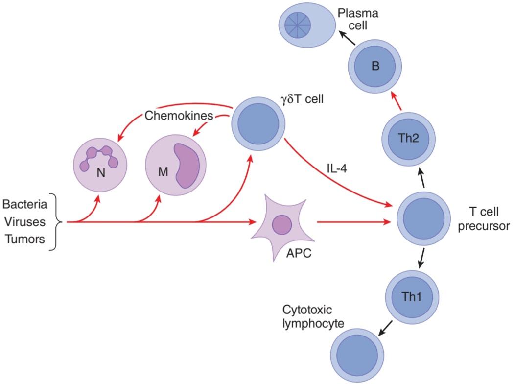
FIGURE 3-1 How bacteria, viruses, and tumors trigger innate immunity and initiate the acquired immune response. Red arrows indicate mediators/cytokines that act on the target cell shown; black arrows show pathways of differentiation. APC, antigen-presenting cell; M, monocyte; N, neutrophil; Th1 and Th2, type 1 and type 2 helper T cells, respectively.
شکل 3-1 چگونه باکتری ها، ویروس ها و تومورها باعث ایجاد ایمنی ذاتی و شروع پاسخ ایمنی اکتسابی می شوند. فلشهای قرمز واسطهها/سیتوکینهایی را نشان میدهند که روی سلول هدف نشان داده شده عمل میکنند. فلش های سیاه مسیرهای تمایز را نشان می دهند. APC، سلول ارائه دهنده آنتی ژن؛ M، مونوسیت؛ N، نوتروفیل؛ سلول های T کمکی Th1 و Th2 به ترتیب نوع 1 و نوع 2.
INNATE IMMUNITY
A wide variety of innate immune cells respond to molecular patterns produced by bacteria and to other substances characteristic of viruses, tumors, and transplanted cells. Many cells that are not professional immunocytes contribute to innate immune responses, such as endothelial and epithelial cells. The activated cells produce their effects via the release of cytokines, as well as, in some cases, complement and other systems.
مصونیت ذاتی
طیف گسترده ای از سلول های ایمنی ذاتی به الگوهای مولکولی تولید شده توسط باکتری ها و سایر مواد مشخصه ویروس ها، تومورها و سلول های پیوندی پاسخ می دهند. بسیاری از سلول ها که ایمونوسیت حرفه ای نیستند، به پاسخ های ایمنی ذاتی، مانند سلول های اندوتلیال و اپیتلیال کمک می کنند. سلول های فعال شده اثرات خود را از طریق انتشار سیتوکین ها و همچنین در برخی موارد، مکمل و سایر سیستم ها تولید می کنند.
Innate immunity in Drosophila centers around a receptor protein named toll, which binds fungal antigens and triggers activation of genes coding for antifungal proteins. An expanding list of toll-like receptors (TLRs) has now been identified in humans and other vertebrates. One of these, TLR4, binds bacterial lipopolysaccharide and a protein called CD14, and this initiates intracellular events that activate transcription of genes for a variety of proteins involved in innate immune responses. This is important because bacterial lipopolysaccharide produced by gram-negative organisms is the cause of septic shock. TLR2 mediates the response to microbial lipoproteins, TLR6 cooperates with TLR2 in recognizing certain peptidoglycans, TLR5 recognizes a molecule known as flagellin in bacterial flagellae, and TLR9 recognizes bacterial DNA. TLRs are referred to as pattern recognition receptors (PRRs) because they recognize and respond to the molecular patterns expressed by pathogens. Other PRRs may be intracellular, such as the so-called NOD proteins. One NOD protein, NOD2, has received attention as the product of a candidate gene leading to the intestinal inflammatory condition, Crohn disease (Clinical Box 3-1).
ایمنی ذاتی در مگس سرکه حول پروتئین گیرنده ای به نام toll متمرکز است که آنتی ژن های قارچی را متصل می کند و باعث فعال شدن ژن های کد کننده پروتئین های ضد قارچی می شود. اکنون فهرست در حال گسترشی از گیرنده های شبه تلفات (TLR) در انسان و سایر مهره داران شناسایی شده است. یکی از اینها، TLR4، لیپوپلی ساکارید باکتریایی و پروتئینی به نام CD14 را متصل می کند و این اتفاقات درون سلولی را آغاز می کند که رونویسی ژن ها را برای انواع پروتئین های دخیل در پاسخ های ایمنی ذاتی فعال می کند. این مهم است زیرا لیپوپلی ساکارید باکتریایی تولید شده توسط ارگانیسم های گرم منفی علت شوک سپتیک است. TLR2 واسطه پاسخ به لیپوپروتئین های میکروبی است، TLR6 با TLR2 در شناسایی پپتیدوگلیکان های خاص همکاری می کند، TLR5 مولکولی به نام فلاژلین را در تاژک های باکتریایی شناسایی می کند و TLR9 DNA باکتری ها را تشخیص می دهد. TLR ها به عنوان گیرنده های تشخیص الگو (PRRs) شناخته می شوند زیرا الگوهای مولکولی بیان شده توسط پاتوژن ها را تشخیص می دهند و به آنها پاسخ می دهند. سایر PRR ها ممکن است داخل سلولی باشند، مانند پروتئین های NOD. یک پروتئین NOD، NOD2، به عنوان محصول یک ژن کاندید که منجر به بیماری التهابی روده، بیماری کرون می شود، مورد توجه قرار گرفته است (جعبه بالینی 3-1).
CLINICAL BOX 3 1
Crohn Disease
Crohn disease is a chronic, relapsing, and remitting disease that involves transmural inflammation of the intestine that can occur at any point along the length of the gastrointestinal tract but most commonly is confined to the distal small intestine and colon. Patients with this condition suffer from changes in bowel habits, bloody diarrhea, severe abdominal pain, weight loss, and malnutrition. Evidence is accumulating that the disease reflects a failure to downregulate inflammatory responses to the normal gut commensal microbiota. In genetically susceptible individuals, mutations in genes controlling innate immune responses (eg, NOD2) or regulators of acquired immunity appear to predispose to disease when individuals are exposed to appropriate environmental factors, which can include a change in the microbiota, or stress.
جعبه بالینی 3. 1
بیماری کرون
بیماری کرون یک بیماری مزمن، عودکننده و فروکش کننده است که شامل التهاب فرامورال روده میشود که میتواند در هر نقطه از طول دستگاه گوارش رخ دهد، اما معمولاً به قسمت انتهایی روده کوچک و کولون محدود میشود. بیماران مبتلا به این بیماری از تغییر در عادات روده، اسهال خونی، درد شدید شکم، کاهش وزن و سوء تغذیه رنج می برند. شواهدی در حال انباشته شدن است مبنی بر اینکه این بیماری منعکس کننده عدم تنظیم پاسخ های التهابی به میکروبیوتای معمولی روده است. در افراد مستعد ژنتیکی، جهش در ژنهای کنترل کننده پاسخهای ایمنی ذاتی (مانند NOD2) یا تنظیمکنندههای ایمنی اکتسابی، زمانی که افراد در معرض عوامل محیطی مناسب قرار میگیرند، مستعد ابتلا به بیماری هستند که میتواند شامل تغییر در میکروبیوتا یا استرس باشد.
THERAPEUTIQ HIGHLIGHTS
During flares of Crohn disease, the mainstay of treatment remains high-dose corticosteroids to suppress inflammation nonspecifically. Surgery is often required to treat complications such as strictures, fistulas, and abscesses. Some patients with severe disease also benefit from ongoing treatment with immunosuppressive drugs, or from treatment with antibodies targeted against tumor necrosis factor-a (TNF-α) or other inflammatory cytokines. Probiotics, therapeutic microorganisms designed to restore a “healthy” microbiota, may have some role in prophylaxis. The pathogenesis of Crohn disease, as well as the related inflammatory bowel disease, ulcerative colitis, remains the subject of intense investigation, and therapies that target specific facets of the inflammatory cascade that may be selectively implicated in individual patients with differing genetic backgrounds are under development.
نکات برجسته درمانی
در طول تشدید بیماری کرون، درمان اصلی کورتیکواستروئیدهای با دوز بالا برای سرکوب غیر اختصاصی التهاب است. جراحی اغلب برای درمان عوارضی مانند تنگی، فیستول و آبسه مورد نیاز است. برخی از بیماران مبتلا به بیماری شدید نیز از درمان مداوم با داروهای سرکوب کننده سیستم ایمنی، یا از درمان با آنتی بادی های هدفمند علیه فاکتور نکروز تومور-a (TNF-α) یا سایر سایتوکاین های التهابی سود می برند. پروبیوتیکها، میکروارگانیسمهای درمانی که برای بازیابی میکروبیوتای “سالم” طراحی شدهاند، ممکن است نقشی در پیشگیری داشته باشند. پاتوژنز بیماری کرون، و همچنین بیماری التهابی روده مرتبط، کولیت اولسراتیو، همچنان موضوع تحقیقات شدید است و درمانهایی که جنبههای خاصی از آبشار التهابی را هدف قرار میدهند که ممکن است به طور انتخابی در بیماران فردی با زمینههای ژنتیکی متفاوت دخیل باشند، در دست توسعه هستند.
ACQUIRED IMMUNITY
As noted previously, the key to acquired immunity is the ability of lymphocytes to produce antibodies (in the case of B cells) or cell-surface receptors (in the case of T cells) that are specific for one of the many millions of foreign agents that may invade the body. The antigens stimulating production of T cell receptors or antibodies are usually proteins and polypeptides, but antibodies can also be formed against nucleic acids and lipids if these are presented as nucleoproteins and lipoproteins. Antibodies to small molecules can also be produced experimentally if the molecules are bound to protein. Acquired immunity has two components: humoral immunity and cellular immunity. Humoral immunity is mediated by circulating immunoglobulin antibodies in the y-globulin fraction of the plasma proteins. Immunoglobulins are produced by differentiated forms of B lymphocytes known as plasma cells, and they activate the complement system and attack and neutralize antigens (Figure 3-1). Humoral immunity is a major defense against bacterial infections. Cellular immunity is mediated by T lymphocytes. It is responsible for delayed allergic reactions and rejection of transplants of foreign tissue. Cytotoxic T cells attack and destroy cells bearing the antigen that activated them. They kill by inserting proteins known as perforins that form pores in the membrane of target cells, and by initiating apoptosis. Cellular immunity constitutes a major defense against infections due to viruses, fungi, and a few bacteria such as the tubercle bacillus. It also helps defend against tumors.
مصونیت اکتسابی
همانطور که قبلا ذکر شد، کلید ایمنی اکتسابی، توانایی لنفوسیت ها برای تولید آنتی بادی (در مورد سلول های B) یا گیرنده های سطح سلولی (در مورد سلول های T) است که برای یکی از میلیون ها عامل خارجی که ممکن است به بدن حمله کنند، اختصاص دارد. آنتی ژن های محرک تولید گیرنده ها یا آنتی بادی های سلول T معمولاً پروتئین ها و پلی پپتیدها هستند، اما اگر به صورت نوکلئوپروتئین ها و لیپوپروتئین ها ارائه شوند، آنتی بادی ها نیز می توانند علیه اسیدهای نوکلئیک و لیپیدها تشکیل شوند. اگر مولکولها به پروتئین متصل باشند، میتوان آنتیبادیهای مولکولهای کوچک را نیز بهصورت تجربی تولید کرد. ایمنی اکتسابی دارای دو جزء است: ایمنی هومورال و ایمنی سلولی. ایمنی هومورال توسط آنتی بادی های ایمونوگلوبولین در حال گردش در بخش y-گلوبولین پروتئین های پلاسما ایجاد می شود. ایمونوگلوبولین ها توسط اشکال تمایز یافته لنفوسیت های B معروف به سلول های پلاسما تولید می شوند و سیستم کمپلمان را فعال می کنند و آنتی ژن ها را مورد حمله و خنثی قرار می دهند (شکل 3-1). ایمنی هومورال دفاع اصلی در برابر عفونت های باکتریایی است. ایمنی سلولی توسط لنفوسیت های T ایجاد می شود. مسئول تاخیر در واکنش های آلرژیک و رد پیوند بافت خارجی است. سلول های T سیتوتوکسیک به سلول های حامل آنتی ژنی که آنها را فعال می کند حمله کرده و آنها را از بین می برند. آنها با وارد کردن پروتئین های معروف به پرفورین که منافذی را در غشای سلول های هدف ایجاد می کنند و با شروع آپوپتوز می کشند. ایمنی سلولی یک دفاع اصلی در برابر عفونت های ناشی از ویروس ها، قارچ ها و تعدادی باکتری مانند باسیل سل است. همچنین به دفاع در برابر تومورها کمک می کند.
LYMPHOCYTES
Lymphocytes are the key elements in the production of acquired immunity. After birth, some lymphocytes are formed in the bone marrow. However, most are formed in the lymph nodes (Figure 3-2), thymus, and spleen from precursor cells that originally came from the bone marrow and were processed in the thymus (T cells) or bone marrow (B cells, see below). Lymphocytes enter the bloodstream for the most part via the lymphatics. At any given time, only about 2% of the body lymphocytes are in the peripheral blood. Most of the rest are in the lymphoid organs.
لنفوسیت ها
لنفوسیت ها عناصر کلیدی در تولید ایمنی اکتسابی هستند. پس از تولد، مقداری لنفوسیت در مغز استخوان تشکیل می شود. با این حال، بیشتر در غدد لنفاوی (شکل 3-2)، تیموس، و طحال از سلول های پیش ساز که در اصل از مغز استخوان آمده و در تیموس (سلول های T) یا مغز استخوان (سلول های B، در زیر) پردازش شده اند، تشکیل می شوند. لنفوسیت ها بیشتر از طریق لنفاوی وارد جریان خون می شوند. در هر زمان معین، تنها حدود 2 درصد از لنفوسیت های بدن در خون محیطی هستند. بیشتر بقیه در اندام های لنفاوی هستند.

FIGURE 3–2 Anatomy of a normal lymph node. (Reproduced with permission from Chandra soma. Reproduced with permission from McPhee SJ, Lingappa VR, Ganong WF [editors]: Pathophysiology of Disease, 4th ed. New York, NY: McGraw-Hill; 2003.)
شکل 3-2 آناتومی یک غدد لنفاوی طبیعی. (تکثیر شده با اجازه Chandra soma. تکثیر شده با اجازه McPhee SJ، Lingappa VR، Ganong WF [ویراستاران]: Pathophysiology of Disease، ویرایش چهارم نیویورک، نیویورک: McGraw-Hill؛ 2003.)
During fetal development, and to a much lesser extent during adult life, lymphocyte precursors come from the bone marrow. Those that populate the thymus (Figure 3-3) become transformed by the environment in this organ into T lymphocytes. Another lymphoid subset that forms in the thymus is the NKT cell, so-called because it shares features of both T lymphocytes and natural killer (NK) cells. Transformation to B lymphocytes, on the other hand, occurs in the fetal liver and, after birth, the bone marrow. NK cells also form in these sites. After residence in the thymus, liver, or bone marrow, many of the T and B lymphocytes migrate to the lymph nodes.
در طول رشد جنین و به میزان بسیار کمتری در دوران بزرگسالی، پیش سازهای لنفوسیتی از مغز استخوان می آیند. آنهایی که تیموس را پر می کنند (شکل 3-3) توسط محیط این اندام به لنفوسیت های T تبدیل می شوند. یکی دیگر از زیرمجموعه های لنفوئیدی که در تیموس تشکیل می شود، سلول NKT است، به این دلیل که ویژگی های مشترک لنفوسیت های T و سلول های کشنده طبیعی (NK) را دارد. از سوی دیگر، تبدیل به لنفوسیت های B در کبد جنین و پس از تولد، مغز استخوان اتفاق می افتد. سلول های NK نیز در این مکان ها تشکیل می شوند. پس از اقامت در تیموس، کبد یا مغز استخوان، بسیاری از لنفوسیت های T و B به غدد لنفاوی مهاجرت می کنند.

FIGURE 3-3 Development of the system mediating acquired immunity. Committed lymphoid progenitors arise in the bone marrow. Maturation into the B and NK cell lineages occurs in this site, whereas development into T and NKT cells takes place after lymphoid progenitors migrate to the thymus. NK, natural killer.
شکل 3-3 توسعه سیستم واسطه ایمنی اکتسابی. پیش سازهای لنفوئیدی متعهد در مغز استخوان ایجاد می شوند. بلوغ به دودمان سلولی B و NK در این محل رخ می دهد، در حالی که توسعه به سلول های T و NKT پس از مهاجرت پیش سازهای لنفاوی به تیموس صورت می گیرد. NK، قاتل طبیعی
T and B lymphocytes are morphologically indistinguishable but can be identified by markers on their cell membranes. B cells differentiate sequentially into cells capable of producing the various classes of immunoglobulins and thereafter into plasma cells. There are two major types of T cells: cytotoxic T cells and helper/effector T cells. There are at least four subtypes of helper T cells: T helper 1 (Th1) cells secrete interleukin (IL)-2 and y-interferon and are concerned primarily with stimulating cellular immunity; Th2 cells secrete IL-4 and IL-5 and interact primarily with B cells in relation to humoral immunity. Th17 cells are induced in response to bacterial infections, produce IL-6 and IL- 17, and help recruit neutrophils. They are also implicated in the generation of harmful inflammatory responses that occur in autoimmune diseases. Finally, Treg cells produce IL-10 to dampen T cell-driven responses. Cytotoxic T cells destroy transplanted cells and those expressing foreign antigens (eg, virally infected targets), with their development aided and directed by helper T cells.
لنفوسیت های T و B از نظر مورفولوژی قابل تشخیص نیستند اما می توان آنها را با نشانگرهای روی غشای سلولی آنها شناسایی کرد. سلول های B به طور متوالی به سلول هایی که قادر به تولید کلاس های مختلف ایمونوگلوبولین ها هستند و پس از آن به سلول های پلاسما تمایز می یابند. دو نوع عمده از سلول های T وجود دارد: سلول های T سیتوتوکسیک و سلول های T کمک کننده/اثربخش. حداقل چهار زیرگروه از سلولهای T کمکی وجود دارد: سلولهای T helper 1 (Th1) اینترلوکین (IL)-2 و y-اینترفرون ترشح میکنند و در درجه اول با تحریک ایمنی سلولی مرتبط هستند. سلول های Th2 IL-4 و IL-5 ترشح می کنند و در ابتدا با سلول های B در ارتباط با ایمنی هومورال تعامل دارند. سلول های Th17 در پاسخ به عفونت های باکتریایی القا می شوند، IL-6 و IL-17 تولید می کنند و به جذب نوتروفیل ها کمک می کنند. آنها همچنین در تولید پاسخ های التهابی مضری که در بیماری های خودایمنی رخ می دهد نقش دارند. در نهایت، سلولهای Treg IL-10 را برای کاهش پاسخهای مبتنی بر سلول T تولید میکنند. سلولهای T سیتوتوکسیک سلولهای پیوندی و آنهایی را که آنتیژنهای خارجی را بیان میکنند (مثلاً اهداف آلوده به ویروس) را با کمک و هدایت سلولهای T کمککننده و هدایت میکنند.
Markers on the surface of lymphocytes are assigned CD (clusters of differentiation) numbers on the basis of their reactions to a panel of monoclonal antibodies. Most cytotoxic T cells display the glycoprotein CD8, and helper T cells display the glycoprotein CD4. These proteins are closely associated with the T cell receptors and may function as coreceptors. On the basis of differences in their receptors and functions, cytotoxic T cells are divided into aẞ and yo types (see below). NK and NKT cells (see above) are also cytotoxic lymphocytes. Thus, there are four main types of cytotoxic lymphocytes in the body: aẞ T cells, y8 T cells, NK cells, and NKT cells.
نشانگرهای روی سطح لنفوسیت ها بر اساس واکنش آنها به پانل آنتی بادی های مونوکلونال به شماره CD (خوشه های تمایز) اختصاص داده می شوند. بیشتر سلولهای T سیتوتوکسیک، گلیکوپروتئین CD8 و سلولهای T کمکی، گلیکوپروتئین CD4 را نشان میدهند. این پروتئین ها ارتباط نزدیکی با گیرنده های سلول T دارند و ممکن است به عنوان گیرنده های مرکزی عمل کنند. بر اساس تفاوت در گیرنده ها و عملکردهای آنها، سلول های T سیتوتوکسیک به انواع aẞ و yo تقسیم می شوند (به زیر مراجعه کنید). سلول های NK و NKT (به بالا مراجعه کنید) نیز لنفوسیت های سیتوتوکسیک هستند. بنابراین، چهار نوع اصلی لنفوسیت سیتوتوکسیک در بدن وجود دارد: سلولهای aẞ T، سلولهای y8، سلولهای NK و سلولهای NKT.
MEMORY B CELLS & T CELLS
After exposure to a given antigen, a small number of activated B and T cells persist as memory B and T cells. These cells are readily converted to effector cells by a later encounter with the same antigen. This ability to produce an accelerated response to a second exposure to an antigen is a key characteristic of acquired immunity. The ability persists for long periods of time, and in some instances (eg, immunity to measles) it can be life-long.
سلول های حافظه B و سلول های T
پس از قرار گرفتن در معرض آنتی ژن معین، تعداد کمی از سلول های B و T فعال شده به عنوان سلول های حافظه B و T باقی می مانند. این سلول ها به آسانی با مواجهه بعدی با همان آنتی ژن به سلول های موثر تبدیل می شوند. این توانایی برای ایجاد یک پاسخ سریع به قرار گرفتن در معرض دوم با یک آنتی ژن یکی از ویژگی های کلیدی ایمنی اکتسابی است. این توانایی برای مدت طولانی باقی می ماند و در برخی موارد (مثلاً مصونیت در برابر سرخک) می تواند مادام العمر باشد.
After activation in lymph nodes, lymphocytes disperse widely throughout the body and are especially plentiful in areas where invading organisms enter the body (eg, the mucosa of the respiratory and gastrointestinal tracts). This puts memory cells close to sites of reinfection and may account in part for the rapidity and strength of their response. Chemotactic cytokines known as chemokines (see below) are involved in guiding activated lymphocytes to these locations.
پس از فعال شدن در غدد لنفاوی، لنفوسیت ها به طور گسترده در سراسر بدن پراکنده می شوند و به ویژه در مناطقی که ارگانیسم های مهاجم وارد بدن می شوند (مثلاً مخاط دستگاه تنفسی و دستگاه گوارش) فراوان هستند. این امر سلول های حافظه را به محل های عفونت مجدد نزدیک می کند و ممکن است تا حدودی به دلیل سرعت و قدرت پاسخ آنها باشد. سیتوکین های کموتاکتیک معروف به کموکاین ها (به زیر مراجعه کنید) در هدایت لنفوسیت های فعال به این مکان ها نقش دارند.
ANTIGEN RECOGNITION
The number of different antigens recognized by lymphocytes in the body is extremely large. The repertoire develops initially without exposure to the antigen. Stem cells differentiate into many millions of different T and B lymphocytes, each with the ability to respond to a particular antigen. When the antigen first enters the body, it can bind directly to the appropriate receptors on B cells. However, a full antibody response requires that the B cells contact helper T cells. In the case of T cells, the antigen is taken up by an antigen-presenting cell (APC) and partially digested. A peptide fragment of it is presented to the appropriate receptors on T cells. In either case, the cells are stimulated to divide, forming clones of cells that respond to this antigen (clonal selection). Effector cells are also subject to negative selection, during which lymphocyte precursors that are reactive with self-antigens are normally deleted. This results in immune tolerance. It is this latter process that presumably goes awry in autoimmune diseases, where the body reacts to and destroys cells expressing normal proteins, with accompanying inflammation that may lead to tissue destruction.
تشخیص آنتی ژن
تعداد آنتی ژن های مختلف شناسایی شده توسط لنفوسیت ها در بدن بسیار زیاد است. رپرتوار در ابتدا بدون قرار گرفتن در معرض آنتی ژن ایجاد می شود. سلول های بنیادی به میلیون ها لنفوسیت T و B مختلف تمایز می یابند که هر کدام توانایی پاسخگویی به آنتی ژن خاصی را دارند. هنگامی که آنتی ژن برای اولین بار وارد بدن می شود، می تواند مستقیماً به گیرنده های مناسب روی سلول های B متصل شود. با این حال، پاسخ کامل آنتی بادی مستلزم تماس سلول های B با سلول های T کمکی است. در مورد سلول های T، آنتی ژن توسط یک سلول ارائه دهنده آنتی ژن (APC) گرفته شده و تا حدی هضم می شود. یک قطعه پپتیدی از آن به گیرنده های مناسب روی سلول های T ارائه می شود. در هر صورت، سلول ها برای تقسیم تحریک می شوند و کلون هایی از سلول ها را تشکیل می دهند که به این آنتی ژن پاسخ می دهند (انتخاب کلونال). سلولهای اثرگذار نیز در معرض انتخاب منفی قرار میگیرند، که طی آن پیشسازهای لنفوسیتی که با آنتیژنهای خود واکنش نشان میدهند، معمولاً حذف میشوند. این منجر به تحمل ایمنی می شود. این فرآیند اخیر است که احتمالاً در بیماریهای خودایمنی، جایی که بدن به سلولهایی که پروتئینهای طبیعی را بیان میکنند، واکنش نشان میدهد و آنها را تخریب میکند، با التهاب همراه است که ممکن است منجر به تخریب بافت شود.
ANTIGEN PRESENTATION
APCs include specialized cells called dendritic cells in the lymph nodes and spleen and the Langerhans dendritic cells in the skin. Macrophages and B cells themselves, and likely many other cell types, can also function as APCs. For example, in the intestine, the epithelial cells that line the tract are likely important in presenting antigens derived from commensal bacteria. In APCs, polypeptide products of antigen digestion are coupled to the HLA protein products of the major histocompatibility complex (MHC) genes and presented on the surface of the cell.
ارائه آنتی ژن
APCها شامل سلول های تخصصی به نام سلول های دندریتیک در غدد لنفاوی و طحال و سلول های دندریتیک لانگرهانس در پوست هستند. خود ماکروفاژها و سلول های B و احتمالاً بسیاری از انواع سلول های دیگر نیز می توانند به عنوان APC عمل کنند. به عنوان مثال، در روده، سلول های اپیتلیال که دستگاه را می پوشانند، احتمالاً در ارائه آنتی ژن های مشتق شده از باکتری های مشترک مهم هستند. در APCها، محصولات پلی پپتیدی هضم آنتی ژن با محصولات پروتئینی HLA ژن های مجتمع اصلی سازگاری بافتی (MHC) جفت می شوند و بر روی سطح سلول ارائه می شوند.
The genes of the MHC encode glycoproteins that are divided into two classes on the basis of structure and function. Class I antigens are composed of a 45-kDa heavy chain associated noncovalently with ẞ2-microglobulin encoded by a gene outside the MHC (Figure 3-4). They are found on all nucleated cells. Class II antigens are heterodimers made up of a 29- to 34-kDa a chain associated noncovalently with a 25- to 28-kDa ẞ chain. They are present in “professional” APCs, including B cells, and in activated T cells.
ژنهای MHC گلیکوپروتئینهایی را رمزگذاری میکنند که بر اساس ساختار و عملکرد به دو دسته تقسیم میشوند. آنتی ژن های کلاس I از یک زنجیره سنگین 45 کیلو دالتونی مرتبط با میکروگلوبولین 2 تشکیل شده اند که توسط ژنی خارج از MHC کدگذاری شده است (شکل 3-4). آنها در تمام سلول های هسته یافت می شوند. آنتی ژن های کلاس II هترودیمرهایی هستند که از یک زنجیره 29 تا 34 کیلو دالتون تشکیل شده اند که به صورت غیرکووالانسی با یک زنجیره 25 تا 28 کیلو دالتون مرتبط است. آنها در APC های “حرفه ای”، از جمله سلول های B، و در سلول های T فعال وجود دارند.

FIGURE 3-4 Schematic structure of a class I MHC protein bound to an antigen fragment, based on the structure of the human histocompatibility antigen HLA-A2. The antigen-binding pocket is at the top and is formed by the a1 and a2 parts of the molecule. The α3 portion and the associated ẞ2- microglobulin (ẞ2m) are close to the membrane. MHC, major histocompatibility complex. (Reproduced with permission from Bjorkman PJ, et al: Structure of the human histocompatibility antigen HLA-A2, Nature 1987;October 8- 14;329(6139):506-512.)
شکل 3-4 ساختار شماتیک یک پروتئین MHC کلاس I متصل به یک قطعه آنتی ژن، بر اساس ساختار آنتی ژن سازگاری بافتی انسانی HLA-A2. پاکت اتصال آنتی ژن در بالا قرار دارد و توسط قسمت های a1 و a2 مولکول تشکیل شده است. بخش α3 و میکروگلوبولین ẞ2- مرتبط (ẞ2m) نزدیک به غشاء هستند. MHC، مجتمع اصلی سازگاری بافتی. (با مجوز Bjorkman PJ، و همکاران: Structure of the human histocompatibility antigen HLA-A2, Nature 1987; اکتبر 8-14; 329(6139):506-512.)
The class I MHC proteins (MHC-I proteins) are coupled primarily to peptide fragments generated from proteins synthesized within cells. Peptides to which the host is not tolerant (eg, those from mutant or viral proteins) are recognized by T cells. The digestion of these proteins occurs in complexes of proteolytic enzymes known as proteasomes, and the peptide fragments bind to MHC proteins in the endoplasmic reticulum. The class II MHC proteins (MHC-II proteins) interact primarily with peptide products of extracellular antigens, such as bacteria, that enter the cell by endocytosis and are digested in the late endosomes.
پروتئینهای کلاس I MHC (پروتئینهای MHC-I) عمدتاً به قطعات پپتیدی تولید شده از پروتئینهای سنتز شده در سلولها جفت میشوند. پپتیدهایی که میزبان نسبت به آنها مقاوم نیست (به عنوان مثال، آنهایی که از پروتئین های جهش یافته یا ویروسی هستند) توسط سلول های T شناسایی می شوند. هضم این پروتئین ها در کمپلکس هایی از آنزیم های پروتئولیتیک موسوم به پروتئازوم ها انجام می شود و قطعات پپتیدی به پروتئین های MHC در شبکه آندوپلاسمی متصل می شوند. پروتئینهای کلاس II MHC (پروتئینهای MHC-II) عمدتاً با محصولات پپتیدی آنتیژنهای خارج سلولی، مانند باکتریها که از طریق اندوسیتوز وارد سلول میشوند و در اندوزومهای دیررس هضم میشوند، تعامل دارند.
T CELL RECEPTORS
The MHC protein-peptide complexes on the surface of the APCs bind to appropriate T cells. Therefore, receptors on the T cells must recognize a very wide variety of complexes. Most of the receptors on circulating T cells are made up of two polypeptide units designated a and ẞ. They form heterodimers that recognize the MHC proteins and the antigen fragments with which they are combined (Figure 3-5). These cells are called aẞ T cells. On the other hand, about 10% of circulating T cells have two different polypeptides designated y and 8 in their receptors, and they are called y8 T cells. These T cells are prominent in the mucosa of the gastrointestinal tract, and there is evidence that they form a link between the innate and acquired immune systems by way of the cytokines they secrete (Figure 3-3).
گیرنده های سلول T
کمپلکس های پروتئین پپتیدی MHC روی سطح APCها به سلول های T مناسب متصل می شوند. بنابراین، گیرنده های روی سلول های T باید طیف بسیار گسترده ای از کمپلکس ها را تشخیص دهند. اکثر گیرنده های سلول های T در گردش از دو واحد پلی پپتیدی به نام های a و ẞ تشکیل شده اند. آنها هترودیمرهایی را تشکیل می دهند که پروتئین های MHC و قطعات آنتی ژنی که با آنها ترکیب می شوند را تشخیص می دهند (شکل 3-5). این سلول ها سلول های T نامیده می شوند. از سوی دیگر، حدود 10 درصد از سلولهای T در گردش دارای دو پلی پپتید متفاوت به نامهای y و 8 در گیرندههای خود هستند و به آنها سلولهای T y8 میگویند. این سلولهای T در مخاط دستگاه گوارش برجسته هستند و شواهدی وجود دارد که نشان میدهد از طریق سیتوکینهایی که ترشح میکنند، پیوندی بین سیستم ایمنی ذاتی و اکتسابی ایجاد میکنند (شکل 3-3).
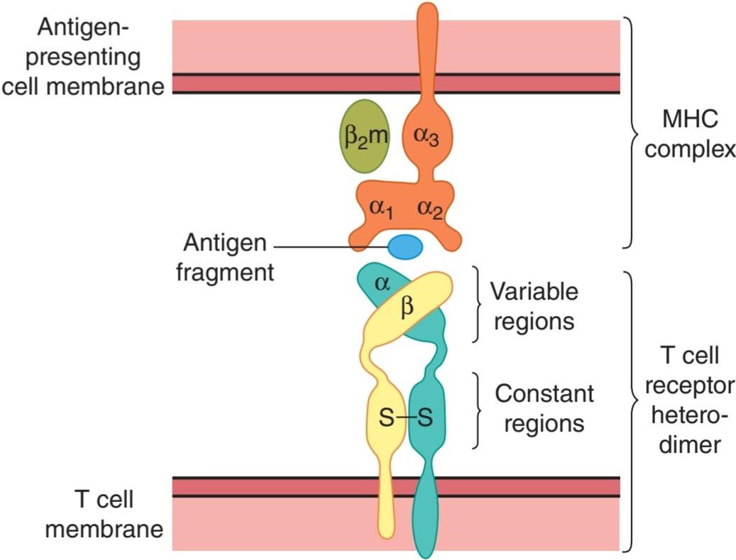
FIGURE 3-5 Interaction between antigen-presenting cell (top) and aẞ T lymphocyte (bottom). The MHC proteins (in this case, MHC-I) and their peptide antigen fragment bind to the a and ẞ units that combine to form the T cell receptor. Some experts refer to this set of protein-protein interactions as the “immune synapse.” MHC, major histocompatibility complex.
شکل 3-5 برهمکنش بین سلول ارائه دهنده آنتی ژن (بالا) و لنفوسیت T a (پایین). پروتئین های MHC (در این مورد، MHC-I) و قطعه آنتی ژن پپتیدی آنها به واحدهای a و ẞ که ترکیب می شوند و گیرنده سلول T را تشکیل می دهند، متصل می شوند. برخی از کارشناسان به این مجموعه از فعل و انفعالات پروتئین-پروتئین به عنوان “سیناپس ایمنی” اشاره می کنند. MHC، مجتمع اصلی سازگاری بافتی.
CD8 occurs on the surface of cytotoxic T cells that bind MHC-I proteins, and CD4 occurs on the surface of helper T cells that bind MHC-II proteins (Figure 3-6). The CD8 and CD4 proteins facilitate the binding of the MHC proteins to the T cell receptors, and they also foster lymphocyte development. The activated CD8 cytotoxic T cells kill their targets directly, whereas the activated CD4 helper T cells secrete cytokines that activate other lymphocytes.
CD8 روی سطح سلولهای T سیتوتوکسیک که به پروتئینهای MHC-I متصل میشوند، و CD4 روی سطح سلولهای T کمکی که پروتئینهای MHC-II را متصل میکنند، رخ میدهد (شکل 3-6). پروتئینهای CD8 و CD4 اتصال پروتئینهای MHC به گیرندههای سلول T را تسهیل میکنند و همچنین رشد لنفوسیتها را تقویت میکنند. سلول های T سیتوتوکسیک فعال CD8 به طور مستقیم اهداف خود را می کشند، در حالی که سلول های T کمکی CD4 فعال شده، سیتوکین هایی ترشح می کنند که سایر لنفوسیت ها را فعال می کنند.
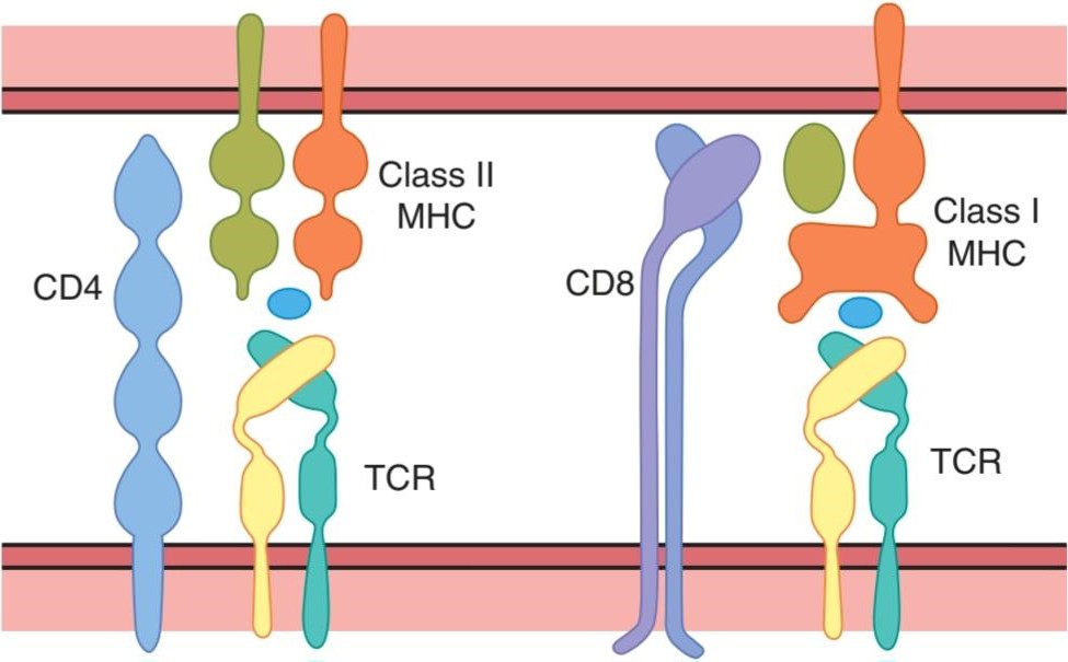
FIGURE 3-6 Diagrammatic summary of the structure of CD4 and CD8, and their relation to MHC-I and MHC-II proteins. Note that CD4 is a single protein, whereas CD8 is a heterodimer. MHC, major histocompatibility complex.
شکل 3-6 خلاصه نموداری از ساختار CD4 و CD8، و ارتباط آنها با پروتئین های MHC-I و MHC-II. توجه داشته باشید که CD4 یک پروتئین منفرد است، در حالی که CD8 یک هترودایمر است. MHC، مجتمع اصلی سازگاری بافتی.
The T cell receptors are surrounded by adhesion molecules and proteins that bind to complementary proteins in the APC when the two cells transiently join to form the “immunologic synapse” that permits T cell activation to occur (Figure 3-7). It is now generally accepted that two signals are necessary to produce activation. One is produced by the binding of the digested antigen to the T cell receptor. The other is produced by the joining of the surrounding proteins in the “synapse.” If the first signal occurs but the second does not, the T cell is inactivated and becomes unresponsive.
گیرندههای سلول T توسط مولکولهای چسبنده و پروتئینهایی احاطه شدهاند که به پروتئینهای مکمل در APC متصل میشوند، زمانی که دو سلول به طور موقت به هم میپیوندند و «سیناپس ایمونولوژیک» را تشکیل میدهند که امکان فعالسازی سلول T را فراهم میکند (شکل 3-7). در حال حاضر به طور کلی پذیرفته شده است که دو سیگنال برای تولید فعال سازی لازم است. یکی از آنها با اتصال آنتی ژن هضم شده به گیرنده سلول T تولید می شود. دیگری از اتصال پروتئین های اطراف در “سیناپس” تولید می شود. اگر سیگنال اول رخ دهد اما سیگنال دوم رخ ندهد، سلول T غیرفعال می شود و پاسخ نمی دهد.

FIGURE 3-7 Summary of acquired immunity. (1) An antigen-presenting cell (depicted here as a macrophage, but other cell types, including dendritic cells, are important) ingests and partially digests an antigen, then presents part of the antigen along with MHC peptides (in this case, MHC II peptides on the cell surface). (2) An “immune synapse” forms with a naive CD4 T cell, which is activated to produce IL-2. (3) IL-2 acts in an autocrine fashion to cause the cell to multiply, forming a clone. (4) The activated CD4 cell may promote B cell activation and the proliferation of plasma cells that produce antibodies or it may activate a cytotoxic CD8 cell. The CD8 cell can also be activated by forming a synapse with an MCH I antigen-presenting cell. MHC, major histocompatibility complex. (Reproduced with permission from McPhee SJ, Lingappa VR, Ganong WF [editors]: Pathophysiology of Disease, 6th ed. New York, NY: McGraw-Hill; 2010.)
شکل 3-7 خلاصه ای از ایمنی اکتسابی. (1) یک سلول ارائه دهنده آنتی ژن (در اینجا به عنوان یک ماکروفاژ نشان داده شده است، اما انواع سلول های دیگر، از جمله سلول های دندریتیک، مهم هستند) یک آنتی ژن را بلعیده و تا حدی هضم می کند، سپس بخشی از آنتی ژن را همراه با پپتیدهای MHC (در این مورد، پپتیدهای MHC II در سطح سلول) ارائه می دهد. (2) یک “سیناپس ایمنی” با یک سلول CD4 T ساده ایجاد می شود که برای تولید IL-2 فعال می شود. (3) IL-2 به روش اتوکرین عمل می کند و باعث تکثیر سلول و تشکیل یک کلون می شود. (4) سلول CD4 فعال شده ممکن است باعث فعال شدن سلول های B و تکثیر سلول های پلاسما که آنتی بادی تولید می کنند یا ممکن است یک سلول CD8 سیتوتوکسیک را فعال کند. سلول CD8 همچنین می تواند با تشکیل یک سیناپس با یک سلول ارائه دهنده آنتی ژن MCH I فعال شود. MHC، مجتمع اصلی سازگاری بافتی. (تکثیر شده با اجازه McPhee SJ، Lingappa VR، Ganong WF [ویراستاران]: Pathophysiology of Disease، ویرایش ششم نیویورک، نیویورک: McGraw-Hill؛ 2010.)
B CELLS
As noted above, B cells can bind antigens directly, but they must contact helper T cells to produce full activation and antibody formation. It is the Th2 subtype that is mainly involved. Helper T cells develop along the T2 lineage in response to IL-4 (see below). On the other hand, IL-12 promotes the Th1 phenotype. IL-2 acts in an autocrine fashion to cause activated T cells to proliferate. The role of various cytokines in B cell and T cell activation is summarized in Figure 3-7.
سلول های B
همانطور که در بالا ذکر شد، سلول های B می توانند به طور مستقیم به آنتی ژن ها متصل شوند، اما آنها باید با سلول های T کمکی تماس بگیرند تا فعال شدن کامل و تشکیل آنتی بادی را ایجاد کنند. این زیرگروه Th2 است که عمدتاً درگیر است. سلول های T کمک کننده در امتداد دودمان T2 در پاسخ به IL-4 رشد می کنند (به زیر مراجعه کنید). از سوی دیگر، IL-12 فنوتیپ Th1 را ترویج می کند. IL-2 به روش اتوکرین عمل می کند و باعث تکثیر سلول های T فعال می شود. نقش سیتوکین های مختلف در فعال سازی سلول های B و T در شکل 3-7 خلاصه شده است.
The activated B cells proliferate and transform into memory B cells (see above) and plasma cells. The plasma cells secrete large quantities of antibodies into the general circulation. The antibodies circulate in the globulin fraction of the plasma and, like antibodies elsewhere, are called immunoglobulins. The immunoglobulins are actually the secreted form of antigen-binding receptors on the B cell membrane.
سلول های B فعال شده تکثیر می شوند و به سلول های B حافظه (به بالا مراجعه کنید) و سلول های پلاسما تبدیل می شوند. سلول های پلاسما مقادیر زیادی آنتی بادی را در گردش خون عمومی ترشح می کنند. آنتی بادی ها در بخش گلوبولین پلاسما گردش می کنند و مانند آنتی بادی ها در جاهای دیگر، ایمونوگلوبولین نامیده می شوند. ایمونوگلوبولین ها در واقع شکل ترشح شده گیرنده های اتصال آنتی ژن بر روی غشای سلول B هستند.
IMMUNOGLOBULINS
Circulating antibodies protect their host by binding to and neutralizing some protein toxins, by blocking the attachment of some viruses and bacteria to cells, by opsonizing bacteria (see below), and by activating complement (see below). Five general types of immunoglobulin antibodies are produced by plasma cells. The basic component of each is a symmetric unit containing four polypeptide chains (Figure 3-8). The two long chains are called heavy chains, whereas the two short chains are called light chains. There are two types of light chains, k and λ, and nine types of heavy chains. The chains are joined by disulfide bridges that permit mobility, and there are intrachain disulfide bridges as well. In addition, the heavy chains are flexible in a region called the hinge. Each heavy chain has a variable (V) segment in which the amino acid sequence is highly variable, a diversity (D) segment in which the amino acid segment is also highly variable, a joining (J) segment in which the sequence is moderately variable, and a constant (C) segment in which the sequence is constant. Each light chain has a V, J, and C segment. The V segments form part of the antigen-binding sites (Fab portion of the molecule [Figure 3-8]). The Fc portion of the molecule is the effector portion, which mediates the reactions initiated by antibodies.
ایمونوگلوبولین ها
آنتی بادی های در گردش از میزبان خود با اتصال به برخی از سموم پروتئینی و خنثی کردن آنها، مسدود کردن اتصال برخی ویروس ها و باکتری ها به سلول ها، با اپسونیزاسیون باکتری ها (در زیر) و با فعال کردن کمپلمان (به پایین) محافظت می کنند. پنج نوع کلی از آنتی بادی های ایمونوگلوبولین توسط سلول های پلاسما تولید می شود. جزء اصلی هر یک واحد متقارن شامل چهار زنجیره پلی پپتیدی است (شکل 3-8). دو زنجیره بلند را زنجیره سنگین و دو زنجیره کوتاه را زنجیره سبک می نامند. دو نوع زنجیر سبک k و λ و 9 نوع زنجیره سنگین وجود دارد. زنجیره ها توسط پل های دی سولفیدی به هم متصل می شوند که امکان تحرک را فراهم می کند و پل های دی سولفید درون زنجیره ای نیز وجود دارد. علاوه بر این، زنجیرهای سنگین در ناحیه ای به نام لولا انعطاف پذیر هستند. هر زنجیره سنگین دارای یک بخش متغیر (V) است که در آن توالی اسید آمینه بسیار متغیر است، یک بخش تنوع (D) که در آن بخش اسید آمینه نیز بسیار متغیر است، یک بخش اتصال (J) که توالی در آن نسبتاً متغیر است، و یک قطعه ثابت (C) که در آن دنباله ثابت است. هر زنجیره سبک دارای یک بخش V، J و C است. بخش های V بخشی از محل های اتصال آنتی ژن را تشکیل می دهند (بخش Fab از مولکول (شکل 3-8). بخش Fc مولکول، بخش مؤثری است که واکنش های آغاز شده توسط آنتی بادی ها را واسطه می کند.
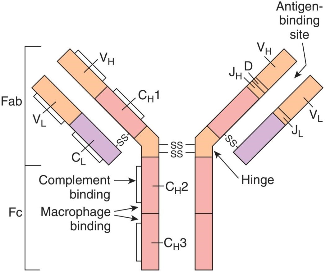
FIGURE 3-8 Typical immunoglobulin G molecule. Fab, portion of the molecule that is concerned with antigen binding; Fc, effector portion of the molecule. The constant regions are pink and purple, and the variable regions are orange. The constant segment of the heavy chain is subdivided into CH1, CH2, and CH3. SS lines indicate intersegmental disulfide bonds. On the right side, the C labels are omitted to show regions JH, D, and JL·
شکل 3-8 مولکول ایمونوگلوبولین G معمولی. Fab، بخشی از مولکول که مربوط به اتصال آنتی ژن است. Fc، بخش عامل مولکول. نواحی ثابت صورتی و بنفش و نواحی متغیر نارنجی هستند. بخش ثابت زنجیره سنگین به CH1، CH2 و CH3 تقسیم می شود. خطوط SS پیوندهای دی سولفیدی بین بخشی را نشان می دهد. در سمت راست، برچسبهای C برای نشان دادن مناطق JH، D و JL حذف شدهاند.
Two of the classes of immunoglobulins contain additional polypeptide components (Table 3-1). In IgM, five of the basic immunoglobulin units join around a polypeptide called the J chain to form a pentamer. In IgA, the secretory immunoglobulin, the immunoglobulin units form dimers and trimers around a J chain and a polypeptide that comes from epithelial cells, the secretory component (SC).
دو دسته از ایمونوگلوبولین ها حاوی اجزای پلی پپتیدی اضافی هستند (جدول 3-1). در IgM، پنج واحد از واحدهای ایمونوگلوبولین پایه به دور پلی پپتیدی به نام زنجیره J می پیوندند و یک پنتامر را تشکیل می دهند. در IgA، ایمونوگلوبولین ترشحی، واحدهای ایمونوگلوبولین دایمرها و تریمرها را در اطراف یک زنجیره J و یک پلی پپتید تشکیل می دهند که از سلول های اپیتلیال، جزء ترشحی (SC) می آید.
TABLE 3-1 Human immune globulins.a
جدول 3-1 گلوبولین های ایمنی انسانی.a

aIn all instances, the light chains are k or y.
aدر همه موارد، زنجیره های سبک k یا y هستند.
In the intestine, bacterial and viral antigens are taken up by M cells (see Chapter 26) and passed on to underlying aggregates of lymphoid tissue (Peyer patches), where they activate naive T cells. These lymphocytes then form B cells that infiltrate mucosa of the gastrointestinal, respiratory, genitourinary, and female reproductive tracts and the breast. There they secrete large amounts of IgA when exposed again to the antigen that initially stimulated them. The epithelial cells produce the SC, which acts as a receptor for, and binds to, IgA. The resulting secretory immunoglobulin passes through the epithelial cell and is secreted by exocytosis. This system of secretory immunity is an important and effective defense mechanism at all mucosal surfaces. Because IgA is secreted into the breast milk, it also accounts for the immune protection that is conferred by the breastfeeding of infants whose immune systems are otherwise immature.
در روده، آنتیژنهای باکتریایی و ویروسی توسط سلولهای M جذب میشوند (به فصل 26 مراجعه کنید) و به تودههای زیرین بافت لنفاوی (تکههای Peyer) منتقل میشوند، جایی که سلولهای T ساده را فعال میکنند. سپس این لنفوسیت ها سلول های B را تشکیل می دهند که به مخاط دستگاه گوارش، تنفسی، دستگاه تناسلی ادراری و تناسلی زنانه و سینه نفوذ می کنند. در آنجا مقادیر زیادی IgA ترشح می کنند که دوباره در معرض آنتی ژنی قرار بگیرند که در ابتدا آنها را تحریک می کرد. سلول های اپیتلیال SC را تولید می کنند که به عنوان گیرنده ای برای IgA عمل می کند و به آن متصل می شود. ایمونوگلوبولین ترشحی حاصل از سلول اپیتلیال عبور کرده و توسط اگزوسیتوز ترشح می شود. این سیستم ایمنی ترشحی یک مکانیسم دفاعی مهم و موثر در تمام سطوح مخاطی است. از آنجایی که IgA در شیر مادر ترشح می شود، محافظت ایمنی را نیز به عهده می گیرد که با شیردهی نوزادانی که سیستم ایمنی آنها نابالغ است، ایجاد می شود.
GENETIC BASIS OF DIVERSITY IN THE IMMUNE SYSTEM
The genetic mechanism for the production of the immensely large number of different configurations of immunoglobulins produced by human B cells, as well as T cell receptors, is a fascinating biologic problem. Diversity is brought about in part by the fact that in immunoglobulin molecules there are two kinds of light chains and nine kinds of heavy chains. As noted previously, there are areas of great variability (hypervariable regions) in each chain. The variable portion of the heavy chains consists of the V, D, and J segments. In the gene family responsible for this region, there are several hundred different coding regions for the V segment, about 20 for the D segment, and four for the J segment. During B cell development, one V coding region, one D coding region, and one J coding region are selected at random and recombined to form the gene that produces that particular variable portion. A similar variable recombination takes place in the coding regions responsible for the two variable segments (V and J) in the light chain. In addition, the J segments are variable because the gene segments join in an imprecise and variable fashion (junctional site diversity) and nucleotides are sometimes added (junctional insertion diversity). It has been calculated that these mechanisms permit the production of about 1015 different immunoglobulin molecules. Additional variability is added by somatic mutation.
مبانی ژنتیکی تنوع در سیستم ایمنی
مکانیسم ژنتیکی برای تولید تعداد بسیار زیادی از پیکربندیهای مختلف ایمونوگلوبولینهای تولید شده توسط سلولهای B انسانی، و همچنین گیرندههای سلول T، یک مشکل بیولوژیکی جذاب است. تنوع تا حدی به دلیل این واقعیت است که در مولکول های ایمونوگلوبولین دو نوع زنجیره سبک و نه نوع زنجیره سنگین وجود دارد. همانطور که قبلا ذکر شد، مناطقی با تنوع زیاد (مناطق بیش متغیر) در هر زنجیره وجود دارد. بخش متغیر زنجیره های سنگین از بخش های V، D و J تشکیل شده است. در خانواده ژن مسئول این ناحیه، صدها ناحیه کدگذاری مختلف برای قطعه V، حدود 20 ناحیه برای بخش D و چهار ناحیه برای قطعه J وجود دارد. در طول توسعه سلول B، یک منطقه کد کننده V، یک منطقه کد کننده D و یک منطقه کد کننده J به طور تصادفی انتخاب شده و برای تشکیل ژنی که آن بخش متغیر خاص را تولید می کند، دوباره ترکیب می شوند. یک نوترکیب متغیر مشابه در مناطق کدگذاری مسئول دو بخش متغیر (V و J) در زنجیره سبک صورت میگیرد. علاوه بر این، بخشهای J متغیر هستند، زیرا بخشهای ژن به شکلی غیردقیق و متغیر به هم میپیوندند (تنوع محل اتصال) و گاهی اوقات نوکلئوتیدها اضافه میشوند (تنوع درج پیوندی). محاسبه شده است که این مکانیسم ها اجازه تولید حدود 1015 مولکول مختلف ایمونوگلوبولین را می دهند. تنوع اضافی با جهش سوماتیک اضافه می شود.
Similar gene rearrangement and joining mechanisms operate to produce the diversity in T cell receptors. In humans, the a subunit has a V region encoded by 1 of about 50 different genes and a J region encoded by 1 of another 50 different genes. The ẞ subunits have a V region encoded by 1 of about 50 genes, a D region encoded by 1 of 2 genes, and a J region encoded by 1 of 13 genes. These variable regions permit the generation of up to an estimated 1015 different T cell receptors (Clinical Box 3-2 and Clinical Box 3-3).
بازآرایی ژن و مکانیسمهای پیوستن مشابهی برای ایجاد تنوع در گیرندههای سلول T عمل میکنند. در انسان، زیرواحد a دارای یک ناحیه V است که توسط 1 از حدود 50 ژن مختلف و یک ناحیه J کدگذاری شده توسط 1 ژن از 50 ژن مختلف دیگر کدگذاری شده است. زیرواحدهای ẞ دارای یک ناحیه V هستند که توسط 1 ژن از حدود 50 ژن، یک ناحیه D کدگذاری شده توسط 1 ژن از 2 ژن، و یک منطقه J کدگذاری شده توسط 1 ژن از 13 ژن. این نواحی متغیر اجازه تولید تا حدود 1015 گیرنده سلول T مختلف را می دهند (جعبه بالینی 3-2 و جعبه بالینی 3-3).
More than 300 primary immunodeficiency states are now known to arise from defects in these various stages of B and T lymphocyte maturation (Clinical Box 3-4). A few important ones are shown in Figure 3-9.
بیش از 300 حالت نقص ایمنی اولیه در حال حاضر شناخته شده است که از نقایص این مراحل مختلف بلوغ لنفوسیت های B و T ناشی می شود (جعبه بالینی 3-4). چند مورد مهم در شکل 3-9 نشان داده شده است.
CLINICAL BOX 3.2
Autoimmunity
Sometimes, the processes that eliminate antibodies against self-antigens fail and a variety of different autoimmune diseases are produced. These can be B cell- or T cell-mediated and can be organ-specific or systemic. They include type 1 diabetes mellitus (antibodies against pancreatic islet B cells), myasthenia gravis (antibodies against nicotinic cholinergic receptors), and multiple sclerosis (antibodies against myelin basic protein and several other components of myelin). In some instances, the antibodies are against receptors and are capable of activating those receptors; for example, antibodies against TSH receptors increase thyroid activity and cause Graves disease (see Chapter 20). Other conditions are due to the production of antibodies against invading organisms that cross-react with normal body constituents (molecular mimicry). An example is rheumatic fever following a streptococcal infection; a portion of cardiac myosin resembles a portion of the streptococcal M protein, and antibodies induced by the latter attack the former and damage the heart. Some conditions may be due to bystander effects, in which inflammation sensitizes T cells in the neighborhood, causing them to become activated when otherwise they would not respond.
جعبه بالینی 3.2
خودایمنی
گاهی اوقات، فرآیندهایی که آنتیبادیها را علیه خود آنتیژنها از بین میبرند با شکست مواجه میشوند و انواع بیماریهای خودایمنی مختلف تولید میشوند. اینها می توانند با واسطه سلول های B یا T باشند و می توانند مختص اندام یا سیستمیک باشند. آنها عبارتند از دیابت نوع 1 (آنتی بادی علیه سلول های جزایر B پانکراس)، میاستنی گراویس (آنتی بادی علیه گیرنده های کولینرژیک نیکوتین)، و مولتیپل اسکلروزیس (آنتی بادی علیه پروتئین پایه میلین و چندین جزء دیگر میلین). در برخی موارد، آنتیبادیها علیه گیرندهها هستند و میتوانند آن گیرندهها را فعال کنند. به عنوان مثال، آنتی بادی ها علیه گیرنده های TSH فعالیت تیروئید را افزایش می دهند و باعث بیماری گریوز می شوند (به فصل 20 مراجعه کنید). سایر شرایط به دلیل تولید آنتی بادی علیه موجودات مهاجم است که با اجزای طبیعی بدن واکنش متقابل دارند (تقلید مولکولی). یک مثال تب روماتیسمی به دنبال عفونت استرپتوکوک است. بخشی از میوزین قلبی شبیه به بخشی از پروتئین استرپتوکوک M است و آنتی بادی های القا شده توسط دومی به پروتئین اولی حمله کرده و به قلب آسیب می زند. برخی از شرایط ممکن است به دلیل اثرات اطرافیان باشد، که در آن التهاب سلولهای T را در همسایگی حساس میکند و باعث میشود در غیر این صورت پاسخ ندهند، فعال شوند.
THERAPEUTIC HIGHLIGHTS
The therapy of autoimmune disorders rests on efforts to replace or restore the damaged function (eg, provision of exogenous insulin in type 1 diabetes) as well as nonspecific efforts to reduce inflammation (using corticosteroids) or to suppress immunity. Recently, agents that deplete or dampen the function of B cells have been shown to have some efficacy in a range of autoimmune disorders, including rheumatoid arthritis, most likely by interrupting the production of autoantibodies that contribute to disease pathogenesis.
نکات برجسته درمانی
درمان اختلالات خودایمنی بر تلاش برای جایگزینی یا بازگرداندن عملکرد آسیب دیده (به عنوان مثال، ارائه انسولین اگزوژن در دیابت نوع 1) و همچنین تلاش های غیر اختصاصی برای کاهش التهاب (استفاده از کورتیکواستروئیدها) یا سرکوب ایمنی استوار است. اخیراً مشخص شده است که عواملی که عملکرد سلولهای B را تحلیل میبرند یا تضعیف میکنند در طیفی از اختلالات خودایمنی، از جمله آرتریت روماتوئید، به احتمال زیاد با قطع تولید اتوآنتیبادیهایی که در پاتوژنز بیماری نقش دارند، کارایی دارند.
CLINICAL BOX 3.3
Tissue Transplantation
The T lymphocyte system is responsible for the rejection of transplanted tissue. When tissues such as skin and kidneys are transplanted from a donor to a recipient of the same species, the transplants “take” and function for a while but then become necrotic and are “rejected” because an immune response to the transplanted tissue develops in the recipient. This is generally true even if the donor and recipient are close relatives, and the only transplants that are never rejected are those from an identical twin. Nevertheless, organ transplantation remains the only viable option in a number of end-stage diseases.
جعبه بالینی 3.3
پیوند بافت
سیستم لنفوسیت T مسئول رد بافت پیوند شده است. هنگامی که بافت هایی مانند پوست و کلیه از اهداکننده به گیرنده همان گونه پیوند زده می شود، پیوندها مدتی طول می کشد و عمل می کنند، اما سپس نکروز می شوند و «رد می شوند» زیرا پاسخ ایمنی به بافت پیوند شده در گیرنده ایجاد می شود. این به طور کلی صادق است حتی اگر اهدا کننده و گیرنده از بستگان نزدیک باشند، و تنها پیوندهایی که هرگز رد نمی شوند پیوندهایی از یک دوقلو همسان هستند. با این وجود، پیوند اعضا تنها گزینه قابل دوام در تعدادی از بیماری های مرحله نهایی باقی مانده است.
THERAPEUTIQ HIGHLIGHTS)
A number of treatments have been developed to overcome the rejection of transplanted organs in humans. The goal of treatment is to stop rejection without leaving the patient vulnerable to massive infections. One approach is to kill T lymphocytes by killing all rapidly dividing cells with drugs such as azathioprine, a purine antimetabolite, but this makes patients susceptible to infections and cancer. Another is to administer corticosteroids, which inhibit cytotoxic T cell proliferation by inhibiting production of IL-2, but these cause osteoporosis, mental changes, and the other facets of Cushing syndrome (see Chapter 19). More recently, immunosuppressive drugs such as cyclosporine or tacrolimus (FK-506) have found favor. Activation of the T cell receptor normally increases intracellular Ca2+, which acts via calmodulin to activate calcineurin. Calcineurin dephosphorylates the transcription factor NF-AT, which moves to the nucleus and increases the activity of genes coding for IL-2 and related stimulatory cytokines. Cyclosporine and tacrolimus prevent the dephosphorylation of NF-AT. However, these drugs inhibit all T cell-mediated immune responses, and cyclosporine causes kidney damage and cancer. A new and promising approach to transplant rejection is the production of T cell unresponsiveness by using drugs that block the costimulation that is required for normal activation (see text). Clinically effective drugs that act in this fashion could be of great value to transplant surgeons.
نکات برجسته درمانی
تعدادی از درمان ها برای غلبه بر رد اعضای پیوندی در انسان ایجاد شده است. هدف از درمان توقف طرد بدون آسیب پذیری بیمار در برابر عفونت های گسترده است. یکی از روش ها کشتن لنفوسیت های T با کشتن تمام سلول هایی که به سرعت تقسیم می شوند با داروهایی مانند آزاتیوپرین، یک آنتی متابولیت پورینی است، اما این امر بیماران را مستعد ابتلا به عفونت ها و سرطان می کند. دیگری تجویز کورتیکواستروئیدها است که با مهار تولید IL-2، تکثیر سلول های T سیتوتوکسیک را مهار می کنند، اما باعث پوکی استخوان، تغییرات ذهنی و سایر جنبه های سندرم کوشینگ می شوند (به فصل 19 مراجعه کنید). اخیراً داروهای سرکوب کننده سیستم ایمنی مانند سیکلوسپورین یا تاکرولیموس (FK-506) مورد توجه قرار گرفته اند. فعال شدن گیرنده سلول T معمولاً +Ca2 داخل سلولی را افزایش می دهد که از طریق کالمودولین برای فعال کردن کلسینئورین عمل می کند. کلسینورین عامل رونویسی NF-AT را دفسفریله می کند که به سمت هسته حرکت می کند و فعالیت ژن های کد کننده IL-2 و سیتوکین های تحریک کننده مرتبط را افزایش می دهد. سیکلوسپورین و تاکرولیموس از دفسفوریلاسیون NF-AT جلوگیری می کنند. با این حال، این داروها تمام پاسخ های ایمنی با واسطه سلول های T را مهار می کنند و سیکلوسپورین باعث آسیب کلیه و سرطان می شود. یک رویکرد جدید و امیدوارکننده برای رد پیوند، تولید عدم پاسخدهی سلولهای T با استفاده از داروهایی است که محرکهای لازم برای فعالسازی طبیعی را مسدود میکنند (متن را ببینید). داروهای موثر بالینی که به این روش عمل می کنند می توانند برای جراحان پیوند ارزش زیادی داشته باشند.
CLINICAL BOX 3.4
Primary Immunodeficiencies
Mutations in enzymes, transcription factors, receptors, and other regulators of acquired or innate immunity lead to primary immunodeficiency disorders. Typically, patients experience frequent infections or have an increased susceptibility to unusual infections, but they may also be subject to autoimmune problems, spontaneous inflammation, and malignancy. The implications depend on the level at which the immune system is disrupted, with mutations that prevent the development of many lymphoid lineages having the most serious consequences (Figure 3-9).
جعبه بالینی 3.4
نقص ایمنی اولیه
جهش در آنزیم ها، فاکتورهای رونویسی، گیرنده ها و سایر تنظیم کننده های ایمنی اکتسابی یا ذاتی منجر به اختلالات نقص ایمنی اولیه می شود. به طور معمول، بیماران عفونت های مکرری را تجربه می کنند یا حساسیت بیشتری به عفونت های غیر معمول دارند، اما ممکن است در معرض مشکلات خود ایمنی، التهاب خود به خود و بدخیمی نیز باشند. پیامدها بستگی به سطحی دارد که در آن سیستم ایمنی مختل شده است، با جهش هایی که مانع از ایجاد بسیاری از دودمان های لنفاوی می شود که شدیدترین عواقب را دارد (شکل 3-9).
THERAPEUTIC HIGHLIGHTS
Mild immunodeficiencies may require only minimal or supportive therapies, but the most serious cause substantial morbidity and early mortality. Patients suffering from the latter conditions, therefore, are candidates for early and definitive therapy, and many have benefitted greatly from the transplant of allogeneic hematopoietic stem cells (HSCs), provided they can tolerate the procedure and a suitable donor can be found. On the other hand, the monogenic nature of many serious primary immunodeficiencies has made them an attractive target for trials of gene therapy approaches. There have been a number of notable successes with transplant of HSCs transduced with retroviral or lentiviral vectors carrying a wild-type copy of the affected gene, such as in X-linked severe combined immune deficiency (SCID), which results in mutations in the gamma chain of the receptor for IL-2.
نکات برجسته درمانی
نقص ایمنی خفیف ممکن است فقط به درمان های حداقلی یا حمایتی نیاز داشته باشد، اما جدی ترین آنها باعث عوارض قابل توجه و مرگ و میر زودرس می شود. بنابراین، بیمارانی که از شرایط اخیر رنج میبرند، کاندیدای درمان زودهنگام و قطعی هستند و بسیاری از پیوند سلولهای بنیادی خونساز آلوژنیک (HSCs) بهره زیادی بردهاند، مشروط بر اینکه بتوانند این روش را تحمل کنند و یک اهداکننده مناسب پیدا شود. از سوی دیگر، ماهیت تک ژنی بسیاری از نقصهای ایمنی اولیه جدی، آنها را به هدفی جذاب برای آزمایشهای رویکردهای ژندرمانی تبدیل کرده است. موفقیتهای قابلتوجهی با پیوند HSCهای انتقال یافته با ناقلهای رتروویروسی یا لنتیویروسی حامل یک کپی از نوع وحشی ژن آسیبدیده، مانند نقص ایمنی ترکیبی شدید مرتبط با X (SCID)، که منجر به جهش در زنجیره گامای گیرنده IL-2 میشود، وجود دارد.

FIGURE 3-9 Sites of congenital blockade of B and T lymphocyte maturation in various primary immunodeficiency states. SCID, severe combined immune deficiency.
شکل 3-9 مکان های بلوغ مادرزادی لنفوسیت های B و T در حالت های مختلف نقص ایمنی اولیه. SCID، کمبود شدید سیستم ایمنی ترکیبی.
SOLUBLE REGULATORS OF IMMUNITY
CYTOKINES
Cytokines are hormone-like molecules that act-generally in a paracrine fashion -to regulate immune responses. They are secreted not only by lymphocytes and macrophages but also by endothelial cells, neurons, glial cells, and other types of cells. Most of the cytokines were initially named for their actions, for example, B cell-differentiating factor, or B cell-stimulating factor 2. However, the nomenclature has since been rationalized by international agreement to that of the interleukins. For example, the name of B cell-differentiating factor was changed to interleukin-4. A number of cytokines selected for their biologic and clinical relevance are listed in Table 3-2, but it would be beyond the scope of this text to list all cytokines, which now number more than 100.
تنظیم کننده های محلول ایمنی
سیتوکین ها
سیتوکینها مولکولهای هورمونمانندی هستند که معمولاً به روش پاراکرینی عمل میکنند و پاسخهای ایمنی را تنظیم میکنند. آنها نه تنها توسط لنفوسیت ها و ماکروفاژها بلکه توسط سلول های اندوتلیال، نورون ها، سلول های گلیال و انواع دیگر سلول ها ترشح می شوند. بیشتر سیتوکین ها در ابتدا به دلیل اعمالشان نامگذاری شدند، به عنوان مثال، فاکتور تمایز سلول B، یا فاکتور 2 محرک سلول B. با این حال، نامگذاری از آن زمان با توافق بین المللی به نام اینترلوکین ها منطقی شده است. به عنوان مثال، نام فاکتور تمایز سلول B به اینترلوکین-4 تغییر یافت. تعدادی از سیتوکینهای انتخابشده برای ارتباط بیولوژیکی و بالینیشان در جدول 3-2 فهرست شدهاند، اما فهرست کردن همه سیتوکینها که اکنون بیش از 100 سیتوکین هستند، خارج از محدوده این متن است.
TABLE 3-2 Examples of cytokines and their clinical relevance.
جدول 3-2 نمونه هایی از سیتوکین ها و ارتباط بالینی آنها.

Modified with permission from Delves PJ, Roitt IM: The immune system. First of two parts, N Engl J Med 2000; July 6:343(1):37-49.
اصلاح شده با مجوز Delves PJ، Roitt IM: سیستم ایمنی. اول از دو بخش، N Engl J Med 2000; ژوئیه 6: 343 (1): 37-49.
Many of the receptors for cytokines and hematopoietic growth factors (see above), as well as the receptors for prolactin (see Chapter 22), and growth hormone (see Chapter 18) are members of a cytokine-receptor superfamily that has three subfamilies (Figure 3-10). The members of subfamily 1, which includes the receptors for IL-4 and IL-7, are homodimers. The members of subfamily 2, which includes the receptors for IL-3, IL-5, and IL-6, are heterodimers. The receptor for IL-2 (and several other cytokines) consists of a heterodimer plus an unrelated protein, the so-called Tac antigen. The other members of subfamily 3 have the same y chain as the IL-2 receptor. The extracellular domain of the homodimer and heterodimer subunits all contain four conserved cysteine residues plus a conserved Trp-Ser-X-Trp-Ser domain, and although the intracellular portions do not contain tyrosine kinase catalytic domains, they activate cytoplasmic tyrosine kinases when ligand binds to the receptors.
بسیاری از گیرنده های سیتوکین ها و فاکتورهای رشد خونساز (نگاه کنید به بالا)، و همچنین گیرنده های پرولاکتین (به فصل 22 مراجعه کنید)، و هورمون رشد (به فصل 18 مراجعه کنید) اعضای یک ابرخانواده گیرنده سیتوکین هستند که دارای سه زیر خانواده است (شکل 3-10). اعضای زیرخانواده 1 که شامل گیرنده های IL-4 و IL-7 است، همودایمر هستند. اعضای زیرخانواده 2 که شامل گیرنده های IL-3، IL-5 و IL-6 است، هترودیمر هستند. گیرنده IL-2 (و چندین سایتوکین دیگر) از یک هترودایمر به اضافه یک پروتئین نامرتبط به نام آنتی ژن Tac تشکیل شده است. سایر اعضای زیرخانواده 3 دارای زنجیره y مشابه با گیرنده IL-2 هستند. دامنه خارج سلولی زیرواحدهای همودایمر و هترودایمر همگی شامل چهار باقیمانده سیستئین حفظ شده به اضافه یک دامنه Trp-Ser-X-Trp-Ser حفظ شده است، و اگرچه بخشهای درون سلولی حاوی حوزههای کاتالیزوری تیروزین کیناز نیستند، زمانی که لیگاندور کینداز تیروزین را فعال میکنند.

FIGURE 3-10 Members of an important cytokine receptor superfamily, showing shared structural elements. Note that all subunits except the a subunit in subfamily 3 have four conserved cysteine residues (C) and a Trp-Ser-X-Trp- Ser motif (X). Many subunits also contain a critical regulatory domain in their cytoplasmic portions (R). CNTF, ciliary neurotrophic factor; Epo, erythropoietin; GH, growth hormone; OSM, oncostatin M; PRL, prolactin.
شکل 3-10 اعضای یک ابرخانواده مهم گیرنده سیتوکین، عناصر ساختاری مشترک را نشان می دهند. توجه داشته باشید که همه زیرواحدها به جز زیرواحد a در زیرخانواده 3 دارای چهار باقیمانده سیستئین (C) و یک موتیف Trp-Ser-X-Trp- Ser (X) هستند. بسیاری از زیرواحدها همچنین دارای یک دامنه تنظیمی حیاتی در بخشهای سیتوپلاسمی خود هستند (R). CNTF، فاکتور نوروتروفیک مژگانی. اپو، اریتروپویتین؛ GH، هورمون رشد؛ OSM، انکوستاتین M؛ PRL، پرولاکتین.
The effects of the principal cytokines are listed in Table 3-2. Some of them have systemic as well as local paracrine effects. For example, IL-1, IL-6, and TNF-α cause fever, and IL-1 increases slow-wave sleep and reduces appetite.
اثرات سیتوکین های اصلی در جدول 3-2 ذکر شده است. برخی از آنها اثرات سیستمیک و همچنین پاراکرین موضعی دارند. به عنوان مثال، IL-1، IL-6 و TNF-α باعث تب می شوند و IL-1 خواب موج آهسته را افزایش می دهد و اشتها را کاهش می دهد.
Another superfamily of cytokines is the chemokine family. Chemokines are substances that attract neutrophils and other immune effector cells to areas of inflammation or an immune response. Over 40 have now been identified, and it is clear that they also play a role in the regulation of cell growth and angiogenesis. The chemokine receptors are G-protein-coupled receptors that cause, among other things, extension of pseudopodia with migration of the cell toward the source of the chemokine.
یکی دیگر از ابرخانوادههای سیتوکینها، خانواده کموکاینها هستند. کموکاین ها موادی هستند که نوتروفیل ها و سایر سلول های عامل ایمنی را به مناطق التهاب یا پاسخ ایمنی جذب می کنند. اکنون بیش از 40 مورد شناسایی شده است و واضح است که آنها همچنین در تنظیم رشد سلولی و رگ زایی نقش دارند. گیرنده های کموکاین گیرنده های جفت شده با پروتئین G هستند که از جمله باعث گسترش شبه پا با مهاجرت سلول به سمت منبع کموکاین می شوند.
HEMATOPOIETIC GROWTH FACTORS
The production of immune effector cells is regulated with great precision in healthy individuals, and the production of granulocytes, in particular, is rapidly and dramatically increased in infections. Many cytokines participate in this regulation, together with other soluble mediators, many of which are referred to as colony-stimulating factors (CSF) because they stimulate progenitors to grow as colonies in soft agar. The overall proliferation and self-renewal of HSCs depends on stem cell factor (SCF). The proliferation and maturation of the cells that enter the blood from the marrow are then regulated by growth factors that cause cells in one or more of the committed cell lines to proliferate and mature (Table 3-3). The factors stimulating the production of committed stem cells include granulocyte/macrophage (GM)-CSF, granulocyte CSF (G-CSF), and macrophage CSF (M-CSF). Interleukins IL-1 and IL-6 followed by IL-3 (Table 3-3) act in sequence to convert pluripotential uncommitted stem cells to committed progenitor cells. IL-3 is also known as multi-CSF. Each of the CSFS has a predominant action, but all the CSFs and interleukins also have other overlapping actions. In addition, they activate and sustain mature blood cells. In fact, it is interesting that basal hematopoiesis is normal in mice in which the GM-CSF gene is knocked out, indicating that loss of one factor can be compensated for by others. On the other hand, the absence of GM-CSF causes accumulation of surfactant in the lungs (see Chapter 34).
فاکتورهای رشد خونساز
تولید سلول های عامل ایمنی با دقت زیادی در افراد سالم تنظیم می شود و تولید گرانولوسیت ها به ویژه در عفونت ها به سرعت و به طور چشمگیری افزایش می یابد. بسیاری از سیتوکینها همراه با سایر واسطههای محلول در این تنظیم شرکت میکنند، که بسیاری از آنها به عنوان عوامل محرک کلنی (CSF) شناخته میشوند زیرا اجداد را تحریک میکنند تا به صورت کلونی در آگار نرم رشد کنند. تکثیر کلی و خود تجدیدی HSC ها به فاکتور سلول های بنیادی (SCF) بستگی دارد. تکثیر و بلوغ سلول هایی که از مغز مغز وارد خون می شوند توسط فاکتورهای رشدی که باعث تکثیر و بلوغ سلول های یک یا چند رده سلولی متعهد می شوند تنظیم می شود (جدول 3-3). عوامل تحریک کننده تولید سلول های بنیادی متعهد عبارتند از گرانولوسیت/ماکروفاژ (GM)-CSF، گرانولوسیت CSF (G-CSF) و CSF ماکروفاژ (M-CSF). اینترلوکین های IL-1 و IL-6 و به دنبال آن IL-3 (جدول 3-3) به ترتیب برای تبدیل سلول های بنیادی غیرمتعهد پرتوان به سلول های پیش ساز متعهد عمل می کنند. IL-3 به عنوان چند CSF نیز شناخته می شود. هر یک از CSFS دارای یک عمل غالب است، اما همه CSFها و اینترلوکین ها دارای اعمال همپوشانی دیگری نیز هستند. علاوه بر این، سلول های خونی بالغ را فعال و حفظ می کنند. در واقع، جالب است که خونسازی پایه در موشهایی که ژن GM-CSF در آنها حذف شده است، طبیعی است، که نشان میدهد از دست دادن یک عامل میتواند توسط دیگران جبران شود. از سوی دیگر، عدم وجود GM-CSF باعث تجمع سورفکتانت در ریه ها می شود (به فصل 34 مراجعه کنید).
The other factors listed are produced by macrophages, activated T cells, fibroblasts, and endothelial cells. For the most part, the factors act locally in the bone marrow.
سایر عوامل ذکر شده توسط ماکروفاژها، سلول های T فعال، فیبروبلاست ها و سلول های اندوتلیال تولید می شوند. در بیشتر موارد، عوامل به صورت موضعی در مغز استخوان عمل می کنند.
TABLE 3-3 Hematopoietic growth factors.
جدول 3-3 فاکتورهای رشد خونساز.

CSF, colony-stimulating factor; G, granulocyte; IL, interleukin; M, macrophage; SCF, stem cell factor.
Reproduced with permission from McPhee SJ, Lingappa VR, Ganong WF (editors): Pathophysiology of Disease, 6th ed. New York, NY: McGraw-Hill; 2010.
CSF، عامل محرک مستعمره؛ G، گرانولوسیت؛ IL، اینترلوکین؛ M، ماکروفاژ; SCF، فاکتور سلول های بنیادی.
تکثیر شده با اجازه McPhee SJ، Lingappa VR، Ganong WF (ویراستاران): Pathophysiology of Disease, 6th ed. نیویورک، نیویورک: McGraw-Hill; 2010.
THE COMPLEMENT SYSTEM
The cell-killing effects of innate and acquired immunity are mediated in part by a system of more than 30 plasma proteins originally named the complement system because they “complemented” the effects of antibodies. Three different pathways or enzyme cascades activate the system: the classic pathway, triggered by immune complexes; the mannose-binding lectin pathway, triggered when this lectin binds mannose groups in bacteria; and the alternative or properdin pathway, triggered by contact with various viruses, bacteria, fungi, and tumor cells. The proteins that are produced have three functions: they help kill invading organisms by opsonization (coating of the organism), chemotaxis, and eventual lysis of the cell; they serve in part as a bridge from innate to acquired immunity by activating B cells and aiding immune memory; and they help dispose of waste products after apoptosis. Cell lysis, one of the principal ways the complement system kills cells, is brought about by inserting proteins called perforins into their cell membranes. These create holes, which permit free flow of ions and thus disruption of membrane polarity.
سیستم مکمل
اثرات کشتن سلولی ایمنی ذاتی و اکتسابی تا حدی توسط سیستمی متشکل از بیش از 30 پروتئین پلاسما که در اصل سیستم مکمل نامیده می شود، انجام می شود زیرا آنها اثرات آنتی بادی ها را “مکمل” می کنند. سه مسیر مختلف یا آبشار آنزیمی سیستم را فعال میکنند: مسیر کلاسیک که توسط کمپلکسهای ایمنی ایجاد میشود. مسیر لکتین اتصال به مانوز، زمانی که این لکتین به گروه های مانوز در باکتری ها متصل می شود، ایجاد می شود. و مسیر جایگزین یا پروپردین که در اثر تماس با ویروسها، باکتریها، قارچها و سلولهای تومور مختلف ایجاد میشود. پروتئین هایی که تولید می شوند سه عملکرد دارند: آنها به کشتن موجودات مهاجم از طریق اپسونیزاسیون (پوشش ارگانیسم)، کموتاکسی و در نهایت لیز سلول کمک می کنند. آنها تا حدی به عنوان پلی از ایمنی ذاتی به اکتسابی با فعال کردن سلول های B و کمک به حافظه ایمنی عمل می کنند. و به دفع مواد زائد پس از آپوپتوز کمک می کنند. لیز سلولی، یکی از روشهای اصلی که سیستم کمپلمان سلولها را میکشد، با وارد کردن پروتئینهایی به نام پرفورین در غشای سلولی آنها انجام میشود. اینها حفره هایی ایجاد می کنند که جریان آزاد یون ها را ممکن می کند و در نتیجه قطبیت غشاء را مختل می کند.
OTHER IMMUNE/INFLAMMATORY EFFECTOR CELLS
Many additional immune effector cells circulate in the blood as the white blood cells. In addition, the blood is the conduit for the precursor cells that eventually develop into the immune cells of the tissues. The circulating immunologic cells include granulocytes (polymorphonuclear leukocytes, PMNs), comprising neutrophils, eosinophils, and basophils; lymphocytes (discussed above); and monocytes. Immune responses in the tissues are further amplified by these cells following their extravascular migration, as well as tissue macrophages (derived from monocytes) and mast cells (related to basophils). Acting together, these cells provide the body with powerful defenses against tumors and viral, bacterial, and parasitic infections.
سایر سلول های عامل ایمنی/التهاب
بسیاری از سلول های عامل ایمنی اضافی به عنوان گلبول های سفید در خون گردش می کنند. علاوه بر این، خون مجرای سلول های پیش ساز است که در نهایت به سلول های ایمنی بافت ها تبدیل می شوند. سلولهای ایمونولوژیک در گردش شامل گرانولوسیتها (لکوسیتهای پلیمورفونوکلئر، PMN) هستند که شامل نوتروفیلها، ائوزینوفیلها و بازوفیلها میشوند. لنفوسیت ها (در بالا بحث شد)؛ و مونوسیت ها پاسخهای ایمنی در بافتها توسط این سلولها به دنبال مهاجرت خارج عروقی، و همچنین ماکروفاژهای بافتی (مشتقشده از مونوسیتها) و ماست سلها (مربوط به بازوفیلها) بیشتر تقویت میشوند. این سلول ها با همکاری یکدیگر، دفاع قدرتمندی را در برابر تومورها و عفونت های ویروسی، باکتریایی و انگلی به بدن می دهند.
GRANULOCYTES
All granulocytes have cytoplasmic granules that contain biologically active substances involved in inflammatory and allergic reactions.
گرانولوسیت ها
همه گرانولوسیت ها دارای گرانول های سیتوپلاسمی هستند که حاوی مواد فعال بیولوژیکی در واکنش های التهابی و آلرژیک هستند.
The average half-life of a neutrophil in the circulation is 6 h. To maintain the normal circulating blood level, it is necessary to produce over 100 billion neutrophils per day. Many neutrophils enter the tissues, particularly if triggered to do so by an infection or by inflammatory cytokines. They are attracted to the endothelial surface by cell adhesion molecules known as selectins, and they roll along it. They then bind firmly to neutrophil adhesion molecules of the integrin family. They next insinuate themselves through the walls of the capillaries between endothelial cells by a process called diapedesis. Many of those that leave the circulation enter the gastrointestinal tract and are eventually lost from the body.
متوسط نیمه عمر یک نوتروفیل در گردش خون 6 ساعت است. برای حفظ سطح طبیعی گردش خون، تولید بیش از 100 میلیارد نوتروفیل در روز ضروری است. بسیاری از نوتروفیلها وارد بافتها میشوند، به خصوص اگر توسط عفونت یا سیتوکینهای التهابی تحریک شوند. آنها توسط مولکول های چسبنده سلولی به نام سلکتین ها به سطح اندوتلیال جذب می شوند و در امتداد آن می چرخند. سپس محکم به مولکول های چسبنده نوتروفیل از خانواده اینتگرین متصل می شوند. آنها سپس خود را از طریق دیواره های مویرگ های بین سلول های اندوتلیال با فرآیندی به نام دیاپدزیس تلقین می کنند. بسیاری از آنهایی که از گردش خون خارج می شوند وارد دستگاه گوارش می شوند و در نهایت از بدن خارج می شوند.
Invasion of the body by bacteria triggers the inflammatory response. The bone marrow is stimulated to produce and release large numbers of neutrophils. Bacterial products interact with plasma factors and cells to produce chemokines and other agents that attract neutrophils to the infected area (chemotaxis). The chemotactic agents include a component of the complement system (C5a); leukotrienes; and polypeptides from lymphocytes, mast cells, and basophils. Other plasma factors act on the bacteria to make them “tasty” to the phagocytes (opsonization). The principal opsonins that coat the bacteria are IgG immunoglobulins and complement proteins. The coated bacteria then bind to G- protein-coupled receptors on the neutrophil cell membrane. This triggers increased motor activity of the cell, exocytosis, and the so-called respiratory burst. The increased motor activity leads to prompt ingestion of the bacteria by endocytosis (phagocytosis). By exocytosis, neutrophil granules discharge their contents into the phagocytic vacuoles containing the bacteria and also into the interstitial space (degranulation). The granules contain various proteases plus antimicrobial proteins called defensins. In addition, the cell membrane-bound enzyme nicotinamide adenine dinucleotide phosphate (NADPH) oxidase is activated, with the production of toxic oxygen metabolites. The combination of the toxic oxygen metabolites and the proteolytic enzymes from the granules makes the neutrophil a very effective killing machine. Disorders that affect the neutrophil’s killing ability increase the susceptibility to infections (Clinical Box 3-5).
هجوم باکتری ها به بدن باعث واکنش التهابی می شود. مغز استخوان برای تولید و آزادسازی تعداد زیادی نوتروفیل تحریک می شود. محصولات باکتریایی با فاکتورهای پلاسما و سلولها برای تولید کموکاینها و سایر عواملی که نوتروفیلها را به ناحیه آلوده جذب میکنند، تعامل میکنند (کموتاکسی). عوامل کموتاکتیک شامل یک جزء از سیستم مکمل (C5a). لکوترین ها؛ و پلی پپتیدهای لنفوسیت ها، ماست سل ها و بازوفیل ها. سایر فاکتورهای پلاسما بر روی باکتری ها عمل می کنند تا آنها را برای فاگوسیت ها “خوشمزه” کنند (opsonization). اپسونین های اصلی که باکتری ها را می پوشانند، ایمونوگلوبولین های IgG و پروتئین های مکمل هستند. سپس باکتری های پوشش داده شده به گیرنده های جفت شده با پروتئین G در غشای سلول نوتروفیل متصل می شوند. این باعث افزایش فعالیت حرکتی سلول، اگزوسیتوز و به اصطلاح انفجار تنفسی می شود. افزایش فعالیت حرکتی منجر به بلع سریع باکتری توسط اندوسیتوز (فاگوسیتوز) می شود. با اگزوسیتوز، گرانول های نوتروفیل محتویات خود را به واکوئل های فاگوسیتی حاوی باکتری و همچنین به فضای بینابینی (دگرانولاسیون) تخلیه می کنند. گرانول ها حاوی پروتئازهای مختلف به علاوه پروتئین های ضد میکروبی به نام دفنسین هستند. علاوه بر این، آنزیم متصل به غشای سلولی نیکوتین آمید آدنین دی نوکلئوتید فسفات اکسیداز (NADPH) با تولید متابولیت های اکسیژن سمی فعال می شود. ترکیبی از متابولیتهای اکسیژن سمی و آنزیمهای پروتئولیتیک از گرانولها، نوتروفیل را به یک ماشین کشتار بسیار مؤثر تبدیل میکند. اختلالاتی که بر توانایی کشتن نوتروفیل ها تأثیر می گذارد، حساسیت به عفونت ها را افزایش می دهد (جعبه بالینی 3-5).
Activation of NADPH oxidase is associated with a sharp increase in O2 uptake and metabolism in the neutrophil (the respiratory burst) and generation of superoxide (O2) by the following reaction:
فعال شدن NADPH اکسیداز با افزایش شدید جذب O2 و متابولیسم در نوتروفیل (ترک تنفسی) و تولید سوپراکسید (O2) با واکنش زیر همراه است:

O2 is a free radical formed by the addition of one electron to O2. Two O2- react with two H+ to form H2O2 in a reaction catalyzed by the cytoplasmic form of superoxide dismutase (SOD-1):
O2 یک رادیکال آزاد است که با افزودن یک الکترون به O2 ایجاد می شود. دو –O2 با دو +H واکنش می دهند تا H2O2 را در واکنشی که توسط شکل سیتوپلاسمی سوپراکسید دیسموتاز (SOD-1) کاتالیز می شود، تشکیل دهند:

O2 and H2O2 are both oxidants that are effective bactericidal agents, but H2O2 is converted to H2O and O2 by the enzyme catalase. The cytoplasmic form of SOD-1 contains both Zn and Cu. It is found in many parts of the body. It is defective as a result of genetic mutation in a familial form of amyotrophic lateral sclerosis (ALS; see Chapter 15). Therefore, it may be that O2 accumulates in motor neurons and kills them in at least one form of this progressive, fatal disease. Two other forms of SOD encoded by at least one different gene are also found in humans.
O2 و H2O2 هر دو اکسیدان هستند که عوامل ضد باکتری موثری هستند، اما H2O2 توسط آنزیم کاتالاز به H2O و O2 تبدیل می شود. شکل سیتوپلاسمی SOD-1 حاوی روی و مس است. در بسیاری از قسمت های بدن یافت می شود. در نتیجه جهش ژنتیکی در شکل خانوادگی اسکلروز جانبی آمیوتروفیک معیوب است (ALS؛ به فصل 15 مراجعه کنید). بنابراین، ممکن است O2 در نورون های حرکتی تجمع پیدا کند و آنها را حداقل در یک شکل از این بیماری پیشرونده و کشنده از بین ببرد. دو شکل دیگر از SOD که توسط حداقل یک ژن مختلف رمزگذاری شده اند نیز در انسان یافت می شود.
Neutrophils also discharge the enzyme myeloperoxidase, which catalyzes the conversion of Cl, Br, I ̄, and SCN to the corresponding acids (HOCI, HOBr, etc). These acids are also potent oxidants. Because Cl is present in greatest abundance in body fluids, the principal product is HOCI.
نوتروفیل ها همچنین آنزیم میلوپراکسیداز را تخلیه می کنند که تبدیل ̄Cl، Br، I و SCN را به اسیدهای مربوطه (HOCI، HOBr و غیره) کاتالیز می کند. این اسیدها همچنین اکسیدان های قوی هستند. از آنجایی که کلر به فراوانی در مایعات بدن وجود دارد، محصول اصلی HOCI است.
In addition to myeloperoxidase and defensins, neutrophil granules contain elastase, metalloproteinases that attack collagen, and a variety of other proteases that help destroy invading organisms. These enzymes act in a cooperative fashion with O2, H2O2, and HOCl to produce a killing zone around the activated neutrophil. This zone is effective in killing invading organisms, but in certain diseases (eg, rheumatoid arthritis) the neutrophils may also cause local destruction of host tissue.
علاوه بر میلوپراکسیداز و دیفنسین ها، گرانول های نوتروفیل حاوی الاستاز، متالوپروتئینازهایی هستند که به کلاژن حمله می کنند و انواع دیگر پروتئازها که به تخریب ارگانیسم های مهاجم کمک می کنند. این آنزیمها با O2، H2O2، و HOCl همکاری میکنند تا یک ناحیه کشنده در اطراف نوتروفیل فعال شده تولید کنند. این منطقه در کشتن ارگانیسمهای مهاجم مؤثر است، اما در برخی بیماریها (مثلاً آرتریت روماتوئید) نوتروفیلها ممکن است باعث تخریب موضعی بافت میزبان شوند.
Like neutrophils, eosinophils have a short half-life in the circulation, are attracted to the surface of endothelial cells by selectins, bind to integrins that attach them to the vessel wall, and enter the tissues by diapedesis. Like neutrophils, they release proteins, cytokines, and chemokines that produce inflammation but are capable of killing invading organisms. However, eosinophils have some selectivity in the way in which they respond and in the killing molecules they secrete. Their maturation and activation in tissues is particularly stimulated by IL-3, IL-5, and GM-CSF. They are especially abundant in the mucosa of the gastrointestinal tract, where they defend against parasites, and in the mucosa of the respiratory and urinary tracts. Circulating eosinophils are increased in allergic diseases such as asthma and in various other respiratory and gastrointestinal diseases.
مانند نوتروفیل ها، ائوزینوفیل ها نیمه عمر کوتاهی در گردش خون دارند، توسط سلکتین ها به سطح سلول های اندوتلیال جذب می شوند، به اینتگرین ها متصل می شوند که آنها را به دیواره عروق متصل می کنند و با دیاپدز وارد بافت ها می شوند. مانند نوتروفیلها، پروتئینها، سیتوکینها و کموکاینها را آزاد میکنند که التهاب ایجاد میکنند، اما میتوانند ارگانیسمهای مهاجم را بکشند. با این حال، ائوزینوفیل ها در نحوه پاسخگویی و مولکول های کشنده ای که ترشح می کنند، گزینش پذیری دارند. بلوغ و فعال شدن آنها در بافت ها به ویژه توسط IL-3، IL-5 و GM-CSF تحریک می شود. آنها به ویژه در مخاط دستگاه گوارش، جایی که در برابر انگل ها دفاع می کنند، و در مخاط دستگاه تنفسی و ادراری فراوان هستند. ائوزینوفیل های در گردش در بیماری های آلرژیک مانند آسم و سایر بیماری های تنفسی و گوارشی افزایش می یابد.
CLINICAL BOX 3.5
Disorders of Phagocytic Function
More than 15 primary defects in neutrophil function have been described, along with at least 30 other conditions in which there is a secondary depression of the function of neutrophils. Patients with these diseases are prone to infections that are relatively mild when only the neutrophil system is involved, but which can be severe when the monocyte-tissue macrophage system is also involved. In one syndrome (neutrophil hypomotility), actin in the neutrophils does not polymerize normally, and the neutrophils move slowly. In another, there is a congenital deficiency of leukocyte integrins. In a more serious disease (chronic granulomatous disease), there is a failure to generate O2 in both neutrophils and monocytes and consequent inability to kill many phagocytosed bacteria. In severe congenital glucose-6-phosphate dehydrogenase deficiency, there are multiple infections because of failure to generate the NADPH necessary for O2 production. In congenital myeloperoxidase deficiency, microbial killing power is reduced because hypochlorous acid is not formed.
جعبه بالینی 3.5
اختلالات عملکرد فاگوسیتوز
بیش از 15 نقص اولیه در عملکرد نوتروفیل ها، همراه با حداقل 30 بیماری دیگر که در آنها کاهش ثانویه عملکرد نوتروفیل ها وجود دارد، شرح داده شده است. بیماران مبتلا به این بیماری ها مستعد ابتلا به عفونت هایی هستند که زمانی که فقط سیستم نوتروفیل درگیر هستند نسبتاً خفیف هستند، اما زمانی که سیستم ماکروفاژ بافت مونوسیت نیز درگیر باشد، می تواند شدید باشد. در یک سندرم (کم حرکت نوتروفیل)، اکتین در نوتروفیل ها به طور طبیعی پلیمریزه نمی شود و نوتروفیل ها به کندی حرکت می کنند. در دیگری، کمبود مادرزادی اینتگرین های لکوسیتی وجود دارد. در یک بیماری جدی تر (بیماری گرانولوماتوز مزمن)، عدم تولید O2 در نوتروفیل ها و مونوسیت ها و در نتیجه ناتوانی در کشتن بسیاری از باکتری های فاگوسیتوز وجود دارد. در کمبود شدید مادرزادی گلوکز-6-فسفات دهیدروژناز، عفونت های متعدد به دلیل عدم تولید NADPH لازم برای تولید O2 وجود دارد. در کمبود مادرزادی میلوپراکسیداز، قدرت کشتن میکروبی کاهش می یابد زیرا اسید هیپوکلرو تشکیل نمی شود.
THERAPEUTIQ HIGHLIGHTS
The cornerstones of treatment in disorders of phagocytic function include scrupulous efforts to avoid exposure to infectious agents, and antibiotic and antifungal prophylaxis. Antimicrobial therapies must also be implemented aggressively if infections occur. Sometimes, surgery is needed to excise and/or drain abscesses and relieve obstructions. HSC transplantation may offer the hope of a definitive cure for severe conditions, such as chronic granulomatous disease. Sufferers of this condition have a significantly reduced life expectancy due to recurrent infections and their complications, and so the risks of bone marrow transplantation may be acceptable. Gene therapy, on the other hand, remains a distant goal.
نکات برجسته درمانی
سنگ بنای درمان در اختلالات عملکرد فاگوسیتی شامل تلاش های دقیق برای جلوگیری از قرار گرفتن در معرض عوامل عفونی و پیشگیری آنتی بیوتیکی و ضد قارچی است. در صورت بروز عفونت، درمان های ضد میکروبی نیز باید به شدت اجرا شوند. گاهی اوقات، جراحی برای برداشتن و/یا تخلیه آبسه و رفع انسداد مورد نیاز است. پیوند HSC ممکن است امید به درمان قطعی برای شرایط شدید، مانند بیماری گرانولوماتوز مزمن را ارائه دهد. افراد مبتلا به این عارضه به دلیل عفونت های مکرر و عوارض ناشی از آن، امید به زندگی به میزان قابل توجهی کاهش می یابد و بنابراین خطرات پیوند مغز استخوان ممکن است قابل قبول باشد. از سوی دیگر، ژن درمانی یک هدف دور باقی مانده است.
Basophils also enter tissues and release proteins and cytokines. They resemble but are not identical to mast cells, and like mast cells they contain histamine (see below). They release histamine and other inflammatory mediators when activated by binding of specific antigens to cell-fixed IgE molecules, and participate in immediate-type hypersensitivity (allergic) reactions. These range from mild urticaria and rhinitis to severe anaphylactic shock. The antigens that trigger IgE formation and basophil (and mast cell) activation are innocuous to most individuals and are referred to as allergens.
بازوفیل ها نیز وارد بافت ها می شوند و پروتئین ها و سیتوکین ها را آزاد می کنند. آنها شبیه ماست سل ها هستند اما یکسان نیستند و مانند ماست سل ها حاوی هیستامین هستند (به زیر مراجعه کنید). آنها با اتصال آنتی ژن های خاص به مولکول های IgE ثابت شده در سلول، هیستامین و سایر واسطه های التهابی را آزاد می کنند و در واکنش های حساسیت مفرط (آلرژیک) نوع فوری شرکت می کنند. این موارد از کهیر خفیف و رینیت تا شوک آنافیلاکتیک شدید متغیر است. آنتی ژن هایی که باعث تشکیل IgE و فعال شدن بازوفیل (و ماست سل) می شوند برای بیشتر افراد بی ضرر هستند و به آنها آلرژن می گویند.
MAST CELLS
Mast cells are heavily granulated cells of the connective tissue that are abundant in tissues that come into contact with the external environment, such as beneath epithelial surfaces. Their granules contain proteoglycans, histamine, and many proteases. Like basophils, they degranulate when allergens bind to cell-bound IgE molecules directed against them. They are involved in inflammatory responses initiated by immunoglobulins IgE and IgG. The inflammation combats invading parasites. In addition to this involvement in acquired immunity, they release TNF-a in response to bacterial products by an antibody-independent mechanism, thus participating in the nonspecific innate immunity that combats infections prior to the development of an adaptive immune response (see following text). Marked mast cell degranulation produces clinical manifestations of allergy up to and including anaphylaxis.
ماست سل
ماست سل ها سلول های به شدت دانه بندی شده بافت همبند هستند که در بافت هایی که با محیط خارجی در تماس هستند، مانند زیر سطوح اپیتلیال، فراوان هستند. دانه های آنها حاوی پروتئوگلیکان، هیستامین و بسیاری از پروتئازها است. مانند بازوفیلها، وقتی آلرژنها به مولکولهای IgE متصل به سلول که علیه آنها هستند، گرانوله میشوند. آنها در پاسخ های التهابی آغاز شده توسط ایمونوگلوبولین های IgE و IgG نقش دارند. التهاب با انگل های مهاجم مبارزه می کند. علاوه بر این دخالت در ایمنی اکتسابی، آنها TNF-a را در پاسخ به محصولات باکتریایی توسط مکانیسمی مستقل از آنتی بادی آزاد می کنند، بنابراین در ایمنی ذاتی غیراختصاصی که قبل از ایجاد یک پاسخ ایمنی تطبیقی با عفونت ها مبارزه می کند شرکت می کنند (متن زیر را ببینید). دگرانولاسیون مشخص ماست سل تظاهرات بالینی آلرژی را تا و از جمله آنافیلاکسی ایجاد می کند.
MONOCYTES
Monocytes enter the blood from the bone marrow and circulate for about 72 h. They then enter the tissues and become tissue macrophages (Figure 3-11). Their life span in the tissues is unknown, but bone marrow transplantation data in humans suggest that they persist for about 3 months. It appears that they do not reenter the circulation. Some may end up as the multinucleated giant cells seen in chronic inflammatory diseases such as tuberculosis. The tissue macrophages include the Kupffer cells of the liver, pulmonary alveolar macrophages (see Chapter 34), and microglia in the brain, all of which come originally from the circulation.
مونوسیت ها
مونوسیت ها از مغز استخوان وارد خون می شوند و حدود 72 ساعت در گردش هستند. سپس وارد بافت ها شده و به ماکروفاژهای بافتی تبدیل می شوند (شکل 3-11). طول عمر آنها در بافت ها ناشناخته است، اما داده های پیوند مغز استخوان در انسان نشان می دهد که آنها برای حدود 3 ماه باقی می مانند. به نظر می رسد که آنها دوباره وارد گردش نمی شوند. برخی ممکن است به سلولهای غول پیکر چند هستهای که در بیماریهای التهابی مزمن مانند سل دیده میشوند، ختم شوند. ماکروفاژهای بافتی شامل سلولهای کوپفر کبد، ماکروفاژهای آلوئولی ریوی (به فصل 34 مراجعه کنید) و میکروگلیا در مغز میشوند که همه آنها در اصل از گردش خون میآیند.
Macrophages are activated by cytokines released from T lymphocytes, among others. Activated macrophages migrate in response to chemotactic stimuli and engulf and kill bacteria by processes generally similar to those occurring in neutrophils. They play a key role in innate immunity (see below). They also secrete up to 100 different substances, including factors that affect lymphocytes and other cells, prostaglandins of the E series, and clot-promoting factors.
ماکروفاژها توسط سیتوکین های آزاد شده از لنفوسیت های T فعال می شوند. ماکروفاژهای فعال شده در پاسخ به محرکهای کموتاکتیک مهاجرت میکنند و باکتریها را با فرآیندهایی که عموماً مشابه آنهایی هستند که در نوتروفیلها اتفاق میافتند میبلعند و میکشند. آنها نقش کلیدی در ایمنی ذاتی دارند (به زیر مراجعه کنید). آنها همچنین تا 100 ماده مختلف از جمله عوامل موثر بر لنفوسیت ها و سایر سلول ها، پروستاگلاندین های سری E و عوامل تحریک کننده لخته ترشح می کنند.

FIGURE 3-11 Macrophages contacting bacteria and preparing to engulf them. Figure is a colorized version of a scanning electron micrograph.
شکل 3-11 تماس ماکروفاژها با باکتری ها و آماده شدن برای بلعیدن آنها. شکل یک نسخه رنگی از میکروگراف الکترونی روبشی است.
PLATELETS
Platelets are non-nucleated circulating blood elements that are important mediators of hemostasis. While not immune cells, per se, they often participate in the response to tissue injury in cooperation with inflammatory cell types. They form in the bone marrow as pinched-off pieces of precursor cells known as megakaryocytes. Thrombopoietin facilitates megakaryocyte maturation (see Figure 31-3) and is produced constitutively by the liver and kidneys, and there are also thrombopoietin receptors on platelets. Consequently, when the number of platelets is low, less is bound and more is available to stimulate production of platelets. Conversely, when the number of platelets is high, more is bound and less is available, producing a form of feedback control of platelet production.
پلاکت ها
پلاکت ها عناصر خونی در گردش بدون هسته هستند که واسطه های مهم هموستاز هستند. در حالی که سلول های ایمنی به خودی خود نیستند، اغلب در پاسخ به آسیب بافتی در همکاری با انواع سلول های التهابی شرکت می کنند. آنها در مغز استخوان به صورت تکه های فشرده شده از سلول های پیش ساز به نام مگاکاریوسیت تشکیل می شوند. ترومبوپویتین بلوغ مگاکاریوسیت ها را تسهیل می کند (شکل 31-3 را ببینید) و به طور اساسی توسط کبد و کلیه ها تولید می شود و همچنین گیرنده های ترومبوپوئیتین روی پلاکت ها وجود دارد. در نتیجه، زمانی که تعداد پلاکتها کم باشد، کمتر محدود شده و تعداد بیشتری برای تحریک تولید پلاکتها در دسترس است. برعکس، زمانی که تعداد پلاکت ها زیاد باشد، تعداد بیشتری محدود شده و کمتر در دسترس است، که نوعی کنترل بازخورد تولید پلاکت را ایجاد می کند.
Platelets have a ring of microtubules around their periphery and an extensively invaginated membrane with an intricate canalicular system in contact with the extracellular fluid. Their membranes contain receptors for collagen, ADP, vessel wall von Willebrand factor (see below), and fibrinogen. Their cytoplasm contains actin, myosin, glycogen, lysosomes, and two types of granules: (1) dense granules, which contain nonprotein substances that are secreted in response to platelet activation, including serotonin, ADP, and other adenine nucleotides; and (2) a-granules, which contain secreted proteins. These proteins include clotting factors and platelet-derived growth factor (PDGF). PDGF stimulates wound healing and is a potent mitogen for vascular smooth muscle. Blood vessel walls as well as platelets contain von Willebrand factor, which, in addition to its role in adhesion, regulates circulating levels of the clotting factor, factor VIII (see below).
پلاکتها دارای حلقهای از میکروتوبولها در اطراف محیط خود و یک غشای انواژن شده با یک سیستم کانالی پیچیده در تماس با مایع خارج سلولی هستند. غشاهای آنها حاوی گیرنده های کلاژن، ADP، فاکتور فون ویلبراند دیواره عروقی و فیبرینوژن است. سیتوپلاسم آنها حاوی اکتین، میوزین، گلیکوژن، لیزوزوم ها و دو نوع گرانول است: (1) گرانول های متراکم، که حاوی مواد غیر پروتئینی هستند که در پاسخ به فعال شدن پلاکت ترشح می شوند، از جمله سروتونین، ADP، و دیگر نوکلئوتیدهای آدنین. و (2) a-granules که حاوی پروتئین های ترشح شده است. این پروتئین ها شامل فاکتورهای لخته شدن و فاکتور رشد مشتق از پلاکت (PDGF) می باشد. PDGF ترمیم زخم را تحریک می کند و یک میتوژن قوی برای عضلات صاف عروق است. دیواره رگ های خونی و همچنین پلاکت ها حاوی فاکتور فون ویلبراند هستند که علاوه بر نقش آن در چسبندگی، سطح گردش فاکتور انعقادی فاکتور هشت را تنظیم می کند (به زیر مراجعه کنید).
When a blood vessel wall is injured, platelets adhere to the exposed collagen and von Willebrand factor in the wall via receptors on the platelet membrane (see also Chapter 31). von Willebrand factor is a large circulating molecule that is produced by endothelial cells. Binding produces platelet activation, which releases the contents of their granules. The released ADP acts on the ADP receptors in the platelet membranes to produce further accumulation of more platelets (platelet aggregation). Humans have at least three different types of platelet ADP receptors: P2Y1, P2Y2, and P2X1. These are obviously attractive targets for drug development, and several new inhibitors have shown promise in the prevention of myocardial infarctions and strokes. Aggregation is also fostered by platelet-activating factor (PAF), a cytokine secreted by neutrophils and monocytes as well as platelets. This compound also has inflammatory activity. It is an ether phospholipid, 1-alkyl-2-acetylglyceryl-3- phosphorylcholine, which is produced from membrane lipids. It acts via a G- protein-coupled receptor to increase the production of arachidonic acid derivatives, including thromboxane A2. The role of this latter compound in the balance between clotting and anticlotting activity at the site of vascular injury is discussed in Chapter 31.
هنگامی که دیواره رگ خونی آسیب می بیند، پلاکت ها از طریق گیرنده های روی غشای پلاکتی به کلاژن در معرض و فاکتور فون ویلبراند در دیواره می چسبند (همچنین به فصل 31 مراجعه کنید). فاکتور فون ویلبراند یک مولکول بزرگ در گردش است که توسط سلول های اندوتلیال تولید می شود. اتصال باعث فعال شدن پلاکت ها می شود که محتویات گرانول های آنها را آزاد می کند. ADP آزاد شده بر روی گیرنده های ADP در غشای پلاکتی عمل می کند تا تجمع بیشتر پلاکت ها (تجمع پلاکتی) ایجاد کند. انسان حداقل دارای سه نوع مختلف گیرنده ADP پلاکتی است: P2Y1، P2Y2 و P2X1. اینها بدیهی است که اهداف جذابی برای توسعه دارو هستند و چندین مهارکننده جدید در پیشگیری از سکته قلبی و سکته نویدبخش بوده اند. تجمع همچنین توسط فاکتور فعال کننده پلاکت (PAF)، سیتوکینی که توسط نوتروفیل ها و مونوسیت ها و همچنین پلاکت ها ترشح می شود، تقویت می شود. این ترکیب همچنین دارای فعالیت التهابی است. این یک فسفولیپید اتر، 1-آلکیل-2-استیل گلیسریل-3-فسفوریل کولین است که از لیپیدهای غشایی تولید می شود. از طریق یک گیرنده جفت شده با پروتئین G برای افزایش تولید مشتقات اسید آراشیدونیک از جمله ترومبوکسان A2 عمل می کند. نقش این ترکیب اخیر در تعادل بین فعالیت لخته شدن و ضد انعقاد در محل آسیب عروقی در فصل 31 مورد بحث قرار گرفته است.
When the platelet count is low, clot retraction is deficient and there is poor constriction of ruptured vessels. The resulting clinical syndrome (thrombocytopenic purpura) is characterized by easy bruisability and multiple subcutaneous hemorrhages. Purpura may also occur when the platelet count is normal, and in some of these cases, the circulating platelets are abnormal (thrombasthenic purpura). Individuals with thrombocytosis are predisposed to thrombotic events.
هنگامی که تعداد پلاکت ها کم باشد، جمع شدن لخته ناقص است و رگ های پاره شده انقباض ضعیفی دارند. سندرم بالینی حاصل (پورپورای ترومبوسیتوپنیک) با کبودی آسان و خونریزی های زیر جلدی متعدد مشخص می شود. پورپورا همچنین ممکن است زمانی رخ دهد که تعداد پلاکتها طبیعی باشد و در برخی از این موارد، پلاکتهای در گردش غیرطبیعی هستند (پورپورای ترومباستنیک). افراد مبتلا به ترومبوسیتوز مستعد حوادث ترومبوتیک هستند.
INFLAMMATION & WOUND HEALING
LOCAL INJURY
Inflammation is a complex localized response to foreign substances such as bacteria or, in some instances, to internally produced substances. It includes a sequence of reactions initially involving cytokines, neutrophils, adhesion molecules, complement, and IgG. PAF also plays a role. Later, monocytes and lymphocytes are involved. Arterioles in the inflamed area dilate, and capillary permeability is increased (see Chapters 32 and 33). When the inflammation occurs in or just under the skin (Figure 3-12), it is characterized by redness, swelling, tenderness, and pain. Elsewhere, it is a key component of asthma, ulcerative colitis, Crohn disease (Clinical Box 3-1), rheumatoid arthritis, and many other diseases.
التهاب و التیام زخم
آسیب محلی
التهاب یک پاسخ موضعی پیچیده به مواد خارجی مانند باکتری ها یا در برخی موارد به مواد تولید داخل است. این شامل یک توالی از واکنشها است که در ابتدا شامل سیتوکینها، نوتروفیلها، مولکولهای چسبنده، مکمل و IgG میشود. PAF نیز نقش دارد. بعداً مونوسیت ها و لنفوسیت ها درگیر می شوند. شریانها در ناحیه ملتهب گشاد میشوند و نفوذپذیری مویرگی افزایش مییابد (به فصلهای 32 و 33 مراجعه کنید). هنگامی که التهاب در داخل یا زیر پوست رخ می دهد (شکل 3-12)، با قرمزی، تورم، حساسیت و درد مشخص می شود. در جاهای دیگر، این یکی از اجزای کلیدی آسم، کولیت اولسراتیو، بیماری کرون (جعبه بالینی 3-1)، آرتریت روماتوئید و بسیاری از بیماریهای دیگر است.
A transcription factor known as nuclear factor-κB (NF-kB) plays the pivotal role in orchestrating the inflammatory response. NF-κB is a heterodimer that normally exists in the cytoplasm of cells bound to IKBα, which renders it inactive. Stimuli such as cytokines, viruses, and oxidants induce signals that allow NF-kB to dissociate from IKBα, which is then degraded. NF-κB moves to the nucleus, where it binds to the DNA of the genes for numerous inflammatory mediators, resulting in their increased production and secretion. Corticosteroids inhibit the activation of NF-κB by increasing the production of IêBα, and this is probably the main basis of their anti-inflammatory action (see Chapter 19).
یک فاکتور رونویسی که به نام فاکتور هسته ای-κB (NF-kB) شناخته می شود، نقش اساسی را در تنظیم پاسخ التهابی ایفا می کند. NF-κB یک هترودایمر است که به طور معمول در سیتوپلاسم سلولهای متصل به IKBα وجود دارد که آن را غیرفعال میکند. محرکهایی مانند سیتوکینها، ویروسها و اکسیدانها سیگنالهایی را القا میکنند که به NF-kB اجازه میدهد از IKBα جدا شود، که سپس تجزیه میشود. NF-κB به سمت هسته حرکت می کند، جایی که برای واسطه های التهابی متعدد به DNA ژن ها متصل می شود و در نتیجه تولید و ترشح آنها افزایش می یابد. کورتیکواستروئیدها با افزایش تولید IêBα از فعال شدن NF-kB جلوگیری می کنند و این احتمالاً پایه اصلی اثر ضد التهابی آنها است (به فصل 19 مراجعه کنید).

FIGURE 3-12 Cutaneous wound 3 days after injury, showing the multiple cytokines and growth factors affecting the repair process. FGF, fibroblast growth factor; IGF, insulin-like growth factor; PDGF, platelet-derived growth factor; TGF, transforming growth factor; VEGF, vascular endothelial growth factor. Note the epidermis growing down under the fibrin clot, restoring skin continuity. (Modified from Singer AJ, Clark RAF: Cutaneous wound healing. N Engl J Med 1999; September 2;341(10):738-746.)
شکل 3-12 زخم پوستی 3 روز پس از آسیب، سیتوکین های متعدد و فاکتورهای رشد موثر بر روند ترمیم را نشان می دهد. FGF، فاکتور رشد فیبروبلاست؛ IGF، فاکتور رشد شبه انسولین؛ PDGF، فاکتور رشد مشتق از پلاکت. TGF، عامل رشد تبدیل کننده. VEGF، فاکتور رشد اندوتلیال عروقی. توجه داشته باشید که اپیدرم زیر لخته فیبرین رشد می کند و تداوم پوست را بازیابی می کند. (اصلاح شده از سینگر ای جی، کلارک RAF: بهبود زخم پوستی. N Engl J Med 1999؛ سپتامبر 2؛ 341 (10): 738-746.)
SYSTEMIC RESPONSE TO INJURY
Cytokines produced in response to inflammation and other injuries, as well as disseminated infection, also produce systemic responses. These include alterations in plasma acute phase proteins, defined as proteins whose concentration is increased or decreased by at least 25% following injury. Many of the proteins are of hepatic origin. A number of them are shown in Figure 3- 13. The causes of the changes in concentration are incompletely understood, but it can be said that many of the changes make homeostatic sense. Thus, for example, an increase in C-reactive protein activates monocytes and causes further production of cytokines. Other changes that occur in response to injury include somnolence, negative nitrogen balance, and fever.
پاسخ سیستمیک به آسیب
سیتوکینهای تولید شده در پاسخ به التهاب و سایر آسیبها و همچنین عفونت منتشر، پاسخهای سیستمیک نیز ایجاد میکنند. اینها شامل تغییرات در پروتئین های فاز حاد پلاسما است که به عنوان پروتئین هایی تعریف می شود که غلظت آنها حداقل 25٪ به دنبال آسیب افزایش یا کاهش می یابد. بسیاری از پروتئین ها منشا کبدی دارند. تعدادی از آنها در شکل 3-13 نشان داده شده است. علل تغییرات غلظت به طور کامل شناخته نشده است، اما می توان گفت که بسیاری از تغییرات حس هموستاتیک دارند. بنابراین، برای مثال، افزایش پروتئین C-reactive باعث فعال شدن مونوسیت ها و تولید بیشتر سیتوکین ها می شود. تغییرات دیگری که در پاسخ به آسیب رخ می دهد عبارتند از: خواب آلودگی، تعادل منفی نیتروژن و تب.

FIGURE 3-13 Time course of changes in some major acute phase proteins. C3, C3 component of complement. (Modified with permission from McAdam KP, Elin RJ, Sipe JD, Wolff SM: Changes in human serum amyloid A and C- reactive protein after etiocholanolone-induced inflammation. J Clin Invest 1978;February;61(2):390-394.)
شکل 3-13 دوره زمانی تغییرات در برخی از پروتئین های اصلی فاز حاد. C3، C3 جزء مکمل. (اصلاح شده با اجازه McAdam KP، Elin RJ، Sipe JD، Wolff SM: تغییرات در آمیلوئید سرم انسانی A و پروتئین واکنشی C پس از التهاب ناشی از اتیوکولانولون. J Clin Invest 1978; فوریه؛ 61 (2): 390-394.)
WOUND HEALING
When tissue is damaged, platelets adhere to exposed matrix via integrins that bind to collagen and laminin (Figure 3-12). Blood coagulation produces thrombin, which promotes platelet aggregation and granule release. The platelet granules generate an inflammatory response. White blood cells are attracted by selectins and bind to integrins on endothelial cells, leading to their extravasation through the blood vessel walls. Cytokines released by the white blood cells and platelets upregulate integrins on macrophages, which migrate to the area of injury, and on fibroblasts and epithelial cells, which mediate wound healing and scar formation. Plasmin aids healing by removing excess fibrin. This aids the migration of keratinocytes into the wound to restore the epithelium under the scab. Collagen synthesis is upregulated, producing the scar. Wounds gain 20% of their ultimate strength in 3 weeks and later gain more strength, but they never reach more than about 70% of the strength of normal skin.
التیام زخم
هنگامی که بافت آسیب می بیند، پلاکت ها از طریق اینتگرین هایی که به کلاژن و لامینین متصل می شوند، به ماتریکس در معرض می چسبند (شکل 3-12). انعقاد خون ترومبین تولید می کند که باعث تجمع پلاکت ها و آزاد شدن گرانول می شود. گرانول های پلاکتی یک پاسخ التهابی ایجاد می کنند. گلبولهای سفید توسط سلکتینها جذب میشوند و به اینتگرینهای روی سلولهای اندوتلیال متصل میشوند که منجر به خارج شدن آنها از طریق دیواره رگهای خونی میشود. سیتوکینهای آزاد شده توسط گلبولهای سفید و پلاکتها اینتگرینها را روی ماکروفاژها که به ناحیه آسیب مهاجرت میکنند و روی فیبروبلاستها و سلولهای اپیتلیال که واسطه بهبود زخم و تشکیل اسکار هستند، تنظیم مثبت میکنند. پلاسمین با حذف فیبرین اضافی به بهبودی کمک می کند. این به مهاجرت کراتینوسیت ها به داخل زخم برای بازیابی اپیتلیوم زیر دلمه کمک می کند. سنتز کلاژن تنظیم می شود و باعث ایجاد اسکار می شود. زخم ها در عرض 3 هفته 20 درصد قدرت نهایی خود را به دست می آورند و بعداً قدرت بیشتری پیدا می کنند، اما هرگز به بیش از 70 درصد قدرت پوست معمولی نمی رسند.
CHAPTER SUMMARY
خلاصه فصل
• Immunity is the means by which the body is protected from foreign invaders or from abberent host cells, such as tumor cells.
• ایمنی وسیله ای است که به وسیله آن بدن از مهاجمان خارجی یا سلول های میزبان نابجا مانند سلول های تومور محافظت می شود.
• Innate immunity represents an evolutionarily conserved, primitive response to stereotypical microbial components.
• مصونیت ذاتی نشان دهنده یک پاسخ اولیه حفظ شده و تکاملی به اجزای میکروبی کلیشه ای است.
• Acquired immunity is slower to develop than innate immunity, but long- lasting and often more effective due to its greater specificity.
• توسعه ایمنی اکتسابی نسبت به ایمنی ذاتی کندتر است، اما به دلیل اختصاصی بودن بیشتر، طولانی مدت و اغلب مؤثرتر است.
• Genetic rearrangements endow B and T lymphocytes with a vast array of receptors capable of recognizing billions of foreign antigens. Self-reactive lymphocytes are normally deleted; a failure of this process leads to autoimmune disease.
• بازآراییهای ژنتیکی به لنفوسیتهای B و T مجموعه وسیعی از گیرندهها را میدهند که قادر به شناسایی میلیاردها آنتی ژن خارجی هستند. لنفوسیت های خود واکنشی معمولاً حذف می شوند. شکست این فرآیند منجر به بیماری خود ایمنی می شود.
• A variety of soluble mediators orchestrate the development of immunologic effector cells, their migration, and their subsequent immune and inflammatory reactions.
• انواع واسطه های محلول توسعه سلول های موثر ایمنی، مهاجرت آنها و واکنش های ایمنی و التهابی بعدی آنها را هماهنگ می کنند.
• Immune and inflammatory responses are mediated by several additional cell types-granulocytes, monocytes, mast cells, tissue macrophages, and antigen-presenting cells that arise predominantly from the bone marrow and may circulate or reside in connective tissues.
• پاسخهای ایمنی و التهابی توسط چندین نوع سلول اضافی مانند گرانولوسیتها، مونوسیتها، ماست سلها، ماکروفاژهای بافتی و سلولهای ارائهدهنده آنتیژن که عمدتاً از مغز استخوان منشأ میگیرند و ممکن است در بافتهای همبند گردش کنند یا ساکن شوند، واسطه میشوند.
• Granulocytes mount phagocytic responses that engulf and destroy bacteria. These are accompanied by the release of reactive oxygen species and other mediators into adjacent tissues that may cause tissue injury. Disease can result from abnormal function or development of granulocytes. Deficient immune responses to microbial threats usually result.
• گرانولوسیت ها پاسخ های فاگوسیتی را ایجاد می کنند که باکتری ها را در خود فرو می برد و از بین می برد. اینها با انتشار گونههای فعال اکسیژن و سایر واسطهها در بافتهای مجاور همراه است که ممکن است باعث آسیب بافتی شود. این بیماری می تواند ناشی از عملکرد غیر طبیعی یا رشد گرانولوسیت ها باشد. معمولاً پاسخهای ایمنی ضعیف به تهدیدات میکروبی ایجاد میشود.
• Mast cells and basophils underpin allergic reactions to substances that would be treated as innocuous by nonallergic individuals.
• ماست سل ها و بازوفیل ها زمینه ساز واکنش های آلرژیک به موادی هستند که توسط افراد غیر آلرژیک به عنوان بی ضرر تلقی می شوند.
• Inflammatory responses occur in response to infection or injury, and serve to resolve the threat, although they may cause damage to otherwise healthy tissue. A number of chronic diseases reflect excessive inflammatory responses that persist even once the threat is controlled, or are triggered by stimuli that healthy individuals would not respond to.
• پاسخهای التهابی در پاسخ به عفونت یا آسیب رخ میدهند و برای رفع تهدید عمل میکنند، اگرچه ممکن است به بافت سالم آسیب وارد کنند. تعدادی از بیماری های مزمن منعکس کننده پاسخ های التهابی بیش از حد هستند که حتی پس از کنترل تهدید ادامه می یابند یا توسط محرک هایی ایجاد می شوند که افراد سالم به آنها پاسخ نمی دهند.
MULTIPLE-CHOICE QUESTIONS
For all questions, select the single best answer unless otherwise directed.
سوالات چند گزینه ای
برای همه سؤالات، بهترین پاسخ را انتخاب کنید، مگر اینکه دستور دیگری داده شود.
1. Cells responsible for innate immunity are activated most commonly by
A. glucocorticoids
B. pollen
C. carbohydrate sequences in bacterial cell walls
D. eosinophils
E. thrombopoietin
1. سلول های مسئول ایمنی ذاتی اغلب توسط فعال می شوند
الف. گلوکوکورتیکوئیدها
ب. گرده
ج. توالی کربوهیدرات در دیواره سلولی باکتری
د. ائوزینوفیل
ی. ترومبوپوئیتین
2. A biotechnology company is working to design a new therapeutic strategy for cancer that involves triggering an enhanced immune response to cellular proteins that are mutated in the disease. Which of the following immune cells or processes will most likely not be required for a successful therapy?
A. Cytotoxic T cells
B. Antigen presentation in the context of MHC-II
C. Proteosomal degradation
D. Gene rearrangements producing T cell receptors
E. The immune synapse
2. یک شرکت بیوتکنولوژی در حال کار برای طراحی یک استراتژی درمانی جدید برای سرطان است که شامل تحریک یک پاسخ ایمنی تقویت شده به پروتئین های سلولی است که در این بیماری جهش یافته اند. کدام یک از سلول ها یا فرآیندهای ایمنی زیر به احتمال زیاد برای یک درمان موفق مورد نیاز نخواهد بود؟
الف. سلول های T سیتوتوکسیک
ب. ارائه آنتی ژن در زمینه MHC-II
ج. تخریب پروتئوزومی
د. بازآرایی ژنی که گیرنده های سلول T را تولید می کند
ی. سیناپس ایمنی
3. A six-year-old boy is brought to the pediatrician with complaints of progressive excessive thirst, increased urination, weight loss, and severe fatigue. A glucose tolerance test results in a diagnosis of type 1 diabetes mellitus and the patient is treated effectively with injected insulin. The emergent symptoms in this patient reflect a failure of which immune process?
A. Antibody synthesis
B. Neutrophil chemotaxis
C. Antigen presentation
D. Tolerance
E. Complement activation
3. پسری شش ساله با شکایت تشنگی بیش از حد پیشرونده، افزایش ادرار، کاهش وزن و خستگی شدید نزد پزشک اطفال آورده می شود. آزمایش تحمل گلوکز منجر به تشخیص دیابت نوع 1 می شود و بیمار به طور موثر با انسولین تزریقی درمان می شود. علائم اضطراری در این بیمار منعکس کننده شکست کدام فرآیند ایمنی است؟
الف. سنتز آنتی بادی
ب. کموتاکسی نوتروفیل
ج. ارائه آنتی ژن
د. تحمل
ی. فعال سازی مکمل
4. A 20-year-old college student comes to the student health center in April complaining of runny nose and congestion, itchy eyes, and wheezing. She reports that similar symptoms have occurred at the same time each year, and that she obtains some relief from over-the-counter antihistamine drugs, although they make her too drowsy to study. Her symptoms can most likely be attributed to inappropriate synthesis of which of the following antibodies specific for tree pollen?
A. IgA
B. IgD
C. IgE
D. IgG
E. IgM
4. یک دانشجوی 20 ساله در ماه آوریل با شکایت از آبریزش بینی و احتقان، خارش چشم و خس خس سینه به مرکز بهداشت دانشجویان مراجعه می کند. او گزارش می دهد که علائم مشابهی هر سال در یک زمان خاص رخ می دهد و با داروهای آنتی هیستامین بدون نسخه تسکین می یابد، اگرچه آنها او را برای مطالعه بیش از حد خواب آلود می کنند. به احتمال زیاد علائم او را می توان به سنتز نامناسب کدام یک از آنتی بادی های زیر اختصاص داد برای گرده درختان نسبت داد؟
الف. IgA
ب. IgD
ج. IgE
د. IgG
ی. IgM
5. The ability of the blood to phagocytose pathogens and mount a respiratory burst is increased by
A. interleukin-2 (IL-2)
B. granulocyte colony-stimulating factor (G-CSF)
C. erythropoietin
D. interleukin-4 (IL-4)
E. interleukin-5 (IL-5)
5. توانایی خون برای فاگوسیتوز کردن پاتوژن ها و ایجاد انفجار تنفسی با افزایش می یابد.
الف. اینترلوکین-2 (IL-2)
ب. فاکتور تحریک کننده کلنی گرانولوسیت (G-CSF)
ج. اریتروپویتین
د. اینترلوکین-4 (IL-4)
ی. اینترلوکین-5 (IL-5)
6. In an experiment, a scientist treats a group of mice with an antiserum that substantially depletes the number of circulating neutrophils. Compared with untreated control animals, the mice with reduced numbers of neutrophils were found to be significantly more susceptible to death induced by bacterial inoculation. The increased mortality can be ascribed to a relative deficit in which of the following?
A. Acquired immunity
B. Oxidants
C. Platelets
D. Granulocyte/macrophage colony stimulating factor (GM-CSF)
E. Integrins
6. در آزمایشی، یک دانشمند گروهی از موش ها را با آنتی سرم درمان می کند که به طور قابل ملاحظه ای تعداد نوتروفیل های در گردش را کاهش می دهد. در مقایسه با حیوانات شاهد درمان نشده، موشهایی که تعداد نوتروفیلهای آنها کاهش یافته است، به طور قابلتوجهی در معرض خطر مرگ ناشی از تلقیح باکتریایی هستند. افزایش مرگ و میر را می توان به کسری نسبی در کدام یک از موارد زیر نسبت داد؟
الف. مصونیت اکتسابی
ب. اکسیدان ها
ج. پلاکت ها
د. فاکتور تحریک کننده کلنی گرانولوسیت/ماکروفاژ (GM-CSF)
ی. اینتگرین
7. If a nasal biopsy were performed on the patient described in Question 4 while symptomatic, histologic examination of the tissue would most likely reveal degranulation of which of the following cell types?
A. Dendritic cells
B. Lymphocytes
C. Neutrophils
D. Monocytes
E. Mast cells
7. اگر بیوپسی بینی بر روی بیمار شرح داده شده در سوال 4 در حالی که علامت دار بود انجام شود، بررسی بافت شناسی بافت به احتمال زیاد دگرانولاسیون کدام یک از انواع سلول های زیر را نشان می دهد؟
الف. سلول های دندریتیک
ب. لنفوسیت ها
ج. نوتروفیل ها
د. مونوسیت ها
ی. ماست سل ها
8. A patient suffering from an acute flare in his rheumatoid arthritis undergoes a procedure where fluid is removed from his swollen and inflamed knee joint. Biochemical analysis of the inflammatory cells recovered from the removed fluid would most likely reveal a decrease in which of the following proteins?
A. Interleukin-1
B. Tumor necrosis factor-α
C. Nuclear factor-KB
D. IκBα
E. von Willebrand factor
8. بیماری که از شعله ور شدن حاد در آرتریت روماتوئید رنج می برد، تحت عملی قرار می گیرد که در آن مایع از مفصل متورم و ملتهب زانوی او خارج می شود. تجزیه و تحلیل بیوشیمیایی سلول های التهابی بازیابی شده از مایع حذف شده به احتمال زیاد کاهش در کدام یک از پروتئین های زیر را نشان می دهد؟
الف. اینترلوکین-1
ب. فاکتور نکروز تومور-α
ج. عامل هسته ای-KB
د. IκBα
ی. فاکتور von Willebrand
کلیک کنید «منابع»
🚀 با ما همراه شوید!
تازهترین مطالب و آموزشهای مغز و اعصاب را از دست ندهید. با فالو کردن کانال تلگرام آیندهنگاران مغز، از ما حمایت کنید!



