
فیزیولوژی پزشکی گانونگ؛ بافت تحریک پذیر: عصب


دعای مطالعه [ نمایش ]
بِسْمِ الله الرَّحْمنِ الرَّحیمِ
اَللّهُمَّ اَخْرِجْنى مِنْ ظُلُماتِ الْوَهْمِ
خدايا مرا بيرون آور از تاريكىهاى وهم،
وَ اَكْرِمْنى بِنُورِ الْفَهْمِ
و به نور فهم گرامى ام بدار،
اَللّهُمَّ افْتَحْ عَلَيْنا اَبْوابَ رَحْمَتِكَ
خدايا درهاى رحمتت را به روى ما بگشا،
وَانْشُرْ عَلَيْنا خَزائِنَ عُلُومِكَ بِرَحْمَتِكَ يا اَرْحَمَ الرّاحِمينَ
و خزانههاى علومت را بر ما باز كن به امید رحمتت اى مهربانترين مهربانان.
کتاب «فیزیولوژی پزشکی گانونگ» بهعنوان یکی از جامعترین و معتبرترین منابع در حوزه علوم پزشکی، همچنان مرجع کلیدی برای درک عملکرد پیچیده بدن انسان است. این اثر با تکیه بر تازهترین پژوهشها و توضیحات دقیق از سازوکارهای فیزیولوژیک، پلی میان علوم پایه پزشکی و کاربردهای بالینی ایجاد میکند و نقشی بیبدیل در آموزش، پژوهش و ارتقای دانش سلامت ایفا مینماید.
ترجمه دقیق و علمی این شاهکار توسط برند علمی آیندهنگاران مغز به مدیریت داریوش طاهری، دسترسی فارسیزبانان به مرزهای نوین دانش فیزیولوژی را ممکن ساخته و رسالتی علمی برای ارتقای آموزش پزشکی، فهم عمیقتر سازوکارهای بدن و توسعه روشهای نوین در حوزه سلامت فراهم آورده است.
» کتاب فیزیولوژی پزشکی گانونگ
» » فصل ۴: بافت تحریک پذیر: عصب
در حال ویرایش
» Ganong’s Review of Medical Physiology
»» CHAPTER 4: Excitable Tissue: Nerve
OBJECTIVES
After studying his chapter, you should be able to:
• Draw a typical neuron and identify the role played by soma, dendrites, axon, and initial segment in impulse generation and conduction.
• Explain the basis for the resting membrane potential of a neuron and the effect of hyperkalemia and hypokalemia on the resting potential.
• Explain the ionic fluxes that occur during an action potential.
• Compare and contrast how unmyelinated and myelinated neurons propagate impulses.
• Compare the conduction velocity and other properties of different types of sensory and motor nerve fibers.
• Explain the importance of orthograde and retrograde axonal transport.
• Compare the functions of the various types of glia found in the nervous system.
• Identify neuropathologies related to dysfunction of myelin proteins or the loss of myelin.
• Describe the function of neurotrophins.
اهداف
پس از مطالعه این فصل باید بتوانید:
• یک نورون معمولی ترسیم کنید و نقش سوما، دندریت، آکسون و بخش اولیه را در تولید و هدایت تکانه مشخص کنید.
• مبنای پتانسیل استراحت غشای نورون و تأثیر هیپرکالمی و هیپوکالمی بر پتانسیل استراحت را توضیح دهید.
• شارهای یونی که در طی یک پتانسیل عمل رخ می دهد را توضیح دهید.
• مقایسه و مقایسه کنید که نورون های غیر میلین دار و میلین دار چگونه تکانه ها را منتشر می کنند.
• سرعت هدایت و سایر خواص انواع مختلف رشته های عصبی حسی و حرکتی را با هم مقایسه کنید.
• اهمیت انتقال آکسونی ارتوگراد و رتروگراد را توضیح دهید.
• کارکردهای انواع گلیا موجود در سیستم عصبی را مقایسه کنید.
• آسیب شناسی عصبی مربوط به اختلال عملکرد پروتئین های میلین یا از دست دادن میلین را شناسایی کنید.
• عملکرد نوروتروفین ها را شرح دهید.

INTRODUCTION
The basic working unit of the central and peripheral nervous system is the nerve cell or neuron. Neurons are identified as excitable cells because they have the ability to be electrically excited resulting in the generation of action potentials. Other examples of excitable cells are skeletal, smooth, and cardiac muscle cells (Chapter 5) and secretory cells of the pancreas. Neurons come in many different shapes and sizes, but they share features that impart in them an ability to receive, process, integrate, and transmit information from external and internal sources to initiate most physiological behaviors. This chapter describes the ionic mechanisms that enable neurons to generate and conduct impulses; Chapter 6 explains how neurons communicate with other neurons and effector organs (synaptic transmission). This chapter also describes the roles played by neuroglia cells and neurotrophins in physiological and pathophysiological processes.
مقدمه
واحد کار اصلی سیستم عصبی مرکزی و محیطی سلول عصبی یا نورون است. نورونها بهعنوان سلولهای تحریکپذیر شناخته میشوند، زیرا توانایی برانگیختگی الکتریکی و در نتیجه تولید پتانسیلهای عمل را دارند. نمونه های دیگر از سلول های تحریک پذیر سلول های اسکلتی، صاف و ماهیچه های قلبی (فصل 5) و سلول های ترشحی پانکراس هستند. نورونها اشکال و اندازههای مختلفی دارند، اما ویژگیهایی مشترک دارند که به آنها توانایی دریافت، پردازش، ادغام و انتقال اطلاعات از منابع خارجی و داخلی برای شروع بیشتر رفتارهای فیزیولوژیکی را میدهد. این فصل مکانیسمهای یونی را توضیح میدهد که نورونها را قادر میسازد تا تکانهها را تولید و هدایت کنند. فصل 6 چگونگی ارتباط نورون ها با سایر نورون ها و اندام های موثر (انتقال سیناپسی) را توضیح می دهد. این فصل همچنین نقش هایی را که سلول های نوروگلیا و نوروتروفین ها در فرآیندهای فیزیولوژیکی و پاتوفیزیولوژیکی ایفا می کنند، شرح می دهد.
THE NEURON: BASIC WORKING UNIT OF THE NERVOUS SYSTEM
Figure 4-1 shows the basic components of a neuron for the prototypical spinal motor neuron. The cell body (soma) contains the nucleus that is the metabolic center of the neuron and stores the hereditary material or DNA. Neurons have several processes called dendrites that extend outward from the cell body and arborize extensively to aid their role in receiving incoming signals, processing the information, and then transmitting the information to the soma of the neuron. A typical neuron also has a long fibrous axon that originates from a thickened area of the cell body (axon hillock). The first portion of the axon is called the initial segment. The axon divides into presynaptic terminals, each ending in a number of synaptic knobs that are also called terminal buttons or boutons. They contain granules or vesicles in which the synaptic transmitters released by the nerves are stored. Based on the number of processes that emanate from their cell body, neurons can be classified as unipolar, bipolar, pseudounipolar, and multipolar (Figure 4-2).
نورون: واحد کار اساسی سیستم عصبی
شکل 4-1 اجزای اصلی یک نورون را برای نورون حرکتی نخاعی اولیه نشان می دهد. بدن سلولی (سوما) حاوی هسته ای است که مرکز متابولیک نورون است و مواد ارثی یا DNA را ذخیره می کند. نورونها فرآیندهای متعددی به نام دندریت دارند که از بدن سلولی به بیرون گسترش مییابند و به طور گسترده درختکاری میکنند تا به نقش خود در دریافت سیگنالهای دریافتی، پردازش اطلاعات و سپس انتقال اطلاعات به سومای نورون کمک کنند. یک نورون معمولی همچنین دارای یک آکسون فیبری طولانی است که از ناحیه ضخیم شده بدن سلولی (آکسون تپه) منشا می گیرد. اولین قسمت آکسون قطعه اولیه نامیده می شود. آکسون به پایانه های پیش سیناپسی تقسیم می شود که هر کدام به تعدادی دستگیره سیناپسی ختم می شود که به آنها دکمه های ترمینال یا بوتون نیز می گویند. آنها حاوی دانه ها یا وزیکول هایی هستند که فرستنده های سیناپسی آزاد شده توسط اعصاب در آنها ذخیره می شوند. بر اساس تعداد فرآیندهایی که از بدن سلولی آنها خارج می شود، نورون ها را می توان به صورت تک قطبی، دو قطبی، شبه تک قطبی و چند قطبی طبقه بندی کرد (شکل 4-2).

FIGURE 4-1 Motor neuron with a myelinated axon. A motor neuron is composed of a cell body (soma) with a nucleus, several processes called dendrites, and a long fibrous axon that originates from the axon hillock. The first portion of the axon is called the initial segment. A myelin sheath forms from Schwann cells and surrounds the axon except at its ending and at the nodes of Ranvier. Terminal buttons (boutons) are located at the terminal endings.
شکل 4-1 نورون حرکتی با آکسون میلین دار. یک نورون حرکتی از یک جسم سلولی (سوما) با یک هسته، چندین فرآیند به نام دندریت و یک آکسون فیبری بلند که از تپه آکسون سرچشمه می گیرد، تشکیل شده است. اولین قسمت آکسون قطعه اولیه نامیده می شود. یک غلاف میلین از سلول های شوان تشکیل می شود و آکسون را به جز انتهای آن و گره های رانویر احاطه می کند. دکمه های ترمینال (بوتون ها) در انتهای ترمینال قرار دارند.

FIGURE 4-2 Some of the types of neurons in the mammalian nervous system. A) Unipolar neurons have one process, with different segments serving as receptive surfaces and releasing terminals. B) Bipolar neurons have two specialized processes: a dendrite that carries information to the cell and an axon that transmits information from the cell. C) Some sensory neurons are in a subclass of bipolar cells called pseudounipolar cells. As the cell develops, a single process splits into two, both of which function as axons—one going to skin or muscle and another to the spinal cord. D) Multipolar cells have one axon and many dendrites. Examples include motor neurons, hippocampal pyramidal cells with dendrites in the apex and base, and cerebellar Purkinje cells with an extensive dendritic tree in a single plane. (Reproduced with permission from Kandel ER, Schwartz JH, Jessell TM, Siegelbaum SA, Hudspeth AJ (editors): Principles of Neural Science, 5th ed. New York, NY: McGraw-Hill; 2013.)
شکل 4-2 برخی از انواع نورون ها در سیستم عصبی پستانداران. الف) نورون های تک قطبی یک فرآیند دارند، با بخش های مختلف به عنوان سطوح پذیرنده و پایانه های آزاد کننده عمل می کنند. ب) نورون های دوقطبی دو فرآیند تخصصی دارند: یک دندریت که اطلاعات را به سلول منتقل می کند و یک آکسون که اطلاعات را از سلول منتقل می کند. ج) برخی از نورون های حسی در زیر گروهی از سلول های دوقطبی به نام سلول های شبه تک قطبی قرار دارند. همانطور که سلول رشد می کند، یک فرآیند واحد به دو بخش تقسیم می شود که هر دو به عنوان آکسون عمل می کنند – یکی به پوست یا ماهیچه و دیگری به نخاع می رود. د) سلول های چند قطبی یک آکسون و دندریت های زیادی دارند. به عنوان مثال می توان به نورون های حرکتی، سلول های هرمی هیپوکامپ با دندریت در راس و قاعده، و سلول های پورکنژ مخچه با درخت دندریتیک گسترده در یک صفحه اشاره کرد. (تکثیر شده با اجازه از Kandel ER، Schwartz JH، Jessell TM، Siegelbaum SA، Hudspeth AJ (ویراستاران): Principles of Neural Science، ویرایش پنجم نیویورک، نیویورک: McGraw-Hill؛ 2013.)
From a functional point of view, neurons generally have four important zones: (1) a dendritic zone where multiple local potential changes generated by synaptic connections are integrated; (2) a site where propagated action potentials are generated (the initial segment in spinal motor neurons, the initial node of Ranvier in cutaneous sensory neurons); (3) an axonal process that transmits propagated impulses to the nerve endings; and (4) the nerve endings, where action potentials cause the release of synaptic transmitters.
از نقطه نظر عملکردی، نورون ها به طور کلی دارای چهار ناحیه مهم هستند: (1) یک منطقه دندریتیک که در آن چندین تغییرات پتانسیل محلی ایجاد شده توسط اتصالات سیناپسی یکپارچه شده است. (2) محلی که در آن پتانسیلهای عمل منتشر شده تولید میشوند (بخش اولیه در نورونهای حرکتی نخاعی، گره اولیه رانویر در نورونهای حسی پوستی). (3) یک فرآیند آکسونی که تکانه های منتشر شده را به انتهای عصبی منتقل می کند. و (4) انتهای عصب، جایی که پتانسیل های عمل باعث آزاد شدن فرستنده های سیناپسی می شود.
The axons of many neurons acquire a myelin sheath, a protein-lipid complex that is wrapped around the axon. In the peripheral nervous system, myelin forms when a Schwann cell (a type of glia) wraps its membrane around an axon up to 100 times (Figure 4-1). The myelin sheath envelops the axon except at the nodes of Ranvier, periodic 1-μm gaps where the axon is unmyelinated (Figure 4-1). The insulating function of myelin and its role in axonal conduction are discussed later in this chapter.
آکسون های بسیاری از نورون ها یک غلاف میلین، یک مجموعه پروتئین-لیپیدی که به دور آکسون پیچیده شده است، به دست می آورند. در سیستم عصبی محیطی، میلین زمانی تشکیل می شود که یک سلول شوان (نوعی گلیا) غشای خود را تا 100 بار به دور آکسون می پیچد (شکل 4-1). غلاف میلین آکسون را می پوشاند به جز در گره های Ranvier، شکاف های دوره ای 1 میکرومتری که آکسون بدون میلین است (شکل 4-1). عملکرد عایق میلین و نقش آن در هدایت آکسونی بعداً در این فصل مورد بحث قرار می گیرد.
EXCITATION & CONDUCTION
A hallmark of nerve cells is their excitable membrane. Neurons respond to electrical, chemical, or mechanical stimuli by producing local (nonpropagated) or propagated potentials reflecting changes in the conduction of ions across the cell membrane. Depending on their location, the nonpropagated potentials are called synaptic, generator, or electrotonic potentials. The propagated action potentials are the primary electrical responses of neurons; they are the main form of communication within the nervous system. The electrical events in neurons are rapid, measured in milliseconds (ms); and the potential changes are small, measured in millivolts (mV).
هیجان و هدایت
مشخصه سلول های عصبی غشای تحریک پذیر آنهاست. نورون ها به محرک های الکتریکی، شیمیایی یا مکانیکی با تولید پتانسیل های موضعی (غیر تکثیر شده) یا انتشار یافته پاسخ می دهند که منعکس کننده تغییرات در هدایت یون ها در سراسر غشای سلولی است. بسته به موقعیت آنها، پتانسیل های غیرقابل انتشار، پتانسیل های سیناپسی، مولد یا الکتروتونیک نامیده می شوند. پتانسیل های عمل منتشر شده پاسخ های الکتریکی اولیه نورون ها هستند. آنها شکل اصلی ارتباط درون سیستم عصبی هستند. رویدادهای الکتریکی در نورونها سریع هستند و در میلیثانیه اندازهگیری میشوند. و تغییرات پتانسیل کوچک هستند، در میلی ولت (mV) اندازه گیری می شود.
The impulse is normally transmitted (conducted) along the axon to its termination. Conduction is an active, self-propagating process, and the impulse moves along the nerve at a constant amplitude and velocity. The process is often compared to what happens when a match is applied to one end of a trail of gunpowder; by igniting the powder particles immediately in front of it, the flame moves steadily down the trail to its end as it is extinguished in its wake.
ایمپالس معمولاً در امتداد آکسون تا انتهای آن منتقل می شود (هدایت می شود). رسانایی یک فرآیند فعال و خود انتشاری است و تکانه در طول عصب با دامنه و سرعت ثابت حرکت می کند. این فرآیند اغلب با اتفاقی مقایسه میشود که یک کبریت در یک انتهای یک دنباله باروت اعمال میشود. با شعله ور کردن ذرات پودر بلافاصله در جلوی آن، شعله به طور پیوسته به سمت پایین دنباله تا انتهای خود حرکت می کند زیرا در پی خود خاموش می شود.
RESTING MEMBRANE POTENTIAL
The voltage difference across the cell membrane of a neuron is called a membrane potential; it is the difference between the electrical potential in the cytoplasm of the cell and the electrical potential in the extracellular space. The membrane potential results from the separation of positive and negative charges across the cell membrane (Figure 4-3). In order for a potential difference to be present across a membrane lipid bilayer, two conditions must be met. First, there must be an unequal distribution of ions of one or more type across the membrane (ie, a concentration gradient). Second, the membrane must be permeable to these ions. The permeability is provided by the existence of channels or pores in the bilayer; these channels are usually permeable to a single type of ion.
پتانسیل استراحت غشاء
اختلاف ولتاژ در غشای سلولی یک نورون پتانسیل غشایی نامیده می شود. این تفاوت بین پتانسیل الکتریکی در سیتوپلاسم سلول و پتانسیل الکتریکی در فضای خارج سلولی است. پتانسیل غشایی ناشی از جداسازی بارهای مثبت و منفی در سراسر غشای سلولی است (شکل 4-3). برای اینکه اختلاف پتانسیل در یک لایه دولایه لیپیدی غشایی وجود داشته باشد، دو شرط باید رعایت شود. اول، باید توزیع نابرابر یون های یک یا چند نوع در سراسر غشاء (یعنی یک گرادیان غلظت) وجود داشته باشد. دوم اینکه غشاء باید در برابر این یون ها نفوذ پذیر باشد. نفوذپذیری با وجود کانال ها یا منافذ در لایه پشت سر هم تامین می شود. این کانال ها معمولاً به یک نوع یون نفوذپذیر هستند.

FIGURE 4-3 A membrane potential results from separation of positive and negative charges across the cell membrane. The excess of positive charges (red circles) outside the cell and negative charges (blue circles) inside the cell at rest represents a small fraction of the total number of ions present. (Reproduced with permission from Kandel ER, Schwartz JH, Jessell TM, Siegelbaum SA, Hudspeth AJ (editors): Principles of Neural Science, 5th ed. New York, NY:McGraw-Hill; 2013.)
شکل 4-3 یک پتانسیل غشایی از جداسازی بارهای مثبت و منفی در سراسر غشای سلولی حاصل می شود. مازاد بارهای مثبت (دایره های قرمز) در خارج از سلول و بارهای منفی (دایره های آبی) در داخل سلول در حالت سکون نشان دهنده کسری کوچک از تعداد کل یون های موجود است. (تکثیر شده با اجازه Kandel ER، Schwartz JH، Jessell TM، Siegelbaum SA، Hudspeth AJ (ویراستاران): Principles of Neural Science، ویرایش پنجم نیویورک، نیویورک: McGraw-Hill؛ 2013.)
The resting membrane potential represents an equilibrium situation at which the driving force for the membrane-permeant ions down their concentration gradients across the membrane is equal and opposite to the driving force for these ions down their electrical gradients. In neurons, the concentration of K* is much higher inside than outside the cell, while the reverse is the case for Na*. This concentration difference is established by Na, K ATPase. The outward K+ concentration gradient results in passive movement of K* out of the cell when K+-selective channels are open. Similarly, the inward Nat concentration gradient results in passive movement of Na+ into the cell when Na+-selective channels are open.
پتانسیل غشاء در حال استراحت نشان دهنده وضعیت تعادلی است که در آن نیروی محرکه برای یونهای نفوذپذیر غشا به سمت پایین شیب غلظت آنها در سراسر غشاء برابر و مخالف نیروی محرکه این یونها در شیب الکتریکی آنها است. در نورونها، غلظت +K در داخل سلول بسیار بیشتر از خارج از سلول است، در حالی که در مورد +Na برعکس است. این تفاوت غلظت توسط Na، K ATPase ایجاد می شود. گرادیان غلظت +K به بیرون منجر به حرکت غیرفعال +K به خارج از سلول زمانی که کانال های انتخابی +K باز هستند، می شود. به طور مشابه، گرادیان غلظت +Na به داخل منجر به حرکت غیرفعال +Na به داخل سلول زمانی که کانالهای انتخابی +Na باز هستند، میشود.
The resting membrane potential of neurons is usually about -70 mV (step 1 in Figure 4-4). Because there are more open K+ channels than Na+ channels at rest, the membrane permeability to K+ is greater. Consequently, the intracellular and extracellular K+ concentrations are the prime determinants of the resting membrane potential, which is therefore close to the equilibrium potential for K*. Steady ion leaks cannot continue forever without eventually dissipating the ion gradients. Na, K ATPase prevents this from occurring by actively moving Na* and K+ against their electrochemical gradients.
پتانسیل غشای استراحت نورون ها معمولاً حدود 70- میلی ولت است (مرحله 1 در شکل 4-4). از آنجایی که در حالت استراحت کانال های +K بازتر از کانال های +Na وجود دارد، نفوذپذیری غشاء به +K بیشتر است. در نتیجه، غلظت +K درون سلولی و خارج سلولی، تعیینکنندههای اصلی پتانسیل غشای استراحت هستند، که بنابراین نزدیک به پتانسیل تعادل برای +K است. نشت یون ثابت نمی تواند برای همیشه ادامه یابد بدون اینکه در نهایت شیب یون را از بین ببرد. Na، K ATPase با حرکت فعال +Na و +K در برابر گرادیان های الکتروشیمیایی آنها از وقوع آن جلوگیری می کند.

FIGURE 4-4 Changes in membrane potential and relative membrane permeability to Na* and K* during an action potential. Steps 1 through 7 are detailed in the text. These changes in threshold for activation (excitability) are correlated with the phases of the action potential. (Modified with permission from Silverthorn DU: Human Physiology: An Integrated Approach, 5th ed. Pearson, 2010.)
شکل 4-4 تغییرات پتانسیل غشاء و نفوذپذیری نسبی غشاء به Na* و K* در طول یک پتانسیل عمل. مراحل 1 تا 7 به تفصیل در متن آمده است. این تغییرات در آستانه برای فعال سازی (تحریک پذیری) با مراحل پتانسیل عمل در ارتباط است. (با مجوز Silverthorn DU: Human Physiology: An Integrated Approach، ویرایش پنجم پیرسون، 2010 اصلاح شده است.)
IONIC FLUXES DURING THE ACTION POTENTIAL
Neuronal cell membranes contain many types of ion channels, including both ligand-gated and voltage-gated ion channels. Ligand-gated ion channels open when a ligand (eg, neurotransmitter) binds to them, and voltage-gated ion channels open when there is a change in the voltage gradient across the membrane. The behavior of these channels, particularly Na* and K+ channels, explains the electrical events in neurons.
غشاهای سلولی عصبی حاوی انواع مختلفی از کانال های یونی هستند، از جمله کانال های یونی دارای دروازه و ولتاژ. کانالهای یونی دارای دروازهای لیگاند زمانی باز میشوند که یک لیگاند (به عنوان مثال، انتقالدهنده عصبی) به آنها متصل شود، و کانالهای یونی دریچه ولتاژ زمانی باز میشوند که در شیب ولتاژ در سراسر غشاء تغییری ایجاد شود. رفتار این کانال ها، به ویژه کانال های +Na و +K، رویدادهای الکتریکی در نورون ها را توضیح می دهد.
The changes in membrane conductance of Na* and K+ that occur during an action potential are shown by steps 1 through 7 in Figure 4-4. The conductance of an ion is the reciprocal of its electrical resistance in the membrane and is a measure of the membrane permeability to that ion. In response to a depolarizing stimulus, some of the voltage-gated Na+ channels open and Na* enters the cell and the membrane is brought to its threshold potential (step 2) and the voltage- gated Na+ channels overwhelm the K* and other channels. The entry of Na* causes the opening of more voltage-gated Na+ channels and further depolarization, setting up a positive feedback loop. The rapid upstroke in the membrane potential ensues (step 3). The membrane potential moves toward the equilibrium potential for Na* (+60 mV) but does not reach it during the action potential (step 4), primarily because the increase in Na+ conductance is short- lived. The Na+ channels rapidly enter a closed state called the inactivated state and remain in this state for a few milliseconds before returning to the resting state, when they again can be activated. The direction of the electrical gradient for Nat is reversed during the overshoot because the membrane potential is reversed which limits Na+ influx; also the voltage-gated K+ channels open. These factors contribute to repolarization. The opening of voltage-gated K+ channels is slower and more prolonged than the opening of the Na+ channels; consequently, much of the increase in K+ conductance comes after the increase in Na+ conductance (step 5). The net movement of positive charge out of the cell due to K* efflux at this time helps complete the process of repolarization. The slow return of the K+ channels to the closed state also explains the after- hyperpolarization (step 6), followed by a return to the resting membrane potential (step 7). Thus, voltage-gated K+ channels bring the action potential to an end and cause closure of their gates through a negative feedback process. Figure 4-5 shows the sequential feedback control in voltage-gated K* and Na* channels during the action potential.
تغییرات رسانایی غشایی +Na و +K که در طی یک پتانسیل عمل رخ می دهد با مراحل 1 تا 7 در شکل 4-4 نشان داده شده است. رسانایی یک یون متقابل مقاومت الکتریکی آن در غشا است و معیاری برای نفوذپذیری غشاء به آن یون است. در پاسخ به یک محرک دپلاریزاسیون، برخی از کانالهای +Na دارای ولتاژ باز میشوند و +Na وارد سلول میشود و غشاء به پتانسیل آستانه خود میرسد (مرحله 2) و کانالهای +Na دارای ولتاژ بر +K و کانالهای دیگر غلبه میکنند. ورود +Na باعث باز شدن کانالهای +Na با ولتاژ بیشتر و دپلاریزاسیون بیشتر می شود و یک حلقه بازخورد مثبت ایجاد می کند. صعود سریع در پتانسیل غشاء به دنبال دارد (مرحله 3). پتانسیل غشا به سمت پتانسیل تعادل برای +Na (60+ میلی ولت) حرکت می کند اما در طول پتانسیل عمل (مرحله 4) به آن نمی رسد، در درجه اول به این دلیل که افزایش رسانایی +Na کوتاه مدت است. کانال های +Na به سرعت وارد یک حالت بسته به نام حالت غیرفعال می شوند و برای چند میلی ثانیه در این حالت باقی می مانند قبل از بازگشت به حالت استراحت، زمانی که دوباره می توانند فعال شوند. جهت گرادیان الکتریکی برای +Na در طول بیش از حد معکوس می شود زیرا پتانسیل غشاء معکوس شده است که هجوم +Na را محدود می کند. همچنین کانال های +K دارای ولتاژ باز می شوند. این عوامل به رپلاریزاسیون کمک می کنند. باز شدن کانال های +K دارای ولتاژ کندتر و طولانی تر از باز شدن کانال های +Na است. در نتیجه، بیشتر افزایش رسانایی +K پس از افزایش رسانایی +Na (مرحله 5) است. حرکت خالص بار مثبت به خارج از سلول به دلیل جریان +K در این زمان به تکمیل فرآیند رپلاریزاسیون کمک می کند. بازگشت آهسته کانالهای +K به حالت بسته، هیپرپلاریزه شدن پس از آن (مرحله 6) و به دنبال آن بازگشت به پتانسیل غشاء استراحت (مرحله 7) را توضیح میدهد. بنابراین، کانالهای +K دارای ولتاژ پتانسیل عمل را به پایان میرسانند و باعث بسته شدن دروازههای خود از طریق یک فرآیند بازخورد منفی میشوند. شکل 4-5 کنترل متوالی فیدبک را در کانال های +K و +Na دارای ولتاژ در طول پتانسیل عمل نشان می دهد.

FIGURE 4-5 Feedback control in voltage-gated ion channels in the membrane. A) Na+ channels exert positive feedback. B) K+ channels exert negative feedback. PNa, PK is permeability to Na+ and K+, respectively. (Reproduced with permission from Widmaier EP, Raff H, Strang KT: Vander’s Human Physiology. New York, NY: McGraw-Hill; 2008.)
شکل 4-5 کنترل بازخورد در کانال های یونی دارای ولتاژ در غشاء. الف) کانال های Na+ بازخورد مثبتی دارند. ب) کانال های +K بازخورد منفی دارند. PNa، PK به ترتیب نفوذپذیری به +Na و +K است. (تکثیر شده با اجازه Widmaier EP, Raff H, Strang KT: Vander’s Human Physiology. نیویورک, نیویورک: McGraw-Hill؛ 2008.)
Decreasing the external Na* concentration reduces the size of the action potential but has little effect on the resting membrane potential. The lack of much effect on the resting membrane potential is predicted because the permeability of the membrane to Na* at rest is relatively low. In contrast, since the resting membrane potential is close to the equilibrium potential for K*, changes in the external concentration of this ion can have major effects on the resting membrane potential. If the extracellular level of K+ is increased (hyperkalemia), the resting potential moves closer to the threshold for eliciting an action potential and the neuron becomes more excitable. If the extracellular level of K+ is decreased (hypokalemia), the membrane potential is reduced and the neuron is hyperpolarized. Clinical Box 4-1 describes some of the common causes, signs, and treatments for the deviation of plasma levels of K* away from the normal value of 3.5-5.0 mEq/L on excitable cells (eg, neurons, heart, skeletal muscle, smooth muscle).
کاهش غلظت +Na خارجی اندازه پتانسیل عمل را کاهش می دهد اما تأثیر کمی بر پتانسیل غشاء استراحت دارد. فقدان تأثیر زیادی بر پتانسیل غشاء در حال استراحت پیشبینی میشود زیرا نفوذپذیری غشاء به +Na در حالت استراحت نسبتاً کم است. در مقابل، از آنجایی که پتانسیل غشاء در حال استراحت نزدیک به پتانسیل تعادل برای +K است، تغییرات در غلظت خارجی این یون میتواند اثرات عمدهای بر پتانسیل غشاء استراحت داشته باشد. اگر سطح خارج سلولی +K افزایش یابد (هیپرکالمی)، پتانسیل استراحت به آستانه برای برانگیختن پتانسیل عمل نزدیکتر میشود و نورون تحریکپذیرتر میشود. اگر سطح خارج سلولی +K کاهش یابد (هیپوکالمی)، پتانسیل غشاء کاهش می یابد و نورون هیپرپلاریزه می شود. جعبه بالینی 4-1 برخی از علل، علائم و درمان های رایج برای انحراف سطح پلاسمایی +K از مقدار طبیعی 3.5-5.0 mEq/L را در سلول های تحریک پذیر (مانند نورون ها، قلب، ماهیچه های اسکلتی، عضلات صاف) توضیح می دهد.
Other ions, notably Ca2+, can affect the membrane potential through both channel movement and membrane interactions. A decrease in extracellular Ca2+ concentration increases the excitability of nerve and muscle cells by decreasing the amount of depolarization needed to initiate the changes in the Na* and K+ conductance that produce the action potential. Conversely, an increase in extracellular Ca2+ concentration can stabilize the membrane by reducing excitability.
یونهای دیگر، به ویژه +Ca2، میتوانند از طریق حرکت کانال و برهمکنشهای غشایی بر پتانسیل غشا تأثیر بگذارند. کاهش غلظت +Ca2 خارج سلولی، تحریک پذیری سلول های عصبی و عضلانی را با کاهش میزان دپلاریزاسیون مورد نیاز برای شروع تغییرات در هدایت +Na و +K که پتانسیل عمل را ایجاد می کند، افزایش می دهد. برعکس، افزایش غلظت +Ca2 خارج سلولی می تواند غشاء را با کاهش تحریک پذیری تثبیت کند.
CLINICAL BOX 4.1
Hyperkalemia and Hypokalemia
Potassium homeostasis is critical for the normal functioning of nerves, skeletal muscle, smooth muscle, and the heart. A primary cause of hyperkalemia is impairment in the ability of the kidney to excrete K+ due to advanced renal failure, adrenal insufficiency, distal renal tubular acidosis, type 1 diabetes, and dehydration. Drug-induced hyperkalemia can result from the use of angiotensin II receptor blockers, angiotensin-converting enzyme (ACE) inhibitors, nonsteroidal anti-inflammatory drugs (NSAIDs), and potassium sparing diuretics. Symptoms of hyperkalemia include muscle pain and weakness, numbness, cardiac arrhythmias, and nausea. Extremely high levels of plasma K+ can lead to cardiac arrest and death. Hyperkalemic periodic paralysis is a rare inherited disorder (prevalence of 1 per 100,000 individuals) in which patients experience transient episodes of muscle paralysis due to hyperkalemia. The resting membrane potential of skeletal muscle in affected individuals shifts from a normal value of -90 mV to a value of -60 mV which inactivates Na+ channels and prevents action potential generation.
جعبه بالینی 4.1
هیپرکالمی و هیپوکالمی
هموستاز پتاسیم برای عملکرد طبیعی اعصاب، ماهیچه های اسکلتی، عضلات صاف و قلب حیاتی است. علت اصلی هیپرکالمی، اختلال در توانایی کلیه برای دفع +K به دلیل نارسایی پیشرفته کلیه، نارسایی آدرنال، اسیدوز توبولار کلیه دیستال، دیابت نوع 1 و کم آبی بدن است. هیپرکالمی ناشی از دارو می تواند ناشی از استفاده از مسدود کننده های گیرنده آنژیوتانسین II، مهارکننده های آنزیم مبدل آنژیوتانسین (ACE)، داروهای ضد التهابی غیراستروئیدی (NSAIDs) و دیورتیک های نگهدارنده پتاسیم باشد. علائم هیپرکالمی شامل درد و ضعف عضلانی، بی حسی، آریتمی قلبی و حالت تهوع است. سطوح بسیار بالای K پلاسما می تواند منجر به ایست قلبی و مرگ شود. فلج دوره ای هیپرکالمیک یک اختلال ارثی نادر (شیوع 1 در هر 100000 نفر) است که در آن بیماران دوره های گذرا فلج عضلانی را به دلیل هیپرکالمی تجربه می کنند. پتانسیل استراحت غشای عضله اسکلتی در افراد مبتلا از مقدار طبیعی 90- میلی ولت به مقدار 60- میلی ولت تغییر می کند که کانال های +Na را غیرفعال می کند و از تولید پتانسیل عمل جلوگیری می کند.
The most common cause of hypokalemia is increased excretion of K+, but it can also occur if there is a shift of K+ from the extracellular to the intracellular space. Hypokalemia can be a side effect of rare genetic disorders of the kidney (Bartter syndrome [see Chapter 37] and Gitelman syndrome), Cushing syndrome, the use of K*-wasting diuretics, diabetic ketoacidosis, renal tubular acidosis, and familial hypokalemia. Symptoms are somewhat nonspecific but can include weakness and fatigue, constipation, muscle cramping, palpitations, and psychological symptoms such as depression or psychosis. Severe hypokalemia (below 2.5 mEq/L) mainly leads to problems with cardiac rhythmicity and can manifest as bradycardia, tachycardia, premature beats, and atrial or ventricular fibrillation.
شایع ترین علت هیپوکالمی افزایش دفع+K است، اما در صورت جابجایی +K از فضای خارج سلولی به داخل سلولی نیز می تواند رخ دهد. هیپوکالمی می تواند یک عارضه جانبی اختلالات ژنتیکی نادر کلیه باشد (سندرم بارتر [به فصل 37 مراجعه کنید] و سندرم گیتلمن)، سندرم کوشینگ، استفاده از دیورتیک های K+-wasting، کتواسیدوز دیابتی، اسیدوز توبولار کلیوی و هیپوکالمی خانوادگی. علائم تا حدودی غیر اختصاصی هستند اما می توانند شامل ضعف و خستگی، یبوست، گرفتگی عضلات، تپش قلب و علائم روانی مانند افسردگی یا روان پریشی باشند. هیپوکالمی شدید (زیر 2.5 میلی اکی والان در لیتر) عمدتاً منجر به مشکلات ریتمیک قلب می شود و می تواند به صورت برادی کاردی، تاکی کاردی، ضربان زودرس و فیبریلاسیون دهلیزی یا بطنی ظاهر شود.
THERAPEUTIQ HIGHLIGHTS
Treatment of hyperkalemia typically includes treatment of the underlying cause. It may also include a low-potassium diet, intravenous administration of calcium to protect the heart and muscles, or administration of sodium bicarbonate to promote movement of K+ from the extracellular space back into the cells. Treatment of hypokalemia focuses on ways to reduce K+ loss (eg, discontinue the use of a diuretic or use a K+-sparing diuretic), replenish K+ stores (oral or intravenous administration of K+), and determining and treating the cause of the hypokalemia.
نکات برجسته درمانی
درمان هیپرکالمی معمولاً شامل درمان علت زمینه ای می شود. همچنین ممکن است شامل یک رژیم غذایی کم پتاسیم، تجویز داخل وریدی کلسیم برای محافظت از قلب و عضلات، یا تجویز بی کربنات سدیم برای ترویج حرکت +K از فضای خارج سلولی به داخل سلول ها باشد. درمان هیپوکالمی بر روشهای کاهش از دست دادن +K (به عنوان مثال، قطع استفاده از یک دیورتیک یا استفاده از یک دیورتیک حفظکننده +K)، پر کردن ذخیرههای +K (تجویز خوراکی یا داخل وریدی +K) و تعیین و درمان علت هیپوکالمی متمرکز است.
ALL-OR-NONE ACTION POTENTIALS
The minimal intensity of stimulating current needed to produce an action potential is called the threshold intensity; it varies with the stimulus duration. A weak stimulus needs a long duration, and a strong stimulus is sufficient at a short duration. The relation between the strength and the duration of a threshold stimulus is called the strength-duration curve. Slowly rising currents fail to induce an action potential in the nerve because the nerve undergoes adaptation. The action potential is all-or-none in character. That is, no action potential occurs if the stimulus is subthreshold in magnitude, and the action potential has a constant amplitude and form at any stimulus strength above the threshold intensity.
پتانسیل های عمل همه یا هیچ
حداقل شدت جریان محرک مورد نیاز برای تولید پتانسیل عمل، شدت آستانه نامیده می شود. با مدت زمان محرک متفاوت است. یک محرک ضعیف نیاز به مدت طولانی دارد و یک محرک قوی در مدت زمان کوتاه کافی است. رابطه بین قدرت و مدت یک محرک آستانه را منحنی قدرت-مدت می گویند. جریان های آهسته در حال افزایش نمی توانند پتانسیل عمل را در عصب ایجاد کنند زیرا عصب تحت تطابق قرار می گیرد. پتانسیل عمل در شخصیت همه یا هیچ است. به این معنا که اگر محرک از نظر قدر آستانه فرعی باشد، پتانسیل عمل رخ نمی دهد و پتانسیل عمل دارای دامنه ثابت و در هر قدرت محرکی بالاتر از شدت آستانه باشد.
ELECTROTONIC POTENTIALS, LOCAL RESPONSE, & FIRING LEVEL
Although subthreshold stimuli do not produce a propagating action potential, they do have an effect on the membrane potential. Applying subthreshold stimuli of fixed duration leads to a localized depolarizing potential change that rises sharply and decays exponentially with time (Figure 4-6). The magnitude of this response drops off rapidly as the distance between the stimulating and recording electrode is increased. Conversely, an anodal current produces a hyperpolarizing potential change of similar duration. These potential changes are called electrotonic potentials. As the strength of the current is increased, the response is greater due to the increasing addition of a local response of the membrane. Finally, at 7-15 mV of depolarization (potential of -55 mV), the threshold potential is reached and an action potential occurs.
پتانسیل های الکتروتونیک، واکنش محلی، و سطح شلیک
اگرچه محرکهای زیرآستانه پتانسیل عمل انتشاری تولید نمیکنند، اما بر پتانسیل غشاء اثر میگذارند. اعمال محرک های زیرآستانه ای با مدت زمان ثابت منجر به تغییر پتانسیل دپلاریزاسیون موضعی می شود که به شدت افزایش می یابد و با گذشت زمان به صورت تصاعدی تحلیل می رود (شکل 4-6). با افزایش فاصله بین الکترود تحریک کننده و ضبط کننده، بزرگی این پاسخ به سرعت کاهش می یابد. برعکس، یک جریان آندال یک تغییر پتانسیل هایپرپلاریزه با مدت زمان مشابه ایجاد می کند. این تغییرات پتانسیل را پتانسیل های الکتروتونیک می نامند. با افزایش قدرت جریان، به دلیل افزایش پاسخ موضعی غشا، پاسخ بیشتر می شود. در نهایت، در 7-15 میلی ولت دپلاریزاسیون (پتانسیل 55- میلی ولت)، به پتانسیل آستانه رسیده و پتانسیل عمل رخ می دهد.

FIGURE 4–6 Electrotonic potentials and local response. The changes in the membrane potential of a neuron following application of stimuli of 0.2, 0.4, 0.6, 0.8, and 1.0 times threshold intensity are shown superimposed on the same time scale. The responses below the horizontal line are those recorded near the anode,and the responses above the line are those recorded near the cathode. The stimulus of threshold intensity was repeated twice. Once it caused a propagated action potential (top line), and once it did not.
شکل 4-6 پتانسیل های الکتروتونیک و پاسخ محلی. تغییرات پتانسیل غشایی یک نورون به دنبال اعمال محرکهای 0.2، 0.4، 0.6، 0.8، و 1.0 برابر شدت آستانه در مقیاس زمانی مشابه نشان داده میشود. پاسخهای زیر خط افقی، پاسخهایی هستند که در نزدیکی آند ثبت میشوند، و پاسخهای بالای خط، پاسخهایی هستند که در نزدیکی کاتد ثبت میشوند. محرک شدت آستانه دو بار تکرار شد. یک بار باعث ایجاد یک پتانسیل عمل منتشر شده (خط بالا) شد و یک بار این کار را نکرد.
REFRACTORY PERIODS
During the action potential, as well as during electrotonic potentials and the local response, the threshold of the neuron to stimulation changes (Figure 4-4). Hyperpolarizing responses elevate the threshold, and depolarizing potentials lower it as they move the membrane potential closer to the threshold potential. During the local response, the threshold is lowered, but during the rising and much of the falling phases of the spike potential, the neuron is refractory to stimulation. The refractory period is divided into an absolute refractory period, corresponding to the period from the time the firing level is reached until repolarization is about one-third complete, and a relative refractory period, lasting from this point to the start of after-depolarization. During the absolute refractory period, no stimulus, no matter how strong, will excite the nerve. However, during the relative refractory period, stronger than normal stimuli can cause excitation. These changes in threshold are correlated with the phases of the action potential in Figure 4-4.
دوره های نسوز
در طول پتانسیل عمل، و همچنین در طول پتانسیل های الکتروتونیک و پاسخ موضعی، آستانه نورون برای تحریک تغییر می کند (شکل 4-4). پاسخ های هایپرپلاریزه کننده آستانه را بالا می برد و پتانسیل های دپلاریزاسیون آن را کاهش می دهند زیرا پتانسیل غشاء را به پتانسیل آستانه نزدیک می کنند. در طول پاسخ موضعی، آستانه کاهش مییابد، اما در طول مراحل افزایش و کاهش پتانسیل سنبله، نورون نسبت به تحریک مقاوم است. دوره نسوز به یک دوره نسوز مطلق، مربوط به دوره زمانی از زمانی که به سطح شلیک می رسد تا زمانی که دوباره قطبی شدن حدود یک سوم کامل شود، و یک دوره نسبی نسبی که از این نقطه تا شروع پس از دپلاریزاسیون طول می کشد، تقسیم می شود. در دوره نسوز مطلق، هیچ محرکی، هر چقدر هم قوی باشد، عصب را تحریک نمی کند. با این حال، در طول دوره نسبی نسبی، محرک های قوی تر از حد معمول می تواند باعث تحریک شود. این تغییرات در آستانه با مراحل پتانسیل عمل در شکل 4-4 مرتبط است.
CONDUCTION OF THE ACTION POTENTIAL
The nerve cell membrane is polarized at rest, with positive charges lined up along the outside of the membrane and negative charges along the inside. During the action potential, this polarity is abolished and for a brief period is actually reversed (Figure 4-7). Positive charges from the membrane ahead of and behind the action potential flow into the area of negativity represented by the action potential (“current sink”). By drawing off positive charges, this flow decreases the polarity of the membrane ahead of the action potential. Such electrotonic depolarization initiates a local response, and when the firing level is reached, a propagated response occurs that in turn electrotonically depolarizes the membrane in front of it.
انجام پتانسیل اقدام
غشای سلول عصبی در حالت استراحت قطبی شده است، با بارهای مثبت در امتداد بیرونی غشاء و بارهای منفی در امتداد داخل قرار دارند. در طی پتانسیل عمل، این قطبیت از بین می رود و برای مدت کوتاهی عملاً معکوس می شود (شکل 4-7). بارهای مثبت از غشاء در جلو و پشت پتانسیل عمل به ناحیه منفی که با پتانسیل عمل نشان داده می شود (“سینک جریان”) جریان می یابد. با برداشتن بارهای مثبت، این جریان قطبیت غشا را جلوتر از پتانسیل عمل کاهش می دهد. چنین دپلاریزاسیون الکتروتونیکی یک پاسخ موضعی را آغاز می کند و هنگامی که به سطح شلیک می رسد، یک پاسخ انتشاری رخ می دهد که به نوبه خود غشاء مقابل را به صورت الکتروتونیک دپلاریزه می کند.

FIGURE 4-7 Local current flow (movement of positive charges) around an impulse in an axon. Top: Unmyelinated axon. Bottom: Myelinated axon. Positive charges from the membrane ahead of and behind the action potential flow into the area of negativity represented by the action potential (“current sink”). In myelinated axons, depolarization appears to “jump” from one node of Ranvier to the next (saltatory conduction).
شکل 4-7 جریان محلی (حرکت بارهای مثبت) حول یک ضربه در آکسون. بالا: آکسون بدون میلین. پایین: آکسون میلین دار. بارهای مثبت از غشاء در جلو و پشت پتانسیل عمل به ناحیه منفی که با پتانسیل عمل نشان داده می شود (“سینک جریان”) جریان می یابد. در آکسون های میلین دار، به نظر می رسد که دپلاریزاسیون از یک گره Ranvier به گره بعدی (رسانایی نمکی) “پرش” می کند.
The spatial distribution of ion channels along the axon plays a key role in the initiation and regulation of the action potential. Voltage-gated Na+ channels are highly concentrated in the nodes of Ranvier and the initial segment in myelinated neurons. The number of Na+ channels per square micrometer of membrane in myelinated mammalian neurons is 50-75 in the cell body, 350-500 in the initial segment, less than 25 on the surface of the myelin, 2000-12,000 at the nodes of Ranvier, and 20-75 at the axon terminals. Along the axons of unmyelinated neurons, the number is about 110. In many myelinated neurons, the Na* channels are flanked by K+ channels that are involved in repolarization.
توزیع فضایی کانالهای یونی در امتداد آکسون نقش کلیدی در شروع و تنظیم پتانسیل عمل دارد. کانال های +Na دارای ولتاژ در گره های Ranvier و بخش اولیه در نورون های میلین دار متمرکز هستند. تعداد کانال های +Na در هر میکرومتر مربع غشاء در نورون های پستانداران میلین دار 50-75 در بدن سلولی، 350-500 در بخش اولیه، کمتر از 25 در سطح میلین، 2000-12000 در گره های Ranvier و 20-75 پایانه های آکسون است. در امتداد آکسون های نورون های بدون میلین، تعداد آنها حدود 110 است. در بسیاری از نورون های میلین دار، کانال های +Na توسط کانال های +K که در رپلاریزاسیون دخیل هستند، احاطه شده اند.
Conduction in myelinated axons depends on a similar pattern of circular current flow as just described. However, myelin is an effective insulator, andcurrent flow through it is negligible. Instead, depolarization in myelinated axons travels from one node of Ranvier to the next, with the current sink at the active node serving to electrotonically depolarize the node ahead of the action potential to the firing level (Figure 4-7). This “jumping” of depolarization from node to node is called saltatory conduction. It is a rapid process that allows myelinated axons to conduct up to 50 times faster than the fastest unmyelinated fibers.
رسانایی در آکسون های میلین دار به الگوی مشابهی از جریان دایره ای که قبلاً توضیح داده شد بستگی دارد. با این حال، میلین یک عایق موثر است و جریان جریان از طریق آن ناچیز است. در عوض، دپلاریزاسیون در آکسونهای میلین دار از یک گره رانویر به گره بعدی منتقل میکند، با سینک جریان در گره فعال برای دپلاریزاسیون الکتروتونیکی گره قبل از پتانسیل عمل تا سطح شلیک (شکل 4-7). این “پرش” دپلاریزاسیون از گره به گره، هدایت شوری نامیده می شود. این یک فرآیند سریع است که به آکسونهای میلیندار اجازه میدهد تا ۵۰ برابر سریعتر از سریعترین رشتههای بدون میلین هدایت شوند.
NERVE FIBER TYPES & FUNCTION
Mammalian nerve fibers are divided into A, B, and C groups, and the A group can be subdivided into a, ẞ, y, and & fibers. In Table 4-1, the various fiber types are listed with their diameters, electrical characteristics, and functions. After a stimulus is applied to a nerve, there is a latent period before the start of the action potential. This interval corresponds to the time it takes the impulse to travel along the axon from the site of stimulation to the recording electrodes. Its duration is proportionate to the distance between the stimulating and recording electrodes and inversely proportionate to the speed of conduction. If the duration of the latent period and the distance between the stimulating and recording electrodes are known, axonal conduction velocity can be calculated. In general, the greater the diameter of a given nerve fiber, the greater its speed of conduction. The large axons are concerned primarily with proprioceptive sensation, somatic motor function, conscious touch, and pressure, while the smaller axons subserve pain and temperature sensations and autonomic function.
انواع فیبر عصبی و عملکرد
رشته های عصبی پستانداران به گروه های A، B و C تقسیم می شوند و گروه A را می توان به رشته های a، ẞ، y و & تقسیم کرد. در جدول 4-1، انواع فیبرهای مختلف با قطر، مشخصات الکتریکی و عملکرد آنها فهرست شده است. پس از اعمال یک محرک به یک عصب، یک دوره نهفته قبل از شروع پتانسیل عمل وجود دارد. این فاصله مربوط به زمانی است که طول می کشد تا ضربه در امتداد آکسون از محل تحریک به الکترودهای ضبط کننده حرکت کند. مدت زمان آن متناسب با فاصله بین الکترودهای محرک و ضبط کننده و نسبت معکوس با سرعت هدایت است. اگر طول دوره نهفته و فاصله بین الکترودهای تحریک کننده و ضبط کننده مشخص باشد، می توان سرعت هدایت آکسونی را محاسبه کرد. به طور کلی، هر چه قطر فیبر عصبی معین بیشتر باشد، سرعت هدایت آن بیشتر است. آکسونهای بزرگ عمدتاً به حس عمقی، عملکرد حرکتی جسمی، لمس آگاهانه و فشار مربوط میشوند، در حالی که آکسونهای کوچکتر از احساس درد و دما و عملکرد خودمختار مراقبت میکنند.
TABLE 4-1 Types of mammalian nerve fibers.
جدول 4-1 انواع رشته های عصبی پستانداران.

Although the letter classification is commonly used to describe motor fibers, a numerical system (Ia, Ib, II, III, and IV) is often used to classify sensory fibers based on their axonal diameter and conduction velocity. Table 4-2 shows the corresponding classification of the number system and the letter system.
اگرچه طبقه بندی حروف معمولاً برای توصیف الیاف موتور استفاده می شود، یک سیستم عددی (Ia، Ib، II، III و IV) اغلب برای طبقه بندی الیاف حسی بر اساس قطر آکسونی و سرعت هدایت آنها استفاده می شود. جدول 4-2 طبقه بندی متناظر سیستم اعداد و سیستم حروف را نشان می دهد.
TABLE 4-2 Numerical classification of sensory nerve fibers.
جدول 4-2 طبقه بندی عددی رشته های عصبی حسی.

In addition to variations in speed of conduction and fiber diameter, the various classes of fibers in peripheral nerves differ in their sensitivity to hypoxia and anesthetics (Table 4-3). This fact has clinical as well as physiologic significance. Local anesthetics depress transmission in the unmyelinated group C fibers before they affect the myelinated group A fibers (Clinical Box 4-2). Conversely, pressure on a nerve can cause loss of conduction in large-diameter motor, touch, and pressure fibers while pain sensation remains relatively intact. Patterns of this type are sometimes seen in individuals who sleep with their arms under their heads for long periods, causing compression of the nerves in the arms. Because of the association of deep sleep with alcoholic intoxication, the syndrome is most common on weekends and has acquired the interesting name Saturday night or Sunday morning paralysis.
علاوه بر تغییرات در سرعت هدایت و قطر فیبر، طبقات مختلف الیاف در اعصاب محیطی از نظر حساسیت به هیپوکسی و داروهای بیهوشی متفاوت است (جدول 4-3). این واقعیت دارای اهمیت بالینی و همچنین فیزیولوژیکی است. بیحسکنندههای موضعی، انتقال را در الیاف غیر میلیندار گروه C کاهش میدهند، قبل از اینکه بر الیاف میلیندار گروه A تأثیر بگذارند (جعبه بالینی 4-2). برعکس، فشار روی یک عصب می تواند باعث از دست دادن رسانایی در فیبرهای حرکتی، لمسی و فشاری با قطر زیاد شود در حالی که احساس درد نسبتاً دست نخورده باقی می ماند. الگوهایی از این نوع گاهی در افرادی دیده می شود که برای مدت طولانی با بازوهای زیر سر خود می خوابند و باعث فشرده شدن اعصاب بازو می شود. به دلیل ارتباط خواب عمیق با مسمومیت الکلی، این سندرم در آخر هفته ها شایع است و نام جالب شنبه شب یا فلج صبح یکشنبه را به خود اختصاص داده است.
TABLE 4-3 Relative susceptibility of mammalian A, B, and C nerve fibers to conduction block produced by various agents.
جدول 4-3 حساسیت نسبی فیبرهای عصبی A، B و C پستانداران به بلوک رسانایی تولید شده توسط عوامل مختلف.

CLINICAL BOX 4.2
Local Anesthesia
Local or regional anesthesia is used to block the conduction of action potentials in sensory and motor nerve fibers. This usually occurs as a result of blockade of voltage-gated Na+ channels on the nerve cell membrane. This causes a gradual increase in the threshold for electrical excitability of the nerve, a reduction in the rate of rise of the action potential, and a slowing of axonal conduction velocity. There are two major categories of local anesthetics: ester-linked (eg, cocaine, procaine, tetracaine) or amide- linked (eg, lidocaine, bupivacaine). In addition to either the ester or amide, all local anesthetics contain an aromatic and an amine group. The structure of the aromatic group determines the drug’s hydrophobic characteristics, and the amine group determines its latency to onset of action and its potency. Application of these drugs into the vicinity of a central (eg, epidural, spinal anesthesia) or peripheral nerve can lead to rapid, temporary, and near complete interruption of neural traffic to allow a surgical or other potentially noxious procedure to be done without eliciting pain. Cocaine (from the coca shrub, Erythroxylan coca) was the first chemical to be identified as having local anesthetic properties and remains the only naturally occurring local anesthetic. Its addictive and toxic properties prompted the development of other local anesthetics. Nociceptive fibers (unmyelinated C fibers) are the most sensitive to the blocking effect of local anesthetics. This is followed by sequential loss of sensitivity to temperature, touch, and deep pressure. Motor nerve fibers are the most resistant to the actions of local anesthetics.
جعبه بالینی 4.2
بی حسی موضعی
از بی حسی موضعی یا منطقه ای برای جلوگیری از هدایت پتانسیل های عمل در رشته های عصبی حسی و حرکتی استفاده می شود. این معمولاً در نتیجه مسدود شدن کانالهای Na+ دارای ولتاژ روی غشای سلول عصبی رخ میدهد. این باعث افزایش تدریجی آستانه تحریک الکتریکی عصب، کاهش سرعت افزایش پتانسیل عمل و کاهش سرعت هدایت آکسونی می شود. دو دسته عمده از بی حس کننده های موضعی وجود دارد: مرتبط با استر (به عنوان مثال، کوکائین، پروکائین، تتراکائین) یا مرتبط با آمید (مثلا لیدوکائین، بوپیواکائین). علاوه بر استر یا آمید، تمام بی حس کننده های موضعی حاوی یک گروه معطر و یک آمین هستند. ساختار گروه معطر خصوصیات آبگریز دارو را تعیین می کند و گروه آمین زمان تأخیر شروع اثر و قدرت آن را تعیین می کند. استفاده از این داروها در مجاورت عصب مرکزی (به عنوان مثال، بی حسی اپیدورال، نخاعی) یا محیطی می تواند منجر به قطع سریع، موقت و تقریباً کامل ترافیک عصبی شود تا امکان انجام یک عمل جراحی یا سایر روش های بالقوه مضر بدون ایجاد درد فراهم شود. کوکائین (از درختچه کوکا، Erythroxylan coca) اولین ماده شیمیایی بود که دارای خواص بی حس کننده موضعی بود و تنها بی حس کننده موضعی طبیعی باقی می ماند. خواص اعتیاد آور و سمی آن باعث ایجاد سایر داروهای بی حس کننده موضعی شد. الیاف درد (الیاف C غیر میلین دار) حساس ترین نسبت به اثر مسدود کننده بی حس کننده های موضعی هستند. این امر با از دست دادن متوالی حساسیت به دما، لمس و فشار عمیق همراه است. فیبرهای عصبی حرکتی بیشترین مقاومت را در برابر اعمال بی حس کننده های موضعی دارند.
AXONAL TRANSPORT
The apparatus for protein synthesis in neurons is located primarily in the soma, with transport of proteins and polypeptides to the axonal ending by axoplasmic flow. Thus, the cell body maintains the functional and anatomic integrity of the axon. Orthograde transport occurs along microtubules located along the length of the axon; it requires two molecular motors, dynein and kinesin (Figure 4-8). Orthograde transport moves from the cell body toward the axon terminals. It has both fast and slow components, fast axonal transport occurs at about 400 mm/day, and slow axonal transport occurs at 0.5-10 mm/day. Retrograde transport from the nerve ending to the cell body occurs at about 200 mm/day. Synaptic vesicles recycle in the membrane, but some used vesicles are carried back to the cell body and deposited in lysosomes. Some materials taken up at the ending by endocytosis, including nerve growth factor (NGF) and some viruses, are also transported back to the cell body.
حمل و نقل آکسونال
دستگاه سنتز پروتئین در نورون ها عمدتاً در سوما قرار دارد و پروتئین ها و پلی پپتیدها را با جریان آکسوپلاسمی به انتهای آکسون منتقل می کند. بنابراین، بدن سلولی یکپارچگی عملکردی و آناتومیک آکسون را حفظ می کند. انتقال ارتوگراد در امتداد میکروتوبولهای واقع در طول آکسون اتفاق میافتد. به دو موتور مولکولی، داینئین و کینزین نیاز دارد (شکل 4-8). انتقال ارتوگراد از بدن سلولی به سمت پایانه های آکسون حرکت می کند. دارای اجزای سریع و آهسته است، انتقال سریع آکسونی در حدود 400 میلی متر در روز و انتقال آکسونی آهسته در 0.5-10 میلی متر در روز رخ می دهد. انتقال رتروگراد از انتهای عصب به بدن سلولی در حدود 200 میلی متر در روز اتفاق می افتد. وزیکول های سیناپسی در غشاء بازیافت می شوند، اما برخی از وزیکول های استفاده شده به بدن سلولی بازگردانده می شوند و در لیزوزوم ها رسوب می کنند. برخی از مواد از جمله فاکتور رشد عصبی (NGF) و برخی ویروس ها در انتهای آن توسط اندوسیتوز جذب می شوند، همچنین به بدن سلولی منتقل می شوند.

FIGURE 4-8 Axonal transport along microtubules by dynein and kinesin. Fast (400 mm/day) and slow (0.5-10 mm/day) axonal orthograde transport occurs along microtubules that run along the length of the axon from the cell body to the terminal. Retrograde transport (200 mm/day) occurs from the terminal to the cell body. (Reproduced with permission from Widmaier EP, Raff H, Strang KT: Vander’s Human Physiology. New York, NY: McGraw-Hill; 2008.)
شکل 4-8 انتقال آکسونی در طول میکروتوبول ها توسط داینئین و کینزین. حمل و نقل ارتوگراد آکسونی سریع (400 میلی متر در روز) و آهسته (5/0 تا 10 میلی متر در روز) در امتداد میکروتوبول هایی که در امتداد طول آکسون از بدنه سلولی تا پایانه قرار دارند، رخ می دهد. انتقال رتروگراد (200 میلی متر در روز) از پایانه به بدنه سلولی رخ می دهد. (تکثیر شده با اجازه Widmaier EP, Raff H, Strang KT: Vander’s Human Physiology. نیویورک, نیویورک: McGraw-Hill؛ 2008.)
GLIA
The word glia is Greek for glue. For many years following their discovery, neuroglia cells were viewed as connective tissue. Today these cells are recognized for their role in communication within the central nervous system (CNS) in partnership with neurons. Unlike most neurons, glia continue to undergo cell division in adulthood and their ability to proliferate is particularly noticeable after brain injury (eg, stroke).
GLIA
کلمه glia یونانی به معنای چسب است. برای سالها پس از کشف، سلولهای نوروگلیا به عنوان بافت همبند در نظر گرفته میشدند. امروزه این سلولها به دلیل نقششان در ارتباط با سیستم عصبی مرکزی (CNS) در مشارکت با نورونها شناخته شدهاند. برخلاف بسیاری از نورونها، گلیا در بزرگسالی به تقسیم سلولی ادامه میدهد و توانایی تکثیر آنها بهویژه پس از آسیب مغزی (مثلاً سکته) قابل توجه است.
Glia are classified as microglia and macroglia depending on their size. Microglia are part of the immune system; they are scavenger cells that resemble tissue macrophages and remove debris resulting from injury, infection, and disease (eg, multiple sclerosis [MS], AIDS-related dementia, Parkinson disease, and Alzheimer disease). Microglia arise from macrophages outside of the nervous system and are physiologically and embryologically unrelated to other neural cell types.
گلیا بسته به اندازه آنها به دو دسته میکروگلیا و ماکروگلیا طبقه بندی می شود. میکروگلیا بخشی از سیستم ایمنی بدن است. آنها سلول های پاک کننده ای هستند که شبیه ماکروفاژهای بافتی هستند و بقایای ناشی از آسیب، عفونت و بیماری را حذف می کنند (به عنوان مثال، ام اس، زوال عقل مرتبط با ایدز، بیماری پارکینسون، و بیماری آلزایمر). میکروگلیا از ماکروفاژهای خارج از سیستم عصبی به وجود می آید و از نظر فیزیولوژیکی و جنینی با انواع دیگر سلول های عصبی ارتباطی ندارد.
There are three types of macroglia: oligodendrocytes, Schwann cells, and astrocytes (Figure 4-9). Oligodendrocytes and Schwann cells are involved in myelin formation around axons in the CNS and peripheral nervous system, respectively. Unlike the Schwann cell, which forms the myelin on a single axon (Figure 4-1), oligodendrocytes emit multiple processes that form myelin on many neighboring axons. In MS, patchy destruction of myelin occurs in the CNS (Clinical Box 4-3). The loss of myelin is associated with delayed or blocked conduction in the demyelinated axons. Myelin protein zero (PO) and a hydrophobic protein PMP22 are components of the myelin sheath in the peripheral nervous system. Autoimmune reactions to these proteins cause Guillain-Barré syndrome, a peripheral demyelinating neuropathy. Mutations in myelin protein genes cause peripheral neuropathies that disrupt myelin and cause axonal degeneration (eg, Charcot-Marie-Tooth disease).
سه نوع ماکروگلیا وجود دارد: اولیگودندروسیت ها، سلول های شوان و آستروسیت ها (شکل 4-9). الیگودندروسیت ها و سلول های شوان به ترتیب در تشکیل میلین در اطراف آکسون ها در CNS و سیستم عصبی محیطی نقش دارند. برخلاف سلول شوان که میلین را روی یک آکسون تشکیل میدهد (شکل 4-1)، الیگودندروسیتها فرآیندهای متعددی را منتشر میکنند که میلین را روی بسیاری از آکسونهای مجاور تشکیل میدهند. در ام اس، تخریب تکه ای میلین در CNS رخ می دهد (جعبه بالینی 4-3). از دست دادن میلین با تاخیر یا مسدود شدن هدایت در آکسون های دمیلینه شده همراه است. پروتئین میلین صفر (PO) و پروتئین آبگریز PMP22 اجزای غلاف میلین در سیستم عصبی محیطی هستند. واکنش های خودایمنی به این پروتئین ها باعث ایجاد سندرم گیلن باره، یک نوروپاتی دمیلینه کننده محیطی می شود. جهش در ژن های پروتئین میلین باعث نوروپاتی های محیطی می شود که میلین را مختل می کند و باعث دژنراسیون آکسون می شود (به عنوان مثال، بیماری شارکو ماری توث).

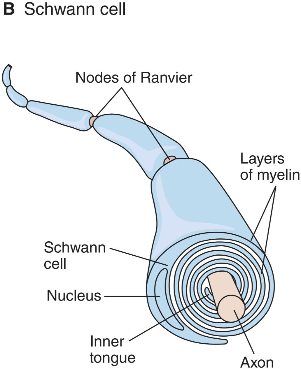
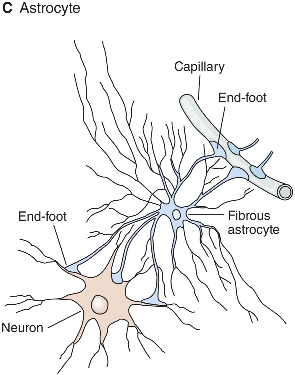
FIGURE 4-9 The principal types of macroglia in the nervous system. A) Oligodendrocytes are small with relatively few processes. Those in the white matter provide myelin, and those in the gray matter support neurons. B) Schwann cells provide myelin to the peripheral nervous system. Each cell forms a segment of myelin sheath about 1 mm long; the sheath assumes its form as the inner tongue of the Schwann cell turns around the axon several times, wrapping in concentric layers. Intervals between segments of myelin are the nodes of Ranvier. C) Astrocytes are the most common glia in the CNS and are characterized by their starlike shape. They contact both capillaries and neurons and are thought to have a nutritive function. They are also involved in forming the blood-brain barrier. (Reproduced with permission from Kandel ER, Schwartz JH, Jessell TM, Siegelbaum SA, Hudspeth AJ (editors): Principles of Neural Science, 5th ed. New York, NY: McGraw-Hill; 2013.)
شکل 4-9 انواع اصلی ماکروگلیا در سیستم عصبی. الف) الیگودندروسیت ها کوچک با فرآیندهای نسبتا کمی هستند. آنهایی که در ماده سفید هستند میلین را تأمین می کنند و آنهایی که در ماده خاکستری هستند از نورون ها پشتیبانی می کنند. ب) سلول های شوان میلین را به سیستم عصبی محیطی می دهند. هر سلول یک بخش از غلاف میلین به طول حدود 1 میلی متر را تشکیل می دهد. غلاف شکل خود را به خود می گیرد زیرا زبان داخلی سلول شوان چندین بار به دور آکسون می چرخد و در لایه های متحدالمرکز پیچیده می شود. فواصل بین بخش های میلین گره های رانویر هستند. ج) آستروسیت ها شایع ترین گلیا در CNS هستند و با شکل ستاره مانند خود مشخص می شوند. آنها با مویرگ ها و نورون ها تماس دارند و تصور می شود که عملکرد تغذیه ای دارند. آنها همچنین در تشکیل سد خونی مغزی نقش دارند. (تکثیر شده با اجازه از Kandel ER، Schwartz JH، Jessell TM، Siegelbaum SA، Hudspeth AJ (ویراستاران): Principles of Neural Science، ویرایش پنجم نیویورک، نیویورک: McGraw-Hill؛ 2013.)
Astrocytes are found throughout the brain and are subdivided into two groups. Fibrous astrocytes are found primarily in white matter and contain many intermediate filaments; protoplasmic astrocytes are found in gray matter and have a granular cytoplasm. Both types of astrocytes send processes to blood vessels, where they induce capillaries to form the tight junctions making up the blood-brain barrier. They also send processes that envelop synapses and the surface of nerve cells. Protoplasmic astrocytes have a membrane potential that varies with the external K+ concentration but do not generate propagated potentials. They produce substances that are tropic to neurons, and they help maintain the appropriate concentration of ions and neurotransmitters by taking up K* and the neurotransmitters glutamate and y-aminobutyrate (GABA).
آستروسیت ها در سراسر مغز یافت می شوند و به دو گروه تقسیم می شوند. آستروسیت های فیبری عمدتاً در ماده سفید یافت می شوند و دارای رشته های میانی زیادی هستند. آستروسیت های پروتوپلاسمی در ماده خاکستری یافت می شوند و سیتوپلاسم دانه ای دارند. هر دو نوع آستروسیت فرآیندهایی را به رگ های خونی می فرستند، جایی که مویرگ ها را وادار می کنند تا اتصالات محکمی را ایجاد کنند که سد خونی مغزی را تشکیل می دهد. آنها همچنین فرآیندهایی را ارسال می کنند که سیناپس ها و سطح سلول های عصبی را در بر می گیرند. آستروسیت های پروتوپلاسمی دارای پتانسیل غشایی هستند که با غلظت خارجی +K متفاوت است، اما پتانسیل انتشار ایجاد نمی کند. آنها موادی تولید می کنند که برای نورون ها گرمسیری هستند و با جذب +K و انتقال دهنده های عصبی گلوتامات و y-aminobutyrat (GABA) به حفظ غلظت مناسب یون ها و انتقال دهنده های عصبی کمک می کنند.
CLINICAL BOX 4-3
Demyelinating Diseases
Normal conduction of action potentials relies on the insulating properties of myelin. Thus, defects in myelin can have major adverse neurologic consequences. One example is MS, an autoimmune disease that affects over 3 million people worldwide, usually striking between the ages of 20 and 50 and affecting women about twice as often as men. The causes of MS include both genetic and environmental factors. It is most common among whites living in countries with temperate climates including Europe, southern Canada, northern United States, and southeastern Australia. Environmental triggers include early exposure to viruses such as Epstein-Barr virus and those that cause measles, herpes, chickenpox, or influenza. In MS, antibodies and white blood cells in the immune system attack myelin, causing inflammation and injury to the sheath and eventually the nerves that it surrounds. Loss of myelin leads to leakage of K+ through voltage-gated channels, hyperpolarization, and failure to conduct action potentials. Initial presentation commonly includes reports of paraparesis (weakness in lower extremities) that may be accompanied by mild spasticity and hyperreflexia; paresthesia; numbness; urinary incontinence; and heat intolerance. Clinical assessment often reports optic neuritis, characterized by blurred vision, a change in color perception, visual field defect (central scotoma), and pain with eye movements; dysarthria; and dysphagia. Symptoms are often exacerbated by increased body temperature or increased ambient temperature. Progression of the disease is variable. In the most common form called relapsing-remitting MS, transient episodes appear suddenly, last a few weeks or months, and then gradually disappear. Subsequent episodes can appear years later, and eventually full recovery does not occur. A steadily worsening course with only minor periods of remission (secondary-progressive MS) develops later in many individuals. Others have a progressive form of the disease in which there are no periods of remission (primary-progressive MS). Diagnosing MS is very difficult and generally is delayed until multiple episodes occur with deficits separated in time and space. Nerve conduction tests can detect slowed conduction in motor and sensory pathways. Cerebral spinal fluid analysis can detect the presence of oligoclonal bands indicative of an abnormal immune reaction against myelin. The most definitive assessment is magnetic resonance imaging (MRI) to visualize multiple scarred (sclerotic) areas or plaques in the brain. These plaques often appear in the periventricular regions of the cerebral hemispheres.
جعبه بالینی 4-3
بیماری های دمیلینه کننده
هدایت طبیعی پتانسیل های عمل به خواص عایق میلین بستگی دارد. بنابراین، نقص در میلین می تواند پیامدهای عصبی نامطلوب عمده ای داشته باشد. یک نمونه ام اس، یک بیماری خودایمنی است که بیش از 3 میلیون نفر را در سراسر جهان تحت تاثیر قرار می دهد، معمولا بین 20 تا 50 سالگی دیده می شود و زنان را تقریبا دو برابر مردان مبتلا می کند. علل ام اس شامل عوامل ژنتیکی و محیطی است. این بیماری در میان سفیدپوستان ساکن در کشورهای دارای آب و هوای معتدل از جمله اروپا، جنوب کانادا، شمال ایالات متحده و جنوب شرقی استرالیا شایع است. محرکهای محیطی شامل قرار گرفتن زودهنگام در معرض ویروسهایی مانند ویروس اپشتین بار و آنهایی است که باعث سرخک، تبخال، آبله مرغان یا آنفولانزا میشوند. در بیماری ام اس، آنتی بادی ها و گلبول های سفید در سیستم ایمنی به میلین حمله می کنند و باعث التهاب و آسیب به غلاف و در نهایت اعصاب اطراف آن می شوند. از دست دادن میلین منجر به نشت K+ از طریق کانال های ولتاژدار، هیپرپلاریزه شدن و عدم انجام پتانسیل عمل می شود. تظاهرات اولیه معمولاً شامل گزارشهایی از پاراپارزی (ضعف در اندامهای تحتانی) است که ممکن است با اسپاستیسیتی خفیف و هایپررفلکسی همراه باشد. پارستزی بی حسی؛ بی اختیاری ادرار؛ و عدم تحمل گرما ارزیابی بالینی اغلب نوریت بینایی را گزارش می کند که با تاری دید، تغییر در درک رنگ، نقص میدان بینایی (اسکوتوم مرکزی) و درد همراه با حرکات چشم مشخص می شود. دیزآرتری؛ و دیسفاژی علائم اغلب با افزایش دمای بدن یا افزایش دمای محیط تشدید می شوند. پیشرفت بیماری متغیر است. در رایج ترین شکل به نام ام اس عود کننده- فروکش کننده، دوره های گذرا به طور ناگهانی ظاهر می شوند، چند هفته یا چند ماه طول می کشند و سپس به تدریج ناپدید می شوند. قسمت های بعدی ممکن است سال ها بعد ظاهر شوند و در نهایت بهبودی کامل رخ نمی دهد. یک دوره به طور پیوسته بدتر با دوره های بهبود جزئی (MS ثانویه-پیشرونده) بعداً در بسیاری از افراد ایجاد می شود. برخی دیگر دارای شکل پیشرونده بیماری هستند که در آن دوره های بهبودی وجود ندارد (MS اولیه-پیشرونده). تشخیص ام اس بسیار دشوار است و عموماً تا زمانی که چندین قسمت اتفاق میافتد با کسریهایی که از نظر زمانی و مکانی از هم جدا میشوند، به تعویق میافتد. آزمایشهای هدایت عصبی میتوانند کندی هدایت را در مسیرهای حرکتی و حسی تشخیص دهند. تجزیه و تحلیل مایع مغزی نخاعی می تواند وجود نوارهای اولیگوکلونال را نشان دهد که نشان دهنده یک واکنش غیر طبیعی ایمنی در برابر میلین است. قطعی ترین ارزیابی، تصویربرداری رزونانس مغناطیسی (MRI) برای تجسم نواحی یا پلاک های متعدد اسکار (اسکلروتیک) در مغز است. این پلاک ها اغلب در نواحی اطراف بطن نیمکره های مغزی ظاهر می شوند.
THERAPEUTIC HIGHLIGHTS
Although there is no cure for MS, corticosteroids (eg, prednisone) are the most common treatment used to reduce the inflammation that is accentuated during a relapse. Some drug treatments are designed to modify the course of the disease. For example, daily injections of ẞ-interferons suppress the immune response to reduce the severity and slow the progression of the disease. Glatiramer acetate may block the immune system’s attack on the myelin. Natalizumab interferes with the ability of potentially damaging immune cells to move from the bloodstream to the CNS. A clinical trial using B cell-depleting therapy with rituximab, an anti-CD20 monoclonal antibody, showed that the progression of the disease was slowed in patients younger than 51 years in whom the primary-progressive form of MS was diagnosed. Another clinical trial has shown that oral administration of fingolimod slowed the progression of the relapsing-remitting form of MS. This immunosuppressive drug acts by sequestering lymphocytes in the lymph nodes, thereby limiting their access to the CNS. In 2017, the Food and Drug Administration approved the use of ocrelizumab for the treatment of primary-progressive MS; it is an immunosuppressive agent that targets the CD20 marker on B lymphocytes.
نکات برجسته درمانی
اگرچه هیچ درمانی برای ام اس وجود ندارد، کورتیکواستروئیدها (مثلا پردنیزون) رایج ترین درمان مورد استفاده برای کاهش التهابی است که در طول عود تشدید می شود. برخی از درمان های دارویی برای تغییر دوره بیماری طراحی شده اند. به عنوان مثال، تزریق روزانه ẞ-اینترفرون، پاسخ ایمنی را سرکوب می کند تا شدت آن را کاهش دهد و پیشرفت بیماری را کند کند. گلاتیرامر استات ممکن است حمله سیستم ایمنی بدن به میلین را مسدود کند. ناتالیزوماب با توانایی سلول های ایمنی بالقوه آسیب رسان برای حرکت از جریان خون به CNS تداخل می کند. یک کارآزمایی بالینی با استفاده از درمان کاهش دهنده سلول B با ریتوکسیماب، یک آنتی بادی مونوکلونال ضد CD20، نشان داد که پیشرفت بیماری در بیماران کمتر از 51 سال که در آنها شکل پیشرونده اولیه ام اس تشخیص داده شده بود، کند شد. کارآزمایی بالینی دیگری نشان داده است که مصرف خوراکی فینگولیمود پیشرفت نوع عودکننده- فروکش کننده ام اس را کند می کند. این داروی سرکوب کننده سیستم ایمنی با جداسازی لنفوسیت ها در غدد لنفاوی عمل می کند و در نتیجه دسترسی آنها به CNS را محدود می کند. در سال 2017، سازمان غذا و دارو استفاده از ocrelizumab را برای درمان ام اس پیشرونده اولیه تایید کرد. این یک عامل سرکوب کننده سیستم ایمنی است که نشانگر CD20 را روی لنفوسیت های B هدف قرار می دهد.
NEUROTROPHINS: THEIR FUNCTION & RECEPTORS
Neurotrophins are necessary for survival and growth of neurons. Some of them are products of muscles or other structures that the neurons innervate, but many in the CNS are produced by astrocytes. These proteins bind to receptors at the endings of a neuron. They are internalized and then transported by retrograde transport to the neuronal cell body where they foster the production of proteins associated with neuronal development, growth, and survival. Other neurotrophins are produced in soma and transported to the nerve ending where they maintain the integrity of the postsynaptic neuron.
نوروتروفین ها: عملکرد و گیرنده های آنها
نوروتروفین ها برای بقا و رشد نورون ها ضروری هستند. برخی از آنها محصولات ماهیچه ها یا ساختارهای دیگری هستند که نورون ها آنها را عصب دهی می کنند، اما بسیاری از آنها در CNS توسط آستروسیت ها تولید می شوند. این پروتئین ها به گیرنده های انتهای یک نورون متصل می شوند. آنها درونی می شوند و سپس با انتقال رتروگراد به بدن سلول عصبی منتقل می شوند، جایی که تولید پروتئین های مرتبط با رشد، رشد و بقای عصبی را تقویت می کنند. سایر نوروتروفین ها در سوما تولید می شوند و به انتهای عصبی منتقل می شوند و در آنجا یکپارچگی نورون پس سیناپسی را حفظ می کنند.
NGF, the first neurotrophin identified, is a protein growth factor that is needed for the growth and maintenance of sympathetic neurons and some sensory neurons. NGF is made up of two a, two ẞ, and two y subunits. The ẞ subunits, each of which has a molecular mass of 13,200 Da, have all the nerve growth-promoting activity, the a subunits have trypsin-like activity, and the y subunits are serine proteases. The function of the proteases is unknown. The structure of the B subunit of NGF resembles that of insulin.
NGF، اولین نوروتروفین شناسایی شده، یک فاکتور رشد پروتئینی است که برای رشد و نگهداری نورون های سمپاتیک و برخی نورون های حسی مورد نیاز است. NGF از دو زیرواحد a، دو ẞ و دو y تشکیل شده است. زیرواحدهای ẞ، که هر کدام دارای جرم مولکولی 13200 دالتون هستند، تمام فعالیتهای محرک رشد عصبی را دارند، زیرواحدهای a دارای فعالیت تریپسین مانند هستند و زیرواحدهای y پروتئازهای سرین هستند. عملکرد پروتئازها ناشناخته است. ساختار زیرواحد B NGF شبیه انسولین است.
NGF is picked up by neurons and transported in a retrograde fashion from the nerve endings to the cell body. NGF may be responsible for the growth and maintenance of cholinergic neurons in the basal forebrain and the striatum. NGF-mediated survival of neurons is due to suppression of apoptosis rather than promotion of cell metabolism.
NGF توسط نورون ها گرفته می شود و به صورت رتروگراد از انتهای عصبی به بدن سلولی منتقل می شود. NGF ممکن است مسئول رشد و نگهداری نورونهای کولینرژیک در پیشمغز قاعدهای و مخطط باشد. بقای سلول های عصبی با واسطه NGF به دلیل سرکوب آپوپتوز است تا ارتقاء متابولیسم سلولی.
In addition to NGF, there are other neurotrophins, including brain-derived neurotrophic factor (BDNF), neurotrophin 3 (NT-3), and NT-4/5. They each maintain a different pattern of neurons, although there is some overlap. NT-3 is important for proprioceptor neurons that innervate the muscle spindle and mechanoreceptors in the skin; NT-4/5 is important for neurons that innervate the hair follicle; NGF is important for skin nociceptive neurons. Sympathetic neurons depend on both NGF and NT-3. BDNF acts rapidly and can actually depolarize neurons. BDNF-deficient mice lose peripheral sensory neurons and have severe degenerative changes in their vestibular ganglia and blunted long- term potentiation.
علاوه بر NGF، نوروتروفین های دیگری از جمله فاکتور نوروتروفیک مشتق از مغز (BDNF)، نوروتروفین 3 (NT-3) و NT-4/5 وجود دارد. هر کدام از آنها الگوی متفاوتی از نورون ها را حفظ می کنند، اگرچه همپوشانی هایی وجود دارد. NT-3 برای نورون های گیرنده عمقی که دوک عضلانی و گیرنده های مکانیکی پوست را عصب دهی می کنند، مهم است. NT-4/5 برای نورون هایی که فولیکول مو را عصب دهی می کنند مهم است. NGF برای نورون های دردناک پوست مهم است. نورون های سمپاتیک به هر دو NGF و NT-3 بستگی دارند. BDNF به سرعت عمل می کند و در واقع می تواند نورون ها را دپولاریزه کند. موشهای دارای کمبود BDNF نورونهای حسی محیطی خود را از دست میدهند و تغییرات شدید دژنراتیو در گانگلیون دهلیزی خود و تقویت طولانیمدت ضعیف میشوند.
These four established neurotrophins and their three high-affinity tyrosine kinase associated (Trk) receptors are listed in Table 4-4. Each of these Trk receptors dimerizes and initiates phosphorylation in the cytoplasmic tyrosine kinase domains of the receptors. An additional low-affinity NGF receptor that is a 75-kDa protein is called the p75 receptor. This receptor binds all four of the listed neurotrophins with equal affinity. Interestingly, if a p75 receptor becomes activated in the absence of exposure to a neurotrophin, it causes apoptosis or cell death, an effect opposite to the usual growth-promoting and nurturing effects of neurotrophins. Research is ongoing to characterize the distinct roles of p75 and Trk receptors and factors that influence their expression in neurons.
این چهار نوروتروفین تاسیس شده و سه گیرنده مرتبط با تیروزین کیناز (Trk) با میل ترکیبی بالا در جدول 4-4 فهرست شده اند. هر یک از این گیرندههای Trk دیمر میکنند و فسفوریلاسیون را در حوزههای تیروزین کیناز سیتوپلاسمی گیرندهها آغاز میکنند. یک گیرنده اضافی NGF با میل ترکیبی پایین که یک پروتئین ۷۵ کیلو دالتونی است، گیرنده p75 نامیده می شود. این گیرنده هر چهار نوروتروفین ذکر شده را با میل ترکیبی یکسان متصل می کند. جالب توجه است، اگر گیرنده p75 در غیاب قرار گرفتن در معرض نوروتروفین فعال شود، باعث آپوپتوز یا مرگ سلولی می شود، اثری برخلاف اثرات معمول رشد و پرورش نوروتروفین ها. تحقیقات برای مشخص کردن نقشهای متمایز گیرندههای p75 و Trk و عوامل مؤثر بر بیان آنها در نورونها ادامه دارد.
TABLE 4-4 Neurotrophins.
جدول 4-4 نوروتروفین ها.

OTHER FACTORS AFFECTING NEURONAL GROWTH
The regulation of neuronal growth is a complex process. Schwann cells and astrocytes produce ciliary neurotrophic factor (CNTF). This factor promotes the survival of damaged and embryonic spinal cord neurons and may prove to be of value in treating human diseases in which motor neurons degenerate. Glial cell line-derived neurotrophic factor (GDNF) maintains the survival of midbrain dopaminergic neurons and prevents the apoptosis of spinal motor neurons. Another factor that enhances the growth of neurons is leukemia inhibitory factor (LIF). In addition, neurons as well as other cells respond to insulin-like growth factor I (IGF-I) and the various forms of transforming growth factor (TGF), fibroblast growth factor (FGF), and platelet-derived growth factor (PDGF).
سایر عوامل مؤثر بر رشد عصبی
تنظیم رشد عصبی یک فرآیند پیچیده است. سلول های شوان و آستروسیت ها فاکتور نوروتروفیک مژگانی (CNTF) تولید می کنند. این عامل بقای نورون های آسیب دیده و جنینی نخاع را افزایش می دهد و ممکن است در درمان بیماری های انسانی که در آن نورون های حرکتی تحلیل می روند، مفید باشد. فاکتور نوروتروفیک مشتق از خط سلول گلیال (GDNF) بقای نورون های دوپامینرژیک مغز میانی را حفظ کرده و از آپوپتوز نورون های حرکتی نخاعی جلوگیری می کند. عامل دیگری که رشد نورون ها را افزایش می دهد فاکتور مهارکننده لوسمی (LIF) است. علاوه بر این، نورون ها و همچنین سایر سلول ها به فاکتور رشد شبه انسولین I (IGF-I) و اشکال مختلف فاکتور رشد تبدیل کننده (TGF)، فاکتور رشد فیبروبلاست (FGF) و فاکتور رشد مشتق از پلاکت (PDGF) پاسخ می دهند.
Clinical Box 4-4 compares the ability to regenerate neurons after central and peripheral nerve injury.
جعبه بالینی 4-4 توانایی بازسازی نورون ها را پس از آسیب عصب مرکزی و محیطی مقایسه می کند.
CLINICAL BOX 4.4
Axonal Regeneration
Peripheral nerve damage is often reversible. Although the axon will degenerate distal to the damage, connective elements of the so-called distal stump often survive. Axonal sprouting occurs from the proximal stump, growing toward the nerve ending. This results from growth-promoting factors secreted by Schwann cells that attract axons toward the distal stump. Adhesion molecules of the immunoglobulin superfamily (eg, the neuron-glia cell adhesion molecule or NgCAM/L1) promote axon growth along cell membranes and extracellular matrices. Inhibitory molecules in the perineurium ensure that the regenerating axons grow in a correct trajectory. Denervated distal stumps are able to upregulate production of neurotrophins that promote growth. Once the regenerated axon reaches its target, a new functional connection (eg, neuromuscular junction) is formed, allowing for considerable, although not full, recovery. For example, fine motor control may be permanently impaired because some motor neurons are guided to an inappropriate motor fiber. Nonetheless, recovery of peripheral nerves from damage far surpasses that of central nerve pathways. The proximal stump of a damaged axon in the CNS will form short sprouts, but distant stump recovery is rare, and the damaged axons are unlikely to form new synapses. This is in part because CNS neurons do not have the growth-promoting chemicals needed for regeneration. In fact, CNS myelin is a potent inhibitor of axonal growth. In addition, after a CNS injury, astrocytic proliferation, activation of microglia, scar formation, inflammation, and invasion of immune cells create an inappropriate environment for regeneration. Thus, treatment of brain and spinal cord injuries focuses on rehabilitation rather than reversing the nerve damage. New research is aiming to identify ways to initiate and maintain axonal growth, to direct regenerating axons to reconnect with their target neurons, and to reconstitute original neuronal circuitry.
جعبه بالینی 4.4
بازسازی آکسون
آسیب اعصاب محیطی اغلب قابل برگشت است. اگرچه آکسون در قسمت دیستال آسیب تخریب می شود، عناصر همبند به اصطلاح استامپ دیستال اغلب زنده می مانند. جوانه زدن آکسون از کنده پروگزیمال رخ می دهد و به سمت انتهای عصب رشد می کند. این ناشی از عوامل محرک رشد ترشح شده توسط سلول های شوان است که آکسون ها را به سمت استامپ دیستال جذب می کند. مولکولهای چسبنده ابرخانواده ایمونوگلوبولینها (به عنوان مثال، مولکول چسبنده سلولهای عصبی-گلیا یا NgCAM/L1) رشد آکسون را در امتداد غشای سلولی و ماتریسهای خارج سلولی افزایش میدهند. مولکول های بازدارنده در پری نوریوم تضمین می کنند که آکسون های بازسازی کننده در یک مسیر صحیح رشد می کنند. استامپ های دیستال عصب کشی شده می توانند تولید نوروتروفین هایی را که باعث رشد می شوند، تنظیم کنند. هنگامی که آکسون بازسازی شده به هدف خود می رسد، یک اتصال عملکردی جدید (به عنوان مثال، اتصال عصبی عضلانی) تشکیل می شود که امکان بهبود قابل توجه، اگرچه نه کامل را فراهم می کند. به عنوان مثال، کنترل حرکت ظریف ممکن است برای همیشه مختل شود زیرا برخی از نورون های حرکتی به سمت یک فیبر حرکتی نامناسب هدایت می شوند. با این وجود، بازیابی اعصاب محیطی از آسیب بسیار بیشتر از مسیرهای عصبی مرکزی است. استامپ پروگزیمال یک آکسون آسیب دیده در CNS جوانه های کوتاهی را تشکیل می دهد، اما بازیابی کنده دور نادر است و آکسون های آسیب دیده سیناپس های جدید تشکیل نمی دهند. این تا حدی به این دلیل است که نورون های CNS مواد شیمیایی محرک رشد مورد نیاز برای بازسازی را ندارند. در واقع، میلین CNS یک مهارکننده قوی رشد آکسون است. علاوه بر این، پس از آسیب CNS، تکثیر آستروسیت، فعال شدن میکروگلیا، تشکیل اسکار، التهاب و تهاجم به سلولهای ایمنی، محیط نامناسبی را برای بازسازی ایجاد میکند. بنابراین، درمان آسیب های مغزی و نخاعی به جای معکوس کردن آسیب عصبی، بر توانبخشی متمرکز است. تحقیقات جدید با هدف شناسایی راههایی برای شروع و حفظ رشد آکسون، هدایت آکسونهای در حال بازسازی برای اتصال مجدد با نورونهای هدف و بازسازی مدارهای عصبی اصلی است.
THERAPEUTIC HIGHLIGHTS
There is evidence showing that the use of NSAIDs such as ibuprofen can overcome the factors that inhibit axonal growth following injury. This effect is thought to be mediated by the ability of NSAIDs to inhibit RhoA, a small GTPase protein that normally prevents repair of neural pathways and axons. Growth cone collapse in response to myelin-associated inhibitors after nerve injury is prevented by drugs (such as pertussis toxin) that interfere with signal transduction via trimeric G-protein. Experimental drugs that inhibit the phosphoinositide 3-kinase (PI3) pathway or the inositol triphosphate (IP3) receptor have also been shown to promote regeneration after nerve injury.
نکات برجسته درمانی
شواهدی وجود دارد که نشان می دهد استفاده از NSAID ها مانند ایبوپروفن می تواند بر عواملی که رشد آکسون را به دنبال آسیب مهار می کنند غلبه کند. تصور می شود که این اثر به واسطه توانایی NSAID ها در مهار RhoA، یک پروتئین GTPase کوچک که به طور معمول از ترمیم مسیرهای عصبی و آکسون ها جلوگیری می کند، ایجاد می شود. فروپاشی مخروط رشد در پاسخ به مهارکنندههای مرتبط با میلین پس از آسیب عصبی توسط داروهایی (مانند سم سیاه سرفه) که با انتقال سیگنال از طریق پروتئین G تریمریک تداخل میکنند، جلوگیری میشود. داروهای تجربی که مسیر فسفوئینوزیتید 3-کیناز (PI3) یا گیرنده اینوزیتول تری فسفات (IP3) را مهار می کنند نیز نشان داده شده است که بازسازی پس از آسیب عصبی را تقویت می کنند.
CHAPTER SUMMARY
خلاصه فصل
• Neurons are composed of a cell body (soma) that is the metabolic center of the neuron, dendrites that extend arborize extensively and are a common zone for receiving input to the neuron, the initial segment of the axon where the action potential is initiated, and a long axon that conducts the action potentials.
• نورونها از یک جسم سلولی (سوما) که مرکز متابولیک نورون است، دندریتهایی که بهطور گسترده گسترش مییابند و یک ناحیه مشترک برای دریافت ورودی به نورون هستند، بخش اولیه آکسون که در آن پتانسیل عمل آغاز میشود و یک آکسون طولانی که پتانسیلهای عمل را هدایت میکند، تشکیل شدهاند.
• The resting membrane potential of neurons is about -70 mV, which is close to the equilibrium potential for K+. During hyperkalemia, the resting potential moves closer to the threshold for eliciting an action potential, thus the neuron becomes more excitable. During hypokalemia, the resting membrane potential is reduced and the neuron is hyperpolarized.
• پتانسیل غشای استراحت نورون ها در حدود -70 میلی ولت است که نزدیک به پتانسیل تعادل برای +K است. در طول هیپرکالمی، پتانسیل استراحت به آستانه برای برانگیختن پتانسیل عمل نزدیکتر میشود، بنابراین نورون تحریکپذیرتر میشود. در طول هیپوکالمی، پتانسیل غشای استراحت کاهش می یابد و نورون هیپرپلاریزه می شود.
• In response to a depolarizing stimulus, voltage-gated Na+ channels are activated; when the threshold potential is reached, an action potential results. The membrane potential moves toward the equilibrium potential for Na*. The Na+ channels are rapidly inactivated before returning to the resting state.
• در پاسخ به یک محرک دپلاریزاسیون، کانال های +Na دارای ولتاژ فعال می شوند. وقتی به پتانسیل آستانه رسید، یک پتانسیل عمل به وجود می آید. پتانسیل غشاء به سمت پتانسیل تعادل برای +Na حرکت می کند. کانال های +Na قبل از بازگشت به حالت استراحت به سرعت غیرفعال می شوند.
• The direction of the electrical gradient for Na+ is reversed during the overshoot because the membrane potential is reversed; this limits Na*influx. Voltage-gated K+ channels open to complete the process of repolarization. The slow return of the K+ channels to the closed state explains after-hyperpolarization, followed by a return to the resting membrane potential.
• جهت گرادیان الکتریکی برای +Na در طول بیش از حد معکوس می شود زیرا پتانسیل غشاء معکوس می شود. این هجوم +Na را محدود می کند. کانالهای +K دارای ولتاژ برای تکمیل فرآیند رپلاریزاسیون باز میشوند. بازگشت آهسته کانال های +K به حالت بسته، پس از هایپرپلاریزاسیون را توضیح می دهد و به دنبال آن بازگشت به پتانسیل غشاء در حال استراحت است.
• The axons of many neurons are wrapped in myelin, a protein-lipid complex that is an effective insulator; depolarization of myelinated axons travels from one node of Ranvier to the next, with the current sink at the active node serving to electrotonically depolarize to the firing level the node ahead of the action potential.
• آکسون های بسیاری از نورون ها در میلین پیچیده شده اند، یک مجموعه پروتئین-لیپیدی که یک عایق موثر است. دپلاریزاسیون آکسون های میلین دار از یک گره Ranvier به گره بعدی منتقل می شود، با سینک جریان در گره فعال برای دپلاریزاسیون الکتروتونیکی تا سطح شلیک گره قبل از پتانسیل عمل.
• Nerve fibers are divided into different categories (A, B, and C) based on axonal diameter, conduction velocity, and function. A numerical classification (Ia, Ib, II, III, and IV) is also used for sensory afferent fibers. These various classes differ in their sensitivity to hypoxia and anesthetics.
• رشته های عصبی بر اساس قطر آکسون، سرعت هدایت و عملکرد به دسته های مختلف (A، B و C) تقسیم می شوند. یک طبقه بندی عددی (Ia، Ib، II، III و IV) نیز برای الیاف آوران حسی استفاده می شود. این دسته های مختلف از نظر حساسیت به هیپوکسی و داروهای بیهوشی متفاوت هستند.
• Orthograde transport occurs along microtubules that run the length of the axon and requires two molecular motors: dynein and kinesin. It moves from the cell body toward the axon terminals and has both fast (400 mm/day) and slow (0.5-10 mm/day) components. Retrograde transport goes in the opposite direction (from nerve ending to cell body) at a rate of about 200 mm/day.
• انتقال ارتوگراد در امتداد میکروتوبول هایی که در طول آکسون قرار دارند و به دو موتور مولکولی نیاز دارد: داینئین و کینزین رخ می دهد. از بدن سلولی به سمت پایانه های آکسون حرکت می کند و دارای اجزای سریع (400 میلی متر در روز) و آهسته (0.5-10 میلی متر در روز) است. انتقال رتروگراد در جهت مخالف (از انتهای عصب به جسم سلولی) با سرعت حدود 200 میلی متر در روز انجام می شود.
• There are two main types of glia: microglia and macroglia. Microglia are scavenger cells. Macroglia include oligodendrocytes, Schwann cells, and astrocytes. Oligodendrocytes and Schwann cells are involved in myelin formation; astrocytes produce substances that are tropic to neurons and help maintain the appropriate concentration of ions and neurotransmitters.
• دو نوع اصلی گلیا وجود دارد: میکروگلیا و ماکروگلیا. میکروگلیاها سلول های پاک کننده هستند. ماکروگلیا شامل الیگودندروسیت ها، سلول های شوان و آستروسیت ها می شود. الیگودندروسیت ها و سلول های شوان در تشکیل میلین نقش دارند. آستروسیت ها موادی تولید می کنند که برای نورون ها گرمسیری هستند و به حفظ غلظت مناسب یون ها و انتقال دهنده های عصبی کمک می کنند.
• Patchy destruction of myelin within the CNS associated with multiple sclerosis delays the conduction in axons. Autoimmune reactions to the myelin proteins PO and PMP22 cause Guillain-Barré syndrome, a peripheral demyelinating neuropathy. Mutations in myelin protein genes cause peripheral neuropathies (eg, Charcot-Marie-Tooth disease).
• تخریب تکه تکه میلین در CNS مرتبط با مولتیپل اسکلروزیس هدایت در آکسون ها را به تاخیر می اندازد. واکنش های خودایمنی به پروتئین های میلین PO و PMP22 باعث سندرم گیلن باره، یک نوروپاتی دمیلینه کننده محیطی می شود. جهش در ژن های پروتئین میلین باعث نوروپاتی های محیطی می شود (به عنوان مثال، بیماری شارکو ماری توث).
• Neurotrophins (eg, NGF) are carried by retrograde transport to the neuronal cell body where they produce proteins associated with neuronal development, growth, and survival and suppress neuronal apoptosis.
• نوروتروفین ها (به عنوان مثال، NGF) با انتقال رتروگراد به بدن سلول عصبی حمل می شوند، جایی که پروتئین های مرتبط با رشد، رشد و بقای نورون ها را تولید می کنند و آپوپتوز عصبی را سرکوب می کنند.
MULTIPLE-CHOICE QUESTIONS
For all questions, select the single best answer unless otherwise directed.
سوالات چند گزینه ای
برای همه سؤالات، بهترین پاسخ را انتخاب کنید، مگر اینکه دستور دیگری داده شود.
1. Glia are critical for the development of the nervous system and have important roles in some neurodegenerative disorder. Which of the following statements correctly describe a property of a type of glia?
A. Microglia arise from macrophages outside of the nervous system and are physiologically and embryologically similar to other neural cell types.
B. Fibrous astrocytes are found primarily in the gray matter and induce capillaries to form tight junctions to form the blood-brain barrier.
C. Protoplasmic astrocytes produce substances that are tropic to neurons to maintain the appropriate concentration of ions and neurotransmitters by taking up K* and the neurotransmitters glutamate and GABA.
D. Oligodendrocytes and Schwann cells are involved in myelin formation around axons in the peripheral nervous system and central nervous system, respectively.
E. Macroglia are scavenger cells that resemble tissue macrophages and remove debris resulting from injury, infection, and disease.
1. گلیا برای رشد سیستم عصبی حیاتی است و نقش مهمی در برخی از اختلالات عصبی دارد. کدام یک از عبارات زیر به درستی خاصیت یک نوع گلیا را توصیف می کند؟
الف. میکروگلیا از ماکروفاژهای خارج از سیستم عصبی به وجود می آید و از نظر فیزیولوژیکی و جنینی شبیه سایر انواع سلول های عصبی است.
ب. آستروسیت های فیبری عمدتاً در ماده خاکستری یافت می شوند و مویرگ ها را وادار می کنند تا اتصالات محکمی را برای تشکیل سد خونی مغزی ایجاد کنند.
ج. آستروسیت های پروتوپلاسمی موادی تولید می کنند که برای نورون ها گرمسیری هستند تا غلظت مناسب یون ها و انتقال دهنده های عصبی را با جذب K* و انتقال دهنده های عصبی گلوتامات و GABA حفظ کنند.
د. الیگودندروسیت ها و سلول های شوان به ترتیب در تشکیل میلین در اطراف آکسون ها در سیستم عصبی محیطی و سیستم عصبی مرکزی نقش دارند.
ی. Macroglia سلولهای روبندهای هستند که شبیه ماکروفاژهای بافتی هستند و بقایای ناشی از آسیب، عفونت و بیماری را حذف میکنند.
2. Primary erythromelalgia due to a peripheral nerve sodium channelopathy was diagnosed in a 13-year-old girl who was experiencing frequent episodes of red, painful, warm extremities. Which part of a neuron has the highest concentration of Na+ channels per square micrometer of cell membrane?
A. dendrites
B. cell body near dendrites
C. initial segment
D. axonal membrane under myelin
E. node of Ranvier
2. اریترومالژی اولیه به دلیل کانالوپاتی سدیم عصب محیطی در یک دختر 13 ساله که دوره های مکرر اندام های قرمز، دردناک و گرم را تجربه می کرد، تشخیص داده شد. کدام بخش از یک نورون دارای بیشترین غلظت کانال های Na+ در هر میکرومتر مربع از غشای سلولی است؟
الف. dendrites
ب. جسم سلولی در نزدیکی دندریت ها
ج. بخش اولیه
د. غشای آکسون زیر میلین
ی. گره Ranvier
3. A 45-year-old woman who works in an office had been experiencing tingling in her index and middle fingers and thumb of her right hand. Recently, her wrist and hand had become weak. Her physician ordered a nerve conduction test to evaluate her for carpal tunnel syndrome. Which one of the following nerves has the slowest conduction velocity?
A. Aa fibers
B. Aẞ fibers
C. Ay fibers
D. B fibers
E. C fibers
3. زن 45 ساله ای که در یک مطب کار می کند، در انگشتان سبابه و میانی و شست دست راست خود احساس گزگز داشت. اخیرا مچ و دستش ضعیف شده بود. پزشک او آزمایش هدایت عصبی را برای ارزیابی سندرم تونل کارپال دستور داد. سرعت هدایت کدام یک از اعصاب زیر کمتر است؟
الف. الیاف Aa
ب. الیاف Aẞ
ج. الیاف Ay
د. الیاف B
ی. الیاف C
4. Axoplasmic flow is a cellular process responsible for movement of proteins and polypeptides within a neuron. Which of the following statements correctly describes a property of orthograde or retrograde axonal transport?
A. Synaptic transmission: Antidromic conduction
B. Molecular motors for orthograde and retrograde transport are dynein and kinesin, respectively.
C. The rate of retrograde fast axonal transport is ~400 mm/day
D. The rate of orthograde slow axonal transport is ~200 mm/day
E. Some viruses use retrograde transport to move from the nerve terminal to the soma.
4. جریان آکسوپلاسمی یک فرآیند سلولی است که مسئول حرکت پروتئین ها و پلی پپتیدها در یک نورون است. کدام یک از عبارات زیر به درستی خاصیت انتقال آکسونی ارتوگراد یا رتروگراد را توصیف می کند؟
الف. انتقال سیناپسی: هدایت آنتی درومیک
ب. موتورهای مولکولی برای انتقال ارتوگراد و رتروگراد به ترتیب داینئین و کینزین هستند.
ج. سرعت انتقال سریع آکسونی رتروگراد ~400 میلی متر در روز است
د. سرعت انتقال آکسون آهسته ارتوگراد ~200 میلی متر در روز است
ی. برخی از ویروس ها از انتقال رتروگراد برای حرکت از پایانه عصبی به سوما استفاده می کنند.
5. For a summer research project, a second-year medical student is introduced to the patch clamp technique in a neurophysiology laboratory. As part of her training, she learns to monitor both membrane potential and individual channel function. Which of the following ionic changes is correctly matched with a component of the action potential?
A. Opening of voltage-gated K+ channels: After-hyperpolarization
B. A decrease in extracellular Ca2+: Repolarization
C. Opening of voltage-gated Na+ channels: Depolarization
D. Rapid closure of voltage-gated Na+ channels: Resting membrane potential
E. Rapid closure of voltage-gated K+ channels: Relative refractory period
5. برای یک پروژه تحقیقاتی تابستانی، یک دانشجوی سال دوم پزشکی با تکنیک پچ گیره در آزمایشگاه فیزیولوژی عصبی آشنا می شود. به عنوان بخشی از آموزش، او یاد می گیرد که هم پتانسیل غشاء و هم عملکرد کانال فردی را نظارت کند. کدام یک از تغییرات یونی زیر به درستی با یک جزء از پتانسیل عمل مطابقت دارد؟
الف. باز شدن کانال های +K دارای ولتاژ: پس از هایپرپلاریزاسیون
ب. کاهش +Ca2 خارج سلولی: رپلاریزاسیون
ج. باز شدن کانال های +Na دارای ولتاژ: دپلاریزاسیون
د. بسته شدن سریع کانال های +Na دارای ولتاژ: پتانسیل غشاء در حال استراحت
ی. بسته شدن سریع کانال های +K دارای ولتاژ: دوره نسبی نسبی
6. A man falls into a deep sleep with one arm under his head. This arm is paralyzed when he awakens, but it tingles, and pain sensation in it is still intact. The reason for the loss of motor function without loss of pain sensation is:
A. A fibers are more susceptible to hypoxia than B fibers.
B. A fibers are more sensitive to pressure than C fibers.
C. C fibers are more sensitive to pressure than A fibers.
D. Motor nerves are more affected by sleep than sensory nerves.
E. Sensory nerves are nearer the bone than motor nerves and hence are less affected by pressure.
6. مردی با یک دست زیر سر به خواب عمیقی فرو می رود. این بازو هنگام بیدار شدن فلج می شود، اما گزگز می کند و احساس درد در آن هنوز سالم است. دلیل از دست دادن عملکرد حرکتی بدون از دست دادن احساس درد عبارتند از:
الف. الیاف A نسبت به فیبرهای B بیشتر مستعد ابتلا به هیپوکسی هستند.
ب. الیاف A نسبت به الیاف C حساسیت بیشتری به فشار دارند.
ج. الیاف C نسبت به الیاف A حساسیت بیشتری نسبت به فشار دارند.
د. اعصاب حرکتی بیشتر از اعصاب حسی تحت تأثیر خواب قرار می گیرند.
ی. اعصاب حسی نسبت به اعصاب حرکتی به استخوان نزدیکتر هستند و بنابراین کمتر تحت تأثیر فشار قرار می گیرند.
7. Neurotrophins foster the production of proteins associated with neuronal development, growth, and survival. Which of the following statements about nerve growth factor (NGF) is true?
A. NGF is made up of one a, two ẞ, and one y polypeptide subunits.
B. NGF is responsible for the growth and maintenance of adrenergic neurons in the basal forebrain and the striatum.
C. NGF uses orthograde transport to move from the soma to the nerve terminal of a neuron.
D. NGF is important for the growth of sensory neurons that innervate the muscle spindle.
E. NGF binds to both p75 receptor and Trk A receptors.
7. نوروتروفین ها تولید پروتئین های مرتبط با رشد، رشد و بقای نورون ها را تقویت می کنند. کدام یک از عبارات زیر در مورد فاکتور رشد عصبی (NGF) درست است؟
الف. NGF از یک زیر واحد پلی پپتیدی a، دو ẞ و یک y تشکیل شده است.
ب. NGF مسئول رشد و نگهداری نورون های آدرنرژیک در پیش مغز قاعده ای و مخطط است.
ج. NGF از انتقال ارتوگراد برای حرکت از سوما به پایانه عصبی یک نورون استفاده می کند.
د. NGF برای رشد نورون های حسی که دوک عضلانی را عصب دهی می کنند، مهم است.
ی. NGF به هر دو گیرنده p75 و گیرنده Trk A متصل می شود.
8. A 20-year-old female student awakens one morning with severe pain and blurry vision in her left eye; the symptoms abate over several days. About 6 months later, on a morning after playing volleyball with friends, she notices weakness but not pain in her right leg; the symptoms intensify while taking a hot shower. Which of the following is most likely to be the case?
A. The two episodes described are not likely to be related.
B. She may have primary-progressive multiple sclerosis.
C. She may have relapsing-remitting multiple sclerosis.
D. She may have a lumbar disk rupture.
E. She may have Guillain-Barré syndrome.
8. یک دختر دانشجوی 20 ساله یک روز صبح با درد شدید و تاری دید در چشم چپ خود از خواب بیدار می شود. علائم طی چند روز کاهش می یابد. حدود 6 ماه بعد، یک روز صبح پس از بازی والیبال با دوستان، او متوجه ضعف و نه درد در پای راست خود می شود. علائم در هنگام گرفتن دوش آب گرم تشدید می شود. کدام یک از موارد زیر احتمال بیشتری دارد؟
الف. دو قسمت توصیف شده احتمالاً به هم مرتبط نیستند.
ب. ممکن است مبتلا به مولتیپل اسکلروز اولیه-پیشرونده باشد.
ج. ممکن است مبتلا به مولتیپل اسکلروزیس عودکننده-روان کننده باشد.
د. ممکن است پارگی دیسک کمر داشته باشد.
ی. ممکن است سندرم گیلن باره داشته باشد.
9. A medical student was working in a neurophysiology lab and was learning factors that determine the resting membrane potential of a neuron. Which of the following statements correctly explains how a change in concentration of an ion inside or outside of the neuron would change its resting membrane potential?
A. A decrease in extracellular Ca2+ concentration would stabilize the membrane and reduce its excitability.
B. A decrease in the extracellular Na+ concentration would reduce the size of the resting membrane potential.
C. An increase in the extracellular K+ concentration would move the resting membrane potential from a normal value of -90 mV to -70 mV.
D. A decrease in the extracellular K+ concentration increases the gradient for K* to leak out of the neuron, making the cell more hyperpolarized.
E. A decrease in intracellular Na*+ concentration would make the resting membrane potential more negative.
9. یک دانشجوی پزشکی در یک آزمایشگاه فیزیولوژی عصبی کار می کرد و در حال یادگیری عواملی بود که پتانسیل غشای استراحت یک نورون را تعیین می کند. کدام یک از عبارات زیر به درستی توضیح می دهد که چگونه تغییر در غلظت یک یون در داخل یا خارج از نورون، پتانسیل غشای استراحت آن را تغییر می دهد؟
الف. کاهش غلظت +Ca2 خارج سلولی غشاء را تثبیت کرده و تحریک پذیری آن را کاهش می دهد.
ب. کاهش غلظت +Na خارج سلولی باعث کاهش اندازه پتانسیل غشاء استراحت می شود.
ج. افزایش غلظت پتاسیم خارج سلولی، پتانسیل غشای در حال استراحت را از مقدار نرمال 90- میلی ولت به 70- میلی ولت منتقل می کند.
د. کاهش غلظت +K خارج سلولی، گرادیان +K را برای نشت از نورون افزایش می دهد و سلول را هیپرپلاریزه تر می کند.
ی. کاهش غلظت +Na داخل سلولی پتانسیل غشاء استراحت را منفی تر می کند.
10. A precocious 6-year-old girl asked her aunt who was a neurologist at a local medical school to explain how a neuron works. Her aunt explained how different parts of the neuron have a specific role in generating and transmitting an action potential. She identified the part of the neuron where the action potential is initiated, a part that receives input from other neurons, and a part that conducts the action potential. These would have been as follows:
A. Soma, dendrite, axon
B. Axon hillock, soma, myelin sheath
C. Cell membrane, soma, dendrite
D. Initial segment, dendrite, axon
E. Axon hillock, soma, myelin sheath
10. یک دختر 6 ساله نابالغ از عمه اش که متخصص مغز و اعصاب در یک دانشکده پزشکی محلی بود، خواست تا توضیح دهد که یک نورون چگونه کار می کند. عمه او توضیح داد که چگونه بخش های مختلف نورون نقش خاصی در تولید و انتقال پتانسیل عمل دارند. او بخشی از نورون را که در آن پتانسیل عمل آغاز می شود، بخشی که ورودی از سایر نورون ها دریافت می کند و بخشی که پتانسیل عمل را هدایت می کند، شناسایی کرد. اینها می توانستند به شرح زیر باشند:
الف. سوما، دندریت، آکسون
ب. تپه آکسون، سوما، غلاف میلین
ج. غشای سلولی، سوما، دندریت
د. قطعه اولیه، دندریت، آکسون
ی. تپه آکسون، سوما، غلاف میلین
کلیک کنید «منابع»
🚀 با ما همراه شوید!
تازهترین مطالب و آموزشهای مغز و اعصاب را از دست ندهید. با فالو کردن کانال تلگرام آیندهنگاران مغز، از ما حمایت کنید!



