
فیزیولوژی پزشکی گانونگ؛ بافت تحریک پذیر: ماهیچه


دعای مطالعه [ نمایش ]
بِسْمِ الله الرَّحْمنِ الرَّحیمِ
اَللّهُمَّ اَخْرِجْنى مِنْ ظُلُماتِ الْوَهْمِ
خدايا مرا بيرون آور از تاريكىهاى وهم،
وَ اَكْرِمْنى بِنُورِ الْفَهْمِ
و به نور فهم گرامى ام بدار،
اَللّهُمَّ افْتَحْ عَلَيْنا اَبْوابَ رَحْمَتِكَ
خدايا درهاى رحمتت را به روى ما بگشا،
وَانْشُرْ عَلَيْنا خَزائِنَ عُلُومِكَ بِرَحْمَتِكَ يا اَرْحَمَ الرّاحِمينَ
و خزانههاى علومت را بر ما باز كن به امید رحمتت اى مهربانترين مهربانان.
کتاب «فیزیولوژی پزشکی گانونگ» بهعنوان یکی از جامعترین و معتبرترین منابع در حوزه علوم پزشکی، همچنان مرجع کلیدی برای درک عملکرد پیچیده بدن انسان است. این اثر با تکیه بر تازهترین پژوهشها و توضیحات دقیق از سازوکارهای فیزیولوژیک، پلی میان علوم پایه پزشکی و کاربردهای بالینی ایجاد میکند و نقشی بیبدیل در آموزش، پژوهش و ارتقای دانش سلامت ایفا مینماید.
ترجمه دقیق و علمی این شاهکار توسط برند علمی آیندهنگاران مغز به مدیریت داریوش طاهری، دسترسی فارسیزبانان به مرزهای نوین دانش فیزیولوژی را ممکن ساخته و رسالتی علمی برای ارتقای آموزش پزشکی، فهم عمیقتر سازوکارهای بدن و توسعه روشهای نوین در حوزه سلامت فراهم آورده است.
» کتاب فیزیولوژی پزشکی گانونگ
» » فصل ۵: بافت تحریک پذیر: ماهیچه
در حال ویرایش
» Ganong’s Review of Medical Physiology
»» CHAPTER 5: Excitable Tissue: Muscle
OBJECTIVES
After studying his chapter, you should be able to:
• Differentiate the major classes of muscle in the body.
• Describe the molecular and electrical makeup of muscle cell excitation- contraction coupling.
• Define elements of the sarcomere that underlie striated muscle contraction.
• Differentiate the role(s) for Ca2+ in skeletal, cardiac, and smooth muscle contraction.
• Appreciate muscle cell diversity and function.
اهداف
پس از مطالعه این فصل باید بتوانید:
• کلاس های اصلی ماهیچه های بدن را متمایز کنید.
• ترکیب مولکولی و الکتریکی کوپلینگ تحریک و انقباض سلول های عضلانی را شرح دهید.
• عناصری از سارکومر که زمینه انقباض ماهیچه مخطط را تشکیل می دهند را تعریف کنید.
• نقش(های) +Ca2 در انقباض اسکلتی، قلبی و عضلات صاف را متمایز کنید.
• از تنوع و عملکرد سلول های عضلانی قدردانی کنید.

INTRODUCTION
Muscle cells, like neurons, can be excited chemically, electrically, and mechanically to produce an action potential that is transmitted along their cell membranes. Unlike neurons, they respond to stimuli by activating a contractile mechanism. The contractile protein myosin and the cytoskeletal protein actin are abundant in muscle, where they are the primary structural components that bring about contraction.
مقدمه
سلولهای عضلانی، مانند نورونها، میتوانند از نظر شیمیایی، الکتریکی و مکانیکی تحریک شوند تا پتانسیل عمل تولید کنند که در طول غشای سلولی آنها منتقل میشود. برخلاف نورون ها، آنها با فعال کردن مکانیسم انقباضی به محرک ها پاسخ می دهند. پروتئین انقباضی میوزین و پروتئین اسکلت سلولی اکتین در عضله فراوان هستند، جایی که آنها اجزای ساختاری اولیه هستند که باعث انقباض می شوند.
Muscle is generally divided into three types: skeletal muscle, cardiac muscle, and smooth muscle, although smooth muscle is not a homogeneous single category. Skeletal muscle makes up the great mass of the somatic musculature. It has well-developed cross-striations, does not normally contract in the absence of nervous stimulation, lacks anatomic and functional connections between individual muscle fibers, and is generally under voluntary control. Cardiac muscle also has cross-striations, but it is functionally syncytial and, although it can be modulated via the autonomic nervous system, it can contract rhythmically in the absence of external innervation owing to the presence in the myocardium of pacemaker cells that discharge spontaneously (see Chapter 29). Smooth muscle lacks cross-striations and can be further subdivided into two broad types: unitary (or visceral) smooth muscle and multiunit smooth muscle. The type found in most hollow viscera is functionally syncytial and contains pacemakers that discharge irregularly. The multiunit type found in the eye and in some other locations is not spontaneously active and resembles skeletal muscle in graded contractile ability. Smooth muscle is also classified on the basis of its function or activity pattern to rhythmic/phasic smooth muscle and continuous/tonic smooth muscle.
عضله به طور کلی به سه نوع تقسیم می شود: ماهیچه اسکلتی، عضله قلبی و ماهیچه صاف، اگرچه ماهیچه صاف یک دسته واحد همگن نیست. عضله اسکلتی توده بزرگ عضلانی بدنی را تشکیل می دهد. دارای خطوط متقاطع به خوبی توسعه یافته است، به طور معمول در غیاب تحریک عصبی منقبض نمی شود، فاقد اتصالات آناتومیک و عملکردی بین فیبرهای عضلانی فردی است، و به طور کلی تحت کنترل ارادی است. عضله قلب نیز دارای خطوط متقاطع است، اما از نظر عملکردی سنسیشیال است و، اگرچه میتواند از طریق سیستم عصبی خودمختار تعدیل شود، میتواند در غیاب عصب خارجی به دلیل وجود سلولهای ضربانساز در میوکارد که خود به خود تخلیه میشوند، به صورت ریتمیک منقبض شود (به فصل 29 مراجعه کنید). ماهیچه صاف فاقد خطوط متقاطع است و می تواند بیشتر به دو نوع کلی تقسیم شود: عضله صاف واحد (یا احشایی) و عضله صاف چند واحدی. نوع موجود در اکثر احشاء توخالی از نظر عملکردی سنسیشیال است و حاوی ضربان سازهایی است که به طور نامنظم ترشح می شوند. نوع چند واحدی که در چشم و در برخی مکانهای دیگر یافت میشود، خود به خود فعال نیست و از نظر توانایی انقباضی درجهبندی شده شبیه ماهیچههای اسکلتی است. عضله صاف نیز بر اساس عملکرد یا الگوی فعالیت آن به عضله صاف ریتمیک/فازیک و ماهیچه صاف پیوسته/تونیک طبقه بندی می شود.
SKELETAL MUSCLE MORPHOLOGY
ORGANIZATION
Skeletal muscle is made up of individual muscle fibers that are the “building blocks” of the muscular system in the same sense that the neurons are the building blocks of the nervous system. Most skeletal muscles begin and end in tendons, and the muscle fibers are arranged in parallel between the tendinous ends, so that the force of contraction of the units is additive. Each muscle fiber is a single cell that is multinucleated, long, cylindrical, and surrounded by a cell membrane, the sarcolemma (Figure 5-1). There are no syncytial bridges between cells. The muscle fibers are made up of myofibrils, which are divisible into individual filaments. These myofilaments contain several proteins that together make up the contractile machinery of the skeletal muscle.
مورفولوژی ماهیچه های اسکلتی
سازمان
عضله اسکلتی از فیبرهای عضلانی منفرد تشکیل شده است که “بلوک های سازنده” سیستم عضلانی هستند به همان مفهومی که نورون ها بلوک های سازنده سیستم عصبی هستند. بیشتر ماهیچه های اسکلتی به تاندون ها شروع و به پایان می رسند و رشته های عضلانی به صورت موازی بین انتهای تاندون قرار می گیرند، به طوری که نیروی انقباض واحدها افزایش می یابد. هر فیبر عضلانی یک سلول منفرد است که چند هسته ای، بلند، استوانه ای شکل است و توسط یک غشای سلولی به نام سارکولما احاطه شده است (شکل 5-1). هیچ پل سنسیشیال بین سلول ها وجود ندارد. فیبرهای عضلانی از میوفیبریل ها تشکیل شده اند که به رشته های جداگانه تقسیم می شوند. این میوفیلامنت ها حاوی چندین پروتئین هستند که با هم ماشین انقباضی عضله اسکلتی را تشکیل می دهند.

FIGURE 5-1 Mammalian skeletal muscle. A) A single muscle fiber surrounded by its sarcolemma has been cut away to show individual myofibrils. The cut surface of the myofibrils shows the arrays of thick and thin filaments. The sarcoplasmic reticulum with its transverse (T) tubules and terminal cisterns surrounds each myofibril. The T tubules invaginate from the sarcolemma and contact the myofibrils twice in every sarcomere. Mitochondria are found between the myofibrils and a basal lamina surrounds the sarcolemma. B and C) Structural elements of myofibril shown in detail (see also Figure 5–2).
شکل 5-1 ماهیچه اسکلتی پستانداران. الف) یک فیبر عضلانی که توسط سارکولم آن احاطه شده است برای نشان دادن میوفیبریل های منفرد بریده شده است. سطح بریده شده میوفیبریل ها آرایه هایی از رشته های ضخیم و نازک را نشان می دهد. شبکه سارکوپلاسمی با لوله های عرضی (T) و مخازن انتهایی هر میوفیبریل را احاطه کرده است. لوله های T از سارکولما وارد می شوند و در هر سارکومر دو بار با میوفیبریل ها تماس می گیرند. میتوکندری ها بین میوفیبریل ها و یک لایه بازال سارکولما را احاطه کرده است. ب و ج) عناصر ساختاری میوفیبریل با جزئیات نشان داده شده است (شکل 5-2 را نیز ببینید).
The contractile mechanism in skeletal muscle largely depends on the proteins myosin-II, actin, tropomyosin, and troponin. Troponin is made up of three subunits: troponin I, troponin T, and troponin C. Other important proteins in muscle are involved in maintaining the proteins that participate in contraction in appropriate structural relation to one another and to the extracellular matrix.
مکانیسم انقباضی در ماهیچه های اسکلتی تا حد زیادی به پروتئین های میوزین-II، اکتین، تروپومیوزین و تروپونین بستگی دارد. تروپونین از سه زیر واحد تشکیل شده است: تروپونین I، تروپونین T و تروپونین C. سایر پروتئین های مهم در عضله در حفظ پروتئین هایی که در انقباض شرکت می کنند در رابطه ساختاری مناسب با یکدیگر و ماتریکس خارج سلولی نقش دارند.
STRIATIONS
Differences in the refractive indexes of the various parts of the muscle fiber are responsible for the characteristic cross-striations seen in skeletal muscle when viewed under the microscope. The parts of the cross-striations are frequently identified by letters A, H, I, M, and Z (Figure 5-2). The light I band is divided by the dark Z-line, and the dark A band has the lighter H band in its center. A transverse M-line is seen in the middle of the H band, and this line plus the narrow light areas on either side of it are sometimes called the pseudo-H zone. The area between two adjacent Z-lines is called a sarcomere. The orderly arrangement of actin, myosin, and related proteins that produces this pattern can also be seen in Figures 5-1 and 5-2. The thick filaments, which are about twice the diameter of the thin filaments, are made up of myosin; the thin filaments are made up of actin, tropomyosin, and troponin. The thick filaments are lined up to form the A bands, whereas the array of thin filaments extends out of the A band and into the less dense staining I bands. The lighter H bands in the center of the A bands are the regions where, when the muscle is relaxed, the thin filaments do not overlap the thick filaments. The Z-lines allow for anchoring of the thin filaments. If a transverse section through the A band is examined under the electron microscope, each thick filament is seen to be surrounded by six thin filaments in a regular hexagonal pattern.
شیارها
تفاوت در ضریب انکسار بخشهای مختلف فیبر عضلانی عامل ایجاد خطوط متقاطع مشخصه در عضله اسکلتی است که در زیر میکروسکوپ مشاهده میشود. قسمت های خطوط متقاطع اغلب با حروف A، H، I، M و Z مشخص می شوند (شکل 5-2). نوار I روشن با خط Z تیره تقسیم می شود و نوار A تیره نوار H روشن تر را در مرکز خود دارد. یک خط M عرضی در وسط نوار H دیده می شود و این خط به اضافه نواحی نور باریک در دو طرف آن گاهی اوقات ناحیه شبه H نامیده می شود. ناحیه بین دو خط Z مجاور را سارکومر می نامند. آرایش منظم اکتین، میوزین و پروتئین های مرتبط که این الگو را تولید می کنند نیز در شکل های 5-1 و 5-2 دیده می شود. رشته های ضخیم، که حدود دو برابر قطر رشته های نازک هستند، از میوزین تشکیل شده اند. رشته های نازک از اکتین، تروپومیوزین و تروپونین تشکیل شده اند. رشته های ضخیم برای تشکیل نوارهای A ردیف می شوند، در حالی که آرایه رشته های نازک از نوار A خارج می شوند و به نوارهای رنگ آمیزی کمتر متراکم I گسترش می یابند. نوارهای سبکتر H در مرکز نوارهای A مناطقی هستند که در آن، زمانی که عضله شل می شود، رشته های نازک روی رشته های ضخیم همپوشانی ندارند. خطوط Z امکان لنگر انداختن رشته های نازک را فراهم می کند. اگر یک مقطع عرضی از طریق نوار A در زیر میکروسکوپ الکترونی بررسی شود، هر رشته ضخیم توسط شش رشته نازک در یک الگوی شش ضلعی منظم احاطه شده است.

FIGURE 5-2 Skeletal muscle sarcomere. A) Electron micrograph of human gastrocnemius muscle (* 13,500). The sarcomere, named bands, and lines are shown. (Used with permission from GM Walker and GR Schrodt.) B) Arrangement of thin (actin) and thick (myosin) filaments and the Z-line in a relaxed skeletal muscle. C) Arrangement of thin and thick filaments and the Z- line in a contracted skeletal muscle. Note that the Z-lines come together as the thick and thin filaments slide next to each other during contraction. The thick and thin filaments do not change in size.
شکل 5-2 سارکومر عضله اسکلتی. الف) میکروگراف الکترونی عضله گاستروکنمیوس انسان (* 13500). سارکومر، نوارهای نامگذاری شده و خطوط نشان داده شده است. (با اجازه GM Walker و GR Schrodt استفاده می شود.) ب) آرایش رشته های نازک (اکتین) و ضخیم (میوزین) و خط Z در یک عضله اسکلتی آرام. ج) چینش رشته های نازک و ضخیم و خط Z در یک عضله اسکلتی منقبض شده. توجه داشته باشید که خطوط Z در هنگام انقباض رشته های ضخیم و نازک در کنار یکدیگر قرار می گیرند. رشته های ضخیم و نازک در اندازه تغییر نمی کنند.
The form of myosin found in muscle is myosin-II, with two globular heads and a long tail. The heads of the myosin molecules form cross-bridges with actin. Myosin contains heavy chains and light chains, and its heads are made up of the light chains and the amino terminal portions of the heavy chains. These heads contain an actin-binding site and a catalytic site that hydrolyzes adenosine triphosphate (ATP). The myosin molecules are arranged symmetrically on either side of the center of the sarcomere, and it is this arrangement that creates the light areas in the pseudo-H zone. The M-line is the site of the reversal of polarity of the myosin molecules in each of the thick filaments. At these points, there are slender cross-connections that hold the thick filaments in proper array. Each thick filament contains several hundred myosin molecules.
شکل میوزین موجود در ماهیچه، میوزین-II است که دارای دو سر کروی و یک دم بلند است. سر مولکول های میوزین با اکتین پل های متقاطع تشکیل می دهند. میوزین شامل زنجیره های سنگین و زنجیره های سبک است و سرهای آن از زنجیره های سبک و بخش های انتهایی آمینو زنجیره های سنگین تشکیل شده است. این سرها حاوی یک محل اتصال اکتین و یک سایت کاتالیزوری هستند که آدنوزین تری فسفات (ATP) را هیدرولیز می کند. مولکول های میوزین به طور متقارن در دو طرف مرکز سارکومر قرار گرفته اند و این آرایش است که مناطق نور را در ناحیه شبه H ایجاد می کند. خط M محل معکوس شدن قطبیت مولکول های میوزین در هر یک از رشته های ضخیم است. در این نقاط، اتصالات متقاطع باریکی وجود دارد که رشته های ضخیم را در آرایه مناسب نگه می دارد. هر رشته ضخیم حاوی چند صد مولکول میوزین است.
The thin filaments are polymers made up of two chains of actin that form a long double helix. Tropomyosin molecules are long filaments located in the groove between the two chains in the actin. Each thin filament contains 300-400 actin molecules and 40-60 tropomyosin molecules. Troponin molecules are small globular units located at intervals along the tropomyosin molecules. Each of the three troponin subunits has a unique function: Troponin T binds the troponin components to tropomyosin, troponin I inhibits the interaction of myosin with actin, and troponin C contains the binding sites for the Ca2+ that initiates contraction.
رشته های نازک پلیمرهایی هستند که از دو زنجیره اکتین تشکیل شده اند که یک مارپیچ دوتایی طولانی را تشکیل می دهند. مولکول های تروپومیوزین رشته های بلندی هستند که در شیار بین دو زنجیره در اکتین قرار دارند. هر رشته نازک شامل 300-400 مولکول اکتین و 40-60 مولکول تروپومیوزین است. مولکول های تروپونین واحدهای کروی کوچکی هستند که در فواصل زمانی در امتداد مولکول های تروپومیوزین قرار دارند. هر یک از سه زیر واحد تروپونین عملکرد منحصر به فردی دارند: تروپونین T اجزای تروپونین را به تروپومیوزین متصل می کند، تروپونین I از تعامل میوزین با اکتین جلوگیری می کند و تروپونین C حاوی محل های اتصال برای +Ca2 است که انقباض را آغاز می کند.
Some additional structural proteins that are important in skeletal muscle function include actinin, titin, and desmin. Actinin binds actin to the Z-lines. Titin, the largest known protein (with a molecular mass near 3,000,000 Da), connects the Z-lines to the M-lines and provides scaffolding for the sarcomere. It contains two kinds of folded domains that provide muscle with its elasticity. At first when the muscle is stretched, there is relatively little resistance as the domains unfold, but with further stretch, there is a rapid increase in resistance that protects the structure of the sarcomere. Desmin adds structure to the Z-lines in part by binding the Z-lines to the plasma membrane. Some muscle disorders associated with these structural components are described in Clinical Box 5-1. It should be noted that although these proteins are important in muscle structure/function, by no means do they represent an exhaustive list.
برخی از پروتئین های ساختاری اضافی که در عملکرد عضلات اسکلتی مهم هستند عبارتند از اکتینین، تیتین و دسمین. اکتینین اکتین را به خطوط Z متصل می کند. تیتین، بزرگترین پروتئین شناخته شده (با جرم مولکولی نزدیک به 3،000،000 Da)، خطوط Z را به خطوط M متصل می کند و داربستی را برای سارکومر فراهم می کند. این شامل دو نوع دامنه چین خورده است که خاصیت ارتجاعی عضله را فراهم می کند. در ابتدا هنگامی که عضله کشیده می شود، با باز شدن دامنه ها مقاومت نسبتا کمی وجود دارد، اما با کشش بیشتر، مقاومت به سرعت افزایش می یابد که از ساختار سارکومر محافظت می کند. دسمین تا حدی با اتصال خطوط Z به غشای پلاسمایی ساختار را به خطوط Z اضافه می کند. برخی از اختلالات عضلانی مرتبط با این اجزای ساختاری در جعبه بالینی 5-1 توضیح داده شده است. لازم به ذکر است که اگرچه این پروتئین ها در ساختار/عملکرد عضلانی مهم هستند، اما به هیچ وجه فهرست جامعی را نشان نمی دهند.
CLINICAL BOX 5.1
Structural & Metabolic Disorders in Muscle Disease
The term muscular dystrophy is applied to diseases that cause progressive weakness of skeletal muscle. About 50 such diseases have been described, some of which include cardiac as well as skeletal muscle. They range from mild to severe and some are eventually fatal. They have multiple causes, but mutations in the genes for the various components of the dystrophin- glycoprotein complex are a prominent cause. The dystrophin gene is one of the largest in the body, and mutations can occur at many different sites in it. Duchenne muscular dystrophy is a serious form of dystrophy in which the dystrophin protein is absent from muscle. It is X-linked and usually fatal by the age of 30. In a milder form of the disease, Becker muscular dystrophy, dystrophin is present but altered or reduced in amount. Limb-girdle muscular dystrophies of various types are associated with mutations of the genes coding for the sarcoglycans or other components of the dystrophin- glycoprotein complex.
جعبه بالینی 5.1
اختلالات ساختاری و متابولیک در بیماری های عضلانی
اصطلاح دیستروفی عضلانی به بیماری هایی اطلاق می شود که باعث ضعف پیشرونده عضلات اسکلتی می شوند. حدود 50 بیماری از این قبیل توصیف شده است که برخی از آنها شامل عضله قلبی و اسکلتی است. آنها از خفیف تا شدید متغیر هستند و برخی در نهایت کشنده هستند. آنها علل متعددی دارند، اما جهش در ژن های اجزای مختلف کمپلکس دیستروفین گلیکوپروتئین یکی از دلایل برجسته است. ژن دیستروفین یکی از بزرگترین ژنهای بدن است و جهشها میتوانند در مکانهای مختلفی در آن رخ دهند. دیستروفی عضلانی دوشن شکل جدی دیستروفی است که در آن پروتئین دیستروفین در ماهیچه وجود ندارد. این بیماری با X مرتبط است و معمولاً در سن 30 سالگی کشنده است. در نوع خفیف تر بیماری، دیستروفی عضلانی بکر، دیستروفین وجود دارد اما مقدار آن تغییر کرده یا کاهش می یابد. دیستروفی های عضلانی کمربند اندام در انواع مختلف با جهش ژن های کد کننده سارکوگلیکان ها یا سایر اجزای مجموعه دیستروفین گلیکوپروتئین همراه است.
Due to its enormous size and structural role in the sarcomere, titin is a prominent target for mutations that give rise to muscle disease. Mutations that encode for shorter titin structure have been associated with dilated cardiomyopathy, while other mutations have been associated with hypertrophic cardiomyopathy. The skeletal muscle-associated tibialis muscular dystrophy is a genetic muscle disease of titin that is predicted to destabilize the folded state of the protein. Interestingly, many of the titin mutations identified thus far are in regions of titin that are expressed in all striated muscles, yet not all muscles are affected in the same way. Such muscle type-specific phenotypes underscore the need to study titin’s multiple functions in different muscles, under both normal and pathologic conditions.
به دلیل اندازه عظیم و نقش ساختاری آن در سارکومر، تیتین یک هدف برجسته برای جهش هایی است که منجر به بیماری عضلانی می شود. جهشهایی که ساختار تیتین کوتاهتر را کد میکنند با کاردیومیوپاتی اتساع یافته مرتبط هستند، در حالی که سایر جهشها با کاردیومیوپاتی هیپرتروفیک مرتبط هستند. دیستروفی عضلانی تیبیالیس مرتبط با عضله اسکلتی یک بیماری عضلانی ژنتیکی تیتین است که پیشبینی میشود حالت چین خوردگی پروتئین را بیثبات کند. جالب اینجاست که بسیاری از جهشهای تیتین که تاکنون شناسایی شدهاند در نواحی تیتین هستند که در همه عضلات مخطط بیان میشوند، اما همه ماهیچهها به یک شکل تحت تأثیر قرار نمیگیرند. چنین فنوتیپهای خاص عضلانی بر نیاز به مطالعه عملکردهای چندگانه تیتین در عضلات مختلف، در شرایط طبیعی و پاتولوژیک تأکید میکند.
Desmin-related myopathies are a very rare heterogeneous group of muscle disorders that typically result in cellular aggregates of desmin. Common symptoms of these diseases are failing and wasting in the distal muscles of the lower limbs that can later be identified in other body areas. Studies in desmin knockout mice have revealed defects in skeletal, smooth, and cardiac muscle, notably in the diaphragm and heart.
میوپاتی های مرتبط با دسمین یک گروه ناهمگن بسیار نادر از اختلالات عضلانی هستند که معمولاً منجر به تجمعات سلولی دسمین می شوند. علائم شایع این بیماری ها نارسایی و تحلیل رفتن عضلات انتهایی اندام تحتانی است که بعداً در سایر نواحی بدن قابل شناسایی است. مطالعات روی موشهای ناک اوت دسمین نقصهایی را در ماهیچههای اسکلتی، صاف و قلبی بهویژه در دیافراگم و قلب نشان دادهاند.
Metabolic Myopathies
Mutations in genes that code for enzymes involved in the metabolism of carbohydrates, fats, and proteins to CO2 and H2O in muscle and the production of ATP can cause metabolic myopathies (eg, McArdle syndrome). Metabolic myopathies all have in common exercise intolerance and the possibility of muscle breakdown due to accumulation of toxic metabolites.
میوپاتی های متابولیک
جهش در ژنهایی که کد آنزیمهای دخیل در متابولیسم کربوهیدراتها، چربیها و پروتئینها به CO2 و H2O در عضله و تولید ATP را میدهند، میتوانند باعث میوپاتی متابولیک شوند (مثلاً سندرم مکآردل). میوپاتی های متابولیک همگی دارای عدم تحمل ورزش و احتمال تجزیه عضلانی به دلیل تجمع متابولیت های سمی هستند.
THERAPEUTIQ HIGHLIGHTS
Although acute muscle pain and soreness can be treated with anti-inflammatory drugs and rest, the genetic dysfunctions described above are not as easily addressed. The overall goals are to slow muscle function/structure loss and, when possible relieve symptoms associated with the disease. Extensive monitoring, physical therapy, and appropriate drugs including corticosteroids can aid to slow disease progression. Assistive devices and surgery are not uncommon as the diseases progress.
نکات برجسته درمانی
اگرچه درد و درد حاد عضلانی را می توان با داروهای ضد التهابی و استراحت درمان کرد، اختلالات ژنتیکی که در بالا توضیح داده شد به راحتی قابل رسیدگی نیستند. اهداف کلی کاهش عملکرد/ساختار عضلانی و در صورت امکان تسکین علائم مرتبط با بیماری است. نظارت گسترده، فیزیوتراپی و داروهای مناسب از جمله کورتیکواستروئیدها می تواند به کند کردن پیشرفت بیماری کمک کند. با پیشرفت بیماری، وسایل کمکی و جراحی غیرمعمول نیستند.
SARCOTUBULAR SYSTEM
The muscle fibrils are surrounded by structures made up of membranes that appear in electron micrographs as vesicles and tubules. These structures form the sarcotubular system, which is made up of a T system and a sarcoplasmic reticulum. The T system of transverse tubules, which is continuous with the sarcolemma of the muscle fiber, forms a grid perforated by the individual muscle fibrils (Figure 5-1). The space between the two layers of the T system is an extension of the extracellular space. The sarcoplasmic reticulum, which forms an irregular curtain around each of the fibrils, has enlarged terminal cisterns in close contact with the T system at the junctions between the A and I bands. At these points of contact, the arrangement of the central T system with a cistern of the sarcoplasmic reticulum on either side has led to the use of the term triads to describe the system. The T system, which is continuous with the sarcolemma, provides a path for the rapid transmission of the action potential from the cell plasma membrane to all the fibrils in the muscle. The sarcoplasmic reticulum is an important store of Ca2+ and also participates in muscle metabolism.
سیستم سارکوتوبولار
فیبرهای عضلانی توسط ساختارهایی از غشاهایی احاطه شده اند که در میکروگراف های الکترونی به صورت وزیکول و لوله ظاهر می شوند. این ساختارها سیستم سارکوتوبولار را تشکیل می دهند که از یک سیستم T و یک شبکه سارکوپلاسمی تشکیل شده است. سیستم T لوله های عرضی، که با سارکولم فیبر عضلانی پیوسته است، شبکه ای را تشکیل می دهد که توسط فیبرهای عضلانی منفرد سوراخ شده است (شکل 5-1). فضای بین دو لایه سیستم T گسترش فضای خارج سلولی است. شبکه سارکوپلاسمی، که یک پرده نامنظم در اطراف هر یک از فیبریل ها تشکیل می دهد، دارای مخازن انتهایی بزرگ شده است که در تماس نزدیک با سیستم T در اتصالات بین نوارهای A و I است. در این نقاط تماس، آرایش سیستم T مرکزی با مخزن شبکه سارکوپلاسمی در دو طرف منجر به استفاده از اصطلاح سه گانه برای توصیف سیستم شده است. سیستم T که با سارکولما پیوسته است، مسیری را برای انتقال سریع پتانسیل عمل از غشای پلاسمایی سلولی به تمام فیبریل های عضله فراهم می کند. شبکه سارکوپلاسمی ذخیره مهمی از Ca2+ است و در متابولیسم عضلانی نیز شرکت می کند.
DYSTROPHIN-GLYCOPROTEIN COMPLEX
The large dystrophin protein (molecular mass 427,000 Da) forms a rod that connects the thin actin filaments to the transmembrane protein ẞ-dystroglycan in the sarcolemma by smaller proteins in the cytoplasm, syntrophins. ẞ- Dystroglycan is connected to merosin (merosin refers to laminins that contain α2 subunit in their trimeric makeup) in the extracellular matrix by a- dystroglycan (Figure 5-3). The dystroglycans are in turn associated with a complex of four transmembrane glycoproteins: a-, ẞ-, y-, and 8-sarcoglycan. This dystrophin-glycoprotein complex adds strength to the muscle by providing a scaffolding for the fibrils and connecting them to the extracellular environment. Disruption of these important structural features can result in several different muscular dystrophies (see Clinical Box 5–1).
کمپلکس دیستروفین-گلیکوپروتئین
پروتئین دیستروفین بزرگ (جرم مولکولی 427000 دا) میله ای را تشکیل می دهد که رشته های نازک اکتین را به پروتئین غشایی ẞ-دیستروگلیکان در سارکولما توسط پروتئین های کوچکتر در سیتوپلاسم، یعنی سنتروفین ها، متصل می کند. دیستروگلیکان در ماتریکس خارج سلولی توسط a-dystroglycan به مروزین (مروزین به لامینینهایی اطلاق میشود که دارای زیرواحد α2 در ترکیب سهگانه خود هستند) متصل میشود (شکل 5-3). دیستروگلیکان ها به نوبه خود با مجموعه ای از چهار گلیکوپروتئین گذرنده مرتبط هستند: a-، ẞ-، y- و 8-sarcoglycan. این کمپلکس دیستروفین-گلیکوپروتئین با ایجاد داربستی برای فیبریل ها و اتصال آنها به محیط خارج سلولی به عضله قدرت می بخشد. اختلال در این ویژگی های ساختاری مهم می تواند منجر به چندین دیستروفی عضلانی مختلف شود (به کادر بالینی 5-1 مراجعه کنید).

FIGURE 5-3 The dystrophin-glycoprotein complex. Dystrophin connects F- actin to the two members of the dystroglycan (DG) complex, a and ẞ- dystroglycan, and these in turn connect to the merosin subunit of laminin 211 in the extracellular matrix. The sarcoglycan complex of four glycoproteins, α-, B-, y-, and 8-sarcoglycan, sarcospan, and syntropins are all associated with the dystroglycan complex. There are muscle disorders associated with loss, abnormalities, or both of the sarcoglycans and merosin. (Used with permission of Justin Fallon and Kevin Campbell.)
شکل 5-3 کمپلکس دیستروفین-گلیکوپروتئین. دیستروفین F-اکتین را به دو عضو کمپلکس دیستروگلیکان (DG) یعنی a و ẞ-dystroglycan متصل می کند و اینها به نوبه خود به زیر واحد مروزین لامینین 211 در ماتریکس خارج سلولی متصل می شوند. مجموعه سارکوگلیکان از چهار گلیکوپروتئین، α-، B-، y- و 8-سارکوگلیکان، سارکوسپان و سینتروپین ها همگی با کمپلکس دیستروگلیکان مرتبط هستند. اختلالات عضلانی مرتبط با از دست دادن، ناهنجاری ها یا هر دو سارکوگلیکان و مروزین وجود دارد. (با اجازه جاستین فالون و کوین کمپبل استفاده شد.)
ELECTRICAL PHENOMENA & IONIC FLUXES
ELECTRICAL CHARACTERISTICS OF SKELETAL MUSCLE
The electrical events in skeletal muscle and the ionic fluxes that underlie them share distinct similarities to those in nerve, with quantitative differences in timing and magnitude. The resting membrane potential of skeletal muscle is about -90 mV. The action potential lasts 2-4 ms and is conducted along the muscle fiber at about 5 m/s. The absolute refractory period is 1-3 ms long, and the after-polarizations, with their related changes in threshold to electrical stimulation, are relatively prolonged. The initiation of impulses at the myoneural junction is discussed in Chapter 6.
پدیده های الکتریکی و شارهای یونی
ویژگی های الکتریکی ماهیچه های اسکلتی
رویدادهای الکتریکی در ماهیچههای اسکلتی و شارهای یونی که زیربنای آنها هستند شباهتهای مشخصی با رویدادهای عصبی دارند، با تفاوتهای کمی در زمان و اندازه. پتانسیل استراحت غشای عضله اسکلتی در حدود -90 میلی ولت است. پتانسیل عمل 2-4 میلی ثانیه طول می کشد و در طول فیبر عضلانی با سرعت 5 متر بر ثانیه انجام می شود. دوره نسوز مطلق 1-3 ms طول دارد و پس پلاریزاسیون ها با تغییرات مربوط به آستانه نسبت به تحریک الکتریکی نسبتا طولانی هستند. شروع تکانه ها در محل اتصال میونورال در فصل 6 مورد بحث قرار گرفته است.
ION DISTRIBUTION & FLUXES
The distribution of ions across the muscle fiber membrane is similar to that across the nerve cell membrane. Approximate values for the various ions and their equilibrium potentials are shown in Table 5-1. As in nerves, membrane depolarization is largely a manifestation of Na+ influx, and repolarization is largely a manifestation of K+ efflux.
توزیع یون و شار
توزیع یون ها در سراسر غشای فیبر عضلانی مشابه در سراسر غشای سلول عصبی است. مقادیر تقریبی یونهای مختلف و پتانسیل تعادل آنها در جدول 5-1 نشان داده شده است. همانند اعصاب، دپلاریزاسیون غشاء عمدتاً تجلی هجوم +Na است و رپلاریزاسیون تا حد زیادی تظاهری از جریان +K است.
TABLE 5-1 Steady-state distribution of ions in the intracellular and extracellular compartments of mammalian skeletal muscle, and the equilibrium potentials for these ions.
جدول 5-1 توزیع حالت پایدار یون ها در بخش های درون سلولی و خارج سلولی ماهیچه های اسکلتی پستانداران و پتانسیل های تعادلی برای این یون ها.
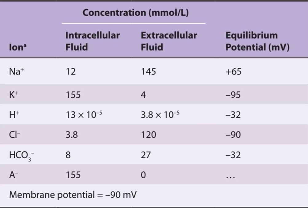
aA represents organic anions. The value for intracellular Cl is calculated from the membrane potential, using the Nernst equation.
Aa نشان دهنده آنیون های آلی است. مقدار کلر درون سلولی از پتانسیل غشا با استفاده از معادله نرنست محاسبه می شود.
CONTRACTILE RESPONSES
It is important to distinguish between the electrical and mechanical events in skeletal muscle. Although one response does not normally occur without the other, their physiological bases and characteristics are different. Muscle fiber membrane depolarization normally starts at the motor endplate, the specialized structure under the motor nerve ending. The action potential is transmitted along the muscle fiber and initiates the contractile response by inducing Ca2+ influx and release.
پاسخ های قراردادی
تمایز بین رویدادهای الکتریکی و مکانیکی در عضله اسکلتی مهم است. اگرچه یک پاسخ به طور معمول بدون دیگری رخ نمی دهد، پایه ها و ویژگی های فیزیولوژیکی آنها متفاوت است. دپلاریزاسیون غشای فیبر عضلانی معمولاً از صفحه انتهایی موتور شروع می شود، ساختار تخصصی زیر انتهای عصب حرکتی. پتانسیل عمل در طول فیبر عضلانی منتقل می شود و با القای هجوم و انتشار +Ca2 پاسخ انقباضی را آغاز می کند.
THE MUSCLE TWITCH
A single action potential causes a brief contraction followed by relaxation. This response is called a muscle twitch. In Figure 5-4, the action potential and the twitch are plotted on the same time scale. The twitch starts about 2 ms after the start of membrane depolarization, before repolarization is complete. The duration of the twitch varies with the type of muscle being tested. “Fast” muscle fibers, primarily those concerned with fine, rapid, precise movement, have twitch durations as short as 7.5 ms. “Slow” muscle fibers, principally those involved in strong, gross, sustained movements, have twitch durations up to 100 ms.
انقباض عضلانی
یک پتانسیل عمل منفرد باعث انقباض مختصری و به دنبال آن آرامش می شود. به این پاسخ انقباض عضلانی می گویند. در شکل 5-4، پتانسیل عمل و انقباض در مقیاس زمانی یکسان ترسیم شده است. انقباض حدود 2 میلی ثانیه پس از شروع دپلاریزاسیون غشا، قبل از اینکه رپلاریزاسیون کامل شود، شروع می شود. مدت انقباض با نوع عضله مورد آزمایش متفاوت است. فیبرهای عضلانی “سریع”، در درجه اول آنهایی که با حرکات ظریف، سریع و دقیق سروکار دارند، مدت زمان انقباض آنها به 7.5 میلی ثانیه می رسد. فیبرهای عضلانی “آهسته”، عمدتاً آنهایی که درگیر حرکات قوی، درشت و پایدار هستند، تا 100 میلی ثانیه طول می کشند.
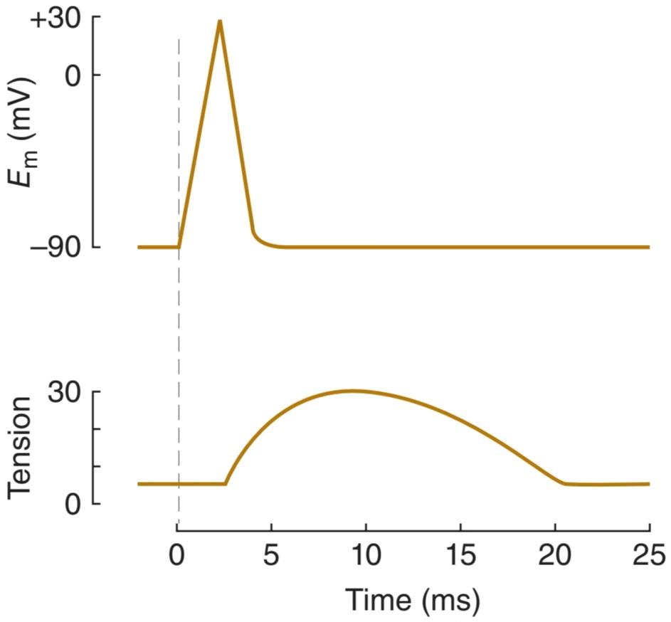
FIGURE 5-4 The electrical and mechanical responses of a mammalian skeletal muscle fiber to a single maximal stimulus. The electrical response or change in membrane potential (Em) in mV and the mechanical response or tension (T) in in arbitrary units are plotted on the same abscissa (time in ms). The mechanical response is relatively long-lived compared to the electrical response that initiates contraction.
شکل 5-4 پاسخ های الکتریکی و مکانیکی یک فیبر ماهیچه ای اسکلتی پستانداران به یک محرک حداکثر. پاسخ الکتریکی یا تغییر در پتانسیل غشاء (Em) بر حسب mV و پاسخ مکانیکی یا کشش (T) در واحدهای دلخواه بر روی همان آبسیسا (زمان بر حسب میلی ثانیه) رسم شده است. پاسخ مکانیکی در مقایسه با پاسخ الکتریکی که انقباض را آغاز می کند نسبتاً طولانی است.
MOLECULAR BASIS OF CONTRACTION
The process by which the contraction of muscle is brought about is a sliding of the thin filaments over the thick filaments. Note that this shortening is not due to changes in the actual lengths of the thick and thin filaments, rather, due to their increased overlap within the muscle cell. The width of the A bands is constant, whereas the Z-lines move closer together when the muscle contracts and farther apart when it relaxes (Figure 5-2).
مبانی مولکولی انقباض
فرآیندی که طی آن انقباض عضله ایجاد می شود، لغزش رشته های نازک روی رشته های ضخیم است. توجه داشته باشید که این کوتاه شدن به دلیل تغییر در طول واقعی رشته های ضخیم و نازک نیست، بلکه به دلیل افزایش همپوشانی آنها در داخل سلول عضلانی است. عرض باندهای A ثابت است، در حالی که خطوط Z زمانی که عضله منقبض می شود به هم نزدیکتر می شوند و زمانی که شل می شوند از هم دورتر می شوند (شکل 5-2).
The sliding during muscle contraction occurs when the myosin heads bind firmly to actin, bend at the junction of the head with the neck, and then detach. This “power stroke” depends on the simultaneous hydrolysis of ATP. Myosin-II molecules are dimers that have two heads, but only one attaches to actin at any given time. The probable sequence of events of the power stroke is outlined in Figure 5-5. In resting muscle, troponin I is bound to actin and tropomyosin and covers the sites where myosin heads interact with actin. Also at rest, the myosin head contains tightly bound adenosine phosphate (ADP) and inorganic phosphate (Pi). Following an action potential, cytosolic Ca2+ is increased (due to Ca2+ release from the sarcoplasmic reticulum and Ca2+ influx through voltage- gated Ca2+ channels) and free Ca2+ binds to troponin C. This binding results in a weakening of the troponin I interaction with actin and exposes the actin binding site for myosin to allow for formation of myosin/actin cross-bridges. Upon formation of the cross-bridge, ADP is released, causing a conformational change in the myosin head that moves the thin filament relative to the thick filament, comprising the cross-bridge “power stroke.” ATP quickly binds to the free site on the myosin, which leads to a detachment of the myosin head from the thin filament. ATP is hydrolyzed and P; released, causing a “re-cocking” of the myosin head and completing the cycle. As long as cytosolic Ca2+ remains elevated and sufficient ATP is available, this cycle repeats. Many myosin heads cycle at or near the same time, and they cycle repeatedly, producing gross muscle contraction. Each power stroke shortens the sarcomere about 10 nm. Each thick filament has about 500 myosin heads, and each head cycles about five times per second during a rapid contraction.
لغزش در حین انقباض عضلانی زمانی اتفاق می افتد که سرهای میوزین محکم به اکتین متصل می شوند، در محل اتصال سر به گردن خم می شوند و سپس جدا می شوند. این “سکته قدرت” به هیدرولیز همزمان ATP بستگی دارد. مولکولهای میوزین-II دایمرهایی هستند که دو سر دارند، اما در هر زمان معین فقط یکی به اکتین متصل میشود. توالی احتمالی رویدادهای ضربه قدرت در شکل 5-5 نشان داده شده است. در عضله در حال استراحت، تروپونین I به اکتین و تروپومیوزین متصل می شود و مکان هایی را که سرهای میوزین با اکتین تعامل می کنند، می پوشاند. همچنین در حالت استراحت، سر میوزین حاوی آدنوزین فسفات (ADP) و فسفات معدنی (Pi) است. به دنبال پتانسیل عمل، +Ca2 سیتوزولی افزایش مییابد (به دلیل آزاد شدن +Ca2 از شبکه سارکوپلاسمی و هجوم +Ca2 از طریق کانالهای +Ca2 دارای ولتاژ) و +Ca2 آزاد به تروپونین C متصل میشود. پل های متقابل پس از تشکیل پل متقاطع، ADP آزاد میشود و باعث تغییر ساختاری در سر میوزین میشود که رشته نازک را نسبت به رشته ضخیم حرکت میدهد، که شامل پل متقاطع “سکته نیرو” است. ATP به سرعت به محل آزاد میوزین متصل می شود که منجر به جدا شدن سر میوزین از رشته نازک می شود. ATP هیدرولیز شده و P. آزاد شده و باعث “خم شدن مجدد” سر میوزین و تکمیل چرخه می شود. تا زمانی که +Ca2 سیتوزولی بالا بماند و ATP کافی در دسترس باشد، این چرخه تکرار می شود. بسیاری از سرهای میوزین در یک زمان یا نزدیک به یک چرخه می چرخند و به طور مکرر چرخه می زنند و انقباض شدید عضلانی ایجاد می کنند. هر ضربه قدرت سارکومر را حدود 10 نانومتر کوتاه می کند. هر رشته ضخیم حدود 500 سر میوزین دارد و هر سر در طول یک انقباض سریع حدود پنج بار در ثانیه چرخه میزند.

FIGURE 5-5 Power stroke of myosin in skeletal muscle. A) At rest, myosin heads are bound to adenosine diphosphate and are said to be in a “cocked” position in relation to the thin filament, which does not have Ca2+ bound to the troponin—tropomyosin complex. B) Ca2+ bound to the troponin―tropomyosin complex induces a conformational change in the thin filament that allows for myosin heads to cross-bridge with thin filament actin. C) Myosin heads rotate, move the attached actin, and shorten the muscle fiber, forming the power stroke. D) At the end of the power stroke, ATP binds to a now exposed site and causes a detachment from the actin filament. E) ATP is hydrolyzed into ADP and inorganic phosphate (P;) and this chemical energy is used to “re-cock” the myosin head. (Data from Huxley AF, Simmons RM: Proposed mechanism of force generation in striated muscle. Nature Oct 22;233(5321):533–538, 1971 and Squire JM: Molecular mechanisms in muscular contraction. Trends Neurosci 6:409-413, 1983.)
شکل 5-5 سکته مغزی قوی میوزین در عضله اسکلتی. الف) در حالت استراحت، سرهای میوزین به آدنوزین دی فسفات متصل میشوند و گفته میشود که نسبت به رشته نازک در وضعیت «خمیده» قرار دارند، که +Ca2 به کمپلکس تروپونین-تروپومیوزین متصل نیست. ب) +Ca2 متصل به کمپلکس تروپونین-تروپومیوزین باعث ایجاد تغییر ساختاری در رشته نازک می شود که به سرهای میوزین اجازه می دهد با اکتین رشته نازک پل شوند. ج) سرهای میوزین میچرخند، اکتین متصل را حرکت میدهند و فیبر عضلانی را کوتاه میکنند و ضربه قدرت را تشکیل میدهند. د) در پایان استروک پاور، ATP به محل در معرض دید متصل می شود و باعث جدا شدن از رشته اکتین می شود. ه) ATP به ADP و فسفات معدنی (P;) هیدرولیز می شود و از این انرژی شیمیایی برای “دوباره کردن” سر میوزین استفاده می شود. (داده ها از هاکسلی AF، سیمونز RM: مکانیسم پیشنهادی برای تولید نیرو در عضله مخطط. Nature Oct 22; 233(5321):533-538، 1971 و Squire JM: مکانیسم های مولکولی در انقباض عضلانی. Trends Neurosci 6:409-4813).
The process by which membrane depolarization of the muscle fiber initiates contraction is called excitation-contraction coupling. The action potential is transmitted to all the fibrils in the fiber via the T system (Figure 5-6). It triggers the release of Ca2+ from the terminal cisterns, the lateral sacs of the sarcoplasmic reticulum next to the T system. Depolarization of the T tubule membrane activates the sarcoplasmic reticulum via dihydropyridine receptors (DHPRs), named for the drug dihydropyridine, which blocks them (Figure 5–7). DHPRS are voltage-gated Ca2+ channels in the T tubule membrane. In cardiac muscle, influx of Ca2+ via these channels triggers the release of Ca2+ stored in the sarcoplasmic reticulum (ie, Ca2+-induced Ca2+ release) by activating the ryanodine receptor (RyR). The RyR is named after the plant alkaloid ryanodine that was used in its discovery. The RyR is a ligand-gated Ca2+ channel with Ca2+ as its natural ligand. In skeletal muscle, Ca2+ entry from the extracellular fluid (ECF) by this route is not required for Ca2+ release. Instead, the DHPR that serves as the voltage sensor triggers release of Ca2+ from the nearby sarcoplasmic reticulum via physical interaction with the RyR. The released Ca2+ is quickly amplified through Ca2+-induced Ca2+ release. Cytosolic Ca2+ concentration is reduced in the muscle cell by the sarcoplasmic or endoplasmic reticulum Ca2+ ATPase (SERCA). The SERCA pump uses energy from ATP hydrolysis to remove Ca2+ from the cytosol back into the terminal cisterns against its concentration gradient, where it is stored until released by the next action potential. Once cytosolic Ca2+ concentration, or the Ca2+ concentration outside the sarcoplasmic reticulum, has been lowered sufficiently, chemical interaction between myosin and actin ceases and the muscle relaxes. Note that ATP provides the energy for both contraction (at the myosin head) and relaxation (via SERCA). If transport or uptake of Ca2+ into the sarcoplasmic reticulum is inhibited, relaxation does not occur even though there are no more action potentials; the resulting sustained contraction is called a contracture. Alterations in the excitable response in muscle underscore many different pathologic conditions (Clinical Box 5-2).
فرآیندی که در آن دپلاریزاسیون غشایی فیبر عضلانی شروع به انقباض می کند، جفت شدن تحریک-انقباض نامیده می شود. پتانسیل عمل از طریق سیستم T به تمام فیبرهای فیبر منتقل می شود (شکل 5-6). باعث آزاد شدن +Ca2 از مخازن انتهایی، کیسه های جانبی شبکه سارکوپلاسمی در کنار سیستم T می شود. دپلاریزاسیون غشای توبول T شبکه سارکوپلاسمی را از طریق گیرنده های دی هیدروپیریدین (DHPRs) که به نام داروی دی هیدروپیریدین نامگذاری شده است، فعال می کند که آنها را مسدود می کند (شکل 5-7). DHPRS کانال های +Ca2 دارای ولتاژ در غشای لوله T هستند. در عضله قلب، هجوم +Ca2 از طریق این کانالها باعث آزادسازی Ca2+ ذخیره شده در شبکه سارکوپلاسمی میشود (یعنی انتشار +Ca2 ناشی از +Ca2) با فعال کردن گیرنده رایانودین (RyR). RyR از روی آلکالوئید گیاهی رایانودین که در کشف آن استفاده شد نامگذاری شده است. RyR یک کانال +Ca2 با لیگاند است که +Ca2 به عنوان لیگاند طبیعی آن است. در عضله اسکلتی، ورود +Ca2 از مایع خارج سلولی (ECF) توسط این مسیر برای انتشار +Ca2 لازم نیست. در عوض، DHPR که به عنوان سنسور ولتاژ عمل می کند، از طریق تعامل فیزیکی با RyR باعث آزاد شدن +Ca2 از شبکه سارکوپلاسمی مجاور می شود. +Ca2 آزاد شده به سرعت از طریق انتشار +Ca2 ناشی از +Ca2 تقویت می شود. غلظت +Ca2 سیتوزولی در سلول عضلانی توسط شبکه سارکوپلاسمی یا آندوپلاسمی +Ca2 ATPase (SERCA) کاهش می یابد. پمپ SERCA از انرژی حاصل از هیدرولیز ATP استفاده می کند تا +Ca2 را از سیتوزول به داخل مخازن انتهایی بر خلاف گرادیان غلظت آن خارج کند، جایی که ذخیره می شود تا زمانی که پتانسیل عمل بعدی آزاد شود. هنگامی که غلظت سیتوزولی +Ca2 یا غلظت +Ca2 در خارج از شبکه سارکوپلاسمی به اندازه کافی کاهش یافت، برهمکنش شیمیایی بین میوزین و اکتین متوقف می شود و عضله شل می شود. توجه داشته باشید که ATP انرژی را برای انقباض (در سر میوزین) و آرامش (از طریق SERCA) فراهم می کند. اگر انتقال یا جذب +Ca2 به داخل شبکه سارکوپلاسمی مهار شود، آرامش رخ نمی دهد حتی اگر پتانسیل عمل بیشتری وجود نداشته باشد. انقباض پایدار حاصل انقباض نامیده می شود. تغییرات در پاسخ تحریک پذیر در عضله بر بسیاری از شرایط پاتولوژیک مختلف تأکید می کند (باکس بالینی 5-2).

FIGURE 5-6 Flow of information that leads to muscle contraction.
شکل 5-6 جریان اطلاعاتی که منجر به انقباض عضلانی می شود.

FIGURE 5-7 Relation of the T tubule to the sarcoplasmic reticulum in Ca2+ transport. In skeletal muscle, the voltage-gated dihydropyridine receptor in the T tubule triggers Ca2+ release from the sarcoplasmic reticulum (SR) via the ryanodine receptor (RyR). Upon sensing a voltage change, there is a physical interaction between the sarcolemmal-bound DHPR and the SR-bound RyR. This interaction gates the RyR and allows for Ca2+ release from the SR.
شکل 5-7 ارتباط توبول T با شبکه سارکوپلاسمی در انتقال +Ca2. در عضله اسکلتی، گیرنده دی هیدروپیریدینی دارای ولتاژ در توبول T باعث آزادسازی +Ca2 از شبکه سارکوپلاسمی (SR) از طریق گیرنده رایانودین (RyR) می شود. پس از احساس تغییر ولتاژ، یک برهمکنش فیزیکی بین DHPR متصل به sarcolemmal و RyR محدود به SR وجود دارد. این تعامل RyR را دریچه میکند و اجازه میدهد تا +Ca2 از SR آزاد شود.
CLINICAL BOX 5.2
Muscle Channelopathies
Channelopathies are diseases that have as their underlying feature mutations or dysregulation of ion channels. Such diseases are frequently associated with excitable cells, including muscle. In the various forms of clinical myotonia, muscle relaxation is prolonged after voluntary contraction. The molecular bases of myotonias are due to dysfunction of channels that shape the action potential. Myotonia dystrophy is caused by an autosomal dominant mutation that leads to overexpression of a K+ channel (although the mutation is not at the K+ channel). A variety of myotonias are associated with mutations in Na* channels (eg, hyperkalemic periodic paralysis, paramyotonia congenita, or Na+ channel congenita) or Cl- channels (eg, dominant or recessive myotonia congenita). Myasthenia, defined as abnormal muscle weakness or disease, can also be related to loss of ion channel function in the muscle. In congenital myasthenia, the patient has an inheritable disorder of one of ion channels necessary for the transmission of neuronal signaling to muscle response. Mutations in Ca2+ channels that allow for neuronal transmitter release or in the acetylcholine receptor nonspecific cation channels, important in recognition of neuronal transmitters, have both been shown to cause congenital myasthenia. Alterations of channel functions can also occur via autoimmune disease, such as that observed in myasthenia gravis. In this disease, antibodies to the nicotinic acetylcholine receptor can reduce its functional presence at the muscle membrane by up to 80%, and thus limit muscle response to neuronal transmitter release.
جعبه بالینی 5.2
کانالوپاتی های عضلانی
کانالوپاتی بیماری هایی هستند که به عنوان ویژگی زمینه ای خود دارای جهش یا اختلال در تنظیم کانال های یونی هستند. چنین بیماری هایی اغلب با سلول های تحریک پذیر از جمله عضله همراه هستند. در اشکال مختلف میتونی بالینی، آرامش عضلانی پس از انقباض ارادی طولانی می شود. پایه های مولکولی میتونی ها به دلیل اختلال در عملکرد کانال هایی است که پتانسیل عمل را شکل می دهند. دیستروفی میتونیا توسط یک جهش اتوزومال غالب ایجاد می شود که منجر به بیان بیش از حد کانال K+ می شود (اگرچه جهش در کانال K+ نیست). انواع میتونی ها با جهش در کانال های Na* (مانند فلج دوره ای هیپرکالمیک، پارامیتونیا مادرزادی یا کانال Na+) یا کانال های Cl- (مثلاً میتونی مادرزادی غالب یا مغلوب) مرتبط هستند. میاستنی که به عنوان ضعف یا بیماری غیرطبیعی عضلانی تعریف می شود، می تواند با از دست دادن عملکرد کانال یونی در عضله نیز مرتبط باشد. در میاستنی مادرزادی، بیمار دارای یک اختلال ارثی در یکی از کانال های یونی است که برای انتقال سیگنال عصبی به پاسخ عضلانی ضروری است. جهش در کانالهای Ca2+ که اجازه آزادسازی فرستنده عصبی یا کانالهای کاتیونی غیر اختصاصی گیرنده استیل کولین را میدهد، که در شناسایی فرستندههای عصبی مهم هستند، هر دو باعث میاستنی مادرزادی میشوند. تغییرات عملکرد کانال همچنین می تواند از طریق بیماری خودایمنی، مانند آنچه در میاستنی گراویس مشاهده می شود، رخ دهد. در این بیماری، آنتی بادی های گیرنده نیکوتین استیل کولین می توانند حضور عملکردی آن را در غشای عضلانی تا 80 درصد کاهش دهند و در نتیجه پاسخ عضلانی به آزاد شدن فرستنده عصبی را محدود کنند.
Channelopathies can also occur in the Ca2+ release channels in muscle (eg, ryanodine receptors) that amplify the Ca2+ response within the cell. Such mutations can cause malignant hyperthermia. Patients with this condition display normal muscle function under normal conditions. However, certain anesthetic agents, or in rare cases exposure to high environmental heat or strenuous exercise, can trigger abnormal release of Ca2+ from the sarcoplasmic reticulum in the muscle cell, resulting in sustained muscle contraction and heat production. In severe cases, fatality can occur.
کانالوپاتی همچنین می تواند در کانال های انتشار +Ca2 در عضله (به عنوان مثال، گیرنده های رایانودین) که پاسخ +Ca2 را در داخل سلول تقویت می کند، رخ دهد. چنین جهش هایی می تواند باعث هیپرترمی بدخیم شود. بیماران مبتلا به این عارضه عملکرد طبیعی عضلات را در شرایط عادی نشان می دهند. با این حال، برخی از عوامل بیهوشی، یا در موارد نادر قرار گرفتن در معرض گرمای محیطی زیاد یا ورزش شدید، میتوانند باعث آزادسازی غیرطبیعی +Ca2 از شبکه سارکوپلاسمی سلول عضلانی شوند که منجر به انقباض پایدار عضلانی و تولید گرما میشود. در موارد شدید، مرگ و میر ممکن است رخ دهد.
THERAPEUTIC HIGHLIGHTS
Although the symptoms associated with each individual channelopathy may be similar, treatments for the individual diseases include a wide variety of drugs that are targeted to the defect in the individual ion channel (or proteins associated with ion channel). Appropriate drug therapy helps improve symptoms and maintain acceptable muscle function. Further interventions related to individual diseases are to avoid muscle movements that exacerbate the disease.
نکات برجسته درمانی
اگرچه علائم مرتبط با هر کانالوپاتی فردی ممکن است مشابه باشد، درمان برای بیماری های فردی شامل طیف گسترده ای از داروهایی است که برای نقص در کانال یونی (یا پروتئین های مرتبط با کانال یونی) هدف قرار می گیرند. درمان دارویی مناسب به بهبود علائم و حفظ عملکرد قابل قبول ماهیچه کمک می کند. مداخلات بیشتر مربوط به بیماری های فردی اجتناب از حرکات ماهیچه ای است که بیماری را تشدید می کند.
TYPES OF CONTRACTION
Muscular contraction involves shortening of the contractile elements, but because muscles have elastic and viscous elements in series with the contractile mechanism, it is possible for contraction to occur without an appreciable decrease in the length of the whole muscle (Figure 5-8). Such a contraction is called isometric (“same measure” or length). Contraction against a constant load with a decrease in muscle length is isotonic (“same tension”). Note that because work is the product of force times distance, isotonic contractions do work,whereas isometric contractions do not. In other situations, muscle can do negative work while lengthening against a constant weight.
انواع انقباض
انقباض عضلانی شامل کوتاه شدن عناصر انقباضی است، اما از آنجا که ماهیچه ها دارای عناصر کشسان و چسبناک به صورت سری با مکانیسم انقباضی هستند، ممکن است انقباض بدون کاهش محسوس در طول کل عضله رخ دهد (شکل 5-8). چنین انقباضی ایزومتریک (“همان اندازه” یا طول) نامیده می شود. انقباض در برابر بار ثابت با کاهش طول عضله ایزوتونیک است (“تنش یکسان”). توجه داشته باشید که چون کار حاصل ضرب نیرو در مسافت است، انقباضات ایزوتونیک کار می کنند، در حالی که انقباضات ایزومتریک این کار را نمی کنند. در موقعیتهای دیگر، عضله میتواند در مقابل وزن ثابت، کار منفی انجام دهد.
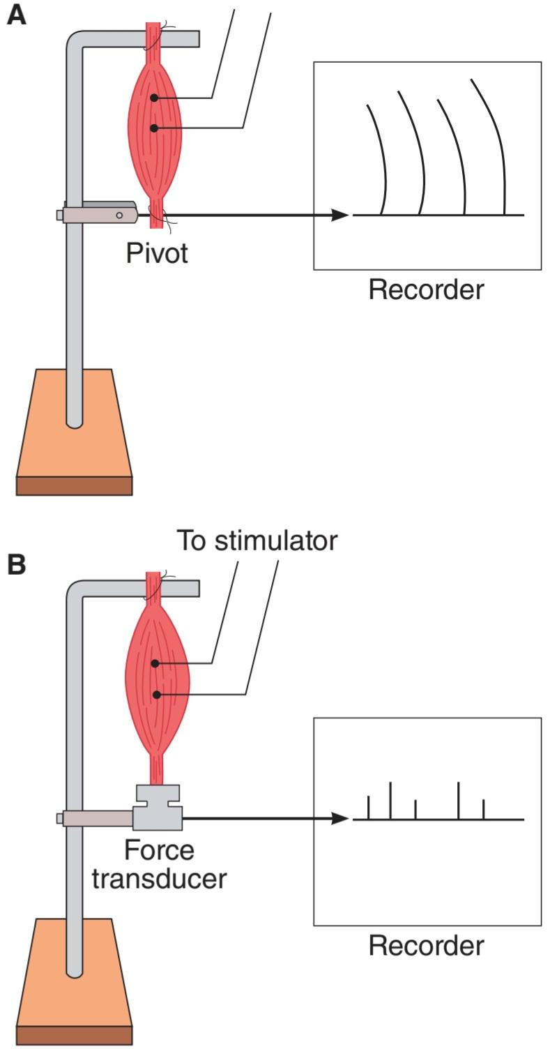
FIGURE 5-8 Isotonic and isometric contractions. A) Muscle preparation arranged for recording isotonic contractions. B) Preparation arranged for recording isometric contractions. In A, the muscle is fastened to a writing lever that swings on a pivot. In B, it is attached to an electronic transducer that measures the force generated without permitting the muscle to shorten.
شکل 5-8 انقباضات ایزوتونیک و ایزومتریک. الف) آماده سازی عضلانی برای ثبت انقباضات ایزوتونیک. ب) آماده سازی برای ثبت انقباضات ایزومتریک. در A، عضله به یک اهرم نوشتاری که روی یک محور می چرخد بسته می شود. در B، به یک مبدل الکترونیکی متصل است که نیروی ایجاد شده را بدون اجازه کوتاه شدن عضله اندازه گیری می کند.
SUMMATION OF CONTRACTIONS
The electrical response of a muscle fiber to repeated stimulation is like that of nerve. The fiber is electrically refractory only during the rising phase and part of the falling phase of the spike potential. At this time, the contraction initiated by the first stimulus is just beginning. However, because the contractile mechanism does not have a refractory period, repeated stimulation before relaxation has occurred produces additional activation of the contractile elements and a response that is added to the contraction already present. This phenomenon is known as summation of contractions. The tension developed during summation is considerably greater than that during the single muscle twitch. With rapidly repeated stimulation, activation of the contractile mechanism occurs repeatedly before any relaxation has occurred, and the individual responses fuse into one continuous contraction. Such a response is called tetanus (tetanic contraction). It is a complete tetanus when no relaxation occurs between stimuli and an incomplete tetanus when periods of incomplete relaxation take place between the summated stimuli. During a complete tetanus, the tension developed is about four times that developed by the individual twitch contractions. The development of an incomplete and a complete tetanus in response to stimuli of increasing frequency is shown in Figure 5-9.
جمع بندی قراردادها
پاسخ الکتریکی فیبر عضلانی به تحریک مکرر مانند عصبی است. فیبر فقط در طول فاز بالا آمدن و بخشی از فاز سقوط پتانسیل سنبله از نظر الکتریکی نسوز است. در این زمان، انقباض آغاز شده توسط اولین محرک تازه شروع شده است. با این حال، از آنجا که مکانیسم انقباضی دوره نسوز ندارد، تحریک مکرر قبل از ایجاد آرامش باعث فعال شدن اضافی عناصر انقباضی و پاسخی می شود که به انقباض موجود اضافه می شود. این پدیده به جمع انقباضات معروف است. تنش ایجاد شده در حین جمع کردن به طور قابل توجهی بیشتر از تنش در طول انقباض عضله است. با تحریک سریع مکرر، فعال شدن مکانیسم انقباض به طور مکرر قبل از هر گونه آرامش رخ می دهد، و پاسخ های فردی در یک انقباض مداوم ترکیب می شوند. چنین پاسخی کزاز (انقباض کزاز) نامیده می شود. زمانی که هیچ آرامشی بین محرکها وجود نداشته باشد، کزاز کامل است و زمانی که دورههایی از آرامش ناقص بین محرکهای جمعشده اتفاق میافتد، کزاز ناقص است. در طی کزاز کامل، تنش ایجاد شده حدود چهار برابر بیشتر از انقباضات تکانشی است. ایجاد یک کزاز ناقص و کامل در پاسخ به محرک های با فرکانس فزاینده در شکل 5-9 نشان داده شده است.
The stimulation frequency at which summation of contractions occurs is determined by the twitch duration of the particular muscle being studied. For example, if the twitch duration is 10 ms, frequencies less than 1/10 ms (100/s) cause discrete responses interrupted by complete relaxation, and frequencies greater than 100/s cause summation.
فرکانس تحریک که در آن جمع انقباضات رخ می دهد با مدت زمان انقباض عضله خاص مورد مطالعه تعیین می شود. به عنوان مثال، اگر مدت انقباض 10 میلی ثانیه باشد، فرکانسهای کمتر از 1/10 میلیثانیه (100 بر ثانیه) باعث ایجاد پاسخهای مجزا میشوند که با آرامش کامل قطع میشوند، و فرکانسهای بیشتر از 100 بر ثانیه باعث جمع میشوند.

FIGURE 5-9 Tetanus. Isometric tension of a single muscle fiber during continuously increasing and decreasing stimulation frequency. Dots at the top are at intervals of 0.2 s. Note the development of incomplete and then complete tetanus as stimulation is increased, and the return of incomplete tetanus, then full response, as stimulation frequency is decreased.
شکل 5-9 کزاز. کشش ایزومتریک یک فیبر عضلانی در حین افزایش و کاهش مداوم فرکانس تحریک. نقاط در بالا در فواصل 0.2 ثانیه هستند. توجه داشته باشید که با افزایش تحریک، ایجاد کزاز ناقص و سپس کامل، و با کاهش فرکانس تحریک، بازگشت کزاز ناقص و سپس پاسخ کامل، کاهش مییابد.
RELATION BETWEEN MUSCLE LENGTH, TENSION, & VELOCITY OF CONTRACTION
Both the tension that a muscle develops when stimulated to contract isometrically (the total tension) and the passive tension exerted by the unstimulated muscle vary with the length of the muscle fiber. This relationship can be studied in a whole skeletal muscle preparation such as that shown in Figure 5-8. The length of the muscle can be varied by changing the distance between its two attachments. At each length, the passive tension is measured, the muscle is then stimulated electrically, and the total tension is measured. The difference between the two values at any length is the amount of tension actually generated by the contractile process, the active tension. The records obtained by plotting passive tension and total tension against muscle length are shown in Figure 5-10. Similar curves are obtained when single muscle fibers are studied. The length of the muscle at which the active tension is maximal is usually called its optimal resting length. The term comes originally from experiments demonstrating that the length of many of the muscles in the body at rest is the length at which they develop maximal tension.
ارتباط بین طول عضله، تنش و سرعت انقباض
هم کششی که عضله ایجاد می کند وقتی که به صورت ایزومتریک منقبض می شود (کشش کل) و هم کشش غیرفعال اعمال شده توسط عضله تحریک نشده با طول فیبر عضلانی متفاوت است. این رابطه را می توان در آماده سازی کل عضله اسکلتی مانند آنچه در شکل 5-8 نشان داده شده است، مطالعه کرد. طول عضله را می توان با تغییر فاصله بین دو اتصال آن تغییر داد. در هر طول، کشش غیرفعال اندازه گیری می شود، سپس عضله به صورت الکتریکی تحریک می شود، و کشش کل اندازه گیری می شود. تفاوت بین این دو مقدار در هر طول، میزان کشش واقعی است که توسط فرآیند انقباضی، کشش فعال ایجاد میشود. رکوردهای به دست آمده با ترسیم کشش غیرفعال و کشش کل در برابر طول عضله در شکل 5-10 نشان داده شده است. منحنی های مشابهی با مطالعه رشته های عضلانی منفرد به دست می آیند. معمولاً طول عضله ای که کشش فعال در آن حداکثر است، طول استراحت مطلوب آن نامیده می شود. این اصطلاح در اصل از آزمایشهایی میآید که نشان میدهد طول بسیاری از عضلات بدن در حالت استراحت، طولی است که در آن حداکثر کشش ایجاد میکنند.
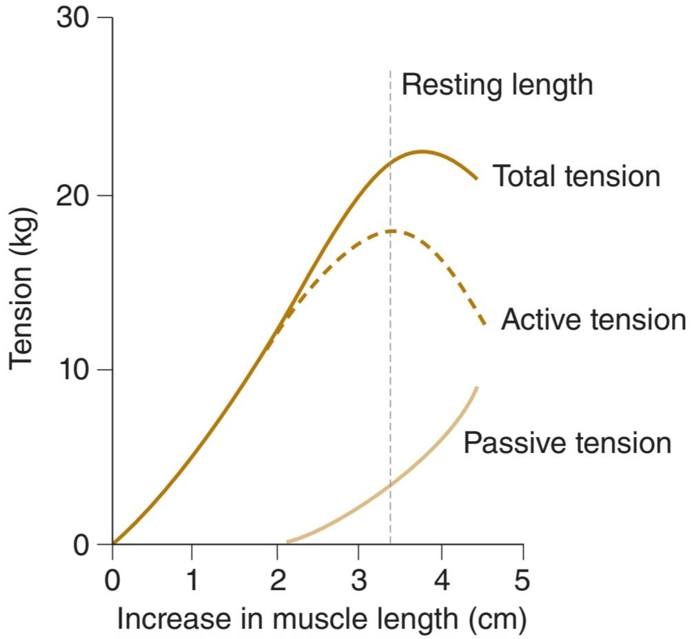
FIGURE 5-10 Length-tension relationship for the human triceps muscle. The passive tension curve measures the tension exerted by this skeletal muscle at each length when it is not stimulated. The total tension curve represents the tension developed when the muscle contracts isometrically in response to a maximal stimulus. The active tension is the difference between the two.
شکل 5-10 رابطه طول-تنش برای عضله سه سر انسان. منحنی تنش غیرفعال کشش اعمال شده توسط این عضله اسکلتی را در هر طول زمانی که تحریک نمی شود اندازه گیری می کند. منحنی کشش کل نشان دهنده تنش ایجاد شده در هنگام انقباض عضله به صورت ایزومتریک در پاسخ به یک محرک حداکثر است. تنش فعال تفاوت بین این دو است.
The observed length-tension relation in skeletal muscle can be explained by the sliding filament mechanism of muscle contraction. When the muscle fiber contracts isometrically, the tension developed is proportional to the number of cross-bridges between the actin and the myosin molecules. When muscle is stretched, the overlap between actin and myosin is reduced and the number of cross-linkages is therefore reduced. Conversely, when the muscle is appreciably shorter than resting length, the distance the thin filaments can move is reduced.
رابطه طول-تنش مشاهده شده در عضله اسکلتی را می توان با مکانیسم رشته لغزنده انقباض عضلانی توضیح داد. هنگامی که فیبر عضلانی به صورت ایزومتریک منقبض می شود، تنش ایجاد شده متناسب با تعداد پل های متقاطع بین مولکول های اکتین و میوزین است. هنگامی که عضله کشیده می شود، همپوشانی بین اکتین و میوزین کاهش می یابد و بنابراین تعداد پیوندهای متقابل کاهش می یابد. برعکس، زمانی که عضله به طور قابل ملاحظهای کوتاهتر از طول استراحت باشد، فاصله رشتههای نازک که میتوانند حرکت کنند کاهش مییابد.
The velocity of muscle contraction varies inversely with the load on the muscle. At a given load, the velocity is maximal at the resting length and declines if the muscle is shorter or longer than this length.
سرعت انقباض عضله با بار روی عضله برعکس تغییر می کند. در یک بار معین، سرعت در طول استراحت حداکثر است و اگر عضله کوتاهتر یا بلندتر از این طول باشد، کاهش می یابد.
FIBER TYPES
Although skeletal muscle fibers resemble one another in a general way, skeletal muscle is a heterogeneous tissue made up of fibers that vary in myosin ATPase activity, contractile speed, and other properties. Muscles are frequently classified into two types: “slow” and “fast.” These muscles can contain a mixture of three fiber types: type I (for slow-oxidative or SO), type IIA (for fast-oxidative- glycolytic or FOG), or type IIB (for fast glycolytic or FG). Some of the properties associated with types I, IIA, and IIB fibers are summarized in Table 5-2. Although these classification schemes are valid for muscles across many mammalian species, there are significant variations of fibers within and between muscles. For example, type I fibers in a given muscle can be larger than type IIA fibers from a different muscle in the same animal. Many of the differences in the fibers that make up muscles stem from differences in the proteins within them. Most of these are encoded by multigene families. Ten different isoforms of the myosin heavy chains (MHCs) have been characterized. Each of the two types of light chains also has isoforms. It appears that there is only one form of actin, but multiple isoforms of tropomyosin and all three components of troponin.
انواع الیاف
اگرچه فیبرهای عضلانی اسکلتی از نظر کلی شبیه یکدیگر هستند، عضله اسکلتی بافت ناهمگنی است که از الیافی تشکیل شده است که از نظر فعالیت ATPase میوزین، سرعت انقباض و سایر خواص متفاوت است. ماهیچه ها اغلب به دو نوع “آهسته” و “سریع” طبقه بندی می شوند. این ماهیچه ها می توانند حاوی مخلوطی از سه نوع فیبر باشند: نوع I (برای آهسته اکسیداتیو یا SO)، نوع IIA (برای سریع اکسیداتیو- گلیکولیتیک یا FOG)، یا نوع IIB (برای گلیکولیتیک سریع یا FG). برخی از خواص مرتبط با الیاف نوع I، IIA و IIB در جدول 5-2 خلاصه شده است. اگرچه این طرحهای طبقهبندی برای ماهیچهها در بسیاری از گونههای پستانداران معتبر است، اما تغییرات قابلتوجهی از فیبرها در داخل و بین ماهیچهها وجود دارد. به عنوان مثال، الیاف نوع I در یک عضله معین می تواند بزرگتر از الیاف نوع IIA از یک عضله متفاوت در همان حیوان باشد. بسیاری از تفاوتهای فیبرهایی که ماهیچهها را تشکیل میدهند، ناشی از تفاوتهای پروتئینهای درون آنها است. اکثر اینها توسط خانواده های چند ژنی کدگذاری می شوند. ده ایزوفرم مختلف از زنجیره های سنگین میوزین (MHCs) مشخص شده است. هر یک از دو نوع زنجیره سبک دارای ایزوفرم نیز هستند. به نظر می رسد که تنها یک شکل از اکتین وجود دارد، اما ایزوفرم های متعدد تروپومیوزین و هر سه جزء تروپونین.
TABLE 5-2 Classification of fiber types in skeletal muscles.
جدول 5-2 طبقه بندی انواع فیبر در عضلات اسکلتی.

ENERGY SOURCES & METABOLISM
Muscle contraction requires energy, and muscle has been called “a machine for converting chemical energy into mechanical work.” The immediate source of this energy is ATP, and this is formed by the metabolism of carbohydrates and lipids (see Chapters 1 and 2).
منابع انرژی و متابولیسم
انقباض عضلانی نیاز به انرژی دارد و ماهیچه را «ماشین تبدیل انرژی شیمیایی به کار مکانیکی» می نامند. منبع مستقیم این انرژی ATP است و این انرژی توسط متابولیسم کربوهیدرات ها و لیپیدها تشکیل می شود (به فصل های 1 و 2 مراجعه کنید).
PHOSPHORYLCREATINE
ATP is resynthesized from ADP by the addition of a phosphate group. Some of the energy for this endothermic reaction is supplied by the breakdown of glucose to CO2 and H2O, but there also exists in muscle another energy-rich phosphate compound that can supply this energy for short periods. This compound is phosphorylcreatine, which is hydrolyzed to creatine and phosphate groups with the release of considerable energy (Figure 5-11). At rest, some ATP in the mitochondria transfers its phosphate to creatine, so that a phosphorylcreatine store is built up. During exercise, the phosphorylcreatine is hydrolyzed at the junction between the myosin heads and actin, forming ATP from ADP and thus permitting contraction to continue.
فسفریل کراتین
ATP با افزودن یک گروه فسفات از ADP دوباره سنتز می شود. بخشی از انرژی برای این واکنش گرماگیر از تجزیه گلوکز به CO2 و H2O تامین می شود، اما ترکیب فسفات غنی از انرژی دیگری نیز در ماهیچه وجود دارد که می تواند این انرژی را برای دوره های کوتاه تامین کند. این ترکیب فسفریل کراتین است که با آزاد شدن انرژی قابل توجهی به گروه های کراتین و فسفات هیدرولیز می شود (شکل 5-11). در حالت استراحت، مقداری ATP در میتوکندری، فسفات خود را به کراتین منتقل می کند، به طوری که یک ذخیره فسفریل کراتین ایجاد می شود. در حین ورزش، فسفریل کراتین در محل اتصال بین سرهای میوزین و اکتین هیدرولیز می شود و از ADP ATP تشکیل می دهد و در نتیجه اجازه می دهد انقباض ادامه یابد.

FIGURE 5-11 Creatine, phosphorylcreatine, and creatinine cycling in muscle. During periods of high activity, cycling of phosphorylcreatine allows for quick release of ATP to sustain muscle activity.
شکل 5-11 چرخه کراتین، فسفریل کراتین و کراتینین در عضلات. در طول دورههای فعالیت زیاد، دوچرخهسواری فسفریل کراتین باعث آزادسازی سریع ATP برای حفظ فعالیت عضلانی میشود.
CARBOHYDRATE & LIPID BREAKDOWN
At rest and during light exercise, muscles utilize lipids in the form of free fatty acids as their energy source. As the intensity of exercise increases, lipids alone cannot supply energy fast enough and so use of carbohydrate becomes the predominant component in the muscle fuel mixture. Thus, during exercise, much of the energy for phosphorylcreatine and ATP resynthesis comes from the breakdown of glucose to CO2 and H2O. Glucose in the bloodstream enters cells, where it is degraded through a series of chemical reactions to pyruvate. Another source of intracellular glucose, and consequently of pyruvate, is glycogen, the carbohydrate polymer that is especially abundant in liver and skeletal muscle. When adequate O2 is present, pyruvate enters the citric acid cycle and is metabolized through this cycle and the so-called respiratory enzyme pathway -to CO2 and H2O. This process is called aerobic glycolysis. The metabolism of glucose or glycogen to CO2 and H2O forms large quantities of ATP from ADP. If O2 supplies are insufficient, the pyruvate formed from glucose does not enter the tricarboxylic acid cycle but is reduced to lactate. This process of anaerobic glycolysis is associated with the net production of much smaller quantities of energy-rich phosphate bonds, but it does not require the presence of O2. A brief overview of the various reactions involved in supplying energy to skeletal muscle is shown in Figure 5-12.
تجزیه کربوهیدرات ها و چربی ها
در حالت استراحت و در حین ورزش سبک، ماهیچه ها از لیپیدها به شکل اسیدهای چرب آزاد به عنوان منبع انرژی خود استفاده می کنند. با افزایش شدت ورزش، لیپیدها به تنهایی نمی توانند انرژی را با سرعت کافی تامین کنند و بنابراین استفاده از کربوهیدرات جزء غالب در مخلوط سوخت ماهیچه ها می شود. بنابراین، در طول ورزش، بیشتر انرژی برای سنتز فسفریل کراتین و ATP از تجزیه گلوکز به CO2 و H2O می آید. گلوکز در جریان خون وارد سلول ها می شود و در آنجا از طریق یک سری واکنش های شیمیایی به پیرووات تجزیه می شود. یکی دیگر از منابع گلوکز داخل سلولی و در نتیجه پیروات، گلیکوژن، پلیمر کربوهیدراتی است که به ویژه در کبد و ماهیچه های اسکلتی فراوان است. هنگامی که O2 کافی وجود داشته باشد، پیروات وارد چرخه اسید سیتریک می شود و از طریق این چرخه و به اصطلاح مسیر آنزیم تنفسی – به CO2 و H2O متابولیزه می شود. این فرآیند گلیکولیز هوازی نامیده می شود. متابولیسم گلوکز یا گلیکوژن به CO2 و H2O مقادیر زیادی ATP از ADP را تشکیل می دهد. اگر ذخایر O2 ناکافی باشد، پیروات تشکیل شده از گلوکز وارد چرخه اسید تری کربوکسیلیک نمی شود بلکه به لاکتات کاهش می یابد. این فرآیند گلیکولیز بی هوازی با تولید خالص مقادیر بسیار کمتر پیوندهای فسفات غنی از انرژی همراه است، اما نیازی به حضور O2 ندارد. یک نمای کلی از واکنش های مختلف درگیر در تامین انرژی به ماهیچه های اسکلتی در شکل 5-12 نشان داده شده است.

FIGURE 5-12 ATP turnover in muscle cells. Energy released by hydrolysis of 1 mol of ATP and reactions responsible for resynthesis of ATP. The amount of ATP formed per mole of free fatty acid (FFA) oxidized is large but varies with the size of the FFA. For example, complete oxidation of 1 mol of palmitic acid generates 140 mol of ATP.
شکل 5-12 گردش ATP در سلول های عضلانی. انرژی آزاد شده از هیدرولیز 1 مول ATP و واکنش های مسئول سنتز مجدد ATP. مقدار ATP تشکیل شده در هر مول اسید چرب آزاد (FFA) اکسید شده زیاد است اما با اندازه FFA متفاوت است. برای مثال، اکسیداسیون کامل 1 مول اسید پالمیتیک، 140 مول ATP تولید می کند.
THE OXYGEN DEBT MECHANISM
During exercise, the muscle blood vessels dilate and blood flow is increased so that the available O2 supply is increased. Up to a point, the increase in O2 consumption is proportional to the energy expended, and all the energy needs are met by aerobic processes. However, when muscular exertion is very great, aerobic resynthesis of energy stores cannot keep pace with their utilization. Under these conditions, phosphorylcreatine is still used to resynthesize ATP. In addition, some ATP synthesis is accomplished by using the energy released by the anaerobic breakdown of glucose to lactate. Use of the anaerobic pathway is self-limiting because in spite of rapid diffusion of lactate into the bloodstream, enough accumulates in the muscles to eventually exceed the capacity of the tissue buffers and produce an enzyme-inhibiting decline in pH. However, for short periods, the presence of an anaerobic pathway for glucose breakdown permits muscular exertion of a far greater magnitude than would be possible without it. For example, in a 100-m dash that takes 10 seconds, 85% of the energy consumed is derived anaerobically; in a 2-mile race that takes 10 minutes, 20% of the energy is derived anaerobically; and in a long-distance race that takes 60 minutes, only 5% of the energy comes from anaerobic metabolism.
مکانیسم بدهی اکسیژن
در حین ورزش، رگهای خونی ماهیچهای گشاد میشوند و جریان خون افزایش مییابد به طوری که عرضه O2 موجود افزایش مییابد. تا حدی افزایش مصرف O2 متناسب با انرژی مصرف شده است و تمام انرژی مورد نیاز با فرآیندهای هوازی تامین می شود. با این حال، هنگامی که فعالیت عضلانی بسیار زیاد است، سنتز مجدد هوازی ذخایر انرژی نمی تواند با استفاده از آنها هماهنگی داشته باشد. تحت این شرایط، فسفریل کراتین هنوز برای سنتز مجدد ATP استفاده می شود. علاوه بر این، برخی از سنتز ATP با استفاده از انرژی آزاد شده توسط تجزیه بی هوازی گلوکز به لاکتات انجام می شود. استفاده از مسیر بی هوازی خود محدود کننده است زیرا علیرغم انتشار سریع لاکتات در جریان خون، به اندازه کافی در ماهیچه ها تجمع می یابد تا در نهایت از ظرفیت بافرهای بافتی فراتر رود و باعث کاهش آنزیم در pH شود. با این حال، برای دورههای کوتاه، وجود یک مسیر بیهوازی برای تجزیه گلوکز اجازه میدهد تا فشار عضلانی بسیار بیشتر از آن چیزی باشد که بدون آن ممکن است. برای مثال، در یک دوش 100 متری که 10 ثانیه طول می کشد، 85 درصد انرژی مصرف شده به صورت بی هوازی به دست می آید. در یک مسابقه 2 مایلی که 10 دقیقه طول می کشد، 20 درصد انرژی به صورت بی هوازی به دست می آید. و در یک مسابقه طولانی که 60 دقیقه طول می کشد، تنها 5 درصد انرژی از متابولیسم بی هوازی تامین می شود.
After a period of exertion is over, extra O2 is consumed to remove the excess lactate, replenish the ATP and phosphorylcreatine stores, and replace the small amounts of O2 that were released by myoglobin. Without replenishment of ATP, muscles enter a state of rigor (Clinical Box 5–3). The amount of extra O2 consumed is proportional to the extent to which the energy demands during exertion exceeded the capacity for the aerobic synthesis of energy stores, that is, the extent to which an oxygen debt was incurred. The O2 debt is measured experimentally by determining O2 consumption after exercise until a constant, basal consumption is reached and subtracting the basal consumption from the total. The amount of this debt may be six times the basal O2 consumption, which indicates that the subject is capable of six times the exertion that would have been possible without it.
پس از اتمام یک دوره فعالیت، O2 اضافی برای حذف لاکتات اضافی، پر کردن ذخایر ATP و فسفریل کراتین و جایگزینی مقادیر کمی از O2 که توسط میوگلوبین آزاد شده است مصرف می شود. بدون پر کردن ATP، عضلات وارد حالت سختی می شوند (جعبه بالینی 5-3). مقدار O2 اضافی مصرف شده متناسب با میزانی است که تقاضای انرژی در طول فعالیت از ظرفیت سنتز هوازی ذخایر انرژی فراتر رفته است، یعنی میزان بدهی اکسیژن ایجاد شده است. بدهی O2 به صورت تجربی با تعیین مصرف O2 پس از ورزش تا رسیدن به مصرف پایه ثابت و کم کردن مصرف پایه از کل اندازه گیری می شود. مقدار این بدهی ممکن است شش برابر مصرف پایه O2 باشد، که نشان می دهد آزمودنی قادر به انجام شش برابر تلاشی است که بدون آن ممکن بود.
CLINICAL BOX 5.3
Muscle Rigor
When muscle fibers are completely depleted of ATP and phosphorylcreatine, they develop a state of rigidity called rigor. When this occurs after death, the condition is called rigor mortis. In rigor, almost all of the myosin heads attach to actin but in an abnormal, fixed, and resistant way. The muscles effectively are locked into place and become quite stiff to the touch.
جعبه بالینی 5.3
سفتی عضلانی
هنگامی که فیبرهای عضلانی به طور کامل از ATP و فسفریل کراتین تخلیه می شوند، حالتی از سفتی به نام سختی ایجاد می کنند. هنگامی که این حالت پس از مرگ اتفاق می افتد، این وضعیت سختی مرگ نامیده می شود. در سختی، تقریباً تمام سرهای میوزین به اکتین میچسبند، اما به صورت غیر طبیعی، ثابت و مقاوم. ماهیچه ها به طور موثر در جای خود قفل می شوند و در لمس کاملا سفت می شوند.
HEAT PRODUCTION IN MUSCLE
Thermodynamically, the energy supplied to a muscle must equal its energy output. The energy output appears in work done by the muscle, in energy-rich phosphate bonds formed for later use, and in heat. The overall mechanical efficiency of skeletal muscle (work done/total energy expenditure) ranges up to 50% while lifting a weight during isotonic contraction and is essentially 0% during isometric contraction. Energy storage in phosphate bonds is a small factor. Consequently, heat production is considerable. The heat produced in muscle can be measured accurately with suitable thermocouples.
تولید گرما در عضله
از نظر ترمودینامیکی، انرژی عرضه شده به عضله باید برابر با انرژی خروجی آن باشد. انرژی خروجی در کار انجام شده توسط عضله، در پیوندهای فسفات غنی از انرژی که برای استفاده بعدی تشکیل شده اند، و در گرما ظاهر می شود. بازده مکانیکی کلی عضله اسکلتی (کار انجام شده/مصرف کل انرژی) در هنگام بلند کردن وزنه در حین انقباض ایزوتونیک تا 50 درصد است و اساساً در طول انقباض ایزومتریک 0 درصد است. ذخیره انرژی در پیوندهای فسفات عامل کوچکی است. در نتیجه، تولید گرما قابل توجه است. گرمای تولید شده در عضله را می توان با ترموکوپل های مناسب به دقت اندازه گیری کرد.
Resting heat, the heat given off at rest, is the external manifestation of basal metabolic processes. The heat produced in excess of resting heat during contraction is called the initial heat. This is made up of activation heat, the heat that muscle produces whenever it is contracting, and shortening heat, which is proportional in amount to the distance the muscle shortens. Shortening heat is apparently due to some change in the structure of the muscle during shortening.
گرمای استراحت، گرمایی که در حالت استراحت منتشر میشود، تجلی بیرونی فرآیندهای متابولیک پایه است. گرمای تولید شده بیش از گرمای استراحت در هنگام انقباض را گرمای اولیه می نامند. این حرارت از گرمای فعال سازی، گرمایی که ماهیچه در زمان انقباض تولید می کند و گرمای کوتاه کننده، که متناسب با فاصله کوتاه شدن عضله است، تشکیل شده است. کوتاه شدن گرما ظاهراً به دلیل برخی تغییرات در ساختار عضله در طول کوتاه شدن است.
Following contraction, heat production in excess of resting heat continues for as long as 30 min. This recovery heat is the heat liberated by the metabolic processes that restore the muscle to its precontraction state. The recovery heat of muscle is approximately equal to the initial heat; that is, the heat produced during recovery is equal to the heat produced during contraction.
پس از انقباض، تولید گرما بیش از گرمای استراحت تا 30 دقیقه ادامه می یابد. این گرمای بازیابی، گرمای آزاد شده توسط فرآیندهای متابولیکی است که عضله را به حالت قبل از انقباض خود باز می گرداند. گرمای بازیابی عضله تقریباً برابر با گرمای اولیه است. یعنی گرمای تولید شده در زمان بازیابی برابر با گرمای تولید شده در حین انقباض است.
If a muscle that has contracted isotonically is restored to its previous length, extra heat in addition to recovery heat is produced (relaxation heat). External work must be done on the muscle to return it to its previous length, and relaxation heat is mainly a manifestation of this work.
اگر عضله ای که به صورت ایزوتونیک منقبض شده است به طول قبلی خود بازگردد، علاوه بر گرمای بازیابی، گرمای اضافی نیز تولید می شود (حرارت آرامش). برای بازگرداندن عضله به طول قبلی باید کار خارجی انجام شود و گرمای آرامش عمدتاً جلوه ای از این کار است.
PROPERTIES OF SKELETAL MUSCLES IN THE INTACT ORGANISM
THE MOTOR UNIT
Innervation of muscle fibers is critical to muscle function (Clinical Box 5-4). Because the axons of the spinal motor neurons supplying skeletal muscle each branch to innervate several muscle fibers, the smallest possible amount of muscle that can contract in response to the excitation of a single motor neuron is not one muscle fiber but all the fibers supplied by the neuron. Each single motor neuron and the muscle fibers it innervates constitute a motor unit. The number of muscle fibers in a motor unit varies. In muscles such as those of the hand and those concerned with motion of the eye (ie, muscles concerned with fine, graded, precise movement), each motor unit innervates very few (on the order of three to six) muscle fibers. On the other hand, values of 600 muscle fibers per motor unit can occur in human leg muscles. The group of muscle fibers that contribute to a motor unit can be intermixed within a muscle. That is, although they contract as a unit, they are not necessarily “neighboring” fibers within the muscle.
ویژگی های ماهیچه های اسکلتی در ارگانیسم دست نخورده
واحد موتور
عصب دهی فیبرهای عضلانی برای عملکرد ماهیچه حیاتی است (جعبه بالینی 5-4). از آنجایی که آکسونهای نورونهای حرکتی ستون فقرات، ماهیچههای اسکلتی را برای عصببندی چندین فیبر عضلانی منشعب میکنند، کمترین مقدار ممکن ماهیچهای که میتواند در پاسخ به تحریک یک نورون حرکتی منفرد منقبض شود، یک فیبر عضلانی نیست، بلکه تمام فیبرهایی است که توسط نورون تامین میشود. هر نورون حرکتی منفرد و فیبرهای عضلانی که عصب میکند، یک واحد حرکتی را تشکیل میدهند. تعداد فیبرهای عضلانی در یک واحد حرکتی متفاوت است. در عضلاتی مانند عضلات دست و عضلات مربوط به حرکت چشم (یعنی عضلات مربوط به حرکت ظریف، درجه بندی شده و دقیق)، هر واحد حرکتی تعداد بسیار کمی (در حد سه تا شش) تارهای عضلانی را عصب دهی می کند. از سوی دیگر، مقادیر 600 فیبر عضلانی در هر واحد حرکتی می تواند در عضلات پای انسان رخ دهد. گروهی از فیبرهای عضلانی که به یک واحد حرکتی کمک می کنند، می توانند در یک عضله مخلوط شوند. یعنی اگرچه آنها به عنوان یک واحد منقبض می شوند، اما لزوماً فیبرهای “همسایه” درون عضله نیستند.
CLINICAL BOX 5.4
Denervation of Muscle
In the intact animal healthy skeletal muscle does not contract except in response to stimulation of its motor nerve supply. Destruction of this nerve supply causes muscle atrophy. It also leads to abnormal excitability of the muscle and increases its sensitivity to circulating acetylcholine (denervation hypersensitivity; see Chapter 6). Fine, irregular contraction of individual fibers (fibrillations) appears. This is the classic picture of a lower motor neuron lesion. If the motor nerve regenerates, the fibrillations disappear. Usually, the contractions are not visible grossly, and they should not be confused with fasciculations, which are jerky, visible contractions of groups of muscle fibers that occur as a result of pathologic discharge of spinal motor neurons.
جعبه بالینی 5.4
عصب کشی عضله
در حیوان دست نخورده عضله اسکلتی سالم منقبض نمی شود مگر در پاسخ به تحریک منبع عصب حرکتی آن. تخریب این منبع عصبی باعث آتروفی عضلانی می شود. همچنین منجر به تحریک پذیری غیرطبیعی عضله و افزایش حساسیت آن به استیل کولین در گردش می شود (حساسیت بیش از حد عصب کشی؛ به فصل 6 مراجعه کنید). انقباض ظریف و نامنظم فیبرهای منفرد (فیبریلاسیون) ظاهر می شود. این تصویر کلاسیک ضایعه نورون حرکتی تحتانی است. اگر عصب حرکتی بازسازی شود، فیبریلاسیون ها از بین می روند. معمولاً انقباضات به شدت قابل مشاهده نیستند و نباید آنها را با فاسیکولاسیون ها اشتباه گرفت که انقباضات ناگهانی و قابل مشاهده گروهی از فیبرهای عضلانی است که در نتیجه تخلیه پاتولوژیک نورون های حرکتی ستون فقرات رخ می دهد.
Each spinal motor neuron innervates only one kind of muscle fiber, so that all the muscle fibers in a motor unit are of the same type. On the basis of the type of muscle fiber they innervate, and thus on the basis of the duration of their twitch contraction, motor units are divided into S (slow), FR (fast, resistant to fatigue), and FF (fast, fatigable) units. Interestingly, there is also a gradation of innervation of these fibers, with S fibers tending to have a low innervation ratio (ie, small units) and FF fibers tending to have a high innervation ratio (ie, large units). The recruitment of motor units during muscle contraction is not random; rather it follows a general scheme, the size principle. In general, a specific muscle action is developed first by the recruitment of S muscle units that contract relatively slowly to produce controlled contraction. Next, FR muscle units are recruited, resulting in more powerful response over a shorter period of time. Lastly, FF muscle units are recruited for the most demanding tasks. For example, in muscles of the leg, the small, slow units are first recruited for standing. As walking motion is initiated, their recruitment of FR units increases. As this motion turns to running or jumping, the FF units are recruited. Of course, there is overlap in recruitment, but, in general, this principle holds true.
هر نورون حرکتی ستون فقرات تنها یک نوع فیبر عضلانی را عصب دهی می کند، به طوری که تمام رشته های عضلانی در یک واحد حرکتی از یک نوع هستند. بر اساس نوع فیبر عضلانی که عصب میکنند، و بنابراین بر اساس مدت زمان انقباض انقباض، واحدهای حرکتی به واحدهای S (آهسته)، FR (سریع، مقاوم در برابر خستگی) و FF (سریع، خستگیپذیر) تقسیم میشوند. جالب توجه است که درجه بندی عصب دهی این الیاف نیز وجود دارد، به طوری که الیاف S تمایل به نسبت عصب دهی پایینی دارند (یعنی واحدهای کوچک) و الیاف FF تمایل به داشتن نسبت عصب دهی بالا (یعنی واحدهای بزرگ) دارند. استخدام واحدهای حرکتی در طول انقباض عضلانی تصادفی نیست. بلکه از یک طرح کلی پیروی می کند، اصل اندازه. به طور کلی، یک عمل عضلانی خاص ابتدا با به کارگیری واحدهای عضله S که نسبتاً آهسته منقبض می شوند تا انقباض کنترل شده را ایجاد کنند، ایجاد می شود. در مرحله بعد، واحدهای عضلانی FR به کار گرفته میشوند و در نتیجه در مدت زمان کوتاهتری پاسخ قویتر میشوند. در نهایت، واحدهای عضلانی FF برای سختترین کارها استخدام میشوند. به عنوان مثال، در ماهیچه های ساق پا، ابتدا واحدهای کوچک و آهسته برای ایستادن به کار گرفته می شوند. همانطور که حرکت راه رفتن آغاز می شود، استخدام آنها از واحدهای FR افزایش می یابد. همانطور که این حرکت به دویدن یا پریدن تبدیل می شود، واحدهای FF استخدام می شوند. البته در استخدام همپوشانی وجود دارد، اما به طور کلی این اصل صادق است.
The differences between types of muscle units are not inherent but are determined by, among other things, their activity. When the nerve to a slow muscle is cut and the nerve to a fast muscle is spliced to the cut end, the fast nerve grows and innervates the previously slow muscle. However, the muscle becomes fast and corresponding changes take place in its muscle protein isoforms and myosin ATPase activity. This change is due to changes in the pattern of activity of the muscle; in stimulation experiments, changes in the expression of MHC genes and consequently of MHC isoforms can be produced by changes in the pattern of electrical activity used to stimulate the muscle. More commonly, muscle fibers can be altered by a change in activity initiated through exercise (or lack thereof). Increased activity can lead to muscle cell hypertrophy, which allows for increase in contractile strength. Types IIA and IIB fibers are most susceptible to these changes. Alternatively, inactivity can lead to muscle cell atrophy and a loss of contractile strength. Type I fibers—that is, the ones used most often are most susceptible to these changes.
تفاوت بین انواع واحدهای عضلانی ذاتی نیست، بلکه با فعالیت آنها مشخص می شود. هنگامی که عصب یک عضله کند قطع می شود و عصب یک عضله سریع به انتهای بریده شده متصل می شود، عصب سریع رشد می کند و عضله قبلا کند را عصب می کند. با این حال، عضله سریع می شود و تغییرات مربوطه در ایزوفرم های پروتئین ماهیچه ای و فعالیت میوزین ATPase آن رخ می دهد. این تغییر به دلیل تغییر در الگوی فعالیت عضله است. در آزمایش های تحریکی، تغییرات در بیان ژن های MHC و در نتیجه ایزوفرم های MHC را می توان با تغییر در الگوی فعالیت الکتریکی مورد استفاده برای تحریک عضله ایجاد کرد. معمولاً، فیبرهای عضلانی را می توان با تغییر در فعالیت شروع شده از طریق ورزش (یا فقدان آن) تغییر داد. افزایش فعالیت می تواند منجر به هیپرتروفی سلول های عضلانی شود که باعث افزایش قدرت انقباضی می شود. الیاف نوع IIA و IIB بیشتر مستعد این تغییرات هستند. از طرف دیگر، عدم تحرک می تواند منجر به آتروفی سلول های عضلانی و از دست دادن قدرت انقباضی شود. الیاف نوع I – یعنی الیافی که بیشتر استفاده می شوند بیشتر مستعد این تغییرات هستند.
ELECTROMYOGRAPHY
Activation of motor units can be studied by electromyography, the process of recording the electrical activity of muscle. This may be done in unanaesthetized humans by using small metal disks on the skin overlying the muscle as the pick- up electrodes or by using needle or fine wire electrodes inserted into the muscle. The record obtained with such electrodes is the electromyogram (EMG). With needle or fine wire electrodes, it is usually possible to pick up the activity of single muscle fibers. The measured EMG depicts the potential difference between the two electrodes, which is altered by the activation of muscles in between the electrodes. A typical EMG is shown in Figure 5-13.
الکترومیوگرافی
فعال شدن واحدهای حرکتی را می توان با الکترومیوگرافی، فرآیند ثبت فعالیت الکتریکی عضله، مطالعه کرد. این کار ممکن است در انسانهای بیهوش با استفاده از دیسکهای فلزی کوچک روی پوست پوشاننده عضله بهعنوان الکترودهای جمعآوری یا با استفاده از الکترودهای سوزنی یا سیم ظریفی که در عضله قرار میگیرند، انجام شود. رکورد به دست آمده با چنین الکترودهایی الکترومیوگرام (EMG) است. با الکترودهای سوزنی یا سیم ریز، معمولاً می توان فعالیت فیبرهای عضلانی منفرد را تشخیص داد. EMG اندازه گیری شده اختلاف پتانسیل بین دو الکترود را نشان می دهد که با فعال شدن عضلات بین الکترودها تغییر می کند. یک EMG معمولی در شکل 5-13 نشان داده شده است.
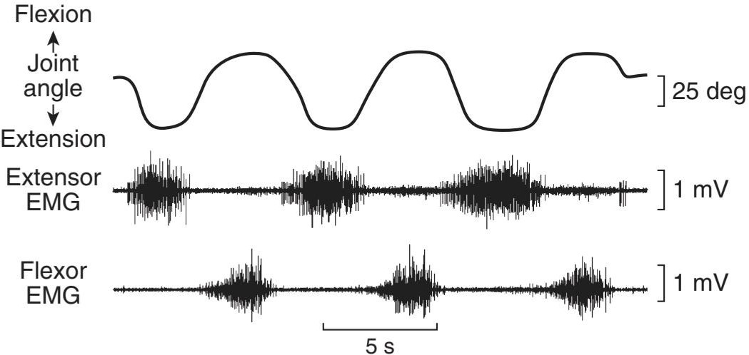
FIGURE 5-13 Relative joint angle and electromyographic tracings from human extensor pollicis longus and flexor pollicis longus during alternate flexion and extension of the distal joint of the thumb. The extensor pollicis longus and flexor pollicis longus extend and flex the distal joint of the thumb, respectively. The distal thumb joint angle (top) is superimposed over the extensor pollicis longus (middle) and flexor pollicus longus (bottom) EMGs. Note the alternate activation and rest patterns as one muscle is used for extension and the other for flexion. (Used with permission of Andrew J. Fuglevand.)
شکل 5-13 زاویه نسبی مفصل و ردیابی های الکترومیوگرافی از اکستانسور پولیسیس لانگوس و فلکسور پولیسیس لانگوس انسان در طول خم شدن متناوب و گسترش مفصل دیستال انگشت شست. Extensor Pollicis longus و flexor pollicis longus به ترتیب مفصل دیستال انگشت شست را امتداد و خم می کنند. زاویه مفصل انتهایی انگشت شست (بالا) بر روی EMGهای اکستانسور پولیسیس لانگوس (وسط) و فلکسور پولیکوس لانگوس (پایین) قرار گرفته است. به الگوهای فعال سازی و استراحت متناوب توجه کنید زیرا یک عضله برای اکستنشن و دیگری برای خم شدن استفاده می شود. (با اجازه اندرو جی فوگلوند استفاده شد.)
It has been shown by electromyography that little if any spontaneous activity occurs in the skeletal muscles of normal individuals at rest. With minimal voluntary activity a few motor units discharge, and with increasing voluntary effort, more and more are brought into play to monitor the recruitment of motor units. Gradation of muscle response is therefore in part a function of the number of motor units activated. In addition, the frequency of discharge in the individual nerve fibers plays a role, the tension developed during a tetanic contraction being greater than that during individual twitches. The length of the muscle is also a factor. Finally, the motor units fire asynchronously, that is, out of phase with one another. This asynchronous firing causes the individual muscle fiber responses to merge into a smooth contraction of the whole muscle. In summary, EMGs can be used to quickly (and roughly) monitor abnormal electrical activity associated with muscle responses.
با الکترومیوگرافی نشان داده شده است که فعالیت خود به خودی کمی در ماهیچه های اسکلتی افراد عادی در حالت استراحت رخ می دهد. با حداقل فعالیت داوطلبانه، تعداد کمی از واحدهای حرکتی تخلیه می شوند و با افزایش تلاش داوطلبانه، بیشتر و بیشتر برای نظارت بر استخدام واحدهای حرکتی وارد بازی می شوند. بنابراین درجه بندی پاسخ عضلانی تا حدی تابعی از تعداد واحدهای حرکتی فعال شده است. علاوه بر این، فرکانس تخلیه در رشتههای عصبی منفرد نقش دارد، تنش ایجاد شده در طول انقباض کزاز بیشتر از کششهای فردی است. طول عضله نیز یک عامل است. در نهایت، واحدهای موتور به صورت ناهمزمان، یعنی خارج از فاز با یکدیگر شلیک می کنند. این شلیک ناهمزمان باعث می شود که پاسخ های فیبر عضلانی منفرد در یک انقباض صاف کل عضله ادغام شوند. به طور خلاصه، EMG را می توان برای نظارت سریع (و تقریباً) فعالیت الکتریکی غیر طبیعی مرتبط با پاسخ های عضلانی استفاده کرد.
THE STRENGTH OF SKELETAL MUSCLES
Human skeletal muscle can exert 3-4 kg of tension per square centimeter of cross-sectional area. This figure is about the same as that obtained in a variety of experimental animals and seems to be constant for mammalian species. Because many of the muscles in humans have a relatively large cross-sectional area, the tension they can develop is quite large. The gastrocnemius, for example, not only supports the weight of the whole body during climbing but resists a force several times this great when the foot hits the ground during running or jumping. An even more striking example is the gluteus maximus, which can exert a tension of 1200 kg. The total tension that could be developed if all muscles in the body of an adult man pulled together is approximately 22,000 kg (nearly 25 tons).
قدرت ماهیچه های اسکلتی
عضله اسکلتی انسان می تواند 3-4 کیلوگرم کشش در هر سانتی متر مربع سطح مقطع ایجاد کند. این رقم تقریباً همان رقمی است که در انواع حیوانات آزمایشی به دست می آید و به نظر می رسد برای گونه های پستانداران ثابت باشد. از آنجایی که بسیاری از ماهیچه های انسان دارای سطح مقطع نسبتاً بزرگی هستند، کششی که می توانند ایجاد کنند بسیار زیاد است. به عنوان مثال، گاستروکنمیوس نه تنها وزن کل بدن را در طول صعود تحمل می کند، بلکه در برابر نیرویی چندین برابر این نیرو در هنگام برخورد پا با زمین در حین دویدن یا پریدن مقاومت می کند. نمونه بارزتر گلوتئوس ماکسیموس است که می تواند 1200 کیلوگرم کشش ایجاد کند. مجموع تنشی که در صورت جمع شدن تمام ماهیچه های بدن یک مرد بالغ می تواند ایجاد شود تقریباً 22000 کیلوگرم (تقریباً 25 تن) است.
BODY MECHANICS
Body movements are generally organized in such a way that they take maximal advantage of the physiological principles outlined above. For example, the attachments of the muscles in the body are such that many of them are normally at or near their resting length when they start to contract. In muscles that extend over more than one joint, movement at one joint may compensate for movement at another in such a way that relatively little shortening of the muscle occurs during contraction. Nearly isometric contractions of this type permit development of maximal tension per contraction. The hamstring muscles extend from the pelvis over the hip joint and the knee joint to the tibia and fibula. Hamstring contraction produces flexion of the leg on the thigh. If the thigh is flexed on the pelvis at the same time, the lengthening of the hamstrings across the hip joint tends to compensate for the shortening across the knee joint. In the course of various activities, the body moves in a way that takes advantage of this. Such factors as momentum and balance are integrated into body movement in ways that make possible maximal motion with minimal muscular exertion. One net effect is that the stress put on tendons and bones rarely exceeds 50% of their failure strength, protecting them from damage.
مکانیک بدن
حرکات بدن عموماً به گونه ای سازماندهی می شوند که از اصول فیزیولوژیکی ذکر شده در بالا حداکثر بهره را ببرند. به عنوان مثال، اتصالات ماهیچهها در بدن به گونهای است که بسیاری از آنها معمولاً در زمان استراحت یا نزدیک به زمان استراحت خود هستند. در ماهیچه هایی که بیش از یک مفصل گسترش می یابند، حرکت در یک مفصل ممکن است حرکت در مفصل دیگر را جبران کند به گونه ای که کوتاه شدن نسبتا کمی عضله در طول انقباض اتفاق می افتد. انقباضات تقریباً ایزومتریک از این نوع امکان ایجاد حداکثر کشش در هر انقباض را فراهم می کند. عضلات همسترینگ از لگن روی مفصل ران و مفصل زانو تا درشت نی و نازک نی امتداد دارند. انقباض همسترینگ باعث خم شدن ساق روی ران می شود. اگر ران به طور همزمان روی لگن خم شود، بلند شدن همسترینگ در سراسر مفصل ران تمایل به جبران کوتاهی در سراسر مفصل زانو دارد. در جریان فعالیت های مختلف بدن به گونه ای حرکت می کند که از این مزیت بهره می برد. عواملی مانند تکانه و تعادل در حرکت بدن به گونهای ادغام میشوند که حداکثر حرکت را با حداقل فشار عضلانی ممکن میسازد. یک اثر خالص این است که فشار وارد شده به تاندون ها و استخوان ها به ندرت از 50 درصد قدرت شکست آنها بیشتر می شود و آنها را از آسیب محافظت می کند.
In walking, each limb passes rhythmically through a support or stance phase when the foot is on the ground and a swing phase when the foot is off the ground. The support phases of the two legs overlap, so that two periods of double support occur during each cycle. There is a brief burst of activity in the leg flexors at the start of each step, and then the leg is swung forward with little more active muscular contraction. Therefore, the muscles are active for only a fraction of each step, and walking for long periods causes relatively little fatigue.
در راه رفتن، هر اندام به طور ریتمیک از یک مرحله حمایت یا ایستادن زمانی که پا روی زمین است و یک مرحله نوسان زمانی که پا از زمین جدا می شود، عبور می کند. مراحل تکیه گاه دو پا با هم همپوشانی دارند، به طوری که در طول هر چرخه دو دوره حمایت مضاعف رخ می دهد. در شروع هر مرحله یک انفجار کوتاه از فعالیت در خم کننده های پا وجود دارد و سپس پا با انقباض عضلانی کمی فعال تر به سمت جلو حرکت می کند. بنابراین، ماهیچه ها تنها برای کسری از هر قدم فعال هستند و پیاده روی طولانی مدت باعث خستگی نسبتا کمی می شود.
A young adult walking at a comfortable pace moves at a velocity of about 80 m/min and generates a power output of 150-175 W per step. A group of young adults asked to walk at their most comfortable rate selected a velocity close to 80 m/min, and it was found that they had selected the velocity at which their energy output was minimal. Walking more rapidly or more slowly took more energy.
یک فرد بالغ جوان که با سرعت راحت راه میرود، با سرعتی حدود 80 متر در دقیقه حرکت میکند و توان خروجی 150-175 وات در هر قدم تولید میکند. گروهی از بزرگسالان جوان که از آنها خواسته شد با راحت ترین سرعت خود راه بروند، سرعتی نزدیک به 80 متر در دقیقه انتخاب کردند و مشخص شد که آنها سرعتی را انتخاب کرده بودند که در آن انرژی خروجی آنها حداقل بود. راه رفتن با سرعت بیشتر یا آهسته تر انرژی بیشتری می گرفت.
CARDIAC MUSCLE MORPHOLOGY
The striations in cardiac muscle are similar to those in skeletal muscle, and Z- lines are present. Large numbers of elongated mitochondria are in close contact with the muscle fibrils. The muscle fibers branch and interdigitate, but each is a complete unit surrounded by a cell membrane. Where the end of one muscle fiber abuts on another, the membranes of both fibers parallel each other through an extensive series of folds. These areas, which always occur at Z-lines, are called intercalated disks (Figure 5-14). They provide a strong union between fibers, maintaining cell-to-cell cohesion, so that the pull of one contractile cell can be transmitted along its axis to the next. Along the sides of the muscle fibers next to the disks, the cell membranes of adjacent fibers fuse for considerable distances, forming gap junctions. These junctions provide low-resistance bridges for the spread of excitation from one fiber to another. They permit cardiac muscle to function as if it were a syncytium, even though no protoplasmic bridges are present between cells. The T system in cardiac muscle is located at the Z-lines rather than at the A-I junction, where it is located in mammalian skeletal muscle.
مورفولوژی ماهیچه های قلب
خطوط در عضله قلب شبیه به عضلات اسکلتی است و خطوط Z وجود دارد. تعداد زیادی از میتوکندری های کشیده در تماس نزدیک با فیبریل های عضلانی هستند. فیبرهای عضلانی منشعب می شوند و بین انگشتان قرار می گیرند، اما هر یک واحد کاملی هستند که توسط یک غشای سلولی احاطه شده است. جایی که انتهای یک فیبر عضلانی به فیبر دیگر متصل می شود، غشای هر دو فیبر از طریق یک سری چین های گسترده با یکدیگر موازی می شوند. این نواحی که همیشه در خطوط Z رخ میدهند، دیسکهای میانپیچیده نامیده میشوند (شکل 5-14). آنها یک اتحاد قوی بین الیاف ایجاد می کنند و انسجام سلول به سلول را حفظ می کنند، به طوری که کشش یک سلول انقباضی می تواند در امتداد محور خود به سلول بعدی منتقل شود. در امتداد کناره های فیبرهای عضلانی در کنار دیسک ها، غشای سلولی رشته های مجاور در فواصل قابل توجهی با هم ترکیب می شوند و اتصالات شکافی را تشکیل می دهند. این اتصالات پل هایی با مقاومت کم برای گسترش تحریک از یک فیبر به فیبر دیگر ایجاد می کنند. آنها به عضله قلب اجازه می دهند تا مانند یک سینسیتیوم عمل کند، حتی اگر هیچ پل پروتوپلاسمی بین سلول ها وجود نداشته باشد. سیستم T در عضله قلبی در خطوط Z قرار دارد نه در محل اتصال A-I، جایی که در عضله اسکلتی پستانداران قرار دارد.
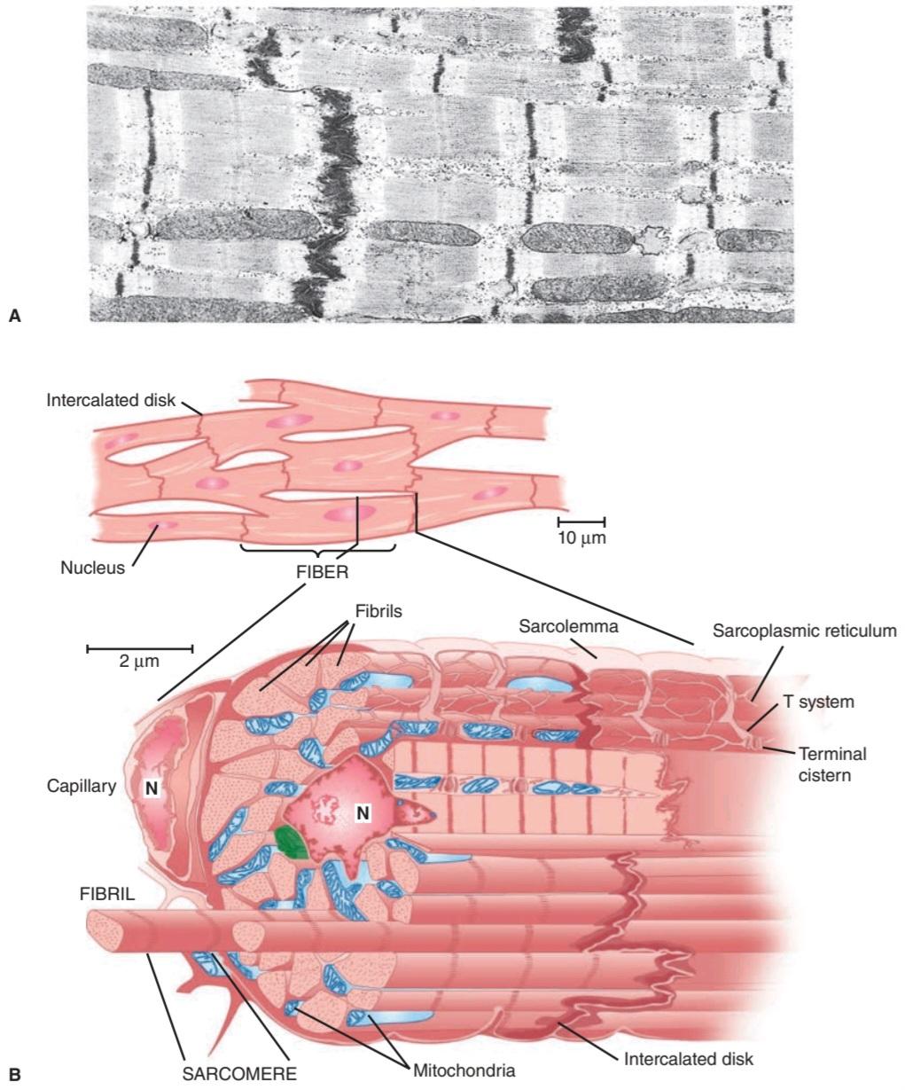
FIGURE 5-14 Cardiac muscle. A) Electron micrograph of cardiac muscle. Note the similarity of the A-I regions seen in the skeletal muscle EM of Figure 5-2. The fuzzy thick lines are intercalated disks and function similarly to the Z-lines but occur at cell membranes (× 12,000). (Reproduced with permission from Bloom W, Fawcett DW: A Textbook of Histology, 10th ed. Saunders, 1975.) B) Artist interpretation of cardiac muscle as seen under the light microscope (top) and the electron microscope (bottom). Again, note the similarity to skeletal muscle structure. N, nucleus. (Reproduced with permission from Braunwald E, Ross J, Sonnenblick EH: Mechanisms of contraction of the normal and failing heart. N Engl J Med 1967 October 12;277(15):794-800.)
شکل 5-14 عضله قلبی. الف) ریزنگار الکترونی عضله قلبی. به شباهت نواحی A-I که در EM عضله اسکلتی شکل 5-2 دیده میشود توجه کنید. خطوط ضخیم مبهم، دیسکهای درهمتنیده هستند و عملکردی مشابه خطوط Z دارند اما در غشاهای سلولی (×12000) رخ میدهند. (با اجازه از Bloom W, Fawcett DW: A Textbook of Histology, 10th ed. Saunders, 1975.) ب) تفسیر هنرمند از عضله قلبی که در زیر میکروسکوپ نوری (بالا) و میکروسکوپ الکترونی (پایین) دیده میشود. مجدداً، به شباهت با ساختار عضله اسکلتی توجه کنید. N، هسته. (با اجازه از Braunwald E, Ross J, Sonnenblick EH: مکانیسمهای انقباض قلب طبیعی و در حال نارسایی، N Engl J Med 1967 October 12;277(15):794-800.)
ELECTRICAL PROPERTIES
RESTING MEMBRANE & ACTION POTENTIALS
The resting membrane potential of individual mammalian cardiac muscle cells is about -90 mV. Stimulation produces a propagated action potential that is responsible for initiating contraction. Although action potentials vary among the cardiomyocytes in different regions of the heart (discussed in Chapter 29), the action potential of a typical ventricular cardiomyocyte can be used as an example (Figure 5-15). Depolarization proceeds rapidly and an overshoot of the zero potential is present, as in skeletal muscle and nerve, but this is followed by a plateau before the membrane potential returns to the baseline. In mammalian hearts, depolarization lasts about 2 ms, but the plateau phase and repolarization last 200 ms or more. Repolarization is therefore not complete until the contraction is half over.
خواص الکتریکی
استراحت غشاء و پتانسیل های عمل
پتانسیل استراحت غشای تک تک سلول های ماهیچه قلب پستانداران حدود 90- میلی ولت است. تحریک یک پتانسیل عمل انتشار ایجاد می کند که مسئول شروع انقباض است. اگرچه پتانسیل عمل در میان کاردیومیوسیت ها در نواحی مختلف قلب متفاوت است (در فصل 29 بحث شد)، می توان از پتانسیل عمل یک کاردیومیوسیت معمولی بطنی به عنوان مثال استفاده کرد (شکل 5-15). دپلاریزاسیون به سرعت ادامه مییابد و بیش از حد پتانسیل صفر وجود دارد، مانند ماهیچههای اسکلتی و عصب، اما قبل از اینکه پتانسیل غشایی به خط پایه بازگردد، یک پلاتو به دنبال دارد. در قلب پستانداران، دپلاریزاسیون حدود 2 میلی ثانیه طول می کشد، اما فاز پلاتو و رپلاریزاسیون 200 میلی ثانیه یا بیشتر طول می کشد. بنابراین تا زمانی که انقباض به نیمه نرسد، رپلاریزاسیون کامل نمی شود.

FIGURE 5-15 Comparison of action potentials and contractile response of a mammalian cardiac muscle fiber in a typical ventricular cell. In the top trace, the intracellular recording of the action potential shows the quick depolarization and extended recovery. In the bottom trace, the mechanical response is matched to the extracellular and intracellular electrical activities. Note that in the absolute refractory period (ARP), the cardiac myocyte cannot be excited, whereas in the relative refractory period (RRP) minimal excitation can occur.
شکل 5-15 مقایسه پتانسیل عمل و پاسخ انقباضی یک فیبر عضلانی قلبی پستانداران در یک سلول بطنی معمولی. در ردیابی بالا، ثبت درون سلولی پتانسیل عمل دپلاریزاسیون سریع و بهبودی طولانی را نشان می دهد. در ردپای پایین، پاسخ مکانیکی با فعالیت های الکتریکی خارج سلولی و درون سلولی مطابقت دارد. توجه داشته باشید که در دوره نسوز مطلق (ARP)، میوسیت قلبی نمی تواند برانگیخته شود، در حالی که در دوره مقاوم نسبی (RRP) حداقل تحریک ممکن است رخ دهد.
As in other excitable tissues, changes in the external K+ concentration affect the resting membrane potential of cardiac muscle, whereas changes in the external Nat concentration affect the magnitude of the action potential. The initial rapid depolarization and the overshoot (phase 0) are due to opening of voltage-gated Na+ channels similar to that occurring in nerve and skeletal muscle (Figure 5-16). The initial rapid repolarization (phase 1) is due to closure of Na+ channels and opening of one type of K+ channel (ie, Kv4.2/KCND2 and/or Kv4.3/CCND3, and KV1.4/KCNA4). The subsequent prolonged plateau (phase 2) is due to a slower but prolonged opening of voltage-gated Ca2+ channels. Final repolarization (phase 3) to the resting membrane potential (phase 4) is due to closure of the Ca2+ channels and a slow, delayed increase of K+ efflux through various types of K+ channels. Cardiac myocytes contain at least two types of voltage-gated Ca2+ channels (T- and L-types), but the Ca2+ current is mostly due to opening of the slower L-type Ca2+ channels. Mutations or dysfunctions in any of these channels lead to serious pathologies of the heart (eg, Clinical Box 5–5).
مانند سایر بافت های تحریک پذیر، تغییرات در غلظت خارجی +K بر پتانسیل غشای استراحت عضله قلب تأثیر می گذارد، در حالی که تغییرات در غلظت +Na خارجی بر بزرگی پتانسیل عمل تأثیر می گذارد. دپلاریزاسیون سریع اولیه و افزایش بیش از حد (فاز 0) به دلیل باز شدن کانال های +Na دردار ولتاژ مشابه آنچه در عصب و عضله اسکلتی رخ می دهد است (شکل 5-16). رپلاریزاسیون سریع اولیه (فاز 1) به دلیل بسته شدن کانال های +Na و باز شدن یک نوع کانال +K (یعنی Kv4.2/KCND2 و/یا Kv4.3/CCND3 و KV1.4/KCNA4) است. فلات طولانی بعدی (فاز 2) به دلیل باز شدن کندتر اما طولانی مدت کانال های +Ca2 دارای ولتاژ است. رپلاریزاسیون نهایی (فاز 3) به پتانسیل غشای در حال استراحت (فاز 4) به دلیل بسته شدن کانال های +Ca2 و افزایش آهسته و تاخیری جریان +K از طریق انواع مختلف کانال های +K است. میوسیتهای قلبی حداقل دو نوع کانال +Ca2 دارای ولتاژ دارند (نوع T و L)، اما جریان +Ca2 بیشتر به دلیل باز شدن کانالهای +Ca2 نوع L کندتر است. جهش یا اختلال در هر یک از این کانال ها منجر به آسیب شناسی جدی قلب می شود (به عنوان مثال، جعبه بالینی 5-5).
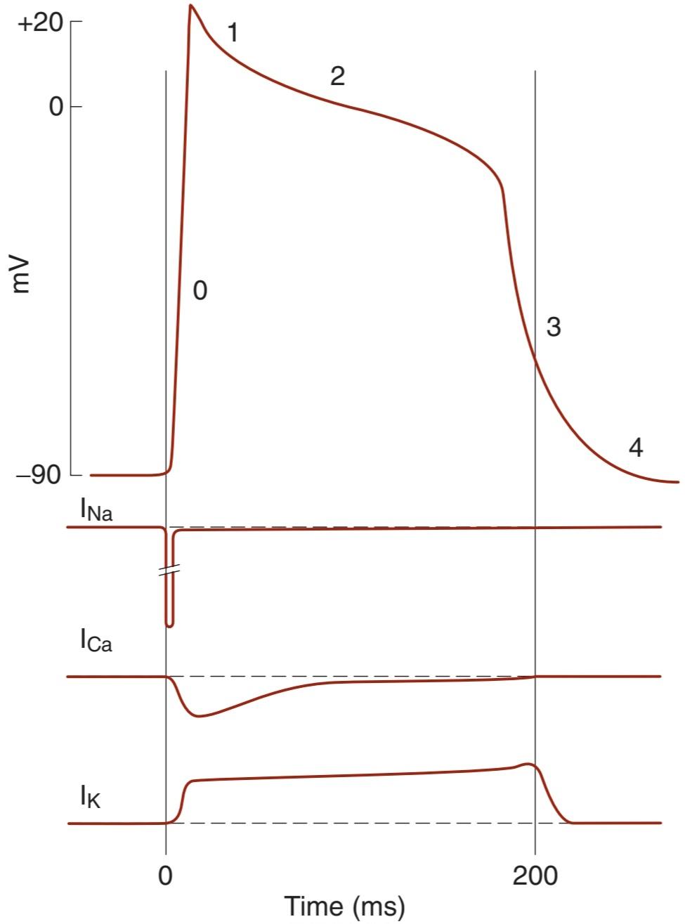
FIGURE 5-16 Dissection of the cardiac action potential. Top: The action potential of a cardiac muscle fiber can be broken down into several phases: 0, depolarization; 1, initial rapid repolarization; 2, plateau phase; 3, late rapid repolarization; 4, baseline. Bottom: Diagrammatic summary of Na*, Ca2+, and cumulative K+ currents during the action potential. As is convention, inward currents are downward, and outward currents are upward.
شکل 5-16 تشریح پتانسیل عمل قلبی. بالا: پتانسیل عمل فیبر عضلانی قلب را می توان به چند مرحله تقسیم کرد: 0، دپلاریزاسیون. 1، رپولاریزاسیون سریع اولیه؛ 2، فاز فلات؛ 3، دیر قطبی شدن سریع. 4، خط مبنا پایین: خلاصه نموداری جریان های +Na+، Ca2 و +K تجمعی در طول پتانسیل عمل. طبق معمول، جریان های داخلی رو به پایین و جریان های بیرونی به سمت بالا هستند.
CLINICAL BOX 5.5
Long QT Syndrome
Long QT syndrome (LQTS) is defined as a prolongation of the QT interval observed on an electrocardiogram. LQTS can lead to irregular heartbeats and subsequent fainting, seizure, cardiac arrest, or even death. Although certain medications can lead to LQTS, it is more frequently associated with genetic mutations in a variety of cardiac-expressed ion channels. Mutations in cardiac-expressed voltage-gated K+ channel genes (KCNQ1 or KCNH2) account for most of the mutation-based cases of LQTS (~90%). Mutations in cardiac-expressed voltage-gated Na+ channel genes (eg, SCN5A) or cardiac- expressed Ca2+ channel genes (eg, CACNA1C) have also been associated with the disease. The fact that mutations in diverse channels all can result in the prolongation of the QT interval and subsequent pathophysiological changes underlies the intricate interplay of these channels in shaping the heart’s electrical response.
جعبه بالینی 5.5
سندرم QT طولانی
سندرم QT طولانی (LQTS) به عنوان طولانی شدن فاصله QT مشاهده شده در نوار قلب تعریف می شود. LQTS می تواند منجر به ضربان قلب نامنظم و متعاقب آن غش، تشنج، ایست قلبی یا حتی مرگ شود. اگرچه برخی داروها میتوانند منجر به LQTS شوند، اما اغلب با جهشهای ژنتیکی در انواع کانالهای یونی بیانشده قلبی مرتبط است. جهش در ژن های کانال K + دردار ولتاژ بیان شده با ولتاژ بیان شده (KCNQ1 یا KCNH2) بیشتر موارد مبتنی بر جهش LQTS (~90٪) را تشکیل می دهد. جهش در ژنهای کانال Na+ دارای ولتاژ بیانشده (مانند SCN5A) یا ژنهای کانال Ca2+ بیانشده در قلب (مانند CACNA1C) نیز با این بیماری مرتبط است. این واقعیت که جهش در کانال های مختلف می تواند منجر به طولانی شدن فاصله QT و تغییرات پاتوفیزیولوژیکی بعدی شود، زمینه ساز تعامل پیچیده این کانال ها در شکل دادن به پاسخ الکتریکی قلب است.
THERAPEUTIC HIGHLIGHTS
Patients with LQTS should avoid drugs that prolong the QT interval or reduce their serum K+ or Mg2+ levels; any K* or Mg2+ deficiencies should be corrected. Drug interventions in asymptomatic patients remain somewhat controversial, although patients with congenital defects that lead to LQTS are considered candidates for intervention independent of symptoms. In general, ẞ- blockers have been used for LQTS to reduce the risk of cardiac arrhythmias. More specific and effective treatments can be introduced once the underlying cause of LQTS is identified.
نکات برجسته درمانی
بیماران مبتلا به LQTS باید از داروهایی که فاصله QT را طولانیتر میکنند یا سطح سرمی K + یا Mg2 + را کاهش میدهند اجتناب کنند. هر گونه کمبود K* یا Mg2+ باید اصلاح شود. مداخلات دارویی در بیماران بدون علامت تا حدودی بحث برانگیز است، اگرچه بیمارانی که نقایص مادرزادی دارند که منجر به LQTS می شود، کاندیدای مداخله مستقل از علائم در نظر گرفته می شوند. به طور کلی، مسدود کننده های ẞ- برای LQTS برای کاهش خطر آریتمی قلبی استفاده شده است. پس از شناسایی علت زمینه ای LQTS می توان درمان های خاص و موثرتری را معرفی کرد.
MECHANICAL PROPERTIES
CONTRACTILE RESPONSE
The contractile response of cardiac muscle begins just after the start of depolarization and lasts about 1.5 times as long as the action potential (Figure 5–15). The role of Ca2+ in excitation-contraction coupling is similar to its role in skeletal muscle (see above). However, it is the influx of extracellular Ca2+ through the voltage-sensitive DHPR or voltage-gated Ca2+ channels in the T system that triggers Ca2+-induced Ca2+ release through the RyR at the sarcoplasmic reticulum. Because there is a net influx of Ca2+ during activation, there is also a more prominent role for plasma membrane Ca2+ ATPases and the Na+/Ca2+ exchanger in recovery of cytosolic Ca2+ concentrations. Specific effects of drugs that indirectly alter intracellular Ca2+ concentrations are discussed in Clinical Box 5-6.
خواص مکانیکی
پاسخ انقباضی
پاسخ انقباضی عضله قلب درست پس از شروع دپلاریزاسیون شروع می شود و حدود 1.5 برابر پتانسیل عمل طول می کشد (شکل 5-15). نقش +Ca2 در جفت شدن تحریک-انقباض مشابه نقش آن در ماهیچه های اسکلتی است (به بالا مراجعه کنید). با این حال، هجوم +Ca2 خارج سلولی از طریق DHPR حساس به ولتاژ یا کانالهای +Ca2 دارای ولتاژ در سیستم T است که باعث انتشار +Ca2 ناشی از +Ca2 از طریق RyR در شبکه سارکوپلاسمی میشود. از آنجایی که جریان خالص +Ca2 در طول فعالسازی وجود دارد، همچنین نقش برجستهتری برای +Ca2 ATPases غشای پلاسمایی و مبدل +Na+/Ca2 در بازیابی غلظتهای +Ca2 سیتوزولی وجود دارد. اثرات خاص داروهایی که به طور غیرمستقیم غلظت های +Ca2 داخل سلولی را تغییر می دهند در جعبه بالینی 5-6 بحث شده است.
During phases 0 to 2 and about half of phase 3 (until the membrane potential reaches approximately -50 mV during repolarization), cardiac muscle cannot be excited again; that is, it is in its absolute refractory period. It remains relatively refractory until phase 4. Therefore, tetanus of the type seen in skeletal muscle cannot occur. Of course, tetanization of cardiac muscle for any length of time would have lethal consequences, and in this sense, the fact that cardiac muscle cannot be tetanized is a safety feature.
در طول فازهای 0 تا 2 و تقریباً نیمی از فاز 3 (تا زمانی که پتانسیل غشاء به حدود -50 میلی ولت در طی رپلاریزاسیون برسد)، عضله قلب نمی تواند دوباره برانگیخته شود. یعنی در دوره نسوز مطلق است. تا فاز 4 نسبتاً نسوز باقی می ماند. بنابراین، کزاز از نوعی که در عضله اسکلتی دیده می شود، نمی تواند رخ دهد. البته تتانیزاسیون عضله قلب برای هر مدت زمان عواقب کشنده ای خواهد داشت و از این نظر این واقعیت که عضله قلب را نمی توان تتانیز کرد یک ویژگی ایمنی است.
CLINICAL BOX 5.6
Glycosidic Drugs & Cardiac Contraction
Ouabain and other digitalis glycosides are commonly used to treat failing hearts. These drugs have the effect of increasing the strength of cardiac contraction. Although there is discussion as to full mechanisms, a working hypothesis is based on the ability of these drugs to inhibit the Na, K ATPase in cell membranes of the cardiomyocytes. The block of the Na, K ATPase in cardiomyocytes would result in an increased intracellular Na+ concentration. Such an increase would result in a decreased inward Na* transportation and hence outward Ca2+ transportation via the Na*-Ca2+ exchange antiport during the Ca2+ recovery period. The resulting increase in intracellular Ca2+ concentration in turn increases the strength of contraction of the cardiac muscle. With this mechanism in mind, these drugs can also be quite toxic. Over inhibition of the Na, K ATPase would result in a depolarized cell that could slow conduction, or even spontaneously activate contraction. Alternatively, an overly increased Ca2+ concentration could also have ill effects on cardiomyocyte function.
جعبه بالینی 5.6
داروهای گلیکوزیدی و انقباضات قلبی
Ouabain و سایر گلیکوزیدهای دیجیتالی معمولاً برای درمان نارسایی قلب استفاده می شوند. این داروها باعث افزایش قدرت انقباضات قلبی می شوند. اگرچه بحث در مورد مکانیسم کامل وجود دارد، یک فرضیه کاری بر اساس توانایی این داروها در مهار Na, K ATPase در غشای سلولی کاردیومیوسیت ها است. بلوک Na، K ATPase در کاردیومیوسیت ها منجر به افزایش غلظت +Na داخل سلولی می شود. چنین افزایشی منجر به کاهش حمل و نقل Na * به داخل و در نتیجه انتقال +Ca2 به خارج از طریق پادپورت تبادل +Na+ – Ca2 در طول دوره بهبود Ca2 می شود. افزایش غلظت +Ca2 داخل سلولی به نوبه خود قدرت انقباض عضله قلب را افزایش می دهد. با در نظر گرفتن این مکانیسم، این داروها می توانند کاملاً سمی باشند. مهار بیش از حد Na، K ATPase منجر به یک سلول دپلاریزه می شود که می تواند رسانش را کند کند یا حتی انقباض را به طور خود به خود فعال کند. از طرف دیگر، افزایش بیش از حد غلظت +Ca2 نیز می تواند اثرات بدی بر عملکرد قلب داشته باشد.
ISOFORMS
Cardiac muscle is generally slow and has relatively low ATPase activity. Its fibers are dependent on oxidative metabolism and hence on a continuous supply of O2. The human heart contains both the a and the ẞ isoforms of the myosin heavy chain (a MHC and ẞ MHC). ẞ MHC has lower myosin ATPase activity than a MHC. Both are present in the atria, with the a isoform predominating, whereas the ẞ isoform predominates in the ventricle. The spatial differences in expression contribute to the well-coordinated contraction of the heart.
ایزوفرمها
عضله قلب به طور کلی کند است و فعالیت ATPase نسبتاً کمی دارد. فیبرهای آن به متابولیسم اکسیداتیو و در نتیجه به عرضه مداوم O2 وابسته هستند. قلب انسان دارای هر دو ایزوفرم a و ẞ زنجیره سنگین میوزین (MHC و ẞ MHC) است. MHC فعالیت ATPase میوزین کمتری نسبت به MHC دارد. هر دو در دهلیزها وجود دارند و ایزوفرم a غالب است، در حالی که ایزوفرم ẞ در بطن غالب است. تفاوت های فضایی در بیان به انقباض خوب هماهنگ قلب کمک می کند.
CORRELATION BETWEEN MUSCLE FIBER LENGTH & TENSION
The relation between initial fiber length and total tension in cardiac muscle is similar to that in skeletal muscle; there is an optimal resting length at which the tension developed on stimulation is maximal. In the body, the initial length of the fibers is determined by the degree of diastolic filling of the heart, and the pressure developed in the ventricle is proportional to the volume of the ventricle at the end of the filling phase (Starling law of the heart). The developed tension (Figure 5-17) increases as the diastolic volume increases until it reaches a maximum, then tends to decrease. However, unlike skeletal muscle, the decrease in developed tension at high degrees of stretch is not due to a decrease in the number of cross-bridges between actin and myosin, because even severely dilated hearts are not stretched to this degree. The decrease is instead due to beginning disruption of the myocardial fibers.
همبستگی بین طول فیبر عضلانی و تنش
رابطه بین طول فیبر اولیه و کشش کلی در عضله قلب شبیه به عضله اسکلتی است. یک طول استراحت بهینه وجود دارد که در آن تنش ایجاد شده در تحریک حداکثر است. در بدن طول اولیه الیاف با درجه پر شدن دیاستولیک قلب تعیین می شود و فشار ایجاد شده در بطن متناسب با حجم بطن در پایان مرحله پر شدن است (قانون استارلینگ قلب). تنش ایجاد شده (شکل 5-17) با افزایش حجم دیاستولیک افزایش می یابد تا زمانی که به حداکثر برسد، سپس تمایل به کاهش دارد. با این حال، بر خلاف عضله اسکلتی، کاهش کشش ایجاد شده در درجات کشش بالا به دلیل کاهش تعداد پل های متقاطع بین اکتین و میوزین نیست، زیرا حتی قلب های به شدت گشاد شده نیز به این درجه کشیده نمی شوند. این کاهش در عوض به دلیل اختلال در شروع فیبرهای میوکارد است.

FIGURE 5–17 Length-tension relationship for cardiac muscle. Comparison of the systolic intraventricular pressure (top trace) and diastolic intraventricular pressure (bottom trace) display the developed tension in the cardiomyocyte. Values shown are for canine heart.
شکل 5-17 رابطه طول-تنش برای عضله قلب. مقایسه فشار داخل بطنی سیستولیک (ردیابی بالا) و فشار داخل بطنی دیاستولیک (ردپای پایین) تنش ایجاد شده در قلب را نشان می دهد. مقادیر نشان داده شده برای قلب سگ است.
The force of contraction of cardiac muscle can be also increased by catecholamines, and this increase occurs without a change in muscle length. This positive ionotropic effect of catecholamines is mediated via innervated ẞ1- adrenergic receptors, cyclic adenosine monophosphate (AMP), and their effects on Ca2+ homeostasis. The heart also contains noninnervated ẞ2-adrenergic receptors, which also act via cyclic AMP, but their ionotropic effect is smaller and is maximal in the atria. Cyclic AMP activates protein kinase A, and this leads to phosphorylation of the voltage-gated Ca2+ channels, causing them to spend more time in the open state. Cyclic AMP also increases the active transport of Ca2+ to the sarcoplasmic reticulum, thus accelerating relaxation and consequently shortening systole. This is important when the cardiac rate is increased because it permits adequate diastolic filling (see Chapter 30).
نیروی انقباض عضله قلب را می توان با کاتکول آمین ها نیز افزایش داد و این افزایش بدون تغییر در طول عضله اتفاق می افتد. این اثر یونوتروپیک مثبت کاتکول آمین ها از طریق گیرنده های 1- آدرنرژیک عصب دهی شده، آدنوزین مونوفسفات حلقوی (AMP) و اثرات آنها بر هموستاز +Ca2 واسطه می شود. قلب همچنین حاوی گیرنده های ẞ2-آدرنرژیک غیرعصبی است که از طریق AMP حلقوی نیز عمل می کنند، اما اثر یونوتروپیک آنها کوچکتر است و در دهلیزها حداکثر است. AMP حلقوی پروتئین کیناز A را فعال می کند و این منجر به فسفوریلاسیون کانال های +Ca2 دارای ولتاژ می شود و باعث می شود زمان بیشتری را در حالت باز بگذرانند. AMP حلقوی همچنین انتقال فعال +Ca2 را به شبکه سارکوپلاسمی افزایش می دهد، بنابراین آرامش را تسریع می کند و در نتیجه سیستول را کوتاه می کند. این امر زمانی مهم است که ضربان قلب افزایش می یابد زیرا امکان پر شدن دیاستولیک کافی را فراهم می کند (به فصل 30 مراجعه کنید).
METABOLISM
Mammalian hearts have an abundant blood supply, numerous mitochondria, and a high content of myoglobin, a muscle pigment that can function as an O2 storage mechanism. Normally, less than 1% of the total energy liberated is provided by anaerobic metabolism. During hypoxia, this figure may increase to nearly 10%, but under totally anaerobic conditions, the energy liberated is inadequate to sustain ventricular contractions. Under basal conditions, 35% of the caloric needs of the human heart are provided by carbohydrate, 5% by ketones and amino acids, and 60% by fat. However, the proportions of substrates utilized vary greatly with the nutritional state. After ingestion of large amounts of glucose, more lactate and pyruvate are used; during prolonged starvation, more fat is used. Circulating free fatty acids normally account for almost 50% of the lipid utilized. In untreated diabetics, the carbohydrate utilization of cardiac muscle is reduced and that of fat is increased.
متابولیسم
قلب پستانداران دارای منبع خون فراوان، میتوکندری های متعدد و محتوای بالای میوگلوبین است، رنگدانه ماهیچه ای که می تواند به عنوان مکانیسم ذخیره O2 عمل کند. به طور معمول، کمتر از 1٪ از کل انرژی آزاد شده توسط متابولیسم بی هوازی تامین می شود. در طول هیپوکسی، این رقم ممکن است تا نزدیک به 10٪ افزایش یابد، اما در شرایط کاملاً بی هوازی، انرژی آزاد شده برای حفظ انقباضات بطنی ناکافی است. در شرایط پایه، 35 درصد از کالری مورد نیاز قلب انسان توسط کربوهیدرات، 5 درصد توسط کتون ها و اسیدهای آمینه و 60 درصد توسط چربی تامین می شود. با این حال، نسبت بسترهای مورد استفاده با وضعیت تغذیه ای بسیار متفاوت است. پس از مصرف مقادیر زیادی گلوکز، لاکتات و پیرووات بیشتری استفاده می شود. در طول گرسنگی طولانی مدت، چربی بیشتری مصرف می شود. اسیدهای چرب آزاد در گردش معمولاً تقریباً 50 درصد از چربی مورد استفاده را تشکیل می دهند. در بیماران دیابتی درمان نشده، استفاده از کربوهیدرات عضله قلب کاهش یافته و چربی افزایش می یابد.
SMOOTH MUSCLE MORPHOLOGY
Smooth muscle is distinguished anatomically from skeletal and cardiac muscle because it lacks visible cross-striations. Actin and myosin-II are present, and they slide on each other to produce contraction. However, they are not arranged in regular arrays, as in skeletal and cardiac muscle, and so the striations are absent. Instead of Z-lines, there are dense bodies in the cytoplasm and attached to the cell membrane, and these are bound by a-actinin to actin filaments. Smooth muscle also contains tropomyosin, but troponin appears to be absent. The isoforms of actin and myosin differ from those in skeletal muscle. A sarcoplasmic reticulum is present, but it is less extensive than those observed in skeletal or cardiac muscle. In general, smooth muscles contain few mitochondria and depend, to a large extent, on glycolysis for their metabolic needs.
مورفولوژی ماهیچه های صاف
ماهیچه صاف از نظر تشریحی از ماهیچه اسکلتی و قلبی متمایز می شود زیرا فاقد خطوط متقاطع قابل مشاهده است. اکتین و میوزین-II وجود دارند و روی هم می لغزند تا انقباض ایجاد کنند. با این حال، مانند ماهیچههای اسکلتی و قلبی در آرایههای منظم چیده نشدهاند، بنابراین خطوط خطوط وجود ندارند. به جای خطوط Z، اجسام متراکمی در سیتوپلاسم وجود دارند که به غشای سلولی متصل هستند و این اجسام توسط a-actinin به رشتههای اکتین متصل میشوند. ماهیچه صاف همچنین حاوی تروپومیوزین است، اما به نظر می رسد تروپونین وجود ندارد. ایزوفرم های اکتین و میوزین با ایزوفرم های عضله اسکلتی متفاوت است. یک شبکه سارکوپلاسمی وجود دارد، اما نسبت به آنهایی که در عضله اسکلتی یا قلبی مشاهده می شود، وسعت کمتری دارد. به طور کلی، ماهیچه های صاف حاوی تعداد کمی میتوکندری هستند و برای نیازهای متابولیکی خود تا حد زیادی به گلیکولیز وابسته هستند.
TYPES
There is considerable variation in the structure and function of smooth muscle in different parts of the body. In general, smooth muscle can be divided into unitary (or visceral) smooth muscle and multiunit smooth muscle. Unitary smooth muscle occurs in large sheets, has many low-resistance gap junctional connections between individual muscle cells, and functions in a syncytial fashion. Unitary smooth muscle is found primarily in the walls of hollow viscera. The musculature of the intestine, the uterus, and the ureters are examples. Multiunit smooth muscle is made up of individual units with few (or no) gap junctional bridges. It is found in structures such as the iris of the eye, in which fine, graded contractions occur. It is not under voluntary control, but it has many functional similarities to skeletal muscle. Each multiunit smooth muscle cell has en passant endings of nerve fibers, but in unitary smooth muscle there are en passant junctions on fewer cells, with excitation spreading to other cells by gap junctions. In addition, these cells respond to hormones and other circulating substances. Blood vessels have both unitary and multiunit smooth muscle in their walls.
انواع
تغییرات قابل توجهی در ساختار و عملکرد عضلات صاف در قسمت های مختلف بدن وجود دارد. به طور کلی، ماهیچه صاف را می توان به عضله صاف واحد (یا احشایی) و عضله صاف چند واحدی تقسیم کرد. عضله صاف واحد در ورقه های بزرگ رخ می دهد، دارای بسیاری از اتصالات اتصالی با فاصله کم مقاومت بین سلول های عضلانی منفرد است و به صورت سنسیشیال عمل می کند. عضله صاف واحد عمدتاً در دیواره احشاء توخالی یافت می شود. ماهیچه های روده، رحم و حالب ها مثال هایی هستند. عضله صاف چند واحدی از واحدهای منفرد با پل های اتصالی شکاف کمی (یا بدون) تشکیل شده است. این در ساختارهایی مانند عنبیه چشم یافت می شود که در آن انقباضات ظریف و درجه بندی شده رخ می دهد. تحت کنترل داوطلبانه نیست، اما شباهت های عملکردی زیادی به ماهیچه های اسکلتی دارد. هر سلول عضله صاف چند واحدی دارای انتهای عبوری رشتههای عصبی است، اما در عضله صاف واحد، اتصالات عبوری روی سلولهای کمتری وجود دارد که تحریک توسط اتصالات شکاف به سلولهای دیگر گسترش مییابد. علاوه بر این، این سلول ها به هورمون ها و سایر مواد در گردش پاسخ می دهند. عروق خونی دارای عضله صاف واحد و چند واحدی در دیواره خود هستند.
ELECTRICAL & MECHANICAL ACTIVITY
Unitary smooth muscle is characterized by the instability of its membrane potential and by the fact that it shows continuous, irregular contractions that are independent of its nerve supply. This maintained state of partial contraction is called tonus, or tone. The membrane potential has no true “resting” value, being relatively low (or less negative) when the tissue is active and higher (or more negative) when it is inhibited, but in periods of relative quiescence values for resting potential are on the order of -20 to -65 mV. Smooth muscle cells can display divergent electrical activity (eg, Figure 5-18). There are slow sine wave- like fluctuations a few millivolts in magnitude and spikes that sometimes overshoot the zero potential line and sometimes do not. In many tissues, the spikes have a duration of about 50 ms, whereas in some tissues the action potentials have a prolonged plateau during repolarization, like the action potentials in cardiac muscle. As in the other muscle types, there are significant contributions of K+, Na+, and Ca2+ channels and Na, K ATPase to this electrical activity. However, discussion of contributions to individual smooth muscle types is beyond the scope of this text.
فعالیت الکتریکی و مکانیکی
عضله صاف یکپارچه با بی ثباتی پتانسیل غشایی خود و این واقعیت که انقباضات مداوم و نامنظم را نشان می دهد که مستقل از تامین عصب آن است مشخص می شود. این حالت حفظ شده انقباض جزئی، تونوس یا تون نامیده می شود. پتانسیل غشاء ارزش «استراحت» واقعی ندارد، زمانی که بافت فعال است نسبتاً کم (یا کمتر منفی) و زمانی که مهار میشود بیشتر (یا منفیتر) است، اما در دورههای سکون نسبی مقادیر پتانسیل استراحت در حد 20- تا 65- میلی ولت است. سلول های ماهیچه صاف می توانند فعالیت الکتریکی واگرا را نشان دهند (به عنوان مثال، شکل 5-18). نوسانات آهسته موج سینوسی مانند چند میلی ولت در بزرگی و سنبله هایی وجود دارد که گاهی از خط پتانسیل صفر فراتر می رود و گاهی اوقات نه. در بسیاری از بافت ها، سنبله ها حدود 50 میلی ثانیه طول می کشند، در حالی که در برخی بافت ها پتانسیل های عمل دارای یک فلات طولانی در طول رپلاریزاسیون هستند، مانند پتانسیل های عمل در عضله قلب. همانند سایر انواع ماهیچهها، کانالهای +K+، Na و +Ca2 و Na، K ATPase سهم قابلتوجهی در این فعالیت الکتریکی دارند. با این حال، بحث در مورد کمک به انواع ماهیچه های صاف فردی فراتر از محدوده این متن است.
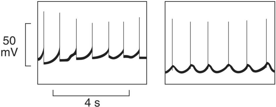
FIGURE 5–18 Electrical activity of individual smooth muscle cells in the guinea pig taenia coli. Left: Pacemaker-like activity with spikes firing at each peak. Right: Sinusoidal fluctuation of membrane potential with firing on the rising phase of each wave. In other fibers, spikes can occur on the falling phase of sinusoidal fluctuations and there can be mixtures of sinusoidal and pacemaker potentials in the same fiber.
شکل 5-18 فعالیت الکتریکی تک تک سلول های ماهیچه صاف در تانیا کلی خوکچه هندی. سمت چپ: فعالیتی شبیه ضربان ساز با پرتاب در هر پیک. سمت راست: نوسان سینوسی پتانسیل غشا با شلیک در فاز بالارونده هر موج. در فیبرهای دیگر، میخها میتوانند در فاز سقوط نوسانات سینوسی رخ دهند و ممکن است مخلوطی از پتانسیلهای سینوسی و ضربان ساز در همان فیبر وجود داشته باشد.
Because of the continuous activity, it is difficult to study the relation between the electrical and mechanical events in unitary smooth muscle, but in some relatively inactive preparations, a single spike can be generated. In such preparations, the excitation-contraction coupling in unitary smooth muscle can occur with as much as a 500-ms delay. Thus, it is a very slow process compared with that in skeletal and cardiac muscle, in which the time from initial depolarization to initiation of contraction is less than 10 ms. Unlike unitary smooth muscle, multiunit smooth muscle is nonsyncytial and contractions do not spread widely through it. Because of this, the contractions of multiunit smooth muscle are more discrete, fine, and localized than those of unitary smooth muscle.
به دلیل فعالیت مداوم، مطالعه رابطه بین رویدادهای الکتریکی و مکانیکی در عضله صاف واحد دشوار است، اما در برخی از آمادهسازیهای نسبتاً غیرفعال، میتوان یک سنبله منفرد ایجاد کرد. در چنین آماده سازی، جفت تحریک-انقباض در عضله صاف واحد می تواند با تاخیر 500 میلی ثانیه رخ دهد. بنابراین، در مقایسه با فرآیند عضله اسکلتی و قلبی که در آن زمان از دپلاریزاسیون اولیه تا شروع انقباض کمتر از 10 میلی ثانیه است، فرآیند بسیار کندی است. بر خلاف عضله صاف واحد، عضله صاف چند واحدی غیرسنسیسیال است و انقباضات به طور گسترده از طریق آن پخش نمی شود. به همین دلیل، انقباضات عضله صاف چند واحدی گسسته، ظریف و موضعی تر از انقباضات عضله صاف واحد است.
MOLECULAR BASIS OF CONTRACTION
As in skeletal and cardiac muscle, an increase in cytosolic Ca2+ plays a prominent role in the initiation of contraction of smooth muscle. However, the source of Ca2+ increase can be quite different in unitary smooth muscle. Depending on the activating stimulus, cytosolic Ca2+ increase can be due to Ca2+ influx through voltage- or ligand-gated channels in the plasma membrane, Ca2+ release or mobilization from intracellular stores (eg, sarcoplasmic reticulum) through the RyR and the inositol trisphosphate receptor (IP3R), Ca2+ release channels in the sarcoplasmic reticulum. In addition, the lack of troponin in smooth muscle prevents Ca2+ activation via troponin binding. Rather, myosin in smooth muscle must be phosphorylated for activation of the myosin ATPase. Phosphorylation and dephosphorylation of myosin also occur in skeletal muscle, but phosphorylation is not necessary for activation of the ATPase. In smooth muscle, Ca2+ binds to calmodulin, and the resulting complex activates calmodulin-dependent myosin light chain kinase. This enzyme catalyzes the phosphorylation of the myosin light chain on serine at position 19, increasing its ATPase activity.
مبانی مولکولی انقباض
همانند ماهیچه های اسکلتی و قلبی، افزایش +Ca2 سیتوزولی نقش مهمی در شروع انقباض عضلات صاف دارد. با این حال، منبع افزایش +Ca2 می تواند در عضله صاف واحد کاملاً متفاوت باشد. بسته به محرک فعالکننده، افزایش +Ca2 سیتوزولی میتواند ناشی از هجوم +Ca2 از طریق کانالهای دارای ولتاژ یا لیگاند در غشای پلاسمایی، انتشار +Ca2 یا حرکت از ذخایر داخل سلولی (مثلاً شبکه سارکوپلاسمی) از طریق RyR و گیرندههای اینوزیتول در +Ca2 باشد. شبکه سارکوپلاسمی علاوه بر این، کمبود تروپونین در ماهیچه صاف از فعال شدن +Ca2 از طریق اتصال تروپونین جلوگیری می کند. بلکه برای فعال شدن میوزین ATPase، میوزین در ماهیچه صاف باید فسفریله شود. فسفوریلاسیون و دفسفوریلاسیون میوزین نیز در عضله اسکلتی رخ می دهد، اما فسفوریلاسیون برای فعال شدن ATPase ضروری نیست. در عضله صاف، +Ca2 به کالمودولین متصل می شود و کمپلکس حاصل، زنجیره سبک میوزین کیناز وابسته به کالمودولین را فعال می کند. این آنزیم فسفوریلاسیون زنجیره سبک میوزین روی سرین در موقعیت 19 را کاتالیز می کند و فعالیت ATPase آن را افزایش می دهد.
Myosin is dephosphorylated by myosin light chain phosphatase in the cell. However, dephosphorylation of myosin light chain kinase does not necessarily lead to relaxation of the smooth muscle. Various mechanisms are involved. One appears to be a latch bridge mechanism by which myosin cross-bridges remain attached to actin for some time after the cytoplasmic Ca2+ concentration falls. This produces sustained contraction with little expenditure of energy, which is especially important in vascular smooth muscle. Relaxation of the muscle presumably occurs when the Ca2+-calmodulin complex finally dissociates or when some other mechanism comes into play. The events leading to contraction and relaxation of unitary smooth muscle are summarized in Figure 5-19. The events in multiunit smooth muscle are generally similar.
میوزین توسط زنجیره سبک فسفاتاز میوزین در سلول دفسفریله می شود. با این حال، دفسفوریلاسیون میوزین زنجیره سبک کیناز لزوما منجر به شل شدن عضله صاف نمی شود. مکانیسم های مختلفی درگیر هستند. به نظر می رسد یکی از آنها مکانیسم پل چفتی باشد که توسط آن پل های متقابل میوزین برای مدتی پس از کاهش غلظت +Ca2 سیتوپلاسمی به اکتین متصل می مانند. این باعث ایجاد انقباض پایدار با صرف انرژی اندک می شود که به ویژه در عضلات صاف عروق اهمیت دارد. شل شدن عضله احتمالاً زمانی اتفاق میافتد که در نهایت کمپلکس +Ca2-calmodulin جدا شود یا مکانیسم دیگری وارد عمل شود. رویدادهایی که منجر به انقباض و شل شدن عضله صاف واحد می شود در شکل 5-19 خلاصه شده است. رویدادهای عضله صاف چند واحدی به طور کلی مشابه هستند.

FIGURE 5-19 Sequence of events in contraction and relaxation of smooth muscle. Flow chart illustrates many of the molecular changes that occur from the initiation of contraction to its relaxation. Note the distinct differences from skeletal and cardiac muscle excitation.
شکل 5-19 توالی رویدادها در انقباض و شل شدن عضلات صاف. نمودار جریان بسیاری از تغییرات مولکولی را که از شروع انقباض تا شل شدن آن رخ می دهد را نشان می دهد. به تفاوت های متمایز از تحریک عضلات اسکلتی و قلبی توجه کنید.
Unitary smooth muscle is unique in that, unlike other types of muscle, it contracts when stretched in the absence of any extrinsic innervation. Stretch is followed by a decline (depolarization) in membrane potential, an increase in the frequency of spikes, and a general increase in tone.
عضله صاف واحد از این نظر منحصر به فرد است که بر خلاف سایر انواع عضله، در صورت انقباض در غیاب عصب دهی بیرونی، منقبض می شود. کشش با کاهش (دپلاریزاسیون) در پتانسیل غشاء، افزایش فراوانی سنبله ها و افزایش کلی تون همراه است.
If epinephrine or norepinephrine is added to a preparation of intestinal smooth muscle arranged for recording of intracellular potentials in vitro, the membrane potential usually becomes larger (more negative or hyperpolarization), the spikes decrease in frequency, and the muscle relaxes (Figure 5-20). Norepinephrine is the chemical mediator released at noradrenergic nerve endings, and stimulation of the noradrenergic nerves to the preparation produces inhibitory potentials. Acetylcholine has an effect opposite to that of norepinephrine on the membrane potential and contractile activity of intestinal smooth muscle. If acetylcholine is added to the fluid bathing a smooth muscle preparation in vitro, the membrane potential decreases (less negative or depolarization) and the spikes become more frequent. The muscle becomes more active, with an increase in tonic tension and the number of rhythmic contractions. The effect is mediated by phospholipase C, which produces IP3 and allows for Ca2+ release through IP3 receptors. In the intact animal, stimulation of cholinergic nerves causes release of acetylcholine, excitatory potentials, and increased intestinal contractions.
اگر اپی نفرین یا نوراپی نفرین به آماده سازی عضله صاف روده که برای ثبت پتانسیل های درون سلولی در شرایط آزمایشگاهی تنظیم شده است اضافه شود، پتانسیل غشاء معمولاً بزرگتر می شود (منفی تر یا هایپرپلاریزه شدن)، تعداد سنبله ها کاهش می یابد و عضله شل می شود (شکل 5-20). نوراپی نفرین واسطه شیمیایی است که در انتهای عصب نورآدرنرژیک آزاد می شود و تحریک اعصاب نورآدرنرژیک به آماده سازی پتانسیل های مهاری ایجاد می کند. استیل کولین بر روی پتانسیل غشایی و فعالیت انقباضی ماهیچه صاف روده تأثیری برخلاف نوراپی نفرین دارد. اگر استیل کولین به مایع آماده سازی عضله صاف در شرایط آزمایشگاهی اضافه شود، پتانسیل غشاء کاهش می یابد (کمتر منفی یا دپلاریزاسیون) و سنبله ها بیشتر می شوند. عضله با افزایش تنش تونیک و تعداد انقباضات ریتمیک فعال تر می شود. این اثر توسط فسفولیپاز C انجام می شود که IP3 تولید می کند و اجازه می دهد تا +Ca2 از طریق گیرنده های IP3 آزاد شود. در حیوان سالم، تحریک اعصاب کولینرژیک باعث آزاد شدن استیل کولین، پتانسیل تحریکی و افزایش انقباضات روده می شود.

FIGURE 5-20 Effects of various agents on the membrane potential of intestinal smooth muscle. Drugs and hormones can alter firing of smooth muscle action potentials by raising (top trace) or lowering (bottom trace) resting membrane potential.
شکل 5-20 اثرات عوامل مختلف بر پتانسیل غشایی عضله صاف روده. داروها و هورمونها میتوانند پتانسیل عمل عضله صاف را با بالا بردن (ردیابی بالا) یا کاهش (ردپای پایین) پتانسیل غشای استراحت تغییر دهند.
Like unitary smooth muscle, multiunit smooth muscle is very sensitive to circulating chemical substances and is normally activated by chemical mediators (acetylcholine and norepinephrine) released at the endings of its motor nerves. Norepinephrine in particular tends to persist in the muscle and to cause repeated firing of the muscle after a single stimulus rather than a single action potential. Therefore, the contractile response produced is usually an irregular tetanus rather than a single twitch. When a single twitch response is obtained, it resembles the twitch contraction of skeletal muscle except that its duration is 10 times long.
مانند عضله صاف واحد، عضله صاف چند واحدی به مواد شیمیایی در گردش بسیار حساس است و معمولاً توسط واسطه های شیمیایی (استیل کولین و نوراپی نفرین) که در انتهای اعصاب حرکتی آزاد می شوند فعال می شود. نوراپی نفرین به طور خاص تمایل دارد در عضله باقی بماند و باعث شلیک مکرر عضله پس از یک محرک به جای یک پتانسیل عمل واحد شود. بنابراین، پاسخ انقباضی ایجاد شده معمولاً یک کزاز نامنظم است تا یک انقباض. هنگامی که یک پاسخ تکانش به دست می آید، شبیه انقباض انقباض عضله اسکلتی است با این تفاوت که مدت زمان آن 10 برابر است.
RELAXATION
In addition to cellular mechanisms that increase contraction of smooth muscle, there are cellular mechanisms that lead to its relaxation (Clinical Box 5-7). This is especially important in smooth muscle that forms the blood vessels or arteries to increase blood flow. The arterial wall consists of three layers: the intima containing mainly the endothelium or endothelial cells, the media containing mainly smooth muscle cells, and the externa or adventitia containing mainly connective tissues, extracellular matrix and fibroblasts. It was long known that endothelial cells that line the inside (the endothelium) of blood vessels could release a substance that relaxed smooth muscle (endothelium-derived relaxing factor, EDRF). EDRF was later identified as the gaseous second messenger molecule, nitric oxide (NO). NO produced in endothelial cells is free to diffuse into the smooth muscle for its effects. Once in muscle, NO directly activates a soluble guanylyl cyclase to produce another second messenger molecule, cyclic guanosine monophosphate (cGMP). This molecule can activate cGMP-specific protein kinases that can affect ion channels, Ca2+ homeostasis, or phosphatases, or all of those mentioned, leading to smooth muscle relaxation (see Chapter 32).
آرامش
علاوه بر مکانیسم های سلولی که باعث افزایش انقباض عضله صاف می شود، مکانیسم های سلولی نیز وجود دارد که منجر به شل شدن آن می شود (جعبه بالینی 5-7). این امر به ویژه در ماهیچه های صاف که رگ های خونی یا شریان ها را برای افزایش جریان خون تشکیل می دهند، مهم است. دیواره شریانی از سه لایه تشکیل شده است: انتیما که عمدتاً حاوی سلولهای اندوتلیوم یا اندوتلیال است، محیطی که عمدتاً سلولهای ماهیچهای صاف را شامل میشود، و خارجی یا adventitia که عمدتاً شامل بافتهای همبند، ماتریکس خارج سلولی و فیبروبلاستها است. مدتها مشخص بود که سلولهای اندوتلیومی که داخل (اندوتلیوم) رگهای خونی را میپوشانند، میتوانند مادهای آزاد کنند که عضلات صاف را شل میکند (عامل آرامشبخش مشتق از اندوتلیوم، EDRF). EDRF بعداً به عنوان مولکول پیام رسان دوم گازی، اکسید نیتریک (NO) شناسایی شد. NO تولید شده در سلولهای اندوتلیال برای تأثیراتش آزادانه در عضله صاف پخش میشود. NO پس از ورود به ماهیچه، مستقیماً یک گوانیلیل سیکلاز محلول را فعال می کند تا مولکول پیام رسان دوم دیگر، گوانوزین مونوفسفات حلقوی (cGMP) را تولید کند. این مولکول می تواند پروتئین کینازهای اختصاصی cGMP را فعال کند که می تواند کانال های یونی، هموستاز +Ca2 یا فسفاتازها یا همه موارد ذکر شده را تحت تاثیر قرار دهد و منجر به شل شدن عضلات صاف شود (به فصل 32 مراجعه کنید).
CLINICAL BOX 5.7
Common Drugs That Act on Smooth Muscle
Overexcitation of smooth muscle in the airways, such as that observed during an asthma attack, can lead to bronchoconstriction or bronchospasm. Inhalers that deliver drugs to the conducting airway are commonly used to offset this bronchial smooth muscle contraction, as well as other symptoms in the asthmatic airways. The rapid effects of drugs in inhalers are related to smooth muscle relaxation. Rapid response inhaler drugs (eg, ventolin, albuterol, sambuterol) target ẞ-adrenergic receptors in the airway smooth muscle to elicit a relaxation. Although these ẞ-adrenergic receptor agonists targeting the smooth muscle do not treat all symptoms associated with asthma (eg, inflammation and increased mucus), they act rapidly and frequently allow for sufficient opening of the conducting airway to restore airflow, and thus allow for other treatments to reduce airway obstruction.
جعبه بالینی 5.7
داروهای رایجی که روی عضلات صاف اثر میگذارند
تحریک بیش از حد ماهیچه صاف در مجاری تنفسی، مانند آنچه در طول حمله آسم مشاهده می شود، می تواند منجر به انقباض برونش یا اسپاسم برونش شود. استنشاقهایی که داروها را به راه هوایی رسانا میرسانند معمولاً برای جبران این انقباض عضله صاف برونش و همچنین سایر علائم در راههای هوایی آسمی استفاده میشوند. اثرات سریع داروها در دستگاه های استنشاقی به آرام سازی عضلات صاف مربوط می شود. داروهای استنشاقی با پاسخ سریع (مانند ونتولین، آلبوترول، سامبوترول) گیرنده های آدرنرژیک در عضله صاف راه هوایی را هدف قرار می دهند تا آرامش ایجاد کنند. اگرچه این آگونیستهای گیرنده آدرنرژیک که عضله صاف را هدف قرار میدهند، همه علائم مرتبط با آسم (به عنوان مثال، التهاب و افزایش مخاط) را درمان نمیکنند، اما به سرعت عمل میکنند و به طور مکرر باعث باز شدن کافی راه هوایی رسانا برای بازگرداندن جریان هوا میشوند و بنابراین امکان درمانهای دیگر را برای کاهش انسداد راه هوایی فراهم میکنند.
Smooth muscle is also a target for drugs developed to increase blood flow and decrease blood pressure. As discussed in the text, NO is a natural signaling molecule that relaxes smooth muscle by raising cyclic guanosine monophosphate (cGMP). This signaling pathway is naturally downregulated by the action of phosphodiesterase (PDE), which transforms cGMP into a nonsignaling form, GMP. The drugs sildenafil, tadalafil, and vardenafil are all specific inhibitors of PDE V, an isoform found mainly in the smooth muscle in the corpus cavernosum of the penis (see Chapters 25 and 32) and the pulmonary vasculature. Thus, oral administration of these drugs can block the action of PDE V, increasing blood flow in a very limited region in the body and offsetting erectile dysfunction. These drugs are also used to treat pulmonary arterial hypertension.
عضله صاف نیز هدف داروهایی است که برای افزایش جریان خون و کاهش فشار خون تولید شده اند. همانطور که در متن بحث شد، NO یک مولکول سیگنال دهنده طبیعی است که با افزایش گوانوزین مونوفسفات حلقوی (cGMP) عضلات صاف را شل می کند. این مسیر سیگنالینگ به طور طبیعی توسط عمل فسفودی استراز (PDE) کاهش می یابد، که cGMP را به شکل غیر سیگنالینگ، GMP تبدیل می کند. داروهای سیلدنافیل، تادالافیل و واردنافیل همگی مهارکننده های اختصاصی PDE V هستند، ایزوفرمی که عمدتاً در عضله صاف در جسم غار آلت تناسلی (به فصل های 25 و 32 مراجعه کنید) و عروق ریوی یافت می شود. بنابراین، تجویز خوراکی این داروها می تواند عملکرد PDE V را مسدود کند، جریان خون را در یک منطقه بسیار محدود در بدن افزایش دهد و اختلال نعوظ را جبران کند. از این داروها برای درمان فشار خون شریانی ریوی نیز استفاده می شود.
FUNCTION OF THE NERVE SUPPLY TO SMOOTH MUSCLE
The effects of acetylcholine and norepinephrine on unitary smooth muscle serve to emphasize two of its important properties: (1) its spontaneous activity in the absence of nervous stimulation and (2) its sensitivity to chemical agents released from nerves locally or brought to it in the circulation. In mammals, unitary muscle usually has a dual nerve supply from the two divisions of the autonomic nervous system. The function of the nerve supply is not to initiate activity in the muscle but rather to modify it. Stimulation of one division of the autonomic nervous system usually increases smooth muscle activity, whereas stimulation of the other decreases it. In some organs, noradrenergic stimulation increases and cholinergic stimulation decreases smooth muscle activity; in others, the reverse is true.
عملکرد تامین عصب برای ماهیچه های صاف
اثرات استیل کولین و نوراپی نفرین بر روی عضلات صاف واحد بر دو ویژگی مهم آن تأکید می کند: (1) فعالیت خود به خودی آن در غیاب تحریک عصبی و (2) حساسیت آن به عوامل شیمیایی آزاد شده از اعصاب به صورت موضعی یا وارد شده به آن در گردش خون. در پستانداران، عضله واحد معمولاً دارای منبع عصبی دوگانه از دو بخش سیستم عصبی خودمختار است. عملکرد منبع عصبی شروع فعالیت در عضله نیست، بلکه اصلاح آن است. تحریک یک بخش از سیستم عصبی خودمختار معمولاً باعث افزایش فعالیت عضلات صاف می شود، در حالی که تحریک بخش دیگر آن را کاهش می دهد. در برخی از اندام ها، تحریک نورآدرنرژیک افزایش می یابد و تحریک کولینرژیک فعالیت عضلات صاف را کاهش می دهد. در برخی دیگر، برعکس است.
FORCE GENERATION & PLASTICITY OF SMOOTH MUSCLE
Smooth muscle displays a unique economy when compared to skeletal muscle. Despite approximately 20% of the myosin content and a 100-fold difference in ATP use when compared with skeletal muscle, they can generate similar force per cross-sectional area. One of the tradeoffs of obtaining force under these conditions is the noticeably slower contractions when compared to skeletal muscle. There are several known reasons for these noticeable changes, including unique isoforms of myosin and contractile-related proteins expressed in smooth muscle and their distinct regulation. The unique architecture of the smooth cell and its coordinated units also likely contribute to these changes.
تولید نیرو و انعطاف پذیری ماهیچه های صاف
ماهیچه صاف در مقایسه با عضله اسکلتی اقتصاد منحصر به فردی را نشان می دهد. با وجود تقریباً 20 درصد محتوای میوزین و تفاوت 100 برابری در استفاده از ATP در مقایسه با عضله اسکلتی، آنها می توانند نیروی مشابهی را در هر سطح مقطع ایجاد کنند. یکی از معاوضه های بدست آوردن نیرو در این شرایط، انقباضات به طور قابل توجهی کندتر در مقایسه با عضلات اسکلتی است. چندین دلیل شناخته شده برای این تغییرات قابل توجه وجود دارد، از جمله ایزوفرم های منحصر به فرد میوزین و پروتئین های مرتبط با انقباض بیان شده در ماهیچه صاف و تنظیم متمایز آنها. معماری منحصر به فرد سلول صاف و واحدهای هماهنگ آن نیز احتمالاً به این تغییرات کمک می کند.
Another special characteristic of smooth muscle is the variability of the tension it exerts at any given length. If a unitary smooth muscle is stretched, it first exerts increased tension. However, if the muscle is held at the greater length after stretching, the tension gradually decreases. Sometimes the tension falls to or below the level exerted before the muscle was stretched. It is consequently impossible to correlate length and developed tension accurately, and no resting length can be assigned. In some ways, therefore, smooth muscle behaves more like a viscous mass than a rigidly structured tissue, and it is this property that is referred to as the plasticity of smooth muscle.
یکی دیگر از ویژگی های خاص عضله صاف، تغییرپذیری کششی است که در هر طول مشخصی اعمال می کند. اگر یک عضله صاف واحد کشیده شود، ابتدا تنش بیشتری اعمال می کند. با این حال، اگر بعد از کشش عضله در طول بیشتری نگه داشته شود، کشش به تدریج کاهش می یابد. گاهی اوقات تنش به سطحی که قبل از کشیده شدن عضله اعمال می شد یا کمتر می شود. در نتیجه ارتباط دقیق طول و کشش ایجاد شده غیرممکن است و هیچ طول استراحتی را نمی توان اختصاص داد. بنابراین، از برخی جهات، عضله صاف بیشتر شبیه یک توده چسبناک رفتار می کند تا یک بافت با ساختار سفت، و این ویژگی است که به عنوان انعطاف پذیری ماهیچه صاف نامیده می شود.
The consequences of plasticity can be demonstrated in humans. For example, the tension exerted by the smooth muscle of the bladder wall can be measured at different degrees of distension as fluid is infused into the bladder via a catheter. Initially, tension increases relatively little as volume is increased because of the plasticity of the bladder wall. However, a point is eventually reached at which the bladder contracts forcefully (see Chapter 37).
عواقب پلاستیسیته را می توان در انسان نشان داد. به عنوان مثال، کشش اعمال شده توسط ماهیچه صاف دیواره مثانه را می توان در درجات مختلف اتساع اندازه گیری کرد، زیرا مایع از طریق یک کاتتر به مثانه تزریق می شود. در ابتدا، با افزایش حجم به دلیل انعطاف پذیری دیواره مثانه، کشش نسبتاً کمی افزایش می یابد. با این حال، در نهایت به نقطه ای می رسد که در آن مثانه به شدت منقبض می شود (به فصل 37 مراجعه کنید).
CHAPTER SUMMARY
خلاصه فصل
• There are three main types of muscle cells: skeletal, cardiac, and smooth.
• سه نوع سلول عضلانی اصلی وجود دارد: اسکلتی، قلبی و صاف.
• Skeletal muscle is a true syncytium under voluntary control. Skeletal muscles receive electrical stimuli from nerve cells to elicit contraction: “excitation-contraction coupling.” Action potentials in muscle cells are developed largely through coordination of Na+ and K+ channels. Contraction in skeletal muscle cells is coordinated through Ca2+ regulation of the actomyosin system that gives the muscle its classic striated pattern under the microscope.
• عضله اسکلتی یک سینسیتیوم واقعی تحت کنترل ارادی است. ماهیچه های اسکلتی محرک های الکتریکی را از سلول های عصبی دریافت می کنند تا انقباض را ایجاد کنند: “کوپلینگ تحریک-انقباض”. پتانسیل عمل در سلول های عضلانی عمدتاً از طریق هماهنگی کانال های +Na و +K ایجاد می شود. انقباض در سلولهای عضلانی اسکلتی از طریق تنظیم +Ca2 سیستم اکتومیوزین هماهنگ میشود که به عضله الگوی کلاسیک مخطط خود را در زیر میکروسکوپ میدهد.
• There are several different types of skeletal muscle fibers (I, IIA, IIB) that have distinct properties in terms of protein makeup and force generation. Skeletal muscle fibers are arranged into motor units of like fibers within a muscle. Skeletal motor units are recruited in a specific pattern as the need for more force is increased.
• چند نوع مختلف از فیبرهای عضلانی اسکلتی (I, IIA, IIB) وجود دارد که از نظر ترکیب پروتئین و تولید نیرو دارای خواص متمایزی هستند. فیبرهای عضلانی اسکلتی در واحدهای حرکتی تارهای مشابه درون یک عضله مرتب شده اند. با افزایش نیاز به نیروی بیشتر، واحدهای حرکتی اسکلتی در یک الگوی خاص به کار گرفته می شوند.
• Cardiac muscle is a collection of individual cells (cardiomyocytes) that are linked as a syncytium by gap junctional communication. Cardiac muscle cells also undergo excitation-contraction coupling. Pacemaker cells in the heart can initiate propagated action potentials. Cardiac muscle cells also have a striated, actomyosin system that underlies contraction.
• عضله قلب مجموعه ای از سلول های منفرد (کاردیومیوسیت ها) است که به عنوان یک سینسیتیوم توسط ارتباط اتصالی شکاف به هم متصل می شوند. سلول های عضلانی قلب نیز تحت جفت شدن تحریک-انقباض قرار می گیرند. سلول های ضربان ساز در قلب می توانند پتانسیل های عمل انتشار را آغاز کنند. سلولهای عضلانی قلب نیز دارای یک سیستم اکتومیوزینی مخطط هستند که زمینه انقباض را فراهم میکند.
• Smooth muscle exists as individual cells and are frequently under control of the autonomic nervous system.
• عضله صاف به صورت سلول های منفرد وجود دارد و اغلب تحت کنترل سیستم عصبی خودمختار است.
• There are two broad categories of smooth muscle cells: unitary and multiunit. Unitary smooth muscle contraction is synchronized by gap junctional communication to coordinate contraction among many cells. Multiunit smooth muscle contraction is coordinated by motor units, functionally similar to skeletal muscle.
• دو دسته کلی از سلول های ماهیچه صاف وجود دارد: واحد و چند واحدی. انقباض عضله صاف واحد با ارتباط اتصالی شکاف برای هماهنگ کردن انقباض بین بسیاری از سلول ها هماهنگ می شود. انقباض عضله صاف چند واحدی توسط واحدهای حرکتی هماهنگ می شود که از نظر عملکردی مشابه عضلات اسکلتی است.
• Smooth muscle cells contract through an actomyosin system, but do not have well-organized striations. Unlike skeletal and cardiac muscle, Ca2+ regulation of contraction is primarily through phosphorylation- dephosphorylation reactions.
• سلول های ماهیچه صاف از طریق سیستم اکتومیوزین منقبض می شوند، اما خطوط به خوبی سازماندهی شده ندارند. بر خلاف عضله اسکلتی و قلبی، تنظیم انقباض +Ca2 در درجه اول از طریق واکنش های فسفوریلاسیون- دفسفوریلاسیون است.
MULTIPLE-CHOICE QUESTIONS
For all questions, select the single best answer unless otherwise directed.
سوالات چند گزینه ای
برای همه سؤالات، بهترین پاسخ را انتخاب کنید، مگر اینکه دستور دیگری داده شود.
1. The action potential of skeletal muscle
A. has a prolonged plateau phase.
B. spreads inward to all parts of the muscle via the T tubules.
C. causes the immediate uptake of Ca2+ into the lateral sacs of the sarcoplasmic reticulum.
D. is longer than the action potential of cardiac muscle.
E. is not essential for contraction.
1. پتانسیل عمل عضله اسکلتی
الف. فاز فلات طولانی دارد.
ب. از طریق لوله های T به داخل به تمام قسمت های عضله گسترش می یابد.
ج. باعث جذب فوری +Ca2 به داخل کیسه های جانبی شبکه سارکوپلاسمی می شود.
د. طولانی تر از پتانسیل عمل عضله قلب است.
ی. برای انقباض ضروری نیست.
2. The functions of tropomyosin in skeletal muscle include
A. sliding on actin to produce shortening.
B. releasing Ca2+ after initiation of contraction.
C. binding to myosin during contraction.
D. acting as a “relaxing protein” at rest by covering up the sites where myosin binds to actin.
E. generating ATP, which it passes to the contractile mechanism.
2. اعمال تروپومیوزین در عضله اسکلتی شامل
الف. لغزش روی اکتین برای تولید شورتنینگ.
ب. انتشار +Ca2 پس از شروع انقباض.
ج. اتصال به میوزین در طول انقباض.
د. با پوشاندن محل هایی که میوزین به اکتین متصل می شود، به عنوان یک “پروتئین آرامش بخش” در حالت استراحت عمل می کند.
ی. تولید ATP که به مکانیسم انقباضی منتقل می شود.
3. The cross-bridges of the sarcomere in skeletal muscle are made up of
A. actin.
B. myosin.
C. troponin.
D. tropomyosin.
E. myelin.
3. پل های متقاطع سارکومر در عضله اسکلتی از تشکیل شده است
الف. اکتین.
ب. میوزین.
ج. تروپونین.
د. tropomyosin.
ی. میلین.
4. The contractile response in skeletal muscle
A. starts after the action potential is over.
B. does not last as long as the action potential.
C. produces more tension when the muscle contracts isometrically than when the muscle contracts isotonically.
D. produces more work when the muscle contracts isometrically than when the muscle contracts isotonically.
E. decreases in magnitude with repeated stimulation.
4. پاسخ انقباضی در عضله اسکلتی
الف. پس از پایان پتانسیل عمل شروع می شود.
ب. به اندازه پتانسیل عمل دوام ندارد.
ج. هنگامی که عضله به صورت ایزومتریک منقبض می شود، کشش بیشتری نسبت به زمانی که عضله به صورت ایزوتونیک منقبض می شود، ایجاد می کند.
د. زمانی که عضله به صورت ایزومتریک منقبض می شود کار بیشتری تولید می کند تا زمانی که عضله به صورت ایزوتونیک منقبض می شود.
ی. اندازه با تحریک مکرر کاهش می یابد.
5. Gap junctions
A. are absent in cardiac muscle.
B. are present but of little functional importance in cardiac muscle.
C. are present and provide the pathway for rapid spread of excitation from one cardiac muscle fiber to another.
D. are absent in smooth muscle.
E. connect the sarcotubular system to individual skeletal muscle cells.
5. اتصالات شکاف
الف. در عضله قلب وجود ندارند.
ب. وجود دارند اما اهمیت عملکردی کمی در عضله قلب دارند.
ج. وجود دارند و مسیر گسترش سریع تحریک از یک فیبر عضلانی قلبی به فیبر دیگر را فراهم می کنند.
د. در عضلات صاف وجود ندارند.
ی. سیستم سارکوتوبول را به سلول های عضلانی اسکلتی فردی متصل کنید.
کلیک کنید «منابع»
🚀 با ما همراه شوید!
تازهترین مطالب و آموزشهای مغز و اعصاب را از دست ندهید. با فالو کردن کانال تلگرام آیندهنگاران مغز، از ما حمایت کنید!



