
فیزیولوژی پزشکی گانونگ؛ انتقال سیناپسی و اتصالی


دعای مطالعه [ نمایش ]
بِسْمِ الله الرَّحْمنِ الرَّحیمِ
اَللّهُمَّ اَخْرِجْنى مِنْ ظُلُماتِ الْوَهْمِ
خدايا مرا بيرون آور از تاريكىهاى وهم،
وَ اَكْرِمْنى بِنُورِ الْفَهْمِ
و به نور فهم گرامى ام بدار،
اَللّهُمَّ افْتَحْ عَلَيْنا اَبْوابَ رَحْمَتِكَ
خدايا درهاى رحمتت را به روى ما بگشا،
وَانْشُرْ عَلَيْنا خَزائِنَ عُلُومِكَ بِرَحْمَتِكَ يا اَرْحَمَ الرّاحِمينَ
و خزانههاى علومت را بر ما باز كن به امید رحمتت اى مهربانترين مهربانان.
کتاب «فیزیولوژی پزشکی گانونگ» بهعنوان یکی از جامعترین و معتبرترین منابع در حوزه علوم پزشکی، همچنان مرجع کلیدی برای درک عملکرد پیچیده بدن انسان است. این اثر با تکیه بر تازهترین پژوهشها و توضیحات دقیق از سازوکارهای فیزیولوژیک، پلی میان علوم پایه پزشکی و کاربردهای بالینی ایجاد میکند و نقشی بیبدیل در آموزش، پژوهش و ارتقای دانش سلامت ایفا مینماید.
ترجمه دقیق و علمی این شاهکار توسط برند علمی آیندهنگاران مغز به مدیریت داریوش طاهری، دسترسی فارسیزبانان به مرزهای نوین دانش فیزیولوژی را ممکن ساخته و رسالتی علمی برای ارتقای آموزش پزشکی، فهم عمیقتر سازوکارهای بدن و توسعه روشهای نوین در حوزه سلامت فراهم آورده است.
» کتاب فیزیولوژی پزشکی گانونگ
» » فصل ۶: انتقال سیناپسی و اتصالی
در حال ویرایش
» Ganong’s Review of Medical Physiology
»» CHAPTER 6: Synaptic & Junctional Transmission
اهداف
OBJECTIVES
After studying his chapter, you should be able to:
• Describe the major location and functional components of a neuron-to- neuron synapse.
• Contrast the ionic fluxes responsible for the fast and slow excitatory and inhibitory postsynaptic potentials.
• Compare and contrast the terms temporal summation and spatial summation and their role in action potential generation in a postsynaptic neuron.
• Explain postsynaptic inhibition, presynaptic inhibition, and presynaptic facilitation.
• Identify the components of the neuromuscular junction and the sequence of events that leads to a propagated action potential in the skeletal muscle fiber.
• Explain how autonomic neurons communicate with their effector organs at a neuroeffector junction.
• Define denervation hypersensitivity.
• Describe some pathologies associated with dysfunction at the neuromuscular junction.
اهداف
پس از مطالعه این فصل، باید بتوانید:
• مکان اصلی و اجزای عملکردی سیناپس نورون به نورون را شرح دهید.
• شارهای یونی مسئول پتانسیل های سریع و آهسته تحریکی و مهاری پس سیناپسی را مقایسه کنید.
• اصطلاح جمع زمانی و جمع فضایی و نقش آنها در تولید پتانسیل عمل در یک نورون پس سیناپسی را مقایسه و مقایسه کنید.
• بازداری پس سیناپسی، بازداری پیش سیناپسی و تسهیل پیش سیناپسی را توضیح دهید.
• اجزای اتصال عصبی عضلانی و توالی رویدادهایی را که منجر به پتانسیل عمل انتشار در فیبر عضلانی اسکلتی می شود، شناسایی کنید.
• توضیح دهید که چگونه نورون های خودمختار با اندام های مؤثر خود در یک اتصال نوروافکتور ارتباط برقرار می کنند.
• حساسیت عصب کشی را تعریف کنید.
• برخی از آسیب شناسی های مرتبط با اختلال عملکرد در محل اتصال عصبی-عضلانی را شرح دهید.
INTRODUCTION
The “all-or-none” type of conduction seen in axons and skeletal muscle has been discussed in Chapters 4 and 5. Impulses are transmitted from one neuron to another at a synapse. This is the region where the axon or some other portion of one neuron (presynaptic neuron) terminates on the dendrites, soma, or axon of another neuron (postsynaptic neuron). Neuron-to-neuron communication occurs across either a chemical or an electrical synapse. Because most synaptic transmission is chemical, consideration in this chapter is primarily related to chemical neurotransmission.
مقدمه
نوع هدایت “همه یا هیچ” که در آکسون ها و ماهیچه های اسکلتی مشاهده می شود در فصل های 4 و 5 مورد بحث قرار گرفته است. تکانه ها از یک نورون به نورون دیگر در یک سیناپس منتقل می شوند. این ناحیه ای است که آکسون یا بخشی دیگر از یک نورون (نرون پیش سیناپسی) به دندریت، سوما یا آکسون نورون دیگر (نرون پس سیناپسی) ختم می شود. ارتباط نورون به نورون از طریق یک سیناپس شیمیایی یا الکتریکی اتفاق می افتد. از آنجا که بیشتر انتقال سیناپسی شیمیایی است، توجه در این فصل در درجه اول مربوط به انتقال عصبی شیمیایی است.
When a neuron terminates on a muscle, the connection is properly called a neuromuscular junction rather than a synapse. Transmission from a nerve to a muscle resembles chemical synaptic transmission from one neuron to another. The contacts between autonomic neurons and smooth and cardiac muscle or glands are less specialized than those between a neuron and skeletal muscle, and transmission in these locations is a more diffuse process. The region where the neuron communicates with the effector organ is called the neuroeffector junction. These forms of transmission are also considered in this chapter.
هنگامی که یک نورون به یک عضله خاتمه مییابد، به این اتصال به جای سیناپس، اتصال عصبی عضلانی گفته میشود. انتقال از یک عصب به یک عضله شبیه انتقال سیناپسی شیمیایی از یک نورون به نورون دیگر است. تماسهای بین نورونهای خودمختار و ماهیچهها یا غدد صاف و قلبی نسبت به تماسهای بین نورون و عضله اسکلتی تخصصیتر است و انتقال در این مکانها فرآیندی پراکندهتر است. ناحیه ای که نورون با اندام مؤثر ارتباط برقرار می کند، پیوند نوروفکتور نامیده می شود. این اشکال انتقال نیز در این فصل مورد توجه قرار گرفته است.
SYNAPTIC TRANSMISSION: FUNCTIONAL ANATOMY
The anatomic structure of synapses varies considerably in the different parts of the mammalian nervous system. The ends of the presynaptic fibers are generally enlarged to form terminal boutons or synaptic knobs (Figure 6–1). In the cerebral and cerebellar cortex, endings are commonly located on dendrites (axodendritic synapse) and frequently on dendritic spines, which are small knobs projecting from dendrites (Figure 6-2). Some presynaptic nerves terminate on the soma (axosomatic synapse) or axons (axoaxonal synapses) of postsynaptic neurons. On average, each neuron divides to form over 2000 synaptic endings; thus, communication between neurons is very complex. Synapses are dynamic structures, increasing and decreasing in complexity and number with use and experience.
انتقال سیناپتیک: آناتومی عملکردی
ساختار آناتومیک سیناپس ها به طور قابل توجهی در قسمت های مختلف سیستم عصبی پستانداران متفاوت است. انتهای الیاف پیش سیناپسی به طور کلی بزرگ می شوند تا بوتون های انتهایی یا دستگیره های سیناپسی را تشکیل دهند (شکل 6-1). در قشر مغز و مخچه، انتها معمولاً روی دندریت ها (سیناپس آکسودندریتیک) و اغلب بر روی خارهای دندریتیک قرار دارند که دستگیره های کوچکی هستند که از دندریت ها بیرون زده می شوند (شکل 6-2). برخی از اعصاب پیش سیناپسی به سوما (سیناپس آکسوسوماتیک) یا آکسون (سیناپس آکسواکسونال) نورون های پس سیناپسی ختم می شوند. به طور متوسط، هر نورون برای تشکیل بیش از 2000 پایان سیناپسی تقسیم می شود. بنابراین، ارتباط بین نورون ها بسیار پیچیده است. سیناپس ها ساختارهای پویایی هستند که با استفاده و تجربه، پیچیدگی و تعدادشان کم و زیاد می شود.

FIGURE 6-1 Electron micrograph of synaptic knob (S) ending on the shaft of a dendrite (D) in the central nervous system. P, postsynaptic density; M, mitochondrion (×56,000).
شکل 6-1 میکروگراف الکترونی دستگیره سیناپسی (S) که به شفت دندریت (D) در سیستم عصبی مرکزی ختم می شود. P، چگالی پس سیناپسی. M، میتوکندری (×56000).

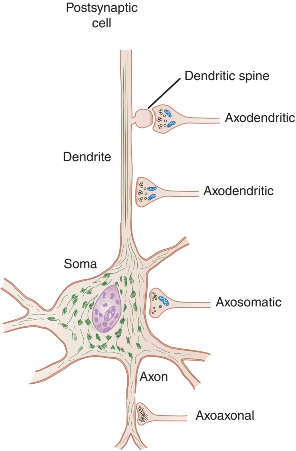
FIGURE 6-2 Axodendritic, axoaxonal, and axosomatic synapses. Many presynaptic neurons terminate on dendritic spines, as shown at the top, but some also end directly on the shafts of dendrites. Note the presence of clear and granulated synaptic vesicles in endings and clustering of clear vesicles at active
zones.
شکل 6-2 سیناپس های آکسودندریتیک، آکسواکسونال و آکسوسوماتیک. بسیاری از نورون های پیش سیناپسی، همانطور که در بالا نشان داده شده است، به خارهای دندریتیک ختم می شوند، اما برخی نیز مستقیماً به شفت دندریت ها ختم می شوند. به حضور وزیکولهای سیناپسی شفاف و دانهدار در انتهای و خوشهبندی وزیکولهای شفاف در زمان فعال توجه کنید.
مناطق
FUNCTIONS OF SYNAPTIC ELEMENTS
The dendrites function as a major site that receives electrical signals from other neurons, processes these signals, and transfers the information to the soma of the neuron. When the dendritic tree of a neuron is extensive and has multiple presynaptic knobs ending on it, there is room for a great interplay of inhibitory and excitatory activity. Dendritic spines can appear, change, and even disappear over a time scale of minutes and hours, not days and months. Also, although protein synthesis occurs mainly in the soma with its nucleus, strands of mRNA migrate into the dendrites. There, each can become associated with a single ribosome in a dendritic spine and produce proteins to alter the effects of input from individual synapses on the spine. These changes in dendritic spines have been implicated in motivation, learning, and long-term memory (see Chapter 15).
توابع عناصر سیناپتیک
دندریت ها به عنوان یک مکان اصلی عمل می کنند که سیگنال های الکتریکی را از نورون های دیگر دریافت می کند، این سیگنال ها را پردازش می کند و اطلاعات را به سومای نورون منتقل می کند. هنگامی که درخت دندریتیک یک نورون گسترده است و دارای چندین دستگیره پیش سیناپسی است که روی آن ختم می شود، فضایی برای تعامل بزرگی از فعالیت مهاری و تحریکی وجود دارد. خارهای دندریتیک می توانند ظاهر شوند، تغییر کنند و حتی در مقیاس زمانی چند دقیقه و ساعت ناپدید شوند، نه روزها و ماه ها. همچنین، اگرچه سنتز پروتئین عمدتاً در سوما با هسته آن اتفاق می افتد، رشته های mRNA به داخل دندریت ها مهاجرت می کنند. در آنجا، هر یک میتوانند با یک ریبوزوم منفرد در ستون فقرات دندریتیک مرتبط شوند و پروتئینهایی تولید کنند تا اثرات ورودی سیناپسهای منفرد روی ستون فقرات را تغییر دهند. این تغییرات در ستون فقرات دندریتیک در انگیزه، یادگیری و حافظه بلند مدت دخیل بوده است (به فصل 15 مراجعه کنید).
Each presynaptic terminal of a chemical synapse is separated from the postsynaptic structure by a synaptic cleft that is 20-40 nm wide. Across the synaptic cleft are many neurotransmitter receptors in the postsynaptic membrane, and usually a postsynaptic thickening called the postsynaptic density (Figure 6-2). The postsynaptic density is an ordered complex of specific receptors, binding proteins, and enzymes induced by postsynaptic effects.
هر پایانه پیش سیناپسی یک سیناپس شیمیایی از ساختار پس سیناپسی توسط یک شکاف سیناپسی به عرض 20-40 نانومتر جدا می شود. در سراسر شکاف سیناپسی تعداد زیادی گیرنده انتقال دهنده عصبی در غشای پس سیناپسی وجود دارد و معمولاً یک ضخیم شدن پس سیناپسی به نام تراکم پس سیناپسی وجود دارد (شکل 6-2). چگالی پس سیناپسی مجموعه ای منظم از گیرنده های خاص، پروتئین های اتصال دهنده و آنزیم های ناشی از اثرات پس سیناپسی است.
Inside the presynaptic terminal are many mitochondria, as well as many membrane-enclosed vesicles, which contain neurotransmitters (see Chapter 7). There are three kinds of synaptic vesicles: small, clear synaptic vesicles contain acetylcholine, glycine, GABA, or glutamate; small dense-core vesicles contain catecholamines; and large dense-core vesicles contain neuropeptides. The vesicles and the proteins contained within them are synthesized in the neuronal cell body and transported along the axon to the endings by fast axoplasmic transport. The neuropeptides in the large dense-core vesicles must also be produced by the protein-synthesizing machinery in the cell body. However, the small clear vesicles and the small dense-core vesicles recycle in the nerve ending (Figure 6-3). These vesicles fuse with the cell membrane and release transmitters through exocytosis and are then recovered by endocytosis to be refilled locally. In some instances, they enter endosomes and are budded off the endosome and refilled, starting the cycle over again. More commonly, however, the synaptic vesicle discharges its contents through a small hole in the cell membrane, then the opening reseals rapidly and the main vesicle stays inside the cell (kiss-and-run discharge). In this way, the full endocytotic process is short- circuited.
در داخل پایانه پیش سیناپسی بسیاری از میتوکندری ها و همچنین وزیکول های محصور در غشاء وجود دارند که حاوی انتقال دهنده های عصبی هستند (به فصل 7 مراجعه کنید). سه نوع وزیکول سیناپسی وجود دارد: وزیکول های سیناپسی کوچک و شفاف حاوی استیل کولین، گلیسین، گابا یا گلوتامات هستند. وزیکول های کوچک با هسته متراکم حاوی کاتکولامین ها هستند. و وزیکول های بزرگ با هسته متراکم حاوی نوروپپتید هستند. وزیکول ها و پروتئین های موجود در آنها در بدن سلول عصبی سنتز می شوند و با انتقال سریع آکسوپلاسمی در امتداد آکسون به انتهای آن منتقل می شوند. نوروپپتیدهای موجود در وزیکول های بزرگ با هسته متراکم نیز باید توسط دستگاه های سنتز پروتئین در بدن سلولی تولید شوند. با این حال، وزیکول های شفاف کوچک و وزیکول های کوچک هسته متراکم در انتهای عصب بازیافت می شوند (شکل 6-3). این وزیکول ها با غشای سلولی ترکیب می شوند و فرستنده ها را از طریق اگزوسیتوز آزاد می کنند و سپس توسط اندوسیتوز بازیابی می شوند تا دوباره به صورت موضعی پر شوند. در برخی موارد، آنها وارد اندوزوم می شوند و از آندوزوم جوانه زده و دوباره پر می شوند و چرخه را دوباره شروع می کنند. با این حال، معمولاً وزیکول سیناپسی محتویات خود را از طریق یک سوراخ کوچک در غشای سلولی تخلیه می کند، سپس دهانه به سرعت بسته می شود و وزیکول اصلی در داخل سلول باقی می ماند (تخلیه بوسه و فرار). به این ترتیب، فرآیند کامل اندوسیتوز اتصال کوتاه می شود.

FIGURE 6-3 Small synaptic vesicle cycle in presynaptic nerve terminals. Vesicles bud off the early endosome and then are filled with a neurotransmitter (NT). The vesicles then translocate toward release sites on the plasma membrane where they dock, and then become primed. Following arrival of an action potential at the nerve ending, Ca2+ influx triggers fusion and exocytosis of the vesicular contents into the synaptic cleft. The vesicle wall then is coated with clathrin, which likely facilitates endocytosis. Once back inside the cytoplasm, the vesicle becomes uncoated and fuses with the early endosome to start the cycle again.
شکل 6-3 چرخه وزیکول سیناپسی کوچک در پایانه های عصبی پیش سیناپسی. وزیکول ها از اندوزوم اولیه جوانه می زنند و سپس با یک انتقال دهنده عصبی (NT) پر می شوند. سپس وزیکولها به سمت محلهای رهاسازی در غشای پلاسمایی که در آن لنگر میافتند، جابهجا میشوند و سپس آماده میشوند. پس از رسیدن پتانسیل عمل به انتهای عصب، هجوم +Ca2 باعث همجوشی و اگزوسیتوز محتویات وزیکولی در شکاف سیناپسی می شود. سپس دیواره وزیکول با کلاترین پوشانده می شود که احتمالاً اندوسیتوز را تسهیل می کند. پس از بازگشت به داخل سیتوپلاسم، وزیکول بدون پوشش می شود و با اندوزوم اولیه ترکیب می شود تا چرخه دوباره شروع شود.
The large dense-core vesicles are found throughout some presynaptic nerve terminals and release their neuropeptide contents by exocytosis from all parts of the terminal. On the other hand, the small vesicles are located near the synaptic cleft and fuse to the membrane, discharging their contents very rapidly into the cleft at areas of membrane thickening called active zones (Figure 6-2). Voltage-gated Ca2+ channels are very close to the release sites at the active zones.
وزیکول های بزرگ با هسته متراکم در برخی از پایانه های عصبی پیش سیناپسی یافت می شوند و محتوای نوروپپتیدی خود را با اگزوسیتوز از تمام قسمت های انتهایی آزاد می کنند. از طرف دیگر، وزیکول های کوچک در نزدیکی شکاف سیناپسی قرار گرفته و به غشاء جوش می خورند، و محتویات خود را به سرعت در قسمت های ضخیم شدن غشاء به نام مناطق فعال به داخل شکاف تخلیه می کنند (شکل 6-2). کانال های +Ca2 دارای ولتاژ بسیار نزدیک به محل انتشار در مناطق فعال هستند.
The Ca2+ that triggers exocytosis of transmitters enters the presynaptic neurons, and transmitter release starts within 200 μs. In addition, the transmitter must be released close to the postsynaptic receptors to be effective on the postsynaptic neuron. This orderly organization of the synapse depends in part on neurexins, presynaptic cell-adhesion molecules that bind to neuroligins on the membrane of postsynaptic neurons. Neurexin-neuroligin interactions not only hold synapses together, but they also provide a mechanism for the production of synaptic specificity.
+Ca2 که باعث اگزوسیتوز فرستنده ها می شود وارد نورون های پیش سیناپسی می شود و آزادسازی فرستنده در عرض 200 میکرو ثانیه شروع می شود. علاوه بر این، فرستنده باید در نزدیکی گیرنده های پس سیناپسی آزاد شود تا روی نورون پس سیناپسی موثر باشد. این سازماندهی منظم سیناپس تا حدی به نورکسین ها، مولکول های چسبنده سلولی پیش سیناپسی که به نورولیژین های روی غشای نورون های پس سیناپسی متصل می شوند، بستگی دارد. فعل و انفعالات Neurexin-neuroligin نه تنها سیناپس ها را کنار هم نگه می دارد، بلکه مکانیسمی را برای تولید ویژگی سیناپسی فراهم می کند.
As noted in Chapter 2, vesicle budding, fusion, and discharge of contents with subsequent retrieval of vesicle membrane are fundamental processes occurring in most, if not all, cells. Thus, neurotransmitter secretion at synapses and the accompanying membrane retrieval are specialized forms of the general processes of exocytosis and endocytosis. The fusion of synaptic vesicles with the cell membrane involves the action of several proteins, including the N- ethylmaleimide-sensitive fusion protein (NSF), SNAPS (soluble NSF attachment proteins), and SNAREs (SNAP receptors; Figure 6-4). Synaptobrevin in the vesicle membrane locks with syntaxin and SNAPs in the cell membrane; GTPases regulate a multiprotein complex that includes Rab and Sec1/Munc18- like proteins as part of the fusion process. The one-way gate at the synapses is necessary for orderly neural function.
همانطور که در فصل 2 اشاره شد، جوانه زدن وزیکول، همجوشی، و تخلیه محتویات با بازیابی بعدی غشای وزیکول فرآیندهای اساسی هستند که در اکثر سلولها اتفاق میافتند. بنابراین، ترشح ناقل عصبی در سیناپس ها و بازیابی غشای همراه، اشکال تخصصی فرآیندهای کلی اگزوسیتوز و اندوسیتوز هستند. ادغام وزیکول های سیناپسی با غشای سلولی شامل عمل چندین پروتئین، از جمله پروتئین همجوشی حساس به N- اتیل مالیمید (NSF)، SNAPS (پروتئین های اتصال محلول NSF) و SNARE ها (گیرنده های SNAP؛ شکل 6-4) است. Synaptobrevin در غشای وزیکول با syntaxin و SNAPs در غشای سلول قفل می شود. GTPase ها یک مجموعه چند پروتئینی را تنظیم می کنند که شامل پروتئین های Rab و Sec1/Munc18 مانند به عنوان بخشی از فرآیند همجوشی است. دروازه یک طرفه در سیناپس ها برای عملکرد منظم عصبی ضروری است.

FIGURE 6-4 Main proteins that interact to produce synaptic vesicle docking and fusion in nerve endings. Several proteins are involved in synaptic vesicle docking and fusion, including the N-ethylmaleimide-sensitive fusion protein (NSF), SNAPS (soluble NSF attachment proteins), and SNAREs (SNAP receptors). Synaptobrevin in the vesicle membrane links with syntaxin and SNAPS in the cell membrane; GTPases regulate a multiprotein complex that includes Rab and Sec1/Munc18-like proteins.
شکل 6-4 پروتئین های اصلی که برای ایجاد اتصال وزیکول سیناپسی و همجوشی در انتهای عصبی با هم تعامل دارند. چندین پروتئین در اتصال و همجوشی وزیکول سیناپسی، از جمله پروتئین فیوژن حساس به N-ethylmaleimide (NSF)، SNAPS (پروتئین های اتصال محلول NSF)، و SNAREs (گیرنده های SNAP) نقش دارند. Synaptobrevin در غشای وزیکول با syntaxin و SNAPS در غشای سلولی پیوند دارد. GTPase ها یک مجموعه چند پروتئینی را تنظیم می کنند که شامل پروتئین های Rab و Sec1/Munc18 است.
Several deadly toxins that block neurotransmitter release are zinc endopeptidases that cleave and hence inactivate proteins in the fusion-exocytosis complex. Clinical Box 6-1 describes how neurotoxins from bacteria called Clostridium tetani and Clostridium botulinum can disrupt neurotransmitter release in either the central nervous system (CNS) or at the neuromuscular junction.
چندین سم کشنده که آزادسازی انتقال دهنده های عصبی را مسدود می کنند اندوپپتیدازهای روی هستند که پروتئین ها را در مجموعه همجوشی-اگزوسیتوز شکسته و غیرفعال می کنند. جعبه بالینی 6-1 توضیح می دهد که چگونه نوروتوکسین های باکتری به نام کلستریدیوم تتانی و کلستریدیوم بوتولینوم می توانند آزادسازی انتقال دهنده های عصبی را در سیستم عصبی مرکزی (CNS) یا در اتصال عصبی عضلانی مختل کنند.
CLINICAL BOXC 6.1
Botulinum and Tetanus Toxins
Clostridia are gram-positive bacteria. Two varieties, Clostridium tetani and Clostridium botulinum, produce some of the most potent biologic toxins (tetanus toxin and botulinum toxin) known to affect humans. These neurotoxins act by preventing the release of neurotransmitters in the CNS and at the neuromuscular junction. Tetanus toxin binds irreversibly to the presynaptic membrane of the neuromuscular junction and uses retrograde axonal transport to travel to the cell body of the motor neuron in the spinal cord. From there it is picked up by the terminals of presynaptic inhibitory interneurons. The toxin attaches to gangliosides in these terminals and blocks the release of glycine and GABA. As a result, the activity of motor neurons is markedly increased. Clinically, tetanus toxin causes spastic paralysis; the characteristic symptom of “lockjaw” is spasms of the masseter muscle. Botulism can result from ingestion of contaminated food, colonization of the gastrointestinal tract in an infant, or wound infection. Botulinum toxins are a family of seven neurotoxins, but it is mainly botulinum toxins A, B, and E that are toxic to humans. Botulinum toxins A and E cleave synaptosome- associated protein-25 (SNAP-25). This is a presynaptic membrane protein needed for fusion of synaptic vesicles containing acetylcholine to the terminal membrane, an important step in transmitter release. Botulinum toxin B cleaves synaptobrevin, a vesicle-associated membrane protein (VAMP). By blocking acetylcholine release at the neuromuscular junction, these toxins cause flaccid paralysis. Symptoms can include ptosis (drooping eyelid), diplopia (double vision), dysarthria (slurred speech), dysphonia (difficulty speaking), and dysphagia (difficulty swallowing).
جعبه بالینی 6.1
سموم بوتولینوم و کزاز
کلستریدیا باکتری های گرم مثبت هستند. دو گونه، کلستریدیوم تتانی و کلستریدیوم بوتولینوم، برخی از قویترین سموم بیولوژیک (سم کزاز و سم بوتولینوم) را تولید میکنند که بر انسانها تأثیر میگذارند. این نوروتوکسین ها با جلوگیری از آزاد شدن انتقال دهنده های عصبی در CNS و در اتصال عصبی عضلانی عمل می کنند. سم کزاز به طور برگشت ناپذیری به غشای پیش سیناپسی اتصال عصبی عضلانی متصل می شود و از انتقال آکسونی رتروگراد برای حرکت به بدنه سلولی نورون حرکتی در نخاع استفاده می کند. از آنجا توسط پایانه های نورون های بازدارنده پیش سیناپسی برداشت می شود. این سم به گانگلیوزیدها در این پایانه ها می چسبد و از آزاد شدن گلیسین و گابا جلوگیری می کند. در نتیجه، فعالیت نورون های حرکتی به طور قابل توجهی افزایش می یابد. از نظر بالینی، سم کزاز باعث فلج اسپاستیک می شود. علامت مشخصه “فک قفل” اسپاسم عضله ماستر است. بوتولیسم می تواند ناشی از خوردن غذای آلوده، کلونیزاسیون دستگاه گوارش در نوزاد یا عفونت زخم باشد. سموم بوتولینوم خانواده ای از هفت نوروتوکسین هستند، اما عمدتاً سموم بوتولینوم A، B و E هستند که برای انسان سمی هستند. سموم بوتولینوم A و E پروتئین 25 مرتبط با سیناپتوزوم (SNAP-25) را می شکافند. این یک پروتئین غشای پیش سیناپسی است که برای ادغام وزیکول های سیناپسی حاوی استیل کولین به غشای انتهایی مورد نیاز است، که یک مرحله مهم در آزادسازی فرستنده است. سم بوتولینوم B سیناپتوبروین، یک پروتئین غشایی مرتبط با وزیکول (VAMP) را می شکافد. این سموم با مسدود کردن آزادسازی استیل کولین در محل اتصال عصبی عضلانی، باعث فلج شل می شوند. علائم می تواند شامل پتوز (افتادگی پلک)، دوبینی (دوبینی)، دیس آرتری (تکلم نامفهوم)، دیسفونی (مشکل در صحبت کردن) و دیسفاژی (مشکل در بلع) باشد.
THERAPEUTIC HIGHLIGHTS
Tetanus can be prevented by treatment with tetanus toxoid vaccine. The widespread use of this vaccine in the United States beginning in the mid-1940s led to a marked decline in the incidence of tetanus toxicity. The incidence of botulinum toxicity is also low (about 100 cases per year in the United States), but in those individuals who are affected, the fatality rate is 5-10%. An antitoxin is available for treatment, and those who are at risk for respiratory failure are placed on a ventilator. On the positive side, local injection of small doses of botulinum toxin (botox) has proven to be effective in the treatment of a wide variety of conditions characterized by muscle hyperactivity. Examples include injection into the lower esophageal sphincter to relieve achalasia, injection into facial muscles to remove wrinkles, and injection into leg muscles to reduce spasticity in individuals with cerebral palsy.
نکات برجسته درمانی
با درمان با واکسن توکسوئید کزاز می توان از کزاز پیشگیری کرد. استفاده گسترده از این واکسن در ایالات متحده در اواسط دهه 1940 منجر به کاهش قابل توجهی در بروز سمیت کزاز شد. بروز سمیت بوتولینوم نیز کم است (حدود 100 مورد در سال در ایالات متحده)، اما در آن دسته از افراد مبتلا، میزان مرگ و میر 5-10٪ است. یک آنتی توکسین برای درمان در دسترس است و کسانی که در معرض خطر نارسایی تنفسی هستند روی دستگاه تنفس مصنوعی قرار می گیرند. از جنبه مثبت، تزریق موضعی دوزهای کوچک سم بوتولینوم (بوتاکس) ثابت کرده است که در درمان طیف گسترده ای از شرایط که با بیش فعالی عضلانی مشخص می شود، موثر است. به عنوان مثال می توان به تزریق در اسفنکتر تحتانی مری برای تسکین آشالازی، تزریق به عضلات صورت برای رفع چین و چروک و تزریق به عضلات پا برای کاهش اسپاستیسیتی در افراد مبتلا به فلج مغزی اشاره کرد.
ELECTRICAL EVENTS IN POSTSYNAPTIC NEURONS
EXCITATORY & INHIBITORY POSTSYNAPTIC POTENTIALS
Stimulation of a dorsal root afferent (sensory neuron) can be used to study both excitatory and inhibitory events in a-motor neurons (Figure 6-5). Once an impulse reaches the presynaptic terminals of the sensory neuron, a response can be obtained in the postsynaptic neuron after a synaptic delay. The delay is due to the time it takes for the synaptic mediator to be released and to act on the receptors on the membrane of the postsynaptic neuron. Because of it, conduction along a chain of neurons is slower if there are many synapses compared to if there are only a few synapses. Because the minimum time for transmission across one synapse is 0.5 ms, it is possible to determine whether a given reflex pathway is monosynaptic or polysynaptic (contains more than one synapse) by measuring the synaptic delay.
رویدادهای الکتریکی در نورونهای پسسیناپتیک
پتانسیل های پس سیناپتیکی تحریکی و بازدارنده
تحریک یک آوران ریشه پشتی (نرون حسی) می تواند برای مطالعه رویدادهای تحریکی و مهاری در نورون های a-motor مورد استفاده قرار گیرد (شکل 6-5). هنگامی که یک تکانه به پایانه های پیش سیناپسی نورون حسی می رسد، پس از تاخیر سیناپسی می توان پاسخی در نورون پس سیناپسی به دست آورد. این تأخیر به دلیل زمانی است که واسطه سیناپسی آزاد می شود و روی گیرنده های روی غشای نورون پس سیناپسی اثر می گذارد. به همین دلیل، اگر سیناپسهای زیادی وجود داشته باشد، هدایت در طول زنجیرهای از نورونها در مقایسه با تعداد سیناپسهای کمی کندتر است. از آنجایی که حداقل زمان انتقال در یک سیناپس 0.5 میلی ثانیه است، می توان با اندازه گیری تأخیر سیناپسی تعیین کرد که آیا یک مسیر رفلکس معین تک سیناپسی است یا پلی سیناپسی (شامل بیش از یک سیناپس).


FIGURE 6-5 Excitatory and inhibitory synaptic connections mediating the stretch reflex provide an example of typical circuits within the CNS. A) The stretch receptor sensory neuron of the quadriceps muscle makes an excitatory connection with the extensor motor neuron of the same muscle and an inhibitory interneuron projecting to flexor motor neurons supplying the antagonistic hamstring muscle. B) Experimental setup to study excitation and inhibition of the extensor motor neuron. Top panel shows two approaches to elicit an excitatory (depolarizing) postsynaptic potential or EPSP in the extensor motor neuron-electrical stimulation of the whole Ia afferent nerve using extracellular electrodes and intracellular current passing through an electrode inserted into the cell body of a sensory neuron. Bottom panel shows that current passing through an inhibitory interneuron elicits an inhibitory (hyperpolarizing) postsynaptic potential or IPSP in the flexor motor neuron. EPSP, excitatory postsynaptic potential; IPSP, inhibitory postsynaptic potential. (Reproduced with permission from Kandel ER, Schwartz JH, Jessell TM [editors]: Principles of Neural Science, 4th ed. New York, NY: McGraw-Hill; 2000.)
شکل 6-5 اتصالات سیناپسی تحریکی و بازدارنده با واسطه رفلکس کششی نمونه ای از مدارهای معمولی در CNS را ارائه می دهند. الف) نورون حسی گیرنده کششی عضله چهارسر ران با نورون حرکتی اکستانسور همان عضله و یک نورون بازدارنده که به نورون های حرکتی خم کننده ای که عضله همسترینگ آنتاگونیست را تامین می کند، ارتباط تحریکی ایجاد می کند. ب) تنظیم تجربی برای مطالعه تحریک و مهار نورون حرکتی اکستانسور. پانل بالایی دو رویکرد را برای برانگیختن پتانسیل پس سیناپسی تحریکی (دپولاریزاسیون) یا EPSP در تحریک الکتریکی نورون حرکتی اکستانسوری کل عصب آوران Ia با استفاده از الکترودهای خارج سلولی و جریان درون سلولی که از الکترود وارد شده در بدنه سلولی یک نورون حسی عبور می کند، نشان می دهد. پانل پایین نشان می دهد که جریان عبوری از یک نورون بازدارنده باعث ایجاد پتانسیل پس سیناپسی مهاری (هیپرپلاریزه کننده) یا IPSP در نورون حرکتی فلکسور می شود. EPSP، پتانسیل پس سیناپسی تحریکی. IPSP، پتانسیل پس سیناپسی بازدارنده. (تکثیر شده با اجازه از Kandel ER، Schwartz JH، Jessell TM [ویراستاران]: Principles of Neural Science، ویرایش چهارم نیویورک، نیویورک: McGraw-Hill؛ 2000.)
A single stimulus applied to the sensory nerves characteristically does not lead to the formation of a propagated action potential in the postsynaptic neuron. Instead, the stimulation produces either a transient partial depolarization or a transient hyperpolarization. The initial depolarizing response produced by a single stimulus to the proper input begins about 0.5 ms after the afferent impulse enters the spinal cord. It reaches its peak 1-1.5 ms later and then declines exponentially (Figure 6-5). During this potential, the excitability of the neuron to other stimuli is increased; thus, it is called a fast excitatory postsynaptic potential (EPSP).
یک محرک منفرد که روی اعصاب حسی اعمال می شود، مشخصاً منجر به تشکیل پتانسیل عمل انتشار یافته در نورون پس سیناپسی نمی شود. در عوض، تحریک یا یک دپلاریزاسیون جزئی گذرا یا یک هیپرپلاریزاسیون گذرا ایجاد می کند. پاسخ دپلاریزاسیون اولیه تولید شده توسط یک محرک به ورودی مناسب حدود 0.5 میلی ثانیه پس از ورود تکانه آوران به نخاع آغاز می شود. 1-1.5 ms بعد به اوج خود می رسد و سپس به طور تصاعدی کاهش می یابد (شکل 6-5). در طول این پتانسیل، تحریک پذیری نورون به سایر محرک ها افزایش می یابد. بنابراین، آن را پتانسیل پس سیناپسی تحریکی سریع (EPSP) می نامند.
The fast EPSP is produced by depolarization of the postsynaptic cell membrane immediately under the presynaptic ending. The excitatory transmitter opens Na+ or Ca2+ channels in the postsynaptic membrane, producing an inward current. The area of current flow thus created is so small that it does not drain off enough positive charge to depolarize the whole membrane. Instead, an EPSP is induced. The EPSP due to activity in one synaptic knob is small, but the depolarizations produced by each of the active knobs summate.
EPSP سریع با دپلاریزاسیون غشای سلولی پس سیناپسی بلافاصله در زیر انتهای پیش سیناپسی تولید می شود. فرستنده تحریکی کانال های +Na یا +Ca2 را در غشای پس سیناپسی باز می کند و جریانی به سمت داخل تولید می کند. ناحیه جریان جریان ایجاد شده به قدری کوچک است که بار مثبت کافی برای دپلاریزاسیون کل غشا تخلیه نمی کند. در عوض، یک EPSP القا می شود. EPSP به دلیل فعالیت در یک دستگیره سیناپسی کوچک است، اما دپلاریزاسیون های تولید شده توسط هر یک از دستگیره های فعال جمع می شود.
EPSPs are produced by stimulation of some inputs, but stimulation of other inputs produces hyperpolarizing responses. Like the EPSPs, they peak 1-1.5 ms after the stimulus and decrease exponentially (Figure 6-5). During this potential, the excitability of the neuron to other stimuli is decreased; thus, it is called a fast inhibitory postsynaptic potential (IPSP).
EPSP ها با تحریک برخی ورودی ها تولید می شوند، اما تحریک سایر ورودی ها پاسخ های هیپرپلاریزه کننده ایجاد می کند. مانند EPSP ها، آنها 1-1.5 میلی ثانیه پس از محرک به اوج خود می رسند و به طور تصاعدی کاهش می یابند (شکل 6-5). در طول این پتانسیل، تحریک پذیری نورون به سایر محرک ها کاهش می یابد. بنابراین، آن را یک پتانسیل پس سیناپسی مهاری سریع (IPSP) می نامند.
A fast IPSP can be produced by a localized increase in Cl transport. When an inhibitory synaptic knob becomes active, the released neurotransmitter triggers the opening of Cl ̄ channels in the area of the postsynaptic cell membrane under the knob. Cl moves down its concentration gradient. The net effect is the transfer of negative charge into the cell, increasing the membrane potential.
یک IPSP سریع را می توان با افزایش موضعی در انتقال کلر تولید کرد. هنگامی که یک دستگیره سیناپسی بازدارنده فعال می شود، انتقال دهنده عصبی آزاد شده باعث باز شدن کانال های ̄Cl در ناحیه غشای سلولی پس سیناپسی در زیر دستگیره می شود. کلر به سمت پایین گرادیان غلظت حرکت می کند. اثر خالص انتقال بار منفی به سلول است که پتانسیل غشاء را افزایش می دهد.
The decreased excitability of the nerve cell during the IPSP is due to movement of the membrane potential away from the firing level. Consequently, more excitatory (depolarizing) activity is necessary to reach the firing level. The fact that an IPSP is mediated by Cl ̄ can be demonstrated by repeating the stimulus while varying the resting membrane potential of the postsynaptic cell. When the membrane potential is at the equilibrium potential for chloride (Ecı), the postsynaptic potential disappears (Figure 6-6), and at more negative membrane potentials, it becomes positive (reversal potential).
کاهش تحریک پذیری سلول عصبی در طول IPSP به دلیل حرکت پتانسیل غشاء به دور از سطح شلیک است. در نتیجه، فعالیت تحریکی (دپلاریزاسیون) بیشتری برای رسیدن به سطح شلیک ضروری است. این واقعیت که IPSP توسط Cl ̄ واسطه می شود را می توان با تکرار محرک در حالی که پتانسیل غشای استراحت سلول پس سیناپسی را تغییر داد، نشان داد. هنگامی که پتانسیل غشاء در پتانسیل تعادل برای کلرید (Ecı) باشد، پتانسیل پس سیناپسی ناپدید می شود (شکل 6-6)، و در پتانسیل های غشایی منفی تر، مثبت می شود (پتانسیل معکوس).
Because IPSPs are net hyperpolarizations, they can be produced by alterations in other ion channels in the neuron. For example, they can be produced by opening K+ channels, with movement of K+ out of the postsynaptic cell, or by closure of Na+ or Ca2+ channels.
از آنجایی که IPSP ها ابرقطبی خالص هستند، می توانند با تغییرات در کانال های یونی دیگر در نورون تولید شوند. به عنوان مثال، آنها را می توان با باز کردن کانال های +K، با حرکت +K به خارج از سلول پس سیناپسی، یا با بستن کانال های +Na یا +Ca2 تولید کرد.

FIGURE 6-6 IPSP is due to increased Cl influx during stimulation. This can be demonstrated by repeating the stimulus while varying the resting membrane potential (RMP) of the postsynaptic cell. When the membrane potential is at Eci, the potential disappears, and at more negative membrane potentials (eg, Ek and below), it becomes positive (reversal potential).
شکل 6-6 IPSP به دلیل افزایش هجوم کلر در طول تحریک است. این را می توان با تکرار محرک در حین تغییر پتانسیل غشای استراحت (RMP) سلول پس سیناپسی نشان داد. وقتی پتانسیل غشاء در Eci باشد، پتانسیل ناپدید می شود و در پتانسیل های غشایی منفی تر (مثلاً Ek و پایین تر)، مثبت می شود (پتانسیل معکوس).
SLOW POSTSYNAPTIC POTENTIALS
In addition to the fast EPSPs and IPSPs, slow EPSPs and IPSPs occur in autonomic ganglia, cardiac and smooth muscle, and cortical neurons. These postsynaptic potentials have a latency of 100-500 ms and last several seconds. The slow EPSPs are generally due to decreases in K* conductance, and the slow IPSPs are due to increases in K+ conductance.
پتانسیل های آهسته پسا سیناپتیک
علاوه بر EPSPها و IPSPهای سریع، EPSPها و IPSPهای آهسته در گانگلیونهای اتونوم، عضلات صاف و قلبی و نورونهای قشر مغز رخ می دهند. این پتانسیل های پس سیناپسی دارای تأخیر 100-500 میلی ثانیه بوده و چند ثانیه طول می کشد. EPSPهای کند عموماً به دلیل کاهش رسانایی +K و IPSPهای کند به دلیل افزایش رسانایی+K هستند.
ELECTRICAL TRANSMISSION
In electrical synapses, the membranes of the presynaptic and postsynaptic neurons come close together, and gap junctions are formed between the cells (see Chapter 2). Like the intercellular junctions in other tissues, these junctions form low-resistance bridges through which ions can pass with relative ease. At these junctions the impulse reaching the presynaptic terminal generates an EPSP in the postsynaptic cell that, because of the low-resistance bridge between the two, has a much shorter latency than the EPSP at a synapse where transmission is chemical.
انتقال برق
در سیناپس های الکتریکی، غشای نورون های پیش سیناپسی و پس سیناپسی به هم نزدیک می شوند و اتصالات شکافی بین سلول ها ایجاد می شود (به فصل 2 مراجعه کنید). مانند اتصالات بین سلولی در سایر بافت ها، این اتصالات پل هایی با مقاومت کم را تشکیل می دهند که یون ها می توانند با سهولت نسبی از آن عبور کنند. در این اتصالات، ضربه ای که به پایانه پیش سیناپسی می رسد، یک EPSP را در سلول پس سیناپسی ایجاد می کند که به دلیل پل کم مقاومت بین این دو، تاخیر بسیار کوتاه تری نسبت به EPSP در سیناپسی دارد که در آن انتقال شیمیایی است.
GENERATION OF AN ACTION POTENTIAL IN THE POSTSYNAPTIC NEURON
The constant interplay of excitatory and inhibitory activity on the postsynaptic neuron produces a fluctuating membrane potential that is the algebraic summation of the hyperpolarizing and depolarizing potentials. The soma of the neuron thus acts as an integrator. When the level of depolarization reaches the threshold voltage, a propagated action potential will occur. However, the discharge of the neuron is slightly more complicated than this. In motor neurons, the portion of the cell with the lowest threshold for the production of an action potential is the initial segment, the portion of the axon at and just beyond the axon hillock (see Chapter 4). This unmyelinated segment is depolarized or hyperpolarized electrotonically by the current sinks and sources under the excitatory and inhibitory synaptic knobs. It is the first part of the neuron to fire, and its discharge is propagated in two directions: down the axon and back into the soma. Retrograde firing of the soma in this fashion probably has value in wiping the slate clean for subsequent renewal of the interplay of excitatory and inhibitory activity on the cell.
تولید یک پتانسیل عمل در نورون پس از سیناپتیک
فعل و انفعال مداوم فعالیت تحریکی و بازدارنده بر روی نورون پس سیناپسی یک پتانسیل غشایی نوسانی ایجاد می کند که جمع جبری پتانسیل هایپرپولاریزاسیون و دپلاریزاسیون است. بنابراین سومای نورون به عنوان یکپارچه کننده عمل می کند. هنگامی که سطح دپلاریزاسیون به ولتاژ آستانه می رسد، پتانسیل عمل انتشار رخ می دهد. با این حال، تخلیه نورون کمی پیچیده تر از این است. در نورون های حرکتی، بخشی از سلول با کمترین آستانه برای تولید پتانسیل عمل، بخش اولیه است، بخشی از آکسون در و درست فراتر از تپه آکسون (به فصل 4 مراجعه کنید). این بخش بدون میلین به صورت الکتروتونیکی توسط سینک های جریان و منابع زیر دستگیره های سیناپسی تحریکی و بازدارنده دپلاریزه یا هیپرپلاریزه می شود. این اولین قسمت از نورون است که شلیک می شود و تخلیه آن در دو جهت منتشر می شود: پایین آکسون و بازگشت به سوما. شلیک رتروگراد سوما به این روش احتمالاً در پاک کردن تخته سنگ برای تجدید متقابل فعالیت تحریکی و مهاری روی سلول ارزش دارد.
TEMPORAL & SPATIAL SUMMATION OF POSTSYNAPTIC POTENTIALS
Two passive membrane properties of a neuron affect the ability of postsynaptic potentials to summate to elicit an action potential (Figure 6-7). The time constant of a neuron determines the time course of the synaptic potential, and the length constant of a neuron determines the degree to which a depolarizing current is reduced as it spreads passively. Figure 6-7 also shows how the time constant of the postsynaptic neuron can affect the amplitude of the depolarization caused by consecutive EPSPs produced by a single presynaptic neuron. The longer the time constant, the greater is the chance for two potentials to summate to induce an action potential. If a second EPSP is elicited before the first EPSP decays, the two potentials summate and, as in this example, their additive effects are sufficient to induce an action potential in the postsynaptic neuron (temporal summation). Figure 6-7 also shows how the length constant of a postsynaptic neuron can affect the amplitude of two EPSPs produced by different presynaptic neurons in a process called spatial summation. If a neuron has a long length constant, the membrane depolarization induced by input arriving at two points on the neuron can spread to the trigger zone of the neuron with minimal decrement. The two potentials can summate and induce an action potential.
جمع زمانی و مکانی پتانسیل های پسا سیناپتیکی
دو ویژگی غشای غیرفعال یک نورون بر توانایی پتانسیل های پس سیناپسی برای جمع شدن برای استخراج پتانسیل عمل تأثیر می گذارد (شکل 6-7). ثابت زمانی یک نورون مسیر زمانی پتانسیل سیناپسی را تعیین می کند و ثابت طول یک نورون درجه کاهش یک جریان دپلاریز کننده را هنگام انتشار غیرفعال تعیین می کند. شکل 6-7 همچنین نشان می دهد که چگونه ثابت زمانی نورون پس سیناپسی می تواند بر دامنه دپلاریزاسیون ناشی از EPSP های متوالی تولید شده توسط یک نورون پیش سیناپسی تأثیر بگذارد. هرچه ثابت زمانی طولانیتر باشد، شانس دو پتانسیل برای ایجاد پتانسیل عمل بیشتر است. اگر یک EPSP دوم قبل از فروپاشی EPSP اول استخراج شود، دو پتانسیل جمع می شوند و، مانند این مثال، اثرات افزایشی آنها برای القای پتانسیل عمل در نورون پس سیناپسی (جمع زمانی) کافی است. شکل 6-7 همچنین نشان می دهد که چگونه ثابت طول یک نورون پس سیناپسی می تواند بر دامنه دو EPSP تولید شده توسط نورون های پیش سیناپسی مختلف در فرآیندی به نام جمع فضایی تأثیر بگذارد. اگر یک نورون ثابت طول طولانی داشته باشد، دپلاریزاسیون غشایی ناشی از ورودی به دو نقطه روی نورون میتواند با حداقل کاهش به ناحیه ماشهای نورون گسترش یابد. این دو پتانسیل می توانند یک پتانسیل عمل را جمع و القا کنند.

FIGURE 6-7 Central neurons integrate a variety of synaptic inputs through temporal and spatial summation. A) The time constant of the postsynaptic neuron affects the amplitude of the depolarization caused by consecutive EPSPs produced by a single presynaptic neuron. In cases of a long time constant, if a second EPSP is elicited before the first EPSP decays, the two potentials summate to induce an action potential. B) The length constant of a postsynaptic cell affects the amplitude of two EPSPs produced by two presynaptic neurons, A andB. If the length constant is long, the depolarization induced at two points on the neuron can spread to the trigger zone with minimal decrement so that the two potentials summate and an action potential is elicited. EPSP, excitatory postsynaptic potential. (Reproduced with permission from Kandel ER, Schwartz JH, Jessell TM [editors]: Principles of Neural Science, 4th ed. New York, NY: McGraw-Hill; 2000.)
شکل 6-7 نورون های مرکزی انواع ورودی های سیناپسی را از طریق جمع زمانی و مکانی ادغام می کنند. الف) ثابت زمانی نورون پس سیناپسی بر دامنه دپلاریزاسیون ناشی از EPSPهای متوالی تولید شده توسط یک نورون پیش سیناپسی تأثیر می گذارد. در موارد ثابت زمانی طولانی، اگر یک EPSP دوم قبل از فروپاشی EPSP اول استخراج شود، این دو پتانسیل جمع می شوند تا پتانسیل عمل را القا کنند. ب) ثابت طول یک سلول پس سیناپسی بر دامنه دو EPSP تولید شده توسط دو نورون پیش سیناپسی A وB تأثیر می گذارد. اگر ثابت طول طولانی باشد، دپلاریزاسیون القا شده در دو نقطه روی نورون میتواند با کمترین کاهش به ناحیه ماشه گسترش یابد تا دو پتانسیل جمع شوند و پتانسیل عمل ایجاد شود. EPSP، پتانسیل پس سیناپسی تحریکی. (تکثیر شده با اجازه از Kandel ER، Schwartz JH، Jessell TM [ویراستاران]: Principles of Neural Science، ویرایش چهارم نیویورک، نیویورک: McGraw-Hill؛ 2000.)
INHIBITION & FACILITATION AT SYNAPSES
Inhibition within the CNS can be either postsynaptic or presynaptic. Examples of the neuronal connections that can mediate presynaptic inhibition and postsynaptic inhibition are compared in Figure 6-8. Postsynaptic inhibition during the course of an IPSP is called direct inhibition because it is not a consequence of previous discharges of the postsynaptic neuron. There are various forms of indirect inhibition that is due to the effects of previous postsynaptic neuron discharge. For example, the postsynaptic cell can be refractory to excitation because it has just fired and is in its refractory period. During after-hyperpolarization it is also less excitable. In spinal neurons, especially after repeated firing, this after- hyperpolarization may be large and prolonged.
مهار و تسهیل در سیناپس
مهار در CNS می تواند پس سیناپسی یا پیش سیناپسی باشد. نمونه هایی از اتصالات عصبی که می توانند واسطه مهار پیش سیناپسی و مهار پس سیناپسی باشند در شکل 6-8 مقایسه شده اند. مهار پس سیناپسی در طول دوره IPSP، بازداری مستقیم نامیده می شود زیرا نتیجه تخلیه قبلی نورون پس سیناپسی نیست. اشکال مختلفی از مهار غیرمستقیم وجود دارد که به دلیل اثرات تخلیه نورون پس سیناپسی قبلی است. به عنوان مثال، سلول پس سیناپسی می تواند در برابر تحریک مقاوم باشد زیرا به تازگی شلیک شده و در دوره نسوز خود قرار دارد. در طول پس از هایپرپلاریزاسیون نیز کمتر تحریک پذیر است. در نورون های نخاعی، به ویژه پس از شلیک مکرر، این هیپرپلاریزاسیون ممکن است بزرگ و طولانی باشد.

FIGURE 6-8 Comparison of neuronal connections that can produce presynaptic and postsynaptic inhibition. A) Presynaptic inhibition is a process mediated by neurons whose terminals are on excitatory nerve endings, forming axoaxonal synapses and reducing transmitter release from the excitatory neuron. B) Postsynaptic inhibition occurs when an inhibitory transmitter such as GABA is released from the nerve terminals of an inhibitory interneuron (dark) that synapses on a postsynaptic neuron.
شکل 6-8 مقایسه اتصالات عصبی که می توانند مهار پیش سیناپسی و پس سیناپسی ایجاد کنند. الف) بازداری پیش سیناپسی فرآیندی است که توسط نورون هایی انجام می شود که پایانه های آنها روی انتهای عصب تحریکی قرار دارند و سیناپس های آکسواکسونال را تشکیل می دهند و آزادسازی فرستنده از نورون تحریکی را کاهش می دهند. ب) بازداری پس سیناپسی زمانی رخ می دهد که یک فرستنده مهاری مانند GABA از پایانه های عصبی یک نورون بازدارنده (تاریک) که روی یک نورون پس سیناپسی سیناپس می شود، آزاد شود.
POSTSYNAPTIC INHIBITION
Postsynaptic inhibition occurs when an inhibitory transmitter such as glycine or GABA is released from a presynaptic nerve terminal onto the postsynaptic neuron to induce an IPSP in the postsynaptic neuron (Figure 6-8B). Various pathways in the nervous system mediate postsynaptic inhibition, and one illustrative example is presented in Figure 6-5. Afferent fibers from the muscle spindles (stretch receptors) in skeletal muscle project directly to the spinal motor neurons supplying the same muscle. Impulses in this afferent fiber cause an EPSP and, with summation, propagated action potentials in the postsynaptic motor neuron. At the same time, an IPSP is produced in motor neurons supplying the antagonistic muscle, which has an inhibitory interneuron interposed between the afferent fiber and the motor neuron. Therefore, activity in the afferent fibers from the muscle spindles excites the motor neurons supplying the muscle from which the impulses come and inhibits the motor neurons supplying its antagonists (reciprocal innervation). These reflexes are considered in more detail in Chapter 12.
مهار پس از سیناپسی
مهار پس سیناپسی زمانی رخ می دهد که یک فرستنده بازدارنده مانند گلیسین یا GABA از یک پایانه عصبی پیش سیناپسی بر روی نورون پس سیناپسی آزاد شود تا IPSP را در نورون پس سیناپسی القا کند (شکل 6-8B). مسیرهای مختلف در سیستم عصبی مهار پس سیناپسی را واسطه می کنند و یک مثال گویا در شکل 6-5 ارائه شده است. فیبرهای آوران از دوک های عضلانی (گیرنده های کششی) در ماهیچه های اسکلتی مستقیماً به نورون های حرکتی ستون فقرات که همان عضله را تأمین می کنند، می ریزند. تکانههای موجود در این فیبر آوران باعث ایجاد EPSP و با جمعبندی، پتانسیلهای عمل در نورون حرکتی پس سیناپسی میشوند. در همان زمان، یک IPSP در نورونهای حرکتی تولید میشود که عضله آنتاگونیست را تأمین میکنند، که دارای یک نورون بازدارنده است که بین فیبر آوران و نورون حرکتی قرار گرفته است. بنابراین، فعالیت در رشتههای آوران از دوکهای عضلانی، نورونهای حرکتی تأمینکننده ماهیچهای را که تکانهها از آن میآیند، تحریک میکند و نورونهای حرکتی تأمینکننده آنتاگونیستهای آن (عصبگیری متقابل) را مهار میکند. این رفلکس ها با جزئیات بیشتری در فصل 12 مورد بررسی قرار می گیرند.
PRESYNAPTIC INHIBITION & FACILITATION
Actions at the presynaptic nerve terminal can either reduce (presynaptic inhibition) or enhance (presynaptic facilitation) neurotransmitter release as a mechanism to fine-tune the strength of synaptic transmission. Presynaptic inhibition is a process mediated by neurons whose terminals are on excitatory endings, forming axoaxonal synapses (Figure 6-2 and 6-8A). Activation of the presynaptic receptors increases Cl- conductance, and this decreases the size of the action potential reaching the excitatory ending (Figure 6-9). This in turn reduces Ca2+ entry and consequently the amount of excitatory transmitter released. Voltage-gated K+ channels are also opened, and the resulting K* efflux causes a decrease in Ca2+ influx.
مهار و تسهیل پیش سیناپتیک
اقدامات در پایانه عصب پیش سیناپسی می تواند باعث کاهش (مهار پیش سیناپسی) یا افزایش (تسهیل پیش سیناپسی) انتشار ناقل عصبی به عنوان مکانیزمی برای تنظیم دقیق قدرت انتقال سیناپسی شود. مهار پیش سیناپسی فرآیندی است که توسط نورون هایی انجام می شود که پایانه های آنها روی انتهای تحریکی قرار دارند و سیناپس های آکسواکسونال را تشکیل می دهند (شکل 6-2 و 6-8A). فعال شدن گیرنده های پیش سیناپسی رسانایی کلر را افزایش می دهد و این باعث کاهش اندازه پتانسیل عمل رسیدن به انتهای تحریکی می شود (شکل 6-9). این به نوبه خود ورود +Ca2 و در نتیجه میزان فرستنده تحریکی آزاد شده را کاهش می دهد. کانال های +K دارای ولتاژ نیز باز می شوند و خروجی +K حاصله باعث کاهش هجوم +Ca2 می شود.

FIGURE 6–9 Effects of presynaptic inhibition and facilitation on the action potential and the Ca2+ current in the presynaptic neuron and the EPSP in the postsynaptic neuron. In each case, the solid lines are the controls and the dashed lines the records obtained during inhibition or facilitation. Presynaptic inhibition occurs when activation of presynaptic receptors increases Cl conductance, which decreases the size of the action potential. This reduces Ca2+ entry and thus the amount of excitatory transmitter released. Presynaptic facilitation is produced when the action potential is prolonged and the Ca2+ channels are open for a longer duration. EPSP, excitatory postsynaptic potential. (Reproduced with permission from Kandel ER, Schwartz JH, Jessell TM [editors]: Principles of Neural Science, 4th ed. New York, NY: McGraw-Hill; 2000.)
شکل 6-9 اثرات مهار و تسهیل پیش سیناپسی بر پتانسیل عمل و جریان +Ca2 در نورون پیش سیناپسی و EPSP در نورون پس سیناپسی. در هر مورد، خطوط توپر، کنترلها و خطوط شکسته، رکوردهایی هستند که در طول بازداری یا تسهیل به دست میآیند. مهار پیش سیناپسی زمانی اتفاق می افتد که فعال شدن گیرنده های پیش سیناپسی باعث افزایش هدایت کلر می شود که اندازه پتانسیل عمل را کاهش می دهد. این باعث کاهش ورود +Ca2 و در نتیجه میزان آزاد شدن فرستنده تحریکی می شود. تسهیل پیشسیناپسی زمانی ایجاد میشود که پتانسیل عمل طولانیتر شود و کانالهای +Ca2 برای مدت طولانیتری باز باشند. EPSP، پتانسیل پس سیناپسی تحریکی. (تکثیر شده با اجازه از Kandel ER، Schwartz JH، Jessell TM [ویراستاران]: Principles of Neural Science، ویرایش چهارم نیویورک، نیویورک: McGraw-Hill؛ 2000.)
The first transmitter shown to produce presynaptic inhibition was GABA.Acting via GABA receptors, GABA increases Cl ̄ conductance. GABAB receptors are also present in the spinal cord and mediate presynaptic inhibition via a G-protein that produces an increase in K+ conductance. Baclofen, a GABAB agonist, is effective in the treatment of the spasticity of spinal cord injury and multiple sclerosis, particularly when administered intrathecally via an implanted pump. Other transmitters also mediate presynaptic inhibition by G- protein-mediated effects on Ca2+ channels and K+ channels.
اولین فرستنده نشان داده شده برای ایجاد مهار پیش سیناپسی GABA بود. GABA با عمل از طریق گیرنده های GABA، هدایت ̄Cl را افزایش می دهد. گیرنده های GABAB همچنین در نخاع وجود دارند و مهار پیش سیناپسی را از طریق یک پروتئین G که باعث افزایش رسانایی +K می شود، واسطه می کنند. باکلوفن، آگونیست گاباب، در درمان اسپاستیسیته آسیب نخاعی و مولتیپل اسکلروزیس، به ویژه هنگامی که به صورت داخل نخاعی از طریق پمپ کاشته شده تجویز شود، موثر است. فرستنده های دیگر نیز مهار پیش سیناپسی را با اثرات واسطه پروتئین G بر روی کانال های +Ca2 و کانال های +K واسطه می کنند.
Conversely, presynaptic facilitation is produced when the action potential is prolonged (Figure 6-9) and the Ca2+ channels are open for a longer period. The molecular events responsible for the production of presynaptic facilitation mediated by serotonin in the sea snail Aplysia have been worked out in detail. Serotonin released at an axoaxonal ending increases intraneuronal cyclic adenosine monophosphate (cAMP) levels, and the resulting phosphorylation of one group of K+ channels closes the channels, slowing repolarization and prolonging the action potential.
برعکس، تسهیل پیشسیناپسی زمانی ایجاد میشود که پتانسیل عمل طولانیتر شود (شکل 6-9) و کانالهای +Ca2 برای مدت طولانیتری باز باشند. رویدادهای مولکولی مسئول تولید تسهیل پیش سیناپسی با واسطه سروتونین در حلزون دریایی Aplysia با جزئیات کار شده است. سروتونین آزاد شده در انتهای آکسوآکسونال سطوح آدنوزین مونوفسفات حلقوی داخل عصبی (cAMP) را افزایش میدهد و فسفوریلاسیون حاصل از یک گروه از کانالهای +K کانالها را میبندد، رپلاریزاسیون را کاهش میدهد و پتانسیل عمل را طولانی میکند.
ORGANIZATION OF INHIBITORY SYSTEMS
Presynaptic inhibition and postsynaptic inhibition are usually produced by stimulation of certain systems converging on a given postsynaptic neuron. Neurons may also inhibit themselves in a negative feedback manner (negative feedback inhibition). For instance, a spinal motor neuron emits a recurrent collateral that synapses on an inhibitory interneuron (Renshaw cell) that terminates on the cell body of spinal motor neuron (Figure 6-10). The Renshaw cell releases glycine to reduce the activity of the motor neuron. Similar inhibition via recurrent collaterals is seen in the cerebral cortex and limbic system. Presynaptic inhibition due to descending pathways that terminate on afferent pathways in the dorsal horn may be involved in the gating of pain transmission.
سازماندهی سیستم های بازدارنده
مهار پیش سیناپسی و بازداری پس سیناپسی معمولاً با تحریک سیستم های خاصی که روی یک نورون پس سیناپسی همگرا می شوند ایجاد می شوند. نورون ها همچنین ممکن است خود را به شیوه ای بازخورد منفی مهار کنند (بازداری بازخورد منفی). به عنوان مثال، یک نورون حرکتی نخاعی یک وثیقه مکرر منتشر می کند که روی یک نورون بازدارنده (سلول رنشاو) سیناپس می شود که به بدنه سلولی نورون حرکتی نخاعی ختم می شود (شکل 6-10). سلول Renshaw گلیسین را آزاد می کند تا فعالیت نورون حرکتی را کاهش دهد. مهار مشابهی از طریق وثیقه های مکرر در قشر مغز و سیستم لیمبیک دیده می شود. مهار پیش سیناپسی به دلیل مسیرهای نزولی که به مسیرهای آوران در شاخ پشتی ختم می شود ممکن است در دروازه انتقال درد نقش داشته باشد.
Another example of inhibition is that resulting from stimulation of cerebellar basket cells that produces IPSPs in the Purkinje cells. The basket cells and Purkinje cells are both excited by the same parallel-fiber input (see Chapter 12). This arrangement is called feed-forward inhibition and may limit the duration of the excitation produced by any given afferent volley.
مثال دیگری از مهار این است که ناشی از تحریک سلول های سبد مخچه ای است که IPSP در سلول های پورکنژ تولید می کند. سلول های سبد و سلول های پورکنژ هر دو با یک ورودی فیبر موازی تحریک می شوند (به فصل 12 مراجعه کنید). این ترتیب، بازداری پیشخور نامیده میشود و ممکن است مدت زمان تحریک تولید شده توسط هر رگبار آوران معین را محدود کند.
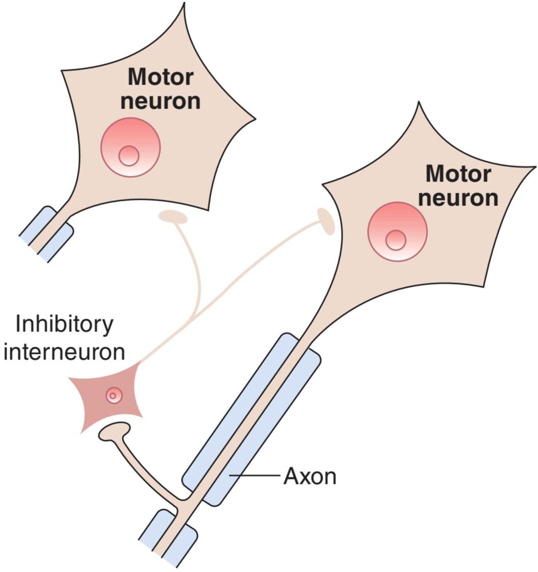
FIGURE 6-10 Negative feedback inhibition of a spinal motor neuron via an inhibitory interneuron. The axon of a spinal motor neuron has a recurrent collateral that synapses on an inhibitory interneuron that terminates on the cell body of the same and other motor neurons. The inhibitory interneuron is called a Renshaw cell and its neurotransmitter is glycine.
شکل 6-10 مهار بازخورد منفی یک نورون حرکتی نخاعی از طریق یک نورون بازدارنده. آکسون یک نورون حرکتی نخاعی دارای یک وثیقه عودکننده است که روی یک نورون بازدارنده سیناپس می شود که به بدنه سلولی همان نورون حرکتی و سایر نورون های حرکتی ختم می شود. نورون بازدارنده سلول رنشاو و انتقال دهنده عصبی آن گلیسین است.
NEUROMUSCULAR TRANSMISSION
NEUROMUSCULAR JUNCTION
Figure 6-11 shows the components of the neuromuscular junction, the specialized area where a motor nerve terminates on a skeletal muscle fiber. As the axon supplying a skeletal muscle fiber approaches its termination, it loses its myelin sheath and divides into a number of terminal boutons. The terminal contains many small, clear vesicles that contain acetylcholine, the transmitter at these junctions. The endings fit into junctional folds or depressions in the motor endplate, the thickened portion of the muscle membrane at this junction. Each endplate receives input from a single nerve fiber. The space between the nerve and the thickened muscle membrane is comparable to the synaptic cleft at a neuron-to-neuron synapse. The whole structure is known as the neuromuscular junction.
انتقال عصبی عضلانی
اتصال عصبی عضلانی
شکل 6-11 اجزای اتصال عصبی عضلانی را نشان می دهد، ناحیه تخصصی که در آن عصب حرکتی به فیبر عضلانی اسکلتی ختم می شود. همانطور که آکسون تامین کننده فیبر عضلانی اسکلتی به پایان خود نزدیک می شود، غلاف میلین خود را از دست می دهد و به تعدادی بوتون انتهایی تقسیم می شود. پایانه شامل بسیاری از وزیکول های کوچک و شفاف است که حاوی استیل کولین، فرستنده در این اتصالات است. انتهای آن در چینهای اتصالی یا فرورفتگیهای صفحه انتهایی موتور، قسمت ضخیمشده غشای عضلانی در این محل اتصال قرار میگیرد. هر صفحه انتهایی ورودی را از یک رشته عصبی دریافت می کند. فضای بین عصب و غشای عضلانی ضخیم شده با شکاف سیناپسی در سیناپس نورون به نورون قابل مقایسه است. کل ساختار به عنوان اتصال عصبی عضلانی شناخته می شود.
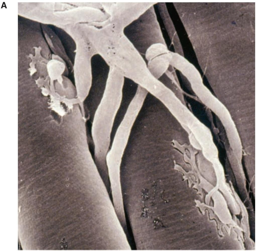

FIGURE 6–11 The neuromuscular junction. A) Scanning electron micrograph showing branching of motor axons with terminals embedded in grooves in the muscle fiber’s surface. B) Structure of a neuromuscular junction. ACh, acetylcholine. (Modified with permission from Widmaier EP, Raff H, Strang KT: Vanders Human Physiology. New York, NY: McGraw-Hill; 2008.)
شکل 6-11 اتصال عصبی عضلانی. الف) میکروگراف الکترونی روبشی که انشعاب آکسون های حرکتی را با پایانه های تعبیه شده در شیارهای سطح فیبر عضلانی نشان می دهد. ب) ساختار اتصال عصبی عضلانی. ACH، استیل کولین. (با اجازه Widmaier EP، Raff H، Strang KT: Vanders Human Physiology اصلاح شده است. نیویورک، نیویورک: McGraw-Hill؛ 2008.)
SEQUENCE OF EVENTS DURING TRANSMISSION
The events occurring during transmission of impulses from the motor nerve to the muscle are somewhat similar to those occurring at neuron-to-neuron synapses (Figure 6-12). The impulse arriving in the terminal of the motor neuron increases its permeability to Ca2+. Ca2+ enters the nerve endings and triggers a marked increase in exocytosis of the acetylcholine-containing synaptic vesicles. The acetylcholine diffuses to nicotinic cholinergic (NM) receptors that are concentrated at the tops of the junctional folds of the membrane of the motor endplate. Binding of acetylcholine to these receptors increases the Na* and K+ conductance, and the resultant influx of Na+ produces a depolarizing potential, the endplate potential. The current sink created by this local potential depolarizes the adjacent muscle membrane to its firing level. Action potentials are generated on either side of the endplate and are conducted away from the endplate in both directions along the muscle fiber. The muscle action potential, in turn, initiates muscle contraction (see Chapter 5). Acetylcholine is then removed from the synaptic cleft by acetylcholinesterase, which is present in high concentration at the neuromuscular junction.
توالی وقایع در حین انتقال
رویدادهایی که در حین انتقال تکانه ها از عصب حرکتی به عضله رخ می دهد تا حدودی شبیه به رویدادهایی است که در سیناپس های نورون به نورون رخ می دهد (شکل 6-12). ضربه ای که به انتهای نورون حرکتی می رسد نفوذپذیری آن را به +Ca2 افزایش می دهد. +Ca2 وارد انتهای عصبی می شود و باعث افزایش قابل توجه اگزوسیتوز وزیکول های سیناپسی حاوی استیل کولین می شود. استیل کولین به گیرنده های کولینرژیک نیکوتینی (NM) منتشر می شود که در بالای چین های اتصالی غشای صفحه انتهایی موتور متمرکز شده اند. اتصال استیل کولین به این گیرنده ها رسانایی +Na و +K را افزایش می دهد و هجوم حاصل از +Na پتانسیل دپلاریزاسیون یعنی پتانسیل صفحه انتهایی را ایجاد می کند. سینک جریان ایجاد شده توسط این پتانسیل موضعی غشای عضلانی مجاور را تا سطح شلیک آن دپلاریزه می کند. پتانسیل های عمل در دو طرف صفحه انتهایی ایجاد می شوند و به دور از صفحه انتهایی در هر دو جهت در امتداد فیبر عضلانی هدایت می شوند. پتانسیل عمل عضلانی، به نوبه خود، انقباض عضلانی را آغاز می کند (به فصل 5 مراجعه کنید). سپس استیل کولین توسط استیل کولین استراز از شکاف سیناپسی خارج می شود که در محل اتصال عصبی عضلانی در غلظت بالایی وجود دارد.

FIGURE 6-12 Events at the neuromuscular junction that lead to an action potential in the muscle fiber plasma membrane. The impulse arriving in the end of the motor neuron increases the permeability of its endings to Ca2+, which enters the endings and triggers exocytosis of the acetylcholine (ACh)-containing synaptic vesicles. ACh diffuses and binds to nicotinic cholinergic (NM) receptors in the motor endplate, which increases Na* and K+ conductance. The resultant influx of Na+ produces the endplate potential. The current sink created by this local potential depolarizes the adjacent muscle membrane to its firing level. Action potentials are generated on either side of the endplate and are conducted away from the endplate in both directions along the muscle fiber and the muscle contracts. ACh is then removed from the synaptic cleft by acetylcholinesterase. (Modified with permission from Widmaier EP, Raff H, Strang KT: Vanders Human Physiology. New York, NY: McGraw-Hill; 2008.)
شکل 6-12 رویدادهایی در محل اتصال عصبی عضلانی که منجر به پتانسیل عمل در غشای پلاسمایی فیبر عضلانی می شود. ضربهای که به انتهای نورون حرکتی میرسد، نفوذپذیری انتهای آن به +Ca2 را افزایش میدهد، که وارد انتهای آن میشود و باعث اگزوسیتوز وزیکولهای سیناپسی حاوی استیل کولین (ACh) میشود. Ach منتشر شده و به گیرنده های نیکوتین کولینرژیک (NM) در صفحه انتهایی موتور متصل می شود که رسانایی +Na و +K را افزایش می دهد. هجوم حاصل از +Na پتانسیل صفحه انتهایی را تولید می کند. سینک جریان ایجاد شده توسط این پتانسیل موضعی غشای عضلانی مجاور را تا سطح شلیک آن دپلاریزه می کند. پتانسیل های عمل در دو طرف صفحه انتهایی ایجاد می شوند و به دور از صفحه انتهایی در هر دو جهت در امتداد فیبر عضلانی و انقباض عضلانی هدایت می شوند. سپس ACH توسط استیل کولین استراز از شکاف سیناپسی خارج می شود. (با اجازه Widmaier EP، Raff H، Strang KT: Vanders Human Physiology اصلاح شده است. نیویورک، نیویورک: McGraw-Hill؛ 2008.)
QUANTAL RELEASE OF TRANSMITTER
Each nerve impulse releases acetylcholine from about 60 synaptic vesicles, and each vesicle contains about 10,000 molecules of the neurotransmitter. Small quanta (packets) of acetylcholine are released randomly from the nerve cell membrane at rest. Each produces a minute depolarizing spike called a miniature endplate potential that is about 0.5 mV in amplitude. The size of the quanta of acetylcholine released in this way varies directly with the Ca2+ concentration and inversely with the Mg2+ concentration at the endplate. When a nerve impulse reaches the ending, the number of quanta released increases by several orders of magnitude, and the result is the large endplate potential that exceeds the firing threshold of the muscle fiber. Quantal release of a neurotransmitter also occurs at noradrenergic, glutamatergic, and other synaptic junctions. Two diseases of the neuromuscular junction, myasthenia gravis and Lambert-Eaton myasthenic syndrome, are described in Clinical Box 6-2 and Clinical Box 6-3, respectively.
انتشار کمی فرستنده
هر تکانه عصبی استیل کولین را از حدود 60 وزیکول سیناپسی آزاد می کند و هر وزیکول حاوی حدود 10000 مولکول انتقال دهنده عصبی است. کوانتوم های کوچک (بسته های) استیل کولین به طور تصادفی از غشای سلول عصبی در حالت استراحت آزاد می شوند. هر کدام یک سنبله دپلاریزاسیون دقیقه ای به نام پتانسیل صفحه انتهایی مینیاتوری تولید می کنند که دامنه آن حدود 0.5 میلی ولت است. اندازه کوانتوم های استیل کولین آزاد شده در این روش به طور مستقیم با غلظت +Ca2 و برعکس با غلظت +Mg2 در صفحه انتهایی متفاوت است. هنگامی که یک تکانه عصبی به پایان می رسد، تعداد کوانتوم های آزاد شده چندین مرتبه افزایش می یابد و نتیجه پتانسیل صفحه انتهایی بزرگی است که از آستانه شلیک فیبر عضلانی فراتر می رود. انتشار کمی یک انتقال دهنده عصبی در اتصالات نورآدرنرژیک، گلوتاماترژیک و سایر اتصالات سیناپسی نیز رخ می دهد. دو بیماری پیوند عصبی عضلانی، میاستنی گراویس و سندرم میاستنی لامبرت-ایتون، به ترتیب در جعبه بالینی 6-2 و جعبه بالینی 6-3 شرح داده شده اند.
CLINICAL BOXG 6.2
Myasthenia Gravis
Myasthenia gravis is a serious and sometimes fatal disease in which skeletal muscles are weak and tire easily. It occurs in 25 to 125 of every 1 million people worldwide and can occur at any age but seems to have a bimodal distribution, with peak occurrences in individuals in their 20s (mainly women) and 60s (mainly men). It is caused by the formation of circulating antibodies to the skeletal muscle type of nicotinic cholinergic receptors. These antibodies destroy some of the receptors and bind others to neighboring receptors, triggering their removal by endocytosis. Normally, the number of quanta released from the motor nerve terminal declines with successive repetitive stimuli. In myasthenia gravis, neuromuscular transmission fails at these low levels of quantal release. This leads to the major clinical feature of the disease, muscle fatigue with sustained or repeated activity. There are two major forms of the disease. In one form, the extraocular muscles are primarily affected. In the second form, there is a generalized skeletal muscle weakness. In severe cases, all muscles, including the diaphragm, can weaken and respiratory failure and death can ensue. The major structural abnormality in myasthenia gravis is the appearance of sparse, shallow, and abnormally wide or absent synaptic clefts in the motor endplate. The postsynaptic membrane has a reduced response to acetylcholine, and there is a 70-90% decrease in the number of receptors per endplate in affected muscles. Patients with myasthenia gravis have a greater than normal tendency to also have rheumatoid arthritis, systemic lupus erythematosus, and polymyositis. About 30% of patients with myasthenia gravis have a maternal relative with an autoimmune disorder. These associations suggest that individuals with myasthenia gravis share a genetic predisposition to autoimmune disease. The thymus may play a role in the pathogenesis of the disease by supplying helper T cells sensitized against thymic proteins that cross-react with acetylcholine receptors. In most patients, the thymus is hyperplastic; and 10-15% have a thymoma.
جعبه بالینی 6.2
میاستنی گراویس
میاستنی گراویس یک بیماری جدی و گاهی کشنده است که در آن عضلات اسکلتی ضعیف شده و به راحتی خسته می شوند. این بیماری در 25 تا 125 نفر از هر 1 میلیون نفر در سراسر جهان رخ می دهد و می تواند در هر سنی رخ دهد، اما به نظر می رسد که دارای توزیع دووجهی است، با حداکثر وقوع در افراد 20 ساله (عمدتا زنان) و 60 سالگی (عمدتاً مردان). این بیماری در اثر تشکیل آنتی بادی های در گردش به نوع عضله اسکلتی گیرنده های کولینرژیک نیکوتین ایجاد می شود. این آنتیبادیها برخی از گیرندهها را از بین میبرند و برخی دیگر را به گیرندههای مجاور متصل میکنند و باعث حذف آنها با اندوسیتوز میشوند. به طور معمول، تعداد کوانتوم های آزاد شده از پایانه عصب حرکتی با محرک های تکراری متوالی کاهش می یابد. در میاستنی گراویس، انتقال عصبی عضلانی در این سطوح کم انتشار کوانتال با شکست مواجه می شود. این منجر به ویژگی بالینی اصلی بیماری، خستگی عضلانی با فعالیت مداوم یا مکرر می شود. دو شکل اصلی این بیماری وجود دارد. در یک شکل، ماهیچه های خارج چشمی در درجه اول تحت تاثیر قرار می گیرند. در شکل دوم، ضعف عمومی عضله اسکلتی وجود دارد. در موارد شدید، تمام عضلات از جمله دیافراگم ممکن است ضعیف شوند و نارسایی تنفسی و مرگ به دنبال داشته باشد. ناهنجاری ساختاری اصلی در میاستنی گراویس، ظهور شکافهای سیناپسی کم، کم عمق و غیرطبیعی گسترده یا غایب در صفحه انتهایی موتور است. غشای پس سیناپسی پاسخ کمتری به استیل کولین دارد و ۷۰ تا ۹۰ درصد کاهش در تعداد گیرنده ها در هر صفحه انتهایی در عضلات آسیب دیده وجود دارد. بیماران مبتلا به میاستنی گراویس تمایل بیشتری نسبت به معمول به آرتریت روماتوئید، لوپوس اریتماتوز سیستمیک و پلی میوزیت دارند. حدود 30 درصد از بیماران مبتلا به میاستنی گراویس یکی از بستگان مادری مبتلا به اختلال خود ایمنی دارند. این ارتباط نشان می دهد که افراد مبتلا به میاستنی گراویس دارای استعداد ژنتیکی برای بیماری خودایمنی هستند. تیموس ممکن است با تامین سلول های T کمکی حساس به پروتئین های تیموس که با گیرنده های استیل کولین واکنش متقابل دارند، در پاتوژنز بیماری نقش داشته باشد. در اکثر بیماران، تیموس هیپرپلاستیک است. و 10-15 درصد تیموما دارند.
THERAPEUTIQ HIGHLIGHTS
Muscle weakness due to myasthenia gravis improves after a period of rest or after administration of an acetylcholinesterase inhibitor such as neostigmine or pyridostigmine. Cholinesterase inhibitors prevent metabolism of acetylcholine and can thus compensate for the normal decline in released neurotransmitters during repeated stimulation. Immunosuppressive drugs (eg, prednisone, azathioprine, or cyclosporine) can suppress antibody production and have been shown to improve muscle strength in some patients with myasthenia gravis. Thymectomy is indicated especially if a thymoma is suspected in the development of myasthenia gravis. Even in those without thymoma, thymectomy induces remission in 35% and improves symptoms in another 45% of patients.
نکات برجسته درمانی
ضعف عضلانی ناشی از میاستنی گراویس پس از یک دوره استراحت یا پس از تجویز یک مهارکننده استیل کولین استراز مانند نئوستیگمین یا پیریدوستیگمین بهبود می یابد. مهارکننده های کولین استراز از متابولیسم استیل کولین جلوگیری می کنند و بنابراین می توانند کاهش طبیعی انتقال دهنده های عصبی آزاد شده را در طی تحریک مکرر جبران کنند. داروهای سرکوب کننده سیستم ایمنی (مانند پردنیزون، آزاتیوپرین یا سیکلوسپورین) می توانند تولید آنتی بادی را سرکوب کنند و نشان داده شده است که در برخی از بیماران مبتلا به میاستنی گراویس قدرت عضلانی را بهبود می بخشند. تیمکتومی به ویژه در صورتی که به تیموما مشکوک به ایجاد میاستنی گراویس باشد، اندیکاسیون دارد. حتی در افرادی که تیموما ندارند، تیمکتومی باعث بهبودی در 35٪ و بهبود علائم در 45٪ از بیماران می شود.
CLINICAL BOX 6.3
Lambert-Eaton Syndrome
In a relatively rare condition called Lambert-Eaton myasthenic syndrome (LEMS), muscle weakness is caused by an autoimmune attack against one of the voltage-gated Ca2+ channels in the nerve endings at the neuromuscular junction. This decreases the normal Ca2* influx that causes acetylcholine release. The incidence of LEMS in the United States is about 1 case per 100,000 people; it is usually an adult-onset disease that has a similar occurrence in men and women. Proximal muscles of the lower extremities are primarily affected, producing a waddling gait and difficulty raising the arms. Repetitive stimulation of the motor nerve facilitates accumulation of Ca2+ in the nerve terminal and increases acetylcholine release, leading to an increase in muscle strength. This is in contrast to myasthenia gravis in which symptoms are exacerbated by repetitive stimulation. About 40% of patients with LEMS also have cancer, especially small cell cancer of the lung. One theory is that antibodies that have been produced to attack the cancer cells may also attack Ca2+ channels, leading to LEMS. LEMS has also been associated with lymphosarcoma; malignant thymoma; and cancer of the breast, stomach, colon, prostate, bladder, kidney, or gallbladder. Clinical signs usually precede the diagnosis of cancer. A syndrome similar to LEMS can occur after the use of aminoglycoside antibiotics, which also impair Ca2+ channel function.
جعبه بالینی 6.3
سندرم لامبرت ایتون
در یک بیماری نسبتاً نادر به نام سندرم میاستنی لامبرت-ایتون (LEMS)، ضعف عضلانی در اثر حمله خود ایمنی علیه یکی از کانالهای +Ca2 دارای ولتاژ در انتهای عصب در محل اتصال عصبی عضلانی ایجاد میشود. این امر هجوم طبیعی +Ca2 را که باعث آزادسازی استیل کولین می شود، کاهش می دهد. بروز LEMS در ایالات متحده حدود 1 مورد در هر 100000 نفر است. این بیماری معمولاً در بزرگسالی شروع می شود و در مردان و زنان اتفاق مشابهی دارد. ماهیچه های پروگزیمال اندام تحتانی در درجه اول تحت تأثیر قرار می گیرند و باعث ایجاد یک راه رفتن و مشکل در بالا بردن بازوها می شوند. تحریک مکرر عصب حرکتی تجمع +Ca2 را در پایانه عصبی تسهیل می کند و ترشح استیل کولین را افزایش می دهد که منجر به افزایش قدرت عضلانی می شود. این برخلاف میاستنی گراویس است که در آن علائم با تحریک مکرر تشدید می شود. حدود 40 درصد از بیماران مبتلا به LEMS نیز به سرطان، به ویژه سرطان سلول کوچک ریه مبتلا هستند. یک نظریه این است که آنتی بادی هایی که برای حمله به سلول های سرطانی تولید شده اند ممکن است به کانال های +Ca2 نیز حمله کنند که منجر به LEMS می شود. LEMS همچنین با لنفوسارکوم همراه است. تیموم بدخیم؛ و سرطان سینه، معده، روده بزرگ، پروستات، مثانه، کلیه یا کیسه صفرا. علائم بالینی معمولاً قبل از تشخیص سرطان است. سندرمی شبیه به LEMS می تواند پس از استفاده از آنتی بیوتیک های آمینوگلیکوزید رخ دهد که عملکرد کانال های +Ca2 را نیز مختل می کند.
THERAPEUTIC HIGHLIGHTS
Since there is a high comorbidity with small cell lung cancer, the first treatment strategy is to determine if the individual also has cancer and, if so, to treat that appropriately. In patients without cancer, immunotherapy is initiated. Prednisone administration, plasmapheresis, and intravenous immunoglobulin are some examples of effective therapies for LEMS. The use of aminopyridines facilitates the release of acetylcholine in the neuromuscular junction and can improve muscle strength in LEMS patients. This class of drugs causes blockade of presynaptic K+ channels and promotes activation of voltage- gated Ca2+ channels. Acetylcholinesterase inhibitors can be used but often do not ameliorate the symptoms of LEMS.
نکات برجسته درمانی
از آنجایی که سرطان ریه سلول کوچک همبودی بالایی دارد، اولین راهبرد درمانی این است که مشخص شود آیا فرد به سرطان نیز مبتلا است یا خیر و اگر چنین است، درمان مناسب آن انجام شود. در بیماران بدون سرطان، ایمونوتراپی آغاز می شود. تجویز پردنیزون، پلاسمافرزیس و ایمونوگلوبولین داخل وریدی نمونه هایی از درمان های موثر برای LEMS هستند. استفاده از آمینوپیریدین ها آزادسازی استیل کولین را در محل اتصال عصبی عضلانی تسهیل می کند و می تواند قدرت عضلانی را در بیماران LEMS بهبود بخشد. این دسته از داروها باعث مسدود شدن کانال های +K پیش سیناپسی شده و باعث فعال شدن کانال های +Ca2 دارای ولتاژ می شود. مهارکننده های استیل کولین استراز را می توان استفاده کرد اما اغلب علائم LEMS را بهبود نمی بخشد.
NERVE ENDINGS IN SMOOTH & CARDIAC MUSCLE
The postganglionic autonomic nerve fibers supplying smooth muscle in various organs branch extensively and come in close contact with the muscle cells (Figure 6-13). Some of these nerve fibers have clear vesicles containing acetylcholine; others have dense-core vesicles containing norepinephrine. There are no recognizable endplates or other postsynaptic specializations at these neuroeffector junctions. The nerve fibers run along the membranes of the muscle cells and sometimes groove their surfaces. The nerve branches are beaded with enlargements (varicosities) that contain synaptic vesicles. In noradrenergic neurons, the varicosities are about 5 μm apart, with up to 20,000 varicosities per neuron. Neurotransmitter can be released at each of the varicosities along the axon; this arrangement permits one neuron to innervate many effector cells. The type of contact in which a neuron forms a synapse on the surface of a smooth muscle cell and then passes on to make similar contacts with other cells is called a synapse en passant.
پایانه های عصبی در ماهیچه های صاف و قلبی
فیبرهای عصبی خودمختار پس گانگلیونی که ماهیچه صاف را در اندام های مختلف تامین می کنند به طور گسترده ای منشعب می شوند و در تماس نزدیک با سلول های عضلانی قرار می گیرند (شکل 6-13). برخی از این رشته های عصبی دارای وزیکول های شفاف حاوی استیل کولین هستند. برخی دیگر دارای وزیکول های هسته متراکم حاوی نوراپی نفرین هستند. هیچ صفحه انتهایی یا سایر تخصص های پس سیناپسی قابل تشخیصی در این اتصالات نوروفکتور وجود ندارد. رشتههای عصبی در امتداد غشای سلولهای عضلانی قرار دارند و گاهی اوقات سطوح آنها را شیار میکنند. شاخه های عصبی با بزرگ شدن (واریس) که حاوی وزیکول های سیناپسی است، مهره هایی دارند. در نورونهای نورآدرنرژیک، واریکوسیتهها حدود 5 میکرومتر از هم فاصله دارند و تا 20000 واریس در هر نورون وجود دارد. انتقال دهنده عصبی می تواند در هر یک از واریکوسیته ها در امتداد آکسون آزاد شود. این آرایش به یک نورون اجازه می دهد تا بسیاری از سلول های عامل را عصب دهی کند. نوع تماسی که در آن یک نورون سیناپسی را بر روی سطح سلول ماهیچه صاف تشکیل می دهد و سپس برای ایجاد تماس های مشابه با سلول های دیگر، سیناپس ان پاسانت نامیده می شود.
In the heart, cholinergic and noradrenergic nerve fibers end on the sinoatrial node, the atrioventricular node, and the bundle of His (see Chapter 29). Noradrenergic fibers also innervate the ventricular muscle. The exact nature of the endings on nodal tissue is not known. In the ventricle, the contacts between the noradrenergic fibers and the cardiac muscle fibers resemble those found in smooth muscle.
در قلب، رشته های عصبی کولینرژیک و نورآدرنرژیک به گره سینوسی دهلیزی، گره دهلیزی و بطنی His ختم می شوند (به فصل 29 مراجعه کنید). فیبرهای نورآدرنرژیک نیز عضله بطنی را عصب دهی می کنند. ماهیت دقیق انتهای روی بافت گره مشخص نیست. در بطن، تماسهای بین رشتههای نورآدرنرژیک و فیبرهای عضلانی قلبی شبیه تماسهایی است که در ماهیچه صاف وجود دارد.

FIGURE 6–13 Endings of postganglionic autonomic neurons on smooth muscle. The nerve fibers run along the membranes of the smooth muscle cells and sometimes groove their surfaces. The multiple branches of postganglionic neurons are beaded with enlargements (varicosities) and contain synaptic vesicles. Neurotransmitter is released from the varicosities and diffuses to receptors on smooth muscle cell plasma membranes. (Reproduced with permission from Widmaier EP, Raff H, Strang KT: Vanders Human Physiology. New York, NY: McGraw-Hill; 2008.)
شکل 6-13 انتهای نورون های اتونوم پس گانگلیونی روی ماهیچه صاف. رشتههای عصبی در امتداد غشای سلولهای ماهیچه صاف قرار دارند و گاهی اوقات سطح آنها را شیار میکنند. شاخه های متعدد نورون های پس گانگلیونی با بزرگ شدن (واریس) و حاوی وزیکول های سیناپسی هستند. انتقال دهنده عصبی از واریس ها آزاد می شود و به گیرنده های غشای پلاسمایی سلول عضله صاف منتشر می شود. (تکثیر شده با اجازه Widmaier EP, Raff H, Strang KT: Vanders Human Physiology. نیویورک, نیویورک: McGraw-Hill؛ 2008.)
JUNCTIONAL POTENTIALS
In smooth muscles in which noradrenergic activity is excitatory, stimulation of the noradrenergic nerves produces discrete partial depolarizations that look like small endplate potentials and are called excitatory junction potentials (EJPs). These potentials summate with repeated stimuli. Similar EJPS are seen in tissues excited by cholinergic discharges. In tissues inhibited by noradrenergic stimuli, hyperpolarizing inhibitory junction potentials (IJPs) are produced by stimulation of the noradrenergic nerves. Junctional potentials spread in an electrotonic fashion.
پتانسیل های متقابل
در ماهیچه های صاف که فعالیت نورآدرنرژیک تحریکی است، تحریک اعصاب نورآدرنرژیک باعث ایجاد دپلاریزاسیون های جزئی مجزا می شود که شبیه پتانسیل های صفحه انتهایی کوچک هستند و به آنها پتانسیل های اتصال تحریکی (EJPs) می گویند. این پتانسیل ها با محرک های مکرر جمع می شوند. EJPS مشابه در بافت هایی که توسط ترشحات کولینرژیک برانگیخته شده اند دیده می شود. در بافتهایی که توسط محرکهای نورآدرنرژیک مهار میشوند، پتانسیلهای اتصال مهاری هیپرپلاریزه (IJPs) با تحریک اعصاب نورآدرنرژیک تولید میشوند. پتانسیل های اتصالی به صورت الکتروتونیک پخش می شوند.
AXONAL INJURY & DENERVATION SUPERSENSITIVITY
Figure 6-14 shows some of the reactions triggered by injury or section of an axon. Orthograde degeneration (Wallerian degeneration) occurs from the point of injury to the nerve terminal, interrupting neural transmission. Distal to the injury, the membrane breaks down and the myelin sheath degenerates. The proximal axon also is affected and may undergo retrograde degeneration and die. The cell body of the injured neuron swells, the nucleus moves to an eccentric position, and the rough endoplasmic reticulum gets fragmented (chromatolytic reaction).
حساسيت فوق العاده آسيب آکسونال و عصب کشي
شکل 6-14 برخی از واکنش های ایجاد شده توسط آسیب یا برش یک آکسون را نشان می دهد. دژنراسیون ارتوگراد (دژنراسیون والری) از نقطه آسیب به پایانه عصبی رخ می دهد و انتقال عصبی را قطع می کند. دیستال آسیب، غشاء شکسته می شود و غلاف میلین تحلیل می رود. آکسون پروگزیمال نیز تحت تأثیر قرار گرفته و ممکن است دچار دژنراسیون رتروگراد شده و بمیرد. بدنه سلولی نورون آسیب دیده متورم می شود، هسته به یک موقعیت خارج از مرکز حرکت می کند و شبکه آندوپلاسمی خشن تکه تکه می شود (واکنش کروماتولیتیک).

FIGURE 6-14 Changes occurring in a neuron when its axon is crushed or injured. The distal axon stump separates from the cell body, orthograde (Wallerian) degeneration occurs from the point of damage to the terminal, and the myelin sheath degenerates. The cell body of the injured neuron swells and the endoplasmic reticulum is fragmented as part of the chromatolytic reaction.
شکل 6-14 تغییراتی که در یک نورون زمانی که آکسون آن له شده یا آسیب می بیند رخ می دهد. استامپ دیستال آکسون از بدنه سلولی جدا می شود، دژنراسیون ارتوگراد (والری) از نقطه آسیب به پایانه رخ می دهد و غلاف میلین تحلیل می رود. بدنه سلولی نورون آسیب دیده متورم می شود و شبکه آندوپلاسمی به عنوان بخشی از واکنش کروماتولیتیک تکه تکه می شود.
The nerve then starts to regrow, with multiple small branches projecting along the path the axon previously followed (regenerative sprouting). Axons sometimes grow back to their original targets, especially in locations like the neuromuscular junction. However, nerve regeneration is generally limited because axons often become entangled in the area of tissue damage at the site where they were disrupted. This difficulty has been reduced by administration of neurotrophins (see Chapter 4).
سپس عصب شروع به رشد مجدد میکند و شاخههای کوچک متعددی در مسیری که آکسون قبلاً دنبال میکرده بیرون میآیند (جوانههای احیاکننده). گاهی اوقات آکسون ها به سمت اهداف اصلی خود رشد می کنند، به خصوص در مکان هایی مانند اتصال عصبی عضلانی. با این حال، بازسازی عصب به طور کلی محدود است زیرا آکسون ها اغلب در ناحیه آسیب بافتی در محلی که در آن مختل شده اند درگیر می شوند. این مشکل با تجویز نوروتروفین ها کاهش یافته است (به فصل 4 مراجعه کنید).
When the motor nerve to skeletal muscle is cut and allowed to degenerate, the muscle gradually becomes extremely sensitive to acetylcholine. This is called denervation hypersensitivity or supersensitivity. Normally nicotinic receptors are located only in the vicinity of the motor endplate where the axon of the motor nerve terminates. When the motor nerve is severed, there is a marked proliferation of nicotinic receptors over a wide region of the neuromuscular junction. Denervation supersensitivity also occurs at autonomic junctions. Smooth muscle, unlike skeletal muscle, does not atrophy when denervated, but it becomes hyperresponsive to the chemical mediator that normally activates it.
هنگامی که عصب حرکتی عضله اسکلتی بریده می شود و اجازه داده می شود که تحلیل رود، عضله به تدریج نسبت به استیل کولین بسیار حساس می شود. به این حساسیت بیش از حد یا فوق حساسیت عصب کشی می گویند. به طور معمول گیرنده های نیکوتین تنها در مجاورت صفحه انتهایی حرکتی جایی که آکسون عصب حرکتی به پایان می رسد قرار دارند. هنگامی که عصب حرکتی قطع می شود، تکثیر مشخصی از گیرنده های نیکوتین در ناحیه وسیعی از اتصال عصبی عضلانی وجود دارد. فوق حساسیت عصب کشی نیز در اتصالات اتونوم رخ می دهد. عضله صاف، بر خلاف عضله اسکلتی، هنگام عصب کشی آتروفی نمی شود، اما به واسطه شیمیایی که به طور معمول آن را فعال می کند، بیش از حد پاسخ می دهد.
Denervation hypersensitivity has multiple causes. As noted in Chapter 2, a deficiency of a given chemical messenger generally produces an upregulation of its receptors. Another factor is a lack of reuptake of secreted neurotransmitters.
حساسیت به عصب کشی دلایل متعددی دارد. همانطور که در فصل 2 اشاره شد، کمبود یک پیام رسان شیمیایی معین، عموماً باعث تنظیم دگرگونی گیرنده های آن می شود. عامل دیگر عدم جذب مجدد انتقال دهنده های عصبی ترشح شده است.
CHAPTER SUMMARY
خلاصه فصل
• Most synapses occur on dendrites (axodendritic), but some presynaptic nerves terminate on the soma (axosomatic) or axon (axoaxonic) of a postsynaptic neuron. At chemical synapses, an impulse in the presynaptic axon causes release of a neurotransmitter from synaptic vesicles that diffuses across a synaptic cleft and binds to receptors on the postsynaptic density, triggering events that open or close ion channels in the membrane of the postsynaptic neuron. At electrical synapses, gap junctions between the presynaptic and postsynaptic neurons form low-resistance bridges through which ions pass with relative ease from one neuron to the next.
• بیشتر سیناپس ها روی دندریت ها (آکسودندریتیک) رخ می دهند، اما برخی از اعصاب پیش سیناپسی به سوما (آکسوسوماتیک) یا آکسون (آکسواکسونیک) یک نورون پس سیناپسی ختم می شوند. در سیناپسهای شیمیایی، یک تکانه در آکسون پیشسیناپسی باعث آزاد شدن یک انتقالدهنده عصبی از وزیکولهای سیناپسی میشود که در شکاف سیناپسی منتشر میشود و به گیرندههای چگالی پس سیناپسی متصل میشود و رویدادهایی را ایجاد میکند که کانالهای یونی را در غشای نورون پس سیناپسی باز یا بسته میکنند. در سیناپس های الکتریکی، اتصالات شکاف بین نورون های پیش سیناپسی و پس سیناپسی، پل هایی با مقاومت کم را تشکیل می دهند که یون ها با سهولت نسبی از یک نورون به نورون بعدی عبور می کنند.
• An excitatory stimulus opens Na* or Ca2+ ion channels to produce an EPSP (inward current) in a postsynaptic neuron after a latency of 0.5 ms. An IPSP is produced by a localized increase in Cl transport. Slow EPSPs and IPSPs are due to decreases and increases in K+ conductance, respectively. They occur after a latency of 100-500 ms in autonomic ganglia, cardiac and smooth muscle, and cortical neurons.
• یک محرک تحریکی کانال های یونی +Na یا +Ca2 را برای تولید یک EPSP (جریان درونی) در یک نورون پس سیناپسی پس از تاخیر 0.5 ms باز می کند. یک IPSP با افزایش موضعی در انتقال کلر تولید می شود. کندی EPSP و IPSP به ترتیب به دلیل کاهش و افزایش رسانایی +K است. آنها پس از تأخیر 100-500 میلیثانیه در گانگلیونهای اتونوم، ماهیچههای صاف و قلبی و نورونهای قشر مغز رخ میدهند.
• The time constant of a neuron determines the time course of the synaptic potential; the longer the time constant, the greater is the chance for two potentials to summate to induce an action potential (temporal summation).The length constant of a neuron determines the degree to which a depolarizing current is reduced as it spreads passively; if a neuron has a long length constant, the depolarization induced at two points on the neuron can summate and induce an action potential (spatial summation).
• ثابت زمانی یک نورون، سیر زمانی پتانسیل سیناپسی را تعیین میکند؛ هر چه ثابت زمانی طولانیتر باشد، احتمال جمع شدن دو پتانسیل برای القای پتانسیل عمل (جمع زمانی) بیشتر است. ثابت طول یک نورون، میزان کاهش جریان دپلاریزه کننده را هنگام پخش شدن غیرفعال آن تعیین میکند. اگر یک نورون ثابت طول طولانی داشته باشد، دپلاریزاسیون القا شده در دو نقطه از نورون میتواند جمع شود و یک پتانسیل عمل (جمع مکانی) را القا کند.
• Postsynaptic inhibition occurs when an inhibitory transmitter (glycine, GABA) is released from a presynaptic nerve terminal and induces an IPSP in the postsynaptic neuron. Presynaptic inhibition is mediated at axoaxonal synapses; activation of presynaptic receptors increases Cl conductance, decreasing the size of the action potentials reaching the excitatory ending, and reducing Ca2+ entry and the amount of excitatory transmitter released. Presynaptic facilitation occurs when the action potential is prolonged and the Ca2+ channels are open for a longer period.
• مهار پس سیناپسی زمانی اتفاق می افتد که یک فرستنده بازدارنده (گلیسین، GABA) از پایانه عصبی پیش سیناپسی آزاد شود و یک IPSP را در نورون پس سیناپسی القا کند. مهار پیشسیناپسی در سیناپسهای آکسواکسونال انجام میشود. فعال شدن گیرنده های پیش سیناپسی هدایت کلر را افزایش می دهد، اندازه پتانسیل های عمل رسیدن به انتهای تحریکی را کاهش می دهد و ورود +Ca2 و مقدار فرستنده تحریکی آزاد شده را کاهش می دهد. تسهیل پیش سیناپسی زمانی رخ می دهد که پتانسیل عمل طولانی شود و کانال های +Ca2 برای مدت طولانی تری باز باشند.
• The axon terminal of motor neurons synapses on the motor endplate on the skeletal muscle membrane to form the neuromuscular junction. An impulse arriving in the motor nerve terminal leads to the entry of Ca2+ that triggers the exocytosis of the acetylcholine-containing synaptic vesicles. The acetylcholine diffuses and binds to nicotinic cholinergic receptors on the motor endplate, causing an increase in Na* and K+ conductance; the influx of Na+ induces the endplate potential and depolarization of the adjacent muscle membrane.
• ترمینال آکسون نورون های حرکتی روی صفحه انتهایی حرکتی غشای عضله اسکلتی سیناپس می شود و اتصال عصبی عضلانی را تشکیل می دهد. ضربه ای که به پایانه عصب حرکتی می رسد منجر به ورود +Ca2 می شود که باعث اگزوسیتوز وزیکول های سیناپسی حاوی استیل کولین می شود. استیل کولین پخش می شود و به گیرنده های نیکوتین کولینرژیک روی صفحه انتهایی موتور متصل می شود و باعث افزایش رسانایی +Na و +K می شود. هجوم +Na باعث پتانسیل صفحه انتهایی و دپلاریزاسیون غشای عضلانی مجاور می شود.
• Autonomic nerve terminals in smooth muscle form a neuroeffector junction; nerve branches are beaded with enlargements (varicosities) that contain synaptic vesicles. Release of neurotransmitter mediates EJPS or IJPs that spread electrotonically.
• پایانه های عصبی اتونومیک در ماهیچه صاف یک اتصال نوروفکتور را تشکیل می دهند. شاخه های عصبی با بزرگ شدن (واریس) که حاوی وزیکول های سیناپسی هستند، مهره ای دارند. انتشار انتقال دهنده عصبی واسطه EJPS یا IJP است که به صورت الکتروتونیک پخش می شود.
• When a nerve is damaged and then degenerates, the postsynaptic structure gradually becomes extremely sensitive to the transmitter released by the nerve (denervation hypersensitivity).
• هنگامی که یک عصب آسیب می بیند و سپس تحلیل می رود، ساختار پس سیناپسی به تدریج نسبت به فرستنده آزاد شده توسط عصب بسیار حساس می شود (حساسیت بیش از حد عصب کشی).
• Neurotoxins from bacteria can disrupt neurotransmitter release at the neuromuscular junction (botulinum toxin, tetanus toxin). Autoimmune diseases of the neuromuscular junction include myasthenia gravis (antibodies against nicotinic cholinergic receptors) and Lambert-Eaton myasthenic syndrome (antibodies against voltage-gated Ca2+ channels).
• نوروتوکسین های باکتری می توانند آزادسازی انتقال دهنده های عصبی را در محل اتصال عصبی عضلانی (سم بوتولینوم، سم کزاز) مختل کنند. بیماریهای خودایمنی اتصال عصبی عضلانی شامل میاستنی گراویس (آنتی بادیها علیه گیرندههای کولینرژیک نیکوتین) و سندرم میاستنی لامبرت-ایتون (آنتیبادیها در برابر کانالهای +Ca2 دارای ولتاژ است).
MULTIPLE-CHOICE QUESTIONS
For all questions, select the single best answer unless otherwise directed.
سوالات چند گزینه ای
برای همه سؤالات، بهترین پاسخ را انتخاب کنید، مگر اینکه دستور دیگری داده شود.
1. A neurology resident was conducting research in an electrophysiology laboratory and was studying the roles of cation channels in mediating nonpropagated potentials. Which of the following electrophysiologic events is correctly paired with the change in ionic currents causing the event?
A. Fast inhibitory postsynaptic potentials (IPSPs) and closing of Cl ̄ channels
B. Fast excitatory postsynaptic potentials (EPSPs) and an increase in Ca2+ conductance
C. Endplate potential and an increase in Na+ conductance
D. Excitatory junctional potentials and closure of voltage-gated K+ channels E. Slow EPSPs and an increase in K+ conductance
1. یک دستیار مغز و اعصاب در حال انجام تحقیقات در آزمایشگاه الکتروفیزیولوژی بود و در حال مطالعه نقش کانال های کاتیونی در میانجیگری پتانسیل های غیرقابل انتشار بود. کدام یک از رویدادهای الکتروفیزیولوژیک زیر به درستی با تغییر در جریان های یونی که باعث این رویداد می شود جفت می شود؟
الف. پتانسیل های پس سیناپسی مهاری سریع (IPSPs) و بسته شدن کانال های Cl ̄
ب. پتانسیل های پس سیناپسی تحریکی سریع (EPSPs) و افزایش رسانایی Ca2+
ج. پتانسیل صفحه انتهایی و افزایش رسانایی Na+
د. پتانسیل های پیوندی تحریکی و بسته شدن کانال های K+ دارای ولتاژ E. EPSPs کند و افزایش رسانایی K+
2. A neurology resident was conducting research in an electrophysiology laboratory and was studying the functional anatomy of synaptic transmission. Which of the following physiologic processes is correctly paired with a structure?
A. Electrical transmission: synaptic cleft
B. Positive feedback inhibition: Renshaw cell
C. Synaptic vesicle docking and fusion: presynaptic nerve terminal
D. Endplate potential: muscarinic cholinergic receptor
E. Action potential initiation: axon hillock
2. یک دستیار مغز و اعصاب مشغول تحقیق در آزمایشگاه الکتروفیزیولوژی بود و در حال مطالعه آناتومی عملکردی انتقال سیناپسی بود. کدام یک از فرآیندهای فیزیولوژیکی زیر به درستی با یک ساختار جفت شده است؟
الف. انتقال الکتریکی: شکاف سیناپسی
ب. بازدارندگی مثبت: سلول رنشا
ج. اتصال و همجوشی وزیکول سیناپسی: پایانه عصبی پیش سیناپسی
د. پتانسیل انتهای صفحه: گیرنده کولینرژیک موسکارینی
ی. شروع پتانسیل عمل: تپه آکسون
3. A medical student was studying the passive membrane properties of neurons and their ability to affect the amplitude of an EPSP recorded from the neuron. She found that in one neuron, applying two stimuli separated by 25 ms to one presynaptic input induced two EPSPs of identical amplitude. In a second neuron, the same type of stimulation induced an EPSP followed an action potential. What can she conclude from this experiment?
A. The second neuron had a longer time constant than the first neuron.
B. The second neuron had a shorter time constant than the first neuron.
C. The second neuron had a longer length constant than the first neuron.
D. The second neuron had a shorter length constant than the first neuron.
3. یک دانشجوی پزشکی در حال مطالعه ویژگیهای غشای غیرفعال نورونها و توانایی آنها برای تأثیرگذاری بر دامنه EPSP ثبتشده از نورون بود. او دریافت که در یک نورون، اعمال دو محرک با فاصله 25 میلی ثانیه روی یک ورودی پیش سیناپسی باعث القای دو EPSP با دامنه یکسان می شود. در نورون دوم، همان نوع تحریک یک EPSP را به دنبال پتانسیل عمل القا کرد. او از این آزمایش چه نتیجه ای می تواند بگیرد؟
الف. نورون دوم نسبت به نورون اول ثابت زمانی بیشتری داشت.
ب. نورون دوم نسبت به نورون اول ثابت زمانی کوتاه تری داشت.
ج. نورون دوم نسبت به نورون اول ثابت طول بیشتری داشت.
د. نورون دوم نسبت به نورون اول ثابت طول کوتاه تری داشت.
4. A medical student was doing research in a laboratory that studies the neuromuscular junction. Which of the following events at the junction are listed in correct sequential order?
A. Motor neuron action potential, acetylcholine release, Na* entry at the endplate
B. Ca2+ entry into the motor nerve terminal, Na* entry at the endplate, muscle fiber action potential
C. Na* entry at the endplate, acetylcholine release, muscle fiber action potential
D. Motor neuron action potential, Na* entry at the endplate, formation of an endplate potential
E. Ca2+ entry into the motor nerve terminal, acetylcholine release, Na* entry at the endplate
4. یک دانشجوی پزشکی در آزمایشگاهی که پیوند عصبی عضلانی را مطالعه می کند، مشغول تحقیق بود. کدام یک از رویدادهای زیر در محل اتصال به ترتیب صحیح فهرست شده است؟
الف. پتانسیل عمل نورون حرکتی، آزادسازی استیل کولین، ورود +Na در صفحه انتهایی
ب. ورود +Ca2 به پایانه عصب حرکتی، ورود +Na در صفحه انتهایی، پتانسیل عمل فیبر عضلانی
ج. ورود +Na در صفحه انتهایی، آزادسازی استیل کولین، پتانسیل عمل فیبر عضلانی
د. پتانسیل عمل نورون حرکتی، ورود +Na در صفحه انتهایی، تشکیل پتانسیل صفحه انتهایی
ی. ورود +Ca2 به انتهای عصب حرکتی، آزادسازی استیل کولین، ورود +Na در صفحه انتهایی
5. Activation of a sensory nerve from the muscle spindle caused contraction of the extensor muscle and relaxation of the flexor muscle. The relaxation of the flexor muscle is an example of
A. negative feedback inhibition.
B. postsynaptic inhibition.
C. Renshaw cell-mediated inhibition.
D. presynaptic inhibition.
E. indirect inhibition.
5. فعال شدن یک عصب حسی از دوک عضلانی باعث انقباض عضله اکستانسور و شل شدن عضله فلکسور شد. شل شدن عضله فلکسور نمونه ای از آن است
الف. مهار بازخورد منفی.
ب. مهار پس سیناپسی.
ج. مهار با واسطه سلول رنشا.
د. مهار پیش سیناپسی.
ی. مهار غیر مستقیم.
6. A medical student was working in a laboratory that studied autonomic neurotransmission. He was recording a potential in a vascular smooth muscle from stimulation of the postganglionic sympathetic nerve. What is the name of the structure on the nerve where the neurotransmitter is stored and what is the name of the response in the smooth muscle?
A. Synaptic vesicle and endplate potential
B. Varicosity and inhibitory postsynaptic potential
C. Large dense-core vesicle and inhibitory junctional potential
D. Varicosity and excitatory junctional potential
E. Small dense-core vesicle and excitatory postsynaptic potential
6. یک دانشجوی پزشکی در آزمایشگاهی کار می کرد که انتقال عصبی اتونومیک را مطالعه می کرد. او در حال ثبت یک پتانسیل در عضله صاف عروقی از تحریک عصب سمپاتیک پس گانگلیونی بود. نام ساختار روی عصب که انتقال دهنده عصبی در آن ذخیره می شود و نام پاسخ در عضله صاف چیست؟
الف. وزیکول سیناپسی و پتانسیل صفحه انتهایی
ب. واریسیته و پتانسیل پس سیناپسی بازدارنده
ج. وزیکول بزرگ متراکم و پتانسیل پیوندی مهاری
د. واریسیته و پتانسیل پیوندی تحریکی
ی. وزیکول کوچک هسته متراکم و پتانسیل پس سیناپسی تحریکی
7. A 35-year-old woman sees her physician to report weakness in the extraocular eye muscles and muscles of the extremities. She states that she feels fine when she gets up in the morning, but the weakness begins soon after she becomes active. The weakness is improved by rest. The physician treats her with an anticholinesterase inhibitor, and she notes immediate return of muscle strength. Her physician diagnoses her with
A. Lambert-Eaton syndrome.
B. myasthenia gravis.
C. multiple sclerosis.
D. Parkinson disease.
E. muscular dystrophy.
7. خانمی 35 ساله برای گزارش ضعف در عضلات خارج چشمی و عضلات اندام به پزشک مراجعه می کند. او اظهار می کند که وقتی صبح از خواب بیدار می شود احساس خوبی دارد، اما این ضعف بلافاصله پس از فعال شدن شروع می شود. ضعف با استراحت بهبود می یابد. پزشک او را با یک مهار کننده آنتی کولین استراز درمان می کند و او به بازگشت فوری قدرت عضلانی اشاره می کند. پزشک او را تشخیص می دهد
الف. سندرم لامبرت ایتون.
ب. میاستنی گراویس.
ج. ام اس.
د. بیماری پارکینسون.
ی. دیستروفی عضلانی.
8. A 55-year-old woman had an autonomic neuropathy that disrupted the sympathetic nerve supply to the pupillary dilator muscle of her right eye. While having her eyes examined, the ophthalmologist placed phenylephrine in her eyes. The right eye became much more dilated than the left eye. This suggests that
A. the sympathetic nerve to the right eye had regenerated.
B. the parasympathetic nerve supply to the right eye remained intact and compensated for the loss of the sympathetic nerve.
C. phenylephrine blocked the pupillary constrictor muscle of the right eye.
D. denervation hypersensitivity had developed.
E. the left eye also had nerve damage and so was not responding as expected.
8. یک زن 55 ساله به نوروپاتی اتونومیک مبتلا بود که باعث اختلال در تامین عصب سمپاتیک به عضله گشاد کننده مردمک چشم راست او شد. در حین معاینه چشم، چشم پزشک فنیل افرین را در چشمانش قرار داد. چشم راست بسیار گشادتر از چشم چپ شد. این نشان می دهد که
الف. عصب سمپاتیک چشم راست بازسازی شده بود.
ب. منبع عصب پاراسمپاتیک به چشم راست دست نخورده باقی ماند و فقدان عصب سمپاتیک را جبران کرد.
ج. فنیل افرین عضله تنگ کننده مردمک چشم راست را مسدود کرد.
د. حساسیت به قطع عصب ایجاد شده بود.
ی. چشم چپ نیز آسیب عصبی داشت و بنابراین آنطور که انتظار می رفت پاسخ نمی داد.
9. A 47-year-old woman was admitted to the hospital after experiencing nausea and vomiting for about 2 days followed by severe muscle weakness and neurologic symptoms, including ptosis and dysphagia. She indicated she had eaten at a restaurant the evening before the symptoms began. Laboratory tests were positive for Clostridium botulinum. The basis for the muscle weakness in this case was most likely because the toxin
A. blocked the reuptake of neurotransmitter into presynaptic terminals.
B. bound irreversibly to the receptor on the postsynaptic membrane at the neuromuscular junction.
C. reached the cell body of the motor neuron by diffusion into the spinal cord.
D. exerted its adverse effects by a direct action on the skeletal muscle.
E. prevented the release of acetylcholine from motor neurons.
9. خانمی 47 ساله پس از حدود 2 روز تهوع و استفراغ و به دنبال آن ضعف شدید عضلانی و علائم عصبی از جمله پتوز و دیسفاژی در بیمارستان بستری شد. او نشان داد که عصر قبل از شروع علائم در یک رستوران غذا خورده است. تست های آزمایشگاهی برای کلستریدیوم بوتولینوم مثبت بود. اساس ضعف عضلانی در این مورد به احتمال زیاد به دلیل سم بود
الف. بازجذب انتقال دهنده عصبی را به پایانه های پیش سیناپسی مسدود کرد.
ب. به طور برگشت ناپذیری به گیرنده روی غشای پس سیناپسی در اتصال عصبی عضلانی متصل می شود.
ج. با انتشار در نخاع به بدن سلولی نورون حرکتی رسید.
د. اثرات نامطلوب خود را با یک اثر مستقیم بر روی عضله اسکلتی اعمال کرد.
ی. از آزاد شدن استیل کولین از نورونهای حرکتی جلوگیری کرد.
کلیک کنید «منابع»
🚀 با ما همراه شوید!
تازهترین مطالب و آموزشهای مغز و اعصاب را از دست ندهید. با فالو کردن کانال تلگرام آیندهنگاران مغز، از ما حمایت کنید!



