
فیزیولوژی پزشکی گانونگ؛ انتقال دهنده های عصبی و تعدیل کننده های عصبی


دعای مطالعه [ نمایش ]
بِسْمِ الله الرَّحْمنِ الرَّحیمِ
اَللّهُمَّ اَخْرِجْنى مِنْ ظُلُماتِ الْوَهْمِ
خدايا مرا بيرون آور از تاريكىهاى وهم،
وَ اَكْرِمْنى بِنُورِ الْفَهْمِ
و به نور فهم گرامى ام بدار،
اَللّهُمَّ افْتَحْ عَلَيْنا اَبْوابَ رَحْمَتِكَ
خدايا درهاى رحمتت را به روى ما بگشا،
وَانْشُرْ عَلَيْنا خَزائِنَ عُلُومِكَ بِرَحْمَتِكَ يا اَرْحَمَ الرّاحِمينَ
و خزانههاى علومت را بر ما باز كن به امید رحمتت اى مهربانترين مهربانان.
کتاب «فیزیولوژی پزشکی گانونگ» بهعنوان یکی از جامعترین و معتبرترین منابع در حوزه علوم پزشکی، همچنان مرجع کلیدی برای درک عملکرد پیچیده بدن انسان است. این اثر با تکیه بر تازهترین پژوهشها و توضیحات دقیق از سازوکارهای فیزیولوژیک، پلی میان علوم پایه پزشکی و کاربردهای بالینی ایجاد میکند و نقشی بیبدیل در آموزش، پژوهش و ارتقای دانش سلامت ایفا مینماید.
ترجمه دقیق و علمی این شاهکار توسط برند علمی آیندهنگاران مغز به مدیریت داریوش طاهری، دسترسی فارسیزبانان به مرزهای نوین دانش فیزیولوژی را ممکن ساخته و رسالتی علمی برای ارتقای آموزش پزشکی، فهم عمیقتر سازوکارهای بدن و توسعه روشهای نوین در حوزه سلامت فراهم آورده است.
» کتاب فیزیولوژی پزشکی گانونگ
» » فصل ۷: انتقال دهنده های عصبی و تعدیل کننده های عصبی
در حال ویرایش
» Ganong’s Review of Medical Physiology
»» CHAPTER 7: Neurotransmitters & Neuromodulators
OBJECTIVES
After studying his chapter, you should be able to:
• List the major types of neurotransmitters and neuromodulators that are broadly characterized as small-molecule transmitters, large-molecule transmitters, and gas transmitters.
• Summarize the five common steps involved in the biosynthesis, release, action, and removal from the synaptic cleft of the major small-molecule and large-molecule neurotransmitters.
• Compare the actions initiated by binding of a neurotransmitter to an ionotropic (ligand-gated) versus metabotropic (G-protein-coupled, GPCR) receptor and identify the second messengers involved in mediating the actions of neurotransmitters that act on GPCRs.
• Recognize the major distribution of the various types of receptors that mediate the functional responses of the common neurotransmitters: amino acids (glutamate and GABA), acetylcholine, monoamines (norepinephrine, epinephrine, dopamine, and serotonin), and opioid peptides.
• List receptor antagonists for each of the common neurotransmitters.
• Describe the role of nitric oxide and carbon monoxide (CO) in modulating synaptic transmission.
• Provide examples of how neurotransmitter dysfunction contributes to some neuropathological disorders.
اهداف
پس از مطالعه فصل او، باید بتوانید:
• انواع اصلی انتقال دهنده های عصبی و تعدیل کننده های عصبی را که به طور کلی به عنوان فرستنده های مولکولی کوچک، فرستنده های مولکولی بزرگ و انتقال دهنده های گاز شناخته می شوند، فهرست کنید.
• پنج مرحله متداول مربوط به بیوسنتز، آزادسازی، عمل و حذف از شکاف سیناپسی انتقال دهنده های عصبی با مولکول کوچک و مولکول بزرگ را خلاصه کنید.
• اقداماتی را که با اتصال یک انتقال دهنده عصبی به گیرنده یونوتروپیک (دارای لیگاند) در مقابل متابوتروپیک (همراه با پروتئین G، GPCR) آغاز می شود، مقایسه کنید و پیام رسان های دوم را که در واسطه اعمال انتقال دهنده های عصبی بر روی GPCR ها عمل می کنند، شناسایی کنید.
• توزیع عمده انواع مختلفی از گیرنده ها را که واسطه پاسخ های عملکردی انتقال دهنده های عصبی رایج هستند: اسیدهای آمینه (گلوتامات و گابا)، استیل کولین، مونوآمین ها (نوراپی نفرین، اپی نفرین، دوپامین، و سروتونین) و پپتیدهای مخدر.
• آنتاگونیست های گیرنده را برای هر یک از انتقال دهنده های عصبی رایج فهرست کنید.
• نقش اکسید نیتریک و مونوکسید کربن (CO) را در تعدیل انتقال سیناپسی شرح دهید.
• مثال هایی از اینکه چگونه اختلال عملکرد ناقل عصبی به برخی از اختلالات آسیب شناختی عصبی کمک می کند، ارائه دهید.

INTRODUCTION
The dominant form of neuron-to-neuron or neuron-to-effector organ communication within the mammalian nervous system is mediated by the release of a chemical neurotransmitter that induces excitation or inhibition of the postsynaptic target. Neuromodulators are chemicals released by neurons that have little or no direct effects on their own but can modify the effects of neurotransmitters. This chapter provides a summary of the major properties of some of the most common chemical neurotransmitters, including excitatory and inhibitory amino acids, acetylcholine, monoamines, and neuropeptides. For many of these chemicals, there are some common steps involved in the process of neurotransmission. These steps include uptake of a neurotransmitter precursor into a nerve terminal, biosynthesis of the neurotransmitter, its storage within synaptic vesicles, its release into the synaptic cleft in response to the arrival of a wave of depolarization into the presynaptic nerve terminal, binding of the neurotransmitter to receptors on the membrane of the postsynaptic target, and finally termination of its actions via diffusion away from the synapse, reuptake into the nerve terminal, or enzymatic degradation.
مقدمه
شکل غالب ارتباط نورون به نورون یا ارگان نورون به عامل در سیستم عصبی پستانداران با انتشار یک انتقال دهنده عصبی شیمیایی که باعث تحریک یا مهار هدف پس سیناپسی می شود، انجام می شود. تعدیل کننده های عصبی مواد شیمیایی آزاد شده توسط نورون ها هستند که به خودی خود تأثیر مستقیم کمی دارند یا هیچ اثر مستقیمی ندارند اما می توانند اثرات انتقال دهنده های عصبی را تغییر دهند. این فصل خلاصه ای از خواص اصلی برخی از رایج ترین انتقال دهنده های عصبی شیمیایی، از جمله اسیدهای آمینه تحریکی و مهاری، استیل کولین، مونوآمین ها و نوروپپتیدها را ارائه می دهد. برای بسیاری از این مواد شیمیایی، مراحل مشترکی در فرآیند انتقال عصبی وجود دارد. این مراحل شامل جذب یک پیشساز انتقالدهنده عصبی به پایانه عصبی، بیوسنتز انتقالدهنده عصبی، ذخیره آن در وزیکولهای سیناپسی، رهاسازی آن در شکاف سیناپسی در پاسخ به ورود موج دپلاریزاسیون به پایانه عصبی پیشسیناپسی، اتصال انتقالدهنده عصبی به گیرندههای درونی و در نهایت غشای هدف غشای سیناپسی آن است. انتشار دور از سیناپس، بازجذب به انتهای عصبی یا تخریب آنزیمی.
CHEMISTRY OF TRANSMITTERS
Many neurotransmitters and the enzymes involved in their synthesis and catabolism are localized in nerve endings. There are three main classes of chemical substances that serve as neurotransmitters and neuromodulators: small- molecule transmitters, large-molecule transmitters, and gas transmitters. Small- molecule transmitters include amino acids (eg, glutamate, y-aminobutyric acid [GABA], and glycine), acetylcholine, and monoamines (eg, norepinephrine, epinephrine, dopamine, and serotonin). Large-molecule transmitters include neuropeptides such as substance P, enkephalin, and vasopressin. Neuropeptides are often co-localized with one of the small-molecule neurotransmitters (Table 7-1). Gas transmitters include nitric oxide (NO) and carbon monoxide (CO).
شیمی فرستنده ها
بسیاری از انتقال دهنده های عصبی و آنزیم های دخیل در سنتز و کاتابولیسم آنها در انتهای عصبی قرار دارند. سه دسته اصلی از مواد شیمیایی وجود دارد که به عنوان انتقال دهنده های عصبی و تعدیل کننده های عصبی عمل می کنند: فرستنده های مولکولی کوچک، فرستنده های مولکولی بزرگ و انتقال دهنده های گاز. انتقال دهنده های مولکولی کوچک شامل اسیدهای آمینه (به عنوان مثال، گلوتامات، اسید y-aminobutyric [GABA]، و گلیسین)، استیل کولین، و مونوآمین ها (مانند نوراپی نفرین، اپی نفرین، دوپامین، و سروتونین). فرستنده های مولکولی بزرگ شامل نوروپپتیدهایی مانند ماده P، انکفالین و وازوپرسین هستند. نوروپپتیدها اغلب با یکی از انتقالدهندههای عصبی با مولکول کوچک همزمان میشوند (جدول 7-1). فرستنده های گاز شامل اکسید نیتریک (NO) و مونوکسید کربن (CO) هستند.
TABLE 7-1 Examples of co-localization of small-molecule transmitters with neuropeptides.
جدول 7-1 نمونه هایی از محلی سازی همزمان فرستنده های مولکول کوچک با نوروپپتیدها.

Figure 7-1 shows the biosynthesis of some common small-molecule transmitters released by neurons in the central nervous system (CNS) or peripheral nervous system. Figure 7-2 shows the location of major groups of neurons that contain norepinephrine, serotonin, dopamine, and acetylcholine. These are some of the major central neurotransmitter and neuromodulatory systems.
شکل 7-1 بیوسنتز برخی از فرستنده های معمولی مولکول کوچک را نشان می دهد که توسط نورون ها در سیستم عصبی مرکزی (CNS) یا سیستم عصبی محیطی آزاد می شوند. شکل 7-2 مکان گروه های اصلی نورون ها را نشان می دهد که حاوی نوراپی نفرین، سروتونین، دوپامین و استیل کولین هستند. اینها برخی از اصلی ترین انتقال دهنده های عصبی مرکزی و سیستم های تعدیل کننده عصبی هستند.

FIGURE 7-1 Biosynthesis of some common small-molecule neurotransmitters. A) Glutamate is synthesized in the Krebs cycle by the conversion of a-ketoglutarate to the amino acid via the enzyme y-aminobutyric acid transferase (GABA-T) or in nerve terminals by the hydrolysis of glutamine by the enzyme glutaminase. GABA is synthesized by the conversion of glutamate by the enzyme glutamic acid decarboxylase (GAD). B) Acetylcholine is synthesized in the cytoplasm of a nerve terminal from acetyl-Co-A and choline by the enzyme choline acetyltransferase. C) Serotonin is synthesized from the amino acid tryptophan in a two-step process: the enzymatic hydroxylation of tryptophan to 5-hydroxytryptophan and the enzymatic decarboxylation of this intermediate to form 5-hydroxytryptamine (also called serotonin). D) Catecholamines are synthesized from the amino acid tyrosine by a multi-step process. Tyrosine is oxidized to dihydroxyphenylalanine (DOPA) by the enzyme tyrosine hydroxylase in the cytoplasm of the neuron; DOPA is then decarboxylated to dopamine. In dopaminergic neurons, the process stops there. In noradrenergic neurons, the dopamine is transported into synaptic vesicles where it is converted to norepinephrine by dopamine-ẞ-hydroxylase. In neurons that also contain the enzyme phenylethanolamine-N-methyltransferase, norepinephrine is converted to epinephrine.
شکل 7-1 بیوسنتز برخی از مولکول های کوچک معمولی انتقال دهنده های عصبی. الف) گلوتامات در چرخه کربس با تبدیل a-ketoglutarate به اسید آمینه از طریق آنزیم y-aminobutyric acid ترانسفراز (GABA-T) یا در پایانه های عصبی توسط هیدرولیز گلوتامین توسط آنزیم گلوتامیناز سنتز می شود. گابا با تبدیل گلوتامات توسط آنزیم گلوتامیک اسید دکربوکسیلاز (GAD) سنتز می شود. ب) استیل کولین در سیتوپلاسم یک پایانه عصبی از استیل-Co-A و کولین توسط آنزیم کولین استیل ترانسفراز سنتز می شود. ج) سروتونین از اسید آمینه تریپتوفان در یک فرآیند دو مرحله ای سنتز می شود: هیدروکسیلاسیون آنزیمی تریپتوفان به 5-هیدروکسی تریپتوفان و دکربوکسیلاسیون آنزیمی این واسطه برای تشکیل 5-هیدروکسی تریپتامین (که سروتونین نیز نامیده می شود). د) کاتکول آمین ها از اسید آمینه تیروزین توسط یک فرآیند چند مرحله ای سنتز می شوند. تیروزین توسط آنزیم تیروزین هیدروکسیلاز در سیتوپلاسم نورون به دی هیدروکسی فنیل آلانین (DOPA) اکسید می شود. سپس DOPA به دوپامین دکربوکسیله می شود. در نورون های دوپامینرژیک، این فرآیند در همانجا متوقف می شود. در نورونهای نورآدرنرژیک، دوپامین به وزیکولهای سیناپسی منتقل میشود و در آنجا توسط دوپامین-هیدروکسیلاز به نوراپی نفرین تبدیل میشود. در نورون هایی که حاوی آنزیم فنیل اتانول آمین-N-متیل ترانسفراز نیز هستند، نوراپی نفرین به اپی نفرین تبدیل می شود.
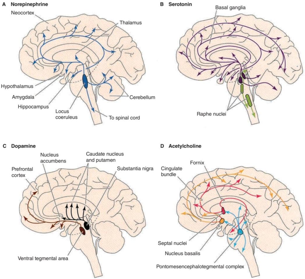
FIGURE 7-2 Four diffusely connected systems of central neuromodulators. A) Noradrenergic neurons in the locus coeruleus innervate the spinal cord, cerebellum, several nuclei of the hypothalamus, thalamus, basal telencephalon, and neocortex. B) Serotonergic neurons in the raphe nuclei project to the hypothalamus, limbic system, neocortex, cerebellum, and spinal cord. C) Dopaminergic neurons in the substantia nigra project to the striatum and those in the ventral tegmental area of the midbrain project to the prefrontal cortex of the limbic system. D) Cholinergic neurons in the basal forebrain complex project to the hippocampus and the neocortex and those in the pontomesencephalotegmental cholinergic complex project to the dorsal thalamus and the forebrain. (Reproduced with permission from Boron WF, Boulpaep EL: Medical Physiology. St. Louis, MO: Elsevier; 2005.)
شکل 7-2 چهار سیستم متصل به هم پخش کننده عصبی مرکزی. الف) نورون های نورآدرنرژیک در لوکوس سرولئوس نخاع، مخچه، چندین هسته هیپوتالاموس، تالاموس، تلانسفالن پایه و نئوکورتکس را عصب دهی می کنند. ب) نورون های سروتونرژیک در هسته های رافه به هیپوتالاموس، سیستم لیمبیک، نئوکورتکس، مخچه و نخاع می روند. ج) نورونهای دوپامینرژیک در جسم سیاه به جسم مخطط و آنهایی که در ناحیه تگمنتال شکمی مغز میانی هستند به قشر جلوی پیشانی سیستم لیمبیک میرسند. د) نورونهای کولینرژیک در کمپلکس قاعدهای پیشمغز به هیپوکامپ و نئوکورتکس و آنهایی که در کمپلکس کولینرژیک پونتومزانسفالوتگمنتال هستند به تالاموس پشتی و جلوی مغز میرسند. (تکثیر شده با مجوز Boron WF، Boulpaep EL: Medical Physiology. St. Louis, MO: Elsevier; 2005.)
RECEPTORS
The action of a chemical mediator on its target structure is more dependent on the type of receptor on which it acts than on the properties of the mediator per se. The individual receptors, along with their ligands (the molecules that bind to them), are discussed in the following parts of this chapter. However, five common themes about the actions of ligands on receptors have emerged.
گیرنده ها
عمل یک واسطه شیمیایی بر ساختار هدف خود بیشتر به نوع گیرنده ای که بر روی آن اثر می کند بستگی دارد تا به خواص واسطه فی نفسه. گیرنده های منفرد، همراه با لیگاندهای آنها (مولکول هایی که به آنها متصل می شوند)، در بخش های بعدی این فصل مورد بحث قرار می گیرند. با این حال، پنج موضوع مشترک در مورد اعمال لیگاندها بر روی گیرنده ها پدیدار شده است.
First, each chemical mediator has the potential to act on many subtypes of receptors. For example, norepinephrine acts on α1-, α2-, B1-, B2-, and ẞ3- adrenergic receptors. This multiplies the possible effects of a given ligand and makes its effects in each cell more selective.
اول، هر واسطه شیمیایی پتانسیل این را دارد که بر روی بسیاری از زیرگروههای گیرنده عمل کند. به عنوان مثال، نوراپی نفرین بر روی گیرنده های -α1-، α2-، B1-، B2 و -ẞ3 آدرنرژیک تأثیر می گذارد. این کار اثرات احتمالی یک لیگاند معین را چند برابر می کند و اثرات آن را در هر سلول انتخابی تر می کند.
Second, receptors for many neurotransmitters are located on both presynaptic and postsynaptic elements. A presynaptic receptor called an autoreceptor often inhibits further release of the transmitter, providing feedback control. For example, norepinephrine acts on α2-presynaptic receptors to inhibit additional norepinephrine release. A presynaptic heteroreceptor is one whose ligand is a chemical other than the transmitter released by the nerve ending on which the receptor is located. For example, norepinephrine acts on a heteroreceptor on a cholinergic nerve terminal to inhibit the release of acetylcholine. In some cases, presynaptic receptors facilitate the release of neurotransmitters.
دوم، گیرنده های بسیاری از انتقال دهنده های عصبی بر روی عناصر پیش سیناپسی و پس سیناپسی قرار دارند. یک گیرنده پیش سیناپسی به نام خود گیرنده اغلب آزاد شدن بیشتر فرستنده را مهار می کند و کنترل بازخورد را فراهم می کند. به عنوان مثال، نوراپی نفرین روی گیرنده های پیش سیناپسی α2 عمل می کند تا آزادسازی نوراپی نفرین اضافی را مهار کند. گیرنده پیش سیناپسی هتروگیرنده ای است که لیگاند آن یک ماده شیمیایی غیر از فرستنده ای است که توسط انتهای عصبی که گیرنده روی آن قرار دارد آزاد می شود. به عنوان مثال، نوراپی نفرین بر روی گیرنده هترورسپتور روی یک پایانه عصبی کولینرژیک عمل می کند تا آزادسازی استیل کولین را مهار کند. در برخی موارد، گیرنده های پیش سیناپسی آزادسازی انتقال دهنده های عصبی را تسهیل می کنند.
Third, receptors are grouped into two large families based on structure and function: ligand-gated channels (also known as ionotropic receptors) and metabotropic receptors (also known as G-protein-coupled receptors [GPCRs]). In the case of ionotropic receptors, a membrane channel is opened when a ligand binds to the receptor; and activation of the channel usually elicits a brief (few to tens of milliseconds) increase in ionic conductance. Thus, these receptors are important for fast synaptic transmission. Metabotropic receptors are 7-transmembrane GPCRs, and binding of a neurotransmitter to these receptors initiates the production of a second messenger that modulates the voltage-gated channels on neuronal membranes. The receptors for some neurotransmitters and neuromodulators are listed in Table 7-2, along with their principal second messengers and, where established, their net effect on ion channels. This table is an over-simplification. For example, activation of a2- adrenergic receptors decreases intracellular cyclic adenosine monophosphate (cAMP) concentrations, but there is evidence that the G-protein activated by a2- adrenergic presynaptic receptors also acts directly on Ca2+ channels to inhibit norepinephrine release.
سوم، گیرندهها بر اساس ساختار و عملکرد به دو خانواده بزرگ گروهبندی میشوند: کانالهای دروازهدار لیگاند (همچنین به عنوان گیرندههای یونوتروپیک شناخته میشوند) و گیرندههای متابوتروپیک (همچنین به عنوان گیرندههای جفت شده با پروتئین G [GPCRs] شناخته میشوند). در مورد گیرنده های یونوتروپیک، زمانی که یک لیگاند به گیرنده متصل می شود، یک کانال غشایی باز می شود. و فعال شدن کانال معمولا باعث افزایش مختصری (چند تا ده ها میلی ثانیه) در رسانایی یونی می شود. بنابراین، این گیرنده ها برای انتقال سریع سیناپسی مهم هستند. گیرنده های متابوتروپیک GPCR های 7 گذرنده هستند و اتصال یک انتقال دهنده عصبی به این گیرنده ها باعث تولید یک پیام رسان دوم می شود که کانال های دارای ولتاژ در غشای عصبی را تعدیل می کند. گیرندههای برخی از انتقالدهندههای عصبی و تعدیلکنندههای عصبی در جدول 7-2 به همراه پیامرسانهای دوم اصلی و، در صورت وجود، اثر خالص آنها بر کانالهای یونی فهرست شدهاند. این جدول یک ساده سازی بیش از حد است. به عنوان مثال، فعال شدن گیرنده های -a2 آدرنرژیک غلظت آدنوزین مونوفسفات حلقوی درون سلولی (cAMP) را کاهش می دهد، اما شواهدی وجود دارد که پروتئین G فعال شده توسط گیرنده های پیش سیناپسی a2- آدرنرژیک نیز مستقیماً روی کانال های +Ca2 برای مهار آزاد شدن نوراپی نفرین عمل می کند.
TABLE 7-2 Pharmacology of a selection of receptors for some small-molecule neurotransmitters.
جدول 7-2 فارماکولوژی انتخابی از گیرنده ها برای برخی از انتقال دهنده های عصبی با مولکول کوچک.
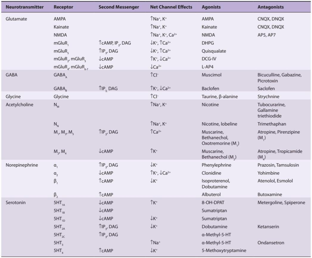
8-OH-DPAT, 8-hydroxy-N,N-dipropyl-2-aminotetralin; AMPA, a-amino-3-hydroxy-5-methyl-4-isoxazole-propionate; DAG, diacylglycerol; DCG-IV, 2-(2,3-dicarboxycyclopropyl) glycine; DHPG, 3-5dihydroxyphenylglycine; IP,, inositol triphosphate; L-AP4, 2-amino-4-phosphonobutyrate; NMDA, N-methyl-D-aspartate.
8-OH-DPAT، 8-هیدروکسی-N،N-دی پروپیل-2-آمینو تترالین. AMPA، a-amino-3-hydroxy-5-methyl-4-isoxazole-propionate. DAG، دی اسیل گلیسرول؛ DCG-IV، 2-(2،3-دی کربوکسی سیکلوپروپیل) گلیسین. DHPG، 3-5 دی هیدروکسی فنیل گلیسین؛ IP،، اینوزیتول تری فسفات؛ L-AP4، 2-amino-4-phosphonobutyrat; NMDA، N-متیل-D-آسپارتات.
Fourth, receptors are concentrated in clusters on the postsynaptic membrane close to the endings of neurons that secrete the neurotransmitters specific for them. This is generally due to the presence of specific binding proteins for them.
چهارم، گیرندهها بهصورت خوشهای روی غشای پس سیناپسی نزدیک به انتهای نورونها متمرکز شدهاند که انتقالدهندههای عصبی مخصوص آنها را ترشح میکنند. این به طور کلی به دلیل وجود پروتئین های اتصال خاص برای آنها است.
Fifth, in response to prolonged exposure to their ligands, most receptors become unresponsive; that is, they undergo desensitization. This can be of two types: homologous desensitization, with loss of responsiveness only to the ligand and maintained responsiveness of the cell to other ligands; and heterologous desensitization, in which the cell becomes unresponsive to other ligands as well.
پنجم، در پاسخ به قرار گرفتن در معرض طولانی مدت با لیگاندهای خود، اکثر گیرنده ها بی پاسخ می شوند. یعنی دچار حساسیت زدایی می شوند. این می تواند دو نوع باشد: حساسیت زدایی همولوگ، با از دست دادن پاسخگویی فقط به لیگاند و حفظ پاسخدهی سلول به لیگاندهای دیگر. و حساسیت زدایی هترولوگ، که در آن سلول به لیگاندهای دیگر نیز پاسخ نمی دهد.
REUPTAKE
Neurotransmitters are rapidly transported from the synaptic cleft back into the cytoplasm of the neurons that released them via a process called reuptake, which involves a high-affinity, Na+-dependent membrane transporter. Figure 7- 3 illustrates the reuptake of norepinephrine in a sympathetic postganglionic nerve ending. After release of norepinephrine into the synaptic cleft, it is rapidly routed back into the sympathetic nerve terminal by a norepinephrine transporter (NET). A portion of the norepinephrine that re-enters the neuron is sequestered into the synaptic vesicles through the vesicular monoamine transporter (VMAT). There are analogous membrane and vesicular transporters for other small-molecule neurotransmitters released at other synapses in the CNS and peripheral nervous system.
جذب مجدد
انتقال دهنده های عصبی به سرعت از شکاف سیناپسی به سیتوپلاسم نورون ها منتقل می شوند که آنها را از طریق فرآیندی به نام بازجذب آزاد می کند که شامل یک انتقال دهنده غشایی وابسته به +Na با میل ترکیبی بالا است. شکل 7-3 بازجذب نوراپی نفرین را در انتهای عصب پس گانگلیونی سمپاتیک نشان می دهد. پس از آزاد شدن نوراپی نفرین در شکاف سیناپسی، به سرعت توسط یک انتقال دهنده نوراپی نفرین (NET) به انتهای عصب سمپاتیک بازگردانده می شود. بخشی از نوراپی نفرین که مجدداً وارد نورون می شود از طریق ناقل مونوآمین تاولی (VMAT) به داخل وزیکول های سیناپسی منزوی می شود. ناقلهای غشایی و تاولی مشابهی برای سایر انتقالدهندههای عصبی مولکولی کوچک وجود دارد که در سیناپسهای دیگر در CNS و سیستم عصبی محیطی آزاد میشوند.

FIGURE 7-3 Fate of monoamines released at synaptic junctions. In each monoamine-secreting neuron, the monoamine is synthesized in the cytoplasm and the secretory granules and its concentration in secretory granules is maintained by the two vesicular monoamine transporters (VMAT). The monoamine is released by exocytosis of the granules, and it acts on G-protein- coupled receptors. In this example, the monoamine is norepinephrine acting on adrenoceptors. Many of these receptors are postsynaptic, but some are presynaptic and some are located on glia. In addition, there is extensive reuptake of the monoamine into the cytoplasm of the presynaptic terminal via a monoamine transporter, in this case the norepinephrine transporter (NET). (Modified with permission from Katzung BG, Masters SB, Trevor AJ: Basic and Clinical Pharmacology, 11th ed. New York, NY: McGraw-Hill; 2009.)
شکل 7-3 سرنوشت مونوآمین های آزاد شده در اتصالات سیناپسی. در هر نورون ترشح کننده مونوآمین، مونوآمین در سیتوپلاسم و گرانول های ترشحی سنتز می شود و غلظت آن در گرانول های ترشحی توسط دو ناقل مونوآمین تاولی (VMAT) حفظ می شود. مونوآمین در اثر اگزوسیتوز گرانول ها آزاد می شود و روی گیرنده های جفت شده با پروتئین G عمل می کند. در این مثال، مونوآمین نوراپی نفرین است که روی گیرنده های آدرنرژیک اثر می کند. بسیاری از این گیرنده ها پس سیناپسی هستند، اما برخی پیش سیناپسی هستند و برخی روی گلیا قرار دارند. علاوه بر این، بازجذب گسترده مونوآمین به سیتوپلاسم پایانه پیش سیناپسی از طریق یک انتقال دهنده مونوآمین، در این مورد، انتقال دهنده نوراپی نفرین (NET) وجود دارد. (اصلاح شده با مجوز Katzung BG, Masters SB, Trevor AJ: Basic and Clinical Pharmacology, ed. 11 New York, NY: McGraw-Hill; 2009.)
Reuptake is a major factor in terminating the action of neurotransmitters, and when it is inhibited, the effects of neurotransmitter release are increased and prolonged. This has clinical consequences. For example, several effective antidepressant drugs are inhibitors of the reuptake of amine transmitters. Glutamate reuptake into neurons and glia is important because glutamate is an excitotoxin that can kill cells by overstimulating them (see Clinical Box 7-1). During ischemia and anoxia, loss of neurons is increased because glutamate reuptake is inhibited.
بازجذب یک عامل اصلی در خاتمه عمل انتقال دهنده های عصبی است و هنگامی که مهار شود، اثرات آزاد شدن انتقال دهنده های عصبی افزایش یافته و طولانی تر می شود. این عواقب بالینی دارد. به عنوان مثال، چندین داروی ضد افسردگی موثر، مهارکننده های بازجذب ترانسمیترهای آمین هستند. بازجذب گلوتامات به نورون ها و گلیا مهم است زیرا گلوتامات یک اکسیتوتوکسین است که می تواند سلول ها را با تحریک بیش از حد آنها را از بین ببرد (به جعبه بالینی 7-1 مراجعه کنید). در طول ایسکمی و آنوکسی، از دست دادن نورون ها افزایش می یابد زیرا بازجذب گلوتامات مهار می شود.
CLINICAL BOX 7.1
Excitotoxins
Glutamate is usually cleared from the brain’s extracellular fluid by Na+- dependent uptake systems in neurons and glia, keeping only micromolar levels of the chemical in the extracellular fluid despite millimolar levels inside neurons. However, excessive levels of glutamate occur in response to ischemia, anoxia, hypoglycemia, or trauma. Glutamate and some of its synthetic agonists are unique in that when they act on neuronal cell bodies, they can produce so much Ca2+ influx that the neurons die. Excitotoxins play a significant role in the damage done to the brain by a stroke. When a cerebral artery is occluded, the cells in the severely ischemic area die. The surrounding partially ischemic cells may survive but lose their ability to maintain the transmembrane Na* gradient. The elevated levels of intracellular Na* prevent the ability of astrocytes to remove glutamate from the brain’s extracellular fluid. Therefore, glutamate accumulates to the point that excitotoxic damage and cell death occurs in the penumbra, the region around the completely infarcted area. In addition, excessive glutamate receptor activation may contribute to the pathophysiology of some neurodegenerative diseases such as amyotrophic lateral sclerosis (ALS), Parkinson disease, and Alzheimer disease.
جعبه بالینی 7.1
اکسیتوتوکسین ها
گلوتامات معمولاً توسط سیستمهای جذب وابسته به +Na در سلولهای عصبی و گلیا از مایع خارج سلولی مغز پاک میشود و با وجود سطوح میلیمولاری درون نورونها، تنها سطوح میکرومولاری این ماده شیمیایی را در مایع خارج سلولی حفظ میکند. با این حال، سطوح بیش از حد گلوتامات در پاسخ به ایسکمی، آنوکسی، هیپوگلیسمی یا تروما رخ می دهد. گلوتامات و برخی از آگونیستهای مصنوعی آن از این نظر منحصربهفرد هستند که وقتی روی سلولهای عصبی اثر میکنند، میتوانند آنقدر هجوم +Ca2 تولید کنند که نورونها بمیرند. اکسیتوتوکسین ها نقش مهمی در آسیب وارده به مغز در اثر سکته دارند. هنگامی که یک شریان مغزی مسدود می شود، سلول های ناحیه ایسکمیک شدید می میرند. سلولهای ایسکمیک نسبی اطراف ممکن است زنده بمانند، اما توانایی خود را برای حفظ گرادیان +Na گذرنده از دست بدهند. سطوح بالای +Na داخل سلولی از توانایی آستروسیت ها برای حذف گلوتامات از مایع خارج سلولی مغز جلوگیری می کند. بنابراین، گلوتامات به حدی انباشته می شود که آسیب اکسیتوتوکسیک و مرگ سلولی در نیم سایه، ناحیه اطراف ناحیه کاملاً انفارکتوس شده رخ می دهد. علاوه بر این، فعال شدن بیش از حد گیرنده گلوتامات ممکن است به پاتوفیزیولوژی برخی از بیماری های نورودژنراتیو مانند اسکلروز جانبی آمیوتروفیک (ALS)، بیماری پارکینسون و بیماری آلزایمر کمک کند.
THERAPEUTIQ HIGHLIGHTS
Riluzole is a voltage-gated channel blocker that may antagonize N-methyl-D- aspartate (NMDA) receptors. It slows the progression of impairment and modestly improves the life expectancy of patients with ALS. Another NMDA receptor antagonist memantine has been used to slow the progressive decline in patients with Alzheimer disease. A third NMDA receptor antagonist, amantadine, in conjunction with levodopa, improves function in patients with Parkinson disease.
نکات برجسته درمانی
ریلوزول یک مسدود کننده کانال با ولتاژ است که ممکن است گیرنده های N-methyl-D-aspartate (NMDA) را متضاد کند. این روند پیشرفت اختلال را کند می کند و امید به زندگی بیماران مبتلا به ALS را به طور متوسط بهبود می بخشد. یکی دیگر از ممانتین آنتاگونیست گیرنده NMDA برای کند کردن کاهش پیشرونده در بیماران مبتلا به بیماری آلزایمر استفاده شده است. سومین آنتاگونیست گیرنده NMDA، آمانتادین، در ارتباط با لوودوپا، عملکرد را در بیماران مبتلا به بیماری پارکینسون بهبود می بخشد.
SMALL-MOLECULE TRANSMITTERS: EXCITATORY & INHIBITORY AMINO ACIDS, ACETYLCHOLINE, AND MONOAMINES
Glutamate
The amino acid glutamate is the main excitatory neurotransmitter in the brain and spinal cord and may be responsible for 75% of the excitatory transmission in the CNS. There are two distinct pathways involved in the synthesis of glutamate (Figure 7-1). In one pathway, a-ketoglutarate produced by the Krebs cycle is converted to glutamate by the enzyme GABA transaminase (GABA-T). In the second pathway, glutamate is released from the nerve terminal into the synaptic cleft by Ca2+ -dependent exocytosis and transported via a glutamate reuptake transporter into glia, where it is converted to glutamine by the enzyme glutamine synthetase (Figure 7-4). Glutamine then diffuses back into the nerve terminal where it is hydrolyzed back to glutamate by the enzyme glutaminase. A membrane transporter also returns glutamate directly into the nerve terminal. Within glutamatergic neurons, glutamate is highly concentrated in synaptic vesicles by a vesicular glutamate transporter.
انتقال دهنده های مولکولی کوچک: اسیدهای آمینه تحریکی و مهاری، استیل کولین و مونوآمین ها
گلوتامات
اسید آمینه گلوتامات، انتقال دهنده عصبی تحریکی اصلی در مغز و نخاع است و ممکن است مسئول 75 درصد از انتقال تحریکی در CNS باشد. دو مسیر متمایز در سنتز گلوتامات وجود دارد (شکل 7-1). در یک مسیر، a-ketoglutarate تولید شده توسط چرخه کربس توسط آنزیم GABA ترانس آمیناز (GABA-T) به گلوتامات تبدیل می شود. در مسیر دوم، گلوتامات از پایانه عصبی به داخل شکاف سیناپسی توسط اگزوسیتوز وابسته به +Ca2 آزاد می شود و از طریق یک انتقال دهنده بازجذب گلوتامات به گلیا منتقل می شود، جایی که توسط آنزیم گلوتامین سنتتاز به گلوتامین تبدیل می شود (شکل 7-4). سپس گلوتامین دوباره به انتهای عصبی منتشر می شود و در آنجا توسط آنزیم گلوتامیناز به گلوتامات هیدرولیز می شود. یک انتقال دهنده غشایی همچنین گلوتامات را مستقیماً به انتهای عصبی باز می گرداند. در نورونهای گلوتاماترژیک، گلوتامات در وزیکولهای سیناپسی توسط یک انتقالدهنده گلوتامات تاولی بسیار متمرکز است.

FIGURE 7-4 Biochemical events at a glutamatergic synapse. Glutamate(Glu) released into the synaptic cleft by Ca2+-dependent exocytosis. Released Glu can act on ionotropic and G-protein-coupled receptors on the postsynaptic neuron. Synaptic transmission is terminated by the active transport of Glu via by a Na*-dependent glutamate transporters located on membranes of the presynaptic terminal [Gl(n)] and glia [Gl(g)]. In glia, Glu is converted to glutamine (Gln) by the enzyme glutamine synthetase; Gln then diffuses into the nerve terminal where it is hydrolyzed back to Glu by the enzyme glutaminase. In the nerve terminal, Glu is highly concentrated in synaptic vesicles by a vesicular glutamate transporter.
شکل 7-4 رویدادهای بیوشیمیایی در یک سیناپس گلوتاماترژیک. گلوتامات (Glu) توسط اگزوسیتوز وابسته به +Ca2 در شکاف سیناپسی آزاد می شود. گلو آزاد شده می تواند بر روی گیرنده های یونوتروپیک و جفت شده با پروتئین G روی نورون پس سیناپسی اثر بگذارد. انتقال سیناپسی با انتقال فعال گلو از طریق یک ناقل گلوتامات وابسته به +Na که بر روی غشاهای پایانه پیش سیناپسی [Gl(n)] و گلیا [Gl(g)] قرار دارد، خاتمه می یابد. در گلیا، گلو توسط آنزیم گلوتامین سنتتاز به گلوتامین (Gln) تبدیل می شود. سپس Gln در پایانه عصبی منتشر می شود و در آنجا توسط آنزیم گلوتامیناز به Glu باز می گردد. در پایانه عصبی، Glu به شدت در وزیکول های سیناپسی توسط یک انتقال دهنده گلوتامات تاولی متمرکز می شود.
Glutamate Receptors
Glutamate acts on both ionotropic and metabotropic receptors in the CNS (Figure 7-4). There are three subtypes of ionotropic glutamate receptors, each named for its relatively specific agonist. These are the AMPA (α-amino-3- hydroxy-5-methylisoxazole-4-propionate), kainate, and NMDA receptors. Table 7-2 summarizes some of the major properties of these receptors. Ionotropic glutamate receptors are tetramers composed of different subunits whose helical domains span the membranes three times and a short sequence that forms the channel pore. Four AMPA (GluR1-GluR4), five kainate (GluR5- GluR7, KA1, KA2), and six NMDA (NR1, NR2A-NR2D) subunits have been identified, and each is coded by a different gene.
گیرنده های گلوتامات
گلوتامات روی هر دو گیرنده یونوتروپیک و متابوتروپیک در CNS عمل می کند (شکل 7-4). سه زیرگروه از گیرنده های گلوتامات یونوتروپیک وجود دارد که هر کدام به دلیل آگونیست نسبتاً خاص خود نامگذاری شده اند. اینها گیرنده های AMPA (α-آمینو-3-هیدروکسی-5-متیل ایزوکسازول-4-پروپیونات)، کاینات و NMDA هستند. جدول 7-2 برخی از خواص اصلی این گیرنده ها را خلاصه می کند. گیرندههای گلوتامات یونوتروپیک تترامرهایی هستند که از زیر واحدهای مختلفی تشکیل شدهاند که حوزههای مارپیچی آنها سه بار روی غشاها و یک توالی کوتاه که منافذ کانال را تشکیل میدهند، تشکیل شدهاند. چهار زیرواحد AMPA (GluR1-GluR4)، پنج کاینات (GluR5-GluR7، KA1، KA2)، و شش زیرواحد NMDA (NR1، NR2A-NR2D) شناسایی شدهاند و هر کدام توسط ژن متفاوتی کدگذاری میشوند.
The release of glutamate and its binding to AMPA or kainate receptors primarily permits the influx of Na* and the efflux of K+, accounting for a fast- excitatory postsynaptic potential (EPSP). Most AMPA receptors have low Ca2+ permeability, but the absence of certain subunits in the receptor complex at some sites allows for the influx of Ca2+, which may contribute to the excitotoxic effect of glutamate (see Clinical Box 7-1).
انتشار گلوتامات و اتصال آن به گیرندههای AMPA یا کاینات در درجه اول اجازه ورود +Na و خروج +K را میدهد که پتانسیل پس سیناپسی تحریککننده سریع (EPSP) را تشکیل میدهد. اکثر گیرنده های AMPA دارای نفوذپذیری پایینی از +Ca2 هستند، اما عدم وجود زیرواحدهای خاص در مجموعه گیرنده در برخی مکان ها امکان هجوم +Ca2 را فراهم می کند، که ممکن است به اثر اکسیتوتوکسیک گلوتامات کمک کند (به جعبه بالینی 7-1 مراجعه کنید).
Activation of the NMDA receptor permits the influx of relatively large amounts of Ca2+ along with Na*. When glutamate is in excess in the synaptic cleft, the NMDA receptor-induced influx of Ca2+ into neurons is the major basis for the excitotoxic actions of glutamate. The NMDA receptor is unique in several ways (Figure 7-5). First, glycine binding to the NMDA receptor is essential for the receptor to respond to glutamate. Second, when glutamate binds to the NMDA receptor, it opens, but at normal membrane potentials, the channel is blocked by extracellular Mg2+. This block is removed only when the neuron containing the receptor is partially depolarized by the activation of adjacent AMPA and kainate receptors. Third, the EPSP induced by activation of NMDA receptors is slower than that elicited by activation of the AMPA and kainate receptors.
فعال شدن گیرنده NMDA اجازه ورود مقادیر نسبتاً زیادی +Ca2 همراه با +Na را می دهد. هنگامی که گلوتامات در شکاف سیناپسی بیش از حد باشد، هجوم +Ca2 ناشی از NMDA به سلولهای عصبی، مبنای اصلی اعمال اکسیتوتوکسیک گلوتامات است. گیرنده NMDA از چند جهت منحصر به فرد است (شکل 7-5). اول، اتصال گلیسین به گیرنده NMDA برای پاسخ دادن گیرنده به گلوتامات ضروری است. دوم، هنگامی که گلوتامات به گیرنده NMDA متصل می شود، باز می شود، اما در پتانسیل های غشایی طبیعی، کانال توسط +Mg2 خارج سلولی مسدود می شود. این بلوک تنها زمانی حذف می شود که نورون حاوی گیرنده تا حدی با فعال شدن گیرنده های AMPA و کاینات مجاور دپلاریزه شود. سوم، EPSP ناشی از فعالسازی گیرندههای NMDA کندتر از آن است که توسط فعالسازی گیرندههای AMPA و kainate ایجاد میشود.

FIGURE 7-5 Diagrammatic representation of the NMDA receptor. When glycine and glutamate bind to the receptor, the closed ion channel (left) opens, but at the resting membrane potential, the channel is blocked by Mg2+ (right). This block is removed if partial depolarization is produced by other inputs to the neuron containing the receptor, and Ca2+ and Na* enter the neuron. Blockade can also be produced by the drug dizocilpine maleate (MK-801).
شکل 7-5 نمایش نموداری گیرنده NMDA. هنگامی که گلیسین و گلوتامات به گیرنده متصل می شوند، کانال یونی بسته (سمت چپ) باز می شود، اما در پتانسیل غشاء استراحت، کانال توسط +Mg2 (راست) مسدود می شود. اگر دپلاریزاسیون جزئی توسط ورودی های دیگر نورون حاوی گیرنده ایجاد شود، این بلوک حذف می شود و +Ca2 و +Na وارد نورون می شوند. بلوک همچنین می تواند توسط داروی dizocilpine maleate (MK-801) تولید شود.
Essentially all neurons in the CNS have both AMPA and NMDA receptors. Kainate receptors are located presynaptically on GABA-secreting nerve endings and postsynaptically at various sites, most notably in the hippocampus, cerebellum, and spinal cord. Kainate and AMPA receptors are also found in glia. The concentration of NMDA receptors in the hippocampus is high, and blockade of these receptors prevents long-term potentiation, a long-lasting facilitation of transmission in neural pathways following a brief period of high-frequency stimulation. Thus, these receptors may be involved in memory and learning (see Chapter 15).
اساساً تمام نورونهای CNS دارای گیرندههای AMPA و NMDA هستند. گیرنده های کاینات به صورت پیش سیناپسی روی انتهای عصبی ترشح کننده گابا و به صورت پس سیناپسی در مکان های مختلف، به ویژه در هیپوکامپ، مخچه و نخاع قرار دارند. گیرنده های کاینات و AMPA نیز در گلیا یافت می شوند. غلظت گیرندههای NMDA در هیپوکامپ بالا است، و مسدود شدن این گیرندهها از تقویت طولانیمدت جلوگیری میکند، تسهیل طولانیمدت انتقال در مسیرهای عصبی پس از یک دوره کوتاه تحریک با فرکانس بالا. بنابراین، این گیرنده ها ممکن است در حافظه و یادگیری نقش داشته باشند (به فصل 15 مراجعه کنید).
Activation of the metabotropic glutamate receptors (mGluR) leads to either an increase in intracellular inositol 1,4,5-triphosphate (IP3) and diacylglycerol (DAG) levels or a decrease in intracellular cAMP levels (Table 7-2). There are eight known subtypes of mGluR located at either presynaptic (mGluR2-4,6-8) or postsynaptic sites (mGluR15) and are widely distributed in the brain. They may be involved in the production of synaptic plasticity, particularly in the hippocampus and the cerebellum.
فعال شدن گیرنده های متابوتروپیک گلوتامات (mGluR) منجر به افزایش سطح اینوزیتول 1،4،5-تری فسفات (IP3) و دی اسیل گلیسرول (DAG) داخل سلولی یا کاهش سطوح cAMP داخل سلولی می شود (جدول 7-2). هشت زیرگروه شناخته شده از mGluR وجود دارد که در محل های پیش سیناپسی (mGluR2-4,6-8) یا پس سیناپسی (mGluR15) قرار دارند و به طور گسترده در مغز توزیع می شوند. آنها ممکن است در تولید شکل پذیری سیناپسی، به ویژه در هیپوکامپ و مخچه نقش داشته باشند.
Activation of presynaptic mGluR autoreceptors on hippocampal neurons limits the release of glutamate from these neurons. Knockout of the gene mGluR1 causes severe motor incoordination and deficits in spatial learning. Dysregulation of brain levels of the mGluR5 has been linked to neurological disorders including schizophrenia, major depressive disorders, and autism.
فعال شدن خود گیرنده های mGluR پیش سیناپسی روی نورون های هیپوکامپ، آزادسازی گلوتامات از این نورون ها را محدود می کند. حذف ژن mGluR1 باعث ناهماهنگی شدید حرکتی و نقص در یادگیری فضایی می شود. اختلال در سطح مغز mGluR5 با اختلالات عصبی از جمله اسکیزوفرنی، اختلالات افسردگی اساسی و اوتیسم مرتبط است.
Pharmacology of Glutamate Synapses
Table 7-2 shows some of the pharmacologic properties of glutamate receptors, examples of the agonists that bind to these receptors, and some of the antagonists that prevent activation of the receptors. The clinical applications of drugs that modulate glutamatergic transmission are still in their infancy. This is because the role of glutamate as a neurotransmitter was discovered much later than that of most other small- molecule transmitters. Intraspinal or extradural administration of NMDA receptor antagonists is used for the treatment of chronic pain. The mGluR5 is a target for development of drugs in the treatment of neuropsychiatric disorders.
فارماکولوژی سیناپس های گلوتامات
جدول 7-2 برخی از خواص دارویی گیرنده های گلوتامات، نمونه هایی از آگونیست هایی که به این گیرنده ها متصل می شوند و برخی از آنتاگونیست هایی که از فعال شدن گیرنده ها جلوگیری می کنند را نشان می دهد. کاربردهای بالینی داروهایی که انتقال گلوتاماترژیک را تعدیل می کنند هنوز در مراحل ابتدایی خود هستند. این به این دلیل است که نقش گلوتامات به عنوان یک انتقال دهنده عصبی بسیار دیرتر از سایر فرستنده های مولکولی کوچک کشف شد. تجویز داخل نخاعی یا اکسترادورال آنتاگونیست های گیرنده NMDA برای درمان درد مزمن استفاده می شود. mGluR5 هدفی برای توسعه داروها در درمان اختلالات عصبی روانی است.
GABA
GABA is the major inhibitory mediator in the brain and mediates both presynaptic and postsynaptic inhibition. GABA is formed by decarboxylation of glutamate (Figure 7-1) by the enzyme glutamate decarboxylase (GAD), in nerve endings in many parts of the brain. A vesicular GABA transporter (VGAT) transports GABA into secretory vesicles. GABA is metabolized primarily by transamination to succinic semialdehyde and then to succinate in the citric acid cycle. GABA-T is the enzyme that catalyzes the transamination. After GABA is released from a neuron, a high-affinity GABA transporter allows for its reuptake.
گابا
GABA واسطه بازدارنده اصلی در مغز است و واسطه مهار پیش سیناپسی و پس سیناپسی است. گابا از دکربوکسیلاسیون گلوتامات (شکل 7-1) توسط آنزیم گلوتامات دکربوکسیلاز (GAD)، در انتهای عصبی در بسیاری از قسمت های مغز تشکیل می شود. یک انتقال دهنده گابا تاولی (VGAT) گابا را به وزیکول های ترشحی منتقل می کند. گابا عمدتاً از طریق ترانس آمیناسیون به نیمه آلدئید سوکسینیک و سپس به سوکسینات در چرخه اسید سیتریک متابولیزه می شود. GABA-T آنزیمی است که ترانس آمیناسیون را کاتالیز می کند. پس از آزاد شدن گابا از یک نورون، یک انتقال دهنده گابا با میل ترکیبی بالا امکان جذب مجدد آن را فراهم می کند.
GABA Receptors
Three subtypes of GABA receptors have been identified: GABAA, GABA, and GABAC (Table 7-2). The GABA and GABA receptors (Figure 7-6) are widely distributed in the CNS, GABA receptors are found almost exclusively in the retina. The GABA and GABA receptors are ionotropic receptors, activation of which allows the entry of Clinto neurons to mediate fast inhibitory postsynaptic potentials (IPSP). The GABA receptors are GPCRs that are linked via G-proteins to alter the influx of K+ and Ca2+; G; inhibits adenylyl cyclase to open a K+ channel, and G inhibits or delays Ca2+ influx. Activation of GABA receptors mediates both presynaptic and slow postsynaptic inhibition.
گیرنده های GABA
سه زیر گروه از گیرنده های گابا شناسایی شده اند: گابا، گابا و گاباک (جدول 7-2). گیرنده های GABA و GABA (شکل 7-6) به طور گسترده در CNS توزیع شده اند، گیرنده های GABA تقریباً منحصراً در شبکیه چشم یافت می شوند. گیرندههای GABA و GABA گیرندههای یونوتروپیک هستند که فعال شدن آنها به نورونهای کلینتو اجازه میدهد تا پتانسیلهای پس سیناپسی مهاری سریع (IPSP) را واسطه کنند. گیرنده های GABA GPCR هایی هستند که از طریق پروتئین های G به یکدیگر متصل می شوند تا هجوم +K و +Ca2 را تغییر دهند. G; آدنیلیل سیکلاز را برای باز کردن کانال +K مهار می کند و G هجوم +Ca2 را مهار یا به تاخیر می اندازد. فعالسازی گیرندههای GABA باعث مهار پیشسیناپسی و آهسته پسسیناپسی میشود.

FIGURE 7-6 Diagram of GABA and GABA receptors, showing their principal actions. Two molecules of GABA (squares) bind to the GABAA receptor to allow an influx of Cl ̄ to mediate fast inhibitory postsynaptic potentials. One molecule of GABA binds to the GABA receptor, which couples to the a-subunit of a G-protein; G; inhibits adenylyl cyclase to open a K+ channel and G inhibits Ca2+ influx.
شکل 7-6 نمودار گیرنده های GABA و GABA که عملکرد اصلی آنها را نشان می دهد. دو مولکول گابا (مربع) به گیرنده گاباآ متصل می شوند تا به هجوم ̄Cl اجازه دهند تا پتانسیل های پس سیناپسی مهاری سریع را واسطه کنند. یک مولکول گابا به گیرنده گابا متصل می شود که به زیرواحد a یک پروتئین G متصل می شود. G; آدنیلیل سیکلاز را برای باز کردن کانال +K مهار می کند و G از هجوم +Ca2 جلوگیری می کند.
The GABAA receptors are pentamers made up of various combinations of six a subunits, four ẞ, four y, one 8, and one ε. This endows them with considerably different properties from one location to another. However, most synaptic GABAA receptors have two a, two ẞ, and one y subunit (Figure 7-6). GABAA receptors on dendrites, axons, or somas often contain 8 and & subunits in place of the y subunit. The GABA receptors are relatively simple in that they are pentamers of three p subunits in various combinations.
گیرنده های گاباآ پنتامرهایی هستند که از ترکیب های مختلف شش زیرواحد a، چهار ẞ، چهار y، یک 8 و یک ε تشکیل شده اند. این به آنها خواص بسیار متفاوتی از یک مکان به مکان دیگر می دهد. با این حال، اکثر گیرنده های سیناپسی GABAA دارای دو زیرواحد a، دو ẞ و یک y هستند (شکل 7-6). گیرندههای گاباآ روی دندریتها، آکسونها یا سوماها اغلب حاوی 8 و زیر واحد به جای زیرواحد y هستند. گیرندههای GABA نسبتاً ساده هستند زیرا پنجتامرهای سه زیرواحد p در ترکیبهای مختلف هستند.
Pharmacology of GABA Synapses
Table 7-2 shows some of the pharmacologic properties of GABA receptors, including examples of agonists that bind to receptors and some of the antagonists that prevent activation of these receptors. The increase in Cl conductance produced by GABA receptors is potentiated by benzodiazepines (eg, diazepam). Thus, these are examples of neuromodulators. These drugs have marked antianxiety activity and are also effective muscle relaxants, anticonvulsants, and sedatives. Benzodiazepines bind to a subunits of GABAA receptors. Barbiturates such as phenobarbital are effective anticonvulsants because they enhance GABA receptor-mediated inhibition and suppress AMPA receptor-mediated excitation. The anesthetic actions of barbiturates (thiopental, pentobarbital, and methoxital) result from their actions as agonists at GABA receptors as well as by acting as neuromodulators of GABA transmission. Regional variation in anesthetic actions in the brain parallels the variation in subtypes of GABA receptors.
فارماکولوژی سیناپس گابا
جدول 7-2 برخی از خواص دارویی گیرنده های GABA را نشان می دهد، از جمله نمونه هایی از آگونیست هایی که به گیرنده ها متصل می شوند و برخی از آنتاگونیست هایی که از فعال شدن این گیرنده ها جلوگیری می کنند. افزایش هدایت کلر تولید شده توسط گیرنده های GABA توسط بنزودیازپین ها (مثلا دیازپام) تقویت می شود. بنابراین، اینها نمونه هایی از تعدیل کننده های عصبی هستند. این داروها فعالیت ضد اضطراب مشخصی دارند و همچنین شل کننده عضلانی، ضد تشنج و آرام بخش هستند. بنزودیازپین ها به زیر واحدهای گیرنده گاباآ متصل می شوند. باربیتوراتهایی مانند فنوباربیتال ضد تشنجهای مؤثری هستند زیرا مهار با واسطه گیرنده GABA را افزایش میدهند و تحریک با واسطه گیرنده AMPA را سرکوب میکنند. اثرات بیهوشی باربیتورات ها (تیوپنتال، پنتوباربیتال و متوکسیتال) ناشی از عملکرد آنها به عنوان آگونیست در گیرنده های GABA و همچنین عمل به عنوان تعدیل کننده عصبی انتقال GABA است. تنوع منطقه ای در اعمال بیهوشی در مغز به موازات تنوع در زیرگروه های گیرنده های GABA است.
Glycine
Glycine has both excitatory and inhibitory effects in the CNS. When it binds to NMDA receptors, it makes them more sensitive to the actions of glutamate. Glycine may spill over from synaptic junctions into the interstitial fluid and in the spinal cord; for example, it may facilitate pain transmission by NMDA receptors in the dorsal horn. However, glycine mediates direct inhibition in the brainstem and spinal cord. The glycine receptor that mediates inhibition is a Cl channel, a pentamer made up of two subunits: the ligand-binding a subunit and the structural ẞ subunit. Like GABA, glycine acts by increasing Cl- conductance. Its action is antagonized by strychnine. The clinical picture of convulsions and muscular hyperactivity produced by strychnine emphasizes the importance of postsynaptic inhibition in normal neuronal function.
گلیسین
گلایسین دارای اثرات تحریکی و مهاری در CNS است. هنگامی که به گیرنده های NMDA متصل می شود، آنها را نسبت به عملکرد گلوتامات حساس تر می کند. گلایسین ممکن است از اتصالات سیناپسی به داخل مایع بینابینی و در نخاع بریزد. به عنوان مثال، ممکن است انتقال درد توسط گیرنده های NMDA در شاخ پشتی را تسهیل کند. با این حال، گلیسین واسطه مهار مستقیم در ساقه مغز و نخاع است. گیرنده گلیسین که واسطه مهار است، یک کانال کلر است، یک پنتامر که از دو زیر واحد تشکیل شده است: یک زیرواحد متصل به لیگاند و زیرواحد ẞ ساختاری. گلیسین مانند گابا با افزایش رسانایی کلر عمل می کند. عمل آن با استریکنین مخالف است. تصویر بالینی تشنج و بیش فعالی عضلانی تولید شده توسط استریکنین بر اهمیت مهار پس سیناپسی در عملکرد طبیعی نورون تاکید می کند.
There are three kinds of spinal cord neurons that mediate direct inhibition: neurons that release glycine, neurons that release GABA, and neurons that release both. Neurons that release only glycine have the glycine transporter GLYT2, those that release only GABA have VGAT, and those that release glycine and GABA have both. Glycine and GABA can be found in the same vesicles in these latter neurons.
سه نوع نورون طناب نخاعی وجود دارد که واسطه مهار مستقیم هستند: نورون هایی که گلیسین را آزاد می کنند، نورون هایی که GABA را آزاد می کنند و نورون هایی که هر دو را آزاد می کنند. نورون هایی که فقط گلیسین آزاد می کنند دارای ناقل گلیسین GLYT2 هستند، آنهایی که فقط GABA آزاد می کنند VGAT دارند و آنهایی که گلیسین و گابا را آزاد می کنند هر دو را دارند. گلیسین و گابا را می توان در همان وزیکول های این نورون های دوم یافت.
Acetylcholine
Acetylcholine is the transmitter at the neuromuscular junction, in autonomic ganglia, and in postganglionic parasympathetic nerve-target organ junctions and some postganglionic sympathetic nerve-target junctions (see Chapter 13). In fact, acetylcholine is the transmitter released by all neurons that exit the CNS (cranial nerves, motor neurons, and preganglionic neurons). Acetylcholine is also found in the basal forebrain complex (septal nuclei and nucleus basalis), which projects to the hippocampus and neocortex, and the pontomesencephalic cholinergic complex, which projects to the dorsal thalamus and forebrain (Figure 7-2). These systems may be involved in regulation of sleep-wake states, learning, and memory (see Chapters 14 and 15).
استیل کولین
استیل کولین فرستنده در اتصال عصبی عضلانی، در گانگلیون های اتونوم، و در اتصالات عصب-ارگان هدف پاراسمپاتیک پس گانگلیونی و برخی از اتصالات عصب سمپاتیک-هدف پس از عقده ای است (به فصل 13 مراجعه کنید). در واقع، استیل کولین فرستنده ای است که توسط تمام نورون هایی که از CNS خارج می شوند (اعصاب جمجمه ای، نورون های حرکتی و نورون های پیش گانگلیونی) آزاد می شود. استیل کولین همچنین در کمپلکس قاعدهای پیشمغز (هستههای سپتال و هسته بازالیس)، که به هیپوکامپ و نئوکورتکس میتابد، و کمپلکس کولینرژیک پونتومزانسفالیک، که به تالاموس پشتی و پیشمغز میتابد، یافت میشود (شکل 7-2). این سیستم ها ممکن است در تنظیم حالت خواب-بیداری، یادگیری و حافظه نقش داشته باشند (به فصل های 14 و 15 مراجعه کنید).
Acetylcholine is found in small, clear synaptic vesicles in high concentration in the terminals of cholinergic neurons. It is synthesized in the nerve terminal from choline and acetyl-CoA by the enzyme choline acetyltransferase (CHAT) (Figure 7-1 and Figure 7-7). Choline used in the synthesis of acetylcholine is transported from the extracellular space into the nerve terminal via a Na+- dependent choline transporter (CHT). Following its synthesis, acetylcholine is transported from the cytoplasm into vesicles by a vesicle-associated transporter (VAT). Acetylcholine is released when a nerve impulse triggers the influx of Ca2+ into the nerve terminal.
استیل کولین در وزیکول های سیناپسی کوچک و شفاف با غلظت بالا در انتهای نورون های کولینرژیک یافت می شود. در پایانه عصبی از کولین و استیل-CoA توسط آنزیم کولین استیل ترانسفراز (CHAT) سنتز می شود (شکل 7-1 و شکل 7-7). کولین مورد استفاده در سنتز استیل کولین از فضای خارج سلولی به پایانه عصبی از طریق یک ناقل کولین وابسته به Na+ (CHT) منتقل می شود. پس از سنتز، استیل کولین از سیتوپلاسم به وزیکول ها توسط یک انتقال دهنده مرتبط با وزیکول (VAT) منتقل می شود. استیل کولین زمانی آزاد می شود که یک تکانه عصبی باعث هجوم +Ca2 به انتهای عصبی شود.

FIGURE 7-7 Biochemical events at a cholinergic synapse. Choline is transported into the presynaptic nerve terminal by a Na+-dependent choline transporter (CHT), which can be blocked by the drug hemicholinium. Acetylcholine (ACh) is synthesized from choline and acetyl Co-A (ACCoA) by the enzyme choline acetyltransferase (CHAT) in the cytoplasm. ACh is then transported from the cytoplasm into vesicles by the vesicle-associated transporter (VAT) along with peptides (P) and adenosine triphosphate (ATP). This step can be blocked by the drug vesamicol. ACh is released from the nerve terminal when voltage-sensitive Ca2+ channels open, allowing an influx of Ca2+, which leads to fusion of vesicles with the surface membrane and expulsion of ACh and co-transmitters into the synaptic cleft. This process involves synaptosome-associated proteins (SNAPS) and vesicle-associated membrane proteins (VAMPs) and can be prevented by the drug botulinum toxin. The released ACh can act on muscarinic G-protein-coupled receptors on the postsynaptic target (eg, smooth muscle) or on nicotinic ionotropic receptors in autonomic ganglia or the endplate of skeletal muscle (not shown). In the synaptic junction, ACh is readily metabolized by the enzyme acetylcholinesterase. Autoreceptors and heteroreceptors on the presynaptic nerve ending modulate neurotransmitter release.
شکل 7-7 رویدادهای بیوشیمیایی در یک سیناپس کولینرژیک. کولین است توسط یک ناقل کولین وابسته به Na+ (CHT)، که می تواند توسط داروی همی کولینیوم مسدود شود، به پایانه عصبی پیش سیناپسی منتقل می شود. استیل کولین (ACh) از کولین و استیل Co-A (ACCoA) توسط آنزیم کولین استیل ترانسفراز (CHAT) در سیتوپلاسم سنتز می شود. سپس ACh توسط ناقل مرتبط با وزیکول (VAT) همراه با پپتیدها (P) و آدنوزین تری فسفات (ATP) از سیتوپلاسم به وزیکول ها منتقل می شود. این مرحله را می توان با داروی وزامیکول مسدود کرد. با باز شدن کانالهای +Ca2 حساس به ولتاژ، Ach از پایانه عصبی آزاد میشود و اجازه ورود +Ca2 را میدهد که منجر به همجوشی وزیکولها با غشای سطحی و بیرون راندن ACh و فرستندههای کمکی به داخل شکاف سیناپسی میشود. این فرآیند شامل پروتئینهای مرتبط با سیناپتوزوم (SNAPS) و پروتئینهای غشایی مرتبط با وزیکول (VAMPs) است و میتوان با سم بوتولینوم از آن جلوگیری کرد. ACh آزاد شده می تواند بر روی گیرنده های موسکارینی جفت شده با پروتئین G در هدف پس سیناپسی (مثلاً عضله صاف) یا روی گیرنده های یونوتروپیک نیکوتین در گانگلیون های اتونوم یا صفحه انتهایی عضله اسکلتی (نمایش داده نشده) عمل کند. در اتصال سیناپسی، ACh به راحتی توسط آنزیم استیل کولین استراز متابولیزه می شود. گیرنده های خودکار و گیرنده های ناهمسان روی انتهای عصب پیش سیناپسی، آزادسازی انتقال دهنده های عصبی را تعدیل می کنند.
Acetylcholine must be rapidly removed from the synapse if repolarization is to occur. The removal occurs by way of hydrolysis of acetylcholine to choline and acetate, a reaction catalyzed by the enzyme acetylcholinesterase in the synaptic cleft. Acetylcholinesterase molecules are clustered in the postsynaptic membrane of cholinergic synapses. Hydrolysis of acetylcholine by this enzyme is rapid enough to explain the observed changes in Na+ conductance and electrical activity during synaptic transmission.
در صورت وقوع رپلاریزاسیون، استیل کولین باید به سرعت از سیناپس خارج شود. حذف از طریق هیدرولیز استیل کولین به کولین و استات انجام می شود، واکنشی که توسط آنزیم استیل کولین استراز در شکاف سیناپسی کاتالیز می شود. مولکول های استیل کولین استراز در غشای پس سیناپسی سیناپس های کولینرژیک جمع شده اند. هیدرولیز استیل کولین توسط این آنزیم به اندازه کافی سریع است تا تغییرات مشاهده شده در رسانایی +Na و فعالیت الکتریکی در طول انتقال سیناپسی را توضیح دهد.
Acetylcholine Receptors
Acetylcholine receptors are divided into two main types based on their pharmacologic properties. In sympathetic ganglia and skeletal muscle, nicotine mimics the stimulatory actions of acetylcholine. These actions of acetylcholine are called nicotinic actions, and the receptors involved are nicotinic cholinergic receptors. Nicotinic receptors are subdivided into those found at the neuromuscular junction (NM) and those found in the CNS and autonomic ganglia (NN). Muscarine, the alkaloid responsible for the toxicity of toadstools, mimics the stimulatory action of acetylcholine on smooth muscle and glands. These actions of acetylcholine are called muscarinic actions, and the receptors involved are muscarinic cholinergic receptors. Both muscarinic and nicotinic acetylcholine receptors are found in the brain.
گیرنده های استیل کولین
گیرنده های استیل کولین بر اساس خواص دارویی به دو نوع اصلی تقسیم می شوند. در گانگلیون های سمپاتیک و ماهیچه های اسکلتی، نیکوتین از اعمال تحریکی استیل کولین تقلید می کند. این اعمال استیل کولین اعمال نیکوتین نامیده می شود و گیرنده های درگیر گیرنده های کولینرژیک نیکوتین هستند. گیرنده های نیکوتینی به گیرنده هایی که در محل اتصال عصبی عضلانی (NM) یافت می شوند و در CNS و گانگلیون های اتونومیک (NN) تقسیم می شوند. موسکارین، آلکالوئید مسئول سمیت مدفوع وزغ، از تحریک کننده استیل کولین بر روی عضلات صاف و غدد تقلید می کند. این اعمال استیل کولین اعمال موسکارینی نامیده می شود و گیرنده های درگیر گیرنده های کولینرژیک موسکارینی هستند. هر دو گیرنده استیل کولین موسکارینی و نیکوتینی در مغز یافت می شوند.
The nicotinic acetylcholine receptors are members of a superfamily of ligand- gated ion channels (ionotropic receptors). Each nicotinic cholinergic receptor is made up of five subunits that form a central channel which, when the receptor is activated, permits the passage of Na+ and other cations. The five subunits are designated as a, ẞ, y, 8, and ɛ that are each coded by different genes. The NM receptor is comprised of two a, one ẞ, one 8, and either one y or one ε subunit (Figure 7-8). The N1 receptors are comprised of only a and ẞ subunits. Each a subunit has a binding site for acetylcholine, and binding of an acetylcholine molecule to each of them induces a conformational change in the protein so that the channel opens. This increases the conductance of Na+, and the resulting influx of Na+ produces a depolarizing potential. A prominent feature of NN receptors is their high permeability to Ca2+. Many of the NN receptors in the brain are located presynaptically on glutamate-secreting axon terminals, and they facilitate the release of this neurotransmitter.
گیرنده های نیکوتین استیل کولین اعضای یک خانواده فوق العاده از کانال های یونی لیگاند شده (گیرنده های یونوتروپیک) هستند. هر گیرنده کولینرژیک نیکوتینی از پنج زیرواحد تشکیل شده است که یک کانال مرکزی را تشکیل می دهند که وقتی گیرنده فعال می شود، اجازه عبور +Na و سایر کاتیون ها را می دهد. پنج زیر واحد بهعنوان a، ẞ، y، 8 و ɛ تعیین میشوند که هر کدام توسط ژنهای مختلف کدگذاری میشوند. گیرنده NM از دو زیرواحد a، یک ẞ، یکی 8 و یکی y یا یک ε تشکیل شده است (شکل 7-8). گیرنده های N1 فقط از زیر واحدهای a و ẞ تشکیل شده اند. هر یک از زیر واحدها یک محل اتصال برای استیل کولین دارند و اتصال یک مولکول استیل کولین به هر یک از آنها باعث ایجاد تغییر ساختاری در پروتئین می شود به طوری که کانال باز می شود. این امر رسانایی +Na را افزایش میدهد و هجوم +Na باعث ایجاد پتانسیل دپلاریزاسیون میشود. یکی از ویژگیهای بارز گیرندههای NN نفوذپذیری بالای آنها نسبت به +Ca2 است. بسیاری از گیرنده های NN در مغز به صورت پیش سیناپسی روی پایانه های آکسون ترشح کننده گلوتامات قرار دارند و آزادسازی این انتقال دهنده عصبی را تسهیل می کنند.

FIGURE 7-8 Three-dimensional model of the nicotinic acetylcholine-gated ion channel. The receptor-channel complex consists of five subunits, all of which contribute to forming the pore. When two molecules of acetylcholine bind to portions of the a-subunits exposed to the membrane surface, the receptor-channel changes conformation. This opens the pore in the portion of the channel embedded in the lipid bilayer, and both K* and Na+ flow through the open channel down their electrochemical gradient. (Reproduced with permission from Kandel ER, Schwartz JH, Jessell TM [editors]: Principles of Neural Science, 4th ed. New York, NY: McGraw-Hill; 2000.)
شکل 7-8 مدل سه بعدی کانال یونی نیکوتین دار با استیل کولین. مجموعه گیرنده-کانال از پنج زیر واحد تشکیل شده است که همه آنها به تشکیل منافذ کمک می کنند. هنگامی که دو مولکول استیل کولین به بخش هایی از زیرواحدهای a در معرض سطح غشاء متصل می شوند، کانال گیرنده تغییر شکل می دهد. این منافذ را در قسمت کانال تعبیه شده در دولایه لیپیدی باز می کند و +K و +Na هر دو از طریق کانال باز به سمت گرادیان الکتروشیمیایی خود جریان می یابند. (تکثیر شده با اجازه از Kandel ER، Schwartz JH، Jessell TM [ویراستاران]: Principles of Neural Science، ویرایش چهارم نیویورک، نیویورک: McGraw-Hill؛ 2000.)
There are five types of muscarinic cholinergic receptors (M1-M5), which are encoded by five separate genes. These are metabotropic receptors that are coupled via G-proteins to adenylyl cyclase, K+ channels, and/or phospholipase C (Table 7-2). M1, M4, and M5 receptors are located in the CNS; M2 receptors are in the heart; M3 receptors are on glands and smooth muscle. M1 receptors are also located on autonomic ganglia where they can modulate neurotransmission.
پنج نوع گیرنده کولینرژیک موسکارینی (M1-M5) وجود دارد که توسط پنج ژن جداگانه کدگذاری می شوند. اینها گیرنده های متابوتروپیک هستند که از طریق پروتئین های G به آدنیلیل سیکلاز، کانال های +K و/یا فسفولیپاز C جفت می شوند (جدول 7-2). گیرنده های M1، M4 و M5 در CNS قرار دارند. گیرنده های M2 در قلب هستند. گیرنده های M3 روی غدد و عضلات صاف قرار دارند. گیرنده های M1 نیز در گانگلیون های اتونوم قرار دارند که می توانند انتقال عصبی را تعدیل کنند.
Pharmacology of Cholinergic Synapses
Table 7-2 shows some of the major agonists that bind to cholinergic receptors as well as some of the cholinergic receptor antagonists. Figure 7-7 also shows the site of action of various drugs that alter cholinergic transmission. For example, hemicholinium blocks the CHT that moves choline into the nerve terminal, and vesamicol blocks the VAT that moves acetylcholine into the synaptic vesicle. Also, botulinum toxin prevents the release of acetylcholine from the nerve terminal. Chapter 13 includes more information regarding the pharmacology of acetylcholine as related to its role in autonomic neurotransmission.
فارماکولوژی سیناپس های کولینرژیک
جدول 7-2 برخی از آگونیست های اصلی را نشان می دهد که به گیرنده های کولینرژیک و همچنین برخی از آنتاگونیست های گیرنده کولینرژیک متصل می شوند. شکل 7-7 همچنین محل اثر داروهای مختلفی را نشان می دهد که انتقال کولینرژیک را تغییر می دهند. به عنوان مثال، همی کولینیوم CHT را که کولین را به سمت پایانه عصبی حرکت می دهد مسدود می کند و وزامیکول VAT را که استیل کولین را به داخل وزیکول سیناپسی منتقل می کند، مسدود می کند. همچنین سم بوتولینوم از آزاد شدن استیل کولین از انتهای عصبی جلوگیری می کند. فصل 13 شامل اطلاعات بیشتری در مورد فارماکولوژی استیل کولین به عنوان نقش آن در انتقال عصبی اتونومیک است.
Norepinephrine & Epinephrine
Norepinephrine is the transmitter released from most sympathetic postganglionic endings where it is found in characteristic small vesicles that have a dense core (granulated vesicles). Norepinephrine and its methyl derivative, epinephrine, are also released by the adrenal medulla (see Chapter 20), but epinephrine is not a mediator at postganglionic sympathetic endings. As discussed in Chapter 6, each sympathetic postganglionic neuron has multiple varicosities along its course, and each of these varicosities is a site at which norepinephrine is released.
نوراپی نفرین و اپی نفرین
نوراپی نفرین فرستنده ای است که از اکثر پایانه های پس گانگلیونی سمپاتیک آزاد می شود که در وزیکول های کوچک مشخصه ای یافت می شود که دارای هسته متراکم هستند (وزیکول های دانه دار). نوراپی نفرین و مشتق متیل آن، اپی نفرین، نیز توسط بصل الکلیه آدرنال آزاد می شوند (به فصل 20 مراجعه کنید)، اما اپی نفرین واسطه ای در انتهای سمپاتیک پس از عقده ای نیست. همانطور که در فصل 6 بحث شد، هر نورون پس گانگلیونی سمپاتیک دارای واریس های متعدد در طول مسیر خود است و هر یک از این واریس ها محلی است که نوراپی نفرین در آن آزاد می شود.
Norepinephrine and epinephrine are also released from neurons in the brain. Norepinephrine-containing neurons are properly called noradrenergic neurons, although the term adrenergic neurons is often used. However, the latter term should be reserved for epinephrine-containing neurons. The cell bodies of the norepinephrine-containing neurons are found in the locus coeruleus and other medullary and pontine nuclei (Figure 7-2). From the locus coeruleus, the axons of the noradrenergic neurons descend into the spinal cord, enter the cerebellum, and ascend to innervate the paraventricular, supraoptic, and periventricular nuclei of the hypothalamus, the thalamus, the basal telencephalon, and the entire neocortex. The action of norepinephrine in these regions is primarily to modulate neurotransmission.
نوراپی نفرین و اپی نفرین نیز از نورون های مغز آزاد می شوند. نورون های حاوی نوراپی نفرین به درستی نورون های نورآدرنرژیک نامیده می شوند، اگرچه اصطلاح نورون های آدرنرژیک اغلب استفاده می شود. با این حال، اصطلاح اخیر باید برای نورون های حاوی اپی نفرین در نظر گرفته شود. اجسام سلولی نورون های حاوی نوراپی نفرین در لوکوس سرولئوس و دیگر هسته های مدولاری و پونتین یافت می شوند (شکل 7-2). از لوکوس سرولئوس، آکسونهای نورونهای نورآدرنرژیک به طناب نخاعی فرود میآیند، وارد مخچه میشوند و بالا میروند تا هستههای پارا بطنی، فوقبطنی و اطراف بطنی هیپوتالاموس، تالاموس، تلنسفالن بازال و کل نئوکورت را عصب دهی کنند. عمل نوراپی نفرین در این مناطق در درجه اول تعدیل انتقال عصبی است.
Biosynthesis & Release of Catecholamines
The principal catecholamines found in the body (norepinephrine, epinephrine, and dopamine) are formed by hydroxylation and decarboxylation of the amino acid tyrosine (Figure 7-1 and Figure 7-9). Some of the tyrosine is formed from phenylalanine, but most is of dietary origin. Phenylalanine hydroxylase is found primarily in the liver (see Clinical Box 7-2). Tyrosine is transported into catecholamine-secreting neurons via a Na*-dependent carrier. It is converted to dihydroxy-phenylalanine (dopa) and then to dopamine in the cytoplasm of the cells by tyrosine hydroxylase and dopa decarboxylase, respectively. The decarboxylase is also called amino acid decarboxylase. The rate-limiting step in synthesis of catecholamines is the conversion of tyrosine to dopa. Tyrosine hydroxylase is subject to feedback inhibition by dopamine and norepinephrine, thus providing internal control of the synthetic process.
بیوسنتز و آزادسازی کاتکولامین ها
کاتکول آمین های اصلی موجود در بدن (نوراپی نفرین، اپی نفرین و دوپامین) از هیدروکسیلاسیون و دکربوکسیلاسیون اسید آمینه تیروزین تشکیل می شوند (شکل 7-1 و شکل 7-9). بخشی از تیروزین از فنیل آلانین تشکیل می شود، اما بیشتر منشاء غذایی دارد. فنیل آلانین هیدروکسیلاز عمدتاً در کبد یافت می شود (به کادر بالینی 7-2 مراجعه کنید). تیروزین از طریق یک حامل وابسته به +Na به نورون های ترشح کننده کاتکول آمین منتقل می شود. در سیتوپلاسم سلول ها به ترتیب توسط تیروزین هیدروکسیلاز و دوپا دکربوکسیلاز به دی هیدروکسی فنیل آلانین (دوپا) و سپس به دوپامین تبدیل می شود. دکربوکسیلاز آمینو اسید دکربوکسیلاز نیز نامیده می شود. مرحله محدود کننده سرعت در سنتز کاتکول آمین ها، تبدیل تیروزین به دوپا است. تیروزین هیدروکسیلاز در معرض مهار بازخورد توسط دوپامین و نوراپی نفرین است، بنابراین کنترل داخلی فرآیند مصنوعی را فراهم می کند.

FIGURE 7-9 Biochemical events at a noradrenergic synapse. Tyrosine (Tyr) is transported into the noradrenergic nerve terminal by a Na+-dependent carrier (A). The steps involved in the conversion of Tyr to dopamine and dopamine to norepinephrine (NE) are described in Figure 7-1. Metyrosine blocks the action of tyrosine hydroxylase (TH), the rate-limiting step in the production of catecholamines. Dopamine is transported from the cytoplasm into the vesicle by the vesicular monoamine transporter (VMAT), which can be blocked by the drug reserpine. NE and other amines can also be carried by VMAT. Dopamine is converted to NE in the vesicle. An action potential opens voltage-sensitive Ca2+ channels to allow an influx of Ca2+, and the vesicles then fuse with the surface membrane to trigger expulsion of NE along with peptides (P) and adenosine triphosphate (ATP). This process involves synaptosome-associated proteins (SNAPs) and vesicle-associated membrane proteins (VAMPs); it can be blocked by drugs such as guanethidine and bretylium. NE released into the nerve terminal can act on G-protein-coupled receptors on the postsynaptic neuron or neuroeffector organ (eg, blood vessels). NE can also diffuse out of the cleft or be transported back into the nerve terminal by the norepinephrine transporter (NET). NET can be blocked by cocaine and tricyclic antidepressants. Autoreceptors and heteroreceptors on the presynaptic nerve ending modulate neurotransmitter release.
شکل 7-9 رویدادهای بیوشیمیایی در یک سیناپس نورآدرنرژیک. تیروزین (Tyr) توسط یک حامل وابسته به Na+ (A) به پایانه عصبی نورآدرنرژیک منتقل می شود. مراحل دخیل در تبدیل Tyr به دوپامین و دوپامین به نوراپی نفرین (NE) در شکل 7-1 توضیح داده شده است. متیروزین عمل تیروزین هیدروکسیلاز (TH) را که مرحله محدودکننده سرعت در تولید کاتکول آمین ها است، مسدود می کند. دوپامین توسط ناقل مونوآمین تاولی (VMAT) از سیتوپلاسم به داخل وزیکول منتقل می شود که می تواند توسط داروی رزرپین مسدود شود. NE و سایر آمین ها نیز می توانند توسط VMAT حمل شوند. دوپامین در وزیکول به NE تبدیل می شود. یک پتانسیل عمل، کانالهای +Ca2 حساس به ولتاژ را باز میکند تا اجازه ورود +Ca2 را بدهد، و سپس وزیکولها با غشای سطحی ترکیب میشوند تا باعث بیرون راندن NE به همراه پپتیدها (P) و آدنوزین تری فسفات (ATP) شوند. این فرآیند شامل پروتئینهای مرتبط با سیناپتوزوم (SNAPs) و پروتئینهای غشایی مرتبط با وزیکول (VAMPs) است. می توان آن را با داروهایی مانند گوانتیدین و برتیلیوم مسدود کرد. NE آزاد شده در پایانه عصبی می تواند بر روی گیرنده های جفت شده با پروتئین G بر روی نورون پس سیناپسی یا اندام نوروفکتور (به عنوان مثال، عروق خونی) عمل کند. NE همچنین می تواند از شکاف منتشر شود یا توسط ناقل نوراپی نفرین (NET) به پایانه عصبی منتقل شود. NET را می توان با کوکائین و داروهای ضد افسردگی سه حلقه ای مسدود کرد. گیرنده های خودکار و گیرنده های ناهمسان روی انتهای عصب پیش سیناپسی، آزادسازی انتقال دهنده های عصبی را تعدیل می کنند.
Once dopamine is synthesized, it is transported into the vesicle by the VMAT. Here the dopamine is converted to norepinephrine by dopamine ẞ-hydroxylase. Norepinephrine is the only small-molecule transmitter that is synthesized in synaptic vesicles instead of being transported into the vesicle after its synthesis in the cytoplasm.
هنگامی که دوپامین سنتز شد، توسط VMAT به داخل وزیکول منتقل می شود. در اینجا دوپامین توسط دوپامین ẞ-hydroxylase به نوراپی نفرین تبدیل می شود. نوراپی نفرین تنها فرستنده مولکولی کوچکی است که به جای اینکه پس از سنتز در سیتوپلاسم به داخل وزیکول منتقل شود، در وزیکول های سیناپسی سنتز می شود.
Some neurons in the CNS and adrenal medullary cells also contain the cytoplasmic enzyme phenylethanolamine-N-methyltransferase, which catalyzes the conversion of norepinephrine to epinephrine. In these cells, norepinephrine leaves the vesicles, is converted to epinephrine in the cytoplasm, and then enters other vesicles for storage until it is released by exocytosis.
برخی از نورونها در CNS و سلولهای مدولاری آدرنال حاوی آنزیم سیتوپلاسمی فنیل اتانول آمین-N-متیل ترانسفراز هستند که تبدیل نوراپی نفرین به اپی نفرین را کاتالیز میکند. در این سلول ها، نوراپی نفرین از وزیکول ها خارج می شود، در سیتوپلاسم به اپی نفرین تبدیل می شود و سپس برای ذخیره سازی وارد وزیکول های دیگر می شود تا زمانی که توسط اگزوسیتوز آزاد شود.
CLINICAL BOX 7.2
Phenylketonuria
Phenylketonuria (PKU) is an example of an inborn error of metabolism. PKU is characterized by severe mental deficiency and the accumulation in the blood, tissues, and urine of large amounts of phenylalanine and its keto acid derivatives. It is usually due to decreased function resulting from mutation of the gene for phenylalanine hydroxylase. This gene is located on the long arm of chromosome 12. Catecholamines are still formed from tyrosine, and the cognitive impairment is largely due to accumulation of phenylalanine and its derivatives in the blood. The condition can also be caused by tetrahydrobiopterin (BH4) deficiency. Because BH4 is a cofactor for tyrosine hydroxylase and tryptophan hydroxylase, as well as phenylalanine hydroxylase, PKU cases due to BH4 deficiency have catecholamine and serotonin deficiencies in addition to hyperphenylalaninemia. These cause hypotonia, inactivity, and developmental problems. BH4 is also essential for the synthesis of NO by NO synthase. Severe BH4 deficiency can lead to impairment of NO formation, and the CNS may be subjected to increased oxidative stress. Blood phenylalanine levels are usually determined in newborns in North America, Australia, and Europe; if PKU is diagnosed, dietary interventions should be started before the age of 3 weeks to prevent the development of mental retardation.
جعبه بالینی 7.2
فنیل کتونوری
فنیل کتونوری (PKU) نمونه ای از خطای ذاتی متابولیسم است. PKU با کمبود شدید ذهنی و تجمع مقادیر زیادی فنیل آلانین و مشتقات کتو اسید آن در خون، بافت ها و ادرار مشخص می شود. معمولاً به دلیل کاهش عملکرد ناشی از جهش در ژن فنیل آلانین هیدروکسیلاز است. این ژن روی بازوی بلند کروموزوم 12 قرار دارد. کاتکولامین ها هنوز از تیروزین تشکیل می شوند و اختلال شناختی عمدتاً به دلیل تجمع فنیل آلانین و مشتقات آن در خون است. این بیماری همچنین می تواند ناشی از کمبود تتراهیدروبیوپترین (BH4) باشد. از آنجایی که BH4 یک کوفاکتور برای تیروزین هیدروکسیلاز و تریپتوفان هیدروکسیلاز و همچنین فنیل آلانین هیدروکسیلاز است، موارد PKU ناشی از کمبود BH4 علاوه بر هیپر فنیل آلانینمی دارای کمبود کاتکول آمین و سروتونین هستند. اینها باعث هیپوتونی، عدم تحرک و مشکلات رشدی می شوند. BH4 همچنین برای سنتز NO توسط NO سنتاز ضروری است. کمبود شدید BH4 می تواند منجر به اختلال در تشکیل NO شود و CNS ممکن است در معرض افزایش استرس اکسیداتیو قرار گیرد. سطح فنیل آلانین خون معمولاً در نوزادان در آمریکای شمالی، استرالیا و اروپا تعیین می شود. در صورت تشخیص PKU، مداخلات غذایی باید قبل از 3 هفتگی شروع شود تا از ایجاد عقب ماندگی ذهنی جلوگیری شود.
THERAPEUTIC HIGHLIGHTS
PKU can usually be treated successfully by markedly reducing the amount of phenylalanine in the diet. This means restricting the intake of high-protein foods such as milk, eggs, cheese, meats, and nuts. In individuals with a BH4 deficiency, treatment can include BH4, levodopa, and 5-hydroxytryptophan in addition to a low-phenylalanine diet. The US Food and Drug Administration approved the drug sapropterin, a synthetic BH4, for the treatment of some people with PKU.
نکات برجسته درمانی
PKU معمولاً با کاهش قابل توجه میزان فنیل آلانین در رژیم غذایی با موفقیت درمان می شود. این به معنای محدود کردن مصرف غذاهای پر پروتئین مانند شیر، تخم مرغ، پنیر، گوشت و آجیل است. در افراد مبتلا به کمبود BH4، درمان می تواند شامل BH4، لوودوپا و 5-هیدروکسی تریپتوفان علاوه بر رژیم غذایی کم فنیل آلانین باشد. سازمان غذا و داروی ایالات متحده داروی ساپروپترین، یک BH4 مصنوعی را برای درمان برخی از افراد مبتلا به PKU تایید کرده است.
Catabolism of Catecholamines
Norepinephrine, like other amine and amino acid transmitters, is removed from the synaptic cleft by binding to postsynaptic receptors, binding to presynaptic receptors, reuptake into the presynaptic neurons, or catabolism. Reuptake via a NET is a major mechanism to terminate the actions of norepinephrine (Figure 7– 3), and the hypersensitivity of sympathetically denervated structures is explained in part on this basis. After the noradrenergic neurons are cut, their endings degenerate with loss of NET to remove norepinephrine from the synaptic cleft. Consequently, more norepinephrine from other sources is available to stimulate the receptors on the autonomic effectors.
کاتابولیسم کاتکول آمین ها
نوراپی نفرین، مانند سایر فرستنده های آمین و اسید آمینه، با اتصال به گیرنده های پس سیناپسی، اتصال به گیرنده های پیش سیناپسی، بازجذب به نورون های پیش سیناپسی یا کاتابولیسم از شکاف سیناپسی خارج می شود. بازجذب از طریق NET مکانیسم اصلی برای پایان دادن به اعمال نوراپی نفرین است (شکل 7-3)، و حساسیت بیش از حد ساختارهای سمپاتیک عصب کشی شده تا حدی بر این اساس توضیح داده شده است. پس از برش نورون های نورآدرنرژیک، انتهای آنها با از دست دادن NET تخریب می شود تا نوراپی نفرین از شکاف سیناپسی خارج شود. در نتیجه، نوراپی نفرین بیشتری از منابع دیگر برای تحریک گیرنده های موثر بر روی اتونومیک موجود است.
Epinephrine and norepinephrine are metabolized to biologically inactive products by oxidation and methylation. The former reaction is catalyzed by monoamine oxidase (MAO) and the latter by catechol-O-methyltransferase (COMT). MAO is located on the outer surface of the mitochondria. MAO is widely distributed and is plentiful in the nerve endings from which catecholamines are released. COMT is also widely distributed, particularly in the liver, kidneys, and smooth muscles. In the brain, it is present in glial cells, and small amounts are found in postsynaptic neurons, but none is found in presynaptic noradrenergic neurons.
اپی نفرین و نوراپی نفرین توسط اکسیداسیون و متیلاسیون به محصولات بیولوژیکی غیر فعال متابولیزه می شوند. واکنش اول توسط مونوآمین اکسیداز (MAO) و دومی توسط کاتکول-O-متیل ترانسفراز (COMT) کاتالیز می شود. MAO در سطح بیرونی میتوکندری قرار دارد. MAO به طور گسترده ای توزیع می شود و در انتهای عصبی که کاتکول آمین ها از آن آزاد می شوند، فراوان است. COMT همچنین به طور گسترده ای به ویژه در کبد، کلیه ها و عضلات صاف توزیع می شود. در مغز، در سلول های گلیال وجود دارد و مقادیر کمی در نورون های پس سیناپسی یافت می شود، اما هیچ کدام در نورون های نورآدرنرژیک پیش سیناپسی یافت نمی شود.
Extracellular epinephrine and norepinephrine are for the most part O- methylated, and measurement of the concentrations of the O-methylated derivatives normetanephrine and metanephrine in the urine is a good index of their rate of release. The O-methylated derivatives that are not excreted are largely oxidized, and vanillylmandelic acid is the most plentiful catecholamine metabolite in the urine.
اپی نفرین و نوراپی نفرین خارج سلولی اکثراً O-متیله هستند و اندازه گیری غلظت مشتقات O-متیله نورمتانفرین و متانفرین در ادرار شاخص خوبی برای سرعت آزاد شدن آنها است. مشتقات O-methylated که دفع نمی شوند تا حد زیادی اکسید می شوند و وانیلیل ماندلیک اسید فراوان ترین متابولیت کاتکول آمین در ادرار است.
In the noradrenergic nerve terminals, some of the norepinephrine is constantly being converted by intracellular MAO to the physiologically inactive deaminated derivatives, 3,4- dihydroxymandelic acid and its corresponding glycol. These are then converted to their corresponding methyl derivatives, vanillylmandelic acid and 3-methoxy-4-hydroxyphenylglycol.
در پایانه های عصبی نورآدرنرژیک، مقداری از نوراپی نفرین دائماً توسط MAO داخل سلولی به مشتقات غیرفعال فیزیولوژیکی دآمینه، اسید 3،4- دی هیدروکسی ماندلیک و گلیکول مربوطه تبدیل می شود. اینها سپس به مشتقات متیل مربوطه خود، اسید وانیللماندلیک و 3-متوکسی-4-هیدروکسی فنیل گلیکول تبدیل می شوند.
a- & ẞ-Adrenoceptors
Epinephrine and norepinephrine both act on a- and ẞ-adrenergic receptors (adrenoceptors), with norepinephrine having a greater affinity for a-adrenoceptors and epinephrine for ß-adrenoceptors. These receptors are GPCRs, and each has multiple subtypes (α1A, 1B, α1D, α2A, 2B, α2c, and ẞ1-3). Most α1- adrenoceptors are coupled via G proteins to phospholipase C, leading to the formation of IP3 and DAG, which mobilizes intracellular Ca2+ stores and activates protein kinase C, respectively. Thus, at many synapses, activation of a1-adrenoceptors is excitatory to the postsynaptic target. In contrast, α2- adrenoceptors activate G; inhibitory proteins to inhibit adenylyl cyclase and decrease cAMP. Other actions of a2-adrenoceptors are to activate G-protein- coupled inward rectifier K+ channels to cause membrane hyperpolarization and to inhibit neuronal Ca2+ channels. Thus, at many synapses, activation of α2-adrenoceptors inhibits the postsynaptic target. Presynaptic α2-adrenoceptors are autoreceptors which, when activated, inhibit further release of norepinephrine from postganglionic sympathetic nerve terminals. ẞ-Adrenoceptors activate a stimulatory Gs protein to activate adenylyl cyclase to increase cAMP.
a- & ẞ-گیرنده های آدرنرژیک
اپی نفرین و نوراپی نفرین هر دو بر روی گیرنده های -a و ẞ-آدرنرژیک (گیرنده های آدرنرژیک) عمل می کنند، با نوراپی نفرین میل بیشتری برای گیرنده های a-آدرنرژیک و اپی نفرین برای گیرنده های β-آدرنرژیک. این گیرنده ها GPCR هستند و هر کدام دارای زیرگروه های متعددی هستند (α1A، 1B، α1D، α2A، 2B، α2c و ẞ1-3). اکثر گیرنده های -α1 آدرنرژیک از طریق پروتئین های G به فسفولیپاز C جفت می شوند که منجر به تشکیل IP3 و DAG می شود که به ترتیب ذخایر +Ca2 داخل سلولی را بسیج کرده و پروتئین کیناز C را فعال می کند. بنابراین، در بسیاری از سیناپس ها، فعال شدن گیرنده های a1 آدرنرژیک برای هدف پس سیناپسی تحریک کننده است. در مقابل، گیرنده های -α2 آدرنرژیک G را فعال می کنند. پروتئین های بازدارنده برای مهار آدنیلیل سیکلاز و کاهش cAMP. سایر اقدامات گیرندههای a2 آدرنرژیک فعال کردن کانالهای +K یکسوکننده درونی جفتشده با پروتئین G برای ایجاد هیپرپلاریزاسیون غشا و مهار کانالهای +Ca2 عصبی است. بنابراین، در بسیاری از سیناپس ها، فعال شدن گیرنده های α2-آدرنرژیک، هدف پس سیناپسی را مهار می کند. گیرنده های پیش سیناپسی α2-آدرنرژیک، گیرنده های خودی هستند که وقتی فعال می شوند، آزادسازی بیشتر نوراپی نفرین را از پایانه های عصبی سمپاتیک پس گانگلیونی مهار می کنند. گیرنده های آدرنرژیک یک پروتئین Gs محرک را فعال می کنند تا آدنیلیل سیکلاز را برای افزایش cAMP فعال کند.
α1-Adrenoceptors are located on smooth muscle and the heart, and α2- adrenoceptors are located in the CNS, pancreatic islets cells, and nerve terminals. ẞ1-Adrenoceptors are found in the heart and renal juxtaglomerular cells, ẞ2-adrenoceptors are located in bronchial smooth muscle and some vascular smooth muscle, and ß3-adrenoceptors are located in adipose tissue.
گیرنده های α1-آدرنرژیک روی ماهیچه صاف و قلب و گیرنده های α2-آدرنرژیک در CNS، سلول های جزایر پانکراس و پایانه های عصبی قرار دارند. گیرندههای آدرنرژیک در قلب و سلولهای ژوکستاگلومرولی کلیه، گیرندههای آدرنرژیک ۲ در عضله صاف برونش و برخی از ماهیچههای صاف عروق و گیرندههای ß۳ در بافت چربی قرار دارند.
Pharmacology of Noradrenergic Synapses
Table 7-2 shows some of the common agonists that bind to adrenoceptors as well as some of the common adrenoceptor antagonists. Figure 7-9 also shows the site of action of various drugs that alter noradrenergic transmission. For example, metyrosine blocks the action of tyrosine hydroxylase, the rate-limiting step in the synthetic pathway for catecholamine production in the nerve terminal. Reserpine blocks the VMAT that moves dopamine into the synaptic vesicle. Also, bretylium and guanethidine prevent the release of norepinephrine from the nerve terminal. Cocaine and tricyclic antidepressants block the NET. In addition to the agonists listed in Table 7-2, some drugs mimic the actions of norepinephrine by releasing stored transmitter from the noradrenergic endings. These are called sympathomimetics and include amphetamines and ephedrine. Chapter 13 includes more information regarding the pharmacology of norepinephrine as related to its role in autonomic neurotransmission.
فارماکولوژی سیناپس های نورآدرنرژیک
جدول 7-2 برخی از آگونیست های رایج را نشان می دهد که به گیرنده های آدرنرژیک و همچنین برخی از آنتاگونیست های رایج گیرنده آدرنرژیک متصل می شوند. شکل 7-9 همچنین محل اثر داروهای مختلفی را نشان می دهد که انتقال نورآدرنرژیک را تغییر می دهند. به عنوان مثال، متیروزین عمل تیروزین هیدروکسیلاز، مرحله محدود کننده سرعت در مسیر مصنوعی تولید کاتکول آمین در پایانه عصبی را مسدود می کند. رزرپین VMAT را که دوپامین را به داخل وزیکول سیناپسی منتقل می کند، مسدود می کند. همچنین برتیلیوم و گوانتیدین از آزاد شدن نوراپی نفرین از انتهای عصبی جلوگیری می کنند. کوکائین و داروهای ضد افسردگی سه حلقه ای NET را مسدود می کنند. علاوه بر آگونیست های فهرست شده در جدول 7-2، برخی از داروها با آزاد کردن فرستنده ذخیره شده از انتهای نورآدرنرژیک، از اعمال نوراپی نفرین تقلید می کنند. این داروها سمپاتومیمتیک نامیده می شوند و شامل آمفتامین ها و افدرین می شوند. فصل 13 شامل اطلاعات بیشتری در مورد فارماکولوژی نوراپی نفرین به دلیل نقش آن در انتقال عصبی اتونوم است.
Dopamine
In some parts of the brain, catecholamine synthesis stops at dopamine (Figure 7- 1), which can then be released into the synaptic cleft. Active reuptake of dopamine occurs via a Na*- and Cl ̄-dependent dopamine transporter. Dopamine is metabolized to inactive compounds by MAO and COMT in a manner analogous to the inactivation of norepinephrine. 3,4- Dihydroxyphenylacetic acid and homovanillic acid are conjugated, primarily to sulfate.
دوپامین
در برخی از قسمت های مغز، سنتز کاتکول آمین در دوپامین متوقف می شود (شکل 7-1)، که سپس می تواند در شکاف سیناپسی آزاد شود. بازجذب فعال دوپامین از طریق یک انتقال دهنده دوپامین وابسته به +Na- و ̄Cl رخ می دهد. دوپامین توسط MAO و COMT به روشی مشابه غیرفعال شدن نوراپی نفرین به ترکیبات غیر فعال متابولیزه می شود. -3،4 دی هیدروکسی فنیل استیک اسید و هومووانیلیک اسید عمدتاً به سولفات کونژوگه می شوند.
Dopaminergic neurons are found in several brain regions (Figure 7-2). One region is the nigrostriatal system, which projects from the midbrain substantia nigra to the striatum in the basal ganglia and is involved in motor control. Another dopaminergic system is the mesocortical system; it arises primarily in the ventral tegmental area, which projects to the nucleus accumbens and limbic subcortical areas. The mesocortical system is involved in reward behavior and addiction and in psychiatric disorders such as schizophrenia (see Clinical Box 7-3). Studies using positive emission tomography (PET) scanning in healthy humans show that a steady loss of dopamine receptors occurs in the basal ganglia with age.
نورون های دوپامینرژیک در چندین ناحیه مغز یافت می شوند (شکل 7-2). یک ناحیه، سیستم سیاهدانهای است که از جسم سیاه مغز میانی تا جسم مخطط در عقدههای قاعدهای پیش میرود و در کنترل حرکتی نقش دارد. سیستم دوپامینرژیک دیگر سیستم مزوکورتیکال است. در درجه اول در ناحیه تگمنتال شکمی، که به هسته اکومبنس و نواحی زیر قشری لیمبیک پیش می رود، ایجاد می شود. سیستم مزوکورتیکال در رفتار پاداش و اعتیاد و در اختلالات روانپزشکی مانند اسکیزوفرنی نقش دارد (به کادر بالینی 7-3 مراجعه کنید). مطالعات با استفاده از اسکن توموگرافی با گسیل مثبت (PET) در انسان های سالم نشان می دهد که با افزایش سن، از دست دادن مداوم گیرنده های دوپامین در گانگلیون های پایه رخ می دهد.
CLINICAL BOX 7.3
Schizophrenia
Schizophrenia is an illness involving deficits of multiple brain systems that alter an individual’s inner thoughts as well as their interactions with others. Individuals with schizophrenia suffer from hallucinations, delusions, and racing thoughts (positive symptoms); and they experience apathy, difficulty dealing with novel situations, and little spontaneity or motivation (negative symptoms). Worldwide, about 1-2% of the population lives with schizophrenia. A combination of genetic, biologic, cultural, and psychologic factors contributes to the illness. A defect in the mesocortical system is responsible for the development of at least some of the symptoms of schizophrenia. Attention was initially focused on overstimulation of limbic D2 dopamine receptors. Amphetamine, which causes release of dopamine and norepinephrine in the brain, causes a schizophrenia-like psychosis; brain levels of D2 receptors are elevated in individuals; and there is a positive correlation between the anti-schizophrenic activity of many drugs and their ability to block D2 receptors. However, several recently developed drugs are effective antipsychotic agents but bind to D2 receptors to a limited degree. Instead, they bind to D4 receptors, and there is ongoing research into the possibility that these receptors are abnormal in individuals with schizophrenia.
جعبه بالینی 7.3
روانگسیختگی
اسکیزوفرنی یک بیماری است که شامل نقص در چندین سیستم مغزی است که افکار درونی فرد و همچنین تعامل آنها با دیگران را تغییر می دهد. افراد مبتلا به اسکیزوفرنی از توهم، هذیان، و افکار مسابقه ای (علائم مثبت) رنج می برند. و آنها بی تفاوتی، مشکل در برخورد با موقعیت های جدید، و خودانگیختگی یا انگیزه کمی (علائم منفی) را تجربه می کنند. در سراسر جهان، حدود 1-2٪ از جمعیت مبتلا به اسکیزوفرنی زندگی می کنند. ترکیبی از عوامل ژنتیکی، بیولوژیکی، فرهنگی و روانی در ایجاد این بیماری نقش دارد. نقص در سیستم مزوکورتیکال مسئول ایجاد حداقل برخی از علائم اسکیزوفرنی است. توجه در ابتدا بر تحریک بیش از حد گیرنده های دوپامین D2 لیمبیک متمرکز شد. آمفتامین که باعث آزاد شدن دوپامین و نوراپی نفرین در مغز می شود، باعث روان پریشی شبیه اسکیزوفرنی می شود. سطوح مغزی گیرنده های D2 در افراد افزایش می یابد. و ارتباط مثبتی بین فعالیت ضد اسکیزوفرنی بسیاری از داروها و توانایی آنها در مسدود کردن گیرنده های D2 وجود دارد. با این حال، چندین دارو که اخیراً تولید شده اند، عوامل ضد روان پریشی موثر هستند اما به میزان محدودی به گیرنده های D2 متصل می شوند. در عوض، آنها به گیرنده های D4 متصل می شوند و تحقیقات مداومی در مورد احتمال غیر طبیعی بودن این گیرنده ها در افراد مبتلا به اسکیزوفرنی وجود دارد.
THERAPEUTIQ HIGHLIGHTS
Since the mid-1950s numerous antipsychotic drugs (eg, chlorpromazine, haloperidol, perphenazine, and fluphenazine) have been used to treat schizophrenia. In the 1990s, new “atypical” antipsychotics were developed. These include clozapine, which reduces psychotic symptoms, hallucinations, suicidal behavior, and breaks with reality. However, a potential adverse side effect is agranulocytosis (a loss of the white blood cells), which impairs the ability to fight infections. Other atypical antipsychotics do not cause agranulocytosis, including risperidone, olanzapine, quetiapine, ziprasidone, aripiprazole, and paliperidone.
نکات برجسته درمانی
از اواسط دهه 1950 داروهای ضد روان پریشی متعددی (مانند کلرپرومازین، هالوپریدول، پرفنازین و فلوفنازین) برای درمان اسکیزوفرنی استفاده شده است. در دهه 1990، داروهای ضد روان پریشی «غیر معمولی» جدید ساخته شد. این داروها شامل کلوزاپین است که علائم روان پریشی، توهمات، رفتار خودکشی را کاهش می دهد و واقعیت را از بین می برد. با این حال، یک عارضه جانبی بالقوه آگرانولوسیتوز (از بین رفتن گلبول های سفید خون) است که توانایی مبارزه با عفونت ها را مختل می کند. سایر آنتی سایکوتیک های آتیپیک باعث آگرانولوسیتوز نمی شوند، از جمله ریسپریدون، اولانزاپین، کوتیاپین، زیپراسیدون، آریپیپرازول و پالیپریدون.
Dopamine Receptors
Five dopamine receptors have been cloned, but they fall into two major categories: D1-like (D1 and D5) and D2-like (D2, D3, and D4). All dopamine receptors are GPCRs. Activation of D1-type receptors leads to an increase in CAMP, whereas activation of D2-like receptors reduces cAMP levels. Overstimulation of D2 receptors may contribute to the pathophysiology of schizophrenia (Clinical Box 7-3). D3 receptors are highly localized, especially to the nucleus accumbens (Figure 7-2). D4 receptors have a greater affinity than the other dopamine receptors for the “atypical” antipsychotic drug clozapine, which is used primarily to treat schizophrenia in individuals who do not respond to other therapies.
گیرنده های دوپامین
پنج گیرنده دوپامین شبیه سازی شده اند، اما آنها به دو دسته اصلی تقسیم می شوند: D1 مانند (D1 و D5) و D2 مانند (D2، D3 و D4). تمام گیرنده های دوپامین GPCR هستند. فعالسازی گیرندههای نوع D1 منجر به افزایش CAMP میشود، در حالی که فعالسازی گیرندههای D2 مانند سطوح cAMP را کاهش میدهد. تحریک بیش از حد گیرنده های D2 ممکن است به پاتوفیزیولوژی اسکیزوفرنی کمک کند (جعبه بالینی 7-3). گیرنده های D3 به خصوص در هسته اکومبنس بسیار موضعی هستند (شکل 7-2). گیرندههای D4 تمایل بیشتری نسبت به سایر گیرندههای دوپامین با داروی ضد روانپریشی «آتیپیک» کلوزاپین دارند، که عمدتاً برای درمان اسکیزوفرنی در افرادی که به درمانهای دیگر پاسخ نمیدهند، استفاده میشود.
Serotonin
Serotonin (5-hydroxytryptamine; 5-HT) is present in highest concentration in blood platelets and in the gastrointestinal tract, where it is found in the enterochromaffin cells and the myenteric plexus. It is also found within the brainstem in the midline raphe nuclei, which project to a wide portion of the CNS including the hypothalamus, limbic system, neocortex, cerebellum, and spinal cord (Figure 7-2).
سروتونین
سروتونین (5-هیدروکسی تریپتامین؛ 5-HT) در بالاترین غلظت در پلاکت های خون و در دستگاه گوارش وجود دارد، جایی که در سلول های انتروکرومافین و شبکه میانتریک یافت می شود. همچنین در داخل ساقه مغز در هستههای رافه خط وسط یافت میشود که به بخش وسیعی از CNS شامل هیپوتالاموس، سیستم لیمبیک، نئوکورتکس، مخچه و نخاع میتابد (شکل 7-2).
Serotonin is synthesized from the essential amino acid tryptophan (Figure 7- 1 and Figure 7-10). The rate-limiting step is the conversion of the amino acid to 5-hydroxytryptophan by tryptophan hydroxylase. This is then converted to serotonin by the aromatic L-amino acid decarboxylase. Serotonin is transported into the vesicles by the VMAT. After release from serotonergic neurons, serotonin is recaptured by the relatively selective serotonin transporter (SERT). Once serotonin is returned to the nerve terminal, it is either taken back into the vesicles or is inactivated by MAO to form 5- hydroxyindoleacetic acid (5-HIAA). This substance is the principal urinary metabolite of serotonin, and urinary output of 5-HIAA is used as an index of the rate of serotonin metabolism.
سروتونین از اسید آمینه ضروری تریپتوفان سنتز می شود (شکل 7-1 و شکل 7-10). مرحله محدود کننده سرعت، تبدیل اسید آمینه به 5-هیدروکسی تریپتوفان توسط تریپتوفان هیدروکسیلاز است. سپس توسط اسید دکربوکسیلاز ال آمینه معطر به سروتونین تبدیل می شود. سروتونین توسط VMAT به داخل وزیکول ها منتقل می شود. پس از آزاد شدن از نورون های سروتونرژیک، سروتونین توسط ناقل نسبتا انتخابی سروتونین (SERT) بازگرفته می شود. هنگامی که سروتونین به پایانه عصبی بازگردانده می شود، یا به داخل وزیکول ها بازگردانده می شود یا توسط MAO غیرفعال می شود تا اسید 5-هیدروکسی اندول استیک (5-HIAA) را تشکیل دهد. این ماده متابولیت ادراری اصلی سروتونین است و برون ده ادراری 5-HIAA به عنوان شاخصی برای میزان متابولیسم سروتونین استفاده می شود.

FIGURE 7-10 Biochemical events at a serotonergic synapse. Tryptophan is transported into the serotonergic nerve terminal by a Na+-dependent aromatic L- amino acid transporter. The steps involved in the conversion of tryptophan to serotonin (5-hydroxytryptamine, 5-HT) are described in Figure 7-1. 5-HT is transported from the cytoplasm into vesicles by the vesicular monoamine transporter (VMAT). 5-HT release occurs when an action potential opens voltage-sensitive Ca2+ channels to allow an influx of Ca2+ and fusion of vesicles with the surface membrane. 5-HT released into the nerve terminal can act on G- protein-coupled receptors on the postsynaptic neuron (not shown). 5-HT can also diffuse out of the cleft or be transported back into the nerve terminal by the 5-HT transporter. 5-HT can act on presynaptic autoreceptors to inhibit further neurotransmitter release. Cytoplasmic 5-HT is either sequestered in vesicles as described or metabolized to 5-hydroxyindole acetaldehyde by mitochondrial monoamine oxidase (MAO).
شکل 7-10 رویدادهای بیوشیمیایی در یک سیناپس سروتونرژیک. تریپتوفان توسط یک انتقال دهنده اسید آمینه آروماتیک وابسته به +Na به پایانه عصبی سروتونرژیک منتقل می شود. مراحل دخیل در تبدیل تریپتوفان به سروتونین (5-هیدروکسی تریپتامین، 5-HT) در شکل 7-1 توضیح داده شده است. 5-HT توسط ناقل مونوآمین تاولی (VMAT) از سیتوپلاسم به وزیکول ها منتقل می شود. انتشار 5-HT زمانی اتفاق میافتد که یک پتانسیل عمل کانالهای +Ca2 حساس به ولتاژ را باز میکند تا اجازه ورود +Ca2 و ادغام وزیکولها با غشای سطحی را بدهد. 5-HT آزاد شده در پایانه عصبی می تواند بر روی گیرنده های جفت شده با پروتئین G در نورون پس سیناپسی (نمایش داده نشده) عمل کند. 5-HT همچنین می تواند از شکاف منتشر شود یا توسط انتقال دهنده 5-HT به پایانه عصبی منتقل شود. 5-HT می تواند بر روی گیرنده های پیش سیناپسی برای مهار آزاد شدن بیشتر انتقال دهنده های عصبی عمل کند. سیتوپلاسمی 5-HT یا همان طور که توضیح داده شد در وزیکول ها جدا می شود یا توسط مونوآمین اکسیداز میتوکندری (MAO) به 5-هیدروکسی اندول استالدهید متابولیزه می شود.
Serotonergic Receptors
There are seven classes of 5-HT receptors, and all except one (5-HT3) are GPCRs and affect adenylyl cyclase or phospholipase C (Table 7–2). Within the 5-HT1 group are the 5-HT1A, 5-HT1B, 5-HT1D, 5-HT1E, and 5-HT1F subtypes. Within the 5-HT2 group there are 5-HT2A, 5-HT2B, and 5-HT2c subtypes. There are two 5-HT subtypes: 5-HT5 and 5-HT5. Some of the serotonin receptors are presynaptic and others are postsynaptic.
گیرنده های سروتونرژیک
هفت کلاس از گیرنده های 5-HT وجود دارد، و همه به جز یک (5-HT3) GPCR هستند و آدنیلیل سیکلاز یا فسفولیپاز C را تحت تأثیر قرار می دهند (جدول 7-2). در گروه 5-HT1 زیرگروه های 5-HT1A، 5-HT1B، 5-HT1D، 5-HT1E و 5-HT1F قرار دارند. در گروه 5-HT2 زیرگروه های 5-HT2A، 5-HT2B و 5-HT2c وجود دارد. دو زیرگروه 5-HT وجود دارد: 5-HT5 و 5-HT5. برخی از گیرنده های سروتونین پیش سیناپسی و برخی دیگر پس سیناپسی هستند.
5-HT2A receptors mediate platelet aggregation and smooth muscle contraction. 5-HT3 receptors are present in the gastrointestinal tract and the area postrema and are related to vomiting. 5-HT receptors are also present in the gastrointestinal tract, where they facilitate secretion and peristalsis, and in the brain. 5-HT and 5-HT7 receptors are distributed throughout the limbic system, and the 5-HT receptors have a high affinity for antidepressant drugs.
گیرنده های 5-HT2A باعث تجمع پلاکت ها و انقباض عضلات صاف می شوند. گیرنده های 5-HT3 در دستگاه گوارش و ناحیه postrema وجود دارد و مربوط به استفراغ است. گیرنده های 5-HT همچنین در دستگاه گوارش، جایی که ترشح و پریستالسیس را تسهیل می کنند و در مغز وجود دارند. گیرنده های 5-HT و 5-HT7 در سراسر سیستم لیمبیک توزیع شده اند و گیرنده های 5-HT تمایل زیادی به داروهای ضد افسردگی دارند.
Pharmacology of Serotonergic Synapses
Table 7-2 shows some of the common agonists that bind to 5-HT receptors as well as some of the common 5-HT receptor antagonists. In addition, tricyclic antidepressants inhibit the uptake of serotonin by blockade of SERT. Selective serotonin uptake inhibitors (SSRIs) such as fluoxetine are widely used in the treatment of depression (see Clinical Box 7-4).
فارماکولوژی سیناپس های سروتونرژیک
جدول 7-2 برخی از آگونیست های رایج را نشان می دهد که به گیرنده های 5-HT و همچنین برخی از آنتاگونیست های رایج گیرنده 5-HT متصل می شوند. علاوه بر این، داروهای ضد افسردگی سه حلقه ای با مسدود کردن SERT جذب سروتونین را مهار می کنند. مهارکننده های انتخابی جذب سروتونین (SSRIs) مانند فلوکستین به طور گسترده در درمان افسردگی استفاده می شود (به کادر بالینی 7-4 مراجعه کنید).
CLINICAL BOX 7.4
Major Depression
According to the National Institutes of Mental Health, nearly 21 million Americans over the age of 18 have a mood disorder that includes major depressive disorder, dysthymia, and bipolar disease. The largest group is those with major depression. Major depression has a median age of onset of 32 years and is more prevalent in women than in men. Symptoms of major depression include depressed mood, anhedonia, loss of appetite, insomnia or hypersomnia, restlessness, fatigue, feelings of worthlessness, diminished ability to think or concentrate, and recurrent thoughts of suicide. Typical depression is characterized by feelings of sadness, early-morning awakenings, decreased appetite, restlessness, and anhedonia. Symptoms of atypical depression include pleasure-seeking behavior and hypersomnia.
جعبه بالینی 7.4
افسردگی اساسی
طبق گزارش موسسه ملی سلامت روان، نزدیک به 21 میلیون آمریکایی بالای 18 سال دارای اختلال خلقی هستند که شامل اختلال افسردگی اساسی، دیس تایمیا و بیماری دوقطبی است. بزرگترین گروه آنهایی هستند که افسردگی شدید دارند. میانگین سن شروع افسردگی 32 سال است و در زنان بیشتر از مردان است. علائم افسردگی اساسی عبارتند از خلق افسرده، بی لذتی، از دست دادن اشتها، بی خوابی یا پرخوابی، بی قراری، خستگی، احساس بی ارزشی، کاهش توانایی تفکر یا تمرکز، و افکار مکرر خودکشی. افسردگی معمولی با احساس غم و اندوه، بیدار شدن در صبح زود، کاهش اشتها، بی قراری و بی لذتی مشخص می شود. علائم افسردگی غیر معمول شامل رفتار لذت جویانه و پرخوابی است.
The precise cause of depression is unknown, but genetic factors likely contribute. There is strong evidence for a role of central monoamines, including norepinephrine, serotonin, and dopamine. The hallucinogenic agent lysergic acid diethylamide (LSD) is a central 5-HT2 receptor agonist. The transient hallucinations produced by this drug were discovered when the chemist who synthesized it inhaled some by accident. Its discovery drew attention to the correlation between behavior and variations in brain serotonin content. 2,5-Dimethoxy-4-methyl-amphetamine and mescaline and other true hallucinogens are phenylethylamines. However, each of these may exert its effect by binding to 5-HT2 receptors. 3,4-Methylenedioxymethamphetamine (MDMA or Ecstasy) is a popular drug of abuse that produces euphoria followed by difficulty in concentrating and depression. The drug causes release of serotonin followed by serotonin depletion; the euphoria may be due to the release and the later symptoms to the depletion.
علت دقیق افسردگی ناشناخته است، اما عوامل ژنتیکی احتمالاً در این امر نقش دارند. شواهد قوی برای نقش مونوآمین های مرکزی از جمله نوراپی نفرین، سروتونین و دوپامین وجود دارد. عامل توهم زا لیسرژیک اسید دی اتیل آمید (LSD) یک آگونیست مرکزی گیرنده 5-HT2 است. توهمات گذرا تولید شده توسط این دارو زمانی کشف شد که شیمیدانی که آن را سنتز کرد به طور تصادفی مقداری از آن را استنشاق کرد. کشف آن توجه را به ارتباط بین رفتار و تغییرات در محتوای سروتونین مغز جلب کرد. 2،5-دی متوکسی-4- متیل آمفتامین و مسکالین و دیگر توهم زاهای واقعی فنیل اتیل آمین ها هستند. با این حال، هر یک از اینها ممکن است اثر خود را با اتصال به گیرنده های 5-HT2 اعمال کنند. 3،4-متیلن دی اکسی مت آمفتامین (MDMA یا اکستازی) یک داروی سوء مصرف رایج است که باعث ایجاد سرخوشی و به دنبال آن مشکل در تمرکز و افسردگی می شود. این دارو باعث ترشح سروتونین و به دنبال آن کاهش سروتونین می شود. سرخوشی ممکن است به دلیل رهاسازی و علائم بعدی به دلیل تخلیه باشد.
THERAPEUTIQ HIGHLIGHTS
In cases of typical depression, drugs such as fluoxetine (Prozac), which are selective serotonin reuptake inhibitors (SSRIs), are effective as antidepressants. SSRIs are also used to treat anxiety disorders. In atypical depression, SSRIs are often ineffective. Instead, monoamine oxidase inhibitors (MAOIS) such as phenelzine and selegiline have been shown to be effective as antidepressants. However, they have adverse consequences including hypertensive crisis if the patient ingests large quantities of products high in tyramine, which include aged cheese, processed meats, avocados, dried fruits, and red wines (especially Chianti). Based on evidence that atypical depression may result from a decrease in both serotonin and dopamine, drugs acting more generally on monoamines have been developed. These drugs, called atypical antidepressants, include bupropion, which resembles amphetamine and increases both serotonin and dopamine levels in the brain. Bupropion is also used as smoking cessation therapy.
نکات برجسته درمانی
در موارد افسردگی معمولی، داروهایی مانند فلوکستین (پروزاک) که مهارکننده های انتخابی بازجذب سروتونین (SSRIs) هستند، به عنوان ضد افسردگی موثر هستند. SSRI ها همچنین برای درمان اختلالات اضطرابی استفاده می شوند. در افسردگی غیر معمول، SSRI ها اغلب بی اثر هستند. در عوض، مهارکنندههای مونوآمین اکسیداز (MAOIS) مانند فنلزین و سلژیلین به عنوان داروهای ضد افسردگی مؤثر هستند. با این حال، اگر بیمار مقادیر زیادی از محصولات سرشار از تیرامین را مصرف کند، پیامدهای نامطلوبی دارد، از جمله بحران فشار خون بالا، که شامل پنیر کهنه، گوشتهای فرآوریشده، آووکادو، میوههای خشک و شرابهای قرمز (مخصوصاً چیانتی) میشود. بر اساس شواهدی مبنی بر اینکه افسردگی غیر معمول ممکن است ناشی از کاهش سروتونین و دوپامین باشد، داروهایی که به طور کلی بر مونوآمینها اثر میگذارند ساخته شدهاند. این داروها که ضد افسردگی های آتیپیک نامیده می شوند شامل بوپروپیون است که شبیه آمفتامین است و سطح سروتونین و دوپامین را در مغز افزایش می دهد. بوپروپیون همچنین به عنوان درمان ترک سیگار استفاده می شود.
LARGE-MOLECULE TRANSMITTERS: NEUROPEPTIDES
Substance P
Substance P is a polypeptide containing 11 amino acid residues that is found in the intestine, various peripheral nerves, and many parts of the CNS. It is one of a family of polypeptides called tachykinins that differ at the amino terminal end but have in common the carboxyl terminal sequence of Phe-X-Gly-LeuMet- NH2, where X is Val, His, Lys, or Phe. Other members of the family include neurokinin A and neurokinin B.
انتقال دهنده های مولکول بزرگ: نوروپپتیدها
ماده P
ماده P یک پلی پپتید حاوی 11 باقیمانده اسید آمینه است که در روده، اعصاب مختلف محیطی و بسیاری از بخشهای CNS یافت میشود. این یکی از خانواده ای از پلی پپتیدها به نام تاکی کینین است که در انتهای آمینو متفاوت است اما توالی پایانی کربوکسیل Phe-X-Gly-LeuMet-NH2 را دارد که X Val، His، Lys یا Phe است. سایر اعضای خانواده شامل نوروکینین A و نوروکینین B هستند.
There are three neurokinin receptors (NK1-NK3), which are GPCRs. Substance P is the preferred ligand for NK1 receptors in the CNS, and activation of this receptor leads to increased formation of IP3 and DAG.
سه گیرنده نوروکینین (NK1-NK3) وجود دارد که GPCR هستند. ماده P لیگاند ترجیحی برای گیرنده های NK1 در CNS است و فعال شدن این گیرنده منجر به افزایش تشکیل IP3 و DAG می شود.
Substance P is found in high concentrations in the endings of primary afferent neurons in the spinal cord, and it is a mediator at the first synapse in pain transmission pathway in the dorsal horn. It is also found in high concentrations in the nigrostriatal system, where its concentration is proportional to that of dopamine, and in the hypothalamus, where it may play a role in neuroendocrine regulation. In the intestine, it is involved in peristalsis. Several recently developed centrally active NK-1 receptor antagonists have been shown to have antidepressant activity. They have also been used as antiemetics in patients undergoing chemotherapy.
ماده P در غلظتهای بالا در انتهای نورونهای آوران اولیه در نخاع یافت میشود و واسطهای در اولین سیناپس در مسیر انتقال درد در شاخ پشتی است. همچنین در غلظتهای بالا در سیستم نیگروستریاتال، جایی که غلظت آن متناسب با دوپامین است، و در هیپوتالاموس، جایی که ممکن است در تنظیم نورواندوکرین نقش داشته باشد، یافت میشود. در روده در پریستالسیس نقش دارد. نشان داده شده است که چندین آنتاگونیست گیرنده NK-1 فعال مرکزی که اخیراً ایجاد شده اند دارای فعالیت ضد افسردگی هستند. آنها همچنین به عنوان ضد استفراغ در بیماران تحت شیمی درمانی استفاده شده است.
Opioid Peptides
The brain and the gastrointestinal tract contain receptors that bind morphine. The search for endogenous ligands for these receptors led to the discovery of two closely related pentapeptides (enkephalins) that bind to these opioid receptors: met-enkephalin and leu-enkephalin. These and other peptides that bind to opioid receptors are called opioid peptides. The enkephalins are found in nerve endings in the gastrointestinal tract and many different parts of the brain, and they function as synaptic transmitters. They are found in the substantia gelatinosa and have analgesic activity when injected into the brainstem. They also decrease intestinal motility. Enkephalins are metabolized primarily by two peptidases: enkephalinase A, which splits the Gly-Phe bond, and enkephalinase B, which splits the Gly-Gly bond. Aminopeptidase, which splits the Tyr-Gly bond, also contributes to their metabolism.
پپتیدهای اپیوئیدی
مغز و دستگاه گوارش حاوی گیرنده هایی هستند که به مورفین متصل می شوند. جستوجوی لیگاندهای درونزا برای این گیرندهها منجر به کشف دو پنتاپپتید نزدیک (انکفالین) شد که به این گیرندههای مواد افیونی متصل میشوند: مت-انکفالین و لئو-انکفالین. این پپتیدها و سایر پپتیدهایی که به گیرنده های مواد افیونی متصل می شوند، پپتیدهای اپیوئیدی نامیده می شوند. انکفالین ها در پایانه های عصبی در دستگاه گوارش و بسیاری از قسمت های مختلف مغز یافت می شوند و به عنوان فرستنده سیناپسی عمل می کنند. آنها در ماده ژلاتینوزا یافت می شوند و هنگامی که به ساقه مغز تزریق می شوند دارای فعالیت ضد درد هستند. آنها همچنین تحرک روده را کاهش می دهند. انکفالین ها عمدتاً توسط دو پپتیداز متابولیزه می شوند: انکفالیناز A که پیوند Gly-Phe را می شکافد و انکفالیناز B که پیوند Gly-Gly را می شکافد. آمینوپپتیداز، که پیوند Tyr-Gly را شکافته است، همچنین به متابولیسم آنها کمک می کند.
Like other small peptides, the endogenous opioid peptides are synthesized as part of larger precursor molecules. More than 20 active opioid peptides have been identified. Each opioid peptide has a prepro form and a pro form from which the signal peptide has been cleaved. Proenkephalin is the precursor for met-enkephalin and leu-enkephalin in the brain and adrenal medulla. Each proenkephalin molecule contains four met-enkephalins, one leuenkephalin, one octapeptide, and one heptapeptide. Proopiomelanocortin, a large precursor molecule found in the anterior and intermediate lobes of the pituitary gland and the brain, contains ẞ-endorphin, a polypeptide of 31 amino acid residues that has metenkephalin at its amino terminal. There are separate enkephalin-secreting and ẞ-endorphin-secreting systems of neurons in the brain. ẞ-Endorphin is also secreted into the bloodstream by the pituitary gland. A third precursor molecule is prodynorphin, a protein that contains three leuenkephalin residues associated with dynorphin and neoendorphin. Different types of dynorphins are found in the duodenum and the posterior pituitary and hypothalamus; ẞ-neoendorphins are also found in the hypothalamus.
مانند سایر پپتیدهای کوچک، پپتیدهای شبه افیونی درون زا به عنوان بخشی از مولکول های پیش ساز بزرگتر سنتز می شوند. بیش از 20 پپتید مخدر فعال شناسایی شده است. هر پپتید اپیوئیدی دارای یک فرم پیش پرو و یک پرو فرم است که پپتید سیگنال از آن جدا شده است. پروانکفالین پیش ساز مت انکفالین و لئو انکفالین در مغز و مدولای آدرنال است. هر مولکول پروانکفالین حاوی چهار مت انکفالین، یک لوانکفالین، یک اکتاپپتید و یک هپتاپپتید است. پروپیوملانوکورتین، یک مولکول پیش ساز بزرگ که در لوب های قدامی و میانی غده هیپوفیز و مغز یافت می شود، حاوی ẞ-اندورفین، پلی پپتیدی متشکل از 31 باقیمانده اسید آمینه است که متنکفالین در پایانه آمینه خود دارد. سیستمهای جداگانهای از نورونهای ترشح کننده انکفالین و ترشح کننده اندورفین در مغز وجود دارد. – اندورفین نیز توسط غده هیپوفیز به جریان خون ترشح می شود. سومین مولکول پیش ساز پرودینورفین است، پروتئینی که حاوی سه باقیمانده لوئنکفالین مرتبط با دینورفین و نئواندورفین است. انواع مختلفی از دینورفین ها در دوازدهه و هیپوفیز خلفی و هیپوتالاموس یافت می شوند. ẞ-نئوآندورفین ها نیز در هیپوتالاموس یافت می شوند.
There are three classes of opioid receptors: μ, K, and 8 with various subtypes of each of these. As shown in Table 7-3, they differ in physiologic effects and affinity for various opioid peptides. All three are GPCRs, and all inhibit adenylyl cyclase. Activation of μ receptors increases K+ conductance, hyperpolarizing central neurons and primary afferents. Activation of k receptors and 8 receptors closes Ca2+ channels.
سه دسته از گیرنده های مواد افیونی وجود دارد: μ، K، و 8 با زیرگروه های مختلف از هر یک از این. همانطور که در جدول 7-3 نشان داده شده است، آنها در اثرات فیزیولوژیکی و میل ترکیبی برای پپتیدهای مخدر مختلف متفاوت هستند. هر سه GPCR هستند و همگی آدنیلیل سیکلاز را مهار می کنند. فعال شدن گیرنده های μ رسانایی +K را افزایش می دهد، نورون های مرکزی و آوران های اولیه را هیپرپلاریزه می کند. فعال شدن گیرنده های k و 8 گیرنده کانال های +Ca2 را می بندد.
TABLE 7-3 Physiologic effects produced by stimulation of opioid receptors.
جدول 7-3 اثرات فیزیولوژیکی ناشی از تحریک گیرنده های مواد افیونی.

Other Polypeptides
Somatostatin is found in various parts of the brain, where it may function as a neurotransmitter with effects on sensory input, locomotor activity, and cognitive function. In the hypothalamus, this growth hormone-inhibiting hormone is secreted into the portal hypophysial vessels; in the endocrine pancreas, it inhibits insulin secretion and the secretion of other pancreatic hormones; and in the gastrointestinal tract, it is an important inhibitory gastrointestinal regulator. A family of five somatostatin receptors have been identified (SSTR1 through SSTR5). All are GPCRs that inhibit adenylyl cyclase and exert various other effects on intracellular messenger systems. SSTR2 mediates cognitive effects and inhibition of growth hormone secretion, whereas SSTR5 mediates the inhibition of insulin secretion.
سایر پلی پپتیدها
سوماتواستاتین در قسمتهای مختلف مغز یافت میشود، جایی که ممکن است به عنوان یک انتقال دهنده عصبی با تأثیر بر ورودی حسی، فعالیت حرکتی و عملکرد شناختی عمل کند. در هیپوتالاموس، این هورمون بازدارنده هورمون رشد در عروق هیپوفیزیال پورتال ترشح می شود. در پانکراس غدد درون ریز، ترشح انسولین و ترشح سایر هورمون های پانکراس را مهار می کند. و در دستگاه گوارش یک تنظیم کننده مهم بازدارنده گوارشی است. یک خانواده از پنج گیرنده سوماتوستاتین شناسایی شده است (SSTR1 تا SSTR5). همه GPCR هایی هستند که آدنیلیل سیکلاز را مهار می کنند و اثرات مختلف دیگری را روی سیستم های پیام رسان درون سلولی اعمال می کنند. SSTR2 واسطه اثرات شناختی و مهار ترشح هورمون رشد است، در حالی که SSTR5 واسطه مهار ترشح انسولین است.
Vasopressin and oxytocin are not only secreted as hormones but also are released by neurons that project to the brainstem and spinal cord. Bradykinin, angiotensin II, and endothelin are also released by neurons in the brain. The gastrointestinal hormones, including vasoactive intestinal polypeptide (VIP), cholecystokinin (CCK-4 and CCK-8), are also found in the brain. There are two kinds of CCK receptors in the brain, CCK-A and CCK-B. CCK-8 acts at both binding sites, whereas CCK-4 acts at the CCK-B sites. Gastrin, neurotensin, galanin, and gastrin-releasing peptide are also found in the gastrointestinal tract and brain. Neurotensin, VIP, and CCK receptors are GPCRs. The hypothalamus contains both gastrin 17 and gastrin 34. VIP produces vasodilation and is found in vasomotor nerve fibers. The functions of these peptides in the nervous system are unknown, although some of the peptides also expressed in the gastrointestinal system have been implicated in satiety (see Chapter 26).
وازوپرسین و اکسی توسین نه تنها بهعنوان هورمون ترشح میشوند، بلکه توسط نورونهایی که به ساقه مغز و نخاع حرکت میکنند نیز ترشح میشوند. برادی کینین، آنژیوتانسین II و اندوتلین نیز توسط نورون های مغز آزاد می شوند. هورمون های گوارشی، از جمله پلی پپتید وازواکتیو روده (VIP)، کوله سیستوکینین (CCK-4 و CCK-8) نیز در مغز یافت می شوند. دو نوع گیرنده CCK در مغز وجود دارد، CCK-A و CCK-B. CCK-8 در هر دو محل اتصال عمل می کند، در حالی که CCK-4 در سایت های CCK-B عمل می کند. گاسترین، نوروتنسین، گالانین و پپتید آزاد کننده گاسترین نیز در دستگاه گوارش و مغز یافت می شوند. گیرنده های نوروتانسین، VIP و CCK GPCR هستند. هیپوتالاموس حاوی گاسترین 17 و گاسترین 34 است. VIP باعث اتساع عروق می شود و در رشته های عصبی وازوموتور یافت می شود. عملکرد این پپتیدها در سیستم عصبی ناشناخته است، اگرچه برخی از پپتیدهایی که در سیستم گوارشی نیز بیان می شوند در سیری نقش دارند (به فصل 26 مراجعه کنید).
Calcitonin gene-related peptide (CGRP) is present in the CNS and peripheral nervous system, gastrointestinal tract, cardiovascular system, and urogenital system. CGRP is colocalized with either substance P or acetylcholine. CGRP-like immunoreactivity is present in the circulation, and injection of CGRP causes vasodilation. CGRP and the calcium-lowering hormone calcitonin are both products of the calcitonin gene. CGRP has little effect on Ca2+ metabolism, and calcitonin is only a weak vasodilator. Release of CGRP from trigeminal afferent fibers may contribute to the pathophysiology of migraine. Actions of this peptide are mediated by two types of metabotropic CGRP receptors.
پپتید مرتبط با ژن کلسی تونین (CGRP) در CNS و سیستم عصبی محیطی، دستگاه گوارش، سیستم قلبی عروقی و دستگاه ادراری تناسلی وجود دارد. CGRP با ماده P یا استیل کولین کلوکالیزه می شود. رنگپذیری CGRP مانند در گردش خون وجود دارد و تزریق CGRP باعث اتساع عروق می شود. CGRP و هورمون کاهنده کلسیم کلسی تونین هر دو محصول ژن کلسی تونین هستند. CGRP تأثیر کمی بر متابولیسم +Ca2 دارد و کلسی تونین تنها یک گشادکننده عروق ضعیف است. انتشار CGRP از فیبرهای آوران سه قلو ممکن است به پاتوفیزیولوژی میگرن کمک کند. اعمال این پپتید توسط دو نوع گیرنده متابوتروپیک CGRP انجام می شود.
Neuropeptide Y is a polypeptide that is very abundant throughout the brain and the autonomic nervous system, and it acts on eight receptors: Y1-Yg; except for Y3, these are GPCRs. Activation of these receptors mobilizes Ca2+ and inhibits adenylyl cyclase. It acts within the CNS to increase food intake, and Y1 and Y5 receptor antagonists may be used to treat obesity. It also acts in the periphery to cause vasoconstriction. It acts on heteroreceptors on postganglionic sympathetic nerve terminals to reduce release of norepinephrine.
نوروپپتید Y یک پلی پپتید است که در سراسر مغز و سیستم عصبی خودمختار بسیار فراوان است و بر روی هشت گیرنده عمل می کند: Y1-Yg. به جز Y3، اینها GPCR هستند. فعال شدن این گیرنده ها +Ca2 را بسیج کرده و آدنیلیل سیکلاز را مهار می کند. در CNS برای افزایش مصرف غذا عمل می کند و آنتاگونیست های گیرنده Y1 و Y5 ممکن است برای درمان چاقی استفاده شوند. همچنین در محیط اطراف عمل می کند و باعث انقباض عروق می شود. این بر روی گیرنده های هتروسیپ در پایانه های عصبی سمپاتیک پس گانگلیونی عمل می کند تا ترشح نوراپی نفرین را کاهش دهد.
GAS TRANSMITTERS
NO, a compound released by the endothelium of blood vessels as endothelium- derived relaxing factor, is also produced in the brain and the enteric nervous system. It is synthesized from arginine, a reaction catalyzed in the brain by one of the three forms of NO synthase. It activates guanylyl cyclase and, unlike most other neurotransmitters, it is a gas, which crosses cell membranes with ease and binds directly to guanylyl cyclase. NO is not stored in vesicles like other classic neurotransmitters; it is synthesized on demand at postsynaptic sites and diffuses to adjacent sites on the neuron. Synthesis may be triggered by activation of NMDA receptors, which leads to an influx of Ca2+ and activation of neuronal nitric oxide synthase. It may be the signal by which postsynaptic neurons communicate with presynaptic endings to enhance release of glutamate. It may also play a role in synaptic plasticity and thus in memory and learning.
انتقال دهنده های گاز
NO، ترکیبی که توسط اندوتلیوم رگ های خونی به عنوان عامل آرامش بخش مشتق از اندوتلیوم آزاد می شود، در مغز و سیستم عصبی روده نیز تولید می شود. از آرژنین، واکنشی که توسط یکی از سه شکل NO سنتاز در مغز کاتالیز می شود، سنتز می شود. این ماده گوانیلیل سیکلاز را فعال می کند و بر خلاف سایر انتقال دهنده های عصبی، گازی است که به راحتی از غشای سلولی عبور می کند و مستقیماً به گوانیلیل سیکلاز متصل می شود. NO مانند سایر انتقال دهنده های عصبی کلاسیک در وزیکول ها ذخیره نمی شود. بر حسب تقاضا در مکانهای پس سیناپسی سنتز میشود و به مکانهای مجاور روی نورون منتشر میشود. سنتز ممکن است با فعال شدن گیرنده های NMDA، که منجر به هجوم +Ca2 و فعال شدن نیتریک اکسید سنتاز عصبی می شود، تحریک شود. ممکن است این سیگنالی باشد که نورون های پس سیناپسی با پایانه های پیش سیناپسی ارتباط برقرار می کنند تا آزادسازی گلوتامات را افزایش دهند. همچنین ممکن است در شکل پذیری سیناپسی و در نتیجه در حافظه و یادگیری نقش داشته باشد.
CO is another diffusible gas that is endogenously formed in CNS neurons and enteric nervous system neurons via the enzymatic degradation of heme by heme oxygenase-2. Like NO it can act as an intracellular messenger to stimulate soluble guanylyl cyclase. It can modulate neurotransmission and has been implicated in having a role in olfaction, pain, and long-term potentiation. Endogenously formed CO may also act centrally to attenuate endotoxin-induced arginine vasopressin release.
CO گاز قابل انتشار دیگری است که به طور درون زا در نورون های CNS و نورون های سیستم عصبی روده از طریق تجزیه آنزیمی هم توسط هم اکسیژناز-2 تشکیل می شود. مانند NO می تواند به عنوان یک پیام رسان درون سلولی برای تحریک گوانیلیل سیکلاز محلول عمل کند. این می تواند انتقال عصبی را تعدیل کند و نقش مهمی در بویایی، درد و تقویت طولانی مدت دارد. CO تشکیل شده درون زا نیز ممکن است به طور مرکزی برای کاهش انتشار وازوپرسین آرژنین ناشی از اندوتوکسین عمل کند.
OTHER CHEMICAL TRANSMITTERS
Two types of endocannabinoids have been identified as neurotransmitters: 2- arachidonyl glycerol and anandamide. They are rapidly synthesized in response to Ca2+ influx after a neuron is depolarized. Both act on a cannabinoid receptor (CB1) with a high affinity for A9-tetrahydrocannabinol, the psychoactive ingredient in marijuana. These receptors are primarily located on presynaptic nerve terminals. The CB1 receptor triggers a G-protein-mediated decrease in intracellular cAMP levels and is common in central pain pathways as well as in parts of the cerebellum, hippocampus, and cerebral cortex. In addition to inducing euphoria, CB1 receptor agonists have an anti-nociceptive effect, and CB1 receptor antagonists enhance nociception. Endocannabinoids also act as retrograde synaptic messengers; they travel back across a synapse after release and bind to presynaptic CB1 receptors to inhibit further transmitter release. A CB2 receptor, which also couples to G-proteins, is located primarily in the periphery. Agonists of this class of receptor do not induce the euphoric effects of activation of CB1 receptors, and they may have potential for use in the treatment of chronic pain.
سایر انتقال دهنده های شیمیایی
دو نوع اندوکانابینوئید به عنوان انتقال دهنده های عصبی شناسایی شده اند: 2- آراشیدونیل گلیسرول و آناندامید. آنها به سرعت در پاسخ به هجوم +Ca2 پس از دپلاریزه شدن یک نورون سنتز می شوند. هر دو بر روی یک گیرنده کانابینوئید (CB1) با میل ترکیبی بالا برای A9-tetrahydrocannabinol، ماده روانگردان در ماری جوانا، عمل می کنند. این گیرنده ها عمدتاً در پایانه های عصبی پیش سیناپسی قرار دارند. گیرنده CB1 باعث کاهش با واسطه پروتئین G در سطوح cAMP داخل سلولی می شود و در مسیرهای درد مرکزی و همچنین در قسمت هایی از مخچه، هیپوکامپ و قشر مغز شایع است. آگونیست های گیرنده CB1 علاوه بر ایجاد سرخوشی، اثر ضد درد دارند و آنتاگونیست های گیرنده CB1 باعث افزایش درد می شوند. اندوکانابینوئیدها همچنین به عنوان پیام رسان سیناپسی رتروگراد عمل می کنند. آنها پس از رها شدن از طریق سیناپس برمی گردند و به گیرنده های CB1 پیش سیناپسی متصل می شوند تا از انتشار بیشتر فرستنده جلوگیری کنند. یک گیرنده CB2، که همچنین با پروتئین های G جفت می شود، عمدتاً در حاشیه قرار دارد. آگونیست های این دسته از گیرنده ها اثرات سرخوشی فعال شدن گیرنده های CB1 را القا نمی کنند و ممکن است پتانسیل استفاده در درمان درد مزمن را داشته باشند.
CHAPTER SUMMARY
خلاصه فصل
• Neurotransmitters and neuromodulators can be divided into three categories: small-molecule transmitters, large-molecule transmitters (neuropeptides), and gas transmitters. Usually neuropeptides are colocalized with one of the small-molecule neurotransmitters.
• انتقال دهنده های عصبی و تعدیل کننده های عصبی را می توان به سه دسته تقسیم کرد: فرستنده های مولکولی کوچک، فرستنده های مولکولی بزرگ (نوروپپتیدها) و فرستنده های گاز. معمولاً نوروپپتیدها با یکی از انتقال دهنده های عصبی با مولکول کوچک کلوکالیزه می شوند.
• For many major neurotransmitters, the common steps involved in the process of neurotransmission include uptake of a neurotransmitter precursor into a nerve terminal; biosynthesis of the neurotransmitter; its storage in synaptic vesicles; its release into the synaptic cleft in response to depolarization of the nerve terminal; binding of the neurotransmitter to receptors on the membrane of the postsynaptic target; and rapid termination of its actions via diffusion away from the synapse, reuptake into the nerve terminal, or enzymatic degradation.
• برای بسیاری از انتقالدهندههای عصبی اصلی، مراحل رایج درگیر در فرآیند انتقال عصبی شامل جذب پیشساز انتقالدهنده عصبی به پایانه عصبی است. بیوسنتز انتقال دهنده عصبی؛ ذخیره آن در وزیکول های سیناپسی؛ انتشار آن در شکاف سیناپسی در پاسخ به دپلاریزاسیون پایانه عصبی. اتصال انتقال دهنده عصبی به گیرنده های روی غشای هدف پس سیناپسی. و خاتمه سریع اعمال آن از طریق انتشار از سیناپس، بازجذب به انتهای عصبی یا تخریب آنزیمی.
• When a neurotransmitter binds to an ionotropic receptor, the membrane channel is opened and activation of the channel usually elicits a brief increase in ionic conductance. When a neurotransmitter binds to a GPCR, it initiates the production of a second messenger (eg, cAMP, IP3, and DAG) that modulates the voltage-gated channels on neuronal membranes.
• هنگامی که یک انتقال دهنده عصبی به یک گیرنده یونوتروپیک متصل می شود، کانال غشایی باز می شود و فعال شدن کانال معمولا باعث افزایش مختصری در هدایت یونی می شود. هنگامی که یک انتقال دهنده عصبی به GPCR متصل می شود، تولید یک پیام رسان دوم (مثلا cAMP، IP3 و DAG) را آغاز می کند که کانال های ولتاژدار در غشای عصبی را تعدیل می کند.
• Major neurotransmitters and neuromodulators include amino acids (glutamate, GABA, and glycine), acetylcholine, monoamines (norepinephrine, epinephrine, dopamine, and serotonin), neuropeptides (substance P and opioids), and gasses (nitric oxide and carbon monoxide).
• انتقال دهنده های عصبی و تعدیل کننده های عصبی اصلی عبارتند از اسیدهای آمینه (گلوتامات، گابا و گلیسین)، استیل کولین، مونوآمین ها (نوراپی نفرین، اپی نفرین، دوپامین و سروتونین)، نوروپپتیدها (ماده P و مواد افیونی)، و گازها (اکسید نیتریک و مونوکسید کربن).
• Glutamate is the main excitatory neurotransmitter in the CNS. There are two major types of glutamate receptors: metabotropic (GPCR) and ionotropic (ligand-gated ion channels receptors, including kainate, AMPA, and NMDA). Riluzole and memantine are examples of clinically relevant NMDA receptor antagonists.
• گلوتامات انتقال دهنده عصبی تحریکی اصلی در CNS است. دو نوع اصلی گیرندههای گلوتامات وجود دارد: متابوتروپیک (GPCR) و یونوتروپیک (گیرندههای کانالهای یونی دردار با لیگاند، از جمله کاینات، AMPA و NMDA). ریلوزول و ممانتین نمونه هایی از آنتاگونیست های گیرنده NMDA بالینی مرتبط هستند.
• GABA is the major inhibitory neurotransmitter in the brain. There are three subtypes of GABA receptors: GABAA and GABAC (ligand-gated ion channel) and GABAB (GPCR). The GABA and GABA receptors are widely distributed in the CNS. Bicuculline and gabazine are examples of clinically relevant GABAA receptor antagonists.
• GABA انتقال دهنده عصبی بازدارنده اصلی در مغز است. سه نوع گیرنده گابا وجود دارد: GABAA و GABAC (کانال یونی دریچه ای لیگاند) و GABAB (GPCR). گیرنده های GABA و GABA به طور گسترده در CNS توزیع شده اند. بیکوکولین و گابازین نمونه هایی از آنتاگونیست های گیرنده گاباآ بالینی مرتبط هستند.
• Acetylcholine is found at the neuromuscular junction, in autonomic ganglia, and in postganglionic parasympathetic nerve-target organ junctions and a few postganglionic sympathetic nerve-target junctions. It is also found in the basal forebrain complex and pontomesencephalic cholinergic complex. There are two major types of cholinergic receptors: muscarinic (GPCR) and nicotinic (ligand-gated ion channel receptors). Atropine is an example of a clinically relevant muscarinic receptor antagonist.
• استیل کولین در محل اتصال عصبی عضلانی، در گانگلیون های اتونوم، و در اتصالات عصب پاراسمپاتیک پس از عقده ای به اندام هدف و چند محل اتصال عصب سمپاتیک پس گانگلیونی به هدف یافت می شود. همچنین در کمپلکس پایه پیش مغز و کمپلکس کولینرژیک پونتومزسفالیک یافت می شود. دو نوع اصلی از گیرندههای کولینرژیک وجود دارد: موسکارینی (GPCR) و نیکوتینی (گیرندههای کانال یونی دردار با لیگاند). آتروپین نمونه ای از یک آنتاگونیست گیرنده موسکارینی مرتبط بالینی است.
• Norepinephrine-containing neurons are found in the locus coeruleus and other medullary and pontine nuclei. Some neurons also contain phenylethanolamine-N-methyltransferase, which catalyzes the conversion of norepinephrine to epinephrine. Epinephrine and norepinephrine act on a- and B-adrenoceptors, with norepinephrine having a greater affinity for a- adrenoceptors and epinephrine for ẞ-adrenoceptors. They are GPCR, and each has multiple subtypes. Prazosin and atenolol are examples of clinically relevant a- and ẞ-adrenoceptor antagonists, respectively.
• نورون های حاوی نوراپی نفرین در لوکوس سرولئوس و دیگر هسته های مدولاری و پونتین یافت می شوند. برخی از نورون ها همچنین حاوی فنیل اتانول آمین-N-متیل ترانسفراز هستند که تبدیل نوراپی نفرین به اپی نفرین را کاتالیز می کند. اپی نفرین و نوراپی نفرین روی گیرنده های a- و B-آدرنرژیک عمل می کنند، به طوری که نوراپی نفرین تمایل بیشتری به گیرنده های آدرنرژیک و اپی نفرین برای گیرنده های آدرنرژیک دارد. آنها GPCR هستند و هر کدام دارای چندین زیرگروه هستند. پرازوسین و آتنولول به ترتیب نمونههایی از آنتاگونیستهای گیرنده a- و ẞ-آدرنرژیک مرتبط بالینی هستند.
• Serotonin (5-HT) is found within the brainstem in the midline raphe nuclei, which project to portions of the hypothalamus, the limbic system, the neocortex, the cerebellum, and the spinal cord. There are at least seven types of 5-HT receptors, and many of these have subtypes. Most are GPCRs.
• سروتونین (5-HT) در داخل ساقه مغز در هستههای رافه خط وسط یافت میشود که به بخشهایی از هیپوتالاموس، سیستم لیمبیک، نئوکورتکس، مخچه و نخاع میتابد. حداقل هفت نوع گیرنده 5-HT وجود دارد و بسیاری از این گیرنده ها دارای انواع فرعی هستند. اکثر آنها GPCR هستند.
• The three types of opioid receptors (μ, к, and 8 with various subtypes of each) are all GPCRs that differ in physiologic effects, distribution in the brain and elsewhere, and affinity for various opioid peptides.
• سه نوع گیرنده اپیوئیدی (μ، к، و 8 با زیرگروه های مختلف هر کدام) همگی GPCR هستند که از نظر اثرات فیزیولوژیک، توزیع در مغز و جاهای دیگر و تمایل به پپتیدهای مختلف مواد افیونی متفاوت هستند.
• Nitric oxide is synthesized on demand in the brain from arginine, a reaction catalyzed by nitric oxide synthase; it readily diffuses across cell membranes and binds to guanylyl cyclase. It may be the signal by which postsynaptic neurons communicate with presynaptic endings to enhance release of glutamate. Carbon monoxide is endogenously formed in neurons via the enzymatic degradation of heme; it also stimulates soluble guanylyl cyclase to modulate neurotransmission.
• نیتریک اکسید بر حسب نیاز در مغز از آرژنین سنتز می شود، واکنشی که توسط نیتریک اکسید سنتاز کاتالیز می شود. به راحتی در غشای سلولی پخش می شود و به گوانیلیل سیکلاز متصل می شود. ممکن است این سیگنالی باشد که نورون های پس سیناپسی با پایانه های پیش سیناپسی ارتباط برقرار می کنند تا آزادسازی گلوتامات را افزایش دهند. مونوکسید کربن به طور درون زا در نورون ها از طریق تخریب آنزیمی هم تشکیل می شود. همچنین گوانیلیل سیکلاز محلول را برای تعدیل انتقال عصبی تحریک می کند.
• Examples of neuropathologies linked to neurotransmitter dysfunction include ALS (excessive glutamate release), schizophrenia (abnormal activation of dopamine receptors), and major depression (abnormal central actions of monoamines).
• نمونه هایی از آسیب شناسی های عصبی مرتبط با اختلال عملکرد انتقال دهنده های عصبی شامل ALS (آزادسازی بیش از حد گلوتامات)، اسکیزوفرنی (فعال شدن غیر طبیعی گیرنده های دوپامین) و افسردگی اساسی (عملکرد غیر طبیعی مرکزی مونوآمین ها).
MULTIPLE-CHOICE QUESTIONS
For all questions, select the single best answer unless otherwise directed.
سوالات چند گزینه ای
برای همه سؤالات، بهترین پاسخ را انتخاب کنید، مگر اینکه دستور دیگری داده شود.
1. Which of the following statements about neurotransmitters is true?
A. All neurotransmitters are derived from amino acid precursors.
B. Small-molecule neurotransmitters include dopamine, glycine, acetylcholine, enkephalin, and norepinephrine.
C. Large-molecule transmitters include GABA, endocannabinoids, substance P, and vasopressin.
D. Norepinephrine can act as a neurotransmitter in the periphery and a neuromodulator in the CNS.
E. Nitrous oxide is a neurotransmitter in the CNS.
1- کدام یک از عبارات زیر در مورد انتقال دهنده های عصبی درست است؟
الف. همه انتقال دهنده های عصبی از پیش سازهای اسید آمینه مشتق شده اند.
ب. انتقال دهنده های عصبی با مولکول کوچک شامل دوپامین، گلیسین، استیل کولین، انکفالین و نوراپی نفرین هستند.
ج. فرستنده های مولکولی بزرگ شامل GABA، اندوکانابینوئیدها، ماده P و وازوپرسین هستند.
د. نوراپی نفرین می تواند به عنوان یک انتقال دهنده عصبی در محیط و یک تعدیل کننده عصبی در CNS عمل کند.
ی. اکسید نیتروژن یک انتقال دهنده عصبی در CNS است.
2. Which of the following statements correctly describes the processes involved in the synthesis, storage, release, binding to a receptor, and termination of action of a common neurotransmitter?
A. Glutamate is synthesized in glia by the enzymatic conversion from glutamine and then diffuses into the neuronal terminal where it is sequestered into vesicles until released by an influx of Ca2+ into the cytoplasm after an action potential reaches the nerve terminal, it binds exclusively to ligand-gated ion channel receptors, and is inactivated by reuptake into the nerve terminal.
B. Serotonin is synthesized from trytophan, stored in synaptic vesicles until its release into the synaptic cleft; it then acts primarily on GPCRs and its actions are terminated primarily by reuptake into the presynaptic nerve terminal.
C. Norepinephrine is the only small-molecule transmitter that is synthesized in synaptic vesicles instead of being transported into the vesicle after its synthesis from the amino acid phenylalanine. After its release in response to depolarization, it binds to ligand-gated ion channels or GPCRs and its action is terminated by reuptake into the nerve terminal.
D. Acetylcholine is synthesized from acetlyene, transported from the cytoplasm into vesicles by a vesicle-associated membrane protein, released into the synaptic cleft in response to neuronal depolarization, acts on GPCRs, and its actions are terminated primarily by enzymatic degradation.
2. کدام یک از عبارات زیر به درستی فرآیندهای دخیل در سنتز، ذخیره سازی، آزادسازی، اتصال به گیرنده و خاتمه عمل یک انتقال دهنده عصبی رایج را توصیف می کند؟
الف. گلوتامات در گلیا با تبدیل آنزیمی از گلوتامین سنتز میشود و سپس در پایانه عصبی منتشر میشود، جایی که در وزیکولها جدا میشود تا زمانی که با هجوم Ca2+ به سیتوپلاسم آزاد میشود، پس از اینکه پتانسیل عمل به پایانه عصبی رسید، منحصراً به انتهای عصبی متصل میشود، و توسط کانالهای یونی بازدارنده در انتهای عصبی به کانال یونی مجدد فعال میشود.
ب. سروتونین از تریتوفان سنتز می شود و در وزیکول های سیناپسی ذخیره می شود تا زمانی که در شکاف سیناپسی آزاد شود. سپس عمدتاً روی GPCR ها عمل می کند و اقدامات آن عمدتاً با بازجذب به پایانه عصبی پیش سیناپسی خاتمه می یابد.
ج. نوراپی نفرین تنها فرستنده مولکولی کوچکی است که به جای اینکه پس از سنتز از اسید آمینه فنیل آلانین به داخل وزیکول منتقل شود، در وزیکول های سیناپسی سنتز می شود. پس از آزاد شدن در پاسخ به دپلاریزاسیون، به کانالهای یونی متصل به لیگاند یا GPCRs متصل میشود و عمل آن با بازجذب به پایانه عصبی پایان مییابد.
د. استیل کولین از استلین سنتز می شود، توسط یک پروتئین غشایی مرتبط با وزیکول از سیتوپلاسم به وزیکول منتقل می شود، در پاسخ به دپلاریزاسیون عصبی در شکاف سیناپسی آزاد می شود، روی GPCR ها عمل می کند، و اعمال آن عمدتاً با تخریب آنزیمی خاتمه می یابد.
3. Which of the following receptors is correctly identified as an ionotropic or a GPCR and correctly paired with the ionic changes and/or second messenger induced by the binding of an agonist?
A. 5-HT1A receptors are GPCRs whose activation increases IP3 and DAG and increases K+ conduction.
B. Nicotinic receptors are ionotropic receptors whose activation decreases Na* and K+ conduction.
C. GABA receptors are GPCRs whose activation increases cAMP and decreases K+ conduction.
D. NMDA receptors are ionotropic receptors whose activation increases Na*, K+, and Ca2+ conductance.
E. Glycine receptors are GPCRs whose activation increases IP3 and DAG and increases K+ conductance.
3. کدام یک از گیرنده های زیر به درستی به عنوان یونوتروپیک یا GPCR شناسایی شده و به درستی با تغییرات یونی و/یا پیام رسان دوم ناشی از اتصال آگونیست جفت شده است؟
الف. گیرنده های 5-HT1A GPCR هایی هستند که فعال شدن آنها باعث افزایش IP3 و DAG و افزایش هدایت +K می شود.
ب. گیرنده های نیکوتینی گیرنده های یونوتروپیک هستند که فعال شدن آنها باعث کاهش هدایت +Na و +K می شود.
ج. گیرنده های GABA GPCR هایی هستند که فعال شدن آنها باعث افزایش cAMP و کاهش هدایت +K می شود.
د. گیرنده های NMDA گیرنده های یونوتروپیک هستند که فعال شدن آنها هدایت Na+، +K و +Ca2 را افزایش می دهد.
ی. گیرنده های گلایسین GPCRهایی هستند که فعال شدن آنها باعث افزایش IP3 و DAG و افزایش هدایت +K می شود.
4. A medical student is studying transmission through autonomic ganglia. She studied the effects of two drugs on the activity of a postganglionic neuron. Drug A induced an EPSP in the postganglionic neuron, and drug B blocked the EPSP produced by electrical stimulation of a preganglionic nerve. Drugs A and B might be the following drugs, respectively.
A. Glutamate and glycine
B. Nicotine and atropine
C. Strychnine and atenolol
D. Nicotine and trimethaphan
E. Acetylcholine and phenylephrine
4. یک دانشجوی پزشکی در حال مطالعه انتقال از طریق عقده های خودمختار است. او اثرات دو دارو را بر روی فعالیت یک نورون پس گانگلیونی مطالعه کرد. داروی A یک EPSP را در نورون پس گانگلیونی القا کرد و داروی B EPSP تولید شده توسط تحریک الکتریکی عصب پیش گانگلیونی را مسدود کرد. داروهای A و B ممکن است به ترتیب داروهای زیر باشند.
الف. گلوتامات و گلیسین
ب. نیکوتین و آتروپین
ج. استریکنین و آتنولول
د. نیکوتین و تری متافان
ی. استیل کولین و فنیل افرین
5. A 38-year-old woman was referred to a psychiatrist after telling her primary care physician that she had difficulty sleeping (awakening at 4 AM frequently for the past few months) and a lack of appetite causing a weight loss of over 20 lb. She also said she no longer enjoyed going out with her friends or doing volunteer service for underprivileged children. What type of drug is her doctor most likely to suggest as an initial step in her therapy?
A. A serotonergic receptor antagonist
B. A selective serotonin reuptake inhibitor
C. An inhibitor of monoamine oxidase
D. An amphetamine-like drug
E. A drug that causes an increase in both serotonin and dopamine levels
5. زن 38 ساله ای پس از گفتن به پزشک مراقبت های اولیه خود مبنی بر اینکه در چند ماه گذشته به طور مکرر در ساعت 4 صبح بیدار می شود و بی اشتهایی که باعث کاهش وزن بیش از 20 پوند شده است، به مشکل می خورد، به روانپزشک ارجاع داده شد. او همچنین گفت که دیگر از بیرون رفتن با دوستانش یا انجام خدمات داوطلبانه برای فرزندانش لذت نمی برد. احتمالاً پزشک او چه نوع دارویی را به عنوان گام اولیه در درمان او پیشنهاد می کند؟
الف. یک آنتاگونیست گیرنده سروتونرژیک
ب. یک مهارکننده انتخابی بازجذب سروتونین
ج. یک مهارکننده مونوآمین اکسیداز
د. داروی آمفتامین مانند
ی. دارویی که باعث افزایش سطح سروتونین و دوپامین می شود
6. A 55-year-old woman had been receiving long-term treatment with phenelzine for her depression. After she consumed Chianti wine, aged cheddar cheese, processed meats, and dried fruits one night at a party, the following symptoms developed: a severe headache, chest pain, rapid heartbeat, enlarged pupils, increased sensitivity to light, and nausea. What is the most likely cause of these symptoms?
A. The foods were contaminated with botulinum toxin.
B. She had a myocardial infarction.
C. She experienced a migraine headache.
D. She had an unexpected adverse reaction to the mixture of alcohol with her antidepressant.
E. She had a hypertensive crisis from eating foods high in tyramine while taking a monoamine oxidase inhibitor for her depression.
6. یک زن 55 ساله برای افسردگی خود تحت درمان طولانی مدت با فنلزین بود. پس از مصرف شراب چیانتی، پنیر چدار کهنه، گوشت های فرآوری شده و میوه های خشک یک شب در یک مهمانی، علائم زیر ظاهر شد: سردرد شدید، درد قفسه سینه، ضربان قلب سریع، بزرگ شدن مردمک چشم، افزایش حساسیت به نور و حالت تهوع. محتمل ترین علت این علائم چیست؟
الف. غذاها آلوده به سم بوتولینوم بودند.
ب. انفارکتوس میوکارد داشت.
ج. او سردرد میگرنی را تجربه کرد.
د. او یک واکنش نامطلوب غیرمنتظره به مخلوط الکل با داروی ضد افسردگی خود داشت.
ی. او به دلیل مصرف غذاهای حاوی تیرامین در حین مصرف یک مهارکننده مونوآمین اکسیداز برای افسردگی خود دچار بحران فشار خون بالا شد.
7. A 27-year-old man was brought to the emergency department by a friend who suspected he had overdosed on a drug. Upon arrival at the emergency department, he had depressed respiration, miosis, and reduced consciousness. Based on these symptoms, what type of drug did the individual likely take and what is its mechanism of action?
A. A drug acting as a D2 receptor agonist
B. A 5-HT2 receptor antagonist
C. A 8- and K-opioid receptor agonist
D. A serotonin reuptake inhibitor
E. A μ-opioid receptor agonist
7. مردی 27 ساله توسط یکی از دوستانش که مشکوک به مصرف بیش از حد دارو بود به اورژانس آورده شد. به محض ورود به بخش اورژانس، او دچار افسردگی، میوز و کاهش هوشیاری شده بود. بر اساس این علائم، فرد احتمالاً چه نوع دارویی مصرف کرده و مکانیسم اثر آن چیست؟
الف. دارویی که به عنوان آگونیست گیرنده D2 عمل می کند
ب. آنتاگونیست گیرنده 5-HT2
ج. آگونیست گیرنده 8- و K-اپیوئیدی
د. یک مهارکننده بازجذب سروتونین
ی. آگونیست گیرنده μ-اپیوئیدی
8. Which of the following statements correctly describes properties of a gas neurotransmitter?
A. Nitric oxide is synthesized by enzymatic degradation of arginine, actively transported across cell membranes, stored in vesicles, released by neuronal depolarization, and binds to nitric oxide receptors.
B. Nitric oxide is synthesized from arginine by nitric oxide synthase, diffuses across cell membranes, is released by neuronal depolarization, and acts on presynaptic nitric oxide receptors to enhance the release of glutamate.
C. Nitric oxide is synthesized from arginine by nitric oxide synthase, diffuses across cell membranes, activates guanylyl cyclase, and may enhance release of glutamate.
D. Carbon monoxide is endogenously formed in neurons by the action of carbon monoxide synthase, stimulates soluble guanylyl cyclase to enhance endotoxin-induced arginine vasopressin release.
E. Carbon monoxide is synthesized in the periphery by enzymatic degradation of heme by heme oxygenase-2, transported to the brain where it activates guanylyl cyclase, and has a role in long-term potentiation.
8. کدام یک از عبارات زیر به درستی خواص انتقال دهنده عصبی گاز را توصیف می کند؟
الف. اکسید نیتریک با تجزیه آنزیمی آرژنین سنتز می شود، به طور فعال در غشای سلولی منتقل می شود، در وزیکول ها ذخیره می شود، توسط دپلاریزاسیون عصبی آزاد می شود و به گیرنده های اکسید نیتریک متصل می شود.
ب. اکسید نیتریک از آرژنین توسط نیتریک اکساید سنتاز سنتز می شود، در غشاهای سلولی منتشر می شود، با دپلاریزاسیون عصبی آزاد می شود و بر روی گیرنده های اکسید نیتریک پیش سیناپسی برای افزایش آزادسازی گلوتامات عمل می کند.
ج. اکسید نیتریک از آرژنین توسط نیتریک اکساید سنتاز سنتز می شود، در غشاهای سلولی پخش می شود، گوانیلیل سیکلاز را فعال می کند و ممکن است آزادسازی گلوتامات را افزایش دهد.
د. مونوکسید کربن به طور درون زا در نورون ها با عمل مونوکسید کربن سنتاز تشکیل می شود، گوانیلیل سیکلاز محلول را تحریک می کند تا انتشار وازوپرسین آرژنین ناشی از اندوتوکسین را افزایش دهد.
ی. مونوکسید کربن در محیط با تجزیه آنزیمی هم توسط هم اکسیژناز-2 سنتز می شود، به مغز منتقل می شود و در آنجا گوانیلیل سیکلاز را فعال می کند و در تقویت طولانی مدت نقش دارد.
9. A full-term infant boy is delivered without complications and has normal APGAR scores. Routine newborn screening tests reveal elevated levels of phenylalanine in his blood, leading to a diagnosis of phenylketonuria (PKU). What is the likely outcome if a dietary intervention with restricted intake of high-protein foods is not initiated by the age of 3 weeks?
A. Development of cholestasis
B. Development of neonatal seizures
C. Malformation of the enteric nervous system
D. Autism
E. Profound intellectual disability
9. نوزاد پسر تمام ترم بدون عارضه به دنیا می آید و دارای نمره آپگار طبیعی است. آزمایشهای معمول غربالگری نوزاد، سطوح بالای فنیل آلانین در خون او را نشان میدهد که منجر به تشخیص فنیل کتونوری (PKU) میشود. اگر یک مداخله غذایی با مصرف محدود غذاهای پر پروتئین تا سن 3 هفتگی شروع نشود، نتیجه محتمل چیست؟
الف. توسعه کلستاز
ب. ایجاد تشنج نوزادی
ج. ناهنجاری سیستم عصبی روده ای
د. اوتیسم
ی. ناتوانی عمیق ذهنی
کلیک کنید «منابع»
🚀 با ما همراه شوید!
تازهترین مطالب و آموزشهای مغز و اعصاب را از دست ندهید. با فالو کردن کانال تلگرام آیندهنگاران مغز، از ما حمایت کنید!



