فیزیولوژی پزشکی گانونگ؛ انتقال عصبی حسی پیکری: لمس، درد و دما

دعای مطالعه [ نمایش ]
بِسْمِ الله الرَّحْمنِ الرَّحیمِ
اَللّهُمَّ اَخْرِجْنى مِنْ ظُلُماتِ الْوَهْمِ
خدايا مرا بيرون آور از تاريكىهاى وهم،
وَ اَكْرِمْنى بِنُورِ الْفَهْمِ
و به نور فهم گرامى ام بدار،
اَللّهُمَّ افْتَحْ عَلَيْنا اَبْوابَ رَحْمَتِكَ
خدايا درهاى رحمتت را به روى ما بگشا،
وَانْشُرْ عَلَيْنا خَزائِنَ عُلُومِكَ بِرَحْمَتِكَ يا اَرْحَمَ الرّاحِمينَ
و خزانههاى علومت را بر ما باز كن به امید رحمتت اى مهربانترين مهربانان.
کتاب «فیزیولوژی پزشکی گانونگ» بهعنوان یکی از جامعترین و معتبرترین منابع در حوزه علوم پزشکی، همچنان مرجع کلیدی برای درک عملکرد پیچیده بدن انسان است. این اثر با تکیه بر تازهترین پژوهشها و توضیحات دقیق از سازوکارهای فیزیولوژیک، پلی میان علوم پایه پزشکی و کاربردهای بالینی ایجاد میکند و نقشی بیبدیل در آموزش، پژوهش و ارتقای دانش سلامت ایفا مینماید.
ترجمه دقیق و علمی این شاهکار توسط برند علمی آیندهنگاران مغز به مدیریت داریوش طاهری، دسترسی فارسیزبانان به مرزهای نوین دانش فیزیولوژی را ممکن ساخته و رسالتی علمی برای ارتقای آموزش پزشکی، فهم عمیقتر سازوکارهای بدن و توسعه روشهای نوین در حوزه سلامت فراهم آورده است.
» کتاب فیزیولوژی پزشکی گانونگ
» » فصل ۸: انتقال عصبی حسی پیکری: لمس، درد و دما
در حال ویرایش
» Ganong’s Review of Medical Physiology
»» CHAPTER 8 Somatosensory Neurotransmission: Touch, Pain, and Temperature
SECTION II
Central & Peripheral Neurophysiology
بخش دوم
نوروفیزیولوژی مرکزی و محیطی
INTRODUCTION TO NEUROPHYSIOLOGY
The central nervous system (CNS) can be likened to a computer processor that is the command center for most if not all of the functions of the body. The peripheral nervous system is like a set of cables that transfers critical data from the CNS to the body and then feeds back information from the body to the CNS. This “computer system” is very sophisticated and is designed to continually make appropriate adjustments to its inputs and outputs in order to allow one to react and adapt to changes in the external and internal environment (sensory systems), to maintain posture, permit locomotion, and use the fine motor control in our hands to create pieces of art (somatomotor system), to maintain homeostasis (autonomic nervous system), to regulate the transitions between sleep and wakefulness (consciousness), and to allow us to recall past events and to communicate with the outside world (higher cortical functions). This section on neurophysiology will describe the fundamental properties and integrative capabilities of neural systems that allow for the exquisite control of this vast array of physiologic functions. Medical fields such as neurology, neurosurgery, and clinical psychology build on the foundation of neurophysiology.
مقدمه ای بر نوروفیزیولوژی
سیستم عصبی مرکزی (CNS) را میتوان به یک پردازنده کامپیوتری تشبیه کرد که مرکز فرماندهی بیشتر یا نه همه عملکردهای بدن است. سیستم عصبی محیطی مانند مجموعه ای از کابلهاست که دادههای حیاتی را از CNS به بدن منتقل میکند و سپس اطلاعات را از بدن به CNS باز میگرداند. این “سیستم کامپیوتری” بسیار پیچیده است و به گونه ای طراحی شده است که به طور مستمر تنظیمات مناسبی را در ورودیها و خروجیهای خود انجام دهد تا به فرد امکان واکنش و انطباق با تغییرات محیط بیرونی و داخلی (سیستمهای حسی)، حفظ وضعیت بدنی، اجازه حرکت و استفاده از کنترل حرکتی ظریف در دستانمان برای خلق آثار هنری (سیستم حرکتی جسمی) و حفظ سیستم عصبی هموستاز (هوموستازی) بیداری (هشیاری)، و به ما اجازه میدهد تا رویدادهای گذشته را به یاد بیاوریم و با دنیای خارج ارتباط برقرار کنیم (عملکردهای بالاتر قشر مغز). این بخش در فیزیولوژی عصبی، ویژگیهای اساسی و قابلیتهای یکپارچه سیستمهای عصبی را توضیح میدهد که امکان کنترل دقیق این مجموعه وسیع از عملکردهای فیزیولوژیکی را فراهم میکند. زمینههای پزشکی مانند مغز و اعصاب، جراحی مغز و اعصاب و روانشناسی بالینی بر پایه فیزیولوژی عصبی بنا شده اند.
This next series of chapters on neurophysiology contains information that is relevant to many clinical problems. One of the most common reasons that an individual seeks medical advice is because they are in pain. Severe chronic pain involves the rewiring of neural circuits that can result in an unpleasant sensation from even simple touch of the skin. Chronic pain is a devastating health problem that affects nearly 1 in 10 Americans (more than 25 million people). Within the past decade or so there have been considerable advancements in understanding how pain alters neural activity and in identifying receptors types that are unique to nociceptive pathways. These findings have led to an expanding research effort to develop novel therapies that specifically target synaptic transmission in central nociceptive pathways and in peripheral sensory transduction. This is welcomed by the many individuals who do not get pain relief from nonsteroidal anti-inflammatory drugs or even opioids. These kinds of research breakthroughs depend on a thorough understanding of how the brain and body communicate with each other.
این مجموعه فصلهای بعدی در مورد نوروفیزیولوژی شامل اطلاعاتی است که با بسیاری از مشکلات بالینی مرتبط است. یکی از رایجترین دلایلی که یک فرد به دنبال مشاوره پزشکی است، درد است. درد مزمن شدید شامل سیمکشی مجدد مدارهای عصبی است که میتواند منجر به احساس ناخوشایندی حتی از لمس ساده پوست شود. درد مزمن یک مشکل سلامتی ویرانگر است که تقریباً از هر 10 آمریکایی 1 نفر (بیش از 25 میلیون نفر) را تحت تأثیر قرار میدهد. در طول دهه گذشته یا بیشتر، پیشرفتهای قابل توجهی در درک چگونگی تغییر فعالیت عصبی توسط درد و شناسایی انواع گیرندههایی که منحصر به مسیرهای درد هستند، حاصل شده است. این یافتهها منجر به تلاش تحقیقاتی گستردهای برای توسعه درمانهای جدید شده است که به طور خاص انتقال سیناپسی را در مسیرهای درد مرکزی و در انتقال حسی محیطی هدف قرار میدهند. این امر مورد استقبال بسیاری از افرادی قرار گرفته است که از داروهای ضد التهابی غیر استروئیدی یا حتی مواد افیونی تسکین درد نمیگیرند. این نوع پیشرفتهای تحقیقاتی به درک کامل از نحوه ارتباط مغز و بدن با یکدیگر بستگی دارد.
In addition to chronic pain, there are over 600 known neurologic disorders. Nearly 50 million people in the United States alone and an estimated 1 billion people worldwide suffer from the effects of damage to the central or peripheral nervous system. Each year, nearly 7 million people die of a neurologic disorder or its complications. Neurologic disorders include genetic disorders (eg, Huntington disease), demyelinating diseases (eg, multiple sclerosis), developmental disorders (eg, cerebral palsy), degenerative diseases that target specific types of neurons (eg, Parkinson disease and Alzheimer disease), an imbalance of neurotransmitters (eg, depression, anxiety, and eating disorders), trauma (eg, spinal cord and head injury), and convulsive disorders (eg, epilepsy). In addition, there are neurologic complications associated with cerebrovascular problems (eg, stroke) and exposure to neurotoxic chemicals (eg, nerve gases, mushroom poisoning, and pesticides).
علاوه بر درد مزمن، بیش از 600 اختلال عصبی شناخته شده است. نزدیک به 50 میلیون نفر تنها در ایالات متحده و حدود 1 میلیارد نفر در سراسر جهان از اثرات آسیب به سیستم عصبی مرکزی یا محیطی رنج میبرند. هر ساله نزدیک به 7 میلیون نفر بر اثر یک اختلال عصبی یا عوارض آن جان خود را از دست میدهند. اختلالات عصبی شامل اختلالات ژنتیکی (به عنوان مثال، بیماریهانتینگتون)، بیماریهای دمیلینه کننده (مانند مولتیپل اسکلروزیس)، اختلالات رشدی (به عنوان مثال، فلج مغزی)، بیماریهای دژنراتیو که انواع خاصی از نورونها را هدف قرار میدهند (مانند بیماری پارکینسون و بیماری آلزایمر)، عدم تعادل انتقال دهندههای عصبی (مانند، افسردگی، اضطراب و اختلالات عصبی) اختلالات تشنجی (به عنوان مثال، صرع). علاوه بر این، عوارض عصبی مرتبط با مشکلات عروق مغزی (مانند سکته مغزی) و قرار گرفتن در معرض مواد شیمیایی نوروتوکسیک (مانند گازهای عصبی، مسمومیت با قارچ و آفت کشها) وجود دارد.
Identifying the pathophysiologic basis for neurologic disorders has benefitted from advances in stem cell biology and brain imaging techniques, a greater understanding of the basis for synaptic plasticity of the brain, knowledge about the regulation of receptors and the release of neurotransmitters, and the detection of genetic and molecular defects that lead to neurologic problems. They have also set the stage for identifying better therapies to prevent, reverse, or stabilize the physiologic deficits that more than 600 neurologic disorders cause.
شناسایی پایه پاتوفیزیولوژیک برای اختلالات عصبی از پیشرفتهای زیستشناسی سلولهای بنیادی و تکنیکهای تصویربرداری مغز، درک بیشتر پایههای شکلپذیری سیناپسی مغز، دانش در مورد تنظیم گیرندهها و آزادسازی انتقالدهندههای عصبی، و تشخیص نقصهای ژنتیکی و مولکولی که منجر به مشکلات عصبی میشود، بهرهمند شده است. آنها همچنین زمینه را برای شناسایی درمانهای بهتر برای پیشگیری، معکوس کردن یا تثبیت نقایص فیزیولوژیکی که بیش از 600 اختلال عصبی ایجاد میکنند، فراهم کرده اند.
OBJECTIVES
After studying his chapter you should be able to:
• Describe the location, type, and function of receptors that mediate the sensations of touch, temperature, and pain.
• Describe the steps involved in sensory transduction and action potential generation in cutaneous mechanoreceptors and nociceptors.
• Explain the basic elements of sensory coding including modality, location, intensity, and duration and how these properties relate to receptor specificity, receptive field, receptor sensitivity, and receptor adaptation.
• Explain the differences between pain and nociception, first and second pain, acute and chronic pain, and hyperalgesia and allodynia.
• Describe and explain the basis for visceral and referred pain.
• Compare the pathway that mediates sensory input from touch, proprioceptive, and vibratory senses to that mediating information from nociceptors and thermoreceptors.
• Describe the deficits caused by lesions of ascending sensory pathways that mediate touch, pain, and temperature.
• Describe processes involved in modulation of transmission in pain pathways.
• Identify drugs used for relief of pain and give the rationale for their use and their clinical effectiveness.
اهداف
پس از مطالعه این فصل باید بتوانید:
• مکان، نوع و عملکرد گیرندههایی که واسطه احساس لامسه، دما و درد هستند را شرح دهید.
• مراحل دخیل در انتقال حسی و تولید پتانسیل عمل در گیرندههای مکانیکی پوستی و گیرندههای درد را شرح دهید.
• عناصر اساسی کدگذاری حسی شامل نحوه، مکان، شدت و مدت زمان و چگونگی ارتباط این ویژگیها با ویژگی گیرنده، میدان گیرنده، حساسیت گیرنده و سازگاری گیرنده را توضیح دهید.
• تفاوت درد و درد درد اول و دوم درد حاد و مزمن و پردردی و آلوداینیا را توضیح دهید.
• اساس درد احشایی و ارجاعی را شرح و توضیح دهید.
• مسیری را مقایسه کنید که ورودی حسی از حسهای لمسی، حس عمقی و ارتعاشی را با اطلاعات واسطه گیرندههای درد و گیرندههای حرارتی واسطه میکند.
• نقایص ناشی از ضایعات مسیرهای حسی صعودی که واسطه لمس، درد و دما هستند را شرح دهید.
• فرآیندهای دخیل در تعدیل انتقال در مسیرهای درد را شرح دهید.
• داروهای مورد استفاده برای تسکین درد را شناسایی کنید و دلیل استفاده و اثربخشی بالینی آنها را بیان کنید.

INTRODUCTION
Table 8-1 provides a list of the principle sensory modalities. Sensory receptors convert specific forms of energy into action potentials in sensory neurons. Cutaneous mechanoreceptors mediate responses to touch and pressure. Proprioceptors in muscles, tendons, and joints relay information about muscle length and tension. Thermoreceptors detect the sensations of warmth and cold. Nociceptors respond to potentially harmful stimuli such as pain, extreme heat, and extreme cold. Chemoreceptors are stimulated by a change in the chemical composition of the local environment. These include receptors for taste and smell as well as visceral receptors that are sensitive to changes in the plasma level of O2, pH, and osmolality. Photoreceptors in the rods and cones in the retina respond to light.
مقدمه
جدول 8-1 فهرستی از شیوههای حسی اصلی را ارائه میدهد. گیرندههای حسی اشکال خاصی از انرژی را به پتانسیلهای عمل در نورونهای حسی تبدیل میکنند. گیرندههای مکانیکی پوستی پاسخ به لمس و فشار را واسطه میکنند. گیرندههای عمقی در ماهیچهها، تاندونها و مفاصل اطلاعاتی را در مورد طول و تنش ماهیچهها منتقل میکنند. گیرندههای حرارتی احساس گرما و سرما را تشخیص میدهند. گیرندههای درد به محرکهای بالقوه مضر مانند درد، گرمای شدید و سرمای شدید پاسخ میدهند. گیرندههای شیمیایی با تغییر در ترکیب شیمیایی محیط محلی تحریک میشوند. اینها شامل گیرندههای طعم و بوی و همچنین گیرندههای احشایی است که به تغییرات در سطح پلاسما O2، pH و اسمولالیته حساس هستند. گیرندههای نوری در میلهها و مخروطها در شبکیه به نور پاسخ میدهند.
TABLE 8–1 Principle sensory modalities.
جدول 8-1 اصل روشهای حسی.

This chapter describes primarily the characteristics of cutaneous receptors that mediate the sensory modalities of touch, pain, and temperature; the way they generate impulses in afferent neurons; and the central pathways that mediate or modulate information from these receptors. Since pain is one of the main reasons an individual seeks the advice of a clinician, this topic gets considerable attention in this chapter. Receptors involved in the somatosensory modality of proprioception are described in Chapter 12 as they play key roles in the control of balance, posture, and limb movement.
این فصل عمدتاً ویژگیهای گیرندههای پوستی را توصیف میکند که روشهای حسی لمس، درد و دما را واسطه میکنند. نحوه تولید تکانهها در نورونهای آوران؛ و مسیرهای مرکزی که واسطه یا تعدیل اطلاعات از این گیرندهها هستند. از آنجایی که درد یکی از دلایل اصلی درخواست مشاوره یک پزشک است، این موضوع در این فصل مورد توجه قابل توجهی قرار میگیرد. گیرندههای دخیل در روش حسی جسمیحس عمقی در فصل 12 توضیح داده شده اند زیرا نقش کلیدی در کنترل تعادل، وضعیت بدنی و حرکت اندام دارند.
SENSORY RECEPTORS
CUTANEOUS MECHANORECEPTORS
Touch and pressure are sensed by four types of mechanoreceptors (Figure 8-1). Meissner corpuscles are dendrites encapsulated in connective tissue beneath the epidermis of glabrous (non-hairy) skin and respond to slow vibration. Merkel cells are expanded dendritic endings in epidermis of glabrous skin that respond to sustained pressure and touch. Ruffini corpuscles are enlarged dendritic endings with elongated capsules in the dermis of glabrous and hairy skin; they respond to stretch and fluttering vibration. Pacinian corpuscles are the largest cutaneous mechanoreceptor, 2 mm long and about 1 mm in diameter, in the dermis of glabrous and hairy skin. They are comprised of a nerve ending encapsulated by concentric layers of connective tissue that give it an onion-like appearance. These receptors respond to fast vibration and deep pressure. The sensory nerves from cutaneous mechanoreceptors are myelinated Aa and Aẞ fibers whose conduction velocities range from ~70-120 to ~40-75 m/s, respectively.
گیرندههای حسی
گیرندههای مکانیکی پوستی
لمس و فشار توسط چهار نوع گیرنده مکانیکی حس میشود (شکل 8-1). سلولهای میسنر دندریتهایی هستند که در بافت همبند زیر اپیدرم پوست کرک (غیر مودار) محصور شدهاند و به لرزش آهسته پاسخ میدهند. سلولهای مرکل انتهای دندریتی منبسط شده در اپیدرم پوست بدون کرک هستند که به فشار و لمس مداوم پاسخ میدهند. سلولهای روفینی انتهای دندریتی بزرگ شده با کپسولهای کشیده در درم پوست کرک و مودار هستند. آنها به ارتعاش کششی و لرزان پاسخ میدهند. سلولهای پاکینین بزرگترین گیرنده مکانیکی پوستی به طول 2 میلیمتر و قطر حدود 1 میلیمتر در درم پوست بدون کرک و مودار هستند. آنها از یک انتهای عصبی تشکیل شده اند که توسط لایههای متحدالمرکز بافت همبند محصور شده است که ظاهری شبیه پیاز به آن میدهد. این گیرندهها به لرزش سریع و فشار عمیق پاسخ میدهند. اعصاب حسی از گیرندههای مکانیکی پوستی فیبرهای Aa و Aẞ میلین دار هستند که سرعت هدایت آنها به ترتیب بین 120-70 تا 40-75 متر بر ثانیه است.

FIGURE 8-1 Sensory systems encode four elementary attributes of stimuli: modality, location, intensity, and duration. A) The human hand has four types of mechanoreceptors; their combined activation produces the sensation of contact with an object. Selective activation of Merkel cells and Ruffini endings causes sensation of steady pressure; selective activation of Meissner and Pacinian corpuscles causes tingling and vibratory sensation. B) Location of a stimulus is encoded by spatial distribution of the population of receptors activated. A receptor fires only when the skin close to its sensory terminals is touched. The receptive fields of mechanoreceptors (shown as red areas on fingertips) differ in size and response to touch. Merkel cells and Meissner corpuscles provide the most precise localization as they have the smallest receptive fields and are most sensitive to pressure applied by a small probe. C) Stimulus intensity is signaled by firing rates of individual receptors; duration of stimulus is signaled by time course of firing. The spike trains indicate action potentials elicited by pressure from a small probe at the center of each receptive field. Meissner and Pacinian corpuscles adapt rapidly, the others adapt slowly. (Reproduced with permission from Kandel ER, Schwartz JH, Jessell TM [editors]: Principles of Neural Science, 4th ed. New York, NY: McGraw-Hill; 2000.)
شکل 8-1 سیستمهای حسی چهار ویژگی اولیه محرکها را کد میکنند: حالت، مکان، شدت و مدت. الف) دست انسان دارای چهار نوع گیرنده مکانیکی است. فعال سازی ترکیبی آنها باعث ایجاد حس تماس با یک جسم میشود. فعال سازی انتخابی سلولهای مرکل و انتهای روفینی باعث احساس فشار ثابت میشود. فعال سازی انتخابی سلولهای مایسنر و پاسینین باعث گزگز و احساس ارتعاش میشود. ب) محل یک محرک با توزیع فضایی جمعیت گیرندههای فعال شده کدگذاری میشود. یک گیرنده تنها زمانی فعال میشود که پوست نزدیک به پایانههای حسی آن لمس شود. میدانهای گیرنده گیرندههای مکانیکی (که به صورت نواحی قرمز روی نوک انگشتان نشان داده میشوند) از نظر اندازه و پاسخ به لمس متفاوت است. سلولهای مرکل و سلولهای مایسنر دقیقترین مکانسازی را ارائه میکنند، زیرا کوچکترین میدانهای پذیرنده را دارند و به فشار اعمال شده توسط یک پروب کوچک حساسترین هستند. ج) شدت محرک با سرعت شلیک گیرندههای فردی مشخص میشود. مدت زمان محرک با دوره زمانی شلیک نشان داده میشود. قطارهای سنبله نشان دهنده پتانسیل عمل است که توسط فشار یک کاوشگر کوچک در مرکز هر میدان پذیرنده ایجاد میشود. سلولهای مایسنر و پاسینین به سرعت تطبیق مییابند، بقیه به آرامیسازگار میشوند. (تکثیر شده با اجازه از Kandel ER، Schwartz JH، Jessell TM [ویراستاران]: Principles of Neural Science، ویرایش چهارم نیویورک، نیویورک: McGraw-Hill؛ 2000.)
NOCICEPTORS
Pain and temperature sensations arise from receptors located on unmyelinated dendrites of sensory neurons located throughout the glabrous and hairy skin as well as deep tissue. Mechanical nociceptors respond to strong pressure (eg, from a sharp object). Thermal nociceptors are activated by skin temperatures above 45°C or by severe cold (<20°C). Chemically sensitive nociceptors respond to chemicals such as bradykinin, histamine, high acidity, and environmental irritants. Polymodal nociceptors respond to combinations of these stimuli.
گیرندههای درد
احساس درد و دما از گیرندههای واقع بر روی دندریتهای غیر میلینی نورونهای حسی که در سراسر پوست کرک و مودار و همچنین بافت عمیق قرار دارند، ناشی میشود. گیرندههای درد مکانیکی به فشار قوی (مثلاً از یک جسم تیز) پاسخ میدهند. گیرندههای درد حرارتی با دمای پوست بالای 45 درجه سانتیگراد یا سرمای شدید (<20 درجه سانتیگراد) فعال میشوند. گیرندههای درد حساس از نظر شیمیایی به مواد شیمیایی مانند برادی کینین، هیستامین، اسیدیته بالا و محرکهای محیطی پاسخ میدهند. گیرندههای درد چند وجهی به ترکیبی از این محرکها پاسخ میدهند.
Impulses from nociceptors are transmitted via thinly myelinated Ad fibers (2- 5 μm in diameter) that conduct at rates of ~12-35 m/s and unmyelinated C fibers (0.4-1.2 μm in diameter) that conduct at low rates of ~0.5-2 m/s. Activation of A8 fibers, which release glutamate, is responsible for first pain (also called fast pain), which is a rapid response and mediates the discriminative aspect of pain or the ability to localize the site and intensity of the noxious stimulus. Activation of C fibers, which release a combination of glutamate and substance P, is responsible for the delayed second pain (also called slow pain), which is the dull, intense, diffuse, and unpleasant feeling associated with a noxious stimulus. Itch is also related to pain sensation (see Clinical Box 8-1).
تکانههای گیرندههای درد از طریق الیاف Ad با میلین نازک (قطر 2 تا 5 میکرومتر) که با سرعت ~ 35-12 متر بر ثانیه هدایت میشوند و فیبرهای C غیر میلین دار (قطر 0.4-1.2 میکرومتر) که با سرعتهای پایین ~ 0.5-2 متر بر ثانیه هدایت میشوند، منتقل میشوند. فعال شدن الیاف A8 که گلوتامات را آزاد میکند، مسئول اولین درد است (که به آن درد سریع نیز میگویند)، که یک پاسخ سریع است و جنبه تمایز درد یا توانایی محلی سازی محل و شدت محرک مضر را واسطه میکند. فعال شدن الیاف C، که ترکیبی از گلوتامات و ماده P را آزاد میکند، مسئول درد دوم تاخیری (که درد آهسته نیز نامیده میشود) است، که احساس کسل کننده، شدید، منتشر و ناخوشایند همراه با یک محرک مضر است. خارش همچنین با احساس درد مرتبط است (به جعبه بالینی 8-1 مراجعه کنید).
A variety of receptors on the endings of nociceptive sensory nerves respond to noxious thermal, mechanical, or chemical stimuli (Figure 8-2). Many of these are part of a family of nonselective cation channels called transient receptor potential (TRP) channels. This includes TRPV1 receptors (the V refers to a group of chemicals called vanilloids) that are activated by intense heat, acids, and chemicals such as capsaicin (the active ingredient in hot peppers and an example of a vanilloid). TRPV1 receptors can also be activated indirectly by initial activation of TRPV3 receptors in keratinocytes in the skin. Noxious mechanical, cold, and chemical stimuli may activate TRPA1 receptors (A, for ankyrin) on sensory nerve terminals. Sensory nerve endings also have acid-sensing ion channel (ASIC) receptors that are activated by pH changes within a physiologic range and may be the dominant receptors mediating acid-induced pain. In addition to direct activation of receptors on nerve endings, some nociceptive stimuli release intermediate molecules that then activate receptors on the nerve ending. For example, nociceptive mechanical stimuli cause the release of adenosine triphosphate (ATP) that acts on purinergic receptors (eg, P2X, an ionotropic receptor, and P2Y, a G-protein-coupled receptor). Tyrosine receptor kinase A (TrkA) is activated by nerve growth factor (NGF) that is released due to tissue damage.
گیرندههای مختلفی در انتهای اعصاب حسی درد به محرکهای حرارتی، مکانیکی یا شیمیایی مضر پاسخ میدهند (شکل 8-2). بسیاری از اینها بخشی از خانواده ای از کانالهای کاتیونی غیرانتخابی به نام کانالهای پتانسیل گیرنده گذرا (TRP) هستند. این شامل گیرندههای TRPV1 است (V به گروهی از مواد شیمیایی به نام وانیلوئیدها اشاره دارد) که با گرمای شدید، اسیدها و مواد شیمیایی مانند کپسایسین (ماده فعال در فلفل تند و نمونه ای از وانیلوئید) فعال میشوند. گیرندههای TRPV1 همچنین میتوانند به طور غیر مستقیم با فعال سازی اولیه گیرندههای TRPV3 در کراتینوسیتهای پوست فعال شوند. محرکهای مضر مکانیکی، سرمایی و شیمیایی ممکن است گیرندههای TRPA1 (A، برای آنکیرین) را در پایانههای عصبی حسی فعال کنند. پایانههای عصبی حسی همچنین گیرندههای کانال یونی حساس به اسید (ASIC) دارند که با تغییرات pH در محدوده فیزیولوژیک فعال میشوند و ممکن است گیرندههای غالب واسطه درد ناشی از اسید باشند. علاوه بر فعال شدن مستقیم گیرندههای انتهای عصبی، برخی از محرکهای درد، مولکولهای میانی را آزاد میکنند که سپس گیرندههای انتهای عصبی را فعال میکنند. به عنوان مثال، محرکهای مکانیکی درد باعث آزاد شدن آدنوزین تری فسفات (ATP) میشوند که روی گیرندههای پورینرژیک (مانند P2X، یک گیرنده یونوتروپیک، و P2Y، یک گیرنده جفت شده با پروتئین G) اثر میگذارد. گیرنده تیروزین کیناز A (TrkA) توسط فاکتور رشد عصبی (NGF) که به دلیل آسیب بافتی آزاد میشود، فعال میشود.
Nerve endings also have a variety of receptors that respond to immune mediators that are released in response to tissue injury and mediate inflammatory pain (see Chapter 3). These include B1 and B2 receptors (bradykinin), prostanoid receptors (prostaglandins), and cytokine receptors (interleukins).
پایانههای عصبی همچنین گیرندههای مختلفی دارند که به واسطههای ایمنی پاسخ میدهند که در پاسخ به آسیب بافتی آزاد میشوند و درد التهابی را واسطه میکنند (به فصل 3 مراجعه کنید). اینها شامل گیرندههای B1 و B2 (برادی کینین)، گیرندههای پروستانوئید (پروستاگلاندینها) و گیرندههای سیتوکین (اینترلوکینها) است.

FIGURE 8-2 Receptors on nociceptive unmyelinated nerve terminals in the skin. Nociceptive stimuli (eg, heat) can activate some receptors directly due to transduction of the stimulus energy by receptors (eg, transient receptor potential (TRP) channel TRPV1) or indirectly by activation of TRP channels on keratinocytes (eg, TRPV3). Nociceptors (eg, mechanoreceptors) can also be activated by the release of intermediate molecules (eg, ATP). ASIC, acid- sensitive ion channel; P2X, ionotropic purinoceptor; P2Y, G-protein-coupled purinergic receptor.
شکل 8-2 گیرندههای روی پایانههای عصبی غیر میلین دار درد در پوست. محرکهای درد (به عنوان مثال، گرما) میتوانند برخی از گیرندهها را مستقیماً به دلیل انتقال انرژی محرک توسط گیرندهها (مثلاً کانال TRPV1 گیرنده پتانسیل گذرا (TRP)) یا به طور غیرمستقیم با فعال کردن کانالهای TRP روی کراتینوسیتها (مثلا TRPV3) فعال کنند. گیرندههای درد (به عنوان مثال، گیرندههای مکانیکی) همچنین میتوانند با آزادسازی مولکولهای میانی (مانند ATP) فعال شوند. ASIC، کانال یونی حساس به اسید. P2X، purinoceptor ionotropic. P2Y، گیرنده پورینرژیک جفت شده با پروتئین G.
CLINICAL BOX 8.1
Itch
Itching (pruritus) is not a major problem for healthy individuals, but severe itching that is difficult to treat occurs in diseases such as chronic kidney disease, some forms of liver disease, atopic dermatitis, and HIV infection. There may be an itch- specific sensory pathway since itch spots have been mapped on the skin and itch-specific fibers have been identified in the ventrolateral spinothalamic tract. Itching can be produced not only by repeated local mechanical stimulation of the skin but also by a variety of chemical agents, including histamine and kinins such as bradykinin, that are released in the skin in response to tissue damage. Kinins exert their effects by activation of two types of G-protein-coupled receptors, B1 and B1⁄2. Activation of bradykinin B2 receptors is a downstream event in protease-activated receptor-2 (PAR-2) activation, which induces both a nociceptive and a pruritogenic response.
جعبه بالینی 8.1
خارش
خارش (خارش) مشکل عمده ای برای افراد سالم نیست، اما خارش شدیدی که درمان آن دشوار است در بیماریهایی مانند بیماری مزمن کلیوی، برخی از اشکال بیماری کبد، درماتیت آتوپیک و عفونت HIV رخ میدهد. ممکن است یک مسیر حسی خاص خارش وجود داشته باشد، زیرا نقاط خارش روی پوست ترسیم شده است و فیبرهای مخصوص خارش در دستگاه اسپینوتالاموس بطنی جانبی شناسایی شده است. خارش میتواند نه تنها با تحریک مکانیکی موضعی مکرر پوست، بلکه توسط عوامل شیمیایی مختلف، از جمله هیستامین و کینینها مانند برادی کینین، که در پاسخ به آسیب بافتی در پوست آزاد میشود، ایجاد شود. کینینها اثرات خود را با فعال شدن دو نوع گیرنده جفت شده با پروتئین G، B1 و B1⁄2 اعمال میکنند. فعالسازی گیرندههای برادیکینین B2 یک رویداد پاییندستی در فعالسازی گیرنده ۲ (PAR-2) فعالشده با پروتئاز است که باعث ایجاد واکنش درد و خارشزایی میشود.
THERAPEUTIQ HIGHLIGHTS
Simple scratching relieves itching because it activates large, fast-conducting afferents that gate transmission in the dorsal horn in a manner analogous to the inhibition of pain by stimulation of similar afferents. Antihistamines are primarily effective in reducing pruritus associated with an allergic reaction. In a mouse model exhibiting scratching behavior in response to activation of PAR-2, treatment with a B2 receptor antagonist reduced the scratching behavior. B2 receptor antagonists may be a useful therapy for treating pruritic conditions.
نکات برجسته درمانی
خاراندن ساده خارش را تسکین میدهد، زیرا آورانهای بزرگ و سریع رسانا را فعال میکند که انتقال را در شاخ پشتی به شیوهای مشابه با مهار درد با تحریک آورانهای مشابه، فعال میکند. آنتی هیستامینها در درجه اول در کاهش خارش مرتبط با یک واکنش آلرژیک موثر هستند. در یک مدل موش که رفتار خاراندنی را در پاسخ به فعالسازی PAR-2 نشان میدهد، درمان با آنتاگونیست گیرنده B2 رفتار خراش را کاهش میدهد. آنتاگونیستهای گیرنده B2 ممکن است یک درمان مفید برای درمان شرایط خارش دار باشند.
THERMORECEPTORS
Innocuous cold receptors are on dendritic endings of A8 and C fibers; innocuous warm receptors are on C fibers. Mapping experiments show that the skin has discrete cold and heat-sensitive spots. There are 4-10 times as many cold-sensitive as heat-sensitive spots.
گیرندههای حرارتی
گیرندههای سرمای بی ضرر روی انتهای دندریتی فیبرهای A8 و C قرار دارند. گیرندههای گرم بی ضرر روی فیبرهای C قرار دارند. آزمایشهای نقشهبرداری نشان میدهد که پوست دارای نقاط حساس به سرما و گرما است. نقاط حساس به سرما 4 تا 10 برابر بیشتر از نقاط حساس به گرما وجود دارد.
TRPM8 receptors are activated by moderate cold; the M refers to menthol, the ingredient in mint that gives it its “cool” taste. Cold receptors are inactive at temperatures of 40°C, but then steadily increase their firing rate as skin temperature falls to about 24°C. As skin temperature further decreases, the firing rate of cold receptors decreases until the temperature reaches 10°C. Below that temperature, they are inactive and cold, which becomes an effective local anesthetic.
گیرندههای TRPM8 با سرمای متوسط فعال میشوند. M به منتول اشاره دارد، عنصری در نعناع که طعم “خنک” آن را میدهد. گیرندههای سرما در دمای 40 درجه سانتیگراد غیرفعال هستند، اما با کاهش دمای پوست به حدود 24 درجه سانتیگراد، به طور پیوسته سرعت شلیک خود را افزایش میدهند. با کاهش بیشتر دمای پوست، سرعت شلیک گیرندههای سرما تا زمانی که دما به 10 درجه سانتی گراد برسد کاهش مییابد. در زیر آن دما، آنها غیرفعال و سرد هستند که به یک بی حس کننده موضعی موثر تبدیل میشود.
TRPV3 and TRPV4 receptors on sensory nerve endings are activated when skin temperatures reach 33-39°C and 25-34°C, respectively. The firing rate of warm receptors can further increase as the skin temperature reaches about 45°C; they become silent if skin temperature is further increased to induce a sensation of pain.
گیرندههای TRPV3 و TRPV4 روی انتهای عصبی حسی زمانی فعال میشوند که دمای پوست به ترتیب به 33-39 درجه سانتی گراد و 25-34 درجه سانتی گراد برسد. با رسیدن دمای پوست به حدود 45 درجه سانتی گراد، سرعت شلیک گیرندههای گرم میتواند بیشتر شود. اگر دمای پوست برای ایجاد احساس درد بیشتر شود، خاموش میشوند.
GENERATION OF IMPULSES IN CUTANEOUS RECEPTORS
The way that activation of a sensory receptors leads to an action potential in the nerve that innervates them varies based on the complexity of the sensory receptor. Figure 8-3 shows how this occurs in the Pacinian corpuscle. A myelinated portion of the sensory nerve along with the first node of Ranvier of the sensory nerve is inside the receptor; the second node of Ranvier is usually near the point at which the nerve fiber leaves the corpuscle.
تولید تکانهها در گیرندههای پوستی
روشی که فعال شدن گیرندههای حسی منجر به پتانسیل عمل در عصبی میشود که آنها را عصب میکند، بر اساس پیچیدگی گیرنده حسی متفاوت است. شکل 8-3 نشان میدهد که چگونه این اتفاق در جسم پاکینین رخ میدهد. بخش میلین دار عصب حسی همراه با اولین گره رانویر عصب حسی در داخل گیرنده قرار دارد. گره دوم رانویر معمولاً نزدیک نقطه ای است که فیبر عصبی از جسم خارج میشود.

FIGURE 8-3 Demonstration that the generator potential in a Pacinian corpuscle originates in the unmyelinated nerve terminal. The electrical responses to pressures (black arrow) of 1, 2x, 3×, and 4× are shown. The strongest stimulus produced an action potential in the sensory nerve, originating in the center of the corpuscle. (Reproduced with permission from Waxman SG: Clinical Neuroanatomy, 26th ed. New York, NY: McGraw-Hill; 2010.)
شکل 8-3 نشان میدهد که پتانسیل مولد در یک جسم پاکینین از پایانه عصبی بدون میلین منشاء میگیرد. پاسخهای الکتریکی به فشار (فلش سیاه) 1، 2x، 3× و 4× نشان داده شده است. قوی ترین محرک یک پتانسیل عمل در عصب حسی ایجاد میکند که از مرکز جسم سرچشمه میگیرد. (بازتولید شده با اجازه Waxman SG: Clinical Neuroanatomy, 26th. New York, NY: McGraw-Hill; 2010.)
When a small amount of pressure is applied to a Pacinian corpuscle, a nonpropagated depolarizing potential resembling an excitatory postsynaptic potential is induced (Figure 8-3). The receptor converts mechanical energy into an electrical response, the magnitude of which is proportional to the intensity of the stimulus (graded receptor potential). When the magnitude of the generator potential reaches about 10 mV, an action potential is produced at the first node of Ranvier. The nerve then repolarizes. If the generator potential is great enough, the neuron fires again as soon as it repolarizes, and it continues to fire if the generator potential is large enough to bring the membrane potential of the node to the firing level. Thus, the node converts the graded response of the receptor into action potentials, the frequency of which is proportional to the magnitude of the applied stimulus.
هنگامیکه مقدار کمیفشار به یک جسم پاسینین اعمال میشود، یک پتانسیل دپلاریزاسیون غیر انتشار یافته شبیه یک پتانسیل پس سیناپسی تحریکی القا میشود (شکل 8-3). گیرنده انرژی مکانیکی را به یک پاسخ الکتریکی تبدیل میکند که بزرگی آن متناسب با شدت محرک (پتانسیل گیرنده درجه بندی شده) است. هنگامیکه بزرگی پتانسیل ژنراتور به حدود 10 میلی ولت میرسد، یک پتانسیل عمل در اولین گره Ranvier تولید میشود. سپس عصب دوباره قطبی میشود. اگر پتانسیل مولد به اندازه کافی بزرگ باشد، نورون به محض اینکه مجدداً قطبی شود دوباره شلیک میکند و اگر پتانسیل ژنراتور به اندازه کافی بزرگ باشد که پتانسیل غشایی گره را به سطح شلیک برساند، به شلیک ادامه میدهد. بنابراین، گره پاسخ درجه بندی شده گیرنده را به پتانسیلهای عمل تبدیل میکند که فرکانس آن متناسب با بزرگی محرک اعمال شده است.
SENSORY CODING
Converting a receptor stimulus to a recognizable sensation is termed sensory coding. All sensory systems code for four elementary attributes of a stimulus: modality, location, intensity, and duration. Modality is the type of energy transmitted by the stimulus. Location is the site on the body or space where the stimulus originated. Intensity is signaled by the response amplitude or frequency of action potential generation. Duration refers to the time from start to end of a response in the receptor. These attributes of sensory coding are shown for the modality of touch in Figure 8-1.
کدگذاری حسی
تبدیل یک محرک گیرنده به یک حس قابل تشخیص را کدگذاری حسی مینامند. همه سیستمهای حسی چهار ویژگی اولیه یک محرک را کد میکنند: حالت، مکان، شدت و مدت. مدالیته نوعی انرژی است که توسط محرک منتقل میشود. موقعیت مکانی در بدن یا فضایی است که محرک از آنجا منشا گرفته است. شدت با دامنه پاسخ یا فرکانس تولید پتانسیل عمل نشان داده میشود. مدت زمان به زمان شروع تا پایان یک پاسخ در گیرنده اشاره دارد. این ویژگیهای کدگذاری حسی برای نحوه لمس در شکل 8-1 نشان داده شده است.
MODALITY
A sensory receptor is specialized to respond to either mechanical, chemical, thermal, or electromagnetic stimuli (receptor specificity). The form of energy to which a receptor is most sensitive is called its adequate stimulus. The adequate stimulus for the rods and cones in the eye, for example, is light. Sensory receptors can respond to forms of energy other than their adequate stimuli, but the threshold for these nonspecific responses is much higher. Pressure on the eyeball will stimulate the rods and cones, for example, but the threshold of these receptors to pressure is much higher than the threshold of the pressure receptors in the skin.
روش
یک گیرنده حسی برای پاسخ دادن به محرکهای مکانیکی، شیمیایی، حرارتی یا الکترومغناطیسی (ویژگی گیرنده) تخصصی است. شکل انرژی که گیرنده نسبت به آن حساس تر است، محرک کافی نامیده میشود. به عنوان مثال، محرک کافی برای میلهها و مخروطهای چشم، نور است. گیرندههای حسی میتوانند به اشکال انرژی غیر از محرکهای کافی خود پاسخ دهند، اما آستانه این پاسخهای غیر اختصاصی بسیار بالاتر است. برای مثال فشار روی کره چشم میلهها و مخروطها را تحریک میکند، اما آستانه فشار این گیرندهها بسیار بالاتر از آستانه گیرندههای فشار در پوست است.
LOCATION
A sensory unit is a single sensory axon and all its peripheral branches. The receptive field of a sensory unit is the spatial distribution from which a stimulus produces a response in the unit (Figure 8-1). Representation of the senses in the skin is punctate. If the skin is carefully mapped, millimeter by millimeter, with a fine hair, a sensation of touch is evoked from spots overlying touch receptors. None is evoked from the intervening areas. Similarly, temperature sensations and pain are produced by stimulation of the skin only over the spots where the receptors for these modalities are located.
مکان
واحد حسی یک آکسون حسی منفرد و تمام شاخههای محیطی آن است. میدان دریافتی یک واحد حسی توزیع فضایی است که از آن یک محرک پاسخی را در واحد تولید میکند (شکل 8-1). بازنمایی حواس در پوست نقطه گذاری است. اگر پوست به دقت، میلی متر به میلی متر، با موهای ظریف نقشه برداری شود، احساس لامسه از لکههای روی گیرندههای لمسی برانگیخته میشود. هیچ یک از مناطق مداخله برانگیخته نمیشود. به طور مشابه، احساس دما و درد با تحریک پوست فقط روی نقاطی که گیرندههای این روشها قرار دارند ایجاد میشود.
One of the most important mechanisms that enable localization of a stimulus site is lateral inhibition. Information from sensory neurons whose receptors are at the peripheral edge of the stimulus is inhibited compared to information from the sensory neurons at the center of the stimulus. Thus, lateral inhibition enhances the contrast between the center and periphery of a stimulated area and increases the ability of the brain to localize a sensory input. Lateral inhibition underlies two-point discrimination (see Clinical Box 8-2).
یکی از مهم ترین مکانیسمهایی که محلی سازی یک محل محرک را امکان پذیر میکند، مهار جانبی است. اطلاعات نورونهای حسی که گیرندههای آنها در لبه محیطی محرک قرار دارند در مقایسه با اطلاعات نورونهای حسی در مرکز محرک مهار میشوند. بنابراین، مهار جانبی کنتراست بین مرکز و محیط یک منطقه تحریک شده را افزایش میدهد و توانایی مغز را برای بومیسازی یک ورودی حسی افزایش میدهد. مهار جانبی زیربنای تبعیض دو نقطه ای است (به جعبه بالینی 8-2 مراجعه کنید).
CLINICAL BOX 8.2
Neurologic Exam
The two-point threshold test is used to measure the size of the receptive fields for light touch. In this procedure, the two points on a pair of calipers are simultaneously positioned on the skin and one determines the minimum distance between the two caliper points that can be perceived as separate points of stimulation (two-point discrimination threshold). If the distance is very small, each caliper point is touching the receptive field of only one sensory neuron. If the distance between stimulation points is less than this threshold, only one point of stimulation can be felt. Thus, the two-point discrimination threshold is a measure of tactile acuity. The magnitude of two-point discrimination thresholds varies from place to place on the body and is smallest where touch receptors are most abundant. Stimulus points on the back, for instance, must be separated by at least 65 mm before they can be distinguished as separate, whereas on the fingertips two stimuli are recognized if they are separated by as little as 2 mm. Blind individuals benefit from the tactile acuity of fingertips to facilitate the ability to read Braille; the dots forming Braille symbols are separated by 2.5 mm. Two-point discrimination is used to test the integrity of the dorsal column (medial lemniscus) system, the central pathway for touch and proprioception.
جعبه بالینی 8.2
معاینه نورولوژیک
آزمون آستانه دو نقطه ای برای اندازه گیری اندازه میدانهای پذیرنده برای لمس سبک استفاده میشود. در این روش، دو نقطه روی یک جفت کولیس به طور همزمان روی پوست قرار میگیرند و یکی حداقل فاصله بین دو نقطه کولیس را که میتواند به عنوان نقاط تحریک جداگانه درک شود (آستانه تشخیص دو نقطه) تعیین میکند. اگر فاصله بسیار کم باشد، هر نقطه کولیس با میدان پذیرای تنها یک نورون حسی تماس میگیرد. اگر فاصله بین نقاط تحریک کمتر از این آستانه باشد، تنها یک نقطه تحریک را میتوان احساس کرد. بنابراین، آستانه تمایز دو نقطه ای معیاری برای دقت لامسه است. میزان آستانههای تشخیص دو نقطهای از جایی به جای دیگر بدن متفاوت است و در جایی که گیرندههای لمسی فراوانترین هستند، کوچکترین است. برای مثال، نقاط محرک در پشت باید حداقل 65 میلیمتر از هم جدا شوند تا بتوان آنها را بهعنوان مجزا از هم تشخیص داد، در حالی که در نوک انگشتان، دو محرک اگر به اندازه 2 میلیمتر از هم جدا شوند، تشخیص داده میشوند. افراد نابینا از دقت لامسه نوک انگشتان برای تسهیل توانایی خواندن خط بریل بهره میبرند. نقاط تشکیل دهنده نمادهای خط بریل 2.5 میلی متر از هم جدا میشوند. تمایز دو نقطه ای برای آزمایش یکپارچگی سیستم ستون پشتی (لمنیسکوس داخلی)، مسیر مرکزی لمس و حس عمقی استفاده میشود.
Vibratory sensibility is tested by applying a vibrating (128-Hz) tuning fork to the skin on the fingertip, tip of the toe, or bony prominences of the toes. The normal response is a “buzzing” sensation that is most marked over bones. Pallesthesia is the ability to feel mechanical vibrations. The receptors involved are those for touch, especially Pacinian corpuscles, but a time factor is also necessary. A pattern of rhythmic pressure stimuli is interpreted as vibration. The impulses responsible for the vibrating sensation are carried in the dorsal columns. Degeneration of this part of the spinal cord occurs in poorly controlled diabetes, pernicious anemia, vitamin B12 deficiencies, or early tabes dorsalis. An increased threshold for vibratory stimuli is an early symptom of this degeneration. Vibratory sensation and proprioception are closely related; when one is diminished, so is the other.
حساسیت ارتعاشی با اعمال یک چنگال تنظیم ارتعاشی (128 هرتز) روی پوست نوک انگشت، نوک انگشت پا، یا برجستگیهای استخوانی انگشتان پا آزمایش میشود. پاسخ طبیعی یک احساس “وزوز” است که بیشتر روی استخوانها مشخص میشود. پالستزیا توانایی احساس ارتعاشات مکانیکی است. گیرندههای درگیر گیرندههای لمسی هستند، به ویژه سلولهای پاکینین، اما فاکتور زمان نیز ضروری است. الگویی از محرکهای فشار ریتمیک به عنوان ارتعاش تفسیر میشود. تکانههای مسئول احساس ارتعاش در ستونهای پشتی حمل میشوند. انحطاط این قسمت از نخاع در دیابت کنترل نشده، کم خونی خطرناک، کمبود ویتامین B12 یا اوایل تاب دورسالیس رخ میدهد. افزایش آستانه برای محرکهای ارتعاشی از علائم اولیه این انحطاط است. احساس ارتعاش و حس عمقی ارتباط نزدیکی دارند. وقتی یکی کم میشود، دیگری هم کم میشود.
Stereognosis is the perception of the form and nature of an object without looking at it. Healthy persons can readily identify objects such as keys and coins of various denominations. This ability depends on intact touch and pressure sensation and is compromised if the dorsal columns are damaged. Tactile agnosia is the inability to identify an object by touch. Impaired stereognosis can be an early sign of damage to the cerebral cortex and sometimes occurs in the absence of any detectable defect in touch and pressure sensation when there is a lesion in the primary sensory cortex. Stereognosia can also be expressed by the failure to identify an object by sight (visual agnosia), the inability to identify sounds or words (auditory agnosia) or color (color agnosia), or the inability to identify the position of an extremity (position agnosia).
Stereognosis درک شکل و ماهیت یک شی بدون نگاه کردن به آن است. افراد سالم به راحتی میتوانند اشیایی مانند کلیدها و سکهها را با ارزشهای مختلف شناسایی کنند. این توانایی به لمس دست نخورده و احساس فشار بستگی دارد و در صورت آسیب دیدن ستونهای پشتی به خطر میافتد. آگنوزی لمسی ناتوانی در شناسایی یک شی با لمس است. اختلال استریوگنوزیس میتواند نشانه اولیه آسیب به قشر مغز باشد و گاهی اوقات در غیاب هر گونه نقص قابل تشخیص در لمس و احساس فشار هنگام وجود ضایعه در قشر حسی اولیه رخ میدهد. استریوگنوزیا همچنین میتواند با عدم شناسایی یک شیء از طریق دید (آگنوزیا بینایی)، ناتوانی در شناسایی صداها یا کلمات (آگنوزیا شنوایی) یا رنگ (آگنوزی رنگ)، یا ناتوانی در شناسایی موقعیت یک اندام (آگنوزیا موقعیت) بیان شود.
INTENSITY
The intensity of sensation is determined by the amplitude of the stimulus applied to the receptor (Figure 8-4). As a greater pressure is applied to the skin, the receptor potential in the mechanoreceptor increases (not shown), and the frequency of the action potentials in a single axon transmitting information to the CNS is increased. Weak stimuli activate the receptors with the lowest thresholds, and stronger stimuli activate those with higher thresholds (receptor sensitivity). As the stimulus strength is increased, it also spreads over a large area to “recruit” fibers in the surrounding area. Some of the receptors activated are part of the same sensory unit, and impulse frequency in the unit therefore increases. Because of overlap and interdigitation of one unit with another, receptors of other units are also stimulated and more sensory fibers fire. In this way, more afferent pathways are activated, which is interpreted in the brain as an increase in intensity of the sensation.
شدت
شدت حس با دامنه محرک اعمال شده به گیرنده تعیین میشود (شکل 8-4). با اعمال فشار بیشتر به پوست، پتانسیل گیرنده در گیرنده مکانیکی افزایش مییابد (نشان داده نمیشود) و فرکانس پتانسیلهای عمل در یک آکسون منفرد که اطلاعات را به CNS منتقل میکند افزایش مییابد. محرکهای ضعیف گیرندههایی را با کمترین آستانه فعال میکنند و محرکهای قوی تر آنهایی را که آستانه بالاتری دارند (حساسیت گیرنده) را فعال میکنند. همانطور که قدرت محرک افزایش مییابد، همچنین در یک منطقه بزرگ پخش میشود تا الیاف را در منطقه اطراف “به کار گیری” کند. برخی از گیرندههای فعال شده بخشی از یک واحد حسی هستند و بنابراین فرکانس ضربه در واحد افزایش مییابد. به دلیل همپوشانی و درهم شدن یک واحد با واحد دیگر، گیرندههای واحدهای دیگر نیز تحریک میشوند و فیبرهای حسی بیشتری شلیک میشوند. به این ترتیب مسیرهای آوران بیشتری فعال میشوند که در مغز به عنوان افزایش شدت حس تعبیر میشود.

FIGURE 8–4 Relationship between stimulus and impulse frequency in an afferent fiber. Action potentials in an afferent fiber from a mechanoreceptor of a single sensory unit increase in frequency as branches of the afferent neuron are stimulated by pressure of increasing magnitude. (Reproduced with permission from Widmaier EP, Raff H, Strang KT: Vanders Human Physiology. 11th ed. New York, NY: McGraw-Hill; 2008.)
شکل 8-4 رابطه بین محرک و فرکانس تکانه در یک فیبر آوران. پتانسیل عمل در یک فیبر آوران از یک گیرنده مکانیکی از یک واحد حسی منفرد در فرکانس افزایش مییابد زیرا شاخههای نورون آوران با فشار افزایشیافته تحریک میشوند. (تکثیر شده با اجازه Widmaier EP، Raff H، Strang KT: Vanders Human Physiology. ویرایش یازدهم. نیویورک، نیویورک: McGraw-Hill؛ 2008.)
DURATION
If a stimulus of constant strength is maintained on a sensory receptor, the frequency of the action potentials in its sensory nerve declines over time. This phenomenon is known as receptor adaptation. The degree to which adaptation occurs varies from one sensation to another. Receptors can be classified as rapidly adapting (phasic) receptors or slowly adapting (tonic) receptors. This is illustrated for the four types of touch receptors in Figure 8-1; Meissner and Pacinian corpuscles are rapidly adapting receptors, and Merkel cells and Ruffini endings are slowly adapting receptors. Muscle spindles and nociceptors are also slowly adapting receptors. Different types of sensory adaptation have some value to the individual. Light touch would be distracting if it were persistent; and, conversely, slow adaptation of spindle input is needed to maintain posture. Similarly, input from nociceptors provides a warning that would be lost if the receptor adapted rapidly.
مدت زمان
اگر یک محرک با قدرت ثابت روی گیرنده حسی حفظ شود، فرکانس پتانسیلهای عمل در عصب حسی آن در طول زمان کاهش مییابد. این پدیده به عنوان سازگاری گیرنده شناخته میشود. درجه ای که سازگاری رخ میدهد از یک احساس به حس دیگر متفاوت است. گیرندهها را میتوان به عنوان گیرندههای سریع تطبیق پذیر (فازیک) یا گیرندههای آهسته سازگار (تونیک) طبقه بندی کرد. این برای چهار نوع گیرنده لمسی در شکل 8-1 نشان داده شده است. سلولهای مایسنر و پاسینین به سرعت گیرندهها را تطبیق میدهند و سلولهای مرکل و انتهای روفینی به آرامیگیرندهها را تطبیق میدهند. دوکهای عضلانی و گیرندههای درد نیز به آرامیگیرندهها را تطبیق میدهند. انواع مختلف سازگاری حسی برای فرد ارزشی دارند. لمس سبک اگر مداوم بود، حواستان را پرت میکرد. و برعکس، انطباق آهسته ورودی دوک برای حفظ وضعیت بدن مورد نیاز است. به طور مشابه، ورودی از گیرندههای درد هشداری را ارائه میدهد که اگر گیرنده به سرعت تطبیق یابد، از بین میرود.
NEUROLOGIC EXAM
The sensory component of a neurologic exam includes an assessment of sensory modalities including touch, proprioception, vibratory sense, and pain. Cortical sensory function can be tested by placing familiar objects in a patient’s hands and asking him or her to identify it with the eyes closed. Clinical Box 8-2 describes some of the common assessments made in a neurologic exam.
معاینه نورولوژیک
جزء حسی یک معاینه عصبی شامل ارزیابی حالتهای حسی از جمله لمس، حس عمقی، حس ارتعاشی و درد است. عملکرد حسی قشر مغز را میتوان با قرار دادن اشیاء آشنا در دستان بیمار و درخواست از او برای شناسایی آن با چشمان بسته آزمایش کرد. جعبه بالینی 8-2 برخی از ارزیابیهای رایج انجام شده در معاینه عصبی را شرح میدهد.
PAIN
One of the most common reasons an individual seeks the advice of a clinician is because he or she is in pain. Painful stimuli generally initiate potent withdrawal and avoidance responses. Pain differs from other sensations in that it warns that something is wrong, preempts other signals, and is associated with an unpleasant affect. Pain is complex because when tissue is damaged, central nociceptive pathways are sensitized and reorganized, which leads to persistent or chronic pain (see Clinical Box 8-3).
درد
یکی از شایع ترین دلایلی که یک فرد به دنبال مشاوره با پزشک است، درد است. محرکهای دردناک به طور کلی پاسخهای قوی کناره گیری و اجتناب را آغاز میکنند. درد با سایر احساسات متفاوت است زیرا هشدار میدهد که چیزی اشتباه است، از سیگنالهای دیگر جلوگیری میکند و با یک عاطفه ناخوشایند همراه است. درد پیچیده است زیرا هنگامیکه بافت آسیب میبیند، مسیرهای درد مرکزی حساس و سازماندهی مجدد میشوند، که منجر به درد مداوم یا مزمن میشود (به جعبه بالینی 8-3 مراجعه کنید).
CLINICAL BOX 8.3
Chronic Pain
A 2009 report in Scientific American indicated that 10-20% of the US and European populations experience chronic pain; 59% of these individuals are women. Based on a survey of primary care clinicians, only 15% indicated that they felt comfortable treating patients with chronic pain; and 41% said they waited until patients specifically requested opioid pain killers before prescribing them. Nearly 20% of adults with chronic pain indicated that they have visited an alternative medicine therapist. Risk factors for chronic neck and back pain include aging, being female, anxiety, repetitive work, obesity, depression, heavy lifting, and nicotine use. One example of chronic pain is neuropathic pain that occurs when nerve fibers are injured. Nerve damage can cause an inflammatory response due to activation of microglia in the spinal cord. Neuropathic pain is excruciating and a difficult condition to treat. The resulting pain lasts much longer than the injury itself. For example, in causalgia, a spontaneous burning pain occurs long after seemingly trivial injuries. The pain is often accompanied by hyperalgesia and allodynia. Reflex sympathetic dystrophy is often present as well. In this condition, the skin in the affected area is thin and shiny, and there is increased hair growth. This results from sprouting and eventual overgrowth of sympathetic nerve fibers into the dorsal root ganglia of the sensory nerves from the injured area. Sympathetic discharge then brings on pain. Thus, it appears that the periphery has been short-circuited and that the relevant altered fibers are being stimulated by norepinephrine at the dorsal root ganglion level.
جعبه بالینی 8.3
درد مزمن
گزارشی در سال 2009 در ساینتیفیک امریکن نشان داد که 10 تا 20 درصد از جمعیت ایالات متحده و اروپا درد مزمن را تجربه میکنند. 59 درصد از این افراد زن هستند. بر اساس یک نظرسنجی از پزشکان مراقبتهای اولیه، تنها 15٪ نشان دادند که در درمان بیماران مبتلا به درد مزمن احساس راحتی میکنند. و 41 درصد گفتند که منتظر میمانند تا بیماران بهطور خاص قبل از تجویز داروهای ضد درد اپیوئیدی درخواست کنند. نزدیک به 20 درصد از بزرگسالان مبتلا به درد مزمن نشان داده اند که به یک درمانگر طب جایگزین مراجعه کرده اند. عوامل خطر برای گردن درد مزمن و کمر عبارتند از: افزایش سن، زن بودن، اضطراب، کارهای تکراری، چاقی، افسردگی، بلند کردن اجسام سنگین و مصرف نیکوتین. یکی از نمونههای درد مزمن، درد نوروپاتیک است که زمانی رخ میدهد که رشتههای عصبی آسیب ببینند. آسیب عصبی میتواند به دلیل فعال شدن میکروگلیا در نخاع باعث پاسخ التهابی شود. درد نوروپاتیک دردناک است و درمان آن مشکل است. درد ناشی از آن بسیار بیشتر از خود آسیب طول میکشد. به عنوان مثال، در کازالژیا، درد سوزشی خود به خودی مدتها پس از صدمات به ظاهر بیاهمیت رخ میدهد. درد اغلب با پردردی و آلوداینیا همراه است. دیستروفی سمپاتیک رفلکس نیز اغلب وجود دارد. در این حالت پوست ناحیه آسیب دیده نازک و براق است و رشد مو افزایش مییابد. این امر از جوانه زدن و رشد بیش از حد نهایی رشتههای عصبی سمپاتیک در گانگلیونهای ریشه پشتی اعصاب حسی از ناحیه آسیب دیده ناشی میشود. سپس ترشح سمپاتیک باعث درد میشود. بنابراین، به نظر میرسد که حاشیه اتصال کوتاه شده است و فیبرهای تغییر یافته مربوطه توسط نوراپی نفرین در سطح گانگلیون ریشه پشتی تحریک میشوند.
THERAPEUTIQ HIGHLIGHTS
Chronic pain is often refractory to most conventional therapies such as nonsteroidal anti-inflammatory drugs and opioids. In new efforts to treat chronic pain, some therapies focus on synaptic transmission in central nociceptive pathways and peripheral sensory transduction mechanisms. TRPV1, a capsaicin receptor, is activated by noxious stimuli such as heat, protons, and products of inflammation. Capsaicin transdermal patches or creams reduce pain by exhausting the supply of substance P in nerves. Nav1.8 (a tetrodotoxin-resistant voltage-gated sodium channel) is uniquely associated with nociceptive neurons in dorsal root ganglia. Lidocaine and mexiletine are useful in some cases of chronic pain and may act by blocking this channel. Ziconotide, a voltage-gated N-type Ca2+ channel blocker, has been approved for intrathecal analgesia in patients with refractory chronic pain. Gabapentin is an anticonvulsant drug that is an analog of GABA; it has been shown to be effective in treatment of neuropathic and inflammatory pain by acting on voltage-gated Ca2+ channels. Two other anticonvulsant drugs, topiramate and valproate (valproic acid), block voltage-gated Na+ channels and are used to treat migraine headaches. NMDA (N-methyl-D-aspartate) receptor antagonists can be co-administered with an opioid to reduce tolerance to an opioid. Endogenous cannabinoids have analgesic actions in addition to their euphoric effects. G-protein-coupled receptor (CB2) receptor agonists that are devoid of euphoric effects are under development for the treatment of inflammatory and neuropathic pain.
نکات برجسته درمانی
درد مزمن اغلب نسبت به اکثر درمانهای مرسوم مانند داروهای ضد التهابی غیر استروئیدی و مواد افیونی مقاوم است. در تلاشهای جدید برای درمان درد مزمن، برخی از درمانها بر انتقال سیناپسی در مسیرهای درد مرکزی و مکانیسمهای انتقال حسی محیطی تمرکز دارند. TRPV1، یک گیرنده کپسایسین، توسط محرکهای مضر مانند گرما، پروتونها و محصولات التهابی فعال میشود. پچها یا کرمهای ترانس درمال کپسایسین با از بین بردن ذخایر ماده P در اعصاب، درد را کاهش میدهند. Nav1.8 (یک کانال سدیم دارای ولتاژ مقاوم در برابر تترودوتوکسین) به طور منحصر به فردی با نورونهای درد در گانگلیونهای ریشه پشتی مرتبط است. لیدوکائین و مکزیلتین در برخی موارد درد مزمن مفید هستند و ممکن است با مسدود کردن این کانال عمل کنند. Ziconotide، مسدود کننده کانالهای Ca2+ نوع N با ولتاژ، برای بی دردی داخل نخاعی در بیماران مبتلا به درد مزمن مقاوم به درمان تایید شده است. گاباپنتین یک داروی ضد تشنج است که آنالوگ GABA است. نشان داده شده است که در درمان دردهای عصبی و التهابی با اثر بر روی کانالهای Ca2+ دارای ولتاژ موثر است. دو داروی ضد تشنج دیگر، توپیرامات و والپروات (اسید والپروئیک)، کانالهای Na+ دارای ولتاژ را مسدود میکنند و برای درمان سردردهای میگرنی استفاده میشوند. آنتاگونیستهای گیرنده NMDA (N-methyl-D-aspartate) را میتوان همزمان با یک اپیوئید برای کاهش تحمل به یک اپیوئید تجویز کرد. کانابینوئیدهای درون زا علاوه بر اثرات سرخوشی، اثرات ضد درد نیز دارند. آگونیستهای گیرنده گیرنده جفت شده با پروتئین G (CB2) که فاقد اثرات سرخوشی هستند، برای درمان دردهای التهابی و نوروپاتیک در دست توسعه هستند.
CLASSIFICATION OF PAIN
For scientific and clinical purposes, pain is defined by the International Association for the Study of Pain (IASP) as, “an unpleasant sensory and emotional experience associated with actual or potential tissue damage or described in terms of such damage.” This is to be distinguished from the term nociception that the IASP defines as the unconscious activity induced by a harmful stimulus applied to sense receptors.
طبقه بندی درد
برای اهداف علمیو بالینی، درد توسط انجمن بین المللی مطالعه درد (IASP) به عنوان “یک تجربه حسی و احساسی ناخوشایند مرتبط با آسیب بافتی واقعی یا بالقوه یا توصیف شده در قالب چنین آسیبی” تعریف شده است. این باید از اصطلاح درد که IASP آن را به عنوان فعالیت ناخودآگاه ناشی از یک محرک مضر اعمال شده بر گیرندههای حسی تعریف میکند متمایز شود.
Pain is frequently classified as physiologic or acute pain and pathologic or chronic pain. Acute pain typically has a sudden onset and recedes during the healing process; it can be regarded as “good pain” as it serves an important protective mechanism. The withdrawal reflex (Chapter 12) is an example of the expression of this protective role of pain. Chronic pain can be considered “bad pain” because it persists long after recovery from an injury and is often refractory to common analgesic agents, including nonsteroidal anti- inflammatory drugs (NSAIDs) and opioids. Chronic pain can result from inflammation (inflammatory pain) or nerve injury (neuropathic pain) including diabetic neuropathy, toxin-induced nerve damage, and ischemia (see Clinical Box 8-3).
درد اغلب به عنوان درد فیزیولوژیک یا حاد و درد پاتولوژیک یا مزمن طبقه بندی میشود. درد حاد معمولاً شروع ناگهانی دارد و در طول فرآیند بهبودی فروکش میکند. میتوان آن را به عنوان “درد خوب” در نظر گرفت زیرا یک مکانیسم محافظتی مهم را ایفا میکند. رفلکس خروج (فصل 12) نمونه ای از بیان این نقش محافظتی درد است. درد مزمن را میتوان «درد بد» در نظر گرفت زیرا مدتها پس از بهبودی از آسیب باقی میماند و اغلب در برابر عوامل مسکن رایج، از جمله داروهای ضد التهابی غیراستروئیدی (NSAIDs) و مواد افیونی مقاوم است. درد مزمن میتواند ناشی از التهاب (درد التهابی) یا آسیب عصبی (درد نوروپاتیک) از جمله نوروپاتی دیابتی، آسیب عصبی ناشی از سم و ایسکمیباشد (به کادر بالینی 8-3 مراجعه کنید).
HYPERALGESIA & ALLODYNIA
Pain is often accompanied by hyperalgesia and allodynia. Hyperalgesia is an exaggerated response to a noxious stimulus, and allodynia is a sensation of pain in response to a normally innocuous stimulus. An example of the latter is the painful sensation from a warm shower when the skin is damaged by sunburn.
هایپرالژزی و آلودینیا
درد اغلب با پردردی و آلوداینیا همراه است. پردردی یک پاسخ اغراق آمیز به یک محرک مضر است و آلودینیا احساس درد در پاسخ به یک محرک معمولی بی ضرر است. یک مثال از دومیاحساس دردناک ناشی از دوش آب گرم زمانی است که پوست در اثر آفتاب سوختگی آسیب میبیند.
Hyperalgesia and allodynia signify increased sensitivity of nociceptive afferent fibers. Figure 8-5 shows how chemicals released at the site of injury can further directly activate receptors on sensory nerve endings leading to inflammatory pain. Injured tissues also release chemicals such as K+ that directly depolarize nerve terminals, making nociceptors more responsive (sensitization). Injured tissues also release bradykinin and substance P to further sensitize nociceptive terminals. Histamine is released from mast cells, serotonin (5-HT) from platelets, and prostaglandins from cell membranes, all contributing to the inflammatory process and they activate or sensitize the nociceptors. Some released substances act by releasing another one (eg, bradykinin activates both A8 and C nerve endings and increases synthesis and release of prostaglandins). Prostaglandin E2 (a cyclooxygenase metabolite of arachidonic acid) is released from damaged cells and produces hyperalgesia. This explains why aspirin and other NSAIDs (nonselective inhibitors of cyclooxygenase) alleviate pain.
پردردی و آلوداینیا نشان دهنده افزایش حساسیت فیبرهای آوران درد است. شکل 8-5 نشان میدهد که چگونه مواد شیمیایی آزاد شده در محل آسیب میتوانند مستقیماً گیرندههای انتهای عصب حسی را که منجر به درد التهابی میشوند، فعال کنند. بافتهای آسیبدیده همچنین مواد شیمیایی مانند K+ را آزاد میکنند که مستقیماً پایانههای عصبی را دپولاریزه میکنند و گیرندههای درد را بیشتر پاسخگو میشوند (حساس شدن). بافتهای آسیب دیده همچنین برادی کینین و ماده P را آزاد میکنند تا پایانههای درد را بیشتر حساس کنند. هیستامین از ماست سلها، سروتونین (5-HT) از پلاکتها و پروستاگلاندینها از غشای سلولی آزاد میشود که همگی در فرآیند التهابی نقش دارند و گیرندههای درد را فعال یا حساس میکنند. برخی از مواد آزاد شده با آزاد کردن ماده دیگری عمل میکنند (به عنوان مثال، برادی کینین هر دو انتهای عصب A8 و C را فعال میکند و سنتز و آزادسازی پروستاگلاندینها را افزایش میدهد). پروستاگلاندین E2 (یک متابولیت سیکلواکسیژناز اسید آراشیدونیک) از سلولهای آسیب دیده آزاد میشود و باعث پردردی میشود. این توضیح میدهد که چرا آسپرین و سایر NSAIDها (مهارکنندههای غیرانتخابی سیکلواکسیژناز) درد را کاهش میدهند.

FIGURE 8-5 Chemical mediators are released in response to tissue damage and can sensitize or directly activate nociceptors. These factors contribute to hyperalgesia and allodynia. Tissue injury releases bradykinin and prostaglandins that sensitize or activate nociceptors, which in turn release substance P and calcitonin gene-related peptide (CGRP). Substance P acts on mast cells to cause degranulation and release histamine, which activates nociceptors. Substance P causes plasma extravasation and CGRP dilates blood vessels; the resulting edema causes additional release of bradykinin. Serotonin (5-HT) is released from platelets and activates nociceptors. (Reproduced with permission from Lembeck F: CIBA Foundation Symposium. Summit, NJ: Pitman Medical; 1981.)
شکل 8-5 واسطههای شیمیایی در پاسخ به آسیب بافتی آزاد میشوند و میتوانند گیرندههای درد را حساس یا مستقیماً فعال کنند. این عوامل به پردردی و آلوداینیا کمک میکنند. آسیب بافتی برادی کینین و پروستاگلاندینها را آزاد میکند که گیرندههای درد را حساس یا فعال میکنند، که به نوبه خود ماده P و پپتید مرتبط با ژن کلسی تونین (CGRP) را آزاد میکنند. ماده P بر روی ماست سلها عمل میکند و باعث دگرانولاسیون میشود و هیستامین آزاد میکند که گیرندههای درد را فعال میکند. ماده P باعث برون ریزی پلاسما میشود و CGRP رگهای خونی را گشاد میکند. ادم حاصل باعث آزاد شدن اضافی برادی کینین میشود. سروتونین (5-HT) از پلاکتها آزاد میشود و گیرندههای درد را فعال میکند. (تکثیر شده با مجوز Lembeck F: CIBA Foundation Symposium. Summit, NJ: Pitman Medical; 1981.)
In addition to sensitization of nerve endings by chemical mediators, several other changes occur within the periphery and the CNS that can contribute to chronic pain. The NGF released by tissue damage is picked up by nerve terminals and transported retrogradely to cell bodies in dorsal root ganglia where it can alter gene expression. Transport may be facilitated by the activation of TrkA receptors on the nerve endings. In the dorsal root ganglia, the NGF increases production of substance P and converts non-nociceptive neurons to nociceptive neurons (a phenotypic change). The NGF also influences expression of a tetrodotoxin-resistant voltage-gated sodium channel (Nav1.8) on dorsal root ganglia cells, further increasing activity.
علاوه بر حساس شدن انتهای عصبی توسط واسطههای شیمیایی، چندین تغییر دیگر در محیط و CNS رخ میدهد که میتواند به درد مزمن کمک کند. NGF آزاد شده در اثر آسیب بافتی توسط پایانههای عصبی گرفته میشود و به صورت رتروگراد به بدن سلولی در گانگلیونهای ریشه پشتی منتقل میشود، جایی که میتواند بیان ژن را تغییر دهد. حمل و نقل ممکن است با فعال شدن گیرندههای TrkA روی انتهای عصبی تسهیل شود. در گانگلیونهای ریشه پشتی، NGF تولید ماده P را افزایش میدهد و نورونهای غیر درد را به نورونهای درد تبدیل میکند (یک تغییر فنوتیپی). NGF همچنین بر بیان کانال سدیم ولتاژدار مقاوم به تترودوتوکسین (Nav1.8) روی سلولهای گانگلیون ریشه پشتی تأثیر میگذارد و فعالیت را بیشتر میکند.
Damaged nerve fibers undergo sprouting; fibers from touch receptors synapse on spinal dorsal horn neurons that normally receive only nociceptive input (see below). This can explain why innocuous stimuli can induce pain after injury. The combined release of substance P and glutamate from nociceptive afferents in the spinal cord causes excessive activation of NMDA receptors on spinal neurons, a phenomenon called “wind-up” that leads to increased activity in pain transmitting pathways. Another change in the spinal cord is due to the activation of microglia near afferent nerve terminals in the spinal cord by the release of transmitters from sensory afferents. This, in turn, leads to the release of pro- inflammatory cytokines and chemokines that modulate pain processing by affecting presynaptic release of neurotransmitters and postsynaptic excitability. There are P2X receptors on microglia; antagonists of these receptors may be a useful therapy for treatment of chronic pain.
رشتههای عصبی آسیب دیده در معرض جوانه زدن هستند. فیبرهای گیرندههای لمسی روی نورونهای شاخ پشتی ستون فقرات که معمولاً فقط ورودی درد دریافت میکنند سیناپس میشوند. این میتواند توضیح دهد که چرا محرکهای بی ضرر میتوانند باعث ایجاد درد پس از آسیب شوند. آزادسازی ترکیبی ماده P و گلوتامات از آورانهای درد در طناب نخاعی باعث فعال شدن بیش از حد گیرندههای NMDA بر روی نورونهای نخاعی میشود، پدیده ای به نام “باد کردن” که منجر به افزایش فعالیت در مسیرهای انتقال درد میشود. تغییر دیگر در طناب نخاعی به دلیل فعال شدن میکروگلیا در نزدیکی پایانههای عصبی آوران در نخاع با آزاد شدن فرستندهها از آورانهای حسی است. این به نوبه خود منجر به آزاد شدن سیتوکینها و کموکاینهای پیش التهابی میشود که با تأثیر بر انتشار پیش سیناپسی انتقال دهندههای عصبی و تحریک پذیری پس سیناپسی، پردازش درد را تعدیل میکنند. گیرندههای P2X روی میکروگلیا وجود دارد. آنتاگونیستهای این گیرندهها ممکن است درمان مفیدی برای درمان درد مزمن باشد.
DEEP & VISCERAL PAIN
The main difference between superficial (cutaneous) and deep or visceral pain is the nature of the pain evoked by noxious stimuli. This may be due to a relative deficiency of Ad nerve fibers in deep structures, so there is little rapid, sharp pain. Also, deep pain and visceral pain are poorly localized, nauseating, and frequently accompanied by sweating and changes in blood pressure. Muscle spasms can result from injuries to bones, tendons, and joints. The steadily contracting muscles become ischemic, and ischemia stimulates the pain receptors in the muscles. The pain in turn initiates more spasms, setting up a vicious cycle.
درد عمیق و احشایی
تفاوت اصلی بین درد سطحی (پوستی) و درد عمیق یا احشایی ماهیت درد ناشی از محرکهای مضر است. این ممکن است به دلیل کمبود نسبی فیبرهای عصبی Ad در ساختارهای عمیق باشد، بنابراین درد سریع و تیز کمیوجود دارد. همچنین، درد عمیق و درد احشایی موضعی ضعیف، حالت تهوع آور و اغلب با تعریق و تغییرات فشار خون همراه است. اسپاسم عضلانی میتواند ناشی از آسیب به استخوانها، تاندونها و مفاصل باشد. ماهیچههایی که به طور پیوسته منقبض میشوند ایسکمیک میشوند و ایسکمیگیرندههای درد در عضلات را تحریک میکند. درد به نوبه خود اسپاسمهای بیشتری را آغاز میکند و یک چرخه معیوب را ایجاد میکند.
Nociceptors are present in visceral organs, but they are more sparsely distributed than in somatic structures. Afferent fibers from visceral structures reach the CNS via sympathetic and parasympathetic nerves. Their cell bodies are in the dorsal root ganglia and cranial nerve ganglia. Specifically, there are visceral afferents in the facial, glossopharyngeal, and vagus nerves and in the thoracic, upper lumbar, and sacral dorsal roots.
گیرندههای درد در اندامهای احشایی وجود دارند، اما پراکندهتر از ساختارهای جسمیهستند. فیبرهای آوران از ساختارهای احشایی از طریق اعصاب سمپاتیک و پاراسمپاتیک به CNS میرسند. بدن سلولی آنها در گانگلیونهای ریشه پشتی و عقدههای عصبی جمجمه ای قرار دارد. به طور خاص، آورانهای احشایی در اعصاب صورت، گلوفارنکس و واگ و در ریشههای سینه ای، کمری فوقانی و پشتی خاجی وجود دارد.
Visceral pain can be severe. The receptors in the walls of the viscera are especially sensitive to distension of these organs. Intestinal colic is a pain that waxes and wanes due to muscle spasms that occur after an intestinal obstruction. When a visceral organ is inflamed, or hyperemic, relatively minor stimuli cause severe pain, a form of hyperalgesia.
درد احشایی میتواند شدید باشد. گیرندههای دیواره احشاء به ویژه به اتساع این اندامها حساس هستند. قولنج روده دردی است که به دلیل اسپاسم عضلانی که پس از انسداد روده رخ میدهد، کاهش مییابد و کاهش مییابد. هنگامیکه یک اندام احشایی ملتهب یا پرخون باشد، محرکهای نسبتاً جزئی باعث درد شدید میشوند که نوعی پردردی است.
REFERRED PAIN
Visceral pain often radiates or is referred to other areas. Irritation of a visceral organ frequently produces pain that is felt not at that site but in a somatic structure that may be some distance away (referred pain). Knowledge of the common sites of pain referral from each of the visceral organs is of importance to a clinician. One of the best-known examples is referral of cardiac pain to the inner aspect of the left arm. Other examples include pain in the tip of the shoulder caused by irritation of the central portion of the diaphragm and pain in the testicle due to distension of the ureter. Sites of reference are not stereotyped, and unusual reference sites occur with considerable frequency. Ischemic cardiac pain, for instance, may be referred to the right arm, the abdominal region, or even the back, neck, or jaw.
درد ارجاع شده
درد احشایی اغلب تابش میکند یا به نواحی دیگر ارجاع میشود. تحریک اندام احشایی اغلب باعث ایجاد دردی میشود که نه در آن محل بلکه در یک ساختار جسمانی که ممکن است مقداری دورتر باشد احساس شود (درد ارجاع شده). آگاهی از مکانهای رایج ارجاع درد از هر یک از اندامهای احشایی برای یک پزشک مهم است. یکی از شناخته شده ترین نمونهها ارجاع درد قلبی به قسمت داخلی بازوی چپ است. نمونههای دیگر عبارتند از درد در نوک شانه ناشی از تحریک قسمت مرکزی دیافراگم و درد در بیضه به دلیل اتساع حالب. سایتهای مرجع کلیشه ای نیستند و سایتهای مرجع غیرعادی با فراوانی قابل توجهی رخ میدهند. به عنوان مثال، درد ایسکمیک قلبی ممکن است به بازوی راست، ناحیه شکم یا حتی پشت، گردن یا فک اشاره شود.
When pain is referred, it is usually to a structure that developed from the same embryonic segment or dermatome as the structure in which the pain originates. For example, the heart and the arm have the same segmental origin, and the testicle migrated with its nerve supply from the primitive urogenital ridge from which the kidney and ureter also developed.
وقتی درد ارجاع میشود، معمولاً به ساختاری است که از همان بخش جنینی یا درماتوم ساختاری که درد در آن منشأ میگیرد، ایجاد میشود. به عنوان مثال، قلب و بازو منشأ سگمنتال یکسانی دارند و بیضه با منبع عصبی خود از برجستگی ادراری تناسلی اولیه که کلیه و حالب نیز از آن رشد کرده اند، مهاجرت کرده است.
The basis for referred pain may be convergence of somatic and visceral pain fibers on the same second-order neurons in the dorsal horn that project to the thalamus and then to the somatosensory cortex (Figure 8-6). This is called the convergence-projection theory. Somatic and visceral neurons converge in the ipsilateral dorsal horn. When the visceral stimulus is prolonged, facilitation of activity from the somatic fiber endings occurs. They now stimulate the second- order neurons, and of course the brain cannot determine whether the stimulus. came from the viscera or from the area of referral.
مبنای درد ارجاعی ممکن است همگرایی رشتههای درد جسمیو احشایی روی همان نورونهای مرتبه دوم در شاخ پشتی باشد که به تالاموس و سپس به قشر حسی تنی میرسند (شکل 8-6). به این نظریه تئوری همگرایی-پیش بینی میگویند. نورونهای جسمیو احشایی در شاخ پشتی همان طرف همگرا میشوند. هنگامیکه محرک احشایی طولانی شود، تسهیل فعالیت از انتهای فیبر سوماتیک رخ میدهد. آنها اکنون نورونهای درجه دوم را تحریک میکنند و البته مغز نمیتواند تعیین کند که آیا محرک است یا خیر. از احشاء یا از ناحیه ارجاع آمده است.
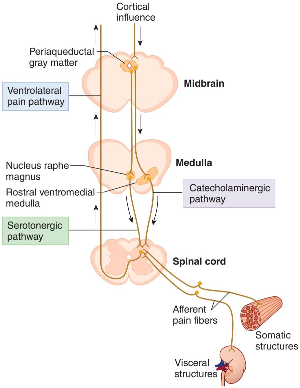
FIGURE 8-6 Schematic illustration of the convergence-projection theory for referred pain and descending pathways involved in pain control. The basis for referred pain may be convergence of somatic and visceral pain fibers on the same second-order neurons in the dorsal horn of the spinal cord that project higher brain regions. The periaqueductal gray is a part of a descending pathway that includes serotonergic neurons in the nucleus raphe magnus and catecholaminergic neurons in the rostral ventromedial medulla to modulate pain transmission by inhibition of primary afferent transmission in the dorsal horn.
شکل 8-6 تصویر شماتیک تئوری همگرایی-پیش بینی برای درد ارجاعی و مسیرهای نزولی درگیر در کنترل درد. مبنای درد ارجاعی ممکن است همگرایی رشتههای درد جسمیو احشایی روی همان نورونهای مرتبه دوم در شاخ پشتی نخاع باشد که نواحی بالاتر مغز را نشان میدهد. خاکستری دور قناتی بخشی از یک مسیر نزولی است که شامل نورونهای سروتونرژیک در هسته رافه مگنوس و نورونهای کاتکول آمینرژیک در بصل النخاع شکمیمنقاری است تا انتقال درد را با مهار انتقال آوران اولیه در شاخ پشتی تعدیل کند.
SOMATOSENSORY PATHWAYS
The sensation evoked by impulses generated in a sensory receptor depends in part on the specific part of the brain they ultimately activate. Below is a comparison of the ascending sensory pathway that mediates touch, vibratory sense, and proprioception (dorsal column or medial lemniscal pathway) and that which mediates pain and temperature (ventrolateral spinothalamic pathway).
مسیرهای حسی جسمی
احساس برانگیخته شده توسط تکانههای تولید شده در یک گیرنده حسی تا حدی به بخش خاصی از مغز که در نهایت فعال میشود بستگی دارد. در زیر مقایسه ای از مسیر حسی صعودی است که واسطه لامسه، حس ارتعاشی و حس عمقی (ستون پشتی یا مسیر لمنیسکال میانی) و چیزی که واسطه درد و دما (مسیر اسپینوتالامیک بطنی جانبی) است.
DORSAL COLUMN PATHWAY
The principal pathways to the cerebral cortex for touch, vibratory sense, and proprioception are shown in Figure 8-7. Fibers mediating these sensations ascend ipsilaterally in the dorsal columns of the spinal cord to the medulla, where they synapse in the gracilus and cuneate nuclei. The second-order neurons from these nuclei cross the midline and ascend in the medial lemniscus to end in the contralateral ventral posterior lateral (VPL) nucleus. This ascending system is called the dorsal column or medial lemniscal system. The fibers within the dorsal column pathway are joined in the brainstem by fibers mediating sensation from the head. Touch and proprioception from the head are relayed mostly via the main sensory and mesencephalic nuclei of the trigeminal nerve.
مسیر ستون پشتی
مسیرهای اصلی به قشر مغز برای لمس، حس ارتعاشی و حس عمقی در شکل 8-7 نشان داده شده است. فیبرهایی که این احساسات را واسطه میکنند به صورت یک طرف در ستونهای پشتی نخاع به سمت مدولا بالا میروند، جایی که در هستههای گراسیلوس و میخی سیناپس میشوند. نورونهای مرتبه دوم از این هستهها از خط وسط عبور کرده و در لمنیسکوس داخلی بالا میروند تا به هسته جانبی خلفی شکمیطرف مقابل (VPL) ختم شوند. این سیستم صعودی، ستون پشتی یا سیستم لمنیسکال میانی نامیده میشود. الیاف در مسیر ستون پشتی در ساقه مغز توسط الیافی که واسطه احساس از سر هستند به هم میپیوندند. لمس و حس عمقی از سر بیشتر از طریق هستههای اصلی حسی و مزانسفالی عصب سه قلو منتقل میشود.

FIGURE 8-7 Ascending tracts carrying sensory information from peripheral receptors to the cerebral cortex. A) Dorsal column pathway mediates touch, vibratory sense, and proprioception. Sensory fibers ascend ipsilaterally via the spinal dorsal columns to medullary gracilus and cuneate nuclei; from there the fibers cross the midline and ascend in the medial lemniscus to the contralateral thalamic ventral posterior lateral (VPL) and then to the primary somatosensory cortex. B) Ventrolateral spinothalamic tract mediates pain and temperature. These sensory fibers terminate in the dorsal horn and projections from there cross the midline and ascend in the ventrolateral quadrant of the spinal cord to the VPL and then to the primary somatosensory cortex.
شکل 8-7 مسیرهای صعودی حامل اطلاعات حسی از گیرندههای محیطی به قشر مغز. الف) مسیر ستون پشتی واسطه لمس، حس ارتعاشی و حس عمقی است. فیبرهای حسی از طریق ستونهای پشتی نخاعی به صورت همان طرف به سمت گراسیلوس مدولاری و هستههای میخی صعود میکنند. از آنجا الیاف از خط وسط عبور کرده و در لمنیسک داخلی به سمت جانبی جانبی خلفی شکمیتالاموس (VPL) و سپس به قشر اولیه حسی تنی صعود میکنند. ب) دستگاه اسپینوتالاموس ونترولترال واسطه درد و دما است. این رشتههای حسی به شاخ پشتی ختم میشوند و برآمدگیها از آنجا از خط وسط عبور میکنند و در ربع بطنی جانبی نخاع به VPL و سپس به قشر اولیه حسی تنی صعود میکنند.
Somatotopic Organization
Within the dorsal columns, fibers arising from different levels of the cord are somatotopically organized (Figure 8-7). Specifically, fibers from the sacral cord are positioned most medially and those from the cervical cord are positioned most laterally. This arrangement continues in the medulla with lower body (eg, foot) representation in the gracilus nucleus and upper body (eg, finger) representation in cuneate nucleus. The medial lemniscus is organized dorsal to ventral representing from neck to foot.
سازمان سوماتوتوپیک
در ستونهای پشتی، الیاف ناشی از سطوح مختلف بند ناف به صورت سوماتوتوپی سازماندهی میشوند (شکل 8-7). به طور خاص، فیبرهای طناب خاجی بیشتر در قسمت داخلی قرار میگیرند و رشتههای طناب گردنی بیشتر در سمت جانبی قرار میگیرند. این آرایش در بصل النخاع با نمایش پایین تنه (مثلاً پا) در هسته گراسیلوس و نمایش قسمت بالایی بدن (به عنوان مثال انگشت) در هسته میخی ادامه مییابد. لمنیسکوس داخلی از پشت تا شکمیسازمان یافته است که از گردن تا پا را نشان میدهد.
Somatotopic organization continues through the thalamus and cortex. The VPL thalamic neurons carrying sensory information project in a highly specific way to the primary somatosensory cortex in the postcentral gyrus of the parietal lobe (Figure 8-8). The arrangement of projections to this region is such that the parts of the body are represented in order along the postcentral gyrus, with the legs on top and the head at the foot of the gyrus. Not only is there detailed localization of the fibers from the various parts of the body in the postcentral gyrus but also the size of the cortical receiving area for impulses from a part of the body is proportional to the use of the part. The relative sizes of the cortical receiving areas are shown dramatically in Figure 8-9, in which the proportions of the homunculus have been distorted to correspond to the size of the cortical receiving areas for each. Note that the cortical areas for sensation from the trunk and back are small, whereas very large areas are concerned with impulses from the hand and the parts of the mouth concerned with speech.
سازمان سوماتوتوپیک از طریق تالاموس و قشر مغز ادامه مییابد. نورونهای تالاموس VPL که اطلاعات حسی را حمل میکنند به روشی بسیار خاص به قشر حسی جسمیاولیه در شکنج پست مرکزی لوب جداری پروژه میدهند (شکل 8-8). چیدمان برجستگیها به این ناحیه به گونه ای است که قسمتهای بدن به ترتیب در امتداد شکنج پست مرکزی نشان داده میشوند، با پاها در بالا و سر در پای شکنج. نه تنها محل دقیق الیاف از قسمتهای مختلف بدن در شکنج پست مرکزی وجود دارد، بلکه اندازه ناحیه دریافت کننده قشر مغز برای تکانههای بخشی از بدن متناسب با استفاده از آن قسمت است. اندازه نسبی نواحی دریافت کننده قشر به طور چشمگیری در شکل 9-8 نشان داده شده است، که در آن نسبت هومونکولوس برای مطابقت با اندازه نواحی دریافت کننده قشر برای هر یک تغییر داده شده است. توجه داشته باشید که نواحی قشری برای احساس از تنه و پشت کوچک هستند، در حالی که نواحی بسیار بزرگ مربوط به تکانههای دست و قسمتهایی از دهان هستند که با گفتار مرتبط هستند.

FIGURE 8-8 A lateral view of the left hemisphere showing some principal cortical areas and their functional correlates in the human brain. The primary somatosensory area is in the postcentral gyrus of the parietal lobe, and the primary motor cortex is in the precentral gyrus. (Reproduced with permission from Waxman SG: Clinical Neuroanatomy, 26th ed. New York, NY: McGraw- Hill; 2010.)
شکل 8-8 نمای جانبی نیمکره چپ که برخی از نواحی اصلی قشر مغز و همبستگیهای عملکردی آنها را در مغز انسان نشان میدهد. ناحیه حسی جسمیاولیه در شکنج پس مرکزی لوب جداری و قشر حرکتی اولیه در شکنج پیش مرکزی قرار دارد. (تکثیر شده با اجازه Waxman SG: Clinical Neuroanatomy, 26th. New York, NY: McGraw- Hill; 2010.)
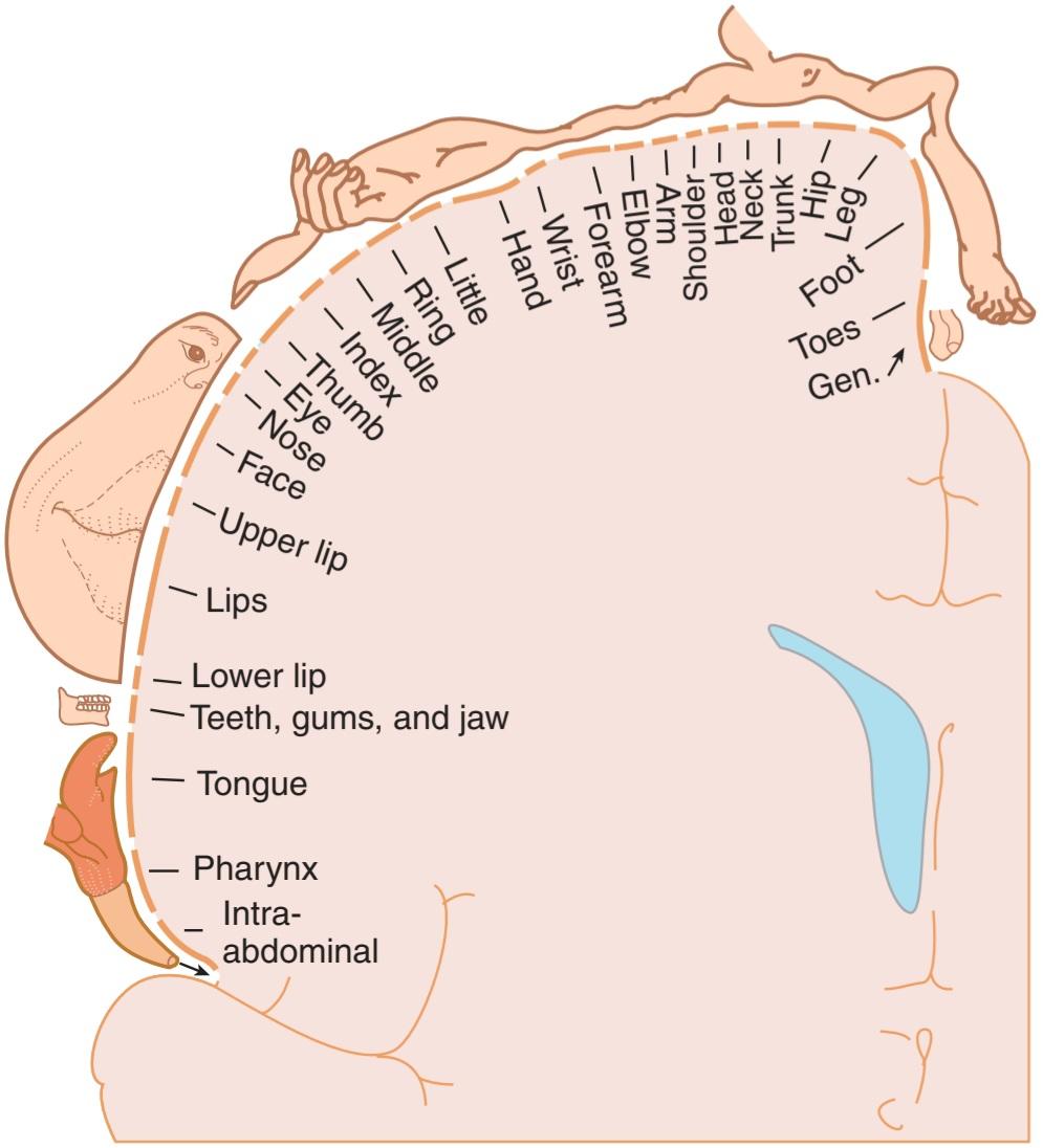
FIGURE 8-9 Sensory homunculus, drawn overlying a coronal section through the postcentral gyrus. The parts of the body are represented in order along the postcentral gyrus, with the legs on top and the head at the foot of the gyrus. The size of the cortical receiving area for impulses from a particular part of the body is proportionate to the use of the part. Gen., genitalia.
شکل 8-9 هومونکولوس حسی، بر روی یک بخش تاجی از شکنج پست مرکزی کشیده شده است. قسمتهای بدن به ترتیب در امتداد شکنج پست مرکزی، با پاها در بالا و سر در پای شکنج نشان داده شده اند. اندازه ناحیه دریافت کننده قشر مغز برای تکانههای یک قسمت خاص از بدن متناسب با استفاده از آن قسمت است. جنرال، دستگاه تناسلی.
In addition to the primary somatosensory cortex, there are two other cortical regions that contribute to the integration of sensory information. The sensory association area is in the parietal cortex, and the secondary somatosensory cortex is located in the wall of the lateral fissure (also called sylvian fissure) that separates the temporal from the frontal and parietal lobes. These regions receive input from the primary somatosensory cortex.
علاوه بر قشر حسی جسمیاولیه، دو ناحیه قشر دیگر نیز وجود دارد که به یکپارچه سازی اطلاعات حسی کمک میکنند. ناحیه تداعی حسی در قشر جداری است و قشر سوماتوسانسوری ثانویه در دیواره شقاق جانبی (که به آن شقاق سیلوین نیز گفته میشود) قرار دارد که قسمت تمپورال را از لوبهای فرونتال و جداری جدا میکند. این نواحی ورودی را از قشر حسی تنی اولیه دریافت میکنند.
Conscious awareness of the positions of the various parts of the body in space depends in part on impulses from sensory receptors in and around the joints. Impulses from these receptors, from touch receptors in the skin and other tissues, and from muscle spindles are synthesized in the cortex into a conscious picture of the position of the body in space (proprioception).
آگاهی آگاهانه از موقعیتهای قسمتهای مختلف بدن در فضا تا حدی به تکانههای گیرندههای حسی در داخل و اطراف مفاصل بستگی دارد. تکانههای این گیرندهها، از گیرندههای لمسی در پوست و سایر بافتها، و از دوکهای عضلانی در قشر به تصویری آگاهانه از موقعیت بدن در فضا سنتز میشوند (حس عمقی).
VENTROLATERAL SPINOTHALAMIC TRACT
Fibers from nociceptors and thermoreceptors synapse on neurons in the dorsal horn of the spinal cord. The axons from these dorsal horn neurons cross the midline and ascend in the ventrolateral quadrant of the spinal cord, where they form the ventrolateral spinothalamic pathway (Figure 8-7). Fibers within this tract synapse in the VPL. Some dorsal horn neurons that receive nociceptive input synapse in the reticular formation of the brainstem (spinoreticular pathway) and then project to the centrolateral nucleus of the thalamus.
دستگاه اسپینوتالامیک بطنی جانبی
فیبرهای گیرندههای درد و گیرندههای حرارتی روی نورونهای شاخ پشتی نخاع سیناپس میکنند. آکسونهای این نورونهای شاخ پشتی از خط میانی عبور کرده و در ربع بطنی جانبی نخاع بالا میروند، جایی که مسیر اسپینوتالامیک بطنی جانبی را تشکیل میدهند (شکل 8-7). فیبرهای داخل این دستگاه در VPL سیناپس میشوند. برخی از نورونهای شاخ پشتی که ورودی درد دریافت میکنند، در تشکیل شبکهای ساقه مغز (مسیر اسپینورتیکولار) سیناپس میشوند و سپس به سمت هسته مرکزی جانبی تالاموس پیش میروند.
Positron emission tomographic (PET) and functional magnetic resonance imaging (fMRI) studies in healthy humans indicate that pain activates the primary and secondary somatosensory cortex and the cingulate gyrus on the side opposite the stimulus. In addition, the amygdala, frontal lobe, and the insular cortex are activated. These technologies were important in distinguishing two components of pain pathways. The pathway to the primary somatosensory cortex is responsible for the discriminative aspect of pain (pain localization). In contrast, the pathway that includes synapses in the brainstem reticular formation and centrolateral thalamic nucleus projects to the frontal lobe, limbic system, and insular cortex. This pathway mediates the motivational-affective component of pain.
مطالعات توموگرافی گسیل پوزیترون (PET) و تصویربرداری تشدید مغناطیسی عملکردی (fMRI) در انسانهای سالم نشان میدهد که درد قشر حسی سوماتوی اولیه و ثانویه و شکنج سینگوله را در سمت مقابل محرک فعال میکند. علاوه بر این، آمیگدال، لوب فرونتال و قشر منزوی فعال میشوند. این فناوریها در تشخیص دو جزء مسیرهای درد مهم بودند. مسیر منتهی به قشر اولیه حسی جسمیمسئول جنبه تمایز درد (محلی سازی درد) است. در مقابل، مسیری که شامل سیناپسها در تشکیل شبکهای ساقه مغز و هسته مرکزی تالاموس میشود به سمت لوب پیشانی، سیستم لیمبیک و قشر منزوی میرود. این مسیر جزء انگیزشی- عاطفی درد را واسطه میکند.
Visceral sensation travels along the same central pathways as somatic sensation in the spinothalamic tracts and thalamic radiations, and the cortical receiving areas for visceral sensation are intermixed with the somatic receiving areas.
حس احشایی در امتداد همان مسیرهای مرکزی حرکت میکند که حس سوماتیک در مجاری اسپینوتالاموس و تشعشعات تالاموس، و نواحی دریافت کننده قشر برای حس احشایی با نواحی دریافت کننده جسمیمخلوط میشوند.
CORTICAL PLASTICITY
The extensive neuronal connections described above are not innate and immutable but can be changed relatively rapidly by experience to reflect the use of the represented area. Clinical Box 8-4 describes remarkable changes in cortical and thalamic organization that occur in response to limb amputation to lead to the phenomenon of phantom limb pain.
پلاستیسیته قشر مغز
اتصالات عصبی گسترده ای که در بالا توضیح داده شد، ذاتی و تغییر ناپذیر نیستند، اما میتوانند با تجربه نسبتاً سریع تغییر کنند تا استفاده از ناحیه نشان داده شده را منعکس کنند.جعبه بالینی 8-4 تغییرات قابل توجهی را در سازمان قشر و تالاموس توصیف میکند که در پاسخ به قطع اندام رخ میدهد و منجر به پدیده درد فانتوم اندام میشود.
CLINICAL BOX 8.4
Phantom Limb Pain
In 1551, Ambroise Pare, a military surgeon, wrote “…the patients, long after the amputation is made, say they still feel pain in the amputated part. Of this they complain strongly, a thing worthy of wonder and almost incredible to people who have not experienced this.” This is perhaps the earliest description of phantom limb pain. Between 50% and 80% of amputees experience phantom sensations, usually pain, in the region of their amputated limb. Phantom sensations may also occur after the removal of body parts other than the limbs, for example, after amputation of the breast, extraction of a tooth (phantom tooth pain), or removal of an eye (phantom eye syndrome). Numerous theories have been evoked to explain this phenomenon. The current theory is based on evidence that the brain can reorganize if sensory input is cutoff. The ventral posterior thalamic nucleus is one example where this change can occur. In patients who have had their leg amputated, single neuron recordings show that the thalamic region that once received input from the leg and foot now respond to stimulation of the stump (thigh). Others have demonstrated remapping of the somatosensory cortex. For example, in some individuals who have had an arm amputated, stroking different parts of the face can lead to the feeling of being touched in the area of the missing limb.
جعبه بالینی 8.4
درد اندام فانتوم
در سال 1551، Ambroise Pare، یک جراح نظامی، نوشت: “… بیماران، مدتها پس از انجام قطع عضو، میگویند که هنوز در قسمت قطع شده احساس درد میکنند. از این موضوع به شدت شکایت میکنند، چیزی که جای تعجب دارد و برای افرادی که این را تجربه نکرده اند تقریبا باورنکردنی است.” این شاید اولین توصیف درد اندام فانتوم باشد. بین 50٪ تا 80٪ از افراد قطع عضو، احساسات فانتوم، معمولاً درد، در ناحیه اندام قطع شده خود را تجربه میکنند. احساس فانتوم ممکن است پس از برداشتن سایر قسمتهای بدن به غیر از اندامها، به عنوان مثال، پس از قطع کردن سینه، کشیدن دندان (درد دندان فانتوم)، یا برداشتن چشم (سندرم چشم فانتوم) نیز رخ دهد. نظریههای متعددی برای توضیح این پدیده برانگیخته شده است. نظریه فعلی مبتنی بر شواهدی است که نشان میدهد اگر ورودی حسی قطع شود، مغز میتواند سازماندهی مجدد کند. هسته تالاموس خلفی شکمییکی از نمونههایی است که این تغییر میتواند رخ دهد. در بیمارانی که پای خود را قطع کرده اند، ضبط تک نورون نشان میدهد که ناحیه تالاموس که زمانی از پا و پا ورودی دریافت میکرد، اکنون به تحریک استامپ (ران) پاسخ میدهد. برخی دیگر نقشه برداری مجدد قشر حسی تنی را نشان داده اند. به عنوان مثال، در برخی از افرادی که دستشان قطع شده است، نوازش قسمتهای مختلف صورت میتواند منجر به احساس لمس در ناحیه اندام از دست رفته شود.
THERAPEUTIQ HIGHLIGHTS
The use of epidural anesthesia during the amputation surgery may prevent the acute pain associated with the surgery, thereby reducing the need for opioid therapy in the immediate postoperative period. This anesthetic procedure resulted in a reduced incidence of phantom pain. Spinal cord stimulation has been shown to be an effective therapy for phantom pain. Electric current is passed through an electrode that is placed next to the spinal cord to stimulate spinal pathways. This interferes with the impulses ascending to the brain and lessens the pain felt in the phantom limb. Instead, amputees feel a tingling sensation in the phantom limb.
نکات برجسته درمانی
استفاده از بی حسی اپیدورال در حین جراحی قطع عضو ممکن است از درد حاد مرتبط با جراحی جلوگیری کند و در نتیجه نیاز به درمان با مواد افیونی را در دوره بلافاصله پس از عمل کاهش دهد. این روش بیهوشی منجر به کاهش بروز درد فانتوم شد. نشان داده شده است که تحریک طناب نخاعی یک درمان موثر برای درد فانتوم است. جریان الکتریکی از الکترودی عبور میکند که در کنار نخاع قرار میگیرد تا مسیرهای نخاعی را تحریک کند. این کار با تکانههای صعودی به مغز تداخل میکند و درد احساس شده در اندام فانتوم را کاهش میدهد. در عوض، افراد قطع عضو در اندام فانتوم احساس گزگز میکنند.
Cortical structures are capable of dramatic reorganization (plasticity) after loss of a body part such as a digit; the cortical representation of the neighboring digits spreads into the cortical area that was formerly occupied by the representation of the amputated digit. Cortical connections of sensory units to the cortex have extensive convergence and divergence, with connections that can become weak with disuse and strong with use.
ساختارهای قشری قادر به سازماندهی مجدد چشمگیر (پلاستیسیته) پس از از دست دادن بخشی از بدن مانند یک انگشت هستند. نمایش قشری ارقام مجاور به ناحیه قشری که قبلاً با نمایش انگشت قطع شده اشغال شده بود گسترش مییابد. اتصالات قشری واحدهای حسی به قشر دارای همگرایی و واگرایی گسترده است، با اتصالاتی که در صورت عدم استفاده ضعیف و با استفاده قوی میشوند.
PET scanning in humans also documents plastic changes, sometimes from one sensory modality to another. Thus, for example, tactile and auditory stimuli increase metabolic activity in the visual cortex in blind individuals. Conversely, deaf individuals respond faster and more accurately than normal individuals to moving stimuli in the visual periphery. Plasticity also occurs in the motor cortex.
اسکن PET در انسان همچنین تغییرات پلاستیک را، گاهی اوقات از یک روش حسی به روش دیگر، مستند میکند. بنابراین، به عنوان مثال، محرکهای لمسی و شنوایی باعث افزایش فعالیت متابولیک در قشر بینایی در افراد نابینا میشود. برعکس، افراد ناشنوا سریعتر و دقیقتر از افراد عادی به محرکهای متحرک در حاشیه بینایی پاسخ میدهند. پلاستیسیته نیز در قشر حرکتی رخ میدهد.
EFFECTS OF CNS LESIONS
Clinical Box 8-2 describes some of the deficits noted after damage within the somatosensory pathways. Clinical Box 8-5 describes the characteristic changes in sensory and motor functions that occur in response to spinal cord hemisection. Damage to the dorsal columns leads to ipsilateral loss of the ability to detect light touch, vibration, and proprioception from body structures represented caudal to the level of damage. Damage to the ventrolateral spinothalamic pathway leads to contralateral loss of pain and temperature sensation below the level of the lesion. Such spinal damage could occur with a penetrating wound or a tumor.
اثرات ضایعات CNS
جعبه بالینی 8-2 برخی از کاستیهایی را که پس از آسیب در مسیرهای حسی تنی ذکر شده است، توصیف میکند. جعبه بالینی 8-5 تغییرات مشخصه در عملکردهای حسی و حرکتی را که در پاسخ به نیمه برش نخاع رخ میدهد، توضیح میدهد. آسیب به ستونهای پشتی منجر به از دست دادن توانایی تشخیص لمس سبک، لرزش و حس عمقی از ساختارهای بدن میشود که به صورت دمیتا سطح آسیب نشان داده میشوند. آسیب به مسیر اسپینوتالامیک بطنی جانبی منجر به از دست دادن درد طرف مقابل و احساس دمای زیر سطح ضایعه میشود. چنین آسیب ستون فقرات میتواند با یک زخم نافذ یا یک تومور
CLINICAL BOX 8.5
Brown-Séquard Syndrome
A functional hemisection of the spinal cord causes a characteristic and easily recognized clinical picture that reflects damage to ascending sensory (dorsal column pathway, ventrolateral spinothalamic tract) and descending motor (corticospinal tract) pathways, which is called the Brown-Séquard syndrome. The lesion to fasciculus gracilus or fasciculus cuneatus leads to ipsilateral loss of discriminative touch, vibration, and proprioception below the level of the lesion. The loss of the spinothalamic tract leads to contralateral loss of pain and temperature sensation beginning one or two segments below the lesion. Damage to the corticospinal tract produces weakness and spasticity in certain muscle groups on the same side of the body. Although a precise spinal hemisection is rare, the syndrome is common because it can be caused by a spinal cord tumor, spinal cord trauma, degenerative disk disease, and ischemia.
جعبه بالینی 8.5
سندرم براون سکوارد
نیم برش عملکردی طناب نخاعی باعث ایجاد یک تصویر بالینی مشخص و به راحتی قابل تشخیص میشود که منعکس کننده آسیب به مسیرهای حسی صعودی (مسیر ستون پشتی، مجرای اسپینوتالامیک بطنی جانبی) و مسیرهای حرکتی نزولی (دستگاه قشر نخاعی) است که به آن سندرم براون سکوارد میگویند. ضایعه به fasciculus gracilus یا fasciculus cuneatus منجر به از دست دادن لمس متمایز، لرزش و حس عمقی در زیر سطح ضایعه میشود. از دست دادن دستگاه اسپینوتالاموس منجر به از دست دادن درد و احساس دما در طرف مقابل میشود که از یک یا دو بخش زیر ضایعه شروع میشود. آسیب به دستگاه قشر نخاعی باعث ایجاد ضعف و اسپاسم در گروههای عضلانی خاصی در همان سمت بدن میشود. اگرچه نیم برش دقیق ستون فقرات نادر است، اما این سندرم شایع است زیرا میتواند ناشی از تومور نخاع، ترومای نخاع، بیماری دژنراتیو دیسک و ایسکمیباشد.
THERAPEUTIQ HIGHLIGHTS
Spinal or vertebral stabilization is needed when spinal trauma is indicated. Drug treatments for Brown-Séquard syndrome are based on the etiology and time since onset. High doses of corticosteroids have been shown to be of value particularly if administered soon after the onset of such a spinal cord injury. Corticosteroids decrease the inflammation by suppressing polymorphonuclear leukocytes and reverse the increase in capillary permeability. Drugs may be needed to treat spasticity, pain, or other possible complications of the spinal cord injury. Physical therapy is important to maintain strength and joint mobility and improving respiratory function.
نکات برجسته درمانی
هنگامیکه ضربه به ستون فقرات نشان داده میشود، تثبیت ستون فقرات یا مهرهها مورد نیاز است. درمانهای دارویی برای سندرم براون سکوارد بر اساس علت و زمان شروع آن است. نشان داده شده است که دوزهای بالای کورتیکواستروئیدها به ویژه اگر بلافاصله پس از شروع چنین آسیب نخاعی تجویز شوند، دارای ارزش هستند. کورتیکواستروئیدها با سرکوب لکوسیتهای پلیمورفونکلئر التهاب را کاهش میدهند و افزایش نفوذپذیری مویرگی را معکوس میکنند. ممکن است برای درمان اسپاستیسیته، درد یا سایر عوارض احتمالی آسیب نخاعی به دارو نیاز باشد. فیزیوتراپی برای حفظ قدرت و تحرک مفاصل و بهبود عملکرد تنفسی مهم است.
Damage to the primary somatosensory cortex does not abolish somatic sensation. Irritation of this region causes paresthesia or an abnormal sensation of numbness and tingling on the contralateral side of the body. Destructive lesions impair the ability to localize noxious stimuli in time, space, and intensity. Damage to the cingulate cortex impairs the recognition of the aversive nature of a noxious stimulus.
آسیب به قشر اولیه حسی جسمی، حس جسمیرا از بین نمیبرد. تحریک این ناحیه باعث پارستزی یا احساس غیر طبیعی بی حسی و گزگز در سمت مقابل بدن میشود. ضایعات مخرب توانایی محلی سازی محرکهای مضر در زمان، مکان و شدت را مختل میکند. آسیب به قشر سینگولیت تشخیص ماهیت بد یک محرک مضر را مختل میکند.
An infarct in the thalamus can lead to a loss of sensation. Thalamic pain syndrome can occur during recovery from a thalamic infarct. The syndrome is characterized by chronic pain on the side of the body contralateral to the stroke.
انفارکتوس در تالاموس میتواند منجر به از دست دادن حس شود. سندرم درد تالاموس میتواند در طول بهبودی از انفارکتوس تالاموس رخ دهد. این سندرم با درد مزمن در سمت بدن در مقابل سکته مشخص میشود.
MODULATION OF PAIN TRANSMISSION
PROCESSING INFORMATION IN THE DORSAL HORN
Transmission in nociceptive pathways can be interrupted by actions within the dorsal horn of the spinal cord at the site of sensory afferent termination. Many people have learned from practical experience that rubbing or shaking an injured area decreases the pain due to the injury. The relief may be due to the simultaneous activation of innocuous cutaneous mechanoreceptors whose afferents emit collaterals that terminate in the dorsal horn. The activity of these cutaneous mechanosensitive afferents may reduce the responsiveness of dorsal horn neurons to their input from nociceptive afferent terminals. This is called the gate-control mechanism of pain modulation and it serves as the rationale behind the use of transcutaneous electrical nerve stimulation (TENS) for pain relief. This method uses electrodes to activate Aa and Aẞ fibers near the injury.
تعدیل انتقال درد
پردازش اطلاعات در شاخ پشتی
انتقال در مسیرهای درد میتواند با اعمال درون شاخ پشتی نخاع در محل خاتمه آوران حسی قطع شود. بسیاری از مردم از تجربه عملی آموخته اند که مالش یا تکان دادن ناحیه آسیب دیده درد ناشی از آسیب را کاهش میدهد. تسکین ممکن است به دلیل فعال شدن همزمان گیرندههای مکانیکی پوستی بیضرر باشد که آورانهای آنها وثیقههایی را منتشر میکنند که به شاخ پشتی ختم میشوند. فعالیت این آورانهای حساس به مکانیکی پوستی ممکن است پاسخدهی نورونهای شاخ پشتی به ورودیهای آنها از پایانههای آوران درد را کاهش دهد. این مکانیسم کنترل دروازه مدولاسیون درد نامیده میشود و به عنوان دلیل استفاده از تحریک الکتریکی عصب از طریق پوست (TENS) برای تسکین درد عمل میکند. این روش از الکترودها برای فعال کردن فیبرهای Aa و Aẞ در نزدیکی آسیب استفاده میکند.
Opioids are a commonly used analgesic that can exert their effects at various places in the CNS, including in the spinal cord and dorsal root ganglia. Figure 8-10 shows some of the various modes of action of opioids to decrease nociceptive transmission. There are interneurons in the superficial regions of the dorsal horn that contain endogenous opioid peptides (enkephalin and dynorphin). These interneurons terminate in the region of the dorsal horn where nociceptive afferents terminate. Opioid receptors are located on the terminals of nociceptive fibers and on dendrites of dorsal horn neurons, allowing for both presynaptic and postsynaptic sites of actions for opioids. Activation of the postsynaptic opioid receptors hyperpolarizes the dorsal horn interneuron by causing an increase in K+ conductance. Activation of the presynaptic opioid receptors leads to a decrease in Ca2+ influx, resulting in a decrease in release of glutamate and substance P. Together these actions reduce the duration of the EPSP in the dorsal horn neuron. Activation of opioid receptors on dorsal root ganglia cell bodies also contributes to reduced transmission from nociceptive afferents.
مواد افیونی یک مسکن رایج مورد استفاده است که میتواند اثرات خود را در مکانهای مختلف CNS از جمله در نخاع و گانگلیونهای ریشه پشتی اعمال کند. شکل 8-10 برخی از حالتهای مختلف عمل اپیوئیدها را برای کاهش انتقال درد نشان میدهد. در نواحی سطحی شاخ پشتی، نورونهای داخلی وجود دارند که حاوی پپتیدهای شبه افیونی درون زا (انکفالین و دینورفین) هستند. این نورونهای داخلی در ناحیه شاخ پشتی که در آن آورانهای درد به پایان میرسند، خاتمه مییابند. گیرندههای اپیوئیدی در انتهای رشتههای درد و روی دندریتهای نورونهای شاخ پشتی قرار گرفتهاند، که امکان فعالیتهای پیشسیناپسی و پس سیناپسی را برای مواد افیونی فراهم میکند. فعال شدن گیرندههای مواد افیونی پس سیناپسی، نورون داخلی شاخ پشتی را با افزایش رسانایی +K، هیپرپلاریزه میکند. فعال شدن گیرندههای مواد افیونی پیش سیناپسی منجر به کاهش هجوم +Ca2 و در نتیجه کاهش انتشار گلوتامات و ماده P میشود. این اقدامات با هم باعث کاهش مدت زمان EPSP در نورون شاخ پشتی میشود. فعال شدن گیرندههای مواد افیونی بر روی بدنه سلولی گانگلیون ریشه پشتی نیز به کاهش انتقال از آورانهای درد کمک میکند.

FIGURE 8-10 Actions of opioids to reduce sensory transmission in pain pathways at the level of the dorsal root ganglion (DRG) and spinal cord dorsal horn region. A) Activation of a nociceptor leads to the release of glutamate and neuropeptides from its nerve terminals that synapse on spinothalamic tract projection neurons. This leads to the depolarization (activation) of spinothalamic tract projection neurons. Enkephalin (ENK)- containing interneurons mediate their effects via opioid receptors on the terminals of nociceptive afferent fibers and on dendrites of dorsal horn neurons to exert both presynaptic and postsynaptic inhibition. B) The action of an opioid (eg, morphine) within the DRG is to decrease Ca2+ influx leading to a decrease in the duration of the invoked action potential in the nociceptive neuron and a reduction in transmitter release from the nociceptive neuron onto a neuron in the dorsal horn. Opioids also hyperpolarize the membrane of dorsal horn neuron by activation of a K+ conductance; opioids also decrease the amplitude of the excitatory postsynaptic potential (EPSP) produced by stimulation of nociceptors.
شکل 8-10 اقدامات مواد افیونی برای کاهش انتقال حسی در مسیرهای درد در سطح گانگلیون ریشه پشتی (DRG) و ناحیه شاخ پشتی نخاع. الف) فعال شدن یک گیرنده درد منجر به آزاد شدن گلوتامات و نوروپپتیدها از پایانههای عصبی آن میشود که بر روی نورونهای برآمدگی دستگاه اسپینوتالاموس سیناپس میشوند. این منجر به دپلاریزاسیون (فعال شدن) نورونهای برآمدگی دستگاه اسپینوتلاموس میشود. انکفالین (ENK) – حاوی نورونهای داخلی، اثرات خود را از طریق گیرندههای اپیوئیدی بر روی پایانههای فیبرهای آوران درد و روی دندریتهای نورونهای شاخ پشتی برای اعمال مهار پیشسیناپسی و پسسیناپسی واسطه میکنند. ب) عمل یک اپیوئید (مثلاً مورفین) در DRG کاهش هجوم +Ca2 است که منجر به کاهش مدت زمان پتانسیل عمل فراخوانی شده در نورون درد و کاهش انتشار فرستنده از نورون درد بر روی یک نورون در شاخ پشتی میشود. مواد افیونی همچنین غشای نورون شاخ پشتی را با فعالسازی رسانایی +K هیپرپلاریزه میکنند. مواد افیونی همچنین دامنه پتانسیل پس سیناپسی تحریکی (EPSP) تولید شده توسط تحریک گیرندههای درد را کاهش میدهند.
Patients who receive long-term pain management with morphine may become resistant to the drug, requiring progressively higher doses for pain relief. This acquired tolerance is different from addiction, which refers to a psychological craving. Psychological addiction rarely occurs when morphine is used to treat chronic pain, provided the patient does not have a history of drug abuse. Clinical Box 8-6 describes mechanisms involved in motivation and addiction.
بیمارانی که درمان طولانیمدت درد با مورفین را دریافت میکنند ممکن است نسبت به دارو مقاوم شوند و برای تسکین درد به دوزهای بالاتری نیاز دارند. این تحمل اکتسابی با اعتیاد که به ولع روانی اشاره دارد، متفاوت است. هنگامیکه از مرفین برای درمان دردهای مزمن استفاده میشود، اعتیاد روانی به ندرت اتفاق میافتد، مشروط بر اینکه بیمار سابقه مصرف مواد مخدر نداشته باشد. جعبه بالینی 8-6 مکانیسمهای دخیل در انگیزش و اعتیاد را شرح میدهد.
CLINICAL BOX 8.6
Motivation & Addiction
Neurons in the forebrain ventral tegmental area and nucleus accumbens are involved in motivated behaviors such as reward, laughter, pleasure, addiction, and fear. These areas are referred to as the brain’s reward center or pleasure center. The mesocortical dopaminergic neurons that project from the midbrain to the nucleus accumbens and the frontal cortex are also involved. Addiction, defined as the repeated compulsive use of a substance despite negative health consequences, can be produced by many different drugs. According to the World Health Organization, over 76 million people worldwide suffer from alcohol abuse, and over 15 million suffer from drug abuse. Not surprisingly, alcohol and drug addiction are associated with the reward system. The best studied addictive drugs are opioids (eg, morphine and heroin); others include cocaine, amphetamine, alcohol, cannabinoids, and nicotine. These drugs affect the brain in different ways, but all have in common the fact that they increase the amount of dopamine available to act on D3 receptors in the nucleus accumbens. Thus, acutely they stimulate the reward system of the brain. Long-term addiction involves the development of tolerance, which is the need for increasing amounts of a drug to produce a high. Also, withdrawal produces psychological and physical symptoms. One of the characteristics of addiction is the tendency of addicts to relapse after treatment. For opioid addicts, the relapse rate in the first year is about 80%. Relapse often occurs on exposure to sights, sounds, and situations that were previously associated with drug use. Even a single dose of an addictive drug facilitates release of excitatory neurotransmitters in brain areas concerned with memory. The medial frontal cortex, hippocampus, and amygdala are concerned with memory, and they project via excitatory glutamatergic pathways to the nucleus accumbens. Relatively little is known about the specific brain mechanisms that cause tolerance and dependence. However, the two can be separated. Absence of ẞ-arrestin-2 blocks tolerance but has no effect on dependence. ẞ-Arrestin-2 is a member of a family of proteins that inhibit heterotrimeric G-proteins by phosphorylating them.
جعبه بالینی 8.6
انگیزه و اعتیاد
نورونهای ناحیه تگمنتال شکمیپیشین مغز و هسته اکومبنس در رفتارهای انگیزشی مانند پاداش، خنده، لذت، اعتیاد و ترس نقش دارند. از این مناطق به عنوان مرکز پاداش مغز یا مرکز لذت یاد میشود. نورونهای دوپامینرژیک مزوکورتیکال که از مغز میانی به سمت هسته اکومبنس و قشر پیشانی پیش میروند نیز درگیر هستند. اعتیاد، که به عنوان استفاده مکرر اجباری از یک ماده با وجود پیامدهای منفی برای سلامتی تعریف میشود، میتواند توسط بسیاری از مواد مخدر ایجاد شود. طبق گزارش سازمان جهانی بهداشت، بیش از 76 میلیون نفر در سراسر جهان از سوء مصرف الکل رنج میبرند و بیش از 15 میلیون نفر از سوء مصرف مواد مخدر رنج میبرند. جای تعجب نیست که اعتیاد به الکل و مواد مخدر با سیستم پاداش مرتبط است. بهترین داروهای اعتیادآور مورد مطالعه، اپیوئیدها (مثلاً مورفین و هروئین) هستند. سایر موارد عبارتند از کوکائین، آمفتامین، الکل، کانابینوئیدها و نیکوتین. این داروها به روشهای مختلفی بر مغز تأثیر میگذارند، اما وجه مشترک همه آنها این است که میزان دوپامین موجود را برای اثر بر گیرندههای D3 در هسته اکومبنس افزایش میدهند. بنابراین، آنها به شدت سیستم پاداش مغز را تحریک میکنند. اعتیاد درازمدت شامل توسعه تحمل است که نیاز به افزایش مقادیر یک دارو برای تولید زیاد است. همچنین کناره گیری علائم روانی و جسمیایجاد میکند. یکی از ویژگیهای اعتیاد، تمایل معتادان به عود پس از درمان است. برای معتادان به مواد افیونی، میزان عود در سال اول حدود 80 درصد است. عود اغلب در مواجهه با مناظر، صداها و موقعیتهایی که قبلاً با مصرف مواد مخدر مرتبط بودند، رخ میدهد. حتی یک دوز از یک داروی اعتیاد آور آزادسازی انتقال دهندههای عصبی تحریکی را در نواحی مغز مرتبط با حافظه تسهیل میکند. قشر فرونتال میانی، هیپوکامپ و آمیگدال مربوط به حافظه هستند و از طریق مسیرهای گلوتاماترژیک تحریکی به هسته اکومبنس میرسند. اطلاعات نسبتا کمیدر مورد مکانیسمهای خاص مغز که باعث تحمل و وابستگی میشوند، شناخته شده است. با این حال، این دو را میتوان از هم جدا کرد. عدم وجود ẞ-arrestin-2 تحمل را مسدود میکند اما تأثیری بر وابستگی ندارد. ẞ-Arrestin-2 عضوی از خانواده پروتئینهایی است که با فسفریله کردن پروتئینهای G هتروتریمری آنها را مهار میکند.
THERAPEUTIC HIGHLIGHTS
Withdrawal symptoms and cravings associated with addiction to opioids can be reversed by treatment with drugs such as methadone and buprenorphine that act on the same CNS receptors as morphine and heroin. The US Federal Drug Administration has approved the use of three drugs for treatment of alcohol abuse: naltrexone, acamprosate, and disulfiram. Naltrexone is an opioid receptor antagonist that blocks the reward system and the craving for alcohol. Acamprosate may reduce the withdrawal effects associated with alcohol abuse. Disulfiram causes an accumulation of acetaldehyde by preventing the full degradation of alcohol. This leads to an unpleasant reaction to alcohol ingestion (eg, flushing, nausea, and palpitations). Topiramate, a Na+ channel blocker, is showing promise in clinical trials of alcohol addiction. This is the same drug that is effective in treatment of migraine headaches.
نکات برجسته درمانی
علائم ترک و هوسهای مرتبط با اعتیاد به مواد افیونی را میتوان با درمان با داروهایی مانند متادون و بوپرنورفین که بر گیرندههای CNS مشابه مورفین و هروئین عمل میکنند، معکوس کرد. اداره داروی فدرال ایالات متحده استفاده از سه دارو را برای درمان سوء مصرف الکل تایید کرده است: نالترکسون، آکامپروسات و دی سولفیرام. نالترکسون یک آنتاگونیست گیرنده مواد افیونی است که سیستم پاداش و میل به الکل را مسدود میکند. آکامپروسات ممکن است اثرات ترک ناشی از سوء مصرف الکل را کاهش دهد. دی سولفیرام با جلوگیری از تخریب کامل الکل باعث تجمع استالدهید میشود. این منجر به واکنش ناخوشایند به مصرف الکل میشود (به عنوان مثال، گرگرفتگی، حالت تهوع، و تپش قلب). توپیرامات، یک مسدود کننده کانال Na+، در آزمایشات بالینی اعتیاد به الکل امیدوار کننده است. این همان دارویی است که در درمان سردردهای میگرنی موثر است.
ROLES OF PERIAQUEDUCTAL GRAY & BRAINSTEM
Another site of action for morphine and endogenous opioid peptides is the midbrain periaqueductal gray (PAG). An injection of opioids into the PAG induces analgesia. The PAG is part of a descending pathway that modulates pain transmission by inhibiting primary afferent transmission in the dorsal horn (Figure 8-6). These PAG neurons project directly to and activate two groups of neurons in the brainstem: serotonergic neurons in the nucleus raphe magnus and catecholaminergic neurons in the rostral ventromedial medulla. Neurons in these regions project to the dorsal horn of the spinal cord where they release serotonin and norepinephrine, respectively, to inhibit the activity of dorsal horn neurons that receive input from nociceptive afferent fibers (Figure 8-6). This inhibition occurs, at least in part, due to the activation of the dorsal horn enkephalin-containing interneurons. A group of pontine catecholaminergic neurons in the locus coeruleus are also elements of this descending pain modulating pathway. These neurons also exert their analgesic effect by the release of norepinephrine in the dorsal horn.
نقشهای خاکستری و ساقه مغز دور قناتی
محل دیگر اثر مرفین و پپتیدهای شبه افیونی درون زا، خاکستری دور قناتی مغز میانی (PAG) است. تزریق مواد افیونی به PAG باعث ایجاد بی دردی میشود. PAG بخشی از یک مسیر نزولی است که انتقال درد را با مهار انتقال آوران اولیه در شاخ پشتی تعدیل میکند (شکل 8-6). این نورونهای PAG مستقیماً به دو گروه از نورونها در ساقه مغز میروند و آنها را فعال میکنند: نورونهای سروتونرژیک در هسته رافه مگنوس و نورونهای کاتکول آمینرژیک در مدولای شکمیمنقاری. نورونها در این نواحی به سمت شاخ پشتی نخاع پیش میروند، جایی که به ترتیب سروتونین و نوراپی نفرین را آزاد میکنند تا از فعالیت نورونهای شاخ پشتی که ورودی از فیبرهای آوران درد دریافت میکنند، جلوگیری کنند (شکل 8-6). این مهار، حداقل تا حدی، به دلیل فعال شدن نورونهای داخلی حاوی انکفالین شاخ پشتی رخ میدهد. گروهی از نورونهای کاتکول آمینرژیک پونتین در لوکوس سرولئوس نیز عناصری از این مسیر نزولی تعدیل کننده درد هستند. این نورونها همچنین اثر ضد درد خود را با آزاد شدن نوراپی نفرین در شاخ پشتی اعمال میکنند.
The analgesic effect of electroacupuncture may involve the release of endogenous opioids and activation of this descending pain modulatory pathway. Electroacupuncture activates ascending sensory pathways that emit collaterals in the PAG and in the brainstem serotonergic and catecholaminergic regions. The analgesic effect of electroacupuncture is prevented by administration of naloxone, an opioid receptor antagonist.
اثر ضد درد طب سوزنی الکتریکی ممکن است شامل آزادسازی مواد افیونی درون زا و فعال شدن این مسیر تعدیل کننده درد نزولی باشد. طب سوزنی الکتریکی مسیرهای حسی صعودی را فعال میکند که وثیقههایی را در PAG و در نواحی سروتونرژیک و کاتکول آمینرژیک ساقه مغز منتشر میکند. اثر ضددردی طب سوزنی الکتریکی با تجویز نالوکسان، یک آنتاگونیست گیرنده مواد افیونی، جلوگیری میشود.
STRESS-INDUCED ANALGESIA
It is well known that soldiers wounded in the heat of battle often feel no pain until the battle is over. This is an example of stress-induced analgesia that can also be exemplified by reduced pain sensitivity when being attacked by a predator or other stressful events. Release of norepinephrine, perhaps from brainstem catecholaminergic neurons, in the amygdala may contribute to this phenomenon. As noted above, the amygdala is a part of the limbic system that is involved in mediating the motivational-affective responses to pain.
بی دردی ناشی از استرس
به خوبی شناخته شده است که سربازانی که در گرماگرم جنگ زخمیمیشوند، اغلب تا زمانی که نبرد به پایان برسد، دردی احساس نمیکنند. این نمونهای از بیدردی ناشی از استرس است که میتوان آن را با کاهش حساسیت درد هنگام حمله شکارچی یا سایر رویدادهای استرسزا مثال زد. انتشار نوراپی نفرین، شاید از نورونهای کاتکول آمینرژیک ساقه مغز، در آمیگدال ممکن است به این پدیده کمک کند. همانطور که در بالا ذکر شد، آمیگدال بخشی از سیستم لیمبیک است که در میانجیگری پاسخهای انگیزشی-عاطفی به درد نقش دارد.
The release of endogenous cannabinoids such as 2-arachidonoylglycerol (2AG) and anandamide may contribute to stress-induced analgesia. These chemicals can act on two types of G-protein-coupled receptors (CB1 and CB2). CB1 receptors are found in many brain regions, and activation of these receptors accounts for the euphoric actions of cannabinoids. CB2 receptors are expressed in activated microglia under pathologies that are associated with chronic neuropathic pain (see Clinical Box 8–3). Binding of an agonist to CB2 receptors on microglia reduces the inflammatory response and has an analgesic effect. Work is underway to develop selective CB2 agonists for therapeutic treatment of neuropathic pain.
انتشار کانابینوئیدهای درون زا مانند 2-arachidonoylglycerol (2AG) و anandamide ممکن است به بی دردی ناشی از استرس کمک کند. این مواد شیمیایی میتوانند روی دو نوع گیرنده جفت شده با پروتئین G (CB1 و CB2) عمل کنند. گیرندههای CB1 در بسیاری از نواحی مغز یافت میشوند، و فعال شدن این گیرندهها باعث اعمال سرخوشی کانابینوئیدها میشود. گیرندههای CB2 در میکروگلیاهای فعال تحت آسیب شناسیهایی که با درد مزمن نوروپاتیک همراه است بیان میشوند (به کادر بالینی 8-3 مراجعه کنید). اتصال آگونیست به گیرندههای CB2 روی میکروگلیا باعث کاهش پاسخ التهابی شده و اثر ضد درد دارد. کار برای ایجاد آگونیستهای انتخابی CB2 برای درمان درد نوروپاتیک در حال انجام است.
CHAPTER SUMMARY
خلاصه فصل
• Touch and pressure are sensed by four types of mechanoreceptors that are innervated by myelinated Aa and Aẞ sensory afferents. They are rapidly adapting Meissner corpuscles (respond to tapping and slow vibrations), slowly adapting Merkel cells (respond to sustained pressure and touch), slowly adapting Ruffini corpuscles (respond to skin stretch and vibration), and rapidly adapting Pacinian corpuscles (respond to fast vibration and deep pressure).
• لمس و فشار توسط چهار نوع گیرنده مکانیکی که توسط آورانهای حسی Aa و A میلین دار عصب دهی میشود، حس میشود. آنها به سرعت در حال تطبیق سلولهای مایسنر (واکنش به ضربه زدن و ارتعاشات آهسته)، به آرامیسلولهای مرکل (واکنش به فشار و لمس مداوم)، به آرامیسلولهای روفینی (واکنش به کشش و ارتعاش پوست) و به سرعت سلولهای پاکینین (واکنش به لرزش سریع و فشار عمیق) هستند.
• Nociceptors and thermoreceptors are located on the free nerve endings of unmyelinated C fibers or lightly myelinated Ad fibers in hairy and glaborous skin and deep tissues. These nerve endings have various types of receptors that are activated by noxious chemical (eg, TRPV1 and ASIC), mechanical (eg, P2X, P2Y and TRPA1), and thermal (eg, TRPV1) stimuli. In addition, chemical mediators (eg, bradykinin, prostaglandin, serotonin, and histamine) released in response to tissue injury directly activate or sensitize nociceptors.
• گیرندههای درد و ترمورسپتورها روی انتهای عصبی آزاد رشتههای C غیر میلین دار یا فیبرهای Ad با میلین کم در پوست مودار و نرم و بافتهای عمیق قرار دارند. این پایانههای عصبی دارای انواع مختلفی از گیرندهها هستند که توسط محرکهای شیمیایی مضر (مانند TRPV1 و ASIC)، مکانیکی (مانند P2X، P2Y و TRPA1) و حرارتی (مثلا TRPV1) فعال میشوند. علاوه بر این، واسطههای شیمیایی (مانند برادی کینین، پروستاگلاندین، سروتونین و هیستامین) که در پاسخ به آسیب بافتی آزاد میشوند، مستقیماً گیرندههای درد را فعال یا حساس میکنند.
• The receptor potential is the nonpropagated depolarizing potential recorded in a sensory receptor after an adequate stimulus is applied. As the stimulus intensity is increased, the size of the receptor potential is increased. When it reaches a critical threshold, an action potential is generated in the sensory nerve.
• پتانسیل گیرنده پتانسیل دپلاریزاسیون غیر انتشار یافته است که پس از اعمال یک محرک کافی در یک گیرنده حسی ثبت میشود. با افزایش شدت محرک، اندازه پتانسیل گیرنده افزایش مییابد. هنگامیکه به آستانه بحرانی میرسد، یک پتانسیل عمل در عصب حسی ایجاد میشود.
• Converting a receptor stimulus to a recognizable sensation is termed “sensory coding.” All sensory systems code for four elementary attributes of a stimulus: modality (receptor selectivity), location (receptive field), intensity (receptor sensitivity), and duration (receptor adaptation).
• تبدیل یک محرک گیرنده به یک حس قابل تشخیص، “کدگذاری حسی” نامیده میشود. همه سیستمهای حسی چهار ویژگی اولیه یک محرک را کد میکنند: حالت (گزینه پذیری گیرنده)، مکان (میدان پذیرنده)، شدت (حساسیت گیرنده) و مدت زمان (سازگاری گیرنده).
• Pain is an unpleasant sensory and emotional experience associated with actual or potential tissue damage, or described in terms of such damage, whereas nociception is the unconscious activity induced by a harmful stimulus applied to sense receptors. First pain is mediated by A8 fibers and causes a sharp, localized sensation. Second pain is mediated by C fibers and causes a dull, intense, diffuse, and unpleasant feeling. Acute pain has a sudden onset, recedes during the healing process, and serves as an important protective mechanism. Chronic pain is persistent and caused by nerve damage or prolonged inflammation; it is often associated with hyperalgesia (an exaggerated response to a noxious stimulus) and allodynia (a sensation of pain in response to an innocuous stimulus).
• درد یک تجربه حسی و احساسی ناخوشایند است که با آسیب واقعی یا بالقوه بافت همراه است، یا با چنین آسیبی توصیف میشود، در حالی که درد، فعالیت ناخودآگاهی است که توسط یک محرک مضر اعمال شده بر گیرندههای حسی ایجاد میشود. درد اول توسط فیبرهای A8 ایجاد میشود و باعث ایجاد یک احساس تیز و موضعی میشود. درد دوم توسط فیبرهای C ایجاد میشود و باعث ایجاد احساس کسل کننده، شدید، منتشر و ناخوشایند میشود. درد حاد شروع ناگهانی دارد، در طول فرآیند بهبودی کاهش مییابد و به عنوان یک مکانیسم محافظتی مهم عمل میکند. درد مزمن مداوم است و ناشی از آسیب عصبی یا التهاب طولانی مدت است. اغلب با پردردی (یک پاسخ اغراق آمیز به یک محرک مضر) و آلودینیا (احساس درد در پاسخ به یک محرک بی ضرر) همراه است.
• Visceral pain is poorly localized, unpleasant, and associated with nausea and autonomic symptoms. It often radiates (or is referred) to other somatic structures perhaps due to convergence of somatic and visceral nociceptive afferent fibers on the same second-order neurons in the spinal dorsal horn that project to the thalamus and then to the primary somatosensory cortex.
• درد احشایی موضعی ضعیف، ناخوشایند و همراه با حالت تهوع و علائم اتونومیک است. اغلب به سایر ساختارهای جسمیتابش میکند (یا به آن اشاره میشود) شاید به دلیل همگرایی فیبرهای آوران درد جسمیو احشایی بر روی همان نورونهای مرتبه دوم در شاخ پشتی نخاعی که به تالاموس و سپس به قشر حسی جسمیاولیه میرسند.
• Discriminative touch, proprioception, and vibratory sensations are relayed via the dorsal column (medial lemniscus) pathway to the VPL in the thalamus and then to the primary somatosensory cortex. Pain and temperature sensations are mediated via the ventrolateral spinothalamic tract, which projects to the VPL and then to cortex. The discriminative aspect of pain results from activation of the primary somatosensory cortex; the motivational-affective component of pain is from activation of the frontal lobe, limbic system, and insular cortex.
• لمس متمایز، حس عمقی و احساس ارتعاش از طریق مسیر ستون پشتی (لمنیسکوس داخلی) به VPL در تالاموس و سپس به قشر حسی تنی اولیه منتقل میشود. احساس درد و دما از طریق مجرای اسپینوتالاموس بطنی جانبی، که به VPL و سپس به قشر بیرون میزند، ایجاد میشود. جنبه تمایز درد ناشی از فعال شدن قشر اولیه حسی تنی است. جزء انگیزشی- عاطفی درد ناشی از فعال شدن لوب پیشانی، سیستم لیمبیک و قشر منزوی است.
• Damage to the dorsal columns leads to ipsilateral loss of the ability to detect light touch, vibration, and proprioception from body structures represented caudal to the level of damage. Damage to the ventrolateral spinothalamic pathway leads to contralateral loss of pain and temperature sensation below the level of the lesion. Damage to the primary somatosensory impairs the ability to localize noxious stimuli in time, space, and intensity. Damage to the cingulate cortex impairs the recognition of the aversive nature of a noxious stimulus.
• آسیب به ستونهای پشتی منجر به از دست دادن توانایی تشخیص لمس سبک، لرزش و حس عمقی از ساختارهای بدن میشود که به صورت دمیتا سطح آسیب نشان داده میشوند. آسیب به مسیر اسپینوتالامیک بطنی جانبی منجر به از دست دادن درد طرف مقابل و احساس دمای زیر سطح ضایعه میشود. آسیب به حس تنی اولیه توانایی محلی سازی محرکهای مضر در زمان، مکان و شدت را مختل میکند. آسیب به قشر سینگولیت تشخیص ماهیت بد یک محرک مضر را مختل میکند.
• Transmission in pain pathways is modulated by endogenous opioids that can act in the PAG, brainstem, spinal cord, and dorsal root ganglia. Descending pain modulating pathways include neurons in the PAG, nucleus raphe magnus, rostral ventromedial medulla, and locus coeruleus.
• انتقال در مسیرهای درد توسط مواد افیونی درون زا تعدیل میشود که میتوانند در PAG، ساقه مغز، نخاع و عقدههای ریشه پشتی عمل کنند. مسیرهای تعدیل کننده درد نزولی شامل نورونها در PAG، هسته رافه مگنوس، مدولای شکمیمنقاری و لوکوس سرولئوس است.
• New pain therapies focus on synaptic transmission in nociception and peripheral sensory transduction. Capsaicin transdermal patches or creams reduce pain by exhausting the supply of substance P in nerves and by acting on TRPV1 receptors in the skin. Lidocaine and mexiletine are useful in some cases of chronic pain and act by blocking Nav1.8, which is uniquely associated with nociceptive neurons in dorsal root ganglia. Ziconotide, a voltage-gated N-type Ca2+ channel blocker, is used for intrathecal analgesia in patients with refractory chronic pain. Gabapentin, an anticonvulsant drug,is effective in treatment of neuropathic and inflammatory pain by acting on voltage-gated Ca2+ channels. Topiramate, a Na* channel blocker, is another anticonvulsant drug that can be used to treat migraines. NMDA receptor antagonists can be co-administered with an opioid to reduce tolerance to an opioid.
• درمان درد جدید بر انتقال سیناپسی در درد و انتقال حسی محیطی تمرکز دارد. پچها یا کرمهای ترانس درمال کپسایسین با از بین بردن ذخایر ماده P در اعصاب و با اثر بر گیرندههای TRPV1 در پوست، درد را کاهش میدهند. لیدوکائین و مکزیلتین در برخی موارد درد مزمن مفید هستند و با مسدود کردن Nav1.8 که به طور منحصر به فردی با نورونهای درد در گانگلیونهای ریشه پشتی مرتبط است، عمل میکنند. زیکونوتید، یک مسدود کننده کانالهای +Ca2 نوع N با ولتاژ، برای بی دردی داخل نخاعی در بیماران مبتلا به درد مزمن مقاوم به درمان استفاده میشود. گاباپنتین، یک داروی ضد تشنج، در درمان دردهای عصبی و التهابی با اثر بر کانالهای +Ca2 دارای ولتاژ موثر است. توپیرامات، مسدود کننده کانال +Na، یکی دیگر از داروهای ضد تشنج است که میتواند برای درمان میگرن استفاده شود. آنتاگونیستهای گیرنده NMDA را میتوان همزمان با یک اپیوئید برای کاهش تحمل به یک تریاک تجویز کرد.
MULTIPLE-CHOICE QUESTIONS
For all questions, select the single best answer unless otherwise directed.
سوالات چند گزینه ای
برای همه سؤالات، بهترین پاسخ را انتخاب کنید، مگر اینکه دستور دیگری داده شود.
1. A 28-year-old man was seen by a neurologist because he had experienced prolonged episodes of tingling and numbness in his right arm. He underwent a neurologic exam to evaluate his sensory nervous system. Which of the following cutaneous mechanoreceptors is correctly paired with the type of stimulus to which it is most apt to respond?
A. Pacinian corpuscle and rapid vibration
B. Meissner corpuscle and skin stretch
C. Merkel cells and slow vibration
D. Ruffini corpuscles and sustained pressure
1. یک مرد 28 ساله به دلیل اینکه دورههای طولانی گزگز و بی حسی در بازوی راست خود را تجربه کرده بود توسط متخصص مغز و اعصاب ویزیت شد. او برای ارزیابی سیستم عصبی حسی خود تحت معاینه عصبی قرار گرفت. کدام یک از گیرندههای مکانیکی پوستی زیر به درستی با نوع محرکی که بیشتر به آن پاسخ میدهد جفت میشود؟
الف. جسم پاسینین و ارتعاش سریع
ب. کشش جسم و پوست مایسنر
ج. سلولهای مرکل و ارتعاش آهسته
د. ذرات Ruffini و فشار پایدار
2. An MD/PhD student was recording responses in different cutaneous receptors and noted the following. One receptor was inactive until the skin temperature was increased to 33°C and then its firing rate continued to increase as the skin temperature was gradually raised to 45°C. A second receptor was inactive until the skin temperature reached 46°C. A third receptor was inactive at skin temperatures of 40°C, but then steadily increases its firing rate as skin temperature was lowered to 24°C. For each of these cases, classify the type of receptor and the nonselective cation channel that was possibly activated.
A. Receptor one is a thermal nociceptor and the channel activated was TRPV1; receptor two is a thermal nociceptor and the channel activated was TRPA1; receptor three was an innocuous cold receptor and the channel activated was TRPV4.
B. Receptor one is an innocuous warm receptor and the channel activated was TRPV1; receptor two is a thermal nociceptor and the channel activated was TRPM8; receptor three was an innocuous cold receptor and the channel activated was TRPV3.
C. Receptor one is an innocuous warm receptor and the channel activated was TRPV3; receptor two is a thermal nociceptor and the channel activated was TRPV1; receptor three was an innocuous cold receptor and the channel activated was TRPM8.
D. Receptor one is a thermal nociceptor and the channel activated was TRPA1; receptor two is a thermal nociceptor and the channel activated was TRPV1; receptor three was an innocuous cold receptor and the channel activated was TRPM8.
2. یک دانشجوی MD/PhD در حال ثبت پاسخها در گیرندههای مختلف پوستی بود و به موارد زیر اشاره کرد. یک گیرنده غیرفعال بود تا اینکه دمای پوست به 33 درجه سانتیگراد افزایش یافت و سپس با افزایش تدریجی دمای پوست به 45 درجه سانتیگراد، سرعت شلیک آن افزایش یافت. گیرنده دوم تا زمانی که دمای پوست به 46 درجه سانتیگراد رسید غیرفعال بود. گیرنده سوم در دمای پوست 40 درجه سانتیگراد غیرفعال بود، اما با کاهش دمای پوست به 24 درجه سانتیگراد، به طور پیوسته سرعت شلیک خود را افزایش میدهد. برای هر یک از این موارد، نوع گیرنده و کانال کاتیونی غیرانتخابی که احتمالاً فعال شده است را طبقه بندی کنید.
الف. گیرنده یک گیرنده درد حرارتی است و کانال فعال شده TRPV1 بود. گیرنده دو یک گیرنده درد حرارتی است و کانال فعال شده TRPA1 بود. گیرنده سه یک گیرنده سرماخوردگی بی ضرر و کانال فعال شده TRPV4 بود.
ب. گیرنده یک یک گیرنده گرم بی ضرر است و کانال فعال شده TRPV1 بود. گیرنده دو یک گیرنده درد حرارتی است و کانال فعال شده TRPM8 بود. گیرنده سه یک گیرنده سرماخوردگی بی ضرر و کانال فعال شده TRPV3 بود.
ج. گیرنده یک یک گیرنده گرم بی ضرر است و کانال فعال شده TRPV3 بود. گیرنده دو یک گیرنده درد حرارتی است و کانال فعال شده TRPV1 بود. گیرنده سه یک گیرنده سرماخوردگی بی ضرر بود و کانال فعال شده TRPM8 بود.
د. گیرنده یک گیرنده درد حرارتی است و کانال فعال شده TRPA1 بود. گیرنده دو یک گیرنده درد حرارتی است و کانال فعال شده TRPV1 بود. گیرنده سه یک گیرنده سرماخوردگی بی ضرر بود و کانال فعال شده TRPM8 بود.
3. List the steps involved in the generation of an action potential in a sensory nerve fiber beginning with the stimulation of a Pacinian corpuscle.
A. Light touch is applied to the Pacinian corpuscle, and a receptor potential is generated; as the pressure is increased, the size of the receptor potential is increased; when it reaches 30 mV, an action potential is produced at a point of the sensory nerve within the corpuscle.
B. Light touch is applied to the Pacinian corpuscle, and a receptor potential is generated; as more receptors are brought into the receptive field, the size of the receptor potential increases; when it reaches 30 mV, an action potential is produced at the first node of Ranvier.
C. Sustained pressure is applied to the Pacinian corpuscle, and a receptor potential is generated; as more receptors are activated, the size of the receptor potential increases; when it reaches 10 mV, an action potential is produced at the first node of Ranvier.
D. Rapid vibration is applied to the Pacinian corpuscle, and a graded receptor potential is generated; when the receptor potential reaches 10 mV, an action potential is produced at the first node of Ranvier.
E. Rapid vibration is applied to the Pacinian corpuscle, and a receptor potential is generated; when the receptor potential reaches 10 mV, an action potential is produced in the unmyelinated portion of the sensory fiber.
3. مراحل مربوط به تولید پتانسیل عمل در فیبر عصبی حسی را فهرست کنید که با تحریک یک جسم پاکینین شروع میشود.
الف. لمس سبک به جسم پاکینین اعمال میشود و پتانسیل گیرنده ایجاد میشود. با افزایش فشار، اندازه پتانسیل گیرنده افزایش مییابد. وقتی به 30 میلی ولت میرسد، یک پتانسیل عمل در نقطه ای از عصب حسی درون جسم ایجاد میشود.
ب. لمس سبک روی جسم پاکینین اعمال میشود و پتانسیل گیرنده ایجاد میشود. همانطور که گیرندههای بیشتری وارد میدان گیرنده میشوند، اندازه پتانسیل گیرنده افزایش مییابد. هنگامیکه به 30 میلی ولت میرسد، یک پتانسیل عمل در اولین گره Ranvier تولید میشود.
ج. فشار مداوم به جسم پاکینین اعمال میشود و پتانسیل گیرنده ایجاد میشود. همانطور که گیرندههای بیشتری فعال میشوند، اندازه پتانسیل گیرنده افزایش مییابد. هنگامیکه به 10 میلی ولت میرسد، یک پتانسیل عمل در اولین گره Ranvier ایجاد میشود.
د. ارتعاش سریع به جسم پاکینین اعمال میشود و یک پتانسیل گیرنده درجه بندی شده ایجاد میشود. هنگامیکه پتانسیل گیرنده به 10 میلی ولت میرسد، یک پتانسیل عمل در اولین گره Ranvier تولید میشود.
ی. ارتعاش سریع به جسم پاکینین اعمال میشود و یک پتانسیل گیرنده ایجاد میشود. هنگامیکه پتانسیل گیرنده به 10 میلی ولت میرسد، پتانسیل عمل در بخش غیر میلین فیبر حسی تولید میشود.
4. A medical student was doing research in a sensory neurophysiology laboratory. In preparation for his research, the principal investigator of the laboratory asked him to compare the four basic attributes of a stimulus to sensory receptors. The four attributes of sensory coding are
A. modality, adequate threshold, sensitivity, and location.
B. adequate threshold, receptive field, adaptation, and projection.
C. specific energy, adequate threshold, sensation, and duration.
D. sensitization, discrimination, energy, and projection.
E. modality, location, intensity, and duration.
4. یک دانشجوی پزشکی در یک آزمایشگاه نوروفیزیولوژی حسی مشغول تحقیق بود. در آماده سازی برای تحقیق خود، محقق اصلی آزمایشگاه از او خواست تا چهار ویژگی اساسی یک محرک را با گیرندههای حسی مقایسه کند. چهار ویژگی کدگذاری حسی عبارتند از:
الف. مدالیته، آستانه کافی، حساسیت و مکان.
ب. آستانه کافی، میدان پذیرا، سازگاری و فرافکنی.
ج.انرژی خاص، آستانه کافی، احساس و مدت.
د. حساس شدن، تبعیض، انرژی و فرافکنی.
ی. روش، مکان، شدت، و مدت.
5. A 23-year-old woman fell asleep on the beach while sunbathing. She awoke a few hours later to find that she had a very bad sunburn. That evening while taking a shower, the lukewarm water (40°C) touching her back caused her to feel pain. What types of receptors were activated by the lukewarm water and why did she experience pain?
A. Thermal nociceptors and nociceptive pain
B. Thermal nociceptors and allodynia
C. Thermal nociceptors and hyperalgesia
D. Innocuous thermal receptors and hyperalgesia
E. Innocuous thermal receptors and allodynia
5. زن 23 ساله ای در ساحل هنگام آفتاب گرفتن خوابش برد. او چند ساعت بعد از خواب بیدار شد و متوجه شد که آفتاب سوختگی بسیار بدی دارد. آن شب هنگام دوش گرفتن، تماس آب ولرم (40 درجه سانتیگراد) با پشت او باعث شد که او احساس درد کند. چه نوع گیرندههایی توسط آب ولرم فعال میشوند و چرا او درد را تجربه میکند؟
الف. گیرندههای درد حرارتی و درد درد
ب. گیرندههای درد حرارتی و آلوداینیا
ج. گیرندههای درد حرارتی و پردردی
د. گیرندههای حرارتی بی ضرر و پردردی
ی. گیرندههای حرارتی بی ضرر و آلوداینیا
6. A 32-year-old woman experienced the sudden onset of a severe cramping pain in the abdominal region. She also became nauseated. List some of the common features of visceral pain.
A. It results from activation of nociceptors in the viscera that are innervated by the same fibers as innervate skin, induces rapid shard pain, causes spasms of the visceral muscle, and shows relatively rapid adaptation.
B. It is mediated by A8 and C fibers in the ventral roots of spinal nerves, radiates to a nearby or distant somatic structure, is accompanied by sweating, and is relayed to the cortex by the spinothalamic tract.
C. It is poorly localized, is accompanied by sweating, radiates to a somatic structure that may be some distance away, and is relayed to the somatosensory cortex via the spinothalamic tract.
D. It requires simultaneous activation of nociceptors within and outside of the viscera, causes spasms of the visceral and skeletal muscle, and is relayed to the cortex by the dorsal column pathway.
E. It is well localized, is accompanied by sweating, radiates to a somatic structure that may be some distance away, and causes spasms of visceral muscle.
6. یک زن 32 ساله شروع ناگهانی درد شدید گرفتگی در ناحیه شکم را تجربه کرد. او هم حالت تهوع داشت. برخی از ویژگیهای رایج درد احشایی را ذکر کنید.
الف. از فعال شدن گیرندههای درد در احشاء ناشی میشود که توسط فیبرهای مشابه پوست عصب عصب دهی میشوند، باعث درد سریع تکه تکه میشود، باعث اسپاسم عضله احشایی میشود و سازگاری نسبتاً سریعی را نشان میدهد.
ب. توسط فیبرهای A8 و C در ریشههای شکمیاعصاب نخاعی ایجاد میشود، به ساختار سوماتیک نزدیک یا دور تابش میکند، همراه با تعریق است و توسط دستگاه اسپینوتلاموس به قشر مغز منتقل میشود.
ج. موضعی ضعیفی دارد، همراه با تعریق است، به یک ساختار جسمانی که ممکن است کمیدورتر باشد تابش میکند و از طریق دستگاه اسپینوتلاموس به قشر حسی تنی منتقل میشود.
د. نیاز به فعال شدن همزمان گیرندههای درد در داخل و خارج احشاء دارد، باعث اسپاسم عضله احشایی و اسکلتی میشود و از طریق مسیر ستون پشتی به قشر مخاطی منتقل میشود.
ی. به خوبی موضعی است، همراه با تعریق است، به یک ساختار جسمانی که ممکن است کمیدورتر باشد تابش میکند و باعث اسپاسم عضله احشایی میشود.
7. A ventrolateral cordotomy is performed that produces relief of pain in the right leg. It is effective because it interrupts the
A. left dorsal column.
B. left ventrolateral spinothalamic tract.
C. right ventrolateral spinothalamic tract.
D. right medial lemniscal pathway.
E. a direct projection to the primary somatosensory cortex.
7. کوردوتومیشکمیجانبی انجام میشود که باعث تسکین درد در پای راست میشود. موثر است زیرا باعث وقفه میشود
الف. ستون پشتی چپ.
ب. دستگاه اسپینوتالامیک بطنی چپ.
ج. دستگاه اسپینوتالاموس بطنی راست.
د. مسیر لمنیسکال میانی راست.
ی. یک برآمدگی مستقیم به قشر اولیه حسی تنی.
8. A medical student is studying neurons that are part of a descending pain modulating pathway. What brain region is correctly paired with the neurotransmitters it releases and the location where the neurotransmitter is released?
A. Periaqueductal gray neurons release endorphins in the spinal dorsal horn.
B. Nucleus raphe magnus releases serotonin in the dorsal root ganglion.
C. Locus coeruleus neurons release serotonin in the nucleus raphe magnus.
D. Locus coeruleus neurons release norepinephrine in the spinal dorsal horn.
E. Periaqueductal gray releases dynorphin in the rostral ventromedial medulla.
8. یک دانشجوی پزشکی در حال مطالعه نورونهایی است که بخشی از مسیر تعدیل کننده درد نزولی هستند. کدام ناحیه مغز به درستی با انتقال دهندههای عصبی آزاد شده و مکانی که انتقال دهنده عصبی آزاد میشود جفت شده است؟
الف. نورونهای خاکستری دور قناتی اندورفین را در شاخ پشتی نخاع آزاد میکنند.
ب. هسته رافه مگنوس سروتونین را در گانگلیون ریشه پشتی آزاد میکند.
ج. نورونهای لوکوس سرولئوس سروتونین را در هسته رافه مگنوس آزاد میکنند.
د. نورونهای لوکوس سرولئوس نوراپی نفرین را در شاخ پشتی نخاع آزاد میکنند.
ی. خاکستری دور قناتی، دینورفین را در بصل النخاع شکمیمنقاری آزاد میکند.
9. A 47-year-old woman experienced a migraine headache that was not relieved by her current pain medications. The doctor opted to prescribe a drug that targets receptors or ion channels within the pain pathway. Which of the following drugs is used to treat migraines and which receptor or ion channel does it affect?
A. Topiramate and voltage-gated Na+ channel
B. Ziconotide and voltage-gated N-type Ca2+ channel
C. Valproate and TRPV1 receptors
D. Carbamazepine and voltage-gated Na+ channels
E. Gabapentin and Nav1.8
9. یک زن 47 ساله سردرد میگرنی را تجربه کرد که با داروهای مسکن فعلی او برطرف نشد. پزشک تصمیم گرفت دارویی تجویز کند که گیرندهها یا کانالهای یونی را در مسیر درد مورد هدف قرار میدهد. کدام یک از داروهای زیر برای درمان میگرن استفاده میشود و کدام گیرنده یا کانال یونی را تحت تأثیر قرار میدهد؟
الف. توپیرامات و کانال +Na دارای ولتاژ
ب. کانال +Ca2 نوع N زیکونوتید و ولتاژدار
ج. گیرندههای والپروات و TRPV1
د. کاربامازپین و کانالهای +Na دارای ولتاژ
ی. Gabapentin و Nav1.8
10. A 40-year-old man loses his right hand in a farm accident. Four years later, he has episodes of severe pain in the missing hand (phantom limb pain). A detailed PET scan study of his cerebral cortex might be expected to show
A. expansion of the right hand area in his right primary somatosensory cortex.
B. expansion of the right hand area in his left primary somatosensory cortex.
C. a metabolically inactive spot where his hand area in his left primary somatosensory cortex would normally be.
D. projection of fibers from neighboring sensory areas into the right hand area of his right primary somatosensory cortex.
E. projection of fibers from neighboring sensory areas into the right hand area of his left primary somatosensory cortex.
10. مردی 40 ساله در تصادف مزرعه دست راست خود را از دست داد. چهار سال بعد، او دورههایی از درد شدید در دست از دست رفته (درد اندام فانتوم) دارد. ممکن است انتظار میرود که یک مطالعه اسکن PET دقیق از قشر مغز او نشان دهد
الف. انبساط ناحیه دست راست در قشر حسی جسمیاولیه راست او.
ب. گسترش ناحیه دست راست در قشر حسی سوماتوی اولیه چپ وی.
ج. یک نقطه غیرفعال متابولیکی که در آن ناحیه دست او در قشر حسی سوماتوی اولیه چپ به طور معمول در آن قرار دارد.
د. طرح الیاف از نواحی حسی مجاور به ناحیه دست راست قشر حسی سوماتوی اولیه راست او.
ی. طرح الیاف از نواحی حسی مجاور به ناحیه دست راست قشر حسی سوماتوی اولیه چپ او.
11. A 50-year-old woman undergoes a neurologic exam that indicates loss of pain and temperature sensitivity, vibratory sense, and proprioception in the left leg. These symptoms could be explained by
A. a tumor on the right medial lemniscal pathway in the sacral spinal cord.
B. a peripheral neuropathy.
C. a tumor on the left medial lemniscal pathway in the sacral spinal cord.
D. a tumor affecting the right posterior paracentral gyrus.
E. a large tumor in the right lumbar ventrolateral spinal cord.
11. یک زن 50 ساله تحت معاینه عصبی قرار میگیرد که نشان دهنده کاهش درد و حساسیت دما، حس ارتعاش و حس عمقی در پای چپ است. این علائم را میتوان با توضیح داد
الف. تومور در مسیر لنیسكال میانی سمت راست در نخاع خاجی.
ب. نوروپاتی محیطی.
ج. تومور در مسیر لمنیسكال میانی چپ در نخاع خاجی.
د. توموری که بر شکنج پاراسنترال خلفی راست تأثیر میگذارد.
ی. یک تومور بزرگ در طناب نخاعی شکمیجانبی کمر راست.
کلیک کنید «منابع»
CHAPTER RESOURCES
Baron R, Maier C: Phantom limb pain: Are cutaneous nociceptors and spinothalamic neurons involved in the signaling and maintenance of spontaneous and touch-evoked pain? A case report. Pain 1995;60:223.
Bell J, Bolanowski S, Holmes MH: The structure and function of Pacinian corpuscles: A review. Prog Neurobiol 1994;42:79.
Blumenfeld H: Neuroanatomy Through Clinical Cases. Sinauer Associates, 2002.
Brownjohn PW, Ashton JC. Novel targets in pain research: The case for CB2 receptors as a biorational pain target. Current Anesth Critical Care 2009;20:198.
Craig AD: How do you feel? Interoception: The sense of the physiological condition of the body. Nat Rev Neurosci 2002;3:655.
Fields RD: New culprits in chronic pain. Scientific American 2009;301:50.
Garry EM, Jones E, Fleetwood-Walker SM: Nociception in vertebrates: Key receptors participating in spinal mechanisms of chronic pain in animals. Brain Res Rev 2004;46: 216.
Herman J: Phantom limb: From medical knowledge to folk wisdom and back. Ann Int Med 1998;128:76.
Hopkins K: Show me where it hurts: Tracing the pathways of pain. J NIH Res 1997;9:37.
Marchand F, Perretti M, McMahon SB: Role of the immune system in chronic pain. Nat Rev Neurosci 2005;6:521.
Mendell JR, Sahenk Z: Painful sensory neuropathy. N Engl J Med 2003;348:1243.
Mountcastle VB: Perceptual Neuroscience. Harvard University Press, 1999.
Willis WD: The somatosensory system, with emphasis on structures important for pain. Brain Res Rev 2007;55:297.
Wu MC, David SV, Gallant JL: Complete functional characterization of sensory neurons by system identification. Annu Rev Neurosci 2006;29:477.
🚀 با ما همراه شوید!
تازهترین مطالب و آموزشهای مغز و اعصاب را از دست ندهید. با فالو کردن کانال تلگرام آیندهنگاران مغز، از ما حمایت کنید!