
علوم اعصاب به زبان ساده؛ گوش دادن: سیستم شنوایی


دعای مطالعه [ نمایش ]
بِسْمِ الله الرَّحْمنِ الرَّحیمِ
اَللّهُمَّ اَخْرِجْنى مِنْ ظُلُماتِ الْوَهْمِ
خدايا مرا بيرون آور از تاريكىهاى وهم،
وَ اَكْرِمْنى بِنُورِ الْفَهْمِ
و به نور فهم گرامى ام بدار،
اَللّهُمَّ افْتَحْ عَلَيْنا اَبْوابَ رَحْمَتِكَ
خدايا درهاى رحمتت را به روى ما بگشا،
وَانْشُرْ عَلَيْنا خَزائِنَ عُلُومِكَ بِرَحْمَتِكَ يا اَرْحَمَ الرّاحِمينَ
و خزانههاى علومت را بر ما باز كن به امید رحمتت اى مهربانترين مهربانان.
کتاب «علوم اعصاب به زبان ساده» اثر دکتر فرانک آمتور، یکی از منابع معتبر و قابل دسترس برای علاقهمندان و دانشجویان در حوزه علوم اعصاب و بسیار مناسب برای مسابقه دانش آموزی دانش مغز (برین بی: Brain Bee) است که مفاهیم پایه و کاربردهای عمومی مغز و سیستم عصبی را به زبان ساده توضیح میدهد. این کتاب تصویری روشن از مغز انسان و عملکرد نورونها ارائه میدهد و به مخاطبان کمک میکند تا مفاهیم شناختی، رفتاری و ارتباط ذهن و بدن را درک کنند.
ترجمه دقیق و علمی این شاهکار توسط برند علمی «آیندهنگاران مغز» به مدیریت داریوش طاهری، دسترسی فارسیزبانان به مرزهای نوین دانش علوم اعصاب را ممکن ساخته و رسالتی علمی برای ارتقای آموزش، فهم عمیقتر عملکرد مغز و سیستم عصبی و توسعه روشهای نوین در حوزه سلامت عصبی فراهم آورده است.
» کتاب علوم اعصاب به زبان ساده
» » فصل ۶: گوش دادن: سیستم شنوایی
در حال ویرایش
» Neuroscience For Dummies
»» CHAPTER 6: Sounding Off: The Auditory System
IN THIS CHAPTER
» Understanding the roles the outer, middle, and inner ear play in hearing
» Learning about sound processing in the brain
» Looking at common hearing impairments
در این فصل
» درک نقش گوش خارجی، میانی و داخلی در شنوایی
» یادگیری در مورد پردازش صدا در مغز
» نگاهی به اختلالات شنوایی رایج

Chapter 6
Sounding Off: The Auditory System
The sense of hearing, like vision, captures energy from the environment to inform us about what is out there. What hearing detects is sound. Sound is in the world.
فصل ۶
صداگذاری: سیستم شنوایی
حس شنوایی، مانند بینایی، انرژی را از محیط میگیرد تا ما را از آنچه در بیرون است آگاه کند. آنچه شنوایی تشخیص میدهد صدا است. صدا در جهان وجود دارد.
Think about a plucked guitar string. The string bows out first in one direction, making air molecules on that side slightly denser for a moment. These molecules then collide with nearby air molecules, making the air denser a little farther away. The density pulse continues traveling away from the string and spreading out in space. When the string bows the other way, it makes the immediate air region less dense. This low pressure pulse then spreads. The series of high and low pressure pulses form a wave whose frequency is the frequency of string vibrations.
به یک سیم گیتار که با ضربهی گیتار نواخته میشود فکر کنید. سیم ابتدا در یک جهت خم میشود و مولکولهای هوا را در آن سمت برای لحظهای کمی متراکمتر میکند. سپس این مولکولها با مولکولهای هوای مجاور برخورد میکنند و هوا را کمی دورتر متراکمتر میکنند. پالس چگالی به حرکت خود از سیم ادامه میدهد و در فضا پخش میشود. وقتی سیم به سمت دیگر خم میشود، ناحیهی هوای مجاور را کمچگالتر میکند. این پالس کمفشار سپس پخش میشود. سری پالسهای پرفشار و کمفشار موجی را تشکیل میدهند که فرکانس آن همان فرکانس ارتعاشات سیم است.
The sound waves spreading out from the sound source form larger and larger spheres of waves, so the sound energy is spread out over a larger and larger area. This means that the sound’s intensity per any receiving area declines with distance. Ultimately, the sound cannot be heard far away because the sound energy per area from the source is on the order of the energy of random air molecule movements. The auditory system’s sensitivity to pressure waves in the air is very close to this absolute threshold; in other words, we can hear (detect) air molecule density changes close to those occurring randomly due to the air motion itself.
امواج صوتی که از منبع صدا پخش میشوند، کرههای بزرگتر و بزرگتری از امواج را تشکیل میدهند، بنابراین انرژی صدا در یک منطقه بزرگتر و بزرگتر پخش میشود. این بدان معناست که شدت صدا در هر منطقه دریافت با فاصله کاهش مییابد. در نهایت، صدا را نمیتوان از فاصله دور شنید زیرا انرژی صدا در هر منطقه از منبع در مرتبه انرژی حرکات تصادفی مولکولهای هوا است. حساسیت سیستم شنوایی به امواج فشار در هوا بسیار نزدیک به این آستانه مطلق است. به عبارت دیگر، ما میتوانیم تغییرات چگالی مولکولهای هوا را نزدیک به آنهایی که به طور تصادفی به دلیل خود حرکت هوا رخ میدهند، بشنویم (تشخیص دهیم).
You can’t do much better than that, even in theory. Our sense of hearing allows us not only to detect that sound exists, but also what made it, what it means, and from where it came.
حتی در تئوری هم نمیتوانید خیلی بهتر از این عمل کنید. حس شنوایی ما نه تنها به ما این امکان را میدهد که وجود صدا را تشخیص دهیم، بلکه میتوانیم تشخیص دهیم که چه چیزی آن را ایجاد کرده، چه معنایی دارد و از کجا آمده است.
Through a complex series of interactions between the different parts of the auditory system the topic of this chapter we can not only recognize a sound’s existence, its origin, and the direction from which it came, but we can also perform higher functions, like appreciating music and understanding language.
از طریق مجموعهای پیچیده از تعاملات بین بخشهای مختلف سیستم شنوایی موضوع این فصل ما نه تنها میتوانیم وجود یک صدا، منشأ آن و جهتی که از آن آمده را تشخیص دهیم، بلکه میتوانیم عملکردهای بالاتری مانند درک موسیقی و درک زبان را نیز انجام دهیم.
The Ear: Capturing and Decoding Sound Waves
The first stages of auditory processing are mechanical rather than neural. Just as the eye, like a camera, has a lens and pupil that focus and regulate light levels, so the auditory system physically transforms incoming sound waves. Figure 6-1 shows the anatomy of the ear.
گوش: دریافت و رمزگشایی امواج صوتی
مراحل اولیه پردازش شنوایی مکانیکی هستند نه عصبی. همانطور که چشم، مانند دوربین، دارای عدسی و مردمک است که سطح نور را متمرکز و تنظیم میکنند، سیستم شنوایی نیز امواج صوتی ورودی را به صورت فیزیکی تبدیل میکند. شکل 6-1 آناتومی گوش را نشان میدهد.

FIGURE 6-1: The anatomy of the ear (outer, middle, and inner).
شکل ۶-۱: آناتومی گوش (خارجی، میانی و داخلی).
The first of these transformations are mechanical and involve the outer ear, the middle ear, and the inner ear. Very generally, here’s what happens to a sound wave that enables you to hear:
اولین مورد از این تبدیلها مکانیکی هستند و گوش خارجی، گوش میانی و گوش داخلی را درگیر میکنند. به طور کلی، در اینجا اتفاقی که برای یک موج صوتی میافتد و شما را قادر به شنیدن میکند، آورده شده است:
» The outer ear: The outer ear has three parts: the pinna, the auditory canal, and the eardrum. The pinna “captures” and filters the sound waves and directs them via the auditory canal to the eardrum.
» گوش خارجی: گوش خارجی سه بخش دارد: لاله گوش، مجرای شنوایی و پرده گوش. لاله گوش امواج صوتی را “میگیرد” و فیلتر میکند و آنها را از طریق مجرای شنوایی به پرده گوش هدایت میکند.
» The middle ear: Three little bones in the middle ear transform vibrations into pressure waves and send them to the inner ear.
» گوش میانی: سه استخوان کوچک در گوش میانی، ارتعاشات را به امواج فشار تبدیل کرده و آنها را به گوش داخلی میفرستند.
» The inner ear: The pressure waves from the middle ear go to the oval window of the cochlea where auditory receptors (hair cells) are located. The mechanical structure of the spiral cochlea causes high frequency vibrations to be concentrated near the oval window, while low frequency vibrations travel to the end of the spiral. The pressure waves in the cochlea bend the auditory hair cells within it, which generate action potentials in the auditory nerve. At this point, the mechanical transformations are complete, and the neurons take over, which you can read about in the later section “Making Sense of Sounds: Central Auditory Projections.”
» گوش داخلی: امواج فشار از گوش میانی به پنجره بیضی حلزون گوش میروند، جایی که گیرندههای شنوایی (سلولهای مویی) قرار دارند. ساختار مکانیکی حلزون حلزونی باعث میشود ارتعاشات فرکانس بالا در نزدیکی پنجره بیضی متمرکز شوند، در حالی که ارتعاشات فرکانس پایین به انتهای مارپیچ میروند. امواج فشار در حلزون، سلولهای مویی شنوایی درون آن را خم میکنند که باعث ایجاد پتانسیلهای عمل در عصب شنوایی میشوند. در این مرحله، تبدیلات مکانیکی کامل میشوند و نورونها کنترل را به دست میگیرند، که میتوانید در بخش بعدی «درک صداها: پیشبینیهای شنوایی مرکزی» در مورد آن بخوانید.
The following sections go into greater detail on the role each part of the ear plays in hearing.
بخشهای بعدی به جزئیات بیشتری در مورد نقش هر بخش از گوش در شنوایی میپردازند.
Gathering sound: The outer ear
The pinna, auditory canal, and eardrum constitute the outer ear. This is the first structure involved in processing sound.
جمعآوری صدا: گوش خارجی
لاله گوش، مجرای شنوایی و پرده صماخ، گوش خارجی را تشکیل میدهند. این اولین ساختاری است که در پردازش صدا نقش دارد.
The pinna, the first part of the outer ear
The first part of the outer ear that sound waves hit is the pinna. Despite its technical name, you’re very familiar with the pinna: It’s the part of the auditory system you can see, tuck your hair behind, pierce, and, if you’re particularly talented, wiggle. So why don’t I just call it the ear? Because technically the ear is made up of several components, the pinna being only one of them, and in neuroscience and several other disciplines you need to be able to distinguish this structure from other auditory structures considered part of the ear.
لاله گوش، اولین قسمت گوش خارجی
اولین قسمتی از گوش خارجی که امواج صوتی به آن برخورد میکنند، لاله گوش است. با وجود نام فنی آن، شما با لاله گوش بسیار آشنا هستید: این بخشی از سیستم شنوایی است که میتوانید آن را ببینید، موهای خود را پشت سرتان جمع کنید، سوراخ کنید و اگر استعداد خاصی دارید، تکان دهید. پس چرا من آن را گوش نمینامم؟ زیرا از نظر فنی گوش از چندین جزء تشکیل شده است که لاله گوش تنها یکی از آنهاست و در علوم اعصاب و چندین رشته دیگر باید بتوانید این ساختار را از سایر ساختارهای شنوایی که بخشی از گوش محسوب میشوند، تشخیص دهید.
The pinna does two things with incoming sound waves:
لاله گوش با امواج صوتی ورودی دو کار انجام میدهد:
» It concentrates sound by reflecting sound from the larger pinna area to the smaller auditory canal opening.
» این وسیله با انعکاس صدا از ناحیه بزرگتر لاله گوش به دهانه کوچکتر کانال شنوایی، صدا را متمرکز میکند.
» Because of its complex shape, the pinna changes the frequency content of the incoming sound based on the elevation from which the sound came. This helps us in vertical localization, or telling where the sound came from (you can read more about this in the later section “Locating Sound”).
» لاله گوش به دلیل شکل پیچیدهاش، محتوای فرکانسی صدای ورودی را بر اساس ارتفاعی که صدا از آن آمده تغییر میدهد. این به ما در مکانیابی عمودی یا تشخیص محل صدا کمک میکند (میتوانید در بخش بعدی «مکانیابی صدا» اطلاعات بیشتری در این مورد بخوانید).
Everyone’s pinna is slightly different and changes as a person grows. What this means is that, during development, we must “learn” our pinna’s elevation directional frequency transformation. This learning almost certainly involves neural plasticity that is, the ability of neural connections to reorganize themselves in the auditory cortex.
لاله گوش هر فرد کمی متفاوت است و با رشد او تغییر میکند. این بدان معناست که در طول رشد، ما باید تغییر فرکانس جهتدار لاله گوش خود را “یاد بگیریم”. این یادگیری تقریباً مطمئناً شامل انعطافپذیری عصبی، یعنی توانایی اتصالات عصبی برای سازماندهی مجدد خود – در قشر شنوایی میشود.
Sailing into the auditory canal
The pinna reflects the sound waves to the auditory canal, which transmits the sound waves to the eardrum. The auditory canal is slightly resonant at midrange frequencies (frequencies important for hearing human voices), so that these frequencies are relatively sustained with little attenuation on their way to the eardrum.
ورود به مجرای شنوایی
لاله گوش امواج صوتی را به مجرای شنوایی منعکس میکند که از آنجا امواج صوتی به پرده گوش منتقل میشوند. مجرای شنوایی در فرکانسهای میانی (فرکانسهای مهم برای شنیدن صدای انسان) کمی تشدید میشود، به طوری که این فرکانسها در مسیر خود به پرده گوش نسبتاً پایدار هستند و تضعیف کمی دارند.
Banging the (ear)drum
The auditory canal ends at the eardrum. When the sound waves reflected by the pinna and transmitted along the auditory canal hit the eardrum, the eardrum begins to vibrate, which triggers responses in the middle ear.
کوبیدن پرده گوش
کانال شنوایی به پرده گوش ختم میشود. هنگامی که امواج صوتی توسط لاله گوش منعکس شده و در امتداد کانال شنوایی منتقل میشوند، به پرده گوش برخورد میکنند و پرده گوش شروع به لرزش میکند که باعث ایجاد پاسخهایی در گوش میانی میشود.
PITCH, AGING, AND PULLING A FAST ONE ON THE TEACHER
The frequency of sound is measured in cycles per second, the term for which is Hertz (abbreviated Hz), named after a German scientist who studied alternating electrical phenomena. (When musicians talk about pitch, this is what they’re referring to.)
زیر و بمی، پیر شدن، و کشیدن یک ضربه سریع روی معلم
فرکانس صدا با واحد سیکل در ثانیه اندازهگیری میشود، که اصطلاح آن هرتز (به اختصار Hz) است، که به نام یک دانشمند آلمانی که پدیدههای الکتریکی متناوب را مطالعه میکرد، نامگذاری شده است. (وقتی نوازندگان در مورد زیر و بمی صحبت میکنند، منظورشان همین است.)
Humans hear frequencies from about 20 to about 20,000 Hz, at least when they’re young. As we age, we tend to lose high frequency sensitivity, even in the absence of any ear disease or damage. In fact, people over 60 rarely hear well above around 16,000 Hz. This loss tends to be relatively greater in men than women. High school students have been known to take advantage of this by setting the ring tones on their cellphones at high frequencies that their teachers cannot hear.
انسانها حداقل در جوانی فرکانسهایی از حدود ۲۰ تا حدود ۲۰۰۰۰ هرتز را میشنوند. با افزایش سن، حتی در غیاب هرگونه بیماری یا آسیب گوش، حساسیت به فرکانسهای بالا را از دست میدهیم. در واقع، افراد بالای ۶۰ سال به ندرت فرکانسهای بالاتر از حدود ۱۶۰۰۰ هرتز را میشنوند. این کاهش شنوایی در مردان نسبتاً بیشتر از زنان است. دانشآموزان دبیرستانی از این موضوع سوءاستفاده میکنند و زنگ تلفنهای همراه خود را در فرکانسهای بالایی تنظیم میکنند که معلمانشان نمیتوانند بشنوند.
The middle ear
On the other side of the eardrum are three tiny bones called the hammer (Latin name: malleus), anvil (incus), and stirrup (stapes) (malleus, incus, and stapes are the original Latin, medical terms). These constitute the middle ear.
گوش میانی
در طرف دیگر پرده گوش سه استخوان کوچک به نامهای چکشی (نام لاتین: malleus)، سندانی (incus) و رکابی (stapes) وجود دارد (malleus، incus و stapes اصطلاحات پزشکی لاتین اصلی هستند). این سه استخوان، گوش میانی را تشکیل میدهند.
The three middle ear bones are the smallest bones in the body, but they have a crucial function: Through them, the vibrations of the eardrum activate the oval window (part of the inner ear) at the entrance to the cochlea.
سه استخوان گوش میانی کوچکترین استخوانهای بدن هستند، اما عملکرد بسیار مهمی دارند: از طریق آنها، ارتعاشات پرده گوش، دریچه بیضی شکل (بخشی از گوش داخلی) را در ورودی حلزون گوش فعال میکند.
Think of these three bones as a lever, which in physics terms, is a rigid object that, when used with a fulcrum (or pivot point), can increase mechanical force at another point. In terms of the function of the three bones of the inner ear, here’s how this idea plays out: The eardrum, which is attached to the hammer bone, vibrates in response to sound as pressure changes in air. At the other end of the three bones is the stirrup, which is attached to the oval window at the cochlea, which is filled with fluid. The ball-like head of the hammer where it connects to the anvil is like a fulcrum in this system, so that a larger, but weaker movement of the eardrum causes a smaller but stronger movement of the stirrup. The leverage of the bones is necessary because fluid in the cochlea is stiffer than air at the eardrum.
این سه استخوان را به عنوان یک اهرم در نظر بگیرید، که در اصطلاح فیزیک، یک جسم سفت و سخت است که وقتی با یک تکیهگاه (یا نقطه محوری) استفاده میشود، میتواند نیروی مکانیکی را در نقطه دیگری افزایش دهد. از نظر عملکرد سه استخوان گوش داخلی، این ایده به این صورت عمل میکند: پرده گوش که به استخوان چکشی متصل است، در پاسخ به صدا با تغییر فشار هوا میلرزد. در انتهای دیگر این سه استخوان، رکاب قرار دارد که به دریچه بیضی شکل در حلزون گوش متصل است که پر از مایع است. سر گوی مانند چکش در جایی که به سندان متصل میشود، مانند یک تکیهگاه در این سیستم است، به طوری که حرکت بزرگتر اما ضعیفتر پرده گوش باعث حرکت کوچکتر اما قویتر رکاب میشود. اهرم استخوانها ضروری است زیرا مایع در حلزون گوش سفتتر از هوای پرده گوش است.
The leverage effect of the middle ear bones is on the order of 100 to 1, which means that the force is amplified a lot. A second mechanical advantage is that the area of the eardrum is larger than the area of the oval window, meaning that the force is amplified by this ratio as well.
اثر اهرمی استخوانهای گوش میانی تقریباً ۱۰۰ به ۱ است، به این معنی که نیرو به میزان زیادی تقویت میشود. مزیت مکانیکی دوم این است که مساحت پرده گوش بزرگتر از مساحت دریچه بیضی است، به این معنی که نیرو نیز به همین نسبت تقویت میشود.
Playing chords to the brain: The inner ear
After the oval window of the cochlea has been vibrated by the bones of the middle ear, the inner ear gets into the action. The inner ear consists of the cochlea and its contents and connections (see Figure 6-2). When the stirrup forcefully vibrates the oval window at the entrance to the cochlea, pressure waves are sent through the fluid that fills the cochlea. These pressure waves bend the transducers for sound, the cilia of the auditory hair cells, located in the Organ of Corti within the cochlea. These hair cells are so sensitive to small movement that they can respond to deflections of the cilia by the widths of no more than an atom!
نواختن آکوردها برای مغز: گوش داخلی
پس از اینکه دریچه بیضی حلزون گوش توسط استخوانهای گوش میانی به ارتعاش درآمد، گوش داخلی وارد عمل میشود. گوش داخلی شامل حلزون گوش و محتویات و اتصالات آن است (شکل 6-2 را ببینید). هنگامی که استخوان رکابی با قدرت دریچه بیضی را در ورودی حلزون گوش میلرزاند، امواج فشاری از طریق مایعی که حلزون گوش را پر میکند، ارسال میشوند. این امواج فشاری، مبدلهای صدا، یعنی مژکهای سلولهای مویی شنوایی، واقع در اندام کورتی درون حلزون گوش را خم میکنند. این سلولهای مویی آنقدر به حرکات کوچک حساس هستند که میتوانند به انحراف مژکها به اندازهای کمتر از یک اتم پاسخ دهند!
Interestingly, the cilia on the auditory hair cells in the cochlea don’t just look like the kind of cilia that other, non neuronal cells have to move things around in the extracellular fluid; they also are apparently derived evolutionarily from motor type cilia, such as used by some single celled organisms to move.
جالب است که مژکهای روی سلولهای مویی شنوایی در حلزون گوش نه تنها شبیه مژکهایی هستند که سایر سلولهای غیر عصبی برای حرکت دادن اجسام در مایع خارج سلولی دارند؛ بلکه ظاهراً از نظر تکاملی از مژکهای نوع حرکتی، مانند آنچه توسط برخی از موجودات تک سلولی برای حرکت استفاده میشود، مشتق شدهاند.

FIGURE 6-2: The cochlea and the Organ of Corti.
شکل ۶-۲: حلزون گوش و اندام کورتی.
Opening ion channels to fire action potentials
At the base of the auditory hair cell cilia are specialized channels that resemble mechanoreceptors (refer to Chapter 4) in that, when the cilia bend, they stretch the hair cell membrane, which in turn opens ion channels that depolarize the hair cell and causes the axon terminals of the auditory nerve fibers at the other end of the hair cell to fire action potentials. These action potentials go down the auditory nerve to the cochlear nucleus.
باز کردن کانالهای یونی برای ایجاد پتانسیلهای عمل
در پایه مژکهای سلولهای مویی شنوایی، کانالهای تخصصی وجود دارند که شبیه گیرندههای مکانیکی هستند (به فصل ۴ مراجعه کنید) به این صورت که وقتی مژکها خم میشوند، غشای سلول مویی را کش میدهند که به نوبه خود کانالهای یونی را باز میکند که سلول مویی را دپلاریزه میکند و باعث میشود پایانههای آکسون فیبرهای عصب شنوایی در انتهای دیگر سلول مویی، پتانسیلهای عمل را ایجاد کنند. این پتانسیلهای عمل از طریق عصب شنوایی به هسته حلزونی میروند.
The Organ of Corti actually contains two types of hair cells, called inner hair cells and outer hair cells. Even though there are fewer inner hair cells than outer ones, the inner hair cells make more connections to the auditory nerve than the outer hair cells do. Still, outer hair cells have an important function: They control the stiffness of the membrane near the hair cell cilia. This outer hair cell motor function boosts the hair cell response to low amplitude sounds, particularly at high frequencies, but weakens it for excessively loud sounds to prevent hair cell damage.
اندام کورتی در واقع شامل دو نوع سلول مویی است که سلولهای مویی داخلی و سلولهای مویی خارجی نامیده میشوند. اگرچه سلولهای مویی داخلی کمتری نسبت به سلولهای مویی خارجی وجود دارد، اما سلولهای مویی داخلی نسبت به سلولهای مویی خارجی، اتصالات بیشتری با عصب شنوایی برقرار میکنند. با این حال، سلولهای مویی خارجی یک عملکرد مهم دارند: آنها سفتی غشاء نزدیک مژکهای سلول مویی را کنترل میکنند. این عملکرد حرکتی سلول مویی خارجی، پاسخ سلول مویی را به صداهای با دامنه کم، به ویژه در فرکانسهای بالا، افزایش میدهد، اما آن را برای صداهای بیش از حد بلند تضعیف میکند تا از آسیب سلول مویی جلوگیری شود.
Sending information about frequency and amplitude
About 30,000 auditory nerve fibers travel from the cochlea to the cochlear nucleus (refer to Figure 6-3) in the brain stem. The structure of these neurons is something like that of mechanoreceptors in the dorsal root ganglia (refer to Chapter 4). In the auditory system, the cell body of the sensory neuron is in the spiral ganglion of the cochlear nucleus. These neurons have two axon branches. One goes to the auditory hair cell in the cochlea, where bending of the cilia induce action potentials in the cochlear neuron axon. The other axon branch projects to the superior olive, carrying the auditory message.
ارسال اطلاعات مربوط به فرکانس و دامنه
حدود 30000 فیبر عصبی شنوایی از حلزون گوش به هسته حلزونی (به شکل 6-3 مراجعه کنید) در ساقه مغز میروند. ساختار این نورونها چیزی شبیه به ساختار گیرندههای مکانیکی در گانگلیون ریشه پشتی است (به فصل 4 مراجعه کنید). در سیستم شنوایی، جسم سلولی نورون حسی در گانگلیون مارپیچی هسته حلزون قرار دارد. این نورونها دو شاخه آکسون دارند. یکی به سلول مویی شنوایی در حلزون گوش میرود، جایی که خم شدن مژکها باعث ایجاد پتانسیلهای عمل در آکسون نورون حلزونی میشود. شاخه آکسون دیگر به زیتون بالایی میرود و پیام شنوایی را حمل میکند.
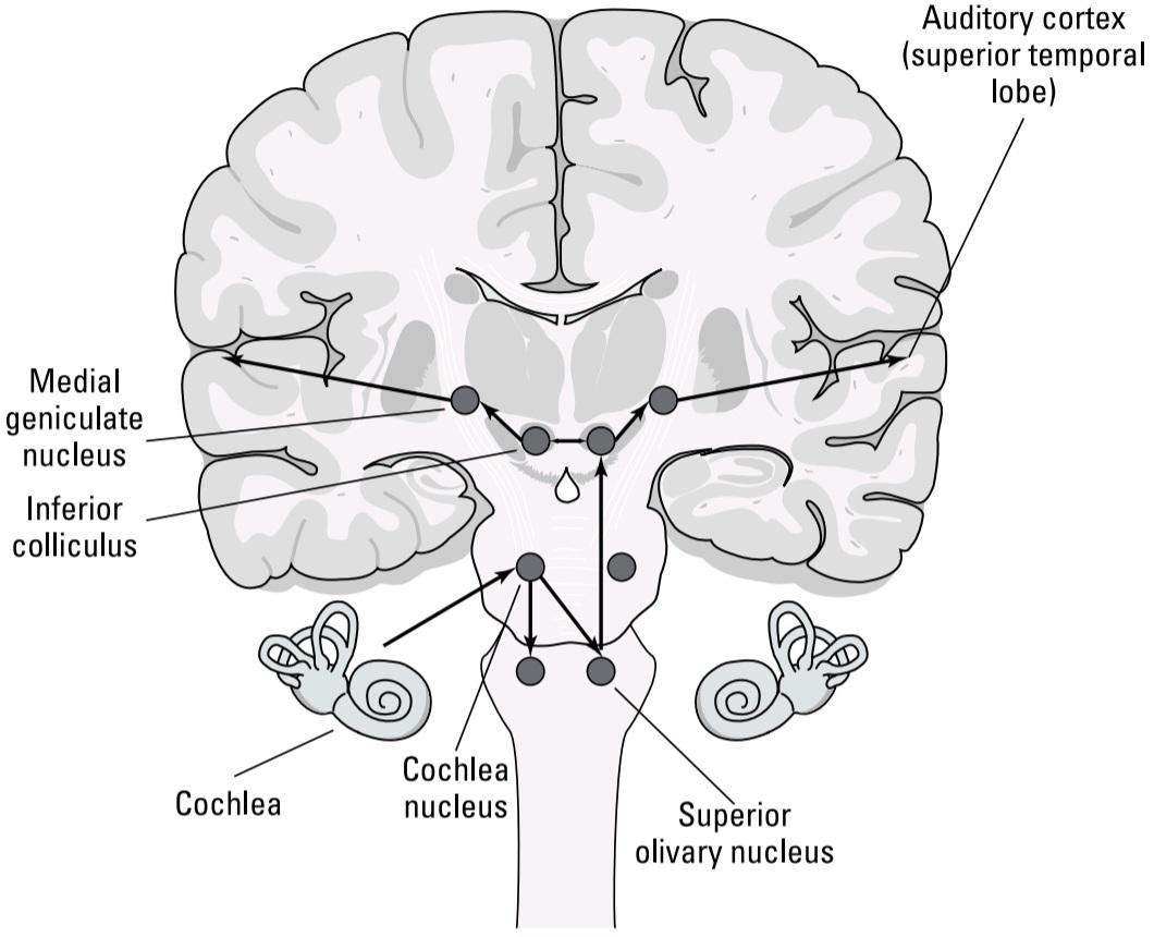
FIGURE 6-3: How the sound message gets to auditory cortex.
شکل ۶-۳: چگونه پیام صوتی به قشرشنوایی می رسد.
The message the auditory fibers send the brain about sound in the environment consists of two basic properties of sound frequency and amplitude similar to the way the retina informs the brain about color and light intensity. However, whereas the retina represents space explicitly in terms of location on the retina, there is no similar link between the location of the sound and position in the cochlea. Instead, when it comes to the auditory sense, the cortex computes the location of the sound in space (you can read more about this in the later section “Locating Sound”). In other words, the auditory nerve output doesn’t explicitly include a representation of space (which is one reason the cochlea can mediate hearing even though it has only 30,000 auditory fibers – compare this to the over one million retinal ganglion cell axons that leave each eye!).
پیامی که فیبرهای شنوایی در مورد صدا در محیط به مغز ارسال میکنند، شامل دو ویژگی اساسی فرکانس و دامنه صدا است، مشابه روشی که شبکیه چشم در مورد رنگ و شدت نور به مغز اطلاع میدهد. با این حال، در حالی که شبکیه فضا را به صراحت بر اساس موقعیت مکانی روی شبکیه نشان میدهد، هیچ ارتباط مشابهی بین موقعیت صدا و موقعیت آن در حلزون گوش وجود ندارد. در عوض، وقتی صحبت از حس شنوایی میشود، قشر مغز موقعیت صدا را در فضا محاسبه میکند (میتوانید در بخش بعدی «مکانیابی صدا» در این مورد بیشتر بخوانید). به عبارت دیگر، خروجی عصب شنوایی به صراحت شامل نمایش فضا نیست (که یکی از دلایلی است که حلزون گوش میتواند واسطه شنوایی باشد، حتی با اینکه فقط 30000 فیبر شنوایی دارد – این را با بیش از یک میلیون آکسون سلول گانگلیونی شبکیه که از هر چشم خارج میشوند مقایسه کنید!).
Although position in the cochlea doesn’t reflect sound location, the cochlea is extended in one dimension, forming a spiral consisting of about 2.5 turns (refer to Figure 6-2). What is represented in this single dimension is frequency.
اگرچه موقعیت در حلزون گوش منعکس کننده مکان صدا نیست، اما حلزون گوش در یک بعد امتداد یافته و یک مارپیچ متشکل از حدود 2.5 دور تشکیل میدهد (به شکل 6-2 مراجعه کنید). آنچه در این بعد واحد نشان داده میشود، فرکانس است.
The mechanical structure of the cochlea (stiffness, size) is such that high frequencies only cause vibrations in the portion of the cochlea near the oval window, while low frequencies produce a larger vibration amplitude toward the end of the cochlea farthest from the oval window (the center of the coil). The position along the cochlea where the vibration occurs codes frequency. Hair cell responses near the oval window indicate high frequencies; those at the farthest end of the cochlea indicate low frequencies. If position along the cochlea can encode frequency, then firing rate and number of cells firing can encode amplitude, thus generating the neural representation of the audio input. Simple enough, right?
ساختار مکانیکی حلزون گوش (سفتی، اندازه) به گونهای است که فرکانسهای بالا فقط باعث ایجاد ارتعاش در بخشی از حلزون گوش نزدیک به پنجره بیضی میشوند، در حالی که فرکانسهای پایین دامنه ارتعاش بزرگتری را به سمت انتهای حلزون گوش که از پنجره بیضی دورتر است (مرکز سیمپیچ) ایجاد میکنند. موقعیتی در امتداد حلزون گوش که ارتعاش در آن رخ میدهد، فرکانس را کدگذاری میکند. پاسخهای سلولهای مویی نزدیک به پنجره بیضی نشان دهنده فرکانسهای بالا هستند؛ پاسخهای سلولهای مویی در دورترین انتهای حلزون گوش، فرکانسهای پایین را نشان میدهند. اگر موقعیت در امتداد حلزون گوش بتواند فرکانس را کدگذاری کند، آنگاه سرعت شلیک و تعداد سلولهای شلیک شده میتوانند دامنه را کدگذاری کنند و در نتیجه نمایش عصبی ورودی صوتی را ایجاد کنند. به اندازه کافی ساده است، درست است؟
Unfortunately, it’s not as simple as place coding for frequency versus a firing rate code for amplitude. The reason has to do with a fundamental property of neurons, that is, their maximum firing rate (refer to Chapter 3). Because the action potential itself lasts about one millisecond, neurons can’t fire more than 1,000 action potentials per second; in general, sustained rates are less than 500 per second.
متأسفانه، این به سادگی کدگذاری مکانی برای فرکانس در مقابل کد نرخ شلیک برای دامنه نیست. دلیل آن به یک ویژگی اساسی نورونها، یعنی حداکثر نرخ شلیک آنها مربوط میشود (به فصل 3 مراجعه کنید). از آنجا که خود پتانسیل عمل حدود یک میلیثانیه طول میکشد، نورونها نمیتوانند بیش از 1000 پتانسیل عمل در ثانیه شلیک کنند. به طور کلی، نرخهای پایدار کمتر از 500 در ثانیه هستند.
For reasons having to do with sound localization (head to the later section “Locating Sound” for more on that), auditory fibers usually fire just at the peak of the sound pressure wave. Here’s what this means for different frequencies:
به دلایلی که مربوط به مکانیابی صدا است (برای اطلاعات بیشتر به بخش «مکانیابی صدا» در ادامه مراجعه کنید)، تارهای شنوایی معمولاً درست در اوج موج فشار صدا فعال میشوند. در اینجا معنای این موضوع برای فرکانسهای مختلف آمده است:
» For frequencies below about 500 Hz: Auditory fibers can fire every sound pressure wave peak, so that their firing rate actually directly codes frequency. In other words, at low frequencies, the fibers fire at the sound frequency.
» برای فرکانسهای کمتر از حدود ۵۰۰ هرتز: فیبرهای شنوایی میتوانند هر اوج موج فشار صدا را شلیک کنند، به طوری که سرعت شلیک آنها در واقع مستقیماً فرکانس را کدگذاری میکند. به عبارت دیگر، در فرکانسهای پایین، فیبرها در فرکانس صدا شلیک میکنند.
» For frequencies higher than 500 Hz: Auditory fibers can’t fire at the sound frequency, so they fire every other pressure peak, or every fifth peak, or tenth, or, you get the idea. In that case, the frequency is encoded by place along the cochlea.
» برای فرکانسهای بالاتر از ۵۰۰ هرتز: تارهای شنوایی نمیتوانند در فرکانس صدا شلیک کنند، بنابراین آنها هر یک پیک فشار در میان، یا هر پنج پیک، یا هر یک دهم پیک، یا، منظورم را میفهمید، شلیک میکنند. در این حالت، فرکانس بر اساس محل قرارگیری در امتداد حلزون گوش کدگذاری میشود.
Sound amplitude for both low and high frequencies is encoded by both the number of spikes that occur at each pressure pulse peak, and the number of active fibers (loud sounds will recruit more fibers to fire than fainter sounds).
دامنه صدا برای هر دو فرکانس پایین و بالا توسط تعداد اسپایکهایی که در هر پیک پالس فشار رخ میدهند و تعداد فیبرهای فعال کدگذاری میشود (صداهای بلند، فیبرهای بیشتری را نسبت به صداهای ضعیفتر برای شلیک جذب میکنند).
WHY AUDITORY PROSTHESES WORK
The one dimensional frequency structure of the cochlea is what has made auditory prostheses so successful. Most hearing loss is associated with death of the auditory hair cells. Successful auditory prostheses consist of linear electrode arrays that are slid into the cochlea. Electronic processing splits the incoming sound into frequency bands and generates stimulating current at the appropriate location of the prosthesis to directly drive the auditory nerve fibers. At the time of this writing, well over 100,000 auditory prostheses have been implanted in the United States, many allowing recipients to communicate verbally with little handicap. There is no current prosthesis with even remotely comparable success for vision loss.
چرا پروتزهای شنوایی کار میکنند؟
ساختار فرکانسی تکبعدی حلزون گوش، پروتزهای شنوایی را بسیار موفق کرده است. بیشتر موارد کمشنوایی با مرگ سلولهای مویی شنوایی مرتبط است. پروتزهای شنوایی موفق از آرایههای الکترود خطی تشکیل شدهاند که به داخل حلزون گوش وارد میشوند. پردازش الکترونیکی، صدای ورودی را به باندهای فرکانسی تقسیم میکند و جریان تحریککنندهای را در محل مناسب پروتز ایجاد میکند تا مستقیماً فیبرهای عصبی شنوایی را هدایت کند. در زمان نگارش این مطلب، بیش از ۱۰۰۰۰۰ پروتز شنوایی در ایالات متحده کاشته شده است که بسیاری از آنها به گیرندگان اجازه میدهند با معلولیت کم، ارتباط کلامی برقرار کنند. در حال حاضر هیچ پروتزی با موفقیت حتی از راه دور قابل مقایسه برای از دست دادن بینایی وجود ندارد.
Making Sense of Sounds: Central Auditory Projections
The 30,000 auditory fibers convey messages about sound to the cochlear nucleus and beyond in a mixed code. Each fiber is sensitive to a particular frequency band, and different fibers have different thresholds and firing rates that encode the magnitude of sound.
درک صداها: پیشبینیهای شنوایی مرکزی
30000 فیبر شنوایی پیامهایی در مورد صدا را به هسته حلزونی و فراتر از آن در یک کد ترکیبی منتقل میکنند. هر فیبر به یک باند فرکانسی خاص حساس است و فیبرهای مختلف آستانهها و نرخهای شلیک متفاوتی دارند که بزرگی صدا را رمزگذاری میکنند.
Stops before the thalamus
Auditory nerve fibers don’t project directly to the thalamus; they relay twice before that. As mentioned previously, when the auditory hair cilia bend, the hair cell membrane stretches and opens up ion channels that depolarize the hair cell and causes the axon terminals of the auditory nerve fibers to fire action potentials.
قبل از تالاموس متوقف میشود
فیبرهای عصبی شنوایی مستقیماً به تالاموس نمیروند؛ آنها دو بار قبل از آن رله میکنند. همانطور که قبلاً ذکر شد، وقتی مژکهای مویی شنوایی خم میشوند، غشای سلول مویی کشیده شده و کانالهای یونی را باز میکند که سلول مویی را دپلاریزه میکند و باعث میشود پایانههای آکسون فیبرهای عصبی شنوایی پتانسیل عمل ایجاد کنند.
The auditory nerve action potentials head to the cochlear nucleus where the neuron cell bodies for those axons are located and cause an action potential in the cell body, which in turn causes an action potential in a second axon branch that crosses the body midline and projects to a nucleus, called the superior olivary nucleus, on the opposite side (see Figure 6-3). In other words, action potentials from the right cochlear nucleus cross to the superior olivary nucleus on the left, and action potentials from the left project to the superior olivary nucleus on the right. The superior olivary nuclei are located in the pons (refer to Chapter 2 for general information about the areas on the brain).
پتانسیلهای عمل عصب شنوایی به هسته حلزونی، جایی که اجسام سلولی نورونهای مربوط به آن آکسونها قرار دارند، میروند و باعث ایجاد یک پتانسیل عمل در جسم سلولی میشوند که به نوبه خود باعث ایجاد یک پتانسیل عمل در شاخه آکسون دوم میشود که از خط میانی جسم عبور کرده و به هستهای به نام هسته زیتونی فوقانی در طرف مقابل منتهی میشود (شکل 6-3 را ببینید). به عبارت دیگر، پتانسیلهای عمل از هسته حلزونی راست به هسته زیتونی فوقانی در سمت چپ و پتانسیلهای عمل از سمت چپ به هسته زیتونی فوقانی در سمت راست منتهی میشوند. هستههای زیتونی فوقانی در پل مغزی قرار دارند (برای اطلاعات کلی در مورد نواحی روی مغز به فصل 2 مراجعه کنید).
Relay cells in the superior olivary nucleus project to the inferior colliculus, located, as you may guess, just below the superior colliculus in the midbrain. Finally, inferior colliculus neurons project to the medial geniculate nucleus of the thalamus, which you can read about in the next section.
سلولهای رله در هسته زیتونی فوقانی به کالیکولوس تحتانی، که همانطور که ممکن است حدس بزنید، درست زیر کالیکولوس فوقانی در مغز میانی قرار دارد، متصل میشوند. در نهایت، نورونهای کالیکولوس تحتانی به هسته زانویی داخلی تالاموس متصل میشوند که میتوانید در بخش بعدی در مورد آن بخوانید.
Most of the projections after the contralateral projection to the superior olivary nucleus are ipsilateral, meaning they stay on the same side. Therefore, the medial geniculate on the right side of the body receives mostly input from the left ear, and the medial geniculate on the left side of the body receives mostly input from the right ear.
بیشتر برآمدگیهای پس از برآمدگی سمت مقابل به هسته زیتونی فوقانی، همسو هستند، به این معنی که در یک طرف باقی میمانند. بنابراین، عضله زانویی داخلی در سمت راست بدن بیشتر ورودی را از گوش چپ و عضله زانویی داخلی در سمت چپ بدن بیشتر ورودی را از گوش راست دریافت میکند.
Why does the auditory message pass through two processing stops before reaching the thalamus? The answer has to do with localizing sound. Both the superior olivary nucleus and inferior colliculus on each side of the brain receive not only contralateral but some ipsilateral projections. Neural comparisons between the outputs from each ear set up the neural coding for sound localization. In the auditory system, sound localization is mediated by a neural computation of the difference in sound volume and sound arrival time between the two ears. These computations require precise neural timing, which means they must occur very early in the auditory processing stream, as close to the two cochlear nuclei as possible. The later section “Locating Sound” goes into the details of the mechanisms for sound localization.
چرا پیام شنوایی قبل از رسیدن به تالاموس از دو ایستگاه پردازش عبور میکند؟ پاسخ به مکانیابی صدا مربوط میشود. هم هسته زیتونی فوقانی و هم کالیکولوس تحتانی در هر طرف مغز نه تنها تصاویر طرف مقابل، بلکه برخی از تصاویر همان طرف را نیز دریافت میکنند. مقایسههای عصبی بین خروجیهای هر گوش، کدگذاری عصبی برای مکانیابی صدا را تنظیم میکند. در سیستم شنوایی، مکانیابی صدا توسط محاسبه عصبی تفاوت در حجم صدا و زمان رسیدن صدا بین دو گوش انجام میشود. این محاسبات نیاز به زمانبندی عصبی دقیقی دارند، به این معنی که باید خیلی زود در جریان پردازش شنوایی، تا حد امکان نزدیک به دو هسته حلزونی، رخ دهند. بخش بعدی «مکانیابی صدا» به جزئیات مکانیسمهای مکانیابی صدا میپردازد.
Off to the thalamus: The medial geniculate nucleus
The medial geniculate nucleus on each side of the brain receives inputs primarily from the ear of the other side. The responses of medial geniculate neurons appear to be similar to those in the auditory nerve; that is, they are frequency coded so that each medial geniculate relay cell prefers a particular band of frequencies similar to its cochlear nucleus axon input, and louder sounds recruit more cells firing at higher rates.
به تالاموس: هسته زانویی میانی
هسته زانویی میانی در هر طرف مغز، ورودیها را عمدتاً از گوش طرف دیگر دریافت میکند. پاسخهای نورونهای زانویی میانی شبیه به پاسخهای عصب شنوایی به نظر میرسد؛ یعنی، آنها فرکانسی کدگذاری شدهاند به طوری که هر سلول رله زانویی میانی، باند خاصی از فرکانسهای مشابه ورودی آکسون هسته حلزونی خود را ترجیح میدهد و صداهای بلندتر، سلولهای بیشتری را با سرعت بالاتر به کار میاندازند.
In addition to these similarities, typical thalamic attentional gating functions almost certainly occur there, as well. For example, if you barely hear a suspicious noise, you can concentrate and attend to hearing the occurrence of another instance of that noise. The neural enhancement of attended auditory input sounds you deliberately try to hear is mediated in multiple brain areas that typically include the thalamus.
علاوه بر این شباهتها، عملکردهای معمول دروازهای توجه تالاموس تقریباً به طور قطع در آنجا نیز رخ میدهد. به عنوان مثال، اگر به سختی صدای مشکوکی را بشنوید، میتوانید تمرکز کنید و به شنیدن وقوع نمونه دیگری از آن صدا توجه کنید. تقویت عصبی ورودی شنوایی مورد توجه – صداهایی که عمداً سعی در شنیدن آنها دارید – در چندین ناحیه مغز واسطهگری میشود که معمولاً شامل تالاموس میشوند.
Processing sound in the brain: The superior temporal lobe
As Figure 6-3 shows, the medial geniculate nucleus of the thalamus conveys auditory information to the superior temporal lobe at an area slightly posterior to the middle of the superior sulcus. This area of the superior temporal lobe is called the primary auditory cortex, or A1. It is also referred to as Heschl’s gyrus.
پردازش صدا در مغز: لوب گیجگاهی فوقانی
همانطور که شکل 6-3 نشان میدهد، هسته زانویی داخلی تالاموس اطلاعات شنوایی را به لوب گیجگاهی فوقانی در ناحیهای کمی عقبتر از وسط شیار فوقانی منتقل میکند. این ناحیه از لوب گیجگاهی فوقانی، قشر شنوایی اولیه یا A1 نامیده میشود. همچنین به آن شکنج هشل نیز گفته میشود.
What varies with position within A1 is the frequency to which neurons respond. Such a frequency map is called tonotopic. Just as real images in the visual system are made of patterns of lines and edges, among other things, that are put together as shapes in the visual cortex, the auditory cortex represents patterns of frequencies whose combination indicates some meaning, such as the word “hello” or a door slamming.
آنچه با موقعیت در A1 تغییر میکند، فرکانسی است که نورونها به آن پاسخ میدهند. چنین نقشه فرکانسی، تونوتوپیک نامیده میشود. همانطور که تصاویر واقعی در سیستم بینایی از الگوهایی از خطوط و لبهها، از جمله چیزهای دیگر، ساخته شدهاند که به صورت اشکال در قشر بینایی کنار هم قرار میگیرند، قشر شنوایی نیز الگوهایی از فرکانسها را نشان میدهد که ترکیب آنها معنایی را نشان میدهد، مانند کلمه “سلام” یا بسته شدن در.
Other complex aspects of sound also appear to either be processed by neurons in A1 or depend on projections from A1, since lesions in A1 impede a person’s ability to identify and locate the direction a sound is coming from. Figure 6-4 shows the primary auditory cortex and other areas important for discriminating between sounds.
به نظر میرسد سایر جنبههای پیچیده صدا نیز یا توسط نورونهای A1 پردازش میشوند یا به تصاویر A1 وابسته هستند، زیرا ضایعات A1 مانع از توانایی فرد در شناسایی و تعیین جهت صدا میشود. شکل 6-4 قشر شنوایی اولیه و سایر نواحی مهم برای تمایز بین صداها را نشان میدهد.
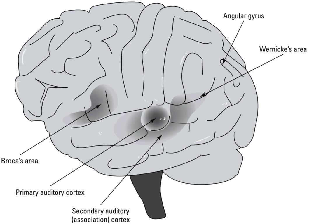
FIGURE 6-4: The primary auditory cortex and other key areas.
شکل۶-۴: قشر شنوایی اولیه و سایر نواحی کلیدی.
Handling complex auditory patterns
Around auditory area A1 are several higher order areas, which, because no standard naming convention for them exists, are usually referred to as auditory association areas.
مدیریت الگوهای پیچیده شنیداری
در اطراف ناحیه شنیداری A1 چندین ناحیه مرتبه بالاتر وجود دارد که به دلیل عدم وجود قرارداد نامگذاری استاندارد برای آنها، معمولاً به عنوان نواحی ارتباطی شنیداری شناخته میشوند.
Many cortical neurons outside A1 respond best to complex or environmentally relevant sounds rather than pure tones. An example of a complex sound is a chirp, where the frequency changes throughout the duration of the sound. Neurons have been recorded that respond well to other real sounds from the environment, such as keys jangling, doors closing, or a table being dragged across the floor, rather than to tones.
بسیاری از نورونهای قشر مغز در خارج از ناحیه A1، به صداهای پیچیده یا مرتبط با محیط، نسبت به صداهای خالص، بهتر پاسخ میدهند. نمونهای از صدای پیچیده، صدای جیرجیر است که در آن فرکانس در طول مدت پخش صدا تغییر میکند. نورونهایی ثبت شدهاند که به صداهای واقعی دیگر از محیط، مانند صدای به هم خوردن کلیدها، بسته شدن درها یا کشیده شدن میز روی زمین، به خوبی پاسخ میدهند تا به صداها.
This cortical preference for higher-order sound patterns is clearly a common theme in cortical processing. At lower levels (those closer to the input) in the auditory system (and other sensory systems, for that matter), most neurons respond to most stimuli, with the neural representation projected by a relatively small number of axon transmission lines. In the cortex, neurons become pickier, preferring more complex patterns, so that, for any given real sound, only a minority of cortical neurons respond. But because billions of cortical neurons are avail- able, your recognition and memory of particular sounds is linked to the firing of a relatively small percentage of particular neurons whose activity is specific to that stimulus. This is called sparse coding. Much of the specificity in the neural responses is achieved by learning and experience.
این ترجیح قشر مغز برای الگوهای صوتی مرتبه بالاتر، به وضوح یک موضوع رایج در پردازش قشر مغز است. در سطوح پایینتر (آنهایی که به ورودی نزدیکترند) در سیستم شنوایی (و سایر سیستمهای حسی)، اکثر نورونها به اکثر محرکها پاسخ میدهند و نمایش عصبی توسط تعداد نسبتاً کمی از خطوط انتقال آکسون نمایش داده میشود. در قشر مغز، نورونها گزینشیتر میشوند و الگوهای پیچیدهتری را ترجیح میدهند، به طوری که برای هر صدای واقعی مشخص، فقط اقلیتی از نورونهای قشر مغز پاسخ میدهند. اما از آنجا که میلیاردها نورون قشر مغز در دسترس هستند، تشخیص و حافظه شما از صداهای خاص با شلیک درصد نسبتاً کمی از نورونهای خاص مرتبط است که فعالیت آنها مختص آن محرک است. به این کدگذاری پراکنده میگویند. بخش زیادی از ویژگی در پاسخهای عصبی با یادگیری و تجربه حاصل میشود.
Hearing with meaning: Specializations for language
Although the functional differentiation among the higher order auditory processing areas (after A1) remains somewhat unclear at present, one stream of auditory processing is well known because of its importance in speech processing. This is the projection to Wernicke’s area (Brodmann area 22), located at the border between the superior, posterior part of the temporal lobe and the parietal lobe. Patients with damage to Wernicke’s area have difficulty understanding language. When they speak, although the word flow is fluent, it is often nonsensical, typically described as a “word salad.”
شنیدن با معنا: تخصصهایی برای زبان
اگرچه تمایز عملکردی بین نواحی پردازش شنوایی رده بالاتر (بعد از A1) در حال حاضر تا حدودی نامشخص است، اما یک جریان از پردازش شنوایی به دلیل اهمیت آن در پردازش گفتار به خوبی شناخته شده است. این جریان، تصویرسازی به ناحیه ورنیکه (ناحیه برودمن ۲۲) است که در مرز بین قسمت فوقانی و خلفی لوب گیجگاهی و لوب آهیانه قرار دارد. بیمارانی که ناحیه ورنیکه آنها آسیب دیده است، در درک زبان مشکل دارند. وقتی آنها صحبت میکنند، اگرچه جریان کلمات روان است، اما اغلب بیمعنی است و معمولاً به عنوان “سالاد کلمات” توصیف میشود.
Wernicke’s area has extensive connections with another language area, Broca’s area, located in the frontal lobe just anterior to the motor areas controlling the tongue, vocal cords, and other language apparatus. Damage to Broca’s area leads to difficulty in producing speech, but deficits in speech understanding are only really apparent for complex sentence constructions, like passive voice.
ناحیه ورنیکه ارتباطات گستردهای با ناحیه زبانی دیگری به نام ناحیه بروکا دارد که در لوب پیشانی و درست جلوی نواحی حرکتی کنترلکننده زبان، تارهای صوتی و سایر دستگاههای زبانی قرار دارد. آسیب به ناحیه بروکا منجر به مشکل در تولید گفتار میشود، اما نقص در درک گفتار فقط برای ساختارهای پیچیده جملات، مانند جمله مجهول، واقعاً آشکار است.
Both Wernicke’s and Broca’s areas, shown in Figure 6-4, are located on the left side of the brain in virtually all (approximately 95 percent) right-handed people.
هر دو ناحیه ورنیکه و بروکا، که در شکل 6-4 نشان داده شده است، تقریباً در تمام افراد راست دست (تقریباً 95 درصد) در سمت چپ مغز قرار دارند.
The situation is more complicated for left handers. A small majority of left handers also have language primarily on the left, but the rest have language on the right side of the brain.
وضعیت برای چپ دستها پیچیدهتر است. اکثریت کوچکی از چپ دستها نیز عمدتاً در سمت چپ مغز خود زبان دارند، اما بقیه در سمت راست مغز خود زبان دارند.
Some research suggests that there are two sorts of left handed people: strong left handers, whose brains really are reversed compared to right handers, and mixed left handers, whose brain lateralization is like right handers but who happen to be left handed. Some evidence has suggested correlated differences in some traits between the two types, with strong left handers writing in the “upside down” hooked hand position, while the mixed left handers tend to write with the hand in a mirror symmetric position to typical right handed writing.
برخی تحقیقات نشان میدهد که دو نوع چپ دست وجود دارد: چپ دستهای قوی، که مغز آنها در مقایسه با راست دستها واقعاً معکوس است، و چپ دستهای مختلط، که جانبی شدن مغز آنها مانند راست دستها است اما اتفاقاً چپ دست هستند. برخی شواهد تفاوتهای همبستهای را در برخی از صفات بین این دو نوع نشان دادهاند، به طوری که چپ دستهای قوی در موقعیت دست قلاب شده “وارونه” مینویسند، در حالی که چپ دستهای مختلط تمایل دارند با دستی در موقعیت متقارن آینهای نسبت به نوشتن معمول با دست راست بنویسند.
The identification of left-sided language areas constituted one of the first instances of known functional lateralization in the brain. Given that damage to Wernicke’s area on the left side causes profound language processing dysfunctions, the question arises as to what function is mediated by the mirror symmetric area on the right side of the brain. Recent evidence suggests that damage to the right side area 22 results in an inability to process prosody in language the changes in tonality and rhythm that conveys meaning. Patients with this damage, for example, have trouble distinguishing sarcastic versus questioning versus other tones of voice, and in particular, don’t “get” jokes and other forms of humor.
شناسایی نواحی زبانی سمت چپ، یکی از اولین نمونههای شناختهشدهی جانبیسازی عملکردی در مغز بود. با توجه به اینکه آسیب به ناحیهی ورنیکه در سمت چپ باعث اختلالات عمیق پردازش زبان میشود، این سوال مطرح میشود که ناحیهی متقارن آینهای در سمت راست مغز چه عملکردی را میانجیگری میکند. شواهد اخیر نشان میدهد که آسیب به ناحیهی سمت راست ۲۲ منجر به ناتوانی در پردازش عروض در زبان تغییرات در تونالیته و ریتمی که معنا را منتقل میکند میشود. به عنوان مثال، بیمارانی که این آسیب را دارند، در تشخیص لحنهای طعنهآمیز در مقابل پرسشی در مقابل سایر لحنهای صدا مشکل دارند و به ویژه، جوکها و سایر اشکال طنز را “درک” نمیکنند.
Perceiving music: I’ve got rhythm
Music is another complex stimulus. However, its processing relies more on the right than the left side of the brain, indicated by the fact that most people recognize melodies better with their left ear (right brain).
درک موسیقی: من ریتم دارم
موسیقی یکی دیگر از محرکهای پیچیده است. با این حال، پردازش آن بیشتر به سمت راست مغز وابسته است تا سمت چپ آن، که این واقعیت را نشان میدهد که اکثر مردم ملودیها را با گوش چپ خود (مغز راست) بهتر تشخیص میدهند.
Interestingly, there are clinical cases of bilateral damage in higher order auditory areas that result in a very specific perception loss, called amusia. In this condition sufferers are unable to recognize melodies but have normal ability to comprehend speech and complex environmental sounds. One patient knew the names of the songs on records she owned but couldn’t recognize the songs when they were played.
جالب توجه است که موارد بالینی آسیب دو طرفه در نواحی شنوایی رده بالاتر وجود دارد که منجر به از دست دادن درک بسیار خاصی به نام آموسیا میشود. در این وضعیت، افراد مبتلا قادر به تشخیص ملودیها نیستند، اما توانایی طبیعی در درک گفتار و صداهای پیچیده محیطی دارند. یکی از بیماران نام آهنگهای روی صفحههای گرامافون خود را میدانست، اما نمیتوانست آهنگها را هنگام پخش تشخیص دهد.
Locating Sound
Localization of the source of a sound is a very different process from localizing it visually. The optics of the eye preserve topography between direction in the external world and position on the retina that is maintained in projections to higher visual centers. But the ears don’t encode auditory direction in any map; instead, sound localization is computed neurally in the auditory system through neural comparisons between the two ears. This computation begins in the superior olivary nucleus and inferior colliculus and is further dependent on the firing of cells in the auditory cortex (A1) that respond best to sound emanating from particular elevation and azimuth (direction in the horizontal plane) ranges.
مکانیابی صدا
مکانیابی منبع صدا فرآیندی بسیار متفاوت از مکانیابی بصری آن است. اپتیک چشم، توپوگرافی بین جهت در دنیای خارجی و موقعیت روی شبکیه را که در تصاویر به مراکز بینایی بالاتر حفظ میشود، حفظ میکند. اما گوشها جهت شنوایی را در هیچ نقشهای کدگذاری نمیکنند؛ در عوض، مکانیابی صدا به صورت عصبی در سیستم شنوایی از طریق مقایسههای عصبی بین دو گوش محاسبه میشود. این محاسبه در هسته زیتونی فوقانی و کالیکولوس تحتانی آغاز میشود و بیشتر به شلیک سلولهایی در قشر شنوایی (A1) وابسته است که به صدای ناشی از محدودههای ارتفاع و آزیموت (جهت در صفحه افقی) خاص، بهترین پاسخ را میدهند.
Computing azimuth (horizontal angle)
The auditory system uses two different methods to compute azimuth: interaural intensity difference and interaural time difference.
محاسبه آزیموت (زاویه افقی)
سیستم شنوایی از دو روش مختلف برای محاسبه آزیموت استفاده میکند: اختلاف شدت بین دو گوش و اختلاف زمان بین دو گوش.
These two methods work best in complementary frequency ranges. Spike firing at the exact sound pressure peak works better at low frequencies because the axons can fire fast enough to follow more cycles and stay in what’s called phase lock. On the other hand, the interaural intensity difference is larger for high rather than low frequencies because the head itself weakens high more than low frequencies in the (auditorily) shadowed ear.
این دو روش در محدودههای فرکانسی مکمل یکدیگر بهترین عملکرد را دارند. شلیک اسپایک در اوج فشار صوتی دقیق در فرکانسهای پایین بهتر عمل میکند زیرا آکسونها میتوانند به اندازه کافی سریع شلیک کنند تا چرخههای بیشتری را دنبال کنند و در چیزی که قفل فاز نامیده میشود، باقی بمانند. از سوی دیگر، اختلاف شدت بین دو گوش برای فرکانسهای بالا بیشتر از فرکانسهای پایین است زیرا خود سر در گوش (از نظر شنوایی) در سایه، فرکانسهای بالا را بیشتر از فرکانسهای پایین ضعیف میکند.
Interaural intensity difference
A sound source that is not directly in front or behind the listener is louder in the closer ear than in the ear farther away. Some neurons in the superior olivary nucleus and inferior colliculus receive both ipsilateral and contralateral inputs. These neurons are arrayed so that some respond best when the inputs are equal between the two ears, while others prefer different percentages of left versus right ear strength. These neurons project in an ordered way to the auditory cortex where auditory location is then represented by position on the surface of the cortex.
تفاوت شدت بین دو گوش
منبع صدایی که مستقیماً در جلو یا پشت شنونده نیست، در گوش نزدیکتر بلندتر از گوش دورتر است. برخی از نورونها در هسته زیتونی فوقانی و کالیکولوس تحتانی، ورودیهای همسو و همسو را دریافت میکنند. این نورونها به گونهای آرایش یافتهاند که برخی از آنها وقتی ورودیها بین دو گوش برابر باشند، بهترین پاسخ را میدهند، در حالی که برخی دیگر درصدهای متفاوتی از قدرت گوش چپ در مقابل راست را ترجیح میدهند. این نورونها به ترتیب به قشر شنوایی میتابند که در آنجا مکان شنوایی با موقعیت روی سطح قشر نمایش داده میشود.
Interaural time difference
Sound source position also affects the relative time that sounds reach the two ears. Most of us are familiar with counting seconds from the time we see lightning until we hear thunder, with a five second delay for each mile. In other words, the speed of sound is very roughly 1,000 feet per second, or one foot per millisecond. If the human head is (very roughly) about 6 inches (one half foot) wide, then the difference in arrival time between the two ears for a source directly to one side is about one half of a millisecond.
اختلاف زمان بین دو گوش
موقعیت منبع صدا نیز بر زمان نسبی رسیدن صدا به دو گوش تأثیر میگذارد. اکثر ما با شمارش ثانیهها از زمانی که رعد و برق را میبینیم تا زمانی که صدای آن را میشنویم، با تأخیر پنج ثانیهای برای هر مایل، آشنا هستیم. به عبارت دیگر، سرعت صدا تقریباً ۱۰۰۰ فوت در ثانیه یا یک فوت در میلیثانیه است. اگر سر انسان (به طور تقریبی) حدود ۶ اینچ (نیم فوت) عرض داشته باشد، اختلاف زمان رسیدن صدا بین دو گوش برای منبعی که مستقیماً در یک طرف قرار دارد، حدود نیم میلیثانیه است.
Given that action potentials last about one millisecond, you may think that the auditory system wouldn’t be able to resolve a one half millisecond difference. But it can! The arrival time of the sound is encoded by auditory fibers because they fire at the exact peak of the sound pressure wave, which of course is delayed between the two ears.
با توجه به اینکه پتانسیلهای عمل حدود یک میلیثانیه طول میکشند، ممکن است فکر کنید که سیستم شنوایی قادر به تشخیص اختلاف نیم میلیثانیهای نیست. اما میتواند! زمان رسیدن صدا توسط تارهای شنوایی رمزگذاری میشود زیرا آنها دقیقاً در اوج موج فشار صدا شلیک میکنند، که البته بین دو گوش با تأخیر انجام میشود.
AUDITORY LOCALIZATION IN BARN OWLS
Barn owls have auditory localization so precise that, with only the sound of a mouse’s movements to guide them, they can locate and catch the critters in total darkness. Because of this ability, barn owls have been used as a model in a number of labs to study neural sound localization mechanisms. Researchers have found that, among other things, the owl’s two ears are not symmetrical but are arranged so that there is an intensity difference processed by the ears as a function of elevation as well as azimuth.
مکانیابی شنیداری در جغدهای انبار
جغدهای انبار مکانیابی شنیداری بسیار دقیقی دارند، به طوری که تنها با هدایت صدای حرکات موش، میتوانند این موجودات را در تاریکی مطلق پیدا کرده و بگیرند. به دلیل این توانایی، جغدهای انبار به عنوان مدلی در تعدادی از آزمایشگاهها برای مطالعه مکانیسمهای مکانیابی عصبی صدا استفاده شدهاند. محققان دریافتهاند که، از جمله موارد دیگر، دو گوش جغد متقارن نیستند، بلکه به گونهای چیده شدهاند که اختلاف شدت صدا توسط گوشها به عنوان تابعی از ارتفاع و همچنین آزیموت پردازش میشود.
The decoding process for interaural time difference is exquisite. Axons from the two cochlear nuclei take slightly different length paths from the left versus the right ear to the superior olivary nucleus and colliculus. The paths to neurons there that are equal result in the two signals arriving at the same time when the sound source is either directly in front or behind. Neurons that receive inputs with a slightly longer path from the left versus right ear respond best if the sound is proportionally closer to the left than right ears so that the spikes arrive at the same time. An array of neurons with different path lengths produces a linear map of neurons, each of which prefers a slightly different direction. This map is also projected to auditory cortex in an orderly way so that position on the A1 cortex represents horizontal direction.
فرآیند رمزگشایی برای اختلاف زمان بین دو گوش، بسیار دقیق است. آکسونهای دو هسته حلزونی، مسیرهایی با طول کمی متفاوت را از گوش چپ در مقابل گوش راست تا هسته زیتونی فوقانی و کولیکولوس طی میکنند. مسیرهای مساوی به نورونهای آنجا منجر به رسیدن همزمان دو سیگنال میشود، زمانی که منبع صدا مستقیماً در جلو یا عقب باشد. نورونهایی که ورودیهایی با مسیر کمی طولانیتر از گوش چپ در مقابل گوش راست دریافت میکنند، اگر صدا به طور متناسب به گوش چپ نزدیکتر از گوش راست باشد، به بهترین شکل پاسخ میدهند، به طوری که اسپایکها همزمان میرسند. مجموعهای از نورونها با طول مسیرهای مختلف، یک نقشه خطی از نورونها تولید میکنند که هر کدام جهت کمی متفاوتی را ترجیح میدهند. این نقشه همچنین به طور منظم به قشر شنوایی منتقل میشود، به طوری که موقعیت روی قشر A1 نشان دهنده جهت افقی است.
Detecting elevation
As long as the head is vertical, no useful binaural signal difference exists between the two ears to enable you to estimate elevation. Instead, estimating elevation depends on the frequency reflection characteristics of the pinna, or outer ear (refer to the earlier section “The Ear: Capturing and Decoding Sound Waves” and Figure 6-1).
تشخیص ارتفاع
تا زمانی که سر عمودی باشد، هیچ تفاوت سیگنال دوگوشی مفیدی بین دو گوش وجود ندارد که شما را قادر به تخمین ارتفاع کند. در عوض، تخمین ارتفاع به ویژگیهای بازتاب فرکانس لاله گوش یا گوش خارجی بستگی دارد (به بخش قبلی “گوش: دریافت و رمزگشایی امواج صوتی” و شکل 6-1 مراجعه کنید).
The complex shape of the pinna functions as a frequency filter when it reflects sound that is elevation dependent. Almost all sounds in the natural world consist of multiple frequencies in particular ratios that you have become familiar with. The degree to which your pinna distorts the sound tells your auditory cortex the sound’s elevation.
شکل پیچیده لاله گوش، هنگامی که صدایی وابسته به ارتفاع را منعکس میکند، به عنوان یک فیلتر فرکانس عمل میکند. تقریباً همه صداها در دنیای طبیعی از فرکانسهای متعدد با نسبتهای خاصی تشکیل شدهاند که شما با آنها آشنا هستید. میزان تحریف صدا توسط لاله گوش شما، به قشر شنوایی شما، میزان ارتفاع صدا را نشان میدهد.
I Can’t Hear You: Deafness and Tinnitus
As with any system, things can go wrong. Glitches in the auditory system can range from the profound, like not being able to hear at all, to merely irritating, like periodic ringing in your ears. This section goes into a little detail on two conditions hearing loss and tinnitus both of which can range from mild to severe.
نمیشنوم: ناشنوایی و وزوز گوش
مانند هر سیستمی، ممکن است مشکلاتی پیش بیاید. مشکلات سیستم شنوایی میتواند از عمیق، مانند عدم توانایی در شنیدن، تا صرفاً آزاردهنده، مانند زنگ زدن دورهای گوش، متغیر باشد. این بخش به جزئیات دو بیماری کمشنوایی و وزوز گوش میپردازد که هر دو میتوانند از خفیف تا شدید متغیر باشند.
Hearing loss
Deafness or partial deafness can be congenital (existing at birth) or the result of disease, damage from loud noises, or aging. Congenital deafness is rare compared to acquired deafness, occurring in less than one in 1,000 births. About one quarter of congenital deafness is due to inner ear malformations; the other causes of con- genital deafness are unknown.
کمشنوایی
ناشنوایی یا ناشنوایی نسبی میتواند مادرزادی (از بدو تولد وجود داشته باشد) یا نتیجه بیماری، آسیب ناشی از صداهای بلند یا پیری باشد. ناشنوایی مادرزادی در مقایسه با ناشنوایی اکتسابی نادر است و در کمتر از یک مورد در هر ۱۰۰۰ تولد رخ میدهد. حدود یک چهارم ناشنوایی مادرزادی ناشی از ناهنجاریهای گوش داخلی است. سایر علل ناشنوایی تناسلی ناشناخته هستند.
The causes of acquired deafness are easier to pinpoint. Severe inner ear infections can cause deafness by destroying or compromising the three middle ear bones. Called conductive hearing loss, in this condition, the sound vibrations are not mechanically transmitted from the eardrum to the cochlea. Otosclerosis, the growth of spongy masses on the middle ear bones, is a hereditary conductive hearing loss with an onset typically between the ages of 15 and 40. It is somewhat more common in women than men. Maternal rubella (German measles) can cause the birth of deaf children from damage by the virus.
علل ناشنوایی اکتسابی را راحتتر میتوان مشخص کرد. عفونتهای شدید گوش داخلی میتوانند با تخریب یا آسیب رساندن به سه استخوانچه گوش میانی باعث ناشنوایی شوند. در این حالت که کمشنوایی انتقالی نامیده میشود، ارتعاشات صدا به صورت مکانیکی از پرده گوش به حلزون گوش منتقل نمیشوند. اتواسکلروز، رشد تودههای اسفنجی روی استخوانچههای گوش میانی، یک کمشنوایی انتقالی ارثی است که معمولاً بین سنین ۱۵ تا ۴۰ سالگی شروع میشود. این بیماری در زنان تا حدودی شایعتر از مردان است. سرخجه مادر (سرخک آلمانی) میتواند باعث تولد کودکان ناشنوا در اثر آسیب ناشی از ویروس شود.
Some infections as well as other unknown causes can result in Meniere’s disease, a partial loss of both hearing and vestibular (balance) function due to a common mechanism that affects both the cochlea and the semicircular canals near the cochlea that function in balance.
برخی عفونتها و همچنین سایر علل ناشناخته میتوانند منجر به بیماری منییر شوند، که عبارت است از از دست دادن جزئی شنوایی و عملکرد دهلیزی (تعادل) به دلیل مکانیسم مشترکی که هم حلزون گوش و هم مجاری نیمدایرهای نزدیک حلزون گوش را که در تعادل عمل میکنند، تحت تأثیر قرار میدهد.
Besides the age-related decline in high frequency sensitivity (presbycusis, which occurs in almost everyone, particularly males), the most frequent cause of hearing loss is damage to the eardrum or auditory hair cells. Eardrums can be damaged mechanically by items entering the auditory canal or by high pressure such as diving into deep water. Loud noises can damage hearing permanently by destroying auditory hair cells. The louder the noise, the less time it takes for hearing loss to occur. Exposure to loud work environments, for example, can accumulate over decades to damage hearing, while hearing loss can occur from single exposures to very loud sounds, such as the levels experienced in rock concerts near the loud speakers. Auditory hair cell damage is permanent. Conventional hearing aids can amplify sounds in the compromised frequency range for limited hair cell damage, but after more severe damage, auditory prostheses are used that are threaded into the cochlea to stimulate the cochlear fibers electrically to restore some hearing.
گذشته از کاهش حساسیت فرکانس بالا در اثر افزایش سن (پیرگوشی، که تقریباً در همه، به ویژه مردان، رخ میدهد)، شایعترین علت کاهش شنوایی، آسیب به پرده گوش یا سلولهای مویی شنوایی است. پرده گوش میتواند به صورت مکانیکی توسط اجسامی که وارد کانال شنوایی میشوند یا با فشار زیاد مانند شیرجه زدن در آب عمیق آسیب ببیند. صداهای بلند میتوانند با از بین بردن سلولهای مویی شنوایی، به شنوایی آسیب دائمی وارد کنند. هر چه صدا بلندتر باشد، زمان کمتری برای بروز کاهش شنوایی لازم است. به عنوان مثال، قرار گرفتن در معرض محیطهای کاری پر سر و صدا میتواند در طول دههها انباشته شود و به شنوایی آسیب برساند، در حالی که کاهش شنوایی میتواند از قرار گرفتن در معرض صداهای بسیار بلند، مانند سطوحی که در کنسرتهای راک در نزدیکی بلندگوهای بلند تجربه میشود، رخ دهد. آسیب سلولهای مویی شنوایی دائمی است. سمعکهای معمولی میتوانند صداها را در محدوده فرکانسی آسیب دیده برای آسیب محدود به سلولهای مویی تقویت کنند، اما پس از آسیب شدیدتر، از پروتزهای شنوایی استفاده میشود که به حلزون گوش متصل میشوند تا فیبرهای حلزونی را به صورت الکتریکی تحریک کنند تا مقداری از شنوایی بازیابی شود.
Oh those bells bells bells bells bells bells bells: Tinnitus
Tinnitus is the perception of ringing in the years. Chronic, severe tinnitus is one of the most psychologically debilitating diseases known, having reportedly led some patients to drug abuse and even suicide. Tinnitus has multiple causes: ear infections, allergies, reactions to certain drugs, and exposure to loud noise (many rock musicians have tinnitus).
آه، آن زنگها، زنگها، زنگها، زنگها، زنگها، زنگها: وزوز گوش
وزوز گوش به احساس زنگ زدن در گوش در طول سالها گفته میشود. وزوز گوش مزمن و شدید یکی از ناتوانکنندهترین بیماریهای روانی شناخته شده است که طبق گزارشها برخی از بیماران را به سوءمصرف مواد مخدر و حتی خودکشی سوق داده است. وزوز گوش علل متعددی دارد: عفونت گوش، آلرژی، واکنش به برخی داروها و قرار گرفتن در معرض صدای بلند (بسیاری از نوازندگان راک وزوز گوش دارند).
So called objective tinnitus that arises from muscle spasms in the ears can be detected by ear doctors with special instruments that detect these vibrations. Subjective tinnitus (the perception of sound when there is none) can arise from numerous causes but is often associated with hearing loss.
وزوز گوش عینی که ناشی از اسپاسم عضلات گوش است، توسط پزشکان گوش با ابزارهای مخصوصی که این ارتعاشات را تشخیص میدهند، قابل تشخیص است. وزوز گوش ذهنی (درک صدا در حالی که صدایی وجود ندارد) میتواند از علل متعددی ناشی شود، اما اغلب با کاهش شنوایی همراه است.
There are few universally effective treatments for tinnitus, but, in extreme cases, sometimes patients wear noise-producing earphones that mask the tinnitus noise with electronically generated white noise, giving some relief. Some hearing aids also do this. In extreme cases, some patients have opted for surgical treatments, in which the surgeon lesions (destroys) some of the auditory nerve for relief, but which also reduces normal hearing. Tinnitus can also sometimes appear suddenly and then disappear within a few months.
درمانهای جهانی مؤثر کمی برای وزوز گوش وجود دارد، اما در موارد شدید، گاهی اوقات بیماران از هدفونهای تولید کننده نویز استفاده میکنند که صدای وزوز گوش را با نویز سفید تولید شده به صورت الکترونیکی میپوشاند و تا حدودی تسکین میدهد. برخی از سمعکها نیز این کار را انجام میدهند. در موارد شدید، برخی از بیماران درمانهای جراحی را انتخاب کردهاند که در آن جراح مقداری از عصب شنوایی را برای تسکین آسیب (تخریب) میکند، اما این کار شنوایی طبیعی را نیز کاهش میدهد. وزوز گوش همچنین گاهی اوقات میتواند به طور ناگهانی ظاهر شود و سپس ظرف چند ماه ناپدید شود.



