
علوم اعصاب به زبان ساده؛ نگاهی به بینایی


دعای مطالعه [ نمایش ]
بِسْمِ الله الرَّحْمنِ الرَّحیمِ
اَللّهُمَّ اَخْرِجْنى مِنْ ظُلُماتِ الْوَهْمِ
خدايا مرا بيرون آور از تاريكىهاى وهم،
وَ اَكْرِمْنى بِنُورِ الْفَهْمِ
و به نور فهم گرامى ام بدار،
اَللّهُمَّ افْتَحْ عَلَيْنا اَبْوابَ رَحْمَتِكَ
خدايا درهاى رحمتت را به روى ما بگشا،
وَانْشُرْ عَلَيْنا خَزائِنَ عُلُومِكَ بِرَحْمَتِكَ يا اَرْحَمَ الرّاحِمينَ
و خزانههاى علومت را بر ما باز كن به امید رحمتت اى مهربانترين مهربانان.
کتاب «علوم اعصاب به زبان ساده» اثر دکتر فرانک آمتور، یکی از منابع معتبر و قابل دسترس برای علاقهمندان، دانشجویان در حوزه علوم اعصاب و بسیار مناسب برای مسابقه دانش آموزی دانش مغز (برین بی: Brain Bee) است که مفاهیم پایه و کاربردهای عمومی مغز و سیستم عصبی را به زبان ساده توضیح میدهد. این کتاب تصویری روشن از مغز انسان و عملکرد نورونها ارائه میدهد و به مخاطبان کمک میکند تا مفاهیم شناختی، رفتاری و ارتباط ذهن و بدن را درک کنند.
ترجمه دقیق و علمی این شاهکار توسط برند علمی «آیندهنگاران مغز» به مدیریت داریوش طاهری، دسترسی فارسیزبانان به مرزهای نوین دانش علوم اعصاب را ممکن ساخته و رسالتی علمی برای ارتقای آموزش، فهم عمیقتر عملکرد مغز و سیستم عصبی و توسعه روشهای نوین در حوزه سلامت عصبی فراهم آورده است.
» کتاب علوم اعصاب به زبان ساده
» » فصل ۵: نگاهی به بینایی
در حال ویرایش
» Neuroscience For Dummies
»» CHAPTER 5: Looking at Vision
IN THIS CHAPTER
» Seeing the role the eye and how its neurons enable vision
» Examining the vision centers of the brain
» Looking at how we see color, depth, and shapes
» Uncovering the causes of visual impairment and the secrets behind optical illusions
در این فصل
» بررسی نقش چشم و چگونگی فعال شدن بینایی توسط نورونهای آن
» بررسی مراکز بینایی مغز
» بررسی چگونگی دیدن رنگ، عمق و اشکال توسط ما
» کشف علل اختلال بینایی و اسرار پشت خطاهای دید

Chapter 5
Looking at Vision
How do you see? Most people think that, when we look at things, the light coming off those things enters the eye, which sends a camera like image of what we’re looking at to the brain. However, the retina itself is an extension of the brain and generates a neural representation of the optical image that it receives. This neural representation is processed and modified by the rest of the brain.
فصل ۵
نگاهی به بینایی
چگونه میبینید؟ اکثر مردم فکر میکنند وقتی به چیزی نگاه میکنیم، نوری که از آن چیز ساطع میشود وارد چشم میشود و تصویری شبیه دوربین از آنچه به آن نگاه میکنیم به مغز میفرستد. با این حال، خود شبکیه امتداد مغز است و یک نمایش عصبی از تصویر نوری که دریافت میکند، ایجاد میکند. این نمایش عصبی توسط بقیه مغز پردازش و اصلاح میشود.
Vision occurs when retinal photoreceptors capture photons and the retina and brain perform a complex analysis of this information. Visual input that reaches consciousness is processed in parallel pathways through the retina, thalamus, and occipital lobe of the cortex. About 15 other visual pathways process visual input unconsciously to do things like control pupil diameter and circadian rhythms. Specific classes of neural cells in the retina sense different aspects of the visual image, such as the colors present, whether something is moving, and the location of edges. These neurons act like little agents that “shout out” the presence of these features so that you can recognize objects and determine their distance from you.
بینایی زمانی اتفاق میافتد که گیرندههای نوری شبکیه، فوتونها را دریافت میکنند و شبکیه و مغز تجزیه و تحلیل پیچیدهای از این اطلاعات انجام میدهند. ورودی بصری که به هوشیاری میرسد، در مسیرهای موازی از طریق شبکیه، تالاموس و لوب پسسری قشر مغز پردازش میشود. حدود ۱۵ مسیر بصری دیگر، ورودی بصری را به صورت ناخودآگاه پردازش میکنند تا کارهایی مانند کنترل قطر مردمک و ریتمهای شبانهروزی را انجام دهند. دستههای خاصی از سلولهای عصبی در شبکیه، جنبههای مختلف تصویر بصری، مانند رنگهای موجود، حرکت چیزی و محل لبهها را حس میکنند. این نورونها مانند عوامل کوچکی عمل میکنند که وجود این ویژگیها را “فریاد میزنند” تا بتوانید اشیاء را تشخیص داده و فاصله آنها را از خود تعیین کنید.
This chapter covers the cellular agents involved in sight. Early in the visual system, these agents are very simple-minded and react to qualities like color and intensity. Higher in the visual system these agents become very sophisticated and very picky, some responding only to certain faces. This is their story. Oh andafter you’ve studied all this visual processing neural circuitry, think about the fact that blind people, without any input from the eyes, still can visualize.
این فصل به عوامل سلولی دخیل در بینایی میپردازد. در اوایل سیستم بینایی، این عوامل بسیار سادهلوح هستند و به ویژگیهایی مانند رنگ و شدت واکنش نشان میدهند. در مراحل بالاتر سیستم بینایی، این عوامل بسیار پیچیده و بسیار مشکلپسند میشوند و برخی فقط به چهرههای خاصی پاسخ میدهند. داستان آنها از این قرار است. و بعد از اینکه تمام این مدارهای عصبی پردازش بینایی را مطالعه کردید، به این واقعیت فکر کنید که افراد نابینا، بدون هیچ ورودی از چشمها، هنوز هم میتوانند تصویرسازی کنند.
The Eyes Have It: A Quick Glance at Your Eyes
You may have heard that the eye is like a camera. As Figure 5-1 shows, this comparison springs from the fact that the eye has evolved tissues that act like the optical elements of a camera, namely, the lens, which acts like a camera’s lens, and the pupil, the opening through which the light enters, acts like an aperture.
چشمها این را دارند: نگاهی سریع به چشمان شما
شاید شنیده باشید که چشم مانند یک دوربین است. همانطور که شکل 5-1 نشان میدهد، این مقایسه از این واقعیت ناشی میشود که چشم بافتهایی را تکامل داده است که مانند عناصر نوری یک دوربین عمل میکنند، یعنی لنز، که مانند لنز دوربین عمل میکند، و مردمک، روزنهای که نور از طریق آن وارد میشود، مانند یک دیافراگم عمل میکند.

FIGURE 5-1: Light entering the eye.
شکل ۵-۱: نور وارد چشم میشود.
When light enters the eye, the cornea, which is the outermost clear layer at the front of the eye, first focuses it, and then the lens focuses it further, and the pupil, located between the cornea and lens, opens and closes to let in more or less light, like a camera aperture. The image formed by the cornea, lens, and pupil is projected onto the retina, the neural lining inside the eye. The retina is where the real action in vision takes place. The following sections outline what happens to this image once it hits the retina.
وقتی نور وارد چشم میشود، قرنیه، که بیرونیترین لایه شفاف در جلوی چشم است، ابتدا آن را متمرکز میکند و سپس عدسی آن را بیشتر متمرکز میکند و مردمک که بین قرنیه و عدسی قرار دارد، مانند دیافراگم دوربین، باز و بسته میشود تا نور بیشتر یا کمتری وارد شود. تصویری که توسط قرنیه، عدسی و مردمک تشکیل میشود، روی شبکیه، لایه عصبی داخل چشم، منعکس میشود. شبکیه جایی است که عمل واقعی بینایی در آن رخ میدهد. بخشهای زیر شرح میدهند که پس از برخورد این تصویر به شبکیه، چه اتفاقی برای آن میافتد.
Although you may think that the eye’s lens functions the same as the camera lens, this isn’t quite right. Instead, the cornea, which is at the air/tissue interface, does about 70 percent of the focusing. The lens of the eye, being slightly more dense than the surrounding tissue, does the remaining 30 percent. The lens changes shape to adjust the focus for near and far objects, a process called accommodation.
اگرچه ممکن است فکر کنید که عدسی چشم مانند عدسی دوربین عمل میکند، اما این کاملاً درست نیست. در عوض، قرنیه که در سطح مشترک هوا/بافت قرار دارد، حدود ۷۰ درصد از فوکوس را انجام میدهد. عدسی چشم که کمی متراکمتر از بافت اطراف است، ۳۰ درصد باقیمانده را انجام میدهد. عدسی برای تنظیم فوکوس برای اشیاء نزدیک و دور، شکل خود را تغییر میدهد، فرآیندی که تطابق نامیده میشود.
The retina: Converting photons to electrical signals
The smallest units of light are called photons. When photons hit the retina, they are absorbed by photoreceptors, specialized neural cells in the retina that convert light into electric current that modulates the release of a neurotransmitter (glutamate). This whole process from photons to electrical current to neurotransmitter release is called phototransduction.
شبکیه: تبدیل فوتونها به سیگنالهای الکتریکی
کوچکترین واحدهای نور فوتون نامیده میشوند. هنگامی که فوتونها به شبکیه برخورد میکنند، توسط گیرندههای نوری، سلولهای عصبی تخصصی در شبکیه که نور را به جریان الکتریکی تبدیل میکنند و آزادسازی یک انتقالدهنده عصبی (گلوتامات) را تنظیم میکنند، جذب میشوند. کل این فرآیند از فوتونها به جریان الکتریکی تا آزادسازی انتقالدهنده عصبی – انتقال نوری نامیده میشود.
Two main types of photoreceptors exist:
دو نوع اصلی گیرنده نوری وجود دارد:
» Rods, which work in very dim light for night vision.
» Cones, which function only in bright daylight. Humans have three different types of cones that allow us to perceive color.
» میلهها، که در نور بسیار کم برای دید در شب کار میکنند.
» مخروطها، که فقط در نور روز روشن عمل میکنند. انسانها سه نوع مختلف مخروط دارند که به ما امکان درک رنگ را میدهند.
At night, when only your rods are absorbing enough photons to generate signals, you have no color vision because the signal from a rod contains no information about the wavelength of the photon absorbed. During the day, however, three different cone types are active (red, green, and blue). Individually, cones don’t signal wavelength either, but the brain can deduce wavelength from the ratio of activity of different cones. In very blue light, for example, the blue cones are relatively more activated than the green and red cones.
در شب، زمانی که فقط سلولهای استوانهای شما فوتونهای کافی برای تولید سیگنال جذب میکنند، شما دید رنگی ندارید زیرا سیگنال یک سلول استوانهای حاوی هیچ اطلاعاتی در مورد طول موج فوتون جذب شده نیست. با این حال، در طول روز، سه نوع مخروط مختلف فعال هستند (قرمز، سبز و آبی). مخروطها به صورت جداگانه طول موج را نیز سیگنال نمیدهند، اما مغز میتواند طول موج را از نسبت فعالیت مخروطهای مختلف استنباط کند. به عنوان مثال، در نور بسیار آبی، مخروطهای آبی نسبتاً فعالتر از مخروطهای سبز و قرمز هستند.
Because you have three types of cones, simulation of color in television and computer screens requires that they have three different colored light emitters: red, green, and blue. If you had only two cones, like many other animals, display screens could get by with two different colored light emitters. Similarly, if humans, like some fish, had four cones, a TV with only three colored light emitters couldn’t simulate color accurately, just like a printer with one color cartridge out of ink can’t simulate all colors.
از آنجا که شما سه نوع مخروط دارید، شبیهسازی رنگ در صفحه نمایش تلویزیون و کامپیوتر مستلزم آن است که آنها سه ساطعکننده نور با رنگهای مختلف داشته باشند: قرمز، سبز و آبی. اگر فقط دو مخروط داشتید، مانند بسیاری از حیوانات دیگر، صفحه نمایشها میتوانستند با دو ساطعکننده نور با رنگهای مختلف کار کنند. به طور مشابه، اگر انسانها، مانند برخی از ماهیها، چهار مخروط داشتند، یک تلویزیون با تنها سه ساطعکننده نور رنگی نمیتوانست رنگ را به طور دقیق شبیهسازی کند، درست مانند چاپگری که یک کارتریج رنگی آن تمام شده است و نمیتواند همه رنگها را شبیهسازی کند.
Catching photons: Light and phototransduction
When a photoreceptor absorbs a photon of light, a cascade of events occurs that result in a message being sent to other neurons in the retina:
دریافت فوتونها: نور و انتقال نوری
هنگامی که یک گیرنده نوری، فوتونی از نور را جذب میکند، سلسلهای از رویدادها رخ میدهد که منجر به ارسال پیام به سایر نورونهای شبکیه میشود:
1. A chemical reaction occurs.
The retinal molecule in rhodopsin (in rods; similar molecules exist in cones), which absorbs photons, starts out in a kinked form, called 11-cis retinal. When this molecule absorbs a photon of light, a molecular bond in the middle of the 11-cis retinal flips from a kinked to a straight configuration, converting it to what’s called all-trans retinal.
۱. یک واکنش شیمیایی رخ میدهد.
مولکول رتینال در رودوپسین (در سلولهای میلهای؛ مولکولهای مشابه در سلولهای مخروطی وجود دارند) که فوتونها را جذب میکند، در ابتدا به شکل پیچخوردهای به نام رتینال ۱۱-سیس ظاهر میشود. هنگامی که این مولکول یک فوتون نور را جذب میکند، یک پیوند مولکولی در وسط رتینال ۱۱-سیس از حالت پیچخورده به حالت مستقیم تغییر میکند و آن را به چیزی تبدیل میکند که رتینال تمام ترانس نامیده میشود.
The all trans retinal is a stereoisomer of 11-cis retinal, which means it has the same chemical composition but a different structure.
رتینال تمام ترانس، یک استریوایزومر از رتینال ۱۱-سیس است، به این معنی که ترکیب شیمیایی یکسانی دارد اما ساختار متفاوتی دارد.
TECHNICAL 2. All trans retinal reduces the concentration of cyclic GMP (cGMP).
CGMP is an intracellular messenger inside the photoreceptor that keeps depolarizing ion channels in the cell membrane open.
فنی ۲. تمام ترانس رتینال غلظت GMP حلقوی (cGMP) را کاهش میدهد.
CGMP یک پیامرسان درون سلولی در داخل گیرنده نوری است که کانالهای یونی دپلاریزه کننده را در غشای سلولی باز نگه میدارد.
These depolarizing ion channels are like the channels of metabotropic receptors (refer to Chapter 3) except that they’re triggered by light absorption rather than a neurotransmitter binding to a receptor. When light reduces the internal concentration of cGMP in the photoreceptor, it reduces the number of these channels that are open via a second messenger cascade, hyperpolarizing the receptor.
این کانالهای یونی دپلاریزه کننده مانند کانالهای گیرندههای متابوتروپیک هستند (به فصل 3 مراجعه کنید) با این تفاوت که آنها به جای اتصال یک انتقالدهنده عصبی به یک گیرنده، توسط جذب نور تحریک میشوند. هنگامی که نور غلظت داخلی cGMP را در گیرنده نوری کاهش میدهد، تعداد این کانالها که از طریق یک آبشار پیامرسان ثانویه باز هستند را کاهش میدهد و گیرنده را بیشقطبی میکند.
In all vertebrates (animals with backbones, like humans), photoreceptors hyperpolarize to light by a similar mechanism, using similar photochemistry. Some non vertebrates, like barnacles and squids, have photoreceptors that use different photochemistry to depolarize to light.
در تمام مهرهداران (حیواناتی که ستون فقرات دارند، مانند انسان)، گیرندههای نوری با مکانیسمی مشابه و با استفاده از فتوشیمی مشابه، نسبت به نور بیشقطبی میشوند. برخی از غیر مهرهداران، مانند کشتیچسبها و ماهیهای مرکب، گیرندههای نوری دارند که از فتوشیمی متفاوتی برای دپلاریزه شدن به نور استفاده میکنند.
3. The hyperpolarization of the photoreceptor causes a structure at its base, called the pedicle, to release less glutamate, the photoreceptor neurotransmitter.
The photoreceptor pedicle is very similar to a conventional axon terminal except that, instead of individual action potentials releasing puffs of neurotransmitter, light absorption continuously modulates the release of neurotransmitter.
۳. هایپرپلاریزاسیون گیرنده نوری باعث میشود ساختاری در پایه آن به نام پدیکول، گلوتامات کمتری، انتقالدهنده عصبی گیرنده نوری، آزاد کند.
پدیکول گیرنده نوری بسیار شبیه به یک پایانه آکسون معمولی است، با این تفاوت که به جای پتانسیلهای عمل منفرد که پافهای انتقالدهنده عصبی را آزاد میکنند، جذب نور به طور مداوم آزادسازی انتقالدهنده عصبی را تنظیم میکند.
4. The modulation of glutamate release drives other cells in the retina.
The outputs of photoreceptors drive two main types of cells called bipolar and horizontal cells. These cells are discussed in the next section.
۴. تنظیم آزادسازی گلوتامات، سلولهای دیگر شبکیه را به حرکت در میآورد.
خروجیهای گیرندههای نوری، دو نوع اصلی سلول به نام سلولهای دوقطبی و افقی را به حرکت در میآورند. این سلولها در بخش بعدی مورد بحث قرار میگیرند.
Photoreceptors do not send an image of the world directly to the brain. Instead, they communicate with other retinal neurons that extract specific information about the image to send to higher brain centers. The following sections explore that communication.
گیرندههای نوری تصویری از جهان را مستقیماً به مغز ارسال نمیکنند. در عوض، آنها با سایر نورونهای شبکیه ارتباط برقرار میکنند که اطلاعات خاصی در مورد تصویر را استخراج کرده و به مراکز بالاتر مغز ارسال میکنند. بخشهای بعدی این ارتباط را بررسی میکنند.
Getting the message to the brain
Why doesn’t the eye just send the electrical signal from all the rod and cone photoreceptors directly to the brain? The main reason is that there are well over 100 million rods and cones but only a million axon transmission lines available to go to the brain (refer to Chapter 3 for more about axons). Even worse, these trans- mission lines work by sending at most a few hundred action potentials per second along the axon, further limiting what information each line can send.
رساندن پیام به مغز
چرا چشم سیگنال الکتریکی را از تمام گیرندههای نوری میلهای و مخروطی مستقیماً به مغز نمیفرستد؟ دلیل اصلی این است که بیش از ۱۰۰ میلیون گیرنده نوری میلهای و مخروطی وجود دارد، اما فقط یک میلیون خط انتقال آکسون برای رفتن به مغز در دسترس است (برای اطلاعات بیشتر در مورد آکسونها به فصل ۳ مراجعه کنید). حتی بدتر از آن، این خطوط انتقال با ارسال حداکثر چند صد پتانسیل عمل در ثانیه در امتداد آکسون کار میکنند و این امر اطلاعات قابل ارسال توسط هر خط را محدودتر میکند.
The retina gets around these limitations on transmission capacity in a few interesting ways, as the following sections explain.
شبکیه از چند طریق جالب، همانطور که در بخشهای بعدی توضیح داده خواهد شد، بر این محدودیتها در ظرفیت انتقال غلبه میکند.
The most common misconception about the retina is that it sends some sort of raw image to the brain. In reality, the retina processes the image and sends information extracted from the image to at least 15 different brain areas by at least 20 parallel pathways.
رایجترین تصور غلط در مورد شبکیه این است که نوعی تصویر خام را به مغز ارسال میکند. در واقع، شبکیه تصویر را پردازش میکند و اطلاعات استخراج شده از تصویر را از طریق حداقل ۲۰ مسیر موازی به حداقل ۱۵ ناحیه مختلف مغز ارسال میکند.
More photoreceptors in the center of the eye
The retina has more photoreceptors and other retinal cells in the center of the eye (called the fovea; refer to Figure 5-1) than it does in the periphery. You notice things happening in your periphery, but to identify something clearly, you have to look directly at it to place the image on the high resolution fovea.
گیرندههای نوری بیشتر در مرکز چشم
شبکیه در مرکز چشم (که گودی چشم نامیده میشود؛ به شکل 5-1 مراجعه کنید) گیرندههای نوری و سایر سلولهای شبکیه بیشتری نسبت به محیط اطراف دارد. شما متوجه اتفاقاتی که در محیط اطرافتان میافتد میشوید، اما برای شناسایی واضح چیزی، باید مستقیماً به آن نگاه کنید تا تصویر روی گودی چشم با وضوح بالا قرار گیرد.
Modulating responses around average light level
The retina uses adaptation in which many ganglion cells, the output cells of the retina, respond briefly whenever the light level changes but then settle down and reduce their output after a few seconds. Adaptation saves energy and spikes if cells don’t have to keep telling the brain, “Yes, the light level is still the same as it was a few seconds ago.” Photoreceptors themselves change their dynamics so that their basal neurotransmitter release modulates their responses around the average current light level. Temporal adaptation occurs in virtually all retinal neurons.
تعدیل پاسخها حول سطح نور متوسط
شبکیه از سازگاری استفاده میکند که در آن بسیاری از سلولهای گانگلیونی، سلولهای خروجی شبکیه، هر زمان که سطح نور تغییر میکند، به طور خلاصه پاسخ میدهند، اما پس از چند ثانیه آرام میشوند و خروجی خود را کاهش میدهند. سازگاری باعث صرفهجویی در انرژی و افزایش ناگهانی میشود اگر سلولها مجبور نباشند به مغز بگویند: “بله، سطح نور هنوز همان است که چند ثانیه پیش بود.” خود گیرندههای نوری پویایی خود را تغییر میدهند به طوری که ترشح انتقالدهنده عصبی پایه آنها پاسخهای آنها را حول سطح نور متوسط فعلی تعدیل میکند. سازگاری زمانی تقریباً در تمام نورونهای شبکیه رخ میدهد.
Minimizing information across space
Adaptation occurs in another way in the retina (and the brain) via neural circuit interactions. This type of adaptation minimizes information across space. A process called lateral inhibition reduces how much information is transmitted to the brain because photoreceptors and other retinal neurons communicate the difference between the light where they are and the surrounding area rather than the absolute level of the light they receive. The next sections explain the neural circuitry in the retina that makes lateral inhibition possible.
به حداقل رساندن اطلاعات در فضا
سازگاری به روش دیگری در شبکیه (و مغز) از طریق تعاملات مدار عصبی رخ میدهد. این نوع سازگاری، اطلاعات را در فضا به حداقل میرساند. فرآیندی به نام مهار جانبی، میزان اطلاعات منتقل شده به مغز را کاهش میدهد، زیرا گیرندههای نوری و سایر نورونهای شبکیه، تفاوت بین نوری که در آن قرار دارند و محیط اطراف را به جای سطح مطلق نوری که دریافت میکنند، بیان میکنند. بخشهای بعدی، مدار عصبی در شبکیه را که مهار جانبی را ممکن میسازد، توضیح میدهند.
Processing signals from the photoreceptors: Horizontal and bipolar cells
After temporal adaptation, lateral inhibition is another way your nervous system overcomes the limitations on how much information can be transmitted from the retina to the brain. In lateral inhibition, photoreceptors don’t convey the absolute level of the light they receive; instead, they communicate the difference between the light they receive and the surrounding light. This section explains the neural circuitry that makes lateral inhibition possible.
پردازش سیگنالهای دریافتی از گیرندههای نوری: سلولهای افقی و دوقطبی
پس از سازگاری زمانی، مهار جانبی راه دیگری است که سیستم عصبی شما بر محدودیتهای میزان اطلاعات قابل انتقال از شبکیه به مغز غلبه میکند. در مهار جانبی، گیرندههای نوری سطح مطلق نوری را که دریافت میکنند منتقل نمیکنند؛ در عوض، تفاوت بین نوری که دریافت میکنند و نور اطراف را منتقل میکنند. این بخش مدار عصبی را که مهار جانبی را ممکن میسازد، توضیح میدهد.
Photoreceptors connect to two retinal neural cell classes: horizontal cells and bipolar cells (see Figure 5-2). Horizontal cells mediate lateral inhibition, and bipolar cells pass the photoreceptor signal, which has been modified by the horizontal cells, on toward the next retinal layer, which then projects to the brain. The next sections explain how this process works.
گیرندههای نوری به دو دسته از سلولهای عصبی شبکیه متصل میشوند: سلولهای افقی و سلولهای دوقطبی (شکل 5-2 را ببینید). سلولهای افقی واسطه مهار جانبی هستند و سلولهای دوقطبی سیگنال گیرنده نوری را که توسط سلولهای افقی اصلاح شده است، به سمت لایه بعدی شبکیه منتقل میکنند که سپس به مغز ارسال میشود. بخشهای بعدی نحوه عملکرد این فرآیند را توضیح میدهند.

FIGURE 5-2: Photoreceptors connect to bipolar and horizontal cells.
شکل ۵-۲: گیرندههای نوری به سلولهای دوقطبی و افقی متصل هستند.
Step 1: Reducing redundant signals (horizontal cells and lateral inhibition)
Suppose you’re staring at a stop sign. You don’t need all the cells responding to different parts of the sign to report with high precision that exactly the same shade of red occurs everywhere over the entire sign. The retina can avoid sending redundant spatial information because lateral inhibition uses horizontal cells to allow photoreceptors to communicate the difference between the light they receive and the surrounding light.
مرحله ۱: کاهش سیگنالهای زائد (سلولهای افقی و مهار جانبی)
فرض کنید به یک تابلوی ایست خیره شدهاید. برای گزارش دقیق اینکه دقیقاً همان سایه قرمز در همه جای تابلو وجود دارد، نیازی نیست که تمام سلولهای پاسخدهنده به قسمتهای مختلف تابلو، دقیقاً همان سایه قرمز را در همه جای تابلو گزارش دهند. شبکیه میتواند از ارسال اطلاعات مکانی زائد جلوگیری کند، زیرا مهار جانبی از سلولهای افقی استفاده میکند تا به گیرندههای نوری اجازه دهد تفاوت بین نوری که دریافت میکنند و نور اطراف را منتقل کنند.
Here’s how it works: Horizontal cells receive excitation from surrounding photoreceptors and subtract a percentage of this excitation from the output of the central photoreceptor. This action allows each photoreceptor to report the difference between the light intensity and color it receives and the average intensity and color nearby. The photoreceptor can then signal small differences in intensity or color from those of nearby areas. These highly precise signals go to the next cells, the bipolar cells.
نحوهی کار آن به این صورت است: سلولهای افقی، تحریک را از گیرندههای نوری اطراف دریافت میکنند و درصدی از این تحریک را از خروجی گیرندهی نوری مرکزی کم میکنند. این عمل به هر گیرندهی نوری اجازه میدهد تا تفاوت بین شدت نور و رنگی که دریافت میکند و میانگین شدت و رنگ نور نزدیک را گزارش دهد. سپس گیرندهی نوری میتواند تفاوتهای کوچک در شدت یا رنگ را از مناطق مجاور سیگنال دهد. این سیگنالهای بسیار دقیق به سلولهای بعدی، یعنی سلولهای دوقطبی، میروند.
Step 2: On to bipolar cells and to the processing layers beyond
The signals from the photoreceptors that have been modified by the horizontal cells are then sent to the bipolar cells (refer to Figure 5-2). The bipolar cells then carry these signals to the next retinal processing layer. Bipolar cells come in two major varieties:
مرحله 2: به سراغ سلولهای دوقطبی و لایههای پردازشی فراتر از آن
سیگنالهای گیرندههای نوری که توسط سلولهای افقی اصلاح شدهاند، سپس به سلولهای دوقطبی ارسال میشوند (به شکل 5-2 مراجعه کنید). سپس سلولهای دوقطبی این سیگنالها را به لایه پردازش شبکیه بعدی منتقل میکنند. سلولهای دوقطبی در دو نوع اصلی وجود دارند:
» Depolarizing bipolar cells, which are excited by light areas of the image.
» Hyperpolarizing bipolar cells, which are excited by dark areas.
» سلولهای دوقطبی دپلاریزه که توسط نواحی روشن تصویر تحریک میشوند.
» سلولهای دوقطبی هایپرپلاریزه که توسط نواحی تاریک تحریک میشوند.
How do depolarizing and hyperpolarizing bipolar cells have the opposite responses to light? Both types of bipolar cells receive glutamate from the photoreceptors, but each has a different receptor for glutamate. The glutamate receptor on depolarizing bipolar cells is an unusually fast metabotropic receptor that is inhibitory. As mentioned previously, photoreceptors are hyperpolarized by light so that, when illuminated, they release less glutamate. This reduction of glutamate is a reduction of the inhibition of depolarizing bipolar cells (in other words, dis-inhibition = more excitation). Hyperpolarizing bipolar cells, on the other hand, have glutamate receptors that respond in a conventional manner, that is, reduction of glutamate results in less excitation. Hyperpolarizing bipolar cells are excited by dark and inhibited by light.
چگونه سلولهای دوقطبی دپلاریزه و هایپرپلاریزه پاسخهای متضادی به نور دارند؟ هر دو نوع سلولهای دوقطبی گلوتامات را از گیرندههای نوری دریافت میکنند، اما هر کدام گیرنده متفاوتی برای گلوتامات دارند. گیرنده گلوتامات در سلولهای دوقطبی دپلاریزه، یک گیرنده متابوتروپیک غیرمعمول سریع است که مهارکننده است. همانطور که قبلاً ذکر شد، گیرندههای نوری توسط نور هایپرپلاریزه میشوند، به طوری که وقتی روشن میشوند، گلوتامات کمتری آزاد میکنند. این کاهش گلوتامات، کاهش مهار سلولهای دوقطبی دپلاریزه است (به عبارت دیگر، مهارزدایی = تحریک بیشتر). از سوی دیگر، سلولهای دوقطبی هایپرپلاریزه، گیرندههای گلوتاماتی دارند که به شیوهای مرسوم پاسخ میدهند، یعنی کاهش گلوتامات منجر به تحریک کمتر میشود. سلولهای دوقطبی هایپرپلاریزه توسط تاریکی تحریک و توسط نور مهار میشوند.
As mentioned, bipolar cells carry the signal forward toward the next layer of retinal processing before the brain. In this second synaptic retinal layer, bipolar cells connect to (synapse on) two kinds of postsynaptic cells:
همانطور که گفته شد، سلولهای دوقطبی سیگنال را به سمت لایه بعدی پردازش شبکیه قبل از مغز حمل میکنند. در این لایه دوم سیناپسی شبکیه، سلولهای دوقطبی به دو نوع سلول پس سیناپسی متصل میشوند (سیناپس میزنند):
» Retinal ganglion cells: These cells send the final output of the retina to the brain.
» Amacrine cells: These cells mediate lateral interactions, something like horizontal cells do with photoreceptors (refer to the preceding section).
» سلولهای گانگلیون شبکیه: این سلولها خروجی نهایی شبکیه را به مغز ارسال میکنند.
» سلولهای آماکرین: این سلولها واسطه تعاملات جانبی هستند، چیزی شبیه به سلولهای افقی که با گیرندههای نوری کار میکنند (به بخش قبل مراجعه کنید).
I cover both of these cell types in more detail in the next section. (Stick with me through the next section: We’re almost to the brain!)
در بخش بعدی هر دو نوع سلول را با جزئیات بیشتری پوشش خواهم داد. (در بخش بعدی با من همراه باشید: تقریباً به مغز رسیدهایم!)
Sending out and shaping the message: Ganglion and amacrine cells
The visual image that the photoreceptors capture and that the horizontal and bipolar cells modify is received by another group of neurons, the retinal ganglion cells, where yet another set of lateral interactions, mediated by amacrine cells, occurs.
ارسال و شکلدهی پیام: سلولهای گانگلیون و آماکرین
تصویر بصری که گیرندههای نوری ثبت میکنند و سلولهای افقی و دوقطبی آن را تغییر میدهند، توسط گروه دیگری از نورونها، سلولهای گانگلیون شبکیه، دریافت میشود، جایی که مجموعهای دیگر از تعاملات جانبی، با واسطه سلولهای آماکرین، رخ میدهد.
Ganglion cells are the output of the retina. Think of them as train depots: The information from the eye is finally onboard and ready for the ride to the brain. The destination? The ganglion cells send their axons to at least 15 retinal recipient zones in the brain. Why so many receiving zones, and exactly what kind of information does each tell the brain? The next sections explain.
سلولهای گانگلیون خروجی شبکیه هستند. آنها را به عنوان ایستگاههای قطار در نظر بگیرید: اطلاعات از چشم بالاخره سوار قطار شده و آمادهی انتقال به مغز میشوند. مقصد؟ سلولهای گانگلیون آکسونهای خود را به حداقل ۱۵ ناحیهی گیرندهی شبکیه در مغز میفرستند. چرا این همه ناحیهی گیرنده وجود دارد و هر کدام دقیقاً چه نوع اطلاعاتی را به مغز منتقل میکنند؟ در بخشهای بعدی توضیح داده خواهد شد.
Converting analog to digital signals to go the distance
As I explain in the previous sections, the connections within the retina are between cells much closer than one millimeter to each other. But the messages going from your eye to your brain have to travel many centimeters. A few centimeters may not sound like a lot to you, but when you’re a cell, it’s a marathon! Traveling this distance requires axons that conduct action potentials, by which ganglion cells convert their analog bipolar cell input into a digital pulse code for transmission to the brain. (See Chapter 3 for a discussion of action potentials.)
تبدیل سیگنالهای آنالوگ به دیجیتال برای پیمودن مسافت
همانطور که در بخشهای قبلی توضیح دادم، اتصالات درون شبکیه بین سلولها بسیار نزدیکتر از یک میلیمتر به یکدیگر هستند. اما پیامهایی که از چشم شما به مغزتان میروند باید چندین سانتیمتر را طی کنند. چند سانتیمتر ممکن است برای شما زیاد به نظر نرسد، اما وقتی شما یک سلول هستید، یک ماراتن است! پیمودن این مسافت نیاز به آکسونهایی دارد که پتانسیلهای عمل را هدایت میکنند، که توسط آنها سلولهای گانگلیون ورودی سلول دوقطبی آنالوگ خود را به یک کد پالس دیجیتال برای انتقال به مغز تبدیل میکنند. (برای بحث در مورد پتانسیلهای عمل به فصل 3 مراجعه کنید.)
Breaking down into ganglion cell types and classes
The depolarizing bipolar cells, which are excited by light, are connected to matching ganglion cells called on center. The hyperpolarizing bipolar cells, which are inhibited by light (but excited by dark), are connected to matching ganglion cells called off center ganglion cells.
تقسیمبندی به انواع و دستههای سلولهای گانگلیونی
سلولهای دوقطبی دپلاریزه که توسط نور تحریک میشوند، به سلولهای گانگلیونی منطبق به نام on center متصل هستند. سلولهای دوقطبی هایپرپلاریزه که توسط نور مهار میشوند (اما توسط تاریکی تحریک میشوند)، به سلولهای گانگلیونی منطبق به نام off center متصل هستند.
In addition to performing other functions, amacrine cells modulate signals from bipolar cells to ganglion cells much the same way horizontal cells modulate signals from photoreceptors before sending them to the bipolar cells. That is, amacrine cells conduct inhibitory signals from the surrounding bipolar cells so that the ganglion cell responds to the difference between the bipolar signal in its center, dendritic tree area versus the surrounding area. This action reduces the amount of redundant information that must be transmitted over the limited number of ganglion cell axon transmission lines.
سلولهای آماکرین علاوه بر انجام سایر وظایف، سیگنالها را از سلولهای دوقطبی به سلولهای گانگلیونی تعدیل میکنند، تقریباً به همان روشی که سلولهای افقی سیگنالها را از گیرندههای نوری قبل از ارسال به سلولهای دوقطبی تعدیل میکنند. یعنی، سلولهای آماکرین سیگنالهای مهاری را از سلولهای دوقطبی اطراف هدایت میکنند به طوری که سلول گانگلیونی به تفاوت بین سیگنال دوقطبی در مرکز خود، ناحیه درخت دندریتیک، در مقابل ناحیه اطراف پاسخ میدهد. این عمل، میزان اطلاعات اضافی را که باید از طریق تعداد محدود خطوط انتقال آکسون سلول گانگلیونی منتقل شود، کاهش میدهد.
Despite this similarity between amacrine and horizontal cell function, amacrine cells come in more varieties and are more complicated than horizontal cells. As a result, the same bipolar cell inputs create different ganglion cell classes. The two most important ganglion cell classes are:
علیرغم این شباهت بین عملکرد سلولهای آماکرین و افقی، سلولهای آماکرین انواع بیشتری دارند و پیچیدهتر از سلولهای افقی هستند. در نتیجه، ورودیهای یکسان سلولهای دوقطبی، دستههای مختلفی از سلولهای گانگلیونی را ایجاد میکنند. دو دسته از مهمترین دستههای سلولهای گانگلیونی عبارتند از:
» Parvocellular (small cells): These are selective for color and fine detail.
» Magnocellular (large cells): These are selective for motion and low contrast.
» پارووسلولار (سلولهای کوچک): این سلولها برای رنگ و جزئیات دقیق گزینشی هستند.
» مگنوسلولار (سلولهای بزرگ): این سلولها برای حرکت و کنتراست پایین گزینشی هستند.
Parvocellular ganglion cells are by far the most numerous ganglion cells in the retina. Both parvocellular and magnocellular ganglion cell classes have on center and off center varieties.
سلولهای گانگلیونی پارووسلولار تاکنون بیشترین تعداد سلولهای گانگلیونی در شبکیه را تشکیل میدهند. هر دو دسته سلولهای گانگلیونی پارووسلولار و مگنوسلولار دارای انواع مرکز روشن و مرکز خاموش هستند.
Other types of amacrine cells produce ganglion cell classes that respond only to specific features from the visual input and project to particular areas of the brain. For example, some ganglion cells respond only to motion in a certain direction and help you track moving objects or keep your balance. Other ganglion cells sense only certain colors, helping you tell ripe from unripe fruit, or red stop lights from green lights. Still others indicate the presence of edges in the scene.
انواع دیگری از سلولهای آماکرین، دستههایی از سلولهای گانگلیونی را تولید میکنند که فقط به ویژگیهای خاصی از ورودی بصری پاسخ میدهند و به نواحی خاصی از مغز ارسال میشوند. به عنوان مثال، برخی از سلولهای گانگلیونی فقط به حرکت در یک جهت خاص پاسخ میدهند و به شما در ردیابی اشیاء متحرک یا حفظ تعادل کمک میکنند. سایر سلولهای گانگلیونی فقط رنگهای خاصی را حس میکنند و به شما کمک میکنند تا میوه رسیده را از نارس یا چراغهای قرمز را از چراغهای سبز تشخیص دهید. برخی دیگر نیز وجود لبهها را در صحنه نشان میدهند.
Ganglion cells report specific features of the visual world “upstream” so that the retina doesn’t have to send an overwhelming amount of millisecond by millisecond “pixel” information to your brain.
سلولهای گانگلیون ویژگیهای خاص دنیای بصری را به صورت «بالادست» گزارش میدهند، به طوری که شبکیه مجبور نباشد حجم عظیمی از اطلاعات «پیکسلی» میلیثانیهای را به مغز شما ارسال کند.
From the Eyes to the Vision Centers of the Brain
The previous section explores how the retina converts light into ganglion cell pulses that signal different things about the visual image. These pulses, called action potentials, can travel the centimeter distances to the brain over the ganglion cell axons. In this section, I finally get to the heart of the (gray) matter and discuss where these pulses go in the brain and what happens after they get there.
از چشمها تا مراکز بینایی مغز
بخش قبلی بررسی میکند که چگونه شبکیه نور را به پالسهای سلولهای گانگلیونی تبدیل میکند که چیزهای مختلفی را در مورد تصویر بصری نشان میدهند. این پالسها، که پتانسیل عمل نامیده میشوند، میتوانند از طریق آکسونهای سلولهای گانگلیونی، مسافتهای سانتیمتری را تا مغز طی کنند. در این بخش، سرانجام به قلب ماده (خاکستری) میرسم و در مورد اینکه این پالسها در مغز به کجا میروند و پس از رسیدن به آنجا چه اتفاقی میافتد، بحث میکنم.
Destination: Thalamus
The main output of the retina is to an area of the brain called the thalamus (refer to Chapter 2 for a general description of the thalamus). The visual sub region of the thalamus is called the dorsal lateral geniculate nucleus (dLGN). Both the parvocellular and magnocellular ganglion cell classes refer to the earlier section “Breaking down into ganglion cell types and classes” project to the dLGN.
مقصد: تالاموس
خروجی اصلی شبکیه به ناحیهای از مغز به نام تالاموس است (برای شرح کلی تالاموس به فصل ۲ مراجعه کنید). زیرناحیه بینایی تالاموس، هسته زانویی جانبی پشتی (dLGN) نامیده میشود. هر دو دسته سلولهای گانگلیونی پارووسلولار و مگنوسلولار به بخش قبلی «تجزیه به انواع و دستههای سلولهای گانگلیونی» که به dLGN مربوط میشود، اشاره دارند.
Figure 5-3 shows that a bundle of axons leaves each eye and that the two bundles join a few centimeters later. The bundles of axons leaving the eyes are called the optic nerves (the term nerve is a general term for a bundle of axons). The junction point where the optic nerves first meet is called the optic chiasm, which means “optic crossing.”
شکل 5-3 نشان میدهد که یک دسته آکسون از هر چشم خارج میشود و دو دسته چند سانتیمتر بعد به هم میرسند. دستههای آکسون که از چشمها خارج میشوند، اعصاب بینایی نامیده میشوند (اصطلاح عصب، اصطلاحی کلی برای دستهای از آکسونها است). نقطه اتصالی که اعصاب بینایی برای اولین بار به هم میرسند، کیاسمای بینایی نامیده میشود که به معنی “تقاطع نوری” است.
Crossing to the other side: The left-right sorting of images
A very interesting thing happens at the optic chiasm. Some of the ganglion cell axons from each eye cross at the chiasm and go to the other side of the brain, and some don’t. Which do and which don’t, you may wonder, and why.
عبور به سمت دیگر: مرتبسازی چپ-راست تصاویر
یک اتفاق بسیار جالب در کیاسمای بینایی رخ میدهد. برخی از آکسونهای سلولهای گانگلیونی از هر چشم در کیاسمای بینایی عبور میکنند و به سمت دیگر مغز میروند و برخی دیگر این کار را نمیکنند. ممکن است از خود بپرسید کدام یک این کار را انجام میدهند و کدام یک نه، و چرا.
Look carefully at the right eye in Figure 5-3. The part of the right retina closest to the nose (called the nasal retina) receives images from the world on the right side (right visual field), while the part of the right retina farthest from the nose (called the temporal retina) receives input from the left side of the world. In the left eye, the right visual field falls on its temporal retina. What happens at the optic chiasm is that axons sort themselves so that the information received from the right visual field is sent to the left side of the brain, while the right side of the brain gets inputs from the left side of visual space (that the left brain deals with right side should be a familiar theme by now).
با دقت به چشم راست در شکل 5-3 نگاه کنید. بخشی از شبکیه راست که به بینی نزدیکتر است (به نام شبکیه بینی) تصاویر را از دنیای سمت راست (میدان بینایی راست) دریافت میکند، در حالی که بخشی از شبکیه راست که از بینی دورتر است (به نام شبکیه گیجگاهی) ورودی را از سمت چپ دنیا دریافت میکند. در چشم چپ، میدان بینایی راست روی شبکیه گیجگاهی آن قرار میگیرد. اتفاقی که در کیاسمای بینایی میافتد این است که آکسونها خود را طوری مرتب میکنند که اطلاعات دریافتی از میدان بینایی راست به سمت چپ مغز ارسال شود، در حالی که سمت راست مغز ورودیها را از سمت چپ فضای بینایی دریافت میکند (اینکه مغز چپ با سمت راست سر و کار دارد باید تا الان موضوع آشنایی باشد).
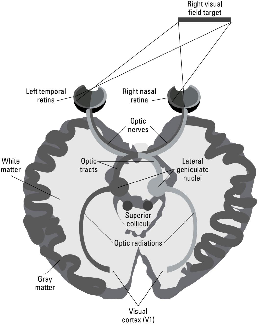
FIGURE 5-3: The thalamus and neocortex.
شکل ۵-۳: تالاموس و نئوکورتکس.
This left right sorting occurs because axons from nasal retinal ganglion cells (that see the visual world on the same side as the eye) cross at the optic chiasm and go to the opposite side of the brain, while the axons of temporal retinal ganglion cells do not cross.
این مرتبسازی چپ به راست به این دلیل رخ میدهد که آکسونهای سلولهای گانگلیونی شبکیه بینی (که دنیای بصری را در همان سمت چشم میبینند) در کیاسمای بینایی تقاطع میکنند و به سمت مقابل مغز میروند، در حالی که آکسونهای سلولهای گانگلیونی شبکیه گیجگاهی تقاطع نمیکنند.
So the nerves after the optic chiasm have different axons than the optic nerves going to the chiasm; they also have a different name, the optic tracts. The left optic tract has axons from both eyes that see the right visual field, while the right optic tract has axons from both eyes that see the left visual field. This means that there are cells in the cortex that are driven by the same visual field location in both eyes, in addition to the left visual cortex dealing with the right visual field, and vice versa.
بنابراین اعصاب بعد از کیاسمای بینایی، آکسونهای متفاوتی نسبت به اعصاب بینایی که به کیاسم میروند، دارند؛ آنها همچنین نام متفاوتی دارند، راههای بینایی. راه بینایی چپ دارای آکسونهایی از هر دو چشم است که میدان بینایی راست را میبینند، در حالی که راه بینایی راست دارای آکسونهایی از هر دو چشم است که میدان بینایی چپ را میبینند. این بدان معناست که سلولهایی در قشر مغز وجود دارند که توسط موقعیت میدان بینایی یکسان در هر دو چشم هدایت میشوند، علاوه بر اینکه قشر بینایی چپ با میدان بینایی راست سروکار دارد و برعکس.
Looking at the visual signal in the thalamus
What happens to the visual signal in the thalamus? Twenty years ago, most researchers would have answered very little because ganglion cell axons synapse on cells in the thalamus that have very similar response properties to their ganglion cell inputs. Each thalamic neuron, called a relay cell, receives inputs from one or a few similar ganglion cells and then it projects to visual cortex. Within the dLGN are six layers in most primates. Some of these layers receive inputs from only parvocellular ganglion cells, and within those layers, on center parvocellular ganglion cells drive on center parvocellular type relay cells. Similarly, off center parvocellular ganglion cells drive off-center parvocellular relay cells. A corresponding situation exists for on and off center magnocellular relay cells in other dLGN layers.
نگاهی به سیگنال بصری در تالاموس
چه اتفاقی برای سیگنال بصری در تالاموس میافتد؟ بیست سال پیش، اکثر محققان پاسخ بسیار کمی میدادند، زیرا آکسونهای سلولهای گانگلیونی روی سلولهایی در تالاموس سیناپس برقرار میکنند که خواص پاسخ بسیار مشابهی با ورودیهای سلولهای گانگلیونی خود دارند. هر نورون تالاموس، که سلول رله نامیده میشود، ورودیهایی را از یک یا چند سلول گانگلیونی مشابه دریافت میکند و سپس آن را به قشر بینایی ارسال میکند. در اکثر نخستیها، درون dLGN شش لایه وجود دارد. برخی از این لایهها فقط از سلولهای گانگلیونی پاروسلولی ورودی دریافت میکنند و در داخل آن لایهها، سلولهای گانگلیونی پاروسلولی روی مرکز، سلولهای رله نوع پاروسلولی مرکزی را فعال میکنند. به طور مشابه، سلولهای گانگلیونی پاروسلولی خارج از مرکز، سلولهای رله پاروسلولی خارج از مرکز را فعال میکنند. وضعیت مشابهی برای سلولهای رله مگنوسلولاری روشن و خاموش در سایر لایههای dLGN وجود دارد.
Why do ganglion cells make this relay stop then? Is it because the ganglion cell axons simply can’t grow far enough? This explanation seems unlikely because all mammals have this visual relay through the thalamus, despite large differences in brain sizes (consider the distances involved in elephant versus mice brains). The more likely explanation is that, although the relay cells in the thalamus seem to respond very much like their parvocellular or magnocellular ganglion cell inputs, other inputs to the dLGN from other parts of the brain allow gating functions associated with attention. (A gating function is a modulation of the strength of a neuron’s responses to any particular stimulus based on the context of importance of that stimulus.)
چرا سلولهای گانگلیونی این رله را متوقف میکنند؟ آیا به این دلیل است که آکسونهای سلولهای گانگلیونی نمیتوانند به اندازه کافی رشد کنند؟ این توضیح بعید به نظر میرسد زیرا همه پستانداران، با وجود تفاوتهای زیاد در اندازه مغز (فاصلههای مغز فیل در مقابل موش را در نظر بگیرید)، این رله بصری را از طریق تالاموس دارند. توضیح محتملتر این است که اگرچه به نظر میرسد سلولهای رله در تالاموس بسیار شبیه به ورودیهای سلولهای گانگلیونی پارووسلولار یا مگنوسلولار خود پاسخ میدهند، اما ورودیهای دیگر به dLGN از سایر قسمتهای مغز، امکان عملکردهای دروازهای مرتبط با توجه را فراهم میکنند. (عملکرد دروازهای، تعدیل قدرت پاسخهای یک نورون به هر محرک خاص بر اساس زمینه اهمیت آن محرک است.)
How does attention mediate a gating function in the thalamus? Imagine you’re meeting someone you’ve never seen before and you’ve been told this person is wearing a red sweater. As you scan the crowd, you orient to and attend to people wearing red. This task is accomplished at numerous places in your visual system, including your thalamus, because cells that respond to red things in your thalamus have their responses enhanced by your attention. If you got a text message that your party took off the red sweater because the plane was too hot and was wearing a green shirt, you could switch your attention to green, with green responding cells’ outputs enhanced.
چگونه توجه، عملکرد دروازهای را در تالاموس میانجیگری میکند؟ تصور کنید با کسی که قبلاً هرگز ندیدهاید ملاقات میکنید و به شما گفته شده است که این شخص یک ژاکت قرمز پوشیده است. همانطور که جمعیت را بررسی میکنید، به سمت افرادی که لباس قرمز پوشیدهاند، جهتیابی و توجه میکنید. این کار در مکانهای مختلفی در سیستم بینایی شما، از جمله تالاموس شما، انجام میشود، زیرا سلولهایی که به چیزهای قرمز در تالاموس شما پاسخ میدهند، با توجه شما پاسخهایشان تقویت میشود. اگر پیامکی دریافت کنید که طرف مقابلتان به دلیل گرم بودن هواپیما ژاکت قرمز را درآورده و پیراهن سبز پوشیده است، میتوانید توجه خود را به رنگ سبز تغییر دهید و خروجی سلولهای پاسخدهنده به رنگ سبز تقویت شود.
The thalamic relay cells in turn send their axons to the visual area of the neocortex at the back of the head, called the occipital lobe (refer to Chapter 2). This fiber tract of axons is called the optic radiation due to the appearance of the axons “fanning out” from a bundle. I discuss cortical processing in later sections starting with “From the thalamus to the occipital lobe.”
سلولهای رله تالاموس به نوبه خود آکسونهای خود را به ناحیه بینایی نئوکورتکس در پشت سر، به نام لوب پسسری (به فصل 2 مراجعه کنید) میفرستند. این رشته فیبری آکسونها به دلیل ظاهر آکسونها که از یک دسته به صورت “پهن مانند” پخش میشوند، تابش نوری نامیده میشود. من در بخشهای بعدی با شروع از “از تالاموس تا لوب پسسری” در مورد پردازش قشری بحث خواهم کرد.
Other destinations
Some ganglion cells project to retinal recipient zones other than the thalamus. These zones, explained in the following sections, carry specific information extracted from images for functions like the control of eye movements, pupillary reflexes, and circadian rhythms.
مقاصد دیگر
برخی از سلولهای گانگلیونی به مناطق گیرنده شبکیه غیر از تالاموس میروند. این مناطق، که در بخشهای بعدی توضیح داده میشوند، اطلاعات خاصی را که از تصاویر برای عملکردهایی مانند کنترل حرکات چشم، رفلکسهای مردمک و ریتمهای شبانهروزی استخراج شدهاند، حمل میکنند.
The superior colliculus: Controlling eye movement
The superior colliculus in the midbrain receives axons from almost all ganglion cell classes except the parvocellular cells. The superior colliculus controls eye movements. Our eyes are almost never still; instead, they jump from fixation to fixation about three to four times per second. These large rapid eye moments are called saccades. Saccades can be voluntary, such as when you’re visually searching for something, or involuntary, as is the case when something appears or moves in your peripheral vision that draws your attention and your gaze (like red sweaters or green shirts). Often, when you make a saccade, your eyes quite accurately move to a new area of interest whose details are below your level of awareness, but that have been processed by the ganglion cell projections to the superior colliculus.
کالیکولوس فوقانی: کنترل حرکت چشم
کالیکولوس فوقانی در مغز میانی، آکسونهایی از تقریباً همه ردههای سلولهای گانگلیونی به جز سلولهای پارووسلولار دریافت میکند. کالیکولوس فوقانی حرکات چشم را کنترل میکند. چشمان ما تقریباً هرگز ثابت نیستند؛ در عوض، آنها حدود سه تا چهار بار در ثانیه از یک نقطه ثابت به نقطه ثابت دیگر میپرند. این لحظات سریع و بزرگ چشمی، ساکاد نامیده میشوند. ساکادها میتوانند ارادی باشند، مانند زمانی که شما به صورت بصری به دنبال چیزی میگردید، یا غیرارادی باشند، مانند زمانی که چیزی در دید محیطی شما ظاهر میشود یا حرکت میکند که توجه و نگاه شما را به خود جلب میکند (مانند ژاکتهای قرمز یا پیراهنهای سبز). اغلب، هنگامی که ساکاد انجام میدهید، چشمان شما کاملاً دقیق به ناحیه مورد علاقه جدیدی حرکت میکنند که جزئیات آن پایینتر از سطح آگاهی شما است، اما توسط پیشبینیهای سلولهای گانگلیونی به کالیکولوس فوقانی پردازش شدهاند.
The accessory optic and pretectal nuclei
Several accessory optic and pretectal nuclei in the brain stem receive inputs from ganglion cells that detect self-movement. These visual nuclei are essential for balance and enable you to maintain fixation on a particular object while you or your head moves. They project to motor areas of the brain that control eye muscles so that no retinal slip (or movement of the image across the retina) occurs when you fixate or track a moving object despite your movement or the object’s movement.
هستههای نوری فرعی و پیشتکتال
چندین هسته نوری فرعی و پیشتکتال در ساقه مغز، ورودیهایی از سلولهای گانگلیونی دریافت میکنند که حرکت خود را تشخیص میدهند. این هستههای بینایی برای تعادل ضروری هستند و شما را قادر میسازند تا در حین حرکت خود یا سرتان، روی یک شیء خاص ثابت بمانید. آنها به نواحی حرکتی مغز که عضلات چشم را کنترل میکنند، ارسال میشوند تا هنگام ثابت ماندن یا ردیابی یک شیء متحرک، علیرغم حرکت خود یا شیء، هیچ لغزش شبکیهای (یا حرکت تصویر در سراسر شبکیه) رخ ندهد.
One important function of this pathway is for visual tracking, the ability to follow, for example, the flight of a bird across the sky while keeping the bird image centered on your high acuity fovea. You can do this kind of tracking fixation not only when you are standing still, but also while you are running a handy skill if you are a wide receiver running to catch a forward pass.
یکی از کارکردهای مهم این مسیر، ردیابی بصری است، توانایی دنبال کردن، برای مثال، پرواز یک پرنده در آسمان در حالی که تصویر پرنده در مرکز حفره چشمی تیزبین شما متمرکز است. شما میتوانید این نوع تثبیت ردیابی را نه تنها زمانی که ثابت ایستادهاید، بلکه در حالی که در حال دویدن هستید نیز انجام دهید، اگر یک دریافتکننده توپ هستید که برای گرفتن پاس رو به جلو میدود، یک مهارت مفید است.
The suprachiasmatic nucleus
Suprachiasmatic means “above the optic chiasm.” This area regulates circadian rhythms, the body’s intrinsic day night cycle, which includes being awake and sleeping. Humans are built to be active during daylight hours and to sleep at night.
هسته سوپراکیاسماتیک
سوپراکیاسماتیک به معنای “بالای کیاسمای بینایی” است. این ناحیه ریتمهای شبانهروزی، چرخه ذاتی روز و شب بدن، را تنظیم میکند که شامل بیداری و خواب میشود. انسانها طوری ساخته شدهاند که در طول ساعات روز فعال باشند و شبها بخوابند.
This natural cycle is activated by a class of ganglion cells that are intrinsically light-sensitive; that is, they have their own photoreceptor molecules and respond to light directly, in addition to being driven by the photoreceptor bipolar cell sequence (explained in the earlier section “Processing signals from the photoreceptors: Horizontal and bipolar cells”). These intrinsically photoreceptive cells, as they’re called, send information about day versus night light levels to the area of the brain that controls your circadian rhythms. (See Chapter 11 for more about what happens during sleep.)
این چرخه طبیعی توسط دستهای از سلولهای گانگلیونی فعال میشود که ذاتاً به نور حساس هستند؛ یعنی مولکولهای گیرنده نوری مخصوص به خود را دارند و مستقیماً به نور پاسخ میدهند، علاوه بر این توسط توالی سلولهای دوقطبی گیرنده نوری هدایت میشوند (که در بخش قبلی «پردازش سیگنالهای گیرندههای نوری: سلولهای افقی و دوقطبی» توضیح داده شده است). این سلولهای ذاتاً گیرنده نوری، همانطور که نامیده میشوند، اطلاعات مربوط به سطح نور روز در مقابل شب را به ناحیهای از مغز ارسال میکنند که ریتمهای شبانهروزی شما را کنترل میکند. (برای اطلاعات بیشتر در مورد آنچه در طول خواب اتفاق میافتد، به فصل 11 مراجعه کنید.)
The Edinger Westphal nucleus
Like the suprachiasmatic nucleus (refer to the preceding section), the Edinger Westphal nucleus receives inputs from intrinsically photoreceptive ganglion cells that inform it of the current overall light level. This nucleus controls your pupil’s level of dilation, which controls the amount of light entering the eye.
هسته ادینگر وستفال
هسته ادینگر-وستفال مانند هسته سوپراکیاسماتیک (به بخش قبل مراجعه کنید)، ورودیهایی از سلولهای گانگلیونی ذاتاً گیرنده نور دریافت میکند که آن را از سطح کلی نور فعلی مطلع میکنند. این هسته سطح گشاد شدن مردمک چشم شما را کنترل میکند که میزان نور ورودی به چشم را کنترل میکند.
From the thalamus to the occipital lobe
The cells in the dLGN of the thalamus that receive projections from the retina project to the occipital lobe of the cerebral cortex at the back of your brain. This is the pathway that mediates almost all of the vision you’re conscious of. (Contrast this with vision functions, such as pupil contraction and dilation, that you’re neither conscious of nor able to voluntarily control.) The area of the occipital lobe that receives this thalamic input is called V1 (meaning, “visual area 1”) and is at the bottom of the horizontal brain section in Figure 5-3.
از تالاموس تا لوب پسسری
سلولهای dLGN تالاموس که تصاویر شبکیه را دریافت میکنند، به لوب پسسری قشر مغز در پشت مغز شما میپیوندند. این مسیری است که تقریباً تمام بینایی که از آن آگاه هستید را هدایت میکند. (این را با عملکردهای بینایی، مانند انقباض و انبساط مردمک چشم، که نه از آن آگاه هستید و نه میتوانید به طور ارادی آن را کنترل کنید، مقایسه کنید.) ناحیهای از لوب پسسری که این ورودی تالاموس را دریافت میکند، V1 (به معنی “ناحیه بینایی ۱”) نامیده میشود و در پایین بخش افقی مغز در شکل ۵-۳ قرار دارد.
In addition to being called V1, this area goes by other names, such as area 17 (according to the general number system of cortical areas from Korbinian Brodmann, a 19th-century German anatomist who assigned numbers to every area of neocortex) and striate cortex (referring to a dense stripe through this area that appears in histological stains for cell bodies unique to V1). To keep things simple, throughout this book, I refer to this area as area V1.
این ناحیه علاوه بر اینکه V1 نامیده میشود، نامهای دیگری نیز دارد، مانند ناحیه ۱۷ (طبق سیستم اعداد عمومی نواحی قشری از کوربینیان برودمن، آناتومیست آلمانی قرن نوزدهم که به هر ناحیه از نئوکورتکس اعدادی اختصاص داد) و قشر مخطط (اشاره به نوار متراکمی که در این ناحیه وجود دارد و در رنگآمیزیهای بافتشناسی برای اجسام سلولی منحصر به V1 دیده میشود). برای ساده نگه داشتن مطالب، در سراسر این کتاب، من از این ناحیه به عنوان ناحیه V1 یاد میکنم.
Neurons in area V1 project to other areas of cortex and these to other areas still so that virtually all the occipital lobe and most of the parietal lobe and inferior temporal lobe have cells that respond to certain types of visual inputs. What all these different visual areas (more than 30 at last count) are doing is responding to and analyzing different features of the image on the retinas, enabling you to recognize and interact with objects out there in the world. The way these visual areas accomplish this feat is through neurons in different visual areas that respond to discrete features of the visual input.
نورونهای ناحیه V1 به سایر نواحی قشر مخ و این نواحی نیز به سایر نواحی مغز مرتبط هستند، به طوری که تقریباً تمام لوب پسسری و بیشتر لوب آهیانه و لوب گیجگاهی تحتانی دارای سلولهایی هستند که به انواع خاصی از ورودیهای بصری پاسخ میدهند. کاری که همه این نواحی بصری مختلف (در نهایت بیش از 30 عدد) انجام میدهند، پاسخ دادن و تجزیه و تحلیل ویژگیهای مختلف تصویر روی شبکیه است و شما را قادر میسازد تا اشیاء موجود در جهان را تشخیص داده و با آنها تعامل داشته باشید. نحوه انجام این کار توسط این نواحی بصری از طریق نورونهای موجود در نواحی بصری مختلف است که به ویژگیهای گسسته ورودی بصری پاسخ میدهند.
What happens in V1 and other visual areas
Take a brief look at the numbers. A little over one million retinal ganglion cells project to about the same number of relay neurons in the dLGN of the thalamus. However, each thalamic relay neuron projects to over 100 V1 neurons. In other words, the tiny area of the visual image subserved by a few retinal ganglion and thalamic cells drives hundreds of V1 neurons.
چه اتفاقی در V1 و سایر نواحی بینایی میافتد؟
نگاهی گذرا به اعداد بیندازید. کمی بیش از یک میلیون سلول گانگلیونی شبکیه به تقریباً همین تعداد نورون رله در dLGN تالاموس متصل میشوند. با این حال، هر نورون رله تالاموس به بیش از ۱۰۰ نورون V1 متصل میشود. به عبارت دیگر، ناحیه کوچک تصویر بصری که توسط چند سلول گانگلیونی شبکیه و تالاموس پشتیبانی میشود، صدها نورون V1 را هدایت میکند.
What the hundreds of V1 neurons are doing with the output of a much smaller number of ganglion cells is extracting local features that exist across several of their inputs.
کاری که صدها نورون V1 با خروجی تعداد بسیار کمتری از سلولهای گانگلیونی انجام میدهند، استخراج ویژگیهای محلی است که در چندین ورودی آنها وجود دارد.
As David Hubel and Torsten Weisel of Harvard University famously showed, V1 cells are almost all sensitive to the orientation of the stimulus that excites them. This means that these cells don’t fire action potentials unless a line or edge of a particular orientation exists in the image, which is represented by several ganglion cells in a line in some direction being activated.
همانطور که دیوید هوبل و تورستن ویزل از دانشگاه هاروارد به طور مشهور نشان دادند، سلولهای V1 تقریباً همگی به جهت محرکی که آنها را تحریک میکند حساس هستند. این بدان معناست که این سلولها پتانسیل عمل ایجاد نمیکنند مگر اینکه یک خط یا لبه از یک جهت خاص در تصویر وجود داشته باشد، که توسط چندین سلول گانگلیونی در یک خط در جهتی فعال نشان داده شده است.
All stimulus orientations (vertical, horizontal, and everything in between) are represented in V1 so that a group of ganglion cells that respond to local light or dark in some area of the image gives rise to V1 cortical cells that respond only to a particular orientation of an edge going through that area.
تمام جهتگیریهای محرک (عمودی، افقی و هر چیزی بین این دو) در V1 نمایش داده میشوند، به طوری که گروهی از سلولهای گانگلیونی که به نور یا تاریکی موضعی در برخی از نواحی تصویر پاسخ میدهند، سلولهای قشری V1 را ایجاد میکنند که فقط به جهتگیری خاصی از لبهای که از آن ناحیه عبور میکند، پاسخ میدهند.
Other V1 neurons only respond to certain directions of motion, as though a particular sequence of ganglion cells has to be stimulated in a certain order. As with orientation, all directions are encoded, each by a particular cell or small set of cells. Other V1 cells are sensitive to the relative displacement of image components between the two eyes due to their slightly different viewing position (called binocular disparity).
سایر نورونهای V1 فقط به جهتهای خاصی از حرکت پاسخ میدهند، گویی توالی خاصی از سلولهای گانگلیونی باید با ترتیب خاصی تحریک شوند. همانند جهتیابی، همه جهتها کدگذاری میشوند، هر کدام توسط یک سلول خاص یا مجموعه کوچکی از سلولها. سایر سلولهای V1 به دلیل موقعیت دید کمی متفاوت آنها (که اختلاف دوچشمی نامیده میشود) به جابجایی نسبی اجزای تصویر بین دو چشم حساس هستند.
Cells in V1 are specific not only for position in space as projected onto the retina but also to specific features such as orientation and movement direction. Any given pattern on the retina stimulates a majority of the ganglion cells there but only a minority of V1 cells that receive from that area. The firing of the selective V1 cells codes more specific visual information than visual field location, or light versus dark.
سلولهای ناحیه V1 نه تنها برای موقعیت در فضا که بر روی شبکیه تصویر میشوند، بلکه برای ویژگیهای خاصی مانند جهتگیری و جهت حرکت نیز اختصاصی هستند. هر الگوی مشخصی روی شبکیه، اکثر سلولهای گانگلیونی آنجا را تحریک میکند، اما فقط تعداد کمی از سلولهای V1 را که از آن ناحیه دریافت میکنند، تحریک میکند. شلیک سلولهای انتخابی V1 اطلاعات بصری خاصتری را نسبت به مکان میدان بینایی یا روشنایی در مقابل تاریکی کدگذاری میکند.
As mentioned earlier, area V1 is at the posterior pole of the occipital lobe. Just anterior to V1 is (you may have guessed) area V2. Anterior to that is V3. Neurons in these areas tend to have relatively similar response properties. For now, just think of V1-V3 as a complex from which projections to other areas arise (yes, I know this is a gross oversimplification of the undoubtedly important differences in their functions that additional research will make clear).
همانطور که قبلاً ذکر شد، ناحیه V1 در قطب خلفی لوب پسسری قرار دارد. درست در جلوی V1 (ممکن است حدس زده باشید) ناحیه V2 قرار دارد. در جلوی آن V3 قرار دارد. نورونهای این نواحی معمولاً ویژگیهای پاسخ نسبتاً مشابهی دارند. فعلاً، فقط V1-V3 را به عنوان یک کمپلکس در نظر بگیرید که از آن به نواحی دیگر پروجکشنها (پیشتابشها) ایجاد میشود (بله، میدانم که این یک سادهسازی بیش از حد و فاحش از تفاوتهای بدون شک مهم در عملکرد آنهاست که تحقیقات بیشتر آن را روشن خواهد کرد).
Looking at the dorsal and ventral streams
Understanding the immensely complex visual processing network that takes up nearly half of all the neocortex is one of the most challenging areas of research in neuroscience today. One of the most important organizing principles we currently have is that there is a structural and functional division in the visual processing hierarchy. This is illustrated in Figure 5-4.
نگاهی به جریانهای پشتی و شکمی
درک شبکه پردازش بصری بسیار پیچیدهای که تقریباً نیمی از کل نئوکورتکس را اشغال میکند، یکی از چالشبرانگیزترین حوزههای تحقیقاتی در علوم اعصاب امروز است. یکی از مهمترین اصول سازماندهی که در حال حاضر داریم این است که در سلسله مراتب پردازش بصری، یک تقسیمبندی ساختاری و عملکردی وجود دارد. این موضوع در شکل 5-4 نشان داده شده است.

FIGURE 5-4: Visual cortical areas.
شکل ۵-۴: نواحی قشر بینایی.
The V1-V3 complex gives rise to two major pathways shown in Figure 5-4: the dorsal stream and the ventral stream. The dorsal stream emanates from the V2/V3 complex and includes areas MT and MST in the parietal lobe. The ventral stream includes V4 and areas such as TE and TEO in the inferior temporal lobe. The response properties of the neurons and the visual deficits after damage to these two streams support the idea that these areas have important functional differences.
کمپلکس V1-V3 دو مسیر اصلی نشان داده شده در شکل 5-4 را ایجاد میکند: جریان پشتی و جریان شکمی. جریان پشتی از کمپلکس V2/V3 سرچشمه میگیرد و شامل نواحی MT و MST در لوب آهیانه است. جریان شکمی شامل V4 و نواحی مانند TE و TEO در لوب گیجگاهی تحتانی است. خواص پاسخ نورونها و نقصهای بینایی پس از آسیب به این دو جریان، از این ایده پشتیبانی میکند که این نواحی تفاوتهای عملکردی مهمی دارند.
The dorsal stream
The dorsal stream is the projection into the parietal lobe. Cortical areas in the dorsal stream, such as areas called MT (middle temporal) and MST (medial superior temporal) are dominated by cells that respond best to image movement. In MST particularly, there are cells that respond best to visual image sequences produced by self-movement, such as rotation of the entire visual field, and optic flow (the motion pattern generated by translation through the world, with low speeds around the direction to which you are heading, but high speeds off to the side). In addition, motion parallax, in which close objects appear to shift more than distant ones when you move your head from side to side, is encoded by motion-selective cells in the dorsal pathway. Many dorsal stream cortical areas project to the frontal lobe via areas such as VIP, LIP, and AIP (ventral, lateral, and anterior intraparietal areas, respectively).
جریان پشتی
جریان پشتی، تصویر را به لوب آهیانه منتقل میکند. نواحی قشری در جریان پشتی، مانند نواحی به نام MT (گیجگاهی میانی) و MST (گیجگاهی فوقانی میانی) تحت سلطه سلولهایی هستند که به حرکت تصویر بهترین پاسخ را میدهند. به طور خاص در MST، سلولهایی وجود دارند که به توالیهای تصویر بصری تولید شده توسط خود حرکتی، مانند چرخش کل میدان بینایی، و جریان نوری (الگوی حرکتی ایجاد شده توسط انتقال در جهان، با سرعت کم در اطراف جهتی که به سمت آن میروید، اما سرعت بالا به سمت کنار) بهترین پاسخ را میدهند. علاوه بر این، اختلاف منظر حرکتی، که در آن اشیاء نزدیک هنگام حرکت سر از یک طرف به طرف دیگر، بیشتر از اشیاء دور جابجا میشوند، توسط سلولهای انتخابی حرکت در مسیر پشتی رمزگذاری میشود. بسیاری از نواحی قشری جریان پشتی از طریق مناطقی مانند VIP، LIP و AIP (به ترتیب نواحی شکمی، جانبی و قدامی داخل آهیانه) به لوب فرونتال تصویر میشوند.
The dorsal stream has been colloquially referred to as the “where” pathway, although more recently many neuroscientists have preferred the phrase the “how to” pathway. Lesion studies show that this pathway is necessary for visually guided behavior such as catching a ball, running through the woods without bumping into trees, and even putting a letter in a mail slot. Damage to this area results in deficits called apraxis, the inability to skillfully execute tasks requiring visual guidance.
جریان پشتی به صورت محاورهای به عنوان مسیر «کجا» شناخته میشود، اگرچه اخیراً بسیاری از دانشمندان علوم اعصاب عبارت مسیر «چگونه» را ترجیح میدهند. مطالعات ضایعه نشان میدهد که این مسیر برای رفتارهای هدایتشده بصری مانند گرفتن توپ، دویدن در جنگل بدون برخورد به درختان و حتی قرار دادن نامه در شکاف نامه ضروری است. آسیب به این ناحیه منجر به نقصهایی به نام آپراکسی میشود، ناتوانی در اجرای ماهرانه وظایفی که نیاز به راهنمایی بصری دارند.
The ventral stream
The ventral stream goes to areas along the inferior aspect of the temporal lobe (the so called infero temporal cortex). It includes areas such as TE and TEO (temporal and temporal occipital areas). These areas project to the hippocampus and frontal lobe. The ventral pathway is often called the “what” pathway. Cortical areas in this stream have neurons that prefer particular patterns or colors (almost all neurons in ventral stream area V4, for example, are color selective) and are not generally motion selective.
جریان شکمی
جریان شکمی به مناطقی در امتداد وجه تحتانی لوب گیجگاهی (به اصطلاح قشر تحتانی گیجگاهی) میرود. این شامل مناطقی مانند TE و TEO (نواحی پسسری گیجگاهی و گیجگاهی) است. این مناطق به هیپوکامپ و لوب پیشانی امتداد دارند. مسیر شکمی اغلب مسیر “چه” نامیده میشود. نواحی قشری در این جریان دارای نورونهایی هستند که الگوها یا رنگهای خاصی را ترجیح میدهند (به عنوان مثال، تقریباً همه نورونهای ناحیه جریان شکمی V4، رنگ را انتخاب میکنند) و عموماً حرکت را انتخاب نمیکنند.
As you move from posterior to anterior along the inferior temporal lobe, you find cells that respond only to increasingly complex patterns such as the shape of a hand. Near the pole of the temporal lobe on its medial side is an area called the fusiform face area, with cells that respond only to faces. Damage to this area has resulted in patients with normal visual acuity but who cannot recognize any faces, including their own.
همانطور که در امتداد لوب گیجگاهی تحتانی از خلف به جلو حرکت میکنید، سلولهایی را میبینید که فقط به الگوهای پیچیدهتر مانند شکل دست پاسخ میدهند. در نزدیکی قطب لوب گیجگاهی در سمت میانی آن، ناحیهای به نام ناحیه دوکیشکل صورت وجود دارد که سلولهای آن فقط به چهرهها پاسخ میدهند. آسیب به این ناحیه منجر به بیمارانی با حدت بینایی طبیعی شده است که نمیتوانند هیچ چهرهای، از جمله چهره خودشان را تشخیص دهند.
LIFE IN A VIRTUAL DISCO
A classic (but unfortunate) case exists in the clinical literature of a woman who suffered bilateral damage to her left and right side MT areas. Although this woman’s vision is normal as assessed by eye charts and object recognition tests, she is severely disabled because she has no ability to judge movement. For example, she cannot cross the street because she cannot gauge when oncoming cars will reach her position. She routinely overfills a cup when pouring tea because she cannot tell when the liquid will reach the top. This woman lives in what is essentially a perpetual strobe light disco, with no ability to assess or deal with continuous motion.
زندگی در یک دیسکو مجازی
یک مورد کلاسیک (اما تاسفبار) در ادبیات بالینی زنی وجود دارد که دچار آسیب دو طرفه در نواحی MT چپ و راست خود شده است. اگرچه بینایی این زن طبق ارزیابی نمودارهای چشمی و آزمایشهای تشخیص اشیا طبیعی است، اما او به شدت ناتوان است زیرا توانایی قضاوت در مورد حرکت را ندارد. به عنوان مثال، او نمیتواند از خیابان عبور کند زیرا نمیتواند زمان رسیدن ماشینهای روبرو به موقعیت خود را تخمین بزند. او معمولاً هنگام ریختن چای، فنجان را بیش از حد پر میکند زیرا نمیتواند بگوید چه زمانی مایع به بالای فنجان میرسد. این زن در مکانی زندگی میکند که اساساً یک دیسکو با نور چشمکزن دائمی است و هیچ توانایی برای ارزیابی یا مقابله با حرکت مداوم ندارد.
Crosstalk between the dorsal and ventral streams
Despite the clear segregation of functions between the dorsal and ventral streams, they clearly exist in a network in which there is crosstalk. For example, in structure from motion experiments, researchers put reflector spots on various body parts of actors who wore black suits and filmed their movements in very low light so that only the dots were visible on the film. Anyone seeing these films can tell, once the film is set in motion, that the dots are on the bodies of people, what the people are doing, and even their gender. In this case, motion-detecting neurons from the dorsal pathway must communicate with object detecting neurons in the ventral pathway.
تداخل بین جریانهای پشتی و شکمی
با وجود تفکیک واضح عملکردها بین جریانهای پشتی و شکمی، آنها به وضوح در شبکهای وجود دارند که در آن تداخل وجود دارد. به عنوان مثال، در ساختار آزمایشهای حرکتی، محققان نقاط بازتابنده را روی قسمتهای مختلف بدن بازیگرانی که لباس مشکی پوشیده بودند قرار دادند و حرکات آنها را در نور بسیار کم فیلمبرداری کردند، به طوری که فقط نقاط روی فیلم قابل مشاهده بودند. هر کسی که این فیلمها را ببیند، میتواند پس از شروع فیلم، بگوید که نقاط روی بدن افراد هستند، افراد چه کاری انجام میدهند و حتی جنسیت آنها را تشخیص دهد. در این حالت، نورونهای تشخیص حرکت از مسیر پشتی باید با نورونهای تشخیص شیء در مسیر شکمی ارتباط برقرار کنند.
Another example of dorsal ventral pathway crosstalk is depth perception. The visual system estimates depth or distance to various objects in the environment in a number of ways. Some cues, such as pictorial depth cues, can be represented in pictures and photographs. These include near objects overlapping objects that are farther away and relative size (nearer objects are larger than more distant ones a tiny car in a picture must be farther away than a large person). Ventral pathway pattern-based cues must work with dorsal pathway motion based cues to give a unified judgment of depth.
نمونه دیگری از تداخل مسیر پشتی-شکمی، درک عمق است. سیستم بینایی عمق یا فاصله اشیاء مختلف در محیط را به روشهای مختلفی تخمین میزند. برخی از نشانهها، مانند نشانههای عمق تصویری، میتوانند در تصاویر و عکسها نمایش داده شوند. این نشانهها شامل اشیاء نزدیک، اشیاء دورتر و همپوشان و اندازه نسبی آنها هستند (اشیاء نزدیکتر بزرگتر از اشیاء دورتر هستند – یک ماشین کوچک در یک تصویر باید دورتر از یک فرد بزرگ باشد). نشانههای مبتنی بر الگوی مسیر شکمی باید با نشانههای مبتنی بر حرکت مسیر پشتی کار کنند تا قضاوت یکپارچهای از عمق ارائه دهند.
Impaired Vision and Visual Illusions
We tend to believe that we see “what is really out there” when, in reality, what we “see” is a construct from a combination of the current images on our retinas and our past experience. If you have a color vision defect, for example, you may show up for work with a pair of socks that you think match but that your coworkers see as being different (to which your best response might be “Funny, but I have another pair at home just like these!”). There are also things such as optical illusions that none of us see as they really are. Visual deficits and illusions both tell neuroscientists a lot about how our visual system is built and works.
اختلال بینایی و توهمات بصری
ما تمایل داریم باور کنیم که “آنچه واقعاً وجود دارد” را میبینیم، در حالی که در واقع، آنچه “میبینیم” ساختاری از ترکیبی از تصاویر فعلی روی شبکیه چشم و تجربیات گذشته ماست. به عنوان مثال، اگر نقص دید رنگی دارید، ممکن است با یک جفت جوراب که فکر میکنید با هم مطابقت دارند، سر کار حاضر شوید، اما همکارانتان آن را متفاوت میبینند (که بهترین پاسخ شما ممکن است این باشد: “خندهدار است، اما من یک جفت جوراب دیگر درست مثل اینها در خانه دارم!”). همچنین مواردی مانند توهمات نوری وجود دارد که هیچ یک از ما آنها را آنطور که واقعاً هستند نمیبینیم. نقص بینایی و توهمات هر دو به دانشمندان علوم اعصاب چیزهای زیادی در مورد نحوه ساخت و عملکرد سیستم بینایی ما میگویند.
Looks the same to me: Color blindness
As I note in the earlier section “The retina: Converting photons to electrical signals,” the three different cone types (red, green, and blue) enable you to see color. Take away any of those particular cone types, and you have what is called color blindness.
از نظر من هم همینطور است: کوررنگی
همانطور که در بخش قبلی «شبکیه: تبدیل فوتونها به سیگنالهای الکتریکی» اشاره کردم، سه نوع مخروط مختلف (قرمز، سبز و آبی) شما را قادر به دیدن رنگ میکنند. اگر هر یک از آن انواع مخروطهای خاص را حذف کنید، چیزی به نام کوررنگی خواهید داشت.
The most common (by far) forms of color blindness results from the absence of one cone type in the retina. About 1 in 20 men and 1 in 400 women are missing either red cones (a condition called protanopia) or green cones (deuteranopia). People with these conditions can’t discriminate red from green colors.
شایعترین (تا حد زیادی) انواع کوررنگی ناشی از عدم وجود یک نوع سلول مخروطی در شبکیه است. حدود ۱ نفر از هر ۲۰ مرد و ۱ نفر از هر ۴۰۰ زن یا سلول مخروطی قرمز (وضعیتی به نام پروتانوپیا) یا سلول مخروطی سبز (دوترانوپیا) را از دست میدهند. افراد مبتلا به این بیماریها نمیتوانند رنگ قرمز را از سبز تشخیص دهند.
Why the difference between males and females? The genes for these pigments are on the X chromosome. Because men, who have an X and a Y chromosome, only get one copy of the gene, they are red green color blind if that gene is defective. Women, on the other hand, have two X chromosomes and therefore two copies of the red and green genes. If they end up with one bad copy of the red-green gene on one chromosome, they still have red-green color vision from the other chromosome. For women to be red-green color blind, both genes on both X chromosomes have to be bad. Red-green color blindness is thus much rarer in women.
چرا بین مردان و زنان تفاوت وجود دارد؟ ژنهای این رنگدانهها روی کروموزوم X قرار دارند. از آنجا که مردان، که یک کروموزوم X و یک کروموزوم Y دارند، فقط یک کپی از این ژن را دریافت میکنند، اگر آن ژن معیوب باشد، کوررنگی قرمز-سبز دارند. از سوی دیگر، زنان دو کروموزوم X و بنابراین دو کپی از ژنهای قرمز و سبز دارند. اگر در نهایت یک کپی معیوب از ژن قرمز-سبز روی یک کروموزوم داشته باشند، همچنان بینایی رنگ قرمز-سبز را از کروموزوم دیگر دارند. برای اینکه زنان کوررنگی قرمز-سبز داشته باشند، هر دو ژن روی هر دو کروموزوم X باید معیوب باشند. بنابراین کوررنگی قرمز-سبز در زنان بسیار نادرتر است.
Even rarer in both men and women is the loss of blue cones, called tritanopia. These folks can’t distinguish blue-green colors.
حتی نادرتر از آن در مردان و زنان، از دست دادن مخروطهای آبی است که تریتانوپی نامیده میشود. این افراد نمیتوانند رنگهای آبی-سبز را تشخیص دهند.
An example of an acquired form of color blindness involves damage to cortical area V4 in the ventral stream, resulting in achromatopsia. Achromatopsia differs from retinal color blindness in that, in retinal color blindness, the person can’t discriminate between certain hues. In achromatopsia, on the other hand, the different colors appear as different shades of gray, enabling the person to discriminate between them, but there is no associated perception of color.
یک نمونه از نوع اکتسابی کوررنگی شامل آسیب به ناحیه قشری V4 در جریان شکمی است که منجر به آکروماتوپسی میشود. آکروماتوپسی با کوررنگی شبکیهای متفاوت است، زیرا در کوررنگی شبکیهای، فرد نمیتواند بین رنگهای خاصی تمایز قائل شود. از سوی دیگر، در آکروماتوپسی، رنگهای مختلف به صورت سایههای مختلف خاکستری ظاهر میشوند و فرد را قادر به تمایز بین آنها میکند، اما هیچ درک مرتبطی از رنگ وجود ندارد.
Understanding blindness
People’s dread of losing their vision has nearly the same intensity as their dread of contracting cancer. Although many blind people have led productive and very satisfying lives, loss of sight is considered one of the most disabling of all possible injuries. In this section, I discuss some of the most common causes of blindness.
درک نابینایی
ترس مردم از از دست دادن بینایی تقریباً به همان شدت ترس آنها از ابتلا به سرطان است. اگرچه بسیاری از افراد نابینا زندگی پربار و بسیار رضایتبخشی داشتهاند، اما از دست دادن بینایی یکی از ناتوانکنندهترین آسیبهای ممکن محسوب میشود. در این بخش، برخی از شایعترین علل نابینایی را مورد بحث قرار میدهم.
Most blindness, at least in the developed world, originates in the retina. The most common forms of retinal blindness are retinopathies, such as retinitis pigmentosa, macular degeneration, and diabetic retinopathy, which cause photoreceptors to die, but numerous other causes of blindness occur as well:
بیشتر نابیناییها، حداقل در کشورهای توسعهیافته، از شبکیه سرچشمه میگیرند. شایعترین انواع نابینایی شبکیه، رتینوپاتیها هستند، مانند رتینیت پیگمنتوزا، دژنراسیون ماکولا و رتینوپاتی دیابتی که باعث از بین رفتن گیرندههای نوری میشوند، اما علل متعدد دیگری نیز برای نابینایی وجود دارد:
» Retinitis pigmentosa: Retinitis pigmentosa involves a hereditary degeneration of retinal rod photoreceptors. This condition progresses from night blindness to loss of all peripheral vision (tunnel vision, sparing central vision until the end because there are no rods in the fovea), progressing to loss of all vision. Currently, unfortunately, there is no treatment for this disease, and the resulting blindness is irreversible. Researchers are working on genetic therapies for some types of photoreceptor degeneration.
» رتینیت پیگمنتوزا: رتینیت پیگمنتوزا شامل تخریب ارثی گیرندههای نوری میلهای شبکیه است. این بیماری از شب کوری تا از دست دادن کامل بینایی محیطی (دید تونلی، که تا انتها دید مرکزی را حفظ میکند زیرا هیچ میلهای در گودی چشم وجود ندارد) پیشرفت میکند و در نهایت به از دست دادن کامل بینایی منجر میشود. متأسفانه در حال حاضر هیچ درمانی برای این بیماری وجود ندارد و نابینایی ناشی از آن برگشتناپذیر است. محققان در حال کار بر روی درمانهای ژنتیکی برای برخی از انواع تخریب گیرندههای نوری هستند.
» Inherited metabolic disorders: Macular degeneration and diabetic retinopathy involve death of retinal cells as a result of inherited metabolic disorders, usually also beginning with photoreceptor death followed by death of other retinal neurons.
» اختلالات متابولیک ارثی: دژنراسیون ماکولا و رتینوپاتی دیابتی شامل مرگ سلولهای شبکیه در نتیجه اختلالات متابولیک ارثی هستند که معمولاً با مرگ گیرندههای نوری و به دنبال آن مرگ سایر نورونهای شبکیه آغاز میشوند.
» Glaucoma: Unlike the preceding retinopathies, glaucoma involves a primary death of retinal ganglion cells, most commonly due to inherited excessive pressure within the eye. One glaucoma subtype, closed angle glaucoma, is treatable with laser surgery. The other form, open angle glaucoma, can often be controlled with medication.
» گلوکوم: برخلاف رتینوپاتیهای قبلی، گلوکوم شامل مرگ اولیه سلولهای گانگلیونی شبکیه است که بیشتر به دلیل فشار بیش از حد ارثی در داخل چشم ایجاد میشود. یکی از زیرگروههای گلوکوم، گلوکوم زاویه بسته، با جراحی لیزر قابل درمان است. نوع دیگر، گلوکوم زاویه باز، اغلب با دارو قابل کنترل است.
» Cataracts: In third world countries, lens cataracts and corneal opacities cause a high percentage of blindness, but most of these conditions can be easily treated with modern surgical technology.
» آب مروارید: در کشورهای جهان سوم، آب مروارید عدسی و کدورت قرنیه باعث درصد بالایی از نابینایی میشود، اما اکثر این بیماریها را میتوان به راحتی با فناوری جراحی مدرن درمان کرد.
» Eye and head injuries: Eye injuries that cause retinal detachment can lead to retinal death. Severe injuries can cause the loss of an entire eye (or both eyes). Head injuries, tumors, or vascular lesions can impact the optic nerve or any of the visual processing areas of the cortex (explained in the earlier section “From the thalamus to the occipital lobe”).
» آسیبهای چشم و سر: آسیبهای چشمی که باعث جدا شدن شبکیه میشوند، میتوانند منجر به مرگ شبکیه شوند. آسیبهای شدید میتوانند باعث از دست دادن کل چشم (یا هر دو چشم) شوند. آسیبهای سر، تومورها یا ضایعات عروقی میتوانند بر عصب بینایی یا هر یک از نواحی پردازش بینایی قشر مغز تأثیر بگذارند (که در بخش قبلی «از تالاموس تا لوب پسسری» توضیح داده شده است).
Amblyopia, often called “lazy eye,” is one form of visual disability that was extremely puzzling to researchers and doctors at least until they gained a better understanding of plasticity mechanisms in visual cortex (Chapter 16 has more on plasticity). It turns out that during development, the eyes compete for cortical synapses. If one eye is optically much worse than the other due to a cataract or extreme near or far sightedness, the ganglion cells in that eye fire much less vigorously than in the other, and the other eye “takes over” all the available syn- apses in V1. This condition is virtually irreversible in humans after about the age of 6, so even if the optical problem in the affected eye is remedied after that age, the disadvantaged eye is still blind, even though it may be, in other ways, normal. For this reason, children younger than 6 should have their eyes examined and any optical defects corrected.
آمبلیوپی، که اغلب “تنبلی چشم” نامیده میشود، یکی از انواع ناتوانیهای بینایی است که برای محققان و پزشکان بسیار گیجکننده بود، حداقل تا زمانی که آنها درک بهتری از مکانیسمهای انعطافپذیری در قشر بینایی به دست آوردند (فصل 16 مطالب بیشتری در مورد انعطافپذیری دارد). مشخص شده است که در طول رشد، چشمها برای سیناپسهای قشری رقابت میکنند. اگر یک چشم به دلیل آب مروارید یا نزدیکبینی یا دوربینی شدید از نظر نوری بسیار بدتر از دیگری باشد، سلولهای گانگلیونی در آن چشم با شدت بسیار کمتری نسبت به چشم دیگر شلیک میکنند و چشم دیگر تمام سیناپسهای موجود در V1 را “به دست میگیرد”. این وضعیت در انسان پس از حدود 6 سالگی عملاً برگشتناپذیر است، بنابراین حتی اگر مشکل بینایی در چشم آسیبدیده پس از آن سن برطرف شود، چشم آسیبدیده هنوز نابینا است، حتی اگر از جهات دیگر طبیعی باشد. به همین دلیل، کودکان زیر 6 سال باید چشمان خود را معاینه کرده و هرگونه نقص بینایی را اصلاح کنند.
Visual illusions
How is it that we sometimes see something that is not there? Some optical illusions, like mirages and rainbows, are due to optical properties of the atmosphere and can be photographed.
توهمات بصری
چگونه است که ما گاهی اوقات چیزی را میبینیم که وجود ندارد؟ برخی از توهمات نوری، مانند سراب و رنگینکمان، به دلیل خواص نوری جو هستند و میتوان از آنها عکس گرفت.
Other illusions, however, seem to be constructions of our brains, so that we perceive something that is not photographable. Typical examples of these include the Ponzo (railroad track) illusion, in which two identical lines appear to be different sizes when placed on parallel lines that converge in the distance, and the Necker cube, where the face of the cube seems to change, depending on which side the viewer focuses on. Another famous visual illusion is the Kanizsa triangle (see Figure 5-5), in which a solid white triangle seems to overlay a black triangle outline. The catch? There is no white triangle.
با این حال، به نظر میرسد سایر توهمات، ساختهی مغز ما هستند، به طوری که ما چیزی را درک میکنیم که قابل عکاسی نیست. نمونههای بارز این توهمات شامل توهم پونزو (ریل راهآهن) است که در آن دو خط یکسان وقتی روی خطوط موازی قرار میگیرند که در فاصله همگرا میشوند، اندازههای متفاوتی دارند و مکعب نکر، که در آن به نظر میرسد وجه مکعب بسته به اینکه بیننده روی کدام طرف تمرکز میکند، تغییر میکند. یکی دیگر از توهمات بصری معروف، مثلث کانیزسا است (شکل 5-5 را ببینید)، که در آن به نظر میرسد یک مثلث سفید توپر روی یک طرح کلی مثلث سیاه قرار دارد. نکته؟ مثلث سفیدی وجود ندارد.

FIGURE 5-5: The Kanizsa triangle; there is no solid white triangle.
شکل ۵-۵: مثلث کانیزسا؛ هیچ مثلث سفید توپر وجود ندارد.
Each of these illusions can be explained similarly. Our visual system evolved to make sense of images projected onto our retinas resulting from three dimensional objects in the real world. In other words, we see what we expect to see. The illusion image of the Kanizsa triangle, for example, is a very complicated two-dimensional image, what with the three precisely spaced angles and the three precisely arranged circle segments. In the three dimensional world, such an image is reasonably possible only when a solid white triangle is present. Hence, that’s what we see.
هر یک از این توهمات را میتوان به طور مشابه توضیح داد. سیستم بینایی ما به گونهای تکامل یافته است که تصاویر تابیده شده بر روی شبکیه چشم ما را که ناشی از اشیاء سهبعدی در دنیای واقعی هستند، درک کند. به عبارت دیگر، ما آنچه را که انتظار داریم ببینیم، میبینیم. به عنوان مثال، تصویر توهمی مثلث کانیزسا، یک تصویر دوبعدی بسیار پیچیده است که شامل سه زاویه با فاصله دقیق و سه بخش دایرهای با چیدمان دقیق است. در دنیای سهبعدی، چنین تصویری فقط زمانی امکانپذیر است که یک مثلث سفید توپر وجود داشته باشد. از این رو، این همان چیزی است که ما میبینیم.
The important point about illusions like these is that they reveal interesting things about how the visual system works. As I discuss earlier in this chapter, the visual system does not convey the image on the retina to some special place where it is looked at by some brain entity. The visual system extracts information that allows us to identify objects and interact with them. To do this, the system “interprets” the visual input according to internal models that come from our own experience and from the evolution of our species in dealing with the real world. Compared to this, we have little evolutionary history dealing with black markings on white paper, something we have to learn to produce and interpret.
نکته مهم در مورد توهماتی از این دست این است که آنها چیزهای جالبی را در مورد نحوه عملکرد سیستم بینایی آشکار میکنند. همانطور که قبلاً در این فصل بحث کردم، سیستم بینایی تصویر روی شبکیه را به مکان خاصی منتقل نمیکند که توسط یک نهاد مغزی دیده شود. سیستم بینایی اطلاعاتی را استخراج میکند که به ما امکان شناسایی اشیاء و تعامل با آنها را میدهد. برای انجام این کار، سیستم ورودی بصری را طبق مدلهای داخلی که از تجربه خودمان و از تکامل گونههای ما در برخورد با دنیای واقعی ناشی میشوند، “تفسیر” میکند. در مقایسه با این، ما سابقه تکاملی کمی در مورد علائم سیاه روی کاغذ سفید داریم، چیزی که باید یاد بگیریم تولید و تفسیر کنیم.



