
علوم اعصاب به زبان ساده؛ حس کردن مسیر خود: حواس پوست


دعای مطالعه [ نمایش ]
بِسْمِ الله الرَّحْمنِ الرَّحیمِ
اَللّهُمَّ اَخْرِجْنى مِنْ ظُلُماتِ الْوَهْمِ
خدايا مرا بيرون آور از تاريكىهاى وهم،
وَ اَكْرِمْنى بِنُورِ الْفَهْمِ
و به نور فهم گرامى ام بدار،
اَللّهُمَّ افْتَحْ عَلَيْنا اَبْوابَ رَحْمَتِكَ
خدايا درهاى رحمتت را به روى ما بگشا،
وَانْشُرْ عَلَيْنا خَزائِنَ عُلُومِكَ بِرَحْمَتِكَ يا اَرْحَمَ الرّاحِمينَ
و خزانههاى علومت را بر ما باز كن به امید رحمتت اى مهربانترين مهربانان.
کتاب «علوم اعصاب به زبان ساده» اثر دکتر فرانک آمتور، یکی از منابع معتبر و قابل دسترس برای علاقهمندان، دانشجویان در حوزه علوم اعصاب و بسیار مناسب برای مسابقه دانش آموزی دانش مغز (برین بی: Brain Bee) است که مفاهیم پایه و کاربردهای عمومی مغز و سیستم عصبی را به زبان ساده توضیح میدهد. این کتاب تصویری روشن از مغز انسان و عملکرد نورونها ارائه میدهد و به مخاطبان کمک میکند تا مفاهیم شناختی، رفتاری و ارتباط ذهن و بدن را درک کنند.
ترجمه دقیق و علمی این شاهکار توسط برند علمی «آیندهنگاران مغز» به مدیریت داریوش طاهری، دسترسی فارسیزبانان به مرزهای نوین دانش علوم اعصاب را ممکن ساخته و رسالتی علمی برای ارتقای آموزش، فهم عمیقتر عملکرد مغز و سیستم عصبی و توسعه روشهای نوین در حوزه سلامت عصبی فراهم آورده است.
» کتاب علوم اعصاب به زبان ساده
» » فصل ۴: حس کردن مسیر خود: حواس پوست
در حال ویرایش
» Neuroscience For Dummies
»» CHAPTER 4: Feeling Your Way: The Skin Senses
Translating the Internal and External Worlds through Your Senses
ترجمه دنیای درون و بیرون از طریق حواس شما
I IN THIS PART
در این بخش
See how our skin senses work to inform us about touch and facilitate the manipulation of objects by active touch.
ببینید که چگونه حواس پوستی ما برای اطلاعرسانی در مورد لمس و تسهیل دستکاری اشیاء از طریق لمس فعال کار میکنند.
Discover how the visual system constitutes almost half of all the brain with multiple parallel processing pathways that make much of vision an active endeavor.
کشف کنید که چگونه سیستم بینایی تقریباً نیمی از کل مغز را با مسیرهای پردازش موازی متعدد تشکیل میدهد که بخش عمدهای از بینایی را به یک تلاش فعال تبدیل میکند.
Find out how our sense of hearing works, from the mechanical reception of sound to its analysis in the auditory cortex.
ببینید که حس شنوایی ما چگونه کار میکند، از دریافت مکانیکی صدا گرفته تا تجزیه و تحلیل آن در قشر شنوایی.
See how neurons for the chemical senses of taste and smell respond to individual molecules in the foods we eat and the air we breathe to allow us to experience smell, taste, and flavor.
ببینید که چگونه نورونهای حواس شیمیایی چشایی و بویایی به مولکولهای منفرد در غذاهایی که میخوریم و هوایی که تنفس میکنیم پاسخ میدهند تا به ما امکان تجربه بو، طعم و مزه را بدهند.
IN THIS CHAPTER
» Getting messages from the sensory neurons
» Sending signals to the brain
» Looking at pain: Its causes, its effects, and ways we lessen it
» Discovering disorders related to the sense of touch
در این فصل
» دریافت پیام از نورونهای حسی
» ارسال سیگنال به مغز
» بررسی درد: علل، اثرات و راههای کاهش آن
» کشف اختلالات مرتبط با حس لامسه

Chapter 4
Feeling Your Way: The Skin Senses
The skin is the boundary between us and what is not us. The sense of touch is called, technically, somatosensory perception. Activation of touch receptors in our skin a sensation leads to the perception of objects contacting us, such as the stroke of another’s hand or the bite of a mosquito. Our skin is sensitive not only to a variety of kinds of touch (tickle, pressure, and movement, for example), but we also can perceive other kinds of sensation with our skin, such as temperature and pain. These different kinds of perceptions are made when different kinds of receptors in the skin get activated.
فصل ۴
حس کردن به روش خود: حواس پوست
پوست مرز بین ما و آنچه ما نیستیم است. حس لامسه، از نظر فنی، ادراک حسی-تنی نامیده میشود. فعال شدن گیرندههای لمسی در پوست ما منجر به درک اشیاء در تماس با ما میشود، مانند لمس دست دیگری یا نیش پشه. پوست ما نه تنها به انواع مختلف لمس (مثلاً قلقلک، فشار و حرکت) حساس است، بلکه میتوانیم انواع دیگری از حسها مانند دما و درد را نیز با پوست خود درک کنیم. این انواع مختلف ادراک زمانی ایجاد میشوند که انواع مختلف گیرندهها در پوست فعال شوند.
This chapter covers the receptors that mediate touch and the brain processes that allow touch perception. It also covers different disorders related to somatosensory perception.
این فصل گیرندههایی را که واسطهی حس لامسه هستند و فرآیندهای مغزی که امکان درک حس لامسه را فراهم میکنند، پوشش میدهد. همچنین اختلالات مختلف مرتبط با ادراک حسی-تنی را نیز بررسی میکند.
How Do You Feel? The Lowdown on the Skin and Its Sensory Neurons
The skin is an organ, one of the largest organs in the body in terms of area, and it has a number of important functions. It forms a protective barrier between you and the rest of the world, keeping what’s you, inside, and what’s not you, including bacteria, dirt, and parasites, outside. The skin is relatively impervious to water, and it also insulates, keeping you warmer on the inside than the outside when outside is cold and cooler on the inside than outside when the outside is hotter than body temperature. To perform all these functions, the skin has several layers with different properties. Different types of senses are sometimes referred to as sensory modalities, whether on a large scale (such as vision versus touch) or within touch (for mechanical versus temperature sensation).
چه احساسی دارید؟ حقایقی در مورد پوست و نورونهای حسی آن
پوست یک عضو است، یکی از بزرگترین اندامهای بدن از نظر مساحت، و وظایف مهمی بر عهده دارد. این عضو یک سد محافظ بین شما و بقیه جهان تشکیل میدهد و آنچه را که شما هستید، در داخل و آنچه را که شما نیستید، از جمله باکتریها، آلودگیها و انگلها، در خارج نگه میدارد. پوست نسبتاً در برابر آب نفوذناپذیر است و همچنین عایقبندی میکند و وقتی بیرون سرد است، شما را در داخل گرمتر از بیرون و وقتی بیرون گرمتر از دمای بدن است، در داخل سردتر از بیرون نگه میدارد. برای انجام همه این وظایف، پوست دارای چندین لایه با خواص مختلف است. انواع مختلف حواس، چه در مقیاس بزرگ (مانند بینایی در مقابل لامسه) و چه در لمس (برای حس مکانیکی در مقابل حس دما)، گاهی اوقات به عنوان روشهای حسی شناخته میشوند.
General properties of the skin
The outermost layer of the skin is called the epidermis (epi means “on” or “above,” and dermis means “skin”). The epidermis is actually several layers of dead cell ghosts that provide mechanical protection from the outside. Because these cells are dead and have no pain receptors, dragging your fingernail lightly along your skin removes a layer or two of these dead cells but doesn’t hurt (much). Cells at the bottom of the epidermis, where it meets the dermis, are constantly dividing, migrating outward, and dying to replace the dead layers as they wear off.
خواص عمومی پوست
بیرونیترین لایه پوست، اپیدرم نامیده میشود (اپی به معنی “روی” یا “بالا” و درم به معنی “پوست”) . اپیدرم در واقع چندین لایه از سلولهای مرده است که از بیرون محافظت مکانیکی میکنند. از آنجا که این سلولها مرده هستند و گیرنده درد ندارند، کشیدن ناخن به آرامی روی پوست، یک یا دو لایه از این سلولهای مرده را از بین میبرد، اما درد زیادی ندارد. سلولهای پایین اپیدرم، جایی که به درم متصل میشود، دائماً در حال تقسیم، مهاجرت به بیرون و مرگ هستند تا با از بین رفتن، جایگزین لایههای مرده شوند.
Below the epidermis is the dermis, the living layer of the skin that includes the bulk of the somatosensory receptors.
زیر اپیدرم، درم قرار دارد، لایه زنده پوست که شامل بخش عمدهای از گیرندههای حسی-بدنی است.
Throughout the skin are a variety of receptors for touch, temperature, and pain. Skin receptors allow us to have both passive and active senses of touch:
در سراسر پوست، گیرندههای متنوعی برای لمس، دما و درد وجود دارد. گیرندههای پوست به ما امکان میدهند حس لامسه را هم به صورت غیرفعال و هم به صورت فعال داشته باشیم:
» Passive sense of touch: The passive touch sense occurs when something brushes our skin and we register the fact of the touch before we know what has done the touching.
» حس لامسه غیرفعال: حس لامسه غیرفعال زمانی رخ میدهد که چیزی پوست ما را لمس میکند و ما قبل از اینکه بدانیم چه چیزی آن را لمس کرده است، واقعیت لمس را تشخیص میدهیم.
» Active sense of touch: The active sense of touch is mostly done with our hands and fingertips. We can hold an egg or an apple or a pinecone in our hands and know what we’re holding without looking. These different kinds of perceptions occur when different kinds of receptors in the skin are activated and the brain processes that information and compares it to memory.
» حس لامسه فعال: حس لامسه فعال عمدتاً با دستها و نوک انگشتان ما انجام میشود. ما میتوانیم یک تخممرغ یا یک سیب یا یک میوه کاج را در دست خود نگه داریم و بدون نگاه کردن بدانیم که چه چیزی در دست داریم. این انواع مختلف ادراک زمانی رخ میدهد که انواع مختلف گیرندهها در پوست فعال میشوند و مغز آن اطلاعات را پردازش کرده و آن را با حافظه مقایسه میکند.
The sense of touch, or somatosensory perception, for most of the body (below the head) is relayed through the spinal cord, to the thalamus, and then to a strip in the parietal lobe just posterior to the central sulcus where a “touch” map of the body exists. This area of the cortex is called the somatosensory cortex. Skin receptors on the face and head project to the brain via cranial nerves.
حس لامسه یا ادراک حسی-تنی، برای بیشتر بدن (زیر سر) از طریق نخاع، به تالاموس و سپس به نواری در لوب آهیانه درست در خلف شیار مرکزی که در آن نقشه “لمس” بدن وجود دارد، منتقل میشود. این ناحیه از قشر مغز، قشر حسی-تنی نامیده میشود. گیرندههای پوستی روی صورت و سر از طریق اعصاب جمجمهای به مغز متصل میشوند.
The epidermis has very few receptors. The bulk of the somatosensory receptors are within the dermis. The next few sections explain the different types of somatosensory receptors.
اپیدرم گیرندههای بسیار کمی دارد. بخش عمدهای از گیرندههای حسی-پیکری در داخل درم قرار دارند. در بخشهای بعدی انواع مختلف گیرندههای حسی-پیکری توضیح داده خواهد شد.
Sensing touch: The mechanoreceptors
Inside the dermis are four distinctive types of touch receptors, termed mechanoreceptors, shown in Figure 4-1. In the next sections, I describe each of these mechanoreceptors.
حس لامسه: گیرندههای مکانیکی
درون درم چهار نوع گیرنده لمسی متمایز وجود دارد که به آنها گیرندههای مکانیکی میگویند و در شکل 4-1 نشان داده شدهاند. در بخشهای بعدی، هر یک از این گیرندههای مکانیکی را شرح میدهم.
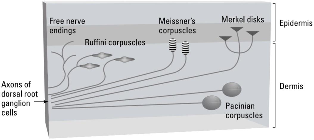
FIGURE 4-1: The layers of the skin and some of its receptors.
شکل ۴-۱: لایههای پوست و برخی از گیرندههای آن.
Merkel disks
Merkel disk receptors, as their name implies, are disk shaped receptors located close to the border between the dermis and epidermis; sometimes they extend into the epidermis.
دیسکهای مرکل
گیرندههای دیسک مرکل، همانطور که از نامشان پیداست، گیرندههای دیسکی شکلی هستند که نزدیک مرز بین درم و اپیدرم قرار دارند؛ گاهی اوقات تا اپیدرم امتداد مییابند.
Merkel disks are receptors for pressure, meaning they’re activated in the areas of your skin pressing against the chair in which you are sitting, for example. If someone were to sit in your lap, receptors in the skin over your thigh muscles would be recruited.
دیسکهای مرکل گیرندههای فشار هستند، به این معنی که در مناطقی از پوست شما که به صندلی که روی آن نشستهاید فشار میآورند، فعال میشوند. برای مثال، اگر کسی روی پای شما بنشیند، گیرندههای پوست روی عضلات ران شما فعال میشوند.
These receptors respond to relatively constant pressure over small areas of the skin, giving you the perception of the amount of force being exerted against different areas of your skin.
این گیرندهها به فشار نسبتاً ثابت روی نواحی کوچک پوست پاسخ میدهند و به شما میزان نیرویی را که بر نواحی مختلف پوست شما اعمال میشود، درک میکنند.
Meissner’s corpuscles
Meissner corpuscles, like Merkel disks, also respond to pressure, but they can respond to more rapid changes in pressure than Merkel disks can, such as those generated by shearing forces. The sensation evoked when the Meissner corpuscles are stimulated is usually called flutter.
اجسام مایسنر
اجسام مایسنر، مانند دیسکهای مرکل، به فشار نیز پاسخ میدهند، اما میتوانند به تغییرات فشار سریعتری نسبت به دیسکهای مرکل، مانند تغییرات ایجاد شده توسط نیروهای برشی، پاسخ دهند. احساسی که هنگام تحریک اجسام مایسنر ایجاد میشود، معمولاً لرزش نامیده میشود.
Like Merkel disks, Meissner corpuscles also have small areas of sensitivity, called their receptive fields. A touch receptive field is the area of skin that, when mechanically disturbed, activates a particular somatosensory receptor there. Small receptive fields are usually associated with shallow receptors (those that are near or in the epidermis), whereas receptors deep in the dermis typically have large receptive fields. This is because pressure at any point on the skin compresses deep parts of the skin at greater distances from the pressure point than more shallow areas of the skin.
مانند دیسکهای مرکل، اجسام مایسنر نیز دارای نواحی کوچک حساسیت هستند که به آنها میدانهای گیرنده میگویند. میدان گیرنده لمسی، ناحیهای از پوست است که وقتی به صورت مکانیکی مختل میشود، یک گیرنده حسی-تنی خاص را در آنجا فعال میکند. میدانهای گیرنده کوچک معمولاً با گیرندههای کمعمق (آنهایی که نزدیک یا در اپیدرم هستند) مرتبط هستند، در حالی که گیرندههای عمیق درم معمولاً میدانهای گیرنده بزرگی دارند. دلیل این امر این است که فشار در هر نقطه از پوست، قسمتهای عمیق پوست را در فواصل بیشتری از نقطه فشار نسبت به نواحی کمعمقتر پوست فشرده میکند.
Ruffini corpuscles
Ruffini corpuscles respond to skin stretch (think of the way your skin pulls when it’s being dragged across a surface). These receptors, which serve an important function in protecting the skin from tearing, have large receptive fields.
گلبولهای رافینی
گلبولهای رافینی به کشش پوست پاسخ میدهند (به نحوه کشیده شدن پوست خود هنگام کشیده شدن روی یک سطح فکر کنید). این گیرندهها که عملکرد مهمی در محافظت از پوست در برابر پارگی دارند، میدانهای گیرنده بزرگی دارند.
Pacinian corpuscles
Pacinian corpuscles, which also have large receptive fields and tend to be deep within the dermis, are the fastest responding of all the touch mechanoreceptors. These receptors have a myelin wrapping similar to the glial wrapping around axons (refer to Chapter 3). However, in Pacinian corpuscles, the function of the wrapping isn’t to allow the action potential to jump from one node to the next. Instead, it allows the receptor to respond to rapid changes in pressure. Through the Pacinian corpuscles, you are able to perceive vibrations. If you drag your fingertips across a coarse surface like sandpaper, you can assess its level of coarseness, thanks to Pacinian corpuscles.
اجسام پاچینی
اجسام پاچینی، که میدانهای گیرنده بزرگی دارند و معمولاً در اعماق درم قرار دارند، سریعترین پاسخدهنده در بین تمام گیرندههای مکانیکی لمسی هستند. این گیرندهها دارای پوشش میلین مشابه پوشش گلیال اطراف آکسونها هستند (به فصل 3 مراجعه کنید). با این حال، در اجسام پاچینی، عملکرد این پوشش این نیست که به پتانسیل عمل اجازه دهد از یک گره به گره بعدی پرش کند. در عوض، به گیرنده اجازه میدهد تا به تغییرات سریع فشار پاسخ دهد. از طریق اجسام پاچینی، شما قادر به درک ارتعاشات هستید. اگر نوک انگشتان خود را روی یک سطح زبر مانند کاغذ سنباده بکشید، به لطف اجسام پاچینی میتوانید میزان زبری آن را ارزیابی کنید.
How mechanoreceptors work
Somatosensory neurons, the tips of which form the mechanoreceptors discussed, have an unusual morphology (structure). This morphology is crucial to their function.
نحوه عملکرد گیرندههای مکانیکی
نورونهای حسی-پیکری، که نوک آنها گیرندههای مکانیکی مورد بحث را تشکیل میدهد، مورفولوژی (ساختار) غیرمعمولی دارند. این مورفولوژی برای عملکرد آنها بسیار مهم است.
Their morphological classification is called pseudounipolar. Although this rather unwieldy name isn’t particularly illuminating about their function, understanding their structure explains some aspects about how these receptors work. Figure 4-2 shows a diagram of a typical mechanoreceptor neuron.
طبقهبندی مورفولوژیکی آنها شبه تکقطبی نامیده میشود. اگرچه این نام نسبتاً دست و پا گیر، عملکرد آنها را به طور خاص روشن نمیکند، اما درک ساختار آنها برخی از جنبههای نحوه عملکرد این گیرندهها را توضیح میدهد. شکل 4-2 نموداری از یک نورون گیرنده مکانیکی معمولی را نشان میدهد.

FIGURE 4-2: A mechanoreceptor: From the skin to the spinal cord.
شکل ۴-۲: یک گیرنده مکانیکی: از پوست تا نخاع.
The cell bodies of somatosensory receptor neurons for most of our skin (below the head and neck) are located in a series of what are called ganglia (concentrations of neural cell bodies) just outside the dorsal root of the spinal cord. They are, thus, called dorsal root ganglia. These ganglia and the neurons they contain are part of the peripheral nervous system.
جسم سلولی نورونهای گیرنده حسی-پیکری در بیشتر پوست ما (زیر سر و گردن) در مجموعهای از نورونها به نام گانگلیون (تجمع اجسام سلولی عصبی) درست در خارج از ریشه پشتی نخاع قرار دارند. بنابراین، به آنها گانگلیون ریشه پشتی میگویند. این گانگلیونها و نورونهای موجود در آنها بخشی از سیستم عصبی محیطی هستند.
The cell bodies of the somatosensory neurons have no dendrites. Instead, a single axon leaves the cell body and then bifurcates, or separates into two paths, a short distance away.
جسم سلولی نورونهای حسی-پیکری دندریت ندارد. در عوض، یک آکسون از جسم سلولی خارج میشود و سپس در فاصله کوتاهی دو شاخه میشود یا به دو مسیر تقسیم میشود.
One end of the axon enters the spinal cord at the dorsal root and makes conventional synapses on spinal motor and interneurons, enabling the stretch reflex and the relaying somatosensory information to other spinal cord segments and up the spinal cord to the brain.
یک سر آکسون از ریشه پشتی وارد نخاع میشود و سیناپسهای مرسوم را روی نورونهای حرکتی و رابط نخاعی تشکیل میدهد و رفلکس کششی و انتقال اطلاعات حسی-پیکری را به سایر بخشهای نخاع و از طریق نخاع به مغز امکانپذیر میسازد.
Here’s the interesting part: The other end of the axon goes away from the dorsal root ganglion in a bundle with other axons and ends up in the skin, where it forms one of the receptor types discussed in the preceding section: Merkel disk, Meissner corpuscle, Ruffini corpuscle, or Pacinian corpuscle. The mechanoreceptors are activated directly when a mechanical force stimulates an axonal ending of one of these neurons. This activation occurs through a special ion channel that responds to the stretching of the membrane (in a typical neurotransmitter receptor, the activation is triggered by voltage or ligand binding). Here’s what happens:
بخش جالب ماجرا اینجاست: انتهای دیگر آکسون از گانگلیون ریشه پشتی در یک دسته با آکسونهای دیگر جدا میشود و به پوست میرسد، جایی که یکی از انواع گیرندههای مورد بحث در بخش قبل را تشکیل میدهد: دیسک مرکل، جسمک مایسنر، جسمک رافینی یا جسمک پاچینی. گیرندههای مکانیکی مستقیماً زمانی فعال میشوند که یک نیروی مکانیکی انتهای آکسونی یکی از این نورونها را تحریک کند. این فعالسازی از طریق یک کانال یونی ویژه رخ میدهد که به کشش غشاء پاسخ میدهد (در یک گیرنده انتقالدهنده عصبی معمولی، فعالسازی توسط ولتاژ یا اتصال لیگاند آغاز میشود). در اینجا اتفاقی که میافتد آمده است:
1. The stretching causes action potentials to originate in the axonal ending and then proceed toward the cell body in the dorsal root ganglion (most axons conduct action potentials away from the cell body).
۱. کشش باعث میشود پتانسیلهای عمل از انتهای آکسونی سرچشمه گرفته و سپس به سمت جسم سلولی در گانگلیون ریشه پشتی حرکت کنند (بیشتر آکسونها پتانسیلهای عمل را از جسم سلولی دور میکنند).
2. The action potential continues past the axonal bifurcation point near the cell body into the spinal cord, where it reaches axon terminals and makes conventional synapses onto spinal motor, transmission, and interneurons.
۲. پتانسیل عمل از نقطه انشعاب آکسونی نزدیک جسم سلولی عبور کرده و به نخاع میرسد، جایی که به پایانههای آکسون میرسد و سیناپسهای مرسوم را با نورونهای حرکتی، انتقالی و رابط نخاعی تشکیل میدهد.
3. The motor neurons mediate reflexes, while the transmission neurons send messages about the receptor activation to other spinal cord segments and up to the brain.
۳. نورونهای حرکتی واسطه رفلکسها هستند، در حالی که نورونهای انتقالدهنده پیامهایی در مورد فعال شدن گیرنده به سایر بخشهای نخاع و تا مغز ارسال میکنند.
Sensing temperature and pain
I’m sure you are aware that you can detect more than just various kinds of pressure on your skin. Two other skin senses are temperature and pain. These receptors have similar structures, or, really, lack of structure. All the mechanoreceptors discussed in the preceding section consist of an axon terminal with ion channel receptors embedded in some sort of structure, such as a corpuscle, disk, or myelin wrapping, that gives the receptor its particular responsiveness to different mechanical stimulation frequencies.
حس کردن دما و درد
مطمئنم که میدانید میتوانید بیش از انواع مختلف فشار روی پوست خود را تشخیص دهید. دو حس پوستی دیگر، دما و درد هستند. این گیرندهها ساختارهای مشابهی دارند، یا در واقع، فاقد ساختار هستند. تمام گیرندههای مکانیکی مورد بحث در بخش قبل شامل یک پایانه آکسون با گیرندههای کانال یونی هستند که در نوعی ساختار، مانند یک جسمک، دیسک یا پوشش میلین، تعبیه شدهاند و به گیرنده پاسخدهی خاص خود را به فرکانسهای مختلف تحریک مکانیکی میدهند.
Receptors for temperature and pain look like the axon terminals without any other structure around them (see Figure 4-3). They are typically called free nerve endings. Free nerve endings for temperature have ion channels that respond to particular temperatures, while other free nerve endings generate action potentials in response to extreme force on the skin or other potentially damaging stimuli that is felt as pain. Some receptors those having what are called transient receptor potential (TRP) channels respond to both.
گیرندههای دما و درد شبیه پایانههای آکسون بدون هیچ ساختار دیگری در اطراف خود هستند (شکل 4-3 را ببینید). آنها معمولاً پایانههای عصبی آزاد نامیده میشوند. پایانههای عصبی آزاد برای دما دارای کانالهای یونی هستند که به دماهای خاص پاسخ میدهند، در حالی که سایر پایانههای عصبی آزاد در پاسخ به نیروی شدید روی پوست یا سایر محرکهای بالقوه آسیبرسان، پتانسیلهای عمل ایجاد میکنند که به صورت درد احساس میشود. برخی از گیرندهها که کانالهایی به نام پتانسیل گیرنده گذرا (TRP) دارند، به هر دو پاسخ میدهند.
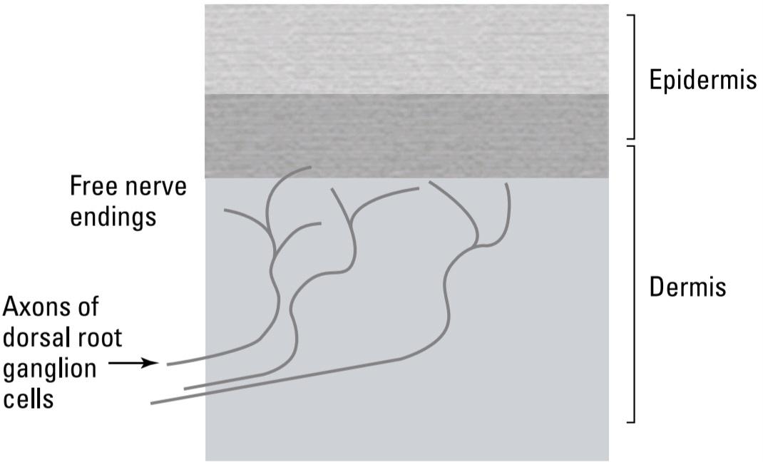
FIGURE 4-3: Free nerve ending receptors for temperature and pain.
شکل ۴-۳: گیرندههای آزاد انتهای عصبی برای دما و درد.
Different temperature receptors respond best to particular temperatures. Warmth receptors respond best to particular temperatures above body temperature (98.6 degrees Fahrenheit), while cold receptors respond best to particular temperatures below body temperature. You judge a wide range of temperatures (cool, damp, chilly, cold, warm, humid, hot, and so on) by sensing the unique ratio of activation of the different receptors firing at any particular temperature.
گیرندههای دمایی مختلف به دماهای خاص بهترین واکنش را نشان میدهند. گیرندههای گرما به دماهای خاص بالاتر از دمای بدن (۹۸.۶ درجه فارنهایت) بهترین واکنش را نشان میدهند، در حالی که گیرندههای سرما به دماهای خاص پایینتر از دمای بدن بهترین واکنش را نشان میدهند. شما با حس کردن نسبت منحصر به فرد فعال شدن گیرندههای مختلف که در هر دمای خاص شلیک میشوند، طیف وسیعی از دماها (خنک، مرطوب، سرد، سرد، گرم، مرطوب، داغ و غیره) را قضاوت میکنید.
Extreme heat, cold, or skin pressure, however, activates receptors whose firing is interpreted as pain. Although different types of pain receptors work by different mechanisms, what they have in common is that the sense of pain signals impending damage to the skin. Pain receptors also exist that respond to chemical damage from acids or bases, and other types of damage such as that caused by a cut. For more information on pain, head to the later section “Understanding the Complex Aspects of Pain.”
با این حال، گرما، سرما یا فشار شدید پوست، گیرندههایی را فعال میکند که تحریک آنها به عنوان درد تفسیر میشود. اگرچه انواع مختلف گیرندههای درد با مکانیسمهای متفاوتی کار میکنند، اما وجه مشترک آنها این است که حس درد، آسیب قریبالوقوع به پوست را نشان میدهد. گیرندههای درد دیگری نیز وجود دارند که به آسیبهای شیمیایی ناشی از اسیدها یا بازها و سایر انواع آسیبها مانند آسیبهای ناشی از بریدگی پاسخ میدهند. برای اطلاعات بیشتر در مورد درد، به بخش بعدی «درک جنبههای پیچیده درد» مراجعه کنید.
Pain and temperature sensations tend to be carried by small diameter axons in a spinal cord tract called the lateral spinothalamic tract. A more medial pathway called the lemnical tract carries fine touch information via larger diameter axons with faster conduction velocities.
حسهای درد و دما معمولاً توسط آکسونهای با قطر کوچک در یک مسیر نخاعی به نام مسیر اسپینوتالامیک جانبی منتقل میشوند. یک مسیر میانیتر به نام مسیر لمنیکال، اطلاعات لمسی ظریف را از طریق آکسونهای با قطر بزرگتر و سرعت هدایت سریعتر منتقل میکند.
Sensing position and movement: Proprioception and kinesthesis
Although not located in the skin, receptors mediating proprioception (position sense) and kinesthesis (movement sense), are either free nerve endings or structures similar to mechanoreceptors like Ruffini corpuscles (refer to Figure 4-1) and have similar layouts as the cell bodies in the dorsal root ganglia (refer to Figure 4-2).
حس موقعیت و حرکت: حس عمقی و حس حرکت
اگرچه گیرندههای حس عمقی (حس موقعیت) و حس حرکت (حس حرکت) در پوست قرار ندارند، اما یا پایانههای عصبی آزاد هستند یا ساختارهایی شبیه به گیرندههای مکانیکی مانند اجسام رافینی (به شکل 4-1 مراجعه کنید) و طرحبندی مشابهی با اجسام سلولی در گانگلیونهای ریشه پشتی دارند (به شکل 4-2 مراجعه کنید).
These receptors are embedded in muscles, tendons, and ligaments around joints. The receptors in muscles and tendons that have relatively sustained responses called proprioreceptors signal muscle force and joint position. Similar receptors with more short-lived, or transient, responses signal when the joint is moving, allowing us to have the movement sense of kinesthesis. For example, proprioreceptors receptors allow you to touch your nose with your eyes closed. Transient, kinesthetic receptors allow you to reach out quickly and then stop your hand in the right place to grab a thrown ball.
این گیرندهها در عضلات، تاندونها و رباطهای اطراف مفاصل تعبیه شدهاند. گیرندههای موجود در عضلات و تاندونها که پاسخهای نسبتاً پایداری دارند و گیرندههای عمقی نامیده میشوند، نیروی عضله و موقعیت مفصل را سیگنال میدهند. گیرندههای مشابه با پاسخهای کوتاهمدتتر یا گذرا، هنگام حرکت مفصل سیگنال میدهند و به ما امکان میدهند حس حرکتی جنبشی داشته باشیم. به عنوان مثال، گیرندههای عمقی به شما این امکان را میدهند که با چشمان بسته بینی خود را لمس کنید. گیرندههای حسی گذرا به شما این امکان را میدهند که به سرعت دست خود را دراز کنید و سپس دست خود را در جای مناسب برای گرفتن توپ پرتاب شده متوقف کنید.
Skin Receptors, Local Spinal Circuits, and Projections to the Brain
Skin receptors allow you to respond to things that contact your skin and to be aware of what those things are. The touch, temperature, and pain messages these receptors encode get passed onto other neurons within the same spinal cord segment, as well as onto other nearby spinal cord segments and to the brain, where the perception occurs.
گیرندههای پوستی، مدارهای نخاعی موضعی و ارتباطات به مغز
گیرندههای پوستی به شما این امکان را میدهند که به چیزهایی که با پوست شما تماس پیدا میکنند پاسخ دهید و از ماهیت آنها آگاه باشید. پیامهای لمسی، دما و دردی که این گیرندهها رمزگذاری میکنند، به سایر نورونها در همان بخش نخاع و همچنین به سایر بخشهای نخاع مجاور و به مغز، جایی که ادراک رخ میدهد، منتقل میشوند.
Somatosensory receptor outputs
The output of most somatosensory receptors participates in at least three different kinds of neural circuits:
خروجیهای گیرندههای حسی-پیکری
خروجی اکثر گیرندههای حسی-پیکری حداقل در سه نوع مختلف از مدارهای عصبی شرکت میکند:
» Local reflexes are those that primarily involve contraction of a single muscle, such as a flexor like the biceps that contracts when, for example, you touch something hot. The circuit for this action consists of the neurons in your fingertip that contain the temperature and pain sensors for heat. These contact spinal cord interneurons in the dorsal root area of the spinal cord (refer to Figure 4-2). These interneurons activate motor neurons in the same spinal segments, which cause contraction of muscles that withdraw your finger.
» رفلکسهای موضعی، رفلکسهایی هستند که در درجه اول شامل انقباض یک عضله واحد میشوند، مانند یک عضله خمکننده مانند عضله دوسر که وقتی مثلاً چیزی داغ را لمس میکنید، منقبض میشود. مدار مربوط به این عمل شامل نورونهایی در نوک انگشت شما است که حاوی حسگرهای دما و درد برای گرما هستند. این نورونهای رابط نخاع در ناحیه ریشه پشتی نخاع با هم تماس پیدا میکنند (به شکل 4-2 مراجعه کنید). این نورونهای رابط، نورونهای حرکتی را در همان بخشهای نخاعی فعال میکنند که باعث انقباض عضلاتی میشوند که انگشت شما را عقب میکشند.
» Coordinated movement involves receptor connections through interneurons to the opposite side of the body within one spinal cord segment, and to other spinal cord segments. When you throw a ball, mechanoreceptors in the skin of the hand work with proprioceptive and kinesthesis receptors associated with muscles in your fingers, hand, arm, and shoulder. Even your leg muscles are involved. Locomotor activities like walking also require coordination between spinal segments so that you do not, for example, try to move one foot before the other has hit the ground. Receptor output from any one segment in the spinal cord can project up or down to other spinal cord segments for coordinated activity of multiple muscles.
» حرکت هماهنگ شامل اتصالات گیرنده از طریق نورونهای رابط به طرف مقابل بدن در یک بخش نخاع و به سایر بخشهای نخاع است. وقتی توپی را پرتاب میکنید، گیرندههای مکانیکی در پوست دست با گیرندههای حس عمقی و حرکتی مرتبط با عضلات انگشتان، دست، بازو و شانه شما کار میکنند. حتی عضلات پای شما نیز درگیر میشوند. فعالیتهای حرکتی مانند راه رفتن نیز نیاز به هماهنگی بین بخشهای نخاع دارند، به طوری که مثلاً سعی نکنید یک پا را قبل از اینکه پای دیگر به زمین برخورد کند، حرکت دهید. خروجی گیرنده از هر بخش در نخاع میتواند برای فعالیت هماهنگ چندین عضله، به بخشهای دیگر نخاع بالا یا پایین برود.
» Messages from skin receptors are also passed to the brain where you become conscious of them. There are two major pathways, the lemniscal pathway and the spinothalamic pathway. Both of these pathways lead to the ventral posterior nucleus of the thalamus on the opposite side of the body from the skin receptors, following the nearly universal principle that the right side of the brain deals mostly with the left side of the body, and vice versa. (For more on the interaction between the right and left sides of the brain, refer to Chapter 2.)
» پیامهای گیرندههای پوستی نیز به مغز منتقل میشوند و در آنجا شما از آنها آگاه میشوید. دو مسیر اصلی وجود دارد، مسیر لمنیسکی و مسیر اسپینوتالامیک. هر دوی این مسیرها به هسته خلفی شکمی تالاموس در طرف مقابل بدن از گیرندههای پوستی منتهی میشوند، که از این اصل تقریباً جهانی پیروی میکند که سمت راست مغز عمدتاً با سمت چپ بدن و برعکس سروکار دارد. (برای اطلاعات بیشتر در مورد تعامل بین سمت راست و چپ مغز، به فصل 2 مراجعه کنید.)
KICKING IT UP AT THE DOCTOR’S OFFICE
The spinal reflex is the most elementary unit of behavior by which a sensation directly causes an action. In a standard neurological exam, while you sit, the doctor uses a little triangular rubber hammer to tap your patellar tendon just below the kneecap. This tap causes the muscle to stretch. This stretching simulates what would happen if you were standing and your knees started to buckle. You have an automatic mechanism by which the output of the muscle proprioceptors cause the quadriceps muscles to contract and extend your knee. When you’re sitting, this action results in the kicking motion. The exam thus tests the integrity of the entire circuit from sensory receptor to muscle contraction, and everything in between.
شروع کار در مطب پزشک
رفلکس نخاعی ابتداییترین واحد رفتاری است که از طریق آن یک حس مستقیماً باعث یک عمل میشود. در یک معاینه عصبی استاندارد، در حالی که شما نشستهاید، پزشک با استفاده از یک چکش لاستیکی مثلثی کوچک، به تاندون کشکک زانوی شما درست زیر کاسه زانو ضربه میزند. این ضربه باعث کشش عضله میشود. این کشش، اتفاقی را که اگر ایستاده باشید و زانوهایتان شروع به خم شدن کنند، رخ میدهد، شبیهسازی میکند. شما یک مکانیسم خودکار دارید که توسط آن، خروجی گیرندههای عمقی عضله باعث میشود عضلات چهارسر ران منقبض و کشیده شوند. وقتی نشستهاید، این عمل منجر به حرکت لگد زدن میشود. بنابراین، این معاینه، یکپارچگی کل مدار را از گیرنده حسی گرفته تا انقباض عضلات و هر چیزی که بین آنها قرار دارد، آزمایش میکند.
Locating the sensation: Specialized cortical sensory areas
Reflexes (like the spinal reflex you can read about in the sidebar “Kicking it up at the doctor’s office) occur in a local circuit, faster than you are aware of it. Awareness has to wait until the signal reaches the brain. This section discusses how the signals from the skin go to the brain, where in the brain they go, and how brain activity is related to your consciousness of skin sensation. In this section, I also give details about the neural processing in somatosensory pathways (for a refresher on brain organization and processing, refer to Chapter 2).
مکانیابی حس: نواحی حسی قشری تخصصی
رفلکسها (مانند رفلکس نخاعی که میتوانید در نوار کناری «شروع آن در مطب پزشک» در مورد آن بخوانید) در یک مدار موضعی، سریعتر از آنچه از آن آگاه هستید، رخ میدهند. آگاهی باید منتظر بماند تا سیگنال به مغز برسد. این بخش در مورد چگونگی رفتن سیگنالها از پوست به مغز، محل قرارگیری آنها در مغز و چگونگی ارتباط فعالیت مغز با آگاهی شما از حس پوست بحث میکند. در این بخش، جزئیاتی در مورد پردازش عصبی در مسیرهای حسی-تنی نیز ارائه میدهم (برای آشنایی بیشتر با سازماندهی و پردازش مغز، به فصل 2 مراجعه کنید).
The somatosensory part of the thalamus (the ventral posterior nucleus) projects to a narrow strip of cortex just posterior to the central sulcus (called the primary somatosensory cortex), making it the most anterior part of the parietal lobe (see Figure 4-4).
بخش حسی-پیکری تالاموس (هسته خلفی شکمی) به نوار باریکی از قشر مغز، درست در خلف شیار مرکزی (به نام قشر حسی-پیکری اولیه) امتداد مییابد و آن را به قدامیترین بخش لوب آهیانه تبدیل میکند (شکل 4-4 را ببینید).
Proprioceptive and kinesthesis receptors in the muscles and joints also project to the cerebellum via the spinal cerebellar pathway. This pathway is essential for learning and implementing complex motor operations such as throwing a baseball.
گیرندههای حس عمقی و حس حرکت در عضلات و مفاصل نیز از طریق مسیر مخچهای نخاعی به مخچه منتقل میشوند. این مسیر برای یادگیری و اجرای عملیات حرکتی پیچیده مانند پرتاب توپ بیسبال ضروری است.

FIGURE 4-4: The parietal lobe: the somatosensory receiving area.
شکل ۴-۴: لوب آهیانه: ناحیه گیرنده حسی-پیکری.
Mapping skin receptors to specific brain areas: Cortical maps
The mapping of skin receptors to a specific area of neocortex illustrates one of the most fundamental principles of brain organization, cortical maps. The projection from the thalamus is orderly in the sense that receptors on nearby parts of the skin project to nearby cortical neurons. Figure 4-5 shows a representation of the skin map on the somatosensory cortex.
نقشهبرداری گیرندههای پوست به نواحی خاص مغز: نقشههای قشری
نقشهبرداری گیرندههای پوست به ناحیه خاصی از نئوکورتکس، یکی از اساسیترین اصول سازماندهی مغز، یعنی نقشههای قشری را نشان میدهد. تصویر از تالاموس منظم است به این معنا که گیرندههای روی قسمتهای مجاور پوست به نورونهای قشری مجاور تصویر میدهند. شکل ۴-۵ نمایشی از نقشه پوست روی قشر حسی-پیکری را نشان میدهد.

FIGURE 4-5: The skin map on the somatosensory cortex.
شکل ۴-۵: نقشه پوست روی قشر حسی-پیکری.
The fundamental idea about a given area of cortex being devoted to receptors in a given skin area is that activity in this area of cortex (along with other parts of the brain) is necessary for the perception of the skin sense. We perceive activity in that bit of cortex area as a skin sensation not because that area has some special skin perceiving neurons, but because it receives inputs from the skin and has out- puts that connect to memories of previous skin sensation and other associated sensations. In other words, the perception produced by activity in this and other areas of cortex is a function of what neural input goes to it and where the outputs of that area of cortex go.
ایده اساسی در مورد اختصاص ناحیهای خاص از قشر مغز به گیرندههای یک ناحیه خاص از پوست این است که فعالیت در این ناحیه از قشر مغز (همراه با سایر قسمتهای مغز) برای درک حس پوست ضروری است. ما فعالیت در آن ناحیه از قشر مغز را به عنوان یک حس پوستی درک میکنیم، نه به این دلیل که آن ناحیه دارای نورونهای ادراک پوستی خاصی است، بلکه به این دلیل که ورودیهایی از پوست دریافت میکند و خروجیهایی دارد که به خاطرات حس پوستی قبلی و سایر حسهای مرتبط متصل میشوند. به عبارت دیگر، ادراکی که توسط فعالیت در این ناحیه و سایر نواحی قشر مغز ایجاد میشود، تابعی از ورودیهای عصبی است که به آن وارد میشوند و خروجیهای آن ناحیه از قشر مغز به کجا میروند.
This skin map on the cortex is called a homunculus, which means “little man.” However, the surface area of the somatosensory cortex onto which the skin receptors project is not really a miniature picture of the body; it is more like a band or strip, as established in the studies by Canadian neurosurgeon Wilder Penfield. Because of the difficulty in mapping a three dimensional surface onto a two dimensional sheet (think of how two-dimensional maps of the earth compare to the three dimensional globes), the image is distorted, depending on choices that the “map maker” makes about what is relatively more important to represent accurately versus what is less.
این نقشه پوستی روی قشر مغز، هومونکولوس نامیده میشود که به معنی “آدم کوچک” است. با این حال، سطح قشر حسی-تنی که گیرندههای پوستی روی آن تصویر میاندازند، در واقع یک تصویر مینیاتوری از بدن نیست؛ بلکه بیشتر شبیه یک نوار یا نوار است، همانطور که در مطالعات جراح مغز و اعصاب کانادایی، وایلدر پنفیلد، مشخص شده است. به دلیل دشواری در نقشهبرداری از یک سطح سهبعدی روی یک صفحه دوبعدی (به نحوه مقایسه نقشههای دوبعدی زمین با کرههای سهبعدی فکر کنید)، تصویر بسته به انتخابهایی که “نقشهساز” در مورد آنچه که نمایش دقیق آن نسبتاً مهمتر است در مقابل آنچه که اهمیت کمتری دارد، انجام میدهد، دچار اعوجاج میشود.
Also note that some areas of the body, such as the hands and fingers, are located in the cortex map close to areas such as the face that are actually quite distant, body wise. Some researchers suggest that the phantom limb feelings (including pain) that sometimes occur after an amputation may occur because some neural projections from the face invade the cortical area once stimulated by the limb and cause sensations to be perceived as being located in the limb, even when the limb is gone.
همچنین توجه داشته باشید که برخی از نواحی بدن، مانند دستها و انگشتان، در نقشه قشر مغز نزدیک به نواحیای مانند صورت قرار دارند که در واقع از نظر بدنی کاملاً دور هستند. برخی از محققان معتقدند که احساسات اندام خیالی (از جمله درد) که گاهی اوقات پس از قطع عضو رخ میدهد، ممکن است به این دلیل رخ دهد که برخی از برآمدگیهای عصبی از صورت، پس از تحریک توسط اندام، به ناحیه قشر مغز حمله میکنند و باعث میشوند که احساسات به عنوان واقع در اندام درک شوند، حتی زمانی که اندام از بین رفته است.
Receptor densities
Equal areas of the skin do not map to equal areas of cortex because different areas of the skin have very different receptor densities. The skin in the fingertip, for example, has many more receptors per area than the same areas in the skin on the stomach or back. The higher density of receptors in the fingertip allow fine two point discrimination, which is the distance over which one can distinguish a single point pressing on the skin from two points close together. This distance is millimeters on the fingertip, lips, and forehead, for example, but centimeters on the stomach, back, and legs. You need high two point discrimination in your fingertips to manipulate objects precisely and in the skin on your face to make precise facial expressions.
تراکم گیرندهها
مناطق مساوی پوست به نواحی مساوی قشر مخ متصل نمیشوند، زیرا نواحی مختلف پوست تراکم گیرندههای بسیار متفاوتی دارند. به عنوان مثال، پوست نوک انگشت در هر ناحیه گیرندههای بسیار بیشتری نسبت به همان نواحی در پوست شکم یا کمر دارد. تراکم بالاتر گیرندهها در نوک انگشت امکان تشخیص دقیق دو نقطه را فراهم میکند، یعنی فاصلهای که میتوان یک نقطه واحد را که روی پوست فشار میدهد از دو نقطه نزدیک به هم تشخیص داد. به عنوان مثال، این فاصله در نوک انگشت، لبها و پیشانی میلیمتر است، اما در شکم، کمر و پاها سانتیمتر است. برای دستکاری دقیق اشیاء و در پوست صورت برای ایجاد حالات چهره دقیق، به تشخیص دو نقطهای بالایی در نوک انگشتان خود نیاز دارید.
A given area of cortex processes inputs from about the same absolute number of receptors, so that skin areas with high receptor density, such as the fingertips, get proportionally more cortical area compared to the skin area, than areas with low receptor density. You can look at Figure 4-5 and see where skin receptor density is relatively high or low.
یک ناحیه مشخص از قشر مغز، ورودیهای تقریباً یکسانی از تعداد مطلق گیرندهها را پردازش میکند، به طوری که نواحی پوستی با تراکم گیرنده بالا، مانند نوک انگشتان، در مقایسه با ناحیه پوست با تراکم گیرنده پایین، به طور متناسب مساحت قشری بیشتری دارند. میتوانید به شکل ۴-۵ نگاه کنید و ببینید که تراکم گیرندههای پوست در کجا نسبتاً زیاد یا کم است.
Understanding the Complex Aspects of Pain
Although pain, like other skin senses, is usually associated with something happening at some particular part of your body that you need to do something about, it also has other causes and effects. Pain can produce long lasting changes in mood and can arise from causes that are not themselves perceived and not well localized, including depression not associated with any physical source. Moreover, unlike the topographic but distorted mechanoreceptor somatosensory map in the anterior parietal cortex (refer to Figure 4-5), no similar map exists for pain. Although you can localize pain to some part of your skin, the perception of pain has a more general effect on mood, similar to pain that cannot be localized. Pain pathways to the central nervous system are diffuse, arise from multiple causes (multimodal), and impact general mood often in long lasting ways.
درک جنبههای پیچیده درد
اگرچه درد، مانند سایر حواس پوستی، معمولاً با اتفاقی در قسمت خاصی از بدن شما مرتبط است که باید کاری در مورد آن انجام دهید، اما علل و اثرات دیگری نیز دارد. درد میتواند تغییرات طولانیمدتی در خلق و خو ایجاد کند و میتواند از عللی ناشی شود که خودشان درک نمیشوند و به خوبی محلیسازی نشدهاند، از جمله افسردگی که با هیچ منبع فیزیکی مرتبط نیست. علاوه بر این، برخلاف نقشه توپوگرافی اما تحریفشده گیرندههای مکانیکی حسی-تنی در قشر جداری قدامی (به شکل ۴-۵ مراجعه کنید)، هیچ نقشه مشابهی برای درد وجود ندارد. اگرچه میتوانید درد را در قسمتی از پوست خود محلیسازی کنید، اما درک درد تأثیر کلیتری بر خلق و خو دارد، مشابه دردی که نمیتوان محلیسازی کرد. مسیرهای درد به سیستم عصبی مرکزی پراکنده هستند، از علل متعددی (چندوجهی) ناشی میشوند و اغلب به روشهای طولانیمدت بر خلق و خوی عمومی تأثیر میگذارند.
Not only does pain affect general mood, but it goes the other way, too: Mood can affect the feeling of pain. A person’s mental state and attitude can cause that person to ignore or not even feel pain, while anxiety can intensify the pain associated with any particular physical stimulus.
درد نه تنها بر خلق و خوی عمومی تأثیر میگذارد، بلکه برعکس نیز عمل میکند: خلق و خو میتواند بر احساس درد تأثیر بگذارد. وضعیت روانی و نگرش یک فرد میتواند باعث شود که آن فرد درد را نادیده بگیرد یا حتی احساس نکند، در حالی که اضطراب میتواند درد مرتبط با هر محرک فیزیکی خاصی را تشدید کند.
Reducing or overlooking pain
For thousands of years, people have been aware that it is possible to cognitively mitigate the sensation of pain. Think of soldiers ignoring significant wounds, women in childbirth, and subjects ignoring pain after hypnosis. Drugs have also been found that specifically reduce pain without causing loss of consciousness or other sensations. Many of these phenomena arise because the pain system in the brain uses a particular set of neurotransmitters.
کاهش یا نادیده گرفتن درد
هزاران سال است که مردم از این موضوع آگاه بودهاند که میتوان به صورت شناختی احساس درد را کاهش داد. به سربازانی فکر کنید که زخمهای قابل توجه را نادیده میگیرند، زنانی که زایمان میکنند و افرادی که پس از هیپنوتیزم درد را نادیده میگیرند. همچنین داروهایی یافت شدهاند که به طور خاص درد را بدون ایجاد از دست دادن هوشیاری یا سایر احساسات کاهش میدهند. بسیاری از این پدیدهها به این دلیل ایجاد میشوند که سیستم درد در مغز از مجموعهای خاص از انتقالدهندههای عصبی استفاده میکند.
Neurotransmitters that reduce or block pain
Here’s a mystery that puzzled researchers for a long time: Why does a substance produced by a poppy plant (morphine) relieve pain? Most psychoactive drugs mimic the action of known neurotransmitters, but until a few decades ago, there was no known neurotransmitter that mediated the general effects of pain.
انتقالدهندههای عصبی که درد را کاهش میدهند یا مسدود میکنند
اینجا یک معما وجود دارد که مدتها محققان را گیج کرده بود: چرا مادهای که توسط گیاه خشخاش (مورفین) تولید میشود، درد را تسکین میدهد؟ اکثر داروهای روانگردان عملکرد انتقالدهندههای عصبی شناخته شده را تقلید میکنند، اما تا چند دهه پیش، هیچ انتقالدهنده عصبی شناخته شدهای وجود نداشت که واسطه اثرات عمومی درد باشد.
This all changed with the discovery of endogenous opioids (that is, opioids that occur naturally within the body). Of these morphinelike substances, the most common are the endorphins (a term which is an abbreviation of endogenous morphines). Common situations in which endorphins are produced include childbirth and running the last few miles of a marathon.
همه اینها با کشف مواد افیونی درونزا (یعنی مواد افیونی که به طور طبیعی در بدن وجود دارند) تغییر کرد. از بین این مواد شبه مورفین، رایجترین آنها اندورفینها هستند (اصطلاحی که مخفف مورفینهای درونزا است). موقعیتهای رایجی که در آنها اندورفین تولید میشود شامل زایمان و دویدن چند مایل آخر یک ماراتن است.
Opiates like morphine, heroin, and fentanyl reduce the feeling of pain because they mimic the action of substances the body produces on its own to control pain. These drugs bind these same receptors and, at low doses, produce similar effects. However, when injected in large doses, these drugs produce the opposite of pain a “high” and are addictive. The drug naloxone antagonizes the effects of these opioids and is often given to addicts to reverse the effects of heroin they have injected.
مواد افیونی مانند مورفین، هروئین و فنتانیل احساس درد را کاهش میدهند زیرا عملکرد موادی را که بدن به طور طبیعی برای کنترل درد تولید میکند، تقلید میکنند. این داروها به همین گیرندهها متصل میشوند و در دوزهای پایین، اثرات مشابهی ایجاد میکنند. با این حال، وقتی این داروها در دوزهای بالا تزریق میشوند، برعکس درد، یعنی “نشئگی” ایجاد میکنند و اعتیادآور هستند. داروی نالوکسان اثرات این مواد افیونی را خنثی میکند و اغلب به معتادان داده میشود تا اثرات هروئین تزریق شده را معکوس کند.
The existence of endorphins also explains another mystery of pain management, the placebo effect. The placebo effect occurs when patients are given a substance that itself has no pain-blocking potential but, because the patients believe they have been given a real drug that will alleviate the pain, find that the pain is actually alleviated.
وجود اندورفینها همچنین یکی دیگر از رازهای مدیریت درد، یعنی اثر دارونما، را توضیح میدهد. اثر دارونما زمانی رخ میدهد که به بیماران مادهای داده میشود که خود هیچ پتانسیلی برای مسدود کردن درد ندارد، اما از آنجا که بیماران معتقدند داروی واقعی به آنها داده شده است که درد را تسکین میدهد، متوجه میشوند که درد واقعاً تسکین یافته است.
Although the placebo effect is robust and common, some in the medical community tend to dismiss it as being “psychological,” that is, not based on any physiologically demonstrable or quantifiable basis. However, it turns out that the drug naloxone not only reduces the effects of opioids, such as heroin, but it also reduces the placebo effect. What this means is that the placebo effect isn’t just psychological; it actually has a physiological component, involving the cognitive stimulation, from belief, of the body’s internal endorphin production that objectively and measurably reduces pain by binding the endorphin receptors.
اگرچه اثر دارونما قوی و رایج است، برخی در جامعه پزشکی تمایل دارند آن را به عنوان یک اثر “روانشناختی” رد کنند، یعنی بر هیچ مبنای فیزیولوژیکی قابل اثبات یا قابل اندازهگیری مبتنی نیست. با این حال، مشخص شده است که داروی نالوکسان نه تنها اثرات مواد افیونی مانند هروئین را کاهش میدهد، بلکه اثر دارونما را نیز کاهش میدهد. این بدان معناست که اثر دارونما فقط روانشناختی نیست؛ در واقع یک جزء فیزیولوژیکی دارد که شامل تحریک شناختی، از روی باور، تولید اندورفین داخلی بدن است که با اتصال گیرندههای اندورفین، درد را به طور عینی و قابل اندازهگیری کاهش میدهد.
Using distraction to alleviate pain: The gate theory
Another mystery about the sense of pain is that it is often reduced by cognitive distraction. Numerous well-documented cases exist of people enduring in some survival situation despite being seriously wounded and not even realizing it. A hypothesis put forward to explain this effect is called the Melzack and Wall gate theory.
استفاده از حواسپرتی برای کاهش درد: نظریه دروازه
یکی دیگر از رازهای مربوط به حس درد این است که اغلب با حواسپرتی شناختی کاهش مییابد. موارد مستند متعددی از افرادی وجود دارد که در شرایط سخت بقا، با وجود آسیب جدی، مقاومت میکنند و حتی متوجه آن نمیشوند. فرضیهای که برای توضیح این اثر ارائه شده است، نظریه دروازه ملزاک و وال نامیده میشود.
According to the gate theory, messages from pain receptors in the skin mingle within the central nervous system with messages from ordinary mechanoreceptors. The operation of the neural circuit is such that when only pain receptors are activated they pass through the gate in the spinal cord and reach the brain. However, if other mechanoreceptors are sufficiently activated, even in other parts of the body, they can block the neural gate and suppress the pain signal to the brain. As it turns out, even cognitive activity can be sufficiently distracting to close the pain gate at high spinal or even brain levels.
طبق نظریه دروازه، پیامهای گیرندههای درد در پوست در سیستم عصبی مرکزی با پیامهای گیرندههای مکانیکی معمولی در هم میآمیزند. عملکرد مدار عصبی به گونهای است که وقتی فقط گیرندههای درد فعال میشوند، از دروازه در نخاع عبور کرده و به مغز میرسند. با این حال، اگر گیرندههای مکانیکی دیگری، حتی در سایر قسمتهای بدن، به اندازه کافی فعال باشند، میتوانند دروازه عصبی را مسدود کرده و سیگنال درد به مغز را سرکوب کنند. همانطور که مشخص است، حتی فعالیت شناختی نیز میتواند به اندازه کافی حواسپرتکننده باشد تا دروازه درد را در سطوح بالای نخاع یا حتی مغز ببندد.
Pain free and hating it: Peripheral neuropathy
In the preceding sections, I discuss several ways in which the sense of pain can be reduced, including the body’s own production of endorphins. It has occurred to many people to wonder why we have a sense of pain at all. Feeling pain is, well, painful. Wouldn’t we be better off if we could just eliminate pain?
بدون درد و از آن متنفر بودن: نوروپاتی محیطی
در بخشهای قبلی، چندین روش برای کاهش حس درد، از جمله تولید اندورفین توسط بدن، را مورد بحث قرار دادم. برای بسیاری از افراد این سوال پیش آمده است که چرا اصلاً حس درد داریم. خب، احساس درد دردناک است. آیا اگر میتوانستیم درد را از بین ببریم، بهتر نبود؟
The answer to the question of whether we would be better off without a sense of pain is a resounding no. We know this because this situation occurs in some people. One of these is a condition called peripheral neuropathy, in which many neurons such as pain receptors in the peripheral nervous system die or become inactive due, for example, to vascular problems associated with diabetes. Loss of pain sense in parts of the body can also be the result of certain strokes and types of brain damage.
پاسخ به این سوال که آیا بدون حس درد حالمان بهتر خواهد بود، یک نه قاطع است. ما این را میدانیم زیرا این وضعیت در برخی افراد رخ میدهد. یکی از این موارد، وضعیتی به نام نوروپاتی محیطی است که در آن بسیاری از نورونها مانند گیرندههای درد در سیستم عصبی محیطی به دلایلی مانند مشکلات عروقی مرتبط با دیابت میمیرند یا غیرفعال میشوند. از دست دادن حس درد در قسمتهایی از بدن همچنین میتواند نتیجه سکتههای مغزی خاص و انواع آسیبهای مغزی باشد.
People with peripheral neuropathy tend to injure themselves without knowing: They burn themselves while cooking, break bones during routine physical activity, and develop asymptomatic skin lesions that are ignored until they become serious infections. The sense of pain is necessary to prevent harm to the body. The loss of feeling in a limb is so disabling that often people with sensory peripheral neuropathy are effectively paralyzed in that limb, refusing to use it, even if the motor neuron circuitry is actually intact.
افراد مبتلا به نوروپاتی محیطی تمایل دارند بدون اینکه خودشان بدانند به خودشان آسیب بزنند: آنها هنگام آشپزی خود را میسوزند، در طول فعالیت بدنی معمول استخوانهایشان میشکند و ضایعات پوستی بدون علامتی ایجاد میکنند که تا زمانی که به عفونتهای جدی تبدیل نشوند، نادیده گرفته میشوند. حس درد برای جلوگیری از آسیب به بدن ضروری است. از دست دادن حس در یک اندام آنقدر ناتوانکننده است که اغلب افراد مبتلا به نوروپاتی حسی محیطی عملاً در آن اندام فلج میشوند و از استفاده از آن امتناع میکنند، حتی اگر مدار نورون حرکتی در واقع سالم باشد.
Chronic pain and individual differences in pain perception
Although pain is a necessary function for preventing damage to the body, in some cases, pain itself becomes disabling. Chronic pain can occur in disease conditions such as cancer, in which case the normal function of pain that forces you to rest, protect, or not use some injured part of the body until it heals is simply inappropriate in a disease state in which destruction is occurring from the cancer all over the body that cannot be healed from rest. Pain can also arise from psychological factors or from factors that cannot be medically identified and are assumed to be psychological. Examples include some types of chronic pain and depression.
درد مزمن و تفاوتهای فردی در درک درد
اگرچه درد یک عملکرد ضروری برای جلوگیری از آسیب به بدن است، اما در برخی موارد، خود درد ناتوانکننده میشود. درد مزمن میتواند در شرایط بیماری مانند سرطان رخ دهد، که در این صورت عملکرد طبیعی درد که شما را مجبور به استراحت، محافظت یا عدم استفاده از برخی قسمتهای آسیبدیده بدن تا زمان بهبودی میکند، در حالت بیماری که در آن تخریب ناشی از سرطان در سراسر بدن رخ میدهد و با استراحت قابل درمان نیست، به سادگی نامناسب است. درد همچنین میتواند ناشی از عوامل روانی یا عواملی باشد که از نظر پزشکی قابل شناسایی نیستند و فرض میشود روانی هستند. از جمله این موارد میتوان به برخی از انواع درد مزمن و افسردگی اشاره کرد.
Pain from both medically identified sources and that which is psychological (or cognitive) appears to activate a brain area called the anterior cingulate cortex. The anterior cingulate cortex is the anterior portion of an area of the mesocortex, just above the corpus callosum. (Refer to Chapter 2 for discussion on the different areas of the brain; for more on the development and function of the mesocortex, head to Chapter 12.)
به نظر میرسد دردی که منشأ آن از نظر پزشکی مشخص شده باشد و همچنین دردی که منشأ روانی (یا شناختی) داشته باشد، ناحیهای از مغز به نام قشر کمربندی قدامی را فعال میکند. قشر کمربندی قدامی، بخش قدامی ناحیهای از مزوکورتکس، درست بالای جسم پینهای است. (برای بحث در مورد نواحی مختلف مغز به فصل ۲ مراجعه کنید؛ برای اطلاعات بیشتر در مورد رشد و عملکرد مزوکورتکس، به فصل ۱۲ مراجعه کنید.)
The anterior cingulate appears to be a high-level cortical monitoring center. It tends to be activated by pain, anticipation of pain, and failure in goal-seeking activity. Its function seems to be to arbitrate between taking different strategies in response to experience. At a low level, after placing your hand on a hot stove burner, it may make you cautious when you’re around the stove. At a higher level, getting reprimanded for sending a flaming email at work may make you wary of doing so again.
به نظر میرسد که سینگولیت قدامی یک مرکز نظارت قشری سطح بالا باشد. این مرکز معمولاً توسط درد، پیشبینی درد و شکست در فعالیتهای هدفمند فعال میشود. به نظر میرسد عملکرد آن داوری بین اتخاذ استراتژیهای مختلف در پاسخ به تجربه است. در سطح پایین، پس از قرار دادن دست خود روی شعله اجاق گاز داغ، ممکن است شما را هنگام قرار گرفتن در کنار اجاق گاز محتاط کند. در سطح بالاتر، توبیخ شدن به دلیل ارسال یک ایمیل آتشین در محل کار ممکن است شما را از انجام مجدد این کار محتاط کند.
Considerable individual differences with respect to pain tolerance exist, just as there can be differences in tolerance in different situations for a particular person. Men are reported to be less tolerant of chronic pain than women, though they are more tolerant of acute pain. Pain tolerance generally increases with age, based on tests for pain tolerance such as the total time one can stand to have one’s arm immersed in ice water. It is not clear whether the increase in tolerance with age is based on psychological or physical factors. Athletic training and strong motivation to obtain some goal can significantly reduce the disabling effects of pain.
تفاوتهای فردی قابل توجهی در رابطه با تحمل درد وجود دارد، همانطور که در موقعیتهای مختلف برای یک فرد خاص، میتواند تفاوتهایی در تحمل وجود داشته باشد. گزارش شده است که مردان نسبت به زنان تحمل درد مزمن کمتری دارند، اگرچه تحمل درد حاد بیشتری دارند. تحمل درد به طور کلی با افزایش سن افزایش مییابد، بر اساس آزمایشهای تحمل درد مانند کل زمانی که فرد میتواند تحمل کند تا بازویش در آب یخ فرو رود. مشخص نیست که آیا افزایش تحمل با افزایش سن مبتنی بر عوامل روانی است یا جسمی. تمرینات ورزشی و انگیزه قوی برای رسیدن به یک هدف میتواند اثرات ناتوانکننده درد را به میزان قابل توجهی کاهش دهد.
Suggestions that different cultures or ethnic groups have intrinsically different pain thresholds in other words, there’s a physiological difference among cultures about pain tolerance have almost always been shown to be the effect of at what point the perceived stimulus is reported as painful or unbearably painful, not whether the pain itself is perceived. Cultures that encourage expression of emotions in general tend to be associated with lower pain reporting tolerance.
پیشنهادهایی مبنی بر اینکه فرهنگها یا گروههای قومی مختلف ذاتاً آستانه درد متفاوتی دارند، به عبارت دیگر، تفاوت فیزیولوژیکی بین فرهنگها در مورد تحمل درد تقریباً همیشه نشان داده شده است که این تفاوت به نقطهای بستگی دارد که محرک درک شده به عنوان دردناک یا غیرقابل تحمل گزارش میشود، نه اینکه آیا خود درد درک میشود یا خیر. فرهنگهایی که ابراز احساسات را به طور کلی تشویق میکنند، معمولاً با تحمل پایینتر گزارش درد مرتبط هستند.



