علوم اعصاب به زبان ساده؛ درک چگونگی عملکرد نورونها


دعای مطالعه [ نمایش ]
بِسْمِ الله الرَّحْمنِ الرَّحیمِ
اَللّهُمَّ اَخْرِجْنى مِنْ ظُلُماتِ الْوَهْمِ
خدايا مرا بيرون آور از تاريكىهاى وهم،
وَ اَكْرِمْنى بِنُورِ الْفَهْمِ
و به نور فهم گرامى ام بدار،
اَللّهُمَّ افْتَحْ عَلَيْنا اَبْوابَ رَحْمَتِكَ
خدايا درهاى رحمتت را به روى ما بگشا،
وَانْشُرْ عَلَيْنا خَزائِنَ عُلُومِكَ بِرَحْمَتِكَ يا اَرْحَمَ الرّاحِمينَ
و خزانههاى علومت را بر ما باز كن به امید رحمتت اى مهربانترين مهربانان.
کتاب «علوم اعصاب به زبان ساده» اثر دکتر فرانک آمتور، یکی از منابع معتبر و قابل دسترس برای علاقهمندان و دانشجویان در حوزه علوم اعصاب است که مفاهیم پایه و کاربردهای عمومی مغز و سیستم عصبی را به زبان ساده توضیح میدهد. این کتاب تصویری روشن از مغز انسان و عملکرد نورونها ارائه میدهد و به مخاطبان کمک میکند تا مفاهیم شناختی، رفتاری و ارتباط ذهن و بدن را درک کنند.
ترجمه دقیق و علمی این شاهکار توسط برند علمی «آیندهنگاران مغز» به مدیریت داریوش طاهری، دسترسی فارسیزبانان به مرزهای نوین دانش علوم اعصاب را ممکن ساخته و رسالتی علمی برای ارتقای آموزش، فهم عمیقتر عملکرد مغز و سیستم عصبی و توسعه روشهای نوین در حوزه سلامت عصبی فراهم آورده است.
» کتاب علوم اعصاب به زبان ساده
»» فصل ۳: درک چگونگی عملکرد نورونها
در حال ویرایش
» Neuroscience For Dummies
»» Chapter 3: Understanding How Neurons Work
IN THIS CHAPTER
» Communicating between neurons: The role of neurotransmitters
» Looking at the role of glial cells
» Examining the various techniques used to study neural activity
» Understanding what neurons do and how they work
در این فصل
» ارتباط بین نورونها: نقش انتقالدهندههای عصبی
» نگاهی به نقش سلولهای گلیال
» بررسی تکنیکهای مختلف مورد استفاده برای مطالعه فعالیت عصبی
» درک عملکرد نورونها و نحوه عملکرد آنها

Chapter 3
Understanding How Neurons Work
Everything you think, everything you do, and, in fact, everything you are, is the result of the actions of about 86 billion neurons. But what are neurons, and how do they enable minds?
فصل ۳
درک چگونگی عملکرد نورونها
هر آنچه که فکر میکنید، هر آنچه که انجام میدهید و در واقع، هر آنچه که هستید، نتیجهی اعمال حدود ۸۶ میلیارد نورون است. اما نورونها چه هستند و چگونه ذهن را فعال میکنند؟
Neurons are specialized cells that process information. Some neurons are recep- tors that convert sensory stimuli into messages processed by the brain, while other neurons stimulate muscles and glands. Most neurons are connected to other neurons and perform computations that allow an organism — you — to behave in complex ways, based on what the body senses internally and externally, now, in the experienced past, and in the expected future. This chapter explains how neurons function, how we know what we do about how the nervous system oper- ates, and what new techniques are extending our knowledge of brain function.
نورونها سلولهای تخصصی هستند که اطلاعات را پردازش میکنند. برخی از نورونها گیرندههایی هستند که محرکهای حسی را به پیامهایی که توسط مغز پردازش میشوند تبدیل میکنند، در حالی که سایر نورونها عضلات و غدد را تحریک میکنند. اکثر نورونها به نورونهای دیگر متصل هستند و محاسباتی را انجام میدهند که به یک موجود زنده – شما – اجازه میدهد تا بر اساس آنچه بدن به صورت داخلی و خارجی، در حال حاضر، در گذشته تجربه شده و در آینده مورد انتظار حس میکند، به روشهای پیچیدهای رفتار کند. این فصل توضیح میدهد که نورونها چگونه کار میکنند، چگونه میدانیم که در مورد نحوه عملکرد سیستم عصبی چه کاری انجام میدهیم و چه تکنیکهای جدیدی دانش ما را در مورد عملکرد مغز گسترش میدهند.
Neuron Basics: Not Just Another Cell in the Body
First of all, neurons are cells. Neurons, like other cells, consist of a bag of cyto- plasm with a membrane separating their cytoplasm from the outside world. Within this cytoplasm are organelles, such as mitochondria that provide energy for the cell, and structures, such as the Golgi apparatus and the endoplasmic reticulum, that manufacture and distribute proteins. The nucleus contains the DNA that codes for the identity of both the organism itself and the cell within the organism.
مبانی نورون: نه فقط یک سلول دیگر در بدن
اول از همه، نورونها سلول هستند. نورونها، مانند سایر سلولها، از یک کیسه سیتوپلاسم با غشایی تشکیل شدهاند که سیتوپلاسم آنها را از دنیای خارج جدا میکند. در داخل این سیتوپلاسم، اندامکهایی مانند میتوکندری وجود دارند که انرژی سلول را تأمین میکنند و ساختارهایی مانند دستگاه گلژی و شبکه آندوپلاسمی که پروتئینها را تولید و توزیع میکنند. هسته حاوی DNA است که هویت خود ارگانیسم و سلول درون ارگانیسم را کدگذاری میکند.
Here’s what makes neurons different from other cells (if you’re unfamiliar with general cell structure and components, you can find that information in most basic biology books, such as Biology For Dummies, 2nd Edition [John Wiley & Sons, Inc.]):
در اینجا چیزی است که نورونها را از سایر سلولها متمایز میکند (اگر با ساختار و اجزای کلی سلول آشنا نیستید، میتوانید این اطلاعات را در اکثر کتابهای زیستشناسی پایه، مانند Biology For Dummies، ویرایش دوم [John Wiley & Sons, Inc.] پیدا کنید):
» A membrane with selective ion channels that can be opened by electrical events or binding by neurotransmitters
» غشایی با کانالهای یونی انتخابی که میتوانند توسط رویدادهای الکتریکی یا اتصال توسط انتقالدهندههای عصبی باز شوند.
» Dendrites, branching extensions from the cell body that receive inputs from other neurons
» دندریتها، زائدههای منشعب از جسم سلولی که ورودیها را از سایر نورونها دریافت میکنند.
» An axon, a single extension from the cell body that travels long distances (up to several feet) to contact other neurons
» آکسون، یک زائده منفرد از جسم سلولی که مسافتهای طولانی (تا چند فوت) را برای تماس با سایر نورونها طی میکند.
Figure 3-1 shows the main parts of a typical neuron. There are obvious structural differences between neurons and most other cells. While most non-neuronal cells resemble spheroids, neurons typically have a “dendritic tree” of branches (or pro- cesses) arising from the cell body (or soma), plus a single process called an axon that also emanates from the cell body but runs for large distances (sometimes even up to several feet) before it branches. Not so evident from Figure 3-1 is the fact that the membrane that encloses a neuron’s cell body, axon, and dendrites is very different from the membrane that encloses most other cells because of its multiple types of ion channels.
شکل 3-1 بخشهای اصلی یک نورون معمولی را نشان میدهد. تفاوتهای ساختاری آشکاری بین نورونها و اکثر سلولهای دیگر وجود دارد. در حالی که اکثر سلولهای غیر نورونی شبیه به کرهها هستند، نورونها معمولاً دارای یک “درخت دندریتی” از شاخهها (یا زوائد) هستند که از جسم سلولی (یا سوما) منشأ میگیرند، به علاوه یک زوائد واحد به نام آکسون که از جسم سلولی نیز سرچشمه میگیرد اما قبل از شاخه شدن، مسافتهای زیادی (گاهی اوقات حتی تا چند فوت) را طی میکند. چیزی که از شکل 3-1 چندان مشهود نیست این واقعیت است که غشایی که جسم سلولی، آکسون و دندریتهای نورون را در بر میگیرد، به دلیل انواع مختلف کانالهای یونی، با غشایی که اکثر سلولهای دیگر را در بر میگیرد، بسیار متفاوت است.
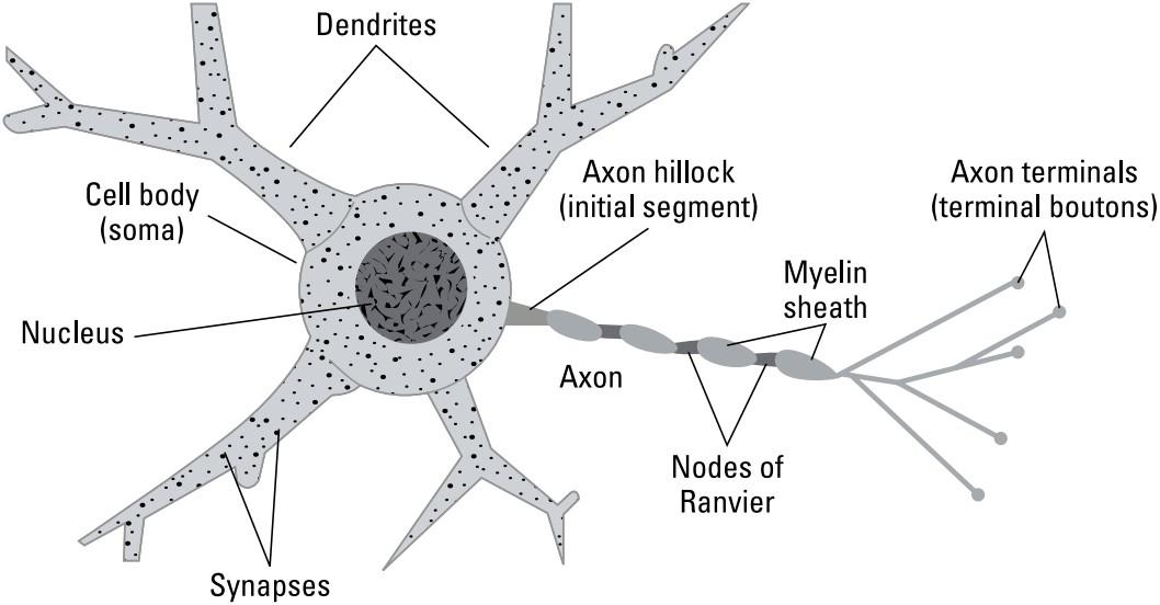
FIGURE 3-1: The main structural parts of a neuron.
شکل ۳-۱: بخشهای اصلی ساختاری یک نورون.
Sending and receiving info between neurons: Synaptic receptors
What are the dendrites and axons for? Let’s start with the dendrites, or dendritic tree.
ارسال و دریافت اطلاعات بین نورونها: گیرندههای سیناپسی
دندریتها و آکسونها چه کاربردی دارند؟ بیایید با دندریتها یا درخت دندریتیک شروع کنیم.
Think about what the btanches of a normal tree are for: They′re the structures that give rise to the leaves, whose purpose is to gather sunlight for energy and to build organic a way. It′s the structure that creates places for synaptic receptors that gather information from other neurons. Just as tree may have thousands of leaves, neurons have thousands of these receptors.
به این فکر کنید که شاخههای یک درخت معمولی برای چه هستند: آنها ساختارهایی هستند که باعث ایجاد برگها میشوند، که هدف آنها جمعآوری نور خورشید برای انرژی و ساختن یک مسیر ارگانیک است. این ساختار است که مکانهایی را برای گیرندههای سیناپسی ایجاد میکند که اطلاعات را از نورونهای دیگر جمعآوری میکنند. همانطور که درخت ممکن است هزاران برگ داشته باشد، نورونها نیز هزاران عدد از این گیرندهها را دارند.
While the dendrites receive inputs from other cell, the axon sends the output of the cell to other cells. The axon often looks like another dendrite, except that it typically doesn′t branch very near the cell body like most dendrites.
در حالی که دندریتها ورودیها را از سلولهای دیگر دریافت میکنند، آکسون خروجی سلول را به سلولهای دیگر ارسال میکند. آکسون اغلب شبیه دندریت دیگری است، با این تفاوت که معمولاً مانند اکثر دندریتها خیلی نزدیک به جسم سلولی منشعب نمیشود.
Looking at the receptors: Pre- and postsynaptic components
What are the receptors for, you ask? Well, not for sunlight. Neural synaptic receptors receive information via little squirts of neurotransmitters from other neurons. Each little squirt of neurotransmitter tells the postsynaptic receptor (the receptor on the receiving neuron’s dendritic tree) a message from the presynaptic cell (the one doing the squirting).
نگاهی به گیرندهها: اجزای پیشسیناپسی و پسسیناپسی
میپرسید گیرندهها برای چه هستند؟ خب، نه برای نور خورشید. گیرندههای سیناپسی عصبی اطلاعات را از طریق فوارههای کوچک انتقالدهندههای عصبی از نورونهای دیگر دریافت میکنند. هر فواره کوچک انتقالدهنده عصبی به گیرنده پسسیناپسی (گیرنده روی درخت دندریتیک نورون گیرنده) پیامی از سلول پیشسیناپسی (که فواره میزند) میدهد.
For the receiving neuron, it’s like listening to hundreds of brokers simultaneously on the floor of a stock exchange. Some of the brokers are squirting happy mes- sages about something or other and make the postsynaptic neuron happy, or excited, while others are sending low-key messages that make the postsynaptic neuron less excited or inhibited. Millisecond by millisecond, the excitement level of the postsynaptic neuron is determined by which inputs, among the thousands it receives, are active and how active they are.
برای نورون گیرنده، مانند گوش دادن همزمان به صدها دلال در تالار بورس است. برخی از دلالان پیامهای شادیآوری در مورد چیزی یا چیزی ارسال میکنند و نورون پسسیناپسی را خوشحال یا هیجانزده میکنند، در حالی که برخی دیگر پیامهای کماهمیتی ارسال میکنند که نورون پسسیناپسی را کمتر هیجانزده یا مهار شده میکند. میلیثانیه به میلیثانیه، سطح هیجان نورون پسسیناپسی با این تعیین میشود که کدام ورودیها، از بین هزاران ورودی که دریافت میکند، فعال هستند و میزان فعالیت آنها چقدر است.
The meaning of the activity (or excitement level) of a neuron is a function of who is talking to it and whom it is talking to. If our particular stockbroker neuron, for example, were listening to the output of other neurons talking about things like electrical wiring, certain mines in Chile, and production of pennies, our stockbroker neuron might embody information about the price of copper.
معنای فعالیت (یا سطح هیجان) یک نورون، تابعی از این است که چه کسی با آن صحبت میکند و نورون با چه کسی صحبت میکند. برای مثال، اگر نورون دلال سهام ما به خروجی نورونهای دیگر که در مورد چیزهایی مانند سیمکشی برق، معادن خاص در شیلی و تولید سکه صحبت میکنند، گوش میداد، نورون دلال سهام ما ممکن است اطلاعاتی در مورد قیمت مس را در خود جای دهد.
Many synaptic connections create associations as a result of learning. Learning occurs when experiences modify the strength and identity of the interconnections between neurons and thus create memory. Suppose, for example, that your neu- ron receives inputs from some neurons that indicate you’re seeing the color gray; other neurons that indicate it’s big; others that indicate that it has a trunk; and others that respond to the image of tusks. Your postsynaptic neuron is going to be an elephant detector. The postsynaptic targets of this neuron might get other neuronal messages that the ears are big and that lions and giraffes are also nearby, in which case their activity would mean “We’re in Africa, and this is an African elephant.”
بسیاری از اتصالات سیناپسی در نتیجه یادگیری، ارتباطاتی ایجاد میکنند. یادگیری زمانی رخ میدهد که تجربیات، قدرت و هویت اتصالات بین نورونها را تغییر میدهند و بنابراین حافظه را ایجاد میکنند. برای مثال، فرض کنید که نورون شما ورودیهایی از برخی نورونها دریافت میکند که نشان میدهد شما رنگ خاکستری را میبینید؛ نورونهای دیگر که نشان میدهند بزرگ است؛ برخی دیگر که نشان میدهند خرطوم دارد؛ و برخی دیگر که به تصویر عاجها پاسخ میدهند. نورون پسسیناپسی شما قرار است یک آشکارساز فیل باشد. ممکن است نورونهای پسسیناپسیِ هدف این نورون، پیامهای نورونی دیگری مبنی بر بزرگ بودن گوشها و حضور شیرها و زرافهها در همان نزدیکی دریافت کنند که در این صورت فعالیت آنها به این معنی خواهد بود که «ما در آفریقا هستیم و این یک فیل آفریقایی است».
Firing off spikes to other neurons
When the postsynaptic neuron is excited, it fires off electrical pulses, called spikes, or action potentials, that travel away from its cell body and along its axon until they reach the places where the axon branches and end in what are called axon terminals (also called terminal boutons). These axon terminals are presynaptic, and they’re next to other, postsynaptic neurons. These thousands of axon terminals are the places where your presynaptic neuron converts its electrical action potential into squirts of neurotransmitter that convey a message to other neurons. And so it goes, times 86 billion, of course.
شلیک پالسها به نورونهای دیگر
وقتی نورون پسسیناپسی تحریک میشود، پالسهای الکتریکی به نام پالسها یا پتانسیلهای عمل را شلیک میکند که از جسم سلولی آن و در امتداد آکسون آن حرکت میکنند تا به مکانهایی که آکسون منشعب میشود برسند و در جایی که ترمینالهای آکسون (که به آنها دستههای ترمینال نیز گفته میشود) نامیده میشوند، پایان یابند. این ترمینالهای آکسون پیشسیناپسی هستند و در کنار سایر نورونهای پسسیناپسی قرار دارند. این هزاران ترمینال آکسون مکانهایی هستند که نورون پیشسیناپسی شما پتانسیل عمل الکتریکی خود را به قطرات انتقالدهنده عصبی تبدیل میکند که پیامی را به نورونهای دیگر منتقل میکنند. و به همین ترتیب، ضربدر ۸۶ میلیارد، البته.
The junction between the presynaptic neurotransmitter squirting axon and the postsynaptic neurotransmitter-receiving dendrite is called a synapse, the most fundamental computing element in the nervous system. The gap between the pre- and postsynaptic neurons is called the synaptic cleft. The presynaptic spike causes a squirt of neurotransmitter to diffuse across the synaptic cleft from the presynaptic releasing site to receptors on the postsynaptic dendrite. The postsynaptic dendrite has receptors that bind the neurotransmitter, causing ion channels to open or close, producing an electrical event in the postsynaptic cell.
محل اتصال بین آکسون ترشحکنندهی انتقالدهندهی عصبی پیشسیناپسی و دندریت دریافتکنندهی انتقالدهندهی عصبی پسسیناپسی، سیناپس نامیده میشود که اساسیترین عنصر محاسباتی در سیستم عصبی است. شکاف بین نورونهای پیشسیناپسی و پسسیناپسی، شکاف سیناپسی نامیده میشود. اسپایک پیشسیناپسی باعث میشود که یک ترشح از انتقالدهندهی عصبی از محل ترشح پیشسیناپسی به گیرندههای روی دندریت پسسیناپسی در شکاف سیناپسی پخش شود. دندریت پسسیناپسی گیرندههایی دارد که به انتقالدهندهی عصبی متصل میشوند و باعث باز یا بسته شدن کانالهای یونی میشوند و یک رویداد الکتریکی در سلول پسسیناپسی ایجاد میکنند.
Many neuroscientists believe that if you are going to compare brains to digital computers, the transistor comparison should be at the level of the synapse. Because each neuron has about 10,000 synapses, and there are around 86 billion neurons, the folks at Intel are going to have to burn a lot more midnight oil if they want their CPUs to catch up to the brain’s computing power.
بسیاری از دانشمندان علوم اعصاب معتقدند که اگر قرار است مغز را با رایانههای دیجیتال مقایسه کنید، مقایسهی ترانزیستور باید در سطح سیناپس باشد. از آنجا که هر نورون حدود ۱۰،۰۰۰ سیناپس دارد و حدود ۸۶ میلیارد نورون وجود دارد، اگر قرار باشد پردازندههای اینتل به قدرت محاسباتی مغز برسند، باید انرژی بسیار بیشتری مصرف کنند.
Receiving input from the environment: Specialized receptors
Some neural receptors receive inputs not as neurotransmitter squirts from other neurons, but as energy from the environment:
دریافت ورودی از محیط: گیرندههای تخصصی
برخی از گیرندههای عصبی ورودیها را نه به صورت انتقالدهندههای عصبی از نورونهای دیگر، بلکه به صورت انرژی از محیط دریافت میکنند:
» In the eye, specialized neurons called photoreceptors catch photons of light and convert those into neurotransmitter release.
» در چشم، نورونهای تخصصی به نام گیرندههای نوری، فوتونهای نور را دریافت کرده و آنها را به انتقالدهندههای عصبی تبدیل میکنند.
» In the ear, cells called auditory hair cells bend in response to sound pressure in the inner ear.
» در گوش، سلولهایی به نام سلولهای مویی شنوایی در پاسخ به فشار صدا در گوش داخلی خم میشوند.
» In the skin, somatosensory receptors respond to pressure and pain.
» در پوست، گیرندههای حسی-تنی به فشار و درد پاسخ میدهند.
» In the nose, olfacto ry neurons respond to odorants. Think of the odorants from the world outside the body as acting like external neurotransmitters, with different olfactory receptors responding to different odorants.
» در بینی، نورونهای بویایی به بوها پاسخ میدهند. بوهای دنیای خارج از بدن را مانند انتقالدهندههای عصبی خارجی در نظر بگیرید، به طوری که گیرندههای بویایی مختلف به بوهای مختلف پاسخ میدهند.
» On the tongue, sweet, sour, bitter, and salty molecules excite different receptors in the taste buds.
» در زبان، مولکولهای شیرین، ترش، تلخ و شور، گیرندههای مختلفی را در جوانههای چشایی تحریک میکنند.
Neurons have outputs to other neurons and to muscles. Motor neurons output their neurotransmitter (acetylcholine) onto muscle cells that contract when receptors in the muscle cells receive it. The control of voluntary striated muscle generally occurs after the brain has done a lot of planning that you’re mostly not aware of about what you’re going to do and which muscles in which patterns are going to contract for you to do that.
نورونها به نورونهای دیگر و عضلات خروجی دارند. نورونهای حرکتی، انتقالدهنده عصبی خود (استیل کولین) را به سلولهای عضلانی میفرستند که وقتی گیرندههای سلولهای عضلانی آن را دریافت میکنند، منقبض میشوند. کنترل عضلات مخطط ارادی عموماً پس از آن رخ میدهد که مغز برنامهریزی زیادی انجام داده باشد که شما عمدتاً از آن در مورد کاری که قرار است انجام دهید و اینکه کدام عضلات در چه الگوهایی قرار است منقبض شوند، آگاه نیستید.
Ionotropic versus metabotropic receptors
Just as advice may fall on willing or deaf ears, the effects of a presynaptically released neurotransmitter on the postsynaptic neuron actually is mostly up to the neurotransmitter receptor on the postsynaptic cell. Although receptors are highly specific for a single neurotransmitter, each neurotransmitter has many different types of receptors. Some receptors act faster than others, some open for longer or shorter times, and some have multiple binding sites that might include one for a particular fast neurotransmitter plus additional sites that bind and respond to one or more neuromodulators. Postsynaptic receptor channels that are activated by neurotransmitters released by a presynaptic neuron are generally referred to as ligand-gated ion channels.
گیرندههای یونوتروپیک در مقابل گیرندههای متابوتروپیک
همانطور که ممکن است توصیهها به گوشهای مشتاق یا ناشنوا برسند، اثرات یک انتقالدهنده عصبی آزاد شده از طریق پیشسیناپسی بر روی نورون پسسیناپسی در واقع عمدتاً به گیرنده انتقالدهنده عصبی روی سلول پسسیناپسی بستگی دارد. اگرچه گیرندهها برای یک انتقالدهنده عصبی واحد بسیار اختصاصی هستند، هر انتقالدهنده عصبی انواع مختلفی از گیرندهها را دارد. برخی از گیرندهها سریعتر از سایرین عمل میکنند، برخی برای مدت زمان طولانیتر یا کوتاهتری باز هستند و برخی دارای چندین جایگاه اتصال هستند که ممکن است شامل یکی برای یک انتقالدهنده عصبی سریع خاص به علاوه جایگاههای اضافی باشد که به یک یا چند تعدیلکننده عصبی متصل میشوند و به آنها پاسخ میدهند. کانالهای گیرنده پسسیناپسی که توسط انتقالدهندههای عصبی آزاد شده توسط یک نورون پیشسیناپسی فعال میشوند، عموماً به عنوان کانالهای یونی دریچهدار لیگاندی شناخته میشوند.
One important difference between receptor types depends on a crucial difference between their structures. The left image in Figure 3-2 shows a typical neurotransmitter receptor sitting in the cell membrane. Most neurotransmitter receptors have a binding site for the neurotransmitter on the outside of the membrane, which, when the neurotransmitter is bound, causes the receptor protein to refold in such a way that a pore opens in the receptor complex that allows a particular ion to go through the otherwise impervious cell membrane. These are thus called neurotransmitter-gated (also ligand-gated) receptors. The receptor type with the ion pore in the same receptor structure as the binding site is called an ionotropic receptor. Most fast excitatory and inhibitory receptors are of this type.
یک تفاوت مهم بین انواع گیرندهها به تفاوت اساسی بین ساختارهای آنها بستگی دارد. تصویر سمت چپ در شکل 3-2 یک گیرنده انتقالدهنده عصبی معمولی را نشان میدهد که در غشای سلولی قرار دارد. اکثر گیرندههای انتقالدهنده عصبی دارای یک محل اتصال برای انتقالدهنده عصبی در قسمت بیرونی غشاء هستند که وقتی انتقالدهنده عصبی متصل میشود، باعث میشود پروتئین گیرنده به گونهای دوباره تا بخورد که منفذی در کمپلکس گیرنده باز شود که به یک یون خاص اجازه میدهد از غشای سلولی غیرقابل نفوذ عبور کند. بنابراین، این گیرندهها گیرندههای دریچهدار انتقالدهنده عصبی (همچنین دریچهدار لیگاندی) نامیده میشوند. نوع گیرندهای که منافذ یونی در همان ساختار گیرنده به عنوان محل اتصال قرار دارد، گیرنده یونوتروپیک نامیده میشود. اکثر گیرندههای تحریکی و مهاری سریع از این نوع هستند.
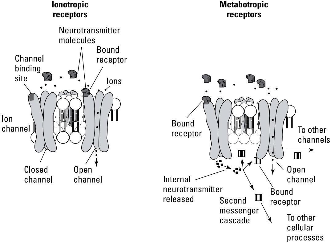
FIGURE 3-2: Ionotropic and metabotropic neurotransmitter receptor types.
شکل۳-۲: انواع گیرندههای انتقالدهنده عصبی یونوتروپیک و متابوتروپیک.
There is another type of receptor structure, called metabotropic, that is common for neuromodulators. In this structure, the ion channel pore is not in the same place as the binding site for the neurotransmitter (refer to the right image in Figure 3-2). Instead, the neurotransmitter binding on the outside of the membrane causes part of the channel protein to change shape on the inside of the membrane such that it releases some internal, intracellular neural modulator. These modulators function as internal messengers inside the cell that often involve biochemical cascades that amplify their actions.
نوع دیگری از ساختار گیرنده به نام متابوتروپیک وجود دارد که برای تعدیلکنندههای عصبی رایج است. در این ساختار، منافذ کانال یونی در همان محل اتصال انتقالدهنده عصبی قرار ندارند (به تصویر سمت راست در شکل 3-2 مراجعه کنید). در عوض، اتصال انتقالدهنده عصبی در قسمت بیرونی غشاء باعث میشود بخشی از پروتئین کانال در داخل غشاء تغییر شکل دهد به طوری که مقداری تعدیلکننده عصبی داخلی و درون سلولی آزاد میکند. این تعدیلکنندهها به عنوان پیامرسانهای داخلی در داخل سلول عمل میکنند که اغلب شامل آبشارهای بیوشیمیایی هستند که عملکرد آنها را تقویت میکنند.
Neurotransmitter binding to metabotropic receptors can cause multiple effects, from opening several nearby channel pores from the inside, to changing the expression of DNA in the nucleus. These types of effects tend to be much slower but longer lasting compared to those mediated by ionotropic receptors.
اتصال انتقالدهنده عصبی به گیرندههای متابوتروپیک میتواند اثرات متعددی ایجاد کند، از باز کردن چندین منافذ کانال مجاور از داخل، تا تغییر بیان DNA در هسته. این نوع اثرات در مقایسه با اثرات واسطهشده توسط گیرندههای یونوتروپیک، بسیار کندتر اما طولانیتر هستند.
The three major functional classes of neurotransmitters
The neurotransmitter “juice” squirted from the presynaptic to the postsynaptic neuron consists of neurotransmitter molecules. A variety of different types of neurotransmitters exist, each varying in its type of effect and time course on the postsynaptic cell. Neurotransmitters are grouped into three major functional classes:
سه دسته اصلی عملکردی انتقالدهندههای عصبی
“شیره” انتقالدهنده عصبی که از نورون پیشسیناپسی به نورون پسسیناپسی ترشح میشود، از مولکولهای انتقالدهنده عصبی تشکیل شده است. انواع مختلفی از انتقالدهندههای عصبی وجود دارد که هر کدام از نظر نوع اثر و دوره زمانی بر روی سلول پسسیناپسی متفاوت هستند. انتقالدهندههای عصبی به سه دسته اصلی عملکردی گروهبندی میشوند:
» Fast, excitatory neurotransmitters: The most important fast, excitatory neurotransmitters are glutamate and acetylcholine. These communicate strong, immediate excitation from the presynaptic to the postsynaptic cell. Most long-distance neural projections in the brain are made by axons that release glutamate. All voluntary muscle contractions are caused by the release of acetylcholine by motor neurons onto muscle cells.
» انتقالدهندههای عصبی سریع و تحریکی: مهمترین انتقالدهندههای عصبی سریع و تحریکی، گلوتامات و استیل کولین هستند. این انتقالدهندهها، تحریک قوی و فوری را از سلول پیشسیناپسی به سلول پسسیناپسی منتقل میکنند. اکثر انشعابات عصبی دوربرد در مغز توسط آکسونهایی انجام میشوند که گلوتامات آزاد میکنند. تمام انقباضات ارادی عضلات ناشی از آزاد شدن استیل کولین توسط نورونهای حرکتی به سلولهای عضلانی است.
» Fast, inhibitory neurotransmitters: Because large interconnected systems made only of excitatory connections tend to be unstable and subject to seizures (strong, uncontrolled, sustained, and recurrent excitation), subtlety and balance in the nervous system is guaranteed by having inhibitory connections, using the fast inhibitory neurotransmitters gamma amino butyric acid (GABA) and glycine, among others. Inhibitory neurotransmitters allow for computations like winner- take-all. For example, if most of the neurons are voting that you’re looking at an elephant but a couple here and there are saying, “If it has four legs, it could also be a donkey,” the inhibitory neurotransmitters suppress the “it could be a donkey” message.
» انتقالدهندههای عصبی سریع و مهاری: از آنجا که سیستمهای بزرگ به هم پیوسته که فقط از اتصالات تحریکی ساخته شدهاند، ناپایدار هستند و در معرض تشنج (تحریک قوی، کنترل نشده، پایدار و مکرر) قرار دارند، ظرافت و تعادل در سیستم عصبی با داشتن اتصالات مهاری، با استفاده از انتقالدهندههای عصبی سریع مهاری گاما آمینو بوتیریک اسید (GABA) و گلیسین و غیره، تضمین میشود. انتقالدهندههای عصبی مهاری امکان محاسباتی مانند «برنده همه چیز را میبرد» را فراهم میکنند. برای مثال، اگر بیشتر نورونها رأی بدهند که شما به یک فیل نگاه میکنید، اما چند نورون اینجا و آنجا بگویند: «اگر چهار پا دارد، میتواند الاغ هم باشد»، انتقالدهندههای عصبی مهاری پیام «میتواند الاغ باشد» را سرکوب میکنند.
» Slow neuromodulators that include both excitatory and inhibitory types: Most of the fast neurotransmitters are amino acids (other than acetylcholine). Modulating neurotransmitters include biogenic amines, such as the catechol- amines (dopamine, epinephrine, and norepinephrine), and the biogenic amines (histamine and serotonin). Other neuromodulators include small proteins like somatostatin, substance P, and enkephalins. The actions of these transmitters are more hormonelike, producing slow but long lasting changes. Think of their action as setting the “mood” within the brain as a background for what the faster neurotransmitters are doing at shorter time scales. For example, you might be resting quietly while digesting a good meal (which is a good idea) when a lightning storm strikes, triggering the release of norepinephrine, the fight or flight neurotransmitter. Its action shuts down digestion and other internal homeostatic processes in favor of increasing your pulse and blood pressure and diverting blood flow to the muscles in preparation for moving quickly to safety.
» تعدیلکنندههای عصبی کند که شامل انواع تحریکی و مهاری میشوند: اکثر انتقالدهندههای عصبی سریع، اسیدهای آمینه هستند (به غیر از استیل کولین). انتقالدهندههای عصبی تعدیلکننده شامل آمینهای بیوژنیک مانند کاتکولآمینها (دوپامین، اپینفرین و نوراپی نفرین) و آمینهای بیوژنیک (هیستامین و سروتونین) هستند. سایر تعدیلکنندههای عصبی شامل پروتئینهای کوچکی مانند سوماتوستاتین، ماده P و انکفالینها هستند. عملکرد این انتقالدهندهها بیشتر شبیه هورمون است و تغییرات آهسته اما طولانیمدت ایجاد میکند. عملکرد آنها را به عنوان تنظیم «خلق و خو» در مغز به عنوان پسزمینهای برای کاری که انتقالدهندههای عصبی سریعتر در مقیاسهای زمانی کوتاهتر انجام میدهند، در نظر بگیرید. به عنوان مثال، ممکن است در حالی که مشغول هضم یک وعده غذایی خوب هستید (که ایده خوبی است) در حال استراحت باشید که ناگهان رعد و برق رخ میدهد و باعث آزاد شدن نوراپی نفرین، انتقالدهنده عصبی جنگ یا گریز، میشود. عملکرد آن، هضم و سایر فرآیندهای هموستاتیک داخلی را به نفع افزایش نبض و فشار خون و هدایت جریان خون به عضلات برای آمادهسازی جهت حرکت سریع به سمت ایمنی، متوقف میکند.
Some neurotransmitters are thought to have originated evolutionarily as hormones. Hormones are typically secreted into the blood stream where they influence cells that are receptive to them all over the body. Neurotransmitters, released by axons, appear to have been a way nervous systems evolved such that one cell could talk specifically to just a few other cells — something like whispering a secret to a few of your best friends rather than shouting news to an entire auditorium.
تصور میشود که برخی از انتقالدهندههای عصبی به صورت تکاملی به عنوان هورمونها منشأ گرفتهاند. هورمونها معمولاً در جریان خون ترشح میشوند و در آنجا بر سلولهایی که در سراسر بدن پذیرای آنها هستند، تأثیر میگذارند. به نظر میرسد انتقالدهندههای عصبی که توسط آکسونها آزاد میشوند، راهی برای تکامل سیستمهای عصبی بودهاند، به طوری که یک سلول میتواند به طور خاص با تنها چند سلول دیگر صحبت کند – چیزی شبیه به زمزمه کردن یک راز برای چند نفر از بهترین دوستانتان به جای فریاد زدن خبر برای کل سالن اجتماعات.
How Shocking! Neurons as Electrical Signaling Devices
A crucial aspect of neuronal function is their unique use of electricity. Neurons use electricity both to secrete their neurotransmitters and for computation. Here’s how. The membrane of the neuron contains a special kind of ion channel that is actually an active pump for ions. Ions are the charged atoms that are dissolved in fluids inside and outside the cell. The most important ions are the chloride salts of sodium (NaCl), potassium (KCl), calcium (CaCl2), and magnesium (MgCl2), which dissociate when dissolved in water into the cations Na+, K+, Ca++ and Mg++, and the anion Cl–. Note: A cation is positively charged (attracted to a negative cathode); an anion is negatively charged (attracted to a positive anode).
چقدر تکان دهنده! نورونها به عنوان دستگاههای سیگنالینگ الکتریکی
یکی از جنبههای حیاتی عملکرد نورونها، استفاده منحصر به فرد آنها از الکتریسیته است. نورونها از الکتریسیته هم برای ترشح انتقالدهندههای عصبی خود و هم برای محاسبه استفاده میکنند. در اینجا نحوه انجام آن آمده است. غشای نورون حاوی نوع خاصی از کانال یونی است که در واقع یک پمپ فعال برای یونها است. یونها اتمهای بارداری هستند که در مایعات داخل و خارج سلول حل میشوند. مهمترین یونها نمکهای کلرید سدیم (NaCl)، پتاسیم (KCl)، کلسیم (CaCl2) و منیزیم (MgCl2) هستند که هنگام حل شدن در آب به کاتیونهای +Na+، K+، Ca و +Mg و آنیون -Cl تفکیک میشوند. توجه: یک کاتیون بار مثبت دارد (به کاتد منفی جذب میشود)؛ یک آنیون بار منفی دارد (به آند مثبت جذب میشود).
The fluid outside cells and in our blood actually has about the same ionic constit- uency as seawater, that is, mostly Na+, much less K+, and even less Ca++ and Mg++, with Cl– being the major anion. However, the ion pumps in neural mem- branes, called sodium-potassium ATPase pumps, actively pump sodium out of the cell and potassium in, in this ratio: 3 Na+ out to 2 K+ in (see Figure 3-3).
مایع خارج از سلولها و در خون ما در واقع تقریباً همان ترکیب یونی آب دریا را دارد، یعنی عمدتاً +Na، مقدار بسیار کمتری +K و حتی کمتر ++Ca و ++Mg، و -Cl آنیون اصلی است. با این حال، پمپهای یونی در غشاهای عصبی، که پمپهای سدیم-پتاسیم ATPase نامیده میشوند، به طور فعال سدیم را از سلول خارج و پتاسیم را به این نسبت پمپ میکنند: ۳ +Na به ۲ +K (شکل ۳-۳ را ببینید).
These ion pumps run all the time, creating a situation in which very little sodium and an excess of potassium exist inside the cell. However, because not as much potassium goes out as sodium comes in, there is a net negative charge inside the cell due to the deficiency of positive ions. The actual voltage we measure with an elec- trode placed inside the cell (compared to the outside) is determined by the con- ductances for the various ions at any particular time. The potassium ion has a much larger leakage conductance than that for sodium, resulting in a measured membrane potential of about –70 millivolts. Because this voltage is measured across the neuron’s membrane (from inside the neuron to outside), it is called the membrane potential.
این پمپهای یونی تمام وقت کار میکنند و وضعیتی را ایجاد میکنند که در آن سدیم بسیار کمی و پتاسیم اضافی در داخل سلول وجود دارد. با این حال، از آنجا که پتاسیم به اندازه سدیم وارد شده، خارج نمیشود، به دلیل کمبود یونهای مثبت، بار منفی خالص در داخل سلول وجود دارد. ولتاژ واقعی که ما با یک الکترود قرار داده شده در داخل سلول (در مقایسه با خارج) اندازهگیری میکنیم، توسط رسانایی یونهای مختلف در هر زمان خاص تعیین میشود. یون پتاسیم رسانایی نشتی بسیار بیشتری نسبت به سدیم دارد، که منجر به پتانسیل غشایی اندازهگیری شده در حدود 70- میلیولت میشود. از آنجا که این ولتاژ در سراسر غشای نورون (از داخل نورون به خارج) اندازهگیری میشود، به آن پتانسیل غشایی میگویند.
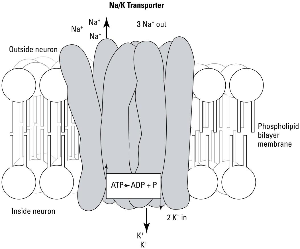
FIGURE 3-3: The neuronal membrane sodium- potassium ATPase ion pump.
شکل ۳-۳: پمپ یونی سدیم-پتاسیم ATPase غشای نورونی.
The membrane potential of a neuron can be changed by ion channels in its membrane. This is where the ionic concentration imbalances matter because, when neurotransmitter receptors open channels for particular ions, the ions flow through these channels in the direction of restoring the imbalance set up by the sodium-potassium pumps, but also according to the voltage or potential across the membrane. The flow of ions is a current that causes the voltage across the membrane (membrane potential) to change accordingly. The fast, excitatory neurotransmitters glutamate and acetylcholine, for example, bind to receptors that allow sodium ions to go through the channel pore. This entry of positive sodium ions can locally reduce and even reverse the polarity of the negative potential inside the cell. This transient polarity change is called depolarization.
پتانسیل غشای یک نورون میتواند توسط کانالهای یونی موجود در غشای آن تغییر کند. اینجاست که عدم تعادل غلظت یونی اهمیت پیدا میکند، زیرا وقتی گیرندههای انتقالدهنده عصبی کانالهایی را برای یونهای خاص باز میکنند، یونها از طریق این کانالها در جهت بازگرداندن عدم تعادل ایجاد شده توسط پمپهای سدیم-پتاسیم، و همچنین مطابق با ولتاژ یا پتانسیل در سراسر غشاء، جریان مییابند. جریان یونها جریانی است که باعث میشود ولتاژ در سراسر غشاء (پتانسیل غشاء) به طور متناسب تغییر کند. به عنوان مثال، انتقالدهندههای عصبی سریع و تحریکی گلوتامات و استیل کولین به گیرندههایی متصل میشوند که به یونهای سدیم اجازه میدهند از منافذ کانال عبور کنند. این ورود یونهای مثبت سدیم میتواند به صورت موضعی قطبیت پتانسیل منفی درون سلول را کاهش داده و حتی معکوس کند. این تغییر قطبیت گذرا، دپلاریزاسیون نامیده میشود.
Ionotropic inhibitory receptors are those that bind either GABA or glycine (among other inhibitory neurotransmitters) and open channels permeable to Cl– (metabotropic GABA receptors open, via second messengers inside the cell, channels for K+ that also inhibit or hyperpolarize the cell). If Cl–, which is negative, enters the cell, it can make the interior more negative, which is called hyperpolarization. When ion channels for K+ are opened, K+ actually goes out of the cell because of its higher concentration inside than outside the cell, and the cell also hyperpolarizes.
گیرندههای مهارکننده یونوتروپیک، گیرندههایی هستند که به GABA یا گلیسین (از جمله سایر انتقالدهندههای عصبی مهارکننده) متصل میشوند و کانالهای نفوذپذیر به -Cl را باز میکنند (گیرندههای متابوتروپیک GABA، از طریق پیامرسانهای ثانویه درون سلول، کانالهایی برای +K باز میکنند که سلول را نیز مهار یا بیشقطبی میکنند). اگر -Cl که منفی است، وارد سلول شود، میتواند فضای داخلی را منفیتر کند که به آن بیشقطبی شدن میگویند. هنگامی که کانالهای یونی برای +K باز میشوند، +K به دلیل غلظت بالاتر آن در داخل نسبت به خارج سلول، در واقع از سلول خارج میشود و سلول نیز بیشقطبی میشود.
Now you can see why a single neuron can be so complicated. Some 10,000 excitatory and inhibitory synapses are causing ion channels to open and close constantly in response to all the various presynaptic inputs, leading to a constantly changing net potential across the cell’s dendritic membrane. The voltage at the cell soma is the summation of all this activity.
حالا میتوانید ببینید که چرا یک نورون واحد میتواند تا این حد پیچیده باشد. حدود ۱۰،۰۰۰ سیناپس تحریکی و مهاری باعث میشوند که کانالهای یونی در پاسخ به تمام ورودیهای مختلف پیشسیناپسی دائماً باز و بسته شوند و منجر به یک پتانسیل خالص دائماً در حال تغییر در غشای دندریتیک سلول شوند. ولتاژ در جسم سلولی، مجموع تمام این فعالیتها است.
Yikes, spikes: The action potential
You can think of the computation done by the neuron as the summation at the cell body of the continuously varying excitatory and inhibitory inputs impinging all over the dendritic tree (some inputs can be on the cell body, also). These inputs produce, via their electrical currents, a net membrane potential at the cell body. What does the neuron do with this computation represented by this net membrane potential? As mentioned previously, neurons communicate their computation to other neurons by sending a series of voltage pulses down the axon. This section explains these voltage pulses and how they move along the axon.
وای، جهشها: پتانسیل عمل
میتوانید محاسبات انجام شده توسط نورون را به عنوان مجموع ورودیهای تحریکی و مهاری متغیر مداوم در جسم سلولی که در سراسر درخت دندریتیک (برخی از ورودیها میتوانند روی جسم سلولی نیز باشند) تأثیر میگذارند، در نظر بگیرید. این ورودیها از طریق جریانهای الکتریکی خود، یک پتانسیل غشایی خالص در جسم سلولی تولید میکنند. نورون با این محاسبه که توسط این پتانسیل غشایی خالص نشان داده میشود، چه میکند؟ همانطور که قبلاً ذکر شد، نورونها محاسبات خود را با ارسال مجموعهای از پالسهای ولتاژ به پایین آکسون به سایر نورونها منتقل میکنند. این بخش این پالسهای ولتاژ و نحوه حرکت آنها در امتداد آکسون را توضیح میدهد.
The problems with getting from here to there
In your primary motor cortex are neurons that drive muscles in your toes. You might assume that axons act like wires — that is, the membrane potential from the neuronal dendrites sums at the motor neuron’s cell body and this voltage travels to the end of the axon as if it were a wire. But that idea doesn’t work. Neuronal membranes, including those of axons, are not totally impervious to ion flow outside of channels and are, thus, leaky. Also, an electrical property of the neuronal membrane, capacitance, shunts (dampens and slows down) rapidly changing signals over the distances that axons need to communicate.
مشکلات رسیدن از اینجا به آنجا
در قشر حرکتی اولیه شما نورونهایی وجود دارند که عضلات انگشتان پا را به حرکت در میآورند. ممکن است فرض کنید که آکسونها مانند سیم عمل میکنند – یعنی پتانسیل غشایی دندریتهای نورونی در جسم سلولی نورون حرکتی جمع میشود و این ولتاژ به انتهای آکسون منتقل میشود، گویی یک سیم است. اما این ایده کار نمیکند. غشاهای نورونی، از جمله غشاهای آکسونها، کاملاً در برابر جریان یونها در خارج از کانالها نفوذناپذیر نیستند و بنابراین نشتی دارند. همچنین، یک ویژگی الکتریکی غشای نورونی، یعنی ظرفیت خازنی، باعث میشود سیگنالهای به سرعت در حال تغییر در فواصلی که آکسونها برای برقراری ارتباط نیاز دارند، شنت (تضعیف و کند میشوند) شوند.
Helping the signal along: Voltage-dependent sodium channels
The answer to this problem is akin to the solution used to solve a similar problem in transatlantic communication cables. In transatlantic cables, devices called repeaters are installed at intervals to boost and clean up the signal, which becomes degraded as it travels from the shore or the last repeater. Neurons use a particular type of ion channel for the same function. This channel is called a voltage- dependent sodium channel, and its action is the basis of all long-distance neural communication by action potentials, colloquially called spikes (because they represent a sudden, brief change in membrane potential).
کمک به سیگنال در طول مسیر: کانالهای سدیم وابسته به ولتاژ
پاسخ به این مشکل مشابه راهحلی است که برای حل مشکل مشابه در کابلهای ارتباطی فرا اقیانوس اطلس استفاده میشود. در کابلهای فرا اقیانوس اطلس، دستگاههایی به نام تکرارکننده در فواصل زمانی نصب میشوند تا سیگنال را تقویت و تمیز کنند، سیگنالی که با عبور از ساحل یا آخرین تکرارکننده، ضعیف میشود. نورونها از نوع خاصی از کانال یونی برای همین عملکرد استفاده میکنند. این کانال، کانال سدیم وابسته به ولتاژ نامیده میشود و عملکرد آن اساس تمام ارتباطات عصبی از راه دور توسط پتانسیلهای عمل است که به طور عامیانه به آن اسپایک میگویند (زیرا آنها نشان دهنده یک تغییر ناگهانی و کوتاه در پتانسیل غشاء هستند).
The voltage-dependent sodium channel does not open by binding a neurotransmitter, like the ligand-gated channels (discussed in the earlier section “Ionotropic versus metabotropic receptors”). Rather, this channel opens when the membrane depolarization created by other channels crosses a threshold.
کانال سدیم وابسته به ولتاژ با اتصال به یک انتقالدهنده عصبی، مانند کانالهای دریچهدار لیگاندی (که در بخش قبلی «گیرندههای یونوتروپیک در مقابل متابوتروپیک» مورد بحث قرار گرفت) باز نمیشود. بلکه، این کانال زمانی باز میشود که دپلاریزاسیون غشایی ایجاد شده توسط کانالهای دیگر از یک آستانه عبور کند.
Here’s how it works: A particularly large number of these channels exist at the beginning of the axon at the cell body, called the initial segment (or sometimes, axon hillock); refer to Figure 3-1. When the summated action of all excitatory and inhibitory synapses on a cell is sufficiently depolarized, the voltage-dependent sodium channels in the initial segment (and often in the cell body as well), open and allow even more Na+ to flow through the membrane into the neuron. This action further depolarizes the membrane so that all the voltage-dependent sodium channels open in that region. These channels typically only stay open for about one millisecond and then close on their own for several milliseconds. The termination of the action potential is also almost always assisted by nearby voltage- gated potassium channels that open and close more slowly than the voltage-gated sodium channels. These delayed rectifier potassium channels are responsible for the late portion of the spike refractory period in which action potentials are harder to elicit quickly after a previous spike.
نحوه کار آن به این صورت است: تعداد بسیار زیادی از این کانالها در ابتدای آکسون در جسم سلولی وجود دارند که قطعه اولیه (یا گاهی اوقات، تپه آکسون) نامیده میشود؛ به شکل 3-1 مراجعه کنید. هنگامی که مجموع عمل همه سیناپسهای تحریکی و مهاری روی یک سلول به اندازه کافی دپلاریزه شود، کانالهای سدیم وابسته به ولتاژ در قطعه اولیه (و اغلب در جسم سلولی) باز میشوند و اجازه میدهند سدیم بیشتری از طریق غشا به داخل نورون جریان یابد. این عمل غشا را بیشتر دپلاریزه میکند به طوری که تمام کانالهای سدیم وابسته به ولتاژ در آن ناحیه باز میشوند. این کانالها معمولاً فقط حدود یک میلیثانیه باز میمانند و سپس به مدت چند میلیثانیه به خودی خود بسته میشوند. خاتمه پتانسیل عمل تقریباً همیشه توسط کانالهای پتاسیم وابسته به ولتاژ مجاور که کندتر از کانالهای سدیم وابسته به ولتاژ باز و بسته میشوند، تسهیل میشود. این کانالهای پتاسیم یکسوکننده تأخیری مسئول بخش پایانی دوره تحریکناپذیری اسپایک هستند که در آن پتانسیلهای عمل پس از یک اسپایک قبلی، به سختی به سرعت ایجاد میشوند.
The pulse created by the voltage-dependent sodium channels at the initial segment is the action potential (spike). The voltage fluctuation created by this spike travels down the axon for some distance but is degraded by the shunting effects mentioned earlier. However, a little distance down the axon is another cluster of these voltage-dependent sodium channels. This distance is such that the degraded spike is still above threshold for these channels, so they produce another full spike at this new location. This spike travels further down the axon, gets degraded, reaches another cluster of voltage-dependent channels, receives the needed boost, travels down the axon some more, gets degraded, and, well, you get the idea.
پالس ایجاد شده توسط کانالهای سدیمی وابسته به ولتاژ در قطعه اولیه، پتانسیل عمل (اسپایک) است. نوسان ولتاژ ایجاد شده توسط این اسپایک تا مسافتی در آکسون حرکت میکند اما توسط اثرات شانتینگ که قبلاً ذکر شد، تضعیف میشود. با این حال، کمی پایینتر از آکسون، خوشه دیگری از این کانالهای سدیمی وابسته به ولتاژ وجود دارد. این فاصله به گونهای است که اسپایک تضعیف شده هنوز بالاتر از آستانه برای این کانالها است، بنابراین آنها در این مکان جدید، اسپایک کامل دیگری تولید میکنند. این اسپایک در امتداد آکسون بیشتر حرکت میکند، تضعیف میشود، به خوشه دیگری از کانالهای وابسته به ولتاژ میرسد، تقویت مورد نیاز را دریافت میکند، کمی بیشتر در آکسون حرکت میکند، تضعیف میشود و خب، متوجه منظورم شدید.
Jumping from node to node
Many axons are sheathed between the clusters of voltage-dependent sodium channels by glial cell processes that provide extra insulation. These are called myelinated axons (refer to Figure 3-1). The gaps between the myelin wrappings where the spike is repeated are called nodes of Ranvier, and the “jumping” of the action potential from one node to the next is called saltatory conduction. Myelinated axons have fast conduction velocities in which the spikes travel at several hundred meters per second. Many smaller axons in the nervous system are unmyelinated and conduct action potentials more slowly. Myelin also protects the axon from damage. (One manifestation of the disease multiple sclerosis is the degradation of the axonal myelin coatings, resulting in slower, erratic, and eventually failed spike conduction along axons.)
پرش از گرهای به گره دیگر
بسیاری از آکسونها توسط زائدههای سلولی گلیال که عایق اضافی ایجاد میکنند، بین خوشههای کانالهای سدیم وابسته به ولتاژ پوشیده شدهاند. اینها آکسونهای میلیندار نامیده میشوند (به شکل 3-1 مراجعه کنید). شکافهای بین پوششهای میلین که در آنها اسپایک تکرار میشود، گرههای رانویه نامیده میشوند و “پرش” پتانسیل عمل از یک گره به گره بعدی، هدایت جهشی نامیده میشود. آکسونهای میلیندار سرعت هدایت سریعی دارند که در آن اسپایکها با سرعت چند صد متر در ثانیه حرکت میکنند. بسیاری از آکسونهای کوچکتر در سیستم عصبی بدون میلین هستند و پتانسیلهای عمل را کندتر هدایت میکنند. میلین همچنین از آکسون در برابر آسیب محافظت میکند. (یکی از تظاهرات بیماری اماس، تخریب پوششهای میلین آکسونی است که منجر به هدایت اسپایک کندتر، نامنظم و در نهایت ناموفق در امتداد آکسونها میشود.)
Closing the loop: From action potential to neurotransmitter release
One last basic detail to consider in the picture of neuronal communication is how to get from action potential to the release of the neurotransmitter. This happens when the spike reaches the thousands of axon terminals after it branches (see Figure 3-4).
بستن حلقه: از پتانسیل عمل تا آزادسازی انتقالدهنده عصبی
یکی از جزئیات اساسی که باید در تصویر ارتباط عصبی در نظر گرفته شود، چگونگی رسیدن از پتانسیل عمل به آزادسازی انتقالدهنده عصبی است. این اتفاق زمانی میافتد که اسپایک پس از انشعاب به هزاران پایانه آکسون میرسد (شکل 3-4 را ببینید).
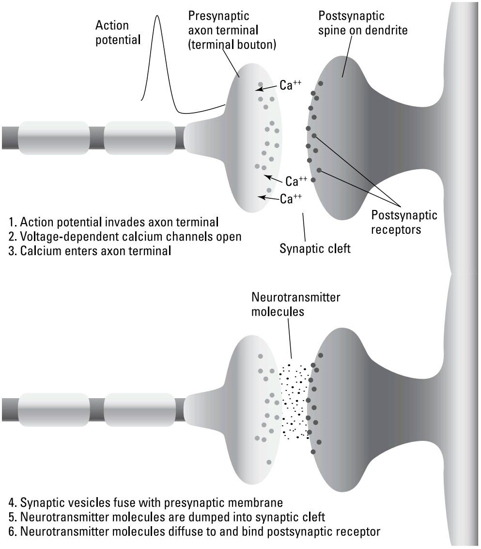
FIGURE 3-4: Synaptic transmission from the presynaptic action potential to the postsynaptic receptor.
شکل ۳-۴: انتقال سیناپسی از پتانسیل عمل پیشسیناپسی به گیرنده پسسیناپسی.
Here’s what happens (refer to Figure 3-4 for a visual of these steps):
آنچه اتفاق میافتد در اینجا آمده است (برای مشاهده تصویری این مراحل به شکل ۳-۴ مراجعه کنید):
» Steps 1 through 3: The action potential arrives. Within the axon terminal is another type of voltage-dependent channel, but this channel allows calcium (Ca++) rather than sodium to flow in. The spike-induced opening of these voltage-dependent calcium channels causes calcium to enter the axon terminal. Within the axon terminal, the neurotransmitter’s molecules are packaged in little membrane-enclosed synaptic vesicles, a few of which are always near the membrane on the presynaptic side of the synapse.
» مراحل ۱ تا ۳: پتانسیل عمل فرا میرسد. درون پایانه آکسون نوع دیگری از کانال وابسته به ولتاژ وجود دارد، اما این کانال به کلسیم (++Ca) به جای سدیم اجازه ورود میدهد. باز شدن این کانالهای کلسیم وابسته به ولتاژ که توسط اسپایک القا میشود، باعث ورود کلسیم به پایانه آکسون میشود. درون پایانه آکسون، مولکولهای انتقالدهنده عصبی در وزیکولهای سیناپسی کوچک محصور در غشاء بستهبندی میشوند که تعدادی از آنها همیشه نزدیک غشاء در سمت پیش سیناپسی سیناپس هستند.
» Step 4: The vesicles bind with the membrane. When calcium enters, it causes the membrane of several of these vesicles to fuse with the cell membrane.
» مرحله ۴: وزیکولها به غشاء متصل میشوند. وقتی کلسیم وارد میشود، باعث میشود غشای چندین مورد از این وزیکولها با غشای سلولی ادغام شود.
» Step 5: The transmitter is released. When vesicles fuse with the cell membrane, the neurotransmitter molecules get dumped into the synaptic cleft.
» مرحله ۵: فرستنده آزاد میشود. وقتی وزیکولها با غشای سلولی ادغام میشوند، مولکولهای انتقالدهنده عصبی به شکاف سیناپسی ریخته میشوند.
» Step 6: The transmitter binds with the receptors. The neurotransmitter molecules that were dumped in the cleft quickly (within about a millisecond) diffuse across the cleft and bind receptors on the postsynaptic cell.
» مرحله ۶: فرستنده به گیرندهها متصل میشود. مولکولهای انتقالدهنده عصبی که در شکاف تخلیه شدهاند، به سرعت (در حدود یک میلیثانیه) در سراسر شکاف پخش میشوند و به گیرندههای روی سلول پسسیناپسی متصل میشوند.
Here’s a shorthand that some neuroscientists use when they think of this circuit: Around a given neuron, the extracellular concentration of various neurotransmitters is converted to membrane potential, which is converted to the rate of action potentials in the axon, which is converted to calcium concentration at the axon terminal, which is finally converted to neurotransmitter concentration in the synaptic cleft next to the postsynaptic cell.
این خلاصهای است که برخی از دانشمندان علوم اعصاب هنگام فکر کردن به این مدار از آن استفاده میکنند: در اطراف یک نورون مشخص، غلظت خارج سلولی انتقالدهندههای عصبی مختلف به پتانسیل غشایی تبدیل میشود که به سرعت پتانسیلهای عمل در آکسون تبدیل میشود که به غلظت کلسیم در ترمینال آکسون تبدیل میشود و در نهایت به غلظت انتقالدهنده عصبی در شکاف سیناپسی کنار سلول پسسیناپسی تبدیل میشود.
Moving Around with Motor Neurons
Neurons originally evolved in many cases to coordinate muscle activity. Large, multi-celled animals can only move efficiently if muscles throughout the animal move in coordination. Coordinated muscle movement is achieved when neurons, embedded in a system that receives sensory input, can activate muscles in such a way as to produce specific muscle contraction sequences — which is precisely what the neuromuscular system does.
حرکت با نورونهای حرکتی
نورونها در ابتدا در بسیاری از موارد برای هماهنگی فعالیت عضلات تکامل یافتهاند. حیوانات بزرگ و چند سلولی تنها در صورتی میتوانند به طور موثر حرکت کنند که عضلات در سراسر بدن حیوان با هماهنگی حرکت کنند. حرکت هماهنگ عضلات زمانی حاصل میشود که نورونها، که در سیستمی قرار دارند که ورودی حسی را دریافت میکند، بتوانند عضلات را به گونهای فعال کنند که توالیهای انقباض عضلانی خاصی تولید کنند – که دقیقاً همان کاری است که سیستم عصبی-عضلانی انجام میدهد.
The neuromuscular system has, as its output, motor neuron axon terminals synapsing on muscle cells within a muscle, one axon terminal per muscle cell (although one motor neuron may have hundreds of axon terminals). This is called the nerve-muscle junction. The motor neuron axon terminal releases acetylcholine as the neurotransmitter (refer to the earlier section “The three major functional classes of neurotransmitters” for more on acetylcholine). Muscle cells have an excitatory, ionotropic receptor for acetylcholine that opens sodium channels in the muscle cell membrane. These channels also flux a small amount of potassium. (To read more about ionotropic receptors, see the earlier section “Ionotropic versus metabotropic receptors.”)
سیستم عصبی-عضلانی، به عنوان خروجی خود، پایانههای آکسون نورون حرکتی را دارد که روی سلولهای عضلانی درون یک عضله سیناپس میکنند، یک پایانه آکسون به ازای هر سلول عضلانی (اگرچه یک نورون حرکتی ممکن است صدها پایانه آکسون داشته باشد). این محل اتصال عصب-عضله نامیده میشود. پایانه آکسون نورون حرکتی، استیل کولین را به عنوان انتقالدهنده عصبی آزاد میکند (برای اطلاعات بیشتر در مورد استیل کولین به بخش قبلی “سه دسته اصلی عملکردی انتقالدهندههای عصبی” مراجعه کنید). سلولهای عضلانی یک گیرنده تحریکی و یونوتروپیک برای استیل کولین دارند که کانالهای سدیم را در غشای سلول عضلانی باز میکند. این کانالها همچنین مقدار کمی پتاسیم را منتقل میکنند. (برای مطالعه بیشتر در مورد گیرندههای یونوتروپیک، به بخش قبلی «گیرندههای یونوتروپیک در مقابل گیرندههای متابوتروپیک» مراجعه کنید.)
Each action potential reaching a given motor neuron axon terminal releases a packet of acetylcholine that causes an action potential in the muscle cell. This muscle cell action potential has a much longer time course than the one millisecond action potential in most neurons. The effect of the muscle cell action potential is to cause actin and myosin filaments within the muscle cell to slide across each other (mediated by calcium), pulling the ends of the cell together (contracting it longitudinally).
هر پتانسیل عملی که به یک پایانه آکسون نورون حرکتی معین میرسد، بستهای از استیل کولین را آزاد میکند که باعث ایجاد پتانسیل عمل در سلول عضلانی میشود. این پتانسیل عمل سلول عضلانی، دوره زمانی بسیار طولانیتری نسبت به پتانسیل عمل یک میلیثانیهای در اکثر نورونها دارد. اثر پتانسیل عمل سلول عضلانی این است که باعث میشود رشتههای اکتین و میوزین درون سلول عضلانی (با واسطه کلسیم) روی یکدیگر بلغزند و انتهای سلول را به سمت هم بکشند (انقباض طولی).
A muscle is a set of chains of these muscle cells. The more each muscle cell is contracted and the more muscle cells in a chain are contracted, the shorter the muscle gets, and the more force the muscle applies. These parameters are con- trolled by the number of activated motor neurons and the rate of firing in those neurons.
یک عضله مجموعهای از زنجیرههای این سلولهای عضلانی است. هرچه هر سلول عضلانی بیشتر منقبض شود و هرچه سلولهای عضلانی بیشتری در یک زنجیره منقبض شوند، عضله کوتاهتر میشود و نیروی بیشتری اعمال میکند. این پارامترها توسط تعداد نورونهای حرکتی فعال شده و میزان شلیک در آن نورونها کنترل میشوند.
Non-neuronal Cells: Glial Cells
Although the figure of about 86 billion neurons in the brain is certainly impressive, within that same volume are as much as ten times as many non-neuronal cells called glia. Glial cells fall into three major types — astrocytes, oligodendrocytes and Schwann cells, and microglia — each with a function, as the following sections explain. There is also a type of glial cell called a satellite glial cell. These cells enclose the surface of neural cell bodies in various ganglia (concentrations of nerve cells) in the peripheral nervous system.
سلولهای غیر عصبی: سلولهای گلیال
اگرچه رقم حدود ۸۶ میلیارد نورون در مغز قطعاً چشمگیر است، اما در همین حجم، ده برابر بیشتر، سلولهای غیر عصبی به نام گلیا وجود دارد. سلولهای گلیال به سه نوع اصلی تقسیم میشوند – آستروسیتها، الیگودندروسیتها و سلولهای شوان، و میکروگلیا – که هر کدام عملکردی دارند، همانطور که در بخشهای بعدی توضیح داده خواهد شد. همچنین نوعی سلول گلیال به نام سلول گلیال ماهوارهای وجود دارد. این سلولها سطح اجسام سلولی عصبی را در گانگلیونهای مختلف (تجمع سلولهای عصبی) در سیستم عصبی محیطی احاطه میکنند.
Astrocytes
Astrocytes are glial cells that form much of the structure of the brain in which the neurons reside. Astrocytes regulate the brain environment and form the blood- brain barrier. In most of the body, the capillaries of the blood system are permeable to many substances so that oxygen, glucose, and amino acids move from the blood to the tissue, while carbon dioxide and other wastes go the other way. In the brain, the astrocytes form an additional barrier that is much more selective, creating a more finely controlled environment for the complex operations taking place there. The astrocytes do this by lining the blood vessels and only allowing traffic of substances between the capillaries and the brain that they themselves control.
آستروسیتها
آستروسیتها سلولهای گلیال هستند که بخش زیادی از ساختار مغز را که نورونها در آن قرار دارند، تشکیل میدهند. آستروسیتها محیط مغز را تنظیم میکنند و سد خونی-مغزی را تشکیل میدهند. در بیشتر بدن، مویرگهای سیستم خونی نسبت به بسیاری از مواد نفوذپذیر هستند، به طوری که اکسیژن، گلوکز و اسیدهای آمینه از خون به بافت منتقل میشوند، در حالی که دی اکسید کربن و سایر مواد زائد به سمت دیگر میروند. در مغز، آستروسیتها یک سد اضافی تشکیل میدهند که بسیار انتخابیتر است و محیطی با کنترل دقیقتر برای عملیات پیچیدهای که در آنجا انجام میشود، ایجاد میکند. آستروسیتها این کار را با پوشاندن رگهای خونی و اجازه دادن به عبور مواد بین مویرگها و مغز که خودشان کنترل میکنند، انجام میدهند.
There are several downsides to the relative chemical isolation of the brain created by the astrocytes, however. One is that many drugs that could potentially help treat brain dysfunctions can’t pass this astrocyte barrier; therefore, treatments can’t be done by injecting these drugs in the bloodstream. Another downside of brain isolation is that brain cancers are not readily attacked by the immune system because antibody cells also have a hard time getting from the blood to brain tumors. Most cancer treatment protocols effectively involve using toxic chemicals and radiation to wipe out the vast majority of dividing cancer cells, hoping the immune system can mop up the last few percent. But because the antibody mop- up operation in the brain is so inefficient, brain cancers typically have very poor prognoses.
با این حال، چندین نکته منفی در جداسازی نسبی شیمیایی مغز که توسط آستروسیتها ایجاد میشود، وجود دارد. یکی از این نکات منفی این است که بسیاری از داروهایی که میتوانند به طور بالقوه به درمان اختلالات مغزی کمک کنند، نمیتوانند از این سد آستروسیتی عبور کنند؛ بنابراین، درمانها را نمیتوان با تزریق این داروها در جریان خون انجام داد. نکته منفی دیگر جداسازی مغز این است که سرطانهای مغز به راحتی توسط سیستم ایمنی مورد حمله قرار نمیگیرند، زیرا سلولهای آنتیبادی نیز برای رسیدن از خون به تومورهای مغزی با مشکل مواجه هستند. اکثر پروتکلهای درمان سرطان به طور مؤثر شامل استفاده از مواد شیمیایی سمی و پرتودرمانی برای از بین بردن اکثریت قریب به اتفاق سلولهای سرطانی در حال تقسیم هستند، به این امید که سیستم ایمنی بتواند چند درصد آخر را پاک کند. اما از آنجا که عملکرد پاکسازی آنتیبادی در مغز بسیار ناکارآمد است، سرطانهای مغز معمولاً پیشآگهی بسیار بدی دارند.
Another type of glial cell with a similar function is the ependymal cell. These glial cells form a barrier at the inner surface of the cerebrospinal fluid-filled ventricles of the brain and the central canal of the spinal cord.
نوع دیگری از سلولهای گلیال با عملکرد مشابه، سلول اپاندیمی است. این سلولهای گلیال سدی در سطح داخلی بطنهای پر از مایع مغزی نخاعی مغز و کانال مرکزی نخاع تشکیل میدهند.
Oligodendrocytes and Schwann cells
The second class of glial cells are called oligodendrocytes and Schwann cells. The function of both of these cells is to do the myelin wrapping of axons, discussed in the earlier section “Jumping from node to node,” for saltatory conduction, wherein the nodes of Ranvier with high voltage-gated sodium channels form “repeaters” allowing long distance spike propagation. Oligodendrocytes perform this function in the central nervous system, while Schwann cells perform the same function in the peripheral nervous system.
الیگودندروسیتها و سلولهای شوان
دسته دوم سلولهای گلیال، الیگودندروسیتها و سلولهای شوان نامیده میشوند. عملکرد هر دوی این سلولها، پوشاندن آکسونها با میلین است که در بخش قبلی «پریدن از گرهای به گره دیگر» برای هدایت جهشی مورد بحث قرار گرفته است، که در آن گرههای رانویر با کانالهای سدیمی با دریچههای ولتاژ بالا، «تکرارکنندههایی» تشکیل میدهند که امکان انتشار اسپایک در فواصل طولانی را فراهم میکنند. الیگودندروسیتها این عملکرد را در سیستم عصبی مرکزی انجام میدهند، در حالی که سلولهای شوان همین عملکرد را در سیستم عصبی محیطی انجام میدهند.
An area of intense research now concerns the fact that peripheral nerves regenerate their axons, while central neurons usually do not. You can, for example, gash your finger and cut the nerves so severely that all sensory and motor control functions are lost in the fingertip. But wait a month or two, and full function generally returns. The same injury in the spinal cord, however, paralyzes you for life. Some differences between oligodendrocytes and Schwann cells in response to injury are thought to be important for regeneration.
اکنون حوزهای از تحقیقات فشرده به این واقعیت مربوط میشود که اعصاب محیطی آکسونهای خود را بازسازی میکنند، در حالی که نورونهای مرکزی معمولاً این کار را نمیکنند. به عنوان مثال، میتوانید انگشت خود را برش دهید و اعصاب را آنقدر شدید ببرید که تمام عملکردهای کنترل حسی و حرکتی در نوک انگشت از بین بروند. اما یک یا دو ماه صبر کنید و عملکرد کامل به طور کلی باز میگردد. با این حال، همان آسیب در نخاع، شما را برای تمام عمر فلج میکند. تصور میشود برخی تفاوتها بین الیگودندروسیتها و سلولهای شوان در پاسخ به آسیب، برای بازسازی مهم هستند.
Microglial cells
The last class of glial cells are microglial cells. These are scavenger cells that migrate through the brain when some area is injured and remove (scavenge) debris. This is just one aspect of the many housekeeping functions that this and other glial cells do, which includes maintaining the proper ionic constituencies in the extracellular space, interacting with blood vessels during brain injuries, and providing the structural framework in which neurons initially grow during development.
سلولهای میکروگلیال
آخرین دسته از سلولهای گلیال، سلولهای میکروگلیال هستند. اینها سلولهای جمعآوریکنندهای هستند که وقتی ناحیهای آسیب میبیند، در مغز مهاجرت میکنند و مواد زائد را جمعآوری (پاکسازی) میکنند. این تنها یکی از جنبههای بسیاری از عملکردهای خانهداری است که این سلول و سایر سلولهای گلیال انجام میدهند، که شامل حفظ حوزههای یونی مناسب در فضای خارج سلولی، تعامل با رگهای خونی در طول آسیبهای مغزی و فراهم کردن چارچوب ساختاری است که نورونها در ابتدا در طول رشد در آن رشد میکنند.
Recording Techniques
The earliest human brain recordings — electroencephalograms, or EEGs — used surface electrodes on the scalp to record ongoing brain potentials from large areas of the brain. Most of what researchers know about individual neuronal function (neurophysiology) began in about the middle of the 20th century with the invention and use of microelectrodes, which could sample the activity of single neurons, and oscilloscopes, which could display events lasting milliseconds or less. As the following sections show, several advances have been made in the kinds of devices used to record the workings of the neurons.
تکنیکهای ضبط
اولین ضبطهای مغز انسان – الکتروانسفالوگرامها یا EEGها – از الکترودهای سطحی روی پوست سر برای ثبت پتانسیلهای مداوم مغز از نواحی بزرگ مغز استفاده میکردند. بیشتر آنچه محققان در مورد عملکرد نورونهای فردی (نوروفیزیولوژی) میدانند، تقریباً در اواسط قرن بیستم با اختراع و استفاده از میکروالکترودها، که میتوانستند فعالیت نورونهای منفرد را نمونهبرداری کنند، و اسیلوسکوپها، که میتوانستند رویدادهایی را که میلیثانیه یا کمتر طول میکشیدند، نمایش دهند، آغاز شد. همانطور که بخشهای بعدی نشان میدهند، پیشرفتهای متعددی در انواع دستگاههای مورد استفاده برای ثبت عملکرد نورونها حاصل شده است.
Single extracellular microelectrodes
Extracellular microelectrodes are very thin, needlelike wires that are electrically insulated except at their very tip. The tips of these are on the order of the size of a neuron’s cell body (typically 20 micrometers across, depending on the neuronal type). The electrode tip is inserted into the neural tissue until the tip happens to be near one particular cell, so that the voltage the microelectrode detects is almost entirely due to the action potential currents from the ion channels opening in that one nearby cell soma. These electrodes also sometimes record action potentials from nearby axons. Extracellular microelectrodes have also been used to stimulate neurons to fire by generating pulses of current through the electrode and shocking the neuron to fire artificially.
میکروالکترودهای تکی خارج سلولی
میکروالکترودهای خارج سلولی سیمهای بسیار نازک و سوزنی شکلی هستند که به جز نوک خود، از نظر الکتریکی عایق هستند. نوک این سیمها به اندازه جسم سلولی نورون است (معمولاً 20 میکرومتر، بسته به نوع نورون). نوک الکترود در بافت عصبی قرار میگیرد تا زمانی که نوک آن نزدیک یک سلول خاص قرار گیرد، به طوری که ولتاژی که میکروالکترود تشخیص میدهد تقریباً به طور کامل ناشی از جریانهای پتانسیل عمل از کانالهای یونی باز شده در آن سلول مجاور است. این الکترودها گاهی اوقات پتانسیلهای عمل را از آکسونهای مجاور نیز ثبت میکنند. میکروالکترودهای خارج سلولی همچنین برای تحریک نورونها به شلیک با تولید پالسهای جریان از طریق الکترود و شوک دادن مصنوعی به نورون برای شلیک استفاده شدهاند.
Microelectrode arrays
Microelectrode arrays are clusters of microelectrodes that sample up to hundreds of cells simultaneously. Some of these have been implanted in paralyzed humans so that, for example, when the person thought of making some movement in his brain and activated his upper motor neurons, the electronically detected signal could be used to bypass the damaged spinal cord and activate muscles directly by sending shocking pulses to them or to the lower motor neurons that drive them. These recordings have also been used to control computer cursors for communication.
آرایههای میکروالکترود
آرایههای میکروالکترود، خوشههایی از میکروالکترودها هستند که به طور همزمان از صدها سلول نمونهبرداری میکنند. برخی از این آرایهها در انسانهای فلج کاشته شدهاند، به عنوان مثال، وقتی فرد به انجام حرکتی در مغز خود فکر میکرد و نورونهای حرکتی فوقانی خود را فعال میکرد، سیگنال شناساییشده الکترونیکی میتوانست برای دور زدن نخاع آسیبدیده و فعال کردن مستقیم عضلات با ارسال پالسهای شوکدهنده به آنها یا به نورونهای حرکتی تحتانی که آنها را هدایت میکنند، استفاده شود. این ضبطها همچنین برای کنترل مکاننماهای کامپیوتر برای ارتباط استفاده شدهاند.
Sharp intracellular electrodes
Intracellular recordings in mammalian neurons were first done with sharp intracellular microelectrodes, also called glass pipettes or micropipettes. These are glass tubes that are heated in the middle and then pulled apart so that the tube “necks down” and breaks in the middle. The tip at the break can have a diameter of less than one micrometer and still be hollow! These glass microelectrodes are sufficiently small compared to a 20-micrometer diameter cell that they can be inserted inside cells.
الکترودهای تیز درون سلولی
ثبتهای درون سلولی در نورونهای پستانداران ابتدا با میکروالکترودهای تیز درون سلولی انجام میشد که به آنها پیپتهای شیشهای یا میکروپیپت نیز گفته میشود. اینها لولههای شیشهای هستند که از وسط گرم میشوند و سپس از هم جدا میشوند تا لوله “گردن پایین” بیاید و از وسط بشکند. نوک در محل شکستگی میتواند قطری کمتر از یک میکرومتر داشته باشد و همچنان توخالی باشد! این میکروالکترودهای شیشهای در مقایسه با یک سلول با قطر 20 میکرومتر به اندازه کافی کوچک هستند که میتوان آنها را درون سلولها قرار داد.
Intracellular electrodes inserted through the cell’s membrane allow researchers to sample the electrical activity inside the cell from its synaptic inputs. Sharp intracellular electrodes are filled with a conductive saline solution to make an electrical connection between their open tip inside the cell and electronic amplifiers and displays connected to the large end of the micropipette. A chloride silver wire inside the micropipette connects the inside of the cell, through the saline solution in the pipette, to an electronic amplifier. Micropipettes yield more information about what’s going on inside the cell, but because they inevitably damage the cell from the penetration, they can’t be implanted permanently as would be needed for neural prostheses that would, for example, record motor cortex commands to artificially drive muscles in people who are paralyzed.
الکترودهای درون سلولی که از طریق غشای سلول وارد میشوند، به محققان این امکان را میدهند که فعالیت الکتریکی درون سلول را از ورودیهای سیناپسی آن نمونهبرداری کنند. الکترودهای تیز درون سلولی با یک محلول نمکی رسانا پر میشوند تا یک اتصال الکتریکی بین نوک باز آنها در داخل سلول و تقویتکنندههای الکترونیکی و نمایشگرهای متصل به انتهای بزرگ میکروپیپت برقرار شود. یک سیم کلرید نقرهای درون میکروپیپت، داخل سلول را از طریق محلول نمکی در پیپت، به یک تقویتکننده الکترونیکی متصل میکند. میکروپیپتها اطلاعات بیشتری در مورد آنچه درون سلول میگذرد ارائه میدهند، اما از آنجا که به ناچار در اثر نفوذ به سلول آسیب میرسانند، نمیتوان آنها را به طور دائم کاشت، همانطور که برای پروتزهای عصبی مورد نیاز است، برای مثال، که دستورات قشر حرکتی را برای تحریک مصنوعی عضلات در افراد فلج ثبت میکنند.
Patch-clamp electrodes
Patch-clamp microelectrodes are made like glass microelectrodes except that, instead of being inserted into the cell, they are placed against the cell membrane so that the glass makes a chemical bond, called a gigaseal, with the cell membrane. In the intact membrane patch, the electrode can monitor currents passing through ion channels in the membrane within the gigaseal area. In another configuration, the membrane within the gigaseal is ruptured by negative pressure, but the gigaseal along the perimeter of the pipette opening remains intact, so that, like a glass intracellular microelectrode, there is now electrical continuity between the interior of the cell through the saline solution in the pipette to the recording apparatus.
الکترودهای گیره-پچ
میکروالکترودهای گیره-پچ مانند میکروالکترودهای شیشهای ساخته میشوند، با این تفاوت که به جای قرار گرفتن در سلول، در مقابل غشای سلولی قرار میگیرند، به طوری که شیشه با غشای سلولی پیوند شیمیایی به نام گیگازیل برقرار میکند. در پچ غشایی سالم، الکترود میتواند جریانهای عبوری از کانالهای یونی در غشای ناحیه گیگازیل را رصد کند. در پیکربندی دیگر، غشای درون گیگازیل با فشار منفی پاره میشود، اما گیگازیل در امتداد محیط دهانه پیپت سالم میماند، به طوری که مانند یک میکروالکترود درون سلولی شیشهای، اکنون پیوستگی الکتریکی بین فضای داخلی سلول از طریق محلول نمکی در پیپت تا دستگاه ثبت وجود دارد.
Optical imaging devices
Optical imaging advances in the late 20th century and the development of reporting dyes led to the use of optical recording techniques for monitoring neural activity. Here are the three main optical techniques for recording neural activity:
دستگاههای تصویربرداری نوری
پیشرفتهای تصویربرداری نوری در اواخر قرن بیستم و توسعه رنگهای گزارشدهنده منجر به استفاده از تکنیکهای ثبت نوری برای نظارت بر فعالیت عصبی شد. در اینجا سه تکنیک اصلی نوری برای ثبت فعالیت عصبی آورده شده است:
» Fluorescent dye-mediated monitoring of ionic concentration changes:
This technique uses fluorescent dyes (dyes that absorb and then re-emit light) that change their fluorescence in response to the presence of ions like calcium, magnesium, or sodium. The most common dyes monitor calcium concentration, which is normally very low inside neurons but typically increases when the neuron is active due to calcium flux through cation channels that are not completely selective for sodium, and through voltage- dependent calcium channels that are sometimes common in neuronal dendritic trees as well as at the axon terminal. This means that activity withinneural dendritic trees can sometimes be directly observed optically. Optical imaging also allows researchers to view the activity in multiple cells through a microscope. Fluorescent proteins can now be expressed in specific neuronal cells in specific parts of the brain by transgenetic techniques of DNA alteration in experimental animals.
» نظارت بر تغییرات غلظت یونی با استفاده از رنگ فلورسنت:
این تکنیک از رنگهای فلورسنت (رنگهایی که نور را جذب و سپس دوباره ساطع میکنند) استفاده میکند که فلورسانس خود را در پاسخ به حضور یونهایی مانند کلسیم، منیزیم یا سدیم تغییر میدهند. رایجترین رنگها، غلظت کلسیم را کنترل میکنند که معمولاً در داخل نورونها بسیار کم است، اما معمولاً هنگام فعال بودن نورون به دلیل شار کلسیم از طریق کانالهای کاتیونی که کاملاً برای سدیم انتخابی نیستند، و از طریق کانالهای کلسیم وابسته به ولتاژ که گاهی اوقات در درختان دندریتیک عصبی و همچنین در ترمینال آکسون رایج هستند، افزایش مییابد. این بدان معناست که فعالیت درون درختان دندریتیک عصبی گاهی اوقات میتواند مستقیماً به صورت نوری مشاهده شود. تصویربرداری نوری همچنین به محققان این امکان را میدهد که فعالیت را در چندین سلول از طریق میکروسکوپ مشاهده کنند. پروتئینهای فلورسنت اکنون میتوانند در سلولهای عصبی خاص در قسمتهای خاصی از مغز با تکنیکهای تراریخته تغییر DNA در حیوانات آزمایشگاهی بیان شوند.
» Fluorescent dye-mediated monitoring of membrane potential:
Potentiometric dyes are dyes that bind neuronal cell membranes and change either their fluorescence or absorption of light in response to the level ofdepolarization of the membrane. The advantage of these dyes is that they give a direct reading of the electrical potential across the membrane so that membrane changes can be observed whether or not there happen to be ion channels there that flux calcium. On the other hand, the signal from these dyes is typically an order of magnitude less than that of calcium indicator dyes. These smaller signals are harder to detect.
» پایش پتانسیل غشا با استفاده از رنگ فلورسنت:
رنگهای پتانسیومتری رنگهایی هستند که به غشاهای سلولهای عصبی متصل میشوند و فلورسانس یا جذب نور آنها را در پاسخ به سطح دپلاریزاسیون غشا تغییر میدهند. مزیت این رنگها این است که آنها خوانش مستقیمی از پتانسیل الکتریکی در سراسر غشا ارائه میدهند، به طوری که میتوان تغییرات غشا را مشاهده کرد، چه کانالهای یونی وجود داشته باشند که کلسیم را جریان دهند یا خیر. از سوی دیگر، سیگنال این رنگها معمولاً یک مرتبه از نظر بزرگی کمتر از رنگهای نشانگر کلسیم است. تشخیص این سیگنالهای کوچکتر دشوارتر است.
» Intrinsic optical changes in excited neural tissue, such as light scattering:
Intrinsic optical changes occur in neuronal tissue when cells are electrically active. The origin of these changes is unclear at the time of this writing but includes changes in light scattering due to transient cell swelling or rearrangement of intracellular organelle structures associated with electrical activation. One advantage of intrinsic optical techniques is that they require no dyes and are therefore less invasive. Intrinsic optical recordings similar to electroencephalograms (EEGs) are routinely carried out in humans using infrared light.
» تغییرات نوری ذاتی در بافت عصبی تحریکشده، مانند پراکندگی نور:
تغییرات نوری ذاتی در بافت عصبی زمانی رخ میدهند که سلولها از نظر الکتریکی فعال باشند. منشأ این تغییرات در زمان نگارش این متن مشخص نیست، اما شامل تغییراتی در پراکندگی نور به دلیل تورم گذرای سلول یا بازآرایی ساختارهای اندامک درون سلولی مرتبط با فعالسازی الکتریکی است. یکی از مزایای تکنیکهای نوری ذاتی این است که به هیچ رنگی نیاز ندارند و بنابراین کمتر تهاجمی هستند. ثبتهای نوری ذاتی مشابه الکتروانسفالوگرامها (EEG) به طور معمول در انسان با استفاده از نور مادون قرمز انجام میشوند.