علوم اعصاب به زبان ساده؛ همه چیز درباره مغز و نخاع


دعای مطالعه [ نمایش ]
بِسْمِ الله الرَّحْمنِ الرَّحیمِ
اَللّهُمَّ اَخْرِجْنى مِنْ ظُلُماتِ الْوَهْمِ
خدايا مرا بيرون آور از تاريكىهاى وهم،
وَ اَكْرِمْنى بِنُورِ الْفَهْمِ
و به نور فهم گرامى ام بدار،
اَللّهُمَّ افْتَحْ عَلَيْنا اَبْوابَ رَحْمَتِكَ
خدايا درهاى رحمتت را به روى ما بگشا،
وَانْشُرْ عَلَيْنا خَزائِنَ عُلُومِكَ بِرَحْمَتِكَ يا اَرْحَمَ الرّاحِمينَ
و خزانههاى علومت را بر ما باز كن به امید رحمتت اى مهربانترين مهربانان.
کتاب «علوم اعصاب به زبان ساده» اثر دکتر فرانک آمتور، یکی از منابع معتبر و قابل دسترس برای علاقهمندان و دانشجویان در حوزه علوم اعصاب است که مفاهیم پایه و کاربردهای عمومی مغز و سیستم عصبی را به زبان ساده توضیح میدهد. این کتاب تصویری روشن از مغز انسان و عملکرد نورونها ارائه میدهد و به مخاطبان کمک میکند تا مفاهیم شناختی، رفتاری و ارتباط ذهن و بدن را درک کنند.
ترجمه دقیق و علمی این شاهکار توسط برند علمی «آیندهنگاران مغز» به مدیریت داریوش طاهری، دسترسی فارسیزبانان به مرزهای نوین دانش علوم اعصاب را ممکن ساخته و رسالتی علمی برای ارتقای آموزش، فهم عمیقتر عملکرد مغز و سیستم عصبی و توسعه روشهای نوین در حوزه سلامت عصبی فراهم آورده است.
» کتاب علوم اعصاب به زبان ساده
»» فصل ۲: همه چیز درباره مغز و نخاع
در حال ویرایش
» Neuroscience For Dummies
»»» Chapter 2: All about the Brain and Spinal Cord
IN THIS CHAPTER
» Looking at the hemispheres and lobes in the brain
» Getting the scoop on gender-based processing differences
» Understanding the spinal cord’s function in the central nervous system
» Understanding the role of the autonomic nervous system
» Focusing on imaging technology
در این فصل
» نگاهی به نیمکرهها و لوبهای مغز
» آشنایی با تفاوتهای پردازش مبتنی بر جنسیت
» درک عملکرد نخاع در سیستم عصبی مرکزی
» درک نقش سیستم عصبی خودکار
» تمرکز بر فناوری تصویربرداری

Chapter 2
All about the Brain and Spinal Cord
Right now, as you’re reading this, your brain is busy processing visual images from this page into letters, words, and sentences. You understand what these images mean because of your memories of English words and gram-mar and because your general knowledge of the world is activated by those words. But your brain is always doing much more than this, no matter how hard you may be concentrating on the reading. The brain constantly controls and monitors all bodily functions, allowing you to sneeze, shift in your chair, lift your coffee cup for a drink, swallow, and breathe.
فصل ۲
همه چیز درباره مغز و نخاع
همین الان که دارید این را میخوانید، مغز شما مشغول پردازش تصاویر بصری از این صفحه به حروف، کلمات و جملات است. شما به دلیل خاطراتتان از کلمات و دستور زبان انگلیسی و به این دلیل که دانش عمومی شما از جهان توسط آن کلمات فعال میشود، معنی این تصاویر را میفهمید. اما مغز شما همیشه کارهای بسیار بیشتری انجام میدهد، مهم نیست چقدر روی خواندن تمرکز کنید. مغز دائماً تمام عملکردهای بدن را کنترل و نظارت میکند و به شما امکان میدهد عطسه کنید، روی صندلی خود جابجا شوید، فنجان قهوه خود را برای نوشیدن بردارید، قورت دهید و نفس بکشید.
The brain is part of your nervous system, which is a network of subsystems and interrelated parts. The brain and spinal cord together (with the retina in the eye) constitute the central nervous system. The spinal cord connects to muscles, organs and receptors via the peripheral nervous system, which includes the autonomic ner- vous system that regulates involuntary movements (think heart rate, digestive processes, and internal temperature regulation, to name just a few). The nervous system consists of neurons, which do the work of processing and communicatingsignals, and glial cells that form the structures in which neurons reside and pro- vide important support for keeping the neurons functional and happy.
مغز بخشی از سیستم عصبی شماست که شبکهای از زیرسیستمها و بخشهای مرتبط با هم است. مغز و نخاع با هم (به همراه شبکیه در چشم) سیستم عصبی مرکزی را تشکیل میدهند. نخاع از طریق سیستم عصبی محیطی به عضلات، اندامها و گیرندهها متصل میشود، که شامل سیستم عصبی خودکار است که حرکات غیرارادی را تنظیم میکند (به ضربان قلب، فرآیندهای گوارشی و تنظیم دمای داخلی فکر کنید، که تنها چند مورد از آنها را نام بردیم). سیستم عصبی شامل نورونها است که کار پردازش و انتقال سیگنالها را انجام میدهند و سلولهای گلیال که ساختارهایی را تشکیل میدهند که نورونها در آنها قرار دارند و پشتیبانی مهمی برای حفظ عملکرد و شادابی نورونها فراهم میکنند.
This chapter presents the gross anatomy of the brain and spinal cord (no, not gross as in disgusting, but gross as in big). The chapter outlines the technology used to understand how the brain works and examines the controversial hypoth- esis that male and female brains process information differently.
این فصل آناتومی کلی مغز و نخاع را ارائه میدهد (نه، نه کلی به معنای «چندشآور»، بلکه کلی به معنای «بزرگ»). این فصل به تشریح فناوری مورد استفاده برای درک نحوه عملکرد مغز میپردازد و فرضیه بحثبرانگیز مبنی بر اینکه مغز مردان و زنان اطلاعات را به طور متفاوت پردازش میکند را بررسی میکند.
Looking Inside the Skull: The Brain and Its Parts
The most complex structure in the universe (that we know of) is the three-pound mass of cells within your skull called the brain. The brain consists of about 86 billion neurons, which is about the same number as all the stars in our Milky Way galaxy and the number of galaxies in the known universe.
نگاهی به درون جمجمه: مغز و اجزای آن
پیچیدهترین ساختار جهان (که ما میشناسیم) توده سلولی سه پوندی درون جمجمه شماست که مغز نامیده میشود. مغز از حدود ۸۶ میلیارد نورون تشکیل شده است که تقریباً به اندازه تمام ستارگان کهکشان راه شیری و تعداد کهکشانهای جهان شناخته شده است.
Like any complex machine, the brain contains a lot of parts, each of which has subparts, each of which has subparts, all the way down to the “nuts and bolts” — the neurons.
مانند هر دستگاه پیچیدهای، مغز شامل بخشهای زیادی است که هر کدام زیربخشهایی دارند و هر کدام زیربخشهایی دارند، تا به “پیچ و مهرهها” – نورونها – برسند.
Figure 2-1 shows the brain from the left side. It highlights the four major lobes (frontal, parietal, temporal, and occipital). You can also see several subcortical areas in this view, namely, the cerebellum at the rear underside of the brain and the brain stem (midbrain, pons, and medulla) connecting the brain to the spinal cord.
شکل ۲-۱ مغز را از سمت چپ نشان میدهد. چهار لوب اصلی (پیشانی، آهیانه، گیجگاهی و پسسری) را برجسته میکند. همچنین میتوانید چندین ناحیه زیرقشری را در این نما مشاهده کنید، یعنی مخچه در قسمت پشتی مغز و ساقه مغز (مغز میانی، پل مغزی و بصلالنخاع) که مغز را به نخاع متصل میکند.
The following sections take a look at the different parts of the brain, from top to bottom.
بخشهای زیر نگاهی به بخشهای مختلف مغز، از بالا به پایین، میاندازند.
The neocortex: Controlling the controllers
Almost all the exposed brain you see in Figure 2-1 (except the cerebellum and brain stem at the bottom) is the neocortex. Neo means “new,” and cortex means something like “husk” or “outer shell.”
نئوکورتکس: کنترل کنترلکنندهها
تقریباً تمام مغز نمایانی که در شکل ۲-۱ میبینید (به جز مخچه و ساقه مغز در پایین) نئوکورتکس است. نئو به معنای “جدید” است و کورتکس به معنای چیزی شبیه “پوسته” یا “پوسته بیرونی” است.
Although the neocortex looks like a blob with some grooves in it, it’s more like a large sheet of tissue that’s been wadded up to fit into the skull. The actual area, if you were to smooth it out, is about 1.5 square feet, roughly equivalent to one and a half standard floor tiles. Obviously a surface with this large an area wouldn’t fit inside your skull without some folding!
اگرچه نئوکورتکس مانند یک توده با شیارهایی در آن به نظر میرسد، اما بیشتر شبیه یک صفحه بزرگ بافتی است که برای قرار گرفتن در جمجمه، فشرده شده است. مساحت واقعی، اگر آن را صاف کنید، حدود ۱.۵ فوت مربع است که تقریباً معادل یک و نیم کاشی استاندارد کف است. بدیهی است که سطحی با این مساحت بزرگ بدون تا شدن در جمجمه شما جای نمیگیرد!
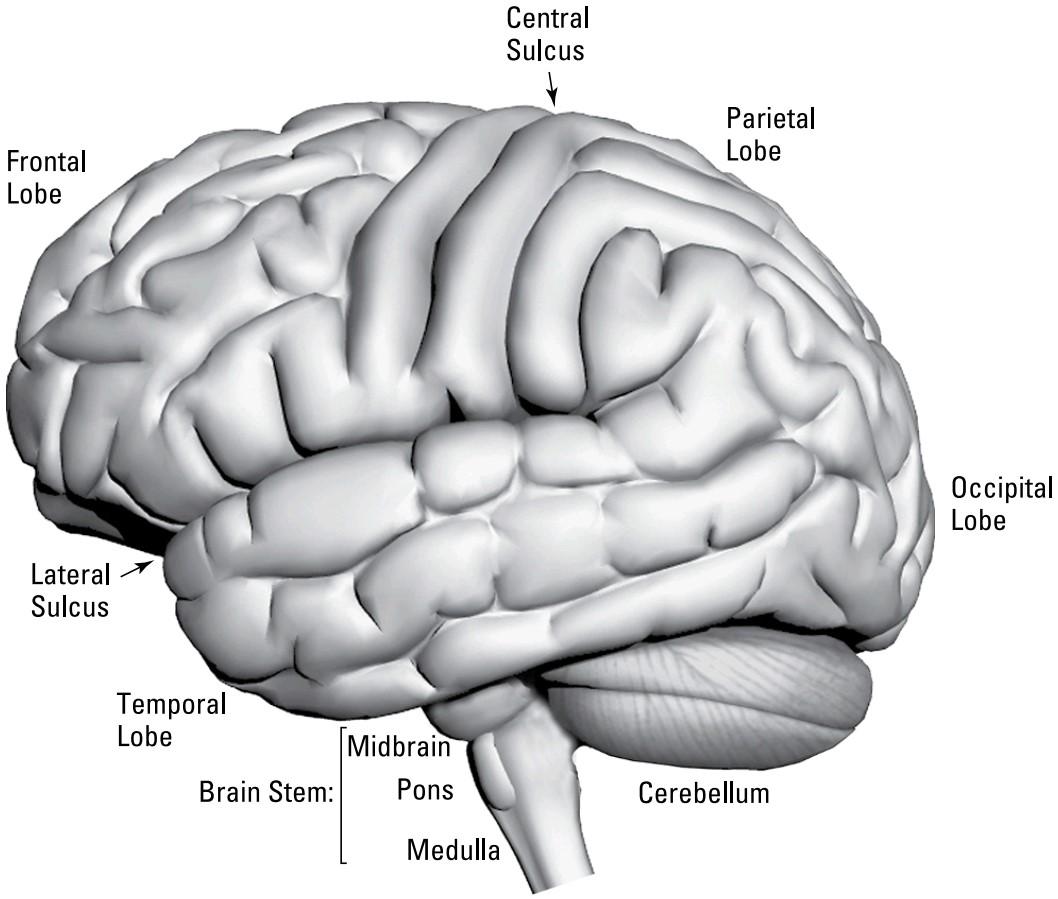
FIGURE 2-1: The neocortex.
شکل ۲-۱: نئوکورتکس.
A large neocortex distinguishes mammals from all other animals. The human neocortex is so large that it completely covers all the rest of the brain except for a bit of cerebellum that sticks out from the back, and the brain stem. The neocortex enables the most complex mental activity that we associate with being human.
نئوکورتکس بزرگ، پستانداران را از سایر حیوانات متمایز میکند. نئوکورتکس انسان آنقدر بزرگ است که به جز کمی از مخچه که از پشت بیرون زده است و ساقه مغز، تمام بقیه مغز را به طور کامل میپوشاند. نئوکورتکس پیچیدهترین فعالیت ذهنی را که ما با انسان بودن مرتبط میدانیم، ممکن میسازد.
The relationship between the neocortex and the rest of the brain is twofold:
رابطه بین نئوکورتکس و بقیه مغز دوگانه است:
» The neocortex acts like a set of complex subroutines in a computer program that allow more complex, nuanced, and specialized computa- tions for processing sensory input and responding to it. Species that existed before mammals could clearly move, sense their environment, and exhibit the type of moderately complex behaviors such as those that are now seen in birds and lizards. These abilities were all enabled by brain structures older than the neocortex that still exist in mammals and still mediate primary control of brain processing. What the neocortex allowed was a new level of advanced behavior — particularly social behavior — culminating in humans with tool making and, ultimately, language and high-level consciousness.
» نئوکورتکس مانند مجموعهای از زیرروالهای پیچیده در یک برنامه کامپیوتری عمل میکند که محاسبات پیچیدهتر، ظریفتر و تخصصیتری را برای پردازش ورودیهای حسی و پاسخ به آن فراهم میکند. گونههایی که قبل از پستانداران وجود داشتند، میتوانستند به وضوح حرکت کنند، محیط خود را حس کنند و رفتارهای نسبتاً پیچیدهای مانند رفتارهایی که اکنون در پرندگان و مارمولکها دیده میشود را از خود نشان دهند. همه این تواناییها توسط ساختارهای مغزی قدیمیتر از نئوکورتکس که هنوز در پستانداران وجود دارند و هنوز هم واسطه کنترل اولیه پردازش مغز هستند، امکانپذیر شده است. آنچه نئوکورتکس اجازه میداد، سطح جدیدی از رفتار پیشرفته – به ویژه رفتار اجتماعی – بود که در انسان با ابزارسازی و در نهایت، زبان و آگاهی سطح بالا به اوج خود رسید.
» The frontal lobe of the neocortex acts as the controller of controllers. This is mediated by the complex multilayered working memory function of the prefrontal cortical areas that allow us to pursue complex multistep plans. In order to finish a term paper due next week, you must plan to gather and read the background literature, set aside time to type the paper, arrange for meals and transportation for other activities during that time, and not be too distracted by homecoming weekend festivities. Long-term goal pursuit is impossible without well-developed frontal lobe function.
» لوب پیشانی نئوکورتکس به عنوان کنترلکنندهی کنترلکنندهها عمل میکند. این امر توسط عملکرد پیچیده و چندلایهی حافظهی کاری نواحی قشر پیشپیشانی انجام میشود که به ما امکان میدهد برنامههای پیچیدهی چندمرحلهای را دنبال کنیم. برای اتمام یک مقالهی پایان ترم که قرار است هفتهی آینده تحویل داده شود، باید برنامهریزی کنید تا ادبیات پیشزمینه را جمعآوری و مطالعه کنید، زمانی را برای تایپ مقاله اختصاص دهید، وعدههای غذایی و حمل و نقل را برای سایر فعالیتها در آن زمان ترتیب دهید و جشنهای آخر هفتهی بازگشت به خانه حواستان را خیلی پرت نکند. پیگیری اهداف بلندمدت بدون عملکرد خوب و توسعهیافتهی لوب پیشانی غیرممکن است.
The following sections introduce and explain the key parts of the neocortex: the four main lobes (frontal, parietal, temporal, and occipital) into which the neocor- tex is divided, and some of the grooves and ridges in the neocortex’s tissue. It also explains the terminology used to refer to positions: dorsal, ventral, anterior, and so on.
بخشهای زیر بخشهای کلیدی نئوکورتکس را معرفی و توضیح میدهند: چهار لوب اصلی (فرونتال، پاریتال، تمپورال و پسسری) که نئوکورتکس به آنها تقسیم میشود، و برخی از شیارها و برآمدگیهای موجود در بافت نئوکورتکس. همچنین اصطلاحات مورد استفاده برای اشاره به موقعیتها: پشتی، شکمی، قدامی و غیره را توضیح میدهد.
Finding your way around the neocortex: Dorsal, ventral, anterior, and posterior
Imagine a shark. The dorsal fin sticks out of the water from the shark’s back, so dorsal refers to the shark’s back, which is on top. The opposite of dorsal, ventral, refers to bottom. It’s the same with a four-legged animal, where dorsal refers to top and ventral refers to bottom. Things get a little complicated with humans and other primates, though, because we stand up on two legs. When referring to the spinal cord, dorsal refers to the back and ventral to the front, which, when we’re standing, isn’t up versus down.
یافتن راه خود در نئوکورتکس: پشتی، شکمی، قدامی و خلفی
یک کوسه را تصور کنید. باله پشتی از پشت کوسه از آب بیرون زده است، بنابراین پشتی به پشت کوسه اشاره دارد که در بالا قرار دارد. نقطه مقابل پشتی، شکمی، به پایین اشاره دارد. در مورد یک حیوان چهارپا نیز همین است، جایی که پشتی به بالا و شکمی به پایین اشاره دارد. با این حال، اوضاع در مورد انسانها و سایر نخستیها کمی پیچیده میشود، زیرا ما روی دو پا میایستیم. هنگام اشاره به نخاع، پشتی به پشت و شکمی به جلو اشاره دارد، که وقتی ما ایستادهایم، بالا در مقابل پایین نیست.
So how do we refer to directions in the human brain? There, dorsal refers to the top and ventral to the bottom. The term anterior refers to the front (forehead) and pos- terior refers to the back (back of the head). Note: Anterior and posterior have more or less replaced (in the brain) the older equivalent terms rostral and caudal, respec- tively, but you may still hear those terms.
بنابراین چگونه میتوانیم به جهات در مغز انسان اشاره کنیم؟ در آنجا، dorsal به بالا و ventral به پایین اشاره دارد. اصطلاح anterior به جلو (پیشانی) و posterior به عقب (پشت سر) اشاره دارد. توجه: anterior و posterior کم و بیش (در مغز) جایگزین اصطلاحات معادل قدیمیتر rostral و caudal شدهاند، اما هنوز هم ممکن است این اصطلاحات را بشنوید.
When the terms anterior and posterior are used to refer to locations in the front and back of the brain, the terms used for the up–down axis are usually superior and inferior. Neuroscientists also use the terms medial and lateral to designate brain regions close to the center of the brain versus those closer to the outside on the left or right.
هنگامی که اصطلاحات anterior و posterior برای اشاره به مکانهایی در جلو و عقب مغز استفاده میشوند، اصطلاحات مورد استفاده برای محور بالا-پایین معمولاً فوقانی و تحتانی هستند. دانشمندان علوم اعصاب همچنین از اصطلاحات medial و lateral برای تعیین نواحی مغزی نزدیک به مرکز مغز در مقابل نواحی نزدیکتر به خارج در چپ یا راست استفاده میکنند.
Grooves and ridges: The sulci and gyri
The grooves in the neocortex are called sulci (singular: sulcus), and the ridges between the grooves are called gyri (singular: gyrus). No real difference exists between the neocortical tissue visible on the surface gyri versus that down in the sulci (see Figure 2-2).
شیارها و برآمدگیها: شیارها و چینهای مغزی
شیارهای موجود در نئوکورتکس، شیار (مفرد: شیار) نامیده میشوند و برآمدگیهای بین شیارها، چینهای مغزی (مفرد: چینهای مغزی) نامیده میشوند. هیچ تفاوت واقعی بین بافت نئوکورتکس قابل مشاهده در چینهای سطحی و بافت زیرین در چینهای مغزی وجود ندارد (شکل 2-2 را ببینید).
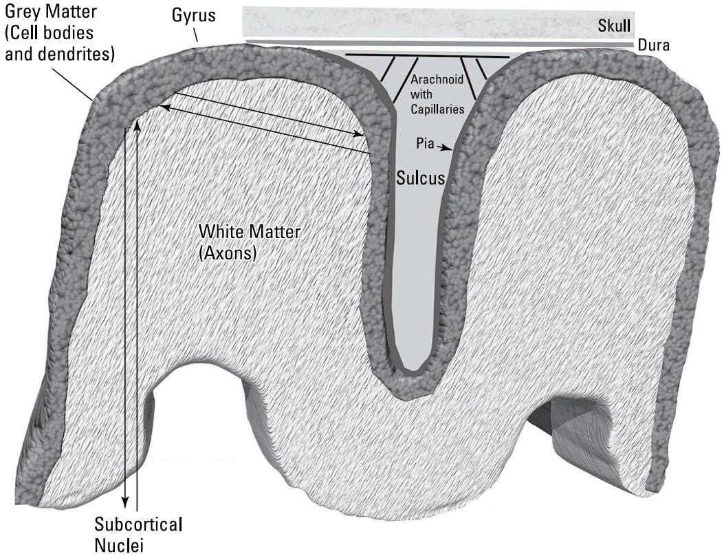
FIGURE 2-2: The grooves (sulci) and ridges (gyri) in the neocortex.
شکل ۲-۲: شیارها (سولکوس) و برآمدگیها (ژیروس) در نئوکورتکس.
In some cases, sulci separate cortical lobes or functions, but in many cases, they don’t. In the brain, areas that interact extensively with each other are often close together, but ultimately, any brain area can interact with any other area, produc- ing a very complex theoretical interconnection problem beyond the scope of this book. So don’t worry, you won’t be asked to deduce the system of gyri and sulci from first principles on Exam 1!
در برخی موارد، شیارها لوبها یا عملکردهای قشر مغز را از هم جدا میکنند، اما در بسیاری از موارد، این کار را نمیکنند. در مغز، مناطقی که به طور گسترده با یکدیگر تعامل دارند، اغلب نزدیک به هم هستند، اما در نهایت، هر ناحیه مغز میتواند با هر ناحیه دیگری تعامل داشته باشد و یک مسئله ارتباط نظری بسیار پیچیده فراتر از محدوده این کتاب ایجاد کند. بنابراین نگران نباشید، در آزمون ۱ از شما خواسته نمیشود که سیستم چینها و شیارها را از اصول اولیه استنباط کنید!
Here’s what you do need to know: Some of the sulci separate parts of the brain that do different things (such as the central sulcus that separates the frontal lobe from the parietal lobe), and some gyri seem to be associated with particular func- tions (such as processing auditory input in the superior temporal sulcus), but most sulci and gyri merely represent how the brain is folded to fit in the head and don’t correspond to any specific, isolated function. Moreover, the pattern of gyri and sulci vary considerably not only between human brains, but even between the two sides of the same brain. The neocortex is the majority of what’s called the tel- encephalon, which means something like “highest” brain area.
این چیزی است که باید بدانید: برخی از شیارها بخشهایی از مغز را که کارهای مختلفی انجام میدهند (مانند شیار مرکزی که لوب پیشانی را از لوب آهیانه جدا میکند) از هم جدا میکنند، و به نظر میرسد برخی از چینها با عملکردهای خاصی مرتبط هستند (مانند پردازش ورودی شنوایی در شیار گیجگاهی فوقانی)، اما اکثر شیارها و چینها صرفاً نشان دهنده نحوه چین خوردگی مغز برای قرار گرفتن در سر هستند و با هیچ عملکرد خاص و جداگانهای مطابقت ندارند. علاوه بر این، الگوی چینها و شیارها نه تنها بین مغزهای مختلف، بلکه حتی بین دو طرف یک مغز نیز به طور قابل توجهی متفاوت است. نئوکورتکس بخش عمدهای از چیزی است که تلانسفالون نامیده میشود، که به معنای چیزی شبیه به “بالاترین” ناحیه مغز است.
Figure 2-2 also shows the layering of the neocortex. The surface layer of the neo- cortex (dark gray in Figure 2-2) consists of six layers of cells, the dendrites of those cells, and axons going to and from those cells to subcortical structures and other cortical areas. It’s about 2 to 4 millimeters thick. This surface sheet contains the same six layers with the same cell types not only everywhere in the neocortex of a given species, but across all mammalian species. The structure and cellular organization of mouse auditory cortex is similar to elephant motor cortex.
شکل ۲-۲ همچنین لایه بندی نئوکورتکس را نشان میدهد. لایه سطحی نئوکورتکس (به رنگ خاکستری تیره در شکل 2-2) از شش لایه سلول، دندریتهای آن سلولها و آکسونهایی که به ساختارهای زیرقشری و سایر نواحی قشری رفت و آمد میکنند، تشکیل شده است. ضخامت آن حدود 2 تا 4 میلیمتر است. این صفحه سطحی نه تنها در همه جای نئوکورتکس یک گونه خاص، بلکه در تمام گونههای پستانداران، شامل شش لایه مشابه با انواع سلولهای یکسان است. ساختار و سازماندهی سلولی قشر شنوایی موش مشابه قشر حرکتی فیل است.
Underneath the neocortical surface layer is the white matter, so called because of its lighter appearance in the unstained brain compared to the darker surface cel- lular layers. The white matter consists almost exclusively of axons (and glial sup- porting cells, particularly oligodendrocytes). The white matter axons arise from neurons in neocortex going to other areas of cortex and subcortical areas, or com- ing from those areas. It’s almost always thicker than the cellular layers above. Most of the volume of the brain is axonal wiring, not cells.
در زیر لایه سطحی نئوکورتیکال، ماده سفید قرار دارد که به دلیل ظاهر روشنتر آن در مغز رنگآمیزی نشده در مقایسه با لایههای سلولی سطحی تیرهتر، به این نام خوانده میشود. ماده سفید تقریباً منحصراً از آکسونها (و سلولهای پشتیبان گلیال، به ویژه الیگودندروسیتها) تشکیل شده است. آکسونهای ماده سفید از نورونهایی در نئوکورتکس که به نواحی دیگر قشر مغز و نواحی زیر قشری میروند یا از آن نواحی میآیند، منشأ میگیرند. تقریباً همیشه ضخیمتر از لایههای سلولی بالایی است. بیشتر حجم مغز، سیمکشی آکسونی است، نه سلول.
There are three structural layers between the neocortex and skull: the pia, the arachnoid, and the dura. The pia is a thin membrane that covers the surface of the neocortex everywhere, extending down the sulci. The arachnoid is a fluid-filled cavity above the pia that includes blood capillaries that nourish the brain. Above the arachnoid is the dura, a tough membrane that does not extend into the sulci. Above the dura is the skull.
سه لایه ساختاری بین نئوکورتکس و جمجمه وجود دارد: نرمشامه، عنکبوتیه و سختشامه. نرمشامه غشای نازکی است که سطح نئوکورتکس را در همه جا میپوشاند و تا شیارها امتداد مییابد. عنکبوتیه حفرهای پر از مایع در بالای نرمشامه است که شامل مویرگهای خونی است که مغز را تغذیه میکنند. بالای عنکبوتیه، سختشامه قرار دارد، غشایی سفت که به داخل شیارها امتداد نمییابد. بالای سختشامه جمجمه قرار دارد.
The left and right hemispheres
The most obvious aspect of the visible brain is that it is composed of two nearly mirror-image lobes called the left and right hemispheres. The left hemisphere receives most inputs from and controls mostly the right side of the body. This hemisphere in humans is also specialized for language, rule-based reasoning, and analytic skills. The right hemisphere deals with the left side of the body, and it is better at visual pattern recognition and more holistic kinds of perception.
نیمکرههای چپ و راست
مشخصترین جنبهی مغز قابل مشاهده این است که از دو لوب تقریباً تصویر آینهای به نام نیمکرههای چپ و راست تشکیل شده است. نیمکره چپ بیشترین ورودیها را از سمت راست بدن دریافت میکند و عمدتاً آن را کنترل میکند. این نیمکره در انسان همچنین برای زبان، استدلال مبتنی بر قانون و مهارتهای تحلیلی تخصص دارد. نیمکره راست با سمت چپ بدن سروکار دارد و در تشخیص الگوهای بصری و انواع ادراک جامعتر بهتر عمل میکند.
In most tasks, the two hemispheres use a divide-and-conquer strategy, where the left hemisphere processes the details, and the right takes in the big picture. The two hemispheres are connected by the largest fiber tract in the brain, the corpus callosum, which contains 200 million fibers.
در بیشتر وظایف، دو نیمکره از استراتژی تقسیم و غلبه استفاده میکنند، جایی که نیمکره چپ جزئیات را پردازش میکند و نیمکره راست تصویر کلی را دریافت میکند. این دو نیمکره توسط بزرگترین رشته فیبری در مغز، جسم پینهای، که شامل ۲۰۰ میلیون فیبر است، به هم متصل میشوند.
How do scientists know about the differences in the abilities of the left versus right hemispheres? One important source of evidence has come from people with intractable epilepsy who have had their corpus callosum cut to stop the spread of seizures from one side of the brain to the other. Stimuli can be presented to only one hemisphere of these patients. When stimuli are presented to the right (non- language) hemisphere of these patients, they can’t verbally report about them, but they can point with their left hand (controlled by the right hemisphere) to a picture of what was presented.
دانشمندان چگونه از تفاوتهای تواناییهای نیمکره چپ در مقابل نیمکره راست مطلع میشوند؟ یکی از منابع مهم شواهد، از افراد مبتلا به صرع مقاوم به درمان است که جسم پینهای آنها برای جلوگیری از گسترش تشنج از یک طرف مغز به طرف دیگر بریده شده است. محرکها را میتوان فقط به یک نیمکره از این بیماران ارائه داد. وقتی محرکها به نیمکره راست (غیرزبانی) این بیماران ارائه میشوند، آنها نمیتوانند به صورت کلامی در مورد آنها گزارش دهند، اما میتوانند با دست چپ خود (که توسط نیمکره راست کنترل میشود) به تصویری از آنچه ارائه شده است اشاره کنند.
A few years ago, a pop psychology fashion suggested that people should shun their in-the-box, analytical left hemispheric processing and learn to use their holistic, creative right hemisphere. There is nothing wrong, of course, with developing right hemisphere–based abilities. But it is clear that we use both hemispheres for virtually all tasks, and that even left hemisphere-oriented tasks, like writing poetry, certainly involve creativity.
چند سال پیش، یک مد روانشناسی عامهپسند پیشنهاد کرد که مردم باید از پردازش دروننگر و تحلیلی نیمکره چپ خود دست بکشند و یاد بگیرند که از نیمکره راست جامع و خلاق خود استفاده کنند. البته هیچ اشکالی ندارد که تواناییهای مبتنی بر نیمکره راست را پرورش دهیم. اما واضح است که ما تقریباً برای همه کارها از هر دو نیمکره استفاده میکنیم و حتی کارهایی که به نیمکره چپ مربوط میشوند، مانند سرودن شعر، قطعاً شامل خلاقیت میشوند.
The four major lobes
The neocortex is divided into four major lobes (refer to Figure 2-1):
چهار لوب اصلی
نئوکورتکس به چهار لوب اصلی تقسیم میشود (به شکل 2-1 مراجعه کنید):
» The frontal lobe, as its name implies, includes all the neocortex from the front, most anterior part of the brain to a major sulcus, called the central sulcus, that runs from side to side at about the middle of the brain.
» لوب پیشانی، همانطور که از نامش پیداست، شامل تمام نئوکورتکس از جلو، قدامیترین قسمت مغز تا یک شیار اصلی به نام شیار مرکزی است که تقریباً در وسط مغز از یک طرف به طرف دیگر امتداد دارد.
» The parietal lobe goes straight back from the central sulcus to almost the most posterior tip.
» لوب آهیانه مستقیماً از شیار مرکزی تا تقریباً خلفیترین نوک به عقب میرود.
» The occipital lobe is the lobe at the most posterior tip. There is no clear continuous border between parietal and occipital lobes in most brains.
» لوب پسسری، لوبی است که در خلفیترین نوک قرار دارد. در بیشتر مغزها مرز پیوسته مشخصی بین لوبهای آهیانه و پسسری وجود ندارد.
» The temporal lobe is the lateral tonguelike extension from the border between the occipital and parietal lobes that extends in the anterior direction.
» لوب گیجگاهی امتداد جانبی زبانمانند از مرز بین لوبهای پسسری و آهیانه است که در جهت قدامی امتداد مییابد.
The next sections explain each lobe in more detail.
بخشهای بعدی هر لوب را با جزئیات بیشتری توضیح میدهند.
THE FRONTAL LOBE
The frontal lobe (the brain has one on each side, of course) of the neocortex is, as its name implies, in the front. The frontal lobe extends from the front of the skull to a sulcus that divides this lobe from the parietal lobe about midway toward the back of the brain (the occipital lobe). This sulcus is named the central sulcus or central fissure (formerly known as the fissure of Rolando). The frontal lobe gyrus just anterior to (in front of) the central fissure is the primary motor cortex.
لوب پیشانی
لوب پیشانی (البته مغز در هر طرف یک لوب دارد) نئوکورتکس، همانطور که از نامش پیداست، در جلو قرار دارد. لوب پیشانی از جلوی جمجمه تا شیاری امتداد دارد که این لوب را از لوب آهیانه تقریباً در نیمه راه به سمت پشت مغز (لوب پس سری) جدا میکند. این شیار، شیار مرکزی یا شکاف مرکزی (که قبلاً به عنوان شکاف رولاندو شناخته میشد) نامیده میشود. شکنج لوب پیشانی که درست در جلوی (جلوی) شیار مرکزی قرار دارد، قشر حرکتی اولیه است.
Cells in the primary motor cortex (upper motor neurons) send their axons (output cables) down the spinal cord and connect directly to motor neurons that activate muscles (lower motor neurons). Researchers know this because, for example, neurosurgeons such as Wilder Penfield found that electrically stimulating this area during exploratory surgery for epilepsy caused muscle movements. Penfield and others showed that there is an orderly (although distorted) mapping between the point stimulated in the primary motor cortex and the location in the body of the muscle that would twitch.
سلولهای قشر حرکتی اولیه (نورونهای حرکتی فوقانی) آکسونهای خود (کابلهای خروجی) را از طریق نخاع به پایین میفرستند و مستقیماً به نورونهای حرکتی که عضلات را فعال میکنند (نورونهای حرکتی تحتانی) متصل میشوند. محققان این را میدانند زیرا، به عنوان مثال، جراحان مغز و اعصاب مانند وایلدر پنفیلد دریافتند که تحریک الکتریکی این ناحیه در طول جراحی اکتشافی برای صرع باعث حرکات عضلانی میشود. پنفیلد و دیگران نشان دادند که یک نقشهبرداری منظم (هرچند تحریفشده) بین نقطه تحریکشده در قشر حرکتی اولیه و محل عضلهای که در بدن میلرزد وجود دارد.
This “motor map” (called a homunculus) has some interesting properties. One is that the area of primary motor cortex devoted to driving muscles in the face and hands, for example, is proportionally much larger than that devoted to driving much larger muscles such as in the leg. This is because the muscles in areas like the face are more finely differentiated to allow very complex and subtle patterns of activation (think of all the myriad facial expressions we use), whereas many large muscles mostly just vary in the amount of contraction of the entire muscle. Another interesting aspect of the homunculus map is that the motor innervation of some areas of the body, such as the face and hands, are close together in the homunculus, but they are not very close together in terms of skin distance. You can read more about this map in Chapters 4 and 10.
این «نقشه حرکتی» (به نام هومونکولوس) ویژگیهای جالبی دارد. یکی از این ویژگیها این است که ناحیه قشر حرکتی اولیه که به عنوان مثال به عضلات محرک در صورت و دستها اختصاص داده شده است، به طور متناسب بسیار بزرگتر از ناحیهای است که به عضلات بسیار بزرگتر مانند پا اختصاص داده شده است. این به این دلیل است که عضلات در مناطقی مانند صورت به طور دقیقتری متمایز شدهاند تا الگوهای بسیار پیچیده و ظریف فعالسازی را ممکن سازند (به تمام حالات چهره بیشماری که استفاده میکنیم فکر کنید)، در حالی که بسیاری از عضلات بزرگ عمدتاً فقط در میزان انقباض کل عضله متفاوت هستند. یکی دیگر از جنبههای جالب نقشه هومونکولوس این است که عصبدهی حرکتی برخی از نواحی بدن مانند صورت و دستها، در هومونکولوس نزدیک به هم هستند، اما از نظر فاصله پوستی خیلی به هم نزدیک نیستند. میتوانید اطلاعات بیشتر در مورد این نقشه را در فصلهای ۴ و ۱۰ مطالعه کنید.
Figure 2-3 shows the location of the motor cortex, and some other prominent divisions of the frontal lobe.
شکل ۲-۳ محل قشر حرکتی و برخی دیگر از بخشهای برجسته لوب پیشانی را نشان میدهد.

FIGURE 2-3: Divisions within the frontal lobe.
شکل ۲-۳: بخشهای درون لوب پیشانی.
The most anterior portion of the frontal lobe is called prefrontal cortex. In humans, the prefrontal cortex takes up the majority of the frontal lobe. Primary motor cor- tex is the most posterior portion of the frontal lobe, starting just anterior to the central sulcus. Moving from the primary motor cortex in the anterior direction (forward, toward the front of the skull), you have a series of regions that perform more and more abstract motor functions. The supplementary and premotor cortex areas are concerned with coordination and sequencing of movement of several muscles. Anterior to that (not labeled) are regions called the frontal eye fields, which are involved in visual attention.
قدامیترین بخش لوب پیشانی، قشر پیشپیشانی نامیده میشود. در انسان، قشر پیشپیشانی بخش عمدهای از لوب پیشانی را اشغال میکند. قشر حرکتی اولیه، خلفیترین بخش لوب پیشانی است که درست از قدامی شیار مرکزی شروع میشود. با حرکت از قشر حرکتی اولیه در جهت قدامی (به جلو، به سمت جلوی جمجمه)، مجموعهای از نواحی وجود دارد که عملکردهای حرکتی انتزاعیتری را انجام میدهند. نواحی قشر تکمیلی و پیشحرکتی مربوط به هماهنگی و توالی حرکت چندین عضله هستند. در جلوی آن (بدون برچسب) مناطقی به نام میدانهای چشمی پیشانی قرار دارند که در توجه بصری نقش دارند.
Anterior to the premotor cortex and supplementary motor cortex and frontal eye fields is the prefrontal cortex, which, despite its name, is part of the frontal lobe. The prefrontal cortex deals with the most abstract aspects of planning. Consider playing tennis: When the ball comes at you, you can choose (if you have time) to hit either a forehand or backhand, with topspin or slice. You can choose to stand there and not hit the ball at all if you think it might be out. These kinds of deci- sions are made in the most anterior parts of the prefrontal cortex.
در جلوی قشر پیشحرکتی و قشر حرکتی تکمیلی و میدانهای چشمی پیشانی، قشر پیشپیشانی قرار دارد که علیرغم نامش، بخشی از لوب پیشانی است. قشر پیشپیشانی با انتزاعیترین جنبههای برنامهریزی سروکار دارد. بازی تنیس را در نظر بگیرید: وقتی توپ به سمت شما میآید، میتوانید (در صورت داشتن وقت) انتخاب کنید که با فورهند یا بکهند، با تاپاسپین یا اسلایس ضربه بزنید. اگر فکر میکنید توپ ممکن است اوت شود، میتوانید همانجا بایستید و اصلاً به آن ضربه نزنید. این نوع تصمیمات در قدامیترین قسمتهای قشر جلوی مغز گرفته میشوند.
Think of the frontal lobe, which is concerned with executing behavior, as “polar- ized” from anterior (front) to posterior (back). Farthest back, at the central sul- cus, are neural wires going almost directly to muscles. In front of that are areas that organize and sequence movements. In front of that are abstract planning levels. At these abstract levels, for example, you select from a variety of different strategies that may involve completely different muscles, muscles sequences, or, as in the tennis shot, the decision to not move at all.
لوب پیشانی، که مربوط به اجرای رفتار است، را به صورت «قطبی» از قدامی (جلو) تا خلفی (عقب) در نظر بگیرید. در انتهای عقب، در شیار مرکزی، سیمهای عصبی تقریباً مستقیماً به عضلات میروند. در جلوی آن مناطقی قرار دارند که حرکات را سازماندهی و توالی میکنند. در جلوی آن سطوح برنامهریزی انتزاعی قرار دارند. به عنوان مثال، در این سطوح انتزاعی، شما از بین استراتژیهای مختلفی که ممکن است شامل عضلات کاملاً متفاوت، توالی عضلات یا مانند ضربه تنیس، تصمیم به عدم حرکت باشد، انتخاب میکنید.
The prefrontal cortex tends to be larger in primates than other mammals, and it’s larger in humans than in other primates. This is correlated with the amount of high level planning done by members of different species. Most mammals operate mostly on instinct and don’t live in complexly differentiated social groups. Pri- mates, on the other hand, have complex male and female hierarchies and may hatch plots against each other that span years of planning. Humans build tools, modify their environments for their own purposes, and have specific relationships with up to hundreds of other individuals (and this was even before Facebook).
قشر جلوی پیشانی در نخستیسانان نسبت به سایر پستانداران بزرگتر است و در انسانها نسبت به سایر نخستیسانان بزرگتر است. این با میزان برنامهریزی سطح بالا که توسط اعضای گونههای مختلف انجام میشود، مرتبط است. اکثر پستانداران عمدتاً بر اساس غریزه عمل میکنند و در گروههای اجتماعی پیچیده و متمایز زندگی نمیکنند. از سوی دیگر، نخستیسانان سلسله مراتب پیچیدهای از نر و ماده دارند و ممکن است علیه یکدیگر توطئههایی کنند که سالها برنامهریزی را در بر میگیرد. انسانها ابزار میسازند، محیط خود را برای اهداف خود تغییر میدهند و با صدها نفر دیگر روابط خاصی دارند (و این حتی قبل از فیسبوک بود).
An area of the prefrontal cortex called the dorsolateral prefrontal cortex (because it’s toward the top/dorsal and outside/lateral) is crucial for what is called working memory, the contents of your thought at any given time. Working memory is essential for all high-level cognitive functions, including decision making and problem solving.
ناحیهای از قشر پیشپیشانی به نام قشر پیشپیشانی پشتی-جانبی (زیرا به سمت بالا/پشتی و بیرونی/جانبی قرار دارد) برای آنچه حافظه کاری نامیده میشود، یعنی محتوای فکر شما در هر زمان معین، بسیار مهم است. حافظه کاری برای همه عملکردهای شناختی سطح بالا، از جمله تصمیمگیری و حل مسئله، ضروری است.
Below and more medial to the dorsolateral prefrontal cortex is an area called the ventromedial prefrontal cortex (which includes the subarea orbitofrontal cortex, shown the Figure 2-3). This area is essential for social decision making (social intelligence) and risk assessment.
در زیر و کمی میانیتر از قشر پیشپیشانی پشتی-جانبی، ناحیهای به نام قشر پیشپیشانی شکمی-میانی وجود دارد (که شامل قشر اوربیتوفرونتال زیرناحیه است، که در شکل 2-3 نشان داده شده است). این ناحیه برای تصمیمگیری اجتماعی (هوش اجتماعی) و ارزیابی ریسک ضروری است.
The parietal lobe
Just posterior to (behind) the central sulcus is the parietal lobe. This lobe extends from the central sulcus back to a not very well structurally defined border with the occipital lobe (refer to Figure 2-1).
لوب آهیانه
درست در خلف (پشت) شیار مرکزی، لوب آهیانه قرار دارد. این لوب از شیار مرکزی به عقب و تا مرز نه چندان مشخص ساختاری با لوب پس سری امتداد مییابد (به شکل 2-1 مراجعه کنید).
The parietal lobe contains neurons that receive sensory information from the skin and tongue, and processes sensory information from the ears and eyes that are received in other lobes. The major sensory inputs from the skin (touch, tempera- ture, and pain receptors) relay through the thalamus to the gyrus just posterior to the central fissure, where a map of the skin exists. This map is distorted so that areas of the skin with high sensitivity, such as the face and fingertips, are dispro- portionately larger than areas with low sensitivity, such as the back. If this reminds you of something going on in the primary motor areas, just on the other side of the central sulcus, you’re right. The skin homunculus map closely resem- bles the primary motor cortex map. In order to execute a complex motor task like tying a knot, you need high touch sensitivity to guide the finally differentiated motor control. Chapter 4 has more on this skin map.
لوب آهیانه حاوی نورونهایی است که اطلاعات حسی را از پوست و زبان دریافت میکنند و اطلاعات حسی را از گوشها و چشمهایی که در لوبهای دیگر دریافت میشوند، پردازش میکنند. ورودیهای حسی اصلی از پوست (گیرندههای لمسی، دما و درد) از طریق تالاموس به شکنج درست در خلف شیار مرکزی منتقل میشوند، جایی که نقشهای از پوست وجود دارد. این نقشه به گونهای تحریف شده است که نواحی پوست با حساسیت بالا، مانند صورت و نوک انگشتان، به طور نامتناسبی بزرگتر از نواحی با حساسیت پایین، مانند پشت، هستند. اگر این موضوع شما را به یاد چیزی میاندازد که در نواحی حرکتی اولیه، درست در طرف دیگر شیار مرکزی، اتفاق میافتد، حق با شماست. نقشه هومونکولوس پوست شباهت زیادی به نقشه قشر حرکتی اولیه دارد. برای انجام یک کار حرکتی پیچیده مانند گره زدن، به حساسیت لمسی بالایی نیاز دارید تا کنترل حرکتی نهایی متمایز شده را هدایت کند. فصل ۴ اطلاعات بیشتری در مورد این نقشه پوستی ارائه میدهد.
The occipital lobe
The back of the brain is the occipital lobe. This area processes visual input that is sent to the brain from the retinas via the thalamus. The retinas project in an orderly, spatiotopic way onto the posterior pole of the occipital lobe, called V1 (for visual area one), so that activity in different areas of V1 is related to whatever is in the image around your current point of gaze. In other words, V1 is another map.
لوب پسسری
قسمت پشتی مغز، لوب پسسری است. این ناحیه ورودیهای بصری را که از شبکیه و از طریق تالاموس به مغز ارسال میشوند، پردازش میکند. شبکیهها به صورت منظم و مکانی بر روی قطب خلفی لوب پسسری، به نام V1 (ناحیه بینایی یک)، تصویر میشوند، به طوری که فعالیت در نواحی مختلف V1 با هر آنچه در تصویر اطراف نقطه نگاه فعلی شما وجود دارد، مرتبط است. به عبارت دیگر، V1 یک نقشه دیگر است.
The occipital lobe has many other visual maps derived from V1 (V2, V3, V4, and so on), all almost entirely devoted to processing vision. Subareas beyond V1 specialize in visual tasks such as color detection, depth perception, and motion detection. The sense of vision is further processed by projections from these higher occipital lobe areas to other areas in the parietal and temporal lobes, but this processing is dependent on early processing by the occipital lobe. Researchers know this because damage to V1 causes blindness in that part of the visual field that projects there.
لوب پسسری نقشههای بصری دیگری نیز دارد که از V1 مشتق شدهاند (V2، V3، V4 و غیره)، که تقریباً بهطور کامل به پردازش بینایی اختصاص دارند. زیرناحیههای فراتر از V1 در وظایف بصری مانند تشخیص رنگ، درک عمق و تشخیص حرکت تخصص دارند. حس بینایی توسط فرافکنیهایی از این نواحی لوب پسسری بالاتر به سایر نواحی در لوبهای آهیانه و گیجگاهی بیشتر پردازش میشود، اما این پردازش به پردازش اولیه توسط لوب پسسری وابسته است. محققان این را میدانند زیرا آسیب به V1 باعث نابینایی در آن بخش از میدان بینایی میشود که در آنجا فرافکنی میکند.
The fact that the visual system gets an entire lobe for processing emphasizes the importance of high visual acuity and processing among our senses.
این واقعیت که سیستم بینایی یک لوب کامل را برای پردازش دریافت میکند، بر اهمیت دقت و پردازش بالای بینایی در بین حواس ما تأکید میکند.
The temporal lobe
The temporal lobe processes auditory and high-level visual information. The superior (upper) and medial (central) aspect of the temporal lobe receives audi- tory input from the part of the thalamus that relays information from the ears.
لوب گیجگاهی
لوب گیجگاهی اطلاعات شنیداری و بصری سطح بالا را پردازش میکند. قسمت فوقانی (فوقانی) و میانی (مرکزی) لوب گیجگاهی، ورودی شنیداری را از بخشی از تالاموس که اطلاعات را از گوشها منتقل میکند، دریافت میکند.
The inferior (lower) part of the temporal lobe does visual processing for object and pattern recognition. The medial and anterior parts of the inferior temporal lobe are involved in very high-order visual recognition (being able to recognize faces, for example) as well as recognition depending on memory.
قسمت تحتانی (تحتانی) لوب گیجگاهی، پردازش بصری را برای تشخیص اشیا و الگوها انجام میدهد. قسمتهای میانی و قدامی لوب گیجگاهی تحتانی در تشخیص بصری بسیار سطح بالا (مثلاً توانایی تشخیص چهرهها) و همچنین تشخیص وابسته به حافظه نقش دارند.
Below the neocortex: The thalamus
So what’s underneath (and hierarchically below) the neocortex? The thalamus. The cortex interacts with the rest of the brain primarily via a structure called the thalamus, which you can see, along with other structures, in Figure 2-4.
زیر نئوکورتکس: تالاموس
خب، زیر (و به صورت سلسله مراتبی زیر) نئوکورتکس چه چیزی قرار دارد؟ تالاموس. قشر مغز عمدتاً از طریق ساختاری به نام تالاموس با بقیه مغز تعامل دارد که میتوانید آن را به همراه سایر ساختارها در شکل 2-4 مشاهده کنید.

FIGURE 2-4: The thalamus and the limbic system.
شکل۲-۴: تالاموس و سیستم لیمبیک.
The thalamus and another structure called the hypothalamus (which controls homeostatic body functions such as temperature and circadian rhythms) constitute the diencephalon. The diencephalon is the name given to the thalamus and hypo- thalamus because of their position just below the neocortex and because of their interrelation during embryonic development.
تالاموس و ساختار دیگری به نام هیپوتالاموس (که عملکردهای هموستاتیک بدن مانند دما و ریتمهای شبانهروزی را کنترل میکند) دیانسفالون را تشکیل میدهند. دیانسفالون نامی است که به تالاموس و هیپوتالاموس داده شده است زیرا آنها درست زیر نئوکورتکس قرار دارند و به دلیل ارتباط متقابل آنها در طول رشد جنینی.
The root of the word thalamus comes from a Greek word (tholos) related to the entrance room to a building, so you can think of the thalamus as the gateway to the cortex. Virtually all signals from the senses are relayed through the thalamus, as are the signals from other subcortical areas. Many areas of the neocortex also communicate with each other through the thalamus.
ریشه کلمه تالاموس از یک کلمه یونانی (tholos) مربوط به اتاق ورودی یک ساختمان گرفته شده است، بنابراین میتوانید تالاموس را به عنوان دروازه قشر مغز در نظر بگیرید. تقریباً همه سیگنالهای حواس از طریق تالاموس منتقل میشوند، همانطور که سیگنالهای سایر نواحی زیر قشری نیز همینطور هستند. بسیاری از نواحی نئوکورتکس نیز از طریق تالاموس با یکدیگر ارتباط برقرار میکنند.
So, what does the thalamus do, exactly? It functions like a command center that controls what information goes between different parts of the neocortex and the rest of the brain. While the neocortex can do very fine-grained analysis of the pat- terns you’re looking at, the thalamus controls where you look. When your neocor- tex is damaged, you lose particular skills. If your thalamus is damaged sufficiently, you lose consciousness.
بنابراین، تالاموس دقیقاً چه کاری انجام میدهد؟ مانند یک مرکز فرماندهی عمل میکند که کنترل میکند چه اطلاعاتی بین قسمتهای مختلف نئوکورتکس و بقیه مغز رد و بدل شود. در حالی که نئوکورتکس میتواند تجزیه و تحلیل بسیار دقیقی از الگوهایی که به آنها نگاه میکنید انجام دهد، تالاموس کنترل میکند که به کجا نگاه کنید. وقتی نئوکورتکس شما آسیب ببیند، مهارتهای خاصی را از دست میدهید. اگر تالاموس شما به اندازه کافی آسیب ببیند، هوشیاری خود را از دست میدهید.
The limbic system and other important subcortical areas
Below the neocortex are several important subcortical brain areas. One of the most important is a network of distinct, phylogenetically old nuclei called the limbic system. (Saying that these limbic system nuclei are phylogenetically old means that they existed in species much older than mammals, such as lizards, birds, and probably dinosaurs). Several important structures are within the limbic system (refer to Figure 2-4).
سیستم لیمبیک و سایر نواحی مهم زیرقشری
در زیر نئوکورتکس چندین ناحیه مهم زیرقشری مغز وجود دارد. یکی از مهمترین آنها شبکهای از هستههای متمایز و از نظر فیلوژنتیکی قدیمی به نام سیستم لیمبیک است. (گفتن اینکه این هستههای سیستم لیمبیک از نظر فیلوژنتیکی قدیمی هستند به این معنی است که در گونههایی بسیار قدیمیتر از پستانداران، مانند مارمولکها، پرندگان و احتمالاً دایناسورها، وجود داشتهاند). چندین ساختار مهم در سیستم لیمبیک وجود دارد (به شکل 2-4 مراجعه کنید).
The limbic system evolved to incorporate memory into overall behavior control via the hippocampus and amygdala. These memory structures interact not only with the neocortex, but also with an older type of cortex called the cingulate cortex. The cingulate cortex is sometimes called mesocortex, a type of cortex that evolved as a high-level controller, before the neocortex, to control behavior.
سیستم لیمبیک تکامل یافته تا حافظه را از طریق هیپوکامپ و آمیگدال در کنترل کلی رفتار بگنجاند. این ساختارهای حافظه نه تنها با نئوکورتکس، بلکه با نوع قدیمیتری از قشر مغز به نام قشر کمربندی نیز تعامل دارند. قشر کمربندی گاهی اوقات مزوکورتکس نامیده میشود، نوعی قشر مغز که به عنوان یک کنترلکننده سطح بالا، قبل از نئوکورتکس، برای کنترل رفتار تکامل یافته است.
Here’s how many brain scientists think this works. Animals like lizards (that resemble animals long before mammals) clearly have the capability for memory, despite not having a neocortex. Memory modifies behavior in such animals via what we think of as emotions. When a lizard loses a fight with another lizard, it becomes fearful of that lizard when it sees it again. When a lizard finds itself in an area where it has fed before, it becomes hungry and may begin stalking for tasty bugs.
در اینجا به نظر بسیاری از دانشمندان مغز در مورد این موضوع میپردازیم. حیواناتی مانند مارمولکها (که بسیار قبل از پستانداران به حیوانات شباهت دارند) با وجود نداشتن نئوکورتکس، به وضوح توانایی حافظه را دارند. حافظه رفتار را در چنین حیواناتی از طریق آنچه ما به عنوان احساسات در نظر میگیریم، تغییر میدهد. وقتی مارمولکی در مبارزه با مارمولک دیگری شکست میخورد، وقتی دوباره آن را میبیند از آن مارمولک میترسد. وقتی مارمولکی خود را در منطقهای مییابد که قبلاً در آن تغذیه کرده است، گرسنه میشود و ممکن است شروع به جستجوی حشرات خوشمزه کند.
General patterns of behavior in animals can be thought of as being organized around particular goals: You’re either seeking food, avoiding predators, seeking a mate, or avoiding cold, for example. Many of these goal-states are produced by instincts and homeostatic mechanisms. However, brain areas within the limbic system add memory of past experiences and circumstances to modify pure instinct: If you haven’t eaten for a while, you’re likely to be hungry, but maybe not if a hawk is circling overhead. The top integrating area for all this limbic compu- tation may once have been the cingulate cortex.
میتوان الگوهای کلی رفتار در حیوانات را حول اهداف خاصی سازماندهی شده در نظر گرفت: برای مثال، شما یا به دنبال غذا هستید، یا از شکارچیان دوری میکنید، یا به دنبال جفت میگردید، یا از سرما دوری میکنید. بسیاری از این حالتهای هدف توسط غرایز و مکانیسمهای هومئوستاتیک تولید میشوند. با این حال، نواحی مغز در سیستم لیمبیک، حافظه تجربیات و شرایط گذشته را برای اصلاح غریزه خالص اضافه میکنند: اگر مدتی غذا نخورده باشید، احتمالاً گرسنه خواهید بود، اما اگر یک شاهین در بالای سرتان در حال چرخش باشد، شاید گرسنه نباشید. ناحیه ادغامکننده برتر برای همه این محاسبات لیمبیک ممکن است زمانی قشر کمربندی بوده باشد.
Mammals, like humans, add the thalamus and neocortical system on top of all the subcortical areas possessed by our non-mammalian vertebrate ancestors. Rather than replace these older systems, we add additional layers that are capable of more computational subtlety. For example, many predatory mammals hunt in packs or family groups that take turns driving prey toward rested members of the pack, using very complex and flexible cooperative strategies. You never see lizards do this. Although lizards have limbic systems and memory areas such as the hip- pocampus, they lack a large neocortex to do really complex memory-contingent calculations.
پستانداران، مانند انسانها، سیستم تالاموس و نئوکورتیکال را علاوه بر تمام نواحی زیرقشری که اجداد مهرهدار غیرپستاندار ما داشتند، اضافه میکنند. به جای جایگزینی این سیستمهای قدیمیتر، لایههای دیگری اضافه میکنیم که قادر به ظرافت محاسباتی بیشتری هستند. به عنوان مثال، بسیاری از پستانداران شکارچی به صورت دستهای یا گروههای خانوادگی شکار میکنند که به نوبت طعمه را به سمت اعضای استراحت کرده دسته هدایت میکنند و از استراتژیهای مشارکتی بسیار پیچیده و انعطافپذیر استفاده میکنند. شما هرگز نمیبینید که مارمولکها این کار را انجام دهند. اگرچه مارمولکها دارای سیستم لیمبیک و نواحی حافظه مانند هیپوکامپ هستند، اما فاقد نئوکورتکس بزرگی برای انجام محاسبات واقعاً پیچیده مشروط به حافظه هستند.
The following sections provide more detail on the main memory systems of the hippocampus and the amygdala, including the other brain areas with which they interact.
بخشهای بعدی جزئیات بیشتری در مورد سیستمهای اصلی حافظه هیپوکامپ و آمیگدال، از جمله سایر نواحی مغز که با آنها در تعامل هستند، ارائه میدهند.
The hippocampus
The limbic system was originally thought to be involved in generating and pro- cessing emotions. However, one brain structure within this system, the hippo- campus, is now known to have a crucial function in the creation of memory. The hippocampus receives inputs from virtually the entire neocortex. Through spe- cialized adjustable synaptic receptors called NMDA receptors, it can associate together virtually any constellation of properties that define an object and its con- text. The kinds of memories the hippocampus are involved in creating are explicit, knowledge-based memories, such as associating the west end of a water hole with danger from a predator. Learning associated with motor skill improvement, such as getting faster at running a particular obstacle course, occurs in the cerebellum and motor cortex.
هیپوکامپ
در ابتدا تصور میشد که سیستم لیمبیک در تولید و پردازش احساسات نقش دارد. با این حال، اکنون مشخص شده است که یک ساختار مغزی در این سیستم، هیپوکامپ، عملکرد حیاتی در ایجاد حافظه دارد. هیپوکامپ ورودیها را تقریباً از کل نئوکورتکس دریافت میکند. از طریق گیرندههای سیناپسی قابل تنظیم تخصصی به نام گیرندههای NMDA، میتواند تقریباً هر مجموعهای از خواصی را که یک شیء و زمینه آن را تعریف میکنند، به هم مرتبط کند. انواع خاطراتی که هیپوکامپ در ایجاد آنها نقش دارد، خاطرات صریح و مبتنی بر دانش هستند، مانند مرتبط کردن انتهای غربی یک گودال آب با خطر یک شکارچی. یادگیری مرتبط با بهبود مهارتهای حرکتی، مانند سریعتر شدن در دویدن یک مسیر مانع خاص، در مخچه و قشر حرکتی رخ میدهد.
The amygdala
Just in front of the hippocampus is another memory structure that is primarily involved with emotional processing, the amygdala. The amygdala interacts with an area of the prefrontal cortex called the orbitofrontal or ventromedial prefrontal cortex to generate and process the major emotions of anger, happiness, disgust, surprise, sadness, and, particularly, fear. People who have sustained damage to their amygdalas have reduced abilities to react to and avoid situations that induce fear.
آمیگدال
درست در جلوی هیپوکامپ، ساختار حافظه دیگری به نام آمیگدال قرار دارد که عمدتاً در پردازش عاطفی نقش دارد. آمیگدال با ناحیهای از قشر جلوی مغز به نام قشر اوربیتوفرونتال یا قشر جلوی مغز شکمی-میانی در تعامل است تا احساسات اصلی خشم، شادی، انزجار، تعجب، غم و به ویژه ترس را ایجاد و پردازش کند. افرادی که آسیب به آمیگدال آنها وارد شده است، توانایی کمتری در واکنش به موقعیتهایی که باعث ترس میشوند و اجتناب از آنها دارند.
Orbitofrontal cortex
The amygdala and other structures of the limbic system interact with the part of the prefrontal cortex called the orbitofrontal cortex. This is the medial and inferior part of the ventromedial prefrontal cortex, excluding the more lateral dorsolateral prefrontal cortex). Suppose that, on some particular Friday evening while driving home, you’re almost hit by another car at a particular intersection. It is very likely that, for a long time after that, when approaching that intersection, particularly on Fridays, you’ll get a little twinge of fear or uneasiness. Your orbitofrontal cor- tex has stored the circumstances, and the amygdala has stored the fear.
قشر اوربیتوفرونتال
آمیگدال و سایر ساختارهای سیستم لیمبیک با بخشی از قشر جلوی مغز به نام قشر اوربیتوفرونتال تعامل دارند. این بخش میانی و تحتانی قشر جلوی مغز شکمی-میانی است، به استثنای قشر جلوی مغز پشتی-جانبی جانبیتر). فرض کنید، در یک عصر جمعه خاص هنگام رانندگی به سمت خانه، در یک تقاطع خاص تقریباً با ماشین دیگری تصادف میکنید. بسیار محتمل است که برای مدت طولانی پس از آن، هنگام نزدیک شدن به آن تقاطع، به ویژه در روزهای جمعه، کمی احساس ترس یا ناراحتی داشته باشید. قشر اوربیتوفرونتال شما شرایط را ذخیره کرده است و آمیگدال ترس را.
The anterior cingulate cortex
The amygdala and other structures of the limbic system also interact with an area of mesocortex called the anterior cingulate cortex, which is the most anterior part of the cingulate cortex (refer to Figure 2-4). This area of the brain seems to monitor the progress toward whatever goal you’re pursuing and generates an “uh-oh” signal when things aren’t working out to indicate a change in strategy may be in order.
قشر کمربندی قدامی
آمیگدال و سایر ساختارهای سیستم لیمبیک همچنین با ناحیهای از مزوکورتکس به نام قشر کمربندی قدامی که قدامیترین قسمت قشر کمربندی است، تعامل دارند (به شکل 2-4 مراجعه کنید). به نظر میرسد این ناحیه از مغز، پیشرفت به سمت هر هدفی را که دنبال میکنید، زیر نظر دارد و وقتی اوضاع خوب پیش نمیرود، سیگنالی با عنوان «اوه اوه» تولید میکند که نشان میدهد ممکن است تغییر در استراتژی لازم باشد.
The basal ganglia
Another group of subcortical brain structures that are crucial in planning, organizing, and executing movement (with the frontal lobes) are the basal ganglia. The basal ganglia consist of five major nuclei: the caudate, putamen, globus palladus, substantia nigra, and subthalamic nucleus (see Figure 2-5).
عقدههای قاعدهای
گروه دیگری از ساختارهای زیرقشری مغز که در برنامهریزی، سازماندهی و اجرای حرکت (همراه با لوبهای پیشانی) بسیار مهم هستند، عقدههای قاعدهای هستند. عقدههای قاعدهای از پنج هسته اصلی تشکیل شدهاند: دمدار، پوتامن، گلوبوس پالادوس، جسم سیاه و هسته ساب تالامیک (شکل 2-5 را ببینید).
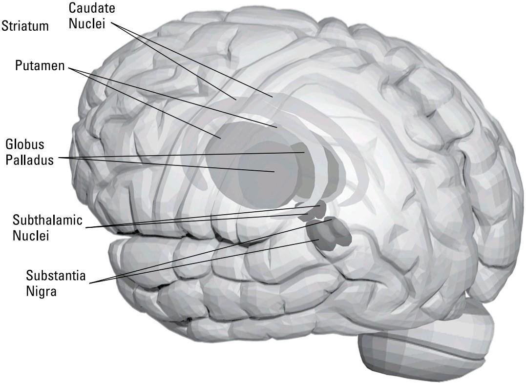
FIGURE 2-5: The basal ganglia and cerebellum.
شکل ۲-۵: عقدههای قاعدهای و مخچه.
These nuclei comprise a highly interconnected system that interacts with the thalamus and neocortex to control behavior. The following list explains each one:
این هستهها شامل یک سیستم بسیار بههمپیوسته هستند که با تالاموس و نئوکورتکس برای کنترل رفتار در تعامل هستند. لیست زیر هر یک را توضیح میدهد:
» The caudate: Inputs to the basal ganglia from the neocortex primarily come into the caudate and another nucleus called the putamen. The caudate-putamen complex is now usually called the striatum. Anatomically, the caudate is a ring of cells that surrounds the basal ganglia complex except on the ventral side. The output of the caudate is to the globus palladus.
» هسته دمی: ورودیهای عقدههای قاعدهای از نئوکورتکس در درجه اول به هسته دمی و هسته دیگری به نام پوتامن وارد میشوند. مجموعه دمی-پوتامن اکنون معمولاً جسم مخطط نامیده میشود. از نظر آناتومیکی، هسته دمی حلقهای از سلولها است که مجموعه عقدههای قاعدهای را به جز در سمت شکمی احاطه میکند. خروجی هسته دمی به گلوبوس پالادوس است.
» The putamen: The putamen is a large nucleus within the basal ganglia complex with similar input-output connections as the caudate.
» پوتامن: پوتامن یک هسته بزرگ در داخل مجموعه عقدههای قاعدهای است که اتصالات ورودی-خروجی مشابه هسته دمی دارد.
» The globus palladus: The globus palladus is the primary output nucleus of the basal ganglia via its inhibitory connections to the thalamus. It is composed of an inner and outer segment.
» گلوبوس پالادوس: گلوبوس پالادوس هسته خروجی اولیه عقدههای قاعدهای از طریق اتصالات مهاری آن به تالاموس است. این هسته از یک بخش داخلی و خارجی تشکیل شده است.
» The substantia nigra: The substantia nigra is a midbrain nucleus that sends “enabling” signals to the basal ganglia through the striatum. The substantia nigra is well known for its involvement in Parkinson’s disease, which is caused by the death of dopaminergic neurons in this nucleus. Without these neurons, initiating voluntary movement or changing an ongoing movement sequence becomes difficult.
» جسم سیاه: جسم سیاه یک هسته مغز میانی است که سیگنالهای «توانمندساز» را از طریق جسم مخطط به عقدههای قاعدهای ارسال میکند. جسم سیاه به دلیل نقشش در بیماری پارکینسون که ناشی از مرگ نورونهای دوپامینرژیک در این هسته است، به خوبی شناخته شده است. بدون این نورونها، شروع حرکت ارادی یا تغییر توالی حرکتی مداوم دشوار میشود.
» The subthalamic nucleus: The subthalamic nucleus also modulates the globus palladus output. This nucleus is frequently the target of deep brain stimulation surgical implants to relieve symptoms of Parkinson’s disease.
» هسته ساب تالاموس: هسته ساب تالاموس همچنین خروجی گلوبوس پالادوس را تعدیل میکند. این هسته اغلب هدف ایمپلنتهای جراحی تحریک عمقی مغز برای تسکین علائم بیماری پارکینسون است.
Even basal ganglia experts readily admit the internal functional architecture of the basal ganglia is poorly understood at present. These nuclei are organized in a similar manner to the cerebellum, a phylogenetically old motor coordination center with which they interact. For more on the cerebellum, head to the later section “Coordinating movement: The cerebellum.”
حتی متخصصان عقدههای قاعدهای نیز به راحتی اذعان میکنند که معماری عملکردی داخلی عقدههای قاعدهای در حال حاضر به خوبی شناخته نشده است. این هستهها به شیوهای مشابه مخچه، یک مرکز هماهنگی حرکتی قدیمی از نظر فیلوژنتیکی که با آن تعامل دارند، سازماندهی شدهاند. برای اطلاعات بیشتر در مورد مخچه، به بخش بعدی «هماهنگی حرکت: مخچه» مراجعه کنید.
Transitioning between the brain and the spinal cord
The next three brain areas I cover — the midbrain, the pons and medulla, and the cerebellum — make the transition between the brain and spinal cord. The mid- brain, pons, and medulla together constitute the brain stem. These areas are phy- logenetically very old, and they’re found in all vertebrates. They control basic behaviors such as locomotion coordination, eye movements, and regulation of body homeostasis such as respiration, heart rate, and temperature. The following sections have the details.
انتقال بین مغز و نخاع
سه ناحیه مغزی بعدی که من پوشش میدهم – مغز میانی، پل مغزی و بصل النخاع، و مخچه – انتقال بین مغز و نخاع را انجام میدهند. مغز میانی، پل مغزی و بصل النخاع با هم ساقه مغز را تشکیل میدهند. این نواحی از نظر فیلوژنتیکی بسیار قدیمی هستند و در همه مهرهداران یافت میشوند. آنها رفتارهای اساسی مانند هماهنگی حرکتی، حرکات چشم و تنظیم هموستاز بدن مانند تنفس، ضربان قلب و دما را کنترل میکنند. بخشهای بعدی جزئیات را ارائه میدهند.
The midbrain
The most superficial of these three areas is called the midbrain. The midbrain con- tains low-level processors that control eye movements and help localize sound.
مغز میانی
سطحیترین قسمت از این سه ناحیه، مغز میانی نامیده میشود. مغز میانی شامل پردازندههای سطح پایینی است که حرکات چشم را کنترل کرده و به مکانیابی صدا کمک میکنند.
The major visual area in the midbrain is the superior colliculus, which controls eye movements called saccades. The superior colliculus receives projections from about one-tenth of all retinal ganglion cells in humans and a much higher percentage in other mammals. In non-mammalian vertebrates such as frogs, the superior col- liculus (called the tectum in frogs and other non-mammals) is the major visual target of the retina, and it’s the crucial coordinating area for most visually guided behavior.
ناحیه اصلی بینایی در مغز میانی، کالیکولوس فوقانی است که حرکات چشم به نام ساکاد را کنترل میکند. کالیکولوس فوقانی، تصاویر را از حدود یک دهم کل سلولهای گانگلیونی شبکیه در انسان و درصد بسیار بالاتری را در سایر پستانداران دریافت میکند. در مهرهداران غیرپستاندار مانند قورباغهها، کالیکولوس فوقانی (که در قورباغهها و سایر غیرپستانداران تکتوم نامیده میشود) هدف اصلی بینایی شبکیه است و ناحیه هماهنگکننده حیاتی برای اکثر رفتارهای هدایتشده بصری است.
Research at MIT in the late 1950s showed that certain ganglion cells in the frog eye responded only to small moving objects the size of bugs the frog preys on — bug detectors. Electrical stimulation in the frog’s tectum caused frogs or toads to snap their tongues at imaginary bugs in the visual field that projected to the tectal area stimulated. In mammals such as humans, the superior colliculus primarily controls eye movements; it leaves the control of visually guided behavior, like hit- ting tennis balls, to the thalamic neocortical system, particularly in the parietal lobe.
تحقیقات در MIT در اواخر دهه 1950 نشان داد که سلولهای گانگلیونی خاصی در چشم قورباغه فقط به اشیاء متحرک کوچک به اندازه حشراتی که قورباغه شکار میکند – آشکارسازهای حشرات – پاسخ میدهند. تحریک الکتریکی در تکتوم قورباغه باعث میشد قورباغهها یا وزغها زبان خود را به حشرات خیالی در میدان بینایی که به ناحیه تکتوم تحریک شده تابانده میشوند، بچسبانند. در پستاندارانی مانند انسان، کالیکولوس فوقانی در درجه اول حرکات چشم را کنترل میکند. این روش، کنترل رفتارهای هدایتشده بصری، مانند ضربه زدن به توپ تنیس، را به سیستم نئوکورتیکال تالاموس، بهویژه در لوب آهیانه، واگذار میکند.
Auditory fibers from the inner ear project to a processing area just below the superior colliculus, called, for a rather obvious reason, the inferior colliculus. Neu- rons in this structure project to neurons in the auditory part of the thalamus, which in turn projects to the auditory part of the neocortex on the superior aspect of the temporal lobe.
فیبرهای شنوایی از گوش داخلی به ناحیه پردازش درست زیر کولیکولوس فوقانی، که به دلیلی نسبتاً واضح، کولیکولوس تحتانی نامیده میشود، پرتاب میشوند. نورونهای این ساختار به نورونهای بخش شنوایی تالاموس پرتاب میشوند، که به نوبه خود به بخش شنوایی نئوکورتکس در سمت فوقانی لوب گیجگاهی پرتاب میشوند.
The midbrain also includes one of the nuclei associated with the basal ganglia, the substantia nigra (refer to Figure 2-5). Other nuclei in the midbrain, such as the locus coeruleus, Raphe, and ventral tegmental areas, make extensive but diffuse modulatory projections throughout the neocortex. This means they make only a few out of the thousands of synapses on specific target cells, thereby contributing small, modulatory excitation or inhibition to large numbers of neurons in many brain areas rather than being the major cause of the firing in a small number of target neurons.
مغز میانی همچنین شامل یکی از هستههای مرتبط با عقدههای قاعدهای، جسم سیاه (به شکل 2-5 مراجعه کنید) است. هستههای دیگر در مغز میانی، مانند لوکوس سرولئوس، رافه و نواحی تگمنتوم شکمی، برآمدگیهای تعدیلی گسترده اما پراکندهای را در سراسر نئوکورتکس ایجاد میکنند. این بدان معناست که آنها تنها تعداد کمی از هزاران سیناپس روی سلولهای هدف خاص را ایجاد میکنند و در نتیجه به جای اینکه عامل اصلی شلیک در تعداد کمی از نورونهای هدف باشند، تحریک یا مهار تعدیلی کوچکی را برای تعداد زیادی از نورونها در بسیاری از نواحی مغز ایجاد میکنند.
The reticular formation
An important brain area that courses throughout much of the subcortex including the midbrain is the reticular formation. The word reticular is derived from the Latin word for “net.” The reticular formation is not so much a defined structure as it is a continuous network that extends through and interacts with numerous brain areas. The reticular formation extends through the midbrain and pons and medulla and is continuous with reticular “zones” above the midbrain and below the medulla into the spinal cord. The reticular formation controls all life functions, such as heart rate, respiration, temperature, and even wakefulness. Damage to the reticular formation typically results in coma or death.
تشکیلات مشبک
یک ناحیه مهم مغز که در بیشتر زیر قشر مغز از جمله مغز میانی امتداد دارد، تشکیلات مشبک است. کلمه مشبک از کلمه لاتین به معنای “شبکه” گرفته شده است. تشکیلات مشبک ساختار چندان مشخصی نیست، بلکه یک شبکه پیوسته است که از طریق مغز میانی و پل مغزی و بصل النخاع امتداد یافته و با نواحی متعدد مغز در تعامل است. تشکیلات مشبک از طریق مغز میانی و پل مغزی و بصل النخاع امتداد دارد و با “مناطق” مشبک بالای مغز میانی و زیر بصل النخاع تا نخاع امتداد دارد. تشکیلات مشبک تمام عملکردهای حیاتی مانند ضربان قلب، تنفس، دما و حتی بیداری را کنترل میکند. آسیب به تشکیلات مشبک معمولاً منجر به کما یا مرگ میشود.
Processing the basics below the midbrain: The pons and medulla
The pons, between the midbrain and medulla in the brain stem (see Figure 2-1), contains nuclei that mediate several auditory and balance functions. Auditory functions include neural comparisons of the loudness and timing between the two ears for auditory localization (in cooperation with the inferior colliculus in the midbrain). Some pons nuclei receive inputs from the vestibular system in the inner ear (the semicircular canals) and communicate with the cerebellum for balance. Damage to these pons nuclei or to the vestibular system can produce not only chronic dizziness but even the inability to balance while standing up. The pons receives sensory input from the face and sends motor neurons to facial mus- cles to control both voluntary and involuntary facial expressions via the trigemi- nal cranial nerve V.
پردازش اصول اولیه زیر مغز میانی: پل مغزی و بصل النخاع
پل مغزی، بین مغز میانی و بصل النخاع در ساقه مغز (به شکل 2-1 مراجعه کنید)، حاوی هستههایی است که واسطه چندین عملکرد شنوایی و تعادلی هستند. عملکردهای شنوایی شامل مقایسه عصبی بلندی و زمانبندی بین دو گوش برای مکانیابی شنوایی (با همکاری کولیکولوس تحتانی در مغز میانی) است. برخی از هستههای پل مغزی ورودیهایی از سیستم دهلیزی در گوش داخلی (مجاری نیمدایرهای) دریافت میکنند و برای تعادل با مخچه ارتباط برقرار میکنند. آسیب به این هستههای پل مغزی یا سیستم دهلیزی میتواند نه تنها سرگیجه مزمن، بلکه حتی ناتوانی در حفظ تعادل هنگام ایستادن را نیز ایجاد کند. پل مغزی ورودی حسی را از صورت دریافت میکند و نورونهای حرکتی را به عضلات صورت میفرستد تا حالات چهره ارادی و غیرارادی را از طریق عصب جمجمهای سه قلو V کنترل کنند.
Below the pons is the most ventral area of the brain stem called the medulla. The lower border of the medulla is the top of the spinal cord. Much of the medulla consists of neural fiber tracts (called pyramids, because of their shape in cross section) that relay sensory information from the skin, muscles, and tendons to the brain, and motor command information from the primary motor cortex in the frontal lobe going down into the spinal cord to control the body’s muscles via their synapses on lower motor neurons that project from the spinal cord to muscles. Two areas in the medulla, called the gracile and cuneate nuclei, integrate the ascending and descending information. Like the pons, some sensory inputs from the mouth (including neurons from the tongue containing taste information), face, and throat also synapse in the medulla.
در زیر پل مغزی، شکمیترین ناحیه ساقه مغز به نام بصلالنخاع (مدولا) قرار دارد. مرز پایینی بصلالنخاع، بالای نخاع است. بخش عمدهای از بصلالنخاع از رشتههای عصبی (به دلیل شکل مقطع عرضیشان، هرم نامیده میشوند) تشکیل شده است که اطلاعات حسی را از پوست، عضلات و تاندونها به مغز و اطلاعات فرمان حرکتی را از قشر حرکتی اولیه در لوب پیشانی که به نخاع میروند، منتقل میکنند تا عضلات بدن را از طریق سیناپسهایشان روی نورونهای حرکتی تحتانی که از نخاع به عضلات امتداد دارند، کنترل کنند. دو ناحیه در بصلالنخاع، به نام هستههای گراسیل و کونئات، اطلاعات صعودی و نزولی را ادغام میکنند. مانند پل مغزی، برخی از ورودیهای حسی از دهان (از جمله نورونهای زبان حاوی اطلاعات چشایی)، صورت و گلو نیز در بصلالنخاع سیناپس میدهند.
Coordinating movement: The cerebellum
The cerebellum (“little brain”) is a complex motor-coordination structure that, by some estimates, contains as many neurons as the entire rest of the nervous system. Like the neocortex, the cerebellum is heavily convoluted with gyri and sulci. It is one of the phylogenetically oldest parts of the brain, so all vertebrates have a cerebellum, even including cartilaginous fish such as sharks. The cerebel- lum helps learn and control the timing of motor sequences. Damage to the cere- bellum produces slow, clumsy, robotlike movement.
هماهنگی حرکت: مخچه
مخچه (“مغز کوچک”) یک ساختار پیچیده هماهنگی حرکتی است که طبق برخی تخمینها، به اندازه کل سیستم عصبی، نورون دارد. مانند نئوکورتکس، مخچه به شدت با چین و شکنجها و شیارها پیچیده شده است. این یکی از قدیمیترین بخشهای مغز از نظر فیلوژنتیکی است، بنابراین همه مهرهداران، حتی از جمله ماهیهای غضروفی مانند کوسهها، دارای مخچه هستند. مخچه به یادگیری و کنترل زمانبندی توالیهای حرکتی کمک میکند. آسیب به مخچه باعث حرکت آهسته، ناشیانه و رباتمانند میشود.
The cerebellum extends out from the pons (refer to Figure 2-1), even though most of its connections are to the motor cortex via the thalamus. Structurally, the cer- ebellum is organized with an outer cortex, internal white matter (axon tracts), and four pairs of nuclei deep within it. Most inputs and outputs of the cerebellumoccur through the deep nuclei. This activity is modulated by the connections between these nuclei and cortical motor processing areas. Most output of the cerebellum modulates cortically generated ongoing motor control to achieve smooth, fine, feedback-adjusted movements. The cerebellar output inhibits parallel-activated inappropriate motor sequences and excites a small subset sequence appropriate for the current action.
مخچه از پل مغزی (به شکل 2-1 مراجعه کنید) امتداد مییابد، اگرچه بیشتر اتصالات آن از طریق تالاموس به قشر حرکتی است. از نظر ساختاری، مخچه با یک قشر بیرونی، ماده سفید داخلی (آکسون تراکتها) و چهار جفت هسته در اعماق آن سازماندهی شده است. بیشتر ورودیها و خروجیهای مخچه از طریق هستههای عمقی رخ میدهند. این فعالیت توسط اتصالات بین این هستهها و نواحی پردازش حرکتی قشر مغز تنظیم میشود. بیشتر خروجی مخچه، کنترل حرکتی مداوم ایجاد شده توسط قشر مغز را برای دستیابی به حرکات روان، ظریف و تنظیمشده با بازخورد تنظیم میکند. خروجی مخچه، توالیهای حرکتی نامناسب فعالشده موازی را مهار میکند و یک توالی زیرمجموعه کوچک مناسب برای عمل فعلی را تحریک میکند.
The structure of the cerebellum is similar to the structure of and interactions between the deep thalamus and more superficial and extensive neocortex in the larger part of the brain. Refer to the earlier section “The neocortex: Controlling the controllers” for more information about the neocortex.
ساختار مخچه مشابه ساختار و تعاملات بین تالاموس عمقی و نئوکورتکس سطحیتر و گستردهتر در بخش بزرگتر مغز است. برای اطلاعات بیشتر در مورد نئوکورتکس به بخش قبلی «نئوکورتکس: کنترل کنترلکنندهها» مراجعه کنید.
Looking at differences: Size, structure, and other variations
Up to this point, I’ve been talking about the human brain as though all human brains were identical. But they’re not. So the question then becomes how much variation is there between people, and is this variation important? The following sections delve into some of the differences.
نگاهی به تفاوتها: اندازه، ساختار و سایر تغییرات
تا این لحظه، من در مورد مغز انسان طوری صحبت میکردم که انگار مغز همه انسانها یکسان است. اما اینطور نیست. بنابراین سوال این است که چقدر تفاوت بین افراد وجود دارد و آیا این تفاوت مهم است؟ بخشهای بعدی به برخی از این تفاوتها میپردازند.
Mine’s bigger than yours!
One obvious difference in brains is size. An average size brain is about 1,300 cc, about the size of a large grapefruit. Among adults, a 10 percent variation around that norm is common, which means the brains of many people are up to 10 percent larger or smaller than the standard 1,300 cc.
مال من از مال تو بزرگتره!
یکی از تفاوتهای آشکار در مغز، اندازه آن است. حجم متوسط مغز حدود ۱۳۰۰ سیسی است، تقریباً به اندازه یک گریپفروت بزرگ. در بین بزرگسالان، اختلاف ۱۰ درصدی در حدود این هنجار رایج است، به این معنی که مغز بسیاری از افراد تا ۱۰ درصد بزرگتر یا کوچکتر از اندازه استاندارد ۱۳۰۰ سیسی است.
Brain size tends to scale with body size, and thus large men tend to have larger brains than small women. How important is this variation? Not very, apparently. There are many documented cases of geniuses with rather small brains, and, of course, we’ve all encountered lots of dimwits with rather large brains (or at least, large heads, which is what we can observe).
اندازه مغز با اندازه بدن متناسب است و بنابراین مردان بزرگ معمولاً مغزهای بزرگتری نسبت به زنان کوچک دارند. این اختلاف چقدر مهم است؟ ظاهراً خیلی مهم نیست. موارد مستند زیادی از نوابغ با مغزهای نسبتاً کوچک وجود دارد و البته، همه ما با تعداد زیادی از افراد کودن با مغزهای نسبتاً بزرگ (یا حداقل سرهای بزرگ، که چیزی است که میتوانیم مشاهده کنیم) مواجه شدهایم.
It’s what’s inside that counts: Looking at differences in structural organization
What about the structural organization of the brain? Everyone has more or less the same major gyri and sulci, but there are variations. In fact, if you look carefully at any real brain, you can see that the two hemispheres are rarely exact mirror images. (That doesn’t mean, however, that you can tell whether the person is right- or left-handed by looking at the two hemispheres.)
آنچه در درون است اهمیت دارد: نگاه به تفاوتها در سازماندهی ساختاری
در مورد سازماندهی ساختاری مغز چطور؟ همه کم و بیش چینهای اصلی و شیارهای مغزی یکسانی دارند، اما تفاوتهایی وجود دارد. در واقع، اگر با دقت به هر مغز واقعی نگاه کنید، میتوانید ببینید که دو نیمکره به ندرت تصاویر آینهای دقیقی هستند. (با این حال، این بدان معنا نیست که با نگاه کردن به دو نیمکره میتوانید بگویید که فرد راست دست است یا چپ دست.)
In fact, this amount of variation between hemispheres is common between brains as well. This is one of the reasons why epilepsy surgeons functionally map brain areas around the area they’re targeting: They need to make sure they’re not removing crucial areas, such as those that control language. (The other reason for functional mapping is that the pathology itself in the epileptic brain may have caused some displacement of function away from the normal areas.)
در واقع، این میزان از تنوع بین نیمکرهها، بین مغزها نیز رایج است. این یکی از دلایلی است که جراحان صرع، نواحی مغز را در اطراف ناحیهای که هدف قرار میدهند، به صورت عملکردی نقشهبرداری میکنند: آنها باید مطمئن شوند که نواحی حیاتی، مانند نواحی کنترلکننده زبان، را حذف نمیکنند. (دلیل دیگر برای نقشهبرداری عملکردی این است که خود آسیبشناسی در مغز مبتلا به صرع ممکن است باعث جابجایی عملکرد از نواحی طبیعی شده باشد.)
What is not observable at the level of the overall structure of the brain is its inter- nal wiring.
آنچه در سطح ساختار کلی مغز قابل مشاهده نیست، سیمکشی داخلی آن است.
Considering gender-based brain differences
Although the overall structure and detailed connectivity of male and female brains are nearly identical, some recent research has revealed male versus female differ- ences in what might be called “styles” of processing, with men preferring big picture, maplike ways of organizing knowledge and woman preferring a more linear, procedural system of knowledge. Ask a man for directions, for example, and he tends to tell you the general layout of where you are in relation to where you are trying to go, whereas a woman tends to tell you the sequence of turns you need to get there. Yes, Virginia, we really do think and speak in different lan- guages, sometimes.
با توجه به تفاوتهای مغزی مبتنی بر جنسیت
اگرچه ساختار کلی و اتصال دقیق مغز مردان و زنان تقریباً یکسان است، برخی تحقیقات اخیر تفاوتهای مردان در مقابل زنان را در چیزی که میتوان آن را «سبکهای» پردازش نامید، آشکار کرده است، به طوری که مردان روشهای کلی و نقشهوار سازماندهی دانش را ترجیح میدهند و زنان یک سیستم دانش خطیتر و رویهایتر را ترجیح میدهند. به عنوان مثال، از یک مرد مسیر را بپرسید، او تمایل دارد طرح کلی جایی که هستید را در رابطه با جایی که میخواهید بروید به شما بگوید، در حالی که یک زن تمایل دارد توالی چرخشهایی را که برای رسیدن به آنجا نیاز دارید به شما بگوید. بله، ویرجینیا، ما واقعاً گاهی اوقات به زبانهای مختلفی فکر میکنیم و صحبت میکنیم.
The following section discusses some of the controversial hypotheses related to differences between male and female brains.
بخش زیر برخی از فرضیههای بحثبرانگیز مربوط به تفاوتهای بین مغز مردان و زنان را مورد بحث قرار میدهد.
WHICH PART OF YOUR BRAIN DO YOU USE? LATERALIZATION DIFFERENCES
One apparent (but controversial) difference that has been suggested is that the amount of specialization between the two hemispheres (lateralization) is different between male and female brains. Brain scans tend to show that women use more, and more symmetrical, brain areas than men do for the same tasks. Women also tend to have a higher percentage of connecting fibers in the corpus callosum, the fiber tract that links the two hemispheres together.
از کدام بخش مغز خود استفاده میکنید؟ تفاوتهای جانبیسازی
یکی از تفاوتهای آشکار (اما بحثبرانگیز) که مطرح شده این است که میزان تخصص بین دو نیمکره (جانبیسازی) بین مغز مردان و زنان متفاوت است. اسکنهای مغزی نشان میدهند که زنان برای انجام وظایف یکسان، از نواحی مغزی بیشتر و متقارنتری نسبت به مردان استفاده میکنند. زنان همچنین درصد بالاتری از فیبرهای اتصالدهنده در جسم پینهای، یعنی رشتهای که دو نیمکره را به هم متصل میکند، دارند.
The general idea is that men’s brains are more lateralized than females, meaning that functions tend to be more restricted to one hemisphere in men than in women.
ایده کلی این است که مغز مردان نسبت به زنان، جانبیتر است، به این معنی که عملکردها در مردان بیشتر از زنان به یک نیمکره محدود میشود.
The correlate of this hypothesis is that men’s brains are therefore more special- ized, particularly with respect to spatial processing in the right hemisphere. The upside for men, according to this idea, is that (in general, not always) they may be able to process spatial, right-hemisphere tasks in some sense more deeply because of lateral specialization. The downside is that they tend to be more disabled by strokes or lesions that affect only one side of the brain because there is less redun- dancy in the representation for that ability in the mirror image location in the other hemisphere.
همبستگی این فرضیه این است که بنابراین مغز مردان، به ویژه در رابطه با پردازش فضایی در نیمکره راست، تخصصیتر است. طبق این ایده، نکته مثبت برای مردان این است که (به طور کلی، نه همیشه) ممکن است به دلیل تخصص جانبی، بتوانند وظایف فضایی نیمکره راست را به نوعی عمیقتر پردازش کنند. نکته منفی این است که آنها در اثر سکته مغزی یا ضایعاتی که فقط یک طرف مغز را تحت تأثیر قرار میدهند، بیشتر دچار ناتوانی میشوند، زیرا در نمایش آن توانایی در محل تصویر آینه در نیمکره دیگر، افزونگی کمتری وجود دارد.
It has also been suggested that not only is autism much more common in males that females, but that autism itself can be regarded as a kind of “ultra-male” brain configuration with savant skills in certain kinds of complex processing accompanied by poor language and social development.
همچنین پیشنهاد شده است که اوتیسم نه تنها در مردان بسیار شایعتر از زنان است، بلکه خود اوتیسم را میتوان نوعی پیکربندی مغزی «فوق مردانه» با مهارتهای ساوانت در انواع خاصی از پردازشهای پیچیده همراه با زبان و توسعه اجتماعی ضعیف دانست.
The lateralization theory explains women’s general superiority in language skills compared to men in this way: Less specialization allows both hemispheres in females’ brains to process language.
نظریه جانبیسازی، برتری کلی زنان در مهارتهای زبانی در مقایسه با مردان را به این شکل توضیح میدهد: تخصص کمتر به هر دو نیمکره مغز زنان اجازه میدهد تا زبان را پردازش کنند.
The issue is far from resolved. Some propose that the observed male/female differences in brain activation and skills are entirely culturally based and have nothing to do with any intrinsic differences in brain structure of organization. Others — usually those who argue more for biologically based differences — suggest that male versus female brains are differentially susceptible during development to lateralization of language versus spatial skills.
این مسئله به هیچ وجه حل نشده است. برخی پیشنهاد میکنند که تفاوتهای مشاهده شده مرد/زن در فعالسازی و مهارتهای مغز کاملاً مبتنی بر فرهنگ است و هیچ ارتباطی با هیچ تفاوت ذاتی در ساختار سازماندهی مغز ندارد. برخی دیگر – معمولاً کسانی که بیشتر به تفاوتهای مبتنی بر زیستشناسی استدلال میکنند – معتقدند که مغز مردان در مقابل زنان در طول رشد به طور متفاوتی مستعد جانبیسازی مهارتهای زبانی در مقابل مهارتهای فضایی است.
HOW BRAIN DEVELOPMENT PLAYS INTO THE DEBATE
One thing making it even more difficult to evaluate any of these hypotheses is that experience clearly changes brain development. Therefore, gender-differentiating cultural experience may modify development to produce true male/female biological differences in adult brains. As you can imagine, this little controversy has generated an industry of Ph.D. dissertations and journal papers that keeps many cognitive scientists on both sides of the issue gainfully productive.
نقش رشد مغز در این بحث
چیزی که ارزیابی هر یک از این فرضیهها را دشوارتر میکند این است که تجربه به وضوح رشد مغز را تغییر میدهد. بنابراین، تجربه فرهنگیِ تمایز جنسیتی ممکن است رشد را تغییر دهد تا تفاوتهای بیولوژیکی واقعی مردانه/زنانه را در مغز بزرگسالان ایجاد کند. همانطور که میتوانید تصور کنید، این جنجال کوچک، صنعتی از پایاننامههای دکترا و مقالات ژورنالی ایجاد کرده است که بسیاری از دانشمندان شناختی را در هر دو طرف این موضوع به طور سودمندی پربار نگه میدارد.
What mechanisms could produce gender-based lateralization differences during development? There appear to be developmental and operational effects on the brain from the hormones testosterone, estrogen, and oxytocin. Artificial testos- terone injections tend to produce malelike behavior and proclivities in females, whereas estrogen does the opposite in males. Oxytocin, a hormone released during the birth process in females, has been suggested to mediate a kind of “mother bear” behavior, with high nurturing toward those within the clan but high aggression against those outside.
چه مکانیسمهایی میتوانند تفاوتهای جانبی مبتنی بر جنسیت را در طول رشد ایجاد کنند؟ به نظر میرسد اثرات رشدی و عملیاتی بر مغز از هورمونهای تستوسترون، استروژن و اکسیتوسین وجود دارد. تزریق تستوسترون مصنوعی تمایل به ایجاد رفتار و تمایلات مردانه در زنان دارد، در حالی که استروژن در مردان برعکس عمل میکند. گفته میشود اکسیتوسین، هورمونی که در طول فرآیند تولد در زنان آزاد میشود، نوعی رفتار “خرس مادر” را ایجاد میکند، با مراقبت زیاد نسبت به افراد درون قبیله اما پرخاشگری زیاد نسبت به افراد خارج از قبیله.
The Spinal Cord: The Intermediary between Nervous Systems
The spinal cord is a key part of the central nervous system. Specifically, within the spinal cord are the connections between the central nervous system (such as motor neurons from primary motor cortex) and the peripheral nervous system (skin, muscle, and tendon receptors going to the spinal cord, and lower or alpha motor neurons relaying motor commands from the spinal cord to the actual muscles). The sensory neurons and lower motor neuron axons constitute the peripheral nervous system, which mediate voluntary behavior and sensing.
نخاع: واسطه بین سیستمهای عصبی
نخاع بخش کلیدی سیستم عصبی مرکزی است. به طور خاص، در داخل نخاع، ارتباطاتی بین سیستم عصبی مرکزی (مانند نورونهای حرکتی از قشر حرکتی اولیه) و سیستم عصبی محیطی (گیرندههای پوست، عضله و تاندون که به نخاع میروند و نورونهای حرکتی تحتانی یا آلفا که دستورات حرکتی را از نخاع به عضلات واقعی منتقل میکنند) وجود دارد. نورونهای حسی و آکسونهای نورون حرکتی تحتانی، سیستم عصبی محیطی را تشکیل میدهند که واسطه رفتار ارادی و حس کردن هستند.
The spinal cord also has integration and coordination functions, although it is close to the lowest level of the hierarchy of controllers. It mediates feedback between sensory and motor pathways for each limb and coordinates limb move- ments for locomotion.
نخاع همچنین دارای عملکردهای ادغام و هماهنگی است، اگرچه نزدیک به پایینترین سطح سلسله مراتب کنترلکنندهها است. این سیستم، بازخورد بین مسیرهای حسی و حرکتی را برای هر اندام واسطهگری میکند و حرکات اندام را برای حرکت هماهنگ میکند.
FINDING CENTRAL PATTERN GENERATORS
Some of the earliest neurological experiments by scientists like Sir Charles Scott Sherrington demonstrated the existence of what are called central pattern generators in the spinal cords of four-legged animals like cats. The central pattern generator is a neural circuit totally within the spinal cord that can control alternation of left and right hind or forelimbs and proper phasing between forelimbs and hind limbs, although this circuit can be overridden or otherwise controlled by central mechanisms.
یافتن مولدهای الگوی مرکزی
برخی از اولین آزمایشهای عصبی توسط دانشمندانی مانند سر چارلز اسکات شرینگتون، وجود چیزی را که مولدهای الگوی مرکزی نامیده میشوند، در نخاع حیوانات چهارپا مانند گربهها نشان داد. مولد الگوی مرکزی، یک مدار عصبی است که کاملاً درون نخاع قرار دارد و میتواند تناوب اندامهای عقبی یا جلویی چپ و راست و همچنین فازبندی مناسب بین اندامهای جلویی و عقبی را کنترل کند، اگرچه این مدار میتواند توسط مکانیسمهای مرکزی نادیده گرفته شود یا به روش دیگری کنترل شود.
Forelimb/hind limb phasing is different for different gaits in four-legged animals. In normal four-legged walking, for example, the two forelimbs are in antiphase with each other, as are the two hindlimbs, and the fore- and hindlimbs on each side are anti- phased, so that as the left forelimb goes back, the left hindlimb goes forward. Other gaits, like the trot, gallop, or canter, have different phase relationships.
فازبندی اندام جلویی/عقبی برای گامهای مختلف در حیوانات چهارپا متفاوت است. به عنوان مثال، در راه رفتن طبیعی چهارپا، دو اندام جلویی در فاز مخالف یکدیگر قرار دارند، همانطور که دو اندام عقبی نیز همینطور هستند، و اندامهای جلویی و عقبی در هر طرف در فاز مخالف یکدیگر هستند، به طوری که با عقب رفتن اندام جلویی چپ، اندام عقبی چپ به جلو میرود. سایر گامها، مانند یورتمه، تاخت یا تاز، روابط فازی متفاوتی دارند.
By severing the connections between the brain and spinal cord in experimental animals, Sherrington showed that these gaits could be organized by the spinal cord itself in the absence of any brain control, when the animals were placed on a treadmill. The brain is needed, of course, to decide whether to walk, trot, run, hop, or turn in normal behavior.
شرینگتون با قطع ارتباط بین مغز و نخاع در حیوانات آزمایشگاهی، نشان داد که این گامها میتوانند در غیاب هرگونه کنترل مغزی، هنگامی که حیوانات روی تردمیل قرار میگیرند، توسط خود نخاع سازماندهی شوند. البته مغز برای تصمیمگیری در مورد راه رفتن، یورتمه، دویدن، پریدن یا چرخش در رفتار طبیعی مورد نیاز است.
Each level of the spinal cord contains a local processing module for the area of the body controlled from that segment, plus connections to other spinal cord seg- ments and to and from the brain (through the medulla all the way to neocortex). The top of the spinal cord deals with muscles and sensory information from the neck, while the bottom spinal cord segments deal with the toes. The spinal cord segments are designated as 8 cervical, 12 thoracic, 5 lumbar, 5 sacral, and 3 coccygeal. These are numbered from the top down, so the highest cervical spinal segment is C1, while the lowest sacral segment is S5.
هر سطح از نخاع شامل یک ماژول پردازش محلی برای ناحیهای از بدن است که از آن بخش کنترل میشود، به علاوه ارتباطاتی با سایر بخشهای نخاع و به و از مغز (از طریق بصل النخاع تا نئوکورتکس). بالای نخاع با عضلات و اطلاعات حسی از گردن سروکار دارد، در حالی که بخشهای پایینی نخاع با انگشتان پا سروکار دارند. بخشهای نخاع به صورت ۸ بخش گردنی، ۱۲ بخش سینهای، ۵ بخش کمری، ۵ بخش خاجی و ۳ بخش دنبالچهای مشخص شدهاند. این بخشها از بالا به پایین شمارهگذاری میشوند، بنابراین بالاترین بخش نخاع گردنی C1 و پایینترین بخش خاجی S5 است.
Looking at the spinal reflex
The fundamental unit of coordinated action mediated by the spinal cord is the spinal reflex; you can see the anatomy of this reflex in Figure 2-6. Striking the kneecap stretches the quadriceps muscle, as though one’s knees were buckling. Muscle spindle receptors in the quadriceps project to the spinal cord and activate alpha motor neurons that contract the muscle to maintain upright posture.
نگاهی به رفلکس نخاعی
واحد اساسی عمل هماهنگ که توسط نخاع انجام میشود، رفلکس نخاعی است؛ میتوانید آناتومی این رفلکس را در شکل 2-6 مشاهده کنید. ضربه زدن به کاسه زانو، عضله چهار سر ران را کشش میدهد، گویی زانوهای فرد در حال خم شدن هستند. گیرندههای دوک عضلانی در عضله چهار سر ران به نخاع متصل میشوند و نورونهای حرکتی آلفا را فعال میکنند که عضله را برای حفظ حالت ایستاده منقبض میکنند.
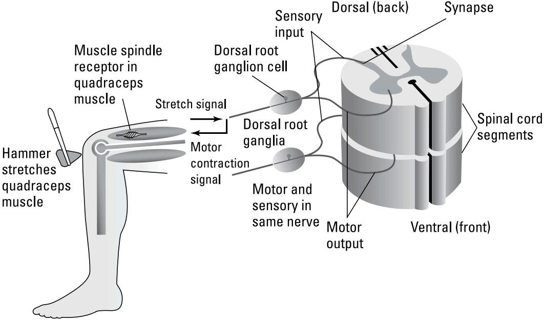
FIGURE 2-6: The spinal reflex.
شکل ۲-۶: رفلکس نخاعی.
Alpha (lower) motor neurons are the neurons that innervate muscle cells, which in turn move the limbs. These motor neurons have their cell bodies in the ventral (toward the stomach side of the body) part of the spinal cord. The axons that drive the muscles exit the spinal cord via a tract called the ventral root.
نورونهای حرکتی آلفا (پایینتر) نورونهایی هستند که سلولهای عضلانی را عصبدهی میکنند و به نوبه خود اندامها را حرکت میدهند. این نورونهای حرکتی، جسم سلولی خود را در قسمت شکمی (به سمت معده بدن) نخاع دارند. آکسونهایی که عضلات را هدایت میکنند، از طریق مسیری به نام ریشه شکمی از نخاع خارج میشوند.
On the dorsal side of the spinal cord are the axons of sensory cells, such as stretch receptors, that enter via the dorsal root.
در قسمت پشتی نخاع، آکسونهای سلولهای حسی، مانند گیرندههای کششی، قرار دارند که از طریق ریشه پشتی وارد میشوند.
When the doctor taps just below your kneecap with a rubber hammer, the tap slightly stretches the tendon from your knee to your leg, stretching the quadriceps muscle and spindle. The spinal circuit causes your leg to extend in compensation. This is a negative feedback system that maintains an upright stance without your conscious control. The clinical test checks the integrity of your peripheral nervous system (sensory and motor nerves acting through the spinal cord).
وقتی پزشک با یک چکش لاستیکی درست زیر کاسه زانوی شما ضربه میزند، ضربه، تاندون را از زانو تا پای شما کمی میکشد و عضله چهارسر ران و دوک عصبی را میکشد. مدار نخاعی باعث میشود پای شما در جبران، کشیده شود. این یک سیستم بازخورد منفی است که بدون کنترل آگاهانه شما، حالت ایستاده را حفظ میکند. این آزمایش بالینی، یکپارچگی سیستم عصبی محیطی شما (اعصاب حسی و حرکتی که از طریق نخاع عمل میکنند) را بررسی میکند.
What doesn’t Figure 2-6 show? It doesn’t show several pathways connecting a spinal cord segment to other segments, and to the brain:
شکل 2-6 چه چیزی را نشان نمیدهد؟ این شکل چندین مسیر را که یک بخش نخاع را به بخشهای دیگر و به مغز متصل میکند، نشان نمیدهد:
» The original message gets sent to the brain. The sensory input coming inthe dorsal root synapses on neurons in the spinal gray area that relay the sensory message all the way to the brain (somatosensory cortex, just poste- rior to the central sulcus).
» پیام اصلی به مغز ارسال میشود. ورودی حسی که از ریشه پشتی میآید، روی نورونهای ناحیه خاکستری نخاع سیناپس ایجاد میکند که پیام حسی را تا مغز (قشر حسی-پیکری، درست پشت شیار مرکزی) منتقل میکند.
» Command messages come down from the brain. Cortical (upper motor) neurons from the primary motor cortex come all the way down the spinal cord via upper motor neurons and synapse on the same alpha motor neurons that innervate muscles for the stretch reflex to allow you to voluntarily extend your leg.
» پیامهای فرمان از مغز به پایین میآیند. نورونهای قشری (حرکتی فوقانی) از قشر حرکتی اولیه از طریق نورونهای حرکتی فوقانی در نخاع پایین میآیند و روی همان نورونهای حرکتی آلفا که عضلات را برای رفلکس کششی عصبدهی میکنند تا به شما امکان دهند پای خود را به طور ارادی دراز کنید، سیناپس برقرار میکنند.
» Communication occurs between left and right portions of a segment and between segments. Motor events occurring in one segment send messages to other segments to coordinate body actions. If, for example, you were standing while your left leg started to buckle at the knee, your right leg would probably stiffen in compensation, and you would probably extend your left hand up, among other things. These connections also are involved in the central pattern generator control of gait mentioned previously.
» ارتباط بین بخشهای چپ و راست یک بخش و بین بخشها برقرار میشود. رویدادهای حرکتی که در یک بخش رخ میدهند، پیامهایی را به بخشهای دیگر میفرستند تا حرکات بدن را هماهنگ کنند. برای مثال، اگر شما ایستاده باشید و پای چپ شما از ناحیه زانو شروع به خم شدن کند، احتمالاً پای راست شما در جبران سفت میشود و احتمالاً دست چپ خود را به سمت بالا دراز میکنید، از جمله موارد دیگر. این اتصالات همچنین در کنترل مولد الگوی مرکزی راه رفتن که قبلاً ذکر شد، نقش دارند.
Getting your muscles moving
How do muscles work? Muscles are groups of cells connected to each other in long parallel chains the ends of which are connected to bones by tendons. The muscle cells contain chains of proteins called actin and myosin. When these proteins are stimulated, they slide over each other, causing the cell to contract along its length.
به حرکت درآوردن عضلات
عضلات چگونه کار میکنند؟ عضلات گروههایی از سلولها هستند که به صورت زنجیرههای موازی طولانی به یکدیگر متصل شدهاند و انتهای آنها توسط تاندونها به استخوانها متصل است. سلولهای عضلانی حاوی زنجیرههایی از پروتئینها به نام اکتین و میوزین هستند. هنگامی که این پروتئینها تحریک میشوند، روی یکدیگر میلغزند و باعث میشوند سلول در امتداد طول خود منقبض شود.
Alpha motor neurons stimulate muscles by releasing the motor neuron neuro- transmitter acetylcholine. When the acetylcholine is received by the receptors on the muscle cells, it causes a muscle cell action potential (an electrical pulse in the cell that causes it to momentarily contract) that causes the actin-myosin to slide (through the raising of intracellular calcium concentration), which in turn causes the contraction.
نورونهای حرکتی آلفا با آزاد کردن استیل کولین، انتقالدهنده عصبی نورون حرکتی، عضلات را تحریک میکنند. هنگامی که استیل کولین توسط گیرندههای روی سلولهای عضلانی دریافت میشود، باعث ایجاد پتانسیل عمل سلول عضلانی (یک پالس الکتریکی در سلول که باعث انقباض لحظهای آن میشود) میشود که باعث لغزش اکتین-میوزین (از طریق افزایش غلظت کلسیم درون سلولی) میشود، که به نوبه خود باعث انقباض میشود.
Fighting or Fleeing: The Autonomic Nervous System
The preceding sections outline the key components of the central nervous system (brain and spinal cord) and explain how the peripheral nervous system (motor and sensory nerves) gets in on the act. But when it comes to nervous systems, your body has more than just these two. There is another nervous system outside the central nervous system that controls not voluntary muscles as the peripheral nervous system does, but the heart, glands, and organs with smooth muscles (not under voluntary control) such as the intestines. This is the autonomic nervous system. It has sensory, motor, and gland-stimulating components.
مبارزه یا فرار: سیستم عصبی خودکار
بخشهای قبلی اجزای کلیدی سیستم عصبی مرکزی (مغز و نخاع) را شرح میدهند و توضیح میدهند که چگونه سیستم عصبی محیطی (اعصاب حرکتی و حسی) وارد عمل میشود. اما وقتی صحبت از سیستمهای عصبی میشود، بدن شما چیزی بیش از این دو سیستم عصبی دارد. یک سیستم عصبی دیگر خارج از سیستم عصبی مرکزی وجود دارد که نه عضلات ارادی مانند سیستم عصبی محیطی، بلکه قلب، غدد و اندامهایی با عضلات صاف (که تحت کنترل ارادی نیستند) مانند رودهها را کنترل میکند. این سیستم عصبی خودکار است. این سیستم دارای اجزای حسی، حرکتی و تحریککننده غدد است.
The autonomic nervous system consists of two divisions, the sympathetic and parasympathetic that often act in opposition to each other. The sympathetic sys- tem prepares the body for immediate action (fight or flight), at the expense of body regulating functions such as digestion. These two branches use two different neurotransmitters — norepinephrine (noradrenaline) for the sympathetic branch and acetylcholine for the parasympathetic branch — that tend to have opposite effects on the target organs. For example, norepinephrine speeds up the heart while acetylcholine slows it.
سیستم عصبی خودکار از دو بخش سمپاتیک و پاراسمپاتیک تشکیل شده است که اغلب در تضاد با یکدیگر عمل میکنند. سیستم سمپاتیک بدن را برای اقدام فوری (جنگ یا گریز) آماده میکند، به قیمت از دست رفتن عملکردهای تنظیمکننده بدن مانند هضم. این دو شاخه از دو انتقالدهنده عصبی مختلف استفاده میکنند – نوراپی نفرین (نورآدرنالین) برای شاخه سمپاتیک و استیل کولین برای شاخه پاراسمپاتیک – که تمایل دارند اثرات متضادی بر اندامهای هدف داشته باشند. به عنوان مثال، نوراپی نفرین قلب را تسریع میکند در حالی که استیل کولین آن را کند میکند.
Norepinephrine also dilates the pupil and lung bronchi, decreases digestive func- tions, and inhibits bladder contraction and blood flow to the genitals. Many of these actions are mediated through the adrenal medulla. However, overstimulation of the sympathetic system from excessive stress is hard on the body and tends to be associated with heart disease and other stress-related chronic illnesses. Because social conflicts can also trigger the sympathetic system, the chronic stress of modern life may result in sympathetic overstimulation and its associated long-term effects.
نوراپی نفرین همچنین مردمک چشم و برونشهای ریه را گشاد میکند، عملکردهای گوارشی را کاهش میدهد و انقباض مثانه و جریان خون به اندام تناسلی را مهار میکند. بسیاری از این اقدامات از طریق مدولای آدرنال انجام میشود. با این حال، تحریک بیش از حد سیستم سمپاتیک ناشی از استرس بیش از حد برای بدن سخت است و معمولاً با بیماریهای قلبی و سایر بیماریهای مزمن مرتبط با استرس مرتبط است. از آنجا که درگیریهای اجتماعی نیز میتوانند سیستم سمپاتیک را تحریک کنند، استرس مزمن زندگی مدرن ممکن است منجر به تحریک بیش از حد سمپاتیک و اثرات طولانی مدت مرتبط با آن شود.
There is also the enteric nervous system, which controls a number of homeostatic mechanisms such as blood flow and mucosal secretions. It also modulates endo- crine and immune functions.
همچنین سیستم عصبی رودهای وجود دارد که تعدادی از مکانیسمهای هموستاتیک مانند جریان خون و ترشحات مخاطی را کنترل میکند. همچنین عملکردهای غدد درون ریز و ایمنی را تعدیل میکند.
How We Know What We Know about Neural Activity
How do we know what we know about how the brain works? It’s not obvious at all upon looking at the brain that it is the seat of thought, consciousness, and body control. Some very prominent ancient scientists thought the brain’s purpose was to cool the body through the skull, while the heart was the seat of intelligence! Well, scientists have come a long way since then. The following sections outline some of the methods that have been — and continue to be — used to reveal the secrets of the brain and the nervous system.
چگونه میدانیم که در مورد فعالیت عصبی چه میدانیم
چگونه میدانیم که در مورد نحوه عملکرد مغز چه میدانیم؟ با نگاه کردن به مغز به هیچ وجه مشخص نیست که جایگاه تفکر، هوشیاری و کنترل بدن است. برخی از دانشمندان بسیار برجسته باستان تصور میکردند که هدف مغز خنک کردن بدن از طریق جمجمه است، در حالی که قلب جایگاه هوش است! خب، دانشمندان از آن زمان تاکنون راه درازی را پیموده اند. بخشهای زیر برخی از روشهایی را که برای آشکار کردن اسرار مغز و سیستم عصبی استفاده شده و همچنان استفاده میشود، شرح میدهد.
Examining problems caused by brain injuries
Probably the first clues about the function of the brain came from dysfunction associated with head injuries documented by the ancient Egyptians and Greeks. In the early 20th century, the scientific study of the brain began advancing signifi- cantly with the study of cytoarchitectonics (using stains to evaluate cell versus tract structure in various parts of the brain), and tract tracing, determining the connec- tions between one area of the brain and another by tracing the axonal processes of cells (I explain axons in Chapter 3). Early tract tracing often involved seeing where secondary degeneration occurred after damage to another brain area. Silver stains such as the Golgi stain allowed researchers to see the structure of individual neu- ral cells in great detail.
بررسی مشکلات ناشی از آسیبهای مغزی
احتمالاً اولین سرنخها در مورد عملکرد مغز از اختلال عملکرد مرتبط با آسیبهای سر که توسط مصریان و یونانیان باستان ثبت شده است، گرفته شده است. در اوایل قرن بیستم، مطالعه علمی مغز با مطالعه سیتوآرکتکتونیک (استفاده از رنگها برای ارزیابی ساختار سلول در مقابل ساختار مسیر عصبی در قسمتهای مختلف مغز) و ردیابی مسیر عصبی، که با ردیابی فرآیندهای آکسونی سلولها، ارتباط بین یک ناحیه از مغز و ناحیه دیگر را تعیین میکند (آکسونها را در فصل 3 توضیح میدهم)، پیشرفت قابل توجهی داشت. ردیابی اولیه مسیر عصبی اغلب شامل مشاهده محل وقوع دژنراسیون ثانویه پس از آسیب به ناحیه دیگری از مغز بود. رنگهای نقرهای مانند رنگآمیزی گلژی به محققان این امکان را داد تا ساختار سلولهای عصبی منفرد را با جزئیات کامل مشاهده کنند.
Using technology to image the brain: From early EEGs to today
On the physiological front, electroencephalographic (EEG) recordings in the early 20th century revealed that the brain was constantly producing electrical oscilla- tions that could be recorded from the surface of the skull. These oscillations changed with external stimulation and were associated with different brain states, such as sleep versus wakefulness. The EEG has a few deficiencies, though, which make it less than ideal for careful brain study.
استفاده از فناوری برای تصویربرداری از مغز: از EEG های اولیه تا امروز
در حوزه فیزیولوژیکی، ثبتهای الکتروانسفالوگرافی (EEG) در اوایل قرن بیستم نشان داد که مغز دائماً نوسانات الکتریکی تولید میکند که میتوان آنها را از سطح جمجمه ثبت کرد. این نوسانات با تحریک خارجی تغییر میکردند و با حالتهای مختلف مغز، مانند خواب در مقابل بیداری، مرتبط بودند. با این حال، EEG دارای چند نقص است که آن را برای مطالعه دقیق مغز ایدهآل نمیکند.
» The EEG averages activity over large areas so localizing the source of the recorded activity in the brain is hard. The averaging also tends to miss muchof the complexity of brain activity.
» EEG میانگین فعالیت را در نواحی وسیعی محاسبه میکند، بنابراین تعیین محل منبع فعالیت ثبت شده در مغز دشوار است. همچنین میانگینگیری تمایل دارد بخش زیادی از پیچیدگی فعالیت مغز را از دست بدهد.
» The intrinsic ongoing activity in the brain tends to be larger than — and tends to swamp — the smaller, transient signal elicited by particular stimuli, such as showing someone a picture, which makes the EEG a poor method to study the effect of stimuli on brain activity.
فعالیت ذاتی مداوم در مغز معمولاً بزرگتر از سیگنال کوچکتر و گذرای ناشی از محرکهای خاص، مانند نشان دادن یک تصویر به کسی، است – و تمایل دارد آن را در خود غرق کند – که این امر EEG را به روشی ضعیف برای مطالعه تأثیر محرکها بر فعالیت مغز تبدیل میکند.
This problem was solved to some extent by repeating the stimulus numerous times and recording each event so that, after hundreds of trials, the responses to the stimulus would be added together to increase the signal-to-noise ratio. Such recordings are generally called ERPs (Evoked Response Potentials). In the visual system, they are typically called VEPs (visual evoked potentials).
این مشکل تا حدودی با تکرار محرک به دفعات متعدد و ثبت هر رویداد حل شد، به طوری که پس از صدها آزمایش، پاسخها به محرک با هم جمع میشدند تا نسبت سیگنال به نویز افزایش یابد. چنین ثبتهایی عموماً ERP (پتانسیلهای پاسخ برانگیخته) نامیده میشوند. در سیستم بینایی، معمولاً VEP (پتانسیلهای برانگیخته بینایی) نامیده میشوند.
In the early to mid-20th century, microelectrodes began to be used in animal experiments to reveal how single neurons produced action potentials. In the visual system, for example, a researcher could follow the signal from the photoreceptor’s electrical response to light, through various cells in the retina, and on to the thalamus and visual cortex. Similarly, on the motor side, muscle action potentials, alpha motor neuron spikes, and motor cortex firing were recorded with micro- electrodes. However, because microelectrode recording is invasive (you have to insert microelectrode needles near the neurons to get a recording), it has had little or no applicability in humans.
در اوایل تا اواسط قرن بیستم، استفاده از میکروالکترودها در آزمایشهای حیوانی برای آشکار کردن چگونگی تولید پتانسیلهای عمل توسط نورونهای منفرد آغاز شد. به عنوان مثال، در سیستم بینایی، یک محقق میتوانست سیگنال را از پاسخ الکتریکی گیرنده نوری به نور، از طریق سلولهای مختلف در شبکیه و تا تالاموس و قشر بینایی دنبال کند. به طور مشابه، در سمت حرکتی، پتانسیلهای عمل عضله، اسپایکهای نورون حرکتی آلفا و شلیک قشر حرکتی با میکروالکترودها ثبت میشدند. با این حال، از آنجا که ثبت میکروالکترود تهاجمی است (برای ثبت باید سوزنهای میکروالکترود را در نزدیکی نورونها قرار دهید)، کاربرد کمی در انسان داشته یا اصلاً کاربرد نداشته است.
After the middle of the 20th century new imaging techniques, explained in the following sections, began to be commonly used.
پس از اواسط قرن بیستم، تکنیکهای جدید تصویربرداری، که در بخشهای بعدی توضیح داده میشوند، به طور رایج مورد استفاده قرار گرفتند.
PET and SPECT
PET stands for Positron Emission Tomography. In this technique, short-term weakly radioactive oxygen or sugar molecules are introduced into patients (or volunteers) while they perform some cognitive task. The most active neurons take up the radioactive substance because of their higher metabolism, and the locus of radio- activity in the brain is ascertained by a complex scanner and sophisticated soft- ware that detects the emission of positrons (anti-electrons).
PET و SPECT
PET مخفف توموگرافی انتشار پوزیترون است. در این تکنیک، مولکولهای اکسیژن یا قند با رادیواکتیو ضعیف کوتاهمدت به بیماران (یا داوطلبان) در حالی که برخی وظایف شناختی را انجام میدهند، وارد میشوند. فعالترین نورونها به دلیل متابولیسم بالاتر، ماده رادیواکتیو را جذب میکنند و محل فعالیت رادیواکتیو در مغز توسط یک اسکنر پیچیده و نرمافزار پیشرفتهای که انتشار پوزیترونها (ضد الکترونها) را تشخیص میدهد، مشخص میشود.
Typically a scan is done at “rest” — before the cognitive task is performed — and then compared with another scan done while the subject is doing the task. In this way, the difference between the two scans is assumed to be task associated. The PET technique has a spatial resolution of millimeters, but because it takes only one snapshot of brain activity, it can’t resolve brain activity changing in time.
معمولاً یک اسکن در حالت “استراحت” – قبل از انجام وظیفه شناختی – انجام میشود و سپس با اسکن دیگری که در حین انجام وظیفه توسط فرد انجام میشود، مقایسه میشود. به این ترتیب، فرض میشود که تفاوت بین دو اسکن مربوط به وظیفه است. تکنیک PET دارای وضوح مکانی میلیمتری است، اما از آنجا که فقط یک تصویر لحظهای از فعالیت مغز میگیرد، نمیتواند فعالیت مغز را در حال تغییر در زمان تشخیص دهد.
SPECT (Single Photon Emission Computed Tomography) is similar to PET except the ingested radioactive emitter releases gamma rays that are detected by the scanner.
SPECT (توموگرافی کامپیوتری انتشار تک فوتون) مشابه PET است، با این تفاوت که ساطعکننده رادیواکتیو بلعیده شده، پرتوهای گاما آزاد میکند که توسط اسکنر شناسایی میشوند.
fMRI
The fMRI (functional Magnetic Resonance Imaging) is a derivative of MRI, which is used to image brain structure at high resolution. The MRI imaging technique uses a high magnetic field and radio frequency to detect transitions in the spin of protons (usually in water molecules) in the brain, and was formerly referred to as nuclear magnetic resonance (NMR).
fMRI
fMRI (تصویربرداری تشدید مغناطیسی عملکردی) مشتقی از MRI است که برای تصویربرداری از ساختار مغز با وضوح بالا استفاده میشود. این تکنیک تصویربرداری MRI از یک میدان مغناطیسی بالا و فرکانس رادیویی برای تشخیص گذارها در اسپین پروتونها (معمولاً در مولکولهای آب) در مغز استفاده میکند و قبلاً به عنوان رزونانس مغناطیسی هستهای (NMR) شناخته میشد.
Reconstructed images have good contrast between areas of the brain with high neural cell body density (gray matter) versus areas consisting mostly of fiber tracts (white matter), at millimeter resolution. In fMRI, blood flow or blood oxygenation/deoxygenation is detected dynamically with a time resolution of several seconds (future instruments may be able to do this even faster). Blood flow changes and blood deoxygenation are metabolic measures believed to reflect real neural activity.
تصاویر بازسازیشده، کنتراست خوبی بین نواحی مغز با تراکم بالای جسم سلولی عصبی (ماده خاکستری) در مقابل نواحی متشکل از عمدتاً مسیرهای فیبری (ماده سفید) با وضوح میلیمتری دارند. در fMRI، جریان خون یا اکسیژنرسانی/داکسیداسیون خون به صورت پویا با وضوح زمانی چند ثانیه تشخیص داده میشود (ابزارهای آینده ممکن است بتوانند این کار را حتی سریعتر انجام دهند). تغییرات جریان خون و داکسیداسیون خون، معیارهای متابولیکی هستند که گمان میرود فعالیت عصبی واقعی را منعکس میکنند.
Although the spatial resolution of fMRI is about an order of magnitude worse than MRI in the same instrument, typically a structural MRI scan is done first and then the fMRI functional scan is superimposed on the structural scan to locate areas of differential activity between task and rest. Because it uses no radioactive isotopes, fMRI is considered safe and protocols can be repeated many times in a single ses- sion to improve signal to noise.
اگرچه وضوح مکانی fMRI تقریباً یک مرتبه بدتر از MRI در همان دستگاه است، اما معمولاً ابتدا یک اسکن MRI ساختاری انجام میشود و سپس اسکن عملکردی fMRI بر روی اسکن ساختاری قرار میگیرد تا نواحی فعالیت متفاوت بین کار و استراحت را پیدا کند. از آنجا که از هیچ ایزوتوپ رادیواکتیوی استفاده نمیکند، fMRI ایمن در نظر گرفته میشود و پروتکلها را میتوان بارها در یک جلسه تکرار کرد تا نسبت سیگنال به نویز بهبود یابد.
MEG
MEG (magnetoencephalography) is based on the physics of electrical and magnetic fields. Active neurons generate electric currents that flow within the neurons and in the surrounding extracellular space. All electric currents produce magnetic fields, which aren’t shunted by the intervening volume electrical conductor. The neural magnetic fields are extremely small, though, so MEG technology requires heavily shielded recording rooms and exotic low strength magnetic field detectors called SQUIDs (super-conducting quantum interference devices). Current research and medical instruments use many of these SQUIDs over the entire brain to image a large region of the brain simultaneously. MEG has temporal resolution nearly as fast as EEGs, and spatial resolution on the order of fMRI.
MEG
MEG (مغناطیسسنجی مغزی) بر اساس فیزیک میدانهای الکتریکی و مغناطیسی است. نورونهای فعال جریانهای الکتریکی تولید میکنند که درون نورونها و در فضای خارج سلولی اطراف جریان مییابند. همه جریانهای الکتریکی میدانهای مغناطیسی تولید میکنند که توسط رسانای الکتریکی حجمی مداخلهگر منحرف نمیشوند. با این حال، میدانهای مغناطیسی عصبی بسیار کوچک هستند، بنابراین فناوری MEG به اتاقهای ضبط با محافظ قوی و آشکارسازهای میدان مغناطیسی کمقدرت عجیب و غریب به نام SQUID (دستگاههای تداخل کوانتومی ابررسانا) نیاز دارد. تحقیقات و ابزارهای پزشکی فعلی از بسیاری از این SQUIDها در کل مغز برای تصویربرداری همزمان از یک منطقه بزرگ از مغز استفاده میکنند. MEG وضوح زمانی تقریباً به سرعت EEG و وضوح مکانی در حد fMRI دارد.
DTI
DTI (diffusion tensor imaging) is a variant of magnetic resonance imaging, and is sometimes called diffusion MRI. It measures the movement (diffusion) of water in the brain. Because water moves most easily down the cytoplasm of long, cylindrical neural axons, DTI effectively measures axon tracts. It is an important clinical instrument because axon tracts may be destroyed by strokes or other brain disorders.
DTI
DTI (تصویربرداری تانسور انتشار) نوعی تصویربرداری تشدید مغناطیسی است و گاهی اوقات MRI انتشاری نامیده میشود. این روش، حرکت (انتشار) آب در مغز را اندازهگیری میکند. از آنجا که آب به راحتی در سیتوپلاسم آکسونهای عصبی بلند و استوانهای حرکت میکند، DTI به طور مؤثر مسیرهای آکسونی را اندازهگیری میکند. این یک ابزار بالینی مهم است زیرا مسیرهای آکسونی ممکن است در اثر سکته مغزی یا سایر اختلالات مغزی از بین بروند.
Optical imaging
Optical imaging is a promising future technology that involves either the changes in absorption or scattering of light by neurons due to their electrical activity, or the use of reporter (typically fluorescent) dyes that respond to electrical responses or changes in the concentration of certain ions (like calcium) entering the neuron during activity. Optical techniques have been used extensively in experiments in isolated tissue obtained from animals. Changes in scattering or absorption of infrared light have been used to a lesser extent in humans where optical access to tissue has existed, such as retina or cortex during surgery. There are also tech- niques to use infrared light to obtain EEG-like recordings optically through the skull.
تصویربرداری نوری
تصویربرداری نوری یک فناوری نویدبخش برای آینده است که یا شامل تغییرات در جذب یا پراکندگی نور توسط نورونها به دلیل فعالیت الکتریکی آنها، یا استفاده از رنگهای گزارشگر (معمولاً فلورسنت) است که به پاسخهای الکتریکی یا تغییرات در غلظت یونهای خاصی (مانند کلسیم) که در حین فعالیت وارد نورون میشوند، پاسخ میدهند. تکنیکهای نوری به طور گسترده در آزمایشها روی بافتهای ایزوله شده از حیوانات استفاده شدهاند. تغییرات در پراکندگی یا جذب نور مادون قرمز به میزان کمتری در انسانهایی که دسترسی نوری به بافت وجود داشته است، مانند شبکیه یا قشر مغز در حین جراحی، استفاده شده است. همچنین تکنیکهایی برای استفاده از نور مادون قرمز برای به دست آوردن ضبطهای نوری مانند EEG از طریق جمجمه وجود دارد.
More recently, neuroscience has exploded with optogenetics, which uses genetic modification to produce neurons that change fluorescence when they’re active or can be activated or inhibited when illuminated. The ability to make transgenic animals with optogenetic reporters or stimulators expressed only in particular neurons in particular areas is revolutionizing brain study. More on this in later chapters.
اخیراً، علوم اعصاب با اپتوژنتیک رونق گرفته است، که از اصلاح ژنتیکی برای تولید نورونهایی استفاده میکند که هنگام فعال بودن، فلورسانس را تغییر میدهند یا میتوانند هنگام روشن شدن فعال یا مهار شوند. توانایی ساخت حیوانات تراریخته با گزارشگرها یا محرکهای اپتوژنتیک که فقط در نورونهای خاص در مناطق خاص بیان میشوند، انقلابی در مطالعه مغز ایجاد میکند. در فصلهای بعدی بیشتر در این مورد صحبت خواهیم کرد.