
عصبدهیهای خودکار: چشم، غدد اشکی، غدد بزاقی، قلب و دستگاه گوارش


دعای مطالعه [ نمایش ]
بِسْمِ الله الرَّحْمنِ الرَّحیمِ
اَللّهُمَّ اَخْرِجْنى مِنْ ظُلُماتِ الْوَهْمِ
خدايا مرا بيرون آور از تاريكىهاى وهم،
وَ اَكْرِمْنى بِنُورِ الْفَهْمِ
و به نور فهم گرامى ام بدار،
اَللّهُمَّ افْتَحْ عَلَيْنا اَبْوابَ رَحْمَتِكَ
خدايا درهاى رحمتت را به روى ما بگشا،
وَانْشُرْ عَلَيْنا خَزائِنَ عُلُومِكَ بِرَحْمَتِكَ يا اَرْحَمَ الرّاحِمينَ
و خزانههاى علومت را بر ما باز كن به امید رحمتت اى مهربانترين مهربانان.
آیندهنگاران مغز: «ما مغز را میشناسیم، تا آینده را بسازیم.»
کتاب «نوروآناتومی بالینی اسنل» اثر دکتر ریچارد اس. اسنل، با ویرایش جدید دکتر رایان اسپلیتگربر، همچنان بهعنوان مرجع طلایی این حوزه شناخته میشود. این اثر با تلفیق تازهترین پژوهشها و کاربردهای بالینی، پلی استوار میان دانش پایه و عمل پزشکی بنا میکند.
تلاشهای علمی و سالها تدریس ارزشمند استاد گرانقدر آقای دکتر محمدتقی جغتایی الهامبخش ترجمه و گسترش این دانش در ایران بوده است.
ترجمه دقیق این کتاب توسط برند علمی آیندهنگاران مغز به مدیریت داریوش طاهری، امکان دسترسی فارسیزبانان به مرزهای نوین علوم اعصاب را فراهم کرده و رسالتی علمی در جهت شناخت عمیقتر مغز و ترسیم آیندهای روشنتر بر عهده گرفته است.
»» فصل ۱۴: سیستم عصبی خودکار؛ بخش مبانی و اصول
»» Chapter 14: Autonomic Nervous System
AUTONOMIC INNERVATIONS
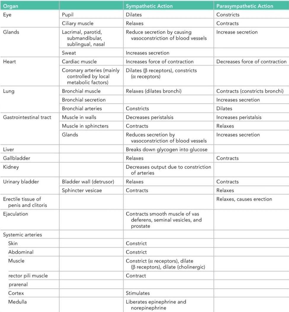
Some important ANS innervations are shown in Table 14-2.
عصبدهیهای خودکار
برخی از عصبدهیهای مهم AND در جدول 14-2 نشان داده شده است.
Table 14-2 Autonomic Nervous System Effects on Body Organs
جدول ۱۴-۲ اثرات سیستم عصبی خودکار بر اندامهای بدن
Eye
Both sympathetic and parasympathetic nervous systems control involuntary function in the iris and Acetylcholinesterase blockers (e.g., neostigmine) Parasympathomimetic drugs Pilocarpine Methacholine Hypothalamus lacrimal glands, whereas the eyelids are only affected by the sympathetic nervous system.
چشم
هر دو سیستم عصبی سمپاتیک و پاراسمپاتیک عملکرد غیرارادی در عنبیه و مسدودکنندههای استیل کولین استراز (به عنوان مثال، نئوستیگمین) را کنترل میکنند. داروهای پاراسمپاتومیمتیک پیلوکارپین متاکولین هیپوتالاموس غدد اشکی، در حالی که پلکها فقط تحت تأثیر سیستم عصبی سمپاتیک قرار میگیرند.
Upper Lid
The upper lid is raised by the levator palpebrae superioris muscle. The major part of this muscle is formed by skeletal muscle innervated by the oculomotor nerve. A small part is composed of smooth muscle fibers innervated by sympathetic postganglionic fibers from the superior cervical sympathetic ganglion (Fig. 14-7).
پلک بالایی
پلک بالایی توسط عضله بالابرنده پلک بالایی بالا میرود. بخش عمده این عضله توسط عضله اسکلتی تشکیل شده است که توسط عصب اوکولوموتور عصبدهی میشود. بخش کوچکی از آن از فیبرهای عضله صاف تشکیل شده است که توسط فیبرهای پسگانگلیونی سمپاتیک از گانگلیون سمپاتیک فوقانی گردنی عصبدهی میشوند (شکل 14-7).

Figure 14-7 Autonomic innervation of the upper eyelid and iris (A) and the sublingual and submandibular salivary glands (B).
شکل ۱۴-۷ عصبدهی خودکار پلک فوقانی و عنبیه (A) و غدد بزاقی زیرزبانی و زیرفکی (B).
Iris
The smooth muscle fibers of the iris consist of circular and radiating fibers. The circular fibers form the sphincter pupillae and the radial fibers form the dilator pupillae.
عنبیه
الیاف عضله صاف عنبیه از الیاف دایرهای و شعاعی تشکیل شدهاند. الیاف دایرهای مردمک اسفنکتر و الیاف شعاعی مردمک گشادکننده را تشکیل میدهند.
The sphincter pupillae is supplied by parasympathetic fibers from the parasympathetic nucleus (Edinger-Westphal nucleus) of the oculomotor nerve.
مردمک اسفنکتر توسط الیاف پاراسمپاتیک از هسته پاراسمپاتیک (هسته ادینگر-وستفال) عصب چشمی-حرکتی تغذیه میشود.
After synapsing in the ciliary ganglion, the postganglionic fibers pass forward to the eyeball in the short ciliary nerves. (The ciliary muscle of the eye is also supplied by the short ciliary nerves; see p. 406.)
پس از سیناپس در گانگلیون مژگانی، الیاف پسگانگلیونی در اعصاب مژگانی کوتاه به سمت کره چشم میروند. (عضله مژگانی چشم نیز توسط اعصاب مژگانی کوتاه تغذیه میشود؛ به صفحه ۴۰۶ مراجعه کنید.)
The dilator pupillae is supplied by postganglionic fibers from the superior cervical sympathetic ganglion. The postganglionic fibers reach the orbit along the internal carotid and ophthalmic arteries. They pass uninterrupted through the ciliary ganglion and reach the eyeball in the short ciliary nerves. Other sympathetic fibers reach the eyeball in the long ciliary nerves.
مردمک گشادکننده توسط الیاف پسگانگلیونی از گانگلیون سمپاتیک گردنی فوقانی تغذیه میشود. الیاف پسگانگلیونی در امتداد شریانهای کاروتید داخلی و افتالمیک به حدقه میرسند. آنها بدون وقفه از گانگلیون مژگانی عبور میکنند و در اعصاب مژگانی کوتاه به کره چشم میرسند. سایر فیبرهای سمپاتیک از طریق اعصاب مژگانی بلند به کره چشم میرسند.
Lacrimal Gland
The parasympathetic secretomotor nerve supply to the lacrimal gland originates in the lacrimatory nucleus of the facial nerve (Fig. 14-8). The preganglionic fibers reach the pterygopalatine ganglion through the nervus intermedius and its great petrosal branch and through the nerve of the pterygoid canal. The postganglionic fibers leave the ganglion and join the maxillary nerve. They then pass into its zygomatic branch and the zygomaticotemporal nerve. They reach the lacrimal gland within the lacrimal nerve.
غده اشکی
عصب ترشحی پاراسمپاتیک که غده اشکی را تغذیه میکند، از هسته اشکی عصب صورت سرچشمه میگیرد (شکل 14-8). فیبرهای پیش عقدهای از طریق عصب میانی و شاخه بزرگ پتروزال آن و از طریق عصب کانال پتریگوئید به گانگلیون پتریگوپالاتین میرسند. فیبرهای پس عقدهای گانگلیون را ترک کرده و به عصب فک بالا میپیوندند. سپس به شاخه زایگوماتیک آن و عصب زایگوماتیکوتمپورال میروند. آنها در داخل عصب اشکی به غده اشکی میرسند.
The sympathetic postganglionic fibers arise from the superior cervical sympathetic ganglion and travel in the plexus of nerves around the internal carotid artery. They join the deep petrosal nerve, the nerve of the pterygoid canal, the maxillary nerve, the zygomatic nerve, zygomaticotemporal nerve, and finally the lacrimal nerve. They function as vasoconstrictor fibers.
فیبرهای پس عقدهای سمپاتیک از گانگلیون سمپاتیک گردنی فوقانی منشأ میگیرند و در شبکه اعصاب اطراف شریان کاروتید داخلی حرکت میکنند. آنها به عصب پتروزال عمقی، عصب کانال پتریگوئید، عصب ماگزیلاری، عصب زایگوماتیکوتمپورال و در نهایت عصب اشکی میپیوندند. آنها به عنوان فیبرهای تنگ کننده عروق عمل میکنند.

Figure 14-8 Autonomic innervation of the parotid salivary gland and lacrimal gland.
شکل ۱۴-۸ عصبدهی خودکار غده بزاقی پاروتید و غده اشکی.
Salivary Glands
The involuntary action of the salivary glands is controlled by the ANS. These three, paired glands have similar sympathetic nerve origination but have unique parasympathetic origins.
غدد بزاقی
عمل غیرارادی غدد بزاقی توسط ANS کنترل میشود. این سه غده جفت، منشأ عصبی سمپاتیک مشابهی دارند اما منشأ پاراسمپاتیک منحصر به فردی دارند.
Submandibular and Sublingual Glands
The parasympathetic secretomotor supply originates in the superior salivatory nucleus of the facial nerve (see Fig. 14-7). The preganglionic fibers pass to the submandibular ganglion and other small ganglia close to the duct through the chorda tympani nerve and the lingual nerve. Postganglionic fibers reach the submandibular gland either directly or along the duct. Postganglionic fibers to the sublingual gland travel through the lingual nerve.
غدد زیر فکی و زیر زبانی
منبع ترشحی پاراسمپاتیک از هسته بزاقی فوقانی عصب صورتی سرچشمه میگیرد (شکل 14-7 را ببینید). فیبرهای پیش عقدهای از طریق عصب طناب صماخ و عصب زبانی به عقده زیر فکی و سایر عقدههای کوچک نزدیک به مجرا میروند. فیبرهای پس عقدهای یا مستقیماً یا در امتداد مجرا به غده زیر فکی میرسند. فیبرهای پس عقدهای به غده زیر زبانی از طریق عصب زبانی حرکت میکنند.
Sympathetic postganglionic fibers arise from the superior cervical sympathetic ganglion and reach the glands as a plexus of nerves around the external carotid, facial, and lingual arteries. They function as vasoconstrictor fibers.
فیبرهای پس عقدهای سمپاتیک از عقده سمپاتیک گردنی فوقانی منشأ میگیرند و به صورت شبکهای از اعصاب در اطراف شریانهای کاروتید خارجی، صورت و زبانی به غدد میرسند. آنها به عنوان فیبرهای تنگکننده عروق عمل میکنند.
Parotid Gland
Parasympathetic secretomotor fibers from the inferior salivatory nucleus of the glossopharyngeal nerve supply the gland (see Fig. 14-8). The preganglionic nerve fibers pass to the otic ganglion through the tympanic branch of the glossopharyngeal nerve and the lesser petrosal nerve. Postganglionic fibers reach the gland through the auriculotemporal nerve.
غده پاروتید
الیاف ترشحی پاراسمپاتیک از هسته بزاقی تحتانی عصب زبانی-حلقی به غده خونرسانی میکنند (شکل 14-8 را ببینید). الیاف عصبی پیش عقدهای از طریق شاخه تمپانیک عصب زبانی-حلقی و عصب پتروزال کوچک به عقده شنوایی منتقل میشوند. الیاف پس عقدهای از طریق عصب گوش-گیجگاهی به غده میرسند.
Sympathetic postganglionic fibers arise from the superior cervical sympathetic ganglion and reach the gland as a plexus of nerves around the external carotid artery. They function as vasoconstrictor fibers.
الیاف پس عقدهای سمپاتیک از عقده سمپاتیک گردنی فوقانی منشأ میگیرند و به صورت شبکهای از اعصاب در اطراف شریان کاروتید خارجی به غده میرسند. آنها به عنوان الیاف تنگکننده عروق عمل میکنند.
Heart
The sympathetic postganglionic fibers arise from the cervical and upper thoracic portions of the sympathetic trunks (Fig. 14-9). Postganglionic fibers reach the heart by way of the superior, middle, and inferior cardiac branches of the cervical portion of the sympathetic trunk and a number of cardiac branches from the thoracic portion of the sympathetic trunk. The fibers pass through the cardiac plexuses and terminate on the sinoatrial and atrioventricular nodes, on cardiac muscle fibers, and on coronary arteries. Activation of these nerves results in cardiac acceleration, increased force of contraction of the cardiac muscle, and dilatation of the coronary arteries. The coronary dilatation is mainly produced in response to local metabolic needs rather than by direct nerve stimulation of the coronary arteries.
قلب
فیبرهای پسگانگلیونی سمپاتیک از بخشهای گردنی و سینهای فوقانی تنههای سمپاتیک منشأ میگیرند (شکل 14-9). فیبرهای پسگانگلیونی از طریق شاخههای قلبی فوقانی، میانی و تحتانی بخش گردنی تنه سمپاتیک و تعدادی شاخه قلبی از بخش سینهای تنه سمپاتیک به قلب میرسند. این فیبرها از طریق شبکههای قلبی عبور کرده و به گرههای سینوسی-دهلیزی و دهلیزی-بطنی، فیبرهای عضلانی قلب و شریانهای کرونری ختم میشوند. فعال شدن این اعصاب منجر به شتاب قلب، افزایش نیروی انقباض عضله قلب و اتساع شریانهای کرونری میشود. اتساع کرونری عمدتاً در پاسخ به نیازهای متابولیک موضعی ایجاد میشود، نه از طریق تحریک عصبی مستقیم شریانهای کرونری.
The parasympathetic preganglionic fibers originate in the dorsal nucleus of the vagus nerve and descend into the thorax in the vagus nerves. The fibers terminate by synapsing with postganglionic neurons in the cardiac plexuses. Postganglionic fibers terminate on the sinoatrial and atrioventricular nodes and on the coronary arteries. Activation of these nerves results in a reduction in the rate and force of contraction of the myocardium and a constriction of the coronary arteries. Here again, the coronary constriction is mainly produced by the reduction in local metabolic needs rather than by neural effects.
فیبرهای پیشگانگلیونی پاراسمپاتیک از هسته پشتی عصب واگ سرچشمه میگیرند و در اعصاب واگ به داخل قفسه سینه نزول میکنند. این فیبرها با سیناپس با نورونهای پسگانگلیونی در شبکههای قلبی خاتمه مییابند. فیبرهای پسگانگلیونی به گرههای سینوسی-دهلیزی و دهلیزی-بطنی و شریانهای کرونری ختم میشوند. فعال شدن این اعصاب منجر به کاهش سرعت و قدرت انقباض میوکارد و انقباض شریانهای کرونری میشود. در اینجا نیز، انقباض کرونری عمدتاً ناشی از کاهش نیازهای متابولیک موضعی است تا اثرات عصبی.

Figure 14-9 Autonomic innervation of the heart and lungs.
شکل ۱۴-۹ عصبدهی خودکار قلب و ریهها.
The sympathetic postganglionic fibers arise from the second to the fifth thoracic ganglia of the sympathetic trunk (see Fig. 14-9). The fibers pass through the pulmonary plexuses and enter the lung, where they form networks around the bronchi and blood vessels. The sympathetic fibers produce bronchodilation and slight vasoconstriction.
فیبرهای پسگانگلیونی سمپاتیک از گانگلیونهای سینهای دوم تا پنجم تنه سمپاتیک منشأ میگیرند (شکل ۱۴-۹ را ببینید). این فیبرها از شبکه ریوی عبور کرده و وارد ریه میشوند، جایی که شبکههایی را در اطراف برونشها و رگهای خونی تشکیل میدهند. فیبرهای سمپاتیک باعث گشاد شدن برونشها و انقباض جزئی عروق میشوند.
The parasympathetic preganglionic fibers arise from the dorsal nucleus of the vagus and descend to the thorax within the vagus nerves. The fibers terminate by synapsing with postganglionic neurons in the pulmonary plexuses. The postganglionic fibers enter the lung, where they form networks around the bronchi and blood vessels. The parasympathetic fibers produce bronchoconstriction and slight vasodilation and increase glandular secretion.
فیبرهای پیشگانگلیونی پاراسمپاتیک از هسته پشتی واگ منشأ گرفته و در داخل اعصاب واگ به سمت قفسه سینه پایین میآیند. این فیبرها با سیناپس با نورونهای پسگانگلیونی در شبکه ریوی خاتمه مییابند. فیبرهای پسگانگلیونی وارد ریه میشوند، جایی که شبکههایی را در اطراف برونشها و رگهای خونی تشکیل میدهند. فیبرهای پاراسمپاتیک باعث انقباض برونشها و گشاد شدن جزئی عروق شده و ترشح غدد را افزایش میدهند.
Gastrointestinal Tract
Stomach and Intestine (to Splenic Flexure)
Preganglionic parasympathetic fibers enter the abdomen in the anterior (left) and posterior (right) vagal trunks (Fig. 14-10). The fibers are distributed to many abdominal viscera and to the gastrointestinal tract from the stomach to the splenic flexure of the colon. The fibers that pass to the gastrointestinal tract terminate on postganglionic neurons in the myenteric (Auerbach) and submucosal (Meissner) plexuses. The postganglionic fibers supply the smooth muscle and glands. The parasympathetic nerves stimulate peristalsis and relax the sphincters; they also stimulate secretion.
دستگاه گوارش
معده و روده (به خمیدگی طحال)
الیاف پاراسمپاتیک پیش عقدهای از طریق تنههای واگ قدامی (چپ) و خلفی (راست) وارد شکم میشوند (شکل 14-10). این الیاف در بسیاری از احشاء شکمی و از معده تا خمیدگی طحالی روده بزرگ به دستگاه گوارش توزیع میشوند. الیافی که به دستگاه گوارش میروند، در نورونهای پس عقدهای در شبکههای میانتریک (اورباخ) و زیرمخاطی (مایسنر) خاتمه مییابند. الیاف پس عقدهای به عضلات صاف و غدد خونرسانی میکنند. اعصاب پاراسمپاتیک حرکات دودی را تحریک کرده و اسفنکتر را شل میکنند. آنها همچنین ترشح را تحریک میکنند.
Sympathetic preganglionic nerve fibers pass through the thoracic part of the sympathetic trunk and enter the greater and lesser splanchnic nerves. These descend into the abdomen and synapse with postganglionic neurons in the celiac and superior mesenteric ganglia. The postganglionic nerve fibers are distributed to the stomach and intestine as nerve plexuses around the branches of the celiac and superior mesenteric arteries. The sympathetic nerves inhibit peristalsis and cause contraction of the sphincters; they also inhibit secretion (see the enteric nervous system, p. 394).
الیاف عصبی پیش عقدهای سمپاتیک از قسمت سینهای تنه سمپاتیک عبور کرده و وارد اعصاب اسپلانکنیک بزرگ و کوچک میشوند. این الیاف به داخل شکم نزول کرده و با نورونهای پس عقدهای در گانگلیونهای سلیاک و مزانتریک فوقانی سیناپس برقرار میکنند. رشتههای عصبی پسگانگلیونی به صورت شبکههای عصبی در اطراف شاخههای شریانهای سلیاک و مزانتریک فوقانی در معده و روده توزیع شدهاند. اعصاب سمپاتیک حرکات دودی را مهار کرده و باعث انقباض اسفنکترها میشوند؛ آنها همچنین ترشح را مهار میکنند (به سیستم عصبی روده، صفحه ۳۹۴ مراجعه کنید).

Figure 14-10 Autonomic innervation of the gastrointestinal tract.
شکل ۱۴-۱۰ عصبدهی خودکار دستگاه گوارش.
Descending Colon, Pelvic Colon, and Rectum
The preganglionic parasympathetic fibers originate in the gray matter of the spinal cord from the second to the fourth sacral segments (see Fig. 14-10). The fibers pass through the pelvic splanchnic nerves and the nerve plexuses around the branches of the inferior mesenteric artery. They terminate on postganglionic neurons in the myenteric (Auerbach) and submucosal (Meissner) plexuses. The postganglionic fibers supply the smooth muscle and glands. The parasympathetic nerves stimulate peristalsis and secretion.
کولون نزولی، کولون لگنی و رکتوم
الیاف پاراسمپاتیک پیش عقدهای از ماده خاکستری نخاع از قطعات دوم تا چهارم خاجی منشأ میگیرند (شکل 14-10 را ببینید). این الیاف از اعصاب اسپلانکنیک لگنی و شبکههای عصبی اطراف شاخههای شریان مزانتریک تحتانی عبور میکنند. آنها به نورونهای پس عقدهای در شبکههای میانتریک (اورباخ) و زیرمخاطی (مایسنر) ختم میشوند. الیاف پس عقدهای به عضلات صاف و غدد خونرسانی میکنند. اعصاب پاراسمپاتیک حرکات دودی و ترشح را تحریک میکنند.
The sympathetic preganglionic nerve fibers pass through the lumbar part of the sympathetic trunk and synapse with postganglionic neurons in the inferior mesenteric plexus. Postganglionic fibers are distributed to the bowel as nerve plexuses around the branches of the inferior mesenteric arteries. The sympathetic nerves inhibit peristalsis and secretion.
الیاف عصبی پیش عقدهای سمپاتیک از قسمت کمری تنه سمپاتیک عبور میکنند و با نورونهای پس عقدهای در شبکه مزانتریک تحتانی سیناپس برقرار میکنند. الیاف پس عقدهای به عنوان شبکههای عصبی در اطراف شاخههای شریانهای مزانتریک تحتانی در روده توزیع میشوند. اعصاب سمپاتیک حرکات دودی و ترشح را مهار میکنند.
Gallbladder and Biliary Ducts
The gallbladder and biliary ducts receive postganglionic parasympathetic and sympathetic fibers from the hepatic plexus. Parasympathetic fibers derived from the vagus are thought to be motor fibers to the smooth muscle of the gallbladder and bile ducts and inhibitory to the sphincter of Oddi.
کیسه صفرا و مجاری صفراوی
کیسه صفرا و مجاری صفراوی، فیبرهای پاراسمپاتیک و سمپاتیک پسگانگلیونی را از شبکه کبدی دریافت میکنند. تصور میشود فیبرهای پاراسمپاتیک مشتق شده از عصب واگ، فیبرهای حرکتی برای عضله صاف کیسه صفرا و مجاری صفراوی و فیبرهای مهاری برای اسفنکتر اودی باشند.
Autonomic afferent fibers are also present. Some of the fibers are believed to leave the hepatic plexus and join the right phrenic nerve, thus partially explaining the phenomenon of referred shoulder pain in the presence of gallbladder disease (see p. 410).
فیبرهای آوران خودکار نیز وجود دارند. اعتقاد بر این است که برخی از فیبرها از شبکه کبدی خارج شده و به عصب فرنیک راست میپیوندند، بنابراین تا حدودی پدیده درد ارجاعی شانه در حضور بیماری کیسه صفرا را توضیح میدهند (به صفحه ۴۱۰ مراجعه کنید).
Kidney
Preganglionic sympathetic fibers pass through the lower thoracic part of the sympathetic trunk and the lowest thoracic splanchnic nerve to join the renal plexus around the renal artery (Fig. 14-11). The preganglionic fibers synapse with postganglionic neurons in the renal plexus. The postganglionic fibers are distributed to the branches of the renal artery. The sympathetic nerves are vasoconstrictor in action to the renal arteries within the kidney.
کلیه
فیبرهای سمپاتیک پیش عقدهای از قسمت پایین قفسه سینه تنه سمپاتیک و پایینترین عصب اسپلانکنیک قفسه سینه عبور میکنند تا به شبکه کلیوی در اطراف شریان کلیوی بپیوندند (شکل 14-11). فیبرهای پیش عقدهای با نورونهای پس عقدهای در شبکه کلیوی سیناپس میکنند. فیبرهای پس عقدهای در شاخههای شریان کلیوی توزیع میشوند. اعصاب سمپاتیک در عمل تنگکننده عروق در شریانهای کلیوی درون کلیه هستند.
Preganglionic parasympathetic fibers enter the renal plexus from the vagus. Here, they synapse with postgan- glionic neurons whose fibers are distributed to the kidney along the branches of the renal artery. The parasympathetic nerves are thought to be vasodilator in action.
فیبرهای پاراسمپاتیک پیش عقدهای از عصب واگ وارد شبکه کلیوی میشوند. در اینجا، آنها با نورونهای پس عقدهای که فیبرهای آنها در امتداد شاخههای شریان کلیوی به کلیه توزیع شدهاند، سیناپس میکنند. تصور میشود که اعصاب پاراسمپاتیک در عمل گشادکننده عروق هستند.

Figure 14-11 Autonomic innervation of the kidney and suprarenal gland.
شکل۱۴-۱۱ عصبدهی خودکار کلیه و غده فوق کلیوی.
Suprarenal Gland Medulla
Preganglionic sympathetic fibers descend to the gland in the greater splanchnic nerve, a branch of the thoracic part of the sympathetic trunk (see Fig. 14-11). The nerve fibers terminate on the secretory cells of the medulla, which are comparable to postganglionic neurons. ACh is the transmitter substance between the nerve endings and the secretory cells, as at any other preganglionic endings. The sympathetic nerves stimulate the secretory cells of the medulla to increase the output of epinephrine and norepinephrine. The medulla of the suprarenal gland lacks parasympathetic innervation.
غده فوق کلیوی مدولا
الیاف سمپاتیک پیش عقدهای در عصب اسپلانکنیک بزرگ، شاخهای از قسمت سینهای تنه سمپاتیک (شکل 14-11 را ببینید) به غده میرسند. الیاف عصبی به سلولهای ترشحی مدولا ختم میشوند که با نورونهای پس عقدهای قابل مقایسه هستند. استیلکولین ماده انتقالدهنده بین انتهای عصب و سلولهای ترشحی است، مانند هر انتهای پیش عقدهای دیگر. اعصاب سمپاتیک سلولهای ترشحی مدولا را تحریک میکنند تا خروجی اپینفرین و نوراپی نفرین را افزایش دهند. مدولای غده فوق کلیوی فاقد عصبدهی پاراسمپاتیک است.
ادامه در قسمت ششم مبانی فصل ۱۴
«فصل چهاردهم کتاب نورواناتومی بالینی اسنل»
🚀 با ما همراه شوید!
تازهترین مطالب و آموزشهای مغز و اعصاب را از دست ندهید. با فالو کردن کانال تلگرام آیندهنگاران مغز، از ما حمایت کنید!



