
فرمانده غدد بدن: هیپوتالاموس؛ فصل سیزدهم نوروآناتومی بالینی اسنل


دعای مطالعه [ نمایش ]
بِسْمِ الله الرَّحْمنِ الرَّحیمِ
اَللّهُمَّ اَخْرِجْنى مِنْ ظُلُماتِ الْوَهْمِ
خدايا مرا بيرون آور از تاريكىهاى وهم،
وَ اَكْرِمْنى بِنُورِ الْفَهْمِ
و به نور فهم گرامى ام بدار،
اَللّهُمَّ افْتَحْ عَلَيْنا اَبْوابَ رَحْمَتِكَ
خدايا درهاى رحمتت را به روى ما بگشا،
وَانْشُرْ عَلَيْنا خَزائِنَ عُلُومِكَ بِرَحْمَتِكَ يا اَرْحَمَ الرّاحِمينَ
و خزانههاى علومت را بر ما باز كن به امید رحمتت اى مهربانترين مهربانان.
آیندهنگاران مغز: «ما مغز را میشناسیم، تا آینده را بسازیم.»
کتاب «نوروآناتومی بالینی اسنل» اثر دکتر ریچارد اس. اسنل، با ویرایش جدید دکتر رایان اسپلیتگربر، همچنان بهعنوان مرجع طلایی این حوزه شناخته میشود. این اثر با تلفیق تازهترین پژوهشها و کاربردهای بالینی، پلی استوار میان دانش پایه و عمل پزشکی بنا میکند.
تلاشهای علمی و سالها تدریس ارزشمند استاد گرانقدر آقای دکتر محمدتقی جغتایی الهامبخش ترجمه و گسترش این دانش در ایران بوده است.
ترجمه دقیق این کتاب توسط برند علمی آیندهنگاران مغز به مدیریت داریوش طاهری، امکان دسترسی فارسیزبانان به مرزهای نوین علوم اعصاب را فراهم کرده و رسالتی علمی در جهت شناخت عمیقتر مغز و ترسیم آیندهای روشنتر بر عهده گرفته است.
»» فصل ۱۳: هیپوتالاموس؛ بخش مبانی و اصول
»» Chapter 13: Hypothalamus
CHAPTER OBJECTIVES
• To learn the location and boundaries of the hypothalamus and the various nuclei that make up this important area
• To review the main connections of the nuclei, especially the connections between the hypothalamus and the pituitary gland
• To review some of the common clinical problems involving the hypothalamus
اهداف فصل
• یادگیری محل و مرزهای هیپوتالاموس و هستههای مختلفی که این ناحیه مهم را تشکیل میدهند
• بررسی ارتباطات اصلی هستهها، به ویژه ارتباطات بین هیپوتالاموس و غده هیپوفیز
• بررسی برخی از مشکلات بالینی رایج مربوط به هیپوتالاموس
A 16-year-old girl is taken by her mother to a pediatrician because she is rapidly losing weight. The mother states that the weight loss started about 1 year ago. The child’s eating habits changed from eating practically anything put before her to being a very choosy eater. Her personality is also changed, and she fears meeting strangers. Her relatives notice that she is impatient and irritable and cries a great deal. On being urged to eat more food, the girl countered by saying she was getting fat and must diet to improve her figure. Although she would not admit to having anything wrong with her, she admitted that menstruation had ceased 3 months previously.
دختری ۱۶ ساله به دلیل کاهش سریع وزن، توسط مادرش نزد متخصص اطفال برده میشود. مادر اظهار میکند که کاهش وزن از حدود ۱ سال پیش شروع شده است. عادات غذایی کودک از خوردن تقریباً هر چیزی که جلویش گذاشته میشد، به فردی بسیار مشکلپسند در غذا تغییر کرده است. شخصیت او نیز تغییر کرده و از ملاقات با غریبهها میترسد. بستگانش متوجه میشوند که او بیصبر و تحریکپذیر است و زیاد گریه میکند. وقتی از او خواسته شد که غذای بیشتری بخورد، دختر با گفتن اینکه چاق شده و باید برای بهبود اندامش رژیم بگیرد، مخالفت کرد. اگرچه او به داشتن هیچ مشکلی اعتراف نکرد، اما اعتراف کرد که قاعدگی ۳ ماه پیش متوقف شده است.
On being questioned by the pediatrician, the girl admits to calorie counting, and sometimes, when she feels she has overeaten, will force herself to vomit by sticking her fingers down her throat. On physical examination, she shows obvious signs of weight loss with hollow facial features, prominent bones, and wasted buttocks. Apart from having cold extremities and a low blood pressure of 85/60 mm Hg, no further abnormalities are discovered.
در پاسخ به پرسشهای متخصص اطفال، دختر اعتراف میکند که کالریها را محاسبه میکند و گاهی اوقات، وقتی احساس میکند پرخوری کرده است، با فرو کردن انگشتانش در گلویش، خود را مجبور به استفراغ میکند. در معاینه فیزیکی، علائم واضحی از کاهش وزن با صورت توخالی، استخوانهای برجسته و باسن تحلیل رفته نشان داده میشود. به جز سردی اندامها و فشار خون پایین ۸۵/۶۰ میلیمتر جیوه، هیچ ناهنجاری دیگری مشاهده نمیشود.
The pediatrician makes the diagnosis of anorexia nervosa and admits her to the local hospital. Psychological treatment includes getting the confidence of the patient by using nursing personnel who understood the condition. The primary treatment is to restore the patient’s weight by persuading the individual to eat adequate amounts of food.
متخصص اطفال، بیاشتهایی عصبی را تشخیص میدهد و او را در بیمارستان محلی بستری میکند. درمان روانشناختی شامل جلب اعتماد بیمار با استفاده از پرسنل پرستاری است که این بیماری را درک میکنند. درمان اولیه، بازگرداندن وزن بیمار با ترغیب فرد به خوردن مقادیر کافی غذا است.
Anorexia nervosa is a disorder of feeding and endocrine function that is normally controlled by the hypothalamus. However, strong evidence suggests that it is also of psychogenic origin.
بیاشتهایی عصبی نوعی اختلال در تغذیه و عملکرد غدد درونریز است که به طور معمول توسط هیپوتالاموس کنترل میشود. با این حال، شواهد قوی نشان میدهد که این بیماری منشأ روانشناختی نیز دارد.
The hypothalamus, although small (0.3% of the total brain), is a very important part of the central nervous system. It is essential for life. It controls the autonomic nervous system and the endocrine system and thus indirectly controls body homeostasis. The hypothalamus is well placed for this purpose, lying in the center of the limbic system. It is the site of numerous converging and diverging neuronal pathways, and through its adequate blood supply, it is able to sample the blood chemistry. The hypothalamus makes appropriate controlling responses following the integration of its nervous and chemical inputs.
هیپوتالاموس، اگرچه کوچک است (0.3٪ از کل مغز)، بخش بسیار مهمی از سیستم عصبی مرکزی است. برای زندگی ضروری است. سیستم عصبی خودکار و سیستم غدد درون ریز را کنترل میکند و بنابراین به طور غیرمستقیم هموستاز بدن را کنترل میکند. هیپوتالاموس برای این منظور در موقعیت خوبی قرار دارد و در مرکز سیستم لیمبیک قرار دارد. این اندام محل مسیرهای عصبی همگرا و واگرای متعددی است و از طریق خونرسانی کافی، قادر به نمونهبرداری از شیمی خون است. هیپوتالاموس پس از ادغام ورودیهای عصبی و شیمیایی خود، پاسخهای کنترلی مناسبی ایجاد میکند.

HYPOTHALAMUS
The hypothalamus is the part of the diencephalon that extends from the region of the optic chiasma to the caudal border of the mammillary bodies. It lies below the thalamus and forms the floor and the inferior part of the lateral walls of the third ventricle (Fig. 13-1). Anterior to the hypothalamus is an area that, for functional reasons, is often included in the hypothalamus. Because it extends forward from the optic chiasma to the lamina terminalis and the anterior commissure, it is referred to as the preoptic area. Caudally, the hypothalamus merges into the tegmentum of the midbrain. The lateral boundary of the hypothalamus is formed by the internal capsule.
هیپوتالاموس
هیپوتالاموس بخشی از دیانسفالون است که از ناحیه کیاسمای بینایی تا مرز خلفی اجسام پستانی امتداد دارد. این قسمت در زیر تالاموس قرار دارد و کف و قسمت تحتانی دیوارههای جانبی بطن سوم را تشکیل میدهد (شکل 13-1). در جلوی هیپوتالاموس ناحیهای وجود دارد که به دلایل عملکردی، اغلب در هیپوتالاموس گنجانده میشود. از آنجایی که از کیاسمای بینایی به سمت جلو تا لامینا ترمینالیس و رابط قدامی امتداد دارد، به آن ناحیه پیش بینایی گفته میشود. در قسمت خلفی، هیپوتالاموس در تگمنتوم مغز میانی ادغام میشود. مرز جانبی هیپوتالاموس توسط کپسول داخلی تشکیل میشود.
When observed from below (Fig. 13-2), the hypothalamus is seen to be related to the following structures, from anterior to posterior: (1) the optic chiasma, (2) the tuber cinereum and the infundibulum, and (3) the mammillary bodies.
وقتی از پایین مشاهده شود (شکل 13-2)، هیپوتالاموس از جلو به عقب با ساختارهای زیر مرتبط است: (1) کیاسمای بینایی، (2) توبر سینروم و اینفاندیبولوم، و (3) اجسام پستانی.
This small area of the brain will be seen in the following paragraphs to control body homeostasis, through the autonomic nervous system and the neuroendocrine system, and to play a vital role in emotional behavior.
این ناحیه کوچک از مغز در پاراگرافهای بعدی نشان داده خواهد شد که هموستاز بدن را از طریق سیستم عصبی خودکار و سیستم نورواندوکرین کنترل میکند و نقش حیاتی در رفتار عاطفی ایفا میکند.

Figure 13-1 Sagittal section of the brain showing the position of the hypothalamus.
شکل ۱۳-۱ برش ساژیتال مغز که موقعیت هیپوتالاموس را نشان میدهد.

Figure 13-2 Inferior surface of the brain showing parts of the hypothalamus.
شکل ۱۳-۲ سطح تحتانی مغز که بخشهایی از هیپوتالاموس را نشان میدهد.
HYPOTHALAMIC NUCLEI
Microscopically, the hypothalamus is composed of small nerve cells that are arranged in groups or nuclei, many of which are not clearly segregated from one another. For functional reasons, the preoptic area is included as part of the hypothalamus. For purposes of description, the nuclei are divided by an imaginary parasagittal plane into medial and lateral zones (Fig. 13-3). Lying within the plane are the columns of the fornix and the mammillothalamic tract, which serve as markers (Fig. 13-4; also see Fig. 13-3).
هستههای هیپوتالاموس
از نظر میکروسکوپی، هیپوتالاموس از سلولهای عصبی کوچکی تشکیل شده است که به صورت گروهها یا هستههایی قرار گرفتهاند که بسیاری از آنها به وضوح از یکدیگر جدا نیستند. به دلایل عملکردی، ناحیه پیشاپتیک به عنوان بخشی از هیپوتالاموس در نظر گرفته میشود. برای اهداف توصیفی، هستهها توسط یک صفحه پاراساژیتال فرضی به مناطق میانی و جانبی تقسیم میشوند (شکل ۱۳-۳). ستونهای فورنیکس و دستگاه مامیلوتالامیک در داخل این صفحه قرار دارند که به عنوان نشانگر عمل میکنند (شکل ۱۳-۴؛ همچنین به شکل ۱۳-۳ مراجعه کنید).

Figure 13-3 Sagittal section of the brain showing the hypothalamic nuclei. A: Medial zone nuclei lying medial to the plane of the fornix and the mammillothalamic tract. B: Lateral zone nuclei lying lateral to the plane of the fornix and the mammillothalamic tract.
شکل ۱۳-۳ برش ساژیتال مغز که هستههای هیپوتالاموس را نشان میدهد. الف: هستههای ناحیه میانی که در سمت داخلی صفحه فورنیکس و مسیر مامیلوتالامیک قرار دارند. ب: هستههای ناحیه جانبی که در سمت خارجی صفحه فورنیکس و مسیر مامیلوتالامیک قرار دارند.

Figure 13-4 Coronal section of the cerebral hemispheres showing the position of the hypothalamic nuclei.
شکل ۱۳-۴ برش کرونال از نیمکرههای مغزی که موقعیت هستههای هیپوتالاموس را نشان میدهد.
Medial Zone
In the medial zone, the following hypothalamic nuclei can be recognized, from anterior to posterior: (1) part of the preoptic nucleus; (2) the anterior nucleus, which merges with the preoptic nucleus; (3) part of the suprachiasmatic nucleus; (4) the paraventricular nucleus; (5) the dorsomedial nucleus; (6) the ventromedial nucleus; (7) the infundibular (arcuate) nucleus; and (8) the posterior nucleus.
ناحیه میانی
در ناحیه میانی، هستههای هیپوتالاموس زیر از قدامی به خلفی قابل تشخیص هستند: (1) بخشی از هسته پیشاپتیک؛ (2) هسته قدامی که با هسته پیشاپتیک ادغام میشود؛ (3) بخشی از هسته سوپراکیاسماتیک؛ (4) هسته پاراونتریکولار؛ (5) هسته پشتی میانی؛ (6) هسته شکمی میانی؛ (7) هسته قیفی (کمانکی)؛ و (8) هسته خلفی.
Lateral Zone
In the lateral zone, the following hypothalamic nuclei can be recognized, from anterior to posterior: (1) part of the preoptic nucleus, (2) part of the suprachiasmatic nucleus, (3) the supraoptic nucleus, (4) the lateral nucleus, (5) the tuberomammillary nucleus, and (6) the lateral tuberal nuclei.
ناحیه جانبی
در ناحیه جانبی، هستههای هیپوتالاموسی زیر از قدامی به خلفی قابل تشخیص هستند: (1) بخشی از هسته پیشاپتیک، (2) بخشی از هسته سوپراکیاسماتیک، (3) هسته سوپراپتیک، (4) هسته جانبی، (5) هسته توبرومامیلیاری، و (6) هستههای توبروما جانبی.
Some of the nuclei, such as the preoptic nucleus, the suprachiasmatic nucleus, and the mammillary nuclei, overlap both zones. It should be emphasized that most of the hypothalamic nuclei have ill-defined boundaries. With the use of modern technology, including histochemical, immunochemical, and anterograde and retrograde tracer studies, groups of neurons and their connections are being more precisely identified. Unfor- tunately, as new nuclear groups are discovered and given names, the reader has difficulty coming to terms with the old and new nomenclature. Only the major nuclear groups with well-established names and their connections are described in this account.
برخی از هستهها، مانند هسته پیشاپتیک، هسته سوپراکیاسماتیک و هستههای پستانی، هر دو منطقه را همپوشانی میکنند. باید تأکید کرد که بیشتر هستههای هیپوتالاموس مرزهای نامشخصی دارند. با استفاده از فناوری مدرن، از جمله مطالعات هیستوشیمیایی، ایمونوشیمیایی و ردیابهای پیشرونده و پسرونده، گروههای نورونی و ارتباطات آنها با دقت بیشتری شناسایی میشوند. متأسفانه، با کشف و نامگذاری گروههای هستهای جدید، خواننده در کنار آمدن با نامگذاری قدیمی و جدید مشکل دارد. در این گزارش فقط گروههای هستهای اصلی با نامهای تثبیتشده و ارتباطات آنها شرح داده شدهاند.
HYPOTHALAMIC LINES OF COMMUNICATION
The hypothalamus receives information from the rest of the body through (1) nervous connections, (2) the bloodstream, and (3) cerebrospinal fluid (CSF). The neurons of the hypothalamic nuclei respond and exert their control via the same routes. The CSF may serve as a conduit between the neurosecretory cells of the hypothalamus and distant sites of the brain.
خطوط ارتباطی هیپوتالاموس
هیپوتالاموس از طریق (1) ارتباطات عصبی، (2) جریان خون و (3) مایع مغزی نخاعی (CSF) اطلاعات را از بقیه بدن دریافت میکند. نورونهای هستههای هیپوتالاموس از طریق همین مسیرها پاسخ میدهند و کنترل خود را اعمال میکنند. CSF ممکن است به عنوان مجرایی بین سلولهای عصبی ترشحی هیپوتالاموس و نواحی دور مغز عمل کند.
Afferent Nervous Connections
The hypothalamus, which lies in the center of the limbic system, receives many afferent fibers from the viscera, the olfactory mucous membrane, the cerebral cortex, and the limbic system.
ارتباطات عصبی آوران
هیپوتالاموس که در مرکز سیستم لیمبیک قرار دارد، فیبرهای آوران زیادی را از احشاء، غشای مخاطی بویایی، قشر مغز و سیستم لیمبیک دریافت میکند.
The afferent connections are numerous and complex; the main pathways (Fig. 13-5) are as follows:
ارتباطات آوران متعدد و پیچیده هستند؛ مسیرهای اصلی (شکل 13-5) به شرح زیر است:

Figure 13-5 Sagittal section of the brain showing the main afferent pathways entering the hypothalamus.
شکل ۱۳-۵ برش ساژیتال مغز که مسیرهای آوران اصلی ورودی به هیپوتالاموس را نشان میدهد.
1. Somatic and visceral afferents. General somatic sensation and gustatory and visceral sensations reach the hypothalamus through collateral branches of the lemniscal afferent fibers and the tractus solitarius and through the reticular formation.
۱. آورانهای سوماتیک و احشایی. حس عمومی سوماتیک و حسهای چشایی و احشایی از طریق شاخههای جانبی فیبرهای آوران لمنسکال و مسیر انفرادی و از طریق تشکیلات مشبک به هیپوتالاموس میرسند.
2. Visual afferents leave the optic chiasma and pass to the suprachiasmatic nucleus.
۲. آورانهای بینایی از کیاسمای بینایی خارج شده و به هسته سوپراکیاسماتیک میروند.
3. Olfaction travels through the medial forebrain bundle.
۳. بویایی از طریق دسته مغز قدامی میانی حرکت میکند.
4. Auditory afferents have not been identified, but since auditory stimuli can influence the activities of the hypothalamus, they must exist.
۴. آورانهای شنوایی شناسایی نشدهاند، اما از آنجایی که محرکهای شنوایی میتوانند بر فعالیتهای هیپوتالاموس تأثیر بگذارند، باید وجود داشته باشند.
5. Corticohypothalamic fibers arise from the frontal lobe of the cerebral cortex and pass directly to the hypothalamus.
۵. فیبرهای کورتیکو هیپوتالاموس از لوب پیشانی قشر مغز منشأ میگیرند و مستقیماً به هیپوتالاموس میروند.
6. Hippocampo hypothalamic fibers pass from the hippocampus through the fornix to the mammillary body. Many neurophysiologists regard the hypothalamus as the main output pathway of the limbic system.
۶. فیبرهای هیپوکامپو هیپوتالاموس از هیپوکامپ از طریق فورنیکس به جسم پستانی میروند. بسیاری از نوروفیزیولوژیستها، هیپوتالاموس را به عنوان مسیر اصلی خروجی سیستم لیمبیک میدانند.
7. Amygdalohypothalamic fibers pass from the amygdaloid complex to the hypothalamus through the stria terminalis and by a route that passes inferior to the lentiform nucleus.
۷. فیبرهای آمیگدالوهیپوتالاموس از کمپلکس آمیگدالوئید به هیپوتالاموس از طریق استریا ترمینالیس و از مسیری که از پایین هسته عدسی شکل عبور میکند، عبور میکنند.
8. Thalamohypothalamic fibers arise from the dorsomedial and midline thalamic nuclei.
۸. فیبرهای تالاموهیپوتالاموس از هستههای تالاموس پشتی میانی و میانی منشأ میگیرند.
9. Tegmental fibers arise from the midbrain.
۹. فیبرهای تگمنتال از مغز میانی منشأ میگیرند.
The main afferent nervous connections of the hypothalamus are summarized in Table 13-1.
ارتباطات عصبی آوران اصلی هیپوتالاموس در جدول 13-1 خلاصه شده است.
Efferent Nervous Connections
The efferent connections of the hypothalamus are also numerous and complex, and only the main pathways (Fig. 13-6) are described here:
اتصالات عصبی وابران
اتصالات عصبی وابران هیپوتالاموس نیز متعدد و پیچیده هستند و فقط مسیرهای اصلی (شکل 13-6) در اینجا شرح داده شدهاند:
1. Descending fibers to the brainstem and spinal cord influence the peripheral neurons of the autonomic nervous system. They descend through a series of neurons in the reticular formation. The hypothalamus is connected to the parasympathetic nuclei of the oculomotor, facial, glossopharyngeal, and vagus nerves in the brainstem. In a similar manner, the reticulospinal fibers connect the hypothalamus with sympathetic cells of origin in the lateral gray horns of the first thoracic segment to the second lumbar segment of the spinal cord and the sacral parasympathetic outflow at the level of the second, third, and fourth sacral segments of the spinal cord.
۱. فیبرهای نزولی به ساقه مغز و نخاع، نورونهای محیطی سیستم عصبی خودکار را تحت تأثیر قرار میدهند. آنها از طریق یک سری نورون در تشکیلات مشبک پایین میآیند. هیپوتالاموس به هستههای پاراسمپاتیک اعصاب چشمی-حرکتی، صورتی، زبانی-حلقی و واگ در ساقه مغز متصل است. به طور مشابه، فیبرهای مشبک-نخاعی، هیپوتالاموس را با سلولهای سمپاتیک منشأ گرفته در شاخهای خاکستری جانبی اولین قطعه سینهای به قطعه کمری دوم نخاع و جریان خروجی پاراسمپاتیک خاجی در سطح قطعات خاجی دوم، سوم و چهارم نخاع متصل میکنند.
2. The mammillothalamic tract arises in the mammillary body and terminates in the anterior nucleus of the thalamus. Here, the pathway is relayed to the cingulate gyrus.
۲. راه مامیلوتالامیک از جسم مامیلاری شروع میشود و در هسته قدامی تالاموس خاتمه مییابد. در اینجا، این مسیر به شکنج کمربندی منتقل میشود.
3. The mammillotegmental tract arises from the mammillary body and terminates in the cells of the reticular formation in the tegmentum of the midbrain.
۳. راه مامیلوتاگمنتال از جسم مامیلاری شروع میشود و در سلولهای تشکیلات مشبک در تگمنتوم مغز میانی خاتمه مییابد.
4. Multiple pathways to the limbic system.
۴. مسیرهای متعدد به سیستم لیمبیک.
The main efferent nervous connections of the hypothalamus are summarized in Table 13-1.
ارتباطات عصبی وابران اصلی هیپوتالاموس در جدول 13-1 خلاصه شده است.
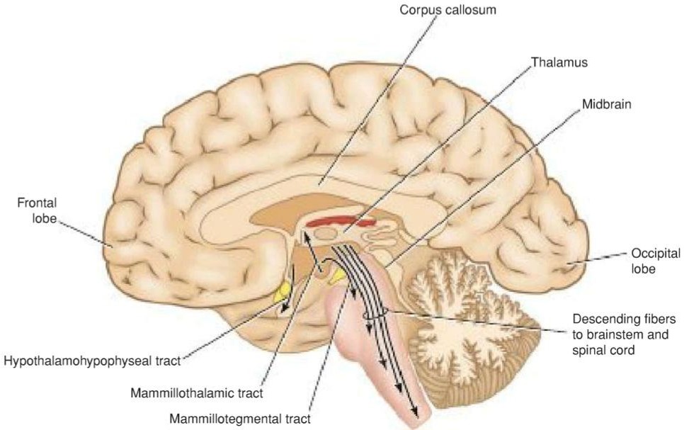
Figure 13-6 Sagittal section of the brain showing the main efferent pathways leaving the hypothalamus.
شکل ۱۳-۶ برش ساژیتال مغز که مسیرهای اصلی وابران را که از هیپوتالاموس خارج میشوند، نشان میدهد.
Table 13-1 Main Hypothalamic Afferent Efferent Nervous Connections
جدول ۱۳-۱ ارتباطات عصبی اصلی هیپوتالاموس آوران وابران

Connections With the Hypophysis Cerebri
The hypothalamus is connected to the hypophysis cerebri (pituitary gland) by two pathways: (1) nerve fibers that travel from the supraoptic and paraventricular nuclei to the posterior lobe of the hypophysis and (2) long and short portal blood vessels that connect sinusoids in the median eminence and infundibulum with capillary plexuses in the anterior lobe of the hypophysis (Fig. 13-7). These pathways enable the hypothalamus to influence the activities of the endocrine glands.
ارتباط با هیپوفیز مغزی
هیپوتالاموس از طریق دو مسیر به هیپوفیز مغزی (غده هیپوفیز) متصل است: (1) رشتههای عصبی که از هستههای سوپرااپتیک و پاراونتریکولار به لوب خلفی هیپوفیز میروند و (2) رگهای خونی پورتال بلند و کوتاه که سینوسوئیدها را در برجستگی میانی و قیفی شکل به شبکههای مویرگی در لوب قدامی هیپوفیز متصل میکنند (شکل 13-7). این مسیرها هیپوتالاموس را قادر میسازند تا بر فعالیتهای غدد درونریز تأثیر بگذارد.

Figure 13-7 A: Hypothalamohypophyseal tract. B: Hypophyseal portal system.
شکل ۱۳-۷ الف: دستگاه هیپوتالاموس-هیپوفیز. ب: سیستم پورتال هیپوفیز.
Hypothalamohypophyseal Tract
The hormones vasopressin and oxytocin are synthesized in the nerve cells of the supraoptic and paraventricular nuclei. The hormones are passed along the axons together with carrier proteins called neurophysins and are released at the axon terminals (see Fig. 13-7). Here, the hormones are absorbed into the bloodstream in fenestrated capillaries of the posterior lobe of the hypophysis. The hormone vasopressin (antidiuretic hormone) is produced mainly in the nerve cells of the supraoptic nucleus. Its function is to cause vasoconstriction. It also has an important antidiuretic function, causing an increased absorption of water in the distal convoluted tubules and collecting tubules of the kidney. The other hormone is oxytocin, which is produced mainly in the paraventricular nucleus. Oxytocin stimulates the contraction of the smooth muscle of the uterus and causes contraction of the myoepithelial cells that surround the alveoli and ducts of the breast. Toward the end of pregnancy, oxytocin is produced in large amounts and stimulates labor contractions of the uterus. Later, when the baby suckles at the breast, a nervous reflex from the nipple stimulates the hypothalamus to produce more of the hormone. This promotes contraction of the myoepithelial cells and assists in the expression of the milk from the breasts.
مسیر هیپوتالاموس-هیپوفیز
هورمونهای وازوپرسین و اکسیتوسین در سلولهای عصبی هستههای سوپرااپتیک و پاراونتریکولار سنتز میشوند. این هورمونها به همراه پروتئینهای حامل به نام نوروفیزینها در امتداد آکسونها عبور میکنند و در پایانههای آکسون آزاد میشوند (شکل ۱۳-۷ را ببینید). در اینجا، هورمونها در مویرگهای منفذدار لوب خلفی هیپوفیز جذب جریان خون میشوند. هورمون وازوپرسین (هورمون ضد ادراری) عمدتاً در سلولهای عصبی هسته سوپرااپتیک تولید میشود. عملکرد آن ایجاد انقباض عروقی است. همچنین عملکرد ضد ادراری مهمی دارد و باعث افزایش جذب آب در لولههای پیچیده دیستال و لولههای جمعآوریکننده کلیه میشود. هورمون دیگر اکسیتوسین است که عمدتاً در هسته پاراونتریکولار تولید میشود. اکسیتوسین انقباض عضله صاف رحم را تحریک میکند و باعث انقباض سلولهای میواپیتلیال اطراف آلوئولها و مجاری پستان میشود. در اواخر بارداری، اکسیتوسین به مقدار زیادی تولید میشود و انقباضات زایمانی رحم را تحریک میکند. بعداً، هنگامی که نوزاد از سینه مادر شیر میخورد، یک رفلکس عصبی از نوک سینه، هیپوتالاموس را برای تولید بیشتر این هورمون تحریک میکند. این امر انقباض سلولهای میواپیتلیال را افزایش داده و به خروج شیر از سینهها کمک میکند.
The supraoptic nucleus, which produces vasopressin, acts as an osmoreceptor. Should the osmotic pressure of the blood circulating through the nucleus be too high, the nerve cells increase their production of vasopressin, and the antidiuretic effect of this hormone will increase the reabsorption of water from the kidney. By this means, the osmotic pressure of the blood will return to normal limits.
هسته سوپرااپتیک که وازوپرسین تولید میکند، به عنوان یک گیرنده اسمزی عمل میکند. اگر فشار اسمزی خون در گردش از طریق هسته خیلی زیاد باشد، سلولهای عصبی تولید وازوپرسین خود را افزایش میدهند و اثر ضد ادراری این هورمون، بازجذب آب از کلیه را افزایش میدهد. به این ترتیب، فشار اسمزی خون به حد طبیعی باز میگردد.
Hypophyseal Portal System
Neurosecretory cells situated mainly in the medial zone of the hypothalamus are responsible for the production of the releasing hormones and release-inhibitory hormones. The hormones are packaged into granules and are transported along the axons of these cells into the median eminence and infundibulum. Here, the granules are released by exocytosis onto fenestrated capillaries at the upper end of the hypophyseal portal system.
سیستم پورتال هیپوفیز
سلولهای عصبی-ترشحی که عمدتاً در ناحیه میانی هیپوتالاموس قرار دارند، مسئول تولید هورمونهای آزادکننده و هورمونهای مهارکننده ترشح هستند. هورمونها در گرانولهایی بستهبندی شده و در امتداد آکسونهای این سلولها به برجستگی میانی و قیفی شکل منتقل میشوند. در اینجا، گرانولها با اگزوسیتوز به مویرگهای منفذدار در انتهای بالایی سیستم پورتال هیپوفیز آزاد میشوند.
The hypophyseal portal system is formed on each side from the superior hypophyseal artery, which is a branch of the internal carotid artery (see Fig. 13-7B). The artery enters the median eminence and divides into tufts of capillaries. These capillaries drain into long and short descending vessels that end in the anterior lobe of the hypophysis by dividing into vascular sinusoids that pass between the secretory cells of the anterior lobe.
سیستم پورتال هیپوفیز در هر طرف از شریان هیپوفیز فوقانی تشکیل میشود که شاخهای از شریان کاروتید داخلی است (شکل 13-7B را ببینید). این شریان وارد برجستگی میانی میشود و به دستههایی از مویرگها تقسیم میشود. این مویرگها با تقسیم شدن به سینوزوئیدهای عروقی که از بین سلولهای ترشحی لوب قدامی عبور میکنند، به رگهای نزولی بلند و کوتاه که در لوب قدامی هیپوفیز ختم میشوند، تخلیه میشوند.
The portal system carries the releasing hormones and the release-inhibiting hormones to the secretory cells of the anterior lobe of the hypophysis. The releasing hormones stimulate the production and release of adrenocorticotropic hormone (ACTH), follicle-stimulating hormone (FSH), luteinizing hormone (LH), thyrotropic hormone or thyroid-stimulating hormone (TSH), and growth hormone (GH). The release of inhibiting hormones inhibits the release of the melanocyte-stimulating hormone (MSH) and luteotropic hormone (LTH). LTH (also known as the lactogenic hormone or prolactin) stimulates the corpus luteum to secrete progesterone and the mammary gland to produce milk. The GH inhibitory hormone (somatostatin) inhibits the release of GH. A summary of the hypothalamic-releasing and inhibitory hormones and their effects on the anterior lobe of the hypophysis are shown in Table 13-2.
سیستم پورتال، هورمونهای آزادکننده و هورمونهای مهارکنندهی آزادشدن را به سلولهای ترشحی لوب قدامی هیپوفیز منتقل میکند. هورمونهای آزادکننده، تولید و آزادشدن هورمون آدرنوکورتیکوتروپیک (ACTH)، هورمون محرک فولیکول (FSH)، هورمون لوتئینیزهکننده (LH)، هورمون تیروتروپیک یا هورمون محرک تیروئید (TSH) و هورمون رشد (GH) را تحریک میکنند. آزادشدن هورمونهای مهارکننده، آزادشدن هورمون محرک ملانوسیت (MSH) و هورمون لوتئوتروپیک (LTH) را مهار میکند. LTH (که به عنوان هورمون لاکتوژنیک یا پرولاکتین نیز شناخته میشود) جسم زرد را برای ترشح پروژسترون و غده پستانی را برای تولید شیر تحریک میکند. هورمون مهارکنندهی GH (سوماتوستاتین) آزادشدن GH را مهار میکند. خلاصهای از هورمونهای آزادکننده و مهارکنندهی هیپوتالاموس و اثرات آنها بر لوب قدامی هیپوفیز در جدول 13-2 نشان داده شده است.
Table 13-2 Hypothalamic-Releasing and Inhibitory Hormones and Their Effects on Hypophysis (Pituitary) Anterior Lobe
جدول ۱۳-۲ هورمونهای آزادکننده و مهارکننده هیپوتالاموس و اثرات آنها بر لوب قدامی هیپوفیز (هیپوفیز)
 The neurons of the hypothalamus that are responsible for the production of the releasing hormones and the release-inhibiting hormones are influenced by the afferent fibers passing to the hypothalamus. They also are influenced by the level of the hormone produced by the target organ controlled by the hypophysis. Should the level of thyroxine in the blood fall, for example, then the releasing factor for the thyrotropic hormone would be produced in increased quantities. Table 13-3 summarizes the presumed nuclear origin of the pituitary-releasing and inhibitory hormones in the hypothalamus.
نورونهای هیپوتالاموس که مسئول تولید هورمونهای آزادکننده و هورمونهای مهارکنندهی آزادسازی هستند، تحت تأثیر فیبرهای آوران عبوری از هیپوتالاموس قرار دارند. آنها همچنین تحت تأثیر سطح هورمون تولید شده توسط اندام هدف که توسط هیپوفیز کنترل میشود، قرار دارند. به عنوان مثال، اگر سطح تیروکسین در خون کاهش یابد، عامل آزادکنندهی هورمون تیروتروپیک در مقادیر بیشتری تولید میشود. جدول 13-3 منشأ هستهای فرضی هورمونهای آزادکننده و مهارکنندهی هیپوفیز در هیپوتالاموس را خلاصه میکند.
Table 13-3 Nuclear Origin a Of the Hypophysis (Pituitary)-Releasing and Inhibitory Hormones in the Hypothalamus
جدول ۱۳-۳ منشأ هستهای هورمونهای آزادکننده و مهارکننده هیپوفیز (هیپوفیز) در هیپوتالاموس
 a Origin is presumed.
» ادامه در قسمت دوم مبانی فصل ۱۳
«فصل سیزدهم کتاب نورواناتومی بالینی اسنل»
🚀 با ما همراه شوید!
تازهترین مطالب و آموزشهای مغز و اعصاب را از دست ندهید. با فالو کردن کانال تلگرام آیندهنگاران مغز، از ما حمایت کنید!



