یادداشتهای بالینی نوروآناتومی بالینی اسنل | سیستم عصبی خودکار (ANS)


دعای مطالعه [ نمایش ]
بِسْمِ الله الرَّحْمنِ الرَّحیمِ
اَللّهُمَّ اَخْرِجْنى مِنْ ظُلُماتِ الْوَهْمِ
خدايا مرا بيرون آور از تاريكىهاى وهم،
وَ اَكْرِمْنى بِنُورِ الْفَهْمِ
و به نور فهم گرامى ام بدار،
اَللّهُمَّ افْتَحْ عَلَيْنا اَبْوابَ رَحْمَتِكَ
خدايا درهاى رحمتت را به روى ما بگشا،
وَانْشُرْ عَلَيْنا خَزائِنَ عُلُومِكَ بِرَحْمَتِكَ يا اَرْحَمَ الرّاحِمينَ
و خزانههاى علومت را بر ما باز كن به امید رحمتت اى مهربانترين مهربانان.
آیندهنگاران مغز: «ما مغز را میشناسیم، تا آینده را بسازیم.»
کتاب «نوروآناتومی بالینی اسنل» اثر دکتر ریچارد اس. اسنل، با ویرایش جدید دکتر رایان اسپلیتگربر، همچنان بهعنوان مرجع طلایی این حوزه شناخته میشود. این اثر با تلفیق تازهترین پژوهشها و کاربردهای بالینی، پلی استوار میان دانش پایه و عمل پزشکی بنا میکند.
تلاشهای علمی و سالها تدریس ارزشمند استاد گرانقدر دکتر محمدتقی جغتایی الهامبخش ترجمه و گسترش این دانش در ایران بوده است.
ترجمه دقیق این کتاب توسط برند علمی آیندهنگاران مغز به مدیریت داریوش طاهری، امکان دسترسی فارسیزبانان به مرزهای نوین علوم اعصاب را فراهم کرده و رسالتی علمی در جهت شناخت عمیقتر مغز و ترسیم آیندهای روشنتر بر عهده گرفته است.
»» فصل ۱۴: سیستم عصبی خودکار؛ بخش یادداشتهای بالینی و مسائل کاربردی
»» Chapter 14: Autonomic Nervous System
Clinical Notes
General Considerations
Clearly, the ANS is not an isolated part of the nervous system. It should be regarded as the part of the nervous system that, with the endocrine system, is particularly involved in maintaining the stability of the internal environment of the body. Its activities are modified by the hypothalamus, whose function is to integrate vast amounts of afferent information received from other areas of the nervous system and to translate changing hormonal levels of the bloodstream into appropriate nervous and hormonal activities.
یادداشتهای بالینی
ملاحظات عمومی
واضح است که ANS بخش جداگانهای از سیستم عصبی نیست. باید آن را به عنوان بخشی از سیستم عصبی در نظر گرفت که به همراه سیستم غدد درونریز، به ویژه در حفظ ثبات محیط داخلی بدن نقش دارد. فعالیتهای آن توسط هیپوتالاموس اصلاح میشود، که وظیفه آن ادغام مقادیر زیادی از اطلاعات آوران دریافتی از سایر نواحی سیستم عصبی و تبدیل سطوح هورمونی در حال تغییر جریان خون به فعالیتهای عصبی و هورمونی مناسب است.
Because the ANS is so important in maintaining normal body homeostasis, not surprisingly, the system is subject to many pharmacologic interventions. Propranolol and atenolol, for example, are ẞ-adrenergic antagonists that can be used in the treatment of hypertension and ischemic heart disease.
از آنجا که ANS در حفظ هموستاز طبیعی بدن بسیار مهم است، جای تعجب نیست که این سیستم تحت مداخلات دارویی زیادی قرار میگیرد. به عنوان مثال، پروپرانولول و آتنولول، آنتاگونیستهای κ-آدرنرژیک هستند که میتوانند در درمان فشار خون بالا و بیماری ایسکمیک قلب استفاده شوند.
Autonomic Nervous System Injury
Trauma to the ANS is usually limited to either sympathetic or parasympathetic components, rarely both at the same time, due to their relative locations in the thorax or cranial cavity, respectively.
آسیب سیستم عصبی خودکار
تروما به ANS معمولاً به اجزای سمپاتیک یا پاراسمپاتیک محدود میشود، و به ندرت هر دو به طور همزمان، به دلیل موقعیت نسبی آنها در قفسه سینه یا حفره جمجمه.
Sympathetic Injuries
The sympathetic trunk in the neck can be injured by stab and bullet wounds. Traction injuries to the first thoracic root of the brachial plexus can damage sympathetic nerves destined for the stellate ganglion. All these conditions can produce a preganglionic type of Horner syndrome. Injuries to the spinal cord or cauda equina can disrupt the sympathetic control of the bladder (see p. 402).
آسیبهای سمپاتیک
تنه سمپاتیک در گردن میتواند در اثر زخمهای ناشی از چاقو و گلوله آسیب ببیند. آسیبهای کششی به اولین ریشه سینهای شبکه بازویی میتواند به اعصاب سمپاتیکی که به گانگلیون ستارهای منتهی میشوند، آسیب برساند. همه این شرایط میتوانند نوعی سندرم هورنر پیش عقدهای ایجاد کنند. آسیبهای نخاع یا دم اسب میتواند کنترل سمپاتیک مثانه را مختل کند (به صفحه ۴۰۲ مراجعه کنید).
Parasympathetic Injuries
The oculomotor nerve is vulnerable in head injuries (herniated uncus) and can be damaged by compression by aneurysms in the junction between the posterior cerebral artery and posterior communicating artery. The preganglionic parasympathetic fibers traveling in this nerve are situated in the periphery of the nerve and can be damaged. Surface aneurysmal compression characteristically causes dilation of the pupil and loss of the visual light reflexes.
آسیبهای پاراسمپاتیک
عصب چشمی-حرکتی در آسیبهای سر (فتق آنکوس) آسیبپذیر است و میتواند در اثر فشرده شدن توسط آنوریسمها در محل اتصال بین شریان مغزی خلفی و شریان ارتباطی خلفی آسیب ببیند. الیاف پاراسمپاتیک پیش عقدهای که در این عصب حرکت میکنند در حاشیه عصب قرار دارند و میتوانند آسیب ببینند. فشرده شدن آنوریسم سطحی به طور مشخص باعث گشاد شدن مردمک چشم و از بین رفتن رفلکسهای نوری بینایی میشود.
The autonomic fibers in the facial nerve can be damaged by fractures of the skull involving the temporal bone. The vestibulocochlear nerve is closely related to the facial nerve in the internal acoustic meatus, so clinical findings involving both nerves are common. Involvement of the parasympathetic fibers in the facial nerve may produce impaired lacrimation in addition to paralysis of the facial muscles.
فیبرهای اتونوم در عصب صورت میتوانند در اثر شکستگیهای جمجمه که استخوان گیجگاهی را درگیر میکنند، آسیب ببینند. عصب دهلیزی-حلقی ارتباط نزدیکی با عصب صورت در مجرای شنوایی داخلی دارد، بنابراین یافتههای بالینی مربوط به هر دو عصب رایج است. درگیری فیبرهای پاراسمپاتیک در عصب صورت ممکن است علاوه بر فلج عضلات صورت، باعث اختلال در اشکریزی شود.
The glossopharyngeal and vagus nerves are at risk in stab and bullet wounds of the neck. The parasympathetic secretomotor fibers to the parotid salivary gland leave the glossopharyngeal nerve just below the skull; therefore, they are rarely damaged.
اعصاب زبانی-حلقی و واگ در زخمهای ناشی از چاقو و گلوله در گردن در معرض خطر هستند. فیبرهای ترشحی پاراسمپاتیک به غده بزاقی پاروتید، عصب زبانی-حلقی را درست زیر جمجمه ترک میکنند. بنابراین، به ندرت آسیب میبینند.
The parasympathetic outflow in the sacral region of the spinal cord (S2-S4) may be damaged by spinal cord and cauda equina injuries, leading to disruption of bladder, rectal, and sexual functions.
خروجی پاراسمپاتیک در ناحیه خاجی نخاع (S2-S4) ممکن است در اثر آسیبهای نخاع و دم اسب آسیب ببیند و منجر به اختلال در عملکرد مثانه، رکتوم و عملکرد جنسی شود.
Degeneration and Regeneration of Autonomic Nerves
The structural changes are identical to those found in other areas of the peripheral and central parts of the nervous system. Functional recoveries following sympathectomy operations can be explained only by the assumption either that the operative procedure was inadequate and nerve fibers were left intact or regenerated or that alternative nervous pathways existed and were left undisturbed.
تخریب و بازسازی اعصاب خودکار
تغییرات ساختاری مشابه تغییرات یافت شده در سایر نواحی بخشهای محیطی و مرکزی سیستم عصبی هستند. بهبود عملکرد پس از عمل سمپاتکتومی تنها با این فرض قابل توضیح است که یا روش عمل ناکافی بوده و فیبرهای عصبی دست نخورده باقی مانده یا بازسازی شدهاند یا اینکه مسیرهای عصبی جایگزین وجود داشته و دست نخورده باقی ماندهاند.
The denervation of viscera supplied by autonomic nerves is followed by their increased sensitivity to the agent that was previously the transmitter substance. One explanation is that following nerve section, there may be an increase in the number of receptor sites on the postsynaptic membrane. Another possibility, which applies to endings where norepinephrine is the transmitter, is that the reuptake of the transmitter by the nerve terminal is interfered with in some way.
قطع عصب احشاء که توسط اعصاب خودکار تأمین میشود، با افزایش حساسیت آنها به عاملی که قبلاً مادهی انتقالدهنده بوده است، همراه است. یک توضیح این است که پس از قطع عصب، ممکن است تعداد جایگاههای گیرنده روی غشای پس سیناپسی افزایش یابد. احتمال دیگر، که در مورد پایانههایی که نوراپی نفرین انتقالدهنده است، صدق میکند، این است که بازجذب انتقالدهنده توسط پایانهی عصبی به نحوی مختل میشود.
Urinary Bladder Dysfunction
Injuries to the spinal cord are followed by disruption of the nervous control of micturition. The normal bladder is innervated as follows:
اختلال عملکرد مثانه
آسیبهای نخاعی منجر به اختلال در کنترل عصبی ادرار میشوند. مثانه طبیعی به شرح زیر عصبدهی میشود:
Sympathetic innervation is from the first and second lumbar segments of the spinal cord.
عصبدهی سمپاتیک از بخشهای اول و دوم کمری نخاع است.
Parasympathetic innervation is from the second, third, and fourth sacral segments of the spinal cord.
عصبدهی پاراسمپاتیک از بخشهای دوم، سوم و چهارم خاجی نخاع است.
Sensory nerve fibers enter the spinal cord at the above segments.
رشتههای عصبی حسی در بخشهای فوق وارد نخاع میشوند.
The atonic bladder occurs during the phase of spinal shock immediately following the injury and may last from a few days to several weeks. The bladder wall muscle is relaxed, the sphincter vesicae is tightly contracted (loss of inhibition from higher levels), and the sphincter urethrae is relaxed. The bladder becomes greatly distended and finally overflows. Depending on the level of the cord injury, the patient may or may not be aware that the bladder is full but has no voluntary control.
مثانه آتونیک در مرحله شوک نخاعی بلافاصله پس از آسیب رخ میدهد و ممکن است از چند روز تا چند هفته طول بکشد. عضله دیواره مثانه شل میشود، وزیکای اسفنکتر محکم منقبض میشود (از دست دادن مهار از سطوح بالاتر) و مجرای ادرار اسفنکتر شل میشود. مثانه به شدت متسع میشود و در نهایت سرریز میشود. بسته به سطح آسیب نخاعی، بیمار ممکن است از پر بودن مثانه آگاه باشد یا نباشد، اما هیچ کنترل ارادی روی آن ندارد.
The automatic reflex bladder occurs after the patient has recovered from spinal shock, provided that the cord lesion lies above the level of the parasympathetic outflow (S2-S4). This is the type of bladder normally found in infancy. The descending fibers in the spinal cord are sectioned, so voluntary control is not possible. The bladder fills and empties reflexly. Stretch receptors in the bladder wall are stimulated as the bladder fills and the afferent impulses pass to the spinal cord ($2-$4). Efferent impulses pass down to the bladder muscle, which contracts; the sphincter vesicae and the urethral sphincter both relax. This simple reflex occurs every 1 to 4 hours.
مثانه رفلکسی خودکار پس از بهبودی بیمار از شوک نخاعی رخ میدهد، مشروط بر اینکه ضایعه نخاعی بالاتر از سطح خروجی پاراسمپاتیک (S2-S4) باشد. این نوع مثانه معمولاً در دوران نوزادی یافت میشود. الیاف نزولی در نخاع بریده میشوند، بنابراین کنترل ارادی امکانپذیر نیست. مثانه به صورت رفلکسی پر و خالی میشود. گیرندههای کششی در دیواره مثانه با پر شدن مثانه تحریک میشوند و تکانههای آوران به نخاع منتقل میشوند (2-4 دلار). تکانههای وابران به عضله مثانه منتقل میشوند که منقبض میشود. وزیکاهای اسفنکتر و اسفنکتر مجرای ادرار هر دو شل میشوند. این رفلکس ساده هر 1 تا 4 ساعت یکبار رخ میدهد.
The autonomous bladder is the condition that occurs if the sacral segment of the spinal cord is destroyed or if the cauda equina is severed. The bladder has no reflex control or voluntary control. The bladder wall is flaccid and the capacity of the bladder is greatly increased. It fills to capacity and overflows, which results in continual dribbling. The bladder may be partially emptied by manual compression of the lower part of the anterior abdominal wall, but infection of the urine and back pressure effects on the ureters and kidneys are inevitable.
مثانهی خودگردان وضعیتی است که در صورت تخریب بخش خاجی نخاع یا قطع شدن دم اسب رخ میدهد. مثانه هیچ کنترل رفلکسی یا کنترل ارادی ندارد. دیوارهی مثانه شل است و ظرفیت مثانه به شدت افزایش مییابد. مثانه تا حد ظرفیت پر میشود و سرریز میکند که منجر به چکه کردن مداوم ادرار میشود. مثانه ممکن است با فشار دستی به قسمت پایینی دیوارهی قدامی شکم تا حدی تخلیه شود، اما عفونت ادرار و اثرات فشار برگشتی بر حالبها و کلیهها اجتنابناپذیر است.
Defecation
The act of defecation involves a coordinated reflex that results in the emptying of the descending colon, pelvic colon, rectum, and anal canal. It is assisted by a rise in the intra-abdominal pressure brought about by contraction of the muscles of the anterior abdominal wall. The involuntary internal sphincter of the anal canal normally is innervated by postganglionic sympathetic fibers from the hypogastric plexuses, and the voluntary external sphincter of the anal canal is innervated by the inferior rectal nerve. The desire to defecate is initiated by stimulation of the stretch receptors in the wall of the rectum.
دفع مدفوع
عمل دفع مدفوع شامل یک رفلکس هماهنگ است که منجر به تخلیه روده بزرگ نزولی، روده بزرگ لگنی، رکتوم و کانال مقعدی میشود. این عمل با افزایش فشار داخل شکمی ناشی از انقباض عضلات دیواره قدامی شکم تسهیل میشود. اسفنکتر داخلی غیرارادی کانال مقعدی معمولاً توسط فیبرهای سمپاتیک پسگانگلیونی از شبکههای هیپوگاستریک عصبدهی میشود و اسفنکتر خارجی ارادی کانال مقعدی توسط عصب رکتوم تحتانی عصبدهی میشود. میل به دفع مدفوع با تحریک گیرندههای کششی در دیواره رکتوم آغاز میشود.
Following severe spinal cord injuries (or cauda equina injuries), the patient is not aware of rectal distention. Moreover, the parasympathetic influence on the peristaltic activity of the descending colon, sigmoid colon, and rectum is lost. In addition, control over the abdominal musculature and sphincters of the anal canal may be severely impaired. The rectum, now an isolated structure, responds by contracting when the pressure within its lumen rises. This local reflex response is much more efficient if the sacral segments of the spinal cord and the cauda equina are intact. At best, however, the force of the contractions of the rectal wall is small and constipation and impaction are the usual outcome. The treatment of patients with spinal cord injuries is to empty the rectum with biweekly enemas; the use of suppositories also may be helpful.
پس از آسیبهای شدید نخاعی (یا آسیبهای دم اسب)، بیمار از اتساع رکتوم آگاه نیست. علاوه بر این، تأثیر پاراسمپاتیک بر فعالیت پریستالتیک روده بزرگ نزولی، روده بزرگ سیگموئید و رکتوم از بین میرود. علاوه بر این، کنترل بر عضلات شکمی و اسفنکترهای کانال مقعد ممکن است به شدت مختل شود. رکتوم، که اکنون یک ساختار جدا شده است، با افزایش فشار درون مجرای خود با انقباض پاسخ میدهد. این پاسخ رفلکس موضعی در صورتی که بخشهای خاجی نخاع و دم اسب سالم باشند، بسیار کارآمدتر است. با این حال، در بهترین حالت، نیروی انقباضات دیواره رکتوم کم است و یبوست و احتقان نتیجه معمول آن است. درمان بیماران مبتلا به آسیبهای نخاعی، تخلیه رکتوم با تنقیه دو هفتهای است. استفاده از شیاف نیز ممکن است مفید باشد.
Erection and Ejaculation
As described previously, erection of the penis or clitoris is controlled by the parasympathetic nerves that originate from the second, third, and fourth sacral segments of the spinal cord. Bilateral damage to the reticulospinal tracts in the spinal cord above the second sacral segment of the spinal cord will result in loss of erection. Later, when the effects of spinal shock have disappeared, spontaneous or reflex erection may occur if the sacral segments of the spinal cord are intact.
نعوظ و انزال
همانطور که قبلاً توضیح داده شد، نعوظ آلت تناسلی یا کلیتوریس توسط اعصاب پاراسمپاتیک که از بخشهای دوم، سوم و چهارم نخاع خاجی منشأ میگیرند، کنترل میشود. آسیب دو طرفه به راههای رتیکولواسپینال در نخاع بالای بخش دوم خاجی نخاع منجر به از دست دادن نعوظ میشود. بعداً، هنگامی که اثرات شوک نخاعی از بین رفت، در صورت سالم بودن بخشهای خاجی نخاع، نعوظ خود به خودی یا رفلکسی ممکن است رخ دهد.
Ejaculation is controlled by sympathetic nerves that originate in the first and second lumbar segments of the spinal cord. Ejaculation brings about a flow of seminal fluid into the prostatic urethra. The final ejection of the fluid from the penis is the result of the rhythmic contractions of the bulbospongiosus muscles, which compress the urethra. The bulbospongiosus muscles are innervated by the pudendal nerve (S2-S4). Discharge of the seminal fluid into the bladder is prevented by the contraction of the sphincter vesicae, which is innervated by the sympathetic nerves (L1-L2). As in the case of erection, severe bilateral damage to the spinal cord results in loss of ejaculation. Later, reflex ejaculation may be possible in patients with spinal cord transections in the thoracic or cervical regions. Some individuals have a normal ejaculation without external emission, and the seminal fluid passes into the bladder owing to paralysis of the sphincter vesicae.
انزال توسط اعصاب سمپاتیکی که از بخشهای اول و دوم کمری نخاع سرچشمه میگیرند، کنترل میشود. انزال باعث جریان مایع منی به داخل مجرای ادرار پروستات میشود. خروج نهایی مایع از آلت تناسلی مرد نتیجه انقباضات ریتمیک عضلات بولبوسپونژیوس است که مجرای ادرار را فشرده میکنند. عضلات بولبوسپونژیوس توسط عصب پودندال (S2-S4) عصبدهی میشوند. تخلیه مایع منی به مثانه توسط انقباض کیسههای اسفنکتر که توسط اعصاب سمپاتیک (L1-L2) عصبدهی میشوند، جلوگیری میشود. همانند مورد نعوظ، آسیب شدید دو طرفه به نخاع منجر به از دست دادن انزال میشود. بعداً، انزال رفلکسی ممکن است در بیمارانی که قطع نخاع در نواحی سینهای یا گردنی دارند، امکانپذیر باشد. برخی از افراد انزال طبیعی بدون خروج خارجی دارند و مایع منی به دلیل فلج کیسههای اسفنکتر به مثانه منتقل میشود.
Diseases Involving the Autonomic Nervous System
The ANS can be affected by a variety of diseases that com- promise the integrity of peripheral nerves containing sympathetic and parasympathetic fibers.
بیماریهایی که سیستم عصبی خودکار را درگیر میکنند
ANS میتواند تحت تأثیر بیماریهای مختلفی قرار گیرد که یکپارچگی اعصاب محیطی حاوی الیاف سمپاتیک و پاراسمپاتیک را به خطر میاندازند.
Diabetes Mellitus
Diabetes mellitus is a common cause of peripheral nerve neuropathy. This involves sensory and motor dysfunction and may also include autonomic dysfunction. The clinical features of autonomic dysfunction include postural hypotension, peripheral edema, pupillary abnormalities, and impaired sweating. The cause is probably associated with chronic hyperglycemia.
دیابت شیرین
دیابت شیرین یکی از علل شایع نوروپاتی اعصاب محیطی است. این بیماری شامل اختلال عملکرد حسی و حرکتی است و ممکن است شامل اختلال عملکرد سیستم عصبی خودکار نیز باشد. ویژگیهای بالینی اختلال عملکرد سیستم عصبی خودکار شامل افت فشار خون وضعیتی، ادم محیطی، ناهنجاریهای مردمک چشم و اختلال تعریق است. علت آن احتمالاً با هیپرگلیسمی مزمن مرتبط است.
Horner Syndrome
Horner syndrome consists of (1) constriction of the pupil (miosis), (2) slight drooping of the eyelid (ptosis), (3) enophthalmos (which, although apparent, is not real and is caused by the ptosis; however, the orbitalis muscle is paralyzed, and involvement may be responsible), (4) vasodilation of skin arterioles, and (5) loss of sweating (anhydrosis). All these symptoms result from an interruption of the sympathetic nerve supply to the head and neck. Pathologic causes include lesions in the brainstem or cervical part of the spinal cord that interrupt the reticulospinal tracts descending from the hypothalamus to the sympathetic outflow in the lateral gray column of the first thoracic segment of the spinal cord. Such lesions include multiple sclerosis and syringomyelia. Traction on the stellate ganglion due to a cervical rib or involvement of the ganglion in a metastatic lesion may interrupt the peripheral part of the sympathetic pathway.
سندرم هورنر
سندرم هورنر شامل موارد زیر است: (1) انقباض مردمک چشم (میوز)، (2) افتادگی خفیف پلک (پتوز)، (3) انوفتالموس (که اگرچه ظاهری است، اما واقعی نیست و در اثر پتوز ایجاد میشود؛ با این حال، عضله اربیتالیس فلج شده است و درگیری ممکن است مسئول آن باشد)، (4) گشاد شدن عروق شریانهای پوست و (5) از دست دادن تعریق (آنهیدروز). همه این علائم ناشی از قطع عصب سمپاتیک به سر و گردن است. علل پاتولوژیک شامل ضایعاتی در ساقه مغز یا قسمت گردنی نخاع است که مسیرهای رتیکولواسپینال را که از هیپوتالاموس به سمت خروجی سمپاتیک در ستون خاکستری جانبی اولین بخش سینهای نخاع نزول میکنند، قطع میکند. چنین ضایعاتی شامل مولتیپل اسکلروزیس و سیرنگومیلی است. کشش روی گانگلیون ستارهای به دلیل دنده گردنی یا درگیری گانگلیون در یک ضایعه متاستاتیک ممکن است قسمت محیطی مسیر سمپاتیک را قطع کند.
All patients with Horner syndrome have miosis and ptosis. However, a distinction should be made between lesions occurring at the first neuron (the descending reticulospinal fibers within the CNS), the second neuron (the preganglionic fibers), and the third neuron (postganglionic fibers). For example, the clinical signs suggestive of a first-neuron defect (central Horner syndrome) could include contralateral hyperesthesia of the body and loss of sweating of the entire half of the body. Signs suggesting a second-neuron involvement (preganglionic Horner syndrome) include loss of sweating limited to the face and neck and the presence of flushing or blanching of the face and neck. Signs suggesting third-neuron involvement (postganglionic Horner syndrome) include facial pain or ear, nose, or throat disease.
همه بیماران مبتلا به سندرم هورنر دچار میوز و افتادگی پلک هستند. با این حال، باید بین ضایعاتی که در نورون اول (فیبرهای شبکیهای نخاعی نزولی در سیستم عصبی مرکزی)، نورون دوم (فیبرهای پیش عقدهای) و نورون سوم (فیبرهای پس عقدهای) رخ میدهند، تمایز قائل شد. به عنوان مثال، علائم بالینی که نشان دهنده نقص نورون اول (سندرم هورنر مرکزی) است، میتواند شامل هیپراستزی طرف مقابل بدن و از دست دادن تعریق در کل نیمه بدن باشد. علائمی که نشان دهنده درگیری نورون دوم (سندرم هورنر پیش عقدهای) است شامل از دست دادن تعریق محدود به صورت و گردن و وجود گرگرفتگی یا سفید شدن صورت و گردن است. علائمی که نشان دهنده درگیری نورون سوم (سندرم هورنر پس عقدهای) است شامل درد صورت یا بیماری گوش، بینی یا گلو است.
The presence or absence of other localizing signs and symptoms may assist in differentiating the three types of Horner syndrome.
وجود یا عدم وجود سایر علائم و نشانههای موضعی ممکن است به تمایز بین سه نوع سندرم هورنر کمک کند.
Argyll Robertson Pupil
Argyll Robertson pupil is characterized by a small pupil, which is of fixed size and does not react to light but does contract with accommodation. It is usually caused by a neurosyphilitic lesion interrupting the fibers that run from the pretectal nucleus to the parasympathetic nuclei (Edinger- Westphal nuclei) of the oculomotor nerve on both sides. The fact that the pupil constricts with accommodation implies that the connections between the parasympathetic nuclei and the constrictor pupillae muscle of the iris are intact.
مردمک آرگیل رابرتسون
مردمک آرگیل رابرتسون با مردمکی کوچک مشخص میشود که اندازه ثابتی دارد و به نور واکنش نشان نمیدهد، اما با تطابق منقبض میشود. این حالت معمولاً در اثر ضایعه نوروسیفیلیسی ایجاد میشود که فیبرهایی را که از هسته پرهتکتال به هستههای پاراسمپاتیک (هستههای ادینگر-وستفال) عصب اوکولوموتور در هر دو طرف امتداد دارند، قطع میکند. این واقعیت که مردمک با تطابق منقبض میشود، نشان میدهد که اتصالات بین هستههای پاراسمپاتیک و عضله منقبض کننده مردمک عنبیه سالم است.
Adie Tonic Pupil Syndrome
In Adie tonic pupil syndrome, the pupil has a decreased or absent light reflex, a slow or delayed contraction to near vision, and a slow or delayed dilatation in the dark. This benign syndrome, which probably results from a disorder of the parasympathetic innervation of the constrictor pupillae muscle, must be distinguished from the Argyll Robertson pupil (see above), which is caused by neurosyphilis. Adie syndrome can be confirmed by looking for hypersensitivity to cholinergic agents. Drops commonly used for this test are 2.5% methacholine (Mecholyl) or 0.1% pilocarpine. The Adie tonic pupil should constrict when these drops are put in the eye. These cholinergic agents do not cause pupillary constriction in mydriasis caused by oculomotor lesion or in drug-related mydriasis.
سندرم مردمک تونیک آدی
در سندرم مردمک تونیک آدی، مردمک چشم رفلکس نوری کاهش یافته یا ندارد، انقباض آن برای دید نزدیک کند یا با تأخیر انجام میشود و در تاریکی گشاد شدن آن کند یا با تأخیر انجام میشود. این سندرم خوشخیم که احتمالاً ناشی از اختلال عصبدهی پاراسمپاتیک عضله تنگکننده مردمک است، باید از مردمک آرگیل رابرتسون (به بالا مراجعه کنید) که در اثر نوروسیفیلیس ایجاد میشود، متمایز شود. سندرم آدی را میتوان با بررسی حساسیت بیش از حد به عوامل کولینرژیک تأیید کرد. قطرههایی که معمولاً برای این آزمایش استفاده میشوند، متاکولین ۲.۵٪ (مکولیل) یا پیلوکارپین ۰.۱٪ هستند. مردمک تونیک آدی باید هنگام ریختن این قطرهها در چشم، تنگ شود. این عوامل کولینرژیک در میدریاز ناشی از ضایعه چشمی-حرکتی یا در میدریاز مرتبط با دارو، باعث انقباض مردمک نمیشوند.
Frey Syndrome
Frey syndrome is an interesting complication that sometimes follows penetrating wounds of the parotid gland. During the process of healing, the postganglionic parasympathetic secretomotor fibers traveling in the auriculotemporal nerve grow out and join the distal end of the great auricular nerve, which supplies the sweat glands of the overlying facial skin. By this means, a stimulus intended for saliva production instead produces sweat secretion.
سندرم فری
سندرم فری عارضه جالبی است که گاهی اوقات پس از زخمهای نفوذی غده پاروتید ایجاد میشود. در طول فرآیند بهبودی، فیبرهای ترشحی پاراسمپاتیک پسگانگلیونی که در عصب گوش و گیجگاهی حرکت میکنند، رشد کرده و به انتهای دیستال عصب گوش بزرگ میپیوندند که غدد عرق پوست صورت را تغذیه میکند. به این ترتیب، محرکی که برای تولید بزاق در نظر گرفته شده است، به جای آن ترشح عرق را ایجاد میکند.
A similar syndrome may follow injury to the facial nerve. During the process of regeneration, parasympathetic fibers normally destined for the submandibular and sublingual salivary glands are diverted to the lacrimal gland. This produces watering of the eyes associated with salivation, the so-called crocodile tears.
سندرم مشابهی ممکن است پس از آسیب به عصب صورت ایجاد شود. در طی فرآیند بازسازی، فیبرهای پاراسمپاتیک که معمولاً برای غدد بزاقی زیر فکی و زیر زبانی در نظر گرفته میشوند، به غده اشکی منحرف میشوند. این امر باعث آبریزش چشم مرتبط با بزاق میشود که به اصطلاح اشک تمساح نامیده میشود.
Hirschsprung Disease
Hirschsprung disease (megacolon) is a congenital condition in which the myenteric plexus (Auerbach plexus) fails to develop in the distal part of the colon. The involved part of the colon possesses no parasympathetic ganglion cells and peristalsis is absent. This effectively blocks the passage of feces and the proximal part of the colon becomes enormously distended.
بیماری هیرشپرونگ
بیماری هیرشپرونگ (مگاکولون) یک بیماری مادرزادی است که در آن شبکه میانتریک (شبکه آورباخ) در قسمت انتهایی روده بزرگ رشد نمیکند. قسمت درگیر روده بزرگ فاقد سلولهای گانگلیون پاراسمپاتیک است و حرکات دودی روده وجود ندارد. این امر به طور موثری عبور مدفوع را مسدود میکند و قسمت ابتدایی روده بزرگ به شدت متسع میشود.
Disease Caused by Botulinum Toxin
A very small amount of botulinum toxin binds irreversibly to the nerve plasma membranes and prevents the release of ACh at cholinergic synapses and neuromuscular junctions, producing an atropinelike syndrome with skeletal muscle weakness.
بیماری ناشی از سم بوتولینوم
مقدار بسیار کمی از سم بوتولینوم به طور برگشتناپذیر به غشاهای پلاسمایی عصب متصل میشود و از آزادسازی استیلکولین در سیناپسهای کولینرژیک و اتصالات عصبی-عضلانی جلوگیری میکند و باعث ایجاد سندرم شبهآتروپین با ضعف عضلات اسکلتی میشود.
Disease Caused by Black Widow Spider Venom
The venom of a black widow spider causes a brief release of ACh at the nerve endings followed by a permanent blockade.
بیماری ناشی از زهر عنکبوت بیوه سیاه
زهر عنکبوت بیوه سیاه باعث آزاد شدن کوتاه مدت استیل کولین در انتهای اعصاب و به دنبال آن انسداد دائمی آن میشود.
Disease Caused by Anticholinesterase Agents
ACHE, which is responsible for hydrolyzing and limiting the action of ACh at nerve endings, can be blocked by certain drugs. Physostigmine, neostigmine, pyridostigmine, and carbamate and organophosphate insecticides are effective AChE inhibitors. Their use results in an excessive stimulation of the cholinergic receptors, producing the “SLUDG syndrome”-salivation, lacrimation, urination, defecation, and gastrointestinal distress.
بیماری ناشی از عوامل آنتی کولین استراز
ACHE که مسئول هیدرولیز و محدود کردن عملکرد ACh در انتهای اعصاب است، میتواند توسط داروهای خاصی مسدود شود. فیزوستیگمین، نئوستیگمین، پیریدوستیگمین و حشرهکشهای کاربامات و ارگانوفسفات مهارکنندههای مؤثر AChE هستند. استفاده از آنها منجر به تحریک بیش از حد گیرندههای کولینرژیک میشود و “سندرم SLUDG” – ترشح بزاق، اشک، ادرار، مدفوع و ناراحتی دستگاه گوارش – را ایجاد میکند.
Sympathectomy
Sympathectomy, or surgical removal of a sympathetic nerve ganglion, can be used to treat arterial disease.
سمپاتکتومی
سمپاتکتومی یا برداشتن جراحی یک گانگلیون عصب سمپاتیک، میتواند برای درمان بیماری شریانی استفاده شود.
Raynaud Disease
Raynaud disease is a vasospastic disorder involving the digital arteries of the upper limb. The disorder is usually bilateral and an attack is provoked by exposure to cold. Pallor or cyanosis of the fingers is seen as well as severe pain. Gangrene of the tips of the fingers may occur.
بیماری رینود
بیماری رینود یک اختلال وازواسپاستیک است که شریانهای انگشتی اندام فوقانی را درگیر میکند. این اختلال معمولاً دو طرفه است و حمله آن با قرار گرفتن در معرض سرما تحریک میشود. رنگپریدگی یا سیانوز انگشتان و همچنین درد شدید مشاهده میشود. ممکن است گانگرن نوک انگشتان رخ دهد.
In mild cases of Raynaud disease, the treatment is the avoidance of cold and no smoking (smoking causes vasoconstriction). In more severe cases, drugs that inhibit sympathetic activity, such as reserpine, bring about arterial vasodilatation with consequent increase in blood flow to the fingers. Cervicothoracic preganglionic sympathectomy has been used, but the long-term results are disappointing.
در موارد خفیف بیماری رینود، درمان شامل اجتناب از سرما و عدم استعمال دخانیات است (سیگار باعث انقباض عروق میشود). در موارد شدیدتر، داروهایی که فعالیت سمپاتیک را مهار میکنند، مانند رزرپین، باعث گشاد شدن عروق شریانی و در نتیجه افزایش جریان خون به انگشتان میشوند. سمپاتکتومی پیش عقدهای گردنی-سینهای مورد استفاده قرار گرفته است، اما نتایج بلندمدت آن ناامیدکننده است.
Intermittent Claudication
Intermittent claudication, which is common in men, is due to arterial occlusive disease of the leg. Ischemia of the muscles produces a cramplike pain on exercise. Lumbar preganglionic sympathectomy may be advocated as a form of treatment in order to bring about vasodilatation and an increase in blood flow through the collateral circulation. Preganglionic sympathectomy is performed by removing the upper three lumbar ganglia and the intervening parts of the sympathetic trunk.
لنگش متناوب
لنگش متناوب که در مردان شایع است، به دلیل بیماری انسداد شریانی پا ایجاد میشود. ایسکمی عضلات باعث ایجاد درد گرفتگی مانند در هنگام ورزش میشود. سمپاتکتومی پیش عقدهای کمری ممکن است به عنوان نوعی درمان برای ایجاد گشاد شدن عروق و افزایش جریان خون از طریق گردش خون جانبی توصیه شود. سمپاتکتومی پیش عقدهای با برداشتن سه عقده کمری فوقانی و قسمتهای میانی تنه سمپاتیک انجام میشود.
Hypertension
In the past, severe essential hypertension was treated by bilateral thoracolumbar sympathectomy to reduce the vasomotor control over the peripheral resistance and thus lower the blood pressure. Today, chemical blocking agents of the sympathetic system are widely used with great success and the resulting reduction in the force of myocardial contraction reduces the arterial blood pressure.
فشار خون بالا
در گذشته، فشار خون بالای اساسی شدید با سمپاتکتومی دو طرفه توراکولومبار درمان میشد تا کنترل وازوموتور بر مقاومت محیطی کاهش یابد و در نتیجه فشار خون پایین بیاید. امروزه، عوامل مسدودکننده شیمیایی سیستم سمپاتیک به طور گسترده و با موفقیت زیادی مورد استفاده قرار میگیرند و کاهش حاصل از نیروی انقباض میوکارد، فشار خون شریانی را کاهش میدهد.
Referred Visceral Pain
Most viscera are innervated only by autonomic nerves. Therefore, it follows that visceral pain is conducted along afferent autonomic nerves. Visceral pain is diffuse and poorly localized, whereas somatic pain is intense and discretely localized. Visceral pain frequently is referred to skin areas that are innervated by the same segments of the spinal cord as the painful viscus (Fig. 14-16). The explanation for referred pain is not known. One theory is that the nerve fibers from the viscus and the dermatome ascend in the CNS along a common pathway and the cerebral cortex is incapable of distinguishing between the sites of origin. Another theory is that under normal conditions, the viscus does not give rise to painful stimuli, whereas the skin area repeatedly receives noxious stimuli. Because both afferent fibers enter the spinal cord at the same segment, the brain interprets the information as coming from the skin rather than from the viscus. Pain arising from the gastrointestinal tract is referred to the midline. This can probably be explained by the fact that the tract arises embryologically as a midline structure and receives a bilateral nerve supply.
درد احشایی ارجاعی
بیشتر احشای بدن فقط توسط اعصاب خودکار عصبدهی میشوند. بنابراین، نتیجه میشود که درد احشایی در امتداد اعصاب خودکار آوران هدایت میشود. درد احشایی منتشر و موضعی ضعیف است، در حالی که درد سوماتیک شدید و موضعی گسسته است. درد احشایی اغلب به نواحی پوستی ارجاع داده میشود که توسط همان بخشهای نخاع به عنوان احشای دردناک عصبدهی میشوند (شکل 14-16). توضیح درد ارجاعی مشخص نیست. یک نظریه این است که رشتههای عصبی احشای بدن و درماتوم در سیستم عصبی مرکزی در امتداد یک مسیر مشترک بالا میروند و قشر مغز قادر به تمایز بین محلهای مبدا نیست. نظریه دیگر این است که در شرایط عادی، احشای بدن محرکهای دردناک ایجاد نمیکند، در حالی که ناحیه پوست به طور مکرر محرکهای مضر دریافت میکند. از آنجا که هر دو فیبر آوران در یک بخش وارد نخاع میشوند، مغز اطلاعات را به عنوان اطلاعات دریافتی از پوست به جای احشای بدن تفسیر میکند. درد ناشی از دستگاه گوارش به خط میانی ارجاع داده میشود. این احتمالاً میتواند با این واقعیت توضیح داده شود که این دستگاه از نظر جنینی به عنوان یک ساختار خط میانی ایجاد میشود و یک عصب دو طرفه دریافت میکند.
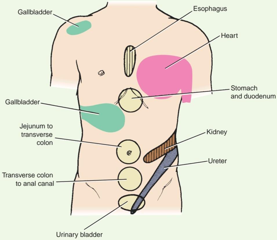
Figure 14-16 Some areas of referred pain from the viscera. In the case of the heart, the pain is usually referred to the left side of the thorax; occasionally, it is referred to both sides.
شکل ۱۴-۱۶ برخی از نواحی درد ارجاعی از احشاء. در مورد قلب، درد معمولاً به سمت چپ قفسه سینه ارجاع میشود؛ گاهی اوقات، به هر دو طرف ارجاع میشود.
Cardiac Pain
Pain originating in the heart as the result of acute myocardial ischemia is assumed to be caused by oxygen deficiency and the accumulation of metabolites, which stimulate the sensory nerve endings in the myocardium. The afferent nerve fibers ascend to the CNS through the cardiac branches of the sympathetic trunk and enter the spinal cord through the posterior roots of the upper four thoracic nerves. The nature of the pain varies considerably, from a severe crushing pain to nothing more than a mild discomfort.
درد قلبی
فرض بر این است که دردی که در نتیجه ایسکمی حاد میوکارد از قلب سرچشمه میگیرد، ناشی از کمبود اکسیژن و تجمع متابولیتهایی است که انتهای اعصاب حسی را در میوکارد تحریک میکنند. رشتههای عصبی آوران از طریق شاخههای قلبی تنه سمپاتیک به سیستم عصبی مرکزی صعود میکنند و از طریق ریشههای خلفی چهار عصب فوقانی قفسه سینه وارد نخاع میشوند. ماهیت درد به طور قابل توجهی متفاوت است، از یک درد شدید و کوبنده تا چیزی بیش از یک ناراحتی خفیف.
The pain is not felt in the heart but is referred to the skin areas supplied by the corresponding spinal nerves. The skin areas supplied by the upper four intercostal nerves and by the intercostobrachial nerve (T2) are therefore affected. The intercostobrachial nerve communicates with the medial cutaneous nerve of the arm and is distributed to skin on the medial side of the upper part of the arm. A certain amount of spread of nervous information must occur within the CNS, as the pain is sometimes felt in the neck and the jaw.
درد در قلب احساس نمیشود، بلکه به نواحی پوستی که توسط اعصاب نخاعی مربوطه عصبدهی میشوند، ارجاع داده میشود. بنابراین، نواحی پوستی که توسط چهار عصب بین دندهای فوقانی و عصب بین دندهای-بازویی (T2) عصبدهی میشوند، تحت تأثیر قرار میگیرند. عصب بین دندهای-بازویی با عصب جلدی داخلی بازو ارتباط برقرار میکند و در پوست سمت داخلی قسمت بالای بازو توزیع میشود. مقدار مشخصی از انتشار اطلاعات عصبی باید در سیستم عصبی مرکزی رخ دهد، زیرا درد گاهی اوقات در گردن و فک احساس میشود.
Myocardial infarction involving the inferior wall or diaphragmatic surface of the heart often gives rise to discomfort in the epigastrium, just below the sternum. One must assume that the afferent pain fibers from the heart ascend in the sympathetic nerves and enter the spinal cord in the posterior roots of the seventh, eighth, and ninth thoracic spinal nerves and give rise to referred pain in the T7-T9 thoracic dermatomes in the epigastrium.
انفارکتوس میوکارد که دیواره تحتانی یا سطح دیافراگم قلب را درگیر میکند، اغلب باعث ناراحتی در اپیگاستر، درست زیر جناغ سینه، میشود. باید فرض کرد که فیبرهای درد آوران از قلب در اعصاب سمپاتیک بالا میروند و در ریشههای خلفی اعصاب نخاعی هفتم، هشتم و نهم قفسه سینه وارد نخاع میشوند و باعث درد ارجاعی در درماتومهای قفسه سینه T7-T9 در اپیگاستر میشوند.
Because the heart and the thoracic part of the esophagus probably have similar afferent pain pathways, pain from acute esophagitis can mimic the pain of myocardial infarction.
از آنجا که قلب و قسمت قفسه سینه مری احتمالاً مسیرهای درد آوران مشابهی دارند، درد ناشی از ازوفاژیت حاد میتواند درد انفارکتوس میوکارد را تقلید کند.
Stomach Pain
Referred pain from the stomach is commonly felt in the epigastrium. The afferent pain fibers from the stomach ascend in company with the sympathetic nerves and pass through the celiac plexus and the greater splanchnic nerves. The sensory fibers enter the spinal cord at segments T5-T9 and give rise to referred pain in the dermatomes T5-T9 on the lower chest and abdominal walls.
درد معده
درد ارجاعی معده معمولاً در ناحیه اپیگاستر احساس میشود. فیبرهای درد آوران از معده به همراه اعصاب سمپاتیک بالا میروند و از شبکه سلیاک و اعصاب اسپلانکنیک بزرگ عبور میکنند. فیبرهای حسی در قطعات T5-T9 وارد نخاع میشوند و باعث درد ارجاعی در درماتومهای T5-T9 در دیوارههای تحتانی قفسه سینه و شکم میشوند.
Appendicular Pain
Visceral pain from the appendix is produced by distention of its lumen or spasm of its muscle. It travels in nerve fibers that accompany sympathetic nerves through the superior mesenteric plexus and the lesser splanchnic nerve to the spinal cord (T10 segment). The vague referred pain is felt in the region of the umbilicus, which is innervated by the 10th intercostal nerve (T10 dermatome). Later, when the inflammatory process involves the parietal peritoneum in the right iliac fossa, which is innervated by the 12th tho- racic and 1st lumbar spinal nerves, the now somatic pain becomes severe and dominates the clinical picture. The somatic pain is localized precisely to the right lower quadrant of the anterior abdominal wall (T12-L1 dermatomes).
درد آپاندیس
درد احشایی آپاندیس در اثر اتساع مجرای آن یا اسپاسم عضلهاش ایجاد میشود. این درد در رشتههای عصبی که همراه اعصاب سمپاتیک هستند، از طریق شبکه مزانتریک فوقانی و عصب اسپلانکنیک کوچک به نخاع (قطعه T10) میرسد. درد مبهم ارجاعی در ناحیه ناف احساس میشود که توسط عصب بین دندهای دهم (درماتوم T10) عصبدهی میشود. بعداً، هنگامی که فرآیند التهابی، صفاق جداری در حفره ایلیاک راست را درگیر میکند، که توسط اعصاب نخاعی سینهای دوازدهم و کمری اول عصبدهی میشود، درد جسمی که اکنون به صورت درد جسمی بروز میکند، شدید میشود و تصویر بالینی غالب را تشکیل میدهد. درد جسمی دقیقاً در ربع تحتانی راست دیواره قدامی شکم (درماتومهای T12-L1) قرار دارد.
Gallbladder Pain
Visceral pain impulses from the gallbladder (acute cholecystitis, gallstone colic) travel in nerve fibers that accompany sympathetic fibers through the celiac plexus and the greater splanchnic nerves to the spinal cord (segments T5-T9). The vague referred pain is felt in the dermatomes (T5-T9) on the lower chest and upper abdominal walls. Should the inflammatory process spread to involve the parietal peritoneum of the anterior abdominal wall or peripheral diaphragm, the now severe somatic pain will be felt in the right upper quadrant of the anterior abdominal wall and through to the back below the inferior angle of the scapula. Involvement of the central diaphragmatic parietal peritoneum, which is innervated by the phrenic nerve (C3-C5), may give rise to referred pain to the tip of the shoulder, since the skin in this area is innervated by the supraclavicular nerves (C3-C4).
درد کیسه صفرا
ایمپالسهای درد احشایی از کیسه صفرا (کوله سیستیت حاد، قولنج سنگ کیسه صفرا) در فیبرهای عصبی که همراه با فیبرهای سمپاتیک هستند، از طریق شبکه سلیاک و اعصاب اسپلانکنیک بزرگ به نخاع (قطعات T5-T9) میروند. درد مبهم ارجاعی در درماتومها (T5-T9) در قسمت پایین قفسه سینه و دیوارههای فوقانی شکم احساس میشود. اگر روند التهابی گسترش یابد و صفاق جداری دیواره قدامی شکم یا دیافراگم محیطی را درگیر کند، درد شدید جسمی در ربع فوقانی راست دیواره قدامی شکم و از طریق پشت به زیر زاویه تحتانی کتف احساس خواهد شد. درگیری صفاق جداری دیافراگم مرکزی، که توسط عصب فرنیک (C3-C5) عصبدهی میشود، ممکن است باعث درد ارجاعی به نوک شانه شود، زیرا پوست این ناحیه توسط اعصاب سوپراکلاویکولار (C3-C4) عصبدهی میشود.
Causalgia
Causalgia is a painful condition of the arm or leg accompanied by trophic changes in the affected skin and nails. It commonly follows crushing or partial division of the median nerve in the arm or the tibial nerve in the leg. It is thought that the descending impulses in the sympathetic postganglionic fibers in some way evoke ascending impulses in the afferent pain fibers at the site of injury. In many instances, sympathectomy has relieved the pain of causalgia.
درد ناشی از گزگز
گزگز یک وضعیت دردناک در بازو یا پا است که با تغییرات تروفیک در پوست و ناخنهای آسیبدیده همراه است. این درد معمولاً پس از له شدن یا تقسیم جزئی عصب مدیان در بازو یا عصب تیبیال در پا ایجاد میشود. تصور میشود که تکانههای نزولی در الیاف پسگانگلیونی سمپاتیک به نوعی باعث ایجاد تکانههای صعودی در الیاف درد آوران در محل آسیب میشوند. در بسیاری از موارد، سمپاتکتومی درد ناشی از گزگز را تسکین داده است.
Key Concepts
Organization
مفاهیم کلیدی
سازماندهی
• The ANS is divided into two parts, sympathetic and parasympathetic.
• ANS به دو بخش سمپاتیک و پاراسمپاتیک تقسیم میشود.
• The function of the sympathetic system is to prepare the body for emergency by increasing heart rate, constricting peripheral blood vessels, increasing blood supply to muscles, and increasing blood pressure. Thus, it redistributes blood to organs that need high performance in emergency situations.
• وظیفه سیستم سمپاتیک آماده سازی بدن برای شرایط اضطراری از طریق افزایش ضربان قلب، انقباض رگ های خونی محیطی، افزایش خون رسانی به عضلات و افزایش فشار خون است. بدین ترتیب، خون را در اندام هایی که در شرایط اضطراری به عملکرد بالا نیاز دارند، مجدداً توزیع می کند.
• Efferent fibers of the sympathetic system originate in the lateral gray columns of the spinal cord. Myelinated axons leave the cord in the anterior root and pass via the white rami communicantes to the paravertebral ganglia of the sympathetic trunk.
• فیبرهای وابران سیستم سمپاتیک از ستونهای خاکستری جانبی نخاع منشأ میگیرند. آکسونهای میلیندار، نخاع را در ریشه قدامی ترک کرده و از طریق شاخههای سفید ارتباطی به گانگلیونهای پاراورتبرال تنه سمپاتیک میروند.
• Once these presynaptic fibers reach the sympathetic trunks, they are distributed as follows:
• هنگامی که این فیبرهای پیش سیناپسی به تنههای سمپاتیک میرسند، به صورت زیر توزیع میشوند:
• Synapse with a postsynaptic sympathetic neuron in the ganglion.
• با یک نورون سمپاتیک پس سیناپسی در گانگلیون سیناپس برقرار می کند.
• Travel superiorly or inferiorly to synapse at a different level of the sympathetic trunk. For instance, T1 fibers must travel superiorly to the superior cervical ganglion.
• به سمت بالا یا پایین به سیناپس در سطوح مختلف تنه سمپاتیک حرکت میکنند. برای مثال، فیبرهای T1 باید به سمت بالا به گانگلیون فوقانی گردنی حرکت کنند.
• Pass through the ganglia without synapsing, as the greater, lesser, or least splanchnic nerve, to synapse in a prevertebral sympathetic ganglion.
• بدون سیناپس زدن، مانند عصب بزرگ، کوچک یا حداقل اسپلانکنیک، از گانگلیونها عبور کنید تا در یک گانگلیون سمپاتیک پیش مهرهای سیناپس ایجاد کنید.
• Afferent information from the viscera travels through the sympathetic ganglia without synapsing and ascends to higher centers.
• اطلاعات آوران از احشاء بدون سیناپس از طریق گانگلیون های سمپاتیک عبور کرده و به مراکز بالاتر صعود می کنند.
• The function of the parasympathetic system is to conserve and restore energy. The heart rate is slowed, pupils are constricted, peristalsis and glandular activity is increased, and the bladder walls are contracted.
• وظیفه سیستم پاراسمپاتیک حفظ و بازیابی انرژی است. ضربان قلب کند میشود، مردمک چشم تنگ میشود، حرکات دودی و فعالیت غدد افزایش مییابد و دیوارههای مثانه منقبض میشوند.
• Efferent fibers of the parasympathetic system originate in the parasympathetic nuclei of cranial nerves III, VII, IX, and X and the sacral spinal cord segments S2-54.
• فیبرهای وابران سیستم پاراسمپاتیک از هستههای پاراسمپاتیک اعصاب جمجمهای III، VII، IX و X و قطعات نخاعی خاجی S2-54 منشأ میگیرند.
• Presynaptic parasympathetic fibers of cranial nerves II, VII, and IX synapse in the parasympathetic ganglia of the head-specifically-ciliary, otic, pterygopalatine, and submandibular.
• فیبرهای پاراسمپاتیک پیش سیناپسی اعصاب جمجمهای II، VII و IX در گانگلیونهای پاراسمپاتیک سر – به طور خاص – مژگانی، اوتیک، پتریگوپالاتین و تحت فکی سیناپس میدهند.
• Afferent fibers from viscera travel with the cranial nerves and the pelvic splanchnic nerves.
• رشتههای آوران از احشاء به همراه اعصاب جمجمهای و اعصاب اسپلانکنیک لگنی حرکت میکنند.
• The sympathetic system has long postganglionic fibers, whereas the parasympathetic has short fibers.
• سیستم سمپاتیک دارای فیبرهای پس عقدهای بلند است، در حالی که پاراسمپاتیک دارای فیبرهای کوتاه است.
Neurotransmitters
• The preganglionic fibers in both the sympathetic and parasympathetic systems are cholinergic, meaning they synthesize and release ACh at the synapse.
انتقالدهندههای عصبی
• فیبرهای پیشگانگلیونی در هر دو سیستم سمپاتیک و پاراسمپاتیک کولینرژیک هستند، به این معنی که استیلکولینول (ACh) را در سیناپس سنتز و آزاد میکنند.
• The postganglionic fibers in the parasympathetic are also cholinergic.
• فیبرهای پس عقده ای در پاراسمپاتیک نیز کولینرژیک هستند.
• Most postganglionic sympathetic neurons release norepinephrine; however, fibers to sweat glands and blood vessels are cholinergic.
• بیشتر نورونهای سمپاتیک پسگانگلیونی، نوراپی نفرین آزاد میکنند؛ با این حال، فیبرهای غدد عرق و رگهای خونی کولینرژیک هستند.
Important Autonomic Innervations
• The skin of the upper eyelid receives sympathetic innervation from the superior cervical ganglion.
عصبدهیهای مهم اتونومیک
• پوست پلک فوقانی از گانگلیون فوقانی گردنی عصبدهی سمپاتیک دریافت میکند.
• The pupil of the iris is constricted by parasympathetic from cranial nerve III and dilated by sympathetic fibers from the superior cervical ganglion.
• مردمک عنبیه توسط پاراسمپاتیک از عصب جمجمهای III تنگ و توسط فیبرهای سمپاتیک از گانگلیون فوقانی گردنی گشاد میشود.
• The lacrimal gland receives parasympathetic fibers from cranial nerve VII and sympathetic vasoconstrictor fibers from the superior cervical ganglion.
• غده اشکی فیبرهای پاراسمپاتیک را از عصب جمجمهای VII و فیبرهای سمپاتیک منقبض کننده عروق را از گانگلیون فوقانی گردنی دریافت میکند.
• Submandibular and sublingual glands receive parasympathetic innervation from cranial nerve VII and sympathetic fibers from the superior cervical ganglion.
• غدد تحت فکی و زیر زبانی عصب پاراسمپاتیک را از عصب جمجمه ای VII و فیبرهای سمپاتیک را از گانگلیون فوقانی گردنی دریافت می کنند.
• The parotid gland receives parasympathetic fibers from cranial nerve IX and sympathetic fibers from the superior cervical ganglion.
• غده پاروتید فیبرهای پاراسمپاتیک را از عصب جمجمه ای IX و فیبرهای سمپاتیک را از گانگلیون فوقانی گردنی دریافت می کند.
• The gastrointestinal tract, up to the splenic flexure, receives parasympathetic innervation from cranial nerve X and sympathetic fibers from the greater and lesser splanchnic nerves.
• دستگاه گوارش، تا خم طحال، عصب پاراسمپاتیک را از عصب جمجمهای X و فیبرهای سمپاتیک را از اعصاب اسپلانکنیک بزرگ و کوچک دریافت میکند.
• The descending colon, sigmoid colon, and rectum receive parasympathetic innervation from the pelvic splanchnic nerves and sympathetic fibers from the lumbar part of the sympathetic trunk.
• کولون نزولی، کولون سیگموئید و رکتوم عصب پاراسمپاتیک را از اعصاب اسپلانکنیک لگنی و فیبرهای سمپاتیک را از قسمت کمری تنه سمپاتیک دریافت می کنند.
«فصل چهاردهم کتاب نورواناتومی بالینی اسنل»
🚀 با ما همراه شوید!
تازهترین مطالب و آموزشهای مغز و اعصاب را از دست ندهید. با فالو کردن کانال تلگرام آیندهنگاران مغز، از ما حمایت کنید!