
فیزیولوژی پزشکی گایتون و هال؛ هضم و جذب در دستگاه گوارش


دعای مطالعه [ نمایش ]
بِسْمِ الله الرَّحْمنِ الرَّحیمِ
اَللّهُمَّ اَخْرِجْنى مِنْ ظُلُماتِ الْوَهْمِ
خدايا مرا بيرون آور از تاريكىهاى وهم،
وَ اَكْرِمْنى بِنُورِ الْفَهْمِ
و به نور فهم گرامى ام بدار،
اَللّهُمَّ افْتَحْ عَلَيْنا اَبْوابَ رَحْمَتِكَ
خدايا درهاى رحمتت را به روى ما بگشا،
وَانْشُرْ عَلَيْنا خَزائِنَ عُلُومِكَ بِرَحْمَتِكَ يا اَرْحَمَ الرّاحِمينَ
و خزانههاى علومت را بر ما باز كن به امید رحمتت اى مهربانترين مهربانان.
کتاب «فیزیولوژی پزشکی گایتون وهال» بهعنوان یکی از جامعترین و معتبرترین منابع در حوزه علوم پزشکی، همچنان مرجع کلیدی برای درک عملکرد پیچیده بدن انسان است. این اثر با تکیه بر تازهترین پژوهشها و توضیحات دقیق از سازوکارهای فیزیولوژیک، پلی میان علوم پایه پزشکی و کاربردهای بالینی ایجاد میکند و نقشی بیبدیل در آموزش، پژوهش و ارتقای دانش سلامت ایفا مینماید.
ترجمه دقیق و علمیاین شاهکار توسط برند علمیآیندهنگاران مغز به مدیریت داریوش طاهری، دسترسی فارسیزبانان به مرزهای نوین دانش فیزیولوژی را ممکن ساخته و رسالتی علمیبرای ارتقای آموزش پزشکی، فهم عمیقتر سازوکارهای بدن و توسعه روشهای نوین در حوزه سلامت فراهم آورده است.
» Guyton and Hall Textbook of Medical Physiology
»» CHAPTER 66: Digestion and Absorption in the Gastrointestinal Tract
The major foods on which the body lives (with the exception of small quantities of substances such as vitamins and minerals) are carbohydrates, fats, and proteins. They generally cannot be absorbed in their natural forms through the gastrointestinal mucosa, and for this reason, they are useless as nutrients without preliminary digestion. This chapter discusses the processes by which carbohydrates, fats, and proteins are digested into small enough compounds for absorption and the mechanisms by which the digestive end products, as well as water, electrolytes, and other substances, are absorbed.
غذاهای اصلی که بدن با آنها زندگی میکند (به استثنای مقادیر کمیاز مواد مانند ویتامینها و مواد معدنی) کربوهیدراتها، چربیها و پروتئینها هستند. آنها عموماً نمیتوانند به شکل طبیعی از طریق مخاط دستگاه گوارش جذب شوند و به همین دلیل بدون هضم اولیه به عنوان مواد مغذی بی فایده هستند. این فصل فرآیندهایی را که توسط آن کربوهیدراتها، چربیها و پروتئینها به ترکیبات کوچک به اندازه کافی برای جذب هضم میشوند و مکانیسمهایی که محصولات نهایی گوارشی و همچنین آب، الکترولیتها و سایر مواد جذب میشوند، مورد بحث قرار میدهد.
DIGESTION OF VARIOUS FOODS BY HYDROLYSIS
Hydrolysis of Carbohydrates. Almost all the carbohydrates of the diet are either large polysaccharides or disaccharides, which are combinations of monosaccharides bound to one another by condensation. This phenomenon means that a hydrogen ion (H+) has been removed from one of the monosaccharides, and a hydroxyl ion (OH-) has been removed from the next one. The two monosaccharides then combine with each other at these sites of removal, and the H+ and OH- then combine to form water (H2O).
هضم مواد غذایی مختلف با هیدرولیز
هیدرولیز کربوهیدراتها. تقریباً تمام کربوهیدراتهای جیره یا پلی ساکاریدهای بزرگ یا دی ساکاریدها هستند که ترکیبی از مونوساکاریدها هستند که توسط تراکم به یکدیگر متصل میشوند. این پدیده به این معنی است که یک یون هیدروژن (+H) از یکی از مونوساکاریدها و یک یون هیدروکسیل (–OH) از یون بعدی حذف شده است. سپس دو مونوساکارید در این مکانهای حذف با یکدیگر ترکیب میشوند و +H و –OH سپس ترکیب میشوند و آب (H2O) را تشکیل میدهند.
When carbohydrates are digested, this process is reversed, and the carbohydrates are converted into mono- saccharides. Specific enzymes in the digestive juices of the gastrointestinal tract return the H+ and OH- from H2O to the polysaccharides and thereby separate the monosaccharides from each other. This process, called hydrolysis, is the following (in which R”-R’ is a disaccharide):
هنگامیکه کربوهیدراتها هضم میشوند، این روند معکوس میشود و کربوهیدراتها به مونو ساکارید تبدیل میشوند. آنزیمهای خاص در شیرههای گوارشی دستگاه گوارش، H+ و OH- را از H2O به پلی ساکاریدها برمیگرداند و در نتیجه مونوساکاریدها را از یکدیگر جدا میکند. این فرآیند که هیدرولیز نامیده میشود به شرح زیر است (که در آن R”-R’ یک دی ساکارید است):

Hydrolysis of Fats. Almost the entire fat portion of the diet consists of triglycerides (neutral fats), which are combinations of three fatty acid molecules condensed with a single glycerol molecule. During condensation, three molecules of water are removed.
هیدرولیز چربیها. تقریباً کل بخش چربی رژیم از تری گلیسیریدها (چربیهای خنثی) تشکیل شده است که ترکیبی از سه مولکول اسید چرب متراکم شده با یک مولکول گلیسرول هستند. در طول تراکم، سه مولکول آب حذف میشود.
Hydrolysis (digestion) of the triglycerides consists of the reverse process: the fat-digesting enzymes return three molecules of water to the triglyceride molecule and thereby split the fatty acid molecules away from the glycerol.
هیدرولیز (هضم) تری گلیسیریدها شامل فرآیند معکوس است: آنزیمهای هضم کننده چربی سه مولکول آب را به مولکول تری گلیسیرید باز میگرداند و در نتیجه مولکولهای اسید چرب را از گلیسرول جدا میکنند.
Hydrolysis of Proteins. Proteins are formed from multiple amino acids that are bound together by peptide link- ages. At each linkage, a OH- has been removed from one amino acid and a H+ has been removed from the succeeding one; thus, the successive amino acids in the protein chain are also bound together by condensation, and digestion occurs by the reverse effect: hydrolysis. That is, the proteolytic enzymes return H+ and OH- from water molecules to the protein molecules to split them into their constituent amino acids.
هیدرولیز پروتئینها. پروتئینها از اسیدهای آمینه متعددی که توسط پیوندهای پپتیدی به یکدیگر متصل شده اند تشکیل میشوند. در هر پیوند، یک –OH از یک اسید آمینه و یک +H از یکی بعدی حذف شده است. بنابراین، اسیدهای آمینه متوالی در زنجیره پروتئین نیز با تراکم به یکدیگر متصل میشوند و هضم با اثر معکوس انجام میشود: هیدرولیز. یعنی آنزیمهای پروتئولیتیک +H و –OH را از مولکولهای آب به مولکولهای پروتئین برمیگردانند تا آنها را به اسیدهای آمینه تشکیلدهنده خود تقسیم کنند.
Therefore, the chemistry of digestion is simple because, in the case of all three major types of food, the same basic process of hydrolysis is involved. The only difference lies in the types of enzymes required to promote the hydrolysis reactions for each type of food.
بنابراین، شیمیهضم ساده است، زیرا در مورد هر سه نوع اصلی غذا، فرآیند اساسی هیدرولیز یکسان درگیر است. تنها تفاوت در انواع آنزیمهای مورد نیاز برای تحریک واکنشهای هیدرولیز برای هر نوع غذا نهفته است.
All the digestive enzymes are proteins. Their secretion by the different gastrointestinal glands was discussed in Chapter 65.
تمام آنزیمهای گوارشی پروتئین هستند. ترشح آنها توسط غدد مختلف گوارشی در فصل 65 مورد بحث قرار گرفت.
DIGESTION OF CARBOHYDRATES
Carbohydrate Foods of the Diet. Only three major sources of carbohydrates exist in the normal human diet. They are sucrose, which is the disaccharide known popularly as cane sugar; lactose, which is a disaccharide found in milk; and starches, which are large polysaccharides present in almost all nonanimal foods, particularly in potatoes and different types of grains. Other carbohydrates ingested to a slight extent are amylose, glycogen, alcohol, lactic acid, pyruvic acid, pectins, dextrins, and minor quantities of carbohydrate derivatives in meats.
هضم کربوهیدراتها
غذاهای کربوهیدراتی رژیم. تنها سه منبع اصلی کربوهیدرات در رژیم غذایی عادی انسان وجود دارد. آنها ساکارز هستند که دی ساکارید معروف به نیشکر است. لاکتوز که یک دی ساکارید موجود در شیر است. و نشاسته، که پلی ساکاریدهای بزرگی هستند که تقریباً در تمام غذاهای غیر حیوانی، به ویژه در سیب زمینی و انواع مختلف غلات وجود دارند. سایر کربوهیدراتهای مصرف شده به میزان جزئی عبارتند از آمیلوز، گلیکوژن، الکل، اسید لاکتیک، اسید پیروویک، پکتین، دکسترین و مقادیر جزئی مشتقات کربوهیدرات در گوشت.
The diet also contains a large amount of cellulose, which is a carbohydrate. However, enzymes capable of hydrolyzing cellulose are not secreted in the human digestive tract. Consequently, cellulose cannot be considered a food for humans.
این رژیم همچنین حاوی مقدار زیادی سلولز است که یک کربوهیدرات است. با این حال، آنزیمهایی که قادر به هیدرولیز سلولز هستند در دستگاه گوارش انسان ترشح نمیشوند. در نتیجه سلولز را نمیتوان غذای انسان دانست.
Digestion of Carbohydrates Begins in the Mouth and Stomach. When food is chewed, it is mixed with saliva, which contains the digestive enzyme ptyalin (an a-amylase) secreted mainly by the parotid glands. This enzyme hydrolyzes starch into the disaccharide maltose and other small polymers of glucose that contain three to nine glucose molecules, as shown in Figure 66-1. How- ever, the food remains in the mouth only a short time, so probably not more than 5% of all the starches become hydrolyzed by the time the food is swallowed.
هضم کربوهیدراتها از دهان و معده شروع میشود. هنگامیکه غذا جویده میشود، با بزاق مخلوط میشود که حاوی آنزیم گوارشی ptyalin (یک a-amylase) است که عمدتاً توسط غدد بناگوشی ترشح میشود. این آنزیم نشاسته را به دی ساکارید مالتوز و دیگر پلیمرهای کوچک گلوکز که حاوی سه تا نه مولکول گلوکز هستند، هیدرولیز میکند، همانطور که در شکل 66-1 نشان داده شده است. با این حال، غذا فقط برای مدت کوتاهی در دهان باقی میماند، بنابراین احتمالاً بیش از 5 درصد از تمام نشاستهها تا زمان بلعیده شدن غذا هیدرولیز نمیشوند.
Starch digestion sometimes continues in the body and fundus of the stomach for as long as 1 hour before the food becomes mixed with the stomach secretions. Activity of the salivary amylase is then blocked by acid of the gastric secretions because the amylase is essentially inactive as an enzyme once the pH of the medium falls below about 4.0. Nevertheless, on average, before food and its accompanying saliva become completely mixed with the gastric secretions, as much as 30% to 40% of the starches will have been hydrolyzed, mainly to form maltose.
هضم نشاسته گاهی در بدن و فوندوس معده تا 1 ساعت قبل از مخلوط شدن غذا با ترشحات معده ادامه مییابد. سپس فعالیت آمیلاز بزاقی توسط اسید ترشحات معده مسدود میشود زیرا زمانی که PH محیط به کمتر از 4 برسد، آمیلاز اساساً به عنوان یک آنزیم غیرفعال است. با این وجود، به طور متوسط، قبل از اینکه غذا و بزاق همراه آن به طور کامل با ترشحات معده مخلوط شوند، 30 تا 40 درصد نشاستهها هیدرولیز شده و عمدتاً به مالتوز تبدیل میشوند.

Figure 66-1. Digestion of carbohydrates.
شکل 66-1. هضم کربوهیدراتها.
DIGESTION OF CARBOHYDRATES IN THE SMALL INTESTINE
Digestion by Pancreatic Amylase. Pancreatic secretion, like saliva, contains a large quantity of a-amylase that is almost identical in its function to the a-amylase of saliva but is several times as powerful. Therefore, within 15 to 30 minutes after the chyme empties from the stomach into the duodenum and mixes with pancreatic juice, virtually all the carbohydrates will have become digested.
هضم کربوهیدراتها در روده کوچک
هضم توسط آمیلاز پانکراس. ترشح لوزالمعده مانند بزاق حاوی مقدار زیادی a-amylase است که عملکرد آن تقریباً مشابه a-amylase بزاق است اما چندین برابر قوی تر است. بنابراین، در عرض 15 تا 30 دقیقه پس از تخلیه کیم از معده به دوازدهه و مخلوط شدن با آب پانکراس، تقریباً تمام کربوهیدراتها هضم میشوند.
In general, the carbohydrates are almost totally converted into maltose and/or other small glucose polymers before passing beyond the duodenum or upper jejunum.
به طور کلی، کربوهیدراتها تقریباً به طور کامل به مالتوز و/یا سایر پلیمرهای گلوکز کوچک قبل از عبور از دوازدهه یا ژژنوم فوقانی تبدیل میشوند.
Hydrolysis of Disaccharides and Small Glucose Polymers Into Monosaccharides by Intestinal Epithelial Enzymes. The enterocytes lining the villi of the small intestine contain four enzymes (lactase, sucrase, maltase, and a-dextrinase), which are capable of splitting the disaccharides lactose, sucrose, and maltose, plus other small glucose polymers, into their constituent monosaccharides. These enzymes are located in the enterocytes covering the intestinal microvilli brush border, so the disaccharides are digested as they come in contact with these enterocytes.
هیدرولیز دی ساکاریدها و پلیمرهای گلوکز کوچک به مونوساکاریدها توسط آنزیمهای اپیتلیال روده. انتروسیتهای پوشاننده پرزهای روده کوچک حاوی چهار آنزیم (لاکتاز، سوکراز، مالتاز و a-دکستریناز) هستند که قادرند دی ساکاریدهای لاکتوز، ساکارز و مالتوز و دیگر پلیمرهای گلوکز کوچک را به مونوساکاریدهای تشکیل دهنده خود تقسیم کنند. این آنزیمها در انتروسیتهایی قرار دارند که مرز برس میکروویلی روده را میپوشانند، بنابراین دی ساکاریدها در تماس با این انتروسیتها هضم میشوند.
Lactose splits into a molecule of galactose and a molecule of glucose. Sucrose splits into a molecule of fructose and a molecule of glucose. Maltose and other small glucose polymers all split into multiple molecules of glucose. Thus, the final products of carbohydrate digestion are all monosaccharides. They are all water soluble and are absorbed immediately into the portal blood.
لاکتوز به یک مولکول گالاکتوز و یک مولکول گلوکز تقسیم میشود. ساکارز به یک مولکول فروکتوز و یک مولکول گلوکز تقسیم میشود. مالتوز و سایر پلیمرهای گلوکز کوچک همگی به چندین مولکول گلوکز تقسیم میشوند. بنابراین، محصولات نهایی هضم کربوهیدراتها همگی مونوساکاریدها هستند. همه آنها محلول در آب هستند و بلافاصله در خون پورت جذب میشوند.
In the ordinary diet, which contains far more starches than all other carbohydrates combined, glucose represents more than 80% of the final products of carbohydrate digestion, and galactose and fructose each seldom represent more than 10%.
در رژیم غذایی معمولی که حاوی نشاسته بسیار بیشتری نسبت به سایر کربوهیدراتها است، گلوکز بیش از 80 درصد از محصولات نهایی هضم کربوهیدرات را تشکیل میدهد و گالاکتوز و فروکتوز هر کدام به ندرت بیش از 10 درصد را نشان میدهند.
The major steps in carbohydrate digestion are summarized in Figure 66-1.
مراحل اصلی هضم کربوهیدرات در شکل 66-1 خلاصه شده است.
DIGESTION OF PROTEINS
Proteins of the Diet. Dietary proteins are chemically long chains of amino acids bound together by peptide linkages. A typical linkage is the following:
هضم پروتئینها
پروتئینهای رژیم غذایی. پروتئینهای غذایی از نظر شیمیایی زنجیرههای بلندی از اسیدهای آمینه هستند که توسط پیوندهای پپتیدی به یکدیگر متصل شده اند. یک پیوند معمولی به شرح زیر است:

The characteristics of each protein are determined by the types of amino acids in the protein molecule and by the sequential arrangements of these amino acids. The physical and chemical characteristics of different proteins important in human tissues are discussed in Chapter 70.
خصوصیات هر پروتئین با توجه به انواع اسیدهای آمینه موجود در مولکول پروتئین و ترتیب متوالی این اسیدهای آمینه تعیین میشود. ویژگیهای فیزیکی و شیمیایی پروتئینهای مختلف مهم در بافتهای انسانی در فصل 70 مورد بحث قرار گرفته است.
Digestion of Proteins in the Stomach. Pepsin, an important peptic enzyme of the stomach, is most active at a pH of 2.0 to 3.0 and is inactive at a pH above about 5.0. Consequently, for this enzyme to cause digestion of protein, the stomach juices must be acidic. As explained in Chapter 65, the gastric glands secrete a large quantity of hydrochloric acid. This hydrochloric acid is secreted by the parietal (oxyntic) cells in the glands at a pH of about 0.8, but by the time it is mixed with the stomach contents and with secretions from the non-oxyntic glandular cells of the stomach, the pH then averages around 2.0 to 3.0, a highly favorable range of acidity for pepsin activity.
هضم پروتئین در معده. پپسین، آنزیم پپتیک مهم معده، در pH 2.0 تا 3.0 بیشترین فعالیت را دارد و در pH بالای حدود 5.0 غیر فعال است. در نتیجه برای اینکه این آنزیم باعث هضم پروتئین شود، شیره معده باید اسیدی باشد. همانطور که در فصل 65 توضیح داده شد، غدد معده مقدار زیادی اسید هیدروکلریک ترشح میکنند. این اسید هیدروکلریک توسط سلولهای جداری (اکسینتیک) در غدد با pH حدود 0.8 ترشح میشود، اما زمانی که با محتویات معده و ترشحات سلولهای غدهای غیر اکسینتیک معده مخلوط میشود، pH به طور متوسط بین 2.0 تا 3.0 میشود که محدوده فعالیت اسیدی بسیار مطلوبی است.
One of the important features of pepsin digestion is its ability to digest the protein collagen, an albuminoid type of protein that is affected little by other digestive enzymes. Collagen is a major constituent of the intercellular connective tissue of meats; therefore, for the digestive enzymes to penetrate meats and digest the other meat proteins, it is necessary that the collagen fibers be digested. Consequently, in people who lack pepsin in the stomach juices, the ingested meats are less well penetrated by the other digestive enzymes and, therefore, may be poorly digested.
یکی از ویژگیهای مهم هضم پپسین، توانایی آن در هضم پروتئین کلاژن است، یک نوع پروتئین آلبومینوئیدی که تحت تأثیر سایر آنزیمهای گوارشی کمیقرار میگیرد. کلاژن یکی از اجزای اصلی بافت همبند بین سلولی گوشت است. بنابراین، برای آنزیمهای گوارشی برای نفوذ به گوشتها و هضم سایر پروتئینهای گوشت، لازم است که الیاف کلاژن هضم شوند. در نتیجه، در افرادی که فاقد پپسین در شیره معده هستند، گوشتهای بلعیده شده کمتر توسط سایر آنزیمهای گوارشی نفوذ میکنند و در نتیجه ممکن است هضم ضعیفی داشته باشند.
As shown in Figure 66-2, pepsin only initiates the process of protein digestion, usually providing only 10% to 20% of the total protein digestion to convert the protein to proteoses, peptones, and a few polypeptides. This split- ting of proteins occurs as a result of hydrolysis at the pep- tide linkages between amino acids.
همانطور که در شکل 66-2 نشان داده شده است، پپسین فقط فرآیند هضم پروتئین را آغاز میکند، معمولاً تنها 10٪ تا 20٪ از کل هضم پروتئین را برای تبدیل پروتئین به پروتئوز، پپتون و چند پلی پپتید فراهم میکند. این تقسیم پروتئینها در نتیجه هیدرولیز در پیوندهای پپتیدی بین اسیدهای آمینه رخ میدهد.
Most Protein Digestion Results From Actions of Pancreatic Proteolytic Enzymes. Most protein digestion occurs in the upper small intestine, in the duodenum and jejunum, under the influence of proteolytic enzymes from pancreatic secretion. Immediately upon entering the small intestine from the stomach, the partial breakdown products of the protein foods are attacked by the major proteolytic pancreatic enzymes trypsin, chymotrypsin, carboxy poly peptidase, and elastase, as shown in Figure 66-2.
بیشتر هضم پروتئین ناشی از فعالیت آنزیمهای پروتئولیتیک پانکراس است. بیشتر هضم پروتئین در قسمت فوقانی روده کوچک، در دوازدهه و ژژنوم، تحت تأثیر آنزیمهای پروتئولیتیک از ترشح پانکراس انجام میشود. همانطور که در شکل 66-2 نشان داده شده است، بلافاصله پس از ورود از معده به روده کوچک، محصولات تجزیه نسبی غذاهای پروتئینی توسط آنزیمهای پروتئولیتیک پانکراس اصلی تریپسین، کیموتریپسین، کربوکسی پلی پپتیداز و الاستاز مورد حمله قرار میگیرند.
Both trypsin and chymotrypsin split proteins into small polypeptides; carboxy poly peptidase then cleaves individual amino acids from the carboxyl ends of the polypeptides. Proelastase, in turn, is converted into elastase, which then digests elastin fibers that partially hold meats together.
تریپسین و کیموتریپسین هر دو پروتئینها را به پلی پپتیدهای کوچک تقسیم میکنند. کربوکسی پلی پپتیداز سپس اسیدهای آمینه منفرد را از انتهای کربوکسیل پلی پپتیدها جدا میکند. پروالاستاز به نوبه خود به الاستاز تبدیل میشود که سپس فیبرهای الاستین را هضم میکند که تا حدی گوشتها را کنار هم نگه میدارد.
Only small percentages of the proteins are digested all the way to their constituent amino acids by the pancreatic juices. Most remain as dipeptides and tripeptides.
فقط درصد کمیاز پروتئینها در تمام مسیر به اسیدهای آمینه تشکیل دهنده خود توسط آب پانکراس هضم میشوند. بیشتر به صورت دی پپتید و تری پپتید باقی میمانند.
Digestion of Peptides by Peptidases in the Enterocytes That Line the Small Intestinal Villi. The last digestive stage of proteins in the intestinal lumen is achieved by enterocytes that line the villi of the small intestine, mainly in the duodenum and jejunum. These cells have a brush border that consists of hundreds of microvilli projecting from the surface of each cell. In the membrane of each of these microvilli are multiple peptidases that protrude through the membranes to the exterior, where they come in contact with the intestinal fluids.
هضم پپتیدها توسط پپتیدازها در انتروسیتهایی که پرزهای روده کوچک را میپوشانند. آخرین مرحله گوارشی پروتئینها در مجرای روده توسط انتروسیتهایی حاصل میشود که پرزهای روده کوچک، عمدتاً در دوازدهه و ژژنوم را پوشش میدهند. این سلولها دارای یک مرز برس هستند که شامل صدها میکروویلی است که از سطح هر سلول بیرون میزند. در غشای هر یک از این میکروویلیها، چندین پپتیداز وجود دارد که از طریق غشاها به بیرون بیرون زده و در تماس با مایعات روده قرار میگیرند.
Two types of peptidase enzymes are especially important, amino poly peptidase and several dipeptidases. They split the remaining larger polypeptides into tripeptides and dipeptides and a few into amino acids. The amino acids, dipeptides, and tripeptides are easily transported through the microvillar membrane to the interior of the enterocyte.
دو نوع آنزیم پپتیداز به ویژه مهم هستند، آمینو پلی پپتیداز و چندین دی پپتیداز. آنها پلی پپتیدهای بزرگتر باقیمانده را به تری پپتیدها و دی پپتیدها و تعدادی به اسیدهای آمینه تقسیم میکنند. اسیدهای آمینه، دی پپتیدها و تری پپتیدها به راحتی از طریق غشای میکروویلار به داخل انتروسیت منتقل میشوند.
Finally, inside the cytosol of the enterocyte are multiple other peptidases that are specific for the remaining types of linkages between amino acids. Within minutes, virtually all the last dipeptides and tripeptides are digested to the final stage to form single amino acids, which then pass on through to the other side of the enterocyte and thence into the blood.
در نهایت، در داخل سیتوزول انتروسیت، چندین پپتیداز دیگر وجود دارد که برای انواع باقی مانده از پیوندهای بین اسیدهای آمینه خاص هستند. در عرض چند دقیقه، تقریباً تمام آخرین دی پپتیدها و تری پپتیدها تا مرحله نهایی هضم میشوند تا اسیدهای آمینه منفرد را تشکیل دهند، که سپس به طرف دیگر انتروسیت و از آنجا به خون منتقل میشوند.
More than 99% of the final protein digestive products that are absorbed are individual amino acids, with only rare absorption of peptides and very rare absorption of whole protein molecules. Even these few absorbed molecules of whole protein can sometimes cause serious allergic or immunologic disturbances, as discussed in Chapter 35.
بیش از 99 درصد از محصولات گوارشی پروتئین نهایی که جذب میشوند، اسیدهای آمینه منفرد هستند، تنها با جذب نادر پپتیدها و جذب بسیار نادر مولکولهای پروتئین کامل. همانطور که در فصل 35 بحث شد، حتی این چند مولکول جذب شده از پروتئین کامل، گاهی اوقات میتواند باعث ایجاد اختلالات جدی آلرژیک یا ایمونولوژیک شود.

Figure 66-2. Digestion of proteins.
شکل 66-2. هضم پروتئینها
DIGESTION OF FATS
Fats of the Diet. By far the most abundant fats of the diet are the neutral fats, also known as triglycerides, each molecule of which is composed of a glycerol nucleus and three fatty acid side chains, as shown in Figure 66-3. Neutral fat is a major constituent in food of animal origin but much less so in food of plant origin.
هضم چربیها
چربیهای رژیم. تا حد زیادی فراوان ترین چربیهای رژیم، چربیهای خنثی هستند که به عنوان تری گلیسیرید نیز شناخته میشوند، هر مولکول آن از یک هسته گلیسرول و سه زنجیره جانبی اسید چرب تشکیل شده است، همانطور که در شکل 66-3 نشان داده شده است. چربی خنثی جزء اصلی مواد غذایی با منشاء حیوانی است اما در مواد غذایی با منشاء گیاهی بسیار کمتر است.
Small quantities of phospholipids, cholesterol, and cholesterol esters are also present in the usual diet. The phospholipids and cholesterol esters contain fatty acid and therefore can be considered fats. Cholesterol is a sterol compound that contains no fatty acid, but it does exhibit some of the physical and chemical characteristics of fats. In addition, it is derived from fats and is metabolized similarly to fats. Therefore, cholesterol is considered, from a dietary point of view, to be a fat.
مقادیر کمیاز فسفولیپیدها، کلسترول و استرهای کلسترول نیز در رژیم غذایی معمول وجود دارد. فسفولیپیدها و استرهای کلسترول حاوی اسید چرب هستند و بنابراین میتوان آنها را چربی در نظر گرفت. کلسترول یک ترکیب استرولی است که حاوی اسید چرب نیست، اما برخی از خصوصیات فیزیکی و شیمیایی چربیها را نشان میدهد. علاوه بر این، از چربیها مشتق شده و مشابه چربیها متابولیزه میشود. بنابراین، از نظر رژیم غذایی، کلسترول به عنوان یک چربی در نظر گرفته میشود.
Digestion of Fats Occurs Mainly in the Small Intestine. A small amount of triglycerides is digested in the stomach by lingual lipase secreted by lingual glands in the mouth and swallowed with the saliva. This amount of digestion is less than 10% and is generally unimportant. Instead, essentially all fat digestion occurs in the small intestine, as follows.
هضم چربیها عمدتاً در روده کوچک اتفاق میافتد. مقدار کمیتری گلیسیرید در معده توسط لیپاز زبانی که توسط غدد زبانی در دهان ترشح میشود هضم میشود و همراه با بزاق بلعیده میشود. این مقدار هضم کمتر از 10 درصد است و به طور کلی بی اهمیت است. در عوض، اساساً تمام هضم چربی در روده کوچک به شرح زیر انجام میشود.
The First Step in Fat Digestion Is Emulsification by Bile Acids and Lecithin. The first step in fat digestion is to physically break the fat globules into small sizes so that the water-soluble digestive enzymes can act on the globule surfaces. This process is called emulsification of the fat, and it begins by agitation in the stomach to mix the fat with the products of stomach digestion.
اولین مرحله در هضم چربی امولسیون کردن توسط اسیدهای صفراوی و لسیتین است. اولین قدم در هضم چربی، شکستن فیزیکی گلبولهای چربی به اندازههای کوچک است تا آنزیمهای گوارشی محلول در آب بتوانند روی سطوح کروی عمل کنند. این فرآیند امولسیون شدن چربی نامیده میشود و با هم زدن در معده شروع میشود تا چربی با محصولات هضم معده مخلوط شود.
Most of the emulsification then occurs in the duodenum under the influence of bile, the liver secretion that does not contain any digestive enzymes. However, bile does contain a large quantity of bile salts, as well as the phospholipid lecithin. Both of these substances, but especially the lecithin, are extremely important for emulsification of the fat. The polar parts (i.e., the points where ionization occurs in water) of the bile salts and lecithin molecules are highly soluble in water, whereas most of the remaining portions of their molecules are highly soluble in fat. Therefore, the fat-soluble portions of these liver secretions dissolve in the surface layer of the fat globules, with the polar portions projecting. The polar projections, in turn, are soluble in the surrounding watery fluids, which greatly decreases the interfacial tension of the fat and makes it soluble as well.
سپس بیشتر امولسیون در اثنی عشر تحت تأثیر صفرا، ترشح کبدی که فاقد آنزیمهای گوارشی است، رخ میدهد. با این حال، صفرا حاوی مقدار زیادی نمک صفراوی و همچنین لسیتین فسفولیپید است. هر دوی این مواد، به ویژه لسیتین، برای امولسیون کردن چربی بسیار مهم هستند. بخشهای قطبی (یعنی نقاطی که یونیزاسیون در آب اتفاق میافتد) نمکهای صفراوی و مولکولهای لسیتین بسیار محلول در آب هستند، در حالی که بیشتر بخشهای باقیمانده مولکولهای آنها در چربی بسیار محلول هستند. بنابراین، بخشهای محلول در چربی این ترشحات کبد در لایه سطحی گلبولهای چربی حل میشوند و بخشهای قطبی بیرون میآیند. برآمدگیهای قطبی به نوبه خود در مایعات آبکی اطراف محلول هستند که کشش سطحی چربی را تا حد زیادی کاهش میدهد و آن را نیز محلول میکند.
When the interfacial tension of a globule of nonmiscible fluid is low, this non-miscible fluid, upon agitation, can be broken up into many tiny particles far more easily than it can when the interfacial tension is great. Consequently, a major function of the bile salts and lecithin in the bile is to make the fat globules readily fragmentable by agitation with the water in the small bowel. This action is the same as that of many detergents that are widely used in household cleaners for removing grease.
هنگامیکه کشش سطحی یک گلبول مایع غیرقابل اختلاط کم است، این سیال غیرقابل اختلاط پس از هم زدن، بسیار راحت تر از زمانی که کشش سطحی زیاد است، میتواند به بسیاری از ذرات ریز تجزیه شود. در نتیجه، عملکرد اصلی نمکهای صفراوی و لسیتین موجود در صفرا این است که گلبولهای چربی را به راحتی با هم زدن با آب موجود در روده کوچک تکه تکه میکنند. این عمل مانند بسیاری از مواد شوینده است که به طور گسترده در پاک کنندههای خانگی برای از بین بردن چربی استفاده میشود.
Each time the diameters of the fat globules are significantly decreased as a result of agitation in the small intestine, the total surface area of the fat increases many- fold. Because the average diameter of the fat particles in the intestine after emulsification has occurred is less than 1 micrometer, this represents an increase of as much as 1000-fold in total surface areas of the fats caused by the emulsification process.
هر بار که قطر گویچههای چربی به طور قابل توجهی در نتیجه هم زدن در روده کوچک کاهش مییابد، سطح کل چربی چندین برابر افزایش مییابد. از آنجایی که قطر متوسط ذرات چربی در روده پس از امولسیون شدن کمتر از 1 میکرومتر است، این نشان دهنده افزایش 1000 برابری در سطح کل چربیها ناشی از فرآیند امولسیون سازی است.
The lipase enzymes are water-soluble and can attack the fat globules only on their surfaces. Consequently, this detergent function of bile salts and lecithin is very important for digestion of fats.
آنزیمهای لیپاز محلول در آب هستند و میتوانند گلبولهای چربی را فقط در سطح آنها مورد حمله قرار دهند. در نتیجه، این عملکرد شوینده نمکهای صفراوی و لسیتین برای هضم چربیها بسیار مهم است.
Triglycerides Are Digested by Pancreatic Lipase. By far the most important enzyme for digestion of the triglycerides is pancreatic lipase, present in enormous quantities in pancreatic juice, enough to digest within 1 minute all triglycerides that it can reach. The enterocytes of the small intestine contain additional lipase, known as enteric lipase, but it is usually not needed.
تری گلیسیرید توسط لیپاز پانکراس هضم میشود. مهم ترین آنزیم برای هضم تری گلیسیریدها، لیپاز پانکراس است که به مقدار زیادی در آب پانکراس وجود دارد، به اندازه ای که در عرض 1 دقیقه تمام تری گلیسیریدهایی را که میتواند به آن برسد هضم کند. انتروسیتهای روده کوچک حاوی لیپاز اضافی هستند که به لیپاز روده معروف است، اما معمولاً نیازی به آن نیست.

Figure 66-3. Hydrolysis of neutral fat catalyzed by lipase.
شکل 66-3. هیدرولیز چربی خنثی کاتالیز شده توسط لیپاز.
End Products of Fat Digestion Are Free Fatty Acids. Most of the triglycerides of the diet are split by pancreatic lipase into free fatty acids and 2-monoglycerides, as shown in Figure 66-4.
محصولات نهایی هضم چربی، اسیدهای چرب رایگان هستند. همانطور که در شکل 66-4 نشان داده شده است، بیشتر تری گلیسیریدهای رژیم غذایی توسط لیپاز پانکراس به اسیدهای چرب آزاد و 2-مونوگلیسرید تقسیم میشوند.

Figure 66-4. Digestion of fats.
شکل 66-4. هضم چربیها.
Bile Salts Form Micelles That Accelerate Fat Digestion. The hydrolysis of triglycerides is a highly reversible process; therefore, accumulation of monoglycerides and free fatty acids in the vicinity of digesting fats quickly blocks further digestion. However, the bile salts play the additional important role of removing the monoglycerides and free fatty acids from the vicinity of the digesting fat globules almost as rapidly as these end products of digestion are formed. This process occurs in the following way.
نمکهای صفراوی میسلهایی را تشکیل میدهند که هضم چربی را تسریع میکنند. هیدرولیز تری گلیسیرید یک فرآیند بسیار برگشت پذیر است. بنابراین، تجمع مونوگلیسریدها و اسیدهای چرب آزاد در مجاورت چربیهای هضم کننده به سرعت هضم بیشتر را مسدود میکند. با این حال، نمکهای صفراوی نقش مهم دیگری در حذف مونوگلیسریدها و اسیدهای چرب آزاد از مجاورت گلبولهای چربی در حال هضم تقریباً به همان سرعتی که این محصولات نهایی هضم تشکیل میشوند، ایفا میکنند. این فرآیند به روش زیر رخ میدهد.
When bile salts are of a high enough concentration in water, they have the propensity to form micelles, which are small spherical, cylindrical globules 3 to 6 nanometers in diameter composed of 20 to 40 molecules of bile salt. These micelles develop because each bile salt molecule is composed of a sterol nucleus that is highly fat-soluble and a polar group that is highly water-soluble. The sterol nucleus encompasses the fat digestate, forming a small fat globule in the middle of a resulting micelle, with polar groups of bile salts projecting outward to cover the surface of the micelle. Because these polar groups are negatively charged, they allow the entire micelle globule to dissolve in the water of the digestive fluids and to remain in stable solution until the fat is absorbed into the blood.
هنگامیکه نمکهای صفراوی در آب به اندازه کافی غلظت داشته باشند، تمایل به تشکیل میسلها را دارند که کرویهای کوچک کروی شکل و استوانه ای به قطر 3 تا 6 نانومتر هستند که از 20 تا 40 مولکول نمک صفراوی تشکیل شده اند. این میسلها به این دلیل رشد میکنند که هر مولکول نمک صفراوی از یک هسته استرول که بسیار محلول در چربی است و یک گروه قطبی که بسیار محلول در آب است تشکیل شده است. هسته استرول هضم چربی را در بر میگیرد و یک گلبول چربی کوچک در وسط یک میسل ایجاد میکند که گروههای قطبی نمکهای صفراوی به سمت بیرون بیرون زده و سطح میسل را میپوشانند. از آنجایی که این گروههای قطبی دارای بار منفی هستند، به کل گلبول میسل اجازه میدهند تا در آب مایعات گوارشی حل شود و تا زمانی که چربی جذب خون شود، در محلول پایدار باقی بماند.
The bile salt micelles also act as a transport medium to carry the monoglycerides and free fatty acids, both of which would otherwise be relatively insoluble, to the brush borders of the intestinal epithelial cells. There the mono- glycerides and free fatty acids are absorbed into the blood, as discussed later, but the bile salts are released back into the chyme to be used again and again for this “ferrying” process.
میسلهای نمک صفراوی همچنین به عنوان یک محیط انتقال برای حمل مونوگلیسریدها و اسیدهای چرب آزاد، که در غیر این صورت هر دو نسبتا نامحلول هستند، به مرزهای برس سلولهای اپیتلیال روده عمل میکنند. در آنجا مونو گلیسریدها و اسیدهای چرب آزاد جذب خون میشوند، همانطور که بعداً بحث شد، اما نمکهای صفراوی دوباره به داخل کیم رها میشوند تا بارها و بارها برای این فرآیند “فرری” استفاده شوند.
Digestion of Cholesterol Esters and Phospholipids. Most cholesterol in the diet is in the form of cholesterol esters, which are combinations of free cholesterol and one molecule of fatty acid. Phospholipids also contain fatty acid within their molecules. Both the cholesterol esters and the phospholipids are hydrolyzed by two other lipases in the pancreatic secretion that free the fatty acids-the enzyme cholesterol ester hydrolase to hydrolyze the cholesterol ester, and phospholipase A2 to hydrolyze the phospholipid.
هضم کلسترول استرها و فسفولیپیدها. بیشتر کلسترول در رژیم غذایی به شکل استرهای کلسترول است که ترکیبی از کلسترول آزاد و یک مولکول اسید چرب است. فسفولیپیدها همچنین حاوی اسید چرب در مولکولهای خود هستند. هم استرهای کلسترول و هم فسفولیپیدها توسط دو لیپاز دیگر در ترشح پانکراس هیدرولیز میشوند که اسیدهای چرب را آزاد میکنند – آنزیم استر کلسترول هیدرولاز برای هیدرولیز استر کلسترول و فسفولیپاز A2 برای هیدرولیز فسفولیپید.
The bile salt micelles play the same role in “ferrying” free cholesterol and phospholipid molecule digestates that they play in ferrying monoglycerides and free fatty acids. Indeed, essentially no cholesterol is absorbed with- out this function of the micelles.
میسلهای نمک صفراوی همان نقشی را در انتقال کلسترول آزاد و هضمهای مولکول فسفولیپید ایفا میکنند که در انتقال مونوگلیسریدها و اسیدهای چرب آزاد بازی میکنند. در واقع، اساساً هیچ کلسترولی بدون این عملکرد میسلها جذب نمیشود.
BASIC PRINCIPLES OF GASTROINTESTINAL ABSORPTION
We suggest that the reader review the basic principles of cell membrane transport of substances discussed in Chapter 4. The following paragraphs present specialized applications of these transport processes during gastrointestinal absorption.
اصول اساسی جذب گوارشی
ما به خواننده پیشنهاد میکنیم که اصول اساسی انتقال غشای سلولی مواد مورد بحث در فصل 4 را بررسی کند.
ANATOMICAL BASIS OF ABSORPTION
The total quantity of fluid that must be absorbed each day by the intestines is equal to the ingested fluid (~1.5 liters) plus that secreted in the various gastrointestinal secretions (~7 liters), which comes to a total of 8 to 9 liters. All but about 1.5 liters of this fluid is absorbed in the small intestine, leaving only 1.5 liters to pass through the ileocecal valve into the colon each day.
اساس آناتومیک جذب
مقدار کل مایعی که باید هر روز توسط رودهها جذب شود برابر است با مایع خورده شده (~ 1.5 لیتر) به اضافه مقدار ترشح شده در ترشحات مختلف دستگاه گوارش (~7 لیتر) که در مجموع به 8 تا 9 لیتر میرسد. همه به جز حدود 1.5 لیتر از این مایع در روده کوچک جذب میشود و تنها 1.5 لیتر باقی میماند تا از دریچه ایلئوسکال به روده بزرگ عبور کند.
The stomach is a poor absorptive area of the gastro- intestinal tract because it lacks the typical villus type of absorptive membrane, and also because the junctions between the epithelial cells are tight junctions. Only a few highly lipid-soluble substances, such as alcohol and some drugs (e.g., aspirin) can be absorbed in small quantities.
معده ناحیه جذب ضعیفی از دستگاه گوارش است زیرا فاقد نوع پرزهای معمولی غشای جاذب است و همچنین به این دلیل که اتصالات بین سلولهای اپیتلیال اتصالات محکمیدارند. فقط چند ماده بسیار محلول در چربی، مانند الکل و برخی داروها (مانند آسپرین) میتوانند در مقادیر کم جذب شوند.
Folds of Kerckring, Villi, and Microvilli Increase the Mucosal Absorptive Area by Nearly 1000-Fold. Figure 66-5 demonstrates the absorptive surface of the small intestinal mucosa, showing many folds called valvulae conniventes (or folds of Kerckring), which increase the surface area of the absorptive mucosa about threefold. These folds extend circularly most of the way around the intestine and are especially well developed in the duodenum and jejunum, where they often protrude up to 8 millimeters into the lumen.
چینهای کرکرینگ، پرزها و میکروویلیها ناحیه جذب مخاطی را نزدیک به 1000 برابر افزایش میدهند. شکل 66-5 سطح جذبی مخاط روده کوچک را نشان میدهد که چینهای زیادی به نام valvulae conniventes (یا چینهای کرکرینگ) را نشان میدهد که سطح مخاط جذبی را حدود سه برابر افزایش میدهد. این چینها بهطور دایرهای در اطراف روده گسترش مییابند و بهویژه در دوازدهه و ژژنوم به خوبی رشد میکنند، جایی که اغلب تا 8 میلیمتر به داخل لومن بیرون زدهاند.
Also located on the epithelial surface of the small intestine all the way down to the ileocecal valve are millions of small villi. These villi project about 1 millimeter from the surface of the mucosa, as shown on the surfaces of the valvulae conniventes in Figure 66-5 and in individual detail in Figure 66-6. The villi lie so close to one another in the upper small intestine that they touch in most areas, but their distribution is less profuse in the distal small intestine. The presence of villi on the mucosal surface enhances the total absorptive area another 10-fold.
همچنین میلیونها پرز کوچک روی سطح اپیتلیال روده کوچک تا دریچه ایلئوسکال قرار دارند. این پرزها در حدود 1 میلی متر از سطح مخاط بیرون میآیند، همانطور که روی سطوح دریچههای conniventes در شکل 66-5 و با جزئیات جداگانه در شکل 66-6 نشان داده شده است. پرزها در قسمت فوقانی روده کوچک به قدری نزدیک به یکدیگر قرار دارند که در بیشتر نواحی لمس میشوند، اما توزیع آنها در قسمت انتهایی روده کوچک کمتر است. وجود پرزها در سطح مخاطی کل ناحیه جذب را 10 برابر افزایش میدهد.
Finally, each intestinal epithelial cell on each villus is characterized by a brush border, consisting of as many as 1000 microvilli that are 1 micrometer in length and 0.1 micrometer in diameter and protrude into the intestinal chyme. These microvilli are shown in the electron micro- graph in Figure 66-7. This brush border increases the surface area exposed to the intestinal materials at least another 20-fold.
در نهایت، هر سلول اپیتلیال روده در هر پرز با یک مرز برس، متشکل از 1000 میکروویلی که 1 میکرومتر طول و 0.1 میکرومتر قطر دارند و به داخل کیم روده بیرون زده، مشخص میشود. این میکروویلها در میکروگراف الکترونی در شکل 66-7 نشان داده شده اند. این حاشیه قلم مو سطحی را که در معرض مواد روده قرار میگیرد حداقل 20 برابر افزایش میدهد.
Thus, the combination of the folds of Kerckring, the villi, and the microvilli increases the total absorptive area of the mucosa perhaps 1000-fold, making a tremendous total area of 250 or more square meters for the entire small intestine-about the surface area of a tennis court.
بنابراین، ترکیب چینهای کرکرینگ، پرزها و میکروویلیها، کل سطح جذب مخاط را شاید 1000 برابر افزایش میدهد و مساحت کل فوقالعاده 250 متر مربع یا بیشتر را برای کل روده کوچک – تقریباً سطح زمین تنیس، ایجاد میکند.
Figure 66-6A shows in longitudinal section the general organization of the villus, emphasizing (1) the advantageous arrangement of the vascular system for absorption of fluid and dissolved material into the portal blood and (2) the arrangement of the “central lacteal” lymph vessel for absorption into the lymph. Figure 66-6B shows a cross section of the villus, and Figure 66-7 shows many small pinocytic vesicles, which are pinched-off portions of infolded enterocyte membrane forming vesicles of absorbed fluids that have been entrapped. Small amounts of substances are absorbed by this physical process of pinocytosis.
شکل 66-6A در بخش طولی سازماندهی کلی پرز را نشان میدهد که بر (1) آرایش مفید سیستم عروقی برای جذب مایع و مواد محلول در خون پورتال و (2) آرایش رگ لنفاوی “لاکتال مرکزی” برای جذب در لنف تاکید دارد. شکل 66-6B یک مقطع پرز را نشان میدهد، و شکل 66-7 بسیاری از وزیکولهای پینوسیتیک کوچک را نشان میدهد که بخشهای فشرده شده غشای انتروسیتی چین خورده هستند و وزیکولهایی از مایعات جذب شده را تشکیل میدهند که به دام افتاده اند. مقادیر کمیاز مواد توسط این فرآیند فیزیکی پینوسیتوز جذب میشوند.
Extending from the epithelial cell body into each micro- villus of the brush border are multiple actin filaments that contract rhythmically to cause continual movement of the microvilli, keeping them constantly exposed to new quantities of intestinal fluid.
رشتههای اکتین متعددی که از بدن سلولهای اپیتلیال به داخل هر میکرو پرزهای مرزی برس کشیده میشوند، بهصورت ریتمیک منقبض میشوند و باعث حرکت مداوم میکروویلیها میشوند و آنها را دائماً در معرض مقادیر جدیدی از مایع روده نگه میدارند.

Figure 66-5. Longitudinal section of the small intestine, showing the valvulae conniventes (Kerckring folds) covered by villi.
شکل 66-5. بخش طولی روده کوچک، دریچههای conniventes (چینهای کرکرینگ) را نشان میدهد که توسط پرزها پوشیده شدهاند.
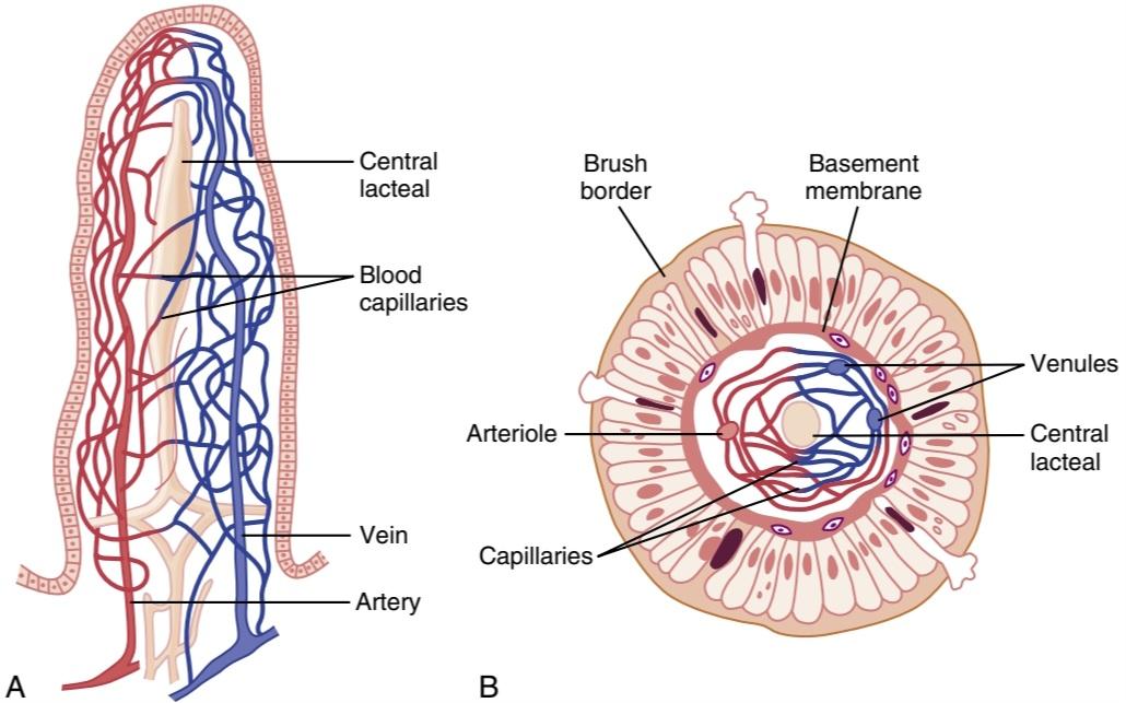
Figure 66-6. Functional organization of the villus. A, Longitudinal section. B, Cross section showing a basement membrane beneath the epithelial cells and a brush border at the other ends of these cells.
شکل 66-6. سازماندهی عملکردی پرز. الف، مقطع طولی. ب، مقطعی که یک غشای پایه در زیر سلولهای اپیتلیال و یک مرز قلم مو در انتهای دیگر این سلولها را نشان میدهد.

Figure 66-7. Brush border of a gastrointestinal epithelial cell, also showing absorbed pinocytic vesicles, mitochondria, and endoplasmic reticulum lying immediately beneath the brush border. (Courtesy Dr. William Lockwood.)
شکل 66-7. مرز برس سلول اپیتلیال دستگاه گوارش، همچنین نشان دهنده وزیکولهای پینوسیتیک جذب شده، میتوکندری و شبکه آندوپلاسمیاست که بلافاصله در زیر مرز قلم مو قرار دارد. (با احترام از دکتر ویلیام لاکوود.)
ABSORPTION IN THE SMALL INTESTINE
Absorption from the small intestine each day consists of several hundred grams of carbohydrates, 100 or more grams of fat, 50 to 100 grams of amino acids, 50 to 100 grams of ions, and 7 to 8 liters of water. The absorptive capacity of the normal small intestine is far greater than this; each day as much as several kilograms of carbohydrates, 500 grams of fat, 500 to 700 grams of proteins, and 20 or more liters of water can be absorbed. The large intestine can absorb still more water and ions, although it can absorb very few nutrients.
جذب در روده کوچک
جذب روزانه از روده کوچک شامل چند صد گرم کربوهیدرات، 100 گرم یا بیشتر چربی، 50 تا 100 گرم اسید آمینه، 50 تا 100 گرم یون و 7 تا 8 لیتر آب است. ظرفیت جذب روده کوچک طبیعی بسیار بیشتر از این است. هر روز میتوان چندین کیلوگرم کربوهیدرات، 500 گرم چربی، 500 تا 700 گرم پروتئین و 20 لیتر یا بیشتر آب جذب کرد. روده بزرگ میتواند آب و یونهای بیشتری را جذب کند، اگرچه میتواند مواد مغذی بسیار کمیرا جذب کند.
ISOSMOTIC ABSORPTION OF WATER
Water is transported through the intestinal membrane entirely by diffusion. Furthermore, this diffusion obeys the usual laws of osmosis. Therefore, when the chyme is dilute enough, water is absorbed through the intestinal mucosa into the blood of the villi almost entirely by osmosis.
جذب ایزوموتیک آب
آب از طریق غشای روده به طور کامل توسط انتشار منتقل میشود. علاوه بر این، این انتشار از قوانین معمول اسمز پیروی میکند. بنابراین، هنگامیکه کیم به اندازه کافی رقیق شد، آب از طریق مخاط روده تقریباً به طور کامل توسط اسمز جذب خون پرزها میشود.
Conversely, water can also be transported in the opposite direction-from plasma into the chyme. This type of transport occurs especially when hyperosmotic solutions are discharged from the stomach into the duodenum. Within minutes, sufficient water usually will be transferred by osmosis to make the chyme isosmotic with the plasma.
برعکس، آب نیز میتواند در جهت مخالف از پلاسما به کیم منتقل شود. این نوع انتقال به ویژه زمانی اتفاق میافتد که محلولهای هیپراسموتیک از معده به دوازدهه تخلیه میشود. در عرض چند دقیقه، معمولاً آب کافی از طریق اسمز منتقل میشود تا کیم با پلاسما ایزوسموز شود.
ABSORPTION OF IONS
Sodium Is Actively Transported Through the Intestinal Membrane.
Twenty to 30 grams of sodium are secreted in the intestinal secretions each day. In addition, the average person eats 5 to 8 grams of sodium each day. Therefore, to prevent net loss of sodium into the feces, the intestines must absorb 25 to 35 grams of sodium each day, which is equal to about one-seventh of all the sodium present in the body.
جذب یونها
سدیم به طور فعال از طریق غشای روده منتقل میشود.
روزانه 20 تا 30 گرم سدیم در ترشحات روده ترشح میشود. علاوه بر این، هر فرد به طور متوسط روزانه 5 تا 8 گرم سدیم میخورد. بنابراین، برای جلوگیری از هدررفت خالص سدیم در مدفوع، رودهها باید روزانه 25 تا 35 گرم سدیم را جذب کنند که برابر با یک هفتم کل سدیم موجود در بدن است.
Whenever significant amounts of intestinal secretions are lost to the exterior, as in extreme diarrhea, the sodium reserves of the body can sometimes be depleted to lethal levels within hours. Normally, however, less than 0.5% of the intestinal sodium is lost in the feces each day because it is rapidly absorbed through the intestinal mucosa. Sodium also plays an important role in helping to absorb sugars and amino acids, as subsequent discussions reveal.
هر گاه مقادیر قابل توجهی از ترشحات روده به بیرون از بین برود، مانند اسهال شدید، ذخایر سدیم بدن گاهی اوقات میتواند در عرض چند ساعت به سطوح کشنده کاهش یابد. با این حال، به طور معمول، روزانه کمتر از 0.5 درصد از سدیم روده در مدفوع از بین میرود زیرا به سرعت از طریق مخاط روده جذب میشود. همانطور که بحثهای بعدی نشان میدهد، سدیم همچنین نقش مهمیدر کمک به جذب قندها و اسیدهای آمینه ایفا میکند.
The basic mechanism of sodium absorption from the intestine is shown in Figure 66-8. The principles of this mechanism, discussed in Chapter 4, are also essentially the same as for absorption of sodium from the gallbladder and renal tubules, as discussed in Chapter 28.
مکانیسم اساسی جذب سدیم از روده در شکل 66-8 نشان داده شده است. اصول این مکانیسم، که در فصل 4 مورد بحث قرار گرفت، همچنین اساساً مانند جذب سدیم از کیسه صفرا و لولههای کلیوی است، همانطور که در فصل 28 بحث شد.
Sodium absorption is powered by active transport of sodium from inside the epithelial cells through the basal and lateral walls of these cells into paracellular spaces. This active transport obeys the usual laws of active trans- port. It requires energy, and the energy process is catalyzed by appropriate adenosine triphosphatase (ATPase) enzymes in the cell membrane (see Chapter 4). Part of the sodium is absorbed along with chloride ions; in fact, the negatively charged chloride ions are mainly passively “dragged” by the positive electrical charges of the sodium ions.
جذب سدیم با انتقال فعال سدیم از داخل سلولهای اپیتلیال از طریق دیوارههای پایه و جانبی این سلولها به فضاهای پاراسلولی انجام میشود. این حمل و نقل فعال از قوانین معمول حمل و نقل فعال تبعیت میکند. به انرژی نیاز دارد و فرآیند انرژی توسط آنزیمهای آدنوزین تری فسفاتاز (ATPase) مناسب در غشای سلولی کاتالیز میشود (به فصل 4 مراجعه کنید). بخشی از سدیم همراه با یونهای کلرید جذب میشود. در واقع، یونهای کلرید با بار منفی عمدتاً توسط بارهای الکتریکی مثبت یونهای سدیم به صورت غیرفعال کشیده میشوند.
Active transport of sodium through the basolateral membranes of the cell reduces the sodium concentration inside the cell to a low value (~50 mEq/L). Because the sodium concentration in the chyme is normally about 142 mEq/L (i.e., about equal to that in plasma), sodium moves down this steep electrochemical gradient from the chyme through the brush border of the epithelial cell into the epithelial cell cytoplasm. Sodium is also cotransported through the brush border membrane by several specific carrier proteins, including the following: (1) the sodium- glucose co-transporter 1 (SGLT1); (2) sodium-amino acid co-transporters; and (3) the sodium-hydrogen exchanger. These transporters function similarly as in the renal tubules, described in Chapter 28, and provide still more sodium ions to be transported by the epithelial cells into the interstitial fluid and paracellular spaces. At the same time, they also provide secondary active absorption of glucose and amino acids, powered by the active sodium-potassium (Na+-K+) ATPase pump on the basolateral membrane.
انتقال فعال سدیم از طریق غشای قاعده جانبی سلول، غلظت سدیم در داخل سلول را به مقدار کم (~50 mEq/L) کاهش میدهد. از آنجایی که غلظت سدیم در کیم به طور معمول حدود 142 میلی اکی والان در لیتر است (یعنی تقریباً برابر با پلاسما)، سدیم به سمت پایین این گرادیان الکتروشیمیایی تند از کیم از طریق مرز برس سلول اپیتلیال به داخل سیتوپلاسم سلول اپیتلیال حرکت میکند. سدیم همچنین از طریق غشای مرزی برس توسط چندین پروتئین حامل خاص، از جمله موارد زیر، به طور مشترک منتقل میشود: (1) انتقال دهنده کمکی سدیم- گلوکز 1 (SGLT1). (2) کمک انتقال دهندههای اسید آمینه. و (3) مبدل سدیم هیدروژن. این ناقلها مانند توبولهای کلیوی که در فصل 28 توضیح داده شد عمل میکنند و یونهای سدیم بیشتری را برای انتقال توسط سلولهای اپیتلیال به داخل مایع بینابینی و فضاهای پاراسلولی فراهم میکنند. در عین حال، آنها همچنین جذب فعال ثانویه گلوکز و اسیدهای آمینه را فراهم میکنند که توسط پمپ فعال سدیم-پتاسیم (+Na+-K) ATPase بر روی غشای قاعده جانبی تغذیه میشود.

Figure 66-8. Absorption of sodium, chloride, glucose, and amino acids through the intestinal epithelium. Note also osmotic absorption of water (i.e., water “follows” sodium through the epithelial mem- brane).
شکل 66-8. جذب سدیم، کلرید، گلوکز و اسیدهای آمینه از طریق اپیتلیوم روده. همچنین به جذب اسمزی آب توجه کنید (یعنی آب از طریق غشای اپیتلیال سدیم را دنبال میکند).
Osmosis of the Water. The next step in the transport process is osmosis of water by transcellular and paracellular pathways. This osmosis occurs because a large osmotic gradient has been created by the elevated concentration of ions in the paracellular space. Much of this osmosis occurs through the tight junctions between the apical borders of the epithelial cells (the paracellular pathway), but much also occurs through the cells themselves-the transcellular pathway. Osmotic movement of water creates flow of fluid into and through the paracellular spaces and, finally, into the circulating blood of the villus.
اسمز آب. مرحله بعدی در فرآیند انتقال، اسمز آب توسط مسیرهای بین سلولی و پاراسلولی است. این اسمز به این دلیل اتفاق میافتد که یک گرادیان اسمزی بزرگ در اثر غلظت بالای یونها در فضای پاراسلولی ایجاد شده است. بیشتر این اسمز از طریق اتصالات محکم بین مرزهای آپیکال سلولهای اپیتلیال (مسیر پاراسلولی) اتفاق میافتد، اما بخش زیادی از این اسمز از طریق خود سلولها – مسیر بین سلولی – رخ میدهد. حرکت اسمزی آب باعث ایجاد جریان مایع به داخل و از طریق فضاهای پاراسلولی و در نهایت به خون در حال گردش پرز میشود.
Aldosterone Greatly Enhances Sodium Absorpotion. When a person becomes dehydrated, large amounts of aldosterone are secreted by the cortices of the adrenal glands. Within 1 to 3 hours this aldosterone causes increased activation of the enzyme and transport mechanisms for all aspects of sodium ab- sorption by the intestinal epithelium. The increased sodium absorption in turn causes secondary increases in absorption of chloride ions, water, and some other substances.
آلدوسترون به میزان زیادی جذب سدیم را افزایش میدهد. هنگامیکه فرد دچار کم آبی میشود، مقادیر زیادی آلدوسترون از قشر غدد فوق کلیوی ترشح میشود. در عرض 1 تا 3 ساعت این آلدوسترون باعث افزایش فعال شدن آنزیم و مکانیسمهای انتقال برای تمام جنبههای جذب سدیم توسط اپیتلیوم روده میشود. افزایش جذب سدیم به نوبه خود باعث افزایش ثانویه جذب یونهای کلرید، آب و برخی مواد دیگر میشود.
This effect of aldosterone is especially important in the colon because it allows virtually no loss of sodium chloride in the feces and also little water loss. Thus, the function of aldosterone in the intestinal tract is the same as that achieved by aldosterone in the renal tubules, which also serves to conserve sodium chloride and water in the body when a person becomes depleted of sodium chloride and dehydrated.
این اثر آلدوسترون به ویژه در روده بزرگ مهم است زیرا عملاً اجازه نمیدهد کلرید سدیم در مدفوع از دست برود و همچنین آب کمیاز دست برود. بنابراین، عملکرد آلدوسترون در دستگاه روده همان عملکردی است که توسط آلدوسترون در لولههای کلیوی به دست میآید، که همچنین برای حفظ کلرید سدیم و آب در بدن زمانی که فرد از کلرید سدیم تهی میشود و کم آب میشود، استفاده میکند.
Absorption of Chloride lons in the Small Intestine. In the upper part of the small intestine, chloride ion absorption is rapid and occurs mainly by diffusion (i.e., absorption of sodium ions through the epithelium creates electronegativity in the chyme and electropositivity in the paracellular spaces between the epithelial cells). Chloride ions then move along this electrical gradient to “follow” the sodium ions. Chloride is also absorbed across the brush border membrane of parts of the ileum and large intestine by a brush border membrane chloride- bicarbonate exchanger (see Figure 66-8). Chloride exits the cell on the basolateral membrane through chloride channels.
جذب کلرید لون در روده کوچک. در قسمت بالایی روده کوچک، جذب یون کلرید سریع است و عمدتاً از طریق انتشار اتفاق میافتد (یعنی جذب یونهای سدیم از طریق اپیتلیوم باعث ایجاد الکترونگاتیوی در کیم و الکتروپوزیتیویتی در فضاهای پاراسلولی بین سلولهای اپیتلیال میشود). سپس یونهای کلرید در امتداد این گرادیان الکتریکی حرکت میکنند تا یونهای سدیم را دنبال کنند. کلرید همچنین در سراسر غشای مرزی برس قسمتهایی از ایلئوم و روده بزرگ توسط یک مبدل کلرید-بی کربنات غشایی برس جذب میشود (شکل 66-8 را ببینید). کلرید از طریق کانالهای کلرید از سلول روی غشای قاعده جانبی خارج میشود.
Absorption of Bicarbonate lons in the Duodenum and Jejunum. Often large quantities of bicarbonate ions (HCO3) must be reabsorbed from the upper small intestine because large amounts of HCO3- have been secreted into the duodenum in both pancreatic secretion and bile. The HCO3- is absorbed in an indirect way as follows: When sodium ions are absorbed, moderate amounts of H+ are secreted into the lumen of the gut in exchange for some of the sodium. These H+, in turn, combine with the HCO3- to form carbonic acid (H2CO3), which then dissociates to form water and carbon dioxide (CO2). The water remains as part of the chyme in the intestines, but the CO2 is readily absorbed into the blood and subsequently expired through the lungs. This process is the so-called “active absorption of HCO3-” It is the same mechanism that occurs in the tubules of the kidneys (see Chapter 31).
جذب لونهای بی کربنات در اثنی عشر و ژژنوم. اغلب مقادیر زیادی یون بی کربنات (HCO3) باید از قسمت فوقانی روده کوچک بازجذب شوند، زیرا مقادیر زیادی -HCO3 هم در ترشح پانکراس و هم در صفرا به دوازدهه ترشح میشود. -HCO3 به صورت غیرمستقیم به شرح زیر جذب میشود: هنگامیکه یونهای سدیم جذب میشوند، مقادیر متوسطی از +H در ازای مقداری سدیم به مجرای روده ترشح میشود. این +H به نوبه خود با -HCO3 ترکیب میشود و اسید کربنیک (H2CO3) را تشکیل میدهد که سپس تجزیه میشود و آب و دی اکسید کربن (CO2) را تشکیل میدهد. آب به عنوان بخشی از کیم در رودهها باقی میماند، اما CO2 به راحتی در خون جذب میشود و متعاقباً از طریق ریهها منفجر میشود. این فرآیند به اصطلاح “جذب فعال -HCO3” است که همان مکانیسمیاست که در لولههای کلیه اتفاق میافتد (به فصل 31 مراجعه کنید).
Secretion of Bicarbonate and Absorption of Chlo- ride lons in the Ileum and Large Intestine. The epithelial cells on the surfaces of the villi in the ileum, as well as on all surfaces of the large intestine, have a special capability of secreting HCO3- in exchange for absorption of chloride ions (see Figure 66-8). This capability is important because it provides alkaline HCO3- that neutralizes acid products formed by bacteria in the large intestine.
ترشح بی کربنات و جذب کلرید در ایلئوم و روده بزرگ. سلولهای اپیتلیال روی سطوح پرزهای ایلئوم و همچنین در تمام سطوح روده بزرگ، قابلیت خاصی برای ترشح -HCO3 در ازای جذب یونهای کلرید دارند (شکل 66-8 را ببینید). این قابلیت مهم است زیرا -HCO3 قلیایی را فراهم میکند که محصولات اسیدی تشکیل شده توسط باکتریها در روده بزرگ را خنثی میکند.
Extreme Secretion of Chloride lons, Sodium lons, and Water From the Large Intestine Epithelium in Some Types of Diarrhea. Immature epithelial cells that continually divide to form new epithelial cells are found deep in the spaces between the intestinal epithelial folds. These new epithelial cells spread outward over the luminal surfaces of the intestines. While still in the deep folds, the epithelial cells secrete sodium chloride and water into the intestinal lumen. This secretion, in turn, is reabsorbed by the older epithelial cells outside the folds, thus providing flow of water for absorbing intestinal digestates.
ترشح شدید لون کلرید، لون سدیم و آب از اپیتلیوم روده بزرگ در برخی از انواع اسهال. سلولهای اپیتلیال نابالغ که به طور مداوم تقسیم میشوند و سلولهای اپیتلیال جدید را تشکیل میدهند در اعماق فضاهای بین چینهای اپیتلیال روده یافت میشوند. این سلولهای اپیتلیال جدید به سمت بیرون در سطوح مجرای رودهها پخش میشوند. در حالی که هنوز در چینهای عمیق هستند، سلولهای اپیتلیال کلرید سدیم و آب را به مجرای روده ترشح میکنند. این ترشح به نوبه خود توسط سلولهای اپیتلیال قدیمیخارج از چینها دوباره جذب میشود، بنابراین جریان آب برای جذب مواد گوارشی روده فراهم میشود.
The toxins of cholera and of some other types of diarrheal bacteria can stimulate the epithelial fold secretion so greatly that this secretion often becomes much greater than can be reabsorbed, thus sometimes causing a loss of 5 to 10 liters of water and sodium chloride as diarrhea each day. Within 1 to 5 days, many severely affected patients die of this loss of fluid alone.
سموم وبا و برخی دیگر از انواع باکتریهای اسهالی میتوانند ترشح چینهای اپیتلیال را به شدت تحریک کنند، به طوری که این ترشح اغلب بسیار بیشتر از آن چیزی است که قابل بازجذب است، بنابراین گاهی اوقات باعث از دست دادن 5 تا 10 لیتر آب و کلرید سدیم به عنوان اسهال در هر روز میشود. در عرض 1 تا 5 روز، بسیاری از بیماران مبتلا به این بیماری به تنهایی از دست دادن مایعات جان خود را از دست میدهند.
Extreme diarrheal secretion is initiated by entry of a subunit of cholera toxin into the epithelial cells. This subunit stimulates formation of excess cyclic adenosine monophosphate, which opens tremendous numbers of chloride channels, al- lowing chloride ions to flow rapidly from inside the cell into the intestinal crypts. In turn, this action is believed to activate a sodium pump that pumps sodium ions into the crypts to go along with the chloride ions. Finally, all this extra sodium chloride causes extreme osmosis of water from the blood, thus providing rapid flow of fluid along with the salt. All this excess fluid washes away most of the bacteria and is of value in combating the disease, but too much of a good thing can be lethal because of serious dehydration of the whole body that might ensue. In most instances, the life of a person with cholera can be saved by the administration of tremendous amounts of sodium chloride solution to make up for the loss.
ترشح اسهالی شدید با ورود زیرواحد سم وبا به سلولهای اپیتلیال آغاز میشود. این زیرواحد تشکیل آدنوزین مونوفسفات حلقوی اضافی را تحریک میکند، که تعداد زیادی کانال کلریدی را باز میکند و به یونهای کلرید اجازه میدهد به سرعت از داخل سلول به داخل کریپتهای روده جریان پیدا کنند. به نوبه خود، اعتقاد بر این است که این عمل یک پمپ سدیم را فعال میکند که یونهای سدیم را به داخل سردابها پمپ میکند تا با یونهای کلرید همراه شود. در نهایت، تمام این کلرید سدیم اضافی باعث اسمز شدید آب از خون میشود، بنابراین جریان سریع مایع همراه با نمک را فراهم میکند. تمام این مایعات اضافی بیشتر باکتریها را از بین میبرد و برای مبارزه با این بیماری مفید است، اما به دلیل کم آبی شدید کل بدن که ممکن است در پی داشته باشد، یک چیز خوب میتواند کشنده باشد. در بیشتر موارد، زندگی یک فرد مبتلا به وبا را میتوان با تجویز مقادیر زیادی محلول کلرید سدیم برای جبران خسارت نجات داد.
Active Absorption of Calcium, Iron, Potassium, Magnesium, and Phosphate. Calcium ions are actively absorbed into the blood, especially from the duodenum, and the amount of calcium ion absorption is exactly controlled to supply the daily need of the body for calcium. One important factor controlling calcium absorption is parathyroid hormone secreted by the parathyroid glands, and an- other is vitamin D. Parathyroid hormone activates vitamin D, and the activated vitamin D in turn greatly enhances calcium absorption. These effects are discussed in Chapter 80.
جذب فعال کلسیم، آهن، پتاسیم، منیزیم و فسفات. یونهای کلسیم به طور فعال در خون جذب میشوند، به ویژه از دوازدهه، و میزان جذب یون کلسیم دقیقاً برای تامین نیاز روزانه بدن به کلسیم کنترل میشود. یکی از عوامل مهم کنترل کننده جذب کلسیم، هورمون پاراتیروئید است که توسط غدد پاراتیروئید ترشح میشود و دیگری ویتامین D است. هورمون پاراتیروئید ویتامین D را فعال میکند و ویتامین D فعال شده نیز به نوبه خود جذب کلسیم را تا حد زیادی افزایش میدهد. این اثرات در فصل 80 مورد بحث قرار گرفته است.
Iron ions are also actively absorbed from the small intestine. The principles of iron absorption and regulation of its absorption in proportion to the body’s need for iron, especially for the formation of hemoglobin, are discussed in Chapter 33.
یونهای آهن نیز به طور فعال از روده کوچک جذب میشوند. اصول جذب آهن و تنظیم جذب آن متناسب با نیاز بدن به آهن به ویژه برای تشکیل هموگلوبین در فصل 33 مورد بحث قرار گرفته است.
Potassium, magnesium, phosphate, and probably still other ions can also be actively absorbed through the intestinal mucosa. In general, the monovalent ions are absorbed with ease and in great quantities. Bivalent ions are normally absorbed in only small amounts; for example, maximum absorption of calcium ions is only soth as great as the normal absorption of sodium ions. Fortunately, only small quantities of the bivalent ions are normally required daily by the body.
پتاسیم، منیزیم، فسفات و احتمالاً سایر یونها نیز میتوانند به طور فعال از طریق مخاط روده جذب شوند. به طور کلی یونهای تک ظرفیتی به راحتی و در مقادیر زیاد جذب میشوند. یونهای دو ظرفیتی معمولاً فقط در مقادیر کمیجذب میشوند. به عنوان مثال، حداکثر جذب یونهای کلسیم به اندازه جذب طبیعی یونهای سدیم است. خوشبختانه، بدن به طور معمول روزانه به مقادیر کمیاز یونهای دو ظرفیتی نیاز دارد.
ABSORPTION OF NUTRIENTS
Carbohydrates Are Mainly Absorbed as Monosaccharides
Essentially all the carbohydrates in food are absorbed in the form of monosaccharides; only a small fraction is absorbed as disaccharides and almost none is absorbed as larger carbohydrate compounds. By far the most abundant of the absorbed monosaccharides is glucose, which usually accounts for more than 80% of the carbohydrate calories absorbed. The reason for this high percentage is that glucose is the final digestion product of our most abundant carbohydrate food, the starches. The remaining 20% of absorbed monosaccharides is composed almost entirely of galactose and fructose-the galactose derived from milk and the fructose as one of the monosaccharides digested from cane sugar.
جذب مواد مغذی
کربوهیدراتها عمدتاً به صورت مونوساکارید جذب میشوند
اساساً تمام کربوهیدراتهای موجود در غذا به شکل مونوساکارید جذب میشوند. فقط بخش کوچکی به عنوان دی ساکارید جذب میشود و تقریباً هیچ یک به عنوان ترکیبات کربوهیدرات بزرگتر جذب نمیشود. تا حد زیادی فراوان ترین مونوساکاریدهای جذب شده گلوکز است که معمولاً بیش از 80 درصد کالری کربوهیدرات جذب شده را تشکیل میدهد. دلیل این درصد بالا این است که گلوکز محصول نهایی هضم فراوان ترین غذای کربوهیدراتی ما یعنی نشاسته است. 20 درصد باقیمانده مونوساکاریدهای جذب شده تقریباً به طور کامل از گالاکتوز و فروکتوز تشکیل شده است – گالاکتوز حاصل از شیر و فروکتوز به عنوان یکی از مونوساکاریدهای هضم شده از قند نیشکر.
Virtually all the monosaccharides are absorbed by a secondary active transport process. We will first discuss the absorption of glucose.
تقریباً تمام مونوساکاریدها توسط یک فرآیند انتقال فعال ثانویه جذب میشوند. ابتدا در مورد جذب گلوکز صحبت خواهیم کرد.
Glucose Is Transported by a Sodium Co-Transport Mechanism. In the absence of sodium transport through the intestinal membrane, virtually no glucose can be absorbed because glucose absorption occurs in a co-transport mode with active transport of sodium (Figure 66-9).
گلوکز توسط یک مکانیسم انتقال همزمان سدیم منتقل میشود. در غیاب انتقال سدیم از طریق غشای روده، عملاً هیچ گلوکزی نمیتواند جذب شود زیرا جذب گلوکز در حالت انتقال همزمان با انتقال فعال سدیم اتفاق میافتد (شکل 66-9).
The transport of sodium and glucose through the intestinal membrane occurs in two stages. First is active transport of sodium ions through the basolateral membranes of the intestinal epithelial cells into the interstitial fluid, thereby depleting sodium inside the epithelial cells. Second, a decrease of sodium inside the cells causes sodium from the intestinal lumen to move through the brush border of the epithelial cells to the cell interiors by a process of secondary active transport. That is, a sodium ion combines with a transport protein, SGLT1, which will not transport sodium to the cell interior until SGLT1 also combines with glucose. Intestinal glucose also combines simultaneously with SGLT1 and both the sodium ion and glucose molecule are then transported together to the interior of the cell. Thus, the low concentration of sodium inside the cell literally “drags” sodium to the interior of the cell, and glucose is dragged along with it. Once inside the epithelial cell, another transport protein, glucose transporter 2 (GLUT2) facilitates diffusion of the glucose through the cell’s basolateral membrane into the paracellular space and from there into the blood (see Figure 66-9).
انتقال سدیم و گلوکز از طریق غشای روده در دو مرحله انجام میشود. اول، انتقال فعال یونهای سدیم از طریق غشای قاعدهای جانبی سلولهای اپیتلیال روده به داخل مایع بینابینی است، در نتیجه سدیم داخل سلولهای اپیتلیال تخلیه میشود. دوم، کاهش سدیم در داخل سلولها باعث میشود که سدیم از مجرای روده از طریق مرز برس سلولهای اپیتلیال به داخل سلول توسط فرآیند انتقال فعال ثانویه حرکت کند. یعنی یک یون سدیم با یک پروتئین انتقالی به نام SGLT1 ترکیب میشود که تا زمانی که SGLT1 با گلوکز ترکیب نشود، سدیم را به داخل سلول منتقل نمیکند. گلوکز روده نیز به طور همزمان با SGLT1 ترکیب میشود و سپس هر دو یون سدیم و مولکول گلوکز با هم به داخل سلول منتقل میشوند. بنابراین، غلظت کم سدیم در داخل سلول به معنای واقعی کلمه سدیم را به داخل سلول میکشاند و گلوکز نیز همراه با آن کشیده میشود. هنگامیکه در داخل سلول اپیتلیال قرار میگیرد، یک پروتئین ناقل دیگر، ناقل گلوکز 2 (GLUT2) انتشار گلوکز را از طریق غشای قاعده جانبی سلول به فضای پاراسلولی و از آنجا به خون تسهیل میکند (شکل 66-9 را ببینید).
To summarize, it is the initial active transport of sodium through the basolateral membranes of the intestinal epithelial cells that provides the eventual force for moving glucose through the membranes as well.
به طور خلاصه، انتقال فعال اولیه سدیم از طریق غشای قاعدهای جانبی سلولهای اپیتلیال روده است که نیروی نهایی را برای حرکت گلوکز از طریق غشاها نیز فراهم میکند.
Absorption of Other Monosaccharides. Galactose is transported by almost exactly the same mechanism as glucose, using the SGLT1 and GLUT2 transporters to cross the luminal and basolateral membranes, respectively (see Figure 66-9). Fructose transport does not occur by the sodium co- transport mechanism. Instead, fructose is transported by facilitated diffusion all the way through the intestinal epithelium and is not coupled with sodium transport. Transport of fructose from the intestinal lumen to the cell interior is facilitated by GLUT5 and exit of fructose from the cell to the paracellular space is facilitated by GLUT2 (see Figure 66-9).
جذب سایر مونوساکاریدها. گالاکتوز تقریباً با مکانیزم مشابه گلوکز، با استفاده از ناقل SGLT1 و GLUT2 برای عبور از غشاهای مجرا و قاعده جانبی، به ترتیب منتقل میشود (شکل 66-9 را ببینید). انتقال فروکتوز توسط مکانیسم انتقال همزمان سدیم انجام نمیشود. درعوض، فروکتوز با انتشار تسهیل شده از طریق اپیتلیوم روده منتقل میشود و با انتقال سدیم همراه نمیشود. انتقال فروکتوز از مجرای روده به داخل سلول توسط GLUT5 و خروج فروکتوز از سلول به فضای پاراسلولی توسط GLUT2 تسهیل میشود (شکل 66-9 را ببینید).
Some of the fructose, upon entering the cell, becomes phosphorylated. It is then converted to glucose and finally transported in the form of glucose the rest of the way into the blood. Because fructose is not co-transported with sodium, its overall rate of transport is only about one-half that of glucose or galactose.
مقداری از فروکتوز با ورود به سلول، فسفریله میشود. سپس به گلوکز تبدیل میشود و در نهایت به شکل گلوکز در بقیه راه به خون منتقل میشود. از آنجایی که فروکتوز همراه با سدیم منتقل نمیشود، سرعت انتقال کلی آن تنها حدود یک دوم گلوکز یا گالاکتوز است.

Figure 66-9. Absorption of glucose, galactose, and fructose through the intestinal epithelium. ATP, Adenosine triphosphate; GLUT2, Glucose transporter 2; GLUTS, glucose transporter 5; SGLT1, sodium- glucose cotransporter 1.
شکل 66-9. جذب گلوکز، گالاکتوز و فروکتوز از طریق اپیتلیوم روده. ATP، آدنوزین تری فسفات؛ GLUT2، انتقال دهنده گلوکز 2; GLUTS، انتقال دهنده گلوکز 5; SGLT1، انتقال دهنده کمکی سدیم-گلوکز 1.
Absorption of Proteins as Dipeptides, Tripeptides, or Amino Acids
As explained earlier, most proteins, after digestion, are absorbed through the luminal membranes of the intestinal epithelial cells in the form of dipeptides, tripeptides, and a few free amino acids. The energy for most of this transport is supplied by a sodium co-transport mechanism in the same way that sodium co-transport of glucose occurs. That is, most peptide or amino acid molecules bind in the cell’s microvillus membrane with a specific transport protein that requires sodium binding before transport can occur. After binding, the sodium ion then moves down its electrochemical gradient to the interior of the cell and pulls the amino acid or peptide along with it. This process is called co-transport (or secondary active transport) of the amino acids and peptides (see Figure 66-8). A few amino acids do not require this sodium co-transport mechanism but instead are transported by special membrane transport proteins in the same way that fructose is transported, by facilitated diffusion.
جذب پروتئینها به صورت دی پپتید، تری پپتید یا اسیدهای آمینه
همانطور که قبلا توضیح داده شد، بیشتر پروتئینها پس از هضم، از طریق غشای مجرای سلولهای اپیتلیال روده به شکل دی پپتید، تری پپتید و چند اسید آمینه آزاد جذب میشوند. انرژی برای بیشتر این انتقال توسط یک مکانیسم انتقال همزمان سدیم به همان روشی که انتقال همزمان سدیم گلوکز انجام میشود تامین میشود. به این معنا که بیشتر مولکولهای پپتید یا اسید آمینه در غشای میکروویل سلولی با یک پروتئین حملونقل خاص متصل میشوند که قبل از انتقال نیاز به اتصال سدیم دارد. پس از اتصال، یون سدیم سپس گرادیان الکتروشیمیایی خود را به سمت داخل سلول حرکت میکند و اسید آمینه یا پپتید را به همراه خود میکشد. این فرآیند را انتقال همزمان (یا انتقال فعال ثانویه) اسیدهای آمینه و پپتیدها مینامند (شکل 66-8 را ببینید). تعداد کمیاز اسیدهای آمینه به این مکانیسم انتقال همزمان سدیم نیاز ندارند، اما در عوض توسط پروتئینهای انتقال غشایی خاص به همان روشی که فروکتوز منتقل میشود، با انتشار تسهیل شده، منتقل میشوند.
At least 10 different types of transport proteins for amino acids and peptides have been found in the intestinal epithelial cells. This multiplicity of transport proteins is required because of the diverse binding properties of different amino acids and peptides.
حداقل 10 نوع مختلف از پروتئینهای انتقال اسیدهای آمینه و پپتیدها در سلولهای اپیتلیال روده یافت شده است. این تعدد پروتئینهای حمل و نقل به دلیل خواص اتصال متنوع اسیدهای آمینه و پپتیدهای مختلف مورد نیاز است.
Absorption of Fats
Earlier in this chapter, we pointed out that when fats are digested to form monoglycerides and free fatty acids, both of these digestive end products first become dissolved in the central lipid portions of bile micelles. Because the molecular dimensions of these micelles are only 3 to 6 nanometers in diameter, and because of their highly charged exterior, they are soluble in chyme. In this form, the monoglycerides and free fatty acids are carried to the surfaces of the microvilli of the intestinal cell brush border and then penetrate into the recesses among the moving, agitating microvilli. Here, both the monoglycerides and fatty acids diffuse immediately out of the micelles and into the interior of the epithelial cells, which is possible because the lipids are also soluble in the epithelial cell membrane. This process leaves the bile micelles still in the chyme, where they function again and again to help absorb still more monoglycerides and fatty acids.
جذب چربیها
در اوایل این فصل، اشاره کردیم که وقتی چربیها برای تشکیل مونوگلیسرید و اسیدهای چرب آزاد هضم میشوند، هر دوی این محصولات نهایی گوارشی ابتدا در بخشهای لیپیدی مرکزی میسلهای صفراوی حل میشوند. زیرا ابعاد مولکولی این میسلها تنها 3 تا 6 نانومتر قطر دارند و به دلیل بار بیرونی بسیار زیاد، در کیم حل میشوند. در این شکل، مونوگلیسریدها و اسیدهای چرب آزاد به سطوح میکروویلیهای مرز برس سلولی روده منتقل میشوند و سپس به داخل فرورفتگیهای میان میکروویلیهای متحرک و متحرک نفوذ میکنند. در اینجا، هم مونوگلیسریدها و هم اسیدهای چرب بلافاصله از میسلها و به داخل سلولهای اپیتلیال منتشر میشوند، که این امکان وجود دارد زیرا لیپیدها نیز در غشای سلول اپیتلیال محلول هستند. این فرآیند باعث میشود که میسلهای صفراوی همچنان در کیم باقی میمانند، جایی که دوباره و دوباره عمل میکنند تا به جذب بیشتر مونوگلیسرید و اسیدهای چرب کمک کنند.
Thus, the micelles perform a “ferrying” function that is highly important for fat absorption. In the presence of an abundance of bile micelles, about 97% of the fat is absorbed; in the absence of the bile micelles, only 40% to 50% can be absorbed.
بنابراین، میسلها یک عملکرد “فرری” را انجام میدهند که برای جذب چربی بسیار مهم است. در حضور فراوانی میسلهای صفراوی، حدود 97 درصد چربی جذب میشود. در غیاب میسلهای صفراوی، تنها 40 تا 50 درصد میتوانند جذب شوند.
After entering the epithelial cell, the fatty acids and monoglycerides are taken up by the cell’s smooth endo- plasmic reticulum. Here, they are mainly used to form new triglycerides that are subsequently released in the form of chylomicrons through the base of the epithelial cell, to flow upward through the thoracic lymph duct and empty into the circulating blood.
پس از ورود به سلول اپیتلیال، اسیدهای چرب و مونوگلیسریدها توسط شبکه آندوپلاسمیصاف سلول جذب میشوند. در اینجا، آنها عمدتاً برای تشکیل تری گلیسیریدهای جدید استفاده میشوند که متعاقباً به شکل شیلومیکرون از طریق قاعده سلول اپیتلیال آزاد میشوند تا از طریق مجرای لنفاوی قفسه سینه به سمت بالا جریان یافته و در خون در گردش تخلیه شوند.
Direct Absorption of Fatty Acids Into the Portal Blood. Small quantities of short- and medium-chain fatty acids, such as those from butterfat, are absorbed directly into the portal blood rather than being converted into triglycerides and absorbed by way of the lymphatics. The cause of this difference between short- and long-chain fatty acid absorption is that the short-chain fatty acids are more water soluble and mostly are not reconverted into triglycerides by the endoplasmic reticulum. This phenomenon allows diffusion of these short-chain fatty acids from the intestinal epithelial cells directly into the capillary blood of the intestinal villi.
جذب مستقیم اسیدهای چرب در خون پورتال. مقادیر کمیاز اسیدهای چرب با زنجیره کوتاه و متوسط، مانند اسیدهای چرب، به جای اینکه به تری گلیسیرید تبدیل شوند و از طریق غدد لنفاوی جذب شوند، مستقیماً در خون ورودی جذب میشوند. علت این تفاوت بین جذب اسیدهای چرب کوتاه و بلند زنجیره این است که اسیدهای چرب با زنجیره کوتاه بیشتر محلول در آب هستند و عمدتاً توسط شبکه آندوپلاسمیبه تری گلیسیرید تبدیل نمیشوند. این پدیده باعث انتشار این اسیدهای چرب با زنجیره کوتاه از سلولهای اپیتلیال روده به طور مستقیم به خون مویرگی پرزهای روده میشود.
ABSORPTION IN THE LARGE INTESTINE: FORMATION OF FECES
About 1500 ml of chyme normally pass through the ileocecal valve into the large intestine each day. Most of the water and electrolytes in this chyme are absorbed in the colon, usually leaving less than 100 ml of fluid to be excreted in the feces. Also, essentially all the ions are absorbed, leaving only 1 to 5 mEq each of sodium and chloride ions to be lost in the feces.
جذب در روده بزرگ: تشکیل مدفوع
روزانه حدود 1500 میلی لیتر کیم از دریچه ایلئوسکال به روده بزرگ عبور میکند. بیشتر آب و الکترولیتهای موجود در این کیم در روده بزرگ جذب میشوند و معمولاً کمتر از 100 میلیلیتر مایع باقی میماند تا از طریق مدفوع دفع شود. همچنین، اساساً تمام یونها جذب میشوند و تنها 1 تا 5 میلی اکی والان از یونهای سدیم و کلرید در مدفوع از بین میروند.
Most of the absorption in the large intestine occurs in the proximal half of the colon, giving this portion the name absorbing colon, whereas the distal colon functions principally for feces storage until a propitious time for feces excretion and is therefore called the storage colon.
بیشتر جذب در روده بزرگ در نیمه پروگزیمال کولون اتفاق میافتد و به این قسمت کولون جذب کننده میگویند، در حالی که کولون دیستال عمدتاً برای ذخیره مدفوع تا زمان مناسب دفع مدفوع عمل میکند و بنابراین کولون ذخیره نامیده میشود.
Absorption and Secretion of Electrolytes and Water. The mucosa of the large intestine, like that of the small intestine, has a high capability for active absorption of sodium, and the electrical potential gradient created by sodium absorption causes chloride absorption as well. The tight junctions between the epithelial cells of the large intestinal epithelium are much tighter than those of the small intestine. This characteristic prevents significant amounts of back-diffusion of ions through these junctions, thus allowing the large intestinal mucosa to absorb sodium ions far more completely-that is, against a much higher concentration gradient-than can occur in the small intestine. This is especially true when large quantities of aldosterone are available because aldosterone greatly enhances sodium transport capability.
جذب و ترشح الکترولیتها و آب. مخاط روده بزرگ مانند روده کوچک توانایی بالایی برای جذب فعال سدیم دارد و گرادیان پتانسیل الکتریکی ایجاد شده در اثر جذب سدیم باعث جذب کلرید نیز میشود. اتصالات محکم بین سلولهای اپیتلیال اپیتلیوم روده بزرگ بسیار محکم تر از اتصالات روده کوچک است. این مشخصه از انتشار مقادیر قابل توجهی یونها از طریق این اتصالات جلوگیری میکند، بنابراین به مخاط روده بزرگ اجازه میدهد تا یونهای سدیم را به مراتب بیشتر جذب کند، یعنی در برابر گرادیان غلظتی بسیار بالاتر از آنچه در روده کوچک اتفاق میافتد. این امر مخصوصاً زمانی صادق است که مقادیر زیادی آلدوسترون در دسترس باشد زیرا آلدوسترون قابلیت انتقال سدیم را تا حد زیادی افزایش میدهد.
In addition, as occurs in the distal portion of the small intestine, the mucosa of the large intestine secretes HCO3 while it simultaneously absorbs an equal number of chloride ions in an exchange transport process (already described). The HCO3- helps neutralize the acidic end products of bacterial action in the large intestine.
علاوه بر این، همانطور که در قسمت انتهایی روده کوچک اتفاق میافتد، مخاط روده بزرگ HCO3 ترشح میکند در حالی که همزمان تعداد مساوی یون کلرید را در یک فرآیند انتقال تبادلی جذب میکند (که قبلاً توضیح داده شده است). -HCO3 به خنثی کردن محصولات نهایی اسیدی اثر باکتری در روده بزرگ کمک میکند.
Absorption of sodium and chloride ions creates an osmotic gradient across the large intestinal mucosa, which in turn causes absorption of water.
جذب یونهای سدیم و کلرید باعث ایجاد گرادیان اسمزی در غشای مخاطی روده بزرگ میشود که به نوبه خود باعث جذب آب میشود.
Maximum Absorption Capacity of the Large Intestine. The large intestine can absorb a maximum of 5 to 8 liters of fluid and electrolytes each day. When the total quantity entering the large intestine through the ileocecal valve or by way of large intestine secretion exceeds this amount, the excess appears in the feces as diarrhea. As noted earlier, toxins from cholera or certain other bacterial infections often cause the crypts in the terminal ileum and large intestine to secrete 10 or more liters of fluid each day, leading to severe and sometimes lethal diarrhea.
حداکثر ظرفیت جذب روده بزرگ روده بزرگ میتواند روزانه حداکثر 5 تا 8 لیتر مایع و الکترولیت جذب کند. هنگامیکه مقدار کل وارد شده به روده بزرگ از طریق دریچه ایلئوسکال یا از طریق ترشح روده بزرگ از این مقدار بیشتر شود، مقدار اضافی در مدفوع به صورت اسهال ظاهر میشود. همانطور که قبلا ذکر شد، سموم ناشی از وبا یا سایر عفونتهای باکتریایی دیگر اغلب باعث میشوند که کریپتها در ایلئوم انتهایی و روده بزرگ روزانه 10 لیتر یا بیشتر مایع ترشح کنند که منجر به اسهال شدید و گاهی کشنده میشود.
Bacterial Action in the Colon. Numerous bacteria, especially colon bacilli, are present even normally in the absorbing colon. They are capable of digesting small amounts of cellulose, in this way providing a few calories of extra nutrition for the body. In herbivorous animals, this source of energy is significant, although it is of negligible importance in human beings.
عمل باکتریایی در روده بزرگ بسیاری از باکتریها، به ویژه باسیلهای روده بزرگ، حتی به طور معمول در روده بزرگ جذب کننده وجود دارند. آنها قادر به هضم مقادیر کمیسلولز هستند و از این طریق مقدار کمیکالری تغذیه اضافی برای بدن فراهم میکنند. در حیوانات گیاهخوار، این منبع انرژی قابل توجه است، اگرچه در انسان از اهمیت ناچیزی برخوردار است.
Other substances formed as a result of bacterial activity are vitamin K, vitamin B12, thiamine, riboflavin, and various gases that contribute to flatus in the colon, especially CO2, hydrogen gas, and methane. The bacteria-formed vitamin K is especially important because the amount of this vitamin in the daily ingested foods is normally insufficient to maintain adequate blood coagulation.
مواد دیگری که در نتیجه فعالیت باکتریها تشکیل میشوند عبارتند از ویتامین K، ویتامین B12، تیامین، ریبوفلاوین و گازهای مختلفی که در ایجاد گاز روده بزرگ به ویژه CO2، گاز هیدروژن و متان نقش دارند. ویتامین K تشکیلشده توسط باکتریها از اهمیت ویژهای برخوردار است، زیرا میزان این ویتامین در غذاهای مصرفشده روزانه معمولاً برای حفظ انعقاد خون کافی کافی نیست.
Composition of the Feces. The feces normally are about three-fourths water and one-fourth solid matter that is composed of about 30% dead bacteria, 10% to 20% fat, 10% to 20% inorganic matter, 2% to 3% protein, and 30% undigested roughage from the food and dried constituents of digestive juices, such as bile pigment and sloughed epithelial cells. The brown color of feces is caused by stercobilin and urobilin, derivatives of bilirubin. The odor is caused principally by products of bacterial action; these products vary from one person to another, depending on each person’s colonic bacterial flora and on the type of food eaten. The actual odoriferous products include indole, skatole, mercaptans, and hydrogen sulfide.
ترکیب مدفوع. مدفوع معمولاً حدود سه چهارم آب و یک چهارم ماده جامد است که از حدود 30 درصد باکتری مرده، 10 تا 20 درصد چربی، 10 تا 20 درصد ماده معدنی، 2 تا 3 درصد پروتئین و 30 درصد علوفه هضم نشده از مواد غذایی و ترکیبات دوغاب شده و خشک شده تشکیل شده است. سلولهای اپیتلیال رنگ قهوه ای مدفوع توسط استرکوبیلین و اوروبیلین، مشتقات بیلی روبین ایجاد میشود. بو عمدتاً توسط محصولات عملکرد باکتری ایجاد میشود. این محصولات بسته به فلور باکتریایی کولون هر فرد و نوع غذای مصرفی از فردی به فرد دیگر متفاوت است. محصولات خوشبو کننده واقعی عبارتند از: ایندول، اسکاتول، مرکاپتانها و سولفید هیدروژن.
کلیک کنید «Bibliography: فهرست کتب مربوطه»
Abumrad NA: Intestinal CD36 and other key proteins of lipid utilization: role in absorption and gut homeostasis. Compr Physiol 8:493, 2018.
Bröer S: Amino acid transport across mammalian intestinal and renal epithelia. Physiol Rev 88:249, 2008.
Bröer S, Fairweather SJ: Amino acid transport across the mammalian intestine. Compr Physiol 9:343, 2018.
Cifarelli V, Eichmann A: The intestinal lymphatic system: functions and metabolic implications. Cell Mol Gastroenterol Hepatol 7:503, 2019.
Ferraris RP, Choe JY, Patel CR: Intestinal absorption of fructose. Annu Rev Nutr 38:41, 2018.
Gehart H, Clevers H: Tales from the crypt: new insights into intestinal stem cells. Nat Rev Gastroenterol Hepatol 16:19, 2019.
Hernando N, Wagner CA: Mechanisms and regulation of intestinal phosphate absorption. Compr Physiol 8:1065, 2013.
Knöpfel T, Himmerkus N, Günzel D et al. Paracellular transport of phosphate along the intestine. Am J Physiol Gastrointest Liver Physiol 317:G233, 2019.
Kunzelmann K, Mall M: Electrolyte transport in the mammalian colon: mechanisms and implications for disease. Physiol Rev 82:245, 2002.
Lehmann A, Hornby PJ: Intestinal SGLT1 in metabolic health and disease. Am J Physiol Gastrointest Liver Physiol 310:G887, 2016.
Rajendran VM, Sandle Gl: Colonic potassium absorption and secretion in health and disease. Compr Physiol 8:1513, 2018.
Rao MC: Physiology of electrolyte transport in the gut: implications for disease. Compr Physiol 9:947, 2019.
Roxas JL, Viswanathan VK: Modulation of intestinal paracellular transport by bacterial pathogens. Compr Physiol 8:823, 2018.
Wright EM, Loo DD, Hirayama BA: Biology of human sodium glucose transporters. Physiol Rev 291:733, 2011.
Xiao C, Stahel P, Carreiro AL, Buhman KK, Lewis GF: Recent advances in triacylglycerol mobilization by the gut. Trends Endocrinol Metab 29:151, 2018.
🚀 با ما همراه شوید!
تازهترین مطالب و آموزشهای مغز و اعصاب را از دست ندهید. با فالو کردن کانال تلگرام آیندهنگاران مغز، از ما حمایت کنید!



