
فیزیولوژی پزشکی گایتون و هال؛ اعمال ترشحی دستگاه گوارش


دعای مطالعه [ نمایش ]
بِسْمِ الله الرَّحْمنِ الرَّحیمِ
اَللّهُمَّ اَخْرِجْنى مِنْ ظُلُماتِ الْوَهْمِ
خدايا مرا بيرون آور از تاريكىهاى وهم،
وَ اَكْرِمْنى بِنُورِ الْفَهْمِ
و به نور فهم گرامى ام بدار،
اَللّهُمَّ افْتَحْ عَلَيْنا اَبْوابَ رَحْمَتِكَ
خدايا درهاى رحمتت را به روى ما بگشا،
وَانْشُرْ عَلَيْنا خَزائِنَ عُلُومِكَ بِرَحْمَتِكَ يا اَرْحَمَ الرّاحِمينَ
و خزانههاى علومت را بر ما باز كن به امید رحمتت اى مهربانترين مهربانان.
کتاب «فیزیولوژی پزشکی گایتون وهال» بهعنوان یکی از جامعترین و معتبرترین منابع در حوزه علوم پزشکی، همچنان مرجع کلیدی برای درک عملکرد پیچیده بدن انسان است. این اثر با تکیه بر تازهترین پژوهشها و توضیحات دقیق از سازوکارهای فیزیولوژیک، پلی میان علوم پایه پزشکی و کاربردهای بالینی ایجاد میکند و نقشی بیبدیل در آموزش، پژوهش و ارتقای دانش سلامت ایفا مینماید.
ترجمه دقیق و علمیاین شاهکار توسط برند علمیآیندهنگاران مغز به مدیریت داریوش طاهری، دسترسی فارسیزبانان به مرزهای نوین دانش فیزیولوژی را ممکن ساخته و رسالتی علمیبرای ارتقای آموزش پزشکی، فهم عمیقتر سازوکارهای بدن و توسعه روشهای نوین در حوزه سلامت فراهم آورده است.
» Guyton and Hall Textbook of Medical Physiology
»» CHAPTER 65: Secretory Functions of the Alimentary Tract

Throughout the gastrointestinal tract, secretory glands subserve two primary functions: (1) digestive enzymes are secreted in most areas of the alimentary tract, from the mouth to the distal end of the ileum; and (2) mucous glands located from the mouth to the anus provide mucus for lubrication and protection of all parts of the alimentary tract.
در سراسر دستگاه گوارش، غدد ترشحی دو وظیفه اصلی را انجام میدهند: (1) آنزیمهای گوارشی در بیشتر نواحی دستگاه گوارش، از دهان تا انتهای دیستال ایلئوم، ترشح میشوند. و (2) غدد مخاطی واقع از دهان تا مقعد مخاطی را برای روانکاری و محافظت از تمام قسمتهای دستگاه گوارش فراهم میکنند.
Most digestive secretions are formed in response to the presence of food in the alimentary tract, and the quantity secreted in each segment of the tract is usually the amount needed for proper digestion. Furthermore, in some portions of the gastrointestinal tract, even the types of enzymes and other constituents of the secretions are varied in accordance with the types of food present. In this chapter we describe the different alimentary secretions, their functions, and regulation of their production.
بیشتر ترشحات گوارشی در پاسخ به وجود غذا در دستگاه گوارش ایجاد میشود و مقدار ترشح شده در هر بخش از دستگاه معمولاً مقدار مورد نیاز برای هضم مناسب است. علاوه بر این، در برخی از بخشهای دستگاه گوارش، حتی انواع آنزیمها و سایر اجزای ترشحات بر حسب نوع غذای موجود، متفاوت است. در این فصل ما ترشحات مختلف گوارشی، عملکرد آنها و تنظیم تولید آنها را شرح میدهیم.
GENERAL PRINCIPLES OF ALIMENTARY TRACT SECRETION
TYPES OF ALIMENTARY TRACT GLANDS
Several types of glands provide the different types of alimentary tract secretions. First, on the surface of the epithelium in most parts of the gastrointestinal tract are billions of single-cell mucous glands called simply mucous cells or sometimes goblet cells because they look like gob- lets. They function mainly in response to local irritation of the epithelium: They extrude mucus directly onto the epithelial surface to act as a lubricant that also protects the surfaces from excoriation and digestion.
اصول کلی ترشح دستگاه گوارش
انواع غدد دستگاه گوارش
انواع مختلفی از غدد انواع مختلفی از ترشحات دستگاه گوارش را فراهم میکنند. اولاً، در سطح اپیتلیوم در اکثر قسمتهای دستگاه گوارش، میلیاردها غدد مخاطی تک سلولی وجود دارد که به آنها سلولهای مخاطی یا گاهی اوقات سلولهای جامیمیگویند، زیرا شبیه جام هستند. آنها عمدتاً در پاسخ به تحریک موضعی اپیتلیوم عمل میکنند: آنها مخاط را مستقیماً روی سطح اپیتلیال بیرون میآورند تا به عنوان روان کننده ای عمل کند که همچنین از سطوح در برابر تخلیه و هضم محافظت میکند.
Second, many surface areas of the gastrointestinal tract are lined by pits that represent invaginations of the epithelium into the submucosa. In the small intestine, these pits, called crypts of Lieberkühn, are deep and contain specialized secretory cells. One of these cells is shown in Figure 65-1.
ثانیاً، بسیاری از نواحی سطحی دستگاه گوارش توسط حفرههایی پوشانده شده اند که نشان دهنده نفوذ اپیتلیوم به زیر مخاط است. در روده کوچک، این گودالها که به آن سردابهای لیبرکوهن میگویند، عمیق هستند و حاوی سلولهای ترشحی تخصصی هستند. یکی از این سلولها در شکل 65-1 نشان داده شده است.
Third, in the stomach and upper duodenum are large numbers of deep tubular glands. A typical tubular gland can be seen in Figure 65-4, which shows an acidand pepsinogen-secreting gland of the stomach (oxyntic gland).
سوم، در معده و دوازدهه فوقانی تعداد زیادی غدد لولهای عمیق وجود دارد. یک غده لوله ای معمولی را میتوان در شکل 65-4 مشاهده کرد که یک غده ترشح کننده اسید و پپسینوژن معده (غده اکسینتیک) را نشان میدهد.
Fourth, also associated with the alimentary tract are several complex glands-the salivary glands, pancreas, and liver that provide secretions for digestion or emulsification of food. The liver has a highly specialized structure that is discussed in Chapter 71. The salivary glands and the pancreas are compound acinous glands of the type shown in Figure 65-2. These glands lie outside the walls of the alimentary tract and, in this aspect, they differ from all other alimentary glands. They contain millions of acini lined with secreting glandular cells; these acini feed into a system of ducts that finally empty into the alimentary tract.
چهارم، همچنین چندین غدد پیچیده با دستگاه گوارش مرتبط هستند – غدد بزاقی، لوزالمعده و کبد که ترشحاتی را برای هضم یا امولسیون کردن غذا فراهم میکنند. کبد دارای ساختار بسیار تخصصی است که در فصل 71 مورد بحث قرار گرفته است. این غدد خارج از دیواره دستگاه گوارش قرار دارند و از این نظر با تمام غدد گوارشی دیگر تفاوت دارند که در شکل 65-2 نشان داده شده است. آنها حاوی میلیونها آسین هستند که با سلولهای غدد ترشحکننده پوشانده شدهاند. این آسینها وارد سیستم مجاری میشوند که در نهایت وارد دستگاه گوارش میشوند.

Figure 65-1. Typical function of a glandular cell for formation and secretion of enzymes and other secretory substances.
شکل 65-1. عملکرد معمول یک سلول غده ای برای تشکیل و ترشح آنزیمها و سایر مواد ترشحی.
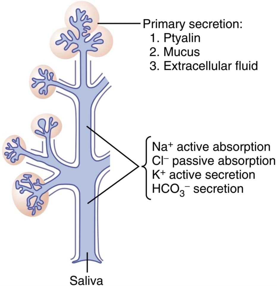
Figure 65-2. Formation and secretion of saliva by a submandibular salivary gland.
شکل 65-2. تشکیل و ترشح بزاق توسط غده بزاقی زیر فکی.
BASIC MECHANISMS OF STIMULATION OF THE ALIMENTARY TRACT GLANDS
Contact of Food With Gut Epithelium Activates the Enteric Nervous System and Stimulates Secretion
The presence of food in a particular segment of the gastrointestinal tract usually stimulates the glands of that region and adjacent regions to secrete moderate to large quantities of juices. Part of this local effect, especially the secretion of mucus by mucous cells, results from direct contact stimulation of the surface glandular cells by the food.
مکانیسمهای اساسی تحریک غدد دستگاه گوارش
تماس غذا با اپیتلیوم روده باعث فعال شدن سیستم عصبی روده و تحریک ترشح میشود.
وجود غذا در بخش خاصی از دستگاه گوارش معمولاً غدد آن ناحیه و نواحی مجاور را برای ترشح مقادیر متوسط تا زیاد آب میوه تحریک میکند. بخشی از این اثر موضعی، به ویژه ترشح مخاط توسط سلولهای مخاطی، ناشی از تحریک تماس مستقیم سلولهای غده سطحی توسط غذا است.
In addition, local epithelial stimulation also activates the enteric nervous system of the gut wall. The types of stimuli that activate this system are (1) tactile stimulation, (2) chemical irritation, and (3) distention of the gut wall. The resulting nervous reflexes stimulate the mucous cells on the gut epithelial surface and the deep glands in the gut wall to increase their secretion.
علاوه بر این، تحریک موضعی اپیتلیال سیستم عصبی روده دیواره روده را نیز فعال میکند. انواع محرکهایی که این سیستم را فعال میکنند عبارتند از (1) تحریک لمسی، (2) تحریک شیمیایی و (3) اتساع دیواره روده. رفلکسهای عصبی حاصل، سلولهای مخاطی روی سطح اپیتلیال روده و غدد عمیق دیواره روده را تحریک میکند تا ترشح آنها را افزایش دهد.
Autonomic Stimulation of Secretion
Parasympathetic Stimulation Increases Alimentary Tract Glandular Secretion Rate. Stimulation of the parasympathetic nerves to the alimentary tract almost invariably increases the rates of alimentary glandular secretion. This increased secretion rate is especially true of the glands in the upper portion of the tract (innervated by the glossopharyngeal and vagus parasympathetic nerves) such as the salivary glands, esophageal glands, gastric glands, pancreas, and Brunner’s glands in the duodenum. It is also true of some glands in the distal portion of the large intestine, which are innervated by pelvic parasympathetic nerves. Secretion in the remainder of the small intestine and in the first two-thirds of the large intestine occurs mainly in response to local neural and hormonal stimuli in each segment of the gut.
تحریک خودکار ترشح
تحریک پاراسمپاتیک میزان ترشح غدد دستگاه گوارش را افزایش میدهد. تحریک اعصاب پاراسمپاتیک به دستگاه گوارش تقریباً همیشه میزان ترشح غدد گوارشی را افزایش میدهد. این افزایش میزان ترشح به ویژه در مورد غدد قسمت فوقانی دستگاه (که توسط اعصاب پاراسمپاتیک گلوفارنکس و واگ عصب دهی میشوند) مانند غدد بزاقی، غدد مری، غدد معده، پانکراس و غدد برونر در دوازدهه صادق است. همچنین در مورد برخی از غدد در قسمت انتهایی روده بزرگ که توسط اعصاب پاراسمپاتیک لگنی عصب دهی میشوند، صادق است. ترشح در بقیه روده کوچک و در دو سوم اول روده بزرگ عمدتاً در پاسخ به محرکهای عصبی و هورمونی موضعی در هر بخش از روده رخ میدهد.
Sympathetic Stimulation Has a Dual Effect on Alimentary Tract Glandular Secretion Rate. Stimulation of the sympathetic nerves going to the gastrointestinal tract causes a slight to moderate increase in secretion by some of the local glands. However, sympathetic stimulation also constricts the blood vessels that supply the glands. There- fore, sympathetic stimulation can have a dual effect: (1) sympathetic stimulation alone usually slightly increases secretion, and (2) if parasympathetic or hormonal stimulation is already causing copious secretion by the glands, superimposed sympathetic stimulation usually reduces secretion, sometimes significantly, mainly because of vasoconstrictive reduction of the blood supply.
تحریک سمپاتیک اثر دوگانه ای بر میزان ترشح غدد دستگاه گوارش دارد. تحریک اعصاب سمپاتیک که به سمت دستگاه گوارش میرود باعث افزایش جزئی تا متوسط ترشح توسط برخی از غدد موضعی میشود. با این حال، تحریک سمپاتیک عروق خونی تامین کننده غدد را نیز منقبض میکند. بنابراین، تحریک سمپاتیک میتواند اثر دوگانه داشته باشد: (1) تحریک سمپاتیک به تنهایی معمولاً ترشح را اندکی افزایش میدهد و (2) اگر تحریک پاراسمپاتیک یا هورمونی قبلاً باعث ترشح زیاد غدد میشود، تحریک سمپاتیک روی هم معمولاً ترشح را کاهش میدهد، گاهی اوقات به طور قابل توجهی، عمدتاً به دلیل کاهش منقبض عروق خونی.
Regulation of Glandular Secretion by Hormones. In the stomach and intestine, several different gastrointestinal hormones help regulate the volume and composition of the secretions. These hormones are liberated from the gastrointestinal mucosa in response to the presence of food in the lumen of the gut. The hormones are then absorbed into the blood and carried to the glands, where they stimulate secretion. This type of stimulation is particularly valuable to increase the output of gastric juice and pancreatic juice when food enters the stomach or duodenum.
تنظیم ترشح غدد توسط هورمونها. در معده و روده، چندین هورمون مختلف گوارشی به تنظیم حجم و ترکیب ترشحات کمک میکنند. این هورمونها در پاسخ به حضور غذا در مجرای روده از مخاط دستگاه گوارش آزاد میشوند. سپس هورمونها جذب خون شده و به غدد منتقل میشوند و در آنجا ترشح را تحریک میکنند. این نوع تحریک به ویژه برای افزایش خروجی شیره معده و شیره لوزالمعده هنگام ورود غذا به معده یا دوازدهه ارزشمند است.
Chemically, the gastrointestinal hormones are poly- peptides or polypeptide derivatives and will be discussed in more detail later.
از نظر شیمیایی، هورمونهای گوارشی پلی پپتید یا مشتقات پلی پپتیدی هستند و بعداً با جزئیات بیشتر مورد بحث قرار خواهند گرفت.
BASIC MECHANISM OF SECRETION BY GLANDULAR CELLS
Secretion of Organic Substances. Although all the basic mechanisms by which glandular cells function are not known, experimental evidence points to the following principles of secretion, as shown in Figure 65-1.
مکانیسم اصلی ترشح توسط سلولهای غده ای
ترشح مواد آلی. اگرچه تمام مکانیسمهای اساسی که توسط آن سلولهای غدهای عمل میکنند شناخته نشدهاند، شواهد تجربی به اصول ترشح زیر اشاره میکنند، همانطور که در شکل 65-1 نشان داده شده است.
1. The nutrient material needed for formation of the secretion must first diffuse or be actively transport- ed by the blood in the capillaries into the base of the glandular cell.
1. مواد مغذی مورد نیاز برای تشکیل ترشح باید ابتدا پخش شود یا به طور فعال توسط خون در مویرگها به پایه سلول غده منتقل شود.
2. Many mitochondria located inside the glandular cell near its base use oxidative energy to form adenosine triphosphate (ATP).
2. بسیاری از میتوکندریهای واقع در داخل سلول غده ای نزدیک پایه آن از انرژی اکسیداتیو برای تشکیل آدنوزین تری فسفات (ATP) استفاده میکنند.
3. Energy from the ATP, along with appropriate substrates provided by the nutrients, is then used to synthesize the organic secretory substances; this synthesis occurs almost entirely in the endoplasmic reticulum and Golgi complex of the glandular cell. Ribosomes adherent to the reticulum are specifically responsible for formation of proteins that are secreted.
3. انرژی حاصل از ATP، همراه با بسترهای مناسبی که توسط مواد مغذی ارائه میشود، سپس برای سنتز مواد ترشحی آلی استفاده میشود. این سنتز تقریباً به طور کامل در شبکه آندوپلاسمیو مجموعه گلژی سلول غده رخ میدهد. ریبوزومهای چسبیده به شبکه به طور خاص مسئول تشکیل پروتئینهایی هستند که ترشح میشوند.
4. The secretory materials are transported through the tubules of the endoplasmic reticulum, passing in about 20 minutes all the way to the vesicles of the Golgi complex.
4. مواد ترشحی از طریق لولههای شبکه آندوپلاسمیمنتقل میشوند و در حدود 20 دقیقه تمام راه را به وزیکولهای مجموعه گلژی میگذرانند.
5. In the Golgi complex, the materials are modified, added to, concentrated, and discharged into the cytoplasm in the form of secretory vesicles, which are stored in the apical ends of the secretory cells.
5. در کمپلکس گلژی، مواد اصلاح شده، به آن افزوده، تغلیظ شده و به صورت وزیکولهای ترشحی به سیتوپلاسم تخلیه میشوند که در انتهای آپیکال سلولهای ترشحی ذخیره میشوند.
6. These vesicles remain stored until nervous or hormonal control signals cause the cells to extrude the vesicular contents through the cells’ surface. This action probably occurs in the following way. The hormone binds to its receptor and, through one of several possible cell signaling mechanisms, increases the cell membrane permeability to calcium ions. Calcium enters the cell and causes many of the vesicles to fuse with the apical cell membrane. The apical cell membrane then breaks open, thus emptying the vesicles to the exterior; this process is called exocytosis.
6. این وزیکولها تا زمانی که سیگنالهای کنترل عصبی یا هورمونی باعث شود سلولها محتویات وزیکولی را از طریق سطح سلولها خارج کنند، ذخیره میشوند. این عمل احتمالاً به روش زیر انجام میشود. این هورمون به گیرنده خود متصل میشود و از طریق یکی از چندین مکانیسم ممکن سیگنال دهی سلولی، نفوذپذیری غشای سلولی را به یونهای کلسیم افزایش میدهد. کلسیم وارد سلول میشود و باعث میشود که بسیاری از وزیکولها با غشای سلولی آپیکال ترکیب شوند. سپس غشای سلولی آپیکال باز میشود و در نتیجه وزیکولها را به سمت بیرون تخلیه میکند. این فرآیند اگزوسیتوز نامیده میشود.
Water and Electrolyte Secretion. A second necessity for glandular secretion is secretion of sufficient water and electrolytes to go along with the organic substances. Secretion by the salivary glands, discussed in more de- tail later, provides an example of how nervous stimulation causes water and salts to pass through the glandular cells in great profusion, washing the organic substances through the secretory border of the cells at the same time. Hormones acting on the cell membrane of some glandular cells also cause secretory effects similar to those caused by nervous stimulation.
ترشح آب و الکترولیت. ضرورت دوم برای ترشح غدد، ترشح آب و الکترولیت کافی برای همراهی با مواد آلی است. ترشح توسط غدد بزاقی، که بعداً با جزئیات بیشتر مورد بحث قرار خواهد گرفت، مثالی از این است که چگونه تحریک عصبی باعث میشود آب و نمکها از سلولهای غدد بهطور فراوان عبور کنند و همزمان مواد آلی را از مرز ترشحی سلولها بشویید. هورمونهایی که بر روی غشای سلولی برخی از سلولهای غدهای اثر میگذارند، اثرات ترشحی مشابه با تحریکات عصبی ایجاد میکنند.
Lubricating and Protective Properties of Mucus in the Gastrointestinal Tract
Mucus is a thick secretion composed mainly of water, electrolytes, and a mixture of several glycoproteins that are composed of large polysaccharides bound with much smaller quantities of protein. Mucus is slightly different in different parts of the gastrointestinal tract, but in all lo- cations it has several important characteristics that make it both an excellent lubricant and a protectant for the gut wall:
خواص روان کنندگی و محافظتی مخاط در دستگاه گوارش
موکوس ترشح غلیظی است که عمدتاً از آب، الکترولیتها و مخلوطی از چندین گلیکوپروتئین تشکیل شده است که از پلی ساکاریدهای بزرگ متصل به مقادیر بسیار کمتر پروتئین تشکیل شده است. مخاط در قسمتهای مختلف دستگاه گوارش کمیمتفاوت است، اما در همه مکانها دارای چندین ویژگی مهم است که آن را روان کننده عالی و محافظ دیواره روده میکند:
1. Mucus has adherent qualities that make it adhere tightly to the food or other particles and to spread as a thin film over the surfaces.
1. مخاط دارای ویژگیهای چسبنده است که باعث میشود آن را محکم به غذا یا ذرات دیگر بچسبد و به صورت یک لایه نازک روی سطوح پخش شود.
2. It has sufficient body that it coats the wall of the gut and prevents actual contact of most food particles with the mucosa.
2. بدن کافی دارد که دیواره روده را میپوشاند و از تماس واقعی بیشتر ذرات غذا با مخاط جلوگیری میکند.
3. Mucus has a low resistance for slippage, so the particles can slide along the epithelium with great ease.
3. مخاط مقاومت کمیدر برابر لغزش دارد، بنابراین ذرات میتوانند به راحتی در امتداد اپیتلیوم بلغزند.
4. Mucus causes fecal particles to adhere to one another to form the feces that are expelled during a bowel movement.
4. مخاط باعث میشود که ذرات مدفوع به یکدیگر بچسبند و مدفوعی را تشکیل دهند که در طول اجابت مزاج خارج میشود.
5. Mucus is strongly resistant to digestion by the gastrointestinal enzymes.
5. مخاط به شدت در برابر هضم توسط آنزیمهای گوارشی مقاوم است.
6. The glycoproteins of mucus have amphoteric properties, which means that they are capable of buffering small amounts of either acids or alkalies; also, mucus often contains moderate quantities of HCO3-, which specifically neutralize acids.
6. گلیکوپروتئینهای مخاطی دارای خواص آمفوتریک هستند، به این معنی که آنها میتوانند مقادیر کمیاسید یا قلیایی را بافر کنند. همچنین، مخاط اغلب حاوی مقادیر متوسطی از HCO3- است که به طور خاص اسیدها را خنثی میکند.
Thus, mucus has the ability to allow easy slippage of food along the gastrointestinal tract and to prevent excoriative or chemical damage to the epithelium. A person becomes acutely aware of the lubricating qualities of mucus when the salivary glands fail to secrete saliva, because then it is difficult to swallow solid food even when it is eaten along with large amounts of water.
بنابراین، مخاط این توانایی را دارد که اجازه دهد غذا به راحتی در امتداد دستگاه گوارش لغزش یابد و از آسیب شیمیایی یا شیمیایی به اپیتلیوم جلوگیری کند. زمانی که غدد بزاقی نتوانند بزاق ترشح کنند، فرد به شدت از ویژگیهای روان کنندگی مخاط آگاه میشود، زیرا در این صورت بلعیدن غذای جامد حتی زمانی که همراه با مقدار زیادی آب خورده شود دشوار است.
SECRETION OF SALIVA
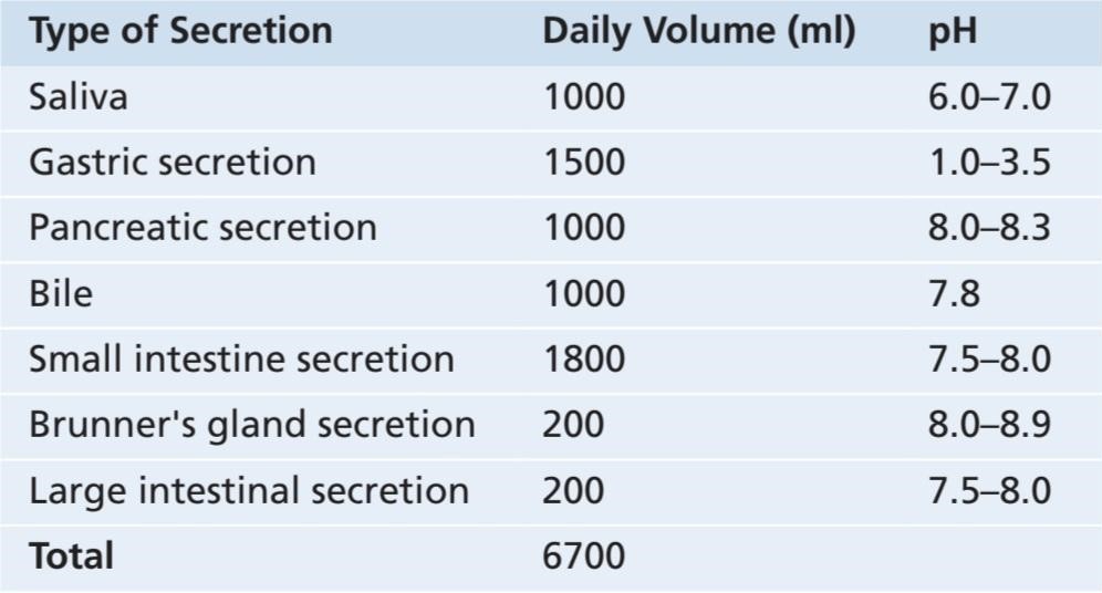
Saliva Contains a Serous Secretion and a Mucus Secretion. The principal glands of salivation are the parotid, submandibular, and sublingual glands; in addition, there are many tiny buccal glands. Daily secretion of saliva normally ranges between 800 and 1500 ml, as shown by the average value of 1000 ml in Table 65-1.
ترشح بزاق
بزاق حاوی یک ترشح سروز و یک ترشح مخاطی است. غدد اصلی ترشح بزاق غدد پاروتید، زیر فکی و زیر زبانی هستند. علاوه بر این، بسیاری از غدد باکال ریز وجود دارد. ترشح روزانه بزاق به طور معمول بین 800 تا 1500 میلی لیتر است، همانطور که با مقدار متوسط 1000 میلی لیتر در جدول 65-1 نشان داده شده است.
Saliva contains two major types of protein secretion: (1) a serous secretion that contains ptyalin (an a-amylase), which is an enzyme for digesting starches, and (2) mucus secretion that contains mucin for lubricating and for surface protective purposes.
بزاق حاوی دو نوع اصلی ترشح پروتئین است: (1) ترشح سروزی حاوی پتیالین (آمیلاز) که آنزیمیبرای هضم نشاسته است و (2) ترشح مخاطی که حاوی موسین برای روانکاری و برای اهداف محافظتی سطحی است.
The parotid glands secrete almost entirely the serous type of secretion, whereas the submandibular and sub- lingual glands secrete both serous secretion and mucus. The buccal glands secrete only mucus. Saliva has a pH between 6.0 and 7.0, which is a favorable range for the digestive action of ptyalin.
غدد پاروتید تقریباً به طور کامل نوع سروزی ترشح میکنند، در حالی که غدد زیر فکی و زیر زبانی هم ترشح سروزی و هم مخاط را ترشح میکنند. غدد باکال فقط مخاط ترشح میکنند. بزاق دارای pH بین 6.0 تا 7.0 است که محدوده مطلوبی برای عملکرد گوارشی پتالین است.
Table 65-1 Daily Alimentary Tract Secretions Juices
جدول 65-1 شیرههای ترشحات روزانه دستگاه گوارش
Secretion of lons in Saliva. Saliva contains especially large quantities of K+ and HCO3. Conversely, the concentrations of both Nat and Cl- are several times less in saliva than in plasma. One can understand these special concentrations of ions in the saliva from the following description of the mechanism for secretion of saliva.
ترشح لون در بزاق. بزاق حاوی مقادیر زیادی +K و HCO3 است. برعکس، غلظت +Na و –Cl در بزاق چندین برابر کمتر از پلاسما است. میتوان این غلظتهای خاص یونها در بزاق را از توضیحات زیر در مورد مکانیسم ترشح بزاق درک کرد.
Figure 65-2 shows secretion by the submandibular gland, a typical compound gland that contains acini and salivary ducts. Salivary secretion is a two-stage operation. The first stage involves the acini, and the second stage involves the salivary ducts. The acini secrete a primary secretion that contains ptyalin and/or mucin in a solution of ions with concentrations not greatly different from those of typical extracellular fluid. As the primary secretion flows through the ducts, two major active transport processes take place that markedly modify the ionic com- position of the fluid in the saliva.
شکل 65-2 ترشح توسط غده زیر فکی را نشان میدهد، یک غده مرکب معمولی که حاوی آسین و مجاری بزاقی است. ترشح بزاق یک عمل دو مرحله ای است. مرحله اول آسینی و مرحله دوم مجاری بزاقی را درگیر میکند. آسینیها ترشح اولیه ای ترشح میکنند که حاوی ptyalin و/یا موسین در محلولی از یونها با غلظتهای بسیار متفاوت از غلظتهای مایع خارج سلولی معمولی نیست. همانطور که ترشح اولیه از طریق مجاری جریان مییابد، دو فرآیند انتقال فعال اصلی انجام میشود که به طور قابل توجهی ترکیب یونی مایع در بزاق را تغییر میدهد.
First, Nat is actively reabsorbed from all the salivary ducts and K+ is actively secreted in exchange for Nat. Therefore, Na+ concentration of the saliva becomes greatly reduced, whereas K+ concentration becomes increased. However, there is excess Na reabsorption compared with K+ secretion, which creates electrical negativity of about -70 millivolts in the salivary ducts; this negativity in turn causes Cl- to be reabsorbed passively. Therefore, Cl- concentration in the salivary fluid falls to a very low level, matching the ductal decrease in Nat concentration.
اول، +Na به طور فعال از تمام مجاری بزاقی بازجذب میشود و +K به طور فعال در ازای +Na ترشح میشود. بنابراین، غلظت +Na بزاق به شدت کاهش مییابد، در حالی که غلظت +K افزایش مییابد. با این حال، بازجذب Na بیش از حد در مقایسه با ترشح +K وجود دارد که منفی الکتریکی در حدود 70- میلی ولت در مجاری بزاقی ایجاد میکند. این منفی بودن به نوبه خود باعث میشود که کلر به طور غیر فعال بازجذب شود. بنابراین، غلظت کلر در مایع بزاقی به سطح بسیار پایینی میرسد که با کاهش مجرای غلظت +Na مطابقت دارد.
Second, HCO3 is secreted by the ductal epithelium into the lumen of the duct. This secretion is at least partly caused by passive exchange of bicarbonate for Cl-, but it may also result partly from an active secretory process.
دوم، HCO3 توسط اپیتلیوم مجرای به لومن مجرا ترشح میشود. این ترشح حداقل تا حدی ناشی از تبادل غیرفعال بی کربنات برای کلر است، اما ممکن است تا حدی از یک فرآیند ترشحی فعال نیز ناشی شود.
The net result of these transport processes is that under resting conditions, the concentrations of Na+ and Cl– in saliva are only about 15 mEq/L each, about one-seventh to one-tenth their concentrations in plasma. Conversely, K+ concentration is about 30 mEq/L, seven times as great as in plasma, and HCO3– concentration is 50 to 70 mEq/L, about two to three times that of plasma.
نتیجه خالص این فرآیندهای انتقال این است که در شرایط استراحت، غلظت +Na و -Cl در بزاق تنها حدود 15 میلیاکیوالان در لیتر است که حدود یک هفتم تا یک دهم غلظت آنها در پلاسما است. برعکس، غلظت پتاسیم در حدود 30 میلی اکی والان در لیتر، هفت برابر بیشتر از پلاسما، و غلظت HCO3– 50 تا 70 میلی اکی والان در لیتر، حدود دو تا سه برابر پلاسما است.
During maximal salivation, the salivary ionic concentrations change considerably because the formation rate of primary secretion by the acini can increase as much as 20-fold. This acinar secretion then flows through the ducts so rapidly that the ductal reconditioning of the secretion is considerably reduced. Therefore, when copious quantities of saliva are being secreted, the sodium chloride concentration is about one-half or two-thirds that of plasma, and potassium concentration rises to only four times that of plasma.
در طول حداکثر ترشح بزاق، غلظت یونی بزاق به طور قابل توجهی تغییر میکند زیرا سرعت تشکیل ترشح اولیه توسط آسین میتواند تا 20 برابر افزایش یابد. سپس این ترشح آسینار آنقدر سریع از طریق مجاری جریان مییابد که بازسازی مجرای ترشح به میزان قابل توجهی کاهش مییابد. بنابراین، هنگامیکه مقادیر زیادی بزاق ترشح میشود، غلظت کلرید سدیم تقریباً یک دوم یا دو سوم پلاسما است و غلظت پتاسیم تنها تا چهار برابر پلاسما افزایش مییابد.
Function of Saliva for Oral Hygiene. Under basal awake conditions, about 0.5 ml of saliva, almost entirely of the mucous type, is secreted each minute; however, during sleep, little secretion occurs. This secretion plays an exceedingly important role for maintaining healthy oral tissues. The mouth is loaded with pathogenic bacteria that can easily destroy tissues and cause dental caries. Saliva helps prevent the deteriorative processes in several ways:
عملکرد بزاق برای بهداشت دهان. در شرایط بیداری پایه، حدود 0.5 میلی لیتر بزاق، تقریباً به طور کامل از نوع مخاطی، در هر دقیقه ترشح میشود. با این حال، در طول خواب، ترشح کمیرخ میدهد. این ترشح نقش بسیار مهمیبرای حفظ سلامت بافتهای دهان ایفا میکند. دهان مملو از باکتریهای بیماری زا است که میتوانند به راحتی بافتها را از بین ببرند و باعث پوسیدگی دندان شوند. بزاق به چندین روش به جلوگیری از فرآیندهای بدتر کمک میکند:
1. The flow of saliva helps wash away pathogenic bacteria, as well as food particles that provide their metabolic support.
1. جریان بزاق به شستشوی باکتریهای بیماری زا و همچنین ذرات غذا که حمایت متابولیک آنها را فراهم میکند، کمک میکند.
2. Saliva contains several factors that destroy bacteria. One of these is thiocyanate ions and another is several proteolytic enzymes-most important, lysozyme-that (a) attack the bacteria, (b) aid thiocyanate ions in enter- ing the bacteria where these ions in turn become bactericidal, and (c) digest food particles, thus helping further to remove the bacterial metabolic support.
2. بزاق حاوی عوامل متعددی است که باکتریها را از بین میبرد. یکی از این یونها یونهای تیوسیانات و دیگری چندین آنزیم پروتئولیتیک است که مهمترین آن لیزوزیم است که (الف) به باکتریها حمله میکند، (ب) به یونهای تیوسیانات در ورود به باکتریها کمک میکند، جایی که این یونها به نوبه خود باکتریکش میشوند، و (ج) ذرات غذا را هضم میکنند، بنابراین به حذف بیشتر متابولیک باکتری کمک میکنند.
3. Saliva often contains significant amounts of antibodies that can destroy oral bacteria, including some that cause dental caries. In the absence of salivation, oral tissues often become ulcerated and otherwise infected, and caries of the teeth can become rampant.
3. بزاق اغلب حاوی مقادیر قابل توجهی آنتی بادی است که میتواند باکتریهای دهان را از بین ببرد، از جمله برخی که باعث پوسیدگی دندان میشوند. در غیاب بزاق، بافتهای دهان اغلب زخمیمیشوند و در غیر این صورت عفونی میشوند و پوسیدگی دندانها ممکن است شایع شود.
NERVOUS REGULATION OF SALIVARY SECRETION
Figure 65-3 shows the parasympathetic nervous pathways for regulating salivation and demonstrates that the salivary glands are controlled mainly by parasympathetic nervous signals all the way from the superior and inferior salivatory nuclei in the brain stem.
تنظیم عصبی ترشح بزاق
شکل 65-3 مسیرهای عصبی پاراسمپاتیک را برای تنظیم ترشح بزاق نشان میدهد و نشان میدهد که غدد بزاقی عمدتاً توسط سیگنالهای عصبی پاراسمپاتیک تا هستههای بزاقی فوقانی و تحتانی در ساقه مغز کنترل میشوند.
The salivatory nuclei are located approximately at the juncture of the medulla and pons and are excited by both taste and tactile stimuli from the tongue and other areas of the mouth and pharynx. Many taste stimuli, especially the sour taste (caused by acids), elicit copious secretion of saliva-often 8 to 20 times the basal rate of secretion. Also, certain tactile stimuli, such as the presence of smooth objects in the mouth (e.g., a pebble), cause marked salivation, whereas rough objects cause less salivation and occasionally even inhibit salivation.
هستههای بزاق تقریباً در محل اتصال مدولا و پونز قرار دارند و توسط محرکهای چشایی و لمسی از زبان و سایر نواحی دهان و حلق تحریک میشوند. بسیاری از محرکهای چشایی، به ویژه طعم ترش (ناشی از اسیدها)، باعث ترشح زیاد بزاق میشوند که اغلب 8 تا 20 برابر میزان ترشح پایه است. همچنین، برخی از محرکهای لمسی، مانند وجود اجسام صاف در دهان (مانند سنگریزه)، باعث ترشح بزاق مشخصی میشوند، در حالی که اجسام خشن باعث ترشح کمتر بزاق میشوند و حتی گاهی اوقات ترشح بزاق را مهار میکنند.
Salivation can also be stimulated or inhibited by nervous signals arriving in the salivatory nuclei from higher centers of the central nervous system. For example, when a person smells or eats favorite foods, salivation is greater than when food that is disliked is smelled or eaten. The appetite area of the brain, which partially regulates these effects, is located in proximity to the parasympathetic centers of the anterior hypothalamus, and it functions to a great extent in response to signals from the taste and smell areas of the cerebral cortex or amygdala.
ترشح بزاق همچنین میتواند توسط سیگنالهای عصبی که از مراکز بالاتر سیستم عصبی مرکزی به هستههای بزاق میرسند تحریک یا مهار شود. به عنوان مثال، وقتی فردی غذاهای مورد علاقه خود را بو میکند یا میخورد، ترشح بزاق بیشتر از زمانی است که غذای مورد علاقه بو داده یا خورده شود. ناحیه اشتها مغز که تا حدی این اثرات را تنظیم میکند، در مجاورت مراکز پاراسمپاتیک هیپوتالاموس قدامیقرار دارد و تا حد زیادی در پاسخ به سیگنالهای نواحی چشایی و بویایی قشر مغز یا آمیگدال عمل میکند.
Salivation also occurs in response to reflexes originating in the stomach and upper small intestines-particularly when irritating foods are swallowed or when a person is nauseated because of some gastrointestinal abnormality. The saliva, when swallowed, helps to remove the irritating factor in the gastrointestinal tract by diluting or neutralizing the irritant substances.
ترشح بزاق همچنین در پاسخ به رفلکسهایی که از معده و رودههای کوچک فوقانی منشا میگیرند رخ میدهد – بهویژه زمانی که غذاهای تحریککننده بلعیده میشوند یا زمانی که فرد به دلیل برخی ناهنجاریهای گوارشی حالت تهوع دارد. بزاق هنگام بلع، با رقیق کردن یا خنثی کردن مواد محرک به حذف عامل تحریک کننده در دستگاه گوارش کمک میکند.
Sympathetic stimulation can also increase salivation a slight amount-much less so than parasympathetic stimulation. Also, the saliva formed in response to sympathetic activity is thicker compared to saliva produced during increased parasympathetic activity. The sympathetic nerves originate from the superior cervical ganglia and travel along the surfaces of the blood vessel walls to the salivary glands.
تحریک سمپاتیک همچنین میتواند ترشح بزاق را کمیافزایش دهد – بسیار کمتر از تحریک پاراسمپاتیک. همچنین، بزاق ایجاد شده در پاسخ به فعالیت سمپاتیک در مقایسه با بزاق تولید شده در هنگام افزایش فعالیت پاراسمپاتیک غلیظ تر است. اعصاب سمپاتیک از گانگلیون فوقانی دهانه رحم منشا میگیرند و در امتداد سطوح دیواره رگهای خونی به غدد بزاقی حرکت میکنند.
A secondary factor that also affects salivary secretion is the blood supply to the glands because secretion always requires adequate nutrients from the blood. The parasympathetic nerve signals that induce copious salivation also moderately dilate the blood vessels. In addition, salivation directly dilates the blood vessels, thus providing increased salivatory gland nutrition as needed by the secreting cells. Part of this additional vasodilator effect is caused by kallikrein secreted by the activated salivary cells, which in turn acts as an enzyme to split one of the blood proteins, an a-globulin, to form bradykinin, a strong vasodilator.
یک عامل ثانویه که بر ترشح بزاق نیز تأثیر میگذارد، خون رسانی به غدد است زیرا ترشح همیشه به مواد مغذی کافی از خون نیاز دارد. سیگنالهای عصب پاراسمپاتیک که باعث ترشح زیاد بزاق میشوند، رگهای خونی را نیز به طور متوسط گشاد میکنند. بعلاوه، ترشح بزاق به طور مستقیم رگهای خونی را گشاد میکند، در نتیجه تغذیه غدد بزاقی را در صورت نیاز سلولهای ترشح کننده افزایش میدهد. بخشی از این اثر گشادکننده عروق اضافی توسط کالیکرئین ترشح شده توسط سلولهای بزاقی فعال ایجاد میشود، که به نوبه خود به عنوان آنزیمیعمل میکند تا یکی از پروتئینهای خون، یک گلوبولین a- را تجزیه کند تا برادی کینین، یک گشادکننده قوی عروق، تشکیل شود.

Figure 65-3. Parasympathetic nervous regulation of salivary secretion.
شکل 65-3. تنظیم عصبی پاراسمپاتیک ترشح بزاق.
Esophageal Secretion
Esophageal secretions are entirely mucous and mainly provide lubrication for swallowing. The main body of the esophagus is lined with many simple mucous glands. At the gastric end and to a lesser extent in the initial portion of the esophagus, many compound mucous glands can also be found. The mucus secreted by the compound glands in the upper esophagus prevents mucosal excoriation by newly entering food, whereas the compound glands located near the esophagogastric junction protect the esophageal wall from digestion by acidic gastric juices that often reflux from the stomach back into the lower esophagus. Despite this protection, a peptic ulcer at times can still occur at the gastric end of the esophagus.
ترشح مری
ترشحات مری کاملاً مخاطی هستند و عمدتاً روانکاری را برای بلع فراهم میکنند. بدنه اصلی مری با بسیاری از غدد مخاطی ساده پوشیده شده است. در انتهای معده و به میزان کمتری در قسمت ابتدایی مری، بسیاری از غدد مخاطی مرکب نیز یافت میشوند. موکوس ترشح شده توسط غدد مرکب در قسمت فوقانی مری از تخلیه مخاطی با غذای تازه وارد شده جلوگیری میکند، در حالی که غدد ترکیبی واقع در نزدیکی محل اتصال مری معده، دیواره مری را از هضم توسط شیرههای اسیدی معده که اغلب از معده به قسمت تحتانی مری برگشت میدهند، محافظت میکند. علیرغم این محافظت، زخم معده گاهی ممکن است در انتهای معده مری ایجاد شود.
GASTRIC SECRETION
In addition to mucus-secreting cells that line the entire surface of the stomach, the stomach mucosa has two important types of tubular glands-oxyntic glands (also called gastric glands) and pyloric glands. The oxyntic (acid-forming) glands secrete hydrochloric acid, pepsinogen, intrinsic factor, and mucus. The pyloric glands secrete mainly mucus for protection of the pyloric mucosa from the stomach acid. They also secrete the hormone gastrin.
ترشح معده
علاوه بر سلولهای ترشح کننده مخاط که تمام سطح معده را میپوشانند، مخاط معده دارای دو نوع مهم غدد لوله ای است: غدد اکسینتیک (که غدد معده نیز نامیده میشوند) و غدد پیلور. غدد اکسینتیک (اسید ساز) اسید هیدروکلریک، پپسینوژن، فاکتور ذاتی و مخاط ترشح میکنند. غدد پیلور عمدتاً برای محافظت از مخاط پیلور در برابر اسید معده، مخاط ترشح میکنند. آنها همچنین هورمون گاسترین را ترشح میکنند.
The oxyntic glands are located on the inside surfaces of the body and fundus of the stomach-the proximal 80% of the stomach. The pyloric glands are located in the antral portion of the stomach-the distal 20% of the stomach.
غدد اکسینتیک در سطوح داخلی بدن و فوندوس معده – نزدیک 80 درصد معده قرار دارند. غدد پیلور در قسمت آنترال معده – 20 درصد دیستال معده – قرار دارند.
Secretions From the Gastric (Oxyntic) Glands
A typical stomach oxyntic gland is shown in Figure 65-4. It is composed of three main types of cells: (1) mucous neck cells, which secrete mainly mucus; (2) peptic (or chief) cells, which secrete large quantities of pepsinogen; and (3) parietal (or oxyntic) cells, which secrete hydrochloric acid and intrinsic factor. Oxyntic glands also contain some additional cells types, including the enterochromaffin-like (ECL) cells that secrete histamine.
ترشحات غدد معده (اکسینتیک).
یک غده اکسینتیک معمولی معده در شکل 65-4 نشان داده شده است. از سه نوع سلول اصلی تشکیل شده است: (1) سلولهای گردنی مخاطی که عمدتاً مخاط ترشح میکنند. (2) سلولهای پپتیک (یا اصلی) که مقادیر زیادی پپسینوژن ترشح میکنند. و (3) سلولهای جداری (یا اکسینتیک) که اسید هیدروکلریک و فاکتور ذاتی ترشح میکنند. غدد اکسینتیک همچنین حاوی برخی از انواع سلولهای اضافی از جمله سلولهای شبه انتروکرومافین (ECL) هستند که هیستامین ترشح میکنند.
Secretion of hydrochloric acid by the parietal cells involves special mechanisms, as follows.
ترشح اسید کلریدریک توسط سلولهای جداری شامل مکانیسمهای خاصی است که به شرح زیر است.
Basic Mechanism of Hydrochloric Acid Secretion. When stimulated, the parietal cells secrete an acid solution that contains about 160 mmol/L of hydrochloric acid, which is nearly isotonic with the body fluids. The pH of this acid is about 0.8, demonstrating its extreme acidity. At this pH, the H+ concentration is about 3 million times that of the arterial blood. To concentrate the H+ this tremendous amount requires more than 1500 calories of energy/L of gastric juice. At the same time that H+ is secreted, HCO3 diffuses into the blood so that gastric venous blood has a higher pH than arterial blood when the stomach is secreting acid.
مکانیسم اساسی ترشح اسید هیدروکلریک. هنگامیکه سلولهای جداری تحریک میشوند، محلول اسیدی ترشح میکنند که حاوی حدود 160 میلی مول در لیتر اسید کلریدریک است که تقریباً با مایعات بدن ایزوتونیک است. pH این اسید حدود 0.8 است که اسیدیته شدید آن را نشان میدهد. در این pH، غلظت +H حدود 3 میلیون برابر خون شریانی است. برای تغلیظ +H این مقدار فوق العاده به بیش از 1500 کالری انرژی در لیتر شیره معده نیاز دارد. همزمان با ترشح H+، HCO3 در خون پخش میشود به طوری که خون وریدی معده PH بالاتری نسبت به خون شریانی دارد که معده اسید ترشح میکند.
Figure 65-5 shows schematically the functional structure of a parietal cell (also called an oxyntic cell), demonstrating that it contains large branching intracellular canaliculi. The hydrochloric acid is formed at the villuslike projections inside these canaliculi and is then conducted through the canaliculi to the secretory end of the cell.
شکل 65-5 ساختار عملکردی یک سلول جداری (که سلول اکسینتیک نیز نامیده میشود) را نشان میدهد و نشان میدهد که حاوی کانالهای داخل سلولی انشعاب بزرگی است. اسید کلریدریک در برجستگیهای پرز مانند داخل این کانالها تشکیل میشود و سپس از طریق کانالها به انتهای ترشحی سلول هدایت میشود.
The main driving force for hydrochloric acid secretion by the parietal cells is a hydrogen-potassium pump (H+-K+ adenosine triphosphatase [ATPase]). The chemical mechanism of hydrochloric acid formation is shown in Figure 65-6 and consists of the following steps:
نیروی محرکه اصلی برای ترشح اسید هیدروکلریک توسط سلولهای جداری یک پمپ هیدروژن پتاسیم (+H+-K آدنوزین تری فسفاتاز [ATPase]) است. مکانیسم شیمیایی تشکیل اسید هیدروکلریک در شکل 65-6 نشان داده شده است و شامل مراحل زیر است:
1. Water inside the parietal cell becomes dissociated into H+ and hydroxide (OH-) in the cell cytoplasm. The H+ is then actively secreted into the canaliculus in exchange for K+, an active exchange process that is catalyzed by H+-K+ ATPase. Potassium ions trans- ported into the cell by the Na+-K+ ATPase pump on the basolateral (extracellular) side of the membrane tend to leak into the lumen but are recycled back into the cell by the H+-K+ ATPase. The basolateral Na+-K+ ATPase creates low intracellular Na+, which contributes to Na+ reabsorption from the lumen of the canaliculus. Thus, most of the K+ and Na+ in the canaliculus is reabsorbed into the cell cytoplasm, and H+ takes their place in the canaliculus.
1. آب داخل سلول جداری به +H و هیدروکسید (-OH) در سیتوپلاسم سلول تجزیه میشود. سپس +H در ازای +K، یک فرآیند تبادل فعال که توسط H+-K+ ATPase کاتالیز میشود، به طور فعال به داخل کانال ترشح میشود. یونهای پتاسیمیکه توسط پمپ Na+-K+ ATPase در سمت قاعدهای (برون سلولی) غشا به داخل سلول منتقل میشوند، تمایل دارند به داخل مجرا نشت کنند، اما توسط H+-K+ ATPase به داخل سلول بازیافت میشوند. Na+-K+ ATPase قاعدهای جانبی +Na داخل سلولی کم ایجاد میکند که به بازجذب +Na از مجرای کانال کمک میکند. بنابراین، بیشتر +K و +Na موجود در کانال به سیتوپلاسم سلولی بازجذب میشود و +H جای آنها را در کانال میگیرد.
2. The pumping of H+ out of the cell by the H+-K+ ATPase permits OH- to accumulate and form HCO3- from CO2, either formed during metabolism in the cell or while entering the cell from the blood. This reaction is catalyzed by carbonic anhydrase. The HCO3 is then transported across the basolateral membrane into the extracellular fluid in exchange for Cl- ions, which enter the cell and are secreted through chloride channels into the canaliculus, giving a strong solution of hydrochloric acid in the canaliculus. The hydrochloric acid is then secreted outward through the open end of the canaliculus into the lumen of the gland.
2. پمپاژ +H از سلول توسط H+-K+ ATPase به -OH اجازه میدهد تا انباشته شده و -HCO3 را از CO2 تشکیل دهد، که یا در طی متابولیسم در سلول یا هنگام ورود از خون به سلول ایجاد میشود. این واکنش توسط کربنیک انیدراز کاتالیز میشود. سپس HCO3 در غشای قاعدهای جانبی به داخل مایع خارج سلولی در ازای یونهای -Cl که وارد سلول میشود و از طریق کانالهای کلریدی به داخل کانال ترشح میشود، به مایع خارج سلولی منتقل میشود و محلول قوی اسید هیدروکلریک در کانال ایجاد میکند. سپس اسید کلریدریک از طریق انتهای باز کانال به داخل لومن غده به بیرون ترشح میشود.
3. Water passes into the canaliculus by osmosis because of extra ions secreted into the canaliculus. Thus, the final secretion from the canaliculus contains water, hydrochloric acid at a concentration of about 150 to 160 mEq/L, potassium chloride at a concentration of 15 mEq/L, and a small amount of sodium chloride.
3. آب از طریق اسمز به داخل کانال عبور میکند، زیرا یونهای اضافی به داخل کانال ترشح میشوند. بنابراین، ترشح نهایی از کانال حاوی آب، اسید کلریدریک با غلظت حدود 150 تا 160 میلی اکی والان در لیتر، کلرید پتاسیم با غلظت 15 میلی اکی والان در لیتر و مقدار کمیکلرید سدیم است.
To produce a concentration of H+ as great as that found in gastric juice requires minimal backleak into the mucosa of the secreted acid. A major part of the stomach’s ability to prevent backleak of acid can be attributed to the gastric barrier due to the formation of alkaline mucus and to tight junctions between epithelia cells, as described later. If this barrier is damaged by toxic substances, such as occurs with excessive use of aspirin or alcohol, the secreted acid does leak down an electrochemical gradient into the mucosa, causing stomach mucosal damage.
برای تولید غلظتی از H+ به اندازه آنچه در شیره معده یافت میشود، نیاز به کمترین نشتی به مخاط اسید ترشح شده دارد. بخش عمده ای از توانایی معده برای جلوگیری از نشت اسید را میتوان به سد معده به دلیل تشکیل مخاط قلیایی و اتصالات محکم بین سلولهای اپیتلیوم، همانطور که بعدا توضیح داد، نسبت داد. اگر این سد توسط مواد سمیآسیب ببیند، مانند استفاده بیش از حد از آسپرین یا الکل، اسید ترشح شده از یک گرادیان الکتروشیمیایی به مخاط نشت میکند و باعث آسیب مخاط معده میشود.

Figure 65-4. Gastric (oxyntic) gland from the body of the stomach. ECL, Enterochromaffin-like cells.
شکل 65-4. غده معده (اکسینتیک) از بدن معده. ECL، سلولهای شبه انتروکرومافین.
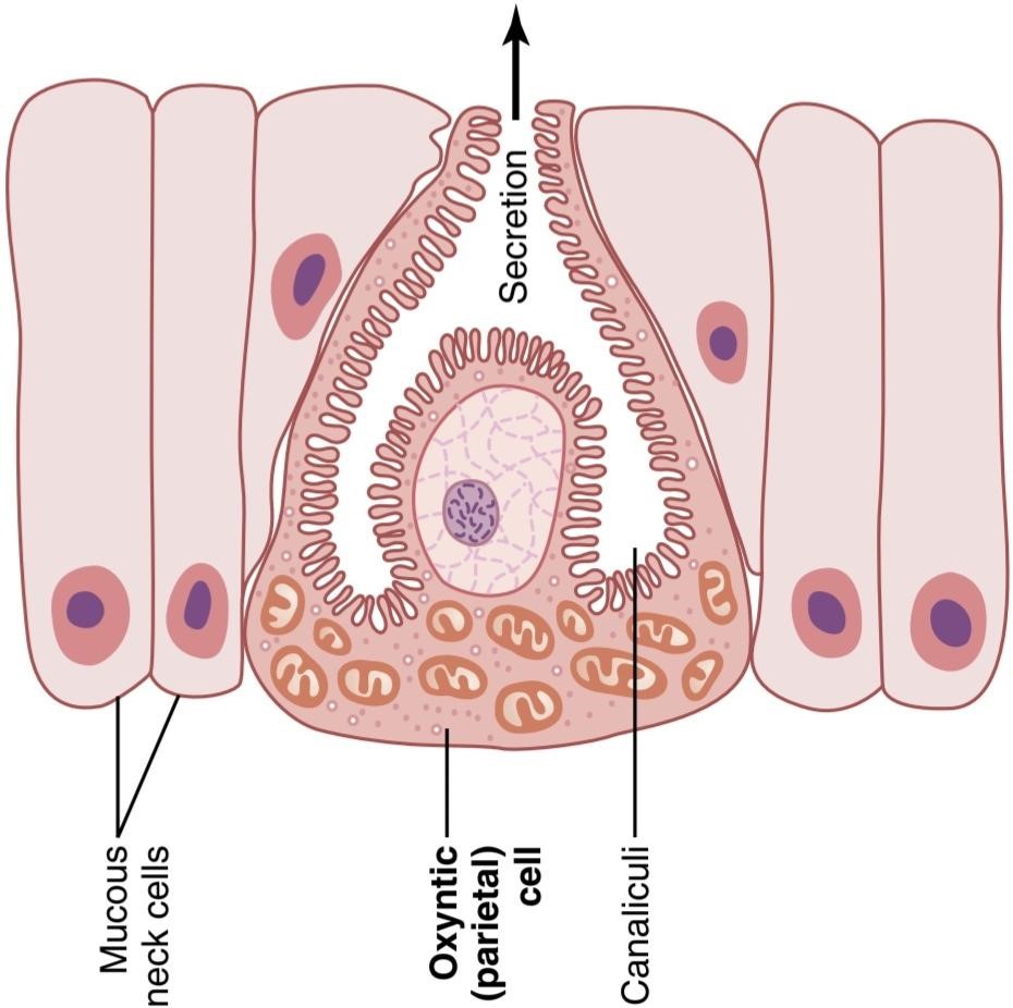
Figure 65-5. Schematic anatomy of the canaliculi in a parietal (oxyntic) cell.
شکل 65-5. آناتومیشماتیک کانالها در یک سلول جداری (اکسینتیک).

Figure 65-6. Postulated mechanism for secretion of hydrochloric acid. (The points labeled “ATP” [adenosine triphosphate] indicate active pumps, and the dashed lines represent free diffusion and osmosis.)
شکل 65-6. مکانیسم فرضی برای ترشح اسید هیدروکلریک (نقاط با برچسب “ATP” [آدنوزین تری فسفات] پمپهای فعال را نشان میدهد، و خطوط نقطه چین نشان دهنده انتشار آزاد و اسمز است.)
The Basic Factors That Stimulate Gastric Secretion Are Acetylcholine, Gastrin, and Histamine. Acetylcholine released by parasympathetic stimulation excites secretion of pepsinogen by peptic cells, hydrochloric acid by parietal cells, and mucus by mucous cells. In comparison, both gastrin and histamine strongly stimulate acid secretion by parietal cells but have little effect on the other cells.
عوامل اساسی که ترشح معده را تحریک میکنند عبارتند از: استیل کولین، گاسترین و هیستامین. استیل کولین آزاد شده توسط تحریک پاراسمپاتیک باعث تحریک ترشح پپسینوژن توسط سلولهای پپتیک، اسید هیدروکلریک توسط سلولهای جداری و مخاط توسط سلولهای مخاطی میشود. در مقایسه، گاسترین و هیستامین به شدت ترشح اسید توسط سلولهای جداری را تحریک میکنند، اما تأثیر کمیبر سایر سلولها دارند.
Secretion and Activation of Pepsinogen. Several slightly different types of pepsinogen are secreted by the peptic and mucous cells of the gastric glands, but all the pepsinogens perform the same basic functions.
ترشح و فعال سازی پپسینوژن چندین نوع پپسینوژن کمیمتفاوت توسط سلولهای پپتیک و مخاطی غدد معده ترشح میشود، اما همه پپسینوژنها عملکردهای اساسی یکسانی را انجام میدهند.
When pepsinogen is first secreted, it has no digestive activity. However, as soon as it comes in contact with hydrochloric acid, it is activated to form active pepsin. In this process, the pepsinogen molecule, having a molecular weight of about 42,500, is split to form a pepsin molecule, having a molecular weight of about 35,000.
هنگامیکه پپسینوژن برای اولین بار ترشح میشود، فعالیت گوارشی ندارد. با این حال، به محض تماس با اسید هیدروکلریک فعال میشود تا پپسین فعال تشکیل شود. در این فرآیند، مولکول پپسینوژن که وزن مولکولی آن حدود 42500 است، برای تشکیل یک مولکول پپسین با وزن مولکولی حدود 35000 تقسیم میشود.
Pepsin functions as an active proteolytic enzyme in a highly acidic medium (optimum pH, 1.8-3.5), but above a pH of about 5 it has almost no proteolytic activity and becomes completely inactivated in a short time. Hydro- chloric acid is as necessary as pepsin for protein digestion in the stomach, as discussed in Chapter 66.
پپسین به عنوان یک آنزیم پروتئولیتیک فعال در یک محیط بسیار اسیدی عمل میکند (PH بهینه، 1.8-3.5)، اما بالاتر از pH حدود 5 تقریباً هیچ فعالیت پروتئولیتیکی ندارد و در مدت کوتاهی کاملاً غیرفعال میشود. همانطور که در فصل 66 بحث شد، اسید کلریدریک به اندازه پپسین برای هضم پروتئین در معده ضروری است.
Secretion of Intrinsic Factor by Parietal Cells. The substance intrinsic factor, which is essential for vitamin B12 absorption in the ileum, is secreted by the parietal cells along with the secretion of hydrochloric acid. When the acid-producing parietal cells of the stomach are destroyed, which frequently occurs in persons with chronic gastritis, not only does achlorhydria (lack of stomach acid secretion) develop, but pernicious anemia also often develops because of failure of red blood cell maturation in the absence of vitamin B12 stimulation of the bone mar- row. This condition is discussed in Chapter 33.
ترشح فاکتور درونی توسط سلولهای جداری. ماده فاکتور ذاتی که برای جذب ویتامین B12 در ایلئوم ضروری است، همراه با ترشح اسید کلریدریک توسط سلولهای جداری ترشح میشود. هنگامیکه سلولهای جداری تولید کننده اسید معده از بین میروند، که اغلب در افراد مبتلا به گاستریت مزمن رخ میدهد، نه تنها آکلرهیدریا (کمبود ترشح اسید معده) ایجاد میشود، بلکه کم خونی خطرناک نیز اغلب به دلیل عدم بلوغ گلبولهای قرمز در غیاب تحریک ویتامین B12 مغز استخوان ایجاد میشود. این شرایط در فصل 33 مورد بحث قرار گرفته است.
PYLORIC GLANDS SECRETE MUCUS AND GASTRIN
The pyloric glands are structurally similar to the oxyntic glands but contain few peptic cells and almost no parietal cells. Instead, they contain mostly mucous cells that are identical with the mucous neck cells of the oxyntic glands. These cells secrete a small amount of pepsinogen, as discussed earlier, and an especially large amount of thin mucus that helps to lubricate food movement, as well as to protect the stomach wall from digestion by the gastric enzymes. The pyloric glands also secrete the hormone gastrin, which plays a key role in controlling gastric secretion, as we discuss shortly.
غدد پیلور مخاط و گاسترین را ترشح میکنند
غدد پیلور از نظر ساختاری شبیه به غدد اکسینتیک هستند اما حاوی تعداد کمیسلول پپتیک و تقریباً هیچ سلول جداری نیستند. در عوض، آنها عمدتا حاوی سلولهای مخاطی هستند که با سلولهای مخاطی گردن غدد اکسینتیک یکسان هستند. این سلولها مقدار کمیپپسینوژن ترشح میکنند، همانطور که قبلاً گفته شد، و مقدار زیادی مخاط نازک ترشح میکنند که به روانسازی حرکت غذا و همچنین محافظت از دیواره معده در برابر هضم توسط آنزیمهای معده کمک میکند. غدد پیلور همچنین هورمون گاسترین را ترشح میکنند که نقش کلیدی در کنترل ترشح معده ایفا میکند.
SURFACE MUCOUS CELLS
The entire surface of the stomach mucosa between glands has a continuous layer of a special type of mucous cells called simply “surface mucous cells.” They secrete large quantities of viscid mucus that coats the stomach mucosa with a gel layer of mucus often more than 1 millimeter thick, thus providing a major shell of protection for the stomach wall, as well as contributing to lubrication of food transport.
سلولهای مخاطی سطحی
تمام سطح مخاط معده بین غدد دارای یک لایه پیوسته از نوع خاصی از سلولهای مخاطی است که به سادگی “سلولهای مخاطی سطحی” نامیده میشوند. آنها مقادیر زیادی موکوس چسبناک ترشح میکنند که مخاط معده را با یک لایه ژل مخاطی اغلب بیش از 1 میلی متر ضخامت میپوشاند، بنابراین یک پوسته محافظ اصلی برای دیواره معده و همچنین به روانکاری حمل و نقل غذا کمک میکند.
Another characteristic of this mucus is that it is alkaline. Therefore, the normal underlying stomach wall is not directly exposed to the highly acidic, proteolytic stomach secretion. Even the slightest contact with food or any irritation of the mucosa directly stimulates the surface mucous cells to secrete additional quantities of this thick, alkaline, viscid mucus.
یکی دیگر از ویژگیهای این مخاط قلیایی بودن آن است. بنابراین، دیواره طبیعی زیرین معده مستقیماً در معرض ترشحات اسیدی و پروتئولیتیک معده قرار نمیگیرد. حتی کوچکترین تماس با غذا یا هرگونه تحریک مخاط مستقیماً سلولهای مخاطی سطحی را تحریک میکند تا مقادیر بیشتری از این مخاط غلیظ، قلیایی و چسبناک ترشح کنند.
STIMULATION OF GASTRIC ACID SECRETION
Parietal Cells of the Oxyntic Glands Are the Only Cells That Secrete Hydrochloric Acid. As noted earlier in the chapter, the acidity of the fluid secreted by the parietal cells of the oxyntic glands can be great, with pH as low as 0.8. However, secretion of this acid is under continuous control by both endocrine and nervous signals. Furthermore, parietal cells operate in close association with another type of cell called enterochromaffin-like cells (ECL cells), the primary function of which is to secrete histamine.
تحریک ترشح اسید معده
سلولهای جداری غدد اکسینتیک تنها سلولهایی هستند که اسید هیدروکلریک ترشح میکنند. همانطور که قبلا در این فصل ذکر شد، اسیدیته مایع ترشح شده توسط سلولهای جداری غدد اکسینتیک میتواند عالی باشد، با pH کمتر از 0.8. با این حال، ترشح این اسید تحت کنترل مداوم هر دو سیگنال غدد درون ریز و عصبی است. علاوه بر این، سلولهای جداری در ارتباط نزدیک با نوع دیگری از سلول به نام سلولهای شبه انتروکرومافین (سلولهای ECL) عمل میکنند که وظیفه اصلی آن ترشح هیستامین است.
The ECL cells lie in the deep recesses of the oxyntic glands and therefore release histamine in direct contact with the parietal cells of the glands. The formation and secretion rates of hydrochloric acid by the parietal cells are directly related to the amount of histamine secreted by the ECL cells. In turn, the ECL cells are stimulated to secrete histamine by the hormone gastrin, which is formed almost entirely in the antral portion of the stomach mucosa in response to proteins in the foods being digested. The ECL cells may also be stimulated by hormones secreted by the enteric nervous system of the stomach wall. We will first discuss the gastrin mechanism for control of the ECL cells and their subsequent control of parietal cell secretion of hydrochloric acid.
سلولهای ECL در فرورفتگیهای عمیق غدد اکسینتیک قرار دارند و بنابراین هیستامین را در تماس مستقیم با سلولهای جداری غدد آزاد میکنند. میزان تشکیل و میزان ترشح اسید هیدروکلریک توسط سلولهای جداری ارتباط مستقیمیبا میزان هیستامین ترشح شده توسط سلولهای ECL دارد. به نوبه خود، سلولهای ECL برای ترشح هیستامین توسط هورمون گاسترین تحریک میشوند که تقریباً به طور کامل در قسمت آنترال مخاط معده در پاسخ به پروتئینهای موجود در غذاهای هضم شده تشکیل میشود. سلولهای ECL همچنین ممکن است توسط هورمونهای ترشح شده توسط سیستم عصبی روده دیواره معده تحریک شوند. ما ابتدا مکانیسم گاسترین برای کنترل سلولهای ECL و کنترل بعدی آنها بر ترشح اسید هیدروکلریک سلول جداری را مورد بحث قرار خواهیم داد.
Stimulation of Acid Secretion by Gastrin. Gastrin is a hormone secreted by gastrin cells, also called G cells. These cells are located in the pyloric glands in the distal end of the stomach. Gastrin is a large polypeptide secreted in two forms-a large form called G-34, which contains 34 amino acids, and a smaller form, G-17, which contains 17 amino acids. Although both of these forms are important, the smaller form is more abundant.
تحریک ترشح اسید توسط گاسترین. گاسترین هورمونی است که توسط سلولهای گاسترین ترشح میشود که سلولهای G نیز نامیده میشوند. این سلولها در غدد پیلور در انتهای انتهایی معده قرار دارند. گاسترین یک پلی پپتید بزرگ است که به دو شکل ترشح میشود: یک فرم بزرگ به نام G-34 که حاوی 34 اسید آمینه است و یک شکل کوچکتر به نام G-17 که حاوی 17 اسید آمینه است. اگرچه هر دوی این اشکال مهم هستند، شکل کوچکتر فراوانتر است.
When meats or other foods containing protein reach the antral end of the stomach, some of the proteins from these foods have a special stimulatory effect on the gas- trin cells in the pyloric glands to cause release of gastrin into the blood to be transported to the ECL cells of the stomach. The vigorous mixing of the gastric juices trans- ports the gastrin rapidly to the ECL cells in the body of the stomach, causing release of histamine directly into the deep oxyntic glands. The histamine then acts quickly to stimulate gastric hydrochloric acid secretion.
هنگامیکه گوشتها یا سایر غذاهای حاوی پروتئین به انتهای آنترال معده میرسند، برخی از پروتئینهای این غذاها اثر تحریکی خاصی بر روی سلولهای گاسترین در غدد پیلور دارند و باعث آزاد شدن گاسترین در خون میشوند تا به سلولهای ECL معده منتقل شوند. اختلاط شدید شیرههای معده، گاسترین را به سرعت به سلولهای ECL در بدن معده منتقل میکند و باعث آزاد شدن هیستامین به طور مستقیم در غدد اکسینتیک عمیق میشود. سپس هیستامین به سرعت برای تحریک ترشح اسید هیدروکلریک معده عمل میکند.
REGULATION OF PEPSINOGEN SECRETION
Stimulation of pepsinogen secretion by the peptic cells in the oxyntic glands occurs in response to two main types of signals: (1) acetylcholine released from the vagus nerves or from the gastric enteric nervous plexus, and (2) acid in the stomach. The acid probably does not stimulate the peptic cells directly but instead elicits additional enteric nervous reflexes that support the original nervous signals to the peptic cells. Therefore, the rate of secretion of pepsinogen, the precursor of the enzyme pepsin that causes protein digestion, is strongly influenced by the amount of acid in the stomach. In people who have lost the ability to secrete normal amounts of acid, secretion of pepsinogen is also decreased, even though the peptic cells may other- wise appear to be normal.
تنظیم ترشح پپسینوژن
تحریک ترشح پپسینوژن توسط سلولهای پپتیک در غدد اکسینتیک در پاسخ به دو نوع سیگنال اصلی رخ میدهد: (1) استیل کولین آزاد شده از اعصاب واگ یا از شبکه عصبی روده معده و (2) اسید در معده. اسید احتمالاً مستقیماً سلولهای پپتیک را تحریک نمیکند، اما در عوض رفلکسهای عصبی رودهای اضافی ایجاد میکند که از سیگنالهای عصبی اصلی به سلولهای پپتیک پشتیبانی میکند. بنابراین، سرعت ترشح پپسینوژن، پیش ساز آنزیم پپسین که باعث هضم پروتئین میشود، به شدت تحت تأثیر میزان اسید معده است. در افرادی که توانایی ترشح مقادیر طبیعی اسید را از دست داده اند، ترشح پپسینوژن نیز کاهش مییابد، حتی اگر سلولهای پپتیک در غیر این صورت طبیعی به نظر برسند.
Phases of Gastric Secretion
Gastric secretion is said to occur in three “phases” (as shown in Figure 65-7): a cephalic phase, a gastric phase, and an intestinal phase.
مراحل ترشح معده
گفته میشود که ترشح معده در سه “فاز” (همانطور که در شکل 65-7 نشان داده شده است) رخ میدهد: فاز سفالیک، فاز معده و فاز روده.
Cephalic Phase. The cephalic phase of gastric secretion occurs even before food enters the stomach, especially while it is being eaten. It results from the sight, smell, thought, or taste of food, and the greater the appetite, the more intense is the stimulation. Neurogenic signals that cause the cephalic phase of gastric secretion originate in the cerebral cortex and in the appetite centers of the amygdala and hypothalamus. They are transmitted through the dorsal motor nuclei of the vagi and thence through the vagus nerves to the stomach. This phase of secretion normally accounts for about 30% of the gastric secretion associated with eating a meal.
فاز سفالیک. فاز سفالیک ترشح معده حتی قبل از ورود غذا به معده، به خصوص در زمان خوردن آن، رخ میدهد. از بینایی، بو، فکر یا طعم غذا ناشی میشود و هر چه اشتها بیشتر باشد، تحریک شدیدتر است. سیگنالهای نوروژنیک که باعث فاز سفالیک ترشح معده میشوند از قشر مغز و مراکز اشتها آمیگدال و هیپوتالاموس منشا میگیرند. آنها از طریق هستههای حرکتی پشتی واگی و از آنجا از طریق اعصاب واگ به معده منتقل میشوند. این مرحله از ترشح معمولاً حدود 30 درصد از ترشح معده مرتبط با خوردن یک وعده غذایی را تشکیل میدهد.
Gastric Phase. Once food enters the stomach, it excites the following: (1) long vagovagal reflexes from the stomach to the brain and back to the stomach; (2) local enteric reflexes; and (3) the gastrin mechanism, all of which cause secretion of gastric juice during several hours while food remains in the stomach. The gastric phase of secretion accounts for about 60% of the total gastric secretion associated with eating a meal and therefore accounts for most of the total daily gastric secretion of about 1500 ml.
فاز معده. هنگامیکه غذا وارد معده میشود، موارد زیر را تحریک میکند: (1) رفلکسهای واگوگال طولانی از معده به مغز و برگشت به معده. (2) رفلکسهای روده ای موضعی. و (3) مکانیسم گاسترین، که همه آنها باعث ترشح شیره معده در طی چند ساعت در حالی که غذا در معده باقی میماند. فاز ترشح معده حدود 60 درصد از کل ترشح معده مرتبط با خوردن یک وعده غذایی را تشکیل میدهد و بنابراین بیشتر کل ترشح معده روزانه حدود 1500 میلی لیتر را تشکیل میدهد.
Intestinal Phase. The presence of food in the upper portion of the small intestine, particularly in the duodenum, will continue to cause stomach secretion of small amounts of gastric juice, probably partly because of small amounts of gastrin released by the duodenal mucosa. This secretion accounts for about 10% of the acid response to a meal.
فاز روده. وجود غذا در قسمت فوقانی روده کوچک، به ویژه در دوازدهه، باعث ترشح مقادیر کمیآب معده در معده میشود، احتمالاً تا حدی به دلیل مقادیر کمیگاسترین آزاد شده توسط مخاط دوازدهه. این ترشح حدود 10 درصد از پاسخ اسید به یک وعده غذایی را تشکیل میدهد.

Figure 65-7. Phases of gastric secretion and their regulation.
شکل 65-7. مراحل ترشح معده و تنظیم آنها
Inhibition of Gastric Secretion by Other Intestinal Factors
Although intestinal chyme slightly stimulates gastric secretion during the early intestinal phase of stomach secretion, it paradoxically inhibits gastric secretion at other times. This inhibition results from at least two influences.
مهار ترشح معده توسط سایر عوامل روده ای
اگرچه کیم روده ترشح معده را در مراحل اولیه ترشح معده کمیتحریک میکند، اما در سایر مواقع به طور متناقضی ترشح معده را مهار میکند. این بازداری حداقل از دو تأثیر ناشی میشود.
1. The presence of food in the small intestine initiates a reverse enterogastric reflex, transmitted through the myenteric nervous system and extrinsic sympathetic and vagus nerves, that inhibits stomach secretion. This reflex can be initiated by (a) distending the small bowel, (b) the presence of acid in the upper intestine, (c) the presence of protein breakdown products, or (d) irritation of the mucosa. This reflex is part of the complex mechanism discussed in Chapter 64 for slowing stomach emptying when the intestines are already filled.
1. وجود غذا در روده کوچک باعث شروع یک رفلکس معکوس روده معده میشود که از طریق سیستم عصبی میروده و اعصاب سمپاتیک و واگ بیرونی منتقل میشود و ترشح معده را مهار میکند. این رفلکس میتواند با (الف) اتساع روده کوچک، (ب) وجود اسید در قسمت فوقانی روده، (ج) وجود محصولات تجزیه پروتئین، یا (د) تحریک مخاط آغاز شود. این رفلکس بخشی از مکانیسم پیچیده ای است که در فصل 64 برای کند کردن تخلیه معده در زمانی که رودهها از قبل پر شده اند مورد بحث قرار گرفته است.
2. The presence of acid, fat, protein breakdown products, hyperosmotic or hypo-osmotic fluids, or any irritating factor in the upper small intestine causes release of several intestinal hormones. One of these hormones is secretin, which is especially important for control of pancreatic secretion. However, secretin opposes stomach secretion. Three other hormones-glucose-dependent insulinotropic peptide (gastric inhibitory peptide), vasoactive intestinal polypeptide, and somatostatin-also have slight to moderate effects in inhibiting gastric secretion. The purpose of intestinal factors that inhibit gastric secretion is presumably to slow passage of chyme from the stomach when the small intestine is already filled or already overactive. In fact, the enterogastric inhibitory reflexes plus inhibitory hormones usually also reduce stomach motility at the same time that they reduce gastric secretion, as dis- cussed in Chapter 64.
2. وجود اسید، چربی، محصولات تجزیه پروتئین، مایعات هیپراسموتیک یا هیپواسموتیک یا هر عامل تحریک کننده در قسمت فوقانی روده کوچک باعث ترشح چندین هورمون روده میشود. یکی از این هورمونها سکرتین است که برای کنترل ترشح پانکراس بسیار مهم است. با این حال، سکرتین با ترشح معده مخالف است. سه هورمون دیگر-پپتید انسولینوتروپیک وابسته به گلوکز (پپتید بازدارنده معده)، پلی پپتید وازواکتیو روده ای و سوماتوستاتین- نیز اثرات جزئی تا متوسطی در مهار ترشح معده دارند. احتمالاً هدف عوامل روده ای که ترشح معده را مهار میکنند، کند کردن عبور کیم از معده در زمانی است که روده کوچک از قبل پر شده یا بیش از حد فعال است. در واقع، همان طور که در فصل 64 بحث شد، رفلکسهای بازدارنده روده معده به اضافه هورمونهای بازدارنده معمولاً حرکت معده را همزمان با کاهش ترشح معده نیز کاهش میدهند.
Gastric Secretion During the Inter digestive Period. The stomach secretes a few milliliters of gastric juice each hour during the “inter digestive period,” when little or no digestion is occurring anywhere in the gut. The secretion that does occur is usually almost entirely of the nonoxyntic type, com- posed mainly of mucus but little pepsin and almost no acid.
ترشح معده در طول دوره بین گوارشی. معده در طی «دوره بین گوارشی»، زمانی که هضم کمیدر روده اتفاق میافتد یا اصلاً در هیچ نقطه ای از روده وجود ندارد، هر ساعت چند میلی لیتر آب معده ترشح میکند. ترشحی که اتفاق میافتد معمولاً تقریباً به طور کامل از نوع غیر اکسینتیک است که عمدتاً از مخاط تشکیل شده است اما پپسین کمیو تقریباً بدون اسید است.
Emotional stimuli may increase inter digestive gastric secretion (which is highly peptic and acidic) to 50 ml or more per hour, in much the same way that the cephalic phase of gastric secretion excites secretion at the onset of a meal. This increase of secretion in response to emotional stimuli may contribute to the development of peptic ulcers, as discussed in Chapter 67.
محرکهای عاطفی ممکن است ترشح معده بین گوارشی را (که به شدت پپتیک و اسیدی است) تا 50 میلی لیتر یا بیشتر در ساعت افزایش دهند، تقریباً به همان روشی که فاز سفالیک ترشح معده ترشحات را در شروع غذا تحریک میکند. این افزایش ترشح در پاسخ به محرکهای عاطفی ممکن است به ایجاد زخمهای گوارشی کمک کند، همانطور که در فصل 67 بحث شد.
Chemical Composition of Gastrin and Other Gastrointestinal Hormones
Gastrin, cholecystokinin (CCK), and secretin are all large poly- peptides with approximate molecular weights of 2000, 4200, and 3400, respectively. The terminal five amino acids in the gastrin and CCK molecular chains are the same. The function- al activity of gastrin resides in the terminal four amino acids, and the activity for CCK resides in the terminal eight amino acids. All the amino acids in the secretin molecule are essential.
ترکیب شیمیایی گاسترین و سایر هورمونهای گوارشی
گاسترین، کوله سیستوکینین (CCK) و سکرتین همه پلی پپتیدهای بزرگ با وزن مولکولی تقریبی 2000، 4200 و 3400 هستند. پنج آمینو اسید نهایی در زنجیرههای مولکولی گاسترین و CCK یکسان هستند. فعالیت عملکردی گاسترین در چهار اسید آمینه پایانی و فعالیت برای CCK در هشت اسید آمینه پایانی قرار دارد. تمام اسیدهای آمینه موجود در مولکول سکرتین ضروری هستند.
A synthetic gastrin, pentagastrin, is composed of the terminal four amino acids of natural gastrin plus the amino acid alanine, has all the same physiological properties as the natural gastrin.
یک گاسترین مصنوعی، پنتاگاسترین، از چهار اسید آمینه پایانی گاسترین طبیعی به اضافه اسید آمینه آلانین تشکیل شده است و دارای تمام خواص فیزیولوژیکی مشابه با گاسترین طبیعی است.
PANCREATIC SECRETION
The pancreas, which lies parallel to and beneath the stomach (illustrated in Figure 65-10), is a large compound gland, and most of its internal structure is similar to that of the salivary glands shown in Figure 65-2. The pancreatic digestive enzymes are secreted by pancreatic acini, and large volumes of sodium bicarbonate solution are secreted by the small ductules and larger ducts leading from the acini. The combined product of enzymes and sodium bicarbonate then flows through a long pancreatic duct that normally joins the hepatic duct immediately before it empties into the duodenum through the papilla of Vater, surrounded by the sphincter of Oddi.
ترشح پانکراس
لوزالمعده که به موازات و زیر معده قرار دارد (نشان داده شده در شکل 65-10)، یک غده مرکب بزرگ است و بیشتر ساختار داخلی آن شبیه به غدد بزاقی نشان داده شده در شکل 65-2 است. آنزیمهای گوارشی پانکراس توسط آسین پانکراس ترشح میشوند و حجم زیادی از محلول بی کربنات سدیم توسط مجراهای کوچک و مجاری بزرگتر منتهی به آسین ترشح میشود. محصول ترکیبی آنزیمها و بی کربنات سدیم سپس از طریق یک مجرای طولانی پانکراس جریان مییابد که به طور معمول به مجرای کبدی بلافاصله قبل از تخلیه به دوازدهه از طریق پاپیلای Vater که توسط اسفنکتر Oddi احاطه شده است، میپیوندد.
Pancreatic juice is secreted most abundantly in response to the presence of chyme in the upper portions of the small intestine, and the characteristics of pancreatic juice are determined to some extent by the types of food in the chyme. The pancreas also secretes insulin, but it is not secreted by the same pancreatic tissue that secretes intestinal pancreatic juice. Instead, insulin is secreted directly into the blood-not into the intestine- by the islets of Langerhans that occur in islet patches throughout the pancreas. These structures are discussed in Chapter 79.
شیره لوزالمعده در پاسخ به وجود کیم در قسمتهای فوقانی روده کوچک به وفور ترشح میشود و ویژگیهای آب پانکراس تا حدودی با انواع مواد غذایی موجود در کیم مشخص میشود. لوزالمعده نیز انسولین ترشح میکند، اما از همان بافت پانکراس که آب پانکراس روده ترشح میکند، ترشح نمیشود. درعوض، انسولین مستقیماً به داخل خون ترشح میشود، نه در روده، توسط جزایر لانگرهانس که در قسمتهای جزایر در سراسر پانکراس وجود دارند. این ساختارها در فصل 79 مورد بحث قرار گرفته اند.
PANCREATIC DIGESTIVE ENZYMES
Pancreatic secretion contains multiple enzymes for digesting all of the three major types of food-proteins, carbohydrates, and fats. It also contains large quantities of HCO3, which play an important role in neutralizing the acidity of the chyme emptied from the stomach into the duodenum.
آنزیمهای گوارشی پانکراس
ترشح لوزالمعده حاوی چندین آنزیم برای هضم هر سه نوع اصلی غذا یعنی پروتئین، کربوهیدرات و چربی است. همچنین حاوی مقادیر زیادی HCO3 است که نقش مهمیدر خنثی کردن اسیدیته کیم تخلیه شده از معده به دوازدهه دارد.
The most important of the pancreatic enzymes for digesting proteins are trypsin, chymotrypsin, and carboxy poly peptidase. By far the most abundant of these is trypsin.
مهمترین آنزیمهای پانکراس برای هضم پروتئینها تریپسین، کیموتریپسین و کربوکسی پلی پپتیداز هستند. تا کنون فراوان ترین آنها تریپسین است.
Trypsin and chymotrypsin split whole and partially digested proteins into peptides of various sizes but do not cause release of individual amino acids. However, carboxy poly peptidase splits some peptides into individual amino acids, thus completing digestion of some proteins all the way to the amino acid state.
تریپسین و کیموتریپسین پروتئینهای کامل و جزئی هضم شده را به پپتیدهایی با اندازههای مختلف تقسیم میکنند اما باعث آزاد شدن اسیدهای آمینه منفرد نمیشوند. با این حال، کربوکسی پلی پپتیداز برخی از پپتیدها را به اسیدهای آمینه منفرد تقسیم میکند، بنابراین هضم برخی از پروتئینها را تا حالت اسید آمینه کامل میکند.
The pancreatic enzyme for digesting carbohydrates is pancreatic amylase, which hydrolyzes starches, glycogen, and most other carbohydrates (except cellulose) to form mostly disaccharides and a few trisaccharides.
آنزیم پانکراس برای هضم کربوهیدراتها آمیلاز پانکراس است که نشاسته، گلیکوژن و اکثر کربوهیدراتهای دیگر (به جز سلولز) را هیدرولیز میکند و عمدتاً دی ساکاریدها و چند تری ساکارید تشکیل میدهد.
The main enzymes for fat digestion are the following: (1) pancreatic lipase, which is capable of hydrolyzing neutral fat into fatty acids and monoglycerides; (2) cholesterol esterase, which causes hydrolysis of cholesterol esters; and (3) phospholipase, which splits fatty acids from phospholipids.
آنزیمهای اصلی برای هضم چربی به شرح زیر است: (1) لیپاز پانکراس، که قادر به هیدرولیز چربی خنثی به اسیدهای چرب و مونوگلیسرید است. (2) کلسترول استراز، که باعث هیدرولیز استرهای کلسترول میشود. و (3) فسفولیپاز، که اسیدهای چرب را از فسفولیپیدها جدا میکند.
When first synthesized in the pancreatic cells, the proteolytic digestive enzymes are in their enzymatically inactive forms-trypsinogen, chymotrypsinogen, and procarboxy poly peptidase. They become activated only after they are secreted into the intestinal tract. Trypsinogen is activated by an enzyme called enterokinase, which is secreted by the intestinal mucosa when chyme comes in contact with the mucosa. Trypsinogen also can be autocatalytically activated by trypsin that has already been formed from previously secreted trypsinogen. Chymotrypsinogen is activated by trypsin to form chymotrypsin, and procarboxypolypeptidase is activated in a similar manner.
هنگامیکه برای اولین بار در سلولهای پانکراس سنتز میشوند، آنزیمهای گوارشی پروتئولیتیک به شکلهای آنزیمیغیرفعال خود هستند – تریپسینوژن، کیموتریپسینوژن و پرو کربوکسی پلی پپتیداز. آنها فقط پس از ترشح در مجرای روده فعال میشوند. تریپسینوژن توسط آنزیمیبه نام انتروکیناز فعال میشود که در تماس کیم با مخاط از مخاط روده ترشح میشود. تریپسینوژن همچنین میتواند به طور خودکار توسط تریپسینی که قبلاً از تریپسینوژن ترشح شده قبلی تشکیل شده است، فعال شود. کیمو تریپسینوژن توسط تریپسین برای تشکیل کیموتریپسین فعال میشود و پروکربوکسی پلی پپتیداز نیز به روشی مشابه فعال میشود.
Secretion of Trypsin Inhibitor Prevents Digestion of the Pancreas. It is important that the proteolytic enzymes of the pancreatic juice not become activated until after they have been secreted into the intestine because the trypsin and the other enzymes would digest the pancreas. Fortunately, the same cells that secrete proteolytic enzymes into the acini of the pancreas simultaneously secrete another substance called trypsin inhibitor. This sub- stance, which is formed in the cytoplasm of the glandular cells, prevents activation of trypsin inside the secretory cells and in the acini and ducts of the pancreas. In addition, because it is trypsin that activates the other pancreatic proteolytic enzymes, trypsin inhibitor prevents activation of the other enzymes as well.
ترشح مهارکننده تریپسین از هضم پانکراس جلوگیری میکند. مهم است که آنزیمهای پروتئولیتیک آب پانکراس تا زمانی که در روده ترشح نشده اند فعال نشوند زیرا تریپسین و سایر آنزیمها لوزالمعده را هضم میکنند. خوشبختانه همان سلولهایی که آنزیمهای پروتئولیتیک را در آسین پانکراس ترشح میکنند، به طور همزمان ماده دیگری به نام مهارکننده تریپسین ترشح میکنند. این ماده که در سیتوپلاسم سلولهای غده ای تشکیل میشود، از فعال شدن تریپسین در داخل سلولهای ترشحی و در آسین و مجاری پانکراس جلوگیری میکند. علاوه بر این، به دلیل اینکه تریپسین است که سایر آنزیمهای پروتئولیتیک پانکراس را فعال میکند، مهار کننده تریپسین از فعال شدن سایر آنزیمها نیز جلوگیری میکند.
When the pancreas becomes severely damaged or when a duct becomes blocked, large quantities of pancreatic secretion sometimes become pooled in the damaged areas of the pancreas. Under these conditions, the effect of trypsin inhibitor is often overwhelmed, in which case the pancreatic secretions rapidly become activated and can literally digest the entire pancreas within a few hours, giving rise to the condition called acute pancreatitis. This condition is sometimes lethal because of accompanying circulatory shock; even if it is not lethal, it usually leads to a lifetime of pancreatic insufficiency.
هنگامیکه پانکراس به شدت آسیب میبیند یا زمانی که یک مجرا مسدود میشود، گاهی اوقات مقادیر زیادی از ترشح پانکراس در نواحی آسیب دیده پانکراس جمع میشود. تحت این شرایط، اثر مهارکننده تریپسین غالباً تحت تأثیر قرار میگیرد، در این صورت ترشحات پانکراس به سرعت فعال میشوند و میتوانند به معنای واقعی کلمه در عرض چند ساعت کل پانکراس را هضم کنند و وضعیتی به نام پانکراتیت حاد را ایجاد کنند. این وضعیت گاهی اوقات به دلیل شوک گردش خون کشنده است. حتی اگر کشنده نباشد، معمولاً منجر به نارسایی پانکراس در طول عمر میشود.
SECRETION OF BICARBONATE IONS
Although the enzymes of the pancreatic juice are secreted entirely by the acini of the pancreatic glands, the other two important components of pancreatic juice, HCO3– and water, are secreted mainly by the epithelial cells of the ductules and ducts that lead from the acini. When the pancreas is stimulated to secrete copious quantities of pancreatic juice, the HCO3- concentration can rise to as high as 145 mEq/L, a value about five times that of HCO3- in the plasma. This high concentration provides a large quantity of alkali in the pancreatic juice that serves to neutralize the hydrochloric acid emptied into the duodenum from the stomach.
ترشح یونهای بی کربنات
اگر چه آنزیمهای آب پانکراس به طور کامل توسط آسین غدد پانکراس ترشح میشوند، دو جزء مهم دیگر آب پانکراس، -HCO3 و آب، عمدتاً توسط سلولهای اپیتلیال مجراها و مجاری که از آسینی منتهی میشوند ترشح میشوند. هنگامیکه لوزالمعده برای ترشح مقادیر زیادی از آب پانکراس تحریک میشود، غلظت -HCO3 میتواند تا 145 میلی اکیوالان در لیتر افزایش یابد، مقداری حدود پنج برابر -HCO3 در پلاسما. این غلظت بالا مقدار زیادی قلیایی را در آب پانکراس فراهم میکند که برای خنثی کردن اسید هیدروکلریک تخلیه شده به دوازدهه از معده عمل میکند.
The basic steps in the cellular mechanism for secreting sodium bicarbonate solution into the pancreatic ductules and ducts, shown in Figure 65-8, are as follows:
مراحل اساسی در مکانیسم سلولی برای ترشح محلول بی کربنات سدیم در مجاری و مجاری پانکراس، که در شکل 65-8 نشان داده شده است، به شرح زیر است:
1. Carbon dioxide diffuses to the interior of the cell from the blood and, under the influence of carbonic anhydrase, combines with water to form carbonic acid (H2CO3). The carbonic acid dissociates into HCO3 and H+. Additional HCO3- enters the cell through the basolateral membrane by co-transport with Nat. The HCO3- is then exchanged for Cl- by secondary active transport through the luminal border of the cell into the lumen of the duct. The Cl- that enters the cell is recycled back into the lumen by special chloride channels.
1. دی اکسید کربن از خون به داخل سلول منتشر میشود و تحت تأثیر کربنیک انیدراز با آب ترکیب میشود و اسید کربنیک (H2CO3) را تشکیل میدهد. اسید کربنیک به HCO3 و +H تجزیه میشود. -HCO3 اضافی از طریق غشای قاعده جانبی با انتقال همزمان با Nat وارد سلول میشود. سپس -HCO3 با انتقال فعال ثانویه از طریق مرز مجرای سلول به لومن مجرا با -Cl مبادله میشود. کلر که وارد سلول میشود توسط کانالهای مخصوص کلرید به داخل لومن بازیافت میشود.
2. The H+ formed by dissociation of carbonic acid inside the cell is exchanged for Na+ through the basolateral membrane of the cell by secondary active transport. Sodium ions also enter the cell by cotransport with HCO3- across the basolateral membrane. Sodium ions are then transported across the luminal border into the pancreatic duct lumen. The negative voltage of the lumen also pulls the positively charged Nat across the tight junctions between the cells.
2. +H که از تفکیک اسید کربنیک در داخل سلول تشکیل میشود، از طریق غشای قاعده جانبی سلول با انتقال فعال ثانویه با +Na مبادله میشود. یونهای سدیم نیز با انتقال همزمان با -HCO3 در غشای قاعدهای وارد سلول میشوند. سپس یونهای سدیم از مرز مجرا به مجرای پانکراس منتقل میشوند. ولتاژ منفی لومن همچنین +Na با بار مثبت را از طریق اتصالات محکم بین سلولها میکشد.
3. The overall movement of Nat and HCO3– from the blood into the duct lumen creates an osmotic pressure gradient that causes osmosis of water also into the pancreatic duct, thus forming an almost completely isosmotic bicarbonate solution.
3. حرکت کلی +Na و -HCO3 از خون به داخل مجرای مجرا، یک گرادیان فشار اسمزی ایجاد میکند که باعث اسمز آب نیز به مجرای پانکراس میشود، بنابراین یک محلول بی کربنات تقریباً کاملاً ایزوسمزی تشکیل میدهد.
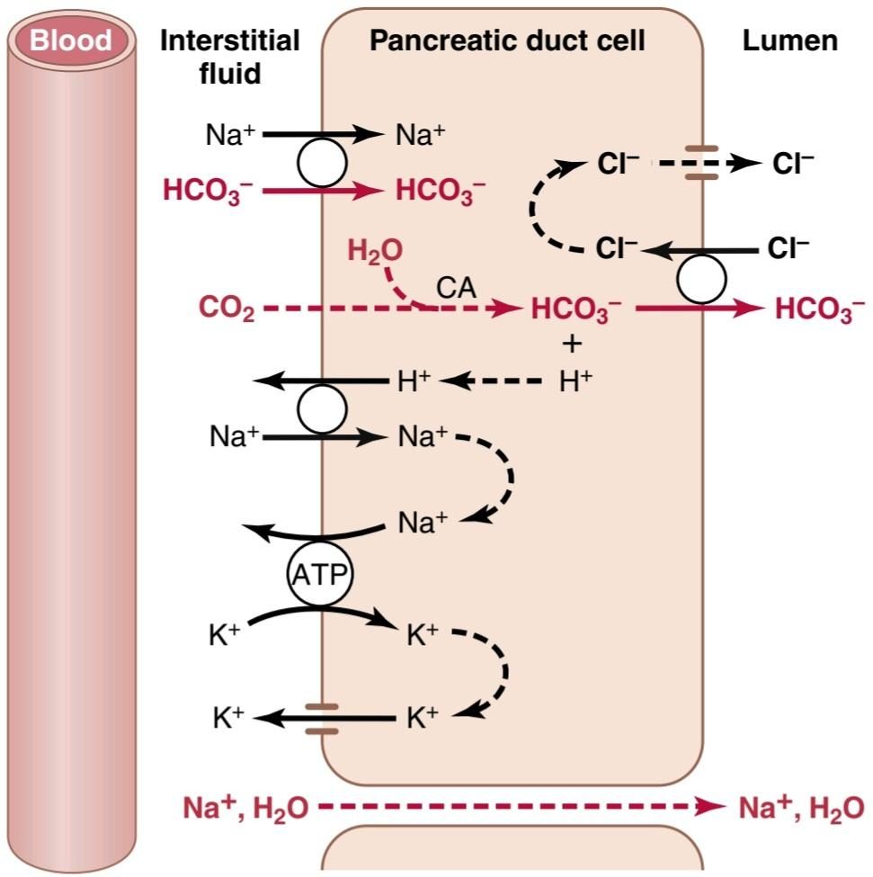
Figure 65-8. Secretion of isosmotic sodium bicarbonate solution by the pancreatic ductules and ducts. ATP, Adenosine triphosphate; CA, carbonic anhydrase.
شکل 65-8. ترشح محلول بی کربنات سدیم ایزوسمتیک توسط مجاری و مجاری پانکراس. ATP، آدنوزین تری فسفات؛ CA، کربنیک انیدراز.
REGULATION OF PANCREATIC SECRETION
Basic Stimuli That Cause Pancreatic Secretion
Three basic stimuli are important in causing pancreatic secretion:
تنظیم ترشح پانکراس
محرکهای اصلی که باعث ترشح پانکراس میشوند
سه محرک اساسی در ایجاد ترشح پانکراس مهم هستند:
1. Acetylcholine, which is released from the parasympathetic vagus nerve endings and from other cholinergic nerves in the enteric nervous system
1. استیل کولین که از انتهای عصب واگ پاراسمپاتیک و سایر اعصاب کولینرژیک در سیستم عصبی روده آزاد میشود.
2. Cholecystokinin (CCK), which is secreted by the duodenal and upper jejunal mucosa when food enters the small intestine
2. کوله سیستوکینین (CCK) که با ورود غذا به روده کوچک توسط مخاط دوازدهه و فوقانی ژژنوم ترشح میشود.
3. Secretin, which is also secreted by the duodenal and jejunal mucosa when highly acidic food enters the small intestine.
3. سکرتین که از مخاط اثنی عشر و ژژنوم نیز در هنگام ورود غذای بسیار اسیدی به روده کوچک ترشح میشود.
The first two of these stimuli, acetylcholine and CCK, stimulate the acinar cells of the pancreas, causing production of large quantities of pancreatic digestive enzymes but relatively small quantities of water and electrolytes to go with the enzymes. Without the water, most of the enzymes remain temporarily stored in the acini and ducts until more fluid secretion comes along to wash them into the duodenum. Secretin, in contrast to the first two basic stimuli, stimulates secretion of large quantities of water solution of sodium bicarbonate by the pancreatic ductal epithelium.
دو مورد اول از این محرکها، استیل کولین و CCK، سلولهای آسینار لوزالمعده را تحریک میکنند و باعث تولید مقادیر زیادی آنزیمهای گوارشی پانکراس میشوند، اما مقادیر نسبتا کمیآب و الکترولیتها با آنزیمها همراه میشوند. بدون آب، بیشتر آنزیمها بهطور موقت در آسین و مجاری ذخیره میشوند تا زمانی که ترشح مایع بیشتری وارد دوازدهه شود. سکرتین بر خلاف دو محرک اساسی اول، ترشح مقادیر زیادی محلول آبی بی کربنات سدیم را توسط اپیتلیوم مجرای پانکراس تحریک میکند.
Multiplicative Effects of Different Stimuli. When all the different stimuli of pancreatic secretion occur at once, the total secretion is far greater than the sum of the secretions caused by each one separately. Therefore, the various stimuli are said to “multiply,” or “potentiate,” one another. Thus, pancreatic secretion normally results from the combined effects of the multiple basic stimuli, not from one alone.
اثرات ضربی محرکهای مختلف. هنگامیکه تمام محرکهای مختلف ترشح پانکراس به یکباره رخ میدهد، ترشح کل به مراتب بیشتر از مجموع ترشحات ایجاد شده توسط هر یک به طور جداگانه است. بنابراین گفته میشود که محرکهای مختلف یکدیگر را «تکثیر میکنند» یا «تقویت میکنند». بنابراین، ترشح لوزالمعده معمولاً از اثرات ترکیبی چند محرک اساسی ناشی میشود، نه از یک محرک به تنهایی.
Phases of Pancreatic Secretion
Pancreatic secretion, as with gastric secretion, occurs in three phases: the cephalic phase, the gastric phase, and the intestinal phase. Their characteristics are described in the following sections.
مراحل ترشح پانکراس
ترشح لوزالمعده مانند ترشح معده در سه مرحله رخ میدهد: فاز سفالیک، فاز معده و فاز روده. ویژگیهای آنها در بخشهای بعدی توضیح داده شده است.
Cephalic and Gastric Phases. During the cephalic phase of pancreatic secretion, the same nervous signals from the brain that cause secretion in the stomach also cause acetylcholine release by the vagal nerve endings in the pancreas. This signaling causes moderate amounts of enzymes to be secreted into the pancreatic acini, accounting for about 20% of the total secretion of pancreatic enzymes after a meal. However, little of the secretion flows immediately through the pancreatic ducts into the intestine because only small amounts of water and electrolytes are secreted along with the enzymes.
فاز سفالیک و معده. در طول فاز سفالیک ترشح پانکراس، همان سیگنالهای عصبی از مغز که باعث ترشح در معده میشوند، باعث آزاد شدن استیل کولین توسط انتهای عصب واگ در پانکراس نیز میشوند. این سیگنال دهی باعث میشود که مقادیر متوسطی از آنزیمها به آسین پانکراس ترشح شود که حدود 20 درصد از کل ترشح آنزیمهای پانکراس را بعد از غذا تشکیل میدهد. با این حال، مقدار کمیاز ترشحات بلافاصله از طریق مجاری پانکراس به روده جریان مییابد، زیرا تنها مقادیر کمیآب و الکترولیتها همراه با آنزیمها ترشح میشوند.
During the gastric phase, the nervous stimulation of enzyme secretion continues, accounting for another 5% to 10% of pancreatic enzymes secreted after a meal. However, again, only small amounts reach the duodenum because of continued lack of significant fluid secretion.
در طول فاز معده، تحریک عصبی ترشح آنزیم ادامه مییابد و 5 تا 10 درصد دیگر از آنزیمهای پانکراس ترشح شده پس از غذا را تشکیل میدهد. با این حال، دوباره، به دلیل عدم ترشح مایع قابل توجه، فقط مقادیر کمیبه دوازدهه میرسد.
Intestinal Phase. After chyme leaves the stomach and enters the small intestine, pancreatic secretion becomes copious, mainly in response to the hormone secretin.
فاز روده. پس از خروج کیم از معده و ورود به روده کوچک، ترشح پانکراس زیاد میشود که عمدتاً در پاسخ به هورمون سکرتین است.
Secretin Stimulates Copious Secretion of Bicarbonate lons, Which Neutralizes Acidic Stomach Chyme. Secretin is a polypeptide containing 27 amino acids (with a molecular weight of ≈3400). It is present in an inactive form, prosecretin, in the S cells in the mucosa of the duodenum and jejunum. When acid chyme with a pH less than 4.5 to 5.0 enters the duodenum from the stomach, it causes duodenal mucosal release and activation of secretin, which is then absorbed into the blood. The one truly potent constituent of chyme that causes secretin release is hydrochloric acid from the stomach.
سکرتین ترشح فراوان بی کربنات لونها را تحریک میکند، که باعث خنثی شدن کیسه اسیدی معده میشود. Secretin یک پلی پپتید حاوی 27 اسید آمینه (با وزن مولکولی ≈3400) است. این ماده به شکل غیر فعال، پروسکرتین، در سلولهای S موجود در مخاط دوازدهه و ژژنوم وجود دارد. هنگامیکه کیم اسید با pH کمتر از 4.5 تا 5.0 از معده وارد دوازدهه میشود، باعث آزاد شدن مخاط دوازدهه و فعال شدن سکرتین میشود که سپس جذب خون میشود. یکی از اجزای واقعاً قوی کیم که باعث ترشح سکرتین میشود، اسید کلریدریک از معده است.
Secretin in turn causes the pancreas to secrete large quantities of fluid containing a high concentration of HCO3 (up to 145 mEq/L) but a low concentration of Cl-. The secretin mechanism is especially important for two reasons. First, secretin begins to be released from the mucosa of the small intestine when the pH of the duodenal contents falls below 4.5 to 5.0, and its release increases greatly as the pH falls to 3.0. This mechanism immediately causes copious secretion of pancreatic juice that contains abundant amounts of sodium bicarbonate. The net result is then the following reaction in the duodenum:
سکرتین به نوبه خود باعث میشود که لوزالمعده مقادیر زیادی مایع حاوی غلظت بالایی از HCO3 (تا 145 میلی اکیوالان در لیتر) اما غلظت کم -Cl ترشح کند. مکانیسم سکرتین به دو دلیل بسیار مهم است. ابتدا، ترشح سکرتین از مخاط روده کوچک زمانی که PH محتویات دوازدهه به زیر 4.5 تا 5.0 میرسد، شروع میشود و با کاهش pH به 3.0، ترشح آن به شدت افزایش مییابد. این مکانیسم بلافاصله باعث ترشح زیاد آب پانکراس میشود که حاوی مقادیر فراوانی بی کربنات سدیم است. سپس نتیجه خالص واکنش زیر در دوازدهه است:

The carbonic acid then immediately dissociates into CO2 and water. The CO2 is absorbed into the blood and expired through the lungs, thus leaving a neutral solution of sodium chloride in the duodenum. In this way, the acid contents that are emptied into the duodenum from the stomach become neutralized, and thus further peptic digestive activity by the gastric juices in the duodenum is immediately blocked. Because the mucosa of the small intestine cannot withstand the digestive action of acid gastric juice, this protective mechanism is essential to prevent the development of duodenal ulcers, as discussed in Chapter 67.
سپس اسید کربنیک بلافاصله به CO2 و آب تجزیه میشود. CO2 در خون جذب میشود و از طریق ریهها منفجر میشود، بنابراین یک محلول خنثی از کلرید سدیم در دوازدهه باقی میماند. به این ترتیب، محتویات اسیدی که از معده به دوازدهه تخلیه میشوند، خنثی میشوند و در نتیجه فعالیت گوارشی بیشتر توسط شیرههای معده در دوازدهه بلافاصله مسدود میشود. از آنجایی که مخاط روده کوچک نمیتواند در مقابل عملکرد گوارشی اسید معده مقاومت کند، این مکانیسم حفاظتی برای جلوگیری از ایجاد زخم اثنی عشر ضروری است، همانطور که در فصل 67 بحث شد.
Bicarbonate ion secretion by the pancreas provides an appropriate pH for action of the pancreatic digestive enzymes, which function optimally in a slightly alkaline or neutral medium, at a pH of 7.0 to 8.0. Fortunately, the pH of the sodium bicarbonate secretion averages 8.0.
ترشح یون بی کربنات توسط لوزالمعده PH مناسبی را برای عملکرد آنزیمهای گوارشی پانکراس فراهم میکند که در یک محیط کمیقلیایی یا خنثی در pH 7.0 تا 8.0 بهینه عمل میکنند. خوشبختانه pH ترشح بی کربنات سدیم به طور متوسط 8.0 است.
Cholecystokinin Contributes to Control of Digestive Enzyme Secretion by the Pancreas. The presence of food in the upper small intestine also causes a second hormone, CCK, a polypeptide containing 33 amino ac- ids, to be released from yet another group of cells, the I cells, in the mucosa of the duodenum and upper jejunum. This release of CCK results especially from the presence of proteoses and peptones (products of partial protein di- gestion) and long-chain fatty acids in the chyme coming from the stomach.
کوله سیستوکینین به کنترل ترشح آنزیمهای گوارشی توسط پانکراس کمک میکند. وجود غذا در قسمت فوقانی روده کوچک همچنین باعث میشود که هورمون دوم، CCK، پلی پپتیدی حاوی 33 آمینو اسید، از گروه دیگری از سلولها، سلولهای I، در مخاط دوازدهه و ژژنوم فوقانی آزاد شود. این آزاد شدن CCK به ویژه از حضور پروتئوزها و پپتونها (محصولات هضم جزئی پروتئین) و اسیدهای چرب با زنجیره بلند در کیم که از معده میآید، ناشی میشود.
CCK, like secretin, passes via the blood to the pancreas, but instead of causing sodium bicarbonate secretion, it mainly causes secretion of much more pancreatic digestive enzymes by the acinar cells. This effect is similar to that caused by vagal stimulation but is even more pronounced, accounting for 70% to 80% of the total secretion of the pancreatic digestive enzymes after a meal.
CCK، مانند سکرتین، از طریق خون به لوزالمعده منتقل میشود، اما به جای اینکه باعث ترشح بی کربنات سدیم شود، عمدتا باعث ترشح آنزیمهای گوارشی پانکراس بسیار بیشتری توسط سلولهای آسینار میشود. این اثر شبیه به آن چیزی است که در اثر تحریک واگ ایجاد میشود، اما حتی بارزتر است و 70 تا 80 درصد از کل ترشح آنزیمهای گوارشی پانکراس را بعد از غذا تشکیل میدهد.
The differences between the pancreatic stimulatory effects of secretin and CCK are shown in Figure 65-9, which demonstrates the following: (1) intense sodium bicarbonate secretion in response to acid in the duodenum, stimulated by secretin; (2) a dual effect in response to soap (a fat); and (3) intense digestive enzyme secretion (when peptones enter the duodenum) stimulated by CCK.
تفاوت بین اثرات تحریکی لوزالمعده سکرتین و CCK در شکل 65-9 نشان داده شده است که موارد زیر را نشان میدهد: (1) ترشح شدید بی کربنات سدیم در پاسخ به اسید در دوازدهه، تحریک شده توسط سکرتین. (2) اثر دوگانه در پاسخ به صابون (یک چربی). و (3) ترشح شدید آنزیم گوارشی (زمانی که پپتونها وارد دوازدهه میشوند) تحریک شده توسط CCK.
Figure 65-10 summarizes the more important factors that regulate pancreatic secretion. The total amount secreted each day is about 1 liter.
شکل 65-10 عوامل مهم تری را که ترشح پانکراس را تنظیم میکنند، خلاصه میکند. مقدار کل ترشح شده در هر روز حدود 1 لیتر است.
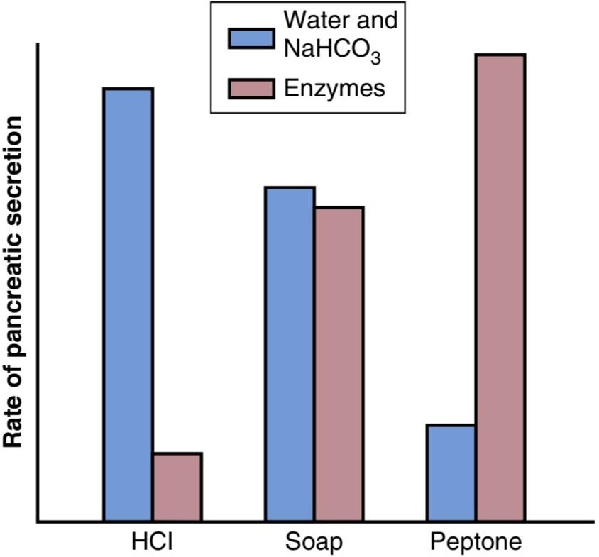
Figure 65-9. Sodium bicarbonate (NaHCO3), water, and enzyme secretion by the pancreas, caused by the presence of acid (HCI), fat (soap), or peptone solutions in the duodenum.
شکل 65-9. بی کربنات سدیم (NaHCO3)، آب و ترشح آنزیم توسط لوزالمعده، ناشی از وجود محلول اسید (HCI)، چربی (صابون) یا پپتون در دوازدهه است.
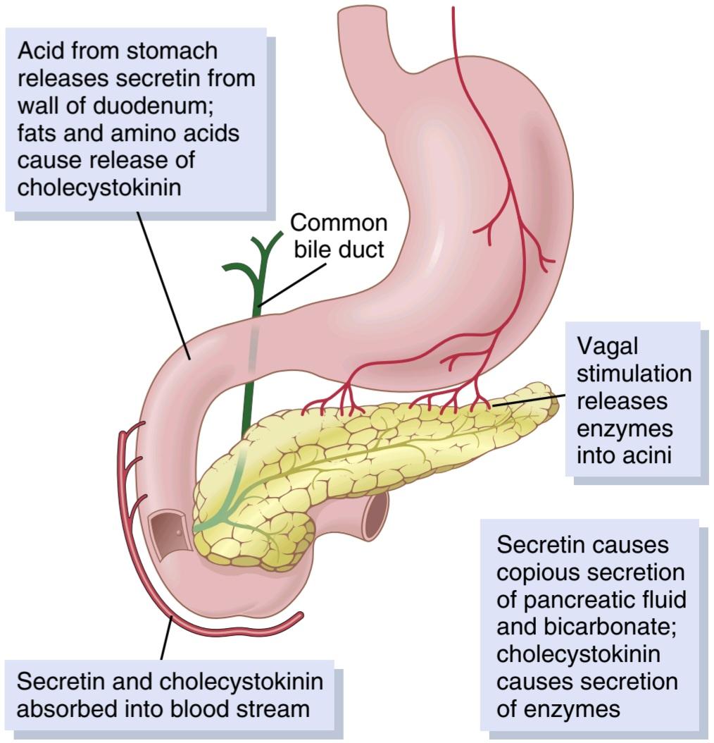
Figure 65-10. Regulation of pancreatic secretion.
شکل 65-10. تنظیم ترشح پانکراس.
BILE SECRETION BY THE LIVER
One of the many functions of the liver is to secrete bile, normally between 600 and 1000 ml/day. Bile serves two important functions.
ترشح صفرا توسط کبد
یکی از بسیاری از وظایف کبد ترشح صفرا است که به طور معمول بین 600 تا 1000 میلی لیتر در روز است. صفرا دو وظیفه مهم را انجام میدهد.
First, bile plays an important role in fat digestion and absorption, not because of any enzymes in the bile that cause fat digestion, but because bile acids perform two functions: (1) they help emulsify large fat particles of the food into many minute particles, the surface of which can then be attacked by lipase enzymes secreted in pancreatic juice, and (2) they aid in absorption of digested fat end products through the intestinal mucosal membrane.
اول، صفرا نقش مهمیدر هضم و جذب چربی ایفا میکند، نه به دلیل آنزیمهای موجود در صفرا که باعث هضم چربی میشود، بلکه به این دلیل که اسیدهای صفراوی دو عملکرد را انجام میدهند: (1) آنها به امولسیون کردن ذرات بزرگ چربی غذا به ذرات بسیار ریز کمک میکنند، که سطح آن میتواند توسط آنزیمهای لیپاز ترشح شده در غدد غدد لنفاوی، هضم چربی در غده لوزالمعده مورد حمله قرار گیرد. محصولات نهایی از طریق غشای مخاطی روده
Second, bile serves as a means for excretion of several important waste products from the blood. These waste products include in particular bilirubin, an end product of hemoglobin destruction, and excesses of cholesterol.
دوم، صفرا به عنوان وسیله ای برای دفع چندین ماده زائد مهم از خون عمل میکند. این مواد زائد به ویژه شامل بیلی روبین، محصول نهایی تخریب هموگلوبین، و کلسترول اضافی است.
PHYSIOLOGIC ANATOMY OF BILIARY SECRETION
Bile is secreted in two stages by the liver:
آناتومیفیزیولوژیکی ترشح صفراوی
صفرا در دو مرحله توسط کبد ترشح میشود:
1. The initial portion is secreted by the principal functional cells of the liver, the hepatocytes. This initial secretion contains large amounts of bile acids, cholesterol, and other organic constituents. It is secreted into minute bile canaliculi that originate between the hepatic cells (see Figure 71-1).
1. قسمت اولیه توسط سلولهای عملکردی اصلی کبد یعنی سلولهای کبدی ترشح میشود. این ترشح اولیه حاوی مقادیر زیادی اسیدهای صفراوی، کلسترول و سایر ترکیبات آلی است. آن را به کانال صفراوی دقیقه که بین سلولهای کبدی منشاء میگیرد ترشح میشود (شکل 71-1 را ببینید).
2. Next, the bile flows in the canaliculi toward the interlobular septa, where the canaliculi empty into terminal bile ducts and then into progressively larger ducts, finally reaching the hepatic duct and common bile duct. From these ducts the bile either empties directly into the duodenum or is diverted for minutes up to several hours through the cystic duct into the gallbladder, shown in Figure 65-11. In its course through the bile ducts, a second portion of liver secretion is added to the initial bile. This additional secretion is a watery solution of Na+ and HCO3- secreted by epithelial cells that line the ductules and ducts. This second secretion sometimes increases the total quantity of bile by as much as 100%. The second secretion is stimulated especially by secretin, which causes release of additional quantities of HCO3- to supplement the HCO3- in pancreatic secretion (for neutralizing acid that empties into the duodenum from the stomach).
2. سپس، صفرا در کانالها به سمت سپتوم بین لوبولار جریان مییابد، جایی که کانالها به مجاری صفراوی انتهایی و سپس به مجاری به تدریج بزرگتر تخلیه میشوند و در نهایت به مجرای کبدی و مجرای صفراوی مشترک میرسند. از این مجاری صفرا یا مستقیماً به دوازدهه تخلیه میشود یا برای چند دقیقه تا چند ساعت از طریق مجرای کیستیک به کیسه صفرا منحرف میشود که در شکل 11-65 نشان داده شده است. در مسیر خود از طریق مجاری صفراوی، بخش دوم ترشح کبد به صفرای اولیه اضافه میشود. این ترشح اضافی محلول آبکی +Na و -HCO3 است که توسط سلولهای اپیتلیال که مجراها و مجاری را میپوشانند ترشح میشود. این ترشح دوم گاهی اوقات مقدار کل صفرا را تا 100٪ افزایش میدهد. ترشح دوم به ویژه توسط سکرتین تحریک میشود که باعث آزاد شدن مقادیر اضافی -HCO3 برای تکمیل -HCO3 در ترشح پانکراس (برای خنثی کردن اسیدی که از معده به دوازدهه تخلیه میشود) میشود.
The Gallbladder Stores and Concentrates Bile. Bile is secreted continually by the liver cells, but most of it is normally stored in the gallbladder until it is needed in the duodenum. The maximum volume that the gallbladder can hold is only 30 to 60 ml. Nevertheless, as much as 12 hours of bile secretion (usually ≈450 ml) can be stored in the gallbladder because water, sodium, chloride, and most other small electrolytes are continually absorbed through the gallbladder mucosa, concentrating the remaining bile constituents that contain the bile salts, cholesterol, lecithin, and bilirubin.
کیسه صفرا صفرا را ذخیره و غلیظ میکند. صفرا به طور مداوم توسط سلولهای کبد ترشح میشود، اما بیشتر آن به طور معمول در کیسه صفرا ذخیره میشود تا زمانی که در دوازدهه مورد نیاز باشد. حداکثر حجمیکه کیسه صفرا میتواند نگه دارد تنها 30 تا 60 میلی لیتر است. با این وجود، تا 12 ساعت ترشح صفرا (معمولاً 450 میلیلیتر) میتواند در کیسه صفرا ذخیره شود، زیرا آب، سدیم، کلرید و اکثر الکترولیتهای کوچک دیگر به طور مداوم از طریق مخاط کیسه صفرا جذب میشوند و ترکیبات صفراوی باقیمانده را که حاوی نمکهای صفراوی و بیلیلینول هستند، متمرکز میکنند.
Most of this gallbladder absorption is caused by active transport of sodium through the gallbladder epithelium, and this transport is followed by secondary absorption of Cl-, water, and most other diffusible constituents. Bile is normally concentrated in this way about 5-fold, but it can be concentrated up to a maximum of 20-fold.
بیشتر این جذب کیسه صفرا ناشی از انتقال فعال سدیم از طریق اپیتلیوم کیسه صفرا است و این انتقال با جذب ثانویه کلر، آب و سایر اجزای قابل انتشار به دنبال دارد. صفرا به طور معمول به این ترتیب حدود 5 برابر غلیظ میشود، اما میتوان آن را حداکثر تا 20 برابر غلیظ کرد.
Composition of Bile. Table 65-2 lists the composition of bile when it is first secreted by the liver and then after it has been concentrated in the gallbladder. By far the most abundant substances secreted in the bile are bile salts, which ac- count for about one-half of the total solutes also in the bile.Also secreted or excreted in large concentrations are bilirubin, cholesterol, lecithin, and the usual electrolytes of plasma.
ترکیب صفرا. جدول 65-2 ترکیب صفرا را هنگامیکه ابتدا توسط کبد ترشح میشود و سپس در کیسه صفرا متمرکز میشود، فهرست میکند. تا حد زیادی فراوان ترین مواد ترشح شده در صفرا نمکهای صفراوی هستند که تقریباً نیمیاز کل املاح را در صفرا تشکیل میدهند. همچنین بیلی روبین، کلسترول، لسیتین و الکترولیتهای معمول پلاسما ترشح یا دفع میشوند.
In the concentrating process in the gallbladder, water and large portions of the electrolytes (except calcium ions) are reabsorbed by the gallbladder mucosa. Essentially all other constituents, especially the bile salts and the lipid substances cholesterol and lecithin, are not reabsorbed and, therefore, become highly concentrated in the gallbladder bile.
در فرآیند تغلیظ در کیسه صفرا، آب و بخش زیادی از الکترولیتها (به جز یونهای کلسیم) توسط مخاط کیسه صفرا دوباره جذب میشوند. اساساً تمام مواد تشکیل دهنده دیگر، به ویژه نمکهای صفراوی و مواد لیپیدی کلسترول و لسیتین، بازجذب نمیشوند و بنابراین در صفرای کیسه صفرا بسیار متمرکز میشوند.
Table 65-2 Composition of Bile
جدول 65-2 ترکیب صفرا

Cholecystokinin Stimulates Gallbladder Emptying. When food begins to be digested in the upper gastrointestinal tract, the gallbladder begins to empty, especially when fatty foods reach the duodenum about 30 minutes after a meal. The mechanism of gallbladder emptying is rhythmic contractions of the gallbladder wall, but effective emptying also requires simultaneous relaxation of the sphincter of Oddi, which guards the exit of the common bile duct into the duodenum.
کوله سیستوکینین تخلیه کیسه صفرا را تحریک میکند. هنگامیکه غذا در قسمت فوقانی دستگاه گوارش شروع به هضم میکند، کیسه صفرا شروع به تخلیه میکند، به خصوص زمانی که غذاهای چرب حدود 30 دقیقه پس از غذا به دوازدهه میرسند. مکانیسم تخلیه کیسه صفرا انقباضات ریتمیک دیواره کیسه صفرا است، اما تخلیه موثر همچنین نیاز به شل شدن همزمان اسفنکتر Oddi دارد که از خروج مجرای صفراوی مشترک به دوازدهه محافظت میکند.
By far the most potent stimulus for causing the gall- bladder contractions is the hormone CCK. As discussed previously, CCK also causes increased secretion of digestive enzymes by the acinar cells of the pancreas. The stimulus for CCK entry into the blood from the duodenal mucosa is mainly the presence of fatty foods in the duodenum.
تا حد زیادی قوی ترین محرک برای ایجاد انقباضات کیسه صفرا، هورمون CCK است. همانطور که قبلاً بحث شد، CCK همچنین باعث افزایش ترشح آنزیمهای گوارشی توسط سلولهای آسینار پانکراس میشود. محرک ورود CCK به خون از مخاط دوازدهه عمدتاً وجود غذاهای چرب در دوازدهه است.
The gallbladder is also stimulated less strongly by acetylcholine-secreting nerve fibers from both the vagi and the intestinal enteric nervous system. They are the same nerves that promote motility and secretion in other parts of the upper gastrointestinal tract.
کیسه صفرا همچنین توسط رشتههای عصبی ترشح کننده استیل کولین از واگی و سیستم عصبی روده روده با شدت کمتری تحریک میشود. آنها همان اعصابی هستند که حرکت و ترشح را در سایر قسمتهای دستگاه گوارش فوقانی تقویت میکنند.
In summary, the gallbladder empties its store of concentrated bile into the duodenum mainly in response to the CCK stimulus that is initiated mainly by fatty foods. When fat is not in the food, the gallbladder empties poorly, but when significant quantities of fat are present, the gallbladder normally empties completely in about 1 hour. Figure 65-11 summarizes the secretion of bile, its storage in the gallbladder, and its ultimate release from the gallbladder to the duodenum.
به طور خلاصه، کیسه صفرا ذخیره صفرای غلیظ خود را به طور عمده در پاسخ به محرک CCK که عمدتاً توسط غذاهای چرب آغاز میشود، به دوازدهه تخلیه میکند. زمانی که چربی در غذا نباشد، کیسه صفرا ضعیف تخلیه میشود، اما زمانی که مقادیر قابل توجهی چربی وجود داشته باشد، کیسه صفرا به طور معمول در عرض یک ساعت کاملا خالی میشود. شکل 11-65 ترشح صفرا، ذخیره آن در کیسه صفرا و آزاد شدن نهایی آن از کیسه صفرا به دوازدهه را خلاصه میکند.
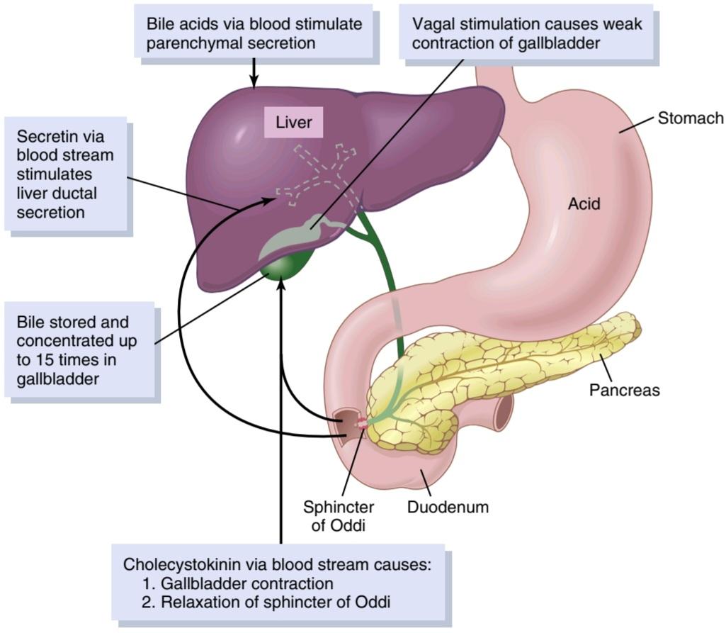
Figure 65-11. Liver secretion and gall- bladder emptying.
شکل 65-11. ترشح کبد و تخلیه کیسه صفرا.
FUNCTION OF BILE SALTS IN FAT DIGESTION AND ABSORPTION
The liver cells synthesize about 6 g/day of bile salts. The precursor of the bile salts is cholesterol, which is either present in the diet or synthesized in the liver cells during the course of fat metabolism. The cholesterol is first converted to cholic acid or chenodeoxycholic acid in about equal quantities. These acids in turn combine principally with glycine and to a lesser extent with taurine to form glyco-conjugated and tauro-conjugated bile acids. The salts of these acids, mainly sodium salts, are then secreted in the bile.
عملکرد نمکهای صفراوی در هضم و جذب چربی
سلولهای کبد حدود 6 گرم در روز نمک صفراوی سنتز میکنند. پیش ساز نمکهای صفراوی کلسترول است که یا در رژیم غذایی وجود دارد یا در طی متابولیسم چربی در سلولهای کبدی سنتز میشود. کلسترول ابتدا تقریباً به مقدار مساوی به اسید کولیک یا چنودوکسی کولیک اسید تبدیل میشود. این اسیدها به نوبه خود عمدتاً با گلیسین و به میزان کمتری با تورین ترکیب میشوند تا اسیدهای صفراوی گلیکو کونژوگه و تائورو کونژوگه را تشکیل دهند. نمکهای این اسیدها، عمدتاً نمکهای سدیم، سپس در صفرا ترشح میشوند.
The bile salts have two important actions in the intestinal tract.
نمکهای صفراوی دو عمل مهم در مجرای روده دارند.
First, they have a detergent action on the fat particles in the food. This action, which decreases surface tension of the particles and allows agitation in the intestinal tract to break the fat globules into minute sizes, is called the emulsifying or detergent function of bile salts.
اول، آنها یک اثر شوینده بر روی ذرات چربی موجود در غذا دارند. این عمل که کشش سطحی ذرات را کاهش میدهد و به هم زدن در دستگاه روده اجازه میدهد تا گلبولهای چربی را به اندازههای کوچک بشکند، عملکرد امولسیون کننده یا شوینده نمکهای صفراوی نامیده میشود.
Second, and even more important than the emulsifying function, bile salts help in the absorption of (1) fatty acids, (2) monoglycerides, (3) cholesterol, and (4) other lipids from the intestinal tract. They help in this absorption by forming small physical complexes with these lipids; the complexes are called micelles, and they are semisoluble in the chyme because of the electrical charges of the bile salts. The intestinal lipids are “ferried” in this form to the intestinal mucosa, where they are then absorbed into the blood, as will be described in detail in Chapter 66. With- out the presence of bile salts in the intestinal tract, up to 40% of the ingested fats are lost into the feces, and a metabolic deficit often develops because of this nutrient loss.
دوم، و حتی مهمتر از عملکرد امولسیونکننده، نمکهای صفراوی به جذب (1) اسیدهای چرب، (2) مونوگلیسریدها، (3) کلسترول و (4) چربیهای دیگر از دستگاه روده کمک میکنند. آنها با تشکیل مجتمعهای فیزیکی کوچک با این لیپیدها به این جذب کمک میکنند. این کمپلکسها میسل نامیده میشوند و به دلیل بارهای الکتریکی نمکهای صفراوی در کیم نیمه محلول هستند. لیپیدهای روده به این شکل به مخاط روده منتقل میشوند و سپس جذب خون میشوند، همانطور که در فصل 66 به تفصیل توضیح داده خواهد شد. بدون وجود نمکهای صفراوی در مجرای روده، تا 40 درصد از چربیهای مصرفشده اغلب در مدفوع از بین میروند، و به دلیل کمبود متابولیک تریها، از بین میرود.
Enterohepatic Circulation of Bile Salts. About 94% of the bile salts are reabsorbed into the blood from the small intestine, about one half of this by diffusion through the mucosa in the early portions of the small intestine and the remainder by an active transport process through the intestinal mucosa in the distal ileum. They then enter the portal blood and pass back to the liver. On reaching the liver and during first passage through the venous sinusoids, these salts are absorbed almost entirely back into the hepatic cells and are then resecreted into the bile.
گردش انتروهپاتیک نمکهای صفراوی. حدود 94 درصد نمکهای صفراوی از روده کوچک به خون بازجذب میشوند، تقریباً نیمیاز آن از طریق انتشار از طریق مخاط در قسمتهای اولیه روده کوچک و بقیه توسط یک فرآیند انتقال فعال از طریق مخاط روده در ایلئوم دیستال. سپس وارد خون پورت شده و دوباره به کبد منتقل میشوند. با رسیدن به کبد و در طی اولین عبور از سینوسی وریدی، این نمکها تقریباً به طور کامل به سلولهای کبدی جذب میشوند و سپس دوباره به صفرا ترشح میشوند.
In this way, about 94% of all the bile salts are recirculated into the bile, so on average these salts make the entire circuit some 17 times before being carried out in the feces. The small quantities of bile salts lost into the feces are replaced by new amounts formed continually by the liver cells. This recirculation of the bile salts is called the enterohepatic circulation of bile salts.
به این ترتیب، حدود 94٪ از تمام نمکهای صفراوی به داخل صفرا گردش میکنند، بنابراین به طور متوسط این نمکها قبل از اینکه در مدفوع وارد شوند، کل مدار را حدود 17 بار میسازند. مقادیر کمیاز نمکهای صفراوی از دست رفته در مدفوع با مقادیر جدیدی که به طور مداوم توسط سلولهای کبدی تشکیل میشود جایگزین میشود. به این گردش نمکهای صفراوی، گردش کبدی نمکهای صفراوی گفته میشود.
The quantity of bile secreted by the liver each day is highly dependent on the availability of bile salts–the greater the quantity of bile salts in the enterohepatic circulation (usually a total of only 2.5 grams), the greater the rate of bile secretion. Indeed, ingestion of supplemental bile salts can increase bile secretion by several hundred milliliters per day.
مقدار صفرای ترشح شده توسط کبد در هر روز به شدت به در دسترس بودن نمکهای صفراوی بستگی دارد – هر چه مقدار نمکهای صفراوی در گردش خون روده کبدی بیشتر باشد (معمولاً در مجموع تنها 2.5 گرم)، سرعت ترشح صفرا بیشتر میشود. در واقع، مصرف نمکهای صفراوی مکمل میتواند ترشح صفرا را تا چند صد میلی لیتر در روز افزایش دهد.
If a bile fistula empties the bile salts to the exterior for several days to several weeks so they cannot be reabsorbed from the ileum, the liver increases its production of bile salts 6- to 10-fold, which increases the rate of bile secretion most of the way back to normal. This demonstrates that the daily rate of liver bile salt secretion is actively controlled by the availability (or lack of availability) of bile salts in the enterohepatic circulation.
اگر فیستول صفراوی نمکهای صفراوی را به مدت چند روز تا چند هفته به سمت بیرون تخلیه کند تا نتوانند از ایلئوم بازجذب شوند، کبد تولید نمکهای صفراوی خود را 6 تا 10 برابر افزایش میدهد که در بیشتر موارد میزان ترشح صفرا را به حالت عادی افزایش میدهد. این نشان میدهد که میزان روزانه ترشح نمک صفراوی کبد به طور فعال توسط در دسترس بودن (یا عدم در دسترس بودن) نمکهای صفراوی در گردش خون روده ای کنترل میشود.
Role of Secretin in Controlling Bile Secretion. In addition to the strong stimulating effect of bile acids to cause bile secretion, the hormone secretin-which also stimulates pancreatic secretion-increases bile secretion, sometimes more than doubling its secretion for several hours after a meal. This increase in secretion consists almost entirely of secretion of a sodium bicarbonate-rich watery solution by the epithelial cells of the bile ductules and ducts and does not represent increased secretion by the liver parenchymal cells. The bicarbonate in turn passes into the small intestine and joins the bicarbonate from the pancreas in neutralizing the hydrochloric acid from the stomach. Thus, the secretin feedback mechanism for neutralizing duodenal acid operates not only through its effects on pancreatic secretion but also to a lesser extent through its effect on secretion by the liver ductules and ducts.
نقش سکرتین در کنترل ترشح صفرا. علاوه بر اثر محرک قوی اسیدهای صفراوی برای ایجاد ترشح صفرا، هورمون سکرتین – که ترشح لوزالمعده را نیز تحریک میکند – ترشح صفرا را افزایش میدهد، گاهی اوقات ترشح آن را تا چند ساعت بعد از غذا بیش از دو برابر میکند. این افزایش ترشح تقریباً به طور کامل شامل ترشح محلول آبکی غنی از بی کربنات سدیم توسط سلولهای اپیتلیال مجاری و مجاری صفراوی است و نشان دهنده افزایش ترشح توسط سلولهای پارانشیمیکبد نیست. بی کربنات به نوبه خود وارد روده کوچک میشود و به بی کربنات از پانکراس میپیوندد تا اسید کلریدریک معده را خنثی کند. بنابراین، مکانیسم بازخورد سکرتین برای خنثی کردن اسید اثنی عشر نه تنها از طریق تأثیر آن بر ترشح پانکراس بلکه به میزان کمتری از طریق تأثیر آن بر ترشح توسط مجاری و مجاری کبدی عمل میکند.
Liver Secretion of Cholesterol and Gallstone Formation
Bile salts are formed in the hepatic cells from cholesterol in the blood plasma. In the process of secreting bile salts, about 1 to 2 grams of cholesterol are removed from the blood plasma and secreted into the bile each day.
ترشح کبد از کلسترول و تشکیل سنگ کیسه صفرا
نمکهای صفراوی در سلولهای کبدی از کلسترول موجود در پلاسمای خون تشکیل میشوند. در فرآیند ترشح نمکهای صفراوی، روزانه حدود 1 تا 2 گرم کلسترول از پلاسمای خون خارج شده و به صفرا ترشح میشود.
Cholesterol is almost completely insoluble in pure water, but the bile salts and lecithin in bile combine physically with the cholesterol to form ultramicroscopic micelles in the form of a colloidal solution, as explained in Chapter 66. When the bile becomes concentrated in the gallbladder, the bile salts and lecithin become concentrated along with the cholesterol, which keeps the cholesterol in solution.
کلسترول تقریباً به طور کامل در آب خالص نامحلول است، اما نمکهای صفراوی و لسیتین موجود در صفرا به طور فیزیکی با کلسترول ترکیب میشوند و میسلهای فوق میکروسکوپی را به شکل یک محلول کلوئیدی تشکیل میدهند، همانطور که در فصل 66 توضیح داده شد. کلسترول در محلول.
Under abnormal conditions, the cholesterol may precipitate in the gallbladder, resulting in the formation of cholesterol gallstones, as shown in Figure 65-12. The amount of cholesterol in the bile is determined partly by the quantity of fat that the person eats, because liver cells synthesize cholesterol as one of the products of fat metabolism in the body. For this reason, people who are obese and consume a high-fat diet over a period of years are prone to development of gallstones. Other risk factors for gallstones include increasing age and female sex, diabetes mellitus, and genetic susceptibility.
در شرایط غیر طبیعی، کلسترول ممکن است در کیسه صفرا رسوب کند و در نتیجه سنگهای صفراوی کلسترولی تشکیل شود، همانطور که در شکل 12-65 نشان داده شده است. مقدار کلسترول در صفرا تا حدی با مقدار چربی که فرد میخورد تعیین میشود، زیرا سلولهای کبد کلسترول را به عنوان یکی از محصولات متابولیسم چربی در بدن سنتز میکنند. به همین دلیل، افرادی که چاق هستند و رژیم غذایی پرچرب را طی سالیان متمادی مصرف میکنند، مستعد ابتلا به سنگ کیسه صفرا هستند. سایر عوامل خطر برای سنگ کیسه صفرا عبارتند از افزایش سن و جنس زن، دیابت شیرین و حساسیت ژنتیکی.
Inflammation of the gallbladder epithelium, often resulting from a low-grade chronic infection, may also change the absorptive characteristics of the gallbladder mucosa, sometimes allowing excessive absorption of water and bile salts but leaving behind the cholesterol in the gallbladder in progressively greater concentrations. The cholesterol then begins to precipitate, first forming many small crystals of cholesterol on the surface of the inflamed mucosa, but then progressing to large gallstones.
التهاب اپیتلیوم کیسه صفرا، که اغلب ناشی از عفونت مزمن درجه پایین است، ممکن است ویژگیهای جذبی مخاط کیسه صفرا را نیز تغییر دهد، گاهی اوقات باعث جذب بیش از حد آب و نمکهای صفراوی میشود، اما کلسترول را در کیسه صفرا در غلظتهای به تدریج بیشتر میکند. سپس کلسترول شروع به رسوب میکند، ابتدا کریستالهای کوچک زیادی از کلسترول روی سطح مخاط ملتهب تشکیل میدهد، اما سپس به سمت سنگهای صفراوی بزرگ پیش میرود.
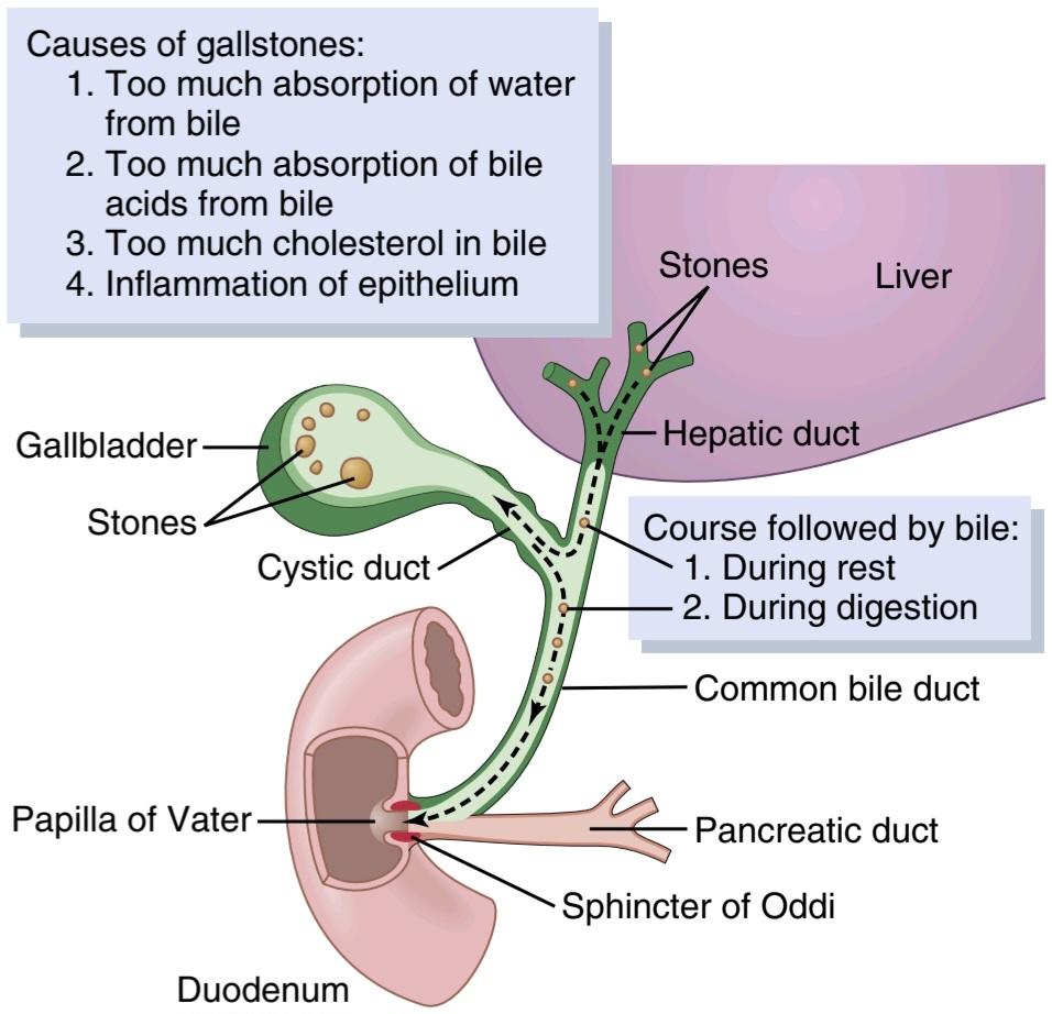
Figure 65-12. Formation of gallstones.
شکل 65-12. تشکیل سنگ کیسه صفرا.
SECRETIONS OF THE SMALL INTESTINE
SECRETION OF MUCUS BY BRUNNER’S GLANDS IN THE DUODENUM
An extensive array of compound mucous glands, called Brunner’s glands, is located in the wall of the first few centi- meters of the duodenum, mainly between the pylorus of the stomach and the papilla of Vater, where pancreatic secretion and bile empty into the duodenum. These glands secrete large amounts of alkaline mucus in response to the following: (1) tactile or irritating stimuli on the duodenal mucosa; (2) vagal stimulation, which causes increased Brunner gland secretion concurrently with increase in stomach secretion; and (3) gastrointestinal hormones, especially secretin.
ترشحات روده کوچک
ترشح مخاط توسط غدد برونر در دوازدهه
مجموعه وسیعی از غدد مخاطی مرکب، به نام غدد برونر، در دیواره چند سانتیمتری اول دوازدهه، عمدتاً بین پیلور معده و پاپیلای Vater، جایی که ترشح پانکراس و صفرا به دوازدهه تخلیه میشود، قرار دارد. این غدد در پاسخ به موارد زیر مقادیر زیادی موکوس قلیایی ترشح میکنند: (1) محرکهای لمسی یا تحریک کننده روی مخاط دوازدهه. (2) تحریک واگ، که باعث افزایش ترشح غده برونر همزمان با افزایش ترشح معده میشود. و (3) هورمونهای گوارشی، به ویژه سکرتین.
The function of the mucus secreted by Brunner’s glands is to protect the duodenal wall from digestion by the highly acidic gastric juice emptying from the stomach. In addition, the mucus contains a large excess of HCO3-, which add to the HCO3- from pancreatic secretion and liver bile in neutralizing the hydrochloric acid entering the duodenum from the stomach.
وظیفه مخاط ترشح شده توسط غدد برونر محافظت از دیواره دوازدهه در برابر هضم توسط آب معده بسیار اسیدی است که از معده تخلیه میشود. علاوه بر این، مخاط حاوی مقدار زیادی -HCO3 است که به -HCO3 از ترشح پانکراس و صفرای کبد در خنثی کردن اسید هیدروکلریک که از معده وارد دوازدهه میشود، اضافه میکند.
Brunner’s glands are inhibited by sympathetic stimulation; therefore, such stimulation in very excitable persons is likely to leave the duodenal bulb unprotected and is perhaps one of the factors that cause this area of the gastro- intestinal tract to be the site of peptic ulcers in about 50% of persons with ulcers.
غدد برونر با تحریک سمپاتیک مهار میشوند. بنابراین، چنین تحریکی در افراد بسیار تحریک پذیر احتمالاً پیاز اثنی عشر را بدون محافظت میگذارد و شاید یکی از عواملی باشد که باعث میشود این ناحیه از دستگاه گوارش در حدود 50 درصد از افراد مبتلا به زخم، محل زخم معده باشد.
SECRETION OF INTESTINAL DIGESTIVE JUICES BY THE CRYPTS OF LIEBERKÜHN
Located over the entire surface of the small intestine are small pits called crypts of Lieberkühn, one of which is illustrated in Figure 65-13. These crypts lie between the intestinal villi. The surfaces of both the crypts and the villi are covered by an epithelium composed of two types of cells: (1) a moderate number of goblet cells, which secrete mucus that lubricates and protects the intestinal surfaces; and (2) a large number of enterocytes, which, in the crypts, secrete large quantities of water and electrolytes and, over the surfaces of adjacent villi, reabsorb the water and electrolytes along with the end products of digestion.
ترشح شیرههای گوارشی روده ای توسط کریپتهای لیبرکوهن
بر روی تمام سطح روده کوچک حفرههای کوچکی به نام دخمههای لیبرکوهن وجود دارد که یکی از آنها در شکل 65-13 نشان داده شده است. این دخمهها بین پرزهای روده قرار دارند. سطوح کریپتها و پرزها توسط یک اپیتلیوم متشکل از دو نوع سلول پوشیده شده است: (1) تعداد متوسطی از سلولهای جام که مخاط ترشح میکنند که سطوح روده را روان و محافظت میکند. و (2) تعداد زیادی انتروسیت، که در دخمهها، مقادیر زیادی آب و الکترولیت ترشح میکنند و بر روی سطوح پرزهای مجاور، آب و الکترولیتها را همراه با محصولات نهایی هضم دوباره جذب میکنند.
The intestinal secretions are formed by the enterocytes of the crypts at a rate of about 1800 ml/day. These secretions are almost pure extracellular fluid and have a slightly alkaline pH in the range of 7.5 to 8.0. The secretions are also rapidly reabsorbed by the villi. This flow of fluid from the crypts into the villi supplies a watery vehicle for absorption of substances from chyme when it comes in contact with the villi. Thus, the primary function of the small intestine is to absorb nutrients and their digestive products into the blood.
ترشحات روده توسط انتروسیتهای کریپتها با سرعت حدود 1800 میلی لیتر در روز تشکیل میشود. این ترشحات تقریباً مایع خارج سلولی خالص هستند و pH کمیقلیایی در محدوده 7.5 تا 8.0 دارند. ترشحات نیز به سرعت توسط پرزها بازجذب میشوند. این جریان مایع از دخمهها به پرزها یک وسیله نقلیه آبکی برای جذب مواد از کیم در هنگام تماس با پرزها تامین میکند. بنابراین، وظیفه اصلی روده کوچک جذب مواد مغذی و محصولات گوارشی آنها به خون است.

Figure 65-13. A crypt of Lieberkühn, found in all parts of the small intestine between the villi, which secretes almost pure extracellular fluid.
شکل 65-13. دخمه ای از Lieberkühn که در تمام قسمتهای روده کوچک بین پرزها یافت میشود که مایع خارج سلولی تقریبا خالص ترشح میکند.
Mechanism of Secretion of the Watery Fluid. The ex- act mechanism that controls the marked secretion of watery fluid by the crypts of Lieberkühn is still unclear, but it is believed to involve at least two active secretory process- es: (1) active secretion of Cl- into the crypts and (2) active secretion of HCO3. The secretion of both ions causes electrical drag of positively charged Na+ ions through the membrane and into the secreted fluid as well. Finally, all these ions together cause osmotic movement of water.
مکانیسم ترشح مایع آبکی. مکانیسم دقیقی که ترشح مشخص مایع آبکی را توسط کریپتهای لیبرکوهن کنترل میکند هنوز نامشخص است، اما اعتقاد بر این است که حداقل دو فرآیند ترشحی فعال را شامل میشود: (1) ترشح فعال -Cl در کریپتها و (2) ترشح فعال HCO3. ترشح هر دو یون باعث کشش الکتریکی یونهای +Na با بار مثبت از طریق غشا و همچنین به داخل مایع ترشح میشود. در نهایت همه این یونها با هم باعث حرکت اسمزی آب میشوند.
Digestive Enzymes in the Small Intestinal Secretion. When secretions of the small intestine are collected with- out cellular debris, they have almost no enzymes. The enterocytes of the mucosa, especially those that cover the villi, contain digestive enzymes that digest specific food substances while they are being absorbed through the epithelium. These enzymes are as follows: (1) several peptidases for splitting small peptides into amino acids; (2) four enzymes-sucrase, maltase, isomaltase, and lactase-for splitting disaccharides into monosaccharides; and (3) small amounts of intestinal lipase for splitting neutral fats into glycerol and fatty acids.
آنزیمهای گوارشی در ترشحات روده کوچک. هنگامیکه ترشحات روده کوچک بدون بقایای سلولی جمع آوری میشوند، تقریباً هیچ آنزیمیندارند. انتروسیتهای مخاط، بهویژه آنهایی که پرزها را میپوشانند، حاوی آنزیمهای گوارشی هستند که مواد غذایی خاصی را در حالی که از طریق اپیتلیوم جذب میشوند، هضم میکنند. این آنزیمها به شرح زیر هستند: (1) چندین پپتیداز برای تقسیم پپتیدهای کوچک به اسیدهای آمینه. (2) چهار آنزیم ساکاراز، مالتاز، ایزومالتاز و لاکتاز برای تقسیم دی ساکاریدها به مونوساکاریدها. و (3) مقادیر کمیلیپاز روده برای تقسیم چربیهای خنثی به گلیسرول و اسیدهای چرب.
The epithelial cells deep in the crypts of Lieberkühn continually undergo mitosis, and new cells migrate along the basement membrane upward out of the crypts toward the tips of the villi, thus continually replacing the villus epithelium and also forming new digestive enzymes. As the villus cells age, they are finally shed into the intestinal secretions. The life cycle of an intestinal epithelial cell is about 5 days. This rapid growth of new cells also allows rapid repair of excoriations that occur in the mucosa.
سلولهای اپیتلیال در اعماق دخمههای لیبرکوهن به طور مداوم تحت میتوز قرار میگیرند و سلولهای جدید در امتداد غشای پایه به سمت بالا به سمت نوک پرزها مهاجرت میکنند، بنابراین به طور مداوم جایگزین اپیتلیوم پرز میشوند و همچنین آنزیمهای گوارشی جدیدی تشکیل میدهند. با پیر شدن سلولهای پرز، در نهایت به ترشحات روده میریزند. چرخه زندگی یک سلول اپیتلیال روده حدود 5 روز است. این رشد سریع سلولهای جدید همچنین امکان ترمیم سریع بریدگیهایی را که در مخاط ایجاد میشود، فراهم میکند.
REGULATION OF SMALL INTESTINE SECRETION-LOCAL STIMULI
By far the most important means for regulating small intestine secretion are local enteric nervous reflexes, especially reflexes initiated by tactile or irritative stimuli from the chyme in the intestines.
تنظیم ترشح روده کوچک-محرکهای موضعی
تا حد زیادی مهم ترین وسیله برای تنظیم ترشح روده کوچک، رفلکسهای عصبی روده ای موضعی هستند، به ویژه رفلکسهایی که توسط محرکهای لمسی یا تحریک کننده از کیم در روده شروع میشوند.
SECRETION OF MUCUS BY THE LARGE INTESTINE
Mucus Secretion. The mucosa of the large intestine, like that of the small intestine, has many crypts of Lieberkühn; however, unlike the small intestine, it contains no villi. The epithelial cells secrete almost no digestive enzymes. Instead, they contain mucous cells that secrete only mucus. This mucus contains moderate amounts of HCO3- secreted by a few non-mucus-secreting epithelial cells. The rate of mucus secretion is regulated principally by direct, tactile stimulation of the epithelial cells lining the large intestine and by local nervous reflexes to the mucous cells in the crypts of Lieberkühn.
ترشح مخاط توسط روده بزرگ
ترشح مخاطی. مخاط روده بزرگ، مانند روده باریک، دارای دخمههای لیبرکون است. با این حال، برخلاف روده کوچک، فاقد پرز است. سلولهای اپیتلیال تقریبا هیچ آنزیم گوارشی ترشح نمیکنند. در عوض، آنها حاوی سلولهای مخاطی هستند که فقط مخاط ترشح میکنند. این مخاط حاوی مقادیر متوسطی از HCO3 است که توسط چند سلول اپیتلیال غیر ترشح کننده موکوس ترشح میشود. سرعت ترشح مخاط عمدتاً با تحریک مستقیم و لمسی سلولهای اپیتلیال پوشش روده بزرگ و با رفلکسهای عصبی موضعی به سلولهای مخاطی در دخمههای لیبرکوهن تنظیم میشود.
Stimulation of the pelvic nerves from the spinal cord, which carry parasympathetic innervation to the distal one-half to two-thirds of the large intestine, also can cause marked increases in mucus secretion along with increased peristaltic motility of the colon, as discussed in Chapter 64.
تحریک اعصاب لگنی از طناب نخاعی، که عصب پاراسمپاتیک را به نیم تا دو سوم دیستال روده بزرگ حمل میکند، همچنین میتواند باعث افزایش قابل توجه ترشح موکوس همراه با افزایش تحرک پریستالتیک روده بزرگ شود، همانطور که در فصل 64 بحث شد.
During extreme parasympathetic stimulation, often caused by emotional disturbances, so much mucus can occasionally be secreted into the large intestine that the person has a bowel movement of ropy mucus as often as every 30 minutes. This mucus often contains little or no fecal material.
در طی تحریک شدید پاراسمپاتیک، که اغلب به دلیل اختلالات عاطفی ایجاد میشود، گهگاه میتوان آنقدر مخاط در روده بزرگ ترشح کرد که فرد هر 30 دقیقه یکبار اجابت مزاج از مخاط طوطی دارد. این مخاط اغلب حاوی مواد مدفوع کمیاست یا اصلاً وجود ندارد.
Mucus in the large intestine protects the intestinal wall against excoriation, but in addition, it provides an adherent medium for holding fecal matter together. Further- more, it protects the intestinal wall from the great amount of bacterial activity that takes place inside the feces, and, finally, the mucus plus the alkalinity of the secretion (a pH of 8.0 caused by large amounts of sodium bicarbonate) provides a barrier to keep acids formed in the feces from attacking the intestinal wall.
مخاط موجود در روده بزرگ از دیواره روده در برابر تخلیه محافظت میکند، اما علاوه بر این، یک محیط چسبنده برای نگه داشتن مواد مدفوع در کنار هم فراهم میکند. علاوه بر این، دیواره روده را از میزان زیادی از فعالیت باکتریایی که در داخل مدفوع انجام میشود محافظت میکند و در نهایت، مخاط به اضافه قلیایی بودن ترشح (PH 8.0 ناشی از مقادیر زیادی بی کربنات سدیم) مانعی برای جلوگیری از حمله اسیدهای تشکیلشده در مدفوع به دیواره روده ایجاد میکند.
Diarrhea Caused by Excess Secretion of Water and Electrolytes in Response to Irritation. Whenever a segment of the large intestine becomes intensely irritated, as occurs when bacterial infection becomes rampant during enteritis, the mucosa secretes extra large quantities of water and electrolytes in addition to the normal viscid alkaline mucus. This secretion acts to dilute the irritating factors and to cause rapid movement of the feces toward the anus. The result is diarrhea, with loss of large quantities of water and electrolytes. However, the diarrhea also washes away irritant factors, which promotes earlier recovery from the disease than might otherwise occur.
اسهال ناشی از ترشح بیش از حد آب و الکترولیتها در پاسخ به تحریک. هرگاه قسمتی از روده بزرگ به شدت تحریک شود، مانند زمانی که عفونت باکتریایی در طول آنتریت شایع میشود، مخاط علاوه بر موکوس قلیایی لزج طبیعی، مقادیر زیادی آب و الکترولیت ترشح میکند. این ترشح باعث رقیق شدن عوامل تحریک کننده و حرکت سریع مدفوع به سمت مقعد میشود. نتیجه اسهال با از دست دادن مقادیر زیادی آب و الکترولیت است. با این حال، اسهال همچنین عوامل تحریک کننده را از بین میبرد، که باعث بهبودی زودتر از بیماری میشود.
کلیک کنید «Bibliography: فهرست کتب مربوطه»
Adriaenssens AE, Reimann F, Gribble FM: Distribution and stimulus secretion coupling of enteroendocrine cells along the intestinal tract. Compr Physiol 8:1603, 2018.
Bhattacharyya A, Chattopadhyay R, Mitra S, Crowe SE: Oxidative stress: an essential factor in the pathogenesis of gastrointestinal mucosal diseases. Physiol Rev 94:329, 2014.
Boyer JL: Bile formation and secretion. Compr Physiol 3:1035, 2013. Camilleri M: Leaky gut: mechanisms, measurement and clinical impli- cations in humans. Gut 68:1516, 2019.
Camilleri M, Sellin JH, Barrett KE: Pathophysiology, evaluation, and management of chronic watery diarrhea. Gastroenterology 152:515, 2017.
Di Ciaula A, Wang DQ, Portincasa P: An update on the pathogenesis of cholesterol gallstone disease. Curr Opin Gastroenterol 34:71, 2018.
Gribble FM, Reimann F: Function and mechanisms of enteroendo- crine cells and gut hormones in metabolism. Nat Rev Endocrinol 15:226, 2019.
Hegyi P, Maléth J, Walters JR, Hofmann AF, Keely SJ: Guts and gall: bile acids in regulation of intestinal epithelial function in health and disease. Physiol Rev 98:1983, 2018.
Housset C, Chrétien Y, Debray D, Chignard N: Functions of the gall- bladder. Compr Physiol 6:1549, 2016.
Lanas A, Chan FKL: Peptic ulcer disease. Lancet 390:613, 2017.
Lee MG, Ohana E, Park HW, et al: Molecular mechanism of pancreatic and salivary gland fluid and HCO3- secretion. Physiol Rev 92:39, 2012.
Lefebvre P, Cariou B, Lien F, et al: Role of bile acids and bile acid re- ceptors in metabolic regulation. Physiol Rev 89:147, 2009.
Liddle RA: Interactions of gut endocrine cells with epithelium and neurons. Compr Physiol 8:1019, 2018.
Trauner M, Boyer JL: Bile salt transporters: molecular characterization, function, and regulation. Physiol Rev 83:633, 2003.
Wallace JL: Prostaglandins, NSAIDs, and gastric mucosal protection: why doesn’t the stomach digest itself? Physiol Rev 88:1547, 2008.
Yao X, Smolka AJ: Gastric parietal cell physiology and Helicobacter pylori-induced disease. Gastroenterology 156:2158, 2019.
🚀 با ما همراه شوید!
تازهترین مطالب و آموزشهای مغز و اعصاب را از دست ندهید. با فالو کردن کانال تلگرام آیندهنگاران مغز، از ما حمایت کنید!



