
فیزیولوژی پزشکی گایتون و هال؛ پیشرانی و مخلوطسازی غذا در دستگاه گوارش


دعای مطالعه [ نمایش ]
بِسْمِ الله الرَّحْمنِ الرَّحیمِ
اَللّهُمَّ اَخْرِجْنى مِنْ ظُلُماتِ الْوَهْمِ
خدايا مرا بيرون آور از تاريكىهاى وهم،
وَ اَكْرِمْنى بِنُورِ الْفَهْمِ
و به نور فهم گرامى ام بدار،
اَللّهُمَّ افْتَحْ عَلَيْنا اَبْوابَ رَحْمَتِكَ
خدايا درهاى رحمتت را به روى ما بگشا،
وَانْشُرْ عَلَيْنا خَزائِنَ عُلُومِكَ بِرَحْمَتِكَ يا اَرْحَمَ الرّاحِمينَ
و خزانههاى علومت را بر ما باز كن به امید رحمتت اى مهربانترين مهربانان.
کتاب «فیزیولوژی پزشکی گایتون وهال» بهعنوان یکی از جامعترین و معتبرترین منابع در حوزه علوم پزشکی، همچنان مرجع کلیدی برای درک عملکرد پیچیده بدن انسان است. این اثر با تکیه بر تازهترین پژوهشها و توضیحات دقیق از سازوکارهای فیزیولوژیک، پلی میان علوم پایه پزشکی و کاربردهای بالینی ایجاد میکند و نقشی بیبدیل در آموزش، پژوهش و ارتقای دانش سلامت ایفا مینماید.
ترجمه دقیق و علمیاین شاهکار توسط برند علمیآیندهنگاران مغز به مدیریت داریوش طاهری، دسترسی فارسیزبانان به مرزهای نوین دانش فیزیولوژی را ممکن ساخته و رسالتی علمیبرای ارتقای آموزش پزشکی، فهم عمیقتر سازوکارهای بدن و توسعه روشهای نوین در حوزه سلامت فراهم آورده است.
» کتاب فیزیولوژی پزشکی گایتون وهال
» » فصل ۶۴: پیشرانی و مخلوطسازی غذا در دستگاه گوارش
در حال ویرایش
» Guyton and Hall Textbook of Medical Physiology
»» CHAPTER 64: Propulsion and Mixing of Food in the Alimentary Tract
The time that food remains in each part of the alimentary tract is critical for optimal processing and absorption of nutrients. In addition, appropriate mixing must be provided. Because the requirements for mixing and propulsion are quite different at each stage of processing, multiple automatic nervous and hormonal mechanisms control the timing of each of these activities so they will occur optimally-not too rapidly and not too slowly.
مدت زمان باقی ماندن غذا در هر قسمت از دستگاه گوارش برای پردازش و جذب بهینه مواد مغذی حیاتی است. علاوه بر این، اختلاط مناسب باید ارائه شود. از آنجایی که الزامات اختلاط و رانش در هر مرحله از پردازش کاملاً متفاوت است، مکانیسمهای عصبی و هورمونی متعدد خودکار زمان هر یک از این فعالیتها را کنترل میکنند تا به طور بهینه انجام شوند – نه خیلی سریع و نه خیلی آهسته.
This chapter discusses these movements, especially the automatic mechanisms of this control.
در این فصل این حرکات، به ویژه مکانیسمهای خودکار این کنترل مورد بحث قرار میگیرد.
INGESTION OF FOOD
The amount of food that a person ingests is determined principally by an intrinsic desire for food called hunger. The type of food that a person preferentially seeks is determined by appetite. These mechanisms are extremely important for maintaining an adequate nutritional supply for the body and are discussed in Chapter 72 in relation to nutrition of the body. The current discussion is confined to the mechanics of food ingestion, especially mastication and swallowing
خوردن غذا
مقدار غذایی که فرد میخورد اصولاً با میل ذاتی به غذا به نام گرسنگی تعیین میشود. نوع غذایی که فرد ترجیحاً به دنبال آن است با اشتها تعیین میشود. این مکانیسمها برای حفظ یک منبع تغذیه کافی برای بدن بسیار مهم هستند و در فصل 72 در رابطه با تغذیه بدن مورد بحث قرار میگیرند. بحث کنونی به مکانیک خوردن غذا، به ویژه جویدن و بلع، محدود میشود.
MASTICATION (CHEWING)
The teeth are admirably designed for chewing. The anterior teeth (incisors) provide a strong cutting action, and the posterior teeth (molars) provide a grinding action. All the jaw muscles working together can close the teeth with a force as great as 55 pounds on the incisors and 200 pounds on the molars.
جویدن (جویدن)
دندانها به طرز قابل تحسینی برای جویدن طراحی شده اند. دندانهای قدامی(ثنایا) عمل برش قوی و دندانهای خلفی (آسیابهای آسیاب) عمل ساییدن را انجام میدهند. تمام عضلات فک که با هم کار میکنند میتوانند دندانها را با نیرویی به اندازه 55 پوند روی دندانهای ثنایا و 200 پوند روی دندانهای آسیاب ببندند.
Most of the muscles of chewing are innervated by the motor branch of the fifth cranial nerve, and the chewing process is controlled by nuclei in the brain stem. Stimulation of specific reticular areas in the brain stem taste centers will cause rhythmic chewing movements. In addition, stimulation of areas in the hypothalamus, amygdala, and even the cerebral cortex near the sensory areas for taste and smell can cause chewing.
بیشتر ماهیچههای جویدن توسط شاخه حرکتی عصب پنجم جمجمه ای عصب دهی میشوند و فرآیند جویدن توسط هستههای ساقه مغز کنترل میشود. تحریک نواحی مشبک خاص در مراکز چشایی ساقه مغز باعث حرکات ریتمیک جویدن میشود. علاوه بر این، تحریک نواحی در هیپوتالاموس، آمیگدال و حتی قشر مغز در نزدیکی نواحی حسی برای طعم و بوی میتواند باعث جویدن شود.
Much of the chewing process is caused by a chewing reflex. The presence of a bolus of food in the mouth at first initiates reflex inhibition of the muscles of mastication, which allows the lower jaw to drop. This drop in turn initiates a stretch reflex of the jaw muscles that leads to rebound contraction. This action automatically raises the jaw to cause closure of the teeth, but it also compresses the bolus again against the linings of the mouth, which inhibits the jaw muscles once again, allowing the jaw to drop and rebound another time; this process is repeated again and again.
یشتر فرآیند جویدن ناشی از یک رفلکس جویدن است. وجود بولوس غذا در دهان ابتدا باعث مهار رفلکس ماهیچههای جویدن میشود که به فک پایین امکان میدهد افت کند. این افت به نوبه خود باعث ایجاد رفلکس کششی در عضلات فک میشود که منجر به انقباض برگشتی میشود. این عمل به طور خودکار فک را بالا میبرد تا باعث بسته شدن دندانها شود، اما همچنین بولوس را مجدداً در برابر دیوارههای دهان فشرده میکند که یک بار دیگر ماهیچههای فک را مهار میکند و به فک اجازه میدهد تا بیفتد و بار دیگر به عقب بازگردد. این روند بارها و بارها تکرار میشود.
Chewing is important for digestion of all foods, but it is especially important for most fruits and raw vegetables because they have indigestible cellulose membranes around their nutrient portions that must be broken before the food can be digested. Furthermore, chewing aids the digestion of food for another simple reason-digestive enzymes act only on the surfaces of food particles. Therefore, the rate of digestion depends on the total surface area exposed to the digestive secretions. In addition, grinding the food to a very fine particulate consistency prevents excoriation of the gastrointestinal tract and increases the ease with which food is emptied from the stomach into the small intestine, then into all succeeding segments of the gut.
جویدن برای هضم همه غذاها مهم است، اما به ویژه برای بیشتر میوهها و سبزیجات خام اهمیت دارد زیرا غشای سلولزی غیرقابل هضم در اطراف بخشهای مغذی خود دارند که باید قبل از هضم غذا شکسته شوند. علاوه بر این، جویدن به دلیل ساده دیگری به هضم غذا کمک میکند، آنزیمهای گوارشی فقط بر روی سطوح ذرات غذا عمل میکنند. بنابراین، سرعت هضم به کل سطح در معرض ترشحات گوارشی بستگی دارد. بعلاوه، آسیاب کردن غذا تا قوام ذرات بسیار ریز از خراشیده شدن دستگاه گوارش جلوگیری میکند و باعث میشود که غذا به راحتی از معده به روده کوچک و سپس به تمام بخشهای بعدی روده تخلیه شود.
SWALLOWING (DEGLUTITION)
Swallowing is a complicated mechanism, principally because the pharynx subserves respiration and swallowing. The pharynx is converted for only a few seconds at a time into a tract for propulsion of food. It is especially important that respiration not be compromised because of swallowing.
بلع (آلودگی)
بلع یک مکانیسم پیچیده است، عمدتاً به این دلیل که حلق از تنفس و بلع محافظت میکند. حلق تنها برای چند ثانیه در یک زمان به یک دستگاه برای حرکت غذا تبدیل میشود. به ویژه مهم است که تنفس به دلیل بلع به خطر نیفتد.
In general, swallowing can be divided into the following stages: (1) a voluntary stage, which initiates the swallowing process; (2) a pharyngeal stage, which is involuntary and constitutes passage of food through the pharynx into the esophagus; and (3) an esophageal stage, another involuntary phase that transports food from the pharynx to the stomach.
به طور کلی، بلع را میتوان به مراحل زیر تقسیم کرد: (1) مرحله داوطلبانه، که فرآیند بلع را آغاز میکند. (2) مرحله حلقی که غیر ارادی است و باعث عبور غذا از حلق به مری میشود. و (3) مرحله مری، مرحله غیر ارادی دیگری که غذا را از حلق به معده منتقل میکند.
Voluntary Stage of Swallowing. When the food is ready for swallowing, it is “voluntarily” squeezed or rolled posteriorly into the pharynx by pressure of the tongue upward and backward against the palate, as shown in Figure 64-1. From here on, swallowing becomes entirely-or almost entirely-automatic and ordinarily cannot be stopped.
مرحله ارادی بلع. همانطور که در شکل 64-1 نشان داده شده است، هنگامیکه غذا برای بلع آماده است، به طور داوطلبانه با فشار زبان به سمت بالا و عقب بر روی کام، به صورت “ارادی” به داخل حلق فشرده یا به سمت خلف غلت میشود. از اینجا به بعد، بلع کاملاً یا تقریباً کاملاً خودکار میشود و معمولاً نمیتوان آن را متوقف کرد.
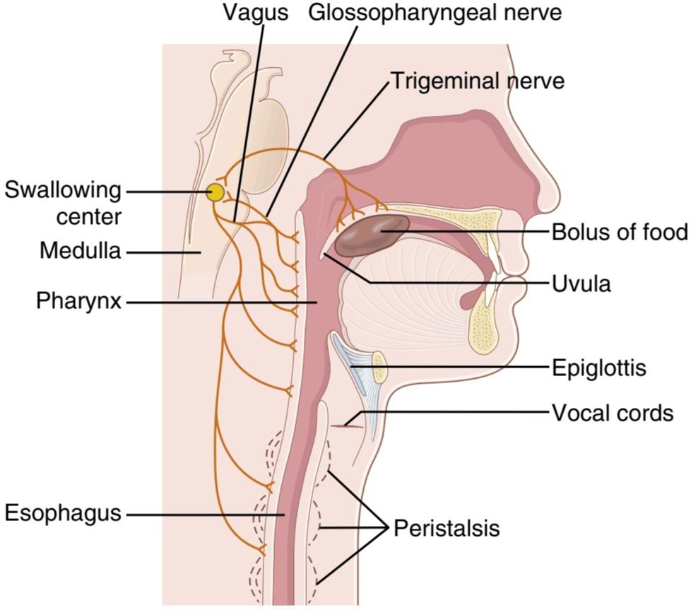
Figure 64-1. Swallowing mechanism.
شکل 64-1. مکانیسم بلع.
Involuntary Pharyngeal Stage of Swallowing. As the bolus of food enters the posterior mouth and pharynx, it stimulates epithelial swallowing receptor areas all around the opening of the pharynx, especially on the tonsillar pillars, and impulses from these areas pass to the brain stem to initiate a series of automatic pharyngeal muscle contractions as follows:
مرحله بلع غیر ارادی حلقی. همانطور که بولوس غذا وارد دهان خلفی و حلق میشود، نواحی گیرنده بلع اپیتلیال را در اطراف دهانه حلق، به ویژه روی ستونهای لوزه تحریک میکند و تکانهها از این نواحی به ساقه مغز میروند تا یک سری انقباضات خودکار عضلات حلق را به شرح زیر آغاز کنند:
1. The soft palate is pulled upward to close the posterior nares to prevent reflux of food into the nasal cavities.
1. کام نرم به سمت بالا کشیده میشود تا دمهای خلفی بسته شود تا از برگشت غذا به داخل حفرههای بینی جلوگیری شود.
2. The palatopharyngeal folds on each side of the pharynx are pulled medially to approximate each other. In this way, these folds form a sagittal slit through which the food must pass into the posterior pharynx. This slit performs a selective action, allowing food that has been masticated sufficiently to pass with ease. Because this stage of swallowing lasts less than 1 second, any large object is usually impeded too much to pass into the esophagus.
2. چینهای پالاتوفارنکس در هر طرف حلق به سمت وسط کشیده میشوند تا به یکدیگر نزدیک شوند. به این ترتیب، این چینها یک شکاف ساژیتال را تشکیل میدهند که غذا باید از طریق آن وارد حلق خلفی شود. این شکاف یک عمل انتخابی انجام میدهد و به غذاهایی که به اندازه کافی جویده شده اند اجازه میدهد به راحتی عبور کنند. از آنجایی که این مرحله از بلع کمتر از 1 ثانیه طول میکشد، معمولاً هر جسم بزرگی بیش از حد مانع عبور آن به مری میشود.
3. The vocal cords of the larynx are strongly approximated, and the larynx is pulled upward and anteriorly by the neck muscles. These actions, combined with the presence of ligaments that prevent upward movement of the epiglottis, cause the epiglottis to swing backward over the opening of the larynx. All these effects acting together prevent passage of food into the nose and trachea. Most essential is the tight approximation of the vocal cords, but the epiglottis helps to prevent food from ever getting as far as the vocal cords. Destruction of the vocal cords or of the muscles that approximate them can cause strangulation.
3. تارهای صوتی حنجره به شدت تقریب دارند و حنجره توسط عضلات گردن به سمت بالا و جلو کشیده میشود. این اعمال، همراه با وجود رباطهایی که از حرکت اپی گلوت به سمت بالا جلوگیری میکند، باعث میشود اپی گلوت بر روی دهانه حنجره به سمت عقب حرکت کند. همه این اثرات با هم از ورود غذا به داخل بینی و نای جلوگیری میکنند. مهمتر از همه، نزدیکی محکم تارهای صوتی است، اما اپی گلوت به جلوگیری از رسیدن غذا به تارهای صوتی کمک میکند. تخریب تارهای صوتی یا ماهیچههای نزدیک به آنها میتواند باعث خفگی شود.
4. The upward movement of the larynx also pulls up and enlarges the opening to the esophagus. At the same time, the upper 3 to 4 centimeters of the esophageal muscular wall, called the upper esophageal sphincter (also called the pharyngoesophageal sphincter), relaxes. Thus, food moves easily and freely from the posterior pharynx into the upper esophagus. Between swallows, this sphincter remains strongly contracted, thereby preventing air from going into the esophagus during respiration. The upward movement of the larynx also lifts the glottis out of the main stream of food flow, so the food mainly passes on each side of the epiglottis rather than over its surface; this action adds still another protection against entry of food into the trachea.
4. حرکت رو به بالا حنجره نیز به سمت بالا کشیده و دهانه مری را بزرگ میکند. در همان زمان، 3 تا 4 سانتی متر بالای دیواره عضلانی مری که اسفنکتر مری بالایی نامیده میشود (که اسفنکتر حلقی مری نیز نامیده میشود) شل میشود. بنابراین، غذا به راحتی و آزادانه از حلق خلفی به سمت مری فوقانی حرکت میکند. بین بلع، این اسفنکتر به شدت منقبض میشود و در نتیجه از ورود هوا به مری در طول تنفس جلوگیری میکند. حرکت رو به بالا حنجره همچنین گلوت را از جریان اصلی جریان غذا خارج میکند، بنابراین غذا عمدتاً از هر طرف اپی گلوت عبور میکند نه از سطح آن. این عمل محافظت دیگری را در برابر ورود غذا به نای اضافه میکند.
5. Once the larynx is raised and the pharyngoesophageal sphincter becomes relaxed, the entire muscular wall of the pharynx contracts, beginning in the superior part of the pharynx, then spreading downward over the middle and inferior pharyngeal areas, which propels the food by peristalsis into the esophagus.
5. هنگامیکه حنجره بلند میشود و اسفنکتر حلق مری شل میشود، تمام دیواره عضلانی حلق منقبض میشود و از قسمت فوقانی حلق شروع میشود، سپس به سمت پایین در نواحی میانی و تحتانی حلق گسترش مییابد، که غذا را به وسیله مری به سمت مری به سمت مری میراند.
To summarize the mechanics of the pharyngeal stage of swallowing: The trachea is closed, the esophagus is opened, and a fast peristaltic wave initiated by the nervous system of the pharynx forces the bolus of food into the upper esophagus, with the entire process occurring in less than 2 seconds.
به طور خلاصه مکانیسم مرحله حلقی بلع: نای بسته میشود، مری باز میشود و یک موج سریع پریستالتیک که توسط سیستم عصبی حلق آغاز میشود، بولوس غذا را وارد مری فوقانی میکند و کل فرآیند در کمتر از ۲ ثانیه انجام میشود.
Nervous Initiation of the Pharyngeal Stage of Swallowing. The most sensitive tactile areas of the posterior mouth and pharynx for initiating the pharyngeal stage of swallowing lie in a ring around the pharyngeal opening, with greatest sensitivity on the tonsillar pillars. Impulses are transmitted from these areas through the sensory portions of the trigeminal and glossopharyngeal nerves into the medulla oblongata, either into or closely associated with the tractus solitarius, which receives essentially all sensory impulses from the mouth.
شروع عصبی مرحله حلقی بلع. حساس ترین نواحی لمسی دهان خلفی و حلق برای شروع مرحله حلق بلع در حلقه ای در اطراف دهانه حلق قرار دارد که بیشترین حساسیت را روی ستونهای لوزه دارد. تکانهها از این نواحی از طریق بخشهای حسی اعصاب سه قلو و گلوسوفارنجئال به بصل النخاع منتقل میشوند، یا به tractus solitarius یا ارتباط نزدیک با آن، که اساساً تمام تکانههای حسی را از دهان دریافت میکند.
The successive stages of the swallowing process are then automatically initiated in orderly sequence by neuronal areas of the reticular substance of the medulla and lower portion of the pons. The sequence of the swallowing reflex is the same from one swallow to the next, and the timing of the entire cycle also remains constant from one swallow to the next. The areas in the medulla and lower pons that control swallowing are collectively called the deglutition or swallowing center.
مراحل متوالی فرآیند بلع سپس به طور خودکار به ترتیب منظم توسط نواحی عصبی ماده مشبک بصل النخاع و قسمت پایینی پونز آغاز میشود. توالی رفلکس بلع از یک بلع به دیگری یکسان است و زمان کل چرخه نیز از یک بلع به دیگری ثابت میماند. نواحی در بصل النخاع و پلکهای پایینی که بلع را کنترل میکنند، مجموعاً مرکز فرورفتگی یا بلع نامیده میشوند.
The motor impulses from the swallowing center to the pharynx and upper esophagus that cause swallowing are transmitted successively by the fifth, ninth, tenth, and twelfth cranial nerves and even a few of the superior cervical nerves.
تکانههای حرکتی از مرکز بلع به حلق و مری فوقانی که باعث بلع میشوند به طور متوالی توسط اعصاب پنجم، نهم، دهم و دوازدهم جمجمه ای و حتی تعدادی از اعصاب گردنی فوقانی منتقل میشوند.
In summary, the pharyngeal stage of swallowing is principally a reflex act. It is almost always initiated by voluntary movement of food into the back of the mouth, which in turn excites involuntary pharyngeal sensory receptors to elicit the swallowing reflex.
به طور خلاصه، مرحله حلقی بلع اساساً یک عمل رفلکس است. تقریباً همیشه با حرکت ارادی غذا به پشت دهان شروع میشود که به نوبه خود گیرندههای حسی حلقی غیرارادی را برای برانگیختن رفلکس بلع تحریک میکند.
The Pharyngeal Stage of Swallowing Momentarily Interrupts Respiration. The entire pharyngeal stage of swallowing usually occurs in less than 6 seconds, thereby interrupting respiration for only a fraction of a usual respiratory cycle. The swallowing center specifically inhibits the respiratory center of the medulla during this time, halting respiration at any point in its cycle to allow swallowing to proceed. Yet, even while a person is talking, swallowing interrupts respiration for such a short time that it is hardly noticeable.
مرحله حلق بلع، تنفس را برای لحظه ای قطع میکند. کل مرحله حلقی بلع معمولاً در کمتر از 6 ثانیه اتفاق میافتد، بنابراین تنفس تنها برای کسری از یک چرخه تنفسی معمول قطع میشود. مرکز بلع به طور خاص مرکز تنفسی مدولا را در این مدت مهار میکند و تنفس را در هر نقطه از چرخه خود متوقف میکند تا امکان بلع ادامه یابد. با این حال، حتی زمانی که فرد در حال صحبت کردن است، بلعیدن تنفس را برای مدت کوتاهی قطع میکند که به سختی قابل توجه است.
The Esophageal Stage of Swallowing Involves Two Types of Peristalsis. The esophagus functions primarily to conduct food rapidly from the pharynx to the stomach, and its movements are organized specifically for this function.
مرحله مری بلع شامل دو نوع پریستالسیس است. مری عمدتاً برای هدایت سریع غذا از حلق به معده عمل میکند و حرکات آن به طور خاص برای این عملکرد سازماندهی شده است.
The esophagus normally exhibits two types of peristaltic movements: primary peristalsis and secondary peristalsis. Primary peristalsis is simply continuation of the peristaltic wave that begins in the pharynx and spreads into the esophagus during the pharyngeal stage of swallowing. This wave passes all the way from the pharynx to the stomach in about 8 to 10 seconds. Food swallowed by a person who is in the upright position is usually transmit- ted to the lower end of the esophagus even more rapidly than the peristaltic wave itself, in about 5 to 8 seconds, because of the additional effect of gravity pulling the food downward.
مری به طور معمول دو نوع حرکت پریستالتیک را نشان میدهد: پریستالسیس اولیه و پریستالسیس ثانویه. پریستالسیس اولیه صرفاً ادامه موج پریستالتیک است که از حلق شروع میشود و در مرحله حلق بلع به مری گسترش مییابد. این موج از حلق تا معده در حدود 8 تا 10 ثانیه عبور میکند. غذای بلعیده شده توسط فردی که در وضعیت عمودی قرار دارد، معمولاً به دلیل اثر اضافی جاذبه که غذا را به سمت پایین میکشد، حتی سریعتر از خود موج پریستالتیک، در حدود 5 تا 8 ثانیه به انتهای تحتانی مری منتقل میشود.
If the primary peristaltic wave fails to move all the food that has entered the esophagus into the stomach, secondary peristaltic waves result from distention of the esophagus by the retained food; these waves continue until all the food has emptied into the stomach. The secondary peristaltic waves are initiated partly by intrinsic neural circuits in the myenteric nervous system and partly by reflexes that begin in the pharynx and are then transmit- ted upward through vagal afferent fibers to the medulla and back again to the esophagus through glossopharyngeal and vagal efferent nerve fibers.
اگر موج پریستالتیک اولیه نتواند تمام غذایی را که وارد مری شده است به معده منتقل کند، امواج پریستالتیک ثانویه ناشی از اتساع مری توسط غذای باقیمانده است. این امواج تا زمانی ادامه مییابد که تمام غذا در معده خالی شود. امواج پریستالتیک ثانویه تا حدی توسط مدارهای عصبی ذاتی در سیستم عصبی میانتریک و تا حدودی توسط رفلکسهایی که از حلق شروع میشوند و سپس از طریق رشتههای آوران واگ به سمت بالا به بصل النخاع و دوباره به مری از طریق عصب گلوسوفارنکس و فیبر واگال منتقل میشوند.
The musculature of the pharyngeal wall and upper third of the esophagus is striated muscle. Therefore, the peristaltic waves in these regions are controlled by skeletal nerve impulses from the glossopharyngeal and vagus nerves. In the lower two-thirds of the esophagus, the musculature is smooth muscle, but this portion of the esophagus is also strongly controlled by the vagus nerves that act through connections with the esophageal myenteric nervous system. When the vagus nerves to the esophagus are cut, the myenteric nerve plexus of the esophagus becomes excitable enough after several days to cause strong secondary peristaltic waves even without support from the vagal reflexes. Therefore, even after paralysis of the brain stem swallowing reflex, food fed by tube or in some other way into the esophagus still passes readily into the stomach.
عضله دیواره حلق و یک سوم فوقانی مری عضله مخطط است. بنابراین امواج پریستالتیک در این نواحی توسط تکانههای عصبی اسکلتی از اعصاب گلوفارنکس و واگ کنترل میشوند. در دو سوم تحتانی مری، ماهیچه ماهیچه صاف است، اما این قسمت از مری نیز به شدت توسط اعصاب واگ کنترل میشود که از طریق اتصالات با سیستم عصبی میانتریک مری عمل میکنند. هنگامیکه اعصاب واگ به مری قطع میشود، شبکه عصبی میانتریک مری پس از چند روز به اندازه کافی تحریک پذیر میشود که حتی بدون حمایت از رفلکسهای واگ، امواج پریستالتیک ثانویه قوی ایجاد میکند. بنابراین، حتی پس از فلج شدن رفلکس بلع ساقه مغز، غذایی که با لوله یا به روشی دیگر وارد مری میشود، همچنان به آسانی وارد معده میشود.
Receptive Relaxation of the Stomach. When the esophageal peristaltic wave approaches the stomach, a wave of relaxation, transmitted through myenteric inhibitory neurons, precedes the peristalsis. Furthermore, the entire stomach and, to a lesser extent, even the duodenum be- come relaxed as this wave reaches the lower end of the esophagus and thus are prepared ahead of time to receive the food propelled into the esophagus during the swallowing act.
آرامش پذیرای معده. هنگامیکه موج پریستالتیک مری به معده نزدیک میشود، یک موج آرامش که از طریق نورونهای مهاری میانتریک منتقل میشود، قبل از پریستالتیک ایجاد میشود. علاوه بر این، با رسیدن این موج به انتهای پایینی مری، کل معده و تا حدی حتی اثنی عشر شل میشود و بنابراین زودتر از موعد آماده میشود تا غذای رانده شده به مری در حین عمل بلع دریافت کند.
Function of the Lower Esophageal Sphincter (Gastroesophageal Sphincter). At the lower end of the esophagus, extending upward about 3 centimeters above its juncture with the stomach, the esophageal circular muscle functions as a broad lower esophageal sphincter, also called the gastroesophageal sphincter. This sphincter normally remains tonically constricted with an intraluminal pressure at this point in the esophagus of about 30 mm Hg, in contrast to the midportion of the esophagus, which normally remains relaxed. When a peristaltic swallowing wave passes down the esophagus, “receptive relaxation” of the lower esophageal sphincter occurs ahead of the peristaltic wave, which allows easy propulsion of the swallowed food into the stomach. Rarely, the sphincter does not relax satisfactorily, resulting in a condition called achalasia. This condition is discussed in Chapter 67.
عملکرد اسفنکتر تحتانی مری (اسفنکتر معده ازوفاژیال). در انتهای تحتانی مری که حدود 3 سانتی متر بالاتر از محل اتصال آن با معده به سمت بالا امتداد مییابد، عضله حلقوی مری به عنوان یک اسفنکتر وسیع تحتانی مری عمل میکند که اسفنکتر معده به مری نیز نامیده میشود. این اسفنکتر معمولاً به صورت تونیک با فشار داخل مجرای در این نقطه از مری حدود 30 میلیمتر جیوه منقبض میشود، برخلاف قسمت میانی مری که معمولاً آرام میماند. هنگامیکه یک موج بلع پریستالتیک از مری عبور میکند، “آرامش پذیرا” اسفنکتر تحتانی مری جلوتر از موج پریستالتیک رخ میدهد، که اجازه میدهد تا غذای بلعیده شده به سمت معده حرکت کند. به ندرت، اسفنکتر به طور رضایت بخشی شل نمیشود و در نتیجه وضعیتی به نام آشالازی ایجاد میشود. این شرط در فصل 67 مورد بحث قرار گرفته است.
The stomach secretions are highly acidic and contain many proteolytic enzymes. The esophageal mucosa, except in the lower one-eighth of the esophagus, is not capable of resisting the digestive action of gastric secretions for long. Fortunately, the tonic constriction of the lower esophageal sphincter helps prevent significant reflux of stomach contents into the esophagus except under abnormal conditions.
ترشحات معده بسیار اسیدی است و حاوی آنزیمهای پروتئولیتیک زیادی است. مخاط مری، به جز در یک هشتم تحتانی مری، قادر به مقاومت طولانی در برابر عملکرد گوارشی ترشحات معده نیست. خوشبختانه، انقباض تونیک اسفنکتر تحتانی مری به جلوگیری از برگشت محتویات معده به مری کمک میکند مگر در شرایط غیر طبیعی.
Prevention of Esophageal Reflux by Valvelike Closure of the Distal End of the Esophagus. Another factor that helps prevent reflux is a valvelike mechanism of a short portion of the esophagus that extends slightly into the stomach. Increased intra-abdominal pressure caves the esophagus inward at this point. Thus, this valvelike closure of the lower esophagus helps to prevent high intra-abdominal pressure from forcing stomach contents backward into the esophagus. Otherwise, whenever we walk, cough, or breathe hard, stomach acid might be expelled into the esophagus.
پیشگیری از رفلاکس مری با بستن دریچه مانند انتهای دیستال مری. یکی دیگر از عواملی که به جلوگیری از رفلاکس کمک میکند، مکانیسم دریچه مانند قسمت کوتاهی از مری است که کمیبه سمت معده امتداد مییابد. افزایش فشار داخل شکمیدر این نقطه مری را به سمت داخل فرو میبرد. بنابراین، این بسته شدن دریچهای در قسمت تحتانی مری به جلوگیری از فشار بالای داخل شکمیاز فشار محتویات معده به سمت عقب به مری کمک میکند. در غیر این صورت، هر زمان که راه میرویم، سرفه میکنیم یا به سختی نفس میکشیم، ممکن است اسید معده به مری دفع شود.
MOTOR FUNCTIONS OF THE STOMACH
The motor functions of the stomach are threefold: (1) storage of large quantities of food until the food can be processed in the stomach, duodenum, and lower intestinal tract; (2) mixing of this food with gastric secretions until it forms a semifluid mixture called chyme; and (3) slow emptying of the chyme from the stomach into the small intestine at a rate suitable for proper digestion and absorption by the small intestine.
عملکردهای حرکتی معده
عملکردهای حرکتی معده سه دسته است: (1) ذخیره مقادیر زیادی غذا تا زمانی که غذا در معده، دوازدهه و دستگاه روده تحتانی پردازش شود. (2) مخلوط کردن این غذا با ترشحات معده تا زمانی که مخلوطی نیمه مایع به نام کیم تشکیل شود. و (3) تخلیه آهسته کیم از معده به روده کوچک با سرعت مناسب برای هضم و جذب مناسب توسط روده کوچک.
Figure 64-2 shows the basic anatomy of the stomach. Anatomically, the stomach is usually divided into two major parts: (1) the body and (2) the antrum. Physiologically, it is more appropriately divided into (1) the “orad” portion, comprising about the first two-thirds of the body, and (2) the “caudad” portion, comprising the remainder of the body plus the antrum.
شکل 64-2 آناتومیاساسی معده را نشان میدهد. از نظر تشریحی، معده معمولاً به دو بخش عمده تقسیم میشود: (1) بدن و (2) آنتروم. از نظر فیزیولوژیکی، به طور مناسبتری به (1) بخش «اوراد» که تقریباً دو سوم اول بدن را شامل میشود، و (2) بخش «دم» که شامل باقیمانده بدن به اضافه آنتروم است، تقسیم میشود.
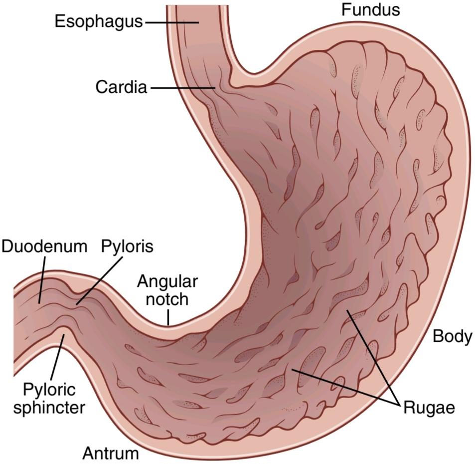
Figure 64-2. Physiological anatomy of the stomach.
شکل 64-2. آناتومیفیزیولوژیکی معده.
STORAGE FUNCTION OF THE STOMACH
As food enters the stomach, it forms concentric circles of the food in the orad portion of the stomach, with the newest food lying closest to the esophageal opening and the oldest food lying nearest the outer wall of the stomach. Normally, when food stretches the stomach, a “vagovagal reflex” from the stomach to the brain stem and then back to the stomach reduces the tone in the muscular wall of the body of the stomach so that the wall bulges progressively outward, accommodating greater and greater quantities of food up to a limit in the completely relaxed stomach of 0.8 to 1.5 liters. The pressure in the stomach remains low until this limit is approached.
عملکرد ذخیره سازی معده
همانطور که غذا وارد معده میشود، دایرههای متحدالمرکز غذا را در قسمت orad معده تشکیل میدهد که جدیدترین غذا در نزدیکترین فاصله به دهانه مری و قدیمیترین غذا در نزدیکترین فاصله به دیواره بیرونی معده قرار دارد. به طور معمول، هنگامیکه غذا معده را کش میدهد، یک “رفلکس واگوگال” از معده به ساقه مغز و سپس بازگشت به معده، تون را در دیواره عضلانی بدن معده کاهش میدهد به طوری که دیواره به تدریج به سمت بیرون برآمده میشود و مقادیر بیشتر و بیشتر غذا را در خود جای میدهد تا حد مجاز در معده کاملاً آرام از 0.8 تا 0.8 لیتر. فشار معده تا رسیدن به این حد کم باقی میماند.
FOOD MIXING AND PROPULSION IN THE STOMACH-BASIC ELECTRICAL RHYTHM OF THE STOMACH WALL
The digestive juices of the stomach are secreted by gastric glands, which are present in almost the entire wall of the body of the stomach except along a narrow strip on the lesser curvature of the stomach. These secretions come immediately into contact with that portion of the stored food lying against the mucosal surface of the stomach. As long as food is in the stomach, weak peristaltic constrictor waves, called mixing waves, begin in the mid to upper portions of the stomach wall and move toward the antrum about once every 15 to 20 seconds. These waves are initiated by the gut wall basic electrical rhythm, which was discussed in Chapter 63, consisting of electrical “slow waves” that occur spontaneously in the stomach wall. As the constrictor waves progress from the body of the stomach into the antrum, they become more intense, some becoming extremely intense and providing powerful peristaltic action potential-driven constrictor rings that force the antral contents under higher and higher pressure toward the pylorus.
اختلاط غذا و رانش در ریتم الکتریکی معده – پایه دیواره معده
شیره گوارشی معده توسط غدد معده ترشح میشود که تقریباً در تمام دیواره معده وجود دارد، به جز در امتداد یک نوار باریک در انحنای کمتر معده. این ترشحات بلافاصله با آن قسمت از غذای ذخیره شده در مقابل سطح مخاطی معده تماس پیدا میکند. تا زمانی که غذا در معده است، امواج ضعیف منقبض کننده پریستالتیک، به نام امواج اختلاط، از قسمتهای میانی تا بالای دیواره معده شروع میشود و تقریباً هر 15 تا 20 ثانیه یک بار به سمت آنتروم حرکت میکند. این امواج توسط ریتم الکتریکی پایه دیواره روده آغاز میشوند، که در فصل 63 مورد بحث قرار گرفت و شامل “امواج آهسته” الکتریکی است که به طور خود به خود در دیواره معده رخ میدهد. همانطور که امواج منقبض کننده از بدنه معده به سمت آنتروم پیشروی میکنند، شدیدتر میشوند، برخی از آنها بسیار شدید میشوند و حلقههای منقبض کننده قوی مبتنی بر پتانسیل عمل پریستالتیک ایجاد میکنند که محتویات آنترال را تحت فشار بالاتر و بالاتری به سمت پیلور وادار میکند.
These constrictor rings also play an important role in mixing the stomach contents in the following way: Each time a peristaltic wave passes down the antral wall toward the pylorus, it digs deeply into the food contents in the antrum. Yet, the opening of the pylorus is still small enough that only a few milliliters or less of antral con- tents are expelled into the duodenum with each peristaltic wave. Also, as each peristaltic wave approaches the pylorus, the pyloric muscle often contracts, which further impedes emptying through the pylorus. Therefore, most of the antral contents are squeezed upstream through the peristaltic ring toward the body of the stomach, not through the pylorus. Thus, the moving peristaltic con- strictive ring, combined with this upstream squeezing action, called “retropulsion,” is an exceedingly important mixing mechanism in the stomach.
این حلقههای منقبض کننده همچنین نقش مهمیدر مخلوط کردن محتویات معده به روش زیر ایفا میکنند: هر بار که یک موج پریستالتیک از دیواره آنترال به سمت پیلور عبور میکند، عمیقاً در محتویات غذایی در آنتروم فرو میرود. با این حال، دهانه پیلور هنوز به اندازهای کوچک است که تنها چند میلیلیتر یا کمتر از محتوای آنترال با هر موج پریستالتیک به داخل دوازدهه دفع میشود. همچنین، با نزدیک شدن هر موج پریستالتیک به پیلور، عضله پیلور اغلب منقبض میشود، که مانع تخلیه بیشتر از طریق پیلور میشود. بنابراین، بیشتر محتویات آنترال در بالادست از طریق حلقه پریستالتیک به سمت بدن معده فشرده میشود، نه از طریق پیلور. بنابراین، حلقه انقباض پریستالتیک متحرک، همراه با این عمل فشردن در بالادست، به نام “retropulsion”، یک مکانیسم اختلاط بسیار مهم در معده است.
Chyme. After food in the stomach has become thoroughly mixed with the stomach secretions, the resulting mixture that passes down the gut is called chyme. The degree of fluidity of the chyme leaving the stomach depends on the relative amounts of food, water, and stomach secretions and on the degree of digestion that has occurred. The appearance of chyme is that of a murky semifluid or paste.
Chyme. پس از اینکه غذا در معده کاملاً با ترشحات معده مخلوط شد، مخلوط حاصل که از روده عبور میکند کیم نامیده میشود. درجه سیال بودن کیم که از معده خارج میشود به مقدار نسبی غذا، آب و ترشحات معده و میزان هضم آن بستگی دارد. ظاهر کیم به صورت نیمه سیال یا خمیر کدر است.
Hunger Contractions. Besides the peristaltic contractions that occur when food is present in the stomach, an- other type of intense contractions, called hunger contractions, often occurs when the stomach has been empty for several hours or more. These contractions are rhythmic peristaltic contractions in the body of the stomach. When the successive contractions become extremely strong, they often fuse to cause a continuing tetanic contraction that sometimes lasts for 2 to 3 minutes.
انقباضات گرسنگی. علاوه بر انقباضات پریستالتیک که هنگام وجود غذا در معده رخ میدهد، نوع دیگری از انقباضات شدید به نام انقباضات گرسنگی، اغلب زمانی رخ میدهد که معده برای چندین ساعت یا بیشتر خالی باشد. این انقباضات، انقباضات پریستالتیک ریتمیک در بدن معده هستند. هنگامیکه انقباضات متوالی بسیار قوی میشوند، اغلب با هم ترکیب میشوند و باعث ایجاد انقباض کزاز مداوم میشوند که گاهی 2 تا 3 دقیقه طول میکشد.
Hunger contractions are most intense in young, healthy people who have high degrees of gastrointestinal tonus; they are also greatly increased by the person’s having lower than normal levels of blood sugar. When hunger contractions occur in the stomach, the person sometimes experiences mild pain in the pit of the stomach, called hunger pangs. Hunger pangs usually do not begin until 12 to 24 hours after the last ingestion of food; in people who are in a state of starvation, they reach their greatest intensity in 3 to 4 days and gradually weaken in succeeding days.
انقباضات گرسنگی در افراد جوان و سالمیکه درجات بالایی از تونوس دستگاه گوارش دارند، شدیدتر است. آنها همچنین با داشتن سطح قند خون پایین تر از حد طبیعی به میزان زیادی افزایش مییابند. هنگامیکه انقباضات گرسنگی در معده رخ میدهد، فرد گاهی اوقات درد خفیفی را در گودال معده تجربه میکند که به آن احساس گرسنگی میگویند. درد گرسنگی معمولاً تا 12 تا 24 ساعت پس از آخرین مصرف غذا شروع نمیشود. در افرادی که در حالت گرسنگی هستند، طی 3 تا 4 روز به بیشترین شدت خود میرسند و در روزهای بعدی به تدریج ضعیف میشوند.
STOMACH EMPTYING
Stomach emptying is promoted by intense peristaltic contractions in the stomach antrum. At the same time, emptying is opposed by varying degrees of resistance to passage of chyme at the pylorus.
تخلیه معده
تخلیه معده با انقباضات پریستالتیک شدید در آنتروم معده تقویت میشود. در عین حال، تخلیه با درجات مختلف مقاومت در برابر عبور کیم در پیلور مخالف است.
Intense Antral Peristaltic Contractions During Stomach Emptying “Pyloric Pump.” Most of the time, the rhythmic stomach contractions are weak and function mainly to cause mixing of food and gastric secretions. However, for about 20% of the time while food is in the stomach, the contractions become intense, beginning in midstomach and spreading through the caudad stomach. These contractions are strong peristaltic, very tight ringlike constrictions that can cause stomach emptying. As the stomach becomes progressively more and more empty, these constrictions begin farther and farther up the body of the stomach, gradually pinching off the food in the body of the stomach and adding this food to the chyme in the antrum. These intense peristaltic contractions often create 50 to 70 centimeters of water pressure, which is about six times as powerful as the usual mixing type of peristaltic waves.
انقباضات پریستالتیک شدید آنترال در حین تخلیه معده “پمپ پیلور”. اغلب اوقات، انقباضات ریتمیک معده ضعیف است و عمدتاً باعث مخلوط شدن غذا و ترشحات معده میشود. با این حال، در حدود 20 درصد از مواقعی که غذا در معده است، انقباضات شدید میشوند و از وسط معده شروع میشوند و از طریق معده دم پخش میشوند. این انقباضات، انقباضات پریستالتیک قوی و حلقه مانند بسیار سفت هستند که میتوانند باعث تخلیه معده شوند. با خالی شدن تدریجی معده، این انقباضات دورتر و دورتر از بدن معده شروع میشود و به تدریج غذا را در بدن معده گرفته و این غذا را به کیم در آنتروم اضافه میکند. این انقباضات شدید پریستالتیک اغلب 50 تا 70 سانتی متر فشار آب ایجاد میکند که تقریباً شش برابر قوی تر از نوع اختلاط معمول امواج پریستالتیک است.
When pyloric tone is normal, each strong peristaltic wave forces up to several milliliters of chyme into the duodenum. Thus, the peristaltic waves, in addition to causing mixing in the stomach, also provide a pumping action called the “pyloric pump.”
هنگامیکه تون پیلور طبیعی است، هر موج پریستالتیک قوی تا چند میلی لیتر کیم را وارد دوازدهه میکند. بنابراین، امواج پریستالتیک، علاوه بر ایجاد اختلاط در معده، عمل پمپاژی به نام “پمپ پیلور” را نیز ارائه میدهند.
Role of the Pylorus in Controlling Stomach Emptying. The distal opening of the stomach is the pylorus. Here the thickness of the circular wall muscle becomes 50% to 100% greater than in the earlier portions of the stomach antrum, and it remains slightly tonically contracted al- most all the time. Therefore, the pyloric circular muscle is called the pyloric sphincter.
نقش پیلور در کنترل تخلیه معده دهانه دیستال معده پیلور است. در اینجا ضخامت عضله دیواره دایرهای 50 تا 100 درصد بیشتر از قسمتهای قبلی آنتروم معده میشود و تقریباً در تمام مدت کمیمنقبض میشود. بنابراین به عضله حلقوی پیلور اسفنکتر پیلور میگویند.
Despite normal tonic contraction of the pyloric sphincter, the pylorus usually is open enough for water and other fluids to empty from the stomach into the duodenum with ease. Yet, the constriction usually prevents passage of food particles until they have become mixed in the chyme to almost fluid consistency. The degree of con- striction of the pylorus is increased or decreased under the influence of nervous and hormonal signals from both the stomach and the duodenum, as discussed shortly.
علیرغم انقباض تونیک طبیعی اسفنکتر پیلور، پیلور معمولاً به اندازه کافی باز است تا آب و سایر مایعات به راحتی از معده به دوازدهه تخلیه شوند. با این حال، انقباض معمولاً مانع از عبور ذرات غذا میشود تا زمانی که در کیم مخلوط شده و به قوام تقریباً مایع تبدیل شوند. میزان انقباض پیلور تحت تأثیر سیگنالهای عصبی و هورمونی از معده و دوازدهه افزایش یا کاهش مییابد، همانطور که به زودی بحث شد.
REGULATION OF STOMACH EMPTYING
The rate at which the stomach empties is regulated by signals from the stomach and the duodenum. However, the duodenum provides far more potent signals, controlling the emptying of chyme into the duodenum at a rate no greater than the rate at which the chyme can be digested and absorbed in the small intestine.
تنظیم تخلیه معده
سرعت تخلیه معده توسط سیگنالهای معده و دوازدهه تنظیم میشود. با این حال، دوازدهه سیگنالهای بسیار قویتری ارائه میدهد و تخلیه کیم به دوازدهه را با سرعتی که بیشتر از سرعت هضم و جذب کیم در روده کوچک نیست، کنترل میکند.
Gastric Factors That Promote Emptying
Effect of Gastric Food Volume on Rate of Emptying. Increased food volume in the stomach promotes in- creased emptying from the stomach. However, it is not increased storage pressure of the food in the stomach that causes the increased emptying because, in the usual normal range of volume, the increase in volume does not increase the pressure much. However, stretching of the stomach wall does elicit local myenteric reflexes in the wall that greatly accentuate activity of the pyloric pump and at the same time inhibit the pylorus.
عواملی که باعث تخلیه معده میشوند
تأثیر حجم غذای معده بر میزان تخلیه. افزایش حجم غذا در معده باعث افزایش تخلیه از معده میشود. با این حال، افزایش فشار ذخیرهسازی غذا در معده نیست که باعث افزایش تخلیه میشود، زیرا در محدوده معمول حجم، افزایش حجم، فشار را زیاد افزایش نمیدهد. با این حال، کشش دیواره معده باعث ایجاد رفلکسهای میانتریک موضعی در دیواره میشود که فعالیت پمپ پیلور را به شدت افزایش میدهد و در عین حال پیلور را مهار میکند.
The Hormone Gastrin Promotes Stomach Emptying. In Chapter 65, we discuss how stretching of the stomach wall and the presence of certain types of foods in the stomach-particularly digestive products of meat-elicit release of the hormone gastrin from the G cells of the antral mucosa. This has potent effects to cause secretion of highly acidic gastric juice by the stomach glands. Gastrin also has mild to moderate stimulatory effects on motor functions in the body of the stomach. Most important, it seems to enhance the activity of the pyloric pump. Thus, gastrin likely promotes stomach emptying.
هورمون گاسترین باعث تخلیه معده میشود. در فصل 65، ما در مورد چگونگی کشش دیواره معده و وجود انواع خاصی از غذاها در معده – به ویژه محصولات گوارشی گوشت که باعث آزاد شدن هورمون گاسترین از سلولهای G مخاط آنترال میشود، بحث میکنیم. این اثرات قوی برای ترشح آب معده بسیار اسیدی توسط غدد معده دارد. گاسترین همچنین دارای اثرات تحریکی خفیف تا متوسط بر عملکرد حرکتی در بدن معده است. مهمتر از همه، به نظر میرسد که فعالیت پمپ پیلور را افزایش میدهد. بنابراین، گاسترین به احتمال زیاد باعث تخلیه معده میشود.
Powerful Duodenal Factors That Inhibit Stomach Emptying
Duodenum Enterogastric Nervous Reflexes Inhibit Stomach Emptying. When food enters the duodenum, multiple nervous reflexes are initiated from the duodenal wall. These reflexes pass back to the stomach to slow or even stop stomach emptying if the volume of chyme in the duodenum becomes too much. These reflexes are mediated by three routes: (1) directly from the duodenum to the stomach through the enteric nervous system in the gut wall; (2) through extrinsic nerves that go to the prevertebral sympathetic ganglia and then back through inhibitory sympathetic nerve fibers to the stomach; and (3) probably to a slight extent through the vagus nerves all the way to the brain stem, where they inhibit the normal excitatory signals transmitted to the stomach through the vagi. All these parallel reflexes have two effects on stomach emptying. First, they strongly inhibit the “pyloric pump” propulsive contractions, and second, they increase the tone of the pyloric sphincter.
عوامل قدرتمند اثنی عشر که مانع از تخلیه معده میشود
رفلکسهای عصبی روده معده تخلیه معده را مهار میکنند. هنگامیکه غذا وارد دوازدهه میشود، رفلکسهای عصبی متعددی از دیواره دوازدهه شروع میشود. این رفلکسها به معده برمیگردند تا اگر حجم کیم در دوازدهه بیش از حد شود، تخلیه معده را کند یا حتی متوقف کنند. این رفلکسها توسط سه مسیر انجام میشوند: (1) مستقیماً از دوازدهه به معده از طریق سیستم عصبی روده در دیواره روده. (2) از طریق اعصاب بیرونی که به گانگلیونهای سمپاتیک پیش مهره ای میروند و سپس از طریق رشتههای عصبی سمپاتیک مهاری به معده بازمیگردند. و (3) احتمالاً تا حدی جزئی از طریق اعصاب واگ تا ساقه مغز، جایی که آنها سیگنالهای تحریکی طبیعی را که از طریق واگی به معده منتقل میشوند، مهار میکنند. همه این رفلکسهای موازی دو اثر بر تخلیه معده دارند. اول اینکه آنها به شدت انقباضات پیشرانه “پمپ پیلور” را مهار میکنند و دوم اینکه تون اسفنکتر پیلور را افزایش میدهند.
The types of factors that are continually monitored in the duodenum and can initiate enterogastric inhibitory reflexes include the following:
انواع فاکتورهایی که به طور مداوم در دوازدهه بررسی میشوند و میتوانند رفلکسهای مهاری آنتروگاستریک را آغاز کنند عبارتند از:
1. Distention of the duodenum
1. اتساع دوازدهه
2. The presence of any irritation of the duodenal mucosa
2. وجود هرگونه تحریک مخاط دوازدهه
3. Acidity of the duodenal chyme
3. اسیدیته کیم اثنی عشر
4. Osmolality of the chyme
4. اسمولالیته کیم
5. The presence of certain breakdown products in the chyme, especially breakdown products of proteins and, perhaps to a lesser extent, of fats The enterogastric inhibitory reflexes are especially sensitive to the presence of irritants and acids in the duodenal chyme, and they often become strongly activated in as little as 30 seconds. For example, whenever the pH of the chyme in the duodenum falls below about 3.5 to 4, the reflexes frequently block further release of acidic stomach contents into the duodenum until the duodenal chyme can be neutralized by pancreatic and other secretions.
5. وجود برخی محصولات تجزیه در کیم، به ویژه محصولات تجزیه پروتئینها و شاید تا حدی چربیها رفلکسهای بازدارنده روده معده به ویژه به حضور محرکها و اسیدها در کیم اثنی عشر حساس هستند و اغلب در کمتر از 30 ثانیه به شدت فعال میشوند. به عنوان مثال، هر زمان که PH کیم در دوازدهه کمتر از حدود 3.5 تا 4 باشد، رفلکسها اغلب از انتشار بیشتر محتویات اسیدی معده در دوازدهه جلوگیری میکنند تا زمانی که کیم اثنی عشر با ترشحات پانکراس و سایر ترشحات خنثی شود.
Breakdown products of protein digestion also elicit inhibitory enterogastric reflexes; by slowing the rate of stomach emptying, sufficient time is ensured for adequate protein digestion in the duodenum and small intestine.
محصولات تجزیه پروتئین هضم نیز باعث ایجاد رفلکسهای بازدارنده روده معده میشود. با کاهش سرعت تخلیه معده، زمان کافی برای هضم پروتئین کافی در دوازدهه و روده کوچک تضمین میشود.
Finally, either hypotonic fluids or, especially, hypertonic fluids elicit the inhibitory reflexes. Thus, flow of nonisotonic fluids into the small intestine at too rapid a rate is prevented, thereby also preventing rapid changes in electrolyte concentrations in the whole-body extracellular fluid during absorption of the intestinal contents.
در نهایت، یا مایعات هیپوتونیک یا، به ویژه، مایعات هیپرتونیک، رفلکسهای مهاری را ایجاد میکنند. بنابراین، از جریان مایعات غیر ایزوتونیک به روده کوچک با سرعت بسیار سریع جلوگیری میشود، در نتیجه از تغییرات سریع در غلظت الکترولیت در مایع خارج سلولی کل بدن در طول جذب محتویات روده نیز جلوگیری میشود.
Hormonal Feedback From the Duodenum Inhibits Gastric Emptying-Role of Fats and the Hormone Cholecystokinin. Hormones released from the upper intestine also inhibit stomach emptying. The stimulus for releasing these inhibitory hormones is mainly fats entering the duodenum, although other types of foods can in- crease the hormones to a lesser degree.
بازخورد هورمونی از اثنی عشر مانع تخلیه معده میشود – نقش چربیها و هورمون کوله سیستوکینین. هورمونهایی که از قسمت فوقانی روده آزاد میشوند نیز از تخلیه معده جلوگیری میکنند. محرک آزادسازی این هورمونهای بازدارنده عمدتاً چربیهایی است که وارد دوازدهه میشوند، اگرچه انواع دیگر غذاها میتوانند هورمونها را به میزان کمتری افزایش دهند.
On entering the duodenum, the fats extract several different hormones from the duodenal and jejunal epithelium, either by binding with “receptors” on the epithelial cells or in some other way. In turn, the hormones are carried via the blood to the stomach, where they inhibit the pyloric pump and at the same time increase the strength of contraction of the pyloric sphincter. These effects are important because fats are much slower to be digested than most other foods.
با ورود به دوازدهه، چربیها چندین هورمون مختلف را از اپیتلیوم اثنی عشر و ژژنوم استخراج میکنند، یا از طریق اتصال به گیرندههای روی سلولهای اپیتلیال یا به روشی دیگر. به نوبه خود، هورمونها از طریق خون به معده منتقل میشوند، جایی که پمپ پیلور را مهار میکنند و در عین حال قدرت انقباض اسفنکتر پیلور را افزایش میدهند. این اثرات مهم هستند زیرا چربیها نسبت به سایر غذاها بسیار کندتر هضم میشوند.
The most potent of these hormones appears to be cholecystokinin (CCK), which is released from the mucosa of the jejunum in response to fatty substances in the chyme. This hormone acts as an inhibitor to block increased stomach motility caused by gastrin.
به نظر میرسد قوی ترین این هورمونها کوله سیستوکینین (CCK) باشد که در پاسخ به مواد چرب موجود در کیم از مخاط ژژنوم آزاد میشود. این هورمون به عنوان یک مهارکننده برای جلوگیری از افزایش تحرک معده ناشی از گاسترین عمل میکند.
Other possible inhibitors of stomach emptying are the hormones secretin and glucose-dependent insulinotropic peptide, also called gastric inhibitory peptide (GIP). Secretin is released mainly from the duodenal mucosa in response to gastric acid passed from the stomach through the pylorus. GIP has a general but weak effect of decreasing gastrointestinal motility.
دیگر مهارکنندههای احتمالی تخلیه معده، هورمونهای ترشح کننده و پپتید انسولینوتروپیک وابسته به گلوکز هستند که پپتید مهاری معده (GIP) نیز نامیده میشود. سکرتین عمدتاً از مخاط دوازدهه در پاسخ به اسید معده که از معده از طریق پیلور منتقل میشود، آزاد میشود. GIP اثر کلی اما ضعیفی در کاهش تحرک دستگاه گوارش دارد.
GIP is released from the upper small intestine mainly in response to fat in the chyme, but also to a lesser extent in response to carbohydrates. Although GIP inhibits gastric motility under some conditions, its main effect at physiological concentrations is probably mainly to stimulate secretion of insulin by the pancreas.
GIP از قسمت فوقانی روده کوچک عمدتاً در پاسخ به چربی در کیم، اما همچنین به میزان کمتر در پاسخ به کربوهیدراتها آزاد میشود. اگرچه GIP تحت برخی شرایط حرکات معده را مهار میکند، اثر اصلی آن در غلظتهای فیزیولوژیکی احتمالاً عمدتاً تحریک ترشح انسولین توسط پانکراس است.
These hormones are discussed at greater length else- where in this text, especially in Chapter 65 in relation to control of gallbladder emptying and control of the rate of pancreatic secretion.
این هورمونها در جای دیگری در این متن بهطور مفصلتر مورد بحث قرار گرفتهاند، بهویژه در فصل 65 در رابطه با کنترل تخلیه کیسه صفرا و کنترل سرعت ترشح پانکراس.
In summary, hormones, especially CCK, scan inhibit gastric emptying when excess quantities of chyme, especially acidic or fatty chyme, enter the duodenum from the stomach.
به طور خلاصه، هورمونها، بهویژه CCK، هنگامیکه مقادیر بیش از حد کیم، بهخصوص کیم اسیدی یا چرب، از معده وارد دوازدهه میشود، از تخلیه معده جلوگیری میکند.
Summary of the Control of Stomach Emptying
Emptying of the stomach is controlled only to a moderate degree by stomach factors such as the degree of filling in the stomach and the excitatory effect of gastrin on stomach peristalsis. Probably the more important control of stomach emptying resides in inhibitory feedback signals from the duodenum, including both enterogastric inhibitory nervous feedback reflexes and hormonal feedback by CCK. These feedback inhibitory mechanisms work together to slow the rate of emptying when (1) too much chyme is already in the small intestine or (2) the chyme is excessively acidic, contains too much unprocessed protein or fat, is hypotonic or hypertonic, or is irritating. In this way, the rate of stomach emptying is limited to the amount of chyme that the small intestine can process.
خلاصه ای از کنترل تخلیه معده
تخلیه معده فقط تا حد متوسطی توسط عوامل معده مانند میزان پر شدن معده و اثر تحریکی گاسترین بر پریستالیس معده کنترل میشود. احتمالاً مهمتر کنترل تخلیه معده در سیگنالهای بازخوردی بازدارنده از دوازدهه است، از جمله رفلکسهای بازخورد عصبی مهاری آنتروگاستریک و بازخورد هورمونی توسط CCK. این مکانیسمهای بازدارنده بازخورد با هم کار میکنند تا سرعت تخلیه را کاهش دهند زمانی که (1) کیم بیش از حد در روده کوچک است یا (2) کیم بیش از حد اسیدی است، حاوی پروتئین یا چربی بیش از حد فرآوری نشده است، هیپوتونیک یا هیپرتونیک یا تحریککننده است. به این ترتیب، سرعت تخلیه معده به مقدار کیمیکه روده کوچک میتواند پردازش کند محدود میشود.
MOVEMENTS OF THE SMALL INTESTINE
Movements of the small intestine, like those elsewhere in the gastrointestinal tract, can be divided into mixing contractions and propulsive contractions. To a great extent, this separation is artificial because essentially all movements of the small intestine cause at least some degree of both mixing and propulsion. The usual classification of these processes is described in the following sections.
حرکات روده کوچک
حرکات روده کوچک، مانند حرکات در سایر نقاط دستگاه گوارش، میتواند به انقباضات مخلوط و انقباضات محرکه تقسیم شود. تا حد زیادی، این جدایی مصنوعی است زیرا اساساً تمام حرکات روده کوچک حداقل درجاتی از اختلاط و رانش را ایجاد میکند. طبقه بندی معمول این فرآیندها در بخشهای بعدی توضیح داده شده است.
MIXING CONTRACTIONS (SEGMENTATION CONTRACTIONS)
When a portion of the small intestine becomes distended with chyme, stretching of the intestinal wall elicits localized concentric contractions spaced at intervals along the intestine and lasting a fraction of a minute. The contractions cause “segmentation” of the small intestine, as shown in Figure 64-3 and Figure 63-5-that is, they divide the intestine into spaced segments that have the appearance of a chain of sausages. As one set of segmentation contractions relaxes, a new set often begins, but the contractions this time occur mainly at new points between the previous contractions. Therefore, the segmentation contractions “chop” the chyme two to three times per minute, in this way promoting progressive mixing of the food with secretions of the small intestine.
انقباضات اختلاط (انقباضات قطعه بندی)
هنگامیکه بخشی از روده کوچک با کیم متسع میشود، کشیده شدن دیواره روده باعث ایجاد انقباضات متحدالمرکز موضعی میشود که در فواصل زمانی در امتداد روده فاصله دارند و کسری از دقیقه طول میکشد. انقباضات باعث “تقسیم بندی” روده کوچک میشود، همانطور که در شکل 64-3 و شکل 63-5 نشان داده شده است – یعنی روده را به بخشهای فاصله دار تقسیم میکنند که ظاهر زنجیره ای از سوسیس را دارند. هنگامیکه یک مجموعه از انقباضات تقسیم بندی آرام میشود، یک مجموعه جدید اغلب شروع میشود، اما انقباضات این بار عمدتا در نقاط جدید بین انقباضات قبلی رخ میدهد. بنابراین، انقباضات تقسیمبندی، کیم را دو تا سه بار در دقیقه «خرد میکند»، به این ترتیب باعث میشود که غذا با ترشحات روده کوچک مخلوط شود.
The maximum frequency of the segmentation contractions in the small intestine is determined by the frequency of electrical slow waves in the intestinal wall, which is the basic electrical rhythm described in Chapter 63. Because this frequency normally is not greater than 12 per minute in the duodenum and proximal jejunum, the maximum frequency of the segmentation contractions in these areas is also about 12 per minute, but this maximum frequency occurs only under extreme conditions of stimulation. In the terminal ileum, the maximum frequency is usually eight to nine contractions per minute.
حداکثر فرکانس انقباضات قطعهبندی در روده کوچک با فرکانس امواج آهسته الکتریکی در دیواره روده تعیین میشود، که ریتم اصلی الکتریکی است که در فصل 63 توضیح داده شده است. از آنجایی که این فرکانس معمولاً در دوازدهه و ژژونوم پروگزیمال از 12 در دقیقه بیشتر نیست، حداکثر فرکانس تقسیمبندی در شرایط حداکثر 2 انقباض در دقیقه است، اما این فرکانس شدید فقط در حدود 2 انقباض در این مناطق رخ میدهد. از تحریک در ایلئوم انتهایی، حداکثر فرکانس معمولاً هشت تا نه انقباض در دقیقه است.
The segmentation contractions become exceedingly weak when the excitatory activity of the enteric nervous system is blocked by the drug atropine. Therefore, even though it is the slow waves in the smooth muscle that cause the segmentation contractions, these contractions are not effective without background excitation mainly from the myenteric nerve plexus.
هنگامیکه فعالیت تحریکی سیستم عصبی روده توسط داروی آتروپین مسدود میشود، انقباضات قطعه بندی بسیار ضعیف میشوند. بنابراین، اگرچه این امواج آهسته در عضله صاف است که باعث انقباضات تقسیمبندی میشود، این انقباضات بدون تحریک پسزمینه عمدتاً از شبکه عصب میانتریک مؤثر نیستند.
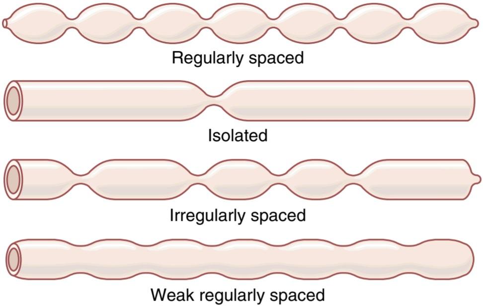
Figure 64-3. Segmentation movements of the small intestine.
شکل 64-3. حرکات قطعه بندی روده کوچک.
PROPULSIVE MOVEMENTS
Peristalsis in the Small Intestine. Chyme is propelled through the small intestine by peristaltic waves, as discussed in Chapter 63 and illustrated in Figure 63-5. These waves can occur in any part of the small intestine and move toward the anus at a velocity of 0.5 to 2.0 cm/sec- faster in the proximal intestine and slower in the terminal intestine. They are normally weak and usually die out after traveling only 3 to 5 centimeters. The waves rarely travel farther than 10 centimeters, so forward movement of the chyme is very slow-so slow that net movement along the small intestine normally averages only 1 cm/min. This rate of travel means that 3 to 5 hours are required for passage of chyme from the pylorus to the ileocecal valve.
حرکات محرکه
پریستالسیس در روده کوچک. همانطور که در فصل 63 بحث شد و در شکل 63-5 نشان داده شده است، شیم توسط امواج پریستالتیک از طریق روده کوچک به حرکت در میآید. این امواج میتوانند در هر قسمت از روده کوچک ایجاد شوند و با سرعت 0.5 تا 2.0 سانتی متر بر ثانیه در روده نزدیک و کندتر در روده انتهایی به سمت مقعد حرکت کنند. آنها معمولاً ضعیف هستند و معمولاً پس از طی مسافت 3 تا 5 سانتی متری از بین میروند. امواج به ندرت بیشتر از 10 سانتیمتر حرکت میکنند، بنابراین حرکت رو به جلو کیم بسیار آهسته است، به طوری که حرکت خالص در امتداد روده کوچک به طور معمول تنها 1 سانتیمتر در دقیقه است. این سرعت حرکت به این معنی است که 3 تا 5 ساعت برای عبور کیم از پیلور به دریچه ایلئوسکال لازم است.
Control of Peristalsis by Nervous and Hormonal Signals. Peristaltic activity of the small intestine increases markedly after a meal. This increased activity is caused partly by the beginning entry of chyme into the duodenum, causing stretch of the duodenal wall. In addition, peristaltic activity is increased by the gastroenteric reflex that is initiated by distention of the stomach and conducted principally through the myenteric plexus from the stomach down along the wall of the small intestine.
کنترل پریستالسیس توسط سیگنالهای عصبی و هورمونی. فعالیت پریستالتیک روده کوچک بعد از غذا به طور قابل توجهی افزایش مییابد. این افزایش فعالیت تا حدی ناشی از ورود اولیه کیم به دوازدهه است که باعث کشیدگی دیواره دوازدهه میشود. علاوه بر این، فعالیت پریستالتیک توسط رفلکس گوارشی افزایش مییابد که با اتساع معده آغاز میشود و عمدتاً از طریق شبکه میانتریک از معده به پایین در امتداد دیواره روده کوچک هدایت میشود.
In addition to the nervous signals that may affect small intestinal peristalsis, several hormonal factors also affect peristalsis. These factors include gastrin, CCK, insulin, motilin, and serotonin, all of which enhance intestinal motility and are secreted during various phases of food processing. Conversely, secretin and glucagon inhibit small intestinal motility. The physiological importance of each of these hormonal factors for controlling motility is still questionable.
علاوه بر سیگنالهای عصبی که ممکن است بر پریستالسیس روده کوچک تأثیر بگذارد، چندین عامل هورمونی نیز بر پریستالسیس تأثیر میگذارد. این عوامل عبارتند از گاسترین، CCK، انسولین، موتیلین و سروتونین که همگی تحرک روده را افزایش میدهند و در طی مراحل مختلف فرآوری مواد غذایی ترشح میشوند. برعکس، سکرتین و گلوکاگون حرکت روده کوچک را مهار میکنند. اهمیت فیزیولوژیکی هر یک از این عوامل هورمونی برای کنترل تحرک هنوز مورد تردید است.
The function of the peristaltic waves in the small intestine is not only to cause progression of chyme toward the ileocecal valve but also to spread out the chyme along the intestinal mucosa. As the chyme enters the intestines from the stomach and elicits peristalsis, the peristalsis immediately spreads the chyme along the intestine, and this process intensifies as additional chyme enters the duodenum. On reaching the ileocecal valve, the chyme is sometimes blocked for several hours until the person eats another meal; at that time, a gastroileal reflex intensifies peristalsis in the ileum and forces the remaining chyme through the ileocecal valve into the cecum of the large intestine.
عملکرد امواج پریستالتیک در روده کوچک نه تنها باعث پیشروی کیم به سمت دریچه ایلئوسکال میشود، بلکه باعث پخش شدن کیم در طول مخاط روده نیز میشود. همانطور که کیم از معده وارد روده میشود و باعث ایجاد پریستالسیس میشود، پریستالیس بلافاصله کیم را در امتداد روده پخش میکند و این روند با ورود کیم اضافی به دوازدهه تشدید میشود. هنگامیکه به دریچه ایلئوسکال میرسد، گاهی اوقات کیم برای چندین ساعت مسدود میشود تا زمانی که فرد یک وعده غذایی دیگر بخورد. در آن زمان، یک رفلکس معده، پریستالسیس را در ایلئوم تشدید میکند و کیم باقیمانده را از طریق دریچه ایلئوسکال به داخل سکوم روده بزرگ وادار میکند.
Propulsive Effect of the Segmentation Movements. The segmentation movements, although lasting for only a few seconds at a time, often also travel 1 centimeter or so in the anal direction, and during that time they help propel the food down the intestine. The difference between the segmentation and the peristaltic movements is not as great as might be implied by their separation into these two classifications.
اثر محرکه حرکات قطعه بندی. حرکات تقسیمبندی، اگرچه فقط چند ثانیه در هر بار طول میکشد، اغلب 1 سانتیمتر یا بیشتر در جهت مقعد حرکت میکنند و در این مدت به حرکت غذا به سمت پایین روده کمک میکنند. تفاوت بین بخشبندی و حرکات پریستالتیک به اندازهای نیست که با تفکیک آنها در این دو طبقهبندی مشخص شود.
Powerful, Rapid Peristalsis-“Peristaltic Rush.” Although peristalsis in the small intestine is normally weak, intense irritation of the intestinal mucosa, as occurs in some severe cases of infectious diarrhea, can cause both powerful and rapid peristalsis, called the peristaltic rush. This phenomenon is initiated partly by nervous reflexes that involve the autonomic nervous system and brain stem and partly by intrinsic enhancement of the myenteric plexus reflexes in the gut wall. The powerful peristaltic contractions travel long distances in the small intestine within minutes, sweeping the contents of the intestine into the colon and thereby relieving the small intestine of irritative chyme and excessive distention.
پریستالسیس قدرتمند و سریع – “پریستالتیک راش”. اگرچه پریستالسیس در روده کوچک به طور معمول ضعیف است، اما تحریک شدید مخاط روده، همانطور که در برخی موارد شدید اسهال عفونی رخ میدهد، میتواند باعث ایجاد پریستالسیس قوی و سریع شود که به آن راش پریستالتیک میگویند. این پدیده تا حدی توسط رفلکسهای عصبی که سیستم عصبی خودمختار و ساقه مغز را درگیر میکند و تا حدودی با تقویت ذاتی رفلکسهای شبکه میانتریک در دیواره روده آغاز میشود. انقباضات پریستالتیک قدرتمند، مسافتهای طولانی را در روده کوچک طی چند دقیقه طی میکنند، محتویات روده را به داخل روده بزرگ میکشانند و در نتیجه روده کوچک را از شر کیم تحریک کننده و اتساع بیش از حد رها میکنند.
Movements Caused by the Muscularis Mucosae and Muscle Fibers of the Villi. The muscularis mucosae can cause short folds to appear in the intestinal mucosa. In addition, individual fibers from this muscle extend into the intestinal villi and cause them to contract intermittently. The mucosal folds increase the surface area exposed to the chyme, thereby increasing absorption. Also, contractions of the villi-shortening, elongating, and shortening again- “milk” the villi so that lymph flows freely from the central lacteals of the villi into the lymphatic system. These mucosal and villous contractions are initiated mainly by local nervous reflexes in the submucosal nerve plexus that occur in response to chyme in the small intestine.
حرکات ناشی از موکوزای عضلانی و فیبرهای عضلانی پرزها. muscularis mucosae میتواند باعث ایجاد چینهای کوتاه در مخاط روده شود. علاوه بر این، فیبرهای منفرد از این عضله به پرزهای روده کشیده شده و باعث انقباض متناوب آنها میشود. چینهای مخاطی سطحی را که در معرض کیم قرار میگیرد افزایش میدهد و در نتیجه جذب را افزایش میدهد. همچنین، انقباضات پرزهای کوتاه، طویل، و دوباره کوتاه شدن پرزها را “شیر” میکنند تا لنف آزادانه از لاکتالهای مرکزی پرزها به سیستم لنفاوی جریان یابد. این انقباضات مخاطی و پرز عمدتاً توسط رفلکسهای عصبی موضعی در شبکه عصبی زیر مخاطی که در پاسخ به کیم در روده کوچک رخ میدهد، آغاز میشود.
THE ILEOCECAL VALVE PREVENTS BACKFLOW FROM THE COLON TO THE SMALL INTESTINE
As shown in Figure 64-4, the ileocecal valve protrudes into the lumen of the cecum and therefore is forcefully closed when excess pressure builds up in the cecum and tries to push cecal contents backward against the valve lips. The valve usually can resist reverse pressure of at least 50 to 60 centimeters of water.
دریچه ایلئوسکال از برگشت از روده بزرگ به روده باریک جلوگیری میکند
همانطور که در شکل 64-4 نشان داده شده است، دریچه ایلئوسکال به داخل لومن سکوم بیرون زده و بنابراین هنگامیکه فشار اضافی در سکوم ایجاد میشود و سعی میکند محتویات سکوم را به سمت لبههای دریچه به عقب براند، به شدت بسته میشود. شیر معمولاً میتواند در برابر فشار معکوس حداقل 50 تا 60 سانتی متر آب مقاومت کند.
In addition, the wall of the ileum for several centimeters immediately upstream from the ileocecal valve has a thickened circular muscle called the ileocecal sphincter. This sphincter normally remains mildly constricted and slows emptying of ileal contents into the cecum. However, immediately after a meal, a gastroileal reflex (described earlier) intensifies peristalsis in the ileum, and emptying of ileal contents into the cecum proceeds.
علاوه بر این، دیواره ایلئوم برای چندین سانتی متر بلافاصله در بالادست دریچه ایلئوسکال دارای یک عضله دایره ای ضخیم شده است که اسفنکتر ایلئوسکال نامیده میشود. این اسفنکتر معمولاً به طور خفیف منقبض میماند و تخلیه محتویات ایلئوم به داخل سکوم را کند میکند. با این حال، بلافاصله پس از غذا، یک رفلکس معده (که قبلا توضیح داده شد) باعث تشدید پریستالسیس در ایلئوم میشود و تخلیه محتویات ایلئوم به داخل سکوم ادامه مییابد.
Resistance to emptying at the ileocecal valve prolongs the stay of chyme in the ileum and thereby facilitates absorption. Normally, only 1500 to 2000 ml of chyme empty into the cecum each day.
مقاومت در برابر تخلیه دریچه ایلئوسکال باعث طولانی شدن ماندن کیم در ایلئوم و در نتیجه تسهیل جذب میشود. به طور معمول، روزانه تنها 1500 تا 2000 میلی لیتر کیم به داخل سکوم تخلیه میشود.
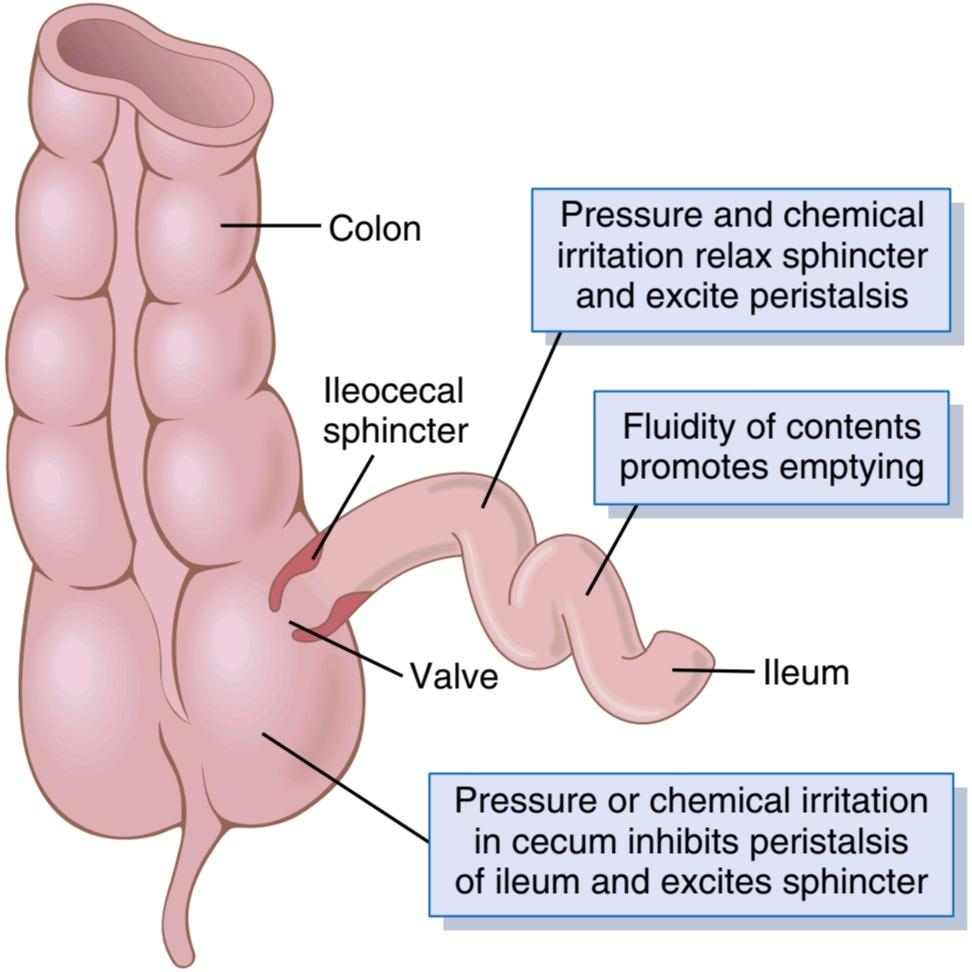
Figure 64-4. Emptying at the ileocecal valve.
شکل 64-4. تخلیه در دریچه ایلئوسکال.
Feedback Control of the Ileocecal Sphincter by Re- flexes From the Cecum. The degree of contraction of the ileocecal sphincter and the intensity of peristalsis in the terminal ileum are controlled significantly by reflexes from the cecum. When the cecum is distended, contraction of the ileocecal sphincter becomes intensified and ileal peristalsis is inhibited, both of which greatly delay emptying of additional chyme into the cecum from the ileum. Also, any irritant in the cecum delays emptying. For example, when a person has an inflamed appendix, the irritation of this vestigial remnant of the cecum can cause such intense spasm of the ileocecal sphincter and partial paralysis of the ileum that these effects together block emptying of the ileum into the cecum. The reflexes from the cecum to the ileocecal sphincter and ileum are mediated both by way of the myenteric plexus in the gut wall and of the extrinsic autonomic nerves, especially by way of the prevertebral sympathetic ganglia.
کنترل بازخورد اسفنکتر ایلئوسکال توسط بازتاب از سکوم. درجه انقباض اسفنکتر ایلئوسکال و شدت پریستالسیس در ایلئوم انتهایی به طور قابل توجهی توسط رفلکسهای سکوم کنترل میشود. هنگامیکه سکوم متسع میشود، انقباض اسفنکتر ایلئوسکال تشدید میشود و پریستالسیس ایلئوم مهار میشود، که هر دو تا حد زیادی تخلیه کیم اضافی به داخل سکوم از ایلئوم را به تاخیر میاندازند. همچنین، هرگونه محرک در سکوم، تخلیه را به تاخیر میاندازد. به عنوان مثال، هنگامیکه فردی آپاندیس ملتهب دارد، تحریک این باقیمانده از سکوم میتواند باعث اسپاسم شدید اسفنکتر ایلئوسکال و فلج نسبی ایلئوم شود که این اثرات با هم مانع از تخلیه ایلئوم به داخل سکوم میشوند. رفلکسهای سکوم به اسفنکتر ایلئوسکال و ایلئوم هم از طریق شبکه میانتریک در دیواره روده و هم از طریق اعصاب اتونوم بیرونی، به ویژه از طریق عقدههای سمپاتیک پیش مهره ای، واسطه میشوند.
MOVEMENTS OF THE COLON
The principal functions of the colon are (1) absorption of water and electrolytes from the chyme to form solid feces and (2) storage of fecal matter until it can be expelled. The proximal half of the colon, shown in Figure 64-5, is concerned principally with absorption, and the distal half with storage. Because intense colon wall movements are not required for these functions, the movements of the colon are normally sluggish. Yet, in a sluggish manner, the movements still have characteristics similar to those of the small intestine and can be divided once again into mixing movements and propulsive movements.
حرکات روده بزرگ
وظایف اصلی روده بزرگ عبارتند از: (1) جذب آب و الکترولیتها از کیم برای تشکیل مدفوع جامد و (2) ذخیره سازی مدفوع تا زمانی که بتوان آن را دفع کرد. نیمه پروگزیمال کولون، که در شکل 64-5 نشان داده شده است، عمدتاً مربوط به جذب است و نیمه دیستال مربوط به ذخیره سازی است. از آنجایی که حرکات شدید دیواره روده بزرگ برای این عملکردها لازم نیست، حرکات روده بزرگ معمولاً کند هستند. با این حال، به شیوه ای کند، حرکات همچنان دارای ویژگیهایی شبیه به روده کوچک هستند و میتوان یک بار دیگر به حرکات ترکیبی و حرکات پیشرانه تقسیم کرد.
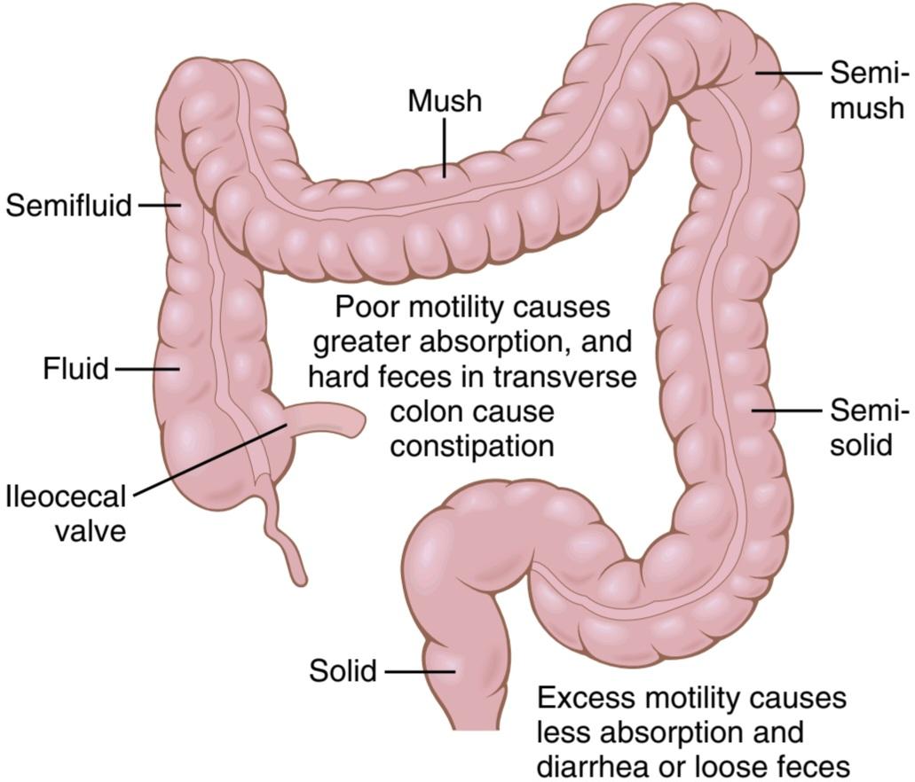
Figure 64-5. Absorptive and storage functions of the large intestine.
شکل 64-5. عملکردهای جذبی و ذخیره سازی روده بزرگ.
Mixing Movements-“Haustrations.” In the same manner that segmentation movements occur in the small intestine, large circular constrictions occur in the large intestine. At each of these constrictions, about 2.5 centimeters of the circular muscle contract, sometimes constricting the lumen of the colon almost to occlusion. At the same time, the longitudinal muscle of the colon, which is aggregated into three longitudinal strips called the teniae coli, contracts. These combined contractions of the circular and longitudinal strips of muscle cause the unstimulated portion of the large intestine to bulge out- ward into baglike sacs called haustrations.
حرکات اختلاط – “آزار و اذیت.” همانطور که حرکات قطعه بندی در روده کوچک رخ میدهد، انقباضات دایره ای بزرگ در روده بزرگ رخ میدهد. در هر یک از این انقباضات، حدود 2.5 سانتیمتر از عضله حلقوی منقبض میشود و گاهی اوقات لومن کولون را تقریباً تا حد انسداد منقبض میکند. همزمان عضله طولی روده بزرگ که در سه نوار طولی به نام teniae coli جمع میشود منقبض میشود. این انقباضات ترکیبی نوارهای دایره ای و طولی عضله باعث میشود که بخش تحریک نشده روده بزرگ به سمت بیرون در کیسههای کیسه ای به نام هوستراسیون برآمده شود.
Each haustration usually reaches peak intensity in about 30 seconds and then disappears during the next 60 seconds. At times they also move slowly toward the anus during contraction, especially in the cecum and ascending colon, and thereby provide a minor amount of forward propulsion of the colonic contents. After another few minutes, new haustral contractions occur in other areas nearby. Therefore, the fecal material in the large intestine is slowly dug into and rolled over in much the same manner that one spades the earth. In this way, all the fecal material is gradually exposed to the mucosal surface of the large intestine, and fluid and dissolved substances are progressively absorbed until only 80 to 200 ml of feces are expelled each day.
هر کسالت معمولاً در حدود 30 ثانیه به اوج شدت میرسد و سپس در 60 ثانیه بعدی ناپدید میشود. گاهی اوقات آنها همچنین در طول انقباض به آرامیبه سمت مقعد حرکت میکنند، به خصوص در سکوم و روده بزرگ صعودی، و در نتیجه مقدار کمیاز محتویات کولون به جلو رانده میشوند. پس از چند دقیقه دیگر، انقباضات جدید در مناطق دیگر نزدیک رخ میدهد. بنابراین، مواد مدفوع موجود در روده بزرگ به آرامیدر داخل آن حفر میشود و تقریباً به همان شکلی که روی زمین میچرخد، غلت میخورد. به این ترتیب تمام مواد مدفوع به تدریج در معرض سطح مخاطی روده بزرگ قرار میگیرند و مایعات و مواد محلول به تدریج جذب میشوند تا جایی که روزانه تنها 80 تا 200 میلی لیتر مدفوع دفع میشود.
Propulsive Movements “Mass Movements.” Much of the propulsion in the cecum and ascending colon results from the slow but persistent haustral contractions,requiring as many as 8 to 15 hours to move the chyme from the ileocecal valve through the colon, while the chyme becomes fecal in quality-a semisolid instead of a semifluid slush.
جنبشهای پیشران “جنبشهای توده ای”. بیشتر نیروی محرکه در سکوم و روده بزرگ صعودی ناشی از انقباضات آهسته اما مداوم است، که به 8 تا 15 ساعت برای حرکت کیم از دریچه ایلئوسکال از طریق کولون نیاز دارد، در حالی که کیم از نظر کیفیت مدفوعی میشود – نیمه جامد به جای لجن نیمه سیال.
From the cecum to the sigmoid, mass movements can, for many minutes at a time, take over the propulsive role. These movements usually occur only one to three times each day, in many people especially for about 15 minutes during the first hour after eating breakfast.
از سکوم گرفته تا سیگموئید، حرکات توده ای میتوانند برای دقایق زیادی در یک زمان نقش محرکه را به عهده بگیرند. این حرکات معمولاً فقط یک تا سه بار در روز، در بسیاری از افراد به ویژه حدود 15 دقیقه در طول یک ساعت اول پس از خوردن صبحانه رخ میدهد.
A mass movement is a modified type of peristalsis characterized by the following sequence of events. First, a constrictive ring occurs in response to a distended or irritated point in the colon, usually in the transverse colon. Then, rapidly, the 20 or more centimeters of colon distal to the constrictive ring lose their haustrations and instead contract as a unit, propelling the fecal material in this segment en masse further down the colon. The contraction develops progressively more force for about 30 seconds, and relaxation occurs during the next 2 to 3 minutes. Another mass movement then occurs, this time perhaps farther along the colon.
جنبش تودهای نوعی اصلاح شده از پریستالسیس است که با توالی رویدادهای زیر مشخص میشود. اول، یک حلقه منقبض کننده در پاسخ به یک نقطه متسع یا تحریک شده در کولون، معمولا در کولون عرضی، رخ میدهد. سپس، به سرعت، 20 سانتیمتر یا بیشتر از روده بزرگ دیستال حلقه منقبض کننده، کسالت خود را از دست میدهند و در عوض بهعنوان یک واحد منقبض میشوند و مواد مدفوع در این بخش را بهطور انبوه به سمت پایینتر کولون سوق میدهند. انقباض به تدریج نیروی بیشتری را برای حدود 30 ثانیه ایجاد میکند و آرامش در طی 2 تا 3 دقیقه بعدی رخ میدهد. سپس یک حرکت توده ای دیگر رخ میدهد، این بار شاید دورتر در امتداد روده بزرگ.
A series of mass movements usually persists for 10 to 30 minutes. They then cease but return perhaps a half day later. When they have forced a mass of feces into the rec- tum, the desire for defecation is felt.
یک سری حرکات توده ای معمولاً 10 تا 30 دقیقه ادامه مییابد. سپس آنها متوقف میشوند اما شاید نیم روز بعد برگردند. هنگامیکه آنها توده ای از مدفوع را به داخل روده وارد میکنند، میل به اجابت مزاج احساس میشود.
Initiation of Mass Movements by Gastrocolic and Duodenocolic Reflexes. The appearance of mass movements after meals is facilitated by gastrocolic and duodenocolic reflexes. These reflexes result from distention of the stomach and duodenum. They occur either not at all or hardly at all when the extrinsic autonomic nerves to the colon have been removed; therefore, the reflexes almost certainly are transmitted by way of the autonomic nervous system.
شروع حرکات توده ای توسط رفلکسهای معده و دوازدهه. ظهور حرکات توده ای بعد از غذا توسط رفلکسهای معده و دوازدهه تسهیل میشود. این رفلکسها ناشی از اتساع معده و اثنی عشر است. وقتی اعصاب اتونوم بیرونی روده بزرگ برداشته شده اند، یا اصلاً یا اصلاً به سختی رخ میدهند. بنابراین، رفلکسها به طور قطع از طریق سیستم عصبی خودمختار منتقل میشوند.
Irritation in the colon can also initiate intense mass movements. For example, a person who has an ulcerated condition of the colon mucosa (ulcerative colitis) frequently has mass movements that persist almost all the time.
تحریک در روده بزرگ همچنین میتواند باعث شروع حرکات توده ای شدید شود. به عنوان مثال، فردی که دارای شرایط زخمیمخاط روده بزرگ (کولیت اولسراتیو) است، اغلب دارای حرکات توده ای است که تقریباً همیشه ادامه دارد.
DEFECATION
Most of the time, the rectum is empty of feces, partly because a weak functional sphincter exists about 20 centimeters from the anus at the juncture between the sigmoid colon and the rectum. A sharp angulation is also present here that contributes additional resistance to filling of the rectum.
مدفوع
بیشتر اوقات، رکتوم خالی از مدفوع است، تا حدی به این دلیل که یک اسفنکتر ضعیف عملکردی در حدود 20 سانتی متر از مقعد در محل اتصال بین کولون سیگموئید و رکتوم وجود دارد. یک زاویه تیز نیز در اینجا وجود دارد که مقاومت بیشتری در پر شدن رکتوم ایجاد میکند.
When a mass movement forces feces into the rectum, the desire for defecation occurs immediately, including reflex contraction of the rectum and relaxation of the anal sphincters.
هنگامیکه یک حرکت توده ای مدفوع را وارد راست روده میکند، میل به اجابت مزاج بلافاصله رخ میدهد، از جمله انقباض رفلکس راست روده و شل شدن اسفنکترهای مقعدی.
Continual dribble of fecal matter through the anus is prevented by tonic constriction of the following: (1) an internal anal sphincter, which is a several-centimeters- long thickening of the circular smooth muscle that lies immediately inside the anus; and (2) an external anal sphincter, composed of striated voluntary muscle that both surrounds the internal sphincter and extends distal to it. The external sphincter is controlled by nerve fibers in the pudendal nerve, which is part of the somatic nervous system and therefore is under voluntary, conscious, or at least subconscious control; subconsciously, the external sphincter is usually kept continuously constricted unless conscious signals inhibit the constriction.
از چکیدن مداوم ماده مدفوع از طریق مقعد با انقباض تونیک موارد زیر جلوگیری میشود: (1) یک اسفنکتر مقعدی داخلی، که ضخیم شدن چند سانتی متر طول عضله صاف حلقوی است که بلافاصله در داخل مقعد قرار دارد. و (2) یک اسفنکتر خارجی مقعدی، متشکل از عضله مخطط ارادی که هم اسفنکتر داخلی را احاطه کرده و هم به سمت دیستال آن امتداد مییابد. اسفنکتر خارجی توسط رشتههای عصبی در عصب پودندال کنترل میشود که بخشی از سیستم عصبی جسمانی است و بنابراین تحت کنترل اختیاری، آگاهانه یا حداقل ناخودآگاه قرار دارد. به طور ناخودآگاه اسفنکتر خارجی معمولاً به طور مداوم منقبض نگه داشته میشود مگر اینکه سیگنالهای آگاهانه مانع انقباض شوند.
Defecation Reflexes. Ordinarily, defecation is initiated by defecation reflexes. One of these reflexes is an intrinsic reflex mediated by the local enteric nervous system in the rectal wall. When feces enter the rectum, distention of the rectal wall initiates afferent signals that spread through the myenteric plexus to initiate peristaltic waves in the descending colon, sigmoid, and rectum, forcing feces toward the anus. As the peristaltic wave approaches the anus, the internal anal sphincter is relaxed by inhibitory signals from the myenteric plexus; if the external anal sphincter is also consciously, voluntarily relaxed at the same time, defecation occurs.
رفلکسهای مدفوع. معمولاً اجابت مزاج با رفلکسهای دفع شروع میشود. یکی از این رفلکسها یک رفلکس ذاتی است که با واسطه سیستم عصبی روده موضعی در دیواره رکتوم ایجاد میشود. هنگامیکه مدفوع وارد رکتوم میشود، اتساع دیواره رکتوم سیگنالهای آوران را آغاز میکند که از طریق شبکه میانتریک پخش میشود تا امواج پریستالتیک را در کولون نزولی، سیگموئید و رکتوم آغاز کند و مدفوع را به سمت مقعد وادار میکند. همانطور که موج پریستالتیک به مقعد نزدیک میشود، اسفنکتر داخلی مقعد با سیگنالهای بازدارنده از شبکه میانتریک شل میشود. اگر اسفنکتر خارجی مقعد نیز آگاهانه و به طور داوطلبانه در همان زمان شل شود، مدفوع رخ میدهد.
Normally when the intrinsic myenteric defecation reflex is functioning by itself, it is relatively weak. To be effective in causing defecation, it usually must be fortified by another type of defecation reflex called a parasympathetic defecation reflex that involves the sacral segments of the spinal cord, shown in Figure 64-6. When the nerve endings in the rectum are stimulated, signals are transmitted first into the spinal cord and then reflexly back to the descending colon, sigmoid, rectum, and anus via parasympathetic nerve fibers in the pelvic nerves. These parasympathetic signals greatly intensify the peristaltic waves and relax the internal anal sphincter, thus converting the intrinsic myenteric defecation reflex from a weak effort into a powerful process of defecation that is sometimes effective in emptying the large bowel all the way from the splenic flexure of the colon to the anus.
به طور معمول هنگامیکه رفلکس اجابت مزاج عضلانی درونی به خودی خود عمل میکند، نسبتا ضعیف است. برای اینکه در ایجاد اجابت مزاج مؤثر باشد، معمولاً باید با نوع دیگری از رفلکس دفع مدفوع به نام رفلکس اجابت مزاج پاراسمپاتیک که بخشهای خاجی نخاع را درگیر میکند، تقویت شود، که در شکل 64-6 نشان داده شده است. هنگامیکه پایانههای عصبی در راست روده تحریک میشوند، سیگنالها ابتدا به نخاع منتقل میشوند و سپس از طریق رشتههای عصبی پاراسمپاتیک در اعصاب لگنی به کولون نزولی، سیگموئید، رکتوم و مقعد باز میگردند. این سیگنالهای پاراسمپاتیک امواج پریستالتیک را تا حد زیادی تشدید میکنند و اسفنکتر داخلی مقعد را شل میکنند، بنابراین رفلکس اجابت مزاج عضلانی درونی را از یک تلاش ضعیف به یک فرآیند قدرتمند اجابت مزاج تبدیل میکنند که گاهی اوقات در تخلیه روده بزرگ از خم شدن طحال کولون تا مقعد مؤثر است.
Defecation signals entering the spinal cord initiate other effects, such as taking a deep breath, closure of the glottis, and contraction of the abdominal wall muscles to force the fecal contents of the colon downward, and at the same time they cause the pelvic floor to relax downward and pull outward on the anal ring to evaginate the feces.
سیگنالهای مدفوع که وارد نخاع میشوند، اثرات دیگری مانند تنفس عمیق، بسته شدن گلوت و انقباض عضلات دیواره شکم را آغاز میکنند تا محتویات مدفوع روده بزرگ را به سمت پایین وادار کند و در عین حال باعث میشود کف لگن به سمت پایین شل شده و حلقه مقعدی را به سمت بیرون بکشد تا مدفوع خارج شود.
When it becomes convenient for the person to defecate, the defecation reflexes can purposely be activated by taking a deep breath to move the diaphragm downward and then contracting the abdominal muscles to increase the pressure in the abdomen, thus forcing fecal contents into the rectum to cause new reflexes. Reflexes initiated in this way are almost never as effective as those that arise naturally, and thus people who too often inhibit their natural reflexes are likely to become severely constipated.
هنگامیکه اجابت مزاج برای فرد راحت شد، رفلکسهای اجابت مزاج میتوانند عمداً با کشیدن یک نفس عمیق برای حرکت دیافراگم به سمت پایین و سپس انقباض عضلات شکم برای افزایش فشار در شکم فعال شوند، در نتیجه محتویات مدفوع را مجبور به وارد شدن به راست روده برای ایجاد رفلکسهای جدید میکند. رفلکسهایی که از این طریق شروع میشوند تقریباً هرگز به اندازه آنهایی که به طور طبیعی ایجاد میشوند مؤثر نیستند، و بنابراین افرادی که اغلب رفلکسهای طبیعی خود را مهار میکنند، احتمالاً به شدت دچار یبوست میشوند.
In newborns and in some people with transected spinal cords, the defecation reflexes cause automatic emptying of the lower bowel at inconvenient times during the day because of lack of conscious control exercised through voluntary contraction or relaxation of the external anal sphincter.
در نوزادان و در برخی از افراد با طناب نخاعی قطع شده، رفلکسهای اجابت مزاج باعث تخلیه خودکار قسمت تحتانی روده در زمانهای نامناسب در طول روز میشود که دلیل آن عدم کنترل آگاهانه از طریق انقباض یا شل شدن اسفنکتر خارجی مقعد است.
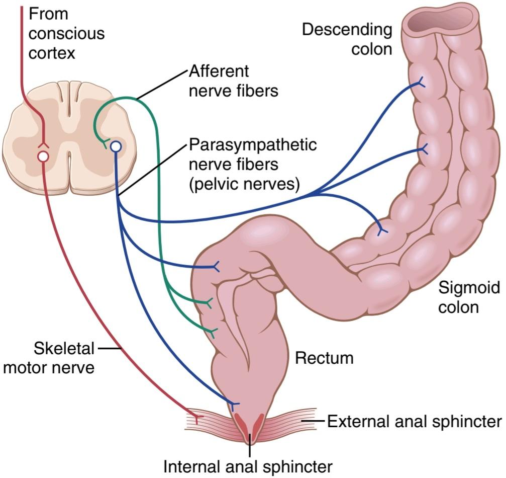
Figure 64-6. Afferent and efferent pathways of the parasympathetic mechanism for enhancing the defecation reflex.
شکل 64-6. مسیرهای آوران و وابران مکانیسم پاراسمپاتیک برای تقویت رفلکس اجابت مزاج.
OTHER AUTONOMIC REFLEXES THAT AFFECT BOWEL ACTIVITY
Aside from the duodenocolic, gastrocolic, gastroileal, enterogastric, and defecation reflexes that have been dis- cussed in this chapter, several other important nervous reflexes also can affect the overall degree of bowel activity. They are the peritoneointestinal reflex, renointestinal reflex, and vesicointestinal reflex.
سایر رفلکسهای خودگردان که بر فعالیت روده تأثیر میگذارد
جدا از رفلکسهای اثنی عشر، معده، معده، روده معده و دفع مدفوع که در این فصل مورد بحث قرار گرفتهاند، چندین رفلکس عصبی مهم دیگر نیز میتوانند بر درجه کلی فعالیت روده تأثیر بگذارند. آنها رفلکس صفاقی روده ای، رفلکس رنوروده ای و رفلکس وزیکوین روده ای هستند.
The peritoneointestinal reflex results from irritation of the peritoneum; it strongly inhibits the excitatory enteric nerves and thereby can cause intestinal paralysis, especially in patients with peritonitis. The renointestinal and vesicointestinal reflexes inhibit intestinal activity as a result of kidney or bladder irritation, respectively.
رفلکس صفاقی روده ای ناشی از تحریک صفاق است. به شدت اعصاب روده ای تحریکی را مهار میکند و در نتیجه میتواند باعث فلج روده به خصوص در بیماران مبتلا به پریتونیت شود. رفلکسهای روده ای و وزیکوینتینال به ترتیب فعالیت روده را در نتیجه تحریک کلیه یا مثانه مهار میکنند.
کلیک کنید «Bibliography: فهرست کتب مربوطه»
Browning KN, Travagli RA: Central nervous system control of gastro- intestinal motility and secretion and modulation of gastrointestinal functions. Compr Physiol 4:1339, 2014.
Camilleri M: Physiological underpinnings of irritable bowel syndrome: neurohormonal mechanisms. J Physiol 592:2967, 2014.
Farré R, Tack J: Food and symptom generation in functional gas- trointestinal disorders: physiological aspects. Am J Gastroenterol 108:698, 2013.
Ford AC, Lacy BE, Talley NJ: Irritable bowel syndrome. N Engl J Med 376:2566, 2017.
Furness JB: The enteric nervous system and neurogastroenterology. Nat Rev Gastroenterol Hepatol 9:286, 2012.
Gracie DJ, Hamlin PJ, Ford AC: The influence of the brain-gut axis in inflammatory bowel disease and possible implications for treat- ment. Lancet Gastroenterol Hepatol 4:632, 2019.
Hockley JRF, Smith ESJ, Bulmer DC: Human visceral nociception: find- ings from translational studies in human tissue. Am J Physiol Gas- trointest Liver Physiol 315:G464, 2018.
Huizinga JD, Lammers WJ: Gut peristalsis is governed by a multitude of cooperating mechanisms. Am J Physiol Gastrointest Liver Physiol 296:G1, 2009.
Kumral D, Zfass AM: Gut movements: a review of the physiology of gastrointestinal transit. Dig Dis Sci 63:2500, 2018.
Lang IM, Medda BK, Shaker R: Characterization and mechanism of the esophago-esophageal contractile reflex of the striated mus- cle esophagus. Am J Physiol Gastrointest Liver Physiol 317:G304, 2019.
Mittal RK: Regulation and dysregulation of esophageal peristalsis by the integrated function of circular and longitudinal muscle lay- ers in health and disease. Am J Physiol Gastrointest Liver Physiol 311:G431, 2016.
Ouyang A, Regan J, McMahon BP: Physiology of the upper segment, body, and lower segment of the esophagus. Ann NY Acad Sci 1300:261, 2013.
Sanders KM, Ward SM, Koh SD: Interstitial cells: regulators of smooth muscle function. Physiol Rev 94:859, 2014.
Spencer NJ, Dinning PG, Brookes SJ, Costa M: Insights into the mech- anisms underlying colonic motor patterns. J Physiol 594:4099, 2016.
Szarka LA, Camilleri M: Methods for measurement of gastric motility. Am J Physiol Gastrointest Liver Physiol 296:G461, 2009.
🚀 با ما همراه شوید!
تازهترین مطالب و آموزشهای مغز و اعصاب را از دست ندهید. با فالو کردن کانال تلگرام آیندهنگاران مغز، از ما حمایت کنید!



