
فیزیولوژی پزشکی گایتون و هال؛ اصول کلی عملکرد دستگاه گوارش-تحرک، کنترل عصبی و گردش خون


دعای مطالعه [ نمایش ]
بِسْمِ الله الرَّحْمنِ الرَّحیمِ
اَللّهُمَّ اَخْرِجْنى مِنْ ظُلُماتِ الْوَهْمِ
خدايا مرا بيرون آور از تاريكىهاى وهم،
وَ اَكْرِمْنى بِنُورِ الْفَهْمِ
و به نور فهم گرامى ام بدار،
اَللّهُمَّ افْتَحْ عَلَيْنا اَبْوابَ رَحْمَتِكَ
خدايا درهاى رحمتت را به روى ما بگشا،
وَانْشُرْ عَلَيْنا خَزائِنَ عُلُومِكَ بِرَحْمَتِكَ يا اَرْحَمَ الرّاحِمينَ
و خزانههاى علومت را بر ما باز كن به امید رحمتت اى مهربانترين مهربانان.
کتاب «فیزیولوژی پزشکی گایتون وهال» بهعنوان یکی از جامعترین و معتبرترین منابع در حوزه علوم پزشکی، همچنان مرجع کلیدی برای درک عملکرد پیچیده بدن انسان است. این اثر با تکیه بر تازهترین پژوهشها و توضیحات دقیق از سازوکارهای فیزیولوژیک، پلی میان علوم پایه پزشکی و کاربردهای بالینی ایجاد میکند و نقشی بیبدیل در آموزش، پژوهش و ارتقای دانش سلامت ایفا مینماید.
ترجمه دقیق و علمیاین شاهکار توسط برند علمیآیندهنگاران مغز به مدیریت داریوش طاهری، دسترسی فارسیزبانان به مرزهای نوین دانش فیزیولوژی را ممکن ساخته و رسالتی علمیبرای ارتقای آموزش پزشکی، فهم عمیقتر سازوکارهای بدن و توسعه روشهای نوین در حوزه سلامت فراهم آورده است.
» کتاب فیزیولوژی پزشکی گایتون وهال
» » فصل ۶۳: اصول کلی عملکرد دستگاه گوارش-تحرک، کنترل عصبی و گردش خون
در حال ویرایش
» Guyton and Hall Textbook of Medical Physiology
»» CHAPTER 63: General Principles of Gastrointestinal Function-Motility, Nervous Control, and Blood Circulation
The alimentary tract provides the body with a continual supply of water, electrolytes, vitamins, and nutrients, which requires the following: (1) movement of food through the alimentary tract; (2) secretion of digestive juices and digestion of the food; (3) absorption of water, various electrolytes, vitamins, and digestive products; (4) circulation of blood through the gastrointestinal organs to carry away the absorbed substances; and (5) control of all these functions by local, nervous, and hormonal systems.
دستگاه گوارش به طور مداوم آب، الکترولیتها، ویتامینها و مواد مغذی را برای بدن فراهم میکند که به موارد زیر نیاز دارد: (1) حرکت غذا از طریق دستگاه گوارش. (2) ترشح شیرههای گوارشی و هضم غذا. (3) جذب آب، الکترولیتهای مختلف، ویتامینها و محصولات گوارشی؛ (4) گردش خون از طریق اندامهای گوارشی برای انتقال مواد جذب شده. و (5) کنترل تمام این عملکردها توسط سیستمهای موضعی، عصبی و هورمونی.
Figure 63-1 shows the entire alimentary tract. Each part is adapted to its specific functions- some parts to simple passage of food, such as the esophagus; others to temporary storage of food, such as the stomach; and others to digestion and absorption, such as the small intestine. In this chapter we discuss the basic principles of function in the entire alimentary tract, and in subsequent chapters the specific functions of different segments of the tract will be addressed.
شکل 63-1 کل دستگاه گوارش را نشان میدهد. هر قسمت با عملکردهای خاص خود سازگار است – برخی از قسمتها برای عبور ساده غذا، مانند مری. دیگران به ذخیره موقت غذا، مانند معده. و دیگران به هضم و جذب، مانند روده کوچک. در این فصل به اصول اساسی عملکرد در کل دستگاه گوارش میپردازیم و در فصلهای بعدی به عملکردهای خاص بخشهای مختلف دستگاه پرداخته میشود.
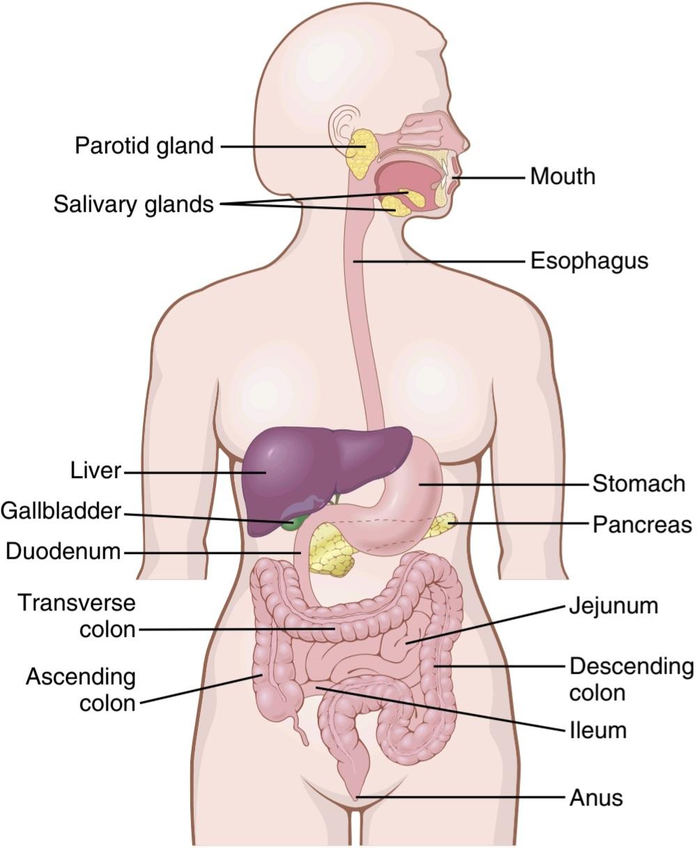
Figure 63-1 Alimentary tract.
شکل 63-1 دستگاه گوارش.
GENERAL PRINCIPLES OF GASTROIN- TESTINAL MOTILITY
Physiologic Anatomy of the Gastrointestinal Wall
Figure 63-2 shows a typical cross section of the intestinal wall, including the following layers from the outer surface inward: (1) the serosa, (2) a longitudinal smooth muscle layer, (3) a circular smooth muscle layer, (4) the submucosa, and (5) the mucosa. In addition, sparse bundles of smooth muscle fibers, the mucosal muscle, lie in the deeper layers of the mucosa. The motor functions of the gut are performed by the different layers of smooth muscle.
اصول کلی حرکت دستگاه گوارش
آناتومیفیزیولوژیکی دیواره دستگاه گوارش
شکل 63-2 یک مقطع معمولی از دیواره روده را نشان میدهد که شامل لایههای زیر از سطح بیرونی به داخل است: (1) سروز، (2) یک لایه عضله صاف طولی، (3) یک لایه عضله صاف دایره ای، (4) زیر مخاط، و (5) مخاط. علاوه بر این، دستههای پراکنده فیبرهای عضلانی صاف، عضله مخاطی، در لایههای عمیقتر مخاط قرار دارند. عملکردهای حرکتی روده توسط لایههای مختلف ماهیچه صاف انجام میشود.
The general characteristics of smooth muscle and its function are discussed in Chapter 8, which should be reviewed as a background for the following sections of this chapter.
ویژگیهای کلی ماهیچه صاف و عملکرد آن در فصل 8 مورد بحث قرار گرفته است که باید به عنوان پیش زمینه ای برای بخشهای بعدی این فصل بررسی شود.
Gastrointestinal Smooth Muscle Functions as a Syncytium. The individual smooth muscle fibers in the gastrointestinal tract are 200 to 500 micrometers in length and 2 to 10 micrometers in diameter, and they are arranged in bundles of as many as 1000 parallel fibers. In the longitudinal muscle layer, the bundles extend longitudinally down the intestinal tract; in the circular muscle layer, they extend around the gut.
عضله صاف دستگاه گوارش به عنوان یک سیسیتیوم عمل میکند. فیبرهای عضلانی صاف مجزا در دستگاه گوارش 200 تا 500 میکرومتر طول و 2 تا 10 میکرومتر قطر دارند و در دستههایی از 1000 فیبر موازی قرار گرفته اند. در لایه عضلانی طولی، دستهها به صورت طولی به سمت پایین دستگاه روده گسترش مییابند. در لایه عضلانی دایره ای، آنها در اطراف روده گسترش مییابند.
Within each bundle, the muscle fibers are electrically connected with one another through large numbers of gap junctions that allow low-resistance movement of ions from one muscle cell to the next. Therefore, electrical signals that initiate muscle contractions can travel readily from one fiber to the next within each bundle but more rapidly along the length of the bundle than sideways.
در داخل هر بسته، فیبرهای عضلانی از طریق تعداد زیادی از اتصالات شکاف به یکدیگر متصل میشوند که امکان حرکت با مقاومت کم یونها از یک سلول عضلانی به سلول دیگر را فراهم میکند. بنابراین، سیگنالهای الکتریکی که انقباضات ماهیچهای را آغاز میکنند، میتوانند به آسانی از یک فیبر به فیبر دیگر در داخل هر بسته، اما با سرعت بیشتری در طول بستهجنسی حرکت کنند تا به طرفین.
Each bundle of smooth muscle fibers is partly separated from the next by loose connective tissue; however, the muscle bundles fuse with one another at many points, so in reality each muscle layer represents a branching latticework of smooth muscle bundles. Therefore, each muscle layer functions as a syncytium; that is, when an action potential is elicited anywhere within the muscle mass, it generally travels in all directions in the muscle. The distance that it travels depends on the excitability of the muscle; sometimes it stops after only a few millimeters, and at other times it travels many centimeters or even the entire length and breadth of the intestinal tract.
هر دسته از فیبرهای عضلانی صاف تا حدی توسط بافت همبند شل از دسته بعدی جدا میشود. با این حال، بستههای عضلانی در بسیاری از نقاط با یکدیگر ترکیب میشوند، بنابراین در واقع هر لایه عضلانی نشاندهنده شبکهای منشعب از دستههای ماهیچه صاف است. بنابراین، هر لایه عضلانی به عنوان یک سنسیتیوم عمل میکند. یعنی زمانی که یک پتانسیل عمل در هر نقطه ای از توده عضلانی برانگیخته میشود، به طور کلی در تمام جهات در عضله حرکت میکند. مسافتی که طی میکند به تحریک پذیری عضله بستگی دارد. گاهی اوقات تنها پس از چند میلی متر متوقف میشود و در زمانهای دیگر چندین سانتی متر یا حتی کل طول و عرض دستگاه روده را طی میکند.
Also, because a few connections exist between the longitudinal and circular muscle layers, excitation of one of these layers often excites the other as well.
همچنین، به دلیل وجود چند اتصال بین لایههای عضلانی طولی و دایرهای، تحریک یکی از این لایهها اغلب دیگری را نیز تحریک میکند.

Figure 63-2. Typical cross section of the gut
شکل 63-2. مقطع معمولی روده
Electrical Activity of Gastrointestinal Smooth Muscle
The smooth muscle of the gastrointestinal tract is excited by almost continual slow, intrinsic electrical activity along the membranes of the muscle fibers. This activity has two basic types of electrical waves: (1) slow waves and (2) spikes, both of which are shown in Figure 63-3. In addition, the voltage of the resting membrane potential of the gastrointestinal smooth muscle can change to different levels, which can also have important effects in control- ling motor activity of the gastrointestinal tract.
فعالیت الکتریکی ماهیچه صاف دستگاه گوارش
ماهیچه صاف دستگاه گوارش با فعالیت الکتریکی آهسته و ذاتی تقریباً مداوم در امتداد غشای فیبرهای عضلانی برانگیخته میشود. این فعالیت دارای دو نوع اصلی امواج الکتریکی است: (1) امواج آهسته و (2) سنبله که هر دو در شکل 63-3 نشان داده شده اند. علاوه بر این، ولتاژ پتانسیل غشای استراحت عضله صاف دستگاه گوارش میتواند به سطوح مختلفی تغییر کند که میتواند اثرات مهمیدر کنترل فعالیت حرکتی دستگاه گوارش داشته باشد.
“Slow Waves” Caused by Undulating Changes in Resting Membrane Potential. Most gastrointestinal contractions occur rhythmically, and this rhythm is determined mainly by the frequency of so-called “slow waves” of smooth muscle membrane potential. These waves, shown in Figure 63-3, are not action potentials. Instead, they are slow, undulating changes in the resting membrane potential. Their intensity usually varies between 5 and 15 millivolts, and their frequency ranges in different parts of the human gastrointestinal tract from 3 to 12/min-about 3 in the body of the stomach, as much as 12 in the duodenum, and about 8 or 9 in the terminal ileum. Therefore, the rhythm of contraction of the body of the stomach, the duodenum, and the ileum is usually about 3/min, about 12/min, and 8 to 9/ min, respectively.
“امواج آهسته” ناشی از تغییرات مواج در پتانسیل استراحت غشایی. بیشتر انقباضات دستگاه گوارش به صورت ریتمیک اتفاق میافتد و این ریتم عمدتاً با فرکانس به اصطلاح “امواج آهسته” پتانسیل غشای عضله صاف تعیین میشود. این امواج که در شکل 63-3 نشان داده شده اند، پتانسیل عمل نیستند. در عوض، آنها تغییرات آهسته و موجی در پتانسیل غشاء استراحت دارند. شدت آنها معمولاً بین 5 تا 15 میلی ولت متغیر است و فرکانس آنها در قسمتهای مختلف دستگاه گوارش انسان از 3 تا 12 در دقیقه – حدود 3 در بدنه معده، تا 12 در دوازدهه و حدود 8 یا 9 در ایلئوم انتهایی متغیر است. بنابراین، ریتم انقباض بدن معده، دوازدهه و ایلئوم معمولاً به ترتیب حدود 3/min، حدود 12/min و 8 تا 9/min است.
The precise cause of the slow waves is not completely understood, although they appear to be caused by com- plex interactions among the smooth muscle cells and specialized cells, called the interstitial cells of Cajal, which are believed to act as electrical pacemakers for smooth muscle cells. These interstitial cells form a network with each other and are interposed between the smooth muscle layers, with synaptic-like contacts to smooth muscle cells. The interstitial cells of Cajal undergo cyclic changes in membrane potential due to unique ion channels that periodically open and produce inward (pacemaker) cur- rents that may generate slow wave activity.
علت دقیق امواج آهسته به طور کامل شناخته نشده است، اگرچه به نظر میرسد که این امواج ناشی از فعل و انفعالات پیچیده بین سلولهای ماهیچه صاف و سلولهای تخصصی به نام سلولهای بینابینی Cajal هستند که اعتقاد بر این است که به عنوان ضربان ساز الکتریکی برای سلولهای عضله صاف عمل میکنند. این سلولهای بینابینی با یکدیگر شبکه ای تشکیل میدهند و بین لایههای ماهیچه صاف قرار میگیرند و با تماسهای سیناپسی مانند به سلولهای ماهیچه صاف قرار میگیرند. سلولهای بینابینی Cajal به دلیل کانالهای یونی منحصربهفردی که بهطور دورهای باز میشوند و جریانهایی به سمت داخل (پیس میکر) تولید میکنند که ممکن است فعالیت موج آهسته ایجاد کند، تحت تغییرات چرخهای در پتانسیل غشایی قرار میگیرند.
The slow waves usually do not by themselves cause muscle contraction in most parts of the gastrointestinal tract, except perhaps in the stomach. Instead, they mainly excite the appearance of intermittent spike potentials, and the spike potentials in turn actually excite the muscle contraction.
امواج آهسته معمولاً به خودی خود باعث انقباض عضلانی در بیشتر قسمتهای دستگاه گوارش نمیشوند، به جز در معده. در عوض، آنها عمدتاً ظاهر پتانسیلهای سنبله متناوب را تحریک میکنند و پتانسیلهای سنبله نیز به نوبه خود در واقع انقباض عضلانی را تحریک میکنند.

Figure 63-3. Membrane potentials in intestinal smooth muscle. Note the slow waves, the spike potentials, total depolarization, and hyperpolarization, all of which occur under different physiological conditions of the intestine.
شکل 63-3. پتانسیلهای غشایی در ماهیچه صاف روده به امواج آهسته، پتانسیلهای سنبله، دپلاریزاسیون کامل وهایپرپلاریزاسیون توجه کنید که همگی در شرایط فیزیولوژیکی مختلف روده رخ میدهند.
Spike Potentials. The spike potentials are true action potentials. They occur automatically when the resting membrane potential of the gastrointestinal smooth muscle becomes more positive than about -40 millivolts (the normal resting membrane potential in the smooth muscle fibers of the gut is between -50 and -60 millivolts). Note in Figure 63-3 that each time the peaks of the slow waves temporarily become more positive than -40 millivolts, spike potentials appear on these peaks. The higher the slow wave potential rises, the greater the frequency of the spike potentials, usually ranging between 1 and 10 spikes per second. The spike potentials last 10 to 40 times as long in gastrointestinal muscle as the action potentials in large nerve fibers, with each gastrointestinal spike lasting as long as 10 to 20 milliseconds.
پتانسیلهای سنبله. پتانسیلهای سنبله پتانسیلهای عمل واقعی هستند. زمانی که پتانسیل غشای استراحت عضله صاف دستگاه گوارش از حدود 40- میلی ولت مثبت تر شود، به طور خودکار رخ میدهند (پتانسیل غشای استراحت طبیعی در فیبرهای عضلانی صاف روده بین 50- تا 60- میلی ولت است). در شکل 63-3 توجه داشته باشید که هر بار که پیکهای امواج آهسته به طور موقت مثبت تر از -40 میلی ولت میشوند، پتانسیلهای سنبله روی این قلهها ظاهر میشوند. هر چه پتانسیل موج آهسته افزایش یابد، فرکانس پتانسیلهای سنبله بیشتر است که معمولاً بین 1 تا 10 سنبله در ثانیه متغیر است. پتانسیلهای سنبله 10 تا 40 برابر در ماهیچههای گوارشی طولانی تر از پتانسیلهای عمل در رشتههای عصبی بزرگ است و هر سنبله دستگاه گوارش بین 10 تا 20 میلی ثانیه طول میکشد.
Another important difference between the action potentials of the gastrointestinal smooth muscle and those of nerve fibers is the manner in which they are generated. In nerve fibers, the action potentials are caused almost entirely by rapid entry of sodium ions through sodium channels to the interior of the fibers. In gastrointestinal smooth muscle fibers, the channels responsible for the action potentials are somewhat different; they allow especially large numbers of calcium ions to enter along with smaller numbers of sodium ions and therefore are called calcium-sodium channels. These channels are much slower to open and close than are the rapid sodium channels of large nerve fibers. The slowness of opening and closing of the calcium-sodium channels accounts for the long duration of the action potentials. Also, the movement of large amounts of calcium ions to the interior of the muscle fiber during the action potential plays a special role in causing the intestinal muscle fibers to contract, as we discuss shortly.
تفاوت مهم دیگر بین پتانسیل عمل ماهیچه صاف دستگاه گوارش و فیبرهای عصبی نحوه تولید آنهاست. در رشتههای عصبی، پتانسیلهای عمل تقریباً به طور کامل توسط ورود سریع یونهای سدیم از طریق کانالهای سدیم به داخل الیاف ایجاد میشود. در فیبرهای عضلانی صاف دستگاه گوارش، کانالهای مسئول پتانسیل عمل تا حدودی متفاوت هستند. آنها به خصوص تعداد زیادی از یونهای کلسیم اجازه میدهند که همراه با تعداد کمتری از یونهای سدیم وارد شوند و بنابراین کانالهای کلسیم-سدیم نامیده میشوند. باز و بسته شدن این کانالها بسیار کندتر از کانالهای سدیمیسریع رشتههای عصبی بزرگ است. کندی باز و بسته شدن کانالهای کلسیم-سدیم دلیل طولانی مدت پتانسیلهای عمل است. همچنین حرکت مقادیر زیادی یون کلسیم به داخل فیبر عضلانی در حین پتانسیل عمل نقش ویژه ای در انقباض فیبرهای عضلانی روده ایفا میکند، همانطور که به زودی به آن میپردازیم.
Changes in Voltage of the Resting Membrane Potential. In addition to the slow waves and spike potentials, the baseline voltage level of the smooth muscle resting membrane potential can also change. Under normal conditions, the resting membrane potential averages about -56 millivolts, but multiple factors can change this level. When the potential becomes less negative, which is called depolarization of the membrane, the muscle fibers be- come more excitable. When the potential becomes more negative, which is called hyperpolarization, the fibers be- come less excitable.
تغییرات در ولتاژ پتانسیل غشاء استراحت. علاوه بر امواج آهسته و پتانسیلهای سنبله، سطح ولتاژ پایه پتانسیل غشای استراحت عضله صاف نیز میتواند تغییر کند. در شرایط عادی، پتانسیل غشای در حال استراحت به طور متوسط حدود 56- میلی ولت است، اما عوامل متعددی میتوانند این سطح را تغییر دهند. هنگامیکه پتانسیل منفی کمتر میشود، که به آن دپلاریزاسیون غشاء میگویند، فیبرهای عضلانی تحریک پذیرتر میشوند. هنگامیکه پتانسیل منفی تر میشود، که به آنهایپرپلاریزاسیون میگویند، الیاف تحریک پذیرتر میشوند.
Factors that depolarize the membrane-that is, make it more excitable-are (1) stretching of the muscle, (2) stimulation by acetylcholine released from the endings of parasympathetic nerves, and (3) stimulation by several specific gastrointestinal hormones.
عواملی که غشاء را دپولاریزه میکنند – یعنی آن را تحریک پذیرتر میکنند – عبارتند از (1) کشش عضله، (2) تحریک توسط استیل کولین آزاد شده از انتهای اعصاب پاراسمپاتیک و (3) تحریک توسط چندین هورمون خاص گوارشی.
Important factors that make the membrane potential more negative—that is, that hyperpolarize the mem- brane and make the muscle fibers less excitable-are (1) the effect of norepinephrine or epinephrine on the fiber membrane and (2) stimulation of the sympathetic nerves that secrete mainly norepinephrine at their endings.
عوامل مهمیکه پتانسیل غشاء را منفیتر میکنند – یعنی غشاء را هیپرپلاریزه میکنند و فیبرهای عضلانی را کمتر تحریک میکنند – (1) اثر نوراپی نفرین یا اپی نفرین بر روی غشای فیبر و (2) تحریک اعصاب سمپاتیک که عمدتاً نوراپی نفرین را در انتهای خود ترشح میکنند.
Entry of Calcium lons Causes Smooth Muscle Contraction. Smooth muscle contraction occurs in response to entry of calcium ions into the muscle fiber. As explained in Chapter 8, calcium ions act through a calmodulin control mechanism to activate the myosin filaments in the fiber, causing attractive forces to develop between the myosin filaments and the actin filaments, thereby causing the muscle to contract.
ورود کلسیم lons باعث انقباض عضلات صاف میشود. انقباض عضلات صاف در پاسخ به ورود یونهای کلسیم به فیبر عضلانی رخ میدهد. همانطور که در فصل 8 توضیح داده شد، یونهای کلسیم از طریق مکانیسم کنترل کالمودولین برای فعال کردن رشتههای میوزین در فیبر عمل میکنند و باعث ایجاد نیروهای جذاب بین رشتههای میوزین و رشتههای اکتین میشوند و در نتیجه باعث انقباض عضله میشوند.
The slow waves do not cause calcium ions to enter the smooth muscle fiber (they only cause entry of sodium ions). Therefore, the slow waves by themselves usually do not cause muscle contraction. Instead, it is during the spike potentials, generated at the peaks of the slow waves, that significant quantities of calcium ions enter the fibers and cause most of the contraction.
امواج آهسته باعث ورود یونهای کلسیم به فیبر ماهیچه صاف نمیشوند (فقط باعث ورود یونهای سدیم میشوند). بنابراین، امواج آهسته به خودی خود معمولاً باعث انقباض عضلانی نمیشوند. در عوض، در طول پتانسیلهای سنبله، که در اوج امواج آهسته ایجاد میشود، مقادیر قابل توجهی از یونهای کلسیم وارد رشتهها شده و باعث بیشتر انقباض میشود.
Tonic Contraction of Some Gastrointestinal Smooth Muscle. Some smooth muscle of the gastrointestinal tract exhibits tonic contraction as well as, or instead of, rhythmic contractions. Tonic contraction is continuous; it is not associated with the basic electrical rhythm of the slow waves but often lasts several minutes or even hours. The tonic contraction may increase or decrease in intensity but it continues.
انقباض مقوی برخی از عضلات صاف دستگاه گوارش. برخی از ماهیچههای صاف دستگاه گوارش انقباضات تونیک و یا به جای انقباضات ریتمیک را نشان میدهند. انقباض تونیک پیوسته است. با ریتم اصلی الکتریکی امواج آهسته مرتبط نیست، اما اغلب چندین دقیقه یا حتی چند ساعت طول میکشد. انقباض تونیک ممکن است از نظر شدت افزایش یا کاهش یابد، اما همچنان ادامه دارد.
Tonic contraction is sometimes caused by continuous repetitive spike potentials—the greater the frequency, the greater the degree of contraction. At other times, tonic contraction is caused by hormones or other factors that bring about continuous partial depolarization of the smooth muscle membrane without causing action potentials. A third cause of tonic contraction is continuous entry of calcium ions into the interior of the cell brought about in ways not associated with changes in membrane potential. The details of these mechanisms are still unclear.
انقباض تونیک گاهی اوقات به دلیل پتانسیلهای مکرر سنبله ایجاد میشود – هر چه فرکانس بیشتر باشد، درجه انقباض بیشتر است. در مواقع دیگر، انقباض تونیک توسط هورمونها یا عوامل دیگری ایجاد میشود که باعث دپلاریزاسیون جزئی مداوم غشای عضله صاف بدون ایجاد پتانسیل عمل میشود. سومین علت انقباض تونیک، ورود مداوم یونهای کلسیم به داخل سلول است که به روشهایی به وجود میآید که با تغییرات پتانسیل غشاء ارتباطی ندارد. جزئیات این مکانیسمها هنوز نامشخص است.
NEURAL CONTROL OF GASTROINTESTINAL FUNCTION- ENTERIC NERVOUS SYSTEM
The gastrointestinal tract has a nervous system all its own called the enteric nervous system. It lies entirely in the wall of the gut, beginning in the esophagus and extending all the way to the anus. The number of neurons in this enteric system is greater than 100 million, more than the number in the entire spinal cord. This highly developed enteric nervous system is especially important in controlling gastrointestinal movements and secretion.
کنترل عصبی عملکرد دستگاه گوارش – سیستم عصبی روده ای
دستگاه گوارش دارای یک سیستم عصبی است که به آن سیستم عصبی روده ای میگویند. به طور کامل در دیواره روده قرار دارد، از مری شروع میشود و تا مقعد امتداد مییابد. تعداد نورونهای این سیستم روده ای بیش از 100 میلیون است که بیشتر از تعداد کل نخاع است. این سیستم عصبی روده ای بسیار توسعه یافته در کنترل حرکات و ترشحات دستگاه گوارش اهمیت ویژه ای دارد.
The enteric nervous system is composed mainly of two plexuses, shown in Figure 63-4: (1) an outer plexus lying between the longitudinal and circular muscle layers, called the myenteric plexus or Auerbach’s plexus; and (2) an inner plexus, called the submucosal plexus or Meissner’s plexus, which lies in the submucosa. The nervous connections within and between these two plexuses are also shown in Figure 63-4.
سیستم عصبی روده عمدتاً از دو شبکه تشکیل شده است که در شکل 63-4 نشان داده شده است: (1) یک شبکه بیرونی که بین لایههای عضلانی طولی و دایره ای قرار دارد، به نام شبکه میانتریک یا شبکه Auerbach. و (2) یک شبکه داخلی، به نام شبکه زیر مخاطی یا شبکه مایسنر، که در زیر مخاط قرار دارد. اتصالات عصبی درون و بین این دو شبکه نیز در شکل 63-4 نشان داده شده است.
The myenteric plexus controls mainly the gastrointestinal movements, and the submucosal plexus controls mainly gastrointestinal secretion and local blood flow.
شبکه میانتریک عمدتاً حرکات گوارشی را کنترل میکند و شبکه زیر مخاطی عمدتاً ترشحات گوارشی و جریان خون موضعی را کنترل میکند.
In Figure 63-4, note especially the extrinsic sympathetic and parasympathetic fibers that connect to both the myenteric and submucosal plexuses. Although the enteric nervous system can function independently of these extrinsic nerves, stimulation by the parasympathetic and sympathetic systems can greatly enhance or inhibit gastrointestinal functions, as we discuss later.
در شکل 63-4، به خصوص به فیبرهای سمپاتیک و پاراسمپاتیک بیرونی که به شبکههای میانتریک و زیر مخاطی متصل میشوند توجه کنید. اگرچه سیستم عصبی روده میتواند مستقل از این اعصاب بیرونی عمل کند، تحریک توسط سیستمهای پاراسمپاتیک و سمپاتیک میتواند تا حد زیادی عملکرد دستگاه گوارش را تقویت یا مهار کند، همانطور که بعداً بحث خواهیم کرد.
Also shown in Figure 63-4 are sensory nerve endings that originate in the gastrointestinal epithelium or gut wall and send afferent fibers to both plexuses of the enteric system, as well as (1) to the prevertebral ganglia of the sympathetic nervous system, (2) the spinal cord, and (3) in the vagus nerves, all the way to the brain stem. These sensory nerves can elicit local reflexes within the gut wall and still other reflexes that are relayed to the gut from either the prevertebral ganglia or the basal regions of the brain.
همچنین در شکل 63-4 پایانههای عصبی حسی نشان داده شده است که از اپیتلیوم گوارشی یا دیواره روده منشأ میگیرند و فیبرهای آوران را به هر دو شبکه سیستم روده و همچنین (1) به گانگلیونهای پیش مهره ای سیستم عصبی سمپاتیک، (2) نخاع، اعصاب از طریق مغز، و اعصاب (3) میفرستند. این اعصاب حسی میتوانند رفلکسهای موضعی را در دیواره روده و سایر رفلکسهایی که از گانگلیونهای پیش مهره ای یا نواحی قاعده ای مغز به روده منتقل میشوند، ایجاد کنند.
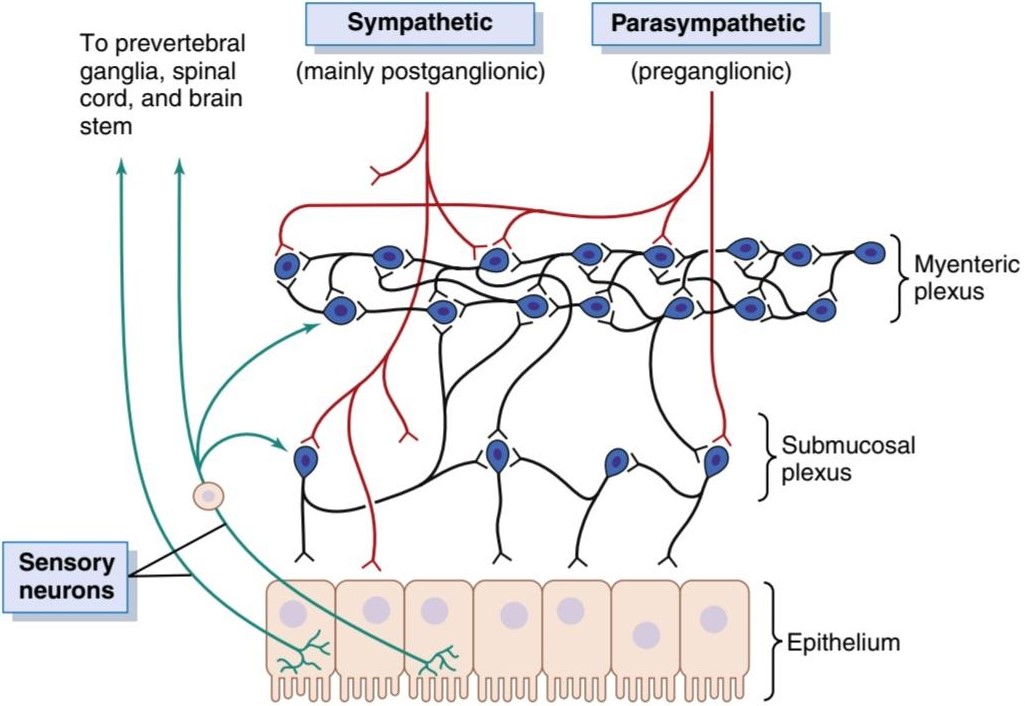
Figure 63-4. Neural control of the gut wall, showing the following: (1) the myenteric and submucosal plexuses (black fibers); (2) extrinsic control of these plexuses by the sympathetic and parasympathetic nervous systems (red fibers); and (3) sensory fibers passing from the luminal epithelium and gut wall to the enteric plexuses, then to the prevertebral ganglia of the spinal cord and directly to the spinal cord and brain stem (green fibers).
شکل 63-4. کنترل عصبی دیواره روده، که موارد زیر را نشان میدهد: (1) شبکههای میرودی و زیر مخاطی (الیاف سیاه). (2) کنترل بیرونی این پیچیدگیها توسط سیستمهای عصبی سمپاتیک و پاراسمپاتیک (الیاف قرمز). و (3) فیبرهای حسی که از اپیتلیوم مجرا و دیواره روده به شبکههای روده، سپس به گانگلیونهای پیش مهره ای نخاع و مستقیماً به نخاع و ساقه مغز (الیاف سبز) عبور میکنند.
DIFFERENCES BETWEEN THE MYENTERIC AND SUBMUCOSAL PLEXUSES
The myenteric plexus consists mostly of a linear chain of many interconnecting neurons that extends the entire length of the gastrointestinal tract. A section of this chain is shown in Figure 63-4.
تفاوت بین شبکههای عضلانی و زیر مخاطی
شبکه میانتریک بیشتر از یک زنجیره خطی از بسیاری از نورونهای متصل به هم تشکیل شده است که تمام طول دستگاه گوارش را گسترش میدهد. بخشی از این زنجیره در شکل 63-4 نشان داده شده است.
Because the myenteric plexus extends all the way along the intestinal wall and lies between the longitudinal and circular layers of intestinal smooth muscle, it is concerned mainly with controlling muscle activity along the length of the gut. When this plexus is stimulated, its principal effects are as follows: (1) increased tonic contraction, or “tone,” of the gut wall; (2) increased intensity of the rhythmic contractions; (3) slightly increased rate of the rhythm of contraction; and (4) increased velocity of conduction of excitatory waves along the gut wall, causing more rapid movement of the gut peristaltic waves.
از آنجایی که شبکه میانتریک در تمام طول دیواره روده امتداد دارد و بین لایههای طولی و دایرهای عضله صاف روده قرار دارد، عمدتاً به کنترل فعالیت عضلانی در طول روده میپردازد. هنگامیکه این شبکه تحریک میشود، اثرات اصلی آن به شرح زیر است: (1) افزایش انقباض تونیک، یا “تن” دیواره روده. (2) افزایش شدت انقباضات ریتمیک. (3) سرعت کمیافزایش ریتم انقباض. و (4) افزایش سرعت هدایت امواج تحریکی در امتداد دیواره روده، باعث حرکت سریعتر امواج پریستالتیک روده میشود.
The myenteric plexus should not be considered entirely excitatory because some of its neurons are inhibitory; their fiber endings secrete an inhibitory transmitter, possibly vasoactive intestinal polypeptide or some other inhibitory peptide. The resulting inhibitory signals are especially useful for inhibiting some of the intestinal sphincter muscles that impede movement of food along successive segments of the gastrointestinal tract, such as the pyloric sphincter, which controls emptying of the stomach into the duodenum, and the sphincter of the ileocecal valve, which controls emptying from the small intestine into the cecum.
شبکه میانتریک نباید کاملاً تحریک کننده در نظر گرفته شود زیرا برخی از نورونهای آن مهار کننده هستند. انتهای فیبر آنها یک فرستنده بازدارنده، احتمالاً پلی پپتید روده ای فعال عروقی یا برخی پپتیدهای بازدارنده دیگر ترشح میکند. سیگنالهای بازدارنده بهویژه برای مهار برخی از ماهیچههای اسفنکتر روده که مانع حرکت غذا در امتداد بخشهای متوالی دستگاه گوارش میشوند، مانند اسفنکتر پیلور، که تخلیه معده به دوازدهه را کنترل میکند، و اسفنکتر اسفنکتر که دریچه ایلئوسکوم کوچک از دریچه مغزی تست به داخل تخلیه میشود، مفید است.
The submucosal plexus, in contrast to the myenteric plexus, is mainly concerned with controlling function in the inner wall of each minute segment of the intestine. For example, many sensory signals originate from the gastro- intestinal epithelium and are then integrated in the submucosal plexus to help control local intestinal secretion, local absorption, and local contraction of the submucosal muscle that causes various degrees of infolding of the gastrointestinal mucosa.
شبکه زیر مخاطی، برخلاف شبکه میانتریک، عمدتاً به کنترل عملکرد دیواره داخلی هر بخش کوچک روده میپردازد. برای مثال، بسیاری از سیگنالهای حسی از اپیتلیوم گوارشی منشأ میگیرند و سپس در شبکه زیر مخاطی ادغام میشوند تا به کنترل ترشح رودهای موضعی، جذب موضعی و انقباض موضعی عضله زیر مخاطی کمک کنند که باعث درجات مختلف تاخوردگی مخاط دستگاه گوارش میشود.
TYPES OF NEUROTRANSMITTERS SECRETED BY ENTERIC NEURONS
Researchers have identified more than 25 potential neurotransmitter substances that are released by the nerve endings of different types of enteric neurons, including the following: (1) acetylcholine, (2) norepinephrine, (3) adenosine triphosphate, (4) serotonin, (5) dopamine, (6) cholecystokinin, (7) substance P, (8) vasoactive intestinal polypeptide, (9) somatostatin, (10) leu-enkephalin, (11) met-enkephalin, (12) bombesin, (13) neuropeptide Y, and (14) nitric oxide. The specific functions of many of these substances are not known well enough to justify discussion here other than to point out the following characteristics.
انواع انتقال دهندههای عصبی که توسط نورونهای روده ای ترشح میشوند
محققان بیش از 25 ماده انتقالدهنده عصبی بالقوه را شناسایی کردهاند که توسط پایانههای عصبی انواع مختلف نورونهای روده آزاد میشوند، از جمله موارد زیر: (1) استیل کولین، (2) نوراپی نفرین، (3) آدنوزین تری فسفات، (4) سروتونین، (5) دوپامین، (6) (7) کوله سیستوکین، (7) کوله سیستوکین پلی پپتید، (9) سوماتوستاتین، (10) لو-انکفالین، (11) مت-انکفالین، (12) بمبسین، (13) نوروپپتید Y، و (14) اکسید نیتریک. کارکردهای خاص بسیاری از این مواد به اندازه کافی برای توجیه بحث در اینجا به جز اشاره به ویژگیهای زیر شناخته نشده است.
Acetylcholine most often excites gastrointestinal activity. Norepinephrine almost always inhibits gastrointestinal activity, as does epinephrine, which reaches the gastro- intestinal tract mainly via the blood after it is secreted by the adrenal medullae into the circulation. The other aforementioned transmitter substances are a mixture of excitatory and inhibitory agents, some of which we will discuss in Chapter 64.
استیل کولین اغلب فعالیت دستگاه گوارش را تحریک میکند. نوراپی نفرین تقریباً همیشه فعالیت دستگاه گوارش را مهار میکند، همانطور که اپی نفرین که عمدتاً از طریق خون پس از ترشح توسط بصل الکلیف آدرنال در گردش خون به دستگاه گوارش میرسد. سایر مواد فرستنده فوق مخلوطی از عوامل تحریک کننده و بازدارنده هستند که در فصل 64 به برخی از آنها خواهیم پرداخت.
AUTONOMIC CONTROL OF THE GASTROINTESTINAL TRACT
Parasympathetic Stimulation Increases Activity of the Enteric Nervous System. The parasympathetic sup- ply to the gut is divided into cranial and sacral divisions, which were discussed in Chapter 61.
کنترل خودکار دستگاه گوارش
تحریک پاراسمپاتیک فعالیت سیستم عصبی روده را افزایش میدهد. تغذیه پاراسمپاتیک به روده به دو بخش جمجمه و خاجی تقسیم میشود که در فصل 61 مورد بحث قرار گرفت.
Except for a few parasympathetic fibers to the mouth and pharyngeal regions of the alimentary tract, the cranial parasympathetic nerve fibers are almost entirely in the vagus nerves. These fibers provide extensive innervation to the esophagus, stomach, and pancreas and somewhat less to the intestines down through the first half of the large intestine.
به جز تعداد کمیاز فیبرهای پاراسمپاتیک در ناحیه دهان و حلق دستگاه گوارش، رشتههای عصبی پاراسمپاتیک جمجمه تقریباً به طور کامل در اعصاب واگ قرار دارند. این فیبرها عصب دهی گسترده ای به مری، معده و لوزالمعده و تا حدودی کمتر به رودهها در نیمه اول روده بزرگ میدهند.
The sacral parasympathetics originate in the second, third, and fourth sacral segments of the spinal cord and pass through the pelvic nerves to the distal half of the large intestine and all the way to the anus. The sigmoidal, rectal, and anal regions are considerably better supplied with parasympathetic fibers than are the other intestinal areas. These fibers function especially to execute the defecation reflexes, discussed in Chapter 64.
پاراسمپاتیک خاجی از بخشهای خاجی دوم، سوم و چهارم طناب نخاعی منشأ میگیرد و از طریق اعصاب لگن به نیمه دیستال روده بزرگ و تمام مسیر به مقعد میرسد. نواحی سیگموئیدی، رکتوم و مقعدی نسبت به سایر نواحی روده ای به میزان قابل توجهی بهتر از فیبرهای پاراسمپاتیک تامین میشوند. این الیاف به ویژه برای اجرای رفلکسهای دفع مدفوع، که در فصل 64 مورد بحث قرار گرفت، عمل میکنند.
The postganglionic neurons of the gastrointestinal parasympathetic system are located mainly in the myenteric and submucosal plexuses. Stimulation of these parasympathetic nerves generally increases activity of the entire enteric nervous system, which in turn enhances activity of most gastrointestinal functions.
نورونهای پس گانگلیونی سیستم پاراسمپاتیک گوارشی عمدتاً در شبکههای میانتریک و زیر مخاطی قرار دارند. تحریک این اعصاب پاراسمپاتیک عموماً باعث افزایش فعالیت کل سیستم عصبی روده میشود که به نوبه خود فعالیت اکثر عملکردهای دستگاه گوارش را افزایش میدهد.
Sympathetic Stimulation Usually Inhibits Gastrointestinal Tract Activity. The sympathetic fibers to the gastrointestinal tract originate in the spinal cord between segments T5 and L2. Most of the preganglionic fibers that innervate the gut, after leaving the cord, enter the sympathetic chains that lie lateral to the spinal column, and many of these fibers then pass on through the chains to outlying ganglia such as to the celiac ganglion and various mesenteric ganglia. Most of the postganglionic sympathetic neuron bodies are in these ganglia, and postganglionic fibers then spread through postganglionic sympathetic nerves to all parts of the gut. The sympathetics innervate essentially all of the gastrointestinal tract, rather than being more extensive nearest the oral cavity and anus, as is true of the parasympathetics. The sympathetic nerve endings secrete mainly norepinephrine.
تحریک سمپاتیک معمولاً فعالیت دستگاه گوارش را مهار میکند. فیبرهای سمپاتیک به دستگاه گوارش از نخاع بین بخشهای T5 و L2 منشا میگیرند. بیشتر فیبرهای پیش گانگلیونی که روده را عصب دهی میکنند، پس از خروج از طناب، وارد زنجیرههای سمپاتیکی میشوند که در کنار ستون فقرات قرار دارند و بسیاری از این رشتهها سپس از طریق زنجیرهها به گانگلیونهای بیرونی مانند گانگلیون سلیاک و عقدههای مختلف مزانتریک منتقل میشوند. اکثر اجسام عصبی سمپاتیک پس گانگلیونی در این گانگلیونها قرار دارند و رشتههای پس گانگلیونی سپس از طریق اعصاب سمپاتیک پس گانگلیونی به تمام قسمتهای روده پخش میشوند. سمپاتیکها اساساً تمام دستگاه گوارش را عصب دهی میکنند، به جای اینکه وسعت بیشتری به حفره دهان و مقعد داشته باشند، همانطور که در مورد پاراسمپاتیکها صادق است. انتهای عصب سمپاتیک عمدتا نوراپی نفرین ترشح میکند.
In general, stimulation of the sympathetic nervous system inhibits activity of the gastrointestinal tract, causing many effects opposite to those of the parasympathetic system. It exerts its effects in two ways: (1) to a slight extent by direct effect of secreted norepinephrine to inhibit intestinal tract smooth muscle (except the mucosal muscle, which it excites) and (2) to a major extent by an inhibitory effect of norepinephrine on the neurons of the entire enteric nervous system.
به طور کلی، تحریک سیستم عصبی سمپاتیک، فعالیت دستگاه گوارش را مهار میکند و اثرات بسیاری بر خلاف سیستم پاراسمپاتیک ایجاد میکند. اثرات خود را به دو صورت اعمال میکند: (1) تا حدی با اثر مستقیم نوراپی نفرین ترشح شده برای مهار ماهیچه صاف مجرای روده (به جز ماهیچه مخاطی که آن را تحریک میکند) و (2) تا حد زیادی توسط اثر مهاری نوراپی نفرین بر روی نورونهای کل سیستم عصبی روده.
Strong stimulation of the sympathetic system can inhibit motor movements of the gut so greatly that this can literally block movement of food through the gastro- intestinal tract.
تحریک قوی سیستم سمپاتیک میتواند حرکات حرکتی روده را به شدت مهار کند که به معنای واقعی کلمه حرکت غذا را از طریق دستگاه گوارش مسدود میکند.
Afferent Sensory Nerve Fibers From the Gut
Many afferent sensory nerve fibers innervate the gut. Some of the nerve fibers have their cell bodies in the enteric nervous system, and some have them in the dorsal root ganglia of the spinal cord. These sensory nerves can be stimulated by (1) irritation of the gut mucosa, (2) excessive gut distention, or (3) the presence of specific chemical substances in the gut. Signals transmitted through the fibers can then cause excitation or, under other conditions, inhibition of intestinal movements or intestinal secretion.
فیبرهای عصبی حسی آوران از روده
بسیاری از رشتههای عصبی حسی آوران روده را عصب دهی میکنند. برخی از رشتههای عصبی بدن سلولی خود را در سیستم عصبی روده ای دارند و برخی در گانگلیونهای ریشه پشتی نخاع قرار دارند. این اعصاب حسی را میتوان با (1) تحریک مخاط روده، (2) اتساع بیش از حد روده، یا (3) وجود مواد شیمیایی خاص در روده تحریک کرد. سیگنالهای منتقل شده از طریق فیبرها میتوانند باعث تحریک یا تحت شرایط دیگر، مهار حرکات روده یا ترشح روده شوند.
In addition, other sensory signals from the gut go all the way to multiple areas of the spinal cord and even to the brain stem. For example, 80% of the nerve fibers in the vagus nerves are afferent rather than efferent. These afferent fibers transmit sensory signals from the gastrointestinal tract into the brain medulla which, in turn, initiates vagal reflex signals that return to the gastrointestinal tract to control many of its functions.
علاوه بر این، سیگنالهای حسی دیگر از روده به نواحی متعددی از نخاع و حتی به ساقه مغز میرسد. به عنوان مثال، 80 درصد از رشتههای عصبی در اعصاب واگ، آوران هستند تا وابران. این فیبرهای آوران سیگنالهای حسی را از دستگاه گوارش به بصل النخاع مغز منتقل میکند که به نوبه خود سیگنالهای رفلکس واگ را آغاز میکند که برای کنترل بسیاری از عملکردهای دستگاه گوارش به دستگاه گوارش باز میگردد.
Gastrointestinal Reflexes
The anatomical arrangement of the enteric nervous system and its connections with the sympathetic and parasympathetic systems support three types of gastrointestinal reflexes that are essential to gastrointestinal control.
رفلکسهای گوارشی
آرایش آناتومیکی سیستم عصبی روده و اتصالات آن با سیستمهای سمپاتیک و پاراسمپاتیک از سه نوع رفلکس گوارشی پشتیبانی میکند که برای کنترل دستگاه گوارش ضروری هستند.
1. Reflexes that are integrated entirely within the gut wall enteric nervous system. These reflexes include, for example, those that control much gastrointestinal secretion, peristalsis, mixing contractions, local inhibitory effects, and so forth.
1. رفلکسهایی که به طور کامل در سیستم عصبی روده دیواره روده یکپارچه شده اند. برای مثال، این رفلکسها شامل آنهایی هستند که ترشحات گوارشی، پریستالسیس، انقباضات مخلوط، اثرات مهاری موضعی و غیره را کنترل میکنند.
2. Reflexes from the gut to the prevertebral sympathetic ganglia and then back to the gastrointestinal tract. These reflexes transmit signals long distances to other areas of the gastrointestinal tract, such as signals from the stomach to cause evacuation of the colon (the gastrocolic reflex), signals from the colon and small intestine to inhibit stomach motility and stomach secretion (the enterogastric reflexes), and reflexes from the colon to inhibit emptying of ileal contents into the colon (the colonoileal reflex).
2. بازتاب از روده به گانگلیون سمپاتیک پیش مهره ای و سپس بازگشت به دستگاه گوارش. این رفلکسها سیگنالهایی را در فواصل طولانی به سایر قسمتهای دستگاه گوارش منتقل میکنند، مانند سیگنالهایی از معده برای تخلیه روده بزرگ (رفلکس گوارشی)، سیگنالهایی از روده بزرگ و روده کوچک برای مهار تحرک معده و ترشح معده (رفلکسهای رودهمعدی)، و رفلکسهایی از معده (محتوی خالی شدن کولون به داخل). رفلکس کولونئیال).
3. Reflexes from the gut to the spinal cord or brain stem and then back to the gastrointestinal tract. These reflexes include especially the following: (1) reflexes from the stomach and duodenum to the brain stem and back to the stomach-via the vagus nerves- to control gastric motor and secretory activity; (2) pain reflexes that cause general inhibition of the entire gastrointestinal tract; and (3) defecation reflexes that travel from the colon and rectum to the spinal cord and back again to produce the powerful colonic, rectal, and abdominal contractions required for defecation (the defecation reflexes).
3. بازتاب از روده به نخاع یا ساقه مغز و سپس بازگشت به دستگاه گوارش. این انعکاسها به ویژه شامل موارد زیر است: (1) رفلکس از معده و دوازدهه به ساقه مغز و برگشت به معده – از طریق اعصاب واگ – برای کنترل فعالیت حرکتی و ترشحی معده. (2) رفلکسهای درد که باعث مهار کلی کل دستگاه گوارش میشود. و (3) رفلکسهای مدفوع که از کولون و رکتوم به نخاع میروند و دوباره برمیگردند تا انقباضات قوی کولون، رکتوم و شکمیرا ایجاد کنند که برای اجابت مزاج لازم است (رفلکسهای مدفوع).
HORMONAL CONTROL OF GASTROINTESTINAL MOTILITY
The gastrointestinal hormones are released into the portal circulation and exert physiological actions on target cells with specific receptors for the hormone. The effects of the hormones persist even after all nervous connections between the site of release and the site of action have been severed. Table 63-1 outlines the actions of each gastrointestinal hormone, as well as the stimuli for secretion and sites at which secretion takes place.
کنترل هورمونی حرکت دستگاه گوارش
هورمونهای گوارشی در گردش خون پورتال آزاد میشوند و بر روی سلولهای هدف با گیرندههای خاص هورمون اعمال فیزیولوژیکی انجام میدهند. اثرات هورمونها حتی پس از قطع شدن تمام ارتباطات عصبی بین محل انتشار و محل عمل ادامه مییابد. جدول 63-1 به تشریح اعمال هر هورمون گوارشی و همچنین محرکهای ترشح و مکانهایی که ترشح در آنها انجام میشود، میپردازد.
In Chapter 65, we discuss the extreme importance of several hormones for controlling gastrointestinal secretion. Most of these same hormones also affect motility in some parts of the gastrointestinal tract. Although the motility effects are usually less important than the secretory effects of the hormones, some of the more important motility effects are described in the following paragraphs.
در فصل 65 به اهمیت فوق العاده چندین هورمون برای کنترل ترشح دستگاه گوارش میپردازیم. بسیاری از همین هورمونها بر تحرک در برخی از قسمتهای دستگاه گوارش نیز تأثیر میگذارند. اگرچه اثرات حرکتی معمولاً کمتر از اثرات ترشحی هورمونها اهمیت دارند، برخی از اثرات حرکتی مهم تر در پاراگرافهای زیر توضیح داده شده اند.
Gastrin is secreted by the “G” cells of the antrum of the stomach in response to stimuli associated with ingestion of a meal, such as distention of the stomach, the products of proteins, and gastrin-releasing peptide, which is released by the nerves of the gastric mucosa during vagal stimulation. The primary actions of gastrin are (1) stimulation of gastric acid secretion and (2) stimulation of growth of the gastric mucosa.
گاسترین توسط سلولهای “G” آنتروم معده در پاسخ به محرکهای مرتبط با خوردن یک وعده غذایی مانند اتساع معده، محصولات پروتئینها و پپتید آزاد کننده گاسترین ترشح میشود که توسط اعصاب مخاط معده در طی تحریک واگ آزاد میشود. اقدامات اولیه گاسترین (1) تحریک ترشح اسید معده و (2) تحریک رشد مخاط معده است.
Cholecystokinin (CCK) is secreted by “I” cells in the mucosa of the duodenum and jejunum mainly in response to digestive products of fat, fatty acids, and monoglycerides in the intestinal contents. This hormone strongly contracts the gallbladder, expelling bile into the small intestine, where the bile, in turn, plays important roles in emulsifying fatty substances and allowing them to be digested and absorbed. CCK also inhibits stomach con- traction moderately. Therefore, at the same time that this hormone causes emptying of the gallbladder, it also slows the emptying of food from the stomach to give adequate time for digestion of the fats in the upper intestinal tract.
کوله سیستوکینین (CCK) توسط سلولهای “I” در مخاط دوازدهه و ژژنوم عمدتاً در پاسخ به محصولات گوارشی چربی، اسیدهای چرب و مونوگلیسرید در محتویات روده ترشح میشود. این هورمون به شدت کیسه صفرا را منقبض میکند و صفرا را به روده کوچک دفع میکند، جایی که صفرا به نوبه خود نقش مهمیدر امولسیون کردن مواد چرب ایفا میکند و به آنها اجازه هضم و جذب را میدهد. CCK همچنین انقباض معده را به طور متوسط مهار میکند. بنابراین، همزمان که این هورمون باعث تخلیه کیسه صفرا میشود، تخلیه غذا از معده را نیز کند میکند تا زمان کافی برای هضم چربیهای قسمت فوقانی روده فراهم شود.
CCK also inhibits appetite to prevent overeating during meals by stimulating sensory afferent nerve fibers in the duodenum; these fibers, in turn, send signals by way of the vagus nerve to inhibit feeding centers in the brain as discussed in Chapter 72.
CCK همچنین با تحریک رشتههای عصبی آوران حسی در دوازدهه اشتها را برای جلوگیری از پرخوری در طول وعدههای غذایی مهار میکند. این فیبرها به نوبه خود سیگنالهایی را از طریق عصب واگ ارسال میکنند تا مراکز تغذیه در مغز را مهار کنند، همانطور که در فصل 72 بحث شد.
Secretin, the first gastrointestinal hormone discovered, is secreted by the “S” cells in the mucosa of the duodenum in response to acidic gastric juice emptying into the duodenum from the pylorus of the stomach. Secretin has a mild effect on motility of the gastrointestinal tract and acts to promote pancreatic secretion of bicarbonate, which in turn helps to neutralize acid in the small intestine.
سکرتین، اولین هورمون گوارشی کشف شده، توسط سلولهای “S” در مخاط دوازدهه در پاسخ به تخلیه اسیدی شیره معده به دوازدهه از پیلور معده ترشح میشود. سکرتین اثر ملایمیبر تحرک دستگاه گوارش دارد و باعث افزایش ترشح بی کربنات پانکراس میشود که به نوبه خود به خنثی کردن اسید در روده کوچک کمک میکند.
Glucose-dependent insulinotropic peptide (also called gastric inhibitory peptide [GIP]) is secreted by the mucosa of the upper small intestine, mainly in response to fatty acids and amino acids but to a lesser extent in response to carbohydrate. It has a mild effect in decreasing motor activity of the stomach and therefore slows emptying of gastric contents into the duodenum when the upper small intestine is already overloaded with food products. Glucose-dependent insulinotropic peptide, at blood levels even lower than those needed to inhibit gastric motility, also stimulates insulin secretion.
پپتید انسولینوتروپیک وابسته به گلوکز (که به آن پپتید بازدارنده معده [GIP] نیز گفته میشود) توسط مخاط قسمت فوقانی روده کوچک ترشح میشود، عمدتاً در پاسخ به اسیدهای چرب و اسیدهای آمینه اما به میزان کمتر در پاسخ به کربوهیدرات. این اثر ملایمیدر کاهش فعالیت حرکتی معده دارد و بنابراین تخلیه محتویات معده به دوازدهه را زمانی که قسمت فوقانی روده کوچک از قبل مملو از محصولات غذایی است کند میکند. پپتید انسولینوتروپیک وابسته به گلوکز، در سطوح خونی حتی کمتر از سطوح مورد نیاز برای مهار تحرک معده، ترشح انسولین را نیز تحریک میکند.
Motilin is secreted by the stomach and upper duodenum during fasting, and the only known function of this hormone is to increase gastrointestinal motility. Motilin is released cyclically and stimulates waves of gastrointestinal motility called interdigestive myoelectric complexes that move through the stomach and small intestine every90 minutes in a person who has fasted. Motilin secretion is inhibited after ingestion of food by mechanisms that are not fully understood.
موتیلین در هنگام روزه داری از معده و دوازدهه فوقانی ترشح میشود و تنها عملکرد شناخته شده این هورمون افزایش تحرک دستگاه گوارش است. موتیلین به صورت چرخهای آزاد میشود و امواج حرکتی دستگاه گوارش به نام کمپلکسهای میوالکتریک میانهضمیرا تحریک میکند که هر ۹۰ دقیقه یکبار در معده و روده کوچک در فردی که روزهدار است حرکت میکند. ترشح موتیلین پس از خوردن غذا توسط مکانیسمهایی که به طور کامل شناخته نشده اند، مهار میشود.
Table 63-1 Gastrointestinal Hormone Actions, Stimuli for Secretion, and Site of Secretion
جدول 63-1 فعالیتهای هورمونی دستگاه گوارش، محرکهای ترشح و محل ترشح

FUNCTIONAL MOVEMENTS IN THE GASTROINTESTINAL TRACT
Two types of movements occur in the gastrointestinal tract: (1) propulsive movements, which cause food to move forward along the tract at an appropriate rate to accommodate digestion and absorption, and (2) mixing movements, which keep the intestinal contents thoroughly mixed at all times.
حرکات عملکردی در دستگاه گوارش
دو نوع حرکت در دستگاه گوارش رخ میدهد: (1) حرکات پیشرانه، که باعث میشود غذا با سرعت مناسبی در امتداد دستگاه به سمت جلو حرکت کند تا هضم و جذب شود، و (2) حرکات اختلاط، که محتویات روده را همیشه به طور کامل مخلوط نگه میدارد.
PROPULSIVE MOVEMENTS-PERISTALSIS
The basic propulsive movement of the gastrointestinal tract is peristalsis, which is illustrated in Figure 63-5A. A contractile ring appears around the gut and then moves forward; this mechanism is analogous to putting one’s fingers around a thin distended tube, then constricting the fingers and sliding them forward along the tube. Any material in front of the contractile ring is moved forward.
حرکات محرکه- پریستالسیس
حرکت اصلی پیشرانه دستگاه گوارش پریستالسیس است که در شکل 63-5A نشان داده شده است. یک حلقه انقباضی در اطراف روده ظاهر میشود و سپس به سمت جلو حرکت میکند. این مکانیسم مشابه قرار دادن انگشتان در اطراف یک لوله نازک متسع، سپس منقبض کردن انگشتان و لغزش آنها به سمت جلو در طول لوله است. هر ماده ای در جلوی حلقه انقباضی به جلو حرکت میکند.
Peristalsis is an inherent property of many syncytial smooth muscle tubes; stimulation at any point in the gut can cause a contractile ring to appear in the circular muscle, and this ring then spreads along the gut tube. (Peristalsis also occurs in the bile ducts, glandular ducts, ureters, and many other smooth muscle tubes of the body.)
پریستالسیس یک ویژگی ذاتی بسیاری از لولههای عضله صاف سنسیشیال است. تحریک در هر نقطه از روده میتواند باعث ایجاد یک حلقه انقباضی در عضله حلقوی شود و سپس این حلقه در طول لوله روده پخش میشود. (پریستالسیس همچنین در مجاری صفراوی، مجاری غده ای، حالب و بسیاری دیگر از لولههای ماهیچه صاف بدن رخ میدهد.)
The usual stimulus for intestinal peristalsis is distention of the gut. That is, if a large amount of food collects at any point in the gut, stretching of the gut wall stimulates the enteric nervous system to contract the gut wall 2 to 3 centimeters behind this point, and a contractile ring appears that initiates a peristaltic movement. Other stimuli that can initiate peristalsis include chemical or physical irritation of the epithelial lining in the gut. Also, strong para- sympathetic nervous signals to the gut will elicit strong peristalsis.
محرک معمول برای پریستالیس روده، اتساع روده است. یعنی اگر مقدار زیادی غذا در هر نقطه از روده جمع شود، کشیده شدن دیواره روده سیستم عصبی روده را تحریک میکند تا دیواره روده 2 تا 3 سانتی متر پشت این نقطه منقبض شود و یک حلقه انقباضی ظاهر میشود که شروع کننده یک حرکت پریستالتیک است. سایر محرکهایی که میتوانند باعث شروع پریستالتیک شوند عبارتند از تحریک شیمیایی یا فیزیکی پوشش اپیتلیال در روده. همچنین، سیگنالهای عصبی پاراسمپاتیک قوی به روده باعث ایجاد پریستالسیس قوی میشود.
Function of the Myenteric Plexus in Peristalsis. Peristalsis occurs only weakly or not at all in any portion of the gastrointestinal tract that has congenital absence of the myenteric plexus. Also, it is greatly depressed or completely blocked in the entire gut when a person is treated with atropine to paralyze the cholinergic nerve endings of the myenteric plexus. Therefore, effectual peristalsis requires an active myenteric plexus.
عملکرد شبکه میانتریک در پریستالسیس. پریستالسیس فقط در هر بخشی از دستگاه گوارش که دارای غیاب مادرزادی شبکه میانتریک است، به طور ضعیف یا اصلاً رخ نمیدهد. همچنین، هنگامیکه فرد تحت درمان با آتروپین قرار میگیرد تا پایانههای عصبی کولینرژیک شبکه میانتریک را فلج کند، به شدت در کل روده مسدود میشود. بنابراین، پریستالسیس مؤثر به یک شبکه میانتریک فعال نیاز دارد.
Peristaltic Waves Move Toward the Anus With Down- stream Receptive Relaxation-“Law of the Gut.” Peristalsis, theoretically, can occur in either direction from a stimulated point, but it normally dies out rapidly in the orad (toward the mouth) direction while continuing for a considerable distance toward the anus. The exact cause of this directional transmission of peristalsis is uncertain, although it probably results mainly from the fact that the myenteric plexus is “polarized” in the anal direction, which can be explained as follows.
امواج پریستالتیک به سمت مقعد حرکت میکنند با آرامش پذیرنده پایین جریان – “قانون روده”. Peristalsis، از نظر تئوری، میتواند در هر جهت از یک نقطه تحریک شده رخ دهد، اما به طور معمول به سرعت در جهت orad (به سمت دهان) از بین میرود در حالی که برای فاصله قابل توجهی به سمت مقعد ادامه مییابد. علت دقیق این انتقال جهت دار پریستالسیس نامشخص است، اگرچه احتمالاً عمدتاً ناشی از این واقعیت است که شبکه میانتریک در جهت مقعدی “قطبی” شده است که میتوان آن را به شرح زیر توضیح داد.
When a segment of the intestinal tract is excited by distention and thereby initiates peristalsis, the contractile ring causing the peristalsis normally begins on the orad side of the distended segment and moves toward the distended segment, pushing the intestinal contents in the anal direction for 5 to 10 centimeters before dying out (Video 63-1). At the same time, the gut sometimes relaxes several centimeters downstream toward the anus, which is called “receptive relaxation,” thus allowing the food to be propelled more easily toward the anus than toward the mouth.
هنگامیکه بخشی از دستگاه روده توسط اتساع برانگیخته میشود و در نتیجه باعث شروع پریستالسیس میشود، حلقه انقباضی که باعث پریستالسیس میشود، معمولاً از سمت اراد بخش متسع شده شروع میشود و به سمت بخش منبسط شده حرکت میکند و محتویات روده را به مدت 5 تا 10 سانتی متر به سمت مقعد فشار میدهد (Video 63-1). در همان زمان، روده گاهی اوقات چندین سانتی متر در پایین دست به سمت مقعد شل میشود که به آن “آرامش پذیری” میگویند، بنابراین به غذا اجازه میدهد راحت تر به سمت مقعد حرکت کند تا به سمت دهان.
This complex pattern does not occur in the absence of the myenteric plexus. Therefore, the complex is called the myenteric reflex or the peristaltic reflex. The peristaltic reflex plus the anal direction of movement of the peristalsis is called the “law of the gut.”
این الگوی پیچیده در غیاب شبکه میانتریک رخ نمیدهد. بنابراین، کمپلکس را رفلکس میانتریک یا رفلکس پریستالتیک مینامند. رفلکس پریستالتیک به علاوه جهت مقعدی حرکت پریستالسیس “قانون روده” نامیده میشود.
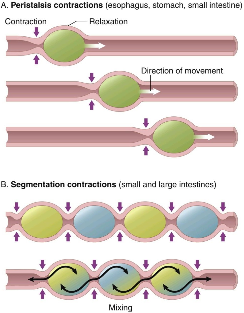
Figure 63-5. Peristalsis involves contraction and downstream relaxation in the esophagus, stomach and small intestine propel the contents toward the anus. Intermittent segmentation contractions in separate sections of the small and large intestines mix their contents with little forward movement.
شکل 63-5. پریستالسیس شامل انقباض و شل شدن پایین دست در مری، معده و روده کوچک است که محتویات را به سمت مقعد میراند. انقباضات بخشبندی متناوب در بخشهای مجزای روده کوچک و بزرگ، محتویات آنها را با حرکت کمیرو به جلو مخلوط میکنند.
SEGMENTATION CONTRACTIONS- MIXING MOVEMENTS
Mixing movements differ in different parts of the alimentary tract. In some areas, the peristaltic contractions cause most of the mixing. This is especially true when for- ward progression of the intestinal contents is blocked by a sphincter so that a peristaltic wave can then only churn the intestinal contents, rather than propelling them for- ward. At other times, local intermittent segmentation con- tractions occur every few centimeters in the gut wall (see Figure 63-5B). These constrictions usually last only 5 to 30 seconds; new constrictions then occur at other points in the gut, thus “chopping” and “shearing” the contents first here and then there. These peristaltic and constrictive movements are modified in different parts of the gastrointestinal tract for proper propulsion and mixing, as discussed for each portion of the tract in Chapter 64.
انقباضات قطعه بندی – حرکات اختلاط
حرکات اختلاط در قسمتهای مختلف دستگاه گوارش متفاوت است. در برخی مناطق، انقباضات پریستالتیک باعث بیشتر اختلاط میشود. این امر به ویژه زمانی صادق است که پیشروی محتویات روده توسط یک اسفنکتر مسدود شود، به طوری که یک موج پریستالتیک فقط میتواند محتویات روده را به هم بزند، نه اینکه آنها را به جلو سوق دهد. در مواقع دیگر، انقباضات بخشبندی متناوب موضعی هر چند سانتیمتر در دیواره روده رخ میدهد (شکل 63-5B را ببینید). این انقباضات معمولاً فقط 5 تا 30 ثانیه طول میکشد. سپس انقباضات جدید در نقاط دیگر روده رخ میدهد، بنابراین محتویات ابتدا در اینجا و سپس آنجا “خرد” و “برش” میشوند. این حرکات پریستالتیک و انقباض در قسمتهای مختلف دستگاه گوارش برای پیشرانش و اختلاط مناسب، همانطور که برای هر بخش از دستگاه در فصل 64 بحث شد، اصلاح میشوند.
GASTROINTESTINAL BLOOD FLOW- SPLANCHNIC CIRCULATION
The blood vessels of the gastrointestinal system are part of a more extensive system called the splanchnic circulation, shown in Figure 63-6. It includes the blood flow through the gut plus blood flows through the spleen, pancreas, and liver. The design of this system is such that all the blood that courses through the gut, spleen, and pancreas then flows immediately into the liver by way of the portal vein. In the liver, the blood passes through millions of minute liver sinusoids and finally leaves the liver via hepatic veins that empty into the vena cava of the general circulation. This flow of blood through the liver, before it empties into the vena cava, allows the reticuloendothelial cells that line the liver sinusoids to remove bacteria and other particulate matter that might enter the blood from the gastrointestinal tract, thus preventing direct transport of potentially harmful agents into the remainder of the body.
جریان خون دستگاه گوارش – گردش خون SPLANCHNIC
رگهای خونی سیستم گوارشی بخشی از یک سیستم گسترده تر به نام گردش خونی هستند که در شکل 63-6 نشان داده شده است. این شامل جریان خون از طریق روده به علاوه جریان خون در طحال، پانکراس و کبد است. طراحی این سیستم به گونهای است که تمام خونی که از روده، طحال و پانکراس عبور میکند، بلافاصله از طریق سیاهرگ باب وارد کبد میشود. در کبد، خون از میلیونها سینوزوئید کبدی عبور میکند و در نهایت از طریق سیاهرگهای کبدی که وارد ورید اجوف گردش خون عمومیمیشود، کبد را ترک میکند. این جریان خون از طریق کبد، قبل از تخلیه به داخل ورید اجوف، به سلولهای رتیکولواندوتلیال که سینوسوئیدهای کبد را میپوشانند، اجازه میدهد تا باکتریها و سایر ذرات معده را که ممکن است از دستگاه گوارش وارد خون شوند، حذف کنند، بنابراین از انتقال مستقیم عوامل بالقوه مضر به بقیه بدن جلوگیری میکند.
The nonfat, water-soluble nutrients absorbed from the gut (e.g., carbohydrates and proteins) are transported in the portal venous blood to the same liver sinusoids. Here, both the reticuloendothelial cells and the principal parenchymal cells of the liver, the hepatic cells, absorb and store temporarily from 50% to 75% of the nutrients. Also, much chemical intermediary processing of these nutrients occurs in the liver cells. These nutritional functions of the liver are discussed in Chapters 68 through 72. Almost all of the fats absorbed from the intestinal tract are not carried in the portal blood but instead are absorbed into the intestinal lymphatics and then conducted to the systemic circulating blood by way of the thoracic duct, bypassing the liver.
مواد مغذی غیر چربی و محلول در آب جذب شده از روده (مانند کربوهیدراتها و پروتئینها) در خون ورید پورتال به همان سینوسوئیدهای کبدی منتقل میشوند. در اینجا، هم سلولهای رتیکولواندوتلیال و هم سلولهای پارانشیم اصلی کبد، سلولهای کبدی، 50 تا 75 درصد مواد مغذی را جذب و ذخیره میکنند. همچنین، پردازش واسطههای شیمیایی این مواد مغذی در سلولهای کبدی انجام میشود. این عملکردهای تغذیهای کبد در فصلهای 68 تا 72 مورد بحث قرار گرفتهاند. تقریباً تمام چربیهای جذبشده از دستگاه روده در خون باب حمل نمیشوند، بلکه در لنفاوی روده جذب میشوند و سپس از طریق مجرای سینهای و با دور زدن کبد به خون در گردش سیستمیک هدایت میشوند.

Figure 63-6. Splanchnic circulation.
شکل 63-6. گردش خون اسپلانکیک.
ANATOMY OF THE GASTROINTESTINAL BLOOD SUPPLY
Figure 63-7 shows the general features of the arterial blood supply to the gut, including the superior mesenteric and inferior mesenteric arteries supplying the walls of the small and large intestines via an arching arterial system. Not shown in the figure is the celiac artery, which provides a similar blood supply to the stomach.
آناتومیتامین خون دستگاه گوارش
شکل 63-7 ویژگیهای کلی جریان خون شریانی به روده را نشان میدهد، از جمله شریانهای مزانتریک فوقانی و مزانتریک تحتانی که دیوارههای روده کوچک و بزرگ را از طریق یک سیستم شریانی قوس دار تامین میکنند. در شکل نشان داده نشده است شریان سلیاک که خونرسانی مشابهی به معده میدهد.
On entering the wall of the gut, the arteries branch and send smaller arteries circling in both directions around the gut, with the tips of these arteries meeting on the side of the gut wall opposite the mesenteric attachment. From the circling arteries, still much smaller arteries penetrate into the intestinal wall and spread (1) along the muscle bundles, (2) into the intestinal villi, and (3) into submucosal vessels beneath the epithelium to serve the secretory and absorptive functions of the gut.
با ورود به دیواره روده، شریانها منشعب میشوند و شریانهای کوچکتری را میفرستند که در هر دو جهت در اطراف روده میچرخند، در حالی که نوک این شریانها در کنار دیواره روده مقابل اتصال مزانتریک قرار میگیرند. از شریانهای در حال چرخش، شریانهای بسیار کوچکتری به دیواره روده نفوذ میکنند و (1) در امتداد بستههای ماهیچهای، (2) به پرزهای روده، و (3) در عروق زیر مخاطی زیر اپیتلیوم پخش میشوند تا عملکردهای ترشحی و جذبی روده را انجام دهند.
Figure 63-8 shows the special organization of the blood flow through an intestinal villus, including a small arteriole and venule that interconnect with a system of multiple looping capillaries. The walls of the arterioles are highly muscular and highly active in controlling villus blood flow.
شکل 63-8 سازماندهی خاص جریان خون را از طریق پرزهای روده نشان میدهد، شامل یک شریان کوچک و ونول که با سیستمیاز مویرگهای حلقهای متعدد به هم متصل میشوند. دیواره سرخرگها بسیار عضلانی است و در کنترل جریان خون پرزها بسیار فعال است.
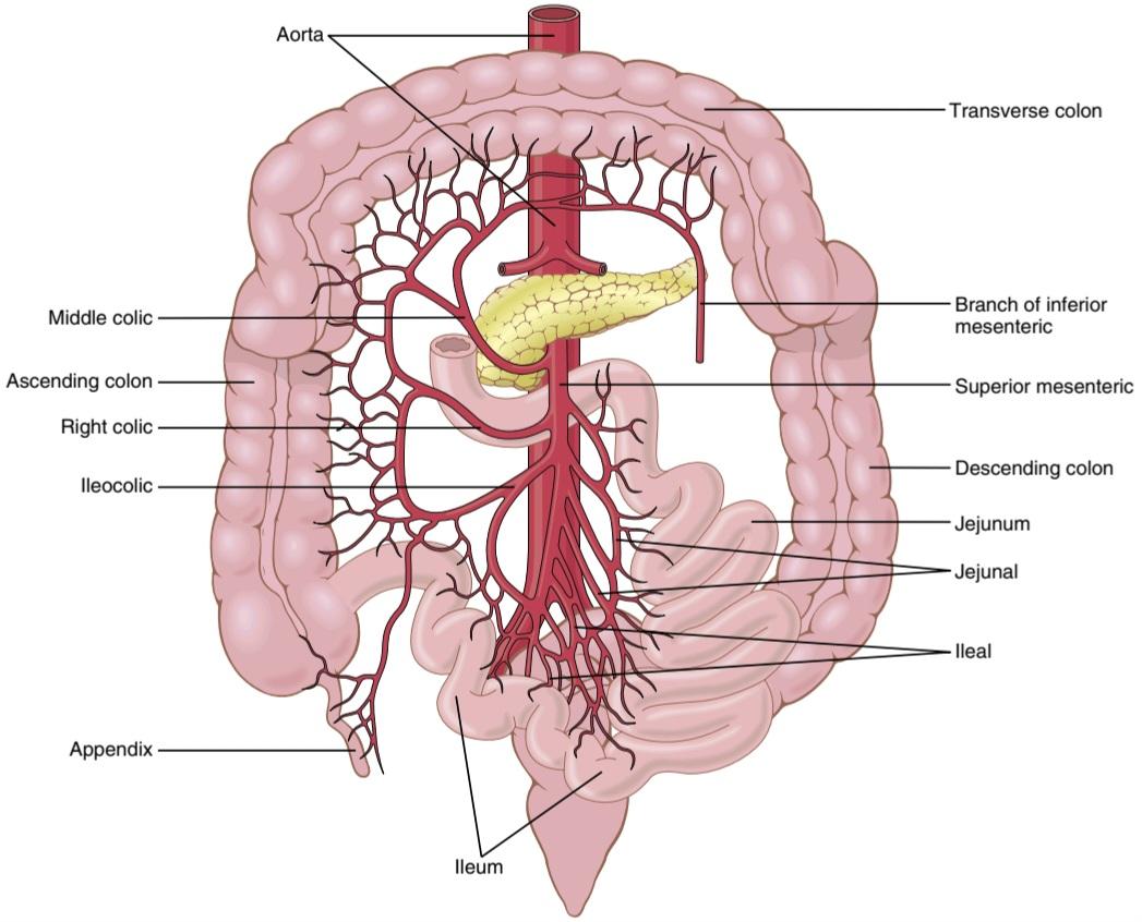
Figure 63-7. Arterial blood supply to the intestines through the mesenteric web.
شکل 63-7. خون شریانی از طریق شبکه مزانتریک به رودهها میرسد.
EFFECT OF GUT ACTIVITY AND METABOLIC FACTORS ON GASTROINTESTINAL BLOOD FLOW
Under normal conditions, the blood flow in each area of the gastrointestinal tract, as well as in each layer of the gut wall, is directly related to the level of local activity. For example, during active absorption of nutrients, blood flow in the villi and adjacent regions of the submucosa increases as much as eightfold. Likewise, blood flow in the muscle layers of the intestinal wall increases with increased motor activity in the gut. After a meal, the motor activity, secretory activity, and absorptive activity all increase; likewise, the blood flow increases greatly but then decreases back to the resting level over another 2 to 4 hours.
تأثیر فعالیت روده و عوامل متابولیک بر جریان خون دستگاه گوارش
در شرایط عادی، جریان خون در هر ناحیه از دستگاه گوارش و همچنین در هر لایه از دیواره روده، ارتباط مستقیمیبا سطح فعالیت موضعی دارد. به عنوان مثال، در طول جذب فعال مواد مغذی، جریان خون در پرزها و مناطق مجاور زیر مخاط تا هشت برابر افزایش مییابد. به همین ترتیب، جریان خون در لایههای عضلانی دیواره روده با افزایش فعالیت حرکتی در روده افزایش مییابد. بعد از غذا، فعالیت حرکتی، فعالیت ترشحی و فعالیت جذبی همگی افزایش مییابد. به همین ترتیب، جریان خون به شدت افزایش مییابد، اما پس از 2 تا 4 ساعت دیگر به سطح استراحت کاهش مییابد.
Mechanisms of Increased Blood Flow During Gastro- intestinal Activity. Although the precise causes of the increased blood flow during increased gastrointestinal activity are still unclear, some facts are known.
مکانیسمهای افزایش جریان خون در طول فعالیت دستگاه گوارش. اگرچه دلایل دقیق افزایش جریان خون در طول افزایش فعالیت دستگاه گوارش هنوز مشخص نیست، برخی از حقایق شناخته شده است.
First, several vasodilator substances are released from the mucosa of the intestinal tract during the digestive process. Most of these substances are peptide hormones, including cholecystokinin, vasoactive intestinal peptide, gastrin, and secretin. These same hormones control specific motor and secretory activities of the gut, as discussed in Chapters 64 and 65.
ابتدا چندین ماده گشادکننده عروق از مخاط دستگاه روده در طی فرآیند گوارش آزاد میشود. بیشتر این مواد، هورمونهای پپتیدی هستند، از جمله کوله سیستوکینین، پپتید وازواکتیو روده ای، گاسترین و سکرتین. همین هورمونها فعالیتهای حرکتی و ترشحی خاص روده را کنترل میکنند، همانطور که در فصلهای 64 و 65 بحث شد.
Second, some of the gastrointestinal glands also release into the gut wall two kinins, kallidin and bradykinin, at the same time that they secrete other substances into the lumen. These kinins are powerful vasodilators that are believed to cause much of the increased mucosal vasodilation that occurs along with secretion.
دوم، برخی از غدد گوارشی نیز دو کینین، کالیدین و برادی کینین را در دیواره روده آزاد میکنند و همزمان مواد دیگری را به لومن ترشح میکنند. این کینینها گشادکننده عروق قوی هستند که اعتقاد بر این است که باعث افزایش گشاد شدن عروق مخاطی میشوند که همراه با ترشح اتفاق میافتد.
Third, decreased oxygen concentration in the gut wall can increase intestinal blood flow at least 50% to 100%; therefore, the increased mucosal and gut wall metabolic rate during gut activity probably lowers the oxygen concentration enough to cause much of the vasodilation. The decrease in oxygen can also lead to as much as a fourfold increase of adenosine, a well-known vasodilator that could be responsible for much of the increased flow.
سوم، کاهش غلظت اکسیژن در دیواره روده میتواند جریان خون روده را حداقل 50 تا 100 درصد افزایش دهد. بنابراین، افزایش میزان متابولیک مخاط و دیواره روده در طول فعالیت روده احتمالاً غلظت اکسیژن را به اندازه کافی کاهش میدهد که باعث اتساع عروق شود. کاهش اکسیژن همچنین میتواند منجر به افزایش چهار برابری آدنوزین شود، یک گشادکننده عروق شناخته شده که میتواند مسئول بیشتر جریان افزایش یابد.
Thus, the increased blood flow during increased gastrointestinal activity is probably a combination of many of the aforementioned factors plus still others yet undiscovered.
بنابراین، افزایش جریان خون در طول افزایش فعالیت دستگاه گوارش احتمالاً ترکیبی از بسیاری از عوامل فوق الذکر به علاوه عوامل دیگری است که هنوز کشف نشده است.
“Countercurrent” Blood Flow in the Villi. Note in Figure 63-8 that the arterial flow into the villus and the venous flow out of the villus are in directions opposite to each other and that the vessels lie in close apposition to each other. Because of this vascular arrangement, much of the blood oxygen diffuses out of the arterioles directly into the adjacent venules without ever being carried in the blood to the tips of the villi. As much as 80% of the oxygen may take this short-circuit route and is therefore not avail- able for local metabolic functions of the villi. This type of countercurrent mechanism in the villi is analogous to the countercurrent mechanism in the vasa recta of the kidney medulla, which was discussed in Chapter 29.
جریان خون “متقابل” در پرزها. در شکل 63-8 توجه داشته باشید که جریان شریانی به داخل پرز و جریان وریدی خارج از پرز در جهت مخالف یکدیگر هستند و رگها در نزدیکی یکدیگر قرار دارند. به دلیل این آرایش عروقی، بیشتر اکسیژن خون از شریانها مستقیماً به داخل وریدهای مجاور منتشر میشود بدون اینکه هرگز در خون به نوک پرزها منتقل شود. 80 درصد اکسیژن ممکن است این مسیر اتصال کوتاه را طی کند و بنابراین برای عملکردهای متابولیکی موضعی پرزها در دسترس نیست. این نوع مکانیسم جریان متقابل در پرزها مشابه مکانیسم جریان مخالف در مجرای راست روده بصل النخاع کلیه است که در فصل 29 مورد بحث قرار گرفت.
Under normal conditions, this shunting of oxygen from the arterioles to the venules is not harmful to the villi, but in disease conditions in which blood flow to the gut becomes greatly curtailed, such as in circulatory shock, the oxygen deficit in the tips of the villi can become so great that the villus tip or even the whole villus undergoes ischemic death and disintegrates. For this reason and other reasons, in many gastrointestinal diseases the villi become seriously blunted, leading to greatly diminished intestinal absorptive capacity.
در شرایط عادی، این انتقال اکسیژن از شریانها به وریدها برای پرزها مضر نیست، اما در شرایط بیماری که در آن جریان خون به روده بسیار محدود میشود، مانند شوک گردش خون، کمبود اکسیژن در نوک پرزها میتواند آنقدر زیاد شود که نوک پرز یا حتی کل پرز دچار مرگ و میر و از هم گسیختگی میشود. به همین دلیل و دلایل دیگر، در بسیاری از بیماریهای گوارشی، پرزها بهطور جدی تیره میشوند که منجر به کاهش شدید ظرفیت جذب روده میشود.

Figure 63-8. Microvasculature of the villus showing a countercurrent arrangement of blood flow in the arterioles and venules.
شکل 63-8. ریز عروق پرز که ترتیب مخالف جریان خون را در شریانها و ونولها نشان میدهد.
NERVOUS CONTROL OF GASTROINTESTINAL BLOOD FLOW
Stimulation of the parasympathetic nerves going to the stomach and lower colon increases local blood flow at the same time that it increases glandular secretion. This increased flow probably results secondarily from the increased glandular activity, not as a direct effect of the nervous stimulation.
کنترل عصبی جریان خون دستگاه گوارش
تحریک اعصاب پاراسمپاتیک که به سمت معده و روده بزرگ میروند، جریان خون موضعی را افزایش میدهد و همزمان ترشح غدد را افزایش میدهد. این افزایش جریان احتمالاً به طور ثانویه ناشی از افزایش فعالیت غدد است، نه به عنوان اثر مستقیم تحریک عصبی.
Sympathetic stimulation, by contrast, has a direct effect on essentially all the gastrointestinal tract to cause intense vasoconstriction of the arterioles and greatly decreased blood flow. After a few minutes of this vasoconstriction, the flow often returns to near normal by means of a mechanism called “autoregulatory escape.” That is, the local metabolic vasodilator mechanisms that are elicited by ischemia override the sympathetic vasoconstriction, returning toward normal the necessary nutrient blood flow to the gastrointestinal glands and muscle.
در مقابل، تحریک سمپاتیک تأثیر مستقیمیبر روی تمام دستگاه گوارش دارد و باعث انقباض عروق شدید شریانها و کاهش شدید جریان خون میشود. پس از چند دقیقه از این انقباض عروق، جریان اغلب با استفاده از مکانیزمیبه نام «فرار خودتنظیمی» به حالت طبیعی نزدیک میشود. به این معنا که مکانیسمهای گشادکننده عروق متابولیک موضعی که توسط ایسکمیایجاد میشوند، انقباض عروق سمپاتیک را نادیده میگیرند و جریان خون مواد مغذی لازم را به غدد گوارشی و ماهیچهها به حالت عادی برمیگردانند.
Importance of Nervous Depression of Gastrointestinal Blood Flow When Other Parts of the Body Need Extra Blood Flow. A major value of sympathetic vasoconstriction in the gut is that it allows shutoff of gastrointestinal and other splanchnic blood flow for short periods during heavy exercise when the skeletal muscle and heart need increased flow. Also, in circulatory shock, when all the body’s vital tissues are in danger of cellular death for lack of blood flow-especially the brain and the heart- sympathetic stimulation can decrease splanchnic blood flow to very little for many hours.
اهمیت افسردگی عصبی جریان خون دستگاه گوارش هنگامیکه سایر قسمتهای بدن به جریان خون اضافی نیاز دارند. یکی از ارزشهای اصلی انقباض عروق سمپاتیک در روده این است که باعث میشود جریان خون دستگاه گوارش و سایر جریان خون برای دورههای کوتاهی در طی تمرینات سنگین، زمانی که ماهیچههای اسکلتی و قلب نیاز به افزایش جریان دارند، قطع شود. همچنین در شوک گردش خون، زمانی که تمام بافتهای حیاتی بدن به دلیل کمبود جریان خون در خطر مرگ سلولی قرار میگیرند، بهویژه مغز و تحریک سمپاتیک قلب، میتواند جریان خون اسپلانیک را برای ساعات زیادی کاهش دهد.
Sympathetic stimulation also causes strong vasoconstriction of the large-volume intestinal and mesenteric veins. This vasoconstriction decreases the volume of these veins, thereby displacing large amounts of blood into other parts of the circulation. In persons experiencing hemorrhagic shock or other states of low blood volume, this mechanism can provide as much as 200 to 400 ml of extra blood to sustain the general circulation.
تحریک سمپاتیک همچنین باعث انقباض شدید عروقی در وریدهای روده ای و مزانتریک با حجم زیاد میشود. این انقباض عروق حجم این وریدها را کاهش میدهد و در نتیجه مقادیر زیادی خون را به قسمتهای دیگر گردش خون منتقل میکند. در افرادی که شوک هموراژیک یا سایر حالتهای کم حجم خون را تجربه میکنند، این مکانیسم میتواند 200 تا 400 میلیلیتر خون اضافی برای حفظ گردش خون فراهم کند.
کلیک کنید «Bibliography: فهرست کتب مربوطه»
Barth BB, Shen X: Computational motility models of neurogastroenterology and neuromodulation. Brain Res 1693:174, 2018.
Chalazonitis A, Rao M: Enteric nervous system manifestations of neurodegenerative disease. Brain Res 1693:207, 2018.
Furness JB: The enteric nervous system and neurogastroenterology. Nat Rev Gastroenterol Hepatol 9:286, 2012.
Furness JB, Stebbing MJ: The first brain: species comparisons and evolutionary implications for the enteric and central nervous systems. Neurogastroenterol Motil 2018 Feb;30(2). http://doi.org/10.1111/ nmo.13234.
Granger DN, Holm L, Kvietys P: The gastrointestinal circulation: physiology and pathophysiology, Compr Physiol 5:1541, 2015.
Kaelberer MM, Bohórquez DV: The now and then of gut-brain signaling. Brain Res 1693:192, 2018.
Kumral D, Zfass AM: Gut movements: a review of the physiology of gastrointestinal transit. Dig Dis Sci 63:2500, 2018.
Lake JI, Heuckeroth RO: Enteric nervous system development: migration, differentiation, and disease. Am J Physiol Gastrointest Liver Physiol 305:G1, 2013.
Lammers WJ: Inhomogeneities in the propagation of the slow wave in the stomach. Neurogastroenterol Motil 27:1349, 2015.
Liddle RA: Interactions of gut endocrine cells with epithelium and neurons. Compr Physiol 8:1019, 2018.
Rao M, Gershon MD: Enteric nervous system development: what could possibly go wrong? Nat Rev Neurosci 19:552, 2018.
Sanders KM, Ward SM: Nitric oxide and its role as a non-adrenergic, non-cholinergic inhibitory neurotransmitter in the gastrointestinal tract. Br J Pharmacol 176:212, 2019.
Sanders KM, Ward SM, Koh SD: Interstitial cells: regulators of smooth muscle function. Physiol Rev 94:859, 2014.
Schemann M, Frieling T, Enck P: To learn, to remember, to forget- How smart is the gut? Acta Physiol (Oxf) 2019 May 7:e13296. doi: 10.1111/apha.13296.
Vergnolle N, Cirillo C: Neurons and glia in the enteric nervous system and epithelial barrier function. Physiology (Bethesda) 33:269, 2018.
Waise TMZ, Dranse HJ, Lam TKT: The metabolic role of vagal afferent innervation. Nat Rev Gastroenterol Hepatol 15:625, 2018.
🚀 با ما همراه شوید!
تازهترین مطالب و آموزشهای مغز و اعصاب را از دست ندهید. با فالو کردن کانال تلگرام آیندهنگاران مغز، از ما حمایت کنید!



