تکامل سیستم عصبی: مغز میانی، پل مغزی، بصلالنخاع و مخچه


دعای مطالعه [ نمایش ]
بِسْمِ الله الرَّحْمنِ الرَّحیمِ
اَللّهُمَّ اَخْرِجْنى مِنْ ظُلُماتِ الْوَهْمِ
خدايا مرا بيرون آور از تاريكىهاى وهم،
وَ اَكْرِمْنى بِنُورِ الْفَهْمِ
و به نور فهم گرامى ام بدار،
اَللّهُمَّ افْتَحْ عَلَيْنا اَبْوابَ رَحْمَتِكَ
خدايا درهاى رحمتت را به روى ما بگشا،
وَانْشُرْ عَلَيْنا خَزائِنَ عُلُومِكَ بِرَحْمَتِكَ يا اَرْحَمَ الرّاحِمينَ
و خزانههاى علومت را بر ما باز كن به امید رحمتت اى مهربانترين مهربانان.
آیندهنگاران مغز: «ما مغز را میشناسیم، تا آینده را بسازیم.»
کتاب «نوروآناتومی بالینی اسنل» اثر دکتر ریچارد اس. اسنل، با ویرایش جدید دکتر رایان اسپلیتگربر، همچنان بهعنوان مرجع طلایی این حوزه شناخته میشود. این اثر با تلفیق تازهترین پژوهشها و کاربردهای بالینی، پلی استوار میان دانش پایه و عمل پزشکی بنا میکند.
تلاشهای علمی و سالها تدریس ارزشمند استاد گرانقدر آقای دکتر محمدتقی جغتایی الهامبخش ترجمه و گسترش این دانش در ایران بوده است.
ترجمه دقیق این کتاب توسط برند علمی آیندهنگاران مغز به مدیریت داریوش طاهری، امکان دسترسی فارسیزبانان به مرزهای نوین علوم اعصاب را فراهم کرده و رسالتی علمی در جهت شناخت عمیقتر مغز و ترسیم آیندهای روشنتر بر عهده گرفته است.
»» فصل ۱۸: رشد سیستم عصبی؛ بخش مبانی و اصول؛ قسمت دوم
»» Chapter 18: Nervous System Development
BRAIN
Once the neural tube has closed, the three primary vesicles the forebrain vesicle, the midbrain vesicle, and the hindbrain vesicle-complete their development (Fig. 18-2). The forebrain vesicle will become the forebrain (prosencephalon), the midbrain vesicle will become the midbrain (mesencephalon), and the hindbrain vesicle will become the hindbrain (rhombencephalon).
مغز
پس از بسته شدن لوله عصبی، سه وزیکول اولیه – وزیکول مغز قدامی، وزیکول مغز میانی و وزیکول مغز خلفی – رشد خود را تکمیل میکنند (شکل 18-2). وزیکول مغز قدامی به مغز قدامی (پروزنسفال)، وزیکول مغز میانی به مغز میانی (مزنسفالون) و وزیکول مغز خلفی به مغز خلفی (رومبانسفال) تبدیل خواهد شد.
By the fifth week, the forebrain and hindbrain vesicles divide into two secondary vesicles. The forebrain vesicle forms (1) the telencephalon, with its primitive cerebral hemispheres, and (2) the diencephalon, which develops optic vesicles. The hindbrain vesicle forms (1) the metencephalon, the future pons and cerebellum, and (2) the myelencephalon, or medulla oblongata (Table 18-1).
تا هفته پنجم، وزیکولهای مغز قدامی و مغز خلفی به دو وزیکول ثانویه تقسیم میشوند. وزیکول مغز قدامی (1) تلانسفالون را با نیمکرههای مغزی اولیهاش و (2) دیانسفالون را تشکیل میدهد که وزیکولهای بینایی را ایجاد میکند. وزیکول مغز خلفی (1) متناسفالون، پل مغزی و مخچه آینده و (2) میلنسفال یا بصل النخاع را تشکیل میدهد (جدول 18-1).
The basic pattern of the ventricular system is now established. The cavity in each cerebral hemisphere is known as the lateral ventricle. The cavity of the diencephalon is known as the third ventricle. With continued growth, the cavity of the midbrain vesicle becomes small and forms the cerebral aqueduct or aqueduct of Sylvius. The cavity of the hindbrain vesicle forms the fourth ventricle, which is continuous with the central canal of the spinal cord. The lateral ventricles communicate with the third ventricle through the interventricular foramina. The ventricular system and the central canal of the spinal cord are lined with ependyma and are filled with CSF. In the earliest stages, the CSF within the ventricular system is not continuous with that of the subarachnoid space.
الگوی اولیه سیستم بطنی اکنون مشخص شده است. حفره در هر نیمکره مغزی به عنوان بطن جانبی شناخته میشود. حفره دیانسفالون به عنوان بطن سوم شناخته میشود. با ادامه رشد، حفره وزیکول مغز میانی کوچک میشود و قنات مغزی یا قنات سیلویوس را تشکیل میدهد. حفره وزیکول مغز خلفی، بطن چهارم را تشکیل میدهد که در امتداد کانال مرکزی نخاع قرار دارد. بطنهای جانبی از طریق سوراخهای بین بطنی با بطن سوم ارتباط برقرار میکنند. سیستم بطنی و کانال مرکزی نخاع با اپاندیم پوشیده شده و با مایع مغزی نخاعی پر شدهاند. در مراحل اولیه، مایع مغزی نخاعی درون سیستم بطنی با مایع مغزی نخاعی فضای زیر عنکبوتیه در امتداد نیست.
Early in development, the embryo is a flat disc, and the neural tube is straight. Later, with the development of the head fold and tail fold, the neural tube becomes curved.
در اوایل رشد، جنین به شکل یک دیسک صاف است و لوله عصبی مستقیم است. بعداً، با رشد چین سر و چین دم، لوله عصبی خمیده میشود.

Figure 18-2 Division of the forebrain vesicle into the telencephalon and the diencephalon, and the hindbrain vesicle into the metencephalon and myelencephalon. Also shown is the way in which the cerebral hemisphere on each side develops as a diverticulum from the telencephalon.
شکل ۱۸-۲ تقسیم وزیکول مغز قدامی به تلانسفالون و دیانسفالون، و وزیکول مغز خلفی به متناسفالون و میلانسفالون. همچنین نحوهی ایجاد نیمکرهی مغزی در هر طرف به صورت یک دیورتیکول از تلانسفالون نشان داده شده است.
Table 18-1 Primary Divisions of the Developing Brain
جدول ۱۸-۱ بخشهای اولیه مغز در حال رشد

Medulla Oblongata (Myelencephalon)
The walls of the hindbrain vesicle initially show the typical organization seen in the neural tube, with the anterior thickenings, known as the basal plates, and the posterior thickenings, known as the alar plates, being separated by the sulcus limitans (Fig. 18-3). As development proceeds, the lateral walls are moved laterally (like an opening clamshell) at higher levels by the expanding fourth ventricle. As a result, the alar plates come to lie lateral to the basal plates. The neurons of the basal plate form the motor nuclei of cranial nerves (CNS) IX, X, XI, and XII and are situated in the floor of the fourth ventricle medial to the sulcus limitans. The neurons of the alar plate form the sensory nuclei of CNs V, VIII, IX, and X and the gracile and cuneate nuclei. Other cells of the alar plate migrate ventrolaterally and form the olivary nuclei.
بصل النخاع (میلانسفالون)
دیوارههای وزیکول مغز خلفی در ابتدا سازماندهی معمول لوله عصبی را نشان میدهند، به این صورت که ضخیم شدنهای قدامی، که به عنوان صفحات پایه شناخته میشوند، و ضخیم شدنهای خلفی، که به عنوان صفحات آلار شناخته میشوند، توسط شیار محدودکننده از هم جدا میشوند (شکل 18-3). با پیشرفت رشد، دیوارههای جانبی توسط بطن چهارم در حال گسترش، به صورت جانبی (مانند صدف بازشونده) در سطوح بالاتر حرکت میکنند. در نتیجه، صفحات آلار در سمت خارجی صفحات پایه قرار میگیرند. نورونهای صفحه پایه، هستههای حرکتی اعصاب جمجمهای (CNS) IX، X، XI و XII را تشکیل میدهند و در کف بطن چهارم، در سمت داخلی شیار محدودکننده قرار دارند. نورونهای صفحه آلار، هستههای حسی CNهای V، VIII، IX و X و هستههای gracile و cuneate را تشکیل میدهند. سایر سلولهای صفحه آلار به صورت شکمی-جانبی مهاجرت میکنند و هستههای زیتونی را تشکیل میدهند.

Figure 18-3 Development of the medulla oblongata (myelencephalon].
شکل ۱۸-۳ تکامل بصل النخاع (میلانسفالون).
The roof plate becomes stretched into a thin layer of ependymal tissue. The vascular mesenchyme lying in contact with the outer surface of the roof plate forms the pia mater, and the two layers together form the tela choroidea. Vascular tufts of tela choroidea project into the cavity of the fourth ventricle to form the choroid plexus. Between the fourth and fifth months, local resorptions of the roof plate occur; forming paired lateral foramina, the foramina of Luschka, and a median foramen, the foramen of Magendie. These important foramina allow the escape of the CSF, which is produced in the ventricles, into the subarachnoid space (see p. 443).
صفحه سقفی کشیده شده و به یک لایه نازک از بافت اپاندیمی تبدیل میشود. مزانشیم عروقی که در تماس با سطح خارجی صفحه سقفی قرار دارد، نرمشامه را تشکیل میدهد و این دو لایه با هم تلا مشیمیه را تشکیل میدهند. دستههای عروقی تلا مشیمیه به داخل حفره بطن چهارم بیرون زده و شبکه مشیمیه را تشکیل میدهند. بین ماههای چهارم و پنجم، تحلیلهای موضعی صفحه سقفی رخ میدهد و سوراخهای جانبی جفت شده، سوراخ لوشکا، و یک سوراخ میانی، سوراخ ماژندی، را تشکیل میدهند. این سوراخهای مهم، خروج مایع مغزی نخاعی (CSF) تولید شده در بطنها را به فضای زیر عنکبوتیه امکانپذیر میکنند (به صفحه ۴۴۳ مراجعه کنید).
The floor plate remains narrow and forms the region of the median sulcus. In the marginal layer on the anterior aspect of the medulla, descending axons from the neurons in the motor areas of the cerebral cortex (precentral gyrus) produce prominent swellings called the pyramids.
صفحه کف باریک باقی میماند و ناحیه شیار میانی را تشکیل میدهد. در لایه حاشیهای در قسمت قدامی بصل النخاع، آکسونهای نزولی از نورونهای نواحی حرکتی قشر مغز (شکنج پیشمرکزی) برآمدگیهای برجستهای به نام هرمها ایجاد میکنند.
Pons (Ventral Part of Metencephalon)
The pons arises from the anterior part of the metencephalon (Fig. 18-4), but it also receives a cellular con- tribution from the alar part of the myelencephalon.
پل مغزی (بخش شکمی متنسفال)
پل مغزی از بخش قدامی متنسفال منشأ میگیرد (شکل 18-4)، اما از بخش بالایی میلنسفال نیز سهم سلولی دریافت میکند.
The neurons of the basal plates form the motor nuclei of CNs V, VI, and VII. The neurons of the ventromedial part of each alar plate form the main sensory nucleus of CN V, a sensory nucleus of CN VII, and the vestibular and cochlear nuclei of CN VIII; they also form the pontine nuclei. The axons of the pontine nuclei grow transversely to enter the developing cerebellum of the opposite side, thus forming the transverse pontine fibers and the middle cerebellar peduncle.
نورونهای صفحات قاعدهای، هستههای حرکتی CNهای V، VI و VII را تشکیل میدهند. نورونهای بخش شکمی-میانی هر صفحه آلار، هسته حسی اصلی CN V، یک هسته حسی CN VII و هستههای دهلیزی و حلزونی CN VIII را تشکیل میدهند؛ آنها همچنین هستههای پونتین را تشکیل میدهند. آکسونهای هستههای پونتین به صورت عرضی رشد میکنند تا وارد مخچه در حال رشد طرف مقابل شوند و بدین ترتیب الیاف پونتین عرضی و پایه مخچه میانی را تشکیل میدهند.
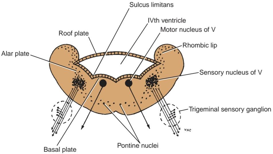
Figure 18-4 Development of the pons from the anterior part of the metencephalon.
شکل 18-4 تکامل پل مغزی از قسمت قدامی متنسفال.
Cerebellum (Posterior Part of Metencephalon)
The cerebellum is formed from the posterior part of the alar plates of the metencephalon. On each side, the alar plates bend medially to form the rhombic lips (Fig. 18-5). As alar plates enlarge, the lips project caudally over the roof plate of the fourth ventricle and unite with each other in the midline to form the cerebellum (Fig. 18-6; also see Fig. 18-5). At the 12th week, a small midline portion, the vermis, and two lateral portions, the cerebellar hemispheres, may be recognized. At about the end of the fourth month, fissures develop on the surface of the cerebellum, and the characteristic folia of the adult cerebellum gradually develop.
مخچه (بخش خلفی متنسفال)
مخچه از بخش خلفی صفحات آلار متنسفال تشکیل میشود. در هر طرف، صفحات آلار به سمت داخل خم میشوند تا لبهای لوزی شکل را تشکیل دهند (شکل 18-5). با بزرگ شدن صفحات آلار، لبها به سمت پایین روی صفحه سقف بطن چهارم بیرون زده و در خط وسط به یکدیگر متصل میشوند تا مخچه را تشکیل دهند (شکل 18-6؛ همچنین به شکل 18-5 مراجعه کنید). در هفته دوازدهم، یک بخش کوچک در خط وسط، ورمیس، و دو بخش جانبی، نیمکرههای مخچه، ممکن است قابل تشخیص باشند. تقریباً در پایان ماه چهارم، شیارهایی روی سطح مخچه ایجاد میشوند و به تدریج شاخکهای مشخصه مخچه بالغ ایجاد میشوند.

Figure 18-5 Development of the cerebellum. Also shown is the fusion of the rhombic lips in the midline to form the dumbbell-shaped cerebellum.
شکل ۱۸-۵ تکامل مخچه. همچنین جوش خوردن لبهای لوزی در خط وسط برای تشکیل مخچه دمبلی شکل نشان داده شده است.
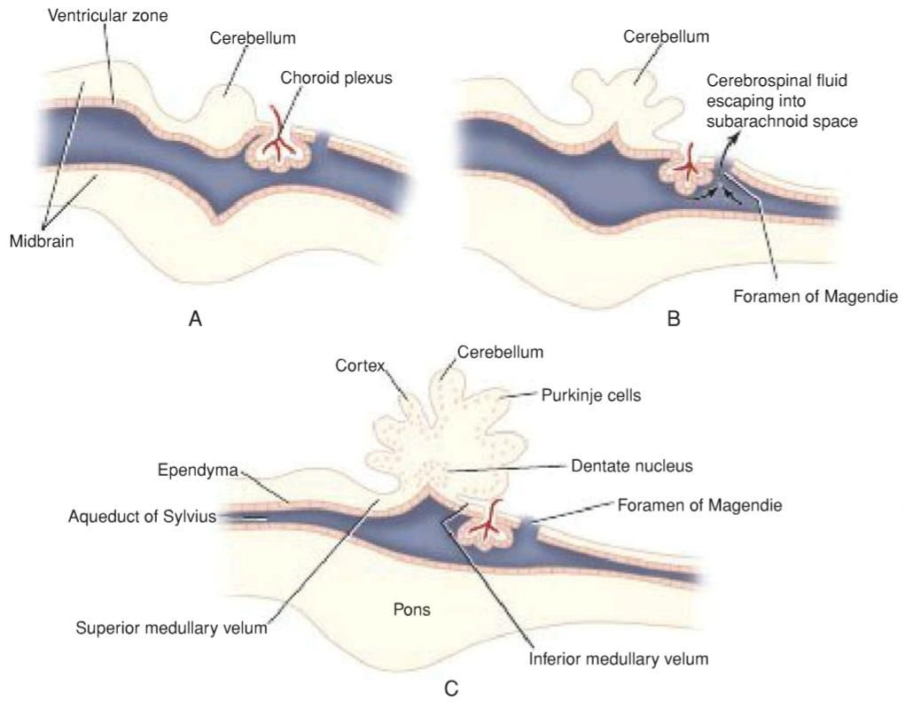
Figure 18-6 Sagittal sections of the developing cerebellum.
شکل ۱۸-۶ برشهای ساژیتال از مخچه در حال رشد.

Figure 18-7 Successive stages in the development of the midbrain.
شکل ۱۸-۷ مراحل متوالی در رشد مغز میانی.
The neuroblasts derived from the matrix cells in the ventricular zone migrate toward the surface of the cerebellum and eventually give rise to the neurons forming the cerebellar cortex. Other neuroblasts remain close to the ventricular surface and differentiate into the dentate and other deep cerebellar nuclei. With further development, the axons of neurons forming these nuclei grow out into the mesencephalon (midbrain) to reach the forebrain, and these fibers will form the greater part of the superior cerebellar peduncle. Later, the growth of the axons of the pontocerebellar fibers and the corticopontine fibers will connect the cerebral cortex with the cerebellum, and so the middle cerebellar peduncle will be formed. The inferior cerebellar peduncle will be formed largely by the growth of sensory axons from the spinal cord, the vestibular nuclei, and olivary nuclei.
نوروبلاستهای مشتقشده از سلولهای ماتریکس در ناحیه بطنی به سمت سطح مخچه مهاجرت میکنند و در نهایت نورونهای تشکیلدهنده قشر مخچه را ایجاد میکنند. سایر نوروبلاستها نزدیک سطح بطن باقی میمانند و به هستههای دندانهدار و سایر هستههای عمقی مخچه تمایز مییابند. با رشد بیشتر، آکسونهای نورونهای تشکیلدهنده این هستهها به سمت مزانسفال (مغز میانی) رشد میکنند تا به مغز پیشین برسند و این فیبرها بخش بزرگتری از پایه مخچه فوقانی را تشکیل میدهند. بعداً، رشد آکسونهای فیبرهای پونتوسربلار و فیبرهای کورتیکوپونتین، قشر مغز را به مخچه متصل میکند و بنابراین پایه مخچه میانی تشکیل میشود. پایه مخچه تحتانی تا حد زیادی با رشد آکسونهای حسی از نخاع، هستههای دهلیزی و هستههای زیتونی تشکیل میشود.
Midbrain (Mesencephalon)
The midbrain develops from the midbrain vesicle, the cavity of which becomes much reduced to form the cerebral aqueduct or aqueduct of Sylvius (Fig. 18-7). The sulcus limitans separates the alar plate from the basal plate on each side, as seen in the developing spinal cord. The neuroblasts in the basal plates will differentiate into the neurons forming the nuclei of CNS III and IV and possibly the red nuclei, the substantia nigra, and the reticular formation. The marginal zone of each basal plate enlarges considerably, thus forming the basis pedunculi by the descent of nerve fibers from the cerebral cortex to the lower motor centers in the pons and spinal cord that is, the corticopontine, corticobulbar, and corticospinal tracts.
مغز میانی (مزانسفالون)
مغز میانی از وزیکول مغز میانی ایجاد میشود که حفره آن بسیار کوچک میشود تا مجرای مغزی یا مجرای سیلویوس را تشکیل دهد (شکل 18-7). شیار محدودکننده، صفحه آلار را از صفحه پایه در هر طرف جدا میکند، همانطور که در نخاع در حال رشد دیده میشود. نوروبلاستهای موجود در صفحات پایه به نورونهایی که هستههای CNS III و IV و احتمالاً هستههای قرمز، جسم سیاه و تشکیلات مشبک را تشکیل میدهند، تمایز مییابند. ناحیه حاشیهای هر صفحه پایه به طور قابل توجهی بزرگ میشود و بنابراین با نزول فیبرهای عصبی از قشر مغز به مراکز حرکتی پایینتر در پل مغزی و نخاع، یعنی راههای قشری-پونتینی، قشری-بولبار و قشری-نخاعی، پایههای ساقهای را تشکیل میدهند.
The two alar plates and the original roof plate form the tectum. The neuroblasts in the alar plates differentiate into the sensory neurons of the superior and inferior colliculi. Four swellings representing the four colliculi appear on the posterior surface of the midbrain. The superior colliculi are associated with visual reflexes, and the inferior colliculi are associated with auditory reflexes.
دو صفحه آلار و صفحه سقفی اصلی، تکتوم را تشکیل میدهند. نوروبلاستهای صفحات آلار به نورونهای حسی کولیکولهای فوقانی و تحتانی تمایز مییابند. چهار برآمدگی که نشاندهنده چهار کولیکول هستند، در سطح خلفی مغز میانی ظاهر میشوند. کولیکولهای فوقانی با رفلکسهای بینایی و کولیکولهای تحتانی با رفلکسهای شنوایی مرتبط هستند.
With further development, the fibers of CN IV emerge on the posterior surface of the midbrain and decussate completely in the superior medullary velum. The fibers of CN III emerge on the anterior surface between the cerebral peduncles.
با تکامل بیشتر، فیبرهای CN IV در سطح خلفی مغز میانی ظاهر میشوند و در نرمکام فوقانی به طور کامل متقاطع میشوند. فیبرهای CN III در سطح قدامی بین پایههای مغزی ظاهر میشوند.
«فصل هجدهم کتاب نورواناتومی بالینی اسنل»
🚀 با ما همراه شوید!
تازهترین مطالب و آموزشهای مغز و اعصاب را از دست ندهید. با فالو کردن کانال تلگرام آیندهنگاران مغز، از ما حمایت کنید!