تکامل سیستم عصبی: پروزنسفالون، دیانسفالون و تلانسفالون


دعای مطالعه [ نمایش ]
بِسْمِ الله الرَّحْمنِ الرَّحیمِ
اَللّهُمَّ اَخْرِجْنى مِنْ ظُلُماتِ الْوَهْمِ
خدايا مرا بيرون آور از تاريكىهاى وهم،
وَ اَكْرِمْنى بِنُورِ الْفَهْمِ
و به نور فهم گرامى ام بدار،
اَللّهُمَّ افْتَحْ عَلَيْنا اَبْوابَ رَحْمَتِكَ
خدايا درهاى رحمتت را به روى ما بگشا،
وَانْشُرْ عَلَيْنا خَزائِنَ عُلُومِكَ بِرَحْمَتِكَ يا اَرْحَمَ الرّاحِمينَ
و خزانههاى علومت را بر ما باز كن به امید رحمتت اى مهربانترين مهربانان.
آیندهنگاران مغز: «ما مغز را میشناسیم، تا آینده را بسازیم.»
کتاب «نوروآناتومی بالینی اسنل» اثر دکتر ریچارد اس. اسنل، با ویرایش جدید دکتر رایان اسپلیتگربر، همچنان بهعنوان مرجع طلایی این حوزه شناخته میشود. این اثر با تلفیق تازهترین پژوهشها و کاربردهای بالینی، پلی استوار میان دانش پایه و عمل پزشکی بنا میکند.
تلاشهای علمی و سالها تدریس ارزشمند استاد گرانقدر آقای دکتر محمدتقی جغتایی الهامبخش ترجمه و گسترش این دانش در ایران بوده است.
ترجمه دقیق این کتاب توسط برند علمی آیندهنگاران مغز به مدیریت داریوش طاهری، امکان دسترسی فارسیزبانان به مرزهای نوین علوم اعصاب را فراهم کرده و رسالتی علمی در جهت شناخت عمیقتر مغز و ترسیم آیندهای روشنتر بر عهده گرفته است.
»» فصل ۱۸: رشد سیستم عصبی؛ بخش مبانی و اصول؛ قسمت سوم
»» Chapter 18: Nervous System Development
Forebrain (Prosencephalon)
The forebrain develops from the forebrain vesicle. The roof and floor plates remain thin, whereas the lateral walls become thick, as in the developing spinal cord. At an early stage, a lateral diverticulum called the optic vesicle appears on each side of the forebrain. That part of the forebrain that lies rostral to the optic vesicle is the telencephalon, and the remainder is the diencephalon (Fig. 18-8). The optic vesicle and stalk ultimately will form the retina and optic nerve.
مغز پیشین (پروزنسفال)
مغز پیشین از کیسهی مغز پیشین تکامل مییابد. صفحات سقف و کف نازک باقی میمانند، در حالی که دیوارههای جانبی ضخیم میشوند، مانند نخاع در حال رشد. در مراحل اولیه، یک دیورتیکول جانبی به نام کیسهی بینایی در هر طرف مغز پیشین ظاهر میشود. آن بخش از مغز پیشین که در قسمت جلویی کیسهی بینایی قرار دارد، تلانسفالون و بقیهی آن دیانسفالون است (شکل 18-8). کیسهی بینایی و ساقه در نهایت شبکیه و عصب بینایی را تشکیل میدهند.
The telencephalon now develops a lateral diverticulum on each side of the cerebral hemisphere, and its cavity is known as the lateral ventricle. The anterior part of the third ventricle, therefore, is formed by the medial part of the telencephalon and ends at the lamina terminalis, which represents the rostral end of the neural tube. The opening into each lateral ventricle is the future interventricular foramen.
اکنون تلانسفالون در هر طرف نیمکره مغزی یک دیورتیکول جانبی ایجاد میکند و حفره آن به عنوان بطن جانبی شناخته میشود. بنابراین، قسمت قدامی بطن سوم توسط قسمت داخلی تلانسفالون تشکیل میشود و در لامینا ترمینالیس، که نشان دهنده انتهای قدامی لوله عصبی است، خاتمه مییابد. دهانه هر بطن جانبی، سوراخ بین بطنی آینده است.
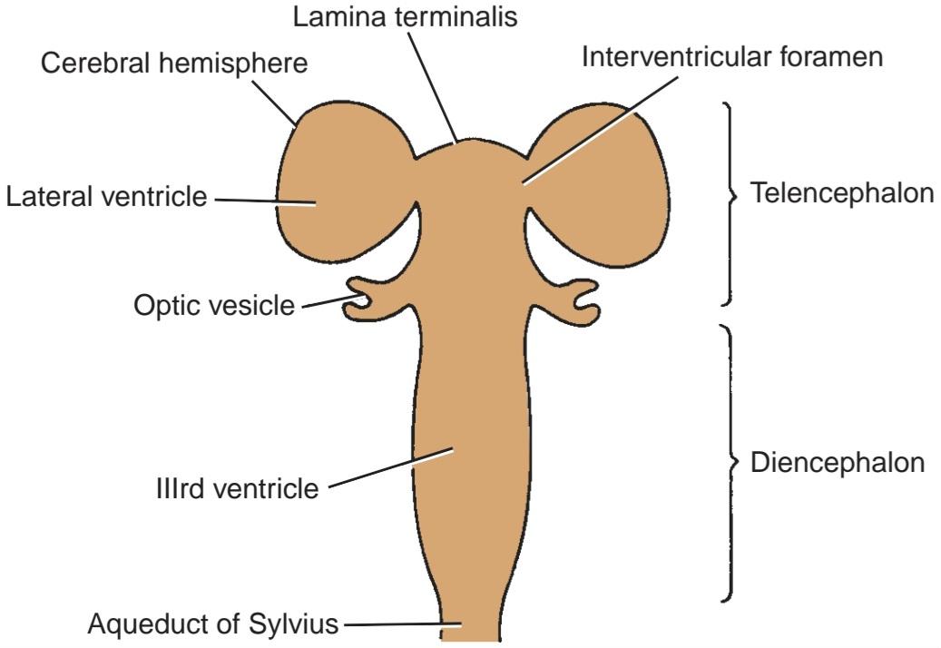
Figure 18-8 Division of the forebrain vesicle into the telencephalon and the diencephalon.
شکل ۱۸-۸ تقسیم وزیکول مغز پیشین به تلانسفالون و دیانسفالون.
Diencephalon Fate
The cavity of the diencephalon forms the greater part of the third ventricle (see Fig. 18-8). Its roof shows a small diverticulum immediately anterior to the midbrain, which will form the pineal body. The remainder of the roof forms the choroid plexus of the third ventricle (Fig. 18-9). In the lateral wall of the third ventricle, the thalamus arises as a thickening of the alar plate on each side. Posterior to the thalamus, the medial and lateral geniculate bodies develop as solid buds. With the continued growth of the two thalami, the ventricular cavity becomes narrowed, and in some individuals, the two thalami may meet and fuse in the midline to form the interthalamic connection of gray matter that crosses the third ventricle (Fig. 18-10).
سرنوشت دیانسفالون
حفره دیانسفالون بخش بزرگتر بطن سوم را تشکیل میدهد (شکل 18-8 را ببینید). سقف آن یک دیورتیکول کوچک را بلافاصله در جلوی مغز میانی نشان میدهد که جسم پینهآل را تشکیل میدهد. بقیه سقف، شبکه کوروئید بطن سوم را تشکیل میدهد (شکل 18-9). در دیواره جانبی بطن سوم، تالاموس به صورت ضخیم شدن صفحه آلار در هر طرف ایجاد میشود. در خلف تالاموس، اجسام زانویی داخلی و خارجی به صورت جوانههای جامد رشد میکنند. با رشد مداوم دو تالاموس، حفره بطن باریک میشود و در برخی افراد، دو تالاموس ممکن است در خط وسط به هم رسیده و جوش بخورند تا اتصال بین تالاموسی ماده خاکستری را که از بطن سوم عبور میکند، تشکیل دهند (شکل 18-10).
The lower part of the alar plate on each side will differentiate into a large number of hypothalamic nuclei. One of these becomes conspicuous on the inferior surface of the hypothalamus and forms a rounded swelling on each side of the midline called the mammillary body.
قسمت پایینی صفحه آلار در هر طرف به تعداد زیادی هسته هیپوتالاموس تمایز مییابد. یکی از این هستهها در سطح تحتانی هیپوتالاموس قابل مشاهده میشود و یک برآمدگی گرد در هر طرف خط وسط به نام جسم پستانی تشکیل میدهد.
The infundibulum develops as a diverticulum from the floor of the diencephalon and from it will originate the stalk and pars nervosa of the hypophysis.
اینفاندیبولوم به صورت یک دیورتیکول از کف دیانسفالون ایجاد میشود و ساقه و پارس عصبی هیپوفیز از آن منشأ میگیرند.
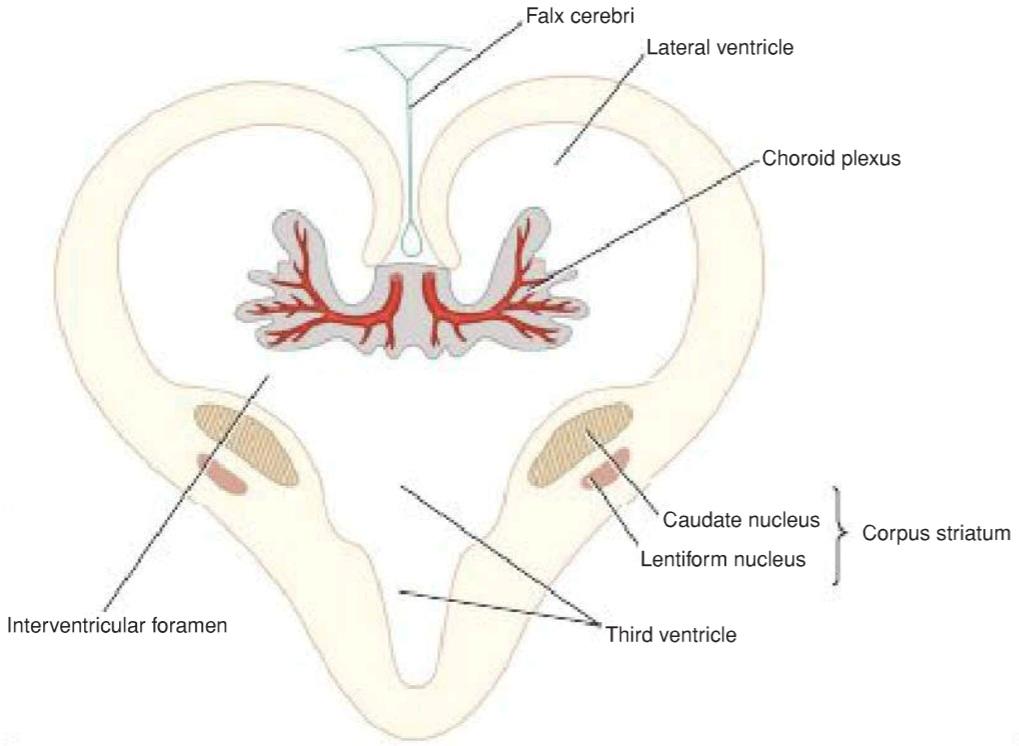
Figure 18-9 Diagrammatic representation of a coronal section of the cerebral hemispheres showing the developing choroid plexuses in the third and lateral ventricles.
شکل ۱۸-۹ نمایش نموداری از برش کرونال نیمکرههای مغزی که شبکههای مشیمیهای در حال رشد را در بطنهای سوم و جانبی نشان میدهد.
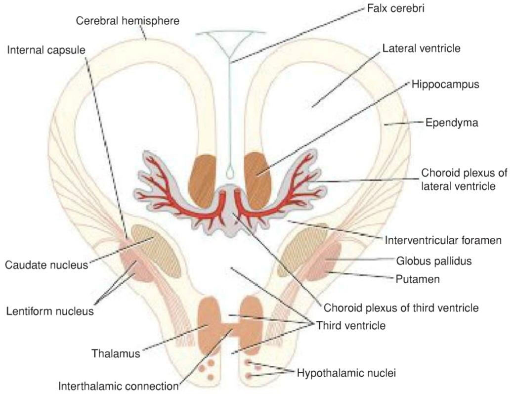
Figure 18-10 Diagrammatic representation of a coronal section of the cerebral hemispheres showing the choroid plexuses in the third and lateral ventricles. Also shown are the caudate and lentiform nuclei and the thalami. The ascending and descending nerve tracts can be seen passing between the masses of gray matter to form the internal capsule.
شکل ۱۸-۱۰ نمایش نموداری از برش تاجی نیمکرههای مغزی که شبکههای مشیمیه را در بطنهای سوم و جانبی نشان میدهد. همچنین هستههای دمی و عدسی شکل و تالاموس نیز نشان داده شدهاند. میتوان مسیرهای عصبی صعودی و نزولی را که از بین تودههای ماده خاکستری عبور میکنند تا کپسول داخلی را تشکیل دهند، مشاهده کرد.
Telencephalon Fate
The telencephalon forms the anterior end of the third ventricle, which is closed by the lamina terminalis, while the diverticulum on either side forms the cerebral hemisphere.
سرنوشت تلانسفالون
تلانسفالون انتهای قدامی بطن سوم را تشکیل میدهد که توسط لامینا ترمینالیس بسته میشود، در حالی که دیورتیکولوم در دو طرف، نیمکره مغزی را تشکیل میدهد.
Cerebral Hemispheres
Each cerebral hemisphere arises at the beginning of the fifth week of development. As it expands superiorly, its walls thicken, and the interventricular foramen becomes reduced in size (see Figs. 18-8 to 18-10). The mesenchyme between each cerebral hemisphere condenses to form the falx cerebri. As development proceeds, the cerebral hemispheres grow and expand rapidly, first anteriorly to form the frontal lobes, then laterally and superiorly to form the parietal lobes, and finally posteriorly and inferiorly to produce the occipital and temporal lobes. As the result of this great expansion, the hemispheres cover the midbrain and hindbrain (Fig. 18-11).
نیمکرههای مغزی
هر نیمکره مغزی در آغاز هفته پنجم رشد ایجاد میشود. با گسترش به سمت بالا، دیوارههای آن ضخیمتر میشود و اندازه سوراخ بین بطنی کاهش مییابد (شکلهای 18-8 تا 18-10 را ببینید). مزانشیم بین هر نیمکره مغزی متراکم میشود و داس مغزی را تشکیل میدهد. با پیشرفت رشد، نیمکرههای مغزی به سرعت رشد و گسترش مییابند، ابتدا به سمت جلو لوبهای پیشانی را تشکیل میدهند، سپس به سمت پهلو و بالا لوبهای آهیانه را تشکیل میدهند و در نهایت به سمت عقب و پایین لوبهای پسسری و گیجگاهی را ایجاد میکنند. در نتیجه این گسترش زیاد، نیمکرهها مغز میانی و مغز عقبی را میپوشانند (شکل 18-11).
The medial wall of the cerebral hemisphere remains thin and is formed by the ependymal cells. This area becomes invaginated by vascular mesoderm, which forms the choroid plexus of the lateral ventricle (see Fig. 18-10). The occipital lobe of the cerebral hemisphere is separated from the cerebellum by mes- enchyme, which condenses to form the tentorium cerebelli.
دیواره داخلی نیمکره مغزی نازک باقی میماند و توسط سلولهای اپاندیمی تشکیل میشود. این ناحیه توسط مزودرم عروقی فرورفته میشود که شبکه مشیمیه بطن جانبی را تشکیل میدهد (شکل 18-10 را ببینید). لوب پسسری نیمکره مغزی توسط مزانشیم از مخچه جدا میشود که متراکم شده و چادرینه مخچه را تشکیل میدهد.
Meanwhile, the matrix cells lining the floor of the forebrain vesicle proliferate, producing large numbers of neuroblasts. These collectively form a projection that encroaches on the cavity of the lateral ventricle and is known as the corpus striatum (see Fig. 18-9). Later, this differentiates into two parts: (1) the dorsomedial portion, the caudate nucleus, and (2) a ventrolateral part, the lentiform nucleus. The latter becomes subdivided into a lateral part, the putamen, and a medial part, the globus pallidus (see Fig. 18-10). As each hemisphere expands, its medial surface approaches the lateral surface of the diencephalon; thus, the caudate nucleus and thalamus come in close contact.
در همین حال، سلولهای ماتریکس که کف وزیکول مغز قدامی را پوشاندهاند، تکثیر میشوند و تعداد زیادی نوروبلاست تولید میکنند. این سلولها به طور جمعی یک برآمدگی تشکیل میدهند که به حفره بطن جانبی نفوذ میکند و به عنوان جسم مخطط شناخته میشود (شکل 18-9 را ببینید). بعداً، این جسم به دو بخش متمایز میشود: (1) بخش پشتی میانی، هسته دمدار، و (2) بخش شکمی جانبی، هسته عدسی شکل. دومی به یک بخش جانبی، پوتامن، و یک بخش داخلی، گلوبوس پالیدوس تقسیم میشود (شکل 18-10 را ببینید). با گسترش هر نیمکره، سطح داخلی آن به سطح جانبی دیانسفالون نزدیک میشود. بنابراین، هسته دمدار و تالاموس در تماس نزدیک قرار میگیرند.
A further longitudinal thickening occurs in the wall of the forebrain vesicle, and the thickening protrudes into the lateral ventricle and forms the hippocampus.
ضخیم شدن طولی بیشتری در دیواره وزیکول مغز قدامی رخ میدهد و این ضخیم شدن به داخل بطن جانبی بیرون زده و هیپوکامپ را تشکیل میدهد.
While these various masses of gray matter are developing within each cerebral hemisphere, maturing neurons in different parts of the nervous system are sending axons either to or from the differentiating cortex. These axons form the large ascending and descending tracts, which, as they develop, are forced to pass between the thalamus and caudate nucleus medially and the lentiform nucleus laterally. The compact bundle of ascending and descending tracts is known as the internal capsule. The external capsule consists of a few cortical projection fibers that pass lateral to the lentiform nucleus.
در حالی که این تودههای مختلف ماده خاکستری در هر نیمکره مغزی در حال رشد هستند، نورونهای بالغ در بخشهای مختلف سیستم عصبی، آکسونها را به قشر در حال تمایز یا از آن میفرستند. این آکسونها مسیرهای صعودی و نزولی بزرگی را تشکیل میدهند که با رشد خود، مجبور به عبور از بین تالاموس و هسته دمدار در قسمت میانی و هسته عدسی شکل در قسمت جانبی میشوند. دسته فشرده مسیرهای صعودی و نزولی به عنوان کپسول داخلی شناخته میشود. کپسول خارجی از چند فیبر برآمدگی قشری تشکیل شده است که از کنار هسته عدسی شکل عبور میکنند.
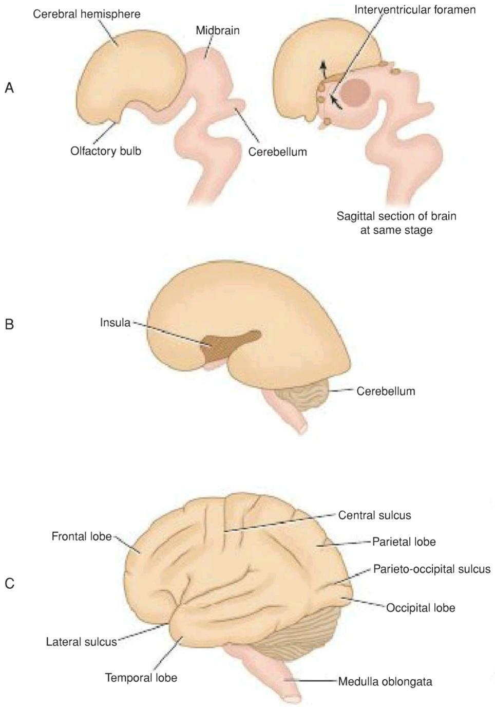
Figur 18-11 Successive stages in the development of the cerebral cortex.
شکل ۱۸-۱۱ مراحل متوالی در رشد قشر مغز.
Cerebral Cortex
As each cerebral hemisphere rapidly expands, the convolutions or gyri separated by fissures or sulci become evident on its surface. The cortex covering the lentiform nucleus remains as a fixed area called the insula (see Fig. 18-11B). Later, this region becomes buried in the lateral sulcus as the result of overgrowth of the adjacent temporal, parietal, and frontal lobes.
قشر مغز
با گسترش سریع هر نیمکره مغزی، پیچشها یا چینهای مغزی که توسط شکاف ها یا شیارها از هم جدا شدهاند، روی سطح آن آشکار میشوند. قشر پوشاننده هسته عدسی شکل به صورت یک ناحیه ثابت به نام اینسولا باقی میماند (شکل 18-11B را ببینید). بعداً، این ناحیه در نتیجه رشد بیش از حد لوبهای گیجگاهی، آهیانه و پیشانی مجاور، در شیار جانبی پنهان میشود.
The matrix cells lining the cavity of the cerebral hemisphere produce large numbers of neuroblasts and neuroglial cells that migrate out into the marginal zone. The remaining matrix cells ultimately will form the ependyma, which lines the lateral ventricle. In the 12th week, the cortex becomes very cellular because of the migration of large numbers of neuroblasts. At term, the neuroblasts have become differentiated and have assumed a stratified appearance as the result of the presence of incoming and outgoing fibers. Different areas of the cortex soon show specific cell types; thus, the motor cortex contains a large number of pyramidal cells, whereas the sensory areas are characterized mainly by granular cells.
سلولهای ماتریکس که حفره نیمکره مغزی را میپوشانند، تعداد زیادی نوروبلاست و سلولهای نوروگلیال تولید میکنند که به ناحیه حاشیهای مهاجرت میکنند. سلولهای ماتریکس باقیمانده در نهایت اپاندیم را تشکیل میدهند که بطن جانبی را میپوشاند. در هفته دوازدهم، قشر مغز به دلیل مهاجرت تعداد زیادی نوروبلاست، بسیار سلولی میشود. در زمان تولد، نوروبلاستها تمایز یافته و به دلیل وجود فیبرهای ورودی و خروجی، ظاهری مطبق پیدا میکنند. نواحی مختلف قشر مغز به زودی انواع خاصی از سلولها را نشان میدهند. بنابراین، قشر حرکتی حاوی تعداد زیادی سلول هرمی است، در حالی که نواحی حسی عمدتاً توسط سلولهای دانهای مشخص میشوند.
Commissures
The lamina terminalis, which is the cephalic end of the neural tube, forms a bridge between the two cerebral hemispheres and enables nerve fibers to pass from one cerebral hemisphere to the other (see Fig. 18-8).
اتصالات
لامینا ترمینالیس، که انتهای سفالیک لوله عصبی است، پلی بین دو نیمکره مغزی تشکیل میدهد و به فیبرهای عصبی اجازه میدهد تا از یک نیمکره مغزی به نیمکره دیگر منتقل شوند (شکل 18-8 را ببینید).
The anterior commissure is the first commissure to develop. It runs in the lamina terminalis and connects the olfactory bulb and the temporal lobe of the cortex on one side with the same structures of the opposite hemisphere.
رابط قدامی اولین رابطی است که توسعه مییابد. این رابط در لامینا ترمینالیس امتداد دارد و پیاز بویایی و لوب گیجگاهی قشر مغز را از یک طرف به ساختارهای مشابه نیمکره مقابل متصل میکند.
The fornix is the second commissure to develop and connects the cortex of the hippocampus in each hemisphere.
فورنیکس دومین رابطی است که توسعه مییابد و قشر هیپوکامپ را در هر نیمکره متصل میکند.
The corpus callosum, the largest and most important commissure, is the third commissure to develop. Its first fibers connect the frontal lobes of both sides and, later, the parietal lobes. As the corpus callosum increases in size because of increased numbers of fibers, it arches back over the roof of the developing third ventricle.
جسم پینهای، بزرگترین و مهمترین رابط، سومین رابطی است که تکامل مییابد. اولین فیبرهای آن، لوبهای پیشانی دو طرف و بعداً، لوبهای جداری را به هم متصل میکنند. با افزایش اندازه جسم پینهای به دلیل افزایش تعداد فیبرها، به سمت عقب و روی سقف بطن سوم در حال رشد قوس پیدا میکند.
The remains of the lamina terminalis, which lie between the corpus callosum and the fornix, become stretched out to form a thin septum, the septum pellucidum. The optic chiasma is formed in the inferior part of the lamina terminalis; it contains fibers from the medial halves of the retinae, which cross the midline to join the optic tract of the opposite side and so pass to the lateral geniculate body and the superior colliculus.
بقایای لامینا ترمینالیس، که بین جسم پینهای و فورنیکس قرار دارد، کشیده شده و یک سپتوم نازک به نام سپتوم پلوسیدوم را تشکیل میدهند. کیاسمای بینایی در قسمت تحتانی لامینا ترمینالیس تشکیل میشود؛ این کیاسما حاوی الیافی از نیمههای داخلی شبکیه است که از خط وسط عبور میکنند تا به راه بینایی طرف مقابل بپیوندند و بنابراین به جسم زانویی خارجی و کولیکولوس فوقانی میروند.
Myelination in the Central Nervous System
The myelin sheath in the central nervous system is formed and maintained by the oligodendrocytes of the neuroglia (see p. 54).
میلینسازی در سیستم عصبی مرکزی
غلاف میلین در سیستم عصبی مرکزی توسط الیگودندروسیتهای نوروگلیا تشکیل و نگهداری میشود (به صفحه ۵۴ مراجعه کنید).
Myelination in the spinal cord begins first in the cervical region, and from here, the process extends caudally. The process of myelination begins within the cord at about the fourth month, and the sensory fibers are affected first. The last affected are the descending motor fibers.
میلینسازی در نخاع ابتدا در ناحیه گردنی شروع میشود و از اینجا، این فرآیند به سمت پایین گسترش مییابد. فرآیند میلینسازی در داخل نخاع حدود ماه چهارم آغاز میشود و فیبرهای حسی ابتدا تحت تأثیر قرار میگیرند. آخرین فیبرهای حرکتی نزولی هستند که تحت تأثیر قرار میگیرند.
Myelination in the brain begins at about the sixth month of fetal life but is restricted to the fibers of the basal ganglia. Later, the sensory fibers passing up from the spinal cord myelinate, but the progress is slow; therefore, at birth, the brain still is largely unmyelinated. Newborns have very little cerebral function; motor reactions such as respiration, sucking, and swallowing are essentially reflex. After birth, the corticobulbar, corticospinal, tectospinal, and corticopontocerebellar fibers begin to myelinate. This process of myelination is not haphazard but systematic, occurring in different nerve fibers at specific times. The corticospinal fibers, for example, start to myelinate at about 6 months after birth, and the process is largely complete by the end of the second year. It is believed that some nerve fibers in the brain and spinal cord do not complete myelination until puberty.
میلینسازی در مغز حدود ماه ششم زندگی جنینی شروع میشود اما به فیبرهای عقدههای قاعدهای محدود میشود. بعداً، فیبرهای حسی که از نخاع بالا میروند، میلیندار میشوند، اما پیشرفت کند است. بنابراین، در هنگام تولد، مغز هنوز تا حد زیادی بدون میلین است. نوزادان عملکرد مغزی بسیار کمی دارند. واکنشهای حرکتی مانند تنفس، مکیدن و بلع اساساً رفلکسی هستند. پس از تولد، فیبرهای قشری-بولبار، قشری-نخاعی، تکاسپاینال و قشری-مخچهای شروع به میلینسازی میکنند. این فرآیند میلینسازی تصادفی نیست، بلکه سیستماتیک است و در فیبرهای عصبی مختلف در زمانهای خاص رخ میدهد. برای مثال، فیبرهای قشری-نخاعی، حدود ۶ ماه پس از تولد شروع به میلیندار شدن میکنند و این فرآیند تا پایان سال دوم تا حد زیادی کامل میشود. اعتقاد بر این است که برخی از فیبرهای عصبی در مغز و نخاع تا زمان بلوغ، میلیندار شدن را کامل نمیکنند.
«فصل هجدهم کتاب نورواناتومی بالینی اسنل»
🚀 با ما همراه شوید!
تازهترین مطالب و آموزشهای مغز و اعصاب را از دست ندهید. با فالو کردن کانال تلگرام آیندهنگاران مغز، از ما حمایت کنید!