
فیزیولوژی پزشکی گایتون و هال؛ اصول فیزیکی تبادل گازها; انتشار اکسیژن و دی اکسید کربن از غشای تنفسی


دعای مطالعه [ نمایش ]
بِسْمِ الله الرَّحْمنِ الرَّحیمِ
اَللّهُمَّ اَخْرِجْنى مِنْ ظُلُماتِ الْوَهْمِ
خدايا مرا بيرون آور از تاريكىهاى وهم،
وَ اَكْرِمْنى بِنُورِ الْفَهْمِ
و به نور فهم گرامى ام بدار،
اَللّهُمَّ افْتَحْ عَلَيْنا اَبْوابَ رَحْمَتِكَ
خدايا درهاى رحمتت را به روى ما بگشا،
وَانْشُرْ عَلَيْنا خَزائِنَ عُلُومِكَ بِرَحْمَتِكَ يا اَرْحَمَ الرّاحِمينَ
و خزانههاى علومت را بر ما باز كن به امید رحمتت اى مهربانترين مهربانان.
کتاب «فیزیولوژی پزشکی گایتون وهال» بهعنوان یکی از جامعترین و معتبرترین منابع در حوزه علوم پزشکی، همچنان مرجع کلیدی برای درک عملکرد پیچیده بدن انسان است. این اثر با تکیه بر تازهترین پژوهشها و توضیحات دقیق از سازوکارهای فیزیولوژیک، پلی میان علوم پایه پزشکی و کاربردهای بالینی ایجاد میکند و نقشی بیبدیل در آموزش، پژوهش و ارتقای دانش سلامت ایفا مینماید.
ترجمه دقیق و علمیاین شاهکار توسط برند علمیآیندهنگاران مغز به مدیریت داریوش طاهری، دسترسی فارسیزبانان به مرزهای نوین دانش فیزیولوژی را ممکن ساخته و رسالتی علمیبرای ارتقای آموزش پزشکی، فهم عمیقتر سازوکارهای بدن و توسعه روشهای نوین در حوزه سلامت فراهم آورده است.
» کتاب فیزیولوژی پزشکی گایتون وهال
» » فصل ۴۰: اصول فیزیکی تبادل گازها; انتشار اکسیژن و دی اکسید کربن از غشای تنفسی
در حال ویرایش
» Guyton and Hall Textbook of Medical Physiology
»» CHAPTER 40: Principles of Gas Exchange; Diffusion of Oxygen and Carbon Dioxide Through the Respiratory Membrane

After the alveoli are ventilated with fresh air, the next step in respiration is diffusion of oxygen (O2) from the alveoli into the pulmonary blood and diffusion of carbon dioxide (CO2) in the opposite direction, out of the blood into the alveoli. The process of diffusion is simply the random motion of molecules in all directions through the respiratory membrane and adjacent fluids. However, in respiratory physiology, we are concerned not only with the basic mechanism by which diffusion occurs but also with the rate at which it occurs, which is a much more complex issue, requiring a deeper understanding of the physics of diffusion and gas exchange.
پس از تهویه آلوئولها با هوای تازه، مرحله بعدی تنفس، انتشار اکسیژن (O2) از آلوئولها به خون ریوی و انتشار دی اکسید کربن (CO2) در جهت مخالف، از خون به داخل آلوئولها است. فرآیند انتشار به سادگی حرکت تصادفی مولکولها در تمام جهات از طریق غشای تنفسی و مایعات مجاور است. با این حال، در فیزیولوژی تنفسی، ما نه تنها به مکانیسم اساسی که توسط آن انتشار رخ میدهد، بلکه به سرعت رخ دادن آن نیز توجه داریم، که موضوع بسیار پیچیده تری است و نیاز به درک عمیق تری از فیزیک انتشار و تبادل گاز دارد.
Physics of Gas Diffusion and Gas Partial Pressures Molecular Basis of Gas Diffusion
All the gases of concern in respiratory physiology are simple molecules that are free to move among one another by diffusion. This is also true of gases dissolved in the fluids and tissues of the body.
فیزیک انتشار گاز و فشارهای جزئی گاز مبنای مولکولی انتشار گاز
همه گازهای مورد توجه در فیزیولوژی تنفسی، مولکولهای ساده ای هستند که آزادانه در بین یکدیگر از طریق انتشار حرکت میکنند. این موضوع در مورد گازهای حل شده در مایعات و بافتهای بدن نیز صادق است.
For diffusion to occur, there must be a source of energy. This source of energy is provided by the kinetic motion of the molecules. Except at absolute zero temperature, all molecules of all matter are continually undergoing motion. For free molecules that are not physically attached to others, this means linear movement at high velocity until they strike other molecules. They then bounce away in new directions and continue moving until they strike other molecules again. In this way, the molecules move rapidly and randomly among one another.
برای انتشار، باید یک منبع انرژی وجود داشته باشد. این منبع انرژی از حرکت جنبشی مولکولها تامین میشود. به جز در دمای صفر مطلق، همه مولکولهای همه ماده به طور مداوم در حال حرکت هستند. برای مولکولهای آزاد که از نظر فیزیکی به دیگران متصل نیستند، این به معنای حرکت خطی با سرعت بالا است تا زمانی که به مولکولهای دیگر برخورد کنند. سپس در جهتهای جدید میپرند و به حرکت خود ادامه میدهند تا دوباره به مولکولهای دیگر برخورد کنند. به این ترتیب، مولکولها به سرعت و به طور تصادفی در میان یکدیگر حرکت میکنند.
Net Diffusion of a Gas in One Direction-Effect of a Concentration Gradient. If a gas chamber or solution has a high concentration of a particular gas at one end of the chamber and a low concentration at the other end, as shown in Figure 40-1, net diffusion of the gas will occur from the highconcentration area toward the low-concentration area. The reason is obvious. There are far more molecules at end A of the chamber to diffuse toward end B than there are molecules to diffuse in the opposite direction. Therefore, the rates of diffusion in each of the two directions are proportionately different, as demonstrated by the lengths of the arrows in the figure.
انتشار خالص گاز در یک جهت-اثر یک گرادیان غلظت. اگر یک محفظه گاز یا محلول دارای غلظت بالایی از یک گاز خاص در یک انتهای محفظه و غلظت کم در انتهای دیگر باشد، همانطور که در شکل 40-1 نشان داده شده است، انتشار خالص گاز از ناحیه با غلظت بالا به ناحیه با غلظت پایین رخ میدهد. دلیلش واضح است. مولکولهای بیشتری در انتهای A محفظه برای انتشار به سمت B وجود دارد تا مولکولهایی که در جهت مخالف منتشر میشوند. بنابراین، نرخ انتشار در هر یک از دو جهت به نسبت متفاوت است، همانطور که با طول فلشها در شکل نشان داده شده است.

Figure 40-1. Diffusion of oxygen from one end of a chamber to the other. The difference between the lengths of the arrows represents net diffusion.
شکل 40-1. انتشار اکسیژن از یک سر محفظه به انتهای دیگر. تفاوت بین طول فلشها نشان دهنده انتشار خالص است.
Gas Pressures in a Mixture of Gases-Partial Pressures of Individual Gases
Pressure is caused by multiple impacts of moving molecules against a surface. Therefore, the pressure of a gas acting on the surfaces of the respiratory passages and alveoli is proportional to the summated force of impact of all the molecules of that gas striking the surface at any given instant. This means that the pressure is directly proportional to the concentration of the gas molecules.
فشارهای گاز در مخلوطی از گازها-فشارهای جزئی گازهای منفرد
فشار ناشی از ضربههای متعدد مولکولهای متحرک بر روی یک سطح است. بنابراین، فشار گازی که بر روی سطوح مجاری تنفسی و آلوئولها تأثیر میگذارد، متناسب با مجموع نیروی ضربه تمام مولکولهای آن گاز است که در هر لحظه به سطح برخورد میکنند. این بدان معنی است که فشار با غلظت مولکولهای گاز نسبت مستقیم دارد.
In respiratory physiology, one deals with mixtures of gases, mainly oxygen, nitrogen, and carbon dioxide. The rate of diffusion of each of these gases is directly proportional to the pressure caused by that gas alone, which is called the partial pressure of that gas. The concept of partial pressure can be explained as follows.
در فیزیولوژی تنفسی، با مخلوطی از گازها، عمدتاً اکسیژن، نیتروژن و دی اکسید کربن سر و کار داریم. سرعت انتشار هر یک از این گازها با فشار ناشی از آن گاز به تنهایی رابطه مستقیم دارد که به آن فشار جزئی آن گاز میگویند. مفهوم فشار جزئی را میتوان به صورت زیر توضیح داد.
Consider air, which has an approximate composition of 79% nitrogen and 21% oxygen. The total pressure of this mixture at sea level averages 760 mm Hg. It is clear from the preceding description of the molecular basis of pressure that each gas contributes to the total pressure in direct proportion to its concentration. Therefore, 79% of the 760 mm Hg is caused by nitrogen (600 mm Hg) and 21% by O2 (160 mm Hg). Thus, the partial pressure of nitrogen in the mixture is 600 mm Hg, and the partial pressure of O2 is 160 mm Hg; the total pressure is 760 mm Hg, the sum of the individual partial pressures. The partial pressures of individual gases in a mixture are designated by the symbols Po2, PCO2, PN2, Pнe, and so forth.
هوا را در نظر بگیرید که دارای ترکیب تقریبی 79 درصد نیتروژن و 21 درصد اکسیژن است. فشار کل این مخلوط در سطح دریا به طور متوسط 760 میلی متر جیوه است. از توضیحات قبلی در مورد مبنای مولکولی فشار واضح است که هر گاز به نسبت مستقیم با غلظت آن به فشار کل کمک میکند. بنابراین، 79٪ از 760 میلی متر جیوه توسط نیتروژن (600 میلی متر جیوه) و 21٪ توسط O2 (160 میلی متر جیوه) ایجاد میشود. بنابراین، فشار جزئی نیتروژن در مخلوط 600 میلی متر جیوه و فشار جزئی O2 160 میلی متر جیوه است. فشار کل 760 میلی متر جیوه است، مجموع فشارهای جزئی فردی. فشار جزئی گازهای جداگانه در یک مخلوط با نمادهای Po2، PCO2، PN2، Pne و غیره مشخص میشود.
Pressures of Gases Dissolved in Water and Tissues
Gases dissolved in water or in body tissues also exert pressure because the dissolved gas molecules are moving randomly and have kinetic energy. Furthermore, when the gas dissolved in fluid encounters a surface, such as the membrane of a cell, it exerts its own partial pressure in the same way as a gas in the gas phase. The partial pressures of the separate dissolved gases are designated the same as the partial pressures in the gas state-that is, Po2, PCO2, PN2, PHе, and so forth.
فشار گازهای محلول در آب و بافت
گازهای محلول در آب یا در بافتهای بدن نیز فشار وارد میکنند زیرا مولکولهای گاز محلول بهطور تصادفی حرکت میکنند و انرژی جنبشی دارند. علاوه بر این، هنگامیکه گاز محلول در سیال با سطحی مانند غشای یک سلول برخورد میکند، فشار جزئی خود را مانند گاز در فاز گاز اعمال میکند. فشار جزئی گازهای محلول جداگانه مانند فشارهای جزئی در حالت گاز تعیین میشود – یعنی Po2، PCO2، PN2، PHe و غیره.
Factors That Determine Partial Pressure of a Gas Dissolved in a Fluid. The partial pressure of a gas in a solution is determined not only by its concentration but also by the solubility coefficient of the gas. That is, some types of molecules, especially CO2, are physically or chemically attracted to water molecules, whereas other types of molecules are repelled. When molecules are attracted, far more of them can be dissolved without building up excess partial pressure within the solution. Conversely, in the case of molecules that are repelled, high partial pressure will develop with fewer dissolved molecules. These relationships are expressed by the following formula, which is Henry’s law:
عواملی که فشار جزئی گاز محلول در یک سیال را تعیین میکنند. فشار جزئی یک گاز در یک محلول نه تنها با غلظت آن بلکه با ضریب حلالیت گاز نیز تعیین میشود. به این معنا که برخی از انواع مولکولها، به ویژه CO2، از نظر فیزیکی یا شیمیایی به مولکولهای آب جذب میشوند، در حالی که انواع دیگر مولکولها دفع میشوند. هنگامیکه مولکولها جذب میشوند، تعداد بیشتری از آنها میتوانند بدون ایجاد فشار جزئی اضافی در محلول حل شوند. برعکس، در مورد مولکولهایی که دفع میشوند، فشار جزئی بالا با مولکولهای محلول کمتری ایجاد میشود. این روابط با فرمول زیر که قانون هنری است بیان میشود:

When partial pressure is expressed in atmospheres (1 atmosphere [1 atm] pressure equals 760 mm Hg) and concentration is expressed in volume of gas dissolved in each volume of water, the solubility coefficients for important respiratory gases at body temperature are the following:
وقتی فشار جزئی بر حسب اتمسفر بیان میشود (فشار 1 اتمسفر برابر با 760 میلی متر جیوه) و غلظت بر حسب حجم گاز محلول در هر حجم آب بیان میشود، ضرایب حلالیت برای گازهای مهم تنفسی در دمای بدن به شرح زیر است:
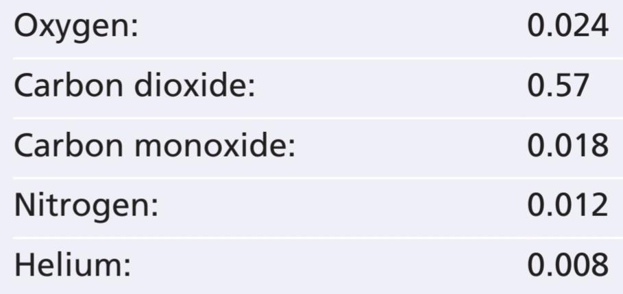
From this list, one can see that CO2 is more than 20 times as soluble as O2. Therefore, the partial pressure of CO2 for a given concentration is less than one-twentieth (5%) of that exerted by O2.
از این لیست میتوان دریافت که CO2 بیش از 20 برابر محلول O2 است. بنابراین، فشار جزئی CO2 برای یک غلظت معین کمتر از یک بیستم (5٪) فشار اعمال شده توسط O2 است.
Diffusion of Gases Between Gas Phase in Alveoli and Dissolved Phase in Pulmonary Blood. The partial pressure of each gas in the alveolar respiratory gas mixture tends to force molecules of that gas into solution in the blood of the alveolar capillaries. Conversely, the molecules of the same gas that are already dissolved in the blood are bouncing randomly in the fluid of the blood, and some of these bouncing molecules escape back into the alveoli. The rate at which they escape is directly proportional to their partial pressure in the blood.
انتشار گازها بین فاز گاز در آلوئول و فاز محلول در خون ریوی. فشار جزئی هر گاز در مخلوط گاز تنفسی آلوئولی تمایل دارد مولکولهای آن گاز را به محلول در خون مویرگهای آلوئولی وادار کند. برعکس، مولکولهای همان گازی که قبلاً در خون حل شدهاند، بهطور تصادفی در مایع خون جهش میکنند و برخی از این مولکولهای جهنده به آلوئولها بازمیگردند. سرعت فرار آنها به طور مستقیم با فشار جزئی آنها در خون متناسب است.
But, in which direction will net diffusion of the gas occur? The answer is that net diffusion is determined by the difference between the two partial pressures. If the partial pressure is greater in the gas phase in the alveoli, as is normally true for oxygen, then more molecules will diffuse into the blood than in the other direction. Alternatively, if the partial pressure of the gas is greater in the dissolved state in the blood, which is normally true for CO2, then net diffusion will occur toward the gas phase in the alveoli.
اما، انتشار خالص گاز در کدام جهت رخ خواهد داد؟ پاسخ این است که انتشار خالص با تفاوت بین دو فشار جزئی تعیین میشود. اگر فشار جزئی در فاز گاز در آلوئولها بیشتر باشد، همانطور که معمولاً برای اکسیژن صادق است، مولکولهای بیشتری نسبت به جهت دیگر در خون منتشر میشوند. از طرف دیگر، اگر فشار جزئی گاز در حالت محلول در خون بیشتر باشد، که معمولاً برای CO2 صادق است، انتشار خالص به سمت فاز گاز در آلوئولها رخ میدهد.
Vapor Pressure of Water
When nonhumidified air is breathed into the respiratory passageways, water immediately evaporates from the surfaces of these passages and humidifies the air. This results from the fact that water molecules, like different dissolved gas molecules, are continually escaping from the water surface into the gas phase. The partial pressure that water molecules exert to escape through the surface is called the vapor pressure of the water. At normal body temperature, 37°C (98.6°F), this vapor pressure is 47 mm Hg. Therefore, once the gas mixture has become fully humidified-that is, once it is in equilibrium with the water-the partial pressure of the water vapor in the gas mixture is 47 mm Hg. This partial pressure, like the other partial pressures, is designated as PH2O.
فشار بخار آب
هنگامیکه هوای بدون رطوبت وارد مجاری تنفسی میشود، آب بلافاصله از سطوح این معابر تبخیر شده و هوا را مرطوب میکند. این امر از این واقعیت ناشی میشود که مولکولهای آب، مانند مولکولهای مختلف گاز محلول، به طور مداوم از سطح آب به فاز گاز میگریزند. فشار جزئی که مولکولهای آب برای خروج از سطح اعمال میکنند، فشار بخار آب نامیده میشود. در دمای طبیعی بدن، 37 درجه سانتی گراد (98.6 درجه فارنهایت)، این فشار بخار 47 میلی متر جیوه است. بنابراین، هنگامیکه مخلوط گاز کاملاً مرطوب شد – یعنی زمانی که با آب در تعادل قرار گرفت – فشار جزئی بخار آب در مخلوط گاز 47 میلی متر جیوه است. این فشار جزئی، مانند سایر فشارهای جزئی، به عنوان PH2O تعیین میشود.
The vapor pressure of water depends entirely on the temperature of the water. The higher the temperature, the great- er the kinetic activity of the molecules and, therefore, the greater the likelihood that the water molecules will escape from the surface of the water into the gas phase. For example, the water vapor pressure at 0°C is 5 mm Hg, and at 100°C it is 760 mm Hg. The most important value to remember is the vapor pressure at body temperature, 47 mm Hg. This value appears in many of our subsequent discussions.
فشار بخار آب کاملاً به دمای آب بستگی دارد. هر چه دما بالاتر باشد، فعالیت جنبشی مولکولها بیشتر است و بنابراین، احتمال فرار مولکولهای آب از سطح آب به فاز گاز بیشتر میشود. به عنوان مثال، فشار بخار آب در دمای 0 درجه سانتی گراد 5 میلی متر جیوه و در دمای 100 درجه سانتی گراد 760 میلی متر جیوه است. مهم ترین مقداری که باید به خاطر بسپارید فشار بخار در دمای بدن، 47 میلی متر جیوه است. این مقدار در بسیاری از بحثهای بعدی ما ظاهر میشود.
Pressure Difference Causes Net Diffusion of Gases Through Fluids
From the preceding discussion, it is clear that when the partial pressure of a gas is greater in one area than in another area, there will be net diffusion from the high-pressure area toward the low-pressure area. For example, returning to Figure 40-1, one can readily see that the molecules in the area of high pressure, because of their greater number, have a greater chance of moving randomly into the area of low pressure than do molecules attempting to go in the other direction. However, some molecules do bounce randomly from the low-pressure area toward the high-pressure area. There- fore, the net diffusion of gas from the area of high pressure to the area of low pressure is equal to the number of molecules bouncing in this forward direction minus the number bouncing in the opposite direction, which is proportional to the gas partial pressure difference between the two areas, called simply the pressure difference for causing diffusion.
اختلاف فشار باعث انتشار خالص گازها از طریق سیالات میشود
از بحث قبل، واضح است که وقتی فشار جزئی گاز در یک ناحیه بیشتر از ناحیه دیگر باشد، انتشار خالص از ناحیه پرفشار به ناحیه کم فشار وجود خواهد داشت. به عنوان مثال، با بازگشت به شکل 40-1، میتوان به راحتی مشاهده کرد که مولکولهای ناحیه فشار بالا، به دلیل تعداد بیشترشان، شانس بیشتری برای حرکت تصادفی به ناحیه کم فشار نسبت به مولکولهایی که سعی در حرکت در جهت دیگر دارند، دارند. با این حال، برخی از مولکولها به طور تصادفی از ناحیه کم فشار به سمت ناحیه پرفشار پرش میکنند. بنابراین، انتشار خالص گاز از ناحیه پرفشار به ناحیه کم فشار برابر است با تعداد مولکولهایی که در این جهت رو به جلو منهای تعداد جهش در جهت مخالف، که متناسب با اختلاف فشار جزئی گاز بین دو ناحیه است که به سادگی اختلاف فشار برای ایجاد انتشار نامیده میشود.
Quantifying Net Rate of Diffusion in Fluids. In addition to the pressure difference, several other factors affect the rate of gas diffusion in a fluid: (1) the solubility of the gas in the fluid; (2) the cross-sectional area of the fluid; (3) the distance through which the gas must diffuse; (4) the molecular weight of the gas; and (5) the temperature of the fluid. In the body, the temperature remains reasonably constant and usually need not be considered.
کمیکردن نرخ خالص انتشار در سیالات. علاوه بر اختلاف فشار، چندین عامل دیگر بر سرعت انتشار گاز در سیال تأثیر میگذارند: (1) حلالیت گاز در سیال. (2) سطح مقطع سیال. (3) فاصله ای که گاز باید از طریق آن پخش شود. (4) وزن مولکولی گاز؛ و (5) دمای سیال. در بدن، دما به طور معقولی ثابت میماند و معمولاً نیازی به در نظر گرفتن نیست.
The greater the solubility of the gas, the greater the number of molecules available to diffuse for any given partial pressure difference. The greater the cross-sectional area of the diffusion pathway, the greater the total number of molecules that diffuse. Conversely, the greater the distance the molecules must diffuse, the longer it will take the molecules to diffuse the entire distance. Finally, the greater the velocity of kinetic movement of the molecules, which is inversely proportional to the square root of the molecular weight, the greater the rate of diffusion of the gas. All these factors can be expressed in a single formula, as follows:
هرچه حلالیت گاز بیشتر باشد، تعداد مولکولهای موجود برای انتشار برای هر اختلاف فشار جزئی بیشتر است. هر چه سطح مقطع مسیر انتشار بیشتر باشد، تعداد کل مولکولهایی که منتشر میشوند بیشتر است. برعکس، هرچه فاصله مولکولها بیشتر باشد، مولکولها در کل فاصله بیشتری پخش میکنند. در نهایت، هر چه سرعت حرکت جنبشی مولکولها بیشتر باشد، که با جذر وزن مولکولی نسبت معکوس دارد، سرعت انتشار گاز بیشتر میشود. همه این عوامل را میتوان در یک فرمول واحد به شرح زیر بیان کرد:

in which D is the diffusion rate, ∆P is the partial pressure difference between the two ends of the diffusion path- way, A is the cross-sectional area of the pathway, S is the solubility of the gas, d is the distance of diffusion, and MW is the molecular weight of the gas.
که در آن D نرخ انتشار، P∆ اختلاف فشار جزئی بین دو انتهای مسیر انتشار، A سطح مقطع مسیر، S حلالیت گاز، d فاصله انتشار و MW وزن مولکولی گاز است.
It is obvious from this formula that the characteristics of the gas determine two factors of the formula-solubility and molecular weight. Together, these two factors determine the diffusion coefficient of the gas, which is proportional to S/VMW; that is, the relative rates at which different gases at the same partial pressure levels will diffuse are proportional to their diffusion coefficients. Assuming that the diffusion coefficient for O2 is 1, the relative diffusion coefficients for different gases of respiratory importance in the body fluids are as follows:
از این فرمول مشخص است که ویژگیهای گاز دو عامل فرمول حلالیت و وزن مولکولی را تعیین میکند. این دو عامل با هم ضریب انتشار گاز را تعیین میکنند که متناسب با S/VMW است. یعنی نرخهای نسبی که در آن گازهای مختلف در همان سطوح فشار جزئی منتشر میشوند با ضرایب انتشار آنها متناسب است. با فرض اینکه ضریب انتشار برای O2 1 باشد، ضرایب انتشار نسبی برای گازهای مختلف با اهمیت تنفسی در مایعات بدن به شرح زیر است:

Diffusion of Gases Through Tissues
The gases that are of respiratory importance are all highly soluble in lipids and, consequently, are highly soluble in cell membranes. Because of this, the major limitation to movement of gases in tissues is the rate at which the gases can diffuse through the tissue water instead of through the cell membranes. Therefore, diffusion of gases through tissues, including through the respiratory membrane, is almost equal to the diffusion of gases in water, as given in the preceding list.
انتشار گازها از طریق بافت
گازهایی که از نظر تنفسی اهمیت دارند، همگی در لیپیدها بسیار محلول هستند و در نتیجه در غشای سلولی بسیار محلول هستند. به همین دلیل، محدودیت عمده برای حرکت گازها در بافتها، سرعتی است که گازها میتوانند از طریق آب بافت به جای غشای سلولی منتشر شوند. بنابراین، انتشار گازها از طریق بافتها، از جمله از طریق غشای تنفسی، تقریباً برابر با انتشار گازها در آب است، همانطور که در لیست قبل ذکر شد.
COMPOSITIONS OF ALVEOLAR AIR AND ATMOSPHERIC AIR ARE DIFFERENT
Alveolar air does not have the same concentrations of gases as atmospheric air (Table 40-1). There are several reasons for the differences. First, alveolar air is only partially replaced by atmospheric air with each breath. Second, O2 is constantly being absorbed into the pulmonary blood from the alveolar air. Third, CO2 is constantly diffusing from the pulmonary blood into the alveoli. And fourth, dry atmospheric air that enters the respiratory passages is humidified even before it reaches the alveoli.
ترکیبات هوای آلوئولی و هوای اتمسفر متفاوت است
هوای آلوئولی دارای غلظت گازهای مشابه هوای جو نیست (جدول 40-1). دلایل مختلفی برای تفاوتها وجود دارد. اول، هوای آلوئولی تنها تا حدی با هوای جوی با هر تنفس جایگزین میشود. دوم، O2 به طور مداوم از هوای آلوئولی به خون ریوی جذب میشود. سوم، CO2 به طور مداوم از خون ریوی به آلوئولها پخش میشود. و چهارم، هوای خشک جوی که وارد مجاری تنفسی میشود حتی قبل از رسیدن به آلوئولها مرطوب میشود.
Air Is Humidified in the Respiratory Passages
Table 40-1 shows that atmospheric air is composed almost entirely of nitrogen and O2; it normally contains almost no CO2 and little water vapor. However, as soon as the atmospheric air enters the respiratory passages, it is exposed to the fluids that cover the respiratory surfaces. Even before the air enters the alveoli, it becomes almost totally humidified.
هوا در مجاری تنفسی مرطوب میشود
جدول 40-1 نشان میدهد که هوای اتمسفر تقریباً به طور کامل از نیتروژن و O2 تشکیل شده است. معمولاً تقریباً هیچ CO2 و بخار آب کمیندارد. اما به محض ورود هوای اتمسفر به مجاری تنفسی، در معرض مایعاتی قرار میگیرد که سطوح تنفسی را میپوشانند. حتی قبل از اینکه هوا وارد آلوئولها شود، تقریباً کاملاً مرطوب میشود.
The partial pressure of water vapor at a normal body temperature of 37°C is 47 mm Hg, which is therefore the partial pressure of water vapor in the alveolar air. Because the total pressure in the alveoli cannot rise to more than the atmospheric pressure (760 mm Hg at sea level), this water vapor simply dilutes all the other gases in the inspired air. Table 40-1 also shows that humidification of the air dilutes the oxygen partial pressure at sea level from an average of 159 mm Hg in atmospheric air to 149 mm Hg in the humidified air, and it dilutes the nitrogen partial pressure from 597 to 563 mm Hg.
فشار جزئی بخار آب در دمای معمولی بدن 37 درجه سانتیگراد 47 میلی متر جیوه است که در نتیجه فشار جزئی بخار آب در هوای آلوئولی است. از آنجایی که فشار کل در آلوئولها نمیتواند به بیش از فشار اتمسفر (760 میلی متر جیوه در سطح دریا) برسد، این بخار آب به سادگی تمام گازهای دیگر موجود در هوای دمیده شده را رقیق میکند. جدول 40-1 همچنین نشان میدهد که رطوبت هوا فشار جزئی اکسیژن در سطح دریا را از میانگین 159 میلی متر جیوه در هوای جو به 149 میلی متر جیوه در هوای مرطوب رقیق میکند و فشار جزئی نیتروژن را از 597 به 563 میلی متر جیوه رقیق میکند.
Table 40-1 Partial Pressures (in mm Hg) and composition (in percentages) of Respiratory Gases as They Enter and Leave the Lungsa
جدول 40-1 فشارهای جزئی (بر حسب میلی متر جیوه) و ترکیب (بر حسب درصد) گازهای تنفسی هنگام ورود و خروج از ریه

Alveolar Air Is Slowly Renewed by Atmospheric Air
In Chapter 38, we pointed out that the average functional residual capacity of the lungs (the volume of air remaining in the lungs at the end of normal expiration) measures about 2300 ml in men. Yet only 350 ml of new air is brought into the alveoli with each normal inspiration, and this same amount of old alveolar air is expired. Therefore, the volume of alveolar air replaced by new atmospheric air with each breath is only one-seventh of the total, so multiple breaths are required to exchange most of the alveolar air. Figure 40-2 shows this slow rate of renewal of the alveolar air. In the first alveolus of the figure, excess gas is present in the alveoli, but note that even at the end of 16 breaths, the excess gas still has not been completely removed from the alveoli.
هوای آلوئولی به آرامیتوسط هوای اتمسفر تجدید میشود
در فصل 38، اشاره کردیم که میانگین ظرفیت باقیمانده عملکردی ریهها (حجم هوای باقی مانده در ریهها در پایان بازدم طبیعی) در مردان حدود 2300 میلی لیتر است. با این حال، تنها 350 میلی لیتر هوای جدید با هر دم طبیعی وارد آلوئولها میشود و همین مقدار هوای آلوئولی قدیمیمنقضی میشود. بنابراین، حجم هوای آلوئولی جایگزین شده با هوای جدید جوی با هر دم، تنها یک هفتم کل است، بنابراین برای تبادل بیشتر هوای آلوئولی به تنفسهای متعدد نیاز است. شکل 40-2 این سرعت کند تجدید هوای آلوئولی را نشان میدهد. در آلوئول اول شکل، گاز اضافی در آلوئولها وجود دارد، اما توجه داشته باشید که حتی در پایان 16 تنفس، گاز اضافی هنوز به طور کامل از آلوئولها خارج نشده است.
Figure 40-3 demonstrates graphically the rate at which excess gas in the alveoli is normally removed, showing that with normal alveolar ventilation, about half the gas is removed in 17 seconds. When a person’s rate of alveolar ventilation is only half-normal, half of the gas is removed in 34 seconds, and when the rate of ventilation is twice normal, half is removed in about 8 seconds.
شکل 40-3 به صورت گرافیکی سرعت حذف گاز اضافی در آلوئولها را نشان میدهد و نشان میدهد که با تهویه طبیعی آلوئولی، حدود نیمیاز گاز در 17 ثانیه حذف میشود. زمانی که میزان تهویه آلوئولی فرد فقط نیمه نرمال باشد، نیمیاز گاز در 34 ثانیه خارج میشود و زمانی که سرعت تهویه دو برابر نرمال باشد، نیمیاز آن در حدود 8 ثانیه خارج میشود.

Figure 40-2. Expiration of a gas from an alveolus with successive breaths.
شکل 40-2. خروج گاز از آلوئول با تنفسهای متوالی.
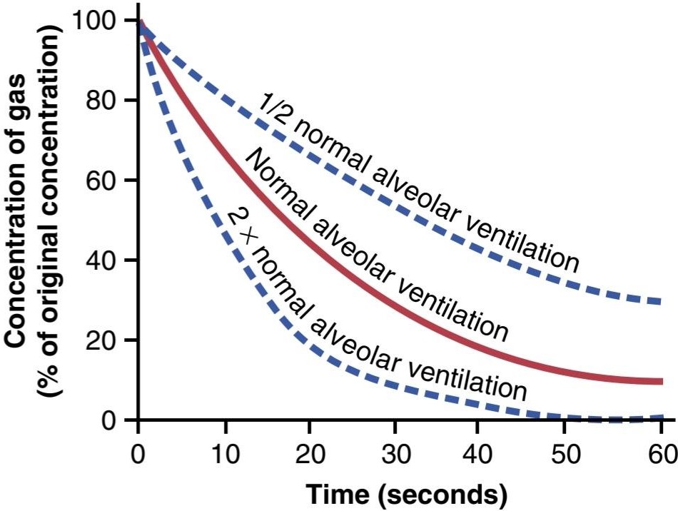
Figure 40-3. Rate of removal of excess gas from alveoli.
شکل 40-3. سرعت حذف گاز اضافی از آلوئولها.
Slow Replacement of Alveolar Air Helps Stabilize Respiratory Control. The slow replacement of alveolar air is of particular importance in preventing sudden changes in gas concentrations in the blood. This makes the respiratory control mechanism much more stable than it would be otherwise, and it helps prevent excessive increases and decreases in tissue oxygenation, tissue CO2 concentration, and tissue pH when respiration is temporarily interrupted.
جایگزینی آهسته هوای آلوئولی به تثبیت کنترل تنفسی کمک میکند. جایگزینی آهسته هوای آلوئولی در جلوگیری از تغییرات ناگهانی غلظت گاز در خون اهمیت ویژه ای دارد. این باعث میشود مکانیسم کنترل تنفسی بسیار پایدارتر از آنچه که در غیر این صورت باشد، و به جلوگیری از افزایش و کاهش بیش از حد در اکسیژن رسانی بافت، غلظت CO2 بافت و pH بافت در هنگام قطع شدن موقت تنفس کمک میکند.
Oxygen Concentration and Partial Pressure in Alveoli
Oxygen is continually being absorbed from the alveoli into the blood of the lungs, and new O2 is continually being breathed into the alveoli from the atmosphere. The more rapidly O2 is absorbed, the lower its concentration in the alveoli becomes; conversely, the more rapidly new O2 is breathed into the alveoli from the atmosphere, the higher its concentration becomes. Therefore, O2 concentration in the alveoli, as well as its partial pressure, is controlled by the following: (1) the rate of absorption of O2 into the blood; and (2) the rate of entry of new O2 into the lungs by the ventilatory process.
غلظت اکسیژن و فشار جزئی در آلوئول
اکسیژن به طور مداوم از آلوئولها به خون ریهها جذب میشود و O2 جدید به طور مداوم از جو به آلوئولها تنفس میشود. هرچه O2 سریعتر جذب شود، غلظت آن در آلوئولها کاهش مییابد. برعکس، هر چه O2 جدید از اتمسفر به داخل آلوئولها تنفس شود، غلظت آن بیشتر میشود. بنابراین، غلظت O2 در آلوئولها، و همچنین فشار جزئی آن، توسط موارد زیر کنترل میشود: (1) سرعت جذب O2 در خون. و (2) سرعت ورود O2 جدید به ریهها توسط فرآیند تهویه.
Figure 40-4 shows the effect of alveolar ventilation and rate of O2 absorption into the blood on the alveolar Po2. One curve represents O2 absorption at a rate of 250 ml/min, and the other curve represents a rate of 1000 ml/ min. At a normal ventilatory rate of 4.2 L/min and an O2 consumption of 250 ml/min, the normal operating point in Figure 40-4 is point A. The figure also shows that when 1000 ml of O2 is being absorbed each minute, as during moderate exercise, the rate of alveolar ventilation must increase fourfold to maintain the alveolar Po2 at the normal value of 104 mm Hg.
شکل 40-4 اثر تهویه آلوئولی و سرعت جذب O2 در خون را بر روی Po2 آلوئولی نشان میدهد. یک منحنی نشان دهنده جذب O2 با سرعت 250 میلی لیتر در دقیقه و منحنی دیگر نشان دهنده سرعت 1000 میلی لیتر در دقیقه است. با سرعت تهویه طبیعی 4.2 لیتر در دقیقه و مصرف O2 250 میلی لیتر در دقیقه، نقطه عملیاتی نرمال در شکل 40-4 نقطه A است. شکل همچنین نشان میدهد که وقتی 1000 میلی لیتر O2 در هر دقیقه جذب میشود، مانند هنگام ورزش متوسط، سرعت تهویه آلوئولی باید چهار برابر افزایش یابد تا مقدار mal20-10 آلوئولی حفظ شود.
Another effect shown in Figure 40-4 is that even an extreme increase in alveolar ventilation can never increase the alveolar Po2 above 149 mm Hg as long as the person is breathing normal atmospheric air at sea level pressure, because 149 mm Hg is the maximum Po2 in humidified air at this pressure. If the person breathes gases that contain partial pressures of O2 higher than 149 mm Hg, the alveolar Po2 can approach these higher pressures at high rates of ventilation.
اثر دیگری که در شکل 40-4 نشان داده شده است این است که حتی افزایش شدید تهویه آلوئولی هرگز نمیتواند Po2 آلوئولی را بیش از 149 میلی متر جیوه افزایش دهد تا زمانی که فرد هوای معمولی جو را در فشار سطح دریا تنفس میکند، زیرا 149 میلی متر جیوه حداکثر Po2 در هوای مرطوب در این فشار است. اگر فرد گازهایی را تنفس کند که حاوی فشار جزئی O2 بالاتر از 149 میلیمتر جیوه است، Po2 آلوئولی میتواند با نرخ بالای تهویه به این فشارهای بالاتر نزدیک شود.

Figure 40-4. Effect of alveolar ventilation on the alveolar partial pressure of oxygen (Po2) at two rates of oxygen absorption from the alveoli-250 ml/min and 1000 ml/min. Point A is the normal operating point.
شکل 40-4. اثر تهویه آلوئولی بر فشار جزئی آلوئولی اکسیژن (Po2) در دو نرخ جذب اکسیژن از آلوئول-250 میلی لیتر در دقیقه و 1000 میلی لیتر در دقیقه. نقطه A نقطه عملیاتی عادی است.
CO2 Concentration and Partial Pressure in Alveoli
Carbon dioxide is continually formed in the body and then carried in the blood to the alveoli; it is continually removed from the alveoli by ventilation. Figure 40-5 shows the effects on the alveolar partial pressure of Pco2 of both alveolar ventilation and two rates of CO2 excretion, 200 and 800 ml/min. One curve represents a normal rate of CO2 excretion of 200 ml/min. At the normal rate of alveolar ventilation of 4.2 L/min, the operating point for alveolar Pco, is at point A in Figure 40-5 (i.e., 40 mm Hg).
غلظت CO2 و فشار جزئی در آلوئول
دی اکسید کربن به طور مداوم در بدن تشکیل میشود و سپس در خون به آلوئولها منتقل میشود. به طور مداوم با تهویه از آلوئولها خارج میشود. شکل 40-5 اثرات فشار جزئی آلوئولی Pco2 هر دو تهویه آلوئولی و دو میزان دفع CO2 200 و 800 میلی لیتر در دقیقه را نشان میدهد. یک منحنی نشان دهنده نرخ طبیعی دفع CO2 200 میلی لیتر در دقیقه است. در سرعت نرمال تهویه آلوئولی 4.2 لیتر در دقیقه، نقطه عمل Pco آلوئولی در نقطه A در شکل 40-5 (یعنی 40 میلی متر جیوه) است.
Two other facts are also evident from Figure 40-5. First, the alveolar Pco, increases directly in proportion to the rate of CO2 excretion, as represented by the fourfold elevation of the curve (when 800 ml of CO2 are excreted per minute). Second, the alveolar Pco, decreases in inverse proportion to alveolar ventilation. Therefore, the concentrations and partial pressures of both O2 and CO2 in the alveoli are determined by the rates of absorption or excretion of the two gases and by the amount of alveolar ventilation.
دو واقعیت دیگر نیز از شکل 40-5 مشهود است. اول، Pco آلوئولی، به طور مستقیم به نسبت میزان دفع CO2 افزایش مییابد، همانطور که با افزایش چهار برابری منحنی نشان داده میشود (زمانی که 800 میلی لیتر CO2 در دقیقه دفع میشود). دوم، Pco آلوئولی، به نسبت معکوس تهویه آلوئولی کاهش مییابد. بنابراین، غلظت و فشار جزئی هر دو O2 و CO2 در آلوئولها با سرعت جذب یا دفع دو گاز و میزان تهویه آلوئولی تعیین میشود.

Figure 40-5. Effect of alveolar ventilation on the alveolar partial pressure of carbon dioxide (Pco2) at two rates of carbon dioxide excretion from the blood-800 ml/min and 200 ml/min. Point A is the normal operating point.
شکل 40-5. اثر تهویه آلوئولی بر فشار جزئی آلوئولی دی اکسید کربن (Pco2) در دو میزان دفع دی اکسید کربن از خون 800 میلی لیتر در دقیقه و 200 میلی لیتر در دقیقه. نقطه A نقطه عملیاتی عادی است.
Expired Air Is a Combination of Dead Space Air and Alveolar Air
The overall composition of expired air is determined by the following: (1) the amount of the expired air that is dead space air; and (2) the amount that is alveolar air. Figure 40-6 shows the progressive changes in O2 and CO2 partial pressures in the expired air during the course of expiration. The first portion of this air, the dead space air from the respiratory passageways, is typical humidified air, as shown in Table 40-1. Then, progressively more and more alveolar air becomes mixed with the dead space air until all the dead space air has finally been washed out, and nothing but alveolar air is expired at the end of expiration. Therefore, the method of collecting alveolar air for study is simply to collect a sample of the last portion of the expired air after forceful expiration has removed all the dead space air.
هوای منقضی شده ترکیبی از هوای فضای مرده و هوای آلوئولی است
ترکیب کلی هوای منقضی شده با موارد زیر تعیین میشود: (1) مقدار هوای منقضی شده که هوای فضای مرده است. و (2) مقداری که هوای آلوئولی است. شکل 40-6 تغییرات پیشرونده فشارهای جزئی O2 و CO2 در هوای منقضی شده را در طول بازدم نشان میدهد. اولین بخش از این هوا، هوای فضای مرده از مجاری تنفسی، هوای معمولی مرطوب است، همانطور که در جدول 40-1 نشان داده شده است. سپس، به تدریج هوای آلوئولی بیشتر و بیشتر با هوای فضای مرده مخلوط میشود تا زمانی که در نهایت تمام هوای فضای مرده شسته شود، و چیزی جز هوای آلوئولی در پایان بازدم منقضی نمیشود. بنابراین، روش جمعآوری هوای آلوئولی برای مطالعه، صرفاً جمعآوری نمونهای از آخرین قسمت هوای منقضی شده پس از انقضای نیرومند تمام هوای فضای مرده را از بین میبرد.
Normal expired air, containing both dead space air and alveolar air, has gas concentrations and partial pressures approximately as shown in Table 40-1 (i.e., concentrations between those of alveolar air and humidified atmospheric air).
هوای منقضی شده معمولی، که هم هوای فضای مرده و هم هوای آلوئولی را در خود دارد، دارای غلظت گاز و فشار جزئی است که تقریباً در جدول 40-1 نشان داده شده است (یعنی غلظت بین هوای آلوئولی و هوای مرطوب شده اتمسفر).

Figure 40-6. Oxygen and carbon dioxide partial pressures (Poz and Pco2) in the various portions of normal expired air.
شکل 40-6. فشارهای جزئی اکسیژن و دی اکسید کربن (Poz و Pco2) در بخشهای مختلف هوای منقضی شده معمولی.
DIFFUSION OF GASES THROUGH THE RESPIRATORY MEMBRANE
Respiratory Unit. Figure 40-7 shows the respiratory unit (also called respiratory lobule), which is composed of a respiratory bronchiole, alveolar ducts, atria, and alveoli. There are about 300 million alveoli in the two lungs, and each alveolus has an average diameter of about 0.2 millimeter. The alveolar walls are extremely thin, and between the alveoli is an almost solid network of interconnecting capillaries, shown in Figure 40-8. Because of the extensiveness of the capillary plexus, the flow of blood in the alveolar wall has been described as a sheet of flowing blood. Thus, it is obvious that the alveolar gases are in very close proximity to the blood of the pulmonary capillaries. Furthermore, gas exchange between the alveolar air and pulmonary blood occurs through the membranes of all the terminal portions of the lungs, not merely in the alveoli. All these membranes are collectively known as the respiratory membrane, also called the pulmonary membrane.
انتشار گازها از طریق غشای تنفسی
واحد تنفس. شکل 40-7 واحد تنفسی (که لوبول تنفسی نیز نامیده میشود) را نشان میدهد که از یک برونشیول تنفسی، مجاری آلوئولی، دهلیزها و آلوئولها تشکیل شده است. حدود 300 میلیون آلوئول در دو ریه وجود دارد و قطر هر آلوئول به طور متوسط حدود 0.2 میلی متر است. دیوارههای آلوئولی بسیار نازک هستند و بین آلوئولها یک شبکه تقریباً جامد از مویرگهای به هم پیوسته وجود دارد که در شکل 40-8 نشان داده شده است. به دلیل گسترده بودن شبکه مویرگی، جریان خون در دیواره آلوئول به عنوان ورقه ای از خون جاری توصیف شده است. بنابراین، واضح است که گازهای آلوئولی در مجاورت خون مویرگهای ریوی قرار دارند. علاوه بر این، تبادل گاز بین هوای آلوئولی و خون ریوی از طریق غشای تمام قسمتهای انتهایی ریهها و نه فقط در آلوئولها انجام میشود. همه این غشاها در مجموع به عنوان غشای تنفسی شناخته میشوند که به آن غشای ریوی نیز میگویند.
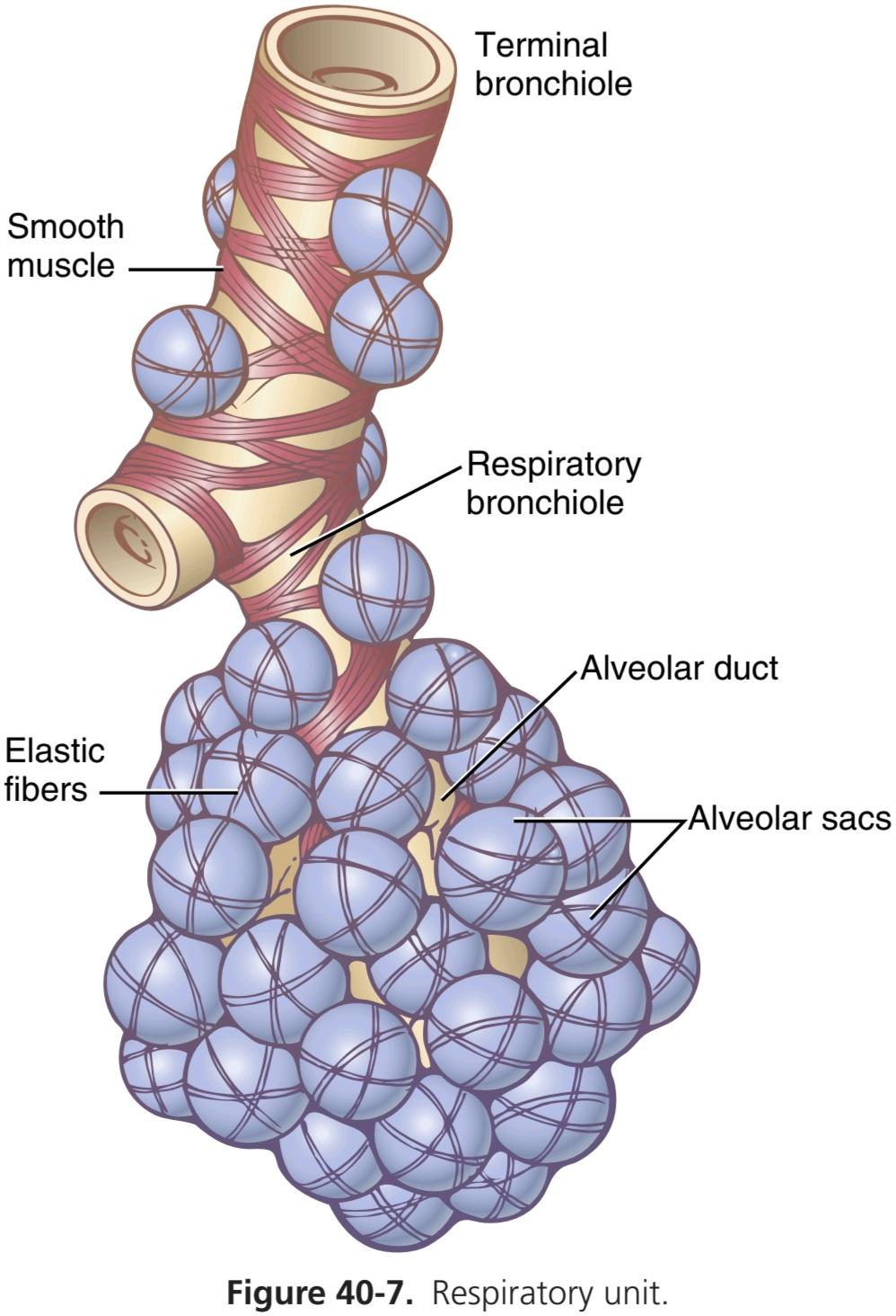
Figure 40-7. Respiratory unit.
شکل 40-7. واحد تنفس.
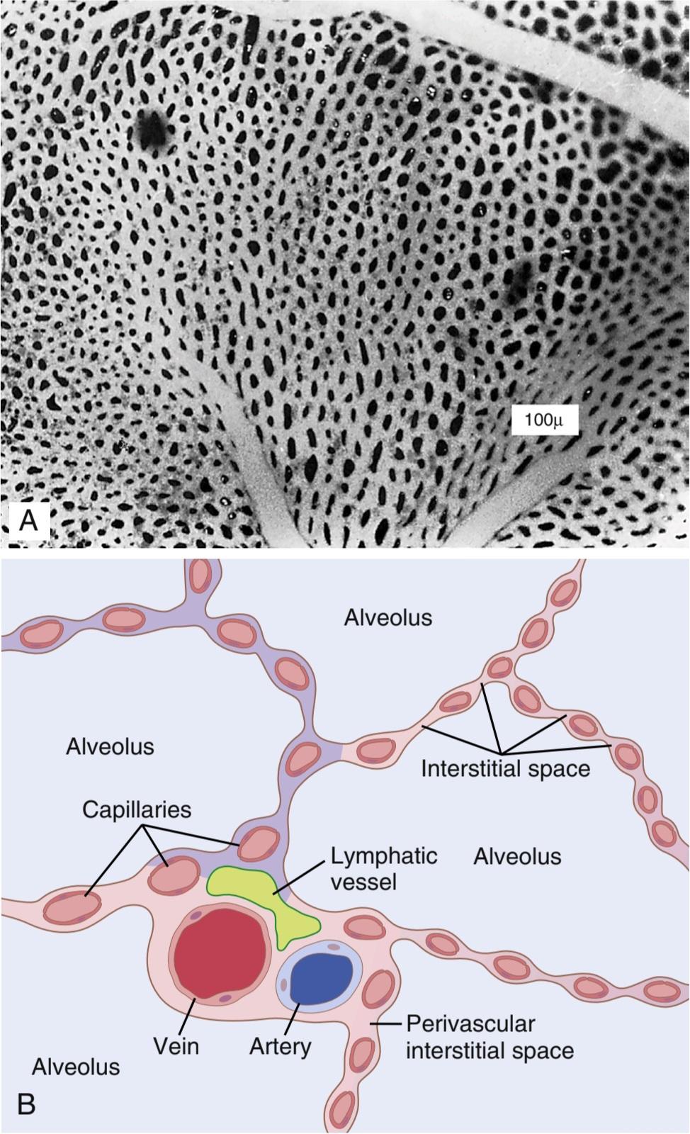
Figure 40-8. A, Surface view of capillaries in an alveolar wall. B, Cross-sectional view of alveolar walls and their vascular supply. (A, From Maloney JE, Castle BL: Pressure-diameter relations of capillaries and small blood vessels in frog lung. Respir Physiol 7:150, 1969.)
شکل 40-8. الف، نمای سطحی مویرگها در دیواره آلوئولی. ب، نمای مقطعی دیواره آلوئول و منبع عروقی آنها. (A, از Maloney JE, Castle BL: روابط فشار-قطر مویرگها و عروق خونی کوچک در ریه قورباغه. Respir Physiol 7:150, 1969.)
Respiratory Membrane. Figure 40-9 shows the ultrastructure of the respiratory membrane drawn in cross section on the left and a red blood cell on the right. It also shows diffusion of O2 from the alveolus into the red blood cell and diffusion of CO2 in the opposite direction. Note the following different layers of the respiratory membrane:
غشای تنفسی. شکل 40-9 ساختار فوق العاده غشای تنفسی را نشان میدهد که به صورت مقطعی در سمت چپ و یک گلبول قرمز در سمت راست کشیده شده است. همچنین انتشار O2 از آلوئول به گلبول قرمز و انتشار CO2 در جهت مخالف را نشان میدهد. به لایههای مختلف غشای تنفسی زیر توجه کنید:
1. A layer of fluid containing surfactant that lines the alveolus and reduces the surface tension of alveolar fluid
1. لایه ای از مایع حاوی سورفکتانت که آلوئول را میپوشاند و کشش سطحی مایع آلوئولی را کاهش میدهد.
2. The alveolar epithelium, composed of thin epithelial cells
2. اپیتلیوم آلوئولی، متشکل از سلولهای اپیتلیال نازک
3. An epithelial basement membrane
3. غشای پایه اپیتلیال
4. A thin interstitial space between the alveolar epithelium and capillary membrane
4. فضای بینابینی نازک بین اپیتلیوم آلوئولی و غشای مویرگی
5. A capillary basement membrane that in many places fuses with the alveolar epithelial basement membrane
5. یک غشای پایه مویرگی که در بسیاری از نقاط با غشای پایه اپیتلیال آلوئولی ترکیب میشود.
6. The capillary endothelial membrane
6. غشای اندوتلیال مویرگی
Despite the large number of layers, the overall thickness of the respiratory membrane in some areas is as little as 0.2 micrometer and averages about 0.6 micrometer, except where there are cell nuclei. From histological studies, it has been estimated that the total surface area of the respiratory membrane is about 70 square meters in healthy men, which is equivalent to the floor area of a 25 x 30-foot room. The total quantity of blood in the capillaries of the lungs at any given instant is 60 to 140 ml. Now, imagine this small amount of blood spread over the entire surface of a 25 x 30-foot floor, and it is easy to understand the rapidity of the respiratory exchange of O2 and CO2.
علیرغم تعداد زیاد لایهها، ضخامت کلی غشای تنفسی در برخی نواحی کمتر از 0.2 میکرومتر و به طور متوسط حدود 0.6 میکرومتر است، به جز جایی که هستههای سلولی وجود دارد. از مطالعات بافت شناسی تخمین زده شده است که سطح کل غشای تنفسی در مردان سالم حدود 70 متر مربع است که معادل سطح کف اتاقی به ابعاد 25×30 فوت است. مقدار کل خون در مویرگهای ریه در هر لحظه 60 تا 140 میلی لیتر است. حال تصور کنید که این مقدار کمیخون در تمام سطح یک طبقه 25×30 فوتی پخش شده است و به راحتی میتوان سرعت تبادل تنفسی O2 و CO2 را درک کرد.
The average diameter of the pulmonary capillaries is only about 5 micrometers, which means that red blood cells must squeeze through them. The red blood cell membrane usually touches the capillary wall, so O2 and CO2 need not pass through significant amounts of plasma as they diffuse between the alveolus and red blood cell. This, too, increases the rapidity of diffusion.
قطر متوسط مویرگهای ریوی تنها حدود 5 میکرومتر است، به این معنی که گلبولهای قرمز باید از طریق آنها فشرده شوند. غشای گلبول قرمز معمولاً دیواره مویرگی را لمس میکند، بنابراین O2 و CO2 نیازی به عبور از مقادیر قابل توجهی از پلاسما ندارند زیرا بین آلوئول و گلبول قرمز پخش میشوند. این نیز باعث افزایش سرعت انتشار میشود.

Figure 40-9. Ultrastructure of the alveolar respiratory membrane, shown in cross section.
شکل 40-9. فراساختار غشای تنفسی آلوئولی، در مقطع نشان داده شده است.
Factors Affecting Rate of Gas Diffusion Through the Respiratory Membrane
Referring to the earlier discussion of diffusion of gases in water, one can apply the same principles to diffusion of gases through the respiratory membrane. Thus, the factors that determine how rapidly a gas will pass through the membrane are the following: (1) the thickness of the membrane; (2) the surface area of the membrane; (3) the diffusion coefficient of the gas in the substance of the membrane; and (4) the partial pressure difference of the gas between the two sides of the membrane.
عوامل موثر بر سرعت انتشار گاز از طریق غشای تنفسی
با اشاره به بحث قبلی در مورد انتشار گازها در آب، میتوان همین اصول را برای انتشار گازها از طریق غشای تنفسی اعمال کرد. بنابراین، عواملی که تعیین میکنند گاز با چه سرعتی از غشا عبور میکند عبارتند از: (1) ضخامت غشاء. (2) سطح غشاء؛ (3) ضریب انتشار گاز در ماده غشاء؛ و (4) اختلاف فشار جزئی گاز بین دو طرف غشا.
The thickness of the respiratory membrane occasionally increases-for example, as a result of edema fluid in the interstitial space of the membrane and in the alveoli-so the respiratory gases must then diffuse not only through the membrane but also through this fluid. Also, some pulmonary diseases cause fibrosis of the lungs, which can increase the thickness of some portions of the respiratory membrane. Because the rate of diffusion through the membrane is inversely proportional to the thickness of the membrane, any factor that increases the thickness to more than two to three times normal can interfere significantly with normal respiratory exchange of gases.
ضخامت غشای تنفسی گهگاه افزایش مییابد – به عنوان مثال، در نتیجه ادم مایع در فضای بین بافتی غشاء و آلوئولها – بنابراین گازهای تنفسی باید نه تنها از طریق غشاء بلکه از طریق این مایع نیز پخش شوند. همچنین برخی از بیماریهای ریوی باعث فیبروز ریه میشود که میتواند ضخامت برخی از قسمتهای غشای تنفسی را افزایش دهد. از آنجایی که سرعت انتشار از طریق غشا با ضخامت غشا نسبت معکوس دارد، هر عاملی که ضخامت را به بیش از دو تا سه برابر نرمال افزایش دهد، میتواند به طور قابل توجهی در تبادل تنفسی طبیعی گازها اختلال ایجاد کند.
The surface area of the respiratory membrane can be greatly decreased by many conditions. For example, removal of an entire lung decreases the total surface area to half-normal. Also, in emphysema, many of the alveoli coalesce, with dissolution of many alveolar walls. Therefore, the new alveolar chambers are much larger than the original alveoli, but the total surface area of the respiratory membrane is often decreased as much as fivefold because of loss of the alveolar walls. When the total surface area is decreased to about one-third to one-fourth normal, exchange of gases through the membrane is substantially impeded, even under resting conditions, and during competitive sports and other strenuous exercise, even the slightest decrease in surface area of the lungs can be a serious detriment to respiratory exchange of gases.
سطح غشای تنفسی را میتوان به میزان زیادی تحت شرایط بسیاری کاهش داد. برای مثال، برداشتن کل ریه، سطح کل را به نصف طبیعی کاهش میدهد. همچنین، در آمفیزم، بسیاری از آلوئولها با انحلال بسیاری از دیوارههای آلوئول به هم میپیوندند. بنابراین، محفظههای آلوئولی جدید بسیار بزرگتر از آلوئولهای اصلی هستند، اما سطح کل غشای تنفسی به دلیل از بین رفتن دیوارههای آلوئولی اغلب تا پنج برابر کاهش مییابد. هنگامیکه سطح کل سطح به حدود یک سوم تا یک چهارم نرمال کاهش مییابد، تبادل گازها از طریق غشاء حتی در شرایط استراحت به طور قابل توجهی با مشکل مواجه میشود و در طول ورزشهای رقابتی و سایر ورزشهای شدید، حتی کوچکترین کاهش در سطح ریهها میتواند آسیب جدی به تبادل گازهای تنفسی باشد.
The diffusion coefficient for transfer of each gas through the respiratory membrane depends on the gas’s solubility in the membrane and, inversely, on the square root of the gas’s molecular weight. The rate of diffusion in the respiratory membrane is almost exactly the same as that in water, for reasons explained earlier. Therefore, for a given pressure difference, CO2 diffuses about 20 times as rapidly as O2. Oxygen diffuses about twice as rapidly as nitrogen.
ضریب انتشار برای انتقال هر گاز از طریق غشای تنفسی به حلالیت گاز در غشاء و برعکس به ریشه دوم وزن مولکولی گاز بستگی دارد. به دلایلی که قبلا توضیح داده شد، سرعت انتشار در غشای تنفسی تقریباً دقیقاً مشابه آب است. بنابراین، برای یک اختلاف فشار معین، CO2 حدود 20 برابر سریعتر از O2 منتشر میشود. اکسیژن تقریباً دو برابر سریعتر از نیتروژن منتشر میشود.
The pressure difference across the respiratory membrane is the difference between the partial pressure of the gas in the alveoli and the partial pressure of the gas in the pulmonary capillary blood. Therefore, the difference between these two pressures is a measure of the net tendency for the gas molecules to move through the membrane.
اختلاف فشار در سراسر غشای تنفسی، تفاوت بین فشار جزئی گاز در آلوئولها و فشار جزئی گاز در خون مویرگی ریوی است. بنابراین، تفاوت بین این دو فشار معیاری از تمایل خالص برای حرکت مولکولهای گاز در غشا است.
When the partial pressure of a gas in the alveoli is greater than the pressure of the gas in the blood, as is true for O2, net diffusion from the alveoli into the blood occurs. When the pressure of the gas in the blood is greater than the partial pressure in the alveoli, as is true for CO2, net diffusion from the blood into the alveoli occurs.
هنگامیکه فشار جزئی یک گاز در آلوئولها از فشار گاز موجود در خون بیشتر باشد، همانطور که برای O2 صادق است، انتشار خالص از آلوئولها به خون رخ میدهد. هنگامیکه فشار گاز در خون بیشتر از فشار جزئی در آلوئولها باشد، همانطور که برای CO2 صادق است، انتشار خالص از خون به آلوئولها رخ میدهد.
Diffusing Capacity of the Respiratory Membrane
The ability of the respiratory membrane to exchange a gas between the alveoli and pulmonary blood is expressed in quantitative terms by the respiratory membrane’s diffusing capacity, which is defined as the volume of a gas that will diffuse through the membrane each minute for a partial pressure difference of 1 mm Hg. All the factors discussed earlier that affect diffusion through the respiratory membrane can affect this diffusing capacity.
ظرفیت انتشار غشای تنفسی
توانایی غشای تنفسی برای تبادل گاز بین آلوئولها و خون ریوی به صورت کمیبا ظرفیت انتشار غشای تنفسی بیان میشود که به عنوان حجم گازی که در هر دقیقه از غشاء پخش میشود برای اختلاف فشار جزئی 1 میلی متر جیوه تعریف میشود. تمام عواملی که قبلاً مورد بحث قرار گرفت و بر انتشار از طریق غشای تنفسی تأثیر میگذارد، میتواند بر این ظرفیت انتشار تأثیر بگذارد.
Diffusing Capacity for Oxygen. In the average young man, the diffusing capacity for O2 under resting conditions averages 21 ml/min per mm Hg. In functional terms, what does this mean? The mean O2 pressure difference across the respiratory membrane during normal quiet breathing is about 11 mm Hg. Multiplying this pressure by the diffusing capacity (11 x 21) gives a total of about 230 ml of oxygen diffusing through the respiratory membrane each minute, which is equal to the rate at which the resting body uses O2.
ظرفیت انتشار برای اکسیژن. در یک مرد جوان متوسط، ظرفیت انتشار O2 در شرایط استراحت به طور متوسط 21 میلی لیتر در دقیقه بر میلی متر جیوه است. از نظر عملکردی، این به چه معناست؟ میانگین اختلاف فشار O2 در سراسر غشای تنفسی در طول تنفس آرام طبیعی حدود 11 میلی متر جیوه است. با ضرب این فشار در ظرفیت انتشار (11×21) در مجموع حدود 230 میلی لیتر اکسیژن در هر دقیقه از غشای تنفسی پخش میشود که برابر با سرعتی است که بدن در حال استراحت از O2 استفاده میکند.
Increased Oxygen Diffusing Capacity During Exercise. During strenuous exercise or other conditions that greatly increase pulmonary blood flow and alveolar ventilation, the diffusing capacity for O2 increases to about three times the diffusing capacity under resting conditions. This increase is caused by several factors, including the following: (1) opening up of many previously dormant pulmonary capillaries or extra dilation of already open capillaries, thereby increasing the surface area of the blood into which the O2 can diffuse; and (2) a better match between the ventilation of the alveoli and perfusion of the alveolar capillaries with blood, called the ventilation-perfusion ratio, explained later in this chapter. Therefore, during exercise, oxygenation of the blood is increased not only by increased alveolar ventilation but also by greater diffusing capacity of the respiratory membrane for transporting O2 into the blood.
افزایش ظرفیت انتشار اکسیژن در حین ورزش. در طول ورزش شدید یا سایر شرایطی که جریان خون ریوی و تهویه آلوئولی را به شدت افزایش میدهد، ظرفیت انتشار O2 تا حدود سه برابر ظرفیت انتشار در شرایط استراحت افزایش مییابد. این افزایش توسط عوامل متعددی ایجاد میشود، از جمله موارد زیر: (1) باز شدن بسیاری از مویرگهای ریوی که قبلاً خوابیده بودند یا اتساع اضافی مویرگهای از قبل باز، در نتیجه سطح خونی که O2 میتواند در آن منتشر شود، افزایش مییابد. و (2) تطابق بهتر بین تهویه آلوئولها و پرفیوژن مویرگهای آلوئولی با خون، به نام نسبت تهویه به پرفیوژن، که بعداً در این فصل توضیح داده شد. بنابراین، در حین ورزش، اکسیژن رسانی به خون نه تنها با افزایش تهویه آلوئولی، بلکه با افزایش ظرفیت انتشار غشای تنفسی برای انتقال O2 به خون افزایش مییابد.
Diffusing Capacity for Carbon Dioxide. The diffusing capacity for CO2 has never been measured because CO2 diffuses through the respiratory membrane so rapidly that the average Pco, in the pulmonary blood is not very different from the Pco, in the alveoli-the average difference is less than 1 mm Hg. With currently available techniques, this difference is too small to be measured.
ظرفیت انتشار دی اکسید کربن. ظرفیت انتشار CO2 هرگز اندازه گیری نشده است زیرا CO2 به سرعت در غشای تنفسی منتشر میشود که میانگین Pco در خون ریوی تفاوت زیادی با Pco ندارد، در آلوئولها – میانگین اختلاف کمتر از 1 میلی متر جیوه است. با تکنیکهای موجود در حال حاضر، این تفاوت برای اندازه گیری بسیار کوچک است.
Nevertheless, measurements of diffusion of other gases have shown that the diffusing capacity varies directly with the diffusion coefficient of the particular gas. Because the diffusion coefficient of CO2 is slightly more than 20 times that of O2, one would expect a diffusing capacity for CO2 under resting conditions of about 400 to 450 ml/min per mm Hg and during exercise of about 1200 to 1300 ml/ min per mm Hg. Figure 40-10 compares the measured or calculated diffusing capacities of carbon monoxide, O2, and CO2 at rest and during exercise, showing the extreme diffusing capacity of CO2 and the effect of exercise on the diffusing capacity of each of these gases.
با این وجود، اندازهگیریهای انتشار گازهای دیگر نشان داده است که ظرفیت انتشار مستقیماً با ضریب انتشار گاز خاص متفاوت است. از آنجایی که ضریب انتشار CO2 کمیبیشتر از 20 برابر O2 است، میتوان انتظار داشت که ظرفیت انتشار CO2 در شرایط استراحت حدود 400 تا 450 میلی لیتر در دقیقه بر میلی متر جیوه و در حین ورزش حدود 1200 تا 1300 میلی لیتر در دقیقه بر میلی متر جیوه باشد. شکل 40-10 ظرفیتهای اندازه گیری شده یا محاسبه شده انتشار مونوکسید کربن، O2 و CO2 را در حالت استراحت و در حین تمرین مقایسه میکند و ظرفیت انتشار شدید CO2 و تأثیر ورزش بر ظرفیت انتشار هر یک از این گازها را نشان میدهد.
Measurement of Diffusing Capacity-Carbon Monoxide Method. The O2 diffusing capacity can be calculated from measurements of the following: (1) alveolar Po2; (2) Po, in the pulmonary capillary blood; and (3) the rate of O2 uptake by the blood. However, measuring the Po2 in the pulmonary capillary blood is so difficult and imprecise that it is not practical to measure oxygen diffusing capacity by such a direct procedure, except on an experimental basis.
اندازه گیری ظرفیت انتشار – روش مونوکسید کربن. ظرفیت انتشار O2 را میتوان از اندازه گیریهای زیر محاسبه کرد: (1) Po2 آلوئولی. (2) پو، در خون مویرگی ریوی. و (3) سرعت جذب O2 توسط خون. با این حال، اندازهگیری Po2 در خون مویرگ ریوی آنقدر دشوار و نادقیق است که اندازهگیری ظرفیت انتشار اکسیژن با چنین روش مستقیمی، جز بر اساس یک مبنای تجربی، عملی نیست.
To obviate the difficulties encountered in measuring oxygen diffusing capacity directly, physiologists usually measure carbon monoxide (CO) diffusing capacity instead and then calculate the O2 diffusing capacity from this. The principle of the CO method is as follows. A small amount of CO is breathed into the alveoli, and the partial pressure of the CO in the alveoli is measured from appropriate alveolar air samples. The CO pressure in the blood is essentially zero because hemoglobin combines with this gas so rapidly that its pressure never has time to build up. Therefore, the pressure difference of CO across the respiratory membrane is equal to its partial pressure in the alveolar air sample. Then, by measuring the volume of CO absorbed in a short period and dividing this by the alveolar CO partial pressure, one can determine the CO diffusing capacity accurately.
برای رفع مشکلاتی که در اندازه گیری ظرفیت انتشار اکسیژن به طور مستقیم با آن مواجه میشود، فیزیولوژیستها معمولا ظرفیت انتشار مونوکسید کربن (CO) را اندازه گیری میکنند و سپس ظرفیت انتشار O2 را از این طریق محاسبه میکنند. اصل روش CO به شرح زیر است. مقدار کمیCO به داخل آلوئولها تنفس میشود و فشار جزئی CO در آلوئولها از نمونههای هوای آلوئولی مناسب اندازه گیری میشود. فشار CO در خون اساساً صفر است، زیرا هموگلوبین با این گاز به سرعت ترکیب میشود که فشار آن هرگز زمان زیادی برای افزایش نمییابد. بنابراین، اختلاف فشار CO در سراسر غشای تنفسی برابر با فشار جزئی آن در نمونه هوای آلوئولی است. سپس با اندازه گیری حجم CO جذب شده در یک دوره کوتاه و تقسیم آن بر فشار جزئی CO آلوئولی، میتوان ظرفیت انتشار CO را به دقت تعیین کرد.
To convert CO diffusing capacity to O2 diffusing capacity, the value is multiplied by a factor of 1.23 because the diffusion coefficient for O2 is 1.23 times that for CO. Thus, the average diffusing capacity for CO in healthy young men at rest is 17 ml/min per mm Hg, and the diffusing capacity for O2 is 1.23 times this, or 21 ml/min per mm Hg.
برای تبدیل ظرفیت انتشار CO به ظرفیت انتشار O2، مقدار در ضریب 1.23 ضرب میشود، زیرا ضریب انتشار برای O2 1.23 برابر CO است. بنابراین، میانگین ظرفیت انتشار CO در مردان جوان سالم در حالت استراحت 17 میلی لیتر در دقیقه بر میلی متر جیوه و ظرفیت انتشار برای O21 برابر این مقدار در میلی لیتر در دقیقه، 1.23 است.

Figure 40-10. Diffusing capacities for carbon monoxide, oxygen, and carbon dioxide in the normal lungs under resting conditions and during exercise.
شکل 40-10. ظرفیت انتشار مونوکسید کربن، اکسیژن و دی اکسید کربن در ریههای طبیعی در شرایط استراحت و در حین ورزش.
Effect of Ventilation-Perfusion Ratio on Alveolar Gas Concentration
Earlier in this chapter, we learned that two factors determine the Po2 and Pco, in the alveoli: (1) the rate of alveolar ventilation; and (2) the rate of transfer of O2 and CO2 through the respiratory membrane. This discussion made the assumption that all the alveoli are ventilated equally, and that blood flow through the alveolar capillaries is the same for each alveolus. However, even normally to some extent, and especially in many lung diseases, some areas of the lungs are well ventilated but have almost no blood flow, whereas other areas may have excellent blood flow but little or no ventilation. In either of these conditions, gas exchange through the respiratory membrane is seriously impaired, and the person may suffer severe respiratory distress, despite normal total ventilation and normal total pulmonary blood flow, but with the ventilation and blood flow going to different parts of the lungs. Therefore, a highly quantitative concept has been developed to help us understand respiratory exchange when there is imbalance between alveolar ventilation and alveolar blood flow. This concept is called the ventilation-perfusion ratio.
تأثیر نسبت تهویه – پرفیوژن بر غلظت گاز آلوئولی
در اوایل این فصل، یاد گرفتیم که دو عامل Po2 و Pco را در آلوئولها تعیین میکنند: (1) میزان تهویه آلوئولی. و (2) سرعت انتقال O2 و CO2 از طریق غشای تنفسی. این بحث این فرض را ایجاد کرد که تمام آلوئولها به طور مساوی تهویه میشوند و جریان خون از طریق مویرگهای آلوئولی برای هر آلوئول یکسان است. با این حال، حتی به طور معمول تا حدودی، و به خصوص در بسیاری از بیماریهای ریوی، برخی از نواحی ریهها به خوبی تهویه میشوند اما تقریباً جریان خون ندارند، در حالی که سایر نواحی ممکن است جریان خون عالی داشته باشند اما تهویه کم یا بدون تهویه باشد. در هر یک از این شرایط، تبادل گاز از طریق غشای تنفسی به طور جدی مختل میشود و فرد ممکن است با وجود تهویه کامل طبیعی و جریان خون کلی ریوی طبیعی، اما با تهویه و جریان خون به قسمتهای مختلف ریه، دچار ناراحتی شدید تنفسی شود. بنابراین، یک مفهوم بسیار کمیایجاد شده است تا به ما کمک کند تبادل تنفسی را در زمانی که عدم تعادل بین تهویه آلوئولی و جریان خون آلوئولی وجود دارد، درک کنیم. این مفهوم نسبت تهویه-پرفیوژن نامیده میشود.
In quantitative terms, the ventilation-perfusion ratio is expressed as VA/Q. When VA (alveolar ventilation) is normal for a given alveolus, and Q (blood flow) is also normal for the same alveolus, the ventilation-perfusion ratio (VA/Q) is also said to be normal. When the ventilation (VA) is zero, yet there is still perfusion (Q) of the alveolus, the VA/Q is zero. Or, at the other extreme, when there is adequate ventilation (VA) but zero perfusion (Q), the ratio VA/Q is infinity. At a ratio of either zero or infinity, there is no exchange of gases through the respiratory membrane of the affected alveoli. Therefore, let us explain the respiratory consequences of these two extremes.
از نظر کمی، نسبت تهویه-پرفیوژن به صورت VA/Q بیان میشود. هنگامیکه VA (تهویه آلوئولی) برای یک آلوئول خاص طبیعی است، و Q (جریان خون) نیز برای همان آلوئول طبیعی است، نسبت تهویه به پرفیوژن (VA/Q) نیز طبیعی است. هنگامیکه تهویه (VA) صفر است، اما هنوز پرفیوژن (Q) آلوئول وجود دارد، VA/Q صفر است. یا در نهایت، وقتی تهویه کافی (VA) اما پرفیوژن صفر (Q) وجود دارد، نسبت VA/Q بی نهایت است. در نسبت صفر یا بی نهایت، هیچ تبادل گازی از طریق غشای تنفسی آلوئولهای آسیب دیده وجود ندارد. بنابراین، اجازه دهید پیامدهای تنفسی این دو افراط را توضیح دهیم.
Alveolar Oxygen and Carbon Dioxide Partial Pressures When VA/Q Equals Zero. When VA/Q is equal to zero-that is, without any alveolar ventilation-the air in the alveolus comes to equilibrium with the blood O2 and CO2 because these gases diffuse between the blood and alveolar air. Because the blood that perfuses the capillaries is venous blood returning to the lungs from the systemic circulation, it is the gases in this blood with which the alveolar gases equilibrate. In Chapter 41, we describe how the normal venous blood (V) has a Po2 of 40 mm Hg and a Pco, of 45 mm Hg. Therefore, these are also the normal partial pressures of these two gases in alveoli that have blood flow but no ventilation.
فشارهای جزئی اکسیژن آلوئولی و دی اکسید کربن وقتی VA/Q برابر با صفر است. وقتی VA/Q برابر با صفر است، یعنی بدون تهویه آلوئولی، هوا در آلوئول با O2 و CO2 خون به تعادل میرسد زیرا این گازها بین خون و هوای آلوئولی پخش میشوند. از آنجایی که خونی که مویرگها را پرفیوژن میکند، خون وریدی است که از گردش سیستمیک به ریهها برمیگردد، گازهای موجود در این خون هستند که گازهای آلوئولی با آنها تعادل برقرار میکنند. در فصل 41، توضیح میدهیم که چگونه خون وریدی طبیعی (V) دارای Po2 40 میلی متر جیوه و Pco 45 میلی متر جیوه است. بنابراین، اینها نیز فشارهای جزئی طبیعی این دو گاز در آلوئولها هستند که جریان خون دارند اما تهویه ندارند.
Alveolar Oxygen and Carbon Dioxide Partial Pressures When VA/Q Equals Infinity. The effect on the alveolar gas partial pressures when VA/Q equals infinity is entirely different from the effect when VA/Q equals zero because now there is no capillary blood flow to carry O2 away or to bring CO2 to the alveoli. Therefore, instead of the alveolar gases coming to equilibrium with the venous blood, the alveolar air becomes equal to the humidified inspired air. That is, the air that is inspired loses no O2 to the blood and gains no CO2 from the blood. Furthermore, because normal inspired and humidified air has a Po2 of 149 mm Hg and a Pco, of 0 mm Hg, these will be the partial pressures of these two gases in the alveoli.
فشارهای جزئی اکسیژن و دی اکسید کربن آلوئولی هنگامیکه VA/Q برابر با بی نهایت است. تأثیر بر فشارهای جزئی گاز آلوئولی زمانی که VA/Q برابر با بی نهایت است، کاملاً متفاوت از تأثیر زمانی است که VA/Q برابر با صفر است، زیرا اکنون جریان خون مویرگی برای انتقال O2 یا آوردن CO2 به آلوئول وجود ندارد. بنابراین، به جای اینکه گازهای آلوئولی با خون وریدی به تعادل برسند، هوای آلوئولی برابر با هوای دمیده شده مرطوب میشود. یعنی هوایی که دم میشود هیچ O2 خون از دست نمیدهد و هیچ CO2 از خون به دست نمیآورد. علاوه بر این، چون هوای نرمال دمیده و مرطوب شده دارای Po2 149 میلی متر جیوه و Pco 0 میلی متر جیوه است، این فشارهای جزئی این دو گاز در آلوئولها خواهد بود.
Gas Exchange and Alveolar Partial Pressures When VA/Q Is Normal. When there is both normal alveolar ventilation and normal alveolar capillary blood flow (normal alveolar perfusion), exchange of O2 and CO2 through the respiratory membrane is nearly optimal, and alveolar Po2 is normally at a level of 104 mm Hg, which lies between that of the inspired air (149 mm Hg) and that of venous blood (40 mm Hg). Likewise, alveolar Pco, lies between two extremes; it is normally 40 mm Hg, in contrast to 45 mm Hg in venous blood and 0 mm Hg in inspired air. Thus, under normal conditions, the alveolar air Po2 averages 104 mm Hg and the Pco2 averages 40 mm Hg.
تبادل گاز و فشارهای جزئی آلوئولی وقتی VA/Q طبیعی است. هنگامیکه هم تهویه طبیعی آلوئولی و هم جریان خون مویرگی آلوئولی طبیعی وجود دارد (پرفیوژن آلوئولی طبیعی)، تبادل O2 و CO2 از طریق غشای تنفسی تقریباً بهینه است و Po2 آلوئولی معمولاً در سطح 104 میلیمتر جیوه است که بین هوای دمیده شده (149 میلیمتر Hg خون) و 4 میلیمتر جیوه قرار دارد. به همین ترتیب، Pco آلوئولی، بین دو حد قرار دارد. به طور معمول 40 میلی متر جیوه است، در مقابل 45 میلی متر جیوه در خون وریدی و 0 میلی متر جیوه در هوای دمیده شده. بنابراین، در شرایط عادی، هوای آلوئولی Po2 به طور متوسط 104 میلی متر جیوه و Pco2 به طور متوسط 40 میلی متر جیوه است.
Po2-PCO2, VA/Q Diagram
The concepts presented in the preceding sections are shown in graphic form in Figure 40-11, called the Po2- PCO2, VA/Q diagram. The curve in the diagram represents all possible Po2 and Pco, combinations between the limits of VA/Q equals zero and VA/Q equals infinity when the gas pressures in the venous blood are normal, and the person is breathing air at sea level pressure. Thus, point VA/Q is the plot of Po2 and Pco2 when VA/Q equals zero. At this point,the Po2 is 40 mm Hg, and the Pco, is 45 mm Hg, which are the values in normal venous blood.
نمودار Po2-PCO2، VA/Q
مفاهیم ارائه شده در بخشهای قبل به صورت گرافیکی در شکل 40-11 نشان داده شده است که نمودار Po2-PCO2، VA/Q نامیده میشود. منحنی در نمودار نشان دهنده تمام Po2 و Pco ممکن است، ترکیبات بین حدود VA/Q برابر با صفر و VA/Q برابر بی نهایت است، زمانی که فشار گاز در خون وریدی طبیعی است، و فرد در حال تنفس هوا در فشار سطح دریا است. بنابراین، نقطه VA/Q نمودار Po2 و Pco2 است که VA/Q برابر با صفر باشد. در این مرحله، Po2 40 میلی متر جیوه، و Pco، 45 میلی متر جیوه است که مقادیر موجود در خون وریدی طبیعی است.
At the other end of the curve, when VA/Q equals infinity, point I represents inspired air, showing Po2 to be 149 mm Hg while Pco, is zero. Also plotted on the curve is the point that represents normal alveolar air when VA/Q is normal. At this point, Po2 is 104 mm Hg, and Pco, is 40 mm Hg.
در انتهای دیگر منحنی، زمانی که VA/Q برابر با بینهایت است، نقطه I نشاندهنده هوای الهامشده است که نشان میدهد Po2 149 میلیمتر جیوه است در حالی که Pco صفر است. همچنین بر روی منحنی نقطه ای است که هوای آلوئولی طبیعی را در زمانی که VA/Q نرمال است نشان میدهد. در این مرحله، Po2 104 میلی متر جیوه و Pco، 40 میلی متر جیوه است.

Figure 40-11. Normal partial pressure of oxygen (Po2)-partial pressure of carbon dioxide (Pco2) ventilation-perfusion (VA/Q) ratio (Po2- Pco2, (VA/Q) diagram.
شکل 40-11. فشار جزئی طبیعی اکسیژن (Po2) – فشار جزئی دی اکسید کربن (Pco2) نسبت تهویه – پرفیوژن (VA/Q) (نمودار Po2– Pco2، (VA/Q).
Concept of Physiological Shunt (When VA/Q Is Below Normal)
Whenever VA/Q is below normal, there is inadequate ventilation to provide the O2 needed to fully oxygenate the blood flowing through the alveolar capillaries. Therefore, a certain fraction of the venous blood passing through the pulmonary capillaries does not become oxygenated. This fraction is called shunted blood. Also, some additional blood flows through bronchial vessels rather than through alveolar capillaries, normally about 2% of the cardiac output; this, too, is unoxygenated, shunted blood.
مفهوم شنت فیزیولوژیکی (زمانی که VA/Q کمتر از حد نرمال است)
هر زمان که VA/Q کمتر از حد نرمال باشد، تهویه ناکافی برای تامین اکسیژن مورد نیاز برای اکسیژن رسانی کامل خون جریان یافته از طریق مویرگهای آلوئولی وجود دارد. بنابراین، بخش خاصی از خون وریدی که از مویرگهای ریوی عبور میکند، اکسیژن نمییابد. این کسری خون شنت شده نامیده میشود. همچنین، مقداری خون اضافی به جای مویرگهای آلوئولی از طریق عروق برونش جریان مییابد که معمولاً حدود 2% از برون ده قلبی است. این نیز خون بدون اکسیژن و شنت شده است.
The total quantitative amount of shunted blood per minute is called the physiological shunt. This physiological shunt is measured in clinical pulmonary function laboratories by analyzing the concentration of O2 in both mixed venous blood and arterial blood, along with simultaneous measurement of cardiac output. From these values, the physiological shunt can be calculated by the following equation:
کل مقدار کمیخون شنت شده در دقیقه را شانت فیزیولوژیکی میگویند. این شنت فیزیولوژیکی در آزمایشگاههای بالینی عملکرد ریوی با تجزیه و تحلیل غلظت O2 در خون وریدی و خون شریانی، همراه با اندازهگیری همزمان برون ده قلبی اندازهگیری میشود. از این مقادیر، شنت فیزیولوژیکی را میتوان با معادله زیر محاسبه کرد:

in which Qps is the physiological shunt blood flow per minute, Q is cardiac output per minute, Cio2 is the concentration of oxygen in the arterial blood if there is an “ideal” ventilation-perfusion ratio, Cao2 is the measured concentration of oxygen in the arterial blood, and CVO2 is the measured concentration of oxygen in the mixed venous blood.
که در آن Qps جریان خون شنت فیزیولوژیکی در دقیقه است، Q برون ده قلبی در دقیقه، Cio2 غلظت اکسیژن در خون شریانی در صورتی که نسبت تهویه به پرفیوژن “ایده آل” وجود داشته باشد، Cao2 غلظت اندازه گیری شده اکسیژن در خون شریانی و CVO2 غلظت اندازه گیری شده اکسیژن در خون است.
The greater the physiological shunt, the greater the amount of blood that fails to be oxygenated as it passes through the lungs.
هر چه شنت فیزیولوژیکی بیشتر باشد، مقدار خونی که در هنگام عبور از ریهها اکسیژن رسانی نمیشود، بیشتر میشود.
Concept of Physiological Dead Space When VA/Q Greater Than Normal
When ventilation of some of the alveoli is great but alveolar blood flow is low, there is far more available oxygen in the alveoli than can be transported away from the alveoli by the flowing blood. Thus, the ventilation of these alveoli is said to be wasted. The ventilation of the anatomical dead space areas of the respiratory passageways is also wasted. The sum of these two types of wasted ventilation is called the physiological dead space. This space is measured in the clinical pulmonary function laboratory by making appropriate blood and expiratory gas measurements and by using the following equation, called the Bohr equation:
مفهوم فضای مرده فیزیولوژیکی زمانی که VA/Q بزرگتر از حالت عادی باشد
هنگامیکه تهویه برخی از آلوئولها عالی است اما جریان خون آلوئولی کم است، اکسیژن موجود در آلوئولها بسیار بیشتر از آن چیزی است که میتواند توسط جریان خون از آلوئولها خارج شود. بنابراین گفته میشود تهویه این آلوئولها هدر میرود. تهویه نواحی فضای مرده آناتومیکی راههای تنفسی نیز به هدر میرود. مجموع این دو نوع تهویه هدر رفته فضای مرده فیزیولوژیکی نامیده میشود. این فضا در آزمایشگاه عملکرد ریوی بالینی با اندازه گیری مناسب خون و گازهای بازدمیو با استفاده از معادله زیر به نام معادله بور اندازه گیری میشود:

in which VDphys is the physiological dead space, VT is the tidal volume, Paco2 is the partial pressure of CO2 in the arterial blood, and Peco2 is the average partial pressure of CO2 in the entire expired air.
که در آن VDphys فضای مرده فیزیولوژیکی است، VT حجم جزر و مدی، Paco2 فشار جزئی CO2 در خون شریانی و Peco2 میانگین فشار جزئی CO2 در کل هوای بازدم شده است.
When the physiological dead space is great, much of the work of ventilation is wasted effort because so much of the ventilating air never reaches the blood.
وقتی فضای مرده فیزیولوژیکی بزرگ باشد، بیشتر کار تهویه به هدر میرود زیرا مقدار زیادی از هوای تهویه هرگز به خون نمیرسد.
Abnormalities of Ventilation-Perfusion Ratio
Abnormal VA/Q in Upper and Lower Normal Lung. In a healthy person in the upright position, both pulmonary capillary blood flow and alveolar ventilation are consider- ably less in the upper part of the lung than in the lower part; however, the decrease of blood flow is considerably greater than the decrease in ventilation. Therefore, at the top of the lung, VA/Q is as much as 2.5 times as great as the ideal value, which causes a moderate degree of physiological dead space in this area of the lung.
ناهنجاریهای نسبت تهویه به پرفیوژن
VA/Q غیر طبیعی در ریه طبیعی بالا و پایین. در یک فرد سالم در وضعیت عمودی، هم جریان خون مویرگی ریوی و هم تهویه آلوئولار در قسمت فوقانی ریه به میزان قابل توجهی کمتر از قسمت تحتانی است. با این حال، کاهش جریان خون به طور قابل توجهی بیشتر از کاهش تهویه است. بنابراین، در بالای ریه، VA/Q به اندازه 2.5 برابر مقدار ایده آل است، که باعث ایجاد درجه متوسطی از فضای مرده فیزیولوژیکی در این ناحیه از ریه میشود.
At the other extreme, at the bottom of the lung, there is slightly too little ventilation in relation to blood flow, with VA/Q as low as 0.6 times the ideal value. In this area, a small fraction of the blood fails to become normally oxygenated, and this represents a physiological shunt.
در انتهای دیگر، در پایین ریه، تهویه کمینسبت به جریان خون وجود دارد، با VA/Q کمتر از 0.6 برابر مقدار ایده آل. در این ناحیه، بخش کوچکی از خون به طور طبیعی اکسیژن رسانی نمیشود و این نشان دهنده یک شانت فیزیولوژیکی است.
In both extremes, inequalities of ventilation and perfusion slightly decrease the lung’s effectiveness for exchanging O2 and CO2. However, during exercise, blood flow to the upper part of the lung increases markedly, so far less physiological dead space occurs, and the effectiveness of gas exchange now approaches optimum.
در هر دو حالت، نابرابریهای تهویه و پرفیوژن اندکی اثربخشی ریه را برای تبادل O2 و CO2 کاهش میدهد. با این حال، در طول ورزش، جریان خون در قسمت بالایی ریه به طور قابل توجهی افزایش مییابد، تا کنون فضای مرده فیزیولوژیکی کمتری رخ میدهد، و کارایی تبادل گاز اکنون به حد مطلوب نزدیک میشود.
Abnormal VA/Q in Chronic Obstructive Lung Disease
Most people who smoke for many years develop various degrees of bronchial obstruction; in many of them, this condition eventually becomes so severe that serious alveolar air trapping develops, with resultant emphysema. The emphysema, in turn, causes many of the alveolar walls to be destroyed. Thus, two abnormalities occur in smokers to cause abnormal VA/Q. First, because many of the small bronchioles are obstructed, the alveoli beyond the obstructions are unventilated, causing a VA/Q that approaches zero. Second, in the areas of the lung where the alveolar walls have mainly been destroyed but there is still alveolar ventilation, most of the ventilation is wasted because of inadequate blood flow to transport the blood gases.
VA/Q غیر طبیعی در بیماری مزمن انسدادی ریه
اکثر افرادی که سالها سیگار میکشند به درجات مختلفی از انسداد برونش مبتلا میشوند. در بسیاری از آنها، این وضعیت در نهایت به قدری شدید میشود که گیر افتادن هوای آلوئولی جدی، همراه با آمفیزم ایجاد میشود. آمفیزم به نوبه خود باعث از بین رفتن بسیاری از دیوارههای آلوئول میشود. بنابراین، دو ناهنجاری در افراد سیگاری رخ میدهد که باعث ایجاد VA/Q غیر طبیعی میشود. اولاً، چون بسیاری از برونشیولهای کوچک مسدود شدهاند، آلوئولهای فراتر از انسدادها تهویه نمیشوند و باعث ایجاد VA/Q میشوند که به صفر نزدیک میشود. دوم، در مناطقی از ریه که دیوارههای آلوئولی عمدتاً تخریب شدهاند، اما هنوز تهویه آلوئولی وجود دارد، بیشتر تهویه به دلیل جریان ناکافی خون برای انتقال گازهای خونی تلف میشود.
Thus, in chronic obstructive lung disease, some areas of the lung exhibit serious physiological shunt, and other areas exhibit serious physiological dead space. Both conditions tremendously decrease the effectiveness of the lungs as gas exchange organs, sometimes reducing their effectiveness to as little as one-tenth normal. In fact, this condition is the most prevalent cause of pulmonary disability today.
بنابراین، در بیماری مزمن انسدادی ریه، برخی از نواحی ریه شانت فیزیولوژیکی جدی را نشان میدهند و سایر نواحی فضای مرده فیزیولوژیکی جدی را نشان میدهند. هر دو شرایط به شدت اثربخشی ریهها را به عنوان اندامهای تبادل گاز کاهش میدهند و گاهی اوقات اثربخشی آنها را به یک دهم طبیعی کاهش میدهند. در واقع، این وضعیت امروزه شایع ترین علت ناتوانی ریوی است.
کلیک کنید «Bibliography: فهرست کتب مربوطه»
Clark A, Tawhai M: Pulmonary vascular dynamics. Compr Physiol 9:1081, 2019.
Del Buono MG, Arena R, Borlaug BA, Carbone S, et al: Exercise intol- erance in patients with heart failure: JACC state-of-the-art review. J Am Coll Cardiol 73:2209, 2019.
Dempsey TM, Scanlon PD: Pulmonary function tests for the general- ist: a brief review. Mayo Clin Proc 93:763, 2018.
Glenny RW, Robertson HT: Spatial distribution of ventilation and per- fusion: mechanisms and regulation. Compr Physiol 1:375, 2011.
Hsia CC, Hyde DM, Weibel ER: Lung structure and the intrinsic chal- lenges of gas exchange. Compr Physiol 6:827, 2016.
Molgat-Seon Y, Schaeffer MR, Ryerson CJ, Guenette JA: Exer- cise pathophysiology in interstitial lung disease. Clin Chest Med 40:405, 2019.
Naeije R, Chesler N: Pulmonary circulation at exercise. Compr Physiol 2:711, 2012.
Neder JA, Berton DC, Muller PT, O’Donnell DE: Incorporating lung diffusing capacity for carbon monoxide in clinical decision making in chest medicine. Clin Chest Med 40:285, 2019.
O’Donnell DE, James MD, Milne KM, Neder JA: the pathophysiology of dyspnea and exercise intolerance in chronic obstructive pulmo- nary disease. Clin Chest Med 40:343, 2019.
Rahn H, Farhi EE: Ventilation, perfusion, and gas exchange-the Va/Q concept. In: Fenn WO, Rahn H (eds): Handbook of Physiology. Sec 3, Vol 1. Baltimore: Williams & Wilkins, 1964, p 125.
Robertson HT: Dead space: the physiology of wasted ventilation. Eur Respir J 45:1704, 2015.
Skloot GS: The Effects of aging on lung structure and function. Clin Geriatr Med 33:447, 2017.
Stickland MK, Lindinger MI, Olfert IM, Heigenhauser GJ, Hopkins SR: Pulmonary gas exchange and acid-base balance during exercise. Compr Physiol 3:693, 2013.
Wagner PD: The physiological basis of pulmonary gas exchange: implications for clinical interpretation of arterial blood gases. Eur Respir J 45:227, 2015
Weibel ER: Lung morphometry: the link between structure and func- tion. Cell Tissue Res 367:413, 2017.
West JB: Role of the fragility of the pulmonary blood-gas barrier in the evolution of the pulmonary circulation. Am J Physiol Regul In- tegr Comp Physiol 304:R171, 2013.
🚀 با ما همراه شوید!
تازهترین مطالب و آموزشهای مغز و اعصاب را از دست ندهید. با فالو کردن کانال تلگرام آیندهنگاران مغز، از ما حمایت کنید!



