
فیزیولوژی پزشکی گایتون و هال؛ گردش خون ریوی، ادم ریوی و مایع پلورال


دعای مطالعه [ نمایش ]
بِسْمِ الله الرَّحْمنِ الرَّحیمِ
اَللّهُمَّ اَخْرِجْنى مِنْ ظُلُماتِ الْوَهْمِ
خدايا مرا بيرون آور از تاريكىهاى وهم،
وَ اَكْرِمْنى بِنُورِ الْفَهْمِ
و به نور فهم گرامى ام بدار،
اَللّهُمَّ افْتَحْ عَلَيْنا اَبْوابَ رَحْمَتِكَ
خدايا درهاى رحمتت را به روى ما بگشا،
وَانْشُرْ عَلَيْنا خَزائِنَ عُلُومِكَ بِرَحْمَتِكَ يا اَرْحَمَ الرّاحِمينَ
و خزانههاى علومت را بر ما باز كن به امید رحمتت اى مهربانترين مهربانان.
کتاب «فیزیولوژی پزشکی گایتون و هال» بهعنوان یکی از جامعترین و معتبرترین منابع در حوزه علوم پزشکی، همچنان مرجع کلیدی برای درک عملکرد پیچیده بدن انسان است. این اثر با تکیه بر تازهترین پژوهشها و توضیحات دقیق از سازوکارهای فیزیولوژیک، پلی میان علوم پایه پزشکی و کاربردهای بالینی ایجاد میکند و نقشی بیبدیل در آموزش، پژوهش و ارتقای دانش سلامت ایفا مینماید.
ترجمه دقیق و علمی این شاهکار توسط برند علمی آیندهنگاران مغز به مدیریت داریوش طاهری، دسترسی فارسیزبانان به مرزهای نوین دانش فیزیولوژی را ممکن ساخته و رسالتی علمی برای ارتقای آموزش پزشکی، فهم عمیقتر سازوکارهای بدن و توسعه روشهای نوین در حوزه سلامت فراهم آورده است.
» Guyton and Hall Textbook of Medical Physiology
»» CHAPTER 39: Pulmonary Circulation, Pulmonary Edema, and Pleural Fluid

The lung has two circulations, a high-pressure, low-flow circulation and a low-pressure, high-flow circulation. The high-pressure, low-flow circulation supplies systemic arterial blood to the trachea, bronchial tree (including the terminal bronchioles), supporting tissues of the lung, and outer coats (adventitia) of the pulmonary arteries and veins. The bronchial arteries, which are branches of the thoracic aorta, supply most of this systemic arterial blood at a pressure that is only slightly lower than the aortic pressure.
ریه دارای دو گردش است، یک گردش خون با فشار بالا، جریان کم و یک گردش خون کم فشار و جریان بالا. گردش خون با فشار بالا و جریان کم، خون شریانی سیستمیک را به نای، درخت برونش (شامل برونشیولهای انتهایی)، بافتهای پشتیبان ریه، و پوششهای بیرونی شریانها و سیاهرگهای ریوی میرساند. شریان های برونش، که شاخه های آئورت قفسه سینه هستند، بیشتر این خون شریانی سیستمیک را با فشاری تامین می کنند که فقط کمی کمتر از فشار آئورت است.
The low-pressure, high-flow circulation supplies venous blood from all parts of the body to the alveolar capillaries where oxygen (O2) is added and carbon dioxide (CO2) is removed. The pulmonary artery, which receives blood from the right ventricle, and its arterial branches carry blood to the alveolar capillaries for gas exchange, and the pulmonary veins then return the blood to the left atrium to be pumped by the left ventricle though the systemic circulation.
گردش خون کم فشار و جریان بالا خون وریدی را از تمام قسمت های بدن به مویرگ های آلوئولی می رساند که در آن اکسیژن (O2) اضافه شده و دی اکسید کربن (CO2) خارج می شود. شریان ریوی که خون را از بطن راست دریافت می کند و شاخه های شریانی آن خون را برای تبادل گاز به مویرگ های آلوئولی می برند و وریدهای ریوی خون را به دهلیز چپ برمی گردانند تا توسط بطن چپ از طریق گردش خون سیستمیک پمپ شود.
In this chapter, we discuss the special aspects of the pulmonary circulation that are important for gas exchange in the lungs.
در این فصل، جنبههای خاص گردش خون ریوی را که برای تبادل گاز در ریهها مهم است، مورد بحث قرار میدهیم.
PHYSIOLOGICAL ANATOMY OF THE PULMONARY CIRCULATORY SYSTEM
Pulmonary Vessels. The pulmonary artery extends only 5 centimeters beyond the apex of the right ventricle and then divides into right and left main branches that supply blood to the two respective lungs. The pulmonary artery has a wall thickness one-third that of the aorta.
آناتومی فیزیولوژیکی سیستم گردش خون ریوی
عروق ریوی. شریان ریوی تنها 5 سانتی متر فراتر از راس بطن راست گسترش می یابد و سپس به شاخه های اصلی راست و چپ تقسیم می شود که خون را به دو ریه مربوطه می رساند. شریان ریوی دارای ضخامت دیواره یک سوم آئورت است.
The pulmonary arterial branches are short, and all the pulmonary arteries, even the smaller arteries and arterioles, have larger diameters than their counterpart systemic arteries. This feature, combined with the fact that the vessels are thin and distensible, gives the pulmonary arterial tree a large compliance, averaging almost 7 ml/mm Hg, which is similar to that of the entire systemic arterial tree. This large compliance allows the pulmonary arteries to accommodate the stroke volume output of the right ventricle.
شاخه های شریان ریوی کوتاه هستند و تمام شریان های ریوی، حتی شریان ها و شریان های کوچکتر، قطر بیشتری نسبت به شریان های سیستمیک همتای خود دارند. این ویژگی، همراه با این واقعیت که عروق نازک و قابل انبساط هستند، به درخت شریان ریوی انطباق زیادی می دهد، به طور متوسط تقریباً 7 میلی لیتر در میلی متر جیوه، که مشابه درخت شریان سیستمیک است. این انطباق زیاد به شریان های ریوی اجازه می دهد تا خروجی حجم ضربه ای بطن راست را تطبیق دهند.
The pulmonary veins, like the pulmonary arteries, are also short. They immediately empty their effluent blood into the left atrium.
سیاهرگ های ریوی نیز مانند شریان های ریوی کوتاه هستند. آنها بلافاصله خون پساب خود را به دهلیز چپ تخلیه می کنند.
Bronchial Vessels. Blood also flows to the lungs through small bronchial arteries that originate from the systemic circulation, amounting to 1% to 2% of the total cardiac output. This bronchial arterial blood is oxygenated blood, in contrast to the partially deoxygenated blood in the pulmonary arteries. It supplies the supporting tissues of the lungs, including the connective tissue, septa, and large and small bronchi. After this bronchial and arterial blood passes through the supporting tissues, it empties into the pulmonary veins and enters the left atrium, rather than passing back to the right atrium. Therefore, the flow into the left atrium and left ventricular output are about 1% to 2% greater than that of the right ventricular output.
عروق برونش. خون همچنین از طریق شریان های برونش کوچک که از گردش خون سیستمیک سرچشمه می گیرند، به ریه ها می رسد و 1٪ تا 2٪ از کل برون ده قلبی را تشکیل می دهد. این خون شریانی برونش، خون اکسیژن دار است، برخلاف خون تا حدی بدون اکسیژن در شریان های ریوی. بافت های پشتیبان ریه ها از جمله بافت همبند، سپتوم و برونش های بزرگ و کوچک را تامین می کند. پس از عبور این خون برونش و شریانی از بافت های نگهدارنده، به جای برگشتن به دهلیز راست، به داخل سیاهرگ های ریوی تخلیه شده و وارد دهلیز چپ می شود. بنابراین، جریان به دهلیز چپ و خروجی بطن چپ حدود 1٪ تا 2٪ بیشتر از خروجی بطن راست است.
Lymphatics. Lymph vessels are present in all the supportive tissues of the lung, beginning in the connective tissue spaces that surround the terminal bronchioles, coursing to the hilum of the lung, and then mainly into the right thoracic lymph duct. Particulate matter entering the alveoli is partly removed by these lymph vessels, and plasma protein leaking from the lung capillaries is also removed from the lung tissues, thereby helping to prevent pulmonary edema.
لنفاوی. عروق لنفاوی در تمام بافتهای حمایتی ریه وجود دارند، از فضاهای بافت همبند که برونشیولهای انتهایی را احاطه کردهاند، شروع میشوند و به سمت ناف ریه میروند و سپس عمدتاً به مجرای لنفاوی قفسه سینه سمت راست میرسند. ذرات معلق وارد شده به آلوئول ها تا حدودی توسط این عروق لنفاوی حذف می شوند و پروتئین پلاسما که از مویرگ های ریه نشت می کند نیز از بافت های ریه خارج می شود و در نتیجه به جلوگیری از ادم ریوی کمک می کند.
PRESSURES IN THE PULMONARY SYSTEM
Pressures in the Right Ventricle. The pressure pulse curves of the right ventricle and pulmonary artery are shown in the lower portion of Figure 39-1. These curves are contrasted with the much higher aortic pressure curve shown in the upper portion of the figure. The normal systolic pressure in the right ventricle averages about 25 mm Hg, and the diastolic pressure averages about 0 to 1 mm Hg, values that are only one-fifth those for the left ventricle.
فشار در سیستم ریوی
فشار در بطن راست. منحنی های پالس فشار بطن راست و شریان ریوی در قسمت پایینی شکل 39-1 نشان داده شده است. این منحنی ها با منحنی فشار آئورت بسیار بالاتر نشان داده شده در قسمت بالایی شکل در تضاد هستند. فشار سیستولیک طبیعی در بطن راست به طور متوسط حدود 25 میلی متر جیوه و فشار دیاستولیک به طور متوسط حدود 0 تا 1 میلی متر جیوه است، مقادیری که تنها یک پنجم میزان فشار بطن چپ است.
Pressures in the Pulmonary Artery. During systole, the pressure in the pulmonary artery is essentially equal to the pressure in the right ventricle, as also shown in Figure 39-1. However, after the pulmonary valve closes at the end of systole, the ventricular pressure falls precipitously, whereas the pulmonary arterial pressure falls more slowly as blood flows through the lungs.
فشار در شریان ریوی. در طول سیستول، فشار در شریان ریوی اساساً برابر با فشار در بطن راست است، همانطور که در شکل 39-1 نیز نشان داده شده است. با این حال، پس از بسته شدن دریچه ریوی در انتهای سیستول، فشار بطنی به شدت کاهش می یابد، در حالی که فشار شریان ریوی با جریان خون در ریه ها کندتر می شود.
As shown in Figure 39-2, the systolic pulmonary arterial pressure normally averages about 25 mm Hg in the human being, the diastolic pulmonary arterial pressure is about 8 mm Hg, and the mean pulmonary arterial pressure is 15 mm Hg.
همانطور که در شکل 39-2 نشان داده شده است، فشار شریانی ریوی سیستولیک به طور معمول در انسان به طور متوسط حدود 25 میلی متر جیوه، فشار شریانی ریوی دیاستولیک حدود 8 میلی متر جیوه و فشار شریانی ریوی متوسط 15 میلی متر جیوه است.

Figure 39-1. Pressure pulse contours in the right ventricle, pulmonary artery, and aorta.
شکل 39-1. خطوط نبض فشار در بطن راست، شریان ریوی و آئورت.
Pulmonary Capillary Pressure. The mean pulmonary capillary pressure, as diagrammed in Figure 39-2, is about 7 mm Hg. The importance of this low capillary pressure is discussed in detail later in the chapter in relation to fluid exchange functions of the pulmonary capillaries.
فشار مویرگی ریوی. فشار متوسط مویرگ ریوی، همانطور که در شکل 39-2 نشان داده شده است، حدود 7 میلی متر جیوه است. اهمیت این فشار کم مویرگی در ادامه فصل در رابطه با عملکردهای تبادل مایع مویرگهای ریوی به تفصیل مورد بحث قرار می گیرد.
Left Atrial and Pulmonary Venous Pressures. The mean pressure in the left atrium and major pulmonary veins averages about 2 mm Hg in the recumbent person, varying from as low as 1 mm Hg to as high as 5 mm Hg. It usually is not feasible to measure someone’s left atrial pressure using a direct measuring device because it is difficult to pass a catheter through the heart chambers into the left atrium. However, the left atrial pressure can be estimated with moderate accuracy by measuring the so-called pulmonary wedge pressure. This is measured by inserting a catheter first through a peripheral vein to the right atrium, then through the right side of the heart and through the pulmonary artery into one of the small branches of the pulmonary artery, and finally pushing the catheter until it wedges tightly in the small branch.
فشار دهلیز چپ و ورید ریوی. فشار متوسط در دهلیز چپ و وریدهای اصلی ریوی در فرد دراز کشیده به طور متوسط حدود 2 میلی متر جیوه است که از 1 میلی متر جیوه تا 5 میلی متر جیوه متغیر است. معمولاً اندازهگیری فشار دهلیز چپ فرد با استفاده از دستگاه اندازهگیری مستقیم امکانپذیر نیست، زیرا عبور کاتتر از حفرههای قلب به دهلیز چپ دشوار است. با این حال، فشار دهلیز چپ را می توان با اندازه گیری به اصطلاح فشار گوه ریوی با دقت متوسطی تخمین زد. این کار با وارد کردن یک کاتتر ابتدا از طریق ورید محیطی به دهلیز راست، سپس از طریق سمت راست قلب و از طریق شریان ریوی به یکی از شاخه های کوچک شریان ریوی و در نهایت فشار دادن کاتتر تا زمانی که در شاخه کوچک محکم شود، اندازه گیری می شود.
The pressure measured through the catheter, called the “wedge pressure,” is about 5 mm Hg. Because all blood flow has been stopped in the small wedged artery, and because the blood vessels extending beyond this artery make a direct connection with the pulmonary capillaries, this wedge pressure is usually only 2 to 3 mm Hg higher than the left atrial pressure. When the left atrial pressure rises to high values, the pulmonary wedge pressure also rises. Therefore, wedge pressure measurements can be used to estimate changes in pulmonary capillary pressure and left atrial pressure in patients with congestive heart failure.
فشار اندازه گیری شده از طریق کاتتر، به نام “فشار گوه”، حدود 5 میلی متر جیوه است. از آنجا که تمام جریان خون در شریان گوهای کوچک متوقف شده است، و از آنجا که رگهای خونی که از این شریان امتداد مییابند ارتباط مستقیمی با مویرگهای ریوی برقرار میکنند، این فشار گوه معمولاً تنها ۲ تا ۳ میلیمتر جیوه بالاتر از فشار دهلیز چپ است. هنگامی که فشار دهلیز چپ به مقادیر بالا افزایش می یابد، فشار گوه ریوی نیز افزایش می یابد. بنابراین، اندازه گیری فشار گوه می تواند برای تخمین تغییرات فشار مویرگ ریوی و فشار دهلیز چپ در بیماران مبتلا به نارسایی احتقانی قلب استفاده شود.

Figure 39-2. Pressures in the different vessels of the lungs. The red curve denotes arterial pulsations. D, Diastolic; M, mean; S, systolic.
شکل 39-2. فشار در عروق مختلف ریه ها. منحنی قرمز نشان دهنده ضربان شریانی است. د، دیاستولیک; M، میانگین S، سیستولیک.
BLOOD VOLUME OF THE LUNGS
The blood volume of the lungs is about 450 ml, about 9% of the total blood volume of the entire circulatory system. Approximately 70 ml of this pulmonary blood volume is in the pulmonary capillaries; the remainder is divided about equally between the pulmonary arteries and veins.
حجم خون ریه ها
حجم خون ریه ها حدود 450 میلی لیتر است که حدود 9 درصد از کل حجم خون کل سیستم گردش خون است. تقریباً 70 میلی لیتر از این حجم خون ریوی در مویرگ های ریوی است. بقیه به طور مساوی بین شریان های ریوی و سیاهرگ ها تقسیم می شود.
Lungs Serve as a Blood Reservoir. Under various physiological and pathological conditions, the quantity of blood in the lungs can vary from as little as half-normal up to twice normal. For example, when a person blows out air so hard that high pressure is built up in the lungs, such as when blowing a trumpet, as much as 250 ml of blood can be expelled from the pulmonary circulatory system into the systemic circulation. Also, loss of blood from the systemic circulation by hemorrhage can be partly compensated for by the automatic shift of blood from the lungs into the systemic vessels.
ریه ها به عنوان یک مخزن خون عمل می کنند. در شرایط مختلف فیزیولوژیکی و پاتولوژیک، مقدار خون در ریه ها می تواند از نیمه نرمال تا دو برابر طبیعی متغیر باشد. به عنوان مثال، هنگامی که فردی هوا را به شدت خارج می کند که فشار بالایی در ریه ها ایجاد می شود، مانند هنگام دمیدن شیپور، تا 250 میلی لیتر خون می تواند از سیستم گردش خون ریوی به گردش خون سیستمیک خارج شود. همچنین، از دست دادن خون از گردش خون سیستمیک توسط خونریزی می تواند تا حدی با انتقال خودکار خون از ریه ها به عروق سیستمیک جبران شود.
Cardiac Pathology May Shift Blood From Systemic Circulation to Pulmonary Circulation. Failure of the left side of the heart, or increased resistance to blood flow through the mitral valve as a result of mitral stenosis or mitral regurgitation, causes blood to dam up in the pulmonary circulation, sometimes increasing the pulmonary blood volume as much as 100% and causing large increases in the pulmonary vascular pressures. Because the volume of the systemic circulation is about nine times that of the pulmonary system, a shift of blood from one system to the other affects the pulmonary system greatly but usually has only mild systemic circulatory effects.
آسیب شناسی قلبی ممکن است خون را از گردش سیستمیک به گردش خون ریوی تغییر دهد. نارسایی سمت چپ قلب، یا افزایش مقاومت در برابر جریان خون از طریق دریچه میترال در نتیجه تنگی میترال یا نارسایی میترال، باعث تجمع خون در گردش خون ریوی می شود و گاهی اوقات حجم خون ریوی را تا 100 درصد افزایش می دهد و باعث افزایش شدید فشار عروق ریوی می شود. از آنجایی که حجم گردش خون سیستمیک حدود 9 برابر حجم سیستم ریوی است، جابجایی خون از یک سیستم به سیستم دیگر به شدت بر سیستم ریوی تأثیر می گذارد، اما معمولاً فقط اثرات گردش خون سیستمیک خفیف دارد.
BLOOD FLOW THROUGH THE LUNGS AND ITS DISTRIBUTION
Blood flow through the lungs is essentially equal to the cardiac output. Therefore, the factors that control cardiac output-mainly peripheral factors, as discussed in Chapter 20-also control pulmonary blood flow. Under most conditions, the pulmonary vessels act as distensible tubes that enlarge with increasing pressure and narrow with decreasing pressure. For adequate aeration of the blood to occur, the blood must be distributed to the segments of the lungs where the alveoli are best oxygenated. This distribution is achieved by the following mechanism.
جریان خون از طریق ریه ها و توزیع آن
جریان خون از طریق ریه ها اساساً برابر با برون ده قلبی است. بنابراین، عواملی که برون ده قلبی را کنترل می کنند، عمدتاً عوامل محیطی، همانطور که در فصل 20 بحث شد، جریان خون ریوی را نیز کنترل می کنند. تحت اکثر شرایط، عروق ریوی به عنوان لوله های قابل انبساط عمل می کنند که با افزایش فشار بزرگ می شوند و با کاهش فشار باریک می شوند. برای تهویه مناسب خون، خون باید در قسمتهایی از ریهها توزیع شود که آلوئولها به بهترین نحو اکسیژنرسانی میکنند. این توزیع با مکانیسم زیر حاصل می شود.
Decreased Alveolar Oxygen Reduces Local Alveolar Blood Flow and Regulates Pulmonary Blood Flow Distribution. When the concentration of O2 in the air of the alveoli decreases below normal, especially when it falls below 70% of normal (i.e., <73 mm Hg Po2), the ad- jacent blood vessels constrict (Figure 39-3) and may in- crease vascular resistance more than fivefold at extremely low O2 levels. This effect is opposite to the effect observed in systemic vessels, which dilate rather than constrict in response to low O2 levels. Although the mechanisms that promote pulmonary vasoconstriction during hypoxia are not completely understood, low O2 concentration may have the following effects: (1) stimulate release of, or increase sensitivity to, vasoconstrictor substances such as endothelin or reactive oxygen species; or (2) decrease release of a vasodilator, such as nitric oxide, from the lung tissue.
کاهش اکسیژن آلوئولی جریان خون آلوئولی موضعی را کاهش می دهد و توزیع جریان خون ریوی را تنظیم می کند. هنگامی که غلظت O2 در هوای آلوئول ها کمتر از حد نرمال است، به ویژه زمانی که به زیر 70 درصد از حد طبیعی می رسد (یعنی <73 میلی متر جیوه Po2)، رگ های خونی مجاور منقبض می شوند (شکل 39-3) و ممکن است مقاومت عروقی را بیش از پنج برابر در سطوح بسیار پایین O2 افزایش دهند. این اثر برخلاف اثر مشاهده شده در عروق سیستمیک است که در پاسخ به سطوح پایین O2 به جای منقبض شدن، گشاد می شوند. اگرچه مکانیسمهایی که باعث ایجاد انقباض عروق ریوی در طول هیپوکسی میشوند کاملاً شناخته نشدهاند، غلظت کم O2 ممکن است اثرات زیر را داشته باشد: (1) تحریک انتشار یا افزایش حساسیت به مواد منقبض کننده عروق مانند اندوتلین یا گونههای اکسیژن فعال. یا (2) کاهش انتشار یک گشادکننده عروق، مانند اکسید نیتریک، از بافت ریه.
Some studies have suggested that hypoxia may directly induce vasoconstriction by inhibition of oxygen-sensitive potassium ion channels in pulmonary vascular smooth muscle cell membranes. With low partial pressures of oxygen, these channels are blocked, leading to depolarization of the cell membrane and activation of calcium channels and causing the influx of calcium ions. The rise of calcium concentration then causes constriction of small arteries and arterioles.
برخی مطالعات نشان دادهاند که هیپوکسی ممکن است مستقیماً با مهار کانالهای یونی پتاسیم حساس به اکسیژن در غشای سلولهای عضله صاف عروق ریوی باعث انقباض عروق شود. با فشار جزئی کم اکسیژن، این کانالها مسدود میشوند که منجر به دپلاریزاسیون غشای سلولی و فعال شدن کانالهای کلسیمی و هجوم یونهای کلسیم میشود. سپس افزایش غلظت کلسیم باعث انقباض شریان ها و شریان های کوچک می شود.
The increase in pulmonary vascular resistance as a result of low O2 concentration has the important function of distributing blood flow where it is most effective. That is, if some alveoli are poorly ventilated and have a low O2 concentration, the local vessels constrict. This constriction causes the blood to flow through other areas of the lungs that are better aerated, thus providing an automatic control system for distributing blood flow to the pulmonary areas in proportion to their alveolar O2 pressures.
افزایش مقاومت عروق ریوی در نتیجه غلظت کم O2 نقش مهمی در توزیع جریان خون در جایی دارد که بیشترین تأثیر را دارد. یعنی اگر برخی آلوئول ها تهویه ضعیفی داشته باشند و غلظت O2 کمی داشته باشند، عروق محلی منقبض می شوند. این انقباض باعث می شود که خون از طریق سایر نواحی ریه که هوادهی بهتری دارند جریان یابد، بنابراین یک سیستم کنترل خودکار برای توزیع جریان خون در نواحی ریوی متناسب با فشار O2 آلوئولی آنها فراهم می کند.
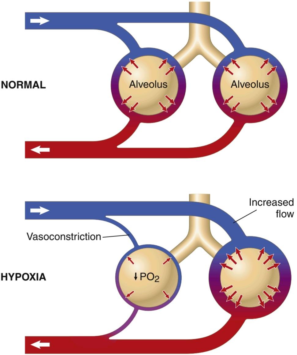
Figure 39-3. Regulation of blood flow in the lungs during local tissue hypoxia. Local tissue blood flow and alveolar ventilation are normally matched for optimal gas exchange (top panel). Decreased Po2 in tissue surrounding poorly ventilated alveoli causes vasoconstriction of adjacent arterioles and diverts blood flow to alveoli that are well ventilated (bottom panel).
شکل 39-3. تنظیم جریان خون در ریه ها در طول هیپوکسی بافت موضعی. جریان خون بافت محلی و تهویه آلوئولی معمولاً برای تبادل گاز بهینه (پانل بالایی) مطابقت دارند. کاهش Po2 در بافت اطراف آلوئول های با تهویه ضعیف باعث انقباض عروق شریان های مجاور و منحرف شدن جریان خون به آلوئول هایی می شود که به خوبی تهویه می شوند (پانل پایینی).
EFFECT OF HYDROSTATIC PRESSURE GRADIENTS IN THE LUNGS ON REGIONAL PULMONARY BLOOD FLOW
In Chapter 15, we pointed out that the blood pressure in the foot of a standing person can be as much as 90 mm Hg greater than the pressure at the level of the heart. This difference is caused by hydrostatic pressure—that is, by the weight of the blood itself in the blood vessels. The same effect, but to a lesser degree, occurs in the lungs. In the upright adult, the lowest point in the lungs is normally about 30 cm below the highest point, which represents a 23-mm Hg pressure difference, about 15 mm Hg of which is above the heart and 8 below. That is, the pulmonary arterial pressure in the uppermost portion of the lung of a standing person is about 15 mm Hg less than the pulmonary arterial pressure at the level of the heart, and the pressure in the lowest portion of the lungs is about 8 mm Hg higher.
اثر گرادیان های فشار هیدرواستاتیک در ریه ها بر جریان خون ریوی منطقه ای
در فصل 15 اشاره کردیم که فشار خون در پای یک فرد ایستاده می تواند 90 میلی متر جیوه بیشتر از فشار در سطح قلب باشد. این تفاوت ناشی از فشار هیدرواستاتیک است، یعنی وزن خود خون در رگهای خونی. همین اثر، اما به میزان کمتر، در ریه ها رخ می دهد. در بزرگسالان قائم، پایینترین نقطه در ریهها معمولاً حدود 30 سانتیمتر زیر بالاترین نقطه است که نشاندهنده اختلاف فشار 23 میلیمتر جیوه است که حدود 15 میلیمتر جیوه آن بالای قلب و 8 میلیمتر جیوه در پایین است. یعنی فشار شریان ریوی در بالاترین قسمت ریه یک فرد ایستاده حدود 15 میلی متر جیوه کمتر از فشار شریان ریوی در سطح قلب است و فشار در پایین ترین قسمت ریه حدود 8 میلی متر جیوه بیشتر است.
Such pressure differences have profound effects on blood flow through the different areas of the lungs. This effect is demonstrated by the lower curve in Figure 39-4, which depicts blood flow per unit of lung tissue at different levels of the lung in the upright person. Note that in the standing position at rest, there is little flow in the top of the lung but about five times as much flow in the bottom. To help explain these differences, the lung is often described as being divided into three zones, as shown in Figure 39-5. In each zone, the patterns of blood flow are quite different.
چنین اختلاف فشاری تأثیرات عمیقی بر جریان خون در نواحی مختلف ریه دارد. این اثر با منحنی پایین تر در شکل 39-4 نشان داده شده است که جریان خون را در هر واحد بافت ریه در سطوح مختلف ریه در فرد ایستاده نشان می دهد. توجه داشته باشید که در حالت ایستاده در حالت استراحت، جریان کمی در بالای ریه وجود دارد، اما جریان در قسمت پایین حدود پنج برابر بیشتر است. برای کمک به توضیح این تفاوت ها، ریه اغلب به سه ناحیه تقسیم می شود، همانطور که در شکل 39-5 نشان داده شده است. در هر منطقه، الگوهای جریان خون کاملاً متفاوت است.

Figure 39-4. Blood flow at different levels in the lung of an upright person at rest (red curve) and during exercise (blue curve). Note that when the person is at rest, the blood flow is very low at the top of the lungs; most of the flow is through the bottom of the lung.
شکل 39-4. جریان خون در سطوح مختلف در ریه یک فرد ایستاده در حالت استراحت (منحنی قرمز) و در حین ورزش (منحنی آبی). توجه داشته باشید که وقتی فرد در حال استراحت است، جریان خون در بالای ریه ها بسیار کم است. بیشتر جریان از ته ریه است.

Figure 39-5. Mechanics of blood flow in the three blood flow zones of the lung: zone 1, no flow-alveolar air pressure (PALV) is greater than arterial pressure; zone 2, intermittent flow-systolic arterial pressure rises higher than alveolar air pressure, but diastolic arterial pressure falls below alveolar air pressure; and zone 3, continuous flow-arterial pressure and pulmonary capillary pressure (Ppc) remain greater than alveolar air pressure at all times.
شکل 39-5. مکانیک جریان خون در سه منطقه جریان خون ریه: منطقه 1، بدون جریان-فشار هوای آلوئولی (PALV) بیشتر از فشار شریانی است. منطقه 2، فشار شریانی سیستولیک متناوب بیشتر از فشار هوای آلوئولی افزایش مییابد، اما فشار شریانی دیاستولیک کمتر از فشار هوای آلوئولی است. و منطقه 3، فشار جریان پیوسته شریانی و فشار مویرگی ریوی (Ppc) همیشه بیشتر از فشار هوای آلوئولی باقی می ماند.
Zones 1, 2, and 3 of Pulmonary Blood Flow
The capillaries in the alveolar walls are distended by the blood pressure inside them but simultaneously are compressed by the alveolar air pressure on their out- sides. Therefore, whenever the lung alveolar air pressure becomes greater than the capillary blood pressure, the capillaries close, and there is no blood flow. Under different normal and pathological lung conditions, one may find any one of three possible zones (patterns) of pulmonary blood flow, as follows:
مناطق 1، 2 و 3 جریان خون ریوی
مویرگهای دیواره آلوئولی در اثر فشار خون داخل آنها متسع میشوند، اما به طور همزمان توسط فشار هوای آلوئولی در قسمت بیرونی خود فشرده میشوند. بنابراین، هرگاه فشار هوای آلوئولی ریه از فشار خون مویرگی بیشتر شود، مویرگ ها بسته می شوند و جریان خون وجود ندارد. تحت شرایط مختلف ریوی طبیعی و پاتولوژیک، ممکن است هر یک از سه منطقه (الگوی) احتمالی جریان خون ریوی را پیدا کنید، به شرح زیر:
• Zone 1: No blood flow during all portions of the cardiac cycle because the local alveolar capillary pressure in that area of the lung never rises higher than the alveolar air pressure during any part of the cardiac cycle
• منطقه 1: جریان خون در تمام بخشهای چرخه قلبی وجود ندارد، زیرا فشار مویرگی آلوئولی موضعی در آن ناحیه از ریه هرگز بالاتر از فشار هوای آلوئولی در هیچ بخشی از چرخه قلبی نیست.
• Zone 2: Intermittent blood flow only during the peaks of pulmonary arterial pressure because the systolic pressure is then greater than the alveolar air pressure, but the diastolic pressure is less than the alveolar air pressure
• منطقه 2: جریان خون متناوب فقط در اوج فشار شریانی ریوی، زیرا فشار سیستولیک بیشتر از فشار هوای آلوئولی است، اما فشار دیاستولیک کمتر از فشار هوای آلوئولی است.
• Zone 3: Continuous blood flow because the alveolar capillary pressure remains greater than alveolar air pressure during the entire cardiac cycle
• منطقه 3: جریان خون مداوم به دلیل اینکه فشار مویرگی آلوئولی بیشتر از فشار هوای آلوئولی در تمام چرخه قلبی باقی می ماند.
Normally, the lungs have only zones 2 and 3 blood flow-zone 2 (intermittent flow) in the apices and zone 3 (continuous flow) in all the lower areas. For example, when a person is in the upright position, the pulmonary arterial pressure at the lung apex is about 15 mm Hg less than the pressure at the level of the heart. Therefore, the apical systolic pressure is only 10 mm Hg (25 mm Hg at heart level minus 15 mm Hg hydrostatic pressure difference). This 10 mm Hg apical blood pressure is greater than the 0 alveolar air pressure, so blood flows through the pulmonary apical capillaries during cardiac systole. Conversely, during diastole, the 8 mm Hg diastolic pressure at the level of the heart is not sufficient to push the blood up the 15 mm Hg hydrostatic pressure gradient required to cause diastolic capillary flow. Therefore, blood flow through the apical part of the lung is intermittent, with flow during systole but cessation of flow during diastole; this is called zone 2 blood flow. Zone 2 blood flow begins in normal lungs about 10 cm above the midlevel of the heart and extends from there to the top of the lungs.
به طور معمول، ریه ها فقط دارای مناطق 2 و 3 جریان خون هستند – منطقه 2 (جریان متناوب) در راس ها و منطقه 3 (جریان مداوم) در تمام نواحی تحتانی. به عنوان مثال، هنگامی که فرد در وضعیت عمودی قرار دارد، فشار شریانی ریوی در راس ریه حدود 15 میلی متر جیوه کمتر از فشار در سطح قلب است. بنابراین، فشار سیستولیک آپیکال فقط 10 میلی متر جیوه است (25 میلی متر جیوه در سطح قلب منهای 15 میلی متر جیوه اختلاف فشار هیدرواستاتیک). این فشار خون آپیکال 10 میلی متر جیوه از فشار هوای آلوئولی 0 بیشتر است، بنابراین خون در طول سیستول قلبی از طریق مویرگ های آپیکال ریوی جریان می یابد. برعکس، در طول دیاستول، فشار دیاستولیک 8 میلیمتر جیوه در سطح قلب برای فشار خون به شیب فشار هیدرواستاتیک 15 میلیمتر جیوه لازم برای ایجاد جریان مویرگی دیاستولیک کافی نیست. بنابراین، جریان خون از طریق قسمت آپیکال ریه متناوب است، با جریان در طول سیستول، اما توقف جریان در طول دیاستول. این جریان خون منطقه 2 نامیده می شود. جریان خون منطقه 2 در ریه های طبیعی حدود 10 سانتی متر بالاتر از سطح میانی قلب شروع می شود و از آنجا تا بالای ریه ها ادامه می یابد.
In the lower regions of the lungs, from about 10 cm above the level of the heart all the way to the bottom of the lungs, the pulmonary arterial pressure during both systole and diastole remains greater than the 0 alveolar air pressure. Therefore, continuous flow occurs through the alveolar capillaries, or zone 3 blood flow. Also, when a person is lying down, no part of the lung is more than a few centimeters above the level of the heart. In this case, blood flow in a normal person is entirely zone 3 blood flow, including the lung apices.
در نواحی پایینی ریه ها، از حدود 10 سانتی متر بالاتر از سطح قلب تا انتهای ریه ها، فشار شریانی ریوی در طول سیستول و دیاستول بیشتر از فشار هوای آلوئولی 0 باقی می ماند. بنابراین، جریان مداوم از طریق مویرگ های آلوئولی یا جریان خون منطقه 3 رخ می دهد. همچنین زمانی که فرد دراز کشیده است، هیچ قسمتی از ریه بیش از چند سانتی متر از سطح قلب بالاتر نیست. در این حالت، جریان خون در یک فرد عادی به طور کامل جریان خون منطقه 3، از جمله رأس ریه است.
Zone 1 Blood Flow Occurs Only Under Abnormal Conditions. Zone 1 blood flow, which means no blood flow at any time during the cardiac cycle, occurs when the pulmonary systolic arterial pressure is too low or the alveolar pressure is too high to allow flow. For ex- ample, if an upright person is breathing against a positive air pressure so that the intra-alveolar air pressure is at least 10 mm Hg greater than normal but the pulmonary systolic blood pressure is normal, one would expect zone 1 blood flow-no blood flow-in the lung apices. Another case in which zone 1 blood flow occurs is in an upright person whose pulmonary systolic arterial pressure is exceedingly low, as might occur after severe blood loss.
جریان خون منطقه 1 فقط در شرایط غیر طبیعی رخ می دهد. جریان خون منطقه 1، به این معنی که هیچ جریان خونی در هیچ زمانی در طول چرخه قلبی وجود ندارد، زمانی اتفاق میافتد که فشار شریانی سیستولیک ریوی خیلی پایین باشد یا فشار آلوئولی بیش از حد بالا باشد که اجازه جریان را نمیدهد. به عنوان مثال، اگر یک فرد ایستاده بر خلاف فشار هوای مثبت نفس بکشد، به طوری که فشار هوای داخل آلوئولی حداقل 10 میلی متر جیوه بیشتر از حد طبیعی باشد، اما فشار خون سیستولیک ریوی طبیعی باشد، می توان انتظار داشت که در ناحیه 1 جریان خون – بدون جریان خون – در رأس ریه باشد. مورد دیگری که در آن جریان خون ناحیه 1 رخ می دهد، در یک فرد ایستاده است که فشار شریانی سیستولیک ریوی بسیار پایین است، همانطور که ممکن است پس از از دست دادن خون شدید رخ دهد.
Exercise Increases Blood Flow Through All Parts of the Lungs. Referring again to Figure 39-4, one sees that the blood flow in all parts of the lung increases during exercise. A major reason for increased blood flow is that the pulmonary vascular pressures rise enough during exercise to convert the lung apices from a zone 2 pattern into a zone 3 pattern of flow.
ورزش باعث افزایش جریان خون در تمام قسمت های ریه می شود. با مراجعه مجدد به شکل 39-4، می بینیم که جریان خون در تمام قسمت های ریه در طول ورزش افزایش می یابد. یکی از دلایل اصلی افزایش جریان خون این است که فشار عروق ریوی در حین ورزش به اندازه کافی افزایش مییابد تا نوک ریه از الگوی منطقه 2 به الگوی جریان منطقه 3 تبدیل شود.
Increased Cardiac Output During Heavy Exercise Is Normally Accommodated by the Pulmonary Circulation Without Large Increases in Pulmonary Artery Pressure
During heavy exercise, blood flow through the lungs may increase fourfold to sevenfold. This extra flow is accommodated in the lungs in three ways: (1) by increasing the number of open capillaries, sometimes as much as three- fold; (2) by distending all the capillaries and increasing the rate of flow through each capillary more than two- fold; and (3) by increasing the pulmonary arterial pressure. Normally, the first two changes decrease pulmonary vascular resistance so much that the pulmonary arterial pressure rises very little, even during maximum exercise. This effect is shown in Figure 39-6.
افزایش برون ده قلبی در طی ورزش های سنگین معمولاً توسط گردش خون ریوی بدون افزایش شدید فشار شریان ریوی انجام می شود.
در طول ورزش سنگین، جریان خون از طریق ریه ها ممکن است چهار تا هفت برابر افزایش یابد. این جریان اضافی به سه طریق در ریه ها جای می گیرد: (1) با افزایش تعداد مویرگ های باز، گاهی اوقات تا سه برابر. (2) با انبساط همه مویرگها و افزایش سرعت جریان در هر مویرگ بیش از دو برابر. و (3) با افزایش فشار شریانی ریوی. به طور معمول، دو تغییر اول مقاومت عروق ریوی را به قدری کاهش می دهد که فشار شریان ریوی حتی در طول حداکثر تمرین بسیار کم افزایش می یابد. این اثر در شکل 39-6 نشان داده شده است.
The ability of the lungs to accommodate greatly increased blood flow during exercise, without increasing the pulmonary arterial pressure, conserves the energy of the right side of the heart. This ability also prevents a major rise in pulmonary capillary pressure and the development of pulmonary edema.
توانایی ریه ها برای انطباق با افزایش جریان خون در طول ورزش، بدون افزایش فشار شریانی ریوی، انرژی سمت راست قلب را حفظ می کند. این توانایی همچنین از افزایش شدید فشار مویرگ ریوی و ایجاد ادم ریوی جلوگیری می کند.

Figure 39-6. Effect on mean pulmonary arterial pressure caused by increasing the cardiac output during exercise.
شکل 39-6. تأثیر بر فشار متوسط شریانی ریوی ناشی از افزایش برون ده قلبی در حین ورزش.
Function of Pulmonary Circulation When Left Atrial Pressure Rises as a Result of Left-Sided Heart Failure
The left atrial pressure in a healthy person almost never rises above +6 mm Hg, even during the most strenuous exercise. These small changes in left atrial pressure have virtually no effect on pulmonary circulatory function because this merely expands the pulmonary venules and opens up more capillaries so that blood continues to flow with almost equal ease from the pulmonary arteries.
عملکرد گردش خون ریوی هنگامی که فشار دهلیز چپ در نتیجه نارسایی قلبی سمت چپ افزایش می یابد
فشار دهلیز چپ در یک فرد سالم تقریباً هرگز از +6 میلیمتر جیوه بالاتر نمیرود، حتی در حین شدیدترین ورزشها. این تغییرات کوچک در فشار دهلیز چپ عملاً هیچ تأثیری بر عملکرد گردش خون ریوی ندارد زیرا این امر فقط رگهای ریوی را گسترش میدهد و مویرگهای بیشتری را باز میکند تا خون با سهولت تقریباً برابر از شریانهای ریوی به جریان خود ادامه دهد.
When the left side of the heart fails, however, blood begins to dam up in the left atrium. As a result, the left atrial pressure can rise on occasion from its normal value of 1 to 5 mm Hg to as high as 40 to 50 mm Hg. The initial rise in atrial pressure, up to about 7 mm Hg, has little effect on pulmonary circulatory function. However, when the left atrial pressure rises to greater than 7 or 8 mm Hg, further increases in left atrial pressure cause almost equally great increases in pulmonary arterial pressure, thus causing a concomitant increased load on the right heart. Any increase in left atrial pressure above 7 or 8 mm Hg increases capillary pressure almost equally as much. When the left atrial pressure rises above 30 mm Hg, causing similar increases in capillary pressure, pulmonary edema is likely to develop, as discussed later in the chapter.
با این حال، هنگامی که سمت چپ قلب از کار می افتد، خون در دهلیز چپ شروع به جمع شدن می کند. در نتیجه، فشار دهلیز چپ می تواند گاهی اوقات از مقدار طبیعی خود از 1 تا 5 میلی متر جیوه به 40 تا 50 میلی متر جیوه افزایش یابد. افزایش اولیه فشار دهلیزی، تا حدود 7 میلی متر جیوه، تأثیر کمی بر عملکرد گردش خون ریوی دارد. با این حال، زمانی که فشار دهلیز چپ به بیش از 7 یا 8 میلیمتر جیوه افزایش مییابد، افزایش بیشتر فشار دهلیز چپ باعث افزایش تقریباً به همان اندازه در فشار شریان ریوی میشود و در نتیجه باعث افزایش همزمان بار روی قلب راست میشود. هر افزایش فشار دهلیز چپ بیش از 7 یا 8 میلی متر جیوه، فشار مویرگی را تقریباً به همان اندازه افزایش می دهد. هنگامی که فشار دهلیز چپ از 30 میلیمتر جیوه بالاتر میرود و باعث افزایش مشابه در فشار مویرگی میشود، ادم ریوی احتمالاً ایجاد میشود، همانطور که بعداً در این فصل بحث شد.
PULMONARY CAPILLARY DYNAMICS
Exchange of gases between the alveolar air and pulmonary capillary blood is discussed in the next chapter. How- ever, it is important to note here that the alveolar walls are lined with so many capillaries that in most places, the capillaries almost touch one another side by side. Therefore, it is often said that the capillary blood flows in the alveolar walls as a “sheet of flow,” rather than in individual capillaries.
دینامیک مویرگهای ریوی
تبادل گازها بین هوای آلوئولی و خون مویرگی ریوی در فصل بعد مورد بحث قرار می گیرد. با این حال، در اینجا ذکر این نکته مهم است که دیوارههای آلوئولی با مویرگهای زیادی پوشیده شدهاند که در بیشتر مکانها، مویرگها تقریباً کنار هم یکدیگر را لمس میکنند. بنابراین، اغلب گفته میشود که خون مویرگی در دیوارههای آلوئول بهعنوان یک “ورقه جریان” جریان دارد تا در مویرگهای منفرد.
Pulmonary Capillary Pressure. Although direct meas-urements of pulmonary capillary pressure have not been reported, indirect estimates have suggested that it normally averages about 7 mm Hg. This value is probably nearly correct because the mean left atrial pressure is about 2 mm Hg, and the mean pulmonary arterial pressure is only 15 mm Hg, so the mean pulmonary capillary pressure must lie somewhere between these two values.
فشار مویرگی ریوی. اگرچه اندازه گیری مستقیم فشار مویرگ ریوی گزارش نشده است، تخمین های غیرمستقیم نشان می دهد که به طور معمول به طور متوسط حدود 7 میلی متر جیوه است. این مقدار احتمالاً تقریباً صحیح است زیرا فشار متوسط دهلیز چپ حدود 2 میلیمتر جیوه است و فشار متوسط شریان ریوی تنها 15 میلیمتر جیوه است، بنابراین فشار متوسط مویرگ ریوی باید جایی بین این دو مقدار باشد.
Length of Time Blood Stays in the Pulmonary Capillaries. From histological studies of the total cross- sectional area of all the pulmonary capillaries, it can be calculated that when the cardiac output is normal, blood passes through the pulmonary capillaries in about 0.8 second. When the cardiac output increases, this time can shorten to as little as 0.3 second. The shortened time would be much greater were it not for the fact that additional capillaries, which normally are collapsed, open up to accommodate the increased blood flow. Thus, in only a fraction of a second, blood passing through the alveolar capillaries becomes oxygenated and loses its excess car- bon dioxide.
مدت زمان ماندن خون در مویرگ های ریوی. از مطالعات بافت شناسی کل سطح مقطع تمام مویرگ های ریوی، می توان محاسبه کرد که وقتی برون ده قلبی طبیعی است، خون در حدود 0.8 ثانیه از مویرگ های ریوی عبور می کند. هنگامی که برون ده قلبی افزایش می یابد، این زمان می تواند به 0.3 ثانیه کاهش یابد. اگر به خاطر این واقعیت نبود که مویرگهای اضافی که معمولاً فرو میروند، برای سازگاری با جریان خون افزایش یافته باز میشوند، زمان کوتاهتر بسیار بیشتر خواهد بود. بنابراین، تنها در کسری از ثانیه، خونی که از مویرگ های آلوئولی عبور می کند، اکسیژن می شود و دی اکسید کربن اضافی خود را از دست می دهد.
Capillary Exchange of Fluid in the Lungs and Pulmonary Interstitial Fluid Dynamics
The dynamics of fluid exchange across the lung capillary membranes are qualitatively the same as for peripheral tissues. However, quantitatively, there are important differences, as follows:
تبادل مویرگی مایع در ریه ها و دینامیک مایع بینابینی ریوی
پویایی تبادل مایع در سراسر غشای مویرگی ریه از نظر کیفی مانند بافتهای محیطی است. با این حال، از نظر کمی، تفاوت های مهمی وجود دارد که به شرح زیر است:
1. The pulmonary capillary pressure is low, about 7 mm Hg, in comparison with a considerably higher functional capillary pressure in many peripheral tissues of about 17 mm Hg.
1. فشار مویرگی ریوی در مقایسه با فشار عملکردی قابل ملاحظهای بالاتر در بسیاری از بافتهای محیطی حدود 17 میلیمتر جیوه، کم است، حدود 7 میلیمتر جیوه.
2. The interstitial fluid pressure in the lung is slightly more negative than that in peripheral subcutaneous tissue. (This pressure has been measured in two ways-by a micropipette inserted into the pulmonary interstitium, giving a value of about -5 mm Hg, and by measuring the absorption pressure of fluid from the alveoli, giving a value of about -8 mm Hg.)
2. فشار مایع بینابینی در ریه کمی منفی تر از بافت زیر جلدی محیطی است. (این فشار به دو روش اندازهگیری شده است: یک میکروپیپت وارد شده به بینابینی ریوی که مقداری در حدود 5- میلیمتر جیوه میدهد و با اندازهگیری فشار جذب مایع از آلوئولها، مقداری در حدود 8- میلیمتر جیوه میدهد.)
3. The colloid osmotic pressure of the pulmonary inter-stitial fluid is about 14 mm Hg, in comparison with less than half this value in most peripheral tissues.
3. فشار اسمزی کلوئیدی مایع بین بافتی ریوی حدود 14 میلی متر جیوه است، در مقایسه با کمتر از نصف این مقدار در اکثر بافت های محیطی.
4. The alveolar walls are extremely thin, and the alveolar epithelium covering the alveolar surfaces is so weak that it can be ruptured by any positive pressure in the interstitial spaces greater than alveolar air pressure (>0 mm Hg), which allows dumping of fluid from the interstitial spaces into the alveoli. Now let us see how these quantitative differences affect pulmonary fluid dynamics.
4. دیواره های آلوئولی بسیار نازک هستند و اپیتلیوم آلوئولی که سطوح آلوئول را می پوشاند به قدری ضعیف است که می تواند با هر فشار مثبت در فضاهای بین بافتی بیشتر از فشار هوای آلوئولی (>0 میلی متر جیوه) پاره شود، که اجازه می دهد مایع از فضاهای بین بافتی به آلوئول ها ریخته شود. حال اجازه دهید ببینیم که چگونه این تفاوت های کمی بر دینامیک مایع ریوی تأثیر می گذارد.
Interrelationships Between Interstitial Fluid Pressure and Other Pressures in the Lung. Figure 39-7 shows a pulmonary capillary, pulmonary alveolus, and lymphatic capillary draining the interstitial space between the blood capillary and alveolus. Note the balance of forces at the blood capillary membrane, as follows:
روابط متقابل بین فشار مایع بینابینی و فشارهای دیگر در ریه. شکل 39-7 یک مویرگ ریوی، آلوئول ریوی و مویرگ لنفاوی را نشان می دهد که فضای بینابینی بین مویرگ خون و آلوئول را تخلیه می کند. به تعادل نیروها در غشای مویرگی خون به شرح زیر توجه کنید:

Thus, the normal outward forces are slightly greater than the inward forces, providing a mean filtration pressure at the pulmonary capillary membrane that can be calculated as +29-28 mm Hg = +1 mm Hg.
بنابراین، نیروهای معمولی به بیرون کمی بیشتر از نیروهای داخل هستند و فشار فیلتراسیون متوسطی را در غشای مویرگی ریوی ایجاد میکنند که میتواند به صورت 29+28- میلیمتر جیوه = 1+ میلیمتر جیوه محاسبه شود.
This filtration pressure causes a slight continual flow of fluid from the pulmonary capillaries into the interstitial spaces and, except for a small amount that evaporates in the alveoli, this fluid is pumped back to the circulation through the pulmonary lymphatic system.
این فشار فیلتراسیون باعث جریان خفیف مایع از مویرگ های ریوی به فضاهای بینابینی می شود و به جز مقدار کمی که در آلوئول ها تبخیر می شود، این مایع از طریق سیستم لنفاوی ریوی به گردش خون بازگردانده می شود.
Negative Pulmonary Interstitial Pressure and Mechanism for Keeping Alveoli Dry. What keeps the alveoli from filling with fluid under normal conditions? The pulmonary capillaries and pulmonary lymphatic system normally maintain a slight negative pressure in the interstitial spaces; whenever extra fluid appears in the alveoli, it is sucked mechanically into the lung interstitium through the small openings between the alveolar epithelial cells. The excess fluid is then carried away through the pulmonary lymphatics. Thus, under normal conditions, the alveoli are kept “dry” except for a small amount of fluid that seeps from the epithelium onto the lining surfaces of the alveoli to keep them moist.
فشار میان بافتی ریوی منفی و مکانیسم خشک نگه داشتن آلوئول ها. چه چیزی مانع از پر شدن آلوئول ها با مایع در شرایط عادی می شود؟ مویرگ های ریوی و سیستم لنفاوی ریوی به طور معمول فشار منفی خفیفی را در فضاهای بینابینی حفظ می کنند. هنگامی که مایع اضافی در آلوئول ظاهر می شود، به طور مکانیکی از طریق سوراخ های کوچک بین سلول های اپیتلیال آلوئول به داخل بینابینی ریه مکیده می شود. سپس مایع اضافی از طریق غدد لنفاوی ریوی منتقل می شود. بنابراین، در شرایط عادی، آلوئول ها به جز مقدار کمی مایع که از اپیتلیوم به سطوح پوششی آلوئول ها تراوش می کند، “خشک” نگه داشته می شوند تا آنها را مرطوب نگه دارد.

Figure 39-7. Hydrostatic and osmotic forces in mm Hg at the capillary (left) and alveolar membrane (right) of the lungs. Also shown is the tip end of a lymphatic vessel (center) that pumps fluid from the pulmonary interstitial spaces.
شکل 39-7. نیروهای هیدرواستاتیک و اسمزی بر حسب میلی متر جیوه در غشای مویرگی (چپ) و آلوئولار (راست) ریه ها. همچنین انتهای نوک یک رگ لنفاوی (مرکز) نشان داده شده است که مایع را از فضاهای بینابینی ریوی پمپ می کند.
Pulmonary Edema
Pulmonary edema occurs in the same way that edema occurs elsewhere in the body. Any factor that increases fluid filtration out of the pulmonary capillaries or that impedes pulmonary lymphatic function and causes the pulmonary interstitial fluid pressure to rise from the negative range into the positive range will tend to cause filling of the pulmonary interstitial spaces and alveoli with free fluid.
ادم ریوی
ادم ریوی به همان شکلی رخ می دهد که ادم در سایر نقاط بدن رخ می دهد. هر عاملی که فیلتراسیون مایع را از مویرگ های ریوی افزایش دهد یا عملکرد لنفاوی ریوی را مختل کند و باعث شود فشار مایع بین بافتی ریوی از محدوده منفی به محدوده مثبت افزایش یابد، باعث پر شدن فضاهای بین بافتی ریوی و آلوئول ها با مایع آزاد می شود.
The most common causes of pulmonary edema are as follows:
شایع ترین علل ادم ریوی به شرح زیر است:
1. Left-sided heart failure or mitral valve disease, with consequent great increases in pulmonary venous pressure and pulmonary capillary pressure and flooding of the interstitial spaces and alveoli
1. نارسایی قلبی سمت چپ یا بیماری دریچه میترال، با افزایش شدید فشار وریدی ریوی و فشار مویرگی ریوی و سیل فضاهای بینابینی و آلوئول ها.
2. Damage to the pulmonary blood capillary membranes caused by infections such as pneumonia or by breathing noxious substances such as chlorine gas or sulfur dioxide gas.
2. آسیب به غشاهای مویرگ ریوی ناشی از عفونت هایی مانند ذات الریه یا تنفس مواد مضر مانند گاز کلر یا گاز دی اکسید گوگرد.
Each of these mechanisms causes rapid leakage of plasma proteins and fluid out of the capillaries and into the lung interstitial spaces and alveoli.
هر یک از این مکانیسم ها باعث نشت سریع پروتئین های پلاسما و مایع از مویرگ ها و به فضاهای بینابینی ریه و آلوئول ها می شود.
Pulmonary Edema Safety Factor. Experiments in animals have shown that the pulmonary capillary pressure normally must rise to a value at least equal to the colloid osmotic pressure of the plasma inside the capillaries before significant pulmonary edema will occur. To give an example, Figure 39-8 shows how different levels of left atrial pressure increase the rate of pulmonary edema formation in dogs. Remember that every time the left atrial pressure rises to high values, the pulmonary capillary pressure rises to a level 1 to 2 mm Hg greater than the left atrial pressure. In these experiments, as soon as the left atrial pressure rose above 23 mm Hg (causing the pulmonary capillary pressure to rise above 25 mm Hg), fluid began to accumulate in the lungs. This fluid accumulation increased even more rapidly with further increases in capillary pressure. The plasma colloid osmotic pressure during these experiments was equal to this 25 mm Hg critical pressure level. Therefore, in a person, whose normal plasma colloid osmotic pressure is 28 mm Hg, one can predict that the pulmonary capillary pressure must rise from the normal level of 7 mm Hg to more than 28 mm Hg to cause substantial pulmonary edema, giving an acute safety factor against pulmonary edema of 21 mm Hg.
فاکتور ایمنی ادم ریوی. آزمایشها روی حیوانات نشان دادهاند که فشار مویرگ ریوی معمولاً باید به مقداری حداقل برابر با فشار اسمزی کلوئیدی پلاسما در داخل مویرگها برسد، قبل از اینکه ادم ریوی قابل توجهی ایجاد شود. برای ارائه یک مثال، شکل 39-8 نشان می دهد که چگونه سطوح مختلف فشار دهلیز چپ باعث افزایش سرعت تشکیل ادم ریوی در سگ ها می شود. به یاد داشته باشید که هر بار که فشار دهلیز چپ به مقادیر بالا افزایش می یابد، فشار مویرگ ریوی به سطح 1 تا 2 میلی متر جیوه بیشتر از فشار دهلیز چپ افزایش می یابد. در این آزمایش ها، به محض اینکه فشار دهلیز چپ به بالای 23 میلی متر جیوه رسید (که باعث شد فشار مویرگ ریوی به بالای 25 میلی متر جیوه برسد)، مایع شروع به تجمع در ریه ها کرد. این تجمع مایع با افزایش بیشتر فشار مویرگی با سرعت بیشتری افزایش یافت. فشار اسمزی کلوئیدی پلاسما در طی این آزمایش ها برابر با این سطح فشار بحرانی 25 میلی متر جیوه بود. بنابراین، در فردی که فشار اسمزی کلوئیدی پلاسماییاش 28 میلیمتر جیوه است، میتوان پیشبینی کرد که فشار مویرگی ریوی باید از سطح نرمال 7 میلیمتر جیوه به بیش از 28 میلیمتر جیوه برسد تا ادم ریوی قابلتوجهی ایجاد کند و فاکتور ایمنی حاد در برابر ادم ریوی 21 میلیمتر جیوه ایجاد کند.
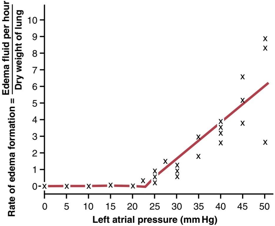
Figure 39-8. Rate of fluid loss into the lung tissues when the left atrial pressure (and pulmonary capillary pressure) is increased. (From Guyton AC, Lindsey AW: Effect of elevated left atrial pressure and decreased plasma protein concentration on the development of pulmonary edema. Circ Res 7:649, 1959.)
شکل 39-8. وقتی فشار دهلیز چپ (و فشار مویرگ ریوی) افزایش مییابد، سرعت از دست دادن مایع در بافتهای ریه افزایش مییابد. (از Guyton AC، Lindsey AW: اثر افزایش فشار دهلیز چپ و کاهش غلظت پروتئین پلاسما بر ایجاد ادم ریوی. Circ Res 7:649، 1959.)
Safety Factor in Chronic Conditions. When the pulmonary capillary pressure remains elevated chronically (for at least 2 weeks), the lungs become even more resistant to pulmonary edema because the lymph vessels expand greatly, increasing their capability of carrying fluid away from the interstitial spaces perhaps as much as 10-fold. Therefore, in patients with chronic mitral stenosis, pulmonary capillary pressures of 40 to 45 mm Hg have been measured without the development of lethal pulmonary edema.
فاکتور ایمنی در شرایط مزمن. هنگامی که فشار مویرگ ریوی به طور مزمن (حداقل برای 2 هفته) بالا میماند، ریهها در برابر ادم ریوی حتی مقاومتر میشوند، زیرا عروق لنفاوی به شدت منبسط میشوند و توانایی آنها را برای انتقال مایع به دور از فضاهای بینابینی تا 10 برابر افزایش میدهند. بنابراین، در بیماران مبتلا به تنگی مزمن میترال، فشار مویرگی ریوی 40 تا 45 میلیمتر جیوه بدون ایجاد ادم ریوی کشنده اندازهگیری شده است.
Rapidity of Death in Persons With Acute Pulmonary Edema. When the pulmonary capillary pressure rises even slightly above the safety factor level, lethal pulmonary edema can occur within hours, or even within 20 to 30 minutes if the capillary pressure rises 25 to 30 mm Hg above the safety factor level. Thus, in acute left-sided heart failure, in which the pulmonary capillary pressure occasionally does rise to 50 mm Hg, death may ensue in less than 30 minutes as a result of acute pulmonary edema.
سرعت مرگ در افراد مبتلا به ادم حاد ریوی. هنگامی که فشار مویرگ ریوی حتی اندکی بالاتر از سطح فاکتور ایمنی باشد، ادم ریوی کشنده می تواند در عرض چند ساعت یا حتی در عرض 20 تا 30 دقیقه اگر فشار مویرگی 25 تا 30 میلی متر جیوه بالاتر از سطح فاکتور ایمنی افزایش یابد، رخ می دهد. بنابراین، در نارسایی حاد قلب سمت چپ، که در آن فشار مویرگ ریوی گهگاه تا 50 میلیمتر جیوه افزایش مییابد، ممکن است در کمتر از 30 دقیقه در نتیجه ادم حاد ریوی، مرگ رخ دهد.
FLUID IN THE PLEURAL CAVITY
When the lungs expand and contract during normal breathing, they slide back and forth within the pleural cavity. To facilitate this movement, a thin layer of mucoid fluid lies between the parietal and visceral pleurae.
مایع در حفره پلورال
هنگامی که ریه ها در طول تنفس طبیعی منبسط و منقبض می شوند، در داخل حفره پلور به جلو و عقب می لغزند. برای تسهیل این حرکت، یک لایه نازک از مایع مخاطی بین جنب جداری و احشایی قرار دارد.
Figure 39-9 shows the dynamics of fluid exchange in the pleural space. The pleural membrane is a porous, mesenchymal, serous membrane through which small amounts of interstitial fluid transude continually into the pleural space. These fluids carry tissue proteins with them, giving the pleural fluid a mucoid characteristic, which is what allows extremely easy slippage of the moving lungs.
شکل 39-9 پویایی تبادل مایع در فضای پلور را نشان می دهد. غشای پلور یک غشای متخلخل، مزانشیمی و سروزی است که از طریق آن مقادیر کمی از مایع بینابینی به طور مداوم به فضای پلور منتقل می شود. این مایعات پروتئینهای بافتی را با خود حمل میکنند و به مایع پلور یک ویژگی مخاطی میدهند، که باعث میشود ریههای متحرک بسیار راحت لغزش کنند.
The total amount of fluid in each pleural cavity is normally slight-only a few milliliters. Whenever the quantity becomes more than barely enough to begin flowing in the pleural cavity, the excess fluid is pumped away by lymphatic vessels opening directly from the pleural cavity into the following: (1) the mediastinum; (2) the superior surface of the diaphragm; and (3) the lateral surfaces of the parietal pleura. Therefore, the pleural space-the space between the parietal and visceral pleurae-is called a potential space because it normally is so narrow that it is not obviously a physical space.
مقدار کل مایع در هر حفره پلور معمولاً اندک است – فقط چند میلی لیتر. هر گاه این مقدار به سختی برای شروع جریان در حفره پلور کافی باشد، مایع اضافی توسط عروق لنفاوی که مستقیماً از حفره پلور باز می شوند به سمت زیر پمپاژ می شود: (1) مدیاستن. (2) سطح فوقانی دیافراگم؛ و (3) سطوح جانبی پلور جداری. بنابراین، فضای پلور – فضای بین جنب جداری و احشایی – فضای بالقوه نامیده می شود زیرا معمولاً آنقدر باریک است که به وضوح یک فضای فیزیکی نیست.

Figure 39-9. Dynamics of fluid exchange in the intrapleural space.
شکل 39-9. دینامیک تبادل مایع در فضای داخل پلورال
Negative Pressure in Pleural Fluid. A negative force is always required on the outside of the lungs to keep the lungs expanded. This force is provided by negative pressure in the normal pleural space. The basic cause of this negative pressure is pumping of fluid from the space by the lymphatics, which is also the basis of the negative pressure found in most tissue spaces of the body. Because the normal collapse tendency of the lungs is about -4 mm Hg, the pleural fluid pressure must al- ways be at least as negative as -4 mm Hg to keep the lungs expanded. Actual measurements have shown that the pressure is usually about -7 mm Hg, which is a few millimeters of mercury more negative than the collapse pressure of the lungs. Thus, the negativity of the pleural fluid pressure keeps the normal lungs pulled against the parietal pleura of the chest cavity, except for an extremely thin layer of mucoid fluid that acts as a lubricant.
فشار منفی در مایع جنب. همیشه یک نیروی منفی در خارج از ریه ها لازم است تا ریه ها منبسط شوند. این نیرو با فشار منفی در فضای طبیعی جنب تامین می شود. علت اصلی این فشار منفی پمپاژ مایع از فضا توسط غدد لنفاوی است که همچنین اساس فشار منفی موجود در بیشتر فضاهای بافتی بدن است. از آنجایی که تمایل طبیعی به فروپاشی ریه ها حدود 4- میلی متر جیوه است، فشار مایع پلور باید حداقل به اندازه 4- میلی متر جیوه منفی باشد تا ریه ها منبسط شوند. اندازهگیریهای واقعی نشان دادهاند که فشار معمولاً حدود 7- میلیمتر جیوه است که چند میلیمتر جیوه منفیتر از فشار فروپاشی ریهها است. بنابراین، منفی بودن فشار مایع جنب، ریه های طبیعی را در مقابل پلورای جداری حفره قفسه سینه نگه می دارد، به جز یک لایه بسیار نازک از مایع مخاطی که به عنوان روان کننده عمل می کند.
Pleural Effusion-Collection of Large Amounts of Free Fluid in the Pleural Space. Pleural effusion is analogous to edema fluid in the tissues and can be called edema of the pleural cavity. The causes of the effusion are the same as the causes of edema in other tissues (discussed in Chapter 25), including the following: (1) blockage of lymphatic drainage from the pleural cavity; (2) cardiac failure, which causes excessively high peripheral and pulmonary capillary pressures, leading to excessive transudation of fluid into the pleural cavity; (3) greatly reduced plasma colloid osmotic pressure, thus allowing excessive transudation of fluid; and (4) infection or any other cause of inflammation of the surfaces of the pleural cavity, which increases permeability of the capillary membranes and allows rapid dumping of plasma proteins and fluid into the cavity.
پلورال افیوژن-مجموعه مقادیر زیادی مایع آزاد در فضای جنب. پلورال افیوژن مشابه مایع ادم در بافت ها است و می توان آن را ادم حفره پلور نامید. علل افیوژن مانند علل ادم در سایر بافت ها است (در فصل 25 بحث شد)، از جمله موارد زیر: (1) انسداد درناژ لنفاوی از حفره پلور. (2) نارسایی قلبی، که باعث فشار بیش از حد مویرگی محیطی و ریوی می شود که منجر به انتقال بیش از حد مایع به داخل حفره پلور می شود. (3) فشار اسمزی کلوئیدی پلاسما را تا حد زیادی کاهش می دهد، بنابراین اجازه می دهد تا انتقال بیش از حد مایعات. و (4) عفونت یا هر علت دیگر التهاب سطوح حفره پلور، که نفوذپذیری غشاهای مویرگی را افزایش می دهد و اجازه می دهد تا پروتئین های پلاسما و مایعات به داخل حفره سریع تخلیه شوند.
کلیک کنید «Bibliography: فهرست کتب مربوطه»
Dunham-Snary KJ, Wu D, Sykes EA, et al: Hypoxic pulmonary va- soconstriction: from molecular mechanisms to medicine. Chest 151:181, 2017.
Effros RM, Parker JC: Pulmonary vascular heterogeneity and the Starling hypothesis. Microvasc Res 78:71, 2009.
Frise MC, Robbins PA: The pulmonary vasculature–lessons from Tibetans and from rare diseases of oxygen sensing. Exp Physiol 100:1233, 2015.
Guyton AC, Lindsey AW: Effect of elevated left atrial pressure and decreased plasma protein concentration on the development of pulmonary edema. Circ Res 7:649, 1959.
Hughes M, West JB: Gravity is the major factor determining the distri- bution of blood flow in the human lung. J Appl Physiol 104:1531, 2008.
Jaitovich A, Jourd’heuil D: A Brief overview of nitric oxide and reactive oxygen species signaling in hypoxia-induced pulmonary hyperten- sion. Adv Exp Med Biol 967:71, 2017.
Lumb AB, Slinger P: Hypoxic pulmonary vasoconstriction: physi- ology and anesthetic implications. Anesthesiology 122:932, 2015.
Parker JC: Hydraulic conductance of lung endothelial phenotypes and Starling safety factors against edema. Am J Physiol Lung Cell Mol Physiol 292:L378, 2007.
Stickland MK, Lindinger MI, Olfert IM, Heigenhauser GJ, Hopkins SR: Pulmonary gas exchange and acid-base balance during exercise. Compr Physiol 3:693, 2013.
Suresh K, Shimoda LA: Lung circulation. Compr Physiol 6:897, 2018.
Sylvester JT, Shimoda LA, Aaronson Pl, Ward JP: Hypoxic pulmonary vasoconstriction. Physiol Rev 92:367, 2012.
Tabima DM, Philip JL, Chesler NC: Right ventricular-pulmonary vascu- lar interactions. Physiology (Bethesda) 32:346, 2017.
Townsley MI: Structure and composition of pulmonary arteries, capil- laries, and veins. Compr Physiol 2:675, 2012.
Zielinska-Krawczyk M, Krenke R, Grabczak EM, Light RW: Pleural manometry-historical background, rationale for use and methods of measurement. Respir Med 136:21, 2018.
🚀 با ما همراه شوید!
تازهترین مطالب و آموزشهای مغز و اعصاب را از دست ندهید. با فالو کردن کانال تلگرام آیندهنگاران مغز، از ما حمایت کنید!



