
فیزیولوژی پزشکی گایتون و هال؛ تهویه ریوی


دعای مطالعه [ نمایش ]
بِسْمِ الله الرَّحْمنِ الرَّحیمِ
اَللّهُمَّ اَخْرِجْنى مِنْ ظُلُماتِ الْوَهْمِ
خدايا مرا بيرون آور از تاريكىهاى وهم،
وَ اَكْرِمْنى بِنُورِ الْفَهْمِ
و به نور فهم گرامى ام بدار،
اَللّهُمَّ افْتَحْ عَلَيْنا اَبْوابَ رَحْمَتِكَ
خدايا درهاى رحمتت را به روى ما بگشا،
وَانْشُرْ عَلَيْنا خَزائِنَ عُلُومِكَ بِرَحْمَتِكَ يا اَرْحَمَ الرّاحِمينَ
و خزانههاى علومت را بر ما باز كن به امید رحمتت اى مهربانترين مهربانان.
کتاب «فیزیولوژی پزشکی گایتون و هال» بهعنوان یکی از جامعترین و معتبرترین منابع در حوزه علوم پزشکی، همچنان مرجع کلیدی برای درک عملکرد پیچیده بدن انسان است. این اثر با تکیه بر تازهترین پژوهشها و توضیحات دقیق از سازوکارهای فیزیولوژیک، پلی میان علوم پایه پزشکی و کاربردهای بالینی ایجاد میکند و نقشی بیبدیل در آموزش، پژوهش و ارتقای دانش سلامت ایفا مینماید.
ترجمه دقیق و علمی این شاهکار توسط برند علمی آیندهنگاران مغز به مدیریت داریوش طاهری، دسترسی فارسیزبانان به مرزهای نوین دانش فیزیولوژی را ممکن ساخته و رسالتی علمی برای ارتقای آموزش پزشکی، فهم عمیقتر سازوکارهای بدن و توسعه روشهای نوین در حوزه سلامت فراهم آورده است.
» Guyton and Hall Textbook of Medical Physiology
»» CHAPTER 38: Pulmonary Ventilation

The main functions of respiration are to provide oxygen to the tissues and remove carbon dioxide. The four major components of respiration are the following: (1) pulmonary ventilation, which means the inflow and outflow of air between the atmosphere and the lung alveoli; (2) diffusion of oxygen (O2) and carbon dioxide (CO2) between the alveoli and the blood; (3) transport of oxygen and carbon dioxide in the blood and body fluids to and from the body’s tissue cells; and (4) regulation of ventilation and other facets of respiration. This chapter is a discussion of pulmonary ventilation; the subsequent five chapters cover other respiratory functions plus the physiology of special respiratory abnormalities.
وظایف اصلی تنفس تامین اکسیژن به بافت ها و حذف دی اکسید کربن است. چهار جزء اصلی تنفس عبارتند از: (1) تهویه ریوی، که به معنای ورود و خروج هوا بین جو و آلوئول ریه است. (2) انتشار اکسیژن (O2) و دی اکسید کربن (CO2) بین آلوئول ها و خون. (3) انتقال اکسیژن و دی اکسید کربن در خون و مایعات بدن به و از سلول های بافت بدن. و (4) تنظیم تهویه و سایر جنبه های تنفس. این فصل بحثی در مورد تهویه ریوی است. پنج فصل بعدی سایر عملکردهای تنفسی به علاوه فیزیولوژی ناهنجاری های تنفسی خاص را پوشش می دهد.
MECHANICS OF PULMONARY VENTILATION
MUSCLES THAT CAUSE LUNG EXPANSION AND CONTRACTION
The lungs can be expanded and contracted in two ways: (1) by downward or upward movement of the diaphragm to lengthen or shorten the chest cavity; and (2) by elevation or depression of the ribs to increase or decrease the anteroposterior diameter of the chest cavity. Figure 38-1 shows these two methods.
مکانیک تهویه ریوی
ماهیچه هایی که باعث انبساط و انقباض ریه می شوند
ریه ها را می توان به دو طریق منبسط و منقبض کرد: (1) با حرکت دیافراگم به سمت پایین یا رو به بالا برای طولانی یا کوتاه کردن حفره قفسه سینه. و (2) با بالا بردن یا فرورفتگی دنده ها برای افزایش یا کاهش قطر قدامی خلفی حفره قفسه سینه. شکل 38-1 این دو روش را نشان می دهد.
Normal quiet breathing is accomplished almost entirely by movement of the diaphragm. During inspiration, contraction of the diaphragm pulls the lower surfaces of the lungs downward. Then, during expiration, the diaphragm simply relaxes, and the elastic recoil of the lungs, chest wall, and abdominal structures compresses the lungs and expels the air. During heavy breathing, however, the elastic forces are not powerful enough to cause the necessary rapid expiration, so extra force is achieved mainly by contraction of the abdominal muscles, which pushes the abdominal contents upward against the bottom of the diaphragm, thereby compressing the lungs.
تنفس آرام طبیعی تقریباً به طور کامل با حرکت دیافراگم انجام می شود. در طول دم، انقباض دیافراگم سطوح پایینی ریه ها را به سمت پایین می کشد. سپس در حین بازدم، دیافراگم به سادگی شل می شود و پس زدن الاستیک ریه ها، دیواره قفسه سینه و ساختارهای شکمی، ریه ها را فشرده می کند و هوا را خارج می کند. با این حال، در طول تنفس سنگین، نیروهای الاستیک به اندازه کافی قدرتمند نیستند که بازدم سریع لازم را ایجاد کنند، بنابراین نیروی اضافی عمدتاً با انقباض عضلات شکم به دست میآید که محتویات شکم را به سمت بالا در مقابل ته دیافراگم فشار میدهد و در نتیجه ریهها را فشرده میکند.
The second method for expanding the lungs is to raise the rib cage. Raising the rib cage expands the lungs because, in the natural resting position, the ribs slant downward, as shown on the left side of Figure 38-1, thus allowing the sternum to fall backward toward the vertebral column. When the rib cage is elevated, however, the ribs project almost directly forward, so the sternum also moves forward, away from the spine, making the anteroposterior thickness of the chest about 20% greater during maximum inspiration than during expiration. Therefore, all the muscles that elevate the chest cage are classified as muscles of inspiration, and the muscles that depress the chest cage are classified as muscles of expiration.
روش دوم برای گسترش ریه ها بالا بردن قفسه سینه است. بالا بردن قفسه سینه ریه ها را منبسط می کند زیرا در حالت استراحت طبیعی، دنده ها به سمت پایین متمایل می شوند، همانطور که در سمت چپ شکل 38-1 نشان داده شده است، بنابراین به جناغ اجازه می دهد تا به سمت عقب به سمت ستون مهره ها بیفتد. با این حال، هنگامی که قفسه سینه بالا می رود، دنده ها تقریباً مستقیماً به سمت جلو حرکت می کنند، بنابراین جناغ نیز به سمت جلو حرکت می کند و از ستون فقرات دور می شود و ضخامت قدامی-خلفی قفسه سینه را در طول حداکثر دم بیش از 20٪ بیشتر از هنگام بازدم می کند. بنابراین، تمام ماهیچه هایی که قفس سینه را بالا می برند به عنوان ماهیچه های دم و عضلاتی که قفس سینه را تحت فشار قرار می دهند به عنوان عضلات بازدم طبقه بندی می شوند.
The most important muscles that raise the rib cage are the external intercostals, but others that help are the following: (1) sternocleidomastoid muscles, which lift upward on the sternum; (2) anterior serrati, which lift many of the ribs; and (3) scaleni, which lift the first two ribs.
مهمترین ماهیچههایی که قفسه سینه را بالا میبرند، بین دندههای خارجی هستند، اما ماهیچههای دیگری که کمک میکنند عبارتند از: (1) عضلات استرنوکلیدوماستوئید، که روی جناغ به سمت بالا بلند میشوند. (2) سراتی قدامی که بسیاری از دنده ها را بلند می کند. و (3) scaleni که دو دنده اول را بلند می کند.
The muscles that pull the rib cage downward during expiration are mainly the following: (1) the abdominal recti, which have the powerful effect of pulling downward on the lower ribs at the same time that they and other abdominal muscles also compress the abdominal con- tents upward against the diaphragm; and (2) the internal intercostals.
ماهیچه هایی که قفسه سینه را در حین بازدم به سمت پایین می کشند عمدتاً به شرح زیر هستند: (1) راست شکم، که تأثیر قدرتمندی در کشیدن دنده های پایینی به سمت پایین دارند، در همان زمان که آنها و سایر عضلات شکم نیز محتویات شکم را به سمت بالا در برابر دیافراگم فشرده می کنند. و (2) بین دنده های داخلی.
Figure 38-1 also shows the mechanism whereby the external and internal intercostals act to cause inspiration and expiration. To the left, the ribs during expiration are angled downward, and the external intercostals are elongated forward and downward. As they contract, they pull the upper ribs forward in relation to the lower ribs, which causes leverage on the ribs to raise them upward, thereby causing inspiration. The internal intercostals function in the opposite manner, functioning as expiratory muscles because they angle between the ribs in the opposite direction and cause opposite leverage.
شکل 38-1 نیز مکانیسمی را نشان می دهد که در آن بین دنده های خارجی و داخلی باعث دم و بازدم می شوند. در سمت چپ، دنده ها در حین بازدم به سمت پایین زاویه دارند و بین دنده های خارجی به سمت جلو و پایین کشیده می شوند. همانطور که آنها منقبض می شوند، دنده های بالایی را نسبت به دنده های پایینی به جلو می کشند، که باعث می شود اهرم روی دنده ها آنها را به سمت بالا بالا ببرد و در نتیجه باعث الهام شود. بین دنده های داخلی برعکس عمل می کنند و به عنوان ماهیچه های بازدم عمل می کنند زیرا بین دنده ها در جهت مخالف زاویه می گیرند و باعث ایجاد اهرم مخالف می شوند.

Figure 38-1 Contraction and expansion of the thoracic cage during expiration and inspiration, demonstrating diaphragmatic contraction, function of the intercostal muscles, and elevation and depression of the rib cage. AP, Anteroposterior.
شکل 38-1 انقباض و انبساط قفس سینه در حین بازدم و دم، نشان دهنده انقباض دیافراگمی، عملکرد عضلات بین دنده ای و بالارفتن و فرورفتگی قفسه سینه است. AP، قدامی خلفی.
PRESSURES THAT CAUSE MOVEMENT OF AIR IN AND OUT OF THE LUNGS
See Video 38-1. The lung is an elastic structure that collapses like a balloon and expels all its air through the trachea whenever there is no force to keep it inflated. Also, there are no attachments between the lung and walls of the chest cage, except where it is suspended at its hilum from the mediastinum, the middle section of the chest cavity. Instead, the lung “floats” in the thoracic cavity, surrounded by a thin layer of pleural fluid that lubricates movement of the lungs within the cavity. Furthermore, continual suction of excess fluid into lymphatic channels maintains a slight suction between the visceral surface of the lung pleura and the parietal pleural surface of the thoracic cavity. Therefore, the lungs are held to the thoracic wall as if glued there, except that they are well lubricated and can slide freely as the chest expands and contracts.
فشارهایی که باعث حرکت هوا در داخل و خارج از ریه ها می شود
ویدیو 38-1 را ببینید. ریه یک ساختار الاستیک است که مانند یک بالون فرو می ریزد و هر زمان که نیرویی برای باد کردن آن وجود نداشته باشد، تمام هوای خود را از طریق نای خارج می کند. همچنین، هیچ اتصالی بین ریه و دیواره قفس قفسه سینه وجود ندارد، به جز جایی که در ناف خود از مدیاستن، بخش میانی حفره قفسه سینه، آویزان است. در عوض، ریه در حفره قفسه سینه شناور میشود و توسط یک لایه نازک از مایع جنب احاطه شده است که حرکت ریهها را در داخل حفره روان میکند. علاوه بر این، مکش مداوم مایع اضافی به کانالهای لنفاوی، مکش جزئی را بین سطح احشایی پلور ریه و سطح پلور جداری حفره قفسه سینه حفظ میکند. بنابراین، ریه ها به دیواره قفسه سینه مانند چسبانده شده اند، با این تفاوت که به خوبی روغن کاری می شوند و می توانند آزادانه با بزرگ شدن و انقباض قفسه سینه سر بخورند.
Pleural Pressure and Its Changes During Respiration. Pleural pressure is the pressure of the fluid in the thin space between the lung pleura and chest wall pleura. This pressure is normally a slight suction, which means a slightly negative pressure. The normal pleural pressure at the beginning of inspiration is about -5 centimeters of water (cm H2O), which is the amount of suction required to hold the lungs open to their resting level. During normal inspiration, expansion of the chest cage pulls outward on the lungs with greater force and creates more negative pressure to an average of about -7.5 cm H2O.
فشار پلورال و تغییرات آن در طی تنفس. فشار پلور فشار مایع در فضای نازک بین پلور ریه و پلور دیواره قفسه سینه است. این فشار معمولاً یک مکش خفیف است که به معنای فشار کمی منفی است. فشار طبیعی پلور در ابتدای دم حدود 5- سانتی متر آب (سانتی متر H2O) است که مقدار مکش لازم برای باز نگه داشتن ریه ها در سطح استراحت است. در طی دم طبیعی، انبساط قفس قفسه سینه با نیروی بیشتری روی ریهها به بیرون کشیده میشود و فشار منفی بیشتری بهطور متوسط در حدود 7.5- سانتیمتر H2O ایجاد میکند.
These relationships between pleural pressure and changing lung volume are demonstrated in Figure 38-2; in the lower panel shows the increasing negativity of the pleural pressure from -5 to -7.5 cm H2O during inspiration and in the upper panel an increase in lung volume of 0.5 liter. Then, during expiration, the events are essentially reversed.
این روابط بین فشار پلور و تغییر حجم ریه در شکل 38-2 نشان داده شده است. در پانل پایینی افزایش منفی فشار پلور از -5 به -7.5 سانتی متر H2O در هنگام دم و در پانل بالایی افزایش حجم ریه 0.5 لیتر را نشان می دهد. سپس، در طول انقضا، رویدادها اساساً معکوس می شوند.
Alveolar Pressure-Air Pressure Inside the Lung Alveoli. When the glottis is open and no air is flowing into or out of the lungs, the pressures in all parts of the respiratory tree, all the way to the alveoli, are equal to atmospheric pressure, which is considered to be zero reference pressure in the airways-that is, 0 cm H2O pressure. To cause inward flow of air into the alveoli during inspiration, the pressure in the alveoli must fall to a value slightly below atmospheric pressure (below 0). The second curve (labeled “alveolar pressure”) of Figure 38-2 demonstrates that during normal inspiration, alveolar pressure decreases to about 1 cm H2O. This slight negative pressure is enough to pull 0.5 liter of air into the lungs in the 2 seconds required for normal quiet inspiration.
فشار آلوئولار-فشار هوا در داخل آلوئول ریه. هنگامی که گلوت باز است و هیچ هوایی به داخل یا خارج از ریهها جریان نمییابد، فشارها در تمام قسمتهای درخت تنفسی تا حبابچهها برابر است با فشار اتمسفر که فشار مرجع صفر در مجاری هوایی در نظر گرفته میشود، یعنی فشار H2O 0 سانتیمتر. برای ایجاد جریان هوا به داخل آلوئول ها در حین دم، فشار در آلوئول ها باید به مقدار کمی کمتر از فشار اتمسفر (زیر 0) کاهش یابد. منحنی دوم (با برچسب “فشار آلوئولی”) شکل 38-2 نشان می دهد که در طول دم طبیعی، فشار آلوئولی به حدود 1 سانتی متر H2O کاهش می یابد. این فشار منفی خفیف برای کشیدن 0.5 لیتر هوا به داخل ریه ها در 2 ثانیه لازم برای دمیدن آرام عادی کافی است.
During expiration, alveolar pressure rises to about +1 cm H2O, which forces the 0.5 liter of inspired air out of the lungs during the 2 to 3 seconds of expiration.
در طی بازدم، فشار آلوئولی تا حدود 1+ سانتی متر H2O افزایش می یابد که 0.5 لیتر هوای دمیده شده را در طی 2 تا 3 ثانیه بازدم از ریه ها خارج می کند.
Transpulmonary Pressure-Difference between Alveolar and Pleural Pressures. Note in Figure 38-2 that the transpulmonary pressure is the pressure difference between that in the alveoli and that on the outer surfaces of the lungs (pleural pressure); it is a measure of the elastic forces in the lungs that tend to collapse the lungs at each instant of respiration, called the recoil pressure.
فشار ترانس ریوی – تفاوت بین فشار آلوئولار و پلور. در شکل 38-2 توجه داشته باشید که فشار ترانس ریوی اختلاف فشار بین فشار در آلوئول ها و سطوح خارجی ریه ها (فشار پلور) است. این معیاری از نیروهای ارتجاعی در ریه ها است که تمایل به فروپاشی ریه ها در هر لحظه از تنفس دارند که فشار پس زدگی نامیده می شود.

Figure 38-2 Changes in lung volume, alveolar pressure, pleural pressure, and transpulmonary pressure during normal breathing.
شکل 38-2 تغییرات در حجم ریه، فشار آلوئولی، فشار پلور و فشار ترانس ریوی در طول تنفس طبیعی.
Compliance of the Lungs
The extent to which the lungs will expand for each unit increase in transpulmonary pressure (if enough time is allowed to reach equilibrium) is called the lung compliance. The total compliance of both lungs together in the normal adult averages about 200 ml of air/cm H2O transpulmonary pressure. That is, every time the transpulmonary pressure increases by 1 cm H2O, the lung volume, after 10 to 20 seconds, will expand 200 ml.
سازگاری ریه ها
میزان انبساط ریه ها به ازای هر واحد افزایش فشار ترانس ریوی (اگر زمان کافی برای رسیدن به تعادل باشد) سازگاری ریه نامیده می شود. انطباق کلی هر دو ریه با هم در بزرگسالان عادی به طور متوسط حدود 200 میلی لیتر فشار ترانس ریوی H2O هوا بر سانتی متر است. یعنی هر بار که فشار ترانس ریوی 1 سانتی متر H2O افزایش یابد، حجم ریه پس از 10 تا 20 ثانیه، 200 میلی لیتر افزایش می یابد.
Compliance Diagram of the Lungs. Figure 38-3 is a diagram relating lung volume changes to changes in pleural pressure, which, in turn, alters transpulmonary pressure. Note that the relationship is different for inspiration and expiration. Each curve is recorded by changing the pleural pressure in small steps and allowing the lung volume to come to a steady level between successive steps. The two curves are called, respectively, the inspiratory compliance curve and the expiratory compliance curve, and the entire diagram is called the compliance diagram of the lungs.
نمودار سازگاری ریه ها. شکل 38-3 نمودار مربوط به تغییرات حجم ریه به تغییرات فشار پلور است که به نوبه خود فشار ترانس ریوی را تغییر می دهد. توجه داشته باشید که رابطه برای الهام و انقضا متفاوت است. هر منحنی با تغییر فشار پلور در مراحل کوچک و اجازه دادن به حجم ریه به یک سطح ثابت بین مراحل متوالی ثبت می شود. این دو منحنی را به ترتیب منحنی انطباق دمی و منحنی انطباق بازدمی می نامند و کل نمودار را نمودار انطباق ریه ها می نامند.
The characteristics of the compliance diagram are determined by the elastic forces of the lungs. These forces can be divided into two parts: (1) elastic forces of the lung tissue; and (2) elastic forces caused by surface tension of the fluid that lines the inside walls of the alveoli and other lung air spaces.
ویژگی های نمودار انطباق توسط نیروهای الاستیک ریه ها تعیین می شود. این نیروها را می توان به دو بخش تقسیم کرد: (1) نیروهای الاستیک بافت ریه. و (2) نیروهای الاستیک ناشی از کشش سطحی مایعی که دیواره های داخلی آلوئول ها و سایر فضاهای هوایی ریه را می پوشاند.
The elastic forces of the lung tissue are determined mainly by elastin and collagen fibers interwoven among the lung parenchyma. In deflated lungs, these fibers are in an elastically contracted and kinked state; then, when the lungs expand, the fibers become stretched and unkinked, thereby elongating and exerting even more elastic force.
نیروهای الاستیک بافت ریه عمدتاً توسط فیبرهای الاستین و کلاژن در هم تنیده شده در پارانشیم ریه تعیین می شود. در ریه های تخلیه شده، این الیاف در حالت انقباض و پیچ خوردگی هستند. سپس، هنگامی که ریه ها منبسط می شوند، الیاف کشیده شده و چروکیده می شوند، در نتیجه کشیده می شوند و نیروی کشسانی بیشتری اعمال می کنند.
The elastic forces caused by surface tension are much more complex. The significance of surface tension is shown in Figure 38-4, which compares the compliance diagram of the lungs when filled with saline solution and when filled with air. When the lungs are filled with air, there is an interface between the alveolar fluid and the air in the alveoli. In lungs filled with saline solution, there is no air-fluid interface and, therefore, the surface tension effect is not present; only tissue elastic forces are operative in the lung filled with saline solution.
نیروهای الاستیک ناشی از کشش سطحی بسیار پیچیده تر هستند. اهمیت کشش سطحی در شکل 38-4 نشان داده شده است که نمودار انطباق ریه ها را هنگام پر شدن با محلول نمکی و زمانی که با هوا پر می شود، مقایسه می کند. هنگامی که ریه ها از هوا پر می شوند، یک رابط بین مایع آلوئولی و هوا در آلوئول ها وجود دارد. در ریههایی که با محلول نمکی پر شدهاند، رابط هوا و مایع وجود ندارد و بنابراین، اثر کشش سطحی وجود ندارد. تنها نیروهای الاستیک بافتی در ریه پر شده با محلول نمک عمل می کنند.
Note that transpleural pressures required to expand air-filled lungs are about three times as great as those required to expand lungs filled with saline solution. Thus, one can conclude that the tissue elastic forces tending to cause collapse of the air-filled lung represent only about one-third of the total lung elasticity, whereas the fluid-air surface tension forces in the alveoli represent about two- thirds.
توجه داشته باشید که فشارهای ترانس پلورال مورد نیاز برای انبساط ریه های پر از هوا حدود سه برابر فشارهای مورد نیاز برای انبساط ریه های پر شده با محلول نمکی است. بنابراین، می توان نتیجه گرفت که نیروهای کشسان بافتی که تمایل به فروپاشی ریه پر از هوا دارند، تنها حدود یک سوم کشش کلی ریه را نشان می دهند، در حالی که نیروهای کشش سطحی مایع-هوا در آلوئول ها حدود دو سوم را نشان می دهند.
The fluid-air surface tension elastic forces of the lungs also increase tremendously when the substance called surfactant is not present in the alveolar fluid.
هنگامی که ماده ای به نام سورفکتانت در مایع آلوئولی وجود نداشته باشد، نیروهای کششی کشش سطحی مایع-هوا در ریه ها نیز به شدت افزایش می یابد.

Figure 38-3 Compliance diagram in a healthy person. This diagram shows changes in lung volume during changes in transpulmonary pressure (alveolar pressure minus pleural pressure).
شکل 38-3 نمودار انطباق در یک فرد سالم. این نمودار تغییرات حجم ریه را در طی تغییرات فشار ترانس ریوی (فشار آلوئولی منهای فشار پلور) نشان می دهد.

Figure 38-4 Comparison of the compliance diagrams of saline-filled and air-filled lungs when the alveolar pressure is maintained at atmospheric pressure (0 cm H2O) and pleural pressure is changed to change the transpulmonary pressure.
شکل 38-4 مقایسه نمودارهای انطباق ریه های پر از نمک و پر از هوا زمانی که فشار آلوئولی در فشار اتمسفر (0 سانتی متر H2O) حفظ می شود و فشار پلور برای تغییر فشار ترانس ریوی تغییر می کند.
Surfactant, Surface Tension, and Collapse of the Alveoli
Principle of Surface Tension. When water forms a surface with air, the water molecules on the surface of the water have an especially strong attraction for one another. As a result, the water surface is always attempting to contract. This is what holds raindrops together a tight contractile membrane of water molecules around the entire surface of the raindrop. Now, let us reverse these principles and see what happens on the inner surfaces of the alveoli. Here, the water surface is also attempting to contract. This tends to force air out of the alveoli through the bronchi and, in doing so, causes the alveoli to try to collapse. The net effect is to cause an elastic contractile force of the entire lungs, which is called the surface tension elastic force.
سورفکتانت، کشش سطحی و فروپاشی آلوئول ها
اصل کشش سطحی. وقتی آب با هوا سطحی را تشکیل میدهد، مولکولهای آب روی سطح آب جذابیت خاصی برای یکدیگر دارند. در نتیجه، سطح آب همیشه در تلاش است تا منقبض شود. این همان چیزی است که قطرات باران را یک غشای انقباضی محکم از مولکول های آب در سراسر سطح قطره باران نگه می دارد. حال، اجازه دهید این اصول را معکوس کنیم و ببینیم در سطوح داخلی آلوئول ها چه اتفاقی می افتد. در اینجا، سطح آب نیز در حال انقباض است. این امر تمایل دارد هوا را از طریق برونش ها از آلوئول ها خارج کند و با انجام این کار باعث می شود که آلوئول ها سعی در فروپاشی داشته باشند. اثر خالص ایجاد نیروی انقباضی الاستیک در کل ریه ها است که به آن نیروی کششی کشش سطحی می گویند.
Surfactant and Its Effect on Surface Tension. Surfactant is a surface-active agent in water, which means that it greatly reduces the surface tension of water. It is secreted by special surfactant-secreting epithelial cells called type II alveolar epithelial cells, which constitute about 10% of the surface area of the alveoli. These cells are granular, containing lipid inclusions that are secreted in the surfactant into the alveoli.
سورفکتانت و تاثیر آن بر کشش سطحی. سورفکتانت یک عامل فعال سطحی در آب است، به این معنی که کشش سطحی آب را تا حد زیادی کاهش می دهد. این توسط سلول های اپیتلیال ترشح کننده سورفکتانت خاصی به نام سلول های اپیتلیال آلوئولی نوع II ترشح می شود که حدود 10 درصد از سطح آلوئول ها را تشکیل می دهند. این سلول ها دانه ای هستند و حاوی ادخال های لیپیدی هستند که در سورفکتانت به داخل آلوئول ها ترشح می شوند.
Surfactant is a complex mixture of several phospholipids, proteins, and ions. The most important components are the phospholipid dipalmitoyl phosphatidylcholine, surfactant apoproteins, and calcium ions. The dipalmitoyl phosphatidylcholine and several less important phospholipids are responsible for reducing the surface tension. They perform this function by not dissolving uniformly in the fluid lining the alveolar surface. Instead, part of the molecule dissolves while the remainder spreads over the surface of the water in the alveoli. This surface has from one-twelfth to one-half the surface tension of a pure water surface.
سورفکتانت مخلوط پیچیده ای از چندین فسفولیپید، پروتئین و یون است. مهم ترین اجزای آن عبارتند از فسفولیپید دی پالمیتویل فسفاتیدیل کولین، آپوپروتئین های سورفکتانت و یون های کلسیم. دی پالمیتوئیل فسفاتیدیل کولین و چندین فسفولیپید کمتر مهم مسئول کاهش کشش سطحی هستند. آنها این عملکرد را با حل نشدن یکنواخت در مایع پوشش سطح آلوئول انجام می دهند. در عوض، بخشی از مولکول حل می شود در حالی که باقی مانده روی سطح آب در آلوئول ها پخش می شود. این سطح از یک دوازدهم تا یک دوم کشش سطحی یک سطح آب خالص دارد.
Quantitatively, the surface tension of different water fluids is approximately the following: pure water, 72 dynes/cm; normal fluids lining the alveoli but without surfactant, 50 dynes/cm; normal fluids lining the alveoli and with normal amounts of surfactant included, between 5 and 30 dynes/cm.
از نظر کمی، کشش سطحی سیالات مختلف آبی تقریباً به شرح زیر است: آب خالص، 72 دینه در سانتی متر. مایعات طبیعی پوشاننده آلوئول ها اما بدون سورفکتانت، 50 dynes/cm. مایعات طبیعی پوشاننده آلوئول ها و با مقادیر نرمال سورفکتانت شامل، بین 5 تا 30 dynes/cm.
Pressure in Occluded Alveoli Caused by Surface Tension. If the air passages leading from the alveoli of the lungs are blocked, the surface tension in the alveoli tends to collapse the alveoli. This collapse creates positive pressure in the alveoli, attempting to push the air out. The amount of pressure generated in this way in an alveolus can be calculated from the following formula:
فشار در آلوئول های مسدود شده ناشی از کشش سطحی. اگر مسیرهای هوایی که از آلوئول های ریه منتهی می شوند مسدود شوند، کشش سطحی در آلوئول ها تمایل به فروپاشی آلوئول ها دارد. این فروپاشی فشار مثبتی را در آلوئول ایجاد می کند و سعی می کند هوا را به بیرون راند. مقدار فشار ایجاد شده به این روش در آلوئول را می توان از فرمول زیر محاسبه کرد:

For the average-sized alveolus with a radius of about 100 micrometers and lined with normal surfactant, this calculates to be about 4 cm H2O pressure (3 mm Hg). If the alveoli were lined with pure water without any surfactant, the pressure would be calculated as about 18 cm H2O pressure 4.5 times as great. Thus, one sees the importance of surfactant in reducing alveolar surface tension and therefore also reducing the effort required by the respiratory muscles to expand the lungs.
برای آلوئول با اندازه متوسط با شعاع حدود 100 میکرومتر و پوشش داده شده با سورفکتانت معمولی، این فشار حدود 4 سانتی متر H2O (3 میلی متر جیوه) محاسبه می شود. اگر آلوئول ها با آب خالص و بدون هیچ سورفکتانت پوشانده شوند، فشار حدود 18 سانتی متر فشار H2O 4.5 برابر بیشتر محاسبه می شود. بنابراین، اهمیت سورفکتانت را در کاهش کشش سطحی آلوئول و در نتیجه کاهش تلاش مورد نیاز عضلات تنفسی برای گسترش ریه ها می بینیم.
Pressure Caused by Surface Tension Is Inversely Related to Alveolar Radius. Note from the preceding formula that the smaller the alveolus, the greater the alveolar pressure caused by the surface tension. Thus, when the alveoli have half the normal radius (50 instead of 100 micrometers), the pressures noted earlier are doubled. This phenomenon is especially significant in small premature infants, many of whom have alveoli with radii less than 25% that of an adult person. Furthermore, surfactant does not normally begin to be secreted into the alveoli until between the sixth and seventh months of gestation and, in some cases, even later. Therefore, many premature infants have little or no surfactant in the alveoli when they are born, and their lungs have an extreme tendency to collapse, sometimes as much as six to eight times that in a normal adult person. This situation causes respiratory distress syndrome of the newborn. It is fatal if not treated with strong measures, especially properly applied continuous positive pressure breathing
فشار ناشی از کشش سطحی با شعاع آلوئولار رابطه معکوس دارد. به فرمول قبل توجه داشته باشید که هرچه آلوئول کوچکتر باشد، فشار آلوئولی ناشی از کشش سطحی بیشتر است. بنابراین، هنگامی که آلوئول ها نصف شعاع طبیعی دارند (50 به جای 100 میکرومتر)، فشارهایی که قبلا ذکر شد دو برابر می شود. این پدیده به ویژه در نوزادان نارس کوچک که بسیاری از آنها حبابچه هایی با شعاع کمتر از 25 درصد نسبت به یک فرد بالغ دارند، قابل توجه است. علاوه بر این، سورفکتانت به طور معمول تا بین ماه های ششم و هفتم بارداری و در برخی موارد حتی بعد از آن شروع به ترشح در آلوئول ها نمی کند. بنابراین، بسیاری از نوزادان نارس هنگام تولد، سورفکتانت کمی در آلوئول دارند یا اصلاً وجود ندارد، و ریههای آنها تمایل شدیدی به فروپاشی دارد، گاهی اوقات تا شش تا هشت برابر بیشتر از یک فرد بالغ طبیعی. این وضعیت باعث ایجاد سندرم دیسترس تنفسی در نوزاد می شود. اگر با اقدامات قوی درمان نشود، به ویژه تنفس فشار مثبت مداوم به درستی درمان نشود، کشنده است
EFFECT OF THE THORACIC CAGE ON LUNG EXPANSIBILITY
Thus far, we have discussed the expansibility of the lungs alone, without considering the thoracic cage. The thoracic cage has its own elastic and viscous characteristics and, even if the lungs were not present in the thorax, muscular effort would still be required to expand the thoracic cage.
اثر قفس سینه بر انبساط پذیری ریه
تا اینجا ما در مورد انبساط ریه ها به تنهایی و بدون در نظر گرفتن قفس سینه بحث کرده ایم. قفس قفسه سینه ویژگیهای کشسان و چسبناک خود را دارد و حتی اگر ریهها در قفسه سینه وجود نداشت، باز هم به تلاش عضلانی برای گسترش قفس سینه نیاز است.
Compliance of Thorax and Lungs Together
The compliance of the entire pulmonary system (the lungs and thoracic cage together) is measured while expanding the lungs of a totally relaxed or paralyzed subject. To measure compliance, air is forced into the lungs a little at a time while recording lung pressures and volumes. To inflate this total pulmonary system, almost twice as much pressure is required compared with the same lungs after removal from the chest cage. Therefore, the compliance of the combined lung-thorax system is almost exactly half that of the lungs alone-110 ml/cm H2O pressure for the combined system, compared with 200 ml/cm H2O for the lungs alone. Furthermore, when the lungs are expanded to high volumes or compressed to low volumes, the limitations of the chest become extreme. When near these limits, the compliance of the combined lung-thorax system can be less than 20% of that of the lungs alone.
تطابق قفسه سینه و ریه ها با هم
انطباق کل سیستم ریوی (ریه ها و قفس قفسه سینه با هم) در حالی که ریه های یک سوژه کاملاً آرام یا فلج گسترش می یابد اندازه گیری می شود. برای اندازهگیری انطباق، هوا هر بار به داخل ریهها وارد میشود، در حالی که فشار و حجم ریه را ثبت میکند. برای باد کردن این سیستم کلی ریوی، تقریباً دو برابر فشار بیشتری در مقایسه با همان ریه ها پس از خارج کردن از قفس سینه مورد نیاز است. بنابراین، انطباق سیستم ترکیبی ریه و قفسه سینه تقریباً نیمی از فشار ریه به تنهایی است – 110 میلی لیتر بر سانتی متر فشار H2O برای سیستم ترکیبی، در مقایسه با 200 میلی لیتر در سانتی متر H2O برای ریه ها به تنهایی. علاوه بر این، هنگامی که ریه ها به حجم های بالا منبسط می شوند یا به حجم های کم فشرده می شوند، محدودیت های قفسه سینه شدید می شود. هنگامی که به این محدودیت ها نزدیک می شود، انطباق سیستم ترکیبی ریه و قفسه سینه می تواند کمتر از 20٪ از انطباق ریه ها به تنهایی باشد.
Work of Breathing
We have already pointed out that during normal quiet breathing, all respiratory muscle contraction occurs during inspiration; expiration is almost entirely a passive process caused by elastic recoil of the lungs and chest cage. Thus, under resting conditions, the respiratory muscles normally perform “work” to cause inspiration but not to cause expiration.
کار تنفس
قبلاً اشاره کردهایم که در طول تنفس آرام عادی، تمام انقباضات ماهیچههای تنفسی در طول دم اتفاق میافتد. بازدم تقریباً به طور کامل یک فرآیند غیرفعال است که ناشی از پس زدن الاستیک ریه ها و قفس قفسه سینه است. بنابراین، در شرایط استراحت، ماهیچههای تنفسی معمولاً «کار» را برای ایجاد دم انجام میدهند، اما باعث بازدم نمیشوند.
The work of inspiration can be divided into three fractions: (1) that required to expand the lungs against the lung and chest elastic forces, called compliance work or elastic work; (2) that required to overcome the viscosity of the lung and chest wall structures, called tissue resistance work; and (3) that required to overcome airway resistance to movement of air into the lungs, called airway resistance work.
کار الهام را می توان به سه بخش تقسیم کرد: (1) که نیاز به انبساط ریه ها در برابر نیروهای الاستیک ریه و قفسه سینه دارد که به آن کار انطباق یا کار الاستیک می گویند. (2) که برای غلبه بر ویسکوزیته ساختارهای دیواره ریه و قفسه سینه لازم است، به نام کار مقاومت بافتی. و (3) که برای غلبه بر مقاومت راه هوایی در برابر حرکت هوا به داخل ریه ها لازم است، کار مقاومت راه هوایی نامیده می شود.
Energy Required for Respiration. During normal quiet respiration, only 3% to 5% of the total energy expended by the body is required for pulmonary ventilation. However, during heavy exercise, the amount of energy required can increase as much as 50-fold, especially if the person has any degree of increased airway resistance or decreased pulmonary compliance. Therefore, one of the major limitations on the intensity of exercise that can be performed is the person’s ability to provide enough muscle energy for the respiratory process alone.
انرژی مورد نیاز برای تنفس. در طول تنفس آرام طبیعی، تنها 3 تا 5 درصد از کل انرژی مصرف شده توسط بدن برای تهویه ریوی مورد نیاز است. با این حال، در طول ورزش سنگین، مقدار انرژی مورد نیاز می تواند تا 50 برابر افزایش یابد، به خصوص اگر فرد درجاتی از افزایش مقاومت راه هوایی یا کاهش انطباق ریوی داشته باشد. بنابراین، یکی از محدودیتهای اصلی در شدت تمرین که میتوان انجام داد، توانایی فرد در تأمین انرژی کافی عضلانی برای فرآیند تنفس به تنهایی است.
PULMONARY VOLUMES AND CAPACITIES
RECORDING CHANGES IN PULMONARY VOLUME-SPIROMETRY
Pulmonary ventilation can be studied by recording the volume movement of air into and out of the lungs, a method called spirometry. A typical basic spirometer is shown in Figure 38-5. It consists of a drum inverted over a chamber of water, with the drum counterbalanced by a weight. In the drum is a breathing gas, usually air or oxygen; a tube connects the mouth with the gas chamber. When the person breathes into and out of the chamber, the drum rises and falls, and an appropriate recording is made.
حجم ها و ظرفیت های ریوی
ثبت تغییرات در حجم ریوی – اسپیرومتری
تهویه ریوی را می توان با ثبت حرکت حجمی هوا به داخل و خارج از ریه ها، روشی به نام اسپیرومتری مطالعه کرد. یک اسپیرومتر پایه معمولی در شکل 38-5 نشان داده شده است. این شامل یک درام است که بر روی یک محفظه آب معکوس شده است که درام با وزنه متعادل می شود. در طبل یک گاز تنفسی، معمولاً هوا یا اکسیژن وجود دارد. یک لوله دهان را به محفظه گاز متصل می کند. هنگامی که فرد به داخل و خارج از محفظه تنفس می کند، طبل بالا و پایین می رود و ضبط مناسب انجام می شود.
Figure 38-6 shows a spirogram indicating changes in lung volume under different conditions of breathing. For ease in describing the events of pulmonary ventilation, the air in the lungs has been subdivided in this diagram into four volumes and four capacities, which are the averages for a young adult man. Table 38-1 summarizes the average pulmonary volumes and capacities for healthy men and women.
شکل 38-6 یک اسپیروگرام را نشان می دهد که تغییرات حجم ریه را در شرایط مختلف تنفس نشان می دهد. برای سهولت در توصیف وقایع تهویه ریوی، هوای ریه ها در این نمودار به چهار حجم و چهار ظرفیت تقسیم شده است که میانگین یک مرد بالغ جوان است. جدول 38-1 میانگین حجم و ظرفیت ریوی را برای مردان و زنان سالم خلاصه می کند.

Figure 38-5 Spirometer.
شکل 38-5 اسپیرومتر.
Table 38-1 Average Pulmonary Volumes and Capacities for Healthy, Young Adult Men and Women
جدول 38-1 متوسط حجم ریوی و ظرفیتهایی برای مردان و زنان سالم و جوان

Pulmonary Volumes
Figure 38-6 lists four pulmonary lung volumes that when added together, equal the maximum volume to which the lungs can be expanded. The lung volumes shown are for average adult males, but lung volumes vary considerably depending on physical fitness, age, height, sex, and other factors, such as the altitude at which a person resides. The significance of each of these lung volumes is the following:
حجم های ریوی
شکل 38-6 چهار حجم ریه ریوی را فهرست می کند که وقتی با هم جمع شوند، برابر با حداکثر حجمی است که ریه ها می توانند به آن منبسط شوند. حجم ریه های نشان داده شده برای مردان بالغ متوسط است، اما حجم ریه ها بسته به آمادگی جسمانی، سن، قد، جنس و عوامل دیگر، مانند ارتفاعی که فرد در آن زندگی می کند، به طور قابل توجهی متفاوت است. اهمیت هر یک از این حجم های ریه به شرح زیر است:
1. The tidal volume is the volume of air inspired or expired with each normal breath; it amounts to about 500 ml in the average healthy man.
1. حجم جزر و مدی، حجم هوایی است که با هر نفس عادی دم یا بازدم می شود. در یک مرد سالم به طور متوسط حدود 500 میلی لیتر است.
2. The inspiratory reserve volume is the extra volume of air that can be inspired over and above the normal tidal volume when the person inspires with full force; it is usually equal to about 3000 ml.
2. حجم ذخیره دمی، حجم اضافی هوایی است که می تواند بیش از حجم جزر و مدی معمولی دمیده شود، زمانی که فرد با نیروی کامل دم می کند. معمولاً برابر با 3000 میلی لیتر است.
3. The expiratory reserve volume is the maximum extra volume of air that can be expired by forceful expiration after the end of a normal tidal expiration; this volume normally amounts to about 1100 ml in men.
3. حجم ذخیره بازدمی حداکثر حجم اضافی هوایی است که می تواند با بازدم شدید پس از پایان یک بازدم معمولی جزر و مدی منقضی شود. این حجم به طور معمول در مردان حدود 1100 میلی لیتر است.
4. The residual volume is the volume of air remaining in the lungs after the most forceful expiration; this volume averages about 1200 ml.
4. حجم باقيمانده، حجم هواي باقيمانده در ريه ها پس از انبساط شديدترين بازدم است. این حجم به طور متوسط حدود 1200 میلی لیتر است.
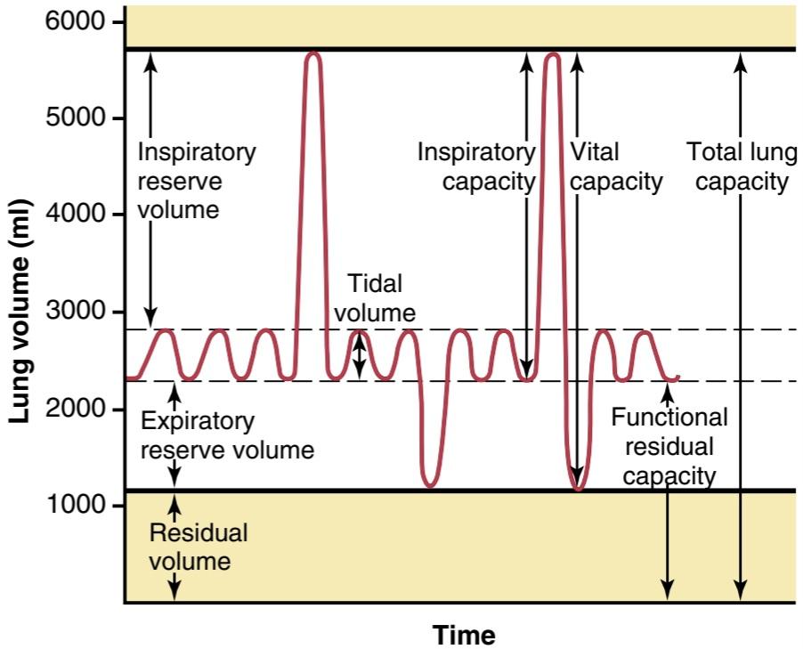
Figure 38-6 Respiratory excursions during normal breathing and during maximal inspiration and maximal expiration.
شکل 38-6 حرکات تنفسی در طول تنفس طبیعی و در طول حداکثر دم و حداکثر بازدم.
Pulmonary Capacities
In describing events in the pulmonary cycle, it is sometimes useful to consider two or more of the volumes together. Such combinations are called pulmonary capacities. To the right in Figure 38-6 are listed the important pulmonary capacities, which can be described as follows:
ظرفیت های ریوی
در توصیف رویدادهای سیکل ریوی، گاهی اوقات مفید است که دو یا چند جلد را با هم در نظر بگیرید. چنین ترکیباتی ظرفیت های ریوی نامیده می شود. در سمت راست در شکل 38-6 ظرفیت های مهم ریوی ذکر شده است که می توان آنها را به شرح زیر توصیف کرد:
1. The inspiratory capacity equals the tidal volume plus the inspiratory reserve volume. This capacity is the amount of air (≈3500 ml) that a person can breathe in, beginning at the normal expiratory level and distending the lungs to the maximum amount.
1. ظرفیت دمی برابر با حجم جزر و مد به اضافه حجم ذخیره دمی است. این ظرفیت مقدار هوایی است (≈3500 میلی لیتر) که فرد می تواند در آن تنفس کند، از سطح بازدم طبیعی شروع می شود و ریه ها را تا حداکثر مقدار منبسط می کند.
2. The functional residual capacity equals the expiratory reserve volume plus the residual volume. This capacity is the amount of air that remains in the lungs at the end of normal expiration ≈2300 ml).
2. ظرفیت باقیمانده عملکردی برابر با حجم ذخیره بازدمی به اضافه حجم باقیمانده است. این ظرفیت مقدار هوایی است که در انتهای بازدم طبیعی ≈2300 میلی لیتر در ریه ها باقی می ماند.
3. The vital capacity equals the inspiratory reserve volume plus the tidal volume plus the expiratory reserve volume. This capacity is the maximum amount of air a person can expel from the lungs after first filling the lungs to their maximum extent and then expiring to the maximum extent (~4600 ml).
3. ظرفیت حیاتی برابر با حجم ذخیره دمی به اضافه حجم جزر و مدی به اضافه حجم ذخیره بازدمی است. این ظرفیت حداکثر مقدار هوایی است که یک فرد می تواند پس از پر کردن ریه ها به حداکثر میزان و سپس منفجر شدن حداکثر (~4600 میلی لیتر) از ریه ها خارج شود.
4. The total lung capacity is the maximum volume to which the lungs can be expanded with the greatest possible effort (~5800 ml); it is equal to the vital capacity plus the residual volume.
4. ظرفیت کل ریه حداکثر حجمی است که می توان ریه ها را با بیشترین تلاش ممکن به آن منبسط کرد (~5800 میلی لیتر). برابر با ظرفیت حیاتی به اضافه حجم باقیمانده است.
Most pulmonary volumes and capacities are usually about 20% to 30% less in women than in men, and they are greater in large and athletic people than in small and asthenic people.
اکثر حجم ها و ظرفیت های ریوی معمولاً در زنان حدود 20 تا 30 درصد کمتر از مردان است و در افراد بزرگ و ورزشکار بیشتر از افراد کوچک و آستنیک است.
ABBREVIATIONS AND SYMBOLS USED IN PULMONARY FUNCTION STUDIES
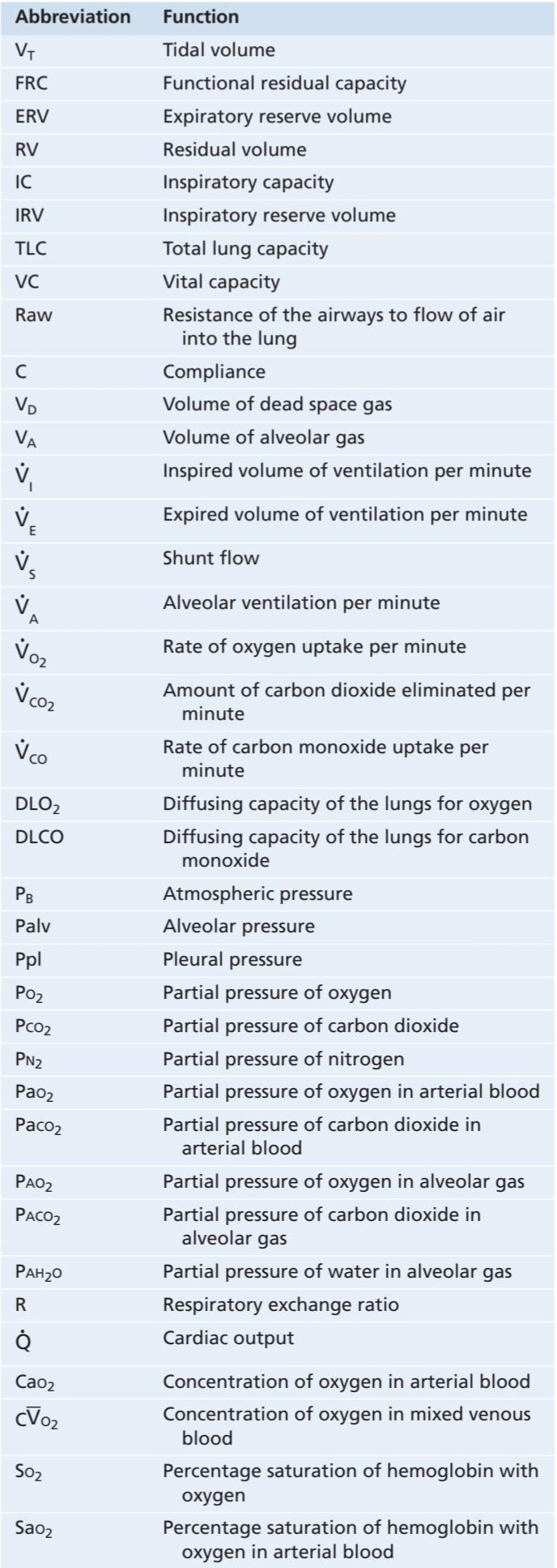
Spirometry is only one of many measurement procedures that pulmonary physicians use daily. Many of these procedures depend heavily on mathematical computations. To simplify these calculations, as well as the presentation of pulmonary function data, several abbreviations and symbols have become standardized. The more important of these are given in Table 38-2. Using these symbols, we present here a few simple algebraic equations showing some of the interrelationships among the pulmonary volumes and capacities; the student should think through and verify these interrelationships.
اختصارات و نمادهای مورد استفاده در مطالعات عملکرد ریوی
اسپیرومتری تنها یکی از بسیاری از روش های اندازه گیری است که پزشکان ریه روزانه از آن استفاده می کنند. بسیاری از این روش ها به شدت به محاسبات ریاضی بستگی دارد. برای ساده کردن این محاسبات، و همچنین ارائه داده های عملکرد ریوی، چندین علامت اختصاری و نماد استاندارد شده اند. مهمترین آنها در جدول 38-2 آورده شده است. با استفاده از این نمادها، ما در اینجا چند معادله جبری ساده را ارائه می کنیم که برخی از روابط متقابل بین حجم ها و ظرفیت های ریوی را نشان می دهد. دانش آموز باید این روابط متقابل را بیندیشد و بررسی کند.

Table 38-2 Abbreviations and Symbols for Pulmonary Function
جدول 38-2 اختصارات و نمادها برای عملکرد ریوی
DETERMINATION OF FUNCTIONAL RESIDUAL CAPACITY, RESIDUAL VOLUME, AND TOTAL LUNG CAPACITY-HELIUM DILUTION METHOD
The functional residual capacity (FRC), which is the volume of air that remains in the lungs at the end of each normal expiration, is important to lung function. Because its value changes markedly in some types of pulmonary disease, it is often desirable to measure this capacity. The spirometer cannot be used in to measure the FRC directly because the air in the residual volume of the lungs cannot be expired into the spirometer, and this volume constitutes about half of the FRC. To measure FRC, the spirometer must be used in an indirect manner, usually by means of a helium dilution method, as follows.
تعیین ظرفیت باقیمانده کارکردی، حجم باقیمانده و ظرفیت کل ریه – روش رقیق سازی هلیوم
ظرفیت باقیمانده عملکردی (FRC)، که حجم هوایی است که در پایان هر بازدم طبیعی در ریه ها باقی می ماند، برای عملکرد ریه مهم است. از آنجایی که مقدار آن در برخی از انواع بیماری های ریوی به طور قابل توجهی تغییر می کند، اغلب اندازه گیری این ظرفیت مطلوب است. از اسپیرومتر برای اندازه گیری مستقیم FRC نمی توان استفاده کرد زیرا هوای موجود در حجم باقیمانده ریه ها نمی تواند به داخل اسپیرومتر منتهی شود و این حجم حدود نیمی از FRC را تشکیل می دهد. برای اندازه گیری FRC، اسپیرومتر باید به صورت غیر مستقیم، معمولاً با روش رقیق سازی هلیوم، به شرح زیر استفاده شود.
A spirometer of known volume is filled with air mixed with helium at a known concentration. Before breathing from the spirometer, the person expires normally. At the end of this expiration, the remaining volume in the lungs is equal to the FRC. At this point, the subject immediately begins to breathe from the spirometer, and the gases of the spirometer mix with the gases of the lungs. As a result, the helium becomes diluted by the FRC gases, and the volume of the FRC can be calculated from the degree of dilution of the helium, using the following formula:
یک اسپیرومتر با حجم مشخص با هوای مخلوط با هلیوم در غلظت مشخص پر می شود. قبل از تنفس از اسپیرومتر، فرد به طور طبیعی منقضی می شود. در پایان این بازدم، حجم باقی مانده در ریه ها برابر با FRC است. در این مرحله، سوژه بلافاصله شروع به تنفس از اسپیرومتر می کند و گازهای اسپیرومتر با گازهای ریه مخلوط می شوند. در نتیجه هلیوم توسط گازهای FRC رقیق می شود و حجم FRC را می توان از درجه رقت هلیوم با استفاده از فرمول زیر محاسبه کرد:

where FRC is functional residual capacity, CiHe is the initial concentration of helium in the spirometer, CfHe is the final concentration of helium in the spirometer, and Vispir is the initial volume of the spirometer.
که در آن FRC ظرفیت باقیمانده عملکردی است، CiHe غلظت اولیه هلیوم در اسپیرومتر، CfHe غلظت نهایی هلیوم در اسپیرومتر و Vispir حجم اولیه اسپیرومتر است.
Once the FRC has been determined, the residual volume (RV) can be determined by subtracting expiratory reserve volume (ERV), as measured by normal spirometry, from the FRC. Also, the total lung capacity (TLC) can be determined by adding the inspiratory capacity (IC) to the FRC. That is:
هنگامی که FRC تعیین شد، حجم باقیمانده (RV) را می توان با کم کردن حجم ذخیره بازدمی (ERV)، که با اسپیرومتری معمولی اندازه گیری می شود، از FRC تعیین کرد. همچنین ظرفیت کل ریه (TLC) را می توان با افزودن ظرفیت دمی (IC) به FRC تعیین کرد. یعنی:

and

MINUTE RESPIRATORY VOLUME EQUALS RESPIRATORY RATE TIMES TIDAL VOLUME
The minute respiratory volume is the total amount of new air moved into the respiratory passages each minute and is equal to the tidal volume times the respiratory rate per minute. The normal tidal volume is about 500 ml, and the normal respiratory rate is about 12 breaths/ min. Therefore, the minute respiratory volume averages about 6 L/min. A person can live for a short period with a minute respiratory volume as low as 1.5 L/min and a respiratory rate of only 2 to 4 breaths/min.
دقیقه حجم تنفسی برابر است با تعداد دفعات تنفس و حجم جزر و مد
حجم تنفسی دقیقه ای کل مقدار هوای جدیدی است که در هر دقیقه به مجاری تنفسی منتقل می شود و برابر است با حجم جزر و مد ضربدر تعداد تنفس در دقیقه. حجم طبیعی جزر و مد حدود 500 میلی لیتر و تعداد تنفس طبیعی حدود 12 تنفس در دقیقه است. بنابراین، حجم دقیقه تنفسی به طور متوسط حدود 6 لیتر در دقیقه است. یک فرد می تواند برای مدت کوتاهی با حجم تنفسی کمتر از 1.5 لیتر در دقیقه و تعداد تنفس تنها 2 تا 4 نفس در دقیقه زندگی کند.
The respiratory rate occasionally rises to 40 to 50 breaths/min, and the tidal volume can become as great as the vital capacity, about 4600 ml in a young man. This can give a minute respiratory volume greater than 200 L/min, or more than 30 times normal. Most people cannot sustain more than half to two-thirds of these values for longer than 1 minute.
سرعت تنفس گهگاه به 40 تا 50 تنفس در دقیقه افزایش می یابد و حجم جزر و مد می تواند به اندازه ظرفیت حیاتی، حدود 4600 میلی لیتر در یک مرد جوان باشد. این می تواند یک دقیقه حجم تنفسی بیش از 200 لیتر در دقیقه یا بیش از 30 برابر نرمال ایجاد کند. اکثر افراد نمی توانند بیش از نیم تا دو سوم این مقادیر را برای بیش از 1 دقیقه حفظ کنند.
ALVEOLAR VENTILATION
The ultimate importance of pulmonary ventilation is to renew the air in the gas exchange areas of the lungs continually, where air is in proximity to the pulmonary blood. These areas include the alveoli, alveolar sacs, alveolar ducts, and respiratory bronchioles. The rate at which new air reaches these areas is called alveolar ventilation.
تهویه آلوئولار
اهمیت نهایی تهویه ریوی، تجدید مداوم هوا در نواحی تبادل گاز ریه ها است، جایی که هوا در مجاورت خون ریوی قرار دارد. این نواحی شامل آلوئول ها، کیسه های آلوئولی، مجاری آلوئولی و برونشیول های تنفسی است. سرعت رسیدن هوای جدید به این مناطق را تهویه آلوئولی می نامند.
DEAD SPACE AND ITS EFFECT ON ALVEOLAR VENTILATION
Some of the air a person breathes never reaches the gas exchange areas but simply fills respiratory passages, such as the nose, pharynx, and trachea, where gas exchange does not occur. This air is called dead space air because it is not useful for gas exchange.
فضای مرده و تأثیر آن بر تهویه آلوئولار
بخشی از هوایی که فرد تنفس می کند هرگز به نواحی تبادل گاز نمی رسد، بلکه صرفاً مجاری تنفسی مانند بینی، حلق و نای را پر می کند، جایی که تبادل گاز در آنها انجام نمی شود. این هوا را هوای فضای مرده می نامند زیرا برای تبادل گاز مفید نیست.
On expiration, the air in the dead space is expired first, before any of the air from the alveoli reaches the atmosphere. Therefore, the dead space is very disadvantageous for removing the expiratory gases from the lungs.
در هنگام بازدم، هوای فضای مرده ابتدا منقضی میشود، قبل از اینکه هوای آلوئولها به جو برسد. بنابراین فضای مرده برای خارج کردن گازهای بازدمی از ریه ها بسیار مضر است.
Measurement of Dead Space Volume. A simple method for measuring dead space volume is demonstrated by the graph in Figure 38-7. In making this measurement, the subject suddenly takes a deep breath of 100% O2, which fills the entire dead space with pure O2. Some oxygen also mixes with the alveolar air but does not completely replace this air. Then the person expires through a rapidly recording nitrogen meter, which makes the record shown in the figure. The first portion of the expired air comes from the dead space regions of the respiratory passageways, where the air has been completely replaced by O2. Therefore, in the early part of the record, only O2 appears, and the nitrogen concentration is 0. Then, when alveolar air begins to reach the nitrogen meter, the nitrogen concentration rises rapidly because alveolar air containing large amounts of nitrogen begins to mix with the dead space air. After still more air has been expired, all the dead space air has been washed from the passages and only alveolar air remains. Therefore, the recorded nitrogen concentration reaches a plateau level equal to its concentration in the alveoli, as shown at the right in the figure. The gray area represents the air that has no nitrogen in it and is a measure of the volume of dead space air. For exact quantification, the following equation is used:
اندازه گیری حجم فضای مرده. یک روش ساده برای اندازه گیری حجم فضای مرده توسط نمودار در شکل 38-7 نشان داده شده است. در انجام این اندازه گیری، سوژه ناگهان یک نفس عمیق 100% O2 می کشد که کل فضای مرده را با O2 خالص پر می کند. مقداری اکسیژن نیز با هوای آلوئولی مخلوط می شود اما به طور کامل جایگزین این هوا نمی شود. سپس فرد از طریق یک نیتروژن متر که به سرعت ثبت می شود منقضی می شود، که رکورد نشان داده شده در شکل را ایجاد می کند. بخش اول هوای منقضی شده از نواحی فضای مرده راه های تنفسی می آید، جایی که هوا به طور کامل توسط O2 جایگزین شده است. بنابراین، در اوایل ثبت، فقط O2 ظاهر می شود و غلظت نیتروژن 0 است. سپس، زمانی که هوای آلوئولی شروع به رسیدن به نیتروژن متر می کند، غلظت نیتروژن به سرعت افزایش می یابد زیرا هوای آلوئولی حاوی مقادیر زیادی نیتروژن شروع به مخلوط شدن با هوای فضای مرده می کند. پس از منقضی شدن هوای بیشتر، تمام هوای فضای مرده از گذرگاه ها شسته شده و تنها هوای آلوئولی باقی می ماند. بنابراین، غلظت نیتروژن ثبت شده به سطح فلاتی برابر با غلظت آن در آلوئول ها می رسد، همانطور که در سمت راست در شکل نشان داده شده است. ناحیه خاکستری نمایانگر هوایی است که نیتروژن در آن وجود ندارد و اندازه گیری حجم هوای فضای مرده است. برای تعیین کمیت دقیق از معادله زیر استفاده می شود:

where VD is dead space air and VE is the total volume of expired air.
که در آن VD هوای فضای مرده و VE حجم کل هوای منقضی شده است.
Let us assume, for example, that the gray area on the graph is 30 square centimeters, the pink area is 70 square centimeters, and the total volume expired is 500 ml. The dead space would be
به عنوان مثال، فرض می کنیم که ناحیه خاکستری نمودار 30 سانتی متر مربع، ناحیه صورتی 70 سانتی متر مربع و حجم کل منقضی شده 500 میلی لیتر است. فضای مرده خواهد بود

Normal Dead Space Volume. The normal dead space air in a young man is about 150 ml. Dead space air increases slightly with age.
حجم عادی فضای مرده. هوای معمولی فضای مرده در یک مرد جوان حدود 150 میلی لیتر است. هوای فضای مرده با افزایش سن کمی افزایش می یابد.
Anatomical Versus Physiological Dead Space. The method just described for measuring the dead space measures the volume of all the space of the respiratory system other than the alveoli and their other closely related gas ex- change areas; this space is called the anatomic dead space. On occasion, some of the alveoli are nonfunctional or only partially functional because of absent or poor blood flow through the adjacent pulmonary capillaries. Therefore, these alveoli must also be considered dead space. When the alveolar dead space is included in the total measurement of dead space, this is called the physiological dead space, in contradistinction to the anatomical dead space. In a person with healthy lungs, the anatomical and physiological dead spaces are nearly equal because all alveoli are functional in the normal lung but, in a person with partially functional or nonfunctional alveoli in some parts of the lungs, the physiological dead space may be as much as 10 times the volume of the anatomical dead space, or 1 to 2 liters. These problems are discussed further in Chapter 40 in relation to pulmonary gaseous exchange and in Chapter 43 in relation to certain pulmonary diseases.
آناتومیک در مقابل فضای مرده فیزیولوژیکی. روشی که برای اندازهگیری فضای مرده توضیح داده شد، حجم تمام فضای دستگاه تنفسی به غیر از آلوئولها و دیگر مناطق تبادل گاز نزدیک به آنها را اندازهگیری میکند. این فضا فضای مرده آناتومیک نامیده می شود. گاهی اوقات، برخی از آلوئول ها به دلیل عدم وجود یا جریان ضعیف خون از طریق مویرگ های ریوی مجاور، غیرعملکردی هستند یا فقط تا حدی کار می کنند. بنابراین، این آلوئول ها را نیز باید فضای مرده در نظر گرفت. وقتی فضای مرده آلوئولی در کل اندازه گیری فضای مرده گنجانده شود، برخلاف فضای مرده آناتومیکی، فضای مرده فیزیولوژیکی نامیده می شود. در فردی که ریههای سالم دارد، فضای مرده آناتومیکی و فیزیولوژیکی تقریباً برابر است، زیرا همه آلوئولها در ریه طبیعی کار میکنند، اما در فردی با آلوئولهای نیمه عملکردی یا غیرعملکردی در برخی از قسمتهای ریه، فضای مرده فیزیولوژیکی ممکن است 10 برابر حجم فضای مرده آناتومیکی یا 1 تا 2 لیتر باشد. این مشکلات بیشتر در فصل 40 در رابطه با تبادل گازی ریوی و در فصل 43 در رابطه با برخی بیماری های ریوی مورد بحث قرار گرفته است.

Figure 38-7 Record of the changes in nitrogen concentration in the expired air after a single previous inspiration of pure oxygen. This record can be used to calculate dead space, as discussed in the text.
شکل 38-7 ثبت تغییرات در غلظت نیتروژن در هوای منقضی شده پس از یک الهام قبلی از اکسیژن خالص. این رکورد می تواند برای محاسبه فضای مرده، همانطور که در متن بحث شده است، استفاده شود.
RATE OF ALVEOLAR VENTILATION
Alveolar ventilation per minute is the total volume of new air entering the alveoli and adjacent gas exchange areas each minute. It is equal to the respiratory rate times the amount of new air that enters these areas with each breath:
نرخ تهویه آلوئولار
تهویه آلوئولار در هر دقیقه حجم کل هوای جدیدی است که در هر دقیقه وارد آلوئول ها و مناطق تبادل گاز مجاور می شود. برابر است با تعداد تنفس ضربدر هوای جدیدی که با هر نفس وارد این مناطق می شود:

where is the volume of alveolar ventilation per minute, Freq is the frequency of respiration per minute, V, is the tidal volume, and Va is the physiological dead space volume.
که در آن حجم تهویه آلوئولی در دقیقه، Freq فرکانس تنفس در دقیقه، V حجم جزر و مدی و Va حجم فضای مرده فیزیولوژیکی است.
Thus, with a normal tidal volume of 500 ml, a normal dead space of 150 ml, and a respiratory rate of 12 breaths/ min, alveolar ventilation equals 12 x (500 – 150), or 4200 ml/min.
بنابراین، با حجم جزر و مد طبیعی 500 میلی لیتر، فضای مرده طبیعی 150 میلی لیتر، و سرعت تنفس 12 تنفس در دقیقه، تهویه آلوئولی برابر با 12 x (500 – 150)، یا 4200 میلی لیتر در دقیقه است.
Alveolar ventilation is one of the major factors deter- mining the concentrations of oxygen and carbon dioxide in the alveoli. Therefore, almost all discussions of gaseous exchange in the following chapters on the respiratory system focus on alveolar ventilation.
تهویه آلوئولار یکی از عوامل اصلی تعیین کننده غلظت اکسیژن و دی اکسید کربن در آلوئول ها است. بنابراین، تقریباً تمام بحث های تبادل گازی در فصل های بعدی در مورد سیستم تنفسی بر تهویه آلوئولار متمرکز است.
Functions of Respiratory Passageways
Trachea, Bronchi, and Bronchioles
Figure 38-8 highlights the respiratory passageways. The air is distributed to the lungs by way of the trachea, bronchi, and bronchioles.
عملکرد راه های تنفسی
نای، برونش و برونشیل
شکل 38-8 راه های تنفسی را نشان می دهد. هوا از طریق نای، برونش ها و نایژه ها به ریه ها منتقل می شود.
One of the most important challenges in the respiratory passageways is to keep them open and allow easy passage of air to and from the alveoli. To keep the trachea from collapsing, multiple cartilage rings extend about five-sixths of the way around the trachea. In the walls of the bronchi, less extensive curved cartilage plates also maintain a reasonable amount of rigidity yet allow sufficient motion for the lungs to expand and contract. These plates become progressively less extensive in the later generations of bronchi and are gone in the bronchioles, which usually have diameters less than 1.5 millimeters. The bronchioles are not prevented from collapsing by the rigidity of their walls. Instead, they are kept expanded mainly by the same transpulmonary pressures that expand the alveoli. That is, as the alveoli enlarge, the bronchioles also enlarge, but not as much.
یکی از مهم ترین چالش ها در مسیرهای تنفسی باز نگه داشتن آنها و عبور آسان هوا به و از آلوئول ها است. برای جلوگیری از فروپاشی نای، حلقه های غضروفی متعددی در حدود پنج ششم دور نای گسترش می یابند. در دیوارههای نایژهها، صفحات غضروفی با انحنای کمتر نیز مقدار معقولی از سفتی را حفظ میکنند و در عین حال امکان حرکت کافی برای انبساط و انقباض ریهها را فراهم میکنند. این صفحات به تدریج در نسل های بعدی برونش ها کمتر گسترده می شوند و در برونشیول ها که معمولاً قطر آنها کمتر از 1.5 میلی متر است از بین می روند. برونشیول ها با سفتی دیواره آنها از فروپاشی جلوگیری نمی کنند. در عوض، آنها عمدتاً توسط همان فشارهای ترانس ریوی که آلوئول ها را گسترش می دهند، منبسط می شوند. یعنی با بزرگ شدن آلوئول ها، برونشیول ها نیز بزرگ می شوند، اما نه به اندازه.
Muscular Wall of the Bronchi and Bronchioles. In all areas of the trachea and bronchi not occupied by cartilage plates, the walls are composed mainly of smooth muscle. Also, the walls of the bronchioles are almost entirely smooth muscle, with the exception of the most terminal bronchiole, called the respiratory bronchiole, which is mainly pulmonary epithelium and underlying fibrous tissue plus a few smooth muscle fibers. Many obstructive diseases of the lung result from narrowing of the smaller bronchi and larger bronchioles, often because of excessive contraction of the smooth muscle.
دیواره عضلانی برونش ها و برونشیل ها. در تمام نواحی نای و برونش که توسط صفحات غضروفی اشغال نشده اند، دیواره ها عمدتاً از عضلات صاف تشکیل شده اند. همچنین، دیوارههای نایژهها تقریباً کاملاً عضلانی صاف هستند، به استثنای انتهاییترین برونشیول، به نام برونشیول تنفسی، که عمدتاً اپیتلیوم ریوی و بافت فیبری زیرین بهعلاوه چند فیبر عضلانی صاف است. بسیاری از بیماری های انسدادی ریه ناشی از باریک شدن برونش های کوچکتر و برونشیول های بزرگتر است که اغلب به دلیل انقباض بیش از حد ماهیچه صاف است.
Resistance to Airflow in the Bronchial Tree. Under normal respiratory conditions, air flows through the respiratory passageways so easily that less than 1 cm H2O pressure gradient from the alveoli to the atmosphere is sufficient to cause enough airflow for quiet breathing. The greatest amount of resistance to airflow occurs not in the tiny air passages of the terminal bronchioles but in some of the larger bronchioles and bronchi near the trachea. The reason for this high resistance is that there are relatively few of these larger bronchi in comparison with the approximately 65,000 parallel terminal bronchioles, through each of which only a minute amount of air must pass.
مقاومت در برابر جریان هوا در درخت برونش. در شرایط عادی تنفسی، هوا از طریق مجاری تنفسی به راحتی جریان مییابد، به طوری که گرادیان فشار H2O کمتر از 1 سانتیمتر از آلوئولها به جو برای ایجاد جریان هوای کافی برای تنفس آرام کافی است. بیشترین مقاومت در برابر جریان هوا نه در گذرگاههای هوای نایژههای انتهایی، بلکه در برخی از برونشیولها و برونشهای بزرگتر نزدیک نای رخ میدهد. دلیل این مقاومت بالا این است که تعداد نسبتاً کمی از این برونش های بزرگتر در مقایسه با تقریباً 65000 برونشیول انتهایی موازی وجود دارد که از هر کدام فقط مقدار کمی هوا باید عبور کند.
In some disease conditions, the smaller bronchioles play a far greater role in determining airflow resistance because of their small size and because they are easily occluded by the following: (1) muscle contraction in their walls; (2) edema in the walls; or (3) mucus collecting in the lumens of the bronchioles.
در برخی بیماریها، برونشیولهای کوچکتر نقش بسیار بیشتری در تعیین مقاومت جریان هوا ایفا میکنند زیرا اندازه کوچکشان هستند و به راحتی توسط موارد زیر مسدود میشوند: (1) انقباض عضلانی در دیوارههایشان. (2) ادم در دیوارها. یا (3) مخاط جمع آوری شده در مجرای برونشیل ها.
Nervous and Local Control of the Bronchiolar Musculature Sympathetic Dilation of the Bronchioles. Direct control of the bronchioles by sympathetic nerve fibers is relatively weak because few of these fibers penetrate to the central portions of the lung. However, the bronchial tree is very much exposed to norepinephrine and epinephrine released into the blood by sympathetic stimulation of the adrenal gland medullae. Both these hormones, especially epinephrine because of its greater stimulation of beta- adrenergic receptors, cause dilation of the bronchial tree.
کنترل عصبی و موضعی عضله برونش اتساع سمپاتیک برونشیل ها. کنترل مستقیم برونشیول ها توسط رشته های عصبی سمپاتیک نسبتا ضعیف است زیرا تعداد کمی از این رشته ها به بخش های مرکزی ریه نفوذ می کنند. با این حال، درخت برونش به شدت در معرض نوراپی نفرین و اپی نفرین است که توسط تحریک سمپاتیک بصل النخاع غده فوق کلیوی در خون آزاد می شود. هر دوی این هورمون ها، به ویژه اپی نفرین به دلیل تحریک بیشتر گیرنده های بتا آدرنرژیک، باعث گشاد شدن درخت برونش می شوند.
Parasympathetic Constriction of the Bronchioles. A few parasympathetic nerve fibers derived from the vagus nerves penetrate the lung parenchyma. These nerves secrete acetylcholine and, when activated, cause mild to moderate constriction of the bronchioles. When a disease process such as asthma has already caused some bronchiolar constriction, superimposed parasympathetic nervous stimulation often worsens the condition. When this situation occurs, administration of drugs that block the effects of acetylcholine, such as atropine, can sometimes relax the respiratory passages enough to relieve the obstruction.
انقباض پاراسمپاتیک برونشیول ها. چند رشته عصبی پاراسمپاتیک مشتق شده از اعصاب واگ به پارانشیم ریه نفوذ می کنند. این اعصاب استیل کولین ترشح می کنند و در صورت فعال شدن باعث انقباض خفیف تا متوسط برونشیول ها می شوند. هنگامی که یک فرآیند بیماری مانند آسم قبلاً باعث انقباض برونش شده است، تحریک عصبی پاراسمپاتیک روی هم اغلب وضعیت را بدتر می کند. هنگامی که این وضعیت رخ می دهد، تجویز داروهایی که اثرات استیل کولین را مسدود می کنند، مانند آتروپین، گاهی اوقات می تواند راه های تنفسی را به اندازه کافی شل کند تا انسداد را برطرف کند.
Sometimes the parasympathetic nerves are also activated by reflexes that originate in the lungs. Most of these reflexes begin with irritation of the epithelial membrane of the respiratory passageways, initiated by noxious gases, dust, cigarette smoke, or bronchial infection. Also, a bronchiolar constrictor reflex often occurs when microemboli occlude small pulmonary arteries.
گاهی اوقات اعصاب پاراسمپاتیک نیز توسط رفلکس هایی که از ریه ها منشا می گیرند فعال می شوند. بیشتر این رفلکس ها با تحریک غشای اپیتلیال مجاری تنفسی شروع می شوند که توسط گازهای مضر، گرد و غبار، دود سیگار یا عفونت برونش آغاز می شود. همچنین، رفلکس منقبض کننده برونش اغلب زمانی رخ می دهد که میکروآمبولی عروق کوچک ریوی را مسدود کند.
Local Secretory Factors May Cause Bronchiolar Constriction. Several substances formed in the lungs are often active in causing bronchiolar constriction. Two of the most important of these are histamine and slow reactive substance of anaphylaxis. Both these substances are released in the lung tissues by mast cells during allergic reactions, especially those caused by pollen in the air. Therefore, they play key roles in causing airway obstruction in allergic asthma; this is especially true of the slow reactive substance of anaphylaxis.
عوامل ترشحی موضعی ممکن است باعث انقباض برونش شود. چندین ماده تشکیل شده در ریه ها اغلب در ایجاد انقباض برونش ها فعال هستند. دو مورد از مهمترین آنها هیستامین و ماده واکنش کند آنافیلاکسی است. هر دوی این مواد در طی واکنش های آلرژیک، به ویژه واکنش های ناشی از گرده های موجود در هوا، توسط ماست سل ها در بافت های ریه آزاد می شوند. بنابراین، آنها نقش کلیدی در ایجاد انسداد راه هوایی در آسم آلرژیک دارند. این به ویژه در مورد ماده واکنش کند آنافیلاکسی صادق است.
The same irritants that cause parasympathetic constrictor reflexes of the airways-smoke, dust, sulfur dioxide, and some of the acidic elements in smog-may also act directly on the lung tissues to initiate local, non-nervous reactions that cause obstructive constriction of the airways.
همان محرک هایی که باعث رفلکس های منقبض کننده پاراسمپاتیک راه های هوایی می شوند – دود، گرد و غبار، دی اکسید گوگرد و برخی از عناصر اسیدی موجود در مه دود – ممکن است مستقیماً روی بافت های ریه عمل کنند و واکنش های موضعی و غیر عصبی را آغاز کنند که باعث انقباض انسدادی راه های هوایی می شود.

Figure 38-8 Respiratory passages.
شکل 38-8 راه های تنفسی.
Mucus Lining the Respiratory Passageways and Cilia Action to Clear the Passageways
All the respiratory passages, from the nose to the terminal bronchioles, are kept moist by a layer of mucus that coats the entire surface. The mucus is secreted partly by individual mucous goblet cells in the epithelial lining of the passages and partly by small submucosal glands. In addition to keeping the surfaces moist, the mucus traps small particles out of the inspired air and keeps most of these particles from ever reaching the alveoli. The mucus is removed from the passages in the following manner.
مخاط پوشاننده گذرگاه های تنفسی و اقدام جلگه ای برای پاکسازی گذرگاه ها
تمام راههای تنفسی، از بینی تا برونشیولهای انتهایی، توسط لایهای از مخاط که تمام سطح را میپوشاند، مرطوب نگه داشته میشوند. مخاط تا حدی توسط سلول های جام مخاطی منفرد در پوشش اپیتلیال معابر و قسمتی از غدد کوچک زیر مخاطی ترشح می شود. موکوس علاوه بر مرطوب نگه داشتن سطوح، ذرات کوچک را از هوای دمیده شده به دام می اندازد و از رسیدن بیشتر این ذرات به آلوئول ها جلوگیری می کند. مخاط به روش زیر از معابر خارج می شود.
The entire surface of the respiratory passages, in the nose and the lower passages, down as far as the terminal bronchioles, is lined with ciliated epithelium, with about 200 cilia on each epithelial cell. These cilia beat continually at a rate of 10 to 20 times/sec by the mechanism explained in Chapter 2, and the direction of their “power stroke” is always toward the pharynx. That is, the cilia in the lungs beat upward, whereas those in the nose beat downward. This continual beating causes the coat of mucus to flow slowly, at a velocity of a few millimeters per minute, toward the pharynx. Then the mucus and its entrapped particles are swallowed or coughed to the exterior.
تمام سطح مجاری تنفسی، در بینی و راههای تحتانی، تا برونشیولهای انتهایی، با اپیتلیوم مژکدار پوشانده شده است که در هر سلول اپیتلیال حدود 200 مژک وجود دارد. این مژک ها با مکانیسم توضیح داده شده در فصل 2 به طور مداوم با سرعت 10 تا 20 بار در ثانیه می زنند و جهت “سکته قدرت” آنها همیشه به سمت حلق است. یعنی مژکهای ریه به سمت بالا میزنند، در حالی که مژکهای بینی به سمت پایین میزنند. این ضرب و شتم مداوم باعث می شود که پوشش مخاطی به آرامی و با سرعت چند میلی متر در دقیقه به سمت حلق جریان یابد. سپس مخاط و ذرات به دام افتاده آن بلعیده می شوند یا به بیرون سرفه می شوند.
Cough Reflex
The bronchi and trachea are so sensitive to light touch that slight amounts of foreign matter or other causes of irritation initiate the cough reflex. The larynx and carina (the point where the trachea divides into the bronchi) are especially sensitive, and the terminal bronchioles and even the alveoli are sensitive to corrosive chemical stimuli such as sulfur dioxide gas or chlorine gas. Afferent nerve impulses pass from the respiratory passages mainly through the vagus nerves to the medulla of the brain. There, an automatic sequence of events is triggered by the neuronal circuits of the medulla, causing the following effects.
رفلکس سرفه
برونش ها و نای به قدری به لمس سبک حساس هستند که مقادیر جزئی ماده خارجی یا سایر علل تحریک رفلکس سرفه را آغاز می کند. حنجره و حنجره (نقطه ای که نای به نایژه ها تقسیم می شود) حساس هستند و برونشیول های انتهایی و حتی آلوئول ها به محرک های شیمیایی خورنده مانند گاز دی اکسید گوگرد یا گاز کلر حساس هستند. تکانه های عصبی آوران از مجاری تنفسی عمدتاً از طریق اعصاب واگ به بصل النخاع مغز منتقل می شوند. در آنجا، یک توالی خودکار از رویدادها توسط مدارهای عصبی بصل النخاع ایجاد می شود و باعث ایجاد اثرات زیر می شود.
1. Up to 2.5 liters of air are rapidly inspired.
1. تا 2.5 لیتر هوا به سرعت دم می شود.
2. The epiglottis closes, and the vocal cords shut tightly to entrap the air within the lungs.
2. اپی گلوت بسته می شود و تارهای صوتی محکم بسته می شوند تا هوا را در داخل ریه ها به دام بیندازند.
3. The abdominal muscles contract forcefully, pushing against the diaphragm while other expiratory muscles, such as the internal intercostals, also contract forcefully. Consequently, the pressure in the lungs rises rapidly, to as much as 100 mm Hg or more.
3. ماهیچه های شکمی به شدت منقبض می شوند و به دیافراگم فشار می آورند در حالی که سایر عضلات بازدمی مانند بین دنده های داخلی نیز به شدت منقبض می شوند. در نتیجه، فشار در ریه ها به سرعت افزایش می یابد و به 100 میلی متر جیوه یا بیشتر می رسد.
4. The vocal cords and epiglottis suddenly open widely, so that air under this high pressure in the lungs explodes outward. Sometimes this air is expelled at velocities ranging from 75 to 100 miles/hour.
4. تارهای صوتی و اپی گلوت به طور ناگهانی به طور گسترده باز می شوند، به طوری که هوا تحت این فشار بالا در ریه ها به سمت بیرون منفجر می شود. گاهی اوقات این هوا با سرعتی بین 75 تا 100 مایل در ساعت دفع می شود.
Importantly, the strong compression of the lungs collapses the bronchi and trachea by causing their noncartilaginous parts to invaginate inward, so the exploding air actually passes through bronchial and tracheal slits. The rapidly moving air usually carries with it any foreign matter that is present in the bronchi or trachea.
نکته مهم این است که فشرده سازی قوی ریه ها باعث فروپاشی برونش ها و نای می شود و باعث می شود قسمت های غیرغضروفی آنها به سمت داخل فرو برود، بنابراین هوای منفجر شده در واقع از شکاف های برونش و نای عبور می کند. هوای به سرعت در حال حرکت معمولاً هرگونه ماده خارجی موجود در برونش یا نای را با خود حمل می کند.
Sneeze Reflex
The sneeze reflex is very much like the cough reflex, except that it applies to the nasal passageways instead of the lower respiratory passages. The initiating stimulus of the sneeze reflex is irritation in the nasal passageways; the afferent impulses pass in the fifth cranial nerve to the medulla, where the reflex is triggered. A series of reactions similar to those for the cough reflex takes place, but the uvula is depressed, so large amounts of air pass rapidly through the nose, thus helping clear the nasal passages of foreign matter.
رفلکس عطسه
رفلکس عطسه بسیار شبیه رفلکس سرفه است، با این تفاوت که به جای مجاری تنفسی تحتانی، روی مجاری بینی اعمال می شود. محرک آغازگر رفلکس عطسه، تحریک در مجاری بینی است. تکانههای آوران در عصب پنجم جمجمهای به مدولا میرسند، جایی که رفلکس تحریک میشود. مجموعهای از واکنشهای مشابه واکنشهای مربوط به رفلکس سرفه انجام میشود، اما یووولا تحت فشار قرار میگیرد، بنابراین مقدار زیادی هوا به سرعت از بینی عبور میکند و در نتیجه به پاکسازی مجرای بینی از مواد خارجی کمک میکند.
Normal Respiratory Functions of the Nose
As air passes through the nose, three distinct normal respiratory functions are performed by the nasal cavities: (1) the air is warmed by the extensive surfaces of the conchae and septum, a total area of about 160 square centimeters (see Figure 38-8); (2) the air is almost completely humidified, even before it passes beyond the nose; and (3) the air is partially filtered. These functions together are called the air-conditioning function of the upper respiratory passageways. Ordinarily, the temperature of the inspired air rises to within 1°F of body temperature and to within 2% to 3% of full saturation with water vapor before it reaches the trachea. When a person breathes air through a tube directly into the trachea (as through a tracheostomy), the cooling and especially the drying effect in the lower lung can lead to serious lung crusting and infection.
عملکردهای طبیعی تنفسی بینی
همانطور که هوا از بینی عبور می کند، سه عملکرد طبیعی تنفسی متمایز توسط حفره های بینی انجام می شود: (1) هوا توسط سطوح وسیع کانکا و سپتوم گرم می شود، مساحت کل حدود 160 سانتی متر مربع (شکل 38-8 را ببینید). (2) هوا تقریباً کاملاً مرطوب شده است، حتی قبل از اینکه از بینی عبور کند. و (3) هوا تا حدی فیلتر شده است. این عملکردها با هم عملکرد تهویه مطبوع مجاری تنفسی فوقانی نامیده می شوند. معمولاً دمای هوای دمیده شده تا 1 درجه فارنهایت دمای بدن و تا 2 تا 3 درصد اشباع کامل با بخار آب قبل از رسیدن به نای افزایش می یابد. هنگامی که فردی هوا را از طریق لوله مستقیماً به داخل نای تنفس می کند (مانند تراکئوستومی)، خنک کننده و به ویژه اثر خشک شدن در قسمت پایینی ریه می تواند منجر به ایجاد پوسته و عفونت جدی ریه شود.
Filtration Function of the Nose. The hairs at the entrance to the nostrils are important for filtering out large particles. Much more important, though, is the removal of particles by turbulent precipitation. That is, the air passing through the nasal passageways hits many obstructing vanes-the conchae (also called turbinates, because they cause turbulence of the air), the septum, and the pharyngeal wall. Each time air hits one of these obstructions, it must change its direction of movement. The particles suspended in the air, having far more mass and momentum than air, cannot change their direction of travel as rapidly as the air can. Therefore, they continue forward, striking the surfaces of the obstructions, and are entrapped in the mucous coating and transported by the cilia to the pharynx to be swallowed.
عملکرد فیلتراسیون بینی. موهای ورودی سوراخ های بینی برای فیلتر کردن ذرات بزرگ مهم هستند. با این حال، بسیار مهم تر، حذف ذرات توسط بارش متلاطم است. یعنی هوایی که از مجاری بینی عبور میکند به بسیاری از پرههای مسدود کننده برخورد میکند – کانکاها (که به آنها شاخک نیز میگویند، زیرا باعث تلاطم هوا میشوند)، سپتوم و دیواره حلق. هر بار که هوا به یکی از این موانع برخورد کند، باید جهت حرکت خود را تغییر دهد. ذرات معلق در هوا که جرم و تکانه بسیار بیشتری نسبت به هوا دارند، نمی توانند به همان سرعتی که هوا می تواند جهت حرکت خود را تغییر دهند. بنابراین، آنها به سمت جلو ادامه می دهند و به سطوح انسداد برخورد می کنند و در پوشش مخاطی محبوس می شوند و توسط مژک ها به حلق منتقل می شوند تا بلعیده شوند.
Size of Particles Entrapped in the Respiratory Passages. The nasal turbulence mechanism for removing particles from air is so effective that almost no particles larger than 6 micrometers in diameter enter the lungs through the nose. This size is smaller than red blood cells.
اندازه ذرات محبوس شده در مجاری تنفسی. مکانیسم تلاطم بینی برای حذف ذرات از هوا به قدری مؤثر است که تقریباً هیچ ذره ای با قطر بزرگتر از 6 میکرومتر از طریق بینی وارد ریه ها نمی شود. این اندازه کوچکتر از گلبول های قرمز است.
Of the remaining particles, many that are between 1 and 5 micrometers settle in the smaller bronchioles as a result of gravitational precipitation. For example, terminal bronchiolar disease is common in coal miners because of settled dust particles. Some of the still smaller particles (<1 micrometer in diameter) diffuse against the walls of the alveoli and adhere to the alveolar fluid. However, many particles smaller than 0.5 micrometer in diameter remain suspended in the alveolar air and are expelled by expiration. For example, the particles of cigarette smoke are about 0.3 micrometer. Almost none of these particles are precipitated in the respiratory passageways before they reach the alveoli. Unfortunately, up to one-third of them do precipitate in the alveoli by the diffusion process, with the balance remaining suspended and expelled in the expired air.
از بین ذرات باقیمانده، بسیاری از ذرات که بین 1 تا 5 میکرومتر هستند در نتیجه بارش گرانشی در برونشیول های کوچکتر می نشینند. به عنوان مثال، بیماری برونشیولار نهایی در معدنچیان زغال سنگ به دلیل ذرات گرد و غبار ته نشین شده رایج است. برخی از ذرات کوچکتر (قطر کمتر از 1 میکرومتر) در برابر دیواره آلوئول ها پخش می شوند و به مایع آلوئولی می چسبند. با این حال، بسیاری از ذرات با قطر کمتر از 0.5 میکرومتر در هوای آلوئولی معلق می مانند و با بازدم خارج می شوند. به عنوان مثال، ذرات دود سیگار حدود 0.3 میکرومتر است. تقریباً هیچ یک از این ذرات قبل از رسیدن به آلوئول ها در مسیرهای تنفسی رسوب نمی کنند. متأسفانه، تا یک سوم از آنها با فرآیند انتشار در آلوئول ها رسوب می کنند و تعادل در هوای منقضی شده معلق مانده و خارج می شود.
Many of the particles that become entrapped in the alveoli are removed by alveolar macrophages, as explained in Chapter 34, and others are carried away by the lung lymphatics. An excess of particles can cause growth of fibrous tissue in the alveolar septa, leading to permanent debility.
همانطور که در فصل 34 توضیح داده شد، بسیاری از ذراتی که در آلوئولها به دام میافتند توسط ماکروفاژهای آلوئولی حذف میشوند و بقیه توسط غدد لنفاوی ریه منتقل میشوند. وجود بیش از حد ذرات می تواند باعث رشد بافت فیبری در سپتوم آلوئول شود و منجر به ناتوانی دائمی شود.
Vocalization
Speech involves not only the respiratory system but also the following: (1) specific speech nervous control centers in the cerebral cortex, discussed in Chapter 58; (2) respiratory control centers of the brain; and (3) the articulation and resonance structures of mouth and nasal cavities. Speech is composed of two mechanical functions: (1) phonation, which is achieved by the larynx; and (2) articulation, which is achieved by the structures of the mouth.
صداسازی
گفتار نه تنها شامل سیستم تنفسی می شود، بلکه شامل موارد زیر نیز می شود: (1) مراکز کنترل عصبی گفتار خاص در قشر مغز، که در فصل 58 مورد بحث قرار گرفت. (2) مراکز کنترل تنفسی مغز. و (3) ساختارهای مفصلی و تشدید حفره های دهان و بینی. گفتار از دو عملکرد مکانیکی تشکیل شده است: (1) آواسازی که توسط حنجره به دست می آید. و (2) مفصل، که توسط ساختارهای دهان به دست می آید.
Phonation. The larynx, shown in Figure 38-94, is especially adapted to act as a vibrator. The vibrating elements are the vocal folds, commonly called the vocal cords. The vocal cords protrude from the lateral walls of the larynx toward the center of the glottis; they are stretched and positioned by several specific muscles of the larynx itself.
آواسازی. حنجره، که در شکل 38-94 نشان داده شده است، به ویژه برای عمل به عنوان یک ویبره سازگار است. عناصر ارتعاشی تارهای صوتی هستند که معمولاً تارهای صوتی نامیده می شوند. تارهای صوتی از دیواره های جانبی حنجره به سمت مرکز گلوت بیرون زده اند. آنها توسط چندین ماهیچه خاص خود حنجره کشیده شده و قرار می گیرند.
Figure 38-9B shows the vocal cords as they are seen when looking into the glottis with a laryngoscope. During normal breathing, the cords are wide open to allow easy pas- sage of air. During phonation, the cords move together so that passage of air between them will cause vibration. The pitch of the vibration is determined mainly by the degree of stretch of the cords, but also by how tightly the cords are approximated to one another and by the mass of their edges.
شکل 38-9B تارهای صوتی را همانطور که هنگام نگاه کردن به گلوت با یک لارنگوسکوپ دیده می شود نشان می دهد. در طول تنفس طبیعی، طنابها کاملاً باز هستند تا هوا به راحتی عبور کند. در حین آواسازی، سیم ها با هم حرکت می کنند به طوری که عبور هوا بین آنها باعث ایجاد لرزش می شود. گام ارتعاش عمدتاً با میزان کشش طنابها تعیین میشود، اما همچنین به دلیل نزدیکی طنابها به یکدیگر و جرم لبههای آنها نیز تعیین میشود.
Figure 38-94 shows a dissected view of the vocal folds after removal of the mucous epithelial lining. Immediately in- side each cord is a strong elastic ligament called the vocal ligament. This ligament is attached anteriorly to the large thyroid cartilage, which is the cartilage that projects forward from the anterior surface of the neck and is called the Adam’s apple. Posteriorly, the vocal ligament is attached to the vocal processes of two arytenoid cartilages. The thyroid cartilage and the arytenoid cartilages articulate from below with another cartilage (not shown in Figure 38-9), the cricoid cartilage.
شکل 38-94 نمای تشریح شده تارهای صوتی را پس از برداشتن پوشش اپیتلیال مخاطی نشان می دهد. بلافاصله در داخل هر طناب یک رباط الاستیک قوی به نام رباط صوتی وجود دارد. این رباط از قدام به غضروف بزرگ تیروئید که غضروفی است که از سطح قدامی گردن به سمت جلو بیرون می زند و سیب آدم نامیده می شود متصل می شود. در عقب، رباط صوتی به فرآیندهای صوتی دو غضروف آریتنوئید متصل می شود. غضروف تیروئید و غضروف های آریتنوئید از پایین با غضروف دیگری (که در شکل 38-9 نشان داده نشده است) یعنی غضروف کریکوئید مفصل می شوند.
The vocal cords can be stretched by forward rotation of the thyroid cartilage or posterior rotation of the arytenoid cartilages, activated by muscles stretching from the thyroid cartilage and arytenoid cartilages to the cricoid cartilage. Muscles located in the vocal cords lateral to the vocal ligaments, the thyroarytenoid muscles, can pull the arytenoid cartilages toward the thyroid cartilage and, therefore, loosen the vocal cords. Also, slips of these muscles in the vocal cords can change the shapes and masses of the vocal cord edges, sharpening them to emit high-pitched sounds and blunting them for the more bass sounds.
تارهای صوتی را می توان با چرخش غضروف تیروئید به سمت جلو یا چرخش خلفی غضروف های آریتنوئید کشید، که توسط عضلات کشیده شده از غضروف تیروئید و غضروف های آریتنوئید به غضروف کریکوئید فعال می شود. ماهیچه های واقع در تارهای صوتی در کنار رباط های صوتی، یعنی عضلات تیروآریتنوئید، می توانند غضروف های آریتنوئید را به سمت غضروف تیروئید بکشند و در نتیجه تارهای صوتی را شل کنند. همچنین، لغزش این ماهیچهها در تارهای صوتی میتواند شکل و جرم لبههای تارهای صوتی را تغییر دهد، آنها را تیز کند تا صداهایی با صدای بلند منتشر کند و برای صدای بم بیشتر، آنها را کمرنگ کند.
Several other sets of small laryngeal muscles lie between the arytenoid cartilages and cricoid cartilage and can rotate these cartilages inward or outward or pull their bases together or apart to give the various configurations of the vocal cords, shown in Figure 38-9B.
چندین مجموعه دیگر از ماهیچههای کوچک حنجره بین غضروفهای آریتنوئید و غضروف کریکوئید قرار دارند و میتوانند این غضروفها را به سمت داخل یا خارج بچرخانند یا پایههای آنها را به هم بکشند یا از هم جدا کنند تا پیکربندیهای مختلف تارهای صوتی را ارائه دهند که در شکل 38-9B نشان داده شده است.
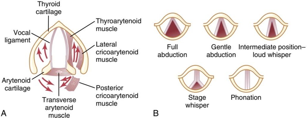
Figure 38-9 A, Anatomy of the larynx. B, Laryngeal function in phonation, showing the positions of the vocal cords during different types of phonation. (Modified from Greene MC: The Voice and Its Disorders, 4th ed. Philadelphia: JB Lippincott, 1980.)
شکل 38-9 الف، آناتومی حنجره. ب، عملکرد حنجره در آواسازی، نشان دادن موقعیت تارهای صوتی در حین انواع مختلف آواسازی. (اصلاح شده از Greene MC: The Voice and Its Disorders، ویرایش چهارم. فیلادلفیا: JB Lippincott، 1980.)
Articulation and Resonance. The three major organs of articulation are the lips, tongue, and soft palate. They need not be discussed in detail here because we are all familiar with their movements during speech and other vocalizations.
بیان و رزونانس. سه عضو اصلی مفصل لب، زبان و کام نرم هستند. در اینجا نیازی به بحث مفصل نیست زیرا همه ما با حرکات آنها در حین گفتار و سایر صداها آشنا هستیم.
The resonators include the mouth, the nose and associated nasal sinuses, the pharynx, and even the chest cavity. Again, we are familiar with the resonating qualities of these structures. For example, the function of the nasal resonators is demonstrated by the change in voice quality when a person has a severe cold that blocks the air passages to these resonators.
تشدید کننده ها شامل دهان، بینی و سینوس های بینی همراه، حلق و حتی حفره قفسه سینه هستند. باز هم با کیفیت طنین انداز این سازه ها آشنا هستیم. به عنوان مثال، عملکرد تشدید کننده های بینی با تغییر کیفیت صدا در زمانی که فرد دچار سرماخوردگی شدید می شود که راه های هوایی به این تشدید کننده ها را مسدود می کند، نشان داده می شود.
کلیک کنید «Bibliography: فهرست کتب مربوطه»
Driessen AK, McGovern AE, Narula M, et al: Central mechanisms of airway sensation and cough hypersensitivity. Pulm Pharmacol Ther 47:9, 2017.
Fahy JV, Dickey BF: Airway mucus function and dysfunction. N Engl J Med 363:2233, 2010.
Hogg JC, Paré PD, Hackett TL: The contribution of small airway ob- struction to the pathogenesis of chronic obstructive pulmonary dis- ease. Physiol Rev 97:529, 2017.
Keller JA, McGovern AE, Mazzone SB: Translating cough mechanisms into better cough suppressants. Chest 152:833, 2017.
Lai-Fook SJ: Pleural mechanics and fluid exchange. Physiol Rev 84:385, 2004.
Levin DL, Schiebler ML, Hopkins SR: Physiology for the pulmonary functional imager. Eur J Radiol 86:308, 2017.
Lopez-Rodriguez E, Pérez-Gil J: Structure-function relationships in pulmonary surfactant membranes: from biophysics to therapy. Bio- chim Biophys Acta 1838:1568, 2014.
Ma J, Rubin BK, Voynow JA: Mucins, mucus, and goblet cells. Chest 154:169, 2018.
Mazzone SB, Undem BJ: Vagal afferent innervation of the airways in health and disease. Physiol Rev 96:975, 2016.
Prakash YS: Emerging concepts in smooth muscle contributions to airway structure and function: implications for health and disease. Am J Physiol Lung Cell Mol Physiol 311:L1113, 2016.
Strohl KP, Butler JP, Malhotra A: Mechanical properties of the upper airway. Compr Physiol 2:1853, 2012.
Suki B, Sato S, Parameswaran H, et al: Emphysema and mechani- cal stress-induced lung remodeling. Physiology (Bethesda) 28:404, 2013.
Whitsett JA, Kalin TV, Xu Y, Kalinichenko W: Building and regenerat- ing the lung cell by cell. Physiol Rev 99:513, 2019.
Widdicombe JH, Wine JJ: Airway gland structure and function. Phys- iol Rev 95:1241, 2015.
🚀 با ما همراه شوید!
تازهترین مطالب و آموزشهای مغز و اعصاب را از دست ندهید. با فالو کردن کانال تلگرام آیندهنگاران مغز، از ما حمایت کنید!



