
فیزیولوژی پزشکی گایتون و هال؛ هموستاز و انعقاد خون


دعای مطالعه [ نمایش ]
بِسْمِ الله الرَّحْمنِ الرَّحیمِ
اَللّهُمَّ اَخْرِجْنى مِنْ ظُلُماتِ الْوَهْمِ
خدايا مرا بيرون آور از تاريكىهاى وهم،
وَ اَكْرِمْنى بِنُورِ الْفَهْمِ
و به نور فهم گرامى ام بدار،
اَللّهُمَّ افْتَحْ عَلَيْنا اَبْوابَ رَحْمَتِكَ
خدايا درهاى رحمتت را به روى ما بگشا،
وَانْشُرْ عَلَيْنا خَزائِنَ عُلُومِكَ بِرَحْمَتِكَ يا اَرْحَمَ الرّاحِمينَ
و خزانههاى علومت را بر ما باز كن به امید رحمتت اى مهربانترين مهربانان.
کتاب «فیزیولوژی پزشکی گایتون وهال» بهعنوان یکی از جامعترین و معتبرترین منابع در حوزه علوم پزشکی، همچنان مرجع کلیدی برای درک عملکرد پیچیده بدن انسان است. این اثر با تکیه بر تازهترین پژوهشها و توضیحات دقیق از سازوکارهای فیزیولوژیک، پلی میان علوم پایه پزشکی و کاربردهای بالینی ایجاد میکند و نقشی بیبدیل در آموزش، پژوهش و ارتقای دانش سلامت ایفا مینماید.
ترجمه دقیق و علمیاین شاهکار توسط برند علمیآیندهنگاران مغز به مدیریت داریوش طاهری، دسترسی فارسیزبانان به مرزهای نوین دانش فیزیولوژی را ممکن ساخته و رسالتی علمیبرای ارتقای آموزش پزشکی، فهم عمیقتر سازوکارهای بدن و توسعه روشهای نوین در حوزه سلامت فراهم آورده است.
» Guyton and Hall Textbook of Medical Physiology
»» CHAPTER 37: Hemostasis and Blood Coagulation
HEMOSTASIS EVENTS
The term hemostasis means prevention of blood loss. Whenever a vessel is severed or ruptured, hemostasis is achieved by several mechanisms: (1) vascular constriction; (2) formation of a platelet plug; (3) formation of a blood clot as a result of blood coagulation; and (4) eventual growth of fibrous tissue into the blood clot to close the hole in the vessel permanently.
رویدادهای هموستاز
اصطلاح هموستاز به معنای جلوگیری از از دست دادن خون است. هرگاه یک رگ قطع یا پاره شود، هموستاز با مکانیسمهای مختلفی به دست میآید: (1) انقباض عروق. (2) تشکیل پلاک پلاکت. (3) تشکیل لخته خون در نتیجه انعقاد خون. و (4) رشد نهایی بافت فیبری در لخته خون برای بسته شدن دائمیسوراخ در رگ.
VASCULAR CONSTRICTION
Immediately after a blood vessel has been cut or ruptured, the trauma to the vessel wall causes smooth muscle in the wall to contract; this instantaneously reduces the flow of blood from the ruptured vessel. The contraction results from the following: (1) local myogenic spasm; (2) local autacoid factors from the traumatized tissues, vascular endothelium, and blood platelets; and (3) nervous reflexes. The nervous reflexes are initiated by pain nerve impulses or other sensory impulses that originate from the traumatized vessel or nearby tissues. However, even more vasoconstriction probably results from local myogenic contraction of the blood vessels initiated by direct dam- age to the vascular wall. And, for the smaller vessels, the platelets are responsible for much of the vasoconstriction by releasing a vasoconstrictor substance, thromboxane A2.
انقباض عروق
بلافاصله پس از بریدگی یا پارگی رگ، ضربه به دیواره رگ باعث انقباض ماهیچه صاف در دیواره میشود. این به طور آنی جریان خون از رگ پاره شده را کاهش میدهد. انقباض ناشی از موارد زیر است: (1) اسپاسم میوژنیک موضعی. (2) عوامل اتاکوئید موضعی از بافتهای آسیب دیده، اندوتلیوم عروقی و پلاکتهای خون. و (3) رفلکسهای عصبی. رفلکسهای عصبی با تکانههای عصبی درد یا سایر تکانههای حسی که از رگ آسیب دیده یا بافتهای مجاور سرچشمه میگیرند، آغاز میشوند. با این حال، انقباض عروقی حتی بیشتر احتمالاً ناشی از انقباض میوژنیک موضعی عروق خونی است که با آسیب مستقیم به دیواره عروقی شروع میشود. و برای رگهای کوچکتر، پلاکتها با آزاد کردن یک ماده منقبض کننده عروق، ترومبوکسان A2، مسئول بخش اعظم انقباض عروق هستند.
The more severely a vessel is traumatized, the greater the degree of vascular spasm. The spasm can last for many minutes or even hours, during which time the processes of platelet plugging and blood coagulation can take place.
هر چه رگ شدیدتر آسیب ببیند، میزان اسپاسم عروقی بیشتر میشود. اسپاسم میتواند برای چندین دقیقه یا حتی ساعتها ادامه داشته باشد، در این مدت فرآیند مسدود شدن پلاکت و انعقاد خون میتواند انجام شود.
FORMATION OF THE PLATELET PLUG
If the cut in the blood vessel is very small-many very small vascular holes develop throughout the body each day-the cut is often sealed by a platelet plug rather than by a blood clot. To understand this process, it is important that we first discuss the nature of platelets themselves.
تشکیل پلاکت پلاکتی
اگر بریدگی رگ خونی بسیار کوچک باشد – هر روز سوراخهای عروقی بسیار کوچکی در سراسر بدن ایجاد میشود – بریدگی اغلب با پلاک پلاکتی به جای لخته خون بسته میشود. برای درک این فرآیند، مهم است که ابتدا ماهیت خود پلاکتها را مورد بحث قرار دهیم.
Physical and Chemical Characteristics
Platelets (also called thrombocytes) are minute discs 1 to 4 micrometers in diameter. They are formed in the bone marrow from megakaryocytes, which are extremely large hematopoietic cells in the marrow; the megakaryocytes fragment into the minute platelets in the bone marrow or soon after entering the blood, especially as they squeeze through capillaries. The normal concentration of platelets in the blood is between 150,000 and 450,000/μl.
خصوصیات فیزیکی و شیمیایی
پلاکتها (که ترومبوسیتها نیز نامیده میشوند) دیسکهای کوچکی به قطر 1 تا 4 میکرومتر هستند. آنها در مغز استخوان از مگاکاریوسیتها تشکیل میشوند که سلولهای خونساز بسیار بزرگ در مغز هستند. مگاکاریوسیتها به پلاکتهای کوچک در مغز استخوان یا بلافاصله پس از ورود به خون، به ویژه هنگامیکه از طریق مویرگها فشرده میشوند، تکه تکه میشوند. غلظت طبیعی پلاکتها در خون بین 150000 تا 450000/μl است.
Platelets have many functional characteristics of whole cells, even though they do not have nuclei and cannot reproduce. In their cytoplasm are the following: (1) actin and myosin molecules, which are contractile proteins similar to those found in muscle cells, and still another contractile protein, thrombosthenin, that can cause the platelets to contract; (2) residuals of both the endo- plasmic reticulum and Golgi apparatus that synthesize various enzymes and especially store large quantities of calcium ions; (3) mitochondria and enzyme systems that are capable of forming adenosine triphosphate (ATP) and adenosine diphosphate (ADP); (4) enzyme systems that synthesize prostaglandins, which are local hormones that cause many vascular and other local tissue reactions; (5) an important protein called fibrin-stabilizing factor, which we discuss later in relation to blood coagulation; and (6) a growth factor that causes vascular endothelial cells, vascular smooth muscle cells, and fibroblasts to multiply and grow, thus causing cellular growth that eventually helps repair damaged vascular walls.
پلاکتها دارای بسیاری از ویژگیهای عملکردی سلولهای کامل هستند، حتی اگر هسته ندارند و نمیتوانند تولید مثل کنند. در سیتوپلاسم آنها موارد زیر وجود دارد: (1) مولکولهای اکتین و میوزین که پروتئینهای انقباضی مشابه پروتئینهای موجود در سلولهای ماهیچهای هستند، و همچنان پروتئین انقباضی دیگری، ترومبوستنین، که میتواند باعث انقباض پلاکتها شود. (2) باقیماندههای شبکه آندوپلاسمیو دستگاه گلژی که آنزیمهای مختلف را سنتز میکنند و به ویژه مقادیر زیادی یون کلسیم را ذخیره میکنند. (3) میتوکندری و سیستمهای آنزیمیکه قادر به تشکیل آدنوزین تری فسفات (ATP) و آدنوزین دی فسفات (ADP) هستند. (4) سیستمهای آنزیمیکه پروستاگلاندینها را سنتز میکنند، که هورمونهای محلی هستند که باعث بسیاری از واکنشهای عروقی و دیگر بافتهای موضعی میشوند. (5) یک پروتئین مهم به نام فاکتور تثبیت کننده فیبرین، که بعداً در رابطه با انعقاد خون بحث خواهیم کرد. و (6) یک فاکتور رشد که باعث تکثیر و رشد سلولهای اندوتلیال عروقی، سلولهای ماهیچه صاف عروق و فیبروبلاستها میشود، در نتیجه باعث رشد سلولی میشود که در نهایت به ترمیم دیوارههای عروقی آسیب دیده کمک میکند.
On the platelet cell membrane surface is a coat of glycoproteins that repulses adherence to normal endothelium and yet causes adherence to injured areas of the vessel wall, especially to injured endothelial cells and even more so to any exposed collagen from deep within the vessel wall. In addition, the platelet membrane contains large amounts of phospholipids that activate multiple stages in the blood-clotting process, as discussed later.
روی سطح غشای سلولی پلاکتی پوششی از گلیکوپروتئین وجود دارد که چسبیدن به اندوتلیوم طبیعی را دفع میکند و در عین حال باعث چسبیدن به نواحی آسیب دیده دیواره عروق، به ویژه به سلولهای اندوتلیال آسیب دیده و حتی بیشتر از آن به هر کلاژن در معرض از اعماق دیواره عروق میشود. علاوه بر این، غشای پلاکتی حاوی مقادیر زیادی فسفولیپید است که مراحل متعددی را در فرآیند انعقاد خون فعال میکند، همانطور که بعداً بحث شد.
Thus, the platelet is an active structure. It has a half-life in the blood of only 8 to 12 days, so over several weeks its functional processes run out; it is then eliminated from the circulation mainly by the tissue macrophage system. More than half of the platelets are removed by macro- phages in the spleen, where the blood passes through a latticework of tight trabeculae.
بنابراین، پلاکت یک ساختار فعال است. نیمه عمر آن در خون تنها 8 تا 12 روز است، بنابراین طی چند هفته فرآیندهای عملکردی آن تمام میشود. سپس عمدتاً توسط سیستم ماکروفاژ بافتی از گردش خون حذف میشود. بیش از نیمیاز پلاکتها توسط ماکروفاژها در طحال، جایی که خون از شبکهای از ترابکولهای تنگ عبور میکند، خارج میشوند.
Mechanism of Platelet Plug Formation
Platelet repair of vascular openings is based on several important functions of the platelet. When platelets come in contact with a damaged vascular surface, especially with collagen fibers in the vascular wall, the platelets rapidly change their own characteristics drastically (Figure 37-1). They begin to swell, they assume irregular forms with numerous irradiating pseudo- pods protruding from their surfaces, their contractile proteins contract forcefully and cause the release of granules that contain multiple active factors, and they become sticky so that they adhere to collagen in the tissues and to a protein called von Willebrand factor (vWF), which leaks into the traumatized tissue from the plasma. The platelet surface glycoproteins bind to vWF in the exposed matrix below the damaged endothelium. The platelets then secrete increased quantities of ADP and platelet-activating factor (PAF), and their enzymes form thromboxane A2. Thromboxane is a vasoconstrictor and, along with ADP and PAF, acts on nearby platelets to activate them as well; the stickiness of these additional activated platelets causes them to adhere to the original activated platelets.
مکانیسم تشکیل پلاک پلاکت
ترمیم پلاکت منافذ عروقی بر اساس چندین عملکرد مهم پلاکت است. هنگامیکه پلاکتها با یک سطح عروقی آسیب دیده، به ویژه با رشتههای کلاژن در دیواره عروقی تماس پیدا میکنند، پلاکتها به سرعت ویژگیهای خود را به شدت تغییر میدهند (شکل 37-1). آنها شروع به متورم شدن میکنند، شکلهای نامنظم با شبه غلافهای تابش دهنده متعددی را به خود میگیرند که از سطح آنها بیرون زده است، پروتئینهای انقباضی آنها به شدت منقبض میشوند و باعث آزاد شدن گرانولهایی میشوند که حاوی چندین عامل فعال هستند و چسبناک میشوند به طوری که به کلاژن در بافتها و به پروتئینی به نام فاکتور فون ویلبراند (vWF) که از بافت traatum وارد میشود، میچسبند. گلیکوپروتئینهای سطح پلاکت به vWF در ماتریکس در معرض زیر اندوتلیوم آسیب دیده متصل میشوند. سپس پلاکتها مقادیر بیشتری از ADP و فاکتور فعال کننده پلاکت (PAF) ترشح میکنند و آنزیمهای آنها ترومبوکسان A2 را تشکیل میدهند. ترومبوکسان یک منقبض کننده عروق است و همراه با ADP و PAF بر روی پلاکتهای مجاور عمل میکند تا آنها را نیز فعال کند. چسبندگی این پلاکتهای فعال اضافی باعث میشود که آنها به پلاکتهای فعال اصلی بچسبند.
Therefore, at the site of a puncture in a blood vessel wall, the damaged vascular wall activates successively increasing numbers of platelets that attract more and more additional platelets, thus forming a platelet plug. This plug is loose at first but is usually successful in blocking blood loss if the vascular opening is small. Then, during the subsequent process of blood coagulation, fibrin threads form. These threads attach tightly to the platelets, thus constructing an unyielding plug.
بنابراین، در محل سوراخ شدن دیواره رگ خونی، دیواره عروقی آسیب دیده تعداد پلاکتهایی را فعال میکند که تعداد پلاکتهای بیشتری را جذب میکنند و در نتیجه پلاکت پلاکتی را تشکیل میدهند. این پلاگ در ابتدا شل است، اما اگر دهانه عروق کوچک باشد معمولاً در جلوگیری از از دست دادن خون موفق است. سپس در طی فرآیند بعدی انعقاد خون، رشتههای فیبرین تشکیل میشود. این رشتهها به طور محکم به پلاکتها میچسبند و در نتیجه یک پلاگ تسلیم ناپذیر ایجاد میکنند.
Endothelial injury and exposure of the vascular extracellular matrix facilitates platelet adhesions and activation, which changes their shape and causes release of adenosine diphosphate (ADP), thromboxane A2 (TxAz), and platelet-activating factor (PAF). These platelet-secreted factors recruit additional platelets (aggregation) to form a hemostatic plug. Von Willebrand factor (vWF) serves as an adhesion bridge between sub- endothelial collagen and the glycoprotein lb (Gplb) platelet receptor.
آسیب اندوتلیال و قرار گرفتن در معرض ماتریکس خارج سلولی عروقی چسبندگی و فعال شدن پلاکتها را تسهیل میکند که شکل آنها را تغییر میدهد و باعث آزاد شدن آدنوزین دی فسفات (ADP)، ترومبوکسان A2 (TxAz) و فاکتور فعال کننده پلاکت (PAF) میشود. این عوامل ترشح شده توسط پلاکت، پلاکتهای اضافی (تجمع) را برای تشکیل یک پلاگ هموستاتیک جذب میکنند. فاکتور فون ویلبراند (vWF) به عنوان پل چسبندگی بین کلاژن زیر اندوتلیال و گیرنده پلاکتی گلیکوپروتئین lb (Gplb) عمل میکند.
Importance of Platelet Mechanism for Closing Vascular Holes. The platelet-plugging mechanism is extremely important for closing minute ruptures in very small blood vessels that occur many thousands of times daily. Indeed, multiple small holes through the endothelial cells themselves are often closed by platelets actually fusing with the endothelial cells to form additional endothelial cell membranes. Literally thousands of small hemorrhagic areas develop each day under the skin (petechiae, which appear as purple or red dots on the skin) and throughout the internal tissues of a person who has few blood platelets. This phenomenon does not occur in persons with normal numbers of platelets.
اهمیت مکانیسم پلاکتی برای بستن سوراخهای عروقی. مکانیسم پلاکتبندی برای بستن پارگیهای کوچک در رگهای خونی بسیار کوچک که هزاران بار در روز اتفاق میافتد بسیار مهم است. در واقع، سوراخهای کوچک متعدد از طریق خود سلولهای اندوتلیال اغلب توسط پلاکتهایی که در واقع با سلولهای اندوتلیال ترکیب میشوند بسته میشوند تا غشای سلولهای اندوتلیال اضافی را تشکیل دهند. به معنای واقعی کلمه هر روز هزاران ناحیه خونریزیدهنده کوچک در زیر پوست (پتشی که به صورت نقاط بنفش یا قرمز روی پوست ظاهر میشوند) و در سراسر بافتهای داخلی فردی که پلاکتهای خونی کمیدارد، ایجاد میشود. این پدیده در افرادی که تعداد پلاکت طبیعی دارند، رخ نمیدهد.

Figure 37-1. Formation of a platelet plug in a severed blood vessel.
شکل 37-1. تشکیل پلاکت پلاکتی در یک رگ خونی قطع شده.
BLOOD COAGULATION IN THE RUPTURED VESSEL
The third mechanism for hemostasis is formation of the blood clot. The clot begins to develop in 15 to 20 seconds if the trauma to the vascular wall is severe and in 1 to 2 minutes if the trauma is minor. Activator substances from the traumatized vascular wall, from platelets, and from blood proteins adhering to the traumatized vascular wall initiate the clotting process. The physical events of this process are shown in Figure 37-2; Table 37-1 lists the most important clotting factors.
انعقاد خون در رگ پاره شده
مکانیسم سوم برای هموستاز، تشکیل لخته خون است. اگر ضربه به دیواره عروقی شدید باشد، لخته در 15 تا 20 ثانیه و اگر ضربه جزئی باشد در 1 تا 2 دقیقه شروع به ایجاد میکند. مواد فعال کننده از دیواره عروقی آسیب دیده، از پلاکتها و از پروتئینهای خونی که به دیواره عروق آسیب دیده میچسبند، فرآیند لخته شدن را آغاز میکنند. رویدادهای فیزیکی این فرآیند در شکل 37-2 نشان داده شده است. جدول 37-1 مهمترین عوامل لخته شدن را فهرست میکند.
Within 3 to 6 minutes after rupture of a vessel, the entire opening or broken end of the vessel is filled with clot if the vessel opening is not too large. After 20 to 60 minutes, the clot retracts, which closes the vessel still further. Platelets also play an important role in this clot retraction, as discussed later.
در عرض 3 تا 6 دقیقه پس از پارگی رگ، اگر دهانه رگ خیلی بزرگ نباشد، کل دهانه یا انتهای شکسته رگ با لخته پر میشود. پس از 20 تا 60 دقیقه، لخته جمع میشود که رگ را بیشتر میبندد. پلاکتها نیز نقش مهمیدر این انقباض لخته ایفا میکنند، همانطور که بعداً بحث شد.
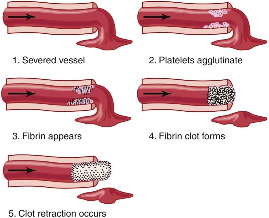
Figure 37-2. Clotting process in a traumatized blood vessel. (Modified from Seegers WH: Hemostatic Agents. Springfield, IL: Charles C Thomas, 1948.)
شکل 37-2. فرآیند لخته شدن در رگ خونی آسیب دیده. (اصلاح شده از Seegers WH: Hemostatic Agents. Springfield, IL: Charles C Thomas, 1948.)
Table 37-1 Clotting Factors in Blood and Their Synonyms
جدول 37-1 عوامل انعقادی در خون و مترادف آنها

FIBROUS ORGANIZATION OR DISSOLUTION OF BLOOD CLOTS
Once a blood clot has formed, it can follow one of two courses: (1) it can become invaded by fibroblasts, which subsequently form connective tissue all through the clot; or (2) it can dissolve. The usual course for a clot that forms in a small hole of a vessel wall is invasion by fibroblasts, beginning within a few hours after the clot is formed, which is promoted at least partially by growth factor secreted by platelets. This process continues to complete organization of the clot into fibrous tissue within about 1 to 2 weeks.
سازمان فیبری یا انحلال لختههای خون
هنگامیکه یک لخته خون تشکیل شد، میتواند یکی از دو دوره را دنبال کند: (1) میتواند توسط فیبروبلاستها مورد تهاجم قرار گیرد، که متعاقباً بافت همبند را در سراسر لخته تشکیل میدهد. یا (2) میتواند حل شود. روند معمول لخته ای که در سوراخ کوچک دیواره عروق ایجاد میشود، تهاجم فیبروبلاستها است که در عرض چند ساعت پس از تشکیل لخته شروع میشود، که حداقل تا حدی توسط فاکتور رشد ترشح شده توسط پلاکتها ایجاد میشود. این فرآیند برای تکمیل سازماندهی لخته در بافت فیبری در حدود 1 تا 2 هفته ادامه مییابد.
Conversely, when excess blood has leaked into the tissues, and tissue clots have formed where they are not needed, special substances in the clot usually become activated. These substances function as enzymes to dis- solve the clot, as discussed later in the chapter.
برعکس، زمانی که خون اضافی به بافتها نشت میکند و لختههای بافتی در جایی که نیازی به آنها نیست تشکیل میشوند، معمولاً مواد خاصی در لخته فعال میشوند. این مواد به عنوان آنزیم برای حل لخته عمل میکنند، همانطور که بعدا در این فصل بحث شد.
MECHANISM OF BLOOD COAGULATION
GENERAL MECHANISM
More than 50 important substances that cause or affect blood coagulation have been found in the blood and in the tissues-some that promote coagulation, called procoagulants, and others that inhibit coagulation, called anticoagulants. Whether blood will coagulate depends on the balance between these two groups of substances. In the blood stream, the anticoagulants normally predominate, so the blood does not coagulate while it is circulating in the blood vessels. However, when a vessel is ruptured, procoagulants from the area of tissue damage become activated and override the anticoagulants, and then a clot does develop.
مکانیسم انعقاد خون
مکانیزم عمومی
بیش از 50 ماده مهمیکه باعث انعقاد خون میشوند یا بر انعقاد خون تأثیر میگذارند در خون و بافتها یافت شده است – برخی از آنها انعقاد را تقویت میکنند که پیش انعقاد نامیده میشوند و برخی دیگر که انعقاد را مهار میکنند به نام ضد انعقادها. انعقاد خون به تعادل بین این دو گروه از مواد بستگی دارد. در جریان خون، داروهای ضد انعقاد معمولاً غالب هستند، بنابراین خون در حالی که در رگهای خونی در گردش است منعقد نمیشود. با این حال، هنگامیکه یک رگ پاره میشود، پیش انعقادها از ناحیه آسیب بافتی فعال میشوند و بر ضد انعقادها غلبه میکنند و سپس لخته ایجاد میشود.
Clotting takes place in three essential steps:
لخته شدن در سه مرحله اساسی انجام میشود:
1. In response to rupture of the vessel or damage to the blood itself, a complex cascade of chemical reactions occurs in the blood involving more than 12 blood coagulation factors. The net result is the formation of a complex of activated substances collectively called prothrombin activator.
1. در پاسخ به پارگی رگ یا آسیب به خود خون، یک آبشار پیچیده از واکنشهای شیمیایی در خون رخ میدهد که شامل بیش از 12 عامل انعقاد خون است. نتیجه خالص تشکیل مجموعه ای از مواد فعال شده است که مجموعاً فعال کننده پروترومبین نامیده میشود.
2. The prothrombin activator catalyzes the conversion of prothrombin into thrombin.
2. فعال کننده پروترومبین تبدیل پروترومبین به ترومبین را کاتالیز میکند.
3. The thrombin acts as an enzyme to convert fibrinogen into fibrin fibers that enmesh platelets, blood cells, and plasma to form the clot.
3. ترومبین به عنوان آنزیمیبرای تبدیل فیبرینوژن به فیبرهای فیبرین عمل میکند که پلاکتها، سلولهای خونی و پلاسما را در هم میبندد و لخته را تشکیل میدهد.
We will first discuss the mechanism whereby the blood clot is formed, beginning with conversion of prothrombin to thrombin, and then come back to the initiating stages in the clotting process whereby prothrombin activator is formed.
ما ابتدا مکانیسم تشکیل لخته خون را مورد بحث قرار خواهیم داد که با تبدیل پروترومبین به ترومبین شروع میشود و سپس به مراحل آغازین فرآیند لخته شدن باز میگردیم که در آن فعال کننده پروترومبین تشکیل میشود.
CONVERSION OF PROTHROMBIN TO THROMBIN
1. Prothrombin activator is formed as a result of rupture of a blood vessel or as a result of damage to special substances in the blood.
تبدیل پروترومبین به ترومبین
1. فعال کننده پروترومبین در نتیجه پارگی رگ خونی یا در نتیجه آسیب به مواد خاصی در خون ایجاد میشود.
2. Prothrombin activator, in the presence of sufficient amounts of ionic calcium (Ca2+), causes conversion of prothrombin to thrombin (Figure 37-3 and 37-4).
2. فعال کننده پروترومبین در حضور مقادیر کافی کلسیم یونی (+Ca2)، باعث تبدیل پروترومبین به ترومبین میشود (شکل 37-3 و 37-4).
3. Thrombin causes polymerization of fibrinogen molecules into fibrin fibers within another 10 to 15 seconds.
3. ترومبین باعث پلیمریزاسیون مولکولهای فیبرینوژن به الیاف فیبرین در 10 تا 15 ثانیه دیگر میشود.
Thus, the rate-limiting factor in causing blood coagulation is usually the formation of prothrombin activator and not the subsequent reactions beyond that point because these terminal steps normally occur rapidly to form the clot.
بنابراین، عامل محدود کننده سرعت در ایجاد انعقاد خون معمولاً تشکیل فعال کننده پروترومبین است و نه واکنشهای بعدی فراتر از آن نقطه، زیرا این مراحل پایانی معمولاً به سرعت برای تشکیل لخته رخ میدهند.
Platelets also play an important role in the conversion of prothrombin to thrombin because much of the prothrombin first attaches to prothrombin receptors on the platelets that are already bound to the damaged tissue.
پلاکتها همچنین نقش مهمیدر تبدیل پروترومبین به ترومبین ایفا میکنند زیرا بیشتر پروترومبین ابتدا به گیرندههای پروترومبین روی پلاکتهایی که قبلاً به بافت آسیب دیده متصل شده اند میچسبد.
Prothrombin and Thrombin. Prothrombin is a plasma protein, an α-globulin, having a molecular weight of 68,700. It is present in normal plasma in a concentration of about 15 mg/dl. It is an unstable protein that can split easily into smaller compounds, one of which is thrombin, which has a molecular weight of 33,700, almost half that of prothrombin.
پروترومبین و ترومبین. پروترومبین یک پروتئین پلاسما، یک آلفا-گلوبولین است که وزن مولکولی آن 68700 است. در پلاسمای طبیعی با غلظت حدود 15 میلی گرم در دسی لیتر وجود دارد. این یک پروتئین ناپایدار است که میتواند به راحتی به ترکیبات کوچکتر تقسیم شود، یکی از آنها ترومبین است که دارای وزن مولکولی 33700 است که تقریباً نصف وزن مولکولی پروترومبین است.
Prothrombin is formed continually by the liver, and it is continually being used throughout the body for blood clotting. If the liver fails to produce prothrombin, in a day or so prothrombin concentration in the plasma falls too low to provide normal blood coagulation.
پروترومبین به طور مداوم توسط کبد تشکیل میشود و به طور مداوم در سراسر بدن برای لخته شدن خون استفاده میشود. اگر کبد نتواند پروترومبین تولید کند، در یک روز یا بیشتر، غلظت پروترومبین در پلاسما به حدی پایین میآید که انعقاد خون طبیعی را فراهم نمیکند.
Vitamin K is required by the liver for normal activation of prothrombin, as well as a few other clotting factors. Therefore, lack of vitamin K or the presence of liver disease that prevents normal prothrombin formation can decrease the prothrombin to such a low level that a bleeding tendency results.
ویتامین K برای فعال سازی طبیعی پروترومبین و همچنین چند فاکتور لخته شدن دیگر مورد نیاز کبد است. بنابراین، کمبود ویتامین K یا وجود بیماری کبدی که از تشکیل پروترومبین طبیعی جلوگیری میکند، میتواند پروترومبین را به حدی پایین بیاورد که تمایل به خونریزی ایجاد شود.

Figure 37-3 Schema for conversion of prothrombin to thrombin and polymerization of fibrinogen to form fibrin fibers.
شکل 37-3 طرحی برای تبدیل پروترومبین به ترومبین و پلیمریزاسیون فیبرینوژن برای تشکیل الیاف فیبرین.
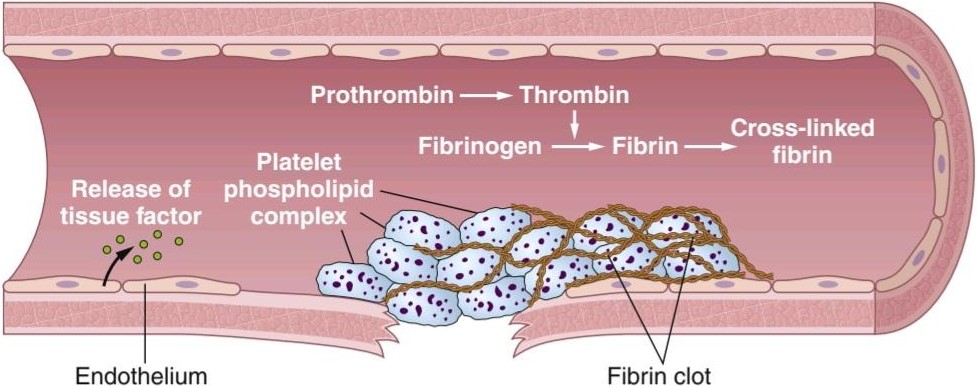
Figure 37-4. Coagulation cascade after vascular injury. Exposure of blood to the vascular wall causes release of tissue factor (also called factor III or thromboplastin) from endothelial cells, phospholipid expression, activation of thrombin, which then acts on fibrinogen to form fibrin, and fibrin polymerization to form a meshwork that stabilizes the platelet plug.
شکل 37-4. آبشار انعقادی پس از آسیب عروقی. قرار گرفتن خون در معرض دیواره عروقی باعث آزاد شدن فاکتور بافتی (همچنین به نام فاکتور III یا ترومبوپلاستین) از سلولهای اندوتلیال، بیان فسفولیپید، فعال شدن ترومبین، که سپس بر روی فیبرینوژن برای تشکیل فیبرین عمل میکند، و پلیمریزاسیون فیبرین برای تشکیل شبکه ای که پلاک پلاکتی را تثبیت میکند، میشود.
CONVERSION OF FIBRINOGEN TO FIBRIN-FORMATION OF THE CLOT
Fibrinogen Formed in the Liver Essential for Clot Formation. Fibrinogen is a high-molecular-weight protein (molecular weight ≈340,000) that occurs in the plasma in quantities of 100 to 700 mg/dl. Fibrinogen is formed in the liver, and liver disease can decrease the concentration of circulating fibrinogen, as it does the concentration of prothrombin, noted earlier.
تبدیل فیبرینوژن به فیبرین تشکیل لخته
فیبرینوژن تشکیل شده در کبد برای تشکیل لخته ضروری است. فیبرینوژن یک پروتئین با وزن مولکولی بالا (وزن مولکولی ≈340000) است که در مقادیر 100 تا 700 میلی گرم در دسی لیتر در پلاسما وجود دارد. فیبرینوژن در کبد تشکیل میشود و بیماری کبد میتواند غلظت فیبرینوژن در گردش را کاهش دهد، همانطور که غلظت پروترومبین را که قبلاً ذکر شد، کاهش میدهد.
Because of its large molecular size, little fibrinogen normally leaks from the blood vessels into the interstitial fluids, and because fibrinogen is one of the essential factors in the coagulation process, interstitial fluids ordinarily do not coagulate. Yet, when the permeability of the capillaries becomes pathologically increased, fibrinogen does leak into the tissue fluids in sufficient quantities to allow clotting of these fluids in much the same way that plasma and whole blood can clot.
به دلیل اندازه مولکولی بزرگ، فیبرینوژن کمیبه طور معمول از رگهای خونی به مایعات بینابینی نشت میکند و از آنجا که فیبرینوژن یکی از عوامل ضروری در فرآیند انعقاد است، مایعات بینابینی معمولاً منعقد نمیشوند. با این حال، هنگامیکه نفوذپذیری مویرگها به طور پاتولوژیک افزایش مییابد، فیبرینوژن به مقدار کافی به مایعات بافتی نشت میکند تا لخته شدن این مایعات را به همان روشی که پلاسما و خون کامل میتوانند لخته شوند، امکان پذیر کند.
Action of Thrombin on Fibrinogen to Form Fibrin. Thrombin is a protein enzyme with weak proteolytic capabilities. It acts on fibrinogen to remove four low- molecular-weight peptides from each molecule of fibrinogen, forming one molecule of fibrin monomer that has the automatic capability to polymerize with other fibrin monomer molecules to form fibrin fibers. Therefore, many fibrin monomer molecules polymerize within seconds into long fibrin fibers that constitute the reticulum of the blood clot.
اثر ترومبین بر فیبرینوژن برای تشکیل فیبرین. ترومبین یک آنزیم پروتئینی با قابلیت پروتئولیتیک ضعیف است. بر روی فیبرینوژن عمل میکند تا چهار پپتید با وزن مولکولی کم را از هر مولکول فیبرینوژن حذف کند و یک مولکول مونومر فیبرین را تشکیل میدهد که قابلیت پلیمریزاسیون خودکار با مولکولهای مونومر دیگر فیبرین را دارد تا فیبرهای فیبرین را تشکیل دهد. بنابراین، بسیاری از مولکولهای مونومر فیبرین در عرض چند ثانیه به الیاف فیبرین طولانی تبدیل میشوند که شبکه لخته خون را تشکیل میدهند.
In the early stages of polymerization, the fibrin monomer molecules are held together by weak noncovalent hydrogen bonding, and the newly forming fibers are not cross-linked with one another. Therefore, the resultant clot is weak and can be broken apart with ease. However, another process occurs during the next few minutes that greatly strengthens the fibrin reticulum. This process involves a substance called fibrin stabilizing factor that is present in small amounts in normal plasma globulins but is also released from platelets entrapped in the clot. Before fibrin stabilizing factor can have an effect on the fibrin fibers, it must be activated. The same thrombin that causes fibrin formation also activates the fibrin stabilizing factor. This activated substance then operates as an enzyme to form covalent bonds between more and more of the fibrin monomer molecules, as well as multiple cross-linkages between adjacent fibrin fibers, thus adding tremendously to the three-dimensional strength of the fibrin meshwork.
در مراحل اولیه پلیمریزاسیون، مولکولهای مونومر فیبرین توسط پیوند هیدروژنی غیرکووالانسی ضعیف در کنار هم نگه داشته میشوند و الیاف تازه تشکیل شده با یکدیگر پیوندی متقابل ندارند. بنابراین، لخته حاصل ضعیف است و میتوان به راحتی از هم جدا شد. با این حال، فرآیند دیگری در طول چند دقیقه آینده رخ میدهد که شبکه فیبرین را تا حد زیادی تقویت میکند. این فرآیند شامل ماده ای به نام فاکتور تثبیت کننده فیبرین است که به مقدار کمیدر گلوبولینهای پلاسما طبیعی وجود دارد اما از پلاکتهای گیر افتاده در لخته نیز آزاد میشود. قبل از اینکه فاکتور تثبیت کننده فیبرین بتواند بر روی الیاف فیبرین اثر بگذارد، باید آن را فعال کرد. همان ترومبین که باعث تشکیل فیبرین میشود، عامل تثبیت کننده فیبرین را نیز فعال میکند. این ماده فعال شده سپس به عنوان یک آنزیم عمل میکند تا پیوندهای کووالانسی بین تعداد بیشتری از مولکولهای مونومر فیبرین و همچنین چندین پیوند متقابل بین الیاف فیبرین مجاور ایجاد کند، بنابراین به شدت به استحکام سه بعدی شبکه فیبرین میافزاید.
Blood Clot. The clot is composed of a meshwork of fibrin fibers running in all directions and entrapping blood cells, platelets, and plasma (see Figure 37-4). The fibrin fibers also adhere to damaged surfaces of blood vessels; therefore, the blood clot becomes adherent to any vascular opening and thereby prevents further blood loss.
لخته خون. لخته از شبکهای از الیاف فیبرین تشکیل شده است که در تمام جهات جریان دارند و سلولهای خون، پلاکتها و پلاسما را به دام میاندازند (شکل 37-4 را ببینید). فیبرهای فیبرین همچنین به سطوح آسیب دیده رگهای خونی میچسبند. بنابراین، لخته خون به هر دهانه عروقی چسبیده و در نتیجه از از دست دادن خون بیشتر جلوگیری میکند.
Clot Retraction and Expression of Serum. Within a few minutes after a clot is formed, it begins to contract and usually expresses most of the fluid from the clot within 20 to 60 minutes. The fluid expressed is called serum because all its fibrinogen and most of the other clotting factors have been removed; in this way, serum differs from plasma and cannot clot because it lacks these factors.
انقباض لخته و بیان سرم. در عرض چند دقیقه پس از تشکیل لخته، شروع به انقباض میکند و معمولاً بیشتر مایع لخته را در عرض 20 تا 60 دقیقه بیان میکند. مایع بیان شده سرم نامیده میشود زیرا تمام فیبرینوژن آن و بسیاری از عوامل لخته شدن دیگر حذف شده اند. به این ترتیب سرم با پلاسما متفاوت است و نمیتواند لخته شود زیرا فاقد این عوامل است.
Platelets are necessary for clot retraction to occur. Therefore, failure of clot retraction is an indication that the number of platelets in the circulating blood might be low. Electron micrographs of platelets in blood clots show that they become attached to the fibrin fibers in such a way that they actually bond different fibers together. Furthermore, platelets entrapped in the clot continue to release procoagulant substances, one of the most important of which is fibrin stabilizing factor, which causes more and more cross-linking bonds between adjacent fibrin fibers. In addition, the platelets contribute directly to clot contraction by activating platelet thrombosthenin, actin, and myosin molecules, which are all contractile proteins in the platelets; they cause strong contraction of the platelet spicules attached to the fibrin. This action also helps compress the fibrin meshwork into a smaller mass. The contraction is activated and accelerated by thrombin and by calcium ions released from calcium stores in the mitochondria, endoplasmic reticulum, and Golgi apparatus of the platelets.
پلاکتها برای انقباض لخته ضروری هستند. بنابراین، شکست در جمع شدن لخته نشانهای از کم بودن تعداد پلاکتها در خون در گردش است. میکروگرافهای الکترونی از پلاکتهای موجود در لختههای خون نشان میدهد که آنها به رشتههای فیبرین به گونه ای متصل میشوند که در واقع فیبرهای مختلف را به یکدیگر متصل میکنند. علاوه بر این، پلاکتهای محبوس شده در لخته همچنان مواد پیشانعقادی آزاد میکنند که یکی از مهمترین آنها فاکتور تثبیتکننده فیبرین است که باعث ایجاد پیوندهای متقابل بیشتر و بیشتر بین الیاف فیبرین مجاور میشود. علاوه بر این، پلاکتها با فعال کردن مولکولهای ترومبوستنین، اکتین و میوزین پلاکتی که همگی پروتئینهای انقباضی در پلاکتها هستند، مستقیماً به انقباض لخته کمک میکنند. آنها باعث انقباض شدید اسپیکولهای پلاکتی متصل به فیبرین میشوند. این عمل همچنین به فشرده سازی شبکه فیبرین به یک جرم کوچکتر کمک میکند. انقباض توسط ترومبین و یونهای کلسیم آزاد شده از ذخایر کلسیم در میتوکندری، شبکه آندوپلاسمیو دستگاه گلژی پلاکتها فعال و تسریع میشود.
As the clot retracts, the edges of the broken blood vessel are pulled together, thus contributing still further to hemostasis.
با جمع شدن لخته، لبههای رگ خونی شکسته به هم کشیده میشوند و در نتیجه به هموستاز کمک میکنند.
POSITIVE FEEDBACK OF CLOT FORMATION
Once a blood clot starts to develop, it normally extends within minutes into the surrounding blood-that is, the clot initiates a positive feedback to promote more clotting. One of the most important causes of this clot pro- motion is that the proteolytic action of thrombin allows it to act on many of the other blood-clotting factors in addition to fibrinogen. For example, thrombin has a direct proteolytic effect on prothrombin, tending to convert it into still more thrombin, and it acts on some of the bloodclotting factors responsible for formation of prothrombin activator. (These effects, discussed in subsequent paragraphs, include acceleration of the actions of factors VIII, IX, X, XI, and XII and aggregation of platelets.) Once a critical amount of thrombin is formed, a positive feedback develops that causes still more blood clotting and more and more thrombin to be formed; thus, the blood clot continues to grow until blood leakage ceases.
بازخورد مثبت تشکیل لخته
هنگامیکه یک لخته خون شروع به ایجاد میکند، معمولاً در عرض چند دقیقه به داخل خون اطراف گسترش مییابد، یعنی لخته بازخورد مثبتی را برای ترویج لخته شدن بیشتر آغاز میکند. یکی از مهم ترین دلایل این افزایش لخته این است که عمل پروتئولیتیک ترومبین به آن اجازه میدهد تا علاوه بر فیبرینوژن بر روی بسیاری از دیگر فاکتورهای انعقاد خون نیز اثر بگذارد. به عنوان مثال، ترومبین دارای اثر پروتئولیتیک مستقیم بر پروترومبین است و تمایل دارد آن را به ترومبین بیشتر تبدیل کند و بر برخی از فاکتورهای انعقاد خون که مسئول تشکیل فعال کننده پروترومبین هستند، تأثیر میگذارد. (این اثرات، که در پاراگرافهای بعدی مورد بحث قرار میگیرند، شامل تسریع عملکرد فاکتورهای VIII، IX، X، XI، XII و تجمع پلاکتها میشود.) هنگامیکه مقدار بحرانی ترومبین تشکیل شد، بازخورد مثبتی ایجاد میشود که باعث لخته شدن خون بیشتر و تشکیل ترومبین بیشتر و بیشتر میشود. بنابراین، لخته خون تا زمانی که نشت خون متوقف شود، به رشد خود ادامه میدهد.
INITIATION OF COAGULATION: FORMATION OF PROTHROMBIN ACTIVATOR
Now that we have discussed the clotting process, the more complex mechanisms that initiate clotting in the first place will be described. These mechanisms are set into play by the following: (1) trauma to the vascular wall and adjacent tissues; (2) trauma to the blood; or (3) contact of the blood with damaged endothelial cells or with collagen and other tissue elements outside the blood vessel. In each case, this leads to the formation of prothrombin activator, which then causes prothrombin conversion to thrombin and all the subsequent clotting steps.
شروع انعقاد: تشکیل فعال کننده پروترومبین
اکنون که فرآیند لخته شدن را مورد بحث قرار دادیم، مکانیسمهای پیچیده تری که در وهله اول لخته شدن را آغاز میکنند، شرح داده میشود. این مکانیسمها با موارد زیر به کار گرفته میشوند: (1) ضربه به دیواره عروقی و بافتهای مجاور. (2) ضربه به خون. یا (3) تماس خون با سلولهای اندوتلیال آسیب دیده یا با کلاژن و سایر عناصر بافتی خارج از رگ خونی. در هر مورد، این منجر به تشکیل فعال کننده پروترومبین میشود که سپس باعث تبدیل پروترومبین به ترومبین و تمام مراحل لخته شدن بعدی میشود.
Prothrombin activator is generally considered to be formed in two ways, although, in reality, the two ways interact constantly with each other: (1) by the extrinsic pathway that begins with trauma to the vascular wall and surrounding tissues; and (2) by the intrinsic pathway that begins in the blood.
فعالکننده پروترومبین عموماً به دو صورت در نظر گرفته میشود، اگرچه، در واقعیت، این دو راه دائماً با یکدیگر تعامل دارند: (1) توسط مسیر بیرونی که با ضربه به دیواره عروق و بافتهای اطراف شروع میشود. و (2) توسط مسیر ذاتی که در خون شروع میشود.
In both the extrinsic and the intrinsic pathways, a series of different plasma proteins called blood-clotting factors plays a major role. Most of these proteins are inactive forms of proteolytic enzymes. When converted to the active forms, their enzymatic actions cause the successive, cascading reactions of the clotting process.
در هر دو مسیر بیرونی و درونی، یک سری پروتئینهای مختلف پلاسما به نام فاکتورهای انعقاد خون نقش اصلی را ایفا میکنند. بیشتر این پروتئینها اشکال غیر فعال آنزیمهای پروتئولیتیک هستند. هنگامیکه به اشکال فعال تبدیل میشوند، اعمال آنزیمیآنها باعث واکنشهای متوالی و آبشاری فرآیند لخته شدن میشود.
Most of the clotting factors listed in Table 37-1 are designated by Roman numerals. To indicate the activated form of the factor, a small letter “a” is added after the Roman numeral, such as factor VIIIa to indicate the activated state of factor VIII.
اکثر فاکتورهای لخته شدن فهرست شده در جدول 37-1 با اعداد رومیمشخص شده اند. برای نشان دادن شکل فعال عامل، یک حرف کوچک “a” بعد از عدد رومیاضافه میشود، مانند فاکتور VIIIa برای نشان دادن وضعیت فعال شده فاکتور هشت.
Extrinsic Pathway for Initiating Clotting
The extrinsic pathway for initiating the formation of prothrombin activator begins with a traumatized vascular wall or traumatized extravascular tissues that come in contact with the blood. This condition leads to the following steps, as shown in Figure 37-4 and Figure 37-5:
مسیر بیرونی برای شروع لخته شدن
مسیر بیرونی برای شروع تشکیل فعال کننده پروترومبین با دیواره عروقی آسیب دیده یا بافتهای خارج عروقی آسیب دیده که در تماس با خون هستند شروع میشود. این شرایط به مراحل زیر منجر میشود، همانطور که در شکل 37-4 و شکل 37-5 نشان داده شده است:
1. Release of tissue factor. Traumatized tissue releases a complex of several factors called tissue factor or tissue thromboplastin. This factor is composed especially of phospholipids from the membranes of the tissue plus a lipoprotein complex that functions mainly as a proteolytic enzyme.
1. آزادسازی فاکتور بافتی. بافت آسیب دیده مجموعه ای از چندین عامل به نام فاکتور بافتی یا ترومبوپلاستین بافتی را آزاد میکند. این فاکتور به ویژه از فسفولیپیدهای غشای بافت به اضافه یک کمپلکس لیپوپروتئینی تشکیل شده است که عمدتاً به عنوان یک آنزیم پروتئولیتیک عمل میکند.
2. Activation of factor X-role of factor VII and tissue factor. The lipoprotein complex of tissue factor further complexes with blood coagulation factor VII and, in the presence of calcium ions, acts enzymatically on factor X to form activated factor X (Xa).
2. فعال شدن فاکتور X-نقش فاکتور VII و فاکتور بافتی. کمپلکس لیپوپروتئین فاکتور بافتی بیشتر با فاکتور انعقاد خون VII کمپلکس میشود و در حضور یونهای کلسیم به صورت آنزیمیروی فاکتور X عمل میکند تا فاکتور X فعال شده (Xa) را تشکیل دهد.
3. Effect of Xa to form prothrombin activator-role of factor V. The activated factor X combines im- mediately with tissue phospholipids that are part of tissue factors or with additional phospholipids released from platelets, as well as with factor V, to form the complex called prothrombin activator. Within a few seconds, in the presence of Ca2+, prothrombin is split to form thrombin, and the clot- ting process proceeds as already explained. At first, the factor V in the prothrombin activator complex is inactive, but once clotting begins and thrombin begins to form, the proteolytic action of thrombin activates factor V. This activation then becomes an additional strong accelerator of prothrombin activation. Thus, in the final prothrombin activator complex, activated factor X is the actual protease that causes splitting of prothrombin to form thrombin. Activated factor V greatly accelerates this pro- tease activity, and platelet phospholipids act as a vehicle that further accelerates the process. Note especially the positive feedback effect of thrombin, acting through factor V, to accelerate the entire process once it begins.
3. اثر Xa برای تشکیل پروترومبین – نقش فعال کننده فاکتور V. فاکتور X فعال شده بلافاصله با فسفولیپیدهای بافتی که بخشی از فاکتورهای بافتی هستند یا با فسفولیپیدهای اضافی آزاد شده از پلاکتها و همچنین با فاکتور V ترکیب میشود و مجموعه ای به نام فعال کننده پروترومبین را تشکیل میدهد. در عرض چند ثانیه، در حضور +Ca2، پروترومبین برای تشکیل ترومبین تقسیم میشود و فرآیند لخته شدن همانطور که قبلا توضیح داده شد ادامه مییابد. ابتدا فاکتور V در مجموعه فعال کننده پروترومبین غیرفعال است، اما هنگامیکه لخته شدن شروع میشود و ترومبین شروع به تشکیل میکند، عمل پروتئولیتیک ترومبین فاکتور V را فعال میکند. این فعال سازی سپس به یک تسریع کننده قوی اضافی برای فعال شدن پروترومبین تبدیل میشود. بنابراین، در مجموعه فعال کننده نهایی پروترومبین، فاکتور X فعال، پروتئاز واقعی است که باعث تقسیم پروترومبین برای تشکیل ترومبین میشود. فاکتور V فعال شده تا حد زیادی این فعالیت پروتئاز را تسریع میکند و فسفولیپیدهای پلاکتی به عنوان وسیلهای عمل میکنند که روند را تسریع میکنند. به ویژه به اثر بازخورد مثبت ترومبین توجه کنید که از طریق فاکتور V عمل میکند تا کل فرآیند را پس از شروع آن تسریع کند.

Figure 37-5. Extrinsic pathway for initiating blood clotting.
شکل 37-5. مسیر بیرونی برای شروع لخته شدن خون
Intrinsic Pathway for Initiating Clotting
The second mechanism for initiating formation of prothrombin activator, and therefore for initiating clotting, begins with trauma to the blood or exposure of the blood to collagen from a traumatized blood vessel wall. Then the process continues through the series of cascading reactions shown in Figure 37-6.
مسیر ذاتی برای شروع لخته شدن
مکانیسم دوم برای شروع تشکیل فعال کننده پروترومبین و در نتیجه برای شروع لخته شدن، با ضربه به خون یا قرار گرفتن خون در معرض کلاژن از دیواره رگ خونی آسیب دیده آغاز میشود. سپس این فرآیند از طریق مجموعه ای از واکنشهای آبشاری نشان داده شده در شکل 37-6 ادامه مییابد.
1. Blood trauma causes (1) activation of factor XII and (2) release of platelet phospholipids. Trauma to the blood or exposure of the blood to vascular wall collagen alters two important clotting factors in the blood: factor XII and the platelets. When factor XII is disturbed, such as by coming into contact with collagen or with a wettable surface such as glass, it takes on a new molecular configuration that converts it into a proteolytic enzyme called activated factor XII. Simultaneously, the blood trauma also damages the platelets because of adherence to collagen or to a wettable surface (or by damage in other ways); this releases platelet phospholipids that contain the lipoprotein called platelet factor 3, which also plays a role in subsequent clotting reactions.
1. ترومای خون باعث (1) فعال شدن فاکتور XII و (2) آزادسازی فسفولیپیدهای پلاکتی میشود. ضربه به خون یا قرار گرفتن خون در معرض کلاژن دیواره عروقی، دو عامل مهم لخته شدن خون را تغییر میدهد: فاکتور XII و پلاکتها. هنگامیکه فاکتور XII مختل میشود، مانند تماس با کلاژن یا یک سطح قابل تر شدن مانند شیشه، پیکربندی مولکولی جدیدی به خود میگیرد که آن را به یک آنزیم پروتئولیتیک به نام فاکتور XII فعال تبدیل میکند. به طور همزمان، ترومای خون نیز به دلیل چسبیدن به کلاژن یا به یک سطح قابل خیس شدن (یا آسیب به روشهای دیگر) به پلاکتها آسیب میرساند. این باعث آزاد شدن فسفولیپیدهای پلاکتی میشود که حاوی لیپوپروتئینی به نام فاکتور پلاکت 3 است که در واکنشهای بعدی لخته شدن نیز نقش دارد.
2. Activation of factor XI. The activated factor XII also acts enzymatically on factor XI to activate this factor, which is the second step in the intrinsic path-way. This reaction also requires high-molecular- weight kininogen and is accelerated by prekallikrein.
2. فعال سازی فاکتور XI. فاکتور XII فعال شده نیز به صورت آنزیمیروی فاکتور XI عمل میکند تا این فاکتور را فعال کند، که دومین مرحله در مسیر ذاتی است. این واکنش همچنین به کینینوژن با وزن مولکولی بالا نیاز دارد و توسط پرکالیکرئین تسریع میشود.
3. Activation of factor IX by activated factor XI. The activated factor XI then acts enzymatically on factor IX to activate this factor as well.
3. فعال شدن فاکتور IX توسط فاکتور XI فعال شده. سپس فاکتور XI فعال شده به صورت آنزیمیروی فاکتور IX عمل میکند تا این فاکتور را نیز فعال کند.
4. Activation of factor X-role of factor VIII. The activated factor IX, acting in concert with activated factor VIII and the platelet phospholipids and factor III from the traumatized platelets, activates factor X. It is clear that when either factor VIII or platelets are in short supply, this step is deficient. Factor VIII is the factor that is missing in a person who has classic hemophilia, so it is called antihemophilic factor. Platelets are the clotting factor that is lacking in the bleeding disease called thrombocytopenia.
4. فعال شدن عامل X-نقش عامل هشتم. فاکتور IX فعال شده، با فاکتور VIII فعال شده و فسفولیپیدهای پلاکتی و فاکتور III از پلاکتهای آسیب دیده، فاکتور X را فعال میکند. واضح است که وقتی فاکتور VIII یا پلاکتها کمبود دارند، این مرحله کمبود دارد. فاکتور هشتم عاملی است که در فرد مبتلا به هموفیلی کلاسیک وجود ندارد، بنابراین به آن عامل ضد هموفیلی میگویند. پلاکتها عامل انعقادی هستند که در بیماری خونریزی دهنده به نام ترومبوسیتوپنی وجود ندارد.
5. Action of activated factor X to form prothrombin activator-role of factor V. This step in the intrinsic pathway is the same as the last step in the extrinsic pathway. That is, activated factor X combines with factor V and platelet or tissue phospholipids to form the complex called prothrombin activator. The prothrombin activator, in turn, initiates the cleavage of prothrombin to form thrombin within seconds, thereby setting into motion the final clotting process, as described earlier.
5. عمل فاکتور X فعال برای تشکیل پروترومبین فعال کننده نقش فاکتور V. این مرحله در مسیر درونی مانند آخرین مرحله در مسیر بیرونی است. یعنی فاکتور X فعال شده با فاکتور V و فسفولیپیدهای پلاکتی یا بافتی ترکیب میشود و مجموعه ای به نام فعال کننده پروترومبین را تشکیل میدهد. فعال کننده پروترومبین، به نوبه خود، جدا شدن پروترومبین را برای تشکیل ترومبین در عرض چند ثانیه آغاز میکند و در نتیجه فرآیند لخته شدن نهایی را همانطور که قبلاً توضیح دادیم به حرکت در میآورد.
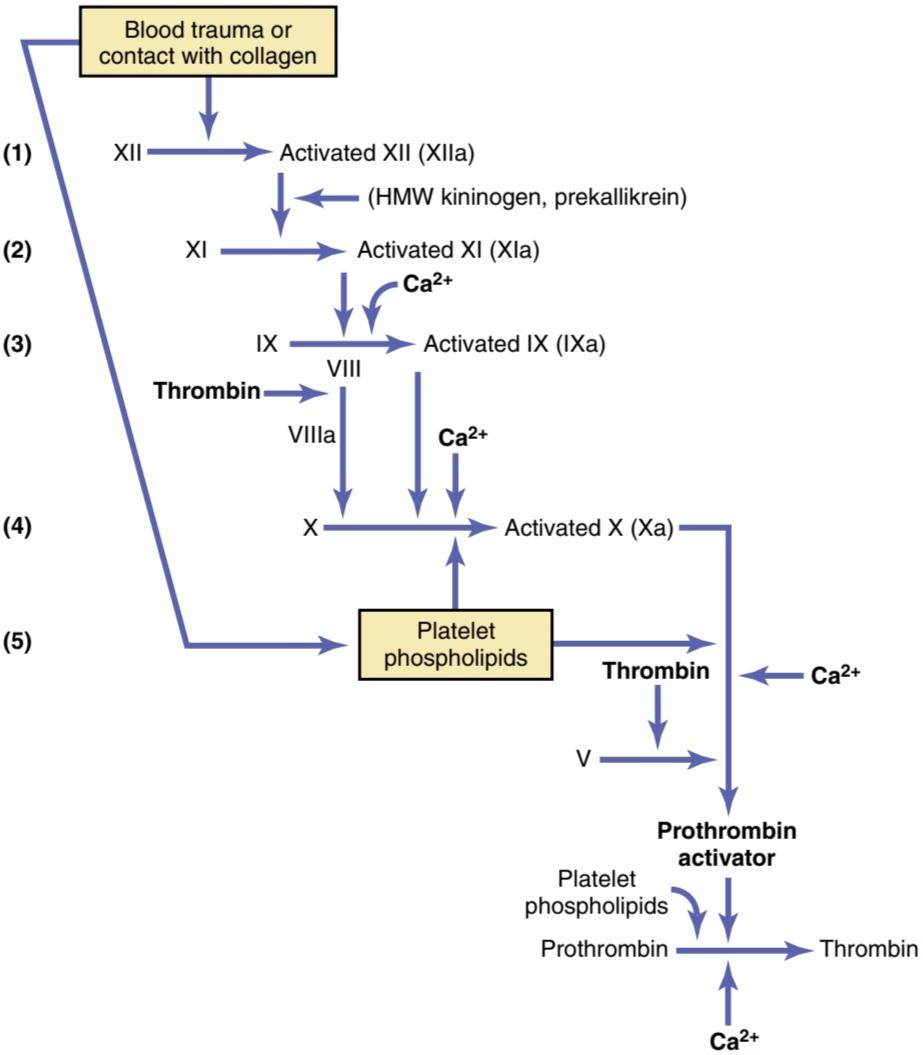
Figure 37-6. Intrinsic pathway for initiating blood clotting. HMW, High-molecular weight.
شکل 37-6. مسیر درونی برای شروع لخته شدن خون. HMW، وزن مولکولی بالا.
Role of Calcium lons in the Intrinsic and Extrinsic Pathways
Except for the first two steps in the intrinsic pathway, calcium ions are required for promotion or acceleration of all the blood-clotting reactions. Therefore, in the absence of calcium ions, blood clotting by either pathway does not occur.
نقش کلسیم لونها در مسیرهای درونی و بیرونی
به جز دو مرحله اول در مسیر ذاتی، یونهای کلسیم برای ارتقا یا تسریع تمام واکنشهای لخته شدن خون مورد نیاز هستند. بنابراین، در غیاب یونهای کلسیم، لخته شدن خون در هر دو مسیر رخ نمیدهد.
In the living body, the calcium ion concentration seldom falls low enough to affect blood-clotting kinetics significantly. However, when blood is removed from someone, it can be prevented from clotting by reducing the calcium ion concentration below the threshold level for clotting by deionizing the calcium by causing it to react with substances such as citrate ion or by precipitating the calcium with substances such as oxalate ion.
در بدن زنده، غلظت یون کلسیم به ندرت به اندازه ای پایین میآید که بر سینتیک لخته شدن خون تأثیر قابل توجهی بگذارد. با این حال، زمانی که خون از فردی خارج میشود، میتوان با کاهش غلظت یون کلسیم به زیر سطح آستانه لخته شدن، با یون زدایی کلسیم از طریق واکنش آن با موادی مانند یون سیترات یا رسوب کلسیم با موادی مانند یون اگزالات، از لخته شدن آن جلوگیری کرد.
Interaction Between Extrinsic and Intrinsic Pathways-Summary of Blood- Clotting Initiation
It is clear from the schemas of the intrinsic and extrinsic systems that after blood vessels rupture, clotting occurs by both pathways simultaneously. Tissue factor initiates the extrinsic pathway, whereas contact of factor XII and platelets with collagen in the vascular wall initiates the intrinsic pathway.
تعامل بین مسیرهای بیرونی و درونی – خلاصه ای از شروع لخته شدن خون
از طرحوارههای سیستم درونی و بیرونی مشخص است که پس از پارگی رگهای خونی، لخته شدن در هر دو مسیر به طور همزمان اتفاق میافتد. فاکتور بافتی مسیر بیرونی را آغاز میکند، در حالی که تماس فاکتور XII و پلاکتها با کلاژن در دیواره عروقی مسیر درونی را آغاز میکند.
An especially important difference between the extrinsic and intrinsic pathways is that the extrinsic pathway can be explosive; once initiated, its speed of completion to the final clot is limited only by the amount of tissue factor released from the traumatized tissues and by the quantities of factors X, VII, and V in the blood. With severe tissue trauma, clotting can occur in as little as 15 seconds. The intrinsic pathway is much slower to proceed, usually requiring 1 to 6 minutes to cause clotting.
یک تفاوت مهم بین مسیرهای بیرونی و درونی این است که مسیر بیرونی میتواند انفجاری باشد. پس از شروع، سرعت تکمیل آن تا لخته نهایی تنها به مقدار فاکتور بافتی آزاد شده از بافتهای آسیب دیده و مقادیر فاکتورهای X، VII و V در خون محدود میشود. با ترومای شدید بافت، لخته شدن میتواند در کمتر از 15 ثانیه رخ دهد. مسیر ذاتی بسیار کندتر پیش میرود و معمولاً برای ایجاد لخته شدن به 1 تا 6 دقیقه زمان نیاز دارد.
Intravascular Anticoagulants Prevent Blood Clotting in the Normal Vascular System
Endothelial Surface Factors. Probably the most important factors for preventing clotting in the normal vascular system are the following: (1) the smoothness of the endothelial cell surface, which prevents contact activation of the intrinsic clotting system; (2) a layer of glycocalyx on the endothelium (glycocalyx is a mucopolysaccharide adsorbed to the surfaces of the endothelial cells), which repels clotting factors and platelets, thereby preventing activation of clotting; and (3) a protein bound with the endothelial membrane, thrombomodulin, which binds thrombin. Not only does the binding of thrombin with thrombomodulin slow the clotting process by removing thrombin, but the thrombomodulin-thrombin complex also activates a plasma protein, protein C, that acts as an anticoagulant by inactivating activated factors V and VIII.
داروهای ضد انعقاد داخل عروقی از لخته شدن خون در سیستم عروقی طبیعی جلوگیری میکنند
فاکتورهای سطح اندوتلیال. احتمالاً مهمترین عوامل برای جلوگیری از لخته شدن در سیستم عروقی طبیعی عبارتند از: (1) صاف بودن سطح سلولهای اندوتلیال، که از فعال شدن تماسی سیستم لخته شدن ذاتی جلوگیری میکند. (2) لایه ای از گلیکوکالیکس روی اندوتلیوم (گلیکوکالیکس یک موکوپلی ساکارید است که به سطوح سلولهای اندوتلیال جذب میشود)، که فاکتورهای لخته شدن و پلاکتها را دفع میکند و در نتیجه از فعال شدن لخته شدن جلوگیری میکند. و (3) یک پروتئین متصل به غشای اندوتلیال، ترومبومودولین، که ترومبین را متصل میکند. نه تنها اتصال ترومبین با ترومبومودولین با حذف ترومبین، روند انعقاد را کند میکند، بلکه کمپلکس ترومبومودولین-ترومبین نیز پروتئین پلاسما، پروتئین C را فعال میکند که با غیرفعال کردن فاکتورهای فعال V و VIII به عنوان یک ضد انعقاد عمل میکند.
When the endothelial wall is damaged, its smoothness and glycocalyx-thrombomodulin layer are lost, which activates both factor XII and the platelets, thus setting off the intrinsic pathway of clotting. If factor XII and platelets come into contact with the subendothelial collagen, the activation is even more powerful.
هنگامیکه دیواره اندوتلیال آسیب میبیند، صافی و لایه گلیکوکالیکس-ترومبومودولین از بین میرود، که هم فاکتور XII و هم پلاکتها را فعال میکند و در نتیجه مسیر ذاتی لخته شدن را شروع میکند. اگر فاکتور XII و پلاکتها با کلاژن ساب اندوتلیال در تماس باشند، فعال شدن آن حتی قوی تر میشود.
Intact endothelial cells also produce other substances such a prostacyclin and nitric oxide (NO) that inhibit platelet aggregation and initiation of blood clotting. Prostacyclin, also called prostaglandin I2 (PGI2), is a member of the eicosanoid family of lipids and is a vasodilator, as well as an inhibitor of platelet aggregation. As discussed in Chapter 17, NO is a powerful vasodilator released from healthy vascular endothelial cells throughout the body, and it is an important inhibitor of platelet aggregation. When endothelial cells are damaged, their production of prostacyclin and NO is greatly diminished.
سلولهای اندوتلیال دست نخورده همچنین مواد دیگری مانند پروستاسیکلین و اکسید نیتریک (NO) تولید میکنند که از تجمع پلاکتها و شروع لخته شدن خون جلوگیری میکند. پروستاسیکلین که پروستاگلاندین I2 (PGI2) نیز نامیده میشود، عضوی از خانواده لیپیدهای ایکوزانوئید است و گشادکننده عروق و همچنین مهارکننده تجمع پلاکتی است. همانطور که در فصل 17 بحث شد، NO یک گشاد کننده عروق قوی است که از سلولهای اندوتلیال عروقی سالم در سراسر بدن آزاد میشود و یک مهار کننده مهم تجمع پلاکتی است. هنگامیکه سلولهای اندوتلیال آسیب میبینند، تولید پروستاسیکلین و NO به شدت کاهش مییابد.
Antithrombin Action of Fibrin and Antithrombin III. Among the most important anticoagulants in the blood are those that remove thrombin from the blood. The most powerful of these are the following: (1) the fibrin fibers that are formed during the process of clotting; and (2) an a globulin called antithrombin III or antithrombin- heparin cofactor.
اثر آنتی ترومبین فیبرین و آنتی ترومبین III. از مهمترین داروهای ضد انعقاد خون میتوان به حذف ترومبین از خون اشاره کرد. قوی ترین آنها موارد زیر است: (1) الیاف فیبرین که در طی فرآیند لخته شدن تشکیل میشوند. و (2) یک گلوبولین به نام آنتی ترومبین III یا کوفاکتور آنتی ترومبین- هپارین.
While a clot is forming, about 85% to 90% of the thrombin formed from the prothrombin becomes adsorbed to the fibrin fibers as they develop. This adsorption helps prevent the spread of thrombin into the remaining blood and, therefore, prevents excessive spread of the clot.
در حالی که یک لخته در حال تشکیل است، حدود 85٪ تا 90٪ از ترومبین تشکیل شده از پروترومبین با رشد فیبرهای فیبرین جذب میشود. این جذب به جلوگیری از انتشار ترومبین در خون باقی مانده کمک میکند و بنابراین از گسترش بیش از حد لخته جلوگیری میکند.
The thrombin that does not adsorb to the fibrin fibers soon combines with antithrombin III. This further blocks the effect of thrombin on the fibrinogen and then also inactivates thrombin itself during the next 12 to 20 minutes.
ترومبین که به فیبرهای فیبرین جذب نمیشود به زودی با آنتی ترومبین III ترکیب میشود. این بیشتر اثر ترومبین را بر فیبرینوژن مسدود میکند و سپس خود ترومبین را در 12 تا 20 دقیقه بعدی غیرفعال میکند.
Heparin. Heparin is another powerful anticoagulant but, because its concentration in the blood is normally low, it has significant anticoagulant effects only under special physiological conditions. However, heparin is used widely as a pharmacological agent in medical practice in much higher concentrations to prevent intravascular clotting.
هپارین. هپارین یکی دیگر از داروهای ضد انعقاد قوی است، اما از آنجایی که غلظت آن در خون معمولاً پایین است، فقط در شرایط فیزیولوژیکی خاص اثرات ضد انعقادی قابل توجهی دارد. با این حال، هپارین به طور گسترده به عنوان یک عامل دارویی در عمل پزشکی در غلظتهای بسیار بالاتر برای جلوگیری از لخته شدن داخل عروقی استفاده میشود.
The heparin molecule is a highly negatively charged conjugated polysaccharide. By itself, it has little or no anticoagulant properties, but when it combines with antithrombin III, the effectiveness of antithrombin III for removing thrombin increases by a hundredfold to a thousandfold and thus acts as an anticoagulant. There- fore, in the presence of excess heparin, the removal of free thrombin from the circulating blood by antithrombin III is almost instantaneous.
مولکول هپارین یک پلی ساکارید مزدوج با بار منفی بالا است. به خودی خود خاصیت ضد انعقادی کمیدارد یا اصلاً وجود ندارد، اما وقتی با آنتی ترومبین III ترکیب میشود، اثربخشی آنتی ترومبین III برای حذف ترومبین صد برابر تا هزار برابر میشود و بنابراین به عنوان یک ضد انعقاد عمل میکند. بنابراین، در حضور هپارین اضافی، حذف ترومبین آزاد از خون در گردش توسط آنتی ترومبین III تقریباً آنی است.
The complex of heparin and antithrombin III removes several other activated coagulation factors in addition to thrombin, further enhancing the effectiveness of anticoagulation. The others include activated factors IX through XII.
مجموعه هپارین و آنتی ترومبین III علاوه بر ترومبین چندین فاکتور انعقادی فعال دیگر را حذف میکند و اثربخشی ضد انعقاد را بیشتر میکند. بقیه شامل فاکتورهای فعال IX تا XII هستند.
Heparin is produced by many different cells of the body, but the largest quantities are formed by the basophilic mast cells located in the pericapillary connective tissue throughout the body. These cells continually secrete small quantities of heparin that diffuse into the circulatory system. The basophil cells of the blood, which are functionally almost identical to the mast cells, release small quantities of heparin into the plasma.
هپارین توسط بسیاری از سلولهای مختلف بدن تولید میشود، اما بیشترین مقدار توسط ماست سلهای بازوفیل واقع در بافت همبند پریکاپیلاری در سراسر بدن تشکیل میشود. این سلولها به طور مداوم مقادیر کمیهپارین ترشح میکنند که در سیستم گردش خون منتشر میشود. سلولهای بازوفیل خون، که از نظر عملکرد تقریباً مشابه ماست سلها هستند، مقادیر کمیهپارین را در پلاسما آزاد میکنند.
Mast cells are abundant in tissue surrounding the capillaries of the lungs and, to a lesser extent, capillaries of the liver. It is easy to understand why large quantities of heparin might be needed in these areas because the capillaries of the lungs and liver receive many embolic clots that have formed in slowly flowing venous blood; sufficient production of heparin prevents further growth of the clots.
ماست سلها در بافت اطراف مویرگهای ریه و تا حدی مویرگهای کبد فراوان هستند. به راحتی میتوان درک کرد که چرا ممکن است مقادیر زیادی هپارین در این نواحی مورد نیاز باشد، زیرا مویرگهای ریهها و کبد لختههای آمبولیک زیادی را دریافت میکنند که در خون وریدی به آرامیجریان دارد. تولید کافی هپارین از رشد بیشتر لختهها جلوگیری میکند.
PLASMIN CAUSES LYSIS OF BLOOD CLOTS
The plasma proteins contain a euglobulin called plasminogen (profibrinolysin) that when activated, becomes a substance called plasmin (fibrinolysin). Plasmin is a proteolytic enzyme that resembles trypsin, the most important proteolytic digestive enzyme of pancreatic secretion. Plasmin digests fibrin fibers and some other protein coagulants, such as fibrinogen, factor V, factor VIII, pro- thrombin, and factor XII. Therefore, whenever plasmin is formed, it can cause lysis of a clot by destroying many of the clotting factors, thereby sometimes even causing hypocoagulability of the blood.
پلاسمین باعث لیز لختههای خون میشود
پروتئینهای پلاسما حاوی یک اوگلوبولین به نام پلاسمینوژن (پروفیبرینولیزین) است که وقتی فعال میشود به ماده ای به نام پلاسمین (فیبرینولیزین) تبدیل میشود. پلاسمین یک آنزیم پروتئولیتیک است که شبیه به تریپسین، مهمترین آنزیم گوارشی پروتئولیتیک ترشح پانکراس است. پلاسمین فیبرهای فیبرین و برخی دیگر از منعقد کنندههای پروتئینی مانند فیبرینوژن، فاکتور V، فاکتور VIII، پروترومبین و فاکتور XII را هضم میکند. بنابراین، هر زمان که پلاسمین تشکیل شود، میتواند با از بین بردن بسیاری از عوامل لختهکننده، لخته لخته را از بین ببرد و در نتیجه گاهی حتی باعث کاهش انعقاد خون شود.
Activation of Plasminogen to Form Plasmin, Then Clot Lysis. When a clot is formed, a large amount of plasminogen is trapped in the clot, along with other plasma proteins. This will not become plasmin or cause lysis of the clot until it is activated. The injured tissues and vascular endothelium very slowly release a powerful activator called tissue plasminogen activator (t-PA); a few days later, after the clot has stopped the bleeding, t-PA eventually converts plasminogen to plasmin, which in turn removes the remaining unnecessary blood clot. In fact, many small blood vessels in which blood flow has been blocked by clots are reopened by this mechanism. Thus, an especially important function of the plasmin system is to remove minute clots from millions of tiny peripheral vessels that eventually would become occluded were there no way to clear them.
فعال سازی پلاسمینوژن برای تشکیل پلاسمین و سپس لخته شدن لخته. هنگامیکه یک لخته تشکیل میشود، مقدار زیادی پلاسمینوژن همراه با سایر پروتئینهای پلاسما در لخته به دام میافتد. تا زمانی که لخته فعال نشود، به پلاسمین تبدیل نمیشود یا باعث لیز لخته نمیشود. بافتهای آسیب دیده و اندوتلیوم عروقی به آرامییک فعال کننده قدرتمند به نام فعال کننده پلاسمینوژن بافتی (t-PA) آزاد میکنند. چند روز بعد، پس از اینکه لخته خونریزی را متوقف کرد، t-PA در نهایت پلاسمینوژن را به پلاسمین تبدیل میکند، که به نوبه خود لخته خون غیر ضروری باقی مانده را حذف میکند. در واقع، بسیاری از رگهای خونی کوچکی که جریان خون در آنها توسط لخته مسدود شده است، با این مکانیسم دوباره باز میشوند. بنابراین، یکی از عملکردهای مهم سیستم پلاسمین، حذف لختههای کوچک از میلیونها رگ کوچک محیطی است که در نهایت اگر راهی برای پاک کردن آنها وجود نداشت، مسدود میشوند.
CONDITIONS THAT CAUSE EXCESSIVE BLEEDING IN HUMANS
Excessive bleeding can result from a deficiency of any of the many blood-clotting factors. Three particular types of bleeding tendencies that have been studied to the greatest extent are discussed here-bleeding caused by (1) vitamin K deficiency, (2) hemophilia, and (3) thrombocytopenia (platelet deficiency).
شرایطی که باعث خونریزی بیش از حد در انسان میشود
خونریزی بیش از حد میتواند ناشی از کمبود هر یک از بسیاری از عوامل انعقاد خون باشد. سه نوع خاص از تمایلات خونریزی که تا حد زیادی مورد مطالعه قرار گرفته اند در اینجا مورد بحث قرار میگیرند: خونریزی ناشی از (1) کمبود ویتامین K، (2) هموفیلی، و (3) ترومبوسیتوپنی (کمبود پلاکت).
DECREASED PROTHROMBIN, FACTOR VII, FACTOR IX, AND FACTOR X CAUSED BY VITAMIN K DEFICIENCY
With few exceptions, almost all the blood-clotting factors are formed by the liver. Therefore, diseases of the liver such as hepatitis, cirrhosis, and acute yellow atrophy (degeneration of the liver caused by toxins, infections, or other agents) can sometimes depress the clotting system so much that the patient develops a severe tendency to bleed.
کاهش پروترومبین، فاکتور VII، فاکتور IX و فاکتور X ناشی از کمبود ویتامین K
به استثنای چند مورد، تقریباً تمام عوامل انعقاد خون توسط کبد تشکیل میشوند. بنابراین، بیماریهای کبدی مانند هپاتیت، سیروز و آتروفی حاد زرد (تخریب کبد ناشی از سموم، عفونتها یا عوامل دیگر) گاهی اوقات میتوانند سیستم انعقادی را به حدی تحت فشار قرار دهند که بیمار تمایل شدید به خونریزی پیدا کند.
Another cause of depressed formation of clotting factors by the liver is vitamin K deficiency. Vitamin K is an essential factor to a liver carboxylase that adds a carboxyl group to glutamic acid residues on five of the important clotting factors-prothrombin, factor VII, factor IX, factor X, and protein C. On adding the carboxyl group to glutamic acid residues on the immature clotting factors, vitamin K is oxidized and becomes inactive. Another enzyme, vitamin K epoxide reductase complex 1 (VKORC1), reduces vitamin K back to its active form. In the absence of active vitamin K, subsequent insufficiency of these coagulation factors in the blood can lead to serious bleeding tendencies.
یکی دیگر از علل تشکیل افسرده فاکتورهای انعقادی توسط کبد، کمبود ویتامین K است. ویتامین K یک عامل ضروری برای کربوکسیلاز کبد است که یک گروه کربوکسیل به باقی ماندههای اسید گلوتامیک در پنج فاکتور لخته کننده مهم پروترومبین، فاکتور VII، فاکتور IX، فاکتور X و پروتئین C اضافه میکند. با افزودن گروه کربوکسیل به بقایای اسید گلوتامیک روی اسیدهای نابالغ، اکسید شده و به فاکتورهای انعقادی نابالغ تبدیل میشود. آنزیم دیگر، ویتامین K اپوکسید ردوکتاز کمپلکس 1 (VKORC1)، ویتامین K را به شکل فعال خود کاهش میدهد. در غیاب ویتامین K فعال، نارسایی بعدی این فاکتورهای انعقادی در خون میتواند منجر به تمایلات جدی خونریزی شود.
Vitamin K is continually synthesized in the intestinal tract by bacteria, so vitamin K deficiency seldom occurs in healthy persons as a result of the absence of vitamin K from the diet (except in neonates, before they establish their intestinal bacterial flora). However, in persons with gastrointestinal disease, vitamin K deficiency often occurs as a result of poor absorption of fats from the gastrointestinal tract because vitamin K is fat-soluble and is ordinarily absorbed into the blood along with the fats.
ویتامین K به طور مداوم در مجرای روده توسط باکتریها سنتز میشود، بنابراین کمبود ویتامین K به ندرت در افراد سالم به دلیل عدم وجود ویتامین K در رژیم غذایی رخ میدهد (به جز در نوزادان، قبل از اینکه فلور باکتریایی روده خود را ایجاد کنند). با این حال، در افراد مبتلا به بیماریهای گوارشی، کمبود ویتامین K اغلب در نتیجه جذب ضعیف چربیها از دستگاه گوارش رخ میدهد، زیرا ویتامین K محلول در چربی است و به طور معمول همراه با چربیها در خون جذب میشود.
One of the most prevalent causes of vitamin K deficiency is failure of the liver to secrete bile into the gastrointestinal tract, which occurs as a result of obstruction of the bile ducts or of liver disease. Lack of bile prevents adequate fat digestion and absorption and, there fore ,depresses vitamin K absorption as well. Thus, liver dis- ease often causes decreased production of prothrombin and some other clotting factors because of poor vitamin K absorption and because of the diseased liver cells. As a result, vitamin K is injected into surgical patients with liver disease or with obstructed bile ducts before the surgical procedure is performed. Ordinarily, if vitamin K is given to a deficient patient 4 to 8 hours before the operation and the liver parenchymal cells are at least half- normal in function, sufficient clotting factors will be produced to prevent excessive bleeding during the operation.
یکی از شایع ترین علل کمبود ویتامین K، عدم ترشح صفرا توسط کبد در دستگاه گوارش است که در نتیجه انسداد مجاری صفراوی یا بیماری کبدی رخ میدهد. کمبود صفرا از هضم و جذب کافی چربی جلوگیری میکند و از این رو جذب ویتامین K را نیز کاهش میدهد. بنابراین، بیماری کبد به دلیل جذب ضعیف ویتامین K و به دلیل سلولهای کبدی بیمار، اغلب باعث کاهش تولید پروترومبین و برخی دیگر از عوامل انعقادی میشود. در نتیجه ویتامین K قبل از انجام عمل جراحی به بیماران جراحی مبتلا به بیماری کبدی یا مجاری صفراوی انسدادی تزریق میشود. به طور معمول، اگر 4 تا 8 ساعت قبل از عمل به بیمار کمبود ویتامین K داده شود و عملکرد سلولهای پارانشیمیکبد حداقل نیمه طبیعی باشد، فاکتورهای انعقادی کافی برای جلوگیری از خونریزی بیش از حد در حین عمل ایجاد میشود.
HEMOPHILIA
Hemophilia is a bleeding disease that occurs almost exclusively in males. In 85% of cases, it is caused by an abnormality or deficiency of factor VIII; this type of hemophilia is called hemophilia A or classic hemophilia. About 1 of every 10,000 males in the United States has classic hemophilia. In the other 15% of patients with hemophilia B, the bleeding tendency is caused by deficiency of factor IX. Both these factors are transmitted genetically by way of the female (X) chromosome and are recessive in their inheritance. Therefore, a woman will rarely have hemophilia because at least one of her two X chromosomes will have the appropriate genes. If one of her X chromosomes is deficient, she will be a hemophilia carrier; her male off- spring will have a 50% chance of inheriting the illness, and her female offspring will have a 50% chance of inheriting the carrier status.
هموفیلی
هموفیلی یک بیماری خونریزی دهنده است که تقریباً منحصراً در مردان رخ میدهد. در 85% موارد، ناهنجاری یا کمبود فاکتور VIII ایجاد میشود. این نوع هموفیلی، هموفیلی A یا هموفیلی کلاسیک نامیده میشود. از هر 10000 مرد در ایالات متحده یک نفر به هموفیلی کلاسیک مبتلا است. در 15 درصد دیگر بیماران مبتلا به هموفیلی B، تمایل به خونریزی ناشی از کمبود فاکتور IX است. هر دوی این عوامل به صورت ژنتیکی از طریق کروموزوم ماده (X) منتقل میشوند و در وراثت مغلوب هستند. بنابراین، یک زن به ندرت به هموفیلی مبتلا میشود زیرا حداقل یکی از دو کروموزوم X او دارای ژنهای مناسب است. اگر یکی از کروموزومهای X او کمبود داشته باشد، ناقل هموفیلی خواهد بود. فرزندان نر او 50 درصد شانس به ارث بردن این بیماری و فرزندان ماده او 50 درصد شانس به ارث بردن وضعیت ناقل را خواهند داشت.
Although female carriers have one normal allele and usually do not develop symptomatic hemophilia, some may experience a mild bleeding trait. It is also possible for female carriers to develop mild hemophilia due to loss of part or all of the normal X chromosome (as in Turner syndrome) or inactivation (lyonization) of the X-chromosomes. For a female to inherit full-blown symptomatic hemophilia A or B, she must receive two deficient X-chromosomes, one from her carrier mother and the other from her father, who must have hemophilia. Most cases of hemophilia are inherited, but approximately one-third of hemophilia patients do not have a family history of the disease, which appears to be caused by novel mutation events.
اگرچه حاملان زن دارای یک آلل طبیعی هستند و معمولاً به هموفیلی علامتی مبتلا نمیشوند، برخی ممکن است یک صفت خونریزی خفیف را تجربه کنند. همچنین ممکن است ناقلین زن به دلیل از دست دادن بخشی یا تمام کروموزوم X طبیعی (مانند سندرم ترنر) یا غیرفعال شدن (لیونیزاسیون) کروموزومهای X به هموفیلی خفیف مبتلا شوند. برای اینکه یک زن هموفیلی علامت دار A یا B را به ارث ببرد، باید دو کروموزوم X کمبود دریافت کند، یکی از مادر ناقل خود و دیگری از پدرش که باید هموفیلی داشته باشد. اکثر موارد هموفیلی ارثی است، اما تقریباً یک سوم از بیماران هموفیلی سابقه خانوادگی این بیماری را ندارند، که به نظر میرسد ناشی از رویدادهای جدید جهش باشد.
The bleeding trait in hemophilia can have various degrees of severity, depending on the genetic deficiency. Bleeding usually does not occur except after trauma, but in some patients, the degree of trauma required to cause severe and prolonged bleeding may be so mild that it is hardly noticeable. For example, bleeding can often last for days after extraction of a tooth.
صفت خونریزی در هموفیلی بسته به کمبود ژنتیکی میتواند درجات مختلفی از شدت داشته باشد. خونریزی معمولاً جز پس از ضربه اتفاق نمیافتد، اما در برخی از بیماران، میزان ضربه مورد نیاز برای ایجاد خونریزی شدید و طولانی مدت ممکن است آنقدر خفیف باشد که به سختی قابل توجه باشد. به عنوان مثال، خونریزی اغلب میتواند تا چند روز پس از کشیدن دندان ادامه یابد.
Factor VIII has two active components, a large component with a molecular weight in the millions and a smaller component with a molecular weight of about 230,000. The smaller component is most important in the intrinsic pathway for clotting, and it is deficiency of this part of factor VIII that causes classic hemophilia. Another bleeding disease with somewhat different characteristics, called von Willebrand disease, results from loss of the large component.
عامل هشتم دارای دو جزء فعال است، یک جزء بزرگ با وزن مولکولی میلیونها و یک جزء کوچکتر با وزن مولکولی حدود 230000. جزء کوچکتر در مسیر ذاتی لخته شدن مهمتر است و کمبود این قسمت از فاکتور VIII است که باعث هموفیلی کلاسیک میشود. یکی دیگر از بیماریهای خونریزیدهنده با ویژگیهای متفاوت، به نام بیماری فون ویلبراند، ناشی از از دست دادن جزء بزرگ است.
When a person with classic hemophilia experiences severe prolonged bleeding, almost the only therapy that is truly effective is injection of purified factor VIII or factor IX. Both these clotting factors are now available as recombinant proteins, although they are expensive and their half-lives are relatively short; therefore, these products are not readily available for many patients with hemophilia, especially in economically disadvantaged countries.
هنگامیکه یک فرد مبتلا به هموفیلی کلاسیک خونریزی شدید طولانی مدت را تجربه میکند، تقریباً تنها درمانی که واقعاً مؤثر است، تزریق فاکتور VIII یا فاکتور IX خالص است. هر دو این فاکتورهای انعقادی در حال حاضر به عنوان پروتئینهای نوترکیب در دسترس هستند، اگرچه گران هستند و نیمه عمر آنها نسبتاً کوتاه است. بنابراین، این محصولات برای بسیاری از بیماران مبتلا به هموفیلی، به ویژه در کشورهایی که از نظر اقتصادی محروم هستند، به راحتی در دسترس نیستند.
THROMBOCYTOPENIA
Thrombocytopenia means the presence of very low numbers of platelets in the circulating blood. People with thrombocytopenia have a tendency to bleed, as do hemophiliacs, except that the bleeding is usually from many small venules or capillaries, rather than from larger vessels, as in hemophilia. As a result, small punctate hemorrhages occur throughout all the body tissues. The skin of such a person displays many small petechiae, red or purplish blotches, giving the disease the name thrombocytopenic purpura. As noted, platelets are especially important for the repair of minute breaks in capillaries and other small vessels.
ترومبوسیتوپنی
ترومبوسیتوپنی به معنای وجود تعداد بسیار کم پلاکت در خون در گردش است. افراد مبتلا به ترومبوسیتوپنی، مانند بیماران هموفیلی، تمایل به خونریزی دارند، با این تفاوت که خونریزی معمولاً از بسیاری از وریدها یا مویرگهای کوچک است، نه از عروق بزرگتر، مانند هموفیلی. در نتیجه، خونریزیهای نقطه ای کوچک در تمام بافتهای بدن رخ میدهد. پوست چنین فردی دارای پتشیهای کوچک، لکههای قرمز یا ارغوانی است که به این بیماری نام پورپورای ترومبوسیتوپنیک داده میشود. همانطور که اشاره شد، پلاکتها به ویژه برای ترمیم شکستگیهای کوچک در مویرگها و سایر عروق کوچک مهم هستند.
Platelet counts below 30,000/μl, compared with the normal value of 150,000 to 450,000/μl, increase the risk for excessive bleeding after surgery or injury. Spontaneous bleeding, however, will not ordinarily occur until the number of platelets in the blood falls below 30,000/μl. Levels as low as 10,000/μl are frequently lethal.
تعداد پلاکتها کمتر از 30000/μl در مقایسه با مقدار طبیعی 150000 تا 450000/μl، خطر خونریزی بیش از حد پس از جراحی یا آسیب را افزایش میدهد. با این حال، معمولاً تا زمانی که تعداد پلاکتهای خون به کمتر از 30000 در میکرولیتر نرسد، خونریزی خودبهخودی اتفاق نمیافتد. سطوح کمتر از 10000/μl اغلب کشنده هستند.
Even without determining specific platelet counts in the blood, sometimes one can suspect the existence of thrombocytopenia if the person’s blood clot fails to retract. As noted earlier, clot retraction is normally dependent on release of multiple coagulation factors from the large numbers of platelets entrapped in the fibrin mesh of the clot.
حتی بدون تعیین تعداد پلاکتهای خاص در خون، گاهی اوقات میتوان به وجود ترومبوسیتوپنی مشکوک شد اگر لخته خون فرد جمع نشود. همانطور که قبلا ذکر شد، جمع شدن لخته به طور معمول به آزاد شدن چندین فاکتور انعقادی از تعداد زیادی پلاکت محبوس شده در مش فیبرین لخته بستگی دارد.
The major causes of thrombocytopenia include the following: (1) decreased platelet production in the bone marrow due to infections or sepsis, nutrient deficiencies, or myelodysplastic disorders, which usually also reduce production of other cells (red blood cells [RBCs] and white blood cells); (2) peripheral platelet destruction by antibodies; (3) sequestration (pooling) of platelets in the spleen, especially in individuals with portal hypertension and excessively large spleens (splenomegaly); (4) consumption of platelets in thrombi; and (4) dilution of the blood from fluid resuscitation or massive transfusion.
علل اصلی ترومبوسیتوپنی شامل موارد زیر است: (1) کاهش تولید پلاکت در مغز استخوان به دلیل عفونت یا سپسیس، کمبود مواد مغذی یا اختلالات میلودیسپلاستیک، که معمولاً باعث کاهش تولید سایر سلولها (گلبولهای قرمز خون [RBCs] و گلبولهای سفید) میشود. (2) تخریب پلاکت محیطی توسط آنتی بادی. (3) جداسازی (تجمع) پلاکتها در طحال، به ویژه در افراد مبتلا به فشار خون پورتال و طحال بیش از حد بزرگ (سپلنومگالی). (4) مصرف پلاکت در ترومبوز. و (4) رقیق شدن خون حاصل از احیای مایع یا انتقال گسترده.
Most people with thrombocytopenia have the disease known as idiopathic thrombocytopenia, which means thrombocytopenia of unknown cause. In most of these people, it has been discovered that, for unknown reasons, specific antibodies have formed and react against the platelets to destroy them. Relief from bleeding for 1 to 4 days can often be effected in a patient with thrombocytopenia by giving fresh whole blood transfusions that contain large numbers of platelets. Also, splenectomy may be helpful, sometimes resulting in an almost complete cure because the spleen normally removes large numbers of platelets from the blood.
اکثر افراد مبتلا به ترومبوسیتوپنی به بیماری معروف به ترومبوسیتوپنی ایدیوپاتیک مبتلا هستند که به معنی ترومبوسیتوپنی با علت ناشناخته است. در اکثر این افراد، مشخص شده است که به دلایل نامعلوم، آنتی بادیهای خاصی تشکیل شده و در برابر پلاکتها واکنش نشان میدهند تا آنها را از بین ببرد. تسکین خونریزی به مدت 1 تا 4 روز اغلب در بیماران مبتلا به ترومبوسیتوپنی با تزریق خون کامل تازه حاوی تعداد زیادی پلاکت قابل انجام است. همچنین، برداشتن طحال ممکن است مفید باشد، و گاهی اوقات منجر به درمان تقریباً کامل میشود، زیرا طحال به طور معمول تعداد زیادی پلاکت را از خون خارج میکند.
THROMBOEMBOLIC CONDITIONS
Thrombi and Emboli. An abnormal clot that develops in a blood vessel is called a thrombus. Once a clot has developed, continued flow of blood past the clot is likely to break it away from its attachment and cause the clot to flow with the blood; such freely flowing clots are known as emboli. Also, emboli that originate in large arteries or in the left side of the heart can flow peripherally and plug arteries or arterioles in the brain, kidneys, or elsewhere. Emboli that originate in the venous system or in the right side of the heart generally flow into the lungs to cause pulmonary arterial embolism.
شرایط ترومبوآمبولیک
ترومبی و آمبولی. لخته غیرطبیعی که در رگ خونی ایجاد میشود، ترومبوز نامیده میشود. هنگامیکه یک لخته ایجاد شد، جریان مداوم خون از کنار لخته احتمالاً آن را از اتصال خود جدا میکند و باعث میشود که لخته همراه با خون جاری شود. چنین لختههایی که آزادانه جریان دارند به عنوان آمبولی شناخته میشوند. همچنین آمبولیهایی که از شریانهای بزرگ یا در سمت چپ قلب منشا میگیرند میتوانند به صورت محیطی جریان داشته باشند و شریانها یا شریانها را در مغز، کلیهها یا جاهای دیگر مسدود کنند. آمبولیهایی که از سیستم وریدی یا سمت راست قلب منشا میگیرند، عموماً به داخل ریهها جریان مییابند و باعث آمبولی شریان ریوی میشوند.
Causes of Thromboembolic Conditions. The causes of thromboembolic conditions in people are usually twofold: (1) a roughened endothelial surface of a vessel-as may be caused by arteriosclerosis, infection, or trauma-is likely to initiate the clotting process; and (2) blood often clots when it flows very slowly through blood vessels, where small quantities of thrombin and other procoagulants are always being formed.
علل شرایط ترومبوآمبولیک. علل شرایط ترومبوآمبولی در افراد معمولاً دو گونه است: (1) سطح اندوتلیال زبر یک رگ – همانطور که ممکن است در اثر تصلب شرایین، عفونت یا تروما ایجاد شود – احتمالاً فرآیند لخته شدن را آغاز میکند. و (2) زمانی که خون به آرامیدر رگهای خونی جریان مییابد، معمولاً لخته میشود، جایی که مقادیر کمیترومبین و سایر پیشانعقادها همیشه در حال تشکیل هستند.
Use of Tissue Plasminogen Activator in Treating Intravascular Clots. Genetically engineered tissue plasminogen activator (t-PA) is available. When delivered through a catheter to an area with a thrombus, it is effective in activating plasminogen to plasmin, which in turn can dissolve some intravascular clots. For example, if used within the 1 or 2 hours after thrombotic occlusion of a coronary artery, the heart is often spared serious damage.
استفاده از فعال کننده پلاسمینوژن بافتی در درمان لختههای داخل عروقی. فعال کننده پلاسمینوژن بافت دستکاری شده ژنتیکی (t-PA) در دسترس است. هنگامیکه از طریق کاتتر به ناحیه ای با ترومبوس منتقل میشود، در فعال کردن پلاسمینوژن به پلاسمین موثر است که به نوبه خود میتواند برخی از لختههای داخل عروقی را حل کند. به عنوان مثال، اگر در 1 یا 2 ساعت پس از انسداد ترومبوتیک شریان کرونر استفاده شود، قلب اغلب از آسیب جدی در امان میماند.
FEMORAL VENOUS THROMBOSIS AND MASSIVE PULMONARY EMBOLISM
Because clotting almost always occurs when blood flow is blocked for many hours in any vessel of the body, the immobility of patients confined to bed, plus the practice of propping the knees up with pillows, often causes intra- vascular clotting because of blood stasis in one or more of the leg veins for hours at a time. Then the clot grows, mainly in the direction of the slowly moving venous blood, sometimes growing the entire length of the leg veins and occasionally even up into the common iliac vein and inferior vena cava. About 10% of the time, a large part of the clot disengages from its attachments to the vessel wall and flows freely with the venous blood through the right side of the heart and into the pulmonary arteries to cause massive blockage of the pulmonary arteries; this is called a massive pulmonary embolism. If the clot is large enough to occlude both pulmonary arteries at the same time, immediate death ensues. If only one pulmonary artery is blocked, death may not occur, or the embolism may lead to death a few hours to several days later because of further growth of the clot in the pulmonary vessels. However, again, t-PA therapy can be a lifesaver.
ترومبوز ورید فمورال و آمبولی بزرگ ریه
از آنجایی که لخته شدن تقریباً همیشه زمانی اتفاق میافتد که جریان خون برای ساعتها در هر رگ بدن مسدود میشود، بیحرکتی بیمارانی که در رختخواب محبوس شدهاند، بهعلاوه تمرین بالا نگه داشتن زانوها با بالش، اغلب باعث لخته شدن داخل عروقی میشود که به دلیل سکون خون در یک یا چند ورید پا برای ساعتها در یک زمان است. سپس لخته، عمدتاً در جهت خون وریدی که به آرامیحرکت میکند، رشد میکند، گاهی اوقات در تمام طول وریدهای پا رشد میکند و گاهی حتی تا ورید ایلیاک مشترک و ورید اجوف تحتانی رشد میکند. در حدود 10 درصد مواقع، بخش بزرگی از لخته از اتصالات خود به دیواره عروق جدا میشود و آزادانه همراه با خون وریدی از سمت راست قلب و به داخل شریانهای ریوی جریان مییابد تا باعث انسداد شدید شریانهای ریوی شود. به این آمبولی بزرگ ریه میگویند. اگر لخته به اندازه ای بزرگ باشد که هر دو شریان ریوی را به طور همزمان مسدود کند، مرگ فوری رخ میدهد. اگر تنها یک شریان ریوی مسدود شود، ممکن است مرگ رخ ندهد، یا آمبولی ممکن است چند ساعت تا چند روز بعد به مرگ منجر شود، زیرا به دلیل رشد بیشتر لخته در عروق ریوی است. با این حال، دوباره، درمان t-PA میتواند یک نجات دهنده باشد.
DISSEMINATED INTRAVASCULAR COAGULATION
Occasionally, the clotting mechanism becomes activated in widespread areas of the circulation, giving rise to the condition called disseminated intravascular coagulation (DIC). This condition often results from the presence of large amounts of traumatized or dying tissue in the body that releases great quantities of tissue factor into the blood. Frequently, the clots are small but numerous, and they plug a large share of the small peripheral blood vessels. This process occurs especially in patients with widespread septicemia, in which circulating bacteria or bacterial toxins-especially endotoxins-activate the clot- ting mechanisms. The plugging of small peripheral vessels greatly diminishes delivery of oxygen and other nutrients to the tissues, a situation that leads to or exacerbates circulatory shock. It is partly for this reason that septicemic shock is lethal in 35% to 50% of patients.
انعقاد درونرگی منتشر
گاهی اوقات، مکانیسم انعقاد در نواحی گسترده گردش خون فعال میشود و باعث ایجاد وضعیتی به نام انعقاد داخل عروقی منتشر (DIC) میشود. این وضعیت اغلب ناشی از وجود مقادیر زیادی بافت آسیب دیده یا در حال مرگ در بدن است که مقادیر زیادی فاکتور بافتی را در خون آزاد میکند. اغلب، لختهها کوچک اما متعدد هستند و بخش بزرگی از رگهای خونی محیطی کوچک را مسدود میکنند. این فرآیند به ویژه در بیماران مبتلا به سپتی سمیگسترده رخ میدهد که در آن باکتریهای در گردش یا سموم باکتریایی – به ویژه اندوتوکسینها – مکانیسمهای لخته شدن را فعال میکنند. بسته شدن رگهای کوچک محیطی به میزان زیادی از تحویل اکسیژن و سایر مواد مغذی به بافتها میکاهد، وضعیتی که منجر به شوک گردش خون یا تشدید آن میشود. تا حدودی به همین دلیل است که شوک سپتی سمیدر 35 تا 50 درصد بیماران کشنده است.
A peculiar effect of disseminated intravascular coagulation is that the patient, on occasion, begins to bleed. The reason for this bleeding is that so many of the clotting factors are removed by the widespread clotting that too few procoagulants remain to allow normal hemostasis of the remaining blood.
یک اثر عجیب انعقاد داخل عروقی منتشر این است که بیمار در مواردی شروع به خونریزی میکند. دلیل این خونریزی این است که بسیاری از عوامل انعقادی توسط لخته شدن گسترده حذف میشوند که تعداد کمیاز پیش انعقادها باقی میماند تا امکان هموستاز طبیعی خون باقی مانده را فراهم کند.
ANTICOAGULANTS FOR CLINICAL USE
In some thromboembolic conditions, it is desirable to delay the coagulation process. Various anticoagulants have been developed for this purpose. The ones most clinically useful are heparin and the coumarins.
داروهای ضد انعقاد برای استفاده بالینی
در برخی از شرایط ترومبوآمبولیک، به تعویق انداختن فرآیند انعقاد مطلوب است. داروهای ضد انعقاد مختلفی برای این منظور ساخته شده است. آنهایی که از نظر بالینی مفید هستند هپارین و کومارین هستند.
HEPARIN INTRAVENOUS ANTICOAGULANT
Commercial heparin is extracted from several different animal tissues and prepared in almost pure form. Injection of relatively small quantities, about 0.5 to 1 mg/kg of body weight, causes the blood-clotting time to increase from a normal of about 6 minutes to 30 or more minutes. Furthermore, this change in clotting time occurs instantaneously, thereby immediately preventing or slowing further development of a thromboembolic condition.
ضد انعقاد داخل وریدی هپارین
هپارین تجاری از چندین بافت مختلف حیوانی استخراج شده و به شکل تقریبا خالص تهیه میشود. تزریق مقادیر نسبتاً کم، حدود 0.5 تا 1 میلی گرم به ازای هر کیلوگرم وزن بدن، باعث میشود که زمان لخته شدن خون از حد طبیعی حدود 6 دقیقه به 30 دقیقه یا بیشتر افزایش یابد. علاوه بر این، این تغییر در زمان انعقاد فوراً اتفاق میافتد و در نتیجه بلافاصله از پیشرفت بیشتر یک بیماری ترومبوآمبولیک جلوگیری یا کند میکند.
The action of heparin lasts about 1.5 to 4 hours. The injected heparin is destroyed by an enzyme in the blood known as heparinase.
اثر هپارین حدود 1.5 تا 4 ساعت طول میکشد. هپارین تزریق شده توسط آنزیمیدر خون به نام هپاریناز از بین میرود.
COUMARINS AS ANTICOAGULANTS
When a coumarin, such as warfarin, is given to a patient, the amounts of active prothrombin and factors VII, IX, and X, all formed by the liver, begin to fall. Warfarin causes this effect by inhibiting the enzyme VKORC1. As discussed previously, this enzyme converts the inactive, oxidized form of vitamin K to its active, reduced form. By inhibiting VKORC1, warfarin decreases the available active form of vitamin K in the tissues. When this decrease occurs, the coagulation factors are no longer car-boxylated and are biologically inactive. Over several days, the body stores of the active coagulation factors degrade and are replaced by inactive factors. Although the coaglation factors continue to be produced, they have greatly decreased coagulant activity.
کومارینها به عنوان ضد انعقاد
هنگامیکه یک کومارین مانند وارفارین به بیمار داده میشود، مقدار پروترومبین فعال و فاکتورهای VII، IX و X که همگی توسط کبد تشکیل میشوند، شروع به کاهش میکنند. وارفارین این اثر را با مهار آنزیم VKORC1 ایجاد میکند. همانطور که قبلاً گفته شد، این آنزیم فرم غیرفعال و اکسید شده ویتامین K را به شکل فعال و کاهش یافته آن تبدیل میکند. با مهار VKORC1، وارفارین فرم فعال موجود ویتامین K را در بافتها کاهش میدهد. هنگامیکه این کاهش رخ میدهد، فاکتورهای انعقادی دیگر بوکسیله نمیشوند و از نظر بیولوژیکی غیر فعال هستند. طی چند روز، ذخایر فاکتورهای انعقادی فعال در بدن تحلیل رفته و با عوامل غیرفعال جایگزین میشوند. اگرچه فاکتورهای انعقادی همچنان تولید میشوند، اما فعالیت انعقادی را تا حد زیادی کاهش داده اند.
After administration of an effective dose of warfarin, the coagulant activity of the blood decreases to about 50% of normal by the end of 12 hours and to about 20% of normal by the end of 24 hours. In other words, the coagulation process is not blocked immediately but must await the degradation of the active prothrombin and the other affected coagulation factors already present in the plasma. Normal coagulation usually returns 1 to 3 days after dis- continuing coumarin therapy.
پس از تجویز دوز مؤثر وارفارین، فعالیت انعقادی خون در پایان 12 ساعت به حدود 50 درصد طبیعی و در پایان 24 ساعت به حدود 20 درصد طبیعی کاهش مییابد. به عبارت دیگر، فرآیند انعقاد بلافاصله مسدود نمیشود، بلکه باید منتظر تخریب پروترومبین فعال و سایر فاکتورهای انعقادی متاثر از قبل در پلاسما باشد. انعقاد طبیعی معمولاً 1 تا 3 روز پس از قطع درمان با کومارین باز میگردد.
PREVENTION OF BLOOD COAGULATION OUTSIDE THE BODY
Although blood removed from the body and held in a glass test tube normally clots in about 6 minutes, blood collected in siliconized containers often does not clot for 1 hour or more. The reason for this delay is that preparing the surfaces of the containers with silicone prevents con- tact activation of platelets and factor XII, the two principal factors that initiate the intrinsic clotting mechanism. Conversely, untreated glass containers allow contact activation of the platelets and factor XII, with the rapid development of clots.
پیشگیری از انعقاد خون در خارج از بدن
اگرچه خونی که از بدن خارج میشود و در یک لوله آزمایش شیشه ای نگهداری میشود معمولاً در حدود 6 دقیقه لخته میشود، خون جمع آوری شده در ظروف سیلیکونی شده اغلب به مدت 1 ساعت یا بیشتر لخته نمیشود. دلیل این تاخیر این است که آماده سازی سطوح ظروف با سیلیکون از فعال شدن تماس پلاکتها و فاکتور XII، دو عامل اصلی که مکانیسم لخته شدن ذاتی را آغاز میکنند، جلوگیری میکند. برعکس، ظروف شیشه ای درمان نشده امکان فعال سازی تماسی پلاکتها و فاکتور XII را با ایجاد سریع لختهها فراهم میکند.
Heparin can be used for preventing coagulation of blood outside the body, as well as in the body. Heparin is especially used in surgical procedures in which the blood must be passed through a heart-lung machine or artificial kidney machine and then back into the patient.
هپارین میتواند برای جلوگیری از انعقاد خون در خارج از بدن و همچنین در بدن استفاده شود. هپارین به ویژه در روشهای جراحی استفاده میشود که در آن خون باید از دستگاه قلب-ریه یا دستگاه کلیه مصنوعی عبور داده شود و سپس به بیمار برگردد.
Various substances that decrease the concentration of calcium ions in the blood can also be used for preventing blood coagulation outside the body. For example, a soluble oxalate compound mixed in a very small quantity with a sample of blood causes precipitation of calcium oxalate from the plasma and thereby decreases the ionic calcium level so much that blood coagulation is blocked.
برای جلوگیری از انعقاد خون در خارج از بدن نیز میتوان از مواد مختلفی که غلظت یون کلسیم در خون را کاهش میدهد، استفاده کرد. به عنوان مثال، یک ترکیب اگزالات محلول که به مقدار بسیار کمیبا نمونه خون مخلوط میشود، باعث رسوب اگزالات کلسیم از پلاسما میشود و در نتیجه سطح کلسیم یونی را به حدی کاهش میدهد که انعقاد خون مسدود میشود.
Any substance that deionizes the blood calcium will prevent coagulation. The negatively charged citrate ion is especially valuable for this purpose; it is mixed with blood usually in the form of sodium, ammonium, or potassium citrate. The citrate ion combines with calcium in the blood to produce a nonionized calcium compound, and the lack of ionic calcium prevents coagulation. Citrate anticoagulants have an important advantage over the oxalate anticoagulants because oxalate is toxic to the body, whereas moderate quantities of citrate can be injected intravenously. After injection, the citrate ion is removed from the blood within a few minutes by the liver and is polymerized into glucose or metabolized directly for energy. Consequently, 500 milliliters of blood that has been rendered noncoagulable by citrate can ordinarily be transfused into a recipient within a few minutes, without dire consequences. However, if the liver is damaged, or if large quantities of citrated blood or plasma are given too rapidly (within fractions of a minute), the citrate ion may not be removed quickly enough, and the citrate can, under these conditions, greatly depress the level of calcium ion in the blood, which can result in tetany and convulsive death.
هر ماده ای که کلسیم خون را دیونیزه کند از انعقاد جلوگیری میکند. یون سیترات با بار منفی به ویژه برای این منظور ارزشمند است. معمولاً به شکل سیترات سدیم، آمونیوم یا پتاسیم با خون مخلوط میشود. یون سیترات با کلسیم در خون ترکیب میشود و یک ترکیب کلسیم غیریونیزه تولید میکند و کمبود کلسیم یونی از انعقاد جلوگیری میکند. داروهای ضد انعقاد سیترات مزیت مهمینسبت به ضد انعقادهای اگزالات دارند زیرا اگزالات برای بدن سمیاست، در حالی که مقادیر متوسطی از سیترات را میتوان به صورت داخل وریدی تزریق کرد. پس از تزریق، یون سیترات در عرض چند دقیقه توسط کبد از خون خارج میشود و به گلوکز پلیمریزه میشود یا مستقیماً برای انرژی متابولیزه میشود. در نتیجه، 500 میلی لیتر خونی که توسط سیترات غیر قابل انعقاد شده است، معمولاً میتواند در عرض چند دقیقه به گیرنده تزریق شود، بدون عواقب ناگوار. با این حال، اگر کبد آسیب دیده باشد، یا اگر مقادیر زیادی خون یا پلاسمای سیترات شده خیلی سریع (در کسری از یک دقیقه) داده شود، یون سیترات ممکن است به سرعت کافی حذف نشود و سیترات در این شرایط میتواند سطح یون کلسیم در خون را تا حد زیادی کاهش دهد که میتواند منجر به مرگ کزاز و تشنج شود.
BLOOD COAGULATION TESTS
BLEEDING TIME
When a sharp-pointed knife is used to pierce the tip of the finger or earlobe, bleeding ordinarily lasts for 1 to 6 minutes. This time depends largely on the depth of the wound and degree of hyperemia in the finger or earlobe at the time of the test. Lack of any one of several of the clotting factors can prolong the bleeding time, but it is especially prolonged by lack of platelets.
تستهای انعقاد خون
زمان خونریزی
هنگامیکه از چاقوی نوک تیز برای سوراخ کردن نوک انگشت یا لاله گوش استفاده میشود، خونریزی معمولاً 1 تا 6 دقیقه طول میکشد. این زمان تا حد زیادی به عمق زخم و میزان پرخونی انگشت یا لاله گوش در زمان آزمایش بستگی دارد. فقدان هر یک از چندین عامل انعقاد میتواند زمان خونریزی را طولانی تر کند، اما به ویژه با کمبود پلاکت طولانی تر میشود.
CLOTTING TIME
Many methods have been devised for determining blood- clotting time. The one most widely used is to collect blood in a chemically clean glass test tube and then to tip the tube back and forth about every 30 seconds until the blood has clotted. By this method, the normal clotting time is 6 to 10 minutes. Procedures using multiple test tubes have also been devised for determining clotting time more accurately.
زمان لخته شدن
روشهای زیادی برای تعیین زمان لخته شدن خون ابداع شده است. پرکاربردترین روش جمع آوری خون در یک لوله آزمایش شیشه ای تمیز از نظر شیمیایی و سپس چرخاندن لوله به جلو و عقب تقریباً هر 30 ثانیه تا زمانی که خون لخته شود است. در این روش زمان لخته شدن طبیعی 6 تا 10 دقیقه است. روشهایی با استفاده از لولههای آزمایش متعدد نیز برای تعیین دقیقتر زمان لخته شدن ابداع شدهاند.
Unfortunately, the clotting time varies widely, depending on the method used for measuring it, so it is no longer used in many clinics. Instead, measurements of the clotting factors themselves are made, using sophisticated chemical procedures.
متأسفانه، بسته به روشی که برای اندازه گیری آن استفاده میشود، زمان لخته شدن بسیار متفاوت است، بنابراین دیگر در بسیاری از کلینیکها استفاده نمیشود. در عوض، اندازهگیریهای خود فاکتورهای لختهکننده با استفاده از روشهای شیمیایی پیچیده انجام میشوند.
PROTHROMBIN TIME AND INTERNATIONAL NORMALIZED RATIO
The prothrombin time indicates the concentration of prothrombin in the blood. Figure 37-7 shows the relationship of prothrombin concentration to prothrombin time. The method for determining prothrombin time is the following.
زمان پروترومبین و نسبت نرمال شده بین المللی
زمان پروترومبین نشان دهنده غلظت پروترومبین در خون است. شکل 37-7 رابطه غلظت پروترومبین را با زمان پروترومبین نشان میدهد. روش تعیین زمان پروترومبین به شرح زیر است.
Blood removed from the patient is immediately oxalated so that none of the prothrombin can change into thrombin. Then, a large excess of calcium ion and tissue factor is quickly mixed with the oxalated blood. The excess calcium nullifies the effect of the oxalate, and the tissue factor activates the prothrombin to thrombin reaction by means of the extrinsic clotting pathway. The time required for coagulation to take place is known as the prothrombin time. The shortness of the time is determined mainly by the prothrombin concentration. The normal prothrombin time is about 12 seconds. In each laboratory, a curve relating prothrombin concentration to prothrombin time, such as that shown in Figure 37-7, is drawn for the method used so that the prothrombin in the blood can be quantified.
خون خارج شده از بیمار بلافاصله اگزاله میشود تا هیچ یک از پروترومبینها نتوانند به ترومبین تبدیل شوند. سپس مقدار زیادی از یون کلسیم و فاکتور بافتی به سرعت با خون اگزال دار مخلوط میشود. کلسیم اضافی اثر اگزالات را از بین میبرد و فاکتور بافتی واکنش پروترومبین به ترومبین را با استفاده از مسیر انعقاد خارجی فعال میکند. زمان لازم برای انجام انعقاد به عنوان زمان پروترومبین شناخته میشود. کوتاهی زمان عمدتاً با غلظت پروترومبین تعیین میشود. زمان طبیعی پروترومبین حدود 12 ثانیه است. در هر آزمایشگاه، منحنی مربوط به غلظت پروترومبین به زمان پروترومبین، مانند آنچه در شکل 37-7 نشان داده شده است، برای روش مورد استفاده ترسیم میشود تا بتوان پروترومبین موجود در خون را کمیت کرد.
The results obtained for prothrombin time may vary considerably, even in the same individual if there are differences in activity of the tissue factor and the analytical system used to perform the test. Tissue factor is isolated from human tissues, such as placental tissue, and different batches may have different activity. The international normalized ratio (INR) was devised as a way to standardize measurements of prothrombin time. For each batch of tissue factor, the manufacturer assigns an international sensitivity index (ISI), which indicates the activity of the tissue factor with a standardized sample. The ISI usually varies between 1.0 and 2.0. The INR is the ratio of the person’s prothrombin time (PT) to a normal control sample raised to the power of the ISI:
نتایج بهدستآمده برای زمان پروترومبین ممکن است بهطور قابلتوجهی متفاوت باشد، حتی در صورت وجود تفاوت در فعالیت فاکتور بافتی و سیستم تحلیلی مورد استفاده برای انجام آزمایش. فاکتور بافتی از بافتهای انسانی مانند بافت جفت جدا میشود و دستههای مختلف ممکن است فعالیت متفاوتی داشته باشند. نسبت نرمال شده بین المللی (INR) به عنوان راهی برای استاندارد کردن اندازه گیری زمان پروترومبین ابداع شد. برای هر دسته از فاکتور بافتی، سازنده یک شاخص بین المللی حساسیت (ISI) را تعیین میکند که نشان دهنده فعالیت فاکتور بافت با یک نمونه استاندارد است. ISI معمولا بین 1.0 و 2.0 متغیر است. INR نسبت زمان پروترومبین فرد (PT) به یک نمونه کنترل طبیعی است که به توان ISI افزایش یافته است:
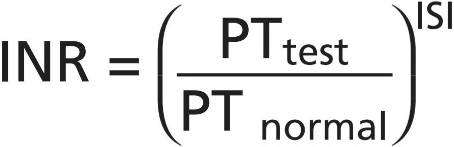
The normal range for INR in a healthy person is 0.9 to 1.3. A high INR level (e.g., 4 or 5) indicates a high risk of bleeding, whereas a low INR (e.g., 0.5) suggests that there is a chance of having a clot. Patients undergoing warfarin therapy usually have an INR of 2.0 to 3.0.
محدوده طبیعی INR در یک فرد سالم 0.9 تا 1.3 است. سطح بالای INR (مثلاً 4 یا 5) نشان دهنده خطر بالای خونریزی است، در حالی که INR پایین (به عنوان مثال 0.5) احتمال لخته شدن خون را نشان میدهد. بیمارانی که تحت درمان با وارفارین قرار میگیرند معمولا INR 2.0 تا 3.0 دارند.
Tests similar to that for prothrombin time and INR have been devised to determine the quantities of other blood- clotting factors. In each of these tests, excesses of calcium ions and all the other factors in addition to the one being tested are added to oxalated blood all at once. Then, the time required for coagulation is determined in the same manner as for prothrombin time. If the factor being tested is deficient, the coagulation time is prolonged. The time itself can then be used to quantitate the concentration of the factor.
آزمایشهایی مشابه آزمایش زمان پروترومبین و INR برای تعیین مقدار سایر عوامل لختهکننده خون ابداع شدهاند. در هر یک از این آزمایشها، یونهای کلسیم اضافی و همه عوامل دیگر بهعلاوه عامل مورد آزمایش به یکباره به خون اگزالات اضافه میشوند. سپس زمان مورد نیاز برای انعقاد به روشی مشابه زمان پروترومبین تعیین میشود. اگر فاکتور مورد آزمایش کمبود داشته باشد، زمان انعقاد طولانی میشود. سپس میتوان از خود زمان برای تعیین کمیت غلظت عامل استفاده کرد.

Figure 37-7. Relationship of prothrombin concentration in the blood to prothrombin time.
شکل 37-7. ارتباط غلظت پروترومبین در خون با زمان پروترومبین.
کلیک کنید «Bibliography: فهرست کتب مربوطه»
Becker RC, Sexton T, Smyth SS: Translational implications of platelets as vascular first responders. Circ Res 122:506, 2018.
Furie B, Furie BC: Mechanisms of thrombus formation. N Engl J Med 359:938, 2008.
Gupta S, Shapiro AD: Optimizing bleed prevention throughout the lifespan: womb to tomb. Haemophilia 24 Suppl 6:76, 2018.
Hess CN, Hiatt WR: Antithrombotic therapy for peripheral artery dis- ease in 2018. JAMA 319:2329, 2018.
Hunt BJ: Bleeding and coagulopathies in critical care. N Engl J Med 370:847, 2014.
Koupenova M, Clancy L, Corkrey HA, Freedman JE: Circulating plate- lets as mediators of immunity, inflammation, and thrombosis. Circ Res 122:337, 2018.
Kucher N: Clinical practice. Deep-vein thrombosis of the upper ex- tremities. N Engl J Med 364:861, 2011.
Leebeek FW, Eikenboom JC: Von Willebrand’s disease. N Engl J Med 375:2067, 2016.
Luyendyk JP, Schoenecker JG, Flick MJ: The multifaceted role of fi- brinogen in tissue injury and inflammation. Blood 133:511, 2019.
Maas C, Renné T: Coagulation factor XII in thrombosis and inflamma- tion. Blood 131:1903, 2018.
McFadyen JD, Schaff M, Peter K: Current and future antiplatelet therapies: emphasis on preserving haemostasis. Nat Rev Cardiol 15:181, 2018.
Mohammed BM, Matafonov A, Ivanov I, et al: An update on factor XI structure and function. Thromb Res 161:94, 2018.
Nachman RL, Rafii S: Platelets, petechiae, and preservation of the vas- cular wall. N Engl J Med 359:1261, 2008.
Negrier C, Shima M, Hoffman M: The central role of thrombin in bleed- ing disorders. Blood Rev 2019 May 22. pii: S0268-960X(18)30097-3. https://www.doi.org/10.1016/j.blre.2019.05.006
Peters R, Harris T: Advances and innovations in haemophilia treat- ment. Nat Rev Drug Discov 17:493, 2018.
Samuelson Bannow B, Recht M, Négrier C, et al: Factor VIII: long- established role in haemophilia A and emerging evidence beyond haemostasis. Blood Rev 35:43, 2019.
Tillman BF, Gruber A, McCarty OJT, Gailani D: Plasma contact factors as therapeutic targets. Blood Rev 32:433, 2018. van der Meijden PEJ, Heemskerk JWM: Platelet biology and functions: new concepts and clinical perspectives. Nat Rev Cardiol 16:166, 2019.
Wells PS, Forgie MA, Rodger MA: Treatment of venous thromboem- bolism. JAMA 311:717, 2014.
Weyand AC, Pipe SW: New therapies for hemophilia. Blood 133:389, 2019.
🚀 با ما همراه شوید!
تازهترین مطالب و آموزشهای مغز و اعصاب را از دست ندهید. با فالو کردن کانال تلگرام آیندهنگاران مغز، از ما حمایت کنید!



