
فیزیولوژی پزشکی گایتون و هال؛ گروههای خونی؛ انتقال خون؛ و پیوند بافت و اندام


دعای مطالعه [ نمایش ]
بِسْمِ الله الرَّحْمنِ الرَّحیمِ
اَللّهُمَّ اَخْرِجْنى مِنْ ظُلُماتِ الْوَهْمِ
خدايا مرا بيرون آور از تاريكىهاى وهم،
وَ اَكْرِمْنى بِنُورِ الْفَهْمِ
و به نور فهم گرامى ام بدار،
اَللّهُمَّ افْتَحْ عَلَيْنا اَبْوابَ رَحْمَتِكَ
خدايا درهاى رحمتت را به روى ما بگشا،
وَانْشُرْ عَلَيْنا خَزائِنَ عُلُومِكَ بِرَحْمَتِكَ يا اَرْحَمَ الرّاحِمينَ
و خزانههاى علومت را بر ما باز كن به امید رحمتت اى مهربانترين مهربانان.
کتاب «فیزیولوژی پزشکی گایتون وهال» بهعنوان یکی از جامعترین و معتبرترین منابع در حوزه علوم پزشکی، همچنان مرجع کلیدی برای درک عملکرد پیچیده بدن انسان است. این اثر با تکیه بر تازهترین پژوهشها و توضیحات دقیق از سازوکارهای فیزیولوژیک، پلی میان علوم پایه پزشکی و کاربردهای بالینی ایجاد میکند و نقشی بیبدیل در آموزش، پژوهش و ارتقای دانش سلامت ایفا مینماید.
ترجمه دقیق و علمیاین شاهکار توسط برند علمیآیندهنگاران مغز به مدیریت داریوش طاهری، دسترسی فارسیزبانان به مرزهای نوین دانش فیزیولوژی را ممکن ساخته و رسالتی علمیبرای ارتقای آموزش پزشکی، فهم عمیقتر سازوکارهای بدن و توسعه روشهای نوین در حوزه سلامت فراهم آورده است.
» کتاب فیزیولوژی پزشکی گایتون وهال
» » فصل ۳۶: گروههای خونی؛ انتقال خون؛ و پیوند بافت و اندام
در حال ویرایش
» Guyton and Hall Textbook of Medical Physiology
»» CHAPTER 36: Blood Types; Transfusion; and Tissue and Organ Transplantation.

ANTIGENICITY CAUSES IMMUNE REACTIONS OF BLOOD
When blood transfusions from one person to another were first attempted, immediate or delayed agglutination and hemolysis of the red blood cells (RBCs) often occurred, resulting in typical transfusion reactions that frequently led to death. Soon it was discovered that the blood types of different people have different antigenic and immune properties so that antibodies in the plasma of one blood type will react with antigens on the surfaces of the RBCs of another blood type. If proper precautions are taken, one can determine ahead of time whether the antibodies and antigens present in the donor and recipient blood will cause a transfusion reaction.
ضد زایی باعث واکنشهای ایمنی خون میشود
هنگامیکه برای اولین بار اقدام به انتقال خون از فردی به فرد دیگر میشد، آگلوتیناسیون فوری یا تاخیری و همولیز گلبولهای قرمز خون (RBCs) اغلب اتفاق میافتاد که منجر به واکنشهای انتقال خون میشد که اغلب منجر به مرگ میشد. به زودی مشخص شد که گروههای خونی افراد مختلف دارای خواص آنتی ژنی و ایمنی متفاوتی هستند به طوری که آنتی بادیهای موجود در پلاسمای یک گروه خونی با آنتی ژنهای روی سطوح گلبولهای قرمز گروه خونی دیگر واکنش نشان میدهند. اگر اقدامات احتیاطی مناسب انجام شود، میتوان از قبل تعیین کرد که آیا آنتی بادیها و آنتی ژنهای موجود در خون اهداکننده و گیرنده باعث واکنش انتقال خون میشوند یا خیر.
Multiplicity of Antigens in the Blood Cells
At least 30 commonly occurring antigens and hundreds of other rare antigens, each of which can at times cause antigen-antibody reactions, have been found on the surfaces of the cell membranes of human blood cells. Most of the antigens are weak and therefore are of importance principally for studying the inheritance of genes to establish parentage.
تعدد آنتی ژنها در سلولهای خونی
حداقل 30 آنتی ژن معمولی و صدها آنتی ژن نادر دیگر که هر یک میتوانند گاهی اوقات باعث واکنش آنتی ژن-آنتی بادی شوند، بر روی سطوح غشای سلولی سلولهای خونی انسان یافت شده اند. بیشتر آنتی ژنها ضعیف هستند و بنابراین اساساً برای مطالعه وراثت ژنها برای ایجاد والدین اهمیت دارند.
Two particular types of antigens are much more likely than the others to cause blood transfusion reactions. They are the O-A-B system of antigens and the Rh system.
دو نوع خاص از آنتی ژنها بسیار بیشتر از سایرین باعث ایجاد واکنشهای انتقال خون میشوند. آنها سیستم آنتی ژن O-A-B و سیستم Rh هستند.
O-A-B BLOOD TYPES
A AND B ANTIGENS-AGGLUTINOGENS
Two antigens-type A and type B-occur on the surfaces of the RBCs in a large proportion of people. It is these antigens (also called agglutinogens because they often cause RBC agglutination) that cause most blood transfusion reactions. Because of how these agglutinogens are inherited, people may have neither of them on their cells, they may have one, or they may have both simultaneously.
O-A-B گروههای خونی
آنتی ژنهای A و B- آگلوتینوژنها
دو آنتی ژن نوع A و نوع B بر روی سطوح گلبولهای قرمز در بخش زیادی از افراد وجود دارد. این آنتی ژنها (همچنین آگلوتینوژن نامیده میشوند زیرا اغلب باعث آگلوتیناسیون RBC میشوند) که باعث بیشتر واکنشهای انتقال خون میشوند. به دلیل اینکه این آگلوتینوژنها چگونه به ارث میرسند، افراد ممکن است هیچ کدام از آنها را روی سلولهای خود نداشته باشند، ممکن است یکی داشته باشند یا ممکن است هر دو را به طور همزمان داشته باشند.
Major O-A-B Blood Types. In transfusing blood from one person to another, the blood of donors and recipients is normally classified into four major O-A-B blood types, as shown in Table 36-1, depending on the presence or absence of the two agglutinogens, the A and B agglutinogens. When neither A nor B agglutinogen is present, the blood is type O. When only type A agglutinogen is present, the blood is type A. When only type B agglutinogen is present, the blood is type B. When both A and B agglutinogens are present, the blood is type AB.
گروههای خونی اصلی O-A-B. در انتقال خون از فردی به فرد دیگر، خون اهداکنندگان و گیرندگان معمولاً به چهار گروه خونی اصلی O-A-B طبقه بندی میشود، همانطور که در جدول 36-1 نشان داده شده است، بسته به وجود یا عدم وجود دو آگلوتینوژن، آگلوتینوژنهای A و B. وقتی آگلوتینوژن A و B وجود نداشته باشد، خون گروه O است. وقتی فقط آگلوتینوژن نوع A وجود داشته باشد، خون گروه A است. وقتی فقط آگلوتینوژن نوع B وجود داشته باشد، خون گروه B است. وقتی هر دو آگلوتینوژن A و B وجود داشته باشند، خون گروه AB است.
Genetic Determination of the Agglutinogens. The ABO blood group genetic locus has three alleles, which means three different forms of the same gene. These three alleles IA, IB, and 10-determine the three blood types. We typically call these alleles A, B, and O, but geneticists often represent alleles of a gene by variations of the same symbol. In this case, the common symbol is the letter “I” which stands for immunoglobulin.
تعیین ژنتیکی آگلوتینوژنها. منبع ژنتیکی گروه خونی ABO دارای سه آلل است که به معنای سه شکل مختلف از یک ژن است. این سه آلل IA، IB و 10 سه گروه خونی را تعیین میکنند. ما معمولاً این آللها را A، B و O مینامیم، اما ژنتیکشناسان اغلب آللهای یک ژن را با تغییراتی از همان نماد نشان میدهند. در این مورد، نماد رایج حرف “I” است که مخفف ایمونوگلوبولین است.
The type O allele is functionless or almost functionless, so it causes no significant type O agglutinogen on the cells. Conversely, the type A and type B alleles do cause strong agglutinogens on the cells. Thus, the O allele is recessive to both the A and B alleles, which show co-dominance.
آلل نوع O بدون عملکرد یا تقریباً بدون عملکرد است، بنابراین باعث ایجاد آگلوتینوژن نوع O قابل توجهی بر روی سلولها نمیشود. برعکس، آللهای نوع A و نوع B باعث ایجاد آگلوتینوژنهای قوی روی سلولها میشوند. بنابراین، آلل O نسبت به هر دو آلل A و B مغلوب است که همغلبه را نشان میدهند.
Because each person has only two sets of chromosomes, only one of these alleles is present on each of the two chromosomes in any individual. However, the presence of three different alleles means that there are six possible combinations of alleles, as shown in Table 36-1: OO, OA, OB, AA, BB, and AB. These combinations of alleles are known as the genotypes, and each person is one of the six genotypes.
از آنجایی که هر فرد فقط دو مجموعه کروموزوم دارد، تنها یکی از این آللها در هر یک از دو کروموزوم در هر فردی وجود دارد. با این حال، وجود سه آلل مختلف به این معنی است که شش ترکیب ممکن از آللها وجود دارد، همانطور که در جدول 36-1 نشان داده شده است: OO، OA، OB، AA، BB، و AB. این ترکیبات آللها به عنوان ژنوتیپ شناخته میشوند و هر فرد یکی از شش ژنوتیپ است.
One can also observe from Table 36-1 that a person with genotype OO produces no agglutinogens, and there- fore the blood type is O. A person with genotype OA or AA produces type A agglutinogens and therefore has blood type A. Genotypes OB and BB give type B blood, and genotype AB gives type AB blood.
همچنین میتوان از جدول 36-1 مشاهده کرد که فردی با ژنوتیپ OO هیچ آگلوتینوژن تولید نمیکند و بنابراین گروه خونی O است. فردی با ژنوتیپ OA یا AA آگلوتینوژنهای نوع A تولید میکند و بنابراین دارای گروه خونی A است. ژنوتیپهای OB و BB گروه خون B و ژنوتیپ AB گروه خون را میدهد.
Relative Frequencies of Different Blood Types. The prevalence of the different blood types among one group of persons studied was approximately as follows:
• O: 47%
• A: 41%
• B: 9%
• AB: 3%
فرکانسهای نسبی گروههای خونی مختلف. شیوع گروههای خونی مختلف در بین یک گروه از افراد مورد مطالعه تقریباً به شرح زیر بود:
• O: 47%
• A: 41%
• B: 9%
• AB: 3%
It is obvious from these percentages that the O and A genes occur frequently, whereas the B gene occurs infrequently.
از این درصدها مشخص است که ژنهای O و A به طور مکرر رخ میدهند، در حالی که ژن B به ندرت رخ میدهد.
Table 36-1 Blood Types With Their Genotypes and Constituent Agglutinogens and Agglutinins
جدول 36-1 گروههای خونی با ژنوتیپها و آگلوتینوژنها و آگلوتینینهای سازنده آنها

AGGLUTININS
When type A agglutinogen is not present in a person’s RBCs, antibodies known as anti-A agglutinins develop in the plasma. Also, when type B agglutinogen is not present in the RBCs, antibodies known as anti-B agglutinins develop in the plasma.
آگلوتینینها
هنگامیکه آگلوتینوژن نوع A در گلبولهای قرمز فرد وجود ندارد، آنتی بادیهایی به نام آگلوتینینهای ضد A در پلاسما ایجاد میشوند. همچنین، هنگامیکه آگلوتینوژن نوع B در گلبولهای قرمز وجود ندارد، آنتی بادیهایی به نام آگلوتینینهای ضد B در پلاسما ایجاد میشوند.
Referring once again to Table 36-1, note that type O blood, although containing no agglutinogens, does contain both anti-A and anti-B agglutinins. Type A blood contains type A agglutinogens and anti-B agglutinins, and type B blood contains type B agglutinogens and anti-A agglutinins. Finally, type AB blood contains both A and B agglutinogens but no agglutinins.
با اشاره مجدد به جدول 36-1، توجه داشته باشید که گروه خونی O، اگرچه حاوی آگلوتینوژن نیست، اما حاوی آگلوتینینهای ضد A و ضد B است. گروه خونی A حاوی آگلوتینوژنهای گروه A و آگلوتینینهای ضد B و گروه خون B حاوی آگلوتینوژنهای گروه B و آگلوتینینهای ضد A است. در نهایت، گروه خون AB حاوی هر دو آگلوتینوژن A و B است اما آگلوتینین ندارد.
Titer of Agglutinins at Different Ages. Immediately after birth, the quantity of agglutinins in the plasma is al- most zero. At 2 to 8 months after birth, an infant begins to produce agglutinins-anti-A agglutinins when type A agglutinogens are not present in the cells, and anti-B agglutinins when type B agglutinogens are not in the cells. Figure 36-1 shows the changing titers of the anti-A and anti-B agglutinins at different ages. A maximum titer is usually reached at 8 to 10 years of age, and this titer gradually declines throughout the remaining years of life.
تیتر آگلوتینینها در سنین مختلف. بلافاصله پس از تولد، مقدار آگلوتینین در پلاسما تقریباً صفر است. در 2 تا 8 ماه پس از تولد، زمانی که آگلوتینوژنهای نوع A در سلولها وجود ندارد، شیرخوار شروع به تولید آگلوتینینهای ضد A آگلوتینین میکند و زمانی که آگلوتینوژنهای نوع B در سلولها وجود ندارد آگلوتینینهای ضد B. شکل 36-1 تغییر تیتر آگلوتینینهای ضد A و آنتی B را در سنین مختلف نشان میدهد. حداکثر تیتر معمولاً در 8 تا 10 سالگی به دست میآید و این تیتر به تدریج در طول سالهای باقی مانده از زندگی کاهش مییابد.
Origin of Agglutinins in Plasma. The agglutinins are gamma globulins, as are almost all antibodies, and they are produced by the same bone marrow and lymph gland cells that produce antibodies to any other antigens. Most of them are IgM and IgG immunoglobulin molecules.
منشا آگلوتینینها در پلاسما. آگلوتینینها مانند تمام آنتی بادیها گاما گلوبولین هستند و توسط همان سلولهای مغز استخوان و غدد لنفاوی تولید میشوند که آنتی بادی برای هر آنتی ژن دیگری تولید میکنند. بیشتر آنها مولکولهای ایمونوگلوبولین IgM و IgG هستند.
But why are these agglutinins produced in people who do not have the respective agglutinogens in their RBCs? A possible answer to this question is that small amounts of type A and B antigens enter the body in food, bacteria, and other ways, and these substances initiate the development of the anti-A and anti-B agglutinins.
اما چرا این آگلوتینینها در افرادی که آگلوتینوژن مربوطه را در گلبولهای قرمز خود ندارند تولید میشوند؟ پاسخ احتمالی به این سوال این است که مقادیر کمیاز آنتی ژنهای نوع A و B از طریق غذا، باکتریها و راههای دیگر وارد بدن میشود و این مواد شروع به ایجاد آگلوتینینهای ضد A و ضد B میکنند.
For example, infusion of group A antigen into a recipient having a non-A blood type causes a typical immune response, with formation of more anti-A agglutinins than ever. Also, the neonate has few, if any, agglutinins, showing that agglutinin formation occurs almost entirely after birth.
به عنوان مثال، تزریق آنتی ژن گروه A به گیرنده ای که دارای گروه خونی غیر A است، باعث پاسخ ایمنی معمولی میشود، با تشکیل آگلوتینینهای ضد A بیشتر از همیشه. همچنین، نوزاد آگلوتینین کمیدارد، که نشان میدهد تشکیل آگلوتینین تقریباً به طور کامل پس از تولد اتفاق میافتد.
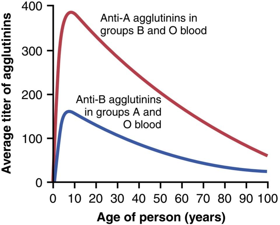
Figure 36-1. Average titers of anti-A and anti-B agglutinins in the plasma of people with different blood types.
شکل 36-1. میانگین تیتر آگلوتینینهای ضد A و آنتی B در پلاسمای افراد با گروههای خونی مختلف.
AGGLUTINATION PROCESS IN TRANSFUSION REACTIONS
When bloods are mismatched so that anti-A or anti-B plasma agglutinins are mixed with RBCs that contain A or B agglutinogens, respectively, the RBCs agglutinate as a result of the agglutinins attaching themselves to the RBCs. Because the agglutinins have two binding sites (IgG type) or ten binding sites (IgM type), a single agglutinin can attach to two or more RBCs at the same time, thereby causing the cells to be bound together by the agglutinin. This binding causes the cells to clump, which is the process of agglutination. Then these clumps plug small blood vessels throughout the circulatory system. During the ensuing hours to days, physical distortion of the cells or attack by phagocytic white blood cells destroys the membranes of the agglutinated cells, releasing hemoglobin into the plasma, called hemolysis of the RBCs.
فرآیند آگلوتیناسیون در واکنشهای انتقال خون
هنگامیکه خونها با هم تطبیق ندارند به طوری که آگلوتینینهای پلاسما ضد A یا ضد B با گلبولهای قرمز حاوی آگلوتینوژنهای A یا B به ترتیب مخلوط میشوند، گلبولهای قرمز در نتیجه چسبیدن آگلوتینینها به گلبولهای قرمز آگلوتینه میشوند. از آنجایی که آگلوتینینها دارای دو محل اتصال (نوع IgG) یا ده محل اتصال (نوع IgM) هستند، یک آگلوتینین منفرد میتواند به طور همزمان به دو یا چند گلبول قرمز متصل شود و در نتیجه باعث اتصال سلولها توسط آگلوتینین به یکدیگر شود. این اتصال باعث جمع شدن سلولها میشود که فرآیند آگلوتیناسیون است. سپس این تودهها رگهای خونی کوچک را در سراسر سیستم گردش خون مسدود میکنند. در طی چند ساعت تا چند روز بعد، اعوجاج فیزیکی سلولها یا حمله توسط گلبولهای سفید فاگوسیتیک، غشای سلولهای آگلوتینه شده را از بین میبرد و هموگلوبین را در پلاسما آزاد میکند که به آن همولیز گلبولهای قرمز میگویند.
Acute Hemolysis Occurs in Some Transfusion Reactions. Sometimes, when recipient and donor bloods are mismatched, immediate hemolysis of RBCs occurs in the circulating blood. In this case, the antibodies cause lysis of the RBCs by activating the complement system and forming a membrane attack complex (also called cytolytic complex) that inserts itself into the lipid bilayer of the cell membranes; this insertion creates membrane pores that are permeable to ions and causes osmotic lysis of the cells, as described in Chapter 35. Immediate intravascular hemolysis is far less common than agglutination followed by delayed hemolysis, because not only does there have to be a high titer of antibodies for lysis to occur, but also a different type of antibody seems to be required, mainly the IgM antibodies; these antibodies are called hemolysins.
همولیز حاد در برخی از واکنشهای انتقال خون رخ میدهد. گاهی اوقات، زمانی که خون گیرنده و اهداکننده با هم مطابقت ندارند، همولیز فوری گلبولهای قرمز در خون در گردش اتفاق میافتد. در این مورد، آنتیبادیها با فعال کردن سیستم کمپلمان و تشکیل یک کمپلکس حمله غشایی (که کمپلکس سیتولیتیک نیز نامیده میشود) باعث لیز گلبولهای قرمز میشوند که خود را به لایههای دوگانه لیپیدی غشای سلولی وارد میکند. این درج منافذ غشایی را ایجاد میکند که نسبت به یونها نفوذپذیر است و باعث لیز اسمزی سلولها میشود، همانطور که در فصل 35 توضیح داده شد. همولیز داخل عروقی فوری بسیار کمتر از آگلوتیناسیون و به دنبال آن همولیز تاخیری است، زیرا نه تنها باید تیتر بالایی از آنتی بادیها وجود داشته باشد، بلکه به نظر میرسد نوع متفاوتی از آنتی بادیها برای لیز رخ دهد. آنتی بادیها؛ این آنتی بادیها همولیزین نامیده میشوند.
BLOOD TYPING
Before giving a transfusion to a person, it is necessary to determine the blood type of the recipient and donor blood so that the bloods can be appropriately matched. This process is called blood typing and blood matching, and these procedures are performed in the following way. The RBCs are first separated from the plasma and diluted with saline solution. One portion is then mixed with anti-A agglutinin and another portion is mixed with anti-B agglutinin. After several minutes, the mixtures are observed under a microscope. If the RBCs have become clumped-that is, agglutinated then an antibody-antigen reaction has resulted.
گروه بندی خون
قبل از تزریق خون به فرد، باید گروه خونی گیرنده و اهداکننده را تعیین کرد تا بتوان خونها را به طور مناسب با هم تطبیق داد. به این فرآیند، گروه بندی خون و تطبیق خون میگویند و این اقدامات به روش زیر انجام میشود. گلبولهای قرمز ابتدا از پلاسما جدا شده و با محلول نمکی رقیق میشوند. سپس یک قسمت با آگلوتینین ضد A و قسمت دیگر با آگلوتینین آنتی B مخلوط میشود. پس از چند دقیقه، مخلوطها زیر میکروسکوپ مشاهده میشوند. اگر گلبولهای قرمز جمع شده باشند، یعنی آگلوتینه شده باشند، واکنش آنتی بادی-آنتی ژن ایجاد میشود.
Table 36-2 lists the presence (+) or absence (-) of agglutination of the four types of RBCs. Type O RBCs have no agglutinogens and therefore do not react with the anti-A or anti-B agglutinins. Type A blood has A agglutinogens and therefore agglutinates with anti-A agglutinins. Type B blood has B agglutinogens and agglutinates with anti-B agglutinins. Type AB blood has both A and B agglutinogens and agglutinates with both types of agglutinins.
جدول 36-2 وجود (+) یا عدم وجود (-) آگلوتیناسیون چهار نوع گلبول قرمز را فهرست میکند. گلبولهای قرمز نوع O فاقد آگلوتینوژن هستند و بنابراین با آگلوتینینهای ضد A یا ضد B واکنش نمیدهند. خون گروه A دارای آگلوتینوژن A است و بنابراین با آگلوتینینهای ضد A آگلوتین میشود. خون گروه B دارای آگلوتینوژنهای B است و با آگلوتینینهای ضد B آگلوتین میشود. خون گروه AB دارای هر دو آگلوتینوژن A و B است و با هر دو نوع آگلوتینین آگلوتین میشود.
Table 36-2 Blood Typing: Agglutination of Cells of Different Blood Types With Anti-A or Anti-B Agglutinins in the Sera
جدول 36-2 گروهبندی خون: آگلوتیناسیون سلولهای گروههای خونی مختلف با آگلوتینینهای Anti-A یا Anti-B در سرم

Rh BLOOD TYPES
Along with the O-A-B blood type system, the Rh blood type system is also important when transfusing blood. The major difference between the O-A-B system and Rh system is the following. In the O-A-B system, the plasma agglutinins responsible for causing transfusion reactions develop spontaneously, whereas in the Rh system, spontaneous agglutinins almost never occur. Instead, the person must first be massively exposed to an Rh antigen-such as by transfusion of blood containing the Rh antigen- before enough agglutinins to cause a significant transfusion reaction will develop.
گروههای خونی Rh
در کنار سیستم گروه خونی O-A-B، سیستم گروه خونی Rh نیز هنگام انتقال خون مهم است. تفاوت عمده بین سیستم O-A-B و سیستم Rh در موارد زیر است. در سیستم O-A-B، آگلوتینینهای پلاسما که مسئول ایجاد واکنشهای انتقال خون هستند، به طور خود به خود رشد میکنند، در حالی که در سیستم Rh، آگلوتینینهای خود به خود تقریباً هرگز رخ نمیدهند. درعوض، قبل از اینکه آگلوتینین کافی برای ایجاد یک واکنش انتقال خون قابل توجه ایجاد شود، ابتدا فرد باید به طور گسترده در معرض یک آنتی ژن Rh باشد، مانند تزریق خون حاوی آنتی ژن Rh.
Rh Antigens-Rh-Positive and Rh-Negative. There are six common types of Rh antigens, each of which is called an Rh factor. These types are designated C, D, E, c, d, and e. A person who has a C antigen does not have the c antigen, but the person missing the C antigen always has the c antigen. The same is true for the D-d and E-e antigens. Also, because of the manner of inheritance of these factors, each person has one of each of the three pairs of antigens.
آنتی ژنهای Rh-Rh-مثبت و Rh-منفی. شش نوع رایج از آنتی ژنهای Rh وجود دارد که به هر یک از آنها فاکتور Rh گفته میشود. این انواع C، D، E، C، D و E مشخص میشوند. فردی که آنتی ژن C دارد، آنتی ژن c ندارد، اما فردی که آنتی ژن C را از دست داده است، همیشه آنتی ژن c را دارد. همین امر در مورد آنتی ژنهای D-d و E-e نیز صادق است. همچنین به دلیل نحوه توارث این عوامل، هر فرد دارای یکی از هر سه جفت آنتی ژن است.
The type D antigen is widely prevalent in the population and is considerably more antigenic than the other Rh antigens. Anyone who has this type of antigen is said to be Rh positive, whereas a person who does not have type D antigen is said to be Rh negative. However, it must be noted that even in Rh-negative people, some of the other Rh antigens can still cause transfusion reactions, although the reactions are usually much milder.
آنتی ژن نوع D به طور گسترده ای در جمعیت شایع است و به طور قابل توجهی آنتی ژنیک تر از سایر آنتی ژنهای Rh است. گفته میشود هر فردی که این نوع آنتی ژن را داشته باشد Rh مثبت است، در حالی که به فردی که آنتی ژن نوع D ندارد گفته میشود Rh منفی است. با این حال، باید توجه داشت که حتی در افراد Rh منفی، برخی از آنتی ژنهای Rh دیگر همچنان میتوانند باعث واکنشهای انتقال خون شوند، اگرچه واکنشها معمولاً خفیف تر هستند.
About 85% of all whites are Rh positive, and 15% are Rh negative. In American blacks, the percentage of Rh- positives is about 95%, whereas in African blacks, it is nearly 100%. Over 95% of Native Americans and Asians living in China, Japan, and Korea are also Rh positive, and it is estimated that the worldwide frequencies of Rh- positive and Rh-negative blood types are 95% and 6%, respectively.
حدود 85 درصد از کل سفیدپوستان Rh مثبت و 15 درصد Rh منفی هستند. در سیاه پوستان آمریکایی، درصد Rh مثبت حدود 95٪ است، در حالی که در سیاه پوستان آفریقایی، این درصد نزدیک به 100٪ است. بیش از 95 درصد از بومیان آمریکا و آسیاییهای ساکن چین، ژاپن و کره نیز Rh مثبت هستند و تخمین زده میشود که فراوانی گروههای خونی Rh مثبت و Rh منفی در سراسر جهان به ترتیب 95 و 6 درصد است.
Rh IMMUNE RESPONSE
Formation of Anti-Rh Agglutinins. When RBCs containing Rh factor are injected into a person whose blood does not contain the Rh factor-that is, into an Rh- negative person—anti-Rh agglutinins develop slowly, reaching a maximum concentration of agglutinins about 2 to 4 months later. This immune response occurs to a much greater extent in some people than in others. With multiple exposures to the Rh factor, an Rh-negative per- son eventually becomes strongly sensitized to Rh factor.
پاسخ ایمنی Rh
تشکیل آگلوتینینهای ضد Rh. وقتی گلبولهای قرمز حاوی فاکتور Rh به فردی که خونش فاکتور Rh ندارد تزریق میشود، یعنی به یک فرد Rh منفی، آگلوتینینهای ضد Rh به آرامیرشد میکنند و حدود ۲ تا ۴ ماه بعد به حداکثر غلظت آگلوتینین میرسند. این پاسخ ایمنی در برخی افراد به میزان بسیار بیشتری نسبت به دیگران رخ میدهد. با قرار گرفتن در معرض چندین عامل Rh، یک فرد Rh منفی در نهایت به شدت به فاکتور Rh حساس میشود.
Characteristics of Rh Transfusion Reactions. If an Rh- negative person has never been exposed to Rh-positive blood, transfusion of Rh-positive blood into that person will likely cause no immediate reaction. However, anti-Rh antibodies can develop in sufficient quantities during the next 2 to 4 weeks to cause agglutination of the transfused cells that are still circulating in the blood. These cells are then hemolyzed by the tissue macrophage system. Thus, a delayed transfusion reaction occurs, although it is usually mild. On subsequent transfusion of Rh-positive blood into the same person, who is now already immunized against the Rh factor, the transfusion reaction is greatly enhanced and can be immediate and as severe as a trans- fusion reaction caused by mismatched type A or B blood.
ویژگیهای واکنشهای انتقال خون Rh. اگر یک فرد Rh منفی هرگز در معرض خون Rh مثبت قرار نگرفته باشد، تزریق خون Rh مثبت به آن فرد احتمالاً واکنش فوری ایجاد نخواهد کرد. با این حال، آنتی بادیهای ضد Rh میتوانند به مقدار کافی در طی 2 تا 4 هفته آینده ایجاد شوند تا باعث آگلوتیناسیون سلولهای تزریق شده شوند که هنوز در خون در گردش هستند. سپس این سلولها توسط سیستم ماکروفاژ بافت همولیز میشوند. بنابراین، واکنش تزریق خون با تاخیر رخ میدهد، اگرچه معمولا خفیف است. در انتقال بعدی خون Rh مثبت به همان فرد، که اکنون در برابر فاکتور Rh ایمن شده است، واکنش انتقال خون به شدت افزایش مییابد و میتواند فوری و به شدت واکنش انتقال خون ناشی از ناهماهنگی نوع A یا B باشد.
Erythroblastosis Fetalis (Hemolytic Disease of the Newborn)
Erythroblastosis fetalis is a disease of the fetus and new- born child characterized by agglutination and phagocytosis of the fetus’s RBCs. In most cases of erythroblastosis fetalis, the mother is Rh negative and the father is Rh positive. The baby has inherited the Rh-positive antigen from the father, and the mother develops anti-Rh agglutinins from exposure to the fetus’s Rh antigen. In turn, the mother’s agglutinins diffuse through the placenta into the fetus and cause RBC agglutination.
اریتروبلاستوز جنینی (بیماری همولیتیک نوزادان)
اریتروبلاستوز فتالیس یک بیماری جنین و نوزاد تازه متولد شده است که با آگلوتیناسیون و فاگوسیتوز گلبولهای قرمز جنین مشخص میشود. در اغلب موارد اریتروبلاستوز جنینی، مادر Rh منفی و پدر Rh مثبت است. نوزاد آنتی ژن Rh مثبت را از پدر به ارث برده است و مادر از قرار گرفتن در معرض آنتی ژن Rh جنین، آگلوتینینهای ضد Rh تولید میکند. به نوبه خود، آگلوتینینهای مادر از طریق جفت به جنین منتشر میشود و باعث آگلوتیناسیون RBC میشود.
Incidence of Erythroblastosis Fetalis. An Rh-negative mother having her first Rh-positive child usually does not develop sufficient anti-Rh agglutinins to cause any harm. However, about 3% of second Rh-positive babies exhibit some signs of erythroblastosis fetalis, about 10% of third babies exhibit the disease, and the incidence rises pro- gressively with subsequent pregnancies.
بروز اریتروبلاستوز جنینی. یک مادر Rh منفی که اولین فرزند خود را Rh مثبت دارد معمولاً آگلوتینین ضد Rh کافی برای ایجاد آسیب ایجاد نمیکند. با این حال، حدود 3٪ از نوزادان دوم Rh مثبت برخی از علائم اریتروبلاستوز جنینی را نشان میدهند، حدود 10٪ از نوزادان سوم این بیماری را نشان میدهند، و بروز با بارداریهای بعدی به تدریج افزایش مییابد.
Effect of Mother’s Antibodies on the Fetus. After anti- Rh antibodies have formed in the mother, they diffuse slowly through the placental membrane into the fetus’s blood. There they cause agglutination of the fetus’s blood. The agglutinated RBCs subsequently hemolyze, releasing hemoglobin into the blood. The fetus’s macrophages then convert the hemoglobin into bilirubin, which causes the baby’s skin to become yellow (jaundiced). The antibodies can also attack and damage other cells of the body.
تاثیر آنتی بادیهای مادر بر جنین. پس از تشکیل آنتی بادیهای ضد Rh در مادر، به آرامیاز طریق غشای جفت به خون جنین منتشر میشود. در آنجا باعث آگلوتیناسیون خون جنین میشوند. گلبولهای قرمز آگلوتینه شده متعاقبا همولیز میشوند و هموگلوبین را در خون آزاد میکنند. ماکروفاژهای جنین سپس هموگلوبین را به بیلی روبین تبدیل میکنند که باعث زرد شدن (زردی) پوست نوزاد میشود. آنتی بادیها همچنین میتوانند به سلولهای دیگر بدن حمله کرده و به آنها آسیب برسانند.
Clinical Picture of Erythroblastosis. The jaundiced, erythroblastotic newborn baby is usually anemic at birth, and the anti-Rh agglutinins from the mother usually circulate in the infant’s blood for another 1 to 2 months after birth, destroying more and more RBCs.
تصویر بالینی اریتروبلاستوز. نوزاد تازه متولد شده زردی و اریتروبلاستوتی معمولاً در بدو تولد کم خون است و آگلوتینینهای ضد Rh مادر معمولاً تا 1 تا 2 ماه دیگر پس از تولد در خون نوزاد گردش میکنند و گلبولهای قرمز هر چه بیشتر را از بین میبرند.
The hematopoietic tissues of the infant attempt to replace the hemolyzed RBCs. The liver and spleen become greatly enlarged and produce RBCs in the same manner that they normally do during the middle of gestation. Because of the rapid production of RBCs, many early forms of RBCs, including many nucleated blastic forms, are passed from the baby’s bone marrow into the circulatory system, and it is because of the presence of these nucleated blastic RBCs that the disease is called erythroblastosis fetalis.
بافتهای خون ساز نوزاد تلاش میکنند تا گلبولهای قرمز همولیز شده را جایگزین کنند. کبد و طحال به شدت بزرگ میشوند و گلبولهای قرمز تولید میکنند به همان روشی که معمولاً در اواسط بارداری انجام میدهند. به دلیل تولید سریع گلبولهای قرمز، بسیاری از اشکال اولیه گلبولهای قرمز، از جمله بسیاری از اشکال بلاستیک هسته دار، از مغز استخوان نوزاد به سیستم گردش خون منتقل میشوند و به دلیل وجود این گلبولهای قرمز بلاستیک هسته دار است که به این بیماری اریتروبلاستوز فتالیس میگویند.
Although the severe anemia of erythroblastosis fetalis is usually the cause of death, many children who barely survive the anemia exhibit permanent mental impairment or damage to motor areas of the brain because of precipitation of bilirubin in the neuronal cells, causing the destruction of many of these cells, a condition called kernicterus.
اگرچه کم خونی شدید اریتروبلاستوز جنینی معمولاً علت مرگ است، بسیاری از کودکانی که به سختی از کم خونی جان سالم به در میبرند، به دلیل رسوب بیلی روبین در سلولهای عصبی، اختلال ذهنی دائمییا آسیب به نواحی حرکتی مغز نشان میدهند که باعث تخریب بسیاری از این سلولها میشود، وضعیتی به نام کرنیکتروس.
Treatment of Neonates With Erythroblastosis Fetalis. One treatment for erythroblastosis fetalis is to replace the neonate’s blood with Rh-negative blood. The Rh-negative blood is infused over a period of 1.5 or more hours while the neonate’s own Rh-positive blood is being removed. This procedure may be repeated several times during the first few weeks of life, mainly to keep the bilirubin level low and thereby prevent kernicterus. By the time these transfused Rh-negative cells are replaced with the infant’s own Rh-positive cells, a process that requires 6 weeks or more, the anti-Rh agglutinins that had come from the mother will have been destroyed.
درمان نوزادان مبتلا به اریتروبلاستوز جنینی. یکی از درمانهای اریتروبلاستوز جنینی، جایگزینی خون نوزاد با خون Rh منفی است. خون Rh منفی در مدت 1.5 ساعت یا بیشتر در حالی که خون Rh مثبت خود نوزاد خارج میشود تزریق میشود. این روش ممکن است چندین بار در چند هفته اول زندگی تکرار شود، عمدتا برای پایین نگه داشتن سطح بیلی روبین و در نتیجه جلوگیری از کرنیکتروس. تا زمانی که این سلولهای Rh منفی با سلولهای Rh مثبت خود نوزاد جایگزین شوند، فرآیندی که به 6 هفته یا بیشتر نیاز دارد، آگلوتینینهای ضد Rh که از مادر وارد شده بود از بین خواهند رفت.
Prevention of Erythroblastosis Fetalis. The D antigen of the Rh blood group system is the primary culprit in causing immunization of an Rh-negative mother to an Rh-positive fetus. In the 1970s, a dramatic reduction in the incidence of erythroblastosis fetalis was achieved with the development of Rh immunoglobulin globin, an anti- D antibody that is administered to the expectant mother starting at 28 to 30 weeks of gestation. The anti-D anti- body is also administered to Rh-negative women who deliver Rh-positive babies to prevent sensitization of the mothers to the D antigen. This step greatly reduces the risk of developing large amounts of D antibodies during the second pregnancy.
پیشگیری از اریتروبلاستوز جنینی. آنتی ژن D سیستم گروه خونی Rh مقصر اصلی در ایجاد ایمن سازی یک مادر Rh منفی به جنین Rh مثبت است. در دهه 1970، کاهش چشمگیری در بروز اریتروبلاستوز جنینی با ایجاد گلوبین ایمونوگلوبولین Rh، یک آنتی بادی ضد D که از هفته 28 تا 30 بارداری برای مادر باردار تجویز میشود، حاصل شد. آنتی بادی ضد D همچنین برای زنان دارای Rh منفی که نوزادان Rh مثبت به دنیا میآورند برای جلوگیری از حساس شدن مادران به آنتی ژن D تجویز میشود. این مرحله تا حد زیادی خطر ایجاد مقادیر زیادی آنتی بادی D را در بارداری دوم کاهش میدهد.
The mechanism whereby Rh immunoglobulin globin prevents sensitization of the D antigen is not completely understood, but one effect of the anti-D antibody is to inhibit antigen-induced, B lymphocyte antibody production in the expectant mother. The administered anti-D antibody also attaches to D antigen sites on Rh-positive fetal RBCs that may cross the placenta and enter the circulation of the expectant mother, thereby interfering with the immune response to the D antigen.
مکانیسمیکه به موجب آن گلوبین ایمونوگلوبولین Rh از حساس شدن آنتی ژن D جلوگیری میکند کاملاً شناخته شده نیست، اما یکی از اثرات آنتی بادی ضد D این است که تولید آنتی بادی لنفوسیت B ناشی از آنتی ژن را در مادر باردار مهار میکند. آنتی بادی ضد D تجویز شده همچنین به محلهای آنتی ژن D روی گلبولهای قرمز Rh مثبت جنین میچسبد که ممکن است از جفت عبور کرده و وارد گردش خون مادر باردار شود و در نتیجه در پاسخ ایمنی به آنتیژن D اختلال ایجاد کند.
TRANSFUSION REACTIONS RESULTING FROM MISMATCHED BLOOD TYPES
If donor blood of one blood type is transfused into a recipient who has another blood type, a transfusion reaction is likely to occur in which the RBCs of the donor blood are agglutinated. It is rare that the transfused blood causes agglutination of the recipient’s cells, for the following reason. The plasma portion of the donor blood immediately becomes diluted by all the plasma of the recipient, thereby decreasing the titer of the infused agglutinins to a level usually too low to cause agglutination. However, the small amount of infused blood does not significantly dilute the agglutinins in the recipient’s plasma. Therefore, the recipient’s agglutinins can still agglutinate the mismatched donor cells.
واکنشهای انتقال خون ناشی از گروههای خونی نامتناسب
اگر خون اهداکننده یک گروه خونی به گیرندهای که گروه خونی دیگری دارد، تزریق شود، احتمالاً واکنش انتقال خون رخ میدهد که در آن گلبولهای قرمز خون اهداکننده آگلوتینه میشوند. به ندرت اتفاق میافتد که خون تزریق شده به دلایل زیر باعث آگلوتیناسیون سلولهای گیرنده شود. بخش پلاسمایی خون اهداکننده بلافاصله توسط تمام پلاسمای گیرنده رقیق میشود و در نتیجه تیتر آگلوتینین تزریق شده به سطحی که معمولاً برای ایجاد آگلوتیناسیون بسیار پایین است کاهش مییابد. با این حال، مقدار کمیخون تزریق شده به طور قابل توجهی آگلوتینینهای پلاسمای گیرنده را رقیق نمیکند. بنابراین، آگلوتینینهای گیرنده همچنان میتوانند سلولهای اهداکننده ناهماهنگ را آگلوتینه کنند.
As explained earlier, all transfusion reactions eventually cause immediate hemolysis resulting from hemolysins or later hemolysis resulting from phagocytosis of agglutinated cells. Hemoglobin released from the RBCs is then converted by the phagocytes into bilirubin and later excreted in the bile by the liver, as discussed in Chapter 71. The concentration of bilirubin in the body fluids often rises high enough to cause jaundice-that is, the person’s internal tissues and skin become colored with yellow bile pigment. However, if liver function is normal, the bile pigment will be excreted into the intestines by way of the liver bile, so jaundice usually does not appear in an adult unless more than 400 milliliters of blood are hemolyzed in less than a day.
همانطور که قبلا توضیح داده شد، تمام واکنشهای انتقال خون در نهایت باعث همولیز فوری ناشی از همولیزینها یا همولیز بعدی ناشی از فاگوسیتوز سلولهای آگلوتینه شده میشوند. هموگلوبین آزاد شده از گلبولهای قرمز سپس توسط فاگوسیتها به بیلی روبین تبدیل میشود و بعداً در صفرا توسط کبد دفع میشود، همانطور که در فصل 71 بحث شد. غلظت بیلی روبین در مایعات بدن اغلب به اندازه ای بالا میرود که باعث یرقان میشود، یعنی بافتهای داخلی و پوست فرد با صفرا زرد رنگ میشود. با این حال، اگر عملکرد کبد طبیعی باشد، رنگدانه صفرا از طریق صفرای کبد به رودهها دفع میشود، بنابراین زردی معمولاً در بزرگسالان ظاهر نمیشود مگر اینکه بیش از 400 میلی لیتر خون در کمتر از یک روز همولیز شود.
Acute Kidney Failure After Transfusion Reactions. One of the most lethal effects of transfusion reactions is kidney failure, which can begin within a few minutes to a few hours and continue until the person dies of acute renal failure.
نارسایی حاد کلیه پس از واکنشهای انتقال خون. یکی از کشنده ترین اثرات واکنشهای انتقال خون، نارسایی کلیه است که میتواند در عرض چند دقیقه تا چند ساعت شروع شود و تا زمان مرگ فرد بر اثر نارسایی حاد کلیه ادامه یابد.
The kidney shutdown seems to have three causes:
به نظر میرسد که تعطیلی کلیه سه علت دارد:
1. The antigen-antibody reaction of the transfusion reaction releases toxic substances from the hemolyzing blood that cause powerful renal vasoconstriction.
1. واکنش آنتی ژن-آنتی بادی در واکنش انتقال خون، مواد سمیرا از خون همولیز کننده آزاد میکند که باعث انقباض قوی عروق کلیه میشود.
2. Loss of circulating RBCs in the recipient, along with production of toxic substances from the hemolyzed cells and the immune reaction, often cause circulatory shock. The arterial blood pressure falls very low, and renal blood flow and urine output decrease.
2. از دست دادن گلبولهای قرمز در گردش در گیرنده، همراه با تولید مواد سمیاز سلولهای همولیز شده و واکنش ایمنی، اغلب باعث شوک گردش خون میشود. فشار خون شریانی بسیار پایین میآید و جریان خون کلیوی و خروجی ادرار کاهش مییابد.
3. If the total amount of free hemoglobin released into the circulating blood is greater than the quantity that can bind with haptoglobin (a plasma protein that binds small amounts of hemoglobin), much of the excess leaks through the glomerular membranes into the kidney tubules.
3. اگر مقدار کل هموگلوبین آزاد آزاد شده در خون در گردش بیشتر از مقداری باشد که میتواند باهاپتوگلوبین (پروتئین پلاسما که مقادیر کمیهموگلوبین را به هم متصل میکند) متصل شود، بسیاری از اضافی از طریق غشای گلومرولی به لولههای کلیه نشت میکند.
If this amount is still slight, it can be reabsorbed through the tubular epithelium into the blood and will cause no harm; if large, then only a small percentage is reabsorbed. Yet, water continues to be reabsorbed, causing the tubular hemoglobin concentration to rise so high that the hemoglobin precipitates and blocks many of the kidney tubules. Thus, renal vasoconstriction, circulatory shock, and renal tubular blockage together cause acute renal shutdown. If the shutdown is complete and fails to resolve, the patient dies within 7 to 12 days, as explained in Chapter 32, unless treated with an artificial kidney.
اگر این مقدار هنوز ناچیز باشد، میتواند از طریق اپیتلیوم لوله ای به خون بازجذب شود و هیچ آسیبی ایجاد نخواهد کرد. اگر بزرگ باشد، تنها درصد کمیدوباره جذب میشود. با این حال، آب همچنان به بازجذب ادامه میدهد و باعث میشود که غلظت هموگلوبین لولهای آنقدر بالا برود که هموگلوبین رسوب کند و بسیاری از لولههای کلیه را مسدود کند. بنابراین، انقباض عروق کلیه، شوک گردش خون و انسداد لولههای کلیوی با هم باعث خاموشی حاد کلیه میشوند. اگر خاموشی کامل باشد و برطرف نشود، بیمار طی 7 تا 12 روز میمیرد، همانطور که در فصل 32 توضیح داده شد، مگر اینکه با کلیه مصنوعی درمان شود.
TRANSPLANTATION OF TISSUES AND ORGANS
Most of the different antigens of RBCs that cause trans- fusion reactions are also widely present in other cells of the body, and each bodily tissue has its own additional complement of antigens. Consequently, foreign cells transplanted anywhere into the body of a recipient can produce an immune reaction. In other words, most recipients are just as able to resist invasion by foreign tissue cells as to resist invasion by foreign bacteria or RBCs.
پیوند بافتها و اندامها
بسیاری از آنتی ژنهای مختلف گلبولهای قرمز که باعث واکنشهای انتقال خون میشوند نیز به طور گسترده در سلولهای دیگر بدن وجود دارند و هر بافت بدن مکمل آنتی ژنهای اضافی خود را دارد. در نتیجه، سلولهای خارجی پیوند شده در هر نقطه از بدن گیرنده میتواند یک واکنش ایمنی ایجاد کند. به عبارت دیگر، اکثر گیرندهها به همان اندازه قادرند در برابر تهاجم سلولهای بافت خارجی مقاومت کنند که در برابر تهاجم باکتریهای خارجی یا گلبولهای قرمز مقاومت کنند.
Autografts, Isografts, Allografts, and Xenografts. A transplant of a tissue or whole organ from one part of the same animal to another part is called an autograft; from one identical twin to another, an isograft; from one person to another or from an animal to another animal of the same species, an allograft; and from a nonhuman animal to a human or from an animal of one species to one of another species, a xenograft.
اتوگرافت، ایزوگرافت، آلوگرافت و زنوگرافت. پیوند یک بافت یا کل عضو از یک قسمت از همان حیوان به قسمت دیگر را اتوگرافت میگویند. از یک دوقلو یکسان به دیگری، یک پیوند همسان. از یک شخص به فرد دیگر یا از حیوانی به حیوانی دیگر از همان گونه، آلوگرافت. و از یک حیوان غیر انسانی به یک انسان یا از یک حیوان از یک گونه به یکی از گونههای دیگر، پیوند زنو.
Transplantation of Cellular Tissues. In the case of autografts and isografts, cells in the transplant contain virtually the same types of antigens as in the tissues of the recipient and will almost always continue to live normally and indefinitely if an adequate blood supply is provided. At the other extreme, immune reactions almost always occur in xenografts, causing death of the cells in the graft within 1 day to 5 weeks after transplantation unless some specific therapy is used to prevent the immune reactions.
پیوند بافتهای سلولی. در مورد اتوگرافتها و ایزوگرافتها، سلولهای پیوند تقریباً حاوی همان انواع آنتی ژنهایی هستند که در بافت گیرنده وجود دارد و تقریباً همیشه در صورت تامین خون کافی به زندگی عادی و نامحدود ادامه میدهند. از طرف دیگر، واکنشهای ایمنی تقریباً همیشه در پیوند زنوگرافت رخ میدهد و باعث مرگ سلولهای پیوند در عرض 1 روز تا 5 هفته پس از پیوند میشود، مگر اینکه از درمان خاصی برای جلوگیری از واکنشهای ایمنی استفاده شود.
Some of the different cellular tissues and organs that have been transplanted from one person to another as allografts, either experimentally or for therapeutic purposes, are skin, kidney, heart, liver, glandular tissue, bone marrow, and lung. With proper matching of tissues between persons, many kidney allografts have been successful for at least 5 to 15 years and allograft liver and heart transplants for 1 to 15 years.
برخی از بافتها و اندامهای سلولی مختلف که بهعنوان آلوگرافت از فردی به فرد دیگر پیوند شدهاند، چه بهصورت تجربی یا برای اهداف درمانی، عبارتند از: پوست، کلیه، قلب، کبد، بافت غدد، مغز استخوان و ریه. با تطبیق مناسب بافتها بین افراد، بسیاری از آلوگرافتهای کلیه برای حداقل 5 تا 15 سال و پیوند کبد و قلب آلوگرافت برای 1 تا 15 سال موفقیت آمیز بوده اند.
ATTEMPTS TO OVERCOME IMMUNE REACTIONS IN TRANSPLANTED TISSUE
Because of the extreme potential importance of transplanting certain tissues and organs, serious attempts have been made to prevent antigen-antibody reactions associated with transplantation. The following specific procedures have met with some degrees of clinical or experimental success.
تلاش برای غلبه بر واکنشهای ایمنی در بافت پیوندی
به دلیل اهمیت بالقوه فوق العاده پیوند بافتها و اندامهای خاص، تلاشهای جدی برای جلوگیری از واکنشهای آنتی ژن-آنتی بادی مرتبط با پیوند صورت گرفته است. رویههای خاص زیر با درجاتی از موفقیت بالینی یا تجربی مواجه شده اند.
Tissue Typing Human Leukocyte Antigen Complex of Antigens. The most important antigens for causing graft rejection are a complex called human leukocyte antigen (HLA) antigens. Six of these antigens are present on the tissue cell membranes of each person, but there are about 150 different HLA antigens to choose from, representing more than 1 trillion possible combinations. Consequently, it is virtually impossible for two persons, except in the case of identical twins, to have the same six HLA antigens. Development of significant immunity against any of these antigens can cause graft rejection.
تایپ بافتی مجموعه آنتی ژنهای لکوسیت انسانی. مهمترین آنتی ژن برای ایجاد رد پیوند، مجموعه ای به نام آنتی ژنهای آنتی ژن لکوسیت انسانی (HLA) است. شش تا از این آنتی ژنها روی غشای سلولی بافتی هر فرد وجود دارد، اما حدود 150 آنتی ژن HLA مختلف برای انتخاب وجود دارد که نشان دهنده بیش از 1 تریلیون ترکیب ممکن است. در نتیجه، برای دو نفر، به جز در مورد دوقلوهای همسان، تقریباً غیرممکن است که همان شش آنتی ژن HLA را داشته باشند. ایجاد ایمنی قابل توجه در برابر هر یک از این آنتی ژنها میتواند باعث رد پیوند شود.
The HLA antigens are on the white blood cells, as well as on the tissue cells. Therefore, tissue typing for these antigens is done on the membranes of lymphocytes that have been separated from the person’s blood. The lymphocytes are mixed with appropriate antisera and complement; after incubation, the cells are tested for membrane damage, usually by determining the rate of transmembrane uptake by the lymphocytic cells of a special dye.
آنتی ژنهای HLA روی گلبولهای سفید و همچنین روی سلولهای بافتی قرار دارند. بنابراین تایپ بافتی برای این آنتی ژنها بر روی غشای لنفوسیتهایی که از خون فرد جدا شده اند انجام میشود. لنفوسیتها با آنتی سرم و مکمل مناسب مخلوط میشوند. پس از انکوباسیون، سلولها برای آسیب غشایی آزمایش میشوند، معمولاً با تعیین میزان جذب غشایی توسط سلولهای لنفوسیتی یک رنگ خاص.
Some of the HLA antigens are not severely antigenic. Therefore, a precise match of some antigens between donor and recipient is not always essential to allow allograft acceptance. By using a more advanced method of genetic testing and obtaining the best possible match between donor and recipient, the grafting procedure has become far less hazardous. The best success has been with tissue type matches between siblings and between parent and child. The match in identical twins is exact, so transplants between identical twins are almost never rejected because of an immune reaction.
برخی از آنتی ژنهای HLA به شدت آنتی ژن نیستند. بنابراین، تطابق دقیق برخی از آنتی ژنها بین دهنده و گیرنده همیشه ضروری نیست تا امکان پذیرش آلوگرافت فراهم شود. با استفاده از روش پیشرفته تر آزمایش ژنتیکی و به دست آوردن بهترین تطابق ممکن بین اهدا کننده و گیرنده، روش پیوند بسیار کم خطرتر شده است. بهترین موفقیت با تطابق نوع بافت بین خواهر و برادر و بین والدین و فرزند بوده است. تطابق در دوقلوهای همسان دقیق است، بنابراین پیوند بین دوقلوهای همسان تقریباً هرگز به دلیل واکنش ایمنی رد نمیشود.
Prevention of Graft Rejection by Suppressing the Immune System
If the immune system were completely suppressed, graft rejection would not occur. In fact, in a person who has serious depression of the immune system, grafts can be successful without the use of significant therapy to prevent rejection. However, in the person with a healthy immune system, even with the best possible tissue typing, allografts seldom resist rejection for more than a few days or weeks without the use of specific therapy to suppress the immune system. Furthermore, because the T cells are mainly the portion of the immune system important for killing grafted cells, their suppression is much more important than suppression of plasma antibodies. Some of the therapeutic agents that have been used for this purpose include the following:
پیشگیری از رد پیوند با سرکوب سیستم ایمنی
اگر سیستم ایمنی به طور کامل سرکوب میشد، رد پیوند رخ نمیداد. در واقع، در فردی که دچار افسردگی شدید سیستم ایمنی است، پیوندها میتوانند بدون استفاده از درمان قابل توجه برای جلوگیری از رد شدن، موفقیت آمیز باشند. با این حال، در افرادی که دارای سیستم ایمنی سالم هستند، حتی با بهترین نوع ممکن بافتی، آلوگرافتها به ندرت در برابر رد شدن بیش از چند روز یا هفته بدون استفاده از درمان خاصی برای سرکوب سیستم ایمنی مقاومت میکنند. علاوه بر این، از آنجایی که سلولهای T عمدتاً بخشی از سیستم ایمنی هستند که برای کشتن سلولهای پیوندی مهم هستند، سرکوب آنها بسیار مهمتر از سرکوب آنتیبادیهای پلاسما است. برخی از عوامل درمانی که برای این منظور استفاده شده اند عبارتند از:
1. Glucocorticoid hormones from adrenal cortex glands (or drugs with glucocorticoid-like activity). These drugs inhibit genes that code for several cytokines, especially interleukin-2 (IL-2). IL-2 is an essential factor that induces T-cell proliferation and antibody formation.
1. هورمونهای گلوکوکورتیکوئیدی از غدد قشر آدرنال (یا داروهایی با فعالیت شبه گلوکوکورتیکوئید). این داروها ژنهایی را که برای چندین سایتوکین کد میکنند، به ویژه اینترلوکین-2 (IL-2) مهار میکنند. IL-2 یک عامل ضروری است که باعث تکثیر سلولهای T و تشکیل آنتی بادی میشود.
2. Various drugs that have a toxic effect on the lymphoid system and therefore block formation of antibodies and T cells, especially the drug azathioprine.
2. داروهای مختلفی که اثر سمیروی سیستم لنفاوی دارند و در نتیجه تشکیل آنتی بادی و سلولهای T را مسدود میکنند، به ویژه داروی آزاتیوپرین.
3. Cyclosporine and tacrolimus, which inhibit formation of T-helper cells and, therefore, are especially efficacious in blocking the T-cell rejection reaction. These agents have proven to be highly valuable drugs because they do not depress some other portions of the immune system.
3. سیکلوسپورین و تاکرولیموس، که از تشکیل سلولهای T-helper جلوگیری میکنند و بنابراین، به ویژه در مسدود کردن واکنش رد سلول T موثر هستند. ثابت شده است که این عوامل داروهای بسیار ارزشمندی هستند زیرا برخی از بخشهای دیگر سیستم ایمنی را سرکوب نمیکنند.
4. Immunosuppressive antibody therapy. including specific antilymphocyte or IL-2 receptor antibodies.
4. درمان آنتی بادی سرکوب کننده سیستم ایمنی. از جمله آنتی بادیهای خاص آنتی لنفوسیت یا گیرنده IL-2.
Use of these agents often leaves the person unprotected from infectious disease; therefore, sometimes bacterial and viral infections become rampant. In addition, the incidence of cancer is several times greater in an immunosuppressed person, presumably because the immune system is important in destroying many early cancer cells before they can begin to proliferate.
استفاده از این عوامل اغلب فرد را از بیماریهای عفونی محافظت نمیکند. بنابراین، گاهی اوقات عفونتهای باکتریایی و ویروسی بیداد میکنند. علاوه بر این، بروز سرطان در یک فرد دچار سرکوب سیستم ایمنی چندین برابر بیشتر است، احتمالاً به این دلیل که سیستم ایمنی در از بین بردن بسیاری از سلولهای سرطانی اولیه قبل از شروع تکثیر مهم است.
Transplantation of living tissues in people has been successful mainly because of the development of drugs that suppress the responses of the immune system. With the introduction of improved immunosuppressive agents, successful organ transplantation has become much more common. The current approach to immunosuppressive
therapy attempts to balance acceptable rates of rejection with moderation of the adverse effects of immunosuppressive drugs.
پیوند بافتهای زنده در افراد عمدتاً به دلیل توسعه داروهایی که پاسخهای سیستم ایمنی را سرکوب میکنند موفقیتآمیز بوده است. با معرفی عوامل بهبود یافته سرکوب کننده سیستم ایمنی، پیوند عضو موفق بسیار رایج تر شده است. رویکرد فعلی به سرکوب کننده سیستم ایمنی درمان تلاش میکند تا میزان قابل قبول پس زدن را با تعدیل اثرات نامطلوب داروهای سرکوب کننده سیستم ایمنی متعادل کند.
کلیک کنید «Bibliography: فهرست کتب مربوطه»
Branch DR: Anti-A and anti-B: what are they and where do they come from? Transfusion 55 Suppl 2:574, 2015.
Burton NM, Anstee DJ: Structure, function and significance of Rh pro- teins in red cells. Curr Opin Hematol 15:625, 2008. Dierickx D,
Habermann TM: Post-transplantation lymphoproliferative disorders in adults. N Engl J Med 378:549, 2018..
Ezekian B, Schroder PM, Freischlag K, et al: Contemporary strate- gies and barriers to transplantation tolerance. Transplantation 102:1213, 2018.
Flegel WA: Pathogenesis and mechanisms of antibody-mediated he- molysis. Transfusion 55 Suppl 2:547, 2015.
Kramer CSM, Israeli M, Mulder A, et al: The long and winding road towards epitope matching in clinical transplantation. Transpl Int 32:16, 2019.
Loupy A, Lefaucheur C: Antibody-mediated rejection of solid-organ allografts. N Engl J Med 379:1150, 2018.
MacDonald KP, Blazar BR, Hill GR: Cytokine mediators of chronic graft-versus-host disease. J Clin Invest 127:2452, 2017.
Montgomery RA, Tatapudi VS, Leffell MS, Zachary AA: HLA in trans- plantation. Nat Rev Nephrol 14:558, 2018.
Watchko JF, Tiribelli C: Bilirubin-induced neurologic damage-mech- anisms and management approaches. N Engl J Med 369:2021, 2013.
Webb J, Delaney M: Red blood cell alloimmunization in the pregnant patient. Transfus Med Rev 32:213, 2018.
Westhoff CM: Blood group genotyping. Blood. 133:1814, 2019 Westhoff CM: The structure and function of the Rh antigen complex. Semin Hematol 44:42, 2007.
Yazer MH, Seheult J, Kleinman S, Sloan SR, Spinella PC: Who’s afraid of incompatible plasma? A balanced approach to the safe trans- fusion of blood products containing ABO-incompatible plasma. Transfusion 58:532, 2018.
🚀 با ما همراه شوید!
تازهترین مطالب و آموزشهای مغز و اعصاب را از دست ندهید. با فالو کردن کانال تلگرام آیندهنگاران مغز، از ما حمایت کنید!


