
فیزیولوژی پزشکی گایتون و هال؛ مقاومت بدن در برابر عفونت: II. ایمنی و آلرژی


دعای مطالعه [ نمایش ]
بِسْمِ الله الرَّحْمنِ الرَّحیمِ
اَللّهُمَّ اَخْرِجْنى مِنْ ظُلُماتِ الْوَهْمِ
خدايا مرا بيرون آور از تاريكىهاى وهم،
وَ اَكْرِمْنى بِنُورِ الْفَهْمِ
و به نور فهم گرامى ام بدار،
اَللّهُمَّ افْتَحْ عَلَيْنا اَبْوابَ رَحْمَتِكَ
خدايا درهاى رحمتت را به روى ما بگشا،
وَانْشُرْ عَلَيْنا خَزائِنَ عُلُومِكَ بِرَحْمَتِكَ يا اَرْحَمَ الرّاحِمينَ
و خزانههاى علومت را بر ما باز كن به امید رحمتت اى مهربانترين مهربانان.
کتاب «فیزیولوژی پزشکی گایتون و هال» بهعنوان یکی از جامعترین و معتبرترین منابع در حوزه علوم پزشکی، همچنان مرجع کلیدی برای درک عملکرد پیچیده بدن انسان است. این اثر با تکیه بر تازهترین پژوهشها و توضیحات دقیق از سازوکارهای فیزیولوژیک، پلی میان علوم پایه پزشکی و کاربردهای بالینی ایجاد میکند و نقشی بیبدیل در آموزش، پژوهش و ارتقای دانش سلامت ایفا مینماید.
ترجمه دقیق و علمیاین شاهکار توسط برند علمیآیندهنگاران مغز به مدیریت داریوش طاهری، دسترسی فارسیزبانان به مرزهای نوین دانش فیزیولوژی را ممکن ساخته و رسالتی علمیبرای ارتقای آموزش پزشکی، فهم عمیقتر سازوکارهای بدن و توسعه روشهای نوین در حوزه سلامت فراهم آورده است.
» کتاب فیزیولوژی پزشکی گایتون وهال
» » فصل ۳۵: مقاومت بدن در برابر عفونت: II. ایمنی و آلرژی
در حال ویرایش
» Guyton and Hall Textbook of Medical Physiology
»» CHAPTER 35: Resistance of the Body to Infection: II. Immunity and Allergy

The human body has the ability to resist almost all types of organisms or toxins that tend to damage the tissues and organs. This capability is called immunity. Much of the immunity is acquired immunity that does not develop until after the body is first attacked by a bacterium, virus, or toxin; often, weeks or months are required for the immunity to develop. An additional element of immunity that results from general processes, rather than from processes directed at specific disease organisms, is called innate immunity, which includes the following aspects:
بدن انسان این توانایی را دارد که تقریباً در برابر انواع موجودات یا سمومیکه تمایل به آسیب به بافتها و اندامها دارند، مقاومت کند. به این قابلیت ایمنی میگویند. بیشتر ایمنی، مصونیت اکتسابی است که تا زمانی که بدن برای اولین بار توسط یک باکتری، ویروس یا سم مورد حمله قرار نگیرد، ایجاد نمیشود. اغلب، هفتهها یا ماهها برای ایجاد ایمنی لازم است. یک عنصر اضافی از ایمنی که ناشی از فرآیندهای عمومیاست، نه از فرآیندهای معطوف به ارگانیسمهای بیماری خاص، مصونیت ذاتی نامیده میشود که شامل جنبههای زیر است:
1. Phagocytosis of bacteria and other invaders by white blood cells and cells of the tissue macrophage system, as described in Chapter 34
1. فاگوسیتوز باکتریها و سایر مهاجمان توسط گلبولهای سفید خون و سلولهای سیستم ماکروفاژ بافت، همانطور که در فصل 34 توضیح داده شد.
2. Destruction of swallowed organisms by the acid secretions of the stomach and the digestive enzymes
2. تخریب ارگانیسمهای بلعیده شده توسط ترشحات اسیدی معده و آنزیمهای گوارشی
3. Resistance of the skin to invasion by organisms
3. مقاومت پوست در برابر هجوم ارگانیسمها
4. Presence in the blood of certain chemicals and cells that attach to foreign organisms or toxins and destroy them. Some of these are: (1) lysozyme, a mucolytic polysaccharide that attacks bacteria and causes them to dissolute; (2) basic polypeptides, which react with and inactivate certain types of gram-positive bacteria; (3) the complement complex, described later, a system of about 20 proteins that can be activated in various ways to destroy bacteria; and (4) natural killer lymphocytes that can recognize and destroy foreign cells, tumor cells, and even some infected cells.
4. وجود برخی مواد شیمیایی و سلولی در خون که به ارگانیسمها یا سموم خارجی چسبیده و آنها را از بین میبرد. برخی از این موارد عبارتند از: (1) لیزوزیم، یک پلی ساکارید موکولیتیک که به باکتریها حمله میکند و باعث انحلال آنها میشود. (2) پلی پپتیدهای اساسی که با انواع خاصی از باکتریهای گرم مثبت واکنش نشان میدهند و آنها را غیرفعال میکنند. (3) مجتمع کمپلمان که بعداً توضیح داده شد، سیستمیمتشکل از 20 پروتئین است که میتواند به روشهای مختلف برای از بین بردن باکتریها فعال شود. و (4) لنفوسیتهای کشنده طبیعی که میتوانند سلولهای خارجی، سلولهای تومور و حتی برخی از سلولهای آلوده را شناسایی کرده و از بین ببرند.
This innate immunity makes the human body resistant to diseases such as some paralytic viral infections of animals, hog cholera, cattle plague, and distemper-a viral disease that kills a large percentage of dogs that become afflicted with it. Likewise, many animals are resistant or even immune to many human diseases, such as polio- myelitis, mumps, human cholera, measles, and syphilis, which are very damaging or even lethal to humans.
این مصونیت ذاتی بدن انسان را در برابر بیماریهایی مانند برخی از عفونتهای ویروسی فلج کننده حیوانات، وبا گراز، طاعون گاوها و دیستمپر – یک بیماری ویروسی که درصد زیادی از سگهای مبتلا به آن را میکشد، مقاوم میکند. به همین ترتیب، بسیاری از حیوانات در برابر بسیاری از بیماریهای انسانی مانند فلج اطفال، اوریون، وبا، سرخک و سیفلیس که برای انسان بسیار مضر و یا حتی کشنده هستند، مقاوم و یا حتی مصون هستند.
ACQUIRED (ADAPTIVE) IMMUNITY
In addition to its generalized innate immunity, the human body has the ability to develop extremely powerful specific immunity again s individual invading agents such as lethal bacteria, viruses, toxins, and even foreign tissues from other animals. This ability is called acquired or adaptive immunity. Acquired immunity is caused by a special immune system that forms antibodies and/or activated lymphocytes that attack and destroy the specific invading organism or toxin.
مصونیت اکتسابی (تطبیقی)
علاوه بر مصونیت ذاتی تعمیم یافته، بدن انسان این توانایی را دارد که مجدداً برای عوامل مهاجم فردی مانند باکتریهای کشنده، ویروسها، سموم و حتی بافتهای خارجی دیگر حیوانات، ایمنی بسیار قوی ایجاد کند. این توانایی را مصونیت اکتسابی یا تطبیقی میگویند. ایمنی اکتسابی توسط یک سیستم ایمنی خاص ایجاد میشود که آنتیبادیها و/یا لنفوسیتهای فعال را تشکیل میدهد که به ارگانیسم یا سم مهاجم خاص حمله میکنند و آن را از بین میبرند.
Acquired immunity can often bestow an extreme degree of protection. For example, certain toxins, such as the paralytic botulinum toxin or the tetanizing toxin of tetanus, can be protected against in doses as high as 100,000 times the amount that would be lethal without immunity. It is for this reason that the treatment process known as immunization is so important in protecting people against disease and against toxins, as explained later in this chapter.
مصونیت اکتسابی اغلب میتواند درجه بالایی از محافظت را ایجاد کند. به عنوان مثال، سموم خاصی مانند سم بوتولینوم فلج کننده یا سم کزاز کزاز را میتوان در دوزهای 100000 برابر بیشتر از مقداری که بدون ایمنی کشنده است محافظت کرد. به همین دلیل است که فرآیند درمانی معروف به ایمن سازی در محافظت از افراد در برابر بیماریها و سموم بسیار مهم است، همانطور که در ادامه این فصل توضیح داده شد.
BASIC TYPES OF ACQUIRED IMMUNITY- HUMORAL AND CELL-MEDIATED
Two basic but closely allied types of acquired immunity occur in the body. In one of these, the body develops circulating antibodies, which are globulin molecules in the blood plasma capable of attacking the invading agent. This type of immunity is called humoral immunity or B-cell immunity because B lymphocytes produce the antibodies. The second type of acquired immunity is achieved through formation of large numbers of activated T lymphocytes, which are specifically crafted in the lymph nodes to destroy the foreign agent. This type of immunity is called cell-mediated immunity or T-cell immunity because the activated lymphocytes are T lymphocytes. Both the antibodies and activated lymphocytes are formed in the lymphoid tissues of the body.
انواع اساسی ایمنی اکتسابی – طنز و واسطه سلولی
دو نوع اساسی اما نزدیک به هم از ایمنی اکتسابی در بدن رخ میدهد. در یکی از این موارد، بدن آنتی بادیهای در گردش تولید میکند که مولکولهای گلوبولین در پلاسمای خون هستند که قادر به حمله به عامل مهاجم هستند. این نوع ایمنی را ایمنی هومورال یا ایمنی سلول B مینامند زیرا لنفوسیتهای B آنتی بادیها را تولید میکنند. نوع دوم ایمنی اکتسابی از طریق تشکیل تعداد زیادی لنفوسیت T فعال به دست میآید که به طور خاص در غدد لنفاوی ساخته میشوند تا عامل خارجی را از بین ببرند. این نوع ایمنی را ایمنی سلولی یا ایمنی سلول T مینامند زیرا لنفوسیتهای فعال شده لنفوسیتهای T هستند. هم آنتی بادیها و هم لنفوسیتهای فعال شده در بافتهای لنفوئیدی بدن تشکیل میشوند.
BOTH TYPES OF ACQUIRED IMMUNITY ARE INITIATED BY ANTIGENS
Because acquired immunity does not develop until after invasion by a foreign organism or toxin, it is clear that the body must have some mechanism for recognizing this invasion. Each toxin or organism almost always contains one or more specific chemical compounds in its makeup that is (are) different from all other compounds. In general, these are proteins or large polysaccharides that initiate the acquired immunity; these substances are called antigens (antibody generators).
هر دو نوع ایمنی اکتسابی توسط آنتی ژنها ایجاد میشوند
از آنجایی که ایمنی اکتسابی تا پس از تهاجم یک ارگانیسم یا سم خارجی ایجاد نمیشود، واضح است که بدن باید مکانیسمیبرای تشخیص این تهاجم داشته باشد. هر سم یا ارگانیسم تقریباً همیشه حاوی یک یا چند ترکیب شیمیایی خاص در ترکیب خود است که با سایر ترکیبات متفاوت است. به طور کلی، اینها پروتئینها یا پلی ساکاریدهای بزرگ هستند که مصونیت اکتسابی را آغاز میکنند. این مواد را آنتی ژن (مولد آنتی بادی) مینامند.
For a substance to be antigenic, it usually must have a high molecular weight of 8000 or more. Furthermore, the process of antigenicity usually depends on regularly recurring molecular groups, called epitopes, on the surface of the large molecule. This factor also explains why proteins and large polysaccharides are almost always antigenic because both these substances have this stereo- chemical characteristic.
برای اینکه یک ماده آنتی ژنی باشد، معمولاً باید دارای وزن مولکولی بالای 8000 یا بیشتر باشد. علاوه بر این، فرآیند آنتی ژنی معمولاً به گروههای مولکولی تکرارشونده، به نام اپی توپ، روی سطح مولکول بزرگ بستگی دارد. این عامل همچنین توضیح میدهد که چرا پروتئینها و پلی ساکاریدهای بزرگ تقریباً همیشه آنتی ژن هستند زیرا هر دو این مواد دارای این ویژگی استریو شیمیایی هستند.
LYMPHOCYTES ARE RESPONSIBLE FOR ACQUIRED IMMUNITY
Acquired immunity is the product of the body’s lymphocytes. In people who have a genetic lack of lymphocytes or whose lymphocytes have been destroyed by radiation or chemicals, no acquired immunity can develop. Within days after birth, such a person dies of fulminating bacterial infection unless he or she is treated by heroic measures. Therefore, it is clear that lymphocytes are essential to the survival of humans.
لنفوسیتها مسئول ایمنی اکتسابی هستند
ایمنی اکتسابی محصول لنفوسیتهای بدن است. در افرادی که کمبود ژنتیکی لنفوسیتها دارند یا لنفوسیتهای آنها در اثر تشعشعات یا مواد شیمیایی از بین رفته است، هیچ ایمنی اکتسابی نمیتواند ایجاد شود. در عرض چند روز پس از تولد، چنین فردی بر اثر عفونت باکتریایی شعله ور میمیرد، مگر اینکه با اقدامات قهرمانانه درمان شود. بنابراین، واضح است که لنفوسیتها برای بقای انسان ضروری هستند.
Lymphocytes are located most extensively in the lymph nodes, but are also found in special lymphoid tissues such as the spleen, submucosal areas of the gastrointestinal tract, thymus, and bone marrow. The lymphoid tissue is distributed advantageously in the body to intercept invading organisms or toxins before they can spread too widely.
لنفوسیتها بیشتر در غدد لنفاوی قرار دارند، اما در بافتهای لنفاوی خاص مانند طحال، نواحی زیر مخاطی دستگاه گوارش، تیموس و مغز استخوان نیز یافت میشوند. بافت لنفاوی به نحو مطلوبی در بدن توزیع میشود تا ارگانیسمها یا سموم مهاجم را قبل از انتشار بیش از حد گسترده رهگیری کند.
In most cases, the invading agent first enters the tissue fluids and then is carried by lymph vessels to the lymph node or other lymphoid tissue. For example, the lymphoid tissue of the gastrointestinal walls is exposed immediately to antigens invading from the gut. The lymphoid tissue of the throat and pharynx (including the tonsils and adenoids) is well located to intercept antigens that enter by way of the upper respiratory tract. The lymphoid tissue in the lymph nodes is exposed to antigens that invade the peripheral tissues of the body, and the lymphoid tissue of the spleen, thymus, and bone marrow plays the specific role of intercepting antigenic agents that have succeeded in reaching the circulating blood.
در بیشتر موارد، عامل مهاجم ابتدا وارد مایعات بافتی میشود و سپس توسط عروق لنفاوی به غدد لنفاوی یا سایر بافتهای لنفاوی منتقل میشود. به عنوان مثال، بافت لنفاوی دیوارههای دستگاه گوارش بلافاصله در معرض آنتی ژنهای مهاجم از روده قرار میگیرد. بافت لنفاوی گلو و حلق (شامل لوزهها و آدنوئیدها) به خوبی برای رهگیری آنتی ژنهایی که از طریق دستگاه تنفسی فوقانی وارد میشوند قرار دارد. بافت لنفاوی در غدد لنفاوی در معرض آنتی ژنهایی قرار میگیرد که به بافتهای محیطی بدن حمله میکنند و بافت لنفاوی طحال، تیموس و مغز استخوان نقش خاص رهگیری عوامل آنتی ژنی را ایفا میکند که موفق به رسیدن به خون در گردش شده اند.
T and B Lymphocytes Promote Cell-Mediated and Humoral Immunity. Although most lymphocytes in normal lymphoid tissue look alike when studied under a microscope, these cells are distinctly divided into two major populations. One of the populations, the T lymphocytes, is responsible for forming the activated lymphocytes that provide cell-mediated immunity, and the other population, the B lymphocytes, is responsible for forming anti- bodies that provide humoral immunity.
لنفوسیتهای T و B ایمنی سلولی و هومورال را تقویت میکنند. اگر چه بیشتر لنفوسیتها در بافت لنفوئیدی طبیعی وقتی زیر میکروسکوپ مطالعه میشوند شبیه به هم هستند، اما این سلولها به طور مشخص به دو جمعیت عمده تقسیم میشوند. یکی از جمعیتها، لنفوسیتهای T، مسئول تشکیل لنفوسیتهای فعال شده است که ایمنی سلولی را ایجاد میکند، و جمعیت دیگر، لنفوسیتهای B، مسئول تشکیل آنتی بادیهایی هستند که ایمنی هومورال را ایجاد میکنند.
Both types of lymphocytes are derived originally in the embryo from multipotent hematopoietic stem cells that form common lymphoid progenitor cells as one of their most important offspring as they differentiate. Almost all the lymphocytes that are formed eventually end up in the lymphoid tissue, but before doing so, they are further differentiated or preprocessed in the following ways.
هر دو نوع لنفوسیت در اصل در جنین از سلولهای بنیادی خونساز چند توانی مشتق میشوند که سلولهای پیش ساز لنفوئیدی مشترک را به عنوان یکی از مهم ترین فرزندان خود در هنگام تمایز تشکیل میدهند. تقریباً تمام لنفوسیتهایی که تشکیل میشوند در نهایت به بافت لنفوئیدی ختم میشوند، اما قبل از انجام این کار، به روشهای زیر بیشتر تمایز یا پیش پردازش میشوند.
The lymphoid progenitor cells that are eventually destined to form activated T lymphocytes first migrate to and are preprocessed in the thymus gland; thus, they are called T lymphocytes to designate the role of the thymus. They are responsible for cell-mediated immunity.
سلولهای پیش ساز لنفوئیدی که در نهایت برای تشکیل لنفوسیتهای T فعال ساخته میشوند، ابتدا به غده تیموس مهاجرت میکنند و در آن پیش پردازش میشوند. بنابراین، آنها لنفوسیت T نامیده میشوند تا نقش تیموس را مشخص کنند. آنها مسئول ایمنی سلولی هستند.
The other population of lymphocytes-the B lymphocytes that are destined to form antibodies are preprocessed in the liver during mid-fetal life and in the bone marrow in late fetal life and after birth. This population of cells was first discovered in birds, which have a special preprocessing organ called the bursa of Fabricius. For this reason, these lymphocytes are called B lymphocytes to designate the role of the bursa, and they are responsible for humoral immunity. Figure 35-1 shows the two lymphocyte systems for formation, respectively, of (1) activated T lymphocytes and (2) antibodies.
جمعیت دیگر لنفوسیتها – لنفوسیتهای B که برای تشکیل آنتی بادیها تعیین شده اند، در کبد در اواسط زندگی جنینی و در مغز استخوان در اواخر عمر جنینی و پس از تولد، پیش پردازش میشوند. این جمعیت سلولی اولین بار در پرندگانی که دارای اندام پیش پردازش ویژه ای به نام بورس فابریسیوس هستند، کشف شد. به همین دلیل، این لنفوسیتها برای تعیین نقش بورسا، لنفوسیت B نامیده میشوند و مسئول ایمنی هومورال هستند. شکل 35-1 دو سیستم لنفوسیتی را برای تشکیل به ترتیب (1) لنفوسیت T فعال و (2) آنتی بادی نشان میدهد.

Figure 35-1. Formation of antibodies and sensitized lymphocytes by a lymph node in response to antigens. This figure also shows the origin of thymic (7) and bursal (B) lymphocytes that, respectively, are responsible for the cell-mediated and humoral immune processes.
شکل 35-1. تشکیل آنتی بادیها و لنفوسیتهای حساس توسط یک گره لنفاوی در پاسخ به آنتی ژنها. این شکل همچنین منشا لنفوسیتهای تیموس (7) و بورس (B) را نشان میدهد که به ترتیب مسئول فرآیندهای ایمنی با واسطه سلولی و هومورال هستند.
PREPROCESSING OF T AND B LYMPHOCYTES
Although all lymphocytes in the body originate from lymphocyte-committed stem cells of the embryo, these stem cells are incapable of forming activated T lymphocytes or antibodies directly. Before they can do so, they must be further differentiated in appropriate processing areas, as follows.
پیش پردازش لنفوسیتهای T و B
اگرچه همه لنفوسیتهای بدن از سلولهای بنیادی جنینی منشأ میگیرند، اما این سلولهای بنیادی قادر به تشکیل مستقیم لنفوسیتهای T فعال یا آنتیبادی نیستند. قبل از اینکه آنها بتوانند این کار را انجام دهند، آنها باید بیشتر در مناطق پردازش مناسب، به شرح زیر، متمایز شوند.
Thymus Gland Preprocesses T Lymphocytes. The T lymphocytes, after origination in the bone marrow, first mi- grate to the thymus gland. Here they divide rapidly and, at the same time, develop extreme diversity for reacting against different specific antigens. That is, one thymic lymphocyte develops specific reactivity against one antigen, and then the next lymphocyte develops specificity against another antigen. This process continues until there are thousands of different types of thymic lymphocytes with specific reactivities against many thousands of different antigens. These different types of preprocessed T lymphocytes now leave the thymus and spread via the blood throughout the body to lodge in lymphoid tissue everywhere.
غده تیموس لنفوسیتهای T را پیش پردازش میکند. لنفوسیتهای T، پس از ایجاد در مغز استخوان، ابتدا به غده تیموس مهاجرت میکنند. در اینجا آنها به سرعت تقسیم میشوند و در عین حال تنوع زیادی برای واکنش در برابر آنتی ژنهای خاص مختلف ایجاد میکنند. به این معنی که یک لنفوسیت تیموس در برابر یک آنتی ژن واکنش پذیری خاصی ایجاد میکند و سپس لنفوسیت بعدی در برابر آنتی ژن دیگر دارای ویژگی است. این روند تا زمانی ادامه مییابد که هزاران نوع مختلف لنفوسیت تیموس با واکنشهای ویژه در برابر هزاران آنتی ژن مختلف وجود داشته باشد. این انواع مختلف لنفوسیتهای T پیش پردازش شده اکنون تیموس را ترک میکنند و از طریق خون در سراسر بدن پخش میشوند تا در بافت لنفوئیدی در همه جا مستقر شوند.
The thymus also makes certain that any T lymphocytes leaving the thymus will not react against proteins or other antigens that are present in the body’s own tissues; otherwise, the T lymphocytes would be lethal to the person’s own body in only a few days. The thymus selects which T lymphocytes will be released by first mixing them with virtually all the specific self-antigens from the body’s own tissues. If a T lymphocyte reacts, it is destroyed and phagocytized instead of being released, which happens in to up to 90% of the cells. Thus, the only cells that are finally released are those that are nonreactive against the body’s own antigens-they react only against antigens from an outside source, such as from a bacterium, toxin, or even transplanted tissue from another person.
تیموس همچنین اطمینان میدهد که هر گونه لنفوسیت T که از تیموس خارج میشود در برابر پروتئینها یا سایر آنتی ژنهایی که در بافتهای بدن وجود دارند واکنش نشان نمیدهد. در غیر این صورت، لنفوسیتهای T تنها در چند روز برای بدن خود فرد کشنده خواهند بود. تیموس انتخاب میکند که کدام لنفوسیتهای T آزاد شوند، ابتدا با مخلوط کردن آنها با تقریباً تمام آنتیژنهای خاص خود از بافتهای بدن. اگر یک لنفوسیت T واکنش نشان دهد، به جای آزاد شدن، از بین میرود و فاگوسیت میشود، که تا 90٪ از سلولها اتفاق میافتد. بنابراین، تنها سلولهایی که در نهایت آزاد میشوند سلولهایی هستند که در برابر آنتیژنهای خود بدن واکنشی نشان نمیدهند – آنها فقط در برابر آنتیژنهای منبع بیرونی، مانند باکتری، سم یا حتی بافت پیوند شده از فرد دیگر، واکنش نشان میدهند.
Most of the preprocessing of T lymphocytes in the thymus occurs shortly before the birth of a baby and for a few months after birth. Beyond this period, removal of the thymus gland diminishes (but does not eliminate) the T-lymphocytic immune system. However, removal of the thymus several months before birth can prevent development of all cell-mediated immunity, including rejection of transplanted organs.
بیشتر پیش پردازش لنفوسیتهای T در تیموس اندکی قبل از تولد نوزاد و چند ماه پس از تولد انجام میشود. فراتر از این دوره، برداشتن غده تیموس باعث کاهش (اما از بین نمیبرد) سیستم ایمنی T-لنفوسیتی میشود. با این حال، برداشتن تیموس چند ماه قبل از تولد میتواند از ایجاد تمام ایمنی سلولی، از جمله رد اعضای پیوندی جلوگیری کند.
Liver and Bone Marrow Preprocess B Lymphocytes. In humans, B lymphocytes are preprocessed in the liver during mid fetal life and in the bone marrow during late fetal life and after birth. B lymphocytes are different from T lymphocytes in two ways:
لنفوسیتهای B کبد و مغز استخوان. در انسان، لنفوسیتهای B در اواسط عمر جنینی در کبد و در اواخر عمر جنینی و پس از تولد در مغز استخوان پیش پردازش میشوند. لنفوسیتهای B از دو جهت با لنفوسیتهای T متفاوت هستند:
1. Instead of the whole cell developing reactivity against the antigen, as occurs for the T lymphocytes, the B lymphocytes actively secrete antibodies that are the reactive agents. These agents are large proteins that are capable of combining with and destroying the antigenic substance, explained else- where in this chapter and in Chapter 34.
1. به جای اینکه کل سلول در برابر آنتی ژن واکنش نشان دهد، همانطور که برای لنفوسیتهای T رخ میدهد، لنفوسیتهای B به طور فعال آنتی بادیهایی ترشح میکنند که عوامل واکنشی هستند. این عوامل پروتئینهای بزرگی هستند که میتوانند با ماده آنتیژنی ترکیب شده و آن را از بین ببرند، در جایی دیگر در این فصل و در فصل 34 توضیح داده شده است.
2. The B lymphocytes have even greater diversity than the T lymphocytes, thus forming many millions of types of B-lymphocyte antibodies with different specific reactivities. After preprocessing, the B lymphocytes, like the T lymphocytes, migrate to lymphoid tissue throughout the body, where they lodge near but slightly removed from the T-lymphocyte areas.
2. لنفوسیتهای B حتی تنوع بیشتری نسبت به لنفوسیتهای T دارند، بنابراین میلیونها نوع آنتی بادی لنفوسیت B با واکنشهای ویژه متفاوت تشکیل میشود. پس از پیش پردازش، لنفوسیتهای B، مانند لنفوسیتهای T، به بافت لنفوئیدی در سراسر بدن مهاجرت میکنند، جایی که در نزدیکی اما کمیاز نواحی لنفوسیت T قرار میگیرند.
T LYMPHOCYTES AND B-LYMPHOCYTE ANTIBODIES REACT AGAINST SPECIFIC ANTIGENS ROLE OF LYMPHOCYTE CLONES
When specific antigens come into contact with T and B lymphocytes in the lymphoid tissue, some of the T lymphocytes become activated to form activated T cells, and some of the B lymphocytes become activated to form antibodies. The activated T cells and antibodies, in turn, react highly specifically against the particular types of antigens that initiated their development. The mechanism of this specificity is described next.
لنفوسیتهای T و آنتی بادیهای لنفوسیت B در برابر آنتی ژنهای خاص واکنش نشان میدهند نقش کلونهای لنفوسیت
هنگامیکه آنتی ژنهای خاص در تماس با لنفوسیتهای T و B در بافت لنفوئیدی قرار میگیرند، برخی از لنفوسیتهای T برای تشکیل سلولهای T فعال شده و برخی از لنفوسیتهای B برای تشکیل آنتی بادی فعال میشوند. سلولهای T فعال شده و آنتیبادیها، به نوبه خود، به طور خاص در برابر انواع خاصی از آنتیژنهایی که شروع به توسعه آنها کردهاند، واکنش نشان میدهند. مکانیسم این ویژگی در ادامه توضیح داده شده است.
Millions of Specific Types of Lymphocytes Are Stored in Lymphoid Tissue. Millions of different types of preformed B lymphocytes and preformed T lymphocytes capable of forming highly specific types of antibodies or T cells are stored in the lymph tissue, as explained earlier. Each of these preformed lymphocytes is capable of forming only one type of antibody or one type of T cell with a single type of specificity, and only the specific type of antigen can activate it. Once the specific lymphocyte is activated by its antigen, it reproduces wildly, forming tremendous numbers of duplicate lymphocytes (Figure 35-2). If it is a B lymphocyte, its progeny will eventually secrete the specific type of antibody that then circulates throughout the body. If it is a T lymphocyte, its progeny are specific sensitized T cells that are released into the lymph, carried to the blood, and then circulated through all the tissue fluids and back into the lymph, sometimes circulating around and around in this circuit for months or years.
میلیونها نوع خاص لنفوسیت در بافت لنفوئیدی ذخیره میشوند. همانطور که قبلاً توضیح داده شد، میلیونها نوع مختلف از لنفوسیتهای B از پیش ساخته شده و لنفوسیتهای T از پیش ساخته شده که قادر به تشکیل انواع بسیار خاص آنتی بادیها یا سلولهای T هستند، در بافت لنفاوی ذخیره میشوند. هر یک از این لنفوسیتهای از پیش ساخته شده تنها قادر به تشکیل یک نوع آنتی بادی یا یک نوع سلول T با یک نوع اختصاصی هستند و تنها نوع خاصی از آنتی ژن میتواند آن را فعال کند. هنگامیکه لنفوسیت خاص توسط آنتی ژن خود فعال میشود، به طور وحشی تولید مثل میکند و تعداد زیادی لنفوسیت تکراری را تشکیل میدهد (شکل 35-2). اگر این یک لنفوسیت B باشد، نتاج آن در نهایت نوع خاصی از آنتی بادی را ترشح میکند که سپس در سراسر بدن گردش میکند. اگر لنفوسیت T باشد، نتاج آن سلولهای T حساس خاص هستند که به داخل لنف رها میشوند، به خون منتقل میشوند و سپس در تمام مایعات بافتی گردش میکنند و به لنف برمیگردند، گاهی اوقات برای ماهها یا سالها در اطراف و اطراف این مدار گردش میکنند.
All the different lymphocytes that are capable of forming one specific antibody or T cell are called a clone of lymphocytes. That is, the lymphocytes in each clone are alike, derived originally from one or a few early lymphocytes of its specific type.
تمام لنفوسیتهای مختلف که قادر به تشکیل یک آنتی بادی یا سلول T خاص هستند، کلون لنفوسیتها نامیده میشوند. یعنی لنفوسیتهای هر کلون شبیه هم هستند که در اصل از یک یا چند لنفوسیت اولیه از نوع خاص آن مشتق شدهاند.

Figure 35-2. An antigen activates only the lymphocytes that have cell surface receptors that are complementary and recognize a specific antigen. Millions of different clones of lymphocytes exist (shown as B1, B2, and B3). When the lymphocyte clone (B2 in this example) is activated by its antigen, it reproduces to form large numbers of duplicate lymphocytes, which then secrete antibodies.
شکل 35-2. یک آنتی ژن فقط لنفوسیتهایی را فعال میکند که دارای گیرندههای سطح سلولی هستند که مکمل یکدیگر هستند و یک آنتی ژن خاص را تشخیص میدهند. میلیونها کلون مختلف لنفوسیت وجود دارد (به عنوان B1، B2 و B3 نشان داده شده است). هنگامیکه کلون لنفوسیت (در این مثال B2) توسط آنتی ژن خود فعال میشود، تولید مثل میکند تا تعداد زیادی لنفوسیت تکراری را تشکیل دهد که سپس آنتی بادی ترشح میکند.
ORIGIN OF THE MANY CLONES OF LYMPHOCYTES
Only several hundred to a few thousand genes code for the millions of different types of antibodies and T lymphocytes. At first, it was a mystery how it was possible for so few genes to code for the millions of different specificities of antibodies or T cells produced by the lymphoid tissue. This mystery has now been solved.
منشا بسیاری از کلونهای لنفوسیتها
تنها چند صد تا چند هزار ژن برای میلیونها نوع مختلف آنتی بادی و لنفوسیت T کد میکنند. در ابتدا، این یک معما بود که چگونه میتوان تعداد کمیاز ژنها را برای میلیونها ویژگی مختلف آنتیبادی یا سلول T تولید شده توسط بافت لنفاوی کدگذاری کرد. این معما اکنون حل شده است.
The whole gene for forming each type of T cell or B cell is never present in the original stem cells from which the functional immune cells are formed. Instead, there are only gene segments-actually, hundreds of such segments-but not whole genes. During preprocessing of the respective T- and B-cell lymphocytes, these gene segments become mixed with one another in random combinations, finally forming whole genes.
کل ژن برای تشکیل هر نوع سلول T یا سلول B هرگز در سلولهای بنیادی اصلی که سلولهای ایمنی عملکردی از آنها تشکیل شده اند وجود ندارد. در عوض، فقط بخشهای ژنی وجود دارد – در واقع صدها بخش از این قبیل – اما ژنهای کامل نیستند. در طول پیش پردازش لنفوسیتهای سلول T و B مربوطه، این بخشهای ژنی در ترکیبهای تصادفی با یکدیگر مخلوط میشوند و در نهایت ژنهای کامل را تشکیل میدهند.
Because there are several hundred types of gene segments, as well as millions of different combinations in which the segments can be arranged in single cells, one can understand the millions of different cell gene types that can occur. For each functional T or B lymphocyte that is finally formed, the gene structure codes for only a single antigen specificity. These mature cells then become the highly specific T and B cells that spread to and populate the lymphoid tissue.
از آنجایی که چندین صد نوع از بخشهای ژنی و همچنین میلیونها ترکیب مختلف وجود دارد که در آنها میتوان بخشها را در سلولهای منفرد مرتب کرد، میتوان میلیونها نوع مختلف ژن سلولی را که ممکن است رخ دهند، درک کرد. برای هر لنفوسیت T یا B عملکردی که در نهایت تشکیل میشود، ساختار ژن تنها یک ویژگی آنتی ژن را کد میکند. سپس این سلولهای بالغ به سلولهای T و B بسیار اختصاصی تبدیل میشوند که به بافت لنفوئیدی گسترش یافته و آن را پر میکنند.
MECHANISM FOR ACTIVATING LYMPHOCYTE CLONES
Each clone of lymphocytes is responsive to only a single type of antigen (or to several similar antigens that have almost exactly the same stereochemical characteristics). The reason for this is the following. In the case of the B lymphocytes, each of these has on its cell surface membrane about 100,000 antibody molecules that will react highly specifically with only one type of antigen. Therefore, when the appropriate antigen comes along, it immediately attaches to the antibody in the cell membrane; this leads to the activation process, described in more detail subsequently. In the case of the T lymphocytes, molecules similar to antibodies, called surface receptor proteins (or T-cell receptors), are on the surface of the T-cell membrane, and these are also highly specific for one specified activating antigen. An antigen therefore stimulates only those cells that have complementary receptors for the antigen and are already committed to respond to it.
مکانیسم برای فعال کردن کلونهای لنفوسیتی
هر کلون از لنفوسیتها تنها به یک نوع آنتی ژن (یا به چندین آنتی ژن مشابه که تقریباً ویژگیهای استریوشیمیایی یکسانی دارند) پاسخ میدهد. دلیل این امر به شرح زیر است. در مورد لنفوسیتهای B، هر یک از اینها در غشای سطح سلولی خود حدود 100000 مولکول آنتی بادی دارند که به طور خاص تنها با یک نوع آنتی ژن واکنش نشان میدهند. بنابراین، هنگامیکه آنتی ژن مناسب از راه میرسد، بلافاصله به آنتی بادی در غشای سلولی متصل میشود. این منجر به فرآیند فعالسازی میشود که در ادامه با جزئیات بیشتر توضیح داده شد. در مورد لنفوسیتهای T، مولکولهای مشابه آنتیبادیها، به نام پروتئینهای گیرنده سطحی (یا گیرندههای سلول T)، روی سطح غشای سلول T قرار دارند، و همچنین برای یک آنتیژن فعالکننده مشخص شده بسیار اختصاصی هستند. بنابراین یک آنتی ژن تنها سلولهایی را تحریک میکند که گیرندههای مکمل آنتی ژن دارند و قبلاً متعهد به پاسخ دادن به آن هستند.
Role of Macrophages in the Activation Process. Aside from the lymphocytes in lymphoid tissue, literally mil- lions of macrophages are also present in the same tissue. These macrophages line the sinusoids of the lymph nodes, spleen, and other lymphoid tissue, and they lie in apposition to many of the lymph node lymphocytes. Most invading organisms are first phagocytized and partially digest- ed by the macrophages, and the antigenic products are liberated into the macrophage cytosol. The macrophages then pass these antigens by cell to cell contact directly to the lymphocytes, thus leading to activation of the specified lymphocytic clones. The macrophages, in addition, secrete a special activating substance, interleukin-1, that promotes still further growth and reproduction of the specific lymphocytes.
نقش ماکروفاژها در فرآیند فعال سازی. جدای از لنفوسیتهای بافت لنفوئیدی، به معنای واقعی کلمه میلیونها ماکروفاژ نیز در همان بافت وجود دارند. این ماکروفاژها سینوسی غدد لنفاوی، طحال و سایر بافتهای لنفاوی را میپوشانند، و در نزدیکی بسیاری از لنفوسیتهای غدد لنفاوی قرار دارند. بیشتر ارگانیسمهای مهاجم ابتدا فاگوسیته میشوند و تا حدی توسط ماکروفاژها هضم میشوند و محصولات آنتی ژنی در سیتوزول ماکروفاژ آزاد میشوند. سپس ماکروفاژها این آنتی ژنها را از طریق تماس سلولی به سلول مستقیماً به لنفوسیتها منتقل میکنند، بنابراین منجر به فعال شدن کلونهای لنفوسیتی مشخص میشود. علاوه بر این، ماکروفاژها یک ماده فعال کننده ویژه به نام اینترلوکین-1 ترشح میکنند که باعث رشد بیشتر و تولید مثل لنفوسیتهای خاص میشود.
Role of T Cells in Activation of B Lymphocytes. Most antigens activate both T lymphocytes and B lymphocytes at the same time. Some of the T cells that are formed, called T-helper cells, secrete specific substances (collectively called lymphokines) that activate the specific B lymphocytes. Indeed, without the aid of these T-helper cells, the quantity of antibodies formed by the B lymphocytes is usually small. We discuss this cooperative relationship between helper T cells and B cells after describing the mechanisms of the T-cell system of immunity.
نقش سلولهای T در فعال سازی لنفوسیتهای B. اکثر آنتی ژنها هر دو لنفوسیت T و لنفوسیت B را همزمان فعال میکنند. برخی از سلولهای T که تشکیل میشوند، به نام سلولهای T-helper، مواد خاصی (که در مجموع لنفوکین نامیده میشوند) ترشح میکنند که لنفوسیتهای B خاص را فعال میکند. در واقع، بدون کمک این سلولهای T-helper، مقدار آنتی بادیهای تشکیل شده توسط لنفوسیتهای B معمولاً کم است. ما این رابطه همکاری بین سلولهای T کمکی و سلولهای B را پس از توصیف مکانیسمهای سیستم ایمنی سلول T مورد بحث قرار میدهیم.
SPECIFIC ATTRIBUTES OF THE B-LYMPHOCYTE SYSTEM-HUMORAL IMMUNITY AND ANTIBODIES
Antibody Formation by Plasma Cells. Before exposure to a specific antigen, the clones of B lymphocytes remain dormant in the lymphoid tissue. On entry of a foreign antigen, macrophages in lymphoid tissue phagocytize the antigen and then present it to adjacent B lymphocytes. In addition, the antigen is presented to T cells at the same time, and activated T-helper cells are formed. These help- er cells also contribute to extreme activation of the B lymphocytes, as discussed later.
ویژگیهای خاص سیستم لنفوسیت B- ایمنی هومورال و آنتی بادیها
تشکیل آنتی بادی توسط سلولهای پلاسما. قبل از قرار گرفتن در معرض یک آنتی ژن خاص، کلونهای لنفوسیتهای B در بافت لنفوئیدی خفته باقی میمانند. با ورود یک آنتی ژن خارجی، ماکروفاژها در بافت لنفوئیدی آنتی ژن را فاگوسیت میکنند و سپس آن را به لنفوسیتهای B مجاور ارائه میکنند. علاوه بر این، آنتی ژن در همان زمان به سلولهای T ارائه میشود و سلولهای T-helper فعال میشوند. این سلولهای کمکی همچنین به فعال سازی شدید لنفوسیتهای B کمک میکنند، همانطور که بعداً بحث شد.
The B lymphocytes specific for the antigen immediately enlarge and take on the appearance of lymphoblasts.
لنفوسیتهای B مخصوص آنتی ژن بلافاصله بزرگ شده و ظاهر لنفوبلاستها را به خود میگیرند.
Some of the lymphoblasts further differentiate to form plasma blasts, which are precursors of plasma cells. In the plasma blasts, the cytoplasm expands, and the rough endoplasmic reticulum proliferates vastly. The plasma- blasts then begin to divide at a rate of about once every 10 hours for about nine divisions, giving a total population of about 500 cells for each original plasma blast in 4 days. The mature plasma cell then produces gamma globulin anti- bodies at an extremely rapid rate-about 2000 molecules per second for each plasma cell. In turn, the antibodies are secreted into the lymph and carried to the circulating blood. This process continues for several days or weeks until, finally, exhaustion and death of the plasma cells occur.
برخی از لنفوبلاستها بیشتر تمایز پیدا میکنند تا بلاستهای پلاسما را تشکیل دهند که پیشساز سلولهای پلاسما هستند. در بلاستهای پلاسما، سیتوپلاسم منبسط میشود و شبکه آندوپلاسمیخشن به شدت تکثیر میشود. سپس پلاسما بلاستها با سرعتی حدوداً هر 10 ساعت یک بار برای حدود 9 تقسیم شروع به تقسیم میکنند و جمعیت کل حدود 500 سلول را برای هر انفجار پلاسما اولیه در 4 روز به دست میآورند. سپس سلولهای پلاسما بالغ، آنتی بادیهای گاما گلوبولین را با سرعت بسیار سریع تولید میکند – حدود 2000 مولکول در ثانیه برای هر سلول پلاسما. به نوبه خود، آنتی بادیها به داخل لنف ترشح میشوند و به خون در گردش منتقل میشوند. این روند برای چند روز یا چند هفته ادامه مییابد تا در نهایت، فرسودگی و مرگ سلولهای پلاسما رخ دهد.
Formation of Memory Cells Enhances Antibody Response to Subsequent Antigen Exposure. A few of the lymphoblasts formed by activation of a clone of B lymphocytes do not go on to form plasma cells but, instead, form moderate numbers of new B lymphocytes similar to those of the original clone. In other words, the B-cell population of the specifically activated clone becomes greatly enhanced, and the new B lymphocytes are added to the original lymphocytes of the same clone. They also circulate throughout the body to populate all the lymphoid tissue; immunologically, however, they re- main dormant until activated once again by a new quantity of the same antigen. These lymphocytes are called memory cells. Subsequent exposure to the same antigen will cause a much more rapid and potent antibody response this second time around because there are many more memory cells than there were original B lymphocytes of the specific clone.
تشکیل سلولهای حافظه پاسخ آنتی بادی به قرار گرفتن در معرض آنتی ژن بعدی را افزایش میدهد. تعداد کمیاز لنفوبلاستهایی که با فعالسازی یک کلون لنفوسیتهای B تشکیل میشوند، سلولهای پلاسما را تشکیل نمیدهند، بلکه تعداد متوسطی از لنفوسیتهای B جدید شبیه به کلون اولیه را تشکیل میدهند. به عبارت دیگر، جمعیت سلولهای B کلون فعال شده به طور خاص بسیار افزایش مییابد و لنفوسیتهای B جدید به لنفوسیتهای اصلی همان کلون اضافه میشوند. آنها همچنین در سراسر بدن گردش میکنند تا تمام بافت لنفاوی را پر کنند. با این حال، از نظر ایمونولوژیک، آنها تا زمانی که یک بار دیگر توسط مقدار جدیدی از همان آنتی ژن فعال شوند، غیر فعال میمانند. این لنفوسیتها سلولهای حافظه نامیده میشوند. قرار گرفتن بعدی در معرض همان آنتی ژن باعث واکنش آنتی بادی بسیار سریعتر و قوی تری برای بار دوم میشود زیرا تعداد سلولهای حافظه بسیار بیشتر از لنفوسیتهای B اصلی کلون خاص است.
Figure 35-3 shows the differences between the primary response for forming antibodies that occurs on first expo- sure to a specific antigen and the secondary response that occurs after second exposure to the same antigen. Note the 1-week delay in the appearance of the primary response, its weak potency, and its short life. The secondary response, by contrast, begins rapidly after exposure to the antigen (often within hours), is far more potent, and forms antibodies for many months rather than for only a few weeks. The increased potency and duration of the secondary response explain why immunization is usually accomplished by injecting antigen in multiple doses, with periods of several weeks or several months between injections.
شکل 3-35 تفاوت بین پاسخ اولیه برای تشکیل آنتی بادی را نشان میدهد که در اولین مواجهه با یک آنتی ژن خاص و پاسخ ثانویه که پس از قرار گرفتن در معرض دوم با همان آنتی ژن رخ میدهد. به تأخیر 1 هفته ای در ظهور پاسخ اولیه، قدرت ضعیف و عمر کوتاه آن توجه کنید. در مقابل، پاسخ ثانویه به سرعت پس از قرار گرفتن در معرض آنتی ژن (اغلب در عرض چند ساعت) شروع میشود، بسیار قوی تر است و به جای چند هفته، برای چندین ماه آنتی بادی تشکیل میدهد. افزایش قدرت و طول مدت پاسخ ثانویه توضیح میدهد که چرا ایمن سازی معمولاً با تزریق آنتی ژن در دوزهای متعدد، با دورههای چند هفته یا چند ماه بین تزریق انجام میشود.
Generation of Lifelong Immunity by Plasma Cells. When naïve B lymphocytes encounter their associated antigens, become activated, and undergo clonal expansion, they differentiate into short-lived or long-lived plasma cells that produce large amounts of antibodies. The short-lived plasma cells provide rapid protection but undergo apoptosis after a few days of intense antibody secretion. However, the long-lived plasma cells reside in tissues such as the bone marrow and gut-associated lymphoid tissue and can continue producing antibodies for many years, providing lifelong immunity against infectious diseases such as measles and smallpox. High titers of smallpox-specific antibodies, for example, have been detected in the blood of subjects vaccinated in childhood, 70 years previously. Also, older survivors of the 1918 H1N1 influenza virus pandemic were shown to possess highly functional, virus-neutralizing antibodies to this virulent virus 90 years after they were infected. Thus, plasma cells that produce virus-neutralizing anti- bodies can be sustained for many decades after exposure, even into the tenth decade of life in humans.
تولید ایمنی مادام العمر توسط سلولهای پلاسما. هنگامیکه لنفوسیتهای B ساده با آنتی ژنهای مرتبط خود مواجه میشوند، فعال میشوند و تحت انبساط کلونال قرار میگیرند، به سلولهای پلاسمایی کوتاه مدت یا طولانی مدت تمایز مییابند که مقادیر زیادی آنتی بادی تولید میکنند. سلولهای پلاسما با عمر کوتاه محافظت سریعی ارائه میدهند اما پس از چند روز ترشح شدید آنتی بادی دچار آپوپتوز میشوند. با این حال، سلولهای پلاسما با عمر طولانی در بافتهایی مانند مغز استخوان و بافت لنفاوی مرتبط با روده قرار دارند و میتوانند سالها به تولید آنتیبادیها ادامه دهند و ایمنی مادامالعمر در برابر بیماریهای عفونی مانند سرخک و آبله ایجاد کنند. برای مثال، تیتر بالایی از آنتیبادیهای اختصاصی آبله در خون افراد واکسینه شده در دوران کودکی، 70 سال قبل، شناسایی شده است. همچنین، بازماندگان مسنتر از همهگیری ویروس آنفولانزای H1N1 در سال 1918، 90 سال پس از آلوده شدن، دارای آنتیبادیهای بسیار کاربردی و خنثیکننده ویروس در برابر این ویروس خطرناک بودند. بنابراین، سلولهای پلاسما که آنتیبادیهای خنثیکننده ویروس را تولید میکنند، میتوانند برای چندین دهه پس از قرار گرفتن در معرض قرار بگیرند، حتی تا دهه دهم زندگی در انسان.
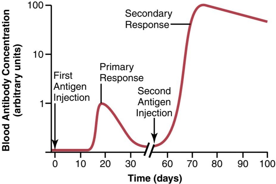
Figure 35-3. Time course of the antibody response in the circulating blood to a primary injection of antigen and to a secondary injection several weeks later.
شکل 35-3. دوره زمانی پاسخ آنتی بادی در خون در گردش به تزریق اولیه آنتی ژن و تزریق ثانویه چند هفته بعد.
Nature of Antibodies
Antibodies are gamma globulins called immunoglobulins (Igs) that have molecular weights between 160,000 and 970,000 and constitute about 20% of all the plasma proteins. All the immunoglobulins are composed of combi- nations of light and heavy polypeptide chains. Most are a combination of two light and two heavy chains, as shown in Figure 35-4. However, some immunoglobulins have combinations of as many as 10 heavy and 10 light chains, which give rise to high-molecular-weight immunoglobulins. Yet, in all immunoglobulins, each heavy chain is paralleled by a light chain at one of its ends, thus forming a heavy-light pair; there are always at least two and as many as 10 such pairs in each immunoglobulin molecule.
ماهیت آنتی بادیها
آنتی بادیها گاما گلوبولینهایی به نام ایمونوگلوبولینها (Igs) هستند که دارای وزن مولکولی بین 160000 تا 970000 هستند و حدود 20٪ از کل پروتئینهای پلاسما را تشکیل میدهند. تمام ایمونوگلوبولینها از ترکیبی از زنجیرههای پلی پپتیدی سبک و سنگین تشکیل شده اند. همانطور که در شکل 35-4 نشان داده شده است اکثر آنها ترکیبی از دو زنجیره سبک و دو زنجیره سنگین هستند. با این حال، برخی از ایمونوگلوبولینها دارای ترکیبی از 10 زنجیره سنگین و 10 زنجیره سبک هستند که باعث ایجاد ایمونوگلوبولینهای با وزن مولکولی بالا میشود. با این حال، در تمام ایمونوگلوبولینها، هر زنجیره سنگین با یک زنجیره سبک در یکی از انتهای آن موازی میشود، بنابراین یک جفت سنگین-سبک را تشکیل میدهد. در هر مولکول ایمونوگلوبولین همیشه حداقل دو تا 10 جفت از این قبیل وجود دارد.
Figure 35-4 shows a designated end of each light and heavy chain, called the variable portion; the remainder of each chain is called the constant portion. The variable portion is different for each specific antibody, and it is this portion that attaches specifically to a particular type of antigen. The constant portion determines other proper- ties of the antibody, establishing such factors as antibody diffusivity in the tissues, adherence to specific structures in the tissues, attachment to the complement complex, ease with which the antibodies pass through membranes, and other biological properties of the antibody. A com- bination of noncovalent and covalent bonds (disulfide) holds the light and heavy chains together.
شکل 35-4 انتهای مشخص شده هر زنجیره سبک و سنگین را نشان میدهد که به آن قسمت متغیر میگویند. باقیمانده هر زنجیره را بخش ثابت مینامند. بخش متغیر برای هر آنتی بادی خاص متفاوت است و این قسمت است که به طور خاص به نوع خاصی از آنتی ژن متصل میشود. بخش ثابت سایر خصوصیات آنتی بادی را تعیین میکند، عواملی مانند انتشار آنتی بادی در بافتها، چسبیدن به ساختارهای خاص در بافتها، اتصال به کمپلمان، سهولت عبور آنتی بادیها از غشاها و سایر خواص بیولوژیکی آنتی بادی را ایجاد میکند. ترکیبی از پیوندهای غیرکووالانسی و کووالانسی (دی سولفید) زنجیرههای سبک و سنگین را کنار هم نگه میدارد.

Figure 35-4. Structure of the typical IgG antibody, showing it to be composed of two heavy polypeptide chains and two light polypeptide chains. The antigen binds at two different sites on the variable portions of the chains.
شکل 35-4. ساختار آنتی بادی معمولی IgG، نشان میدهد که آن از دو زنجیره پلی پپتیدی سنگین و دو زنجیره پلی پپتیدی سبک تشکیل شده است. آنتی ژن در دو مکان مختلف در بخشهای متغیر زنجیرهها متصل میشود.
Specificity of Antibodies. Each antibody is specific for a particular antigen; this characteristic is a result of the unique structural organization of amino acids in the variable portions of the light and heavy chains. The amino acid organization has a different steric shape for each antigen specificity, so when an antigen comes into contact with it, multiple prosthetic groups of the antigen fit as a mirror image with those of the antibody, thus allowing for rapid and tight bonding between the antibody and antigen. When the antibody is highly specific, there are so many bonding sites that the antibody-antigen coupling is exceedingly strong, held together by (1) hydrophobic bonding; (2) hydrogen bonding; (3) ionic attractions; and (4) van der Waals forces. It also obeys the thermodynamic mass action law:
ویژگی آنتی بادیها. هر آنتی بادی برای یک آنتی ژن خاص خاص است. این ویژگی نتیجه سازماندهی ساختاری منحصر به فرد اسیدهای آمینه در بخشهای متغیر زنجیره سبک و سنگین است. سازمان اسید آمینه شکل فضایی متفاوتی برای هر ویژگی آنتی ژنی دارد، بنابراین هنگامیکه یک آنتی ژن با آن تماس پیدا میکند، چندین گروه مصنوعی آنتی ژن به عنوان تصویر آینه ای با آن آنتی بادی قرار میگیرند، بنابراین امکان پیوند سریع و محکم بین آنتی بادی و آنتی ژن را فراهم میکند. هنگامیکه آنتی بادی بسیار اختصاصی است، مکانهای پیوندی زیادی وجود دارد که جفت شدن آنتی بادی-آنتی ژن بسیار قوی است و توسط (1) پیوند آبگریز به هم متصل میشود. (2) پیوند هیدروژنی؛ (3) جاذبههای یونی؛ و (4) نیروهای واندروالس. همچنین از قانون عمل جرم ترمودینامیکی پیروی میکند:

K1 is called the affinity constant and is a measure of how tightly the antibody binds with the antigen.
K1 ثابت میل نامیده میشود و معیاری است که نشان میدهد آنتی بادی چقدر به آنتی ژن متصل میشود.
Note especially in Figure 35-4 that there are two variable sites on the illustrated antibody for attachment of antigens, making this type of antibody bivalent. A small proportion of the antibodies, which consist of combinations of up to 10 light and 10 heavy chains, has as many as 10 binding sites.
به خصوص در شکل 35-4 توجه داشته باشید که دو مکان متغیر روی آنتی بادی نشان داده شده برای اتصال آنتی ژنها وجود دارد که این نوع آنتی بادی را دو ظرفیتی میکند. بخش کوچکی از آنتی بادیها که از ترکیبی از حداکثر 10 زنجیره سبک و 10 زنجیره سنگین تشکیل شده است، دارای 10 محل اتصال است.
Five General Classes of Antibodies. There are five general classes of antibodies, respectively named IgM, IgG, IgA, IgD, and IgE. “Ig” stands for immunoglobulin, and the other five letters designate the respective classes.
پنج کلاس عمومیآنتی بادیها. پنج دسته کلی از آنتی بادیها وجود دارد که به ترتیب IgM، IgG، IgA، IgD و IgE نامیده میشوند. “Ig” مخفف ایمونوگلوبولین است و پنج حرف دیگر کلاسهای مربوطه را مشخص میکند.
For the purpose of our limited discussion, two of these classes of antibodies are of particular importance-IgG, which is a bivalent antibody and constitutes about 75% of the antibodies of the normal person, and IgE, which constitutes only a small percentage of the antibodies but is especially involved in allergies. The IgM class is also interesting because a large share of the antibodies formed during the primary response are of this type. These anti- bodies have 10 binding sites that make them exceedingly effective in protecting the body against invaders, even though there are not many IgM antibodies.
برای هدف بحث محدود ما، دو دسته از این دسته از آنتیبادیها از اهمیت ویژهای برخوردارند: IgG که یک آنتیبادی دو ظرفیتی است و حدود 75 درصد از آنتیبادیهای افراد عادی را تشکیل میدهد، و IgE که تنها درصد کمیاز آنتیبادیها را تشکیل میدهد اما بهویژه در آلرژیها نقش دارد. کلاس IgM نیز جالب است زیرا سهم زیادی از آنتی بادیهای تشکیل شده در طول پاسخ اولیه از این نوع هستند. این آنتی بادیها دارای 10 محل اتصال هستند که آنها را در محافظت از بدن در برابر مهاجمان بسیار موثر میکند، حتی اگر آنتی بادیهای IgM زیادی وجود نداشته باشد.
Mechanisms of Action of Antibodies
Antibodies act mainly in two ways to protect the body against invading agents: (1) by direct attack on the invader; and (2) by activation of the complement system that then has multiple means of its own for destroying the invader.
مکانیسمهای عمل آنتی بادیها
آنتی بادیها عمدتاً به دو روش برای محافظت از بدن در برابر عوامل مهاجم عمل میکنند: (1) با حمله مستقیم به مهاجم. و (2) با فعال کردن سیستم مکمل که پس از آن ابزارهای متعدد خود را برای از بین بردن مهاجم دارد.
Direct Action of Antibodies on Invading Agents. Figure 35-5 shows antibodies (designated by the red Y- shaped bars) reacting with antigens (designated by the shaded objects). Because of the bivalent nature of the antibodies and the multiple antigen sites on most invading agents, the antibodies can inactivate the invading agent in one of several ways, as follows:
اثر مستقیم آنتی بادیها بر عوامل مهاجم. شکل 35-5 آنتیبادیهایی را نشان میدهد (که با میلههای Y شکل قرمز مشخص شدهاند) که با آنتیژنها (که توسط اجسام سایهدار مشخص شدهاند) واکنش نشان میدهند. به دلیل ماهیت دو ظرفیتی آنتیبادیها و مکانهای آنتی ژن متعدد در بیشتر عوامل مهاجم، آنتیبادیها میتوانند عامل مهاجم را به یکی از روشهای زیر غیرفعال کنند:
1. Agglutination, in which multiple large particles with antigens on their surfaces (e.g., bacteria or red cells) are bound together into a clump
1. آگلوتیناسیون، که در آن چندین ذره بزرگ با آنتی ژن روی سطوحشان (مثلاً باکتریها یا گلبولهای قرمز) به صورت توده ای به یکدیگر متصل میشوند.
2. Precipitation, in which the molecular complex of soluble antigen (e.g., tetanus toxin) and antibody becomes so large that it is rendered insoluble and precipitates
2. رسوب، که در آن کمپلکس مولکولی آنتی ژن محلول (مثلاً سم کزاز) و آنتی بادی آنقدر بزرگ میشود که نامحلول میشود و رسوب میکند.
3. Neutralization, in which the antibodies cover the toxic sites of the antigenic agent
3. خنثی سازی، که در آن آنتی بادیها محلهای سمیعامل آنتی ژنی را میپوشانند
4. Lysis, in which some potent antibodies are occasionally capable of directly attacking membranes of cellular agents and thereby cause rupture of the agent These direct actions of antibodies often are not strong enough to play a major role in protecting the body against the invader. Most of the protection occurs through the amplifying effects of the complement system, described next.
4. لیز، که در آن برخی از آنتی بادیهای قوی گهگاه قادر به حمله مستقیم به غشای عوامل سلولی هستند و در نتیجه باعث پارگی عامل میشوند. بیشتر محافظت از طریق اثرات تقویت کننده سیستم مکمل که در ادامه توضیح داده میشود، رخ میدهد.
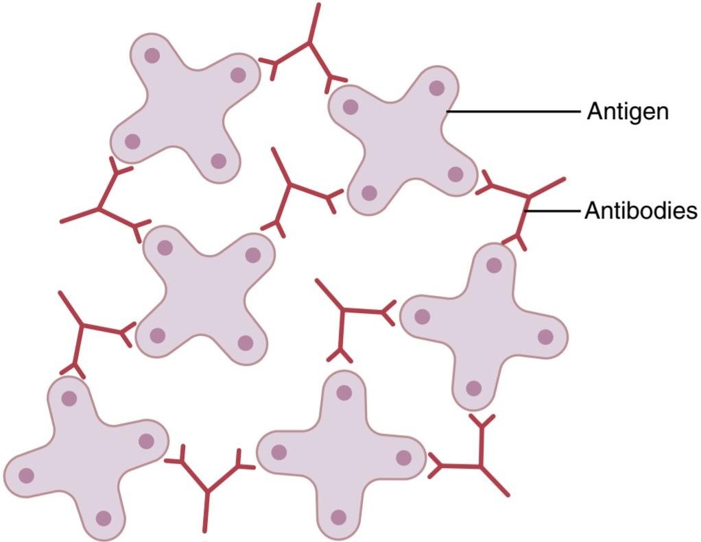
Figure 35-5. Binding of antigen molecules to one another by bivalent antibodies.
شکل 35-5. اتصال مولکولهای آنتی ژن به یکدیگر توسط آنتی بادیهای دو ظرفیتی.
COMPLEMENT SYSTEM FOR ANTIBODY ACTION
The main function of the complement system is to enhance (complement) the actions of antibodies and phagocytic cells in neutralizing and destroying pathogens, removing damaged cells from the body, and promoting inflammation. Complement is a collective term that describes a system of about 20 proteins, many of which are enzyme precursors. The principal actors in this system are 11 proteins designated C1 through C9, B, and D, shown in Figure 35-6. All these are present normally among the plasma proteins in the blood, as well as among the proteins that leak out of the capillaries into the tissue spaces. The enzyme precursors are normally inactive but can be activated by the so-called classical pathway.
سیستم مکمل برای عمل آنتی بادی
عملکرد اصلی سیستم کمپلمان تقویت (مکمل) اعمال آنتی بادیها و سلولهای فاگوسیتی در خنثی سازی و از بین بردن پاتوژنها، حذف سلولهای آسیب دیده از بدن و ترویج التهاب است. مکمل یک اصطلاح جمعی است که سیستمیاز حدود 20 پروتئین را توصیف میکند که بسیاری از آنها پیش سازهای آنزیمیهستند. بازیگران اصلی در این سیستم 11 پروتئین با C1 تا C9، B و D هستند که در شکل 35-6 نشان داده شده است. همه اینها به طور معمول در بین پروتئینهای پلاسما در خون و همچنین در بین پروتئینهایی که از مویرگها به داخل فضاهای بافتی نشت میکنند وجود دارند. پیش سازهای آنزیمیمعمولاً غیرفعال هستند، اما میتوانند توسط مسیر به اصطلاح کلاسیک فعال شوند.
Classical Pathway. The classical pathway is initiated by an antigen-antibody reaction. That is, when an antibody binds with an antigen, a specific reactive site on the constant portion of the antibody becomes uncovered, or activated, and this in turn binds directly with the C1 molecule of the complement system. This sets into motion a cascade of sequential reactions, shown in Figure 35-6, beginning with activation of the proenzyme C1. The C1 enzymes that are formed then activate successively in- creasing quantities of enzymes in the later stages of the system so that from a small beginning, an extremely large, amplified reaction occurs. Multiple end products are formed, as shown at the right in the figure, and several of these have important effects that help prevent damage to the body’s tissues caused by the invading organism or toxin. Among the more important effects are the following:
مسیر کلاسیک مسیر کلاسیک با واکنش آنتی ژن-آنتی بادی آغاز میشود. یعنی هنگامیکه یک آنتی بادی به یک آنتی ژن متصل میشود، یک مکان واکنشی خاص در قسمت ثابت آنتی بادی آشکار میشود یا فعال میشود و این به نوبه خود مستقیماً به مولکول C1 سیستم کمپلمان متصل میشود. این یک آبشار از واکنشهای متوالی را به حرکت در میآورد، که در شکل 35-6 نشان داده شده است، که با فعال شدن پروآنزیم C1 شروع میشود. آنزیمهای C1 که تشکیل میشوند سپس مقادیر فزایندهای از آنزیمها را در مراحل بعدی سیستم فعال میکنند، به طوری که از یک شروع کوچک، یک واکنش بسیار بزرگ و تقویتشده رخ میدهد. همانطور که در شکل در سمت راست نشان داده شده است، چندین محصول نهایی تشکیل میشوند، و چندین مورد از آنها اثرات مهمیدارند که به جلوگیری از آسیب به بافتهای بدن ناشی از ارگانیسم مهاجم یا سم کمک میکند. از جمله اثرات مهمتر موارد زیر است:
1. Opsonization and phagocytosis. One of the products of the complement cascade, C3b, strongly activates phagocytosis by neutrophils and macrophages, causing these cells to engulf the bacteria to which the antigen-antibody complexes are attached. This process is called opsonization. It often enhances the number of bacteria that can be destroyed by many hundredfold.
1. اپسونیزاسیون و فاگوسیتوز. یکی از محصولات آبشار کمپلمان، C3b، فاگوسیتوز را به شدت توسط نوتروفیلها و ماکروفاژها فعال میکند و باعث میشود که این سلولها باکتریهایی را که کمپلکسهای آنتیژن-آنتیبادی به آنها متصل هستند، ببلعند. این فرآیند اپسونیزاسیون نامیده میشود. اغلب تعداد باکتریهایی را که میتوانند صدها برابر از بین ببرند افزایش میدهد.
2. Lysis. One of the most important of all the products of the complement cascade is the membrane attack complex (also called the cytolytic complex), which is a combination of multiple complement factors designated as C5b6789. This membrane attack complex inserts itself into the lipid bilayer of the cell mem- brane, creating pores that are permeable to ions and causing osmotic rupture of the cell membranes of bacteria or other invading organisms.
2. لیز. یکی از مهمترین محصولات آبشار کمپلمان، کمپلکس حمله غشایی (همچنین کمپلکس سیتولیتیک نامیده میشود) است که ترکیبی از فاکتورهای مکمل متعدد به نام C5b6789 است. این کمپلکس حمله غشایی خود را به دولایه لیپیدی غشای سلولی وارد میکند و منافذی را ایجاد میکند که برای یونها نفوذپذیر است و باعث پارگی اسمزی غشای سلولی باکتریها یا سایر موجودات مهاجم میشود.
3. Agglutination. The complement products also change the surfaces of the invading organisms, causing them to adhere to one another, thus promoting agglutination.
3. آگلوتیناسیون. محصولات مکمل همچنین سطوح موجودات مهاجم را تغییر میدهند و باعث میشوند که آنها به یکدیگر بچسبند و در نتیجه باعث آگلوتیناسیون میشوند.
4. Neutralization of viruses. The complement enzymes and other complement products can attack the structures of some viruses and thereby render them nonvirulent.
4. خنثی سازی ویروسها. آنزیمهای مکمل و سایر محصولات مکمل میتوانند به ساختار برخی از ویروسها حمله کنند و در نتیجه آنها را غیر ویروسی کنند.
5. Chemotaxis. Fragment C5a initiates chemotaxis of neutrophils and macrophages, thus causing large numbers of these phagocytes to migrate into the tissue area adjacent to the antigenic agent.
5. کموتاکسی. قطعه C5a کموتاکسی نوتروفیلها و ماکروفاژها را آغاز میکند، بنابراین باعث میشود تعداد زیادی از این فاگوسیتها به ناحیه بافت مجاور عامل آنتی ژنیک مهاجرت کنند.
6. Activation of mast cells and basophils. Fragments C3a, C4a, and C5a activate mast cells and basophils, causing them to release histamine, heparin, and several other substances into the local fluids. These substances, in turn, cause increased local blood flow, increased leakage of fluid and plasma protein into the tissue, and other local tissue reactions that help inactivate or immobilize the antigenic agent. These same factors play a major role in inflammation (dis- cussed in Chapter 34) and allergy, discussed later.
6. فعال سازی ماست سلها و بازوفیلها. قطعات C3a، C4a و C5a ماست سلها و بازوفیلها را فعال میکنند و باعث آزاد شدن هیستامین، هپارین و چندین ماده دیگر در مایعات محلی میشوند. این مواد به نوبه خود باعث افزایش جریان خون موضعی، افزایش نشت مایع و پروتئین پلاسما به داخل بافت و سایر واکنشهای بافتی موضعی میشوند که به غیرفعال یا بیحرکت کردن عامل آنتی ژنی کمک میکنند. همین عوامل در التهاب (مورد بحث در فصل 34) و آلرژی که بعداً مورد بحث قرار گرفت، نقش اساسی دارند.
7. Inflammatory effects. In addition to inflammatory effects caused by activation of the mast cells and basophils, several other complement products con- tribute to local inflammation. These products cause (1) the already increased blood flow to increase still further; (2) the capillary leakage of proteins to be increased; and (3) the interstitial fluid proteins to coagulate in the tissue spaces, thus preventing movement of the invading organism through the tissues.
7. اثرات التهابی. علاوه بر اثرات التهابی ناشی از فعال شدن ماست سلها و بازوفیلها، چندین فرآورده مکمل دیگر به التهاب موضعی کمک میکنند. این محصولات باعث میشوند (1) افزایش جریان خون از قبل افزایش یابد. (2) نشت مویرگی پروتئینها افزایش یابد. و (3) پروتئینهای مایع بینابینی در فضاهای بافتی منعقد میشوند، بنابراین از حرکت ارگانیسم مهاجم از طریق بافتها جلوگیری میکنند.

Figure 35-6. Cascade of reactions during activation of the classical pathway of complement.
شکل 35-6. آبشار واکنشها در طول فعال شدن مسیر کلاسیک مکمل.
SPECIAL ATTRIBUTES OF T-LYMPHOCYTE SYSTEM-ACTIVATED T CELLS AND CELL- MEDIATED IMMUNITY
Release of Activated T Cells From Lymphoid Tissue and Formation of Memory Cells. On exposure to the proper antigen, as presented by adjacent macrophages, the T lymphocytes of a specific lymphocyte clone proliferate and release large numbers of activated, specifically reacting T cells in ways that parallel antibody release by activated B cells. The principal difference is that instead of releasing antibodies, whole activated T cells are formed and released into the lymph. These T cells then pass into the circulation, are distributed throughout the body, pass through the capillary walls into the tissue spaces, back into the lymph and blood again, and circulate again and again throughout the body, sometimes lasting for months or even years.
ویژگیهای خاص سلولهای T و سلولهای فعال شده با سیستم لنفوسیت Tمصونیت واسطه ای
آزادسازی سلولهای T فعال شده از بافت لنفاوی و تشکیل سلولهای حافظه. در مواجهه با آنتی ژن مناسب، همانطور که توسط ماکروفاژهای مجاور ارائه میشود، لنفوسیتهای T یک کلون لنفوسیتی خاص تکثیر میشوند و تعداد زیادی سلول فعال آزاد میکنند، به طور خاص به روشهایی واکنش نشان میدهند که به موازات انتشار آنتی بادی توسط سلولهای B فعال میشود. تفاوت اصلی این است که به جای آزاد کردن آنتی بادیها، سلولهای T فعال کامل تشکیل شده و در لنف آزاد میشوند. این سلولهای T سپس وارد گردش خون میشوند، در سراسر بدن توزیع میشوند، از دیوارههای مویرگی به فضاهای بافتی عبور میکنند، دوباره به لنف و خون بازمیگردند، و بارها و بارها در سراسر بدن گردش میکنند، که گاهی ماهها یا حتی سالها طول میکشد.
Also, T-lymphocyte memory cells are formed in the same way that B memory cells are formed in the anti- body system. That is, when a clone of T lymphocytes is activated by an antigen, many of the newly formed lymphocytes are preserved in the lymphoid tissue to become additional T lymphocytes of that specific clone; in fact, these memory cells even spread throughout the lymphoid tissue of the entire body. Therefore, on subsequent expo- sure to the same antigen anywhere in the body, release of activated T cells occurs far more rapidly and much more powerfully than during first exposure.
همچنین سلولهای حافظه لنفوسیت T به همان روشی که سلولهای حافظه B در سیستم ضد بدن تشکیل میشوند، تشکیل میشوند. یعنی وقتی یک کلون از لنفوسیتهای T توسط یک آنتی ژن فعال میشود، بسیاری از لنفوسیتهای تازه تشکیل شده در بافت لنفوئیدی حفظ میشوند تا به لنفوسیتهای T اضافی آن کلون خاص تبدیل شوند. در واقع، این سلولهای حافظه حتی در سراسر بافت لنفوئیدی کل بدن پخش میشوند. بنابراین، در مواجهه بعدی با همان آنتی ژن در هر نقطه از بدن، آزادسازی سلولهای T فعال شده بسیار سریعتر و قویتر از مواجهه اول اتفاق میافتد.
Antigen-Presenting Cells, Major Histocompatibility Complex Proteins, and Antigen Receptors on T Lymphocytes. T-cell responses are extremely antigen- specific, like the antibody responses of B cells, and are at least as important as antibodies in defending against infection. In fact, acquired immune responses usually require assistance from T cells to begin the process, and T cells play a major role in helping eliminate invading pathogens.
سلولهای ارائه دهنده آنتی ژن، پروتئینهای مجتمع اصلی سازگاری بافتی، و گیرندههای آنتی ژن روی لنفوسیتهای T. پاسخهای سلولهای T بسیار خاص آنتی ژن هستند، مانند پاسخهای آنتی بادی سلولهای B، و حداقل به اندازه آنتی بادیها در دفاع در برابر عفونت مهم هستند. در واقع، پاسخهای ایمنی اکتسابی معمولاً برای شروع فرآیند به کمک سلولهای T نیاز دارند و سلولهای T نقش عمدهای در کمک به حذف پاتوژنهای مهاجم دارند.
Although B lymphocytes recognize intact antigens, T lymphocytes respond to antigens only when they are bound to specific molecules called MHC proteins on the surface of antigen-presenting cells in the lymphoid tissues (Figure 35-7). The three major types of antigen- presenting cells are macrophages, B lymphocytes, and dendritic cells. The dendritic cells (also known as accessory cells), the most potent of the antigen-presenting cells, are located throughout the body, and their main function is to present antigens to T cells. The interaction of cell adhesion proteins is critical in permitting the T cells to bind to antigen-presenting cells long enough to become activated.
اگرچه لنفوسیتهای B آنتی ژنهای دست نخورده را تشخیص میدهند، لنفوسیتهای T تنها زمانی به آنتی ژنها پاسخ میدهند که به مولکولهای خاصی به نام پروتئینهای MHC روی سطح سلولهای ارائه دهنده آنتی ژن در بافتهای لنفاوی متصل شوند (شکل 35-7). سه نوع اصلی سلولهای ارائه دهنده آنتی ژن عبارتند از ماکروفاژها، لنفوسیتهای B و سلولهای دندریتیک. سلولهای دندریتیک (همچنین به عنوان سلولهای کمکی شناخته میشوند)، قویترین سلولهای ارائهدهنده آنتیژن، در سراسر بدن قرار دارند و وظیفه اصلی آنها ارائه آنتیژن به سلولهای T است. تعامل پروتئینهای چسبنده سلولی برای اجازه دادن به سلولهای T برای اتصال به سلولهای ارائه دهنده آنتی ژن به اندازه کافی برای فعال شدن حیاتی است.
The MHC proteins are encoded by a large group of genes called the major histocompatibility complex (MHC). The MHC proteins bind peptide fragments of antigen proteins that are degraded inside antigen-presenting cells and then transport them to the cell surface. There are two types of MHC proteins: (1) MHC I proteins, which present anti- gens to cytotoxic T cells; and (2) MHC II proteins, which present antigens to T-helper cells. The specific functions of cytotoxic and T-helper cells are discussed later.
پروتئینهای MHC توسط گروه بزرگی از ژنها به نام مجتمع اصلی سازگاری بافتی (MHC) کدگذاری میشوند. پروتئینهای MHC قطعات پپتیدی پروتئینهای آنتی ژن را که در داخل سلولهای ارائه دهنده آنتی ژن تجزیه میشوند متصل میکنند و سپس آنها را به سطح سلول منتقل میکنند. دو نوع پروتئین MHC وجود دارد: (1) پروتئینهای MHC I که آنتی ژن را به سلولهای T سیتوتوکسیک ارائه میکنند. و (2) پروتئینهای MHC II، که آنتی ژنها را به سلولهای T-helper ارائه میکنند. عملکردهای خاص سلولهای سیتوتوکسیک و T-helper بعداً مورد بحث قرار میگیرد.
The antigens on the surface of antigen-presenting cells bind with receptor molecules on the surfaces of T cells in the same way that they bind with plasma protein antibodies. These receptor molecules are composed of a variable unit similar to the variable portion of the humoral antibody, but its stem section is firmly bound to the cell membrane of the T lymphocyte. There are as many as 100,000 receptor sites on a single T cell.
آنتی ژنهای روی سطح سلولهای ارائه دهنده آنتی ژن با مولکولهای گیرنده روی سطوح سلولهای T به همان روشی که با آنتی بادیهای پروتئین پلاسما متصل میشوند، متصل میشوند. این مولکولهای گیرنده از یک واحد متغیر شبیه به بخش متغیر آنتی بادی هومورال تشکیل شدهاند، اما بخش ساقه آن به طور محکم به غشای سلولی لنفوسیت T متصل است. حدود 100000 محل گیرنده در یک سلول T وجود دارد.
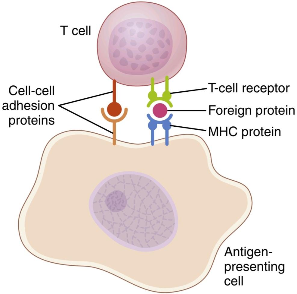
Figure 35-7. Activation of T cells requires interaction of T-cell receptors with an antigen (foreign protein) that is transported to the surface of the antigen-presenting cell by a major histocompatibility complex (MHC) protein. Cell-to-cell adhesion proteins enable the T cell to bind to the antigen-presenting cell long enough to become activated.
شکل 35-7. فعالسازی سلولهای T نیازمند تعامل گیرندههای سلول T با یک آنتی ژن (پروتئین خارجی) است که توسط یک پروتئین پیچیده سازگاری بافتی (MHC) به سطح سلول ارائهدهنده آنتیژن منتقل میشود. پروتئینهای چسبنده سلول به سلول، سلول T را قادر میسازد تا به اندازه کافی به سلول ارائهدهنده آنتیژن متصل شود تا فعال شود.
DIFFERENT TYPES OF T CELLS AND THEIR FUNCTIONS
It has become clear that there are multiple types of T cells, classified into three major groups: (1) T-helper cells; (2) cytotoxic T cells; and (3) regulatory T cells (also called suppressor T cells). The functions of each of these T cells are distinct.
انواع مختلف سلولهای T و عملکرد آنها
مشخص شده است که انواع مختلفی از سلولهای T وجود دارد که به سه گروه عمده طبقه بندی میشوند: (1) سلولهای T-helper. (2) سلولهای T سیتوتوکسیک. و (3) سلولهای T تنظیمی(که سلولهای T سرکوبگر نیز نامیده میشوند). عملکرد هر یک از این سلولهای T متمایز است.
T-Helper Cells Are the Most Numerous T Cells
The T-helper cells are the most numerous of the T cells, usually constituting more than 75% of all of them. As their name implies, they help in the functions of the immune system in many ways. They serve as the major regulator of virtually all immune functions, as shown in Figure 35-8. They do this by forming a series of protein mediators, called lymphokines, that act on other cells of the immune system, as well as on bone marrow cells.
سلولهای T-Helper پرتعدادترین سلولهای T هستند
سلولهای T-helper بیش از 75 درصد از سلولهای T را تشکیل میدهند. همانطور که از نام آنها پیداست، آنها از طرق مختلف به عملکرد سیستم ایمنی کمک میکنند. همانطور که در شکل 35-8 نشان داده شده است، آنها به عنوان تنظیم کننده اصلی تقریباً تمام عملکردهای ایمنی عمل میکنند. آنها این کار را با تشکیل یک سری واسطههای پروتئینی به نام لنفوکینها انجام میدهند که روی سلولهای دیگر سیستم ایمنی و همچنین سلولهای مغز استخوان عمل میکنند.
When stimulated, naïve CD4+ T-helper cells can differentiate into subsets that produce different lymphokines and perform different functions. Table 35-1 summarizes the main subsets of T-helper cells, the lymphokines that induce each subset, the lymphokines produced by the subsets, and the immunological reactions that each subset triggers.
هنگامیکه سلولهای CD4+ T-helper ساده تحریک میشوند، میتوانند به زیر مجموعههایی متمایز شوند که لنفوکینهای متفاوتی تولید میکنند و عملکردهای متفاوتی را انجام میدهند. جدول 35-1 زیرمجموعههای اصلی سلولهای T-helper، لنفوکینهایی که هر زیر مجموعه را القا میکنند، لنفوکینهای تولید شده توسط زیرمجموعهها و واکنشهای ایمونولوژیکی که هر زیر مجموعه ایجاد میکند، خلاصه میکند.
Specific Regulatory Functions of Lymphokines. In the absence of the lymphokines from the T-helper cells, the remainder of the immune system is almost paralyzed. In fact, it is the T-helper cells that are inactivated or destroyed by the human immunodeficiency virus (HIV), which leaves the body almost totally unprotected against infectious disease, therefore leading to the now wellknown debilitating and lethal effects of acquired immunodeficiency syndrome (AIDS). Some of the specific regulatory functions are described in the following sections.
عملکردهای تنظیمیخاص لنفوکینها. در غیاب لنفوکینها از سلولهای T-helper، بقیه سیستم ایمنی تقریباً فلج میشود. در واقع، این سلولهای کمککننده T هستند که توسط ویروس نقص ایمنی انسانی (HIV) غیرفعال یا از بین میروند، که بدن را تقریباً کاملاً در برابر بیماریهای عفونی محافظت نمیکند، بنابراین منجر به اثرات ناتوانکننده و کشنده شناخته شده سندرم نقص ایمنی اکتسابی (ایدز) میشود. برخی از عملکردهای تنظیمیخاص در بخشهای زیر توضیح داده شده است.

Figure 35-8. Regulation of the immune system, emphasizing a pivotal role of the T-helper cells. MHC, Major histocompatibility complex.
شکل 35-8. تنظیم سیستم ایمنی، با تاکید بر نقش محوری سلولهای MHC. T-helper ، مجتمع اصلی سازگاری بافتی.
Table 35-1 Subsets of T-helper Cells
جدول 35-1 زیرمجموعههای سلولهای T-helper

Stimulation of Growth and Proliferation of Cytotoxic T Cells and Regulatory T Cells. In the absence of T-helper cells, the clones for producing cytotoxic T cells and regulatory T cells are activated only slightly by most antigens. The lymphokine interleukin-2 has an especially strong stimulatory effect in causing growth and proliferation of cytotoxic and regulatory T cells. In addition, several of the other lymphokines have less potent effects.
تحریک رشد و تکثیر سلولهای T سیتوتوکسیک و سلولهای T تنظیمی. در غیاب سلولهای T-helper، کلونهای تولید سلولهای T سیتوتوکسیک و سلولهای T تنظیمیتنها اندکی توسط اکثر آنتیژنها فعال میشوند. لنفوکین اینترلوکین-2 دارای یک اثر تحریکی قوی در ایجاد رشد و تکثیر سلولهای T سیتوتوکسیک و تنظیم کننده است. علاوه بر این، چندین لنفوکین دیگر دارای اثرات قوی تری هستند.
Stimulation of B-Cell Growth and Differentiation to Form Plasma Cells and Antibodies. The direct actions of antigens to cause B-cell growth, proliferation, formation of plasma cells, and secretion of antibodies are also slight without the help of the T-helper cells. Almost all the interleukins participate in the B-cell response, but especially interleukins 4, 5, and 6. These three interleukins have such potent effects on the B cells that they have been called B-cell stimulating factors or B-cell growth factors.
تحریک رشد و تمایز سلولهای B برای تشکیل سلولهای پلاسما و آنتی بادیها. اعمال مستقیم آنتی ژنها برای ایجاد رشد سلولهای B، تکثیر، تشکیل سلولهای پلاسما و ترشح آنتی بادیها نیز بدون کمک سلولهای T کمک کننده اندک است. تقریباً همه اینترلوکینها در پاسخ سلولهای B شرکت میکنند، اما به ویژه اینترلوکینهای 4، 5 و 6. این سه اینترلوکین چنان تأثیرات قوی بر سلولهای B دارند که به آنها عوامل تحریک کننده سلول B یا فاکتورهای رشد سلول B میگویند.
Activation of the Macrophage System. The lymphokines also affect the macrophages. First, they slow or stop the migration of the macrophages after they have been chemotactically attracted into the inflamed tissue area, thus causing much accumulation of macrophages. Second, they activate the macrophages to cause more efficient phagocytosis, al- lowing them to attack and destroy increasing numbers of invading bacteria or other tissue-destroying agents.
فعال سازی سیستم ماکروفاژها لنفوکینها نیز بر ماکروفاژها تأثیر میگذارند. اول، آنها مهاجرت ماکروفاژها را پس از جذب کموتاکتیک به ناحیه بافت ملتهب کند یا متوقف میکنند و در نتیجه باعث تجمع زیادی ماکروفاژها میشوند. دوم، آنها ماکروفاژها را فعال میکنند تا فاگوسیتوز مؤثرتری ایجاد کنند و به آنها اجازه حمله و نابودی تعداد فزایندهای از باکتریهای مهاجم یا سایر عوامل تخریبکننده بافت را میدهند.
Feedback Stimulatory Effect on T-Helper Cells. Some of the lymphokines, especially interleukin-2, have a direct positive feedback effect in stimulating activation of T-helper cells. This acts as an amplifier by further enhancing the helper cell response, as well as the entire immune response to an invading antigen.
اثر محرک بازخورد بر سلولهای T-Helper. برخی از لنفوکینها، به ویژه اینترلوکین-2، اثر بازخورد مثبت مستقیمیدر تحریک فعال شدن سلولهای T-helper دارند. این به عنوان یک تقویت کننده با افزایش بیشتر پاسخ سلول کمکی و همچنین کل پاسخ ایمنی به یک آنتی ژن مهاجم عمل میکند.
Cytotoxic T Cells Are Killer Cells
The cytotoxic T cell is a direct attack cell that is capable of killing microorganisms and, at times, even some of the body’s own cells. For this reason, these cells are called killer cells. The receptor proteins on the surfaces of the CD8+ cytotoxic cells cause them to bind tightly to the organisms or cells that contain the appropriate binding-specific antigen. They then kill the attacked cell in the manner shown in Figure 35-9. After binding, the cytotoxic T cell secretes hole-forming proteins, called perforins, that liter- ally punch round holes in the membrane of the attacked cell. Fluid then flows rapidly into the cell from the interstitial space. In addition, the cytotoxic T cell releases cytotoxic substances directly into the attacked cell. Almost immediately, the attacked cell becomes greatly swollen, and it usually dissolves shortly thereafter.
سلولهای T سیتوتوکسیک سلولهای کشنده هستند
سلول T سیتوتوکسیک یک سلول حمله مستقیم است که قادر به کشتن میکروارگانیسمها و گاهی اوقات حتی برخی از سلولهای بدن است. به همین دلیل به این سلولها سلولهای کشنده میگویند. پروتئینهای گیرنده روی سطوح سلولهای سیتوتوکسیک CD8+ باعث میشوند که آنها محکم به ارگانیسمها یا سلولهایی که حاوی آنتیژن خاص اتصال مناسب هستند، متصل شوند. سپس سلول مورد حمله را به روشی که در شکل 35-9 نشان داده شده است، میکشند. پس از اتصال، سلول T سیتوتوکسیک، پروتئینهای سوراخساز به نام پرفورین ترشح میکند که به معنای واقعی کلمه سوراخهای گردی را در غشای سلول مورد حمله ایجاد میکند. سپس مایع به سرعت از فضای بینابینی به داخل سلول جریان مییابد. علاوه بر این، سلول T سیتوتوکسیک مواد سیتوتوکسیک را مستقیماً در سلول مورد حمله آزاد میکند. تقریباً بلافاصله، سلول مورد حمله به شدت متورم میشود و معمولاً مدت کوتاهی پس از آن حل میشود.
Of special importance is that these cytotoxic killer cells can pull away from the victim cells after they have punched holes and delivered cytotoxic substances and then move on to kill more cells. Some of these cells persist for months in the tissues.
از اهمیت ویژهای برخوردار است که این سلولهای کشنده سیتوتوکسیک میتوانند پس از سوراخ کردن سلولهای قربانی و رساندن مواد سیتوتوکسیک از سلولهای قربانی دور شوند و سپس برای کشتن سلولهای بیشتری حرکت کنند. برخی از این سلولها برای ماهها در بافتها باقی میمانند.
Some of the cytotoxic T cells are especially lethal to tissue cells that have been invaded by viruses because many virus particles become entrapped in the membranes of the tissue cells and attract T cells in response to the viral antigenicity. The cytotoxic cells also play an important role in destroying cancer cells, heart transplant cells, or other types of cells that are foreign to the person’s own body.
برخی از سلولهای T سیتوتوکسیک به ویژه برای سلولهای بافتی که توسط ویروسها مورد تهاجم قرار گرفته اند کشنده هستند زیرا بسیاری از ذرات ویروس در غشای سلولهای بافتی به دام افتاده و سلولهای T را در پاسخ به آنتی ژنیسیته ویروسی جذب میکنند. سلولهای سیتوتوکسیک همچنین نقش مهمیدر از بین بردن سلولهای سرطانی، سلولهای پیوند قلب یا سایر انواع سلولهایی که برای بدن خود فرد خارجی هستند، بازی میکنند.

Figure 35-9. Direct destruction of an invading cell by sensitized lymphocytes (cytotoxic T cells).
شکل 35-9. تخریب مستقیم یک سلول مهاجم توسط لنفوسیتهای حساس (سلولهای T سیتوتوکسیک).
Regulatory T Cells
Much less is known about the regulatory T cells than about the others, but they are capable of suppressing the functions of both cytotoxic and T-helper cells. These suppressor functions of the CD4+ regulatory T cells are believed to prevent the cytotoxic cells from causing excessive immune reactions that might damage the body’s own tissues. It is probable that the suppressor T-cell system plays an important role in limiting the ability of the immune system to attack a person’s own body tissues, called immune tolerance, as discussed in the next section.
سلولهای T تنظیم کننده
در مورد سلولهای T تنظیمیبسیار کمتر از سایرین شناخته شده است، اما آنها قادر به سرکوب عملکرد سلولهای سیتوتوکسیک و T-helper هستند. اعتقاد بر این است که این عملکردهای سرکوبکننده سلولهای T تنظیمکننده CD4+ از ایجاد واکنشهای ایمنی بیش از حد توسط سلولهای سیتوتوکسیک که ممکن است به بافتهای بدن آسیب برساند، جلوگیری میکند. این احتمال وجود دارد که سیستم سلولهای T سرکوبگر نقش مهمیدر محدود کردن توانایی سیستم ایمنی برای حمله به بافتهای بدن خود فرد، به نام تحمل ایمنی، ایفا کند، همانطور که در بخش بعدی بحث شد.
TOLERANCE OF ACQUIRED IMMUNITY SYSTEM TO THE BODY’S OWN TISSUES -ROLE OF PREPROCESSING IN THYMUS AND BONE MARROW
The process of acquired immunity would destroy the individual’s own body if that person became immune to his or her own tissues. The immune mechanism normally recognizes a person’s own tissues as being distinctive from bacteria or viruses, and the person’s immunity system forms few antibodies or activated T cells against his or her own antigens.
تحمل سیستم ایمنی اکتسابی به بافتهای خود بدن – نقش پیش پردازش در تیموس و مغز استخوان
فرآیند ایمنی اکتسابی در صورتی که فرد نسبت به بافتهای خود ایمن شود، بدن خود فرد را از بین میبرد. مکانیسم ایمنی معمولاً بافتهای خود فرد را متمایز از باکتریها یا ویروسها تشخیص میدهد و سیستم ایمنی فرد آنتیبادیهای کمییا سلولهای T فعالشدهای را علیه آنتیژنهای خود تشکیل میدهد.
Most Tolerance Results From Clone Selection During Preprocessing. Most tolerance is believed to develop during the preprocessing of T lymphocytes in the thymus and of B lymphocytes in the bone marrow. This is because injecting a strong antigen into a fetus while the lymphocytes are being preprocessed in these two areas prevents development of clones of lymphocytes in the lymphoid tissue that are specific for the injected antigen. Experiments have shown that specific immature lymphocytes in the thymus, when exposed to a strong antigen, become lymphoblastic, proliferate considerably, and then combine with the stimulating antigen-an effect that is believed to cause the cells to be destroyed by the thymic epithelial cells before they can migrate to and colonize the total body lymphoid tissue.
بیشترین نتایج تحمل از انتخاب کلون در طول پیش پردازش. اعتقاد بر این است که بیشترین تحمل در طول پیش پردازش لنفوسیتهای T در تیموس و لنفوسیتهای B در مغز استخوان ایجاد میشود. این به این دلیل است که تزریق یک آنتی ژن قوی به جنین در حالی که لنفوسیتها در این دو ناحیه در حال پیش پردازش هستند از ایجاد کلونهای لنفوسیتها در بافت لنفوئیدی که مخصوص آنتی ژن تزریقی هستند جلوگیری میکند. آزمایشها نشان دادهاند که لنفوسیتهای نابالغ خاص در تیموس، وقتی در معرض یک آنتیژن قوی قرار میگیرند، لنفوبلاستیک میشوند، به طور قابل توجهی تکثیر میشوند و سپس با آنتیژن تحریککننده ترکیب میشوند – اثری که اعتقاد بر این است که باعث میشود سلولها توسط سلولهای اپیتلیال تیموس قبل از مهاجرت به کل بافت لنفوئید بدن و کلونیزه کردن آنها از بین بروند.
During the preprocessing of lymphocytes in the thymus and bone marrow, all or most of the clones of lymphocytes that are specific to damage the body’s own tissues appear to be self-destroyed because of their continual exposure to the body’s antigens.
در طول پیش پردازش لنفوسیتها در تیموس و مغز استخوان، به نظر میرسد که تمام یا بیشتر کلونهای لنفوسیتها که مخصوص آسیب به بافتهای بدن هستند، به دلیل قرار گرفتن مداوم در معرض آنتیژنهای بدن، خود تخریب میشوند.
Failure of the Tolerance Mechanism Causes Auto- immune Diseases. Sometimes, people lose immune tolerance of their own tissues. This phenomenon occurs to a greater extent the older a person becomes. It usually occurs after destruction of some of the body’s own tissues, which releases considerable quantities of self-antigens that circulate in the body and presumably cause acquired immunity in the form of activated T cells or antibodies.
شکست مکانیسم تحمل باعث بیماریهای خودایمنی میشود. گاهی اوقات، افراد تحمل ایمنی نسبت به بافتهای خود را از دست میدهند. این پدیده هر چه فرد مسن تر شود به میزان بیشتری رخ میدهد. معمولاً پس از تخریب برخی از بافتهای بدن رخ میدهد که مقادیر قابلتوجهی از خود آنتیژنها را آزاد میکند که در بدن گردش میکنند و احتمالاً باعث ایجاد ایمنی اکتسابی در قالب سلولهای T فعال یا آنتیبادیها میشوند.
Over 100 diseases that result from autoimmunity have been described and include: (1) rheumatic fever, in which the body becomes immunized against tissues in the joints and heart, especially the heart valves, after exposure to a specific type of streptococcal toxin that has an epitope in its molecular structure similar to the structure of some of the body’s own self-antigens; (2) one type of glomerulonephritis, in which the person becomes immunized against the basement membranes of glomeruli; (3) myasthenia gravis, in which immunity develops against the acetylcholine receptor proteins of the neuromuscular junction, causing paralysis; (4) multiple sclerosis (MS), in which the immune system attacks the myelin that covers nerve fibers, disrupting nervous system communication; and (5) systemic lupus erythematosus (SLE), in which the person becomes immunized against many different body tissues at the same time, a disease that causes extensive damage and even death when SLE is severe.
بیش از 100 بیماری ناشی از خودایمنی شرح داده شده است و عبارتند از: (1) تب روماتیسمی، که در آن بدن پس از قرار گرفتن در معرض نوع خاصی از سم استرپتوکوک که دارای اپی توپی در ساختار مولکولی خود شبیه به ساختار برخی از بدن خودگردان است، در برابر بافتهای مفاصل و قلب، به ویژه دریچههای قلب، ایمن میشود. (2) یک نوع گلومرولونفریت، که در آن فرد در برابر غشای پایه گلومرولها ایمن میشود. (3) میاستنی گراویس، که در آن ایمنی در برابر پروتئینهای گیرنده استیل کولین در اتصال عصبی عضلانی ایجاد میشود و باعث فلج میشود. (4) مولتیپل اسکلروزیس (MS)، که در آن سیستم ایمنی به میلینی که فیبرهای عصبی را میپوشاند حمله میکند و ارتباط سیستم عصبی را مختل میکند. و (5) لوپوس اریتماتوز سیستمیک (SLE)، که در آن فرد به طور همزمان در برابر بسیاری از بافتهای مختلف بدن ایمن میشود، بیماری که باعث آسیب گسترده و حتی مرگ در هنگام SLE شدید میشود.
IMMUNIZATION BY INJECTION OF ANTIGENS
Immunization has been used for many years to produce acquired immunity against specific diseases. A person can be immunized by injecting dead organisms that are no longer capable of causing disease but that still have some of their chemical antigens. This type of immunization is used to protect against typhoid fever, whooping cough, diphtheria, and many other types of bacterial diseases.
ایمن سازی با تزریق آنتی ژن
ایمن سازی سالهاست که برای ایجاد ایمنی اکتسابی در برابر بیماریهای خاص مورد استفاده قرار گرفته است. فرد را میتوان با تزریق ارگانیسمهای مرده ای ایمن کرد که دیگر قادر به ایجاد بیماری نیستند اما هنوز برخی از آنتی ژنهای شیمیایی خود را دارند. این نوع ایمن سازی برای محافظت در برابر تب حصبه، سیاه سرفه، دیفتری و بسیاری از انواع دیگر بیماریهای باکتریایی استفاده میشود.
Immunity can be achieved against toxins that have been treated with chemicals so that their toxic nature has been destroyed, even though their antigens for causing immunity are still intact. This procedure is used in immunizing against tetanus, botulism, and other similar toxic diseases.
میتوان در برابر سمومیکه با مواد شیمیایی درمان شده اند، مصونیت ایجاد کرد، به طوری که ماهیت سمیآنها از بین رفته است، حتی اگر آنتی ژنهای آنها برای ایجاد ایمنی هنوز دست نخورده باشند. این روش در ایمن سازی در برابر کزاز، بوتولیسم و سایر بیماریهای سمیمشابه استفاده میشود.
And, finally, a person can be immunized by being infected with live organisms that have been attenuated. That is, these organisms have been grown in special culture media or have been passed through a series of animals until they have mutated enough that they will not cause disease but do still carry specific antigens required for immunization. This procedure is used to protect against smallpox, yellow fever, poliomyelitis, measles, and many other viral diseases.
و در نهایت، فرد میتواند با آلوده شدن به موجودات زنده ای که ضعیف شده اند، ایمن شود. یعنی این ارگانیسمها در محیطهای کشت ویژه رشد کرده اند یا از طریق یک سری از حیوانات عبور داده شده اند تا زمانی که به اندازه کافی جهش پیدا کنند که باعث بیماری نشوند اما همچنان حامل آنتی ژنهای خاص مورد نیاز برای ایمن سازی هستند. این روش برای محافظت در برابر آبله، تب زرد، فلج اطفال، سرخک و بسیاری از بیماریهای ویروسی دیگر استفاده میشود.
PASSIVE IMMUNITY
Thus far, all the acquired immunity we have discussed has been active immunity-that is, the person’s own body develops antibodies or activated T cells in response to invasion of the body by a foreign antigen. However, temporary immunity can be achieved in a person with- out injecting any antigen. This temporary immunity is achieved by infusing antibodies, activated T cells, or both obtained from the blood of someone else or from some other animal that has been actively immunized against the antigen.
ایمنی غیرفعال
تا اینجا، تمام ایمنی اکتسابی که مورد بحث قرار گرفتیم، ایمنی فعال بوده است، یعنی بدن خود فرد در پاسخ به تهاجم یک آنتی ژن خارجی به بدن، آنتیبادی یا سلولهای T فعال میکند. با این حال، ایمنی موقت را میتوان در فردی بدون تزریق هیچ آنتی ژنی به دست آورد. این مصونیت موقت با تزریق آنتی بادیها، سلولهای T فعال شده یا هر دو از خون شخص دیگری یا حیوان دیگری که به طور فعال در برابر آنتی ژن ایمن شده است، به دست میآید.
Antibodies last in the body of the recipient for 2 to 3 weeks and, during that time, the person is protected against the invading disease. Activated T cells last for a few weeks if transfused from another person but only for a few hours to a few days if transfused from an animal. Such transfusion of antibodies or T lymphocytes to confer immunity is called passive immunity.
آنتی بادیها به مدت 2 تا 3 هفته در بدن گیرنده باقی میمانند و در این مدت فرد در برابر بیماری مهاجم محافظت میشود. سلولهای T فعال شده در صورت تزریق از شخص دیگری چند هفته دوام میآورند، اما اگر از حیوان تزریق شود، تنها چند ساعت تا چند روز باقی میمانند. چنین انتقال آنتی بادیها یا لنفوسیتهای T برای ایجاد ایمنی، ایمنی غیرفعال نامیده میشود.
ALLERGY AND HYPERSENSITIVITY
An important undesirable side effect of immunity is the development, under some conditions, of an allergy or other type of immune hypersensitivity. There are several types of allergy and other hypersensitivities, some of which occur only in people who have a specific allergic tendency.
آلرژی و حساسیت بیش از حد
یک عارضه جانبی مهم و نامطلوب ایمنی ایجاد، تحت برخی شرایط، آلرژی یا سایر انواع حساسیتهای ایمنی است. انواع مختلفی از آلرژی و سایر حساسیتهای مفرط وجود دارد که برخی از آنها فقط در افرادی رخ میدهد که تمایل آلرژیک خاصی دارند.
ALLERGY CAUSED BY ACTIVATED T CELLS: DELAYED-REACTION ALLERGY
Delayed-reaction allergy is caused by activated T cells and not by antibodies. In the case of poison ivy, the toxin of poison ivy in itself does not cause much harm to the tissues. However, on repeated exposure, it does cause the formation of activated helper and cytotoxic T cells. Then, after subsequent exposure to the poison ivy toxin, within a day or so, the activated T cells diffuse from the circulating blood in large numbers into the skin to respond to the poison ivy toxin. At the same time, these T cells elicit a cell-mediated type of immune reaction. Remembering that this type of immunity can cause release of many toxic substances from the activated T cells, as well as extensive invasion of the tissues by macrophages, along with their subsequent effects, one can understand that the eventual result of some delayed-reaction allergies can be serious tissue damage. The damage normally occurs in the tissue area where the instigating antigen is present, such as in the skin in the case of poison ivy or in the lungs to cause lung edema or asthmatic attacks in the case of some airborne antigens.
آلرژی ناشی از سلولهای T فعال شده: آلرژی با واکنش تاخیری
آلرژی با واکنش تاخیری توسط سلولهای T فعال ایجاد میشود و نه توسط آنتی بادیها. در مورد پیچک سمی، سم پیچک سمیبه خودی خود آسیب چندانی به بافتها وارد نمیکند. با این حال، در مواجهه مکرر، باعث تشکیل سلولهای T کمک کننده و سیتوتوکسیک فعال میشود. سپس، پس از مواجهه بعدی با سم پیچک سمی، در عرض یک روز یا بیشتر، سلولهای T فعال شده از خون در گردش به تعداد زیادی به داخل پوست منتشر میشوند تا به سم پیچک سمیپاسخ دهند. در همان زمان، این سلولهای T نوعی واکنش ایمنی با واسطه سلولی را ایجاد میکنند. با یادآوری اینکه این نوع ایمنی میتواند باعث آزاد شدن بسیاری از مواد سمیاز سلولهای T فعال و همچنین تهاجم گسترده ماکروفاژها به بافتها و اثرات بعدی آنها شود، میتوان فهمید که نتیجه نهایی برخی آلرژیهای با واکنش تاخیری میتواند آسیب جدی به بافت باشد. آسیب معمولاً در ناحیه بافتی که آنتی ژن محرک وجود دارد رخ میدهد، مانند پوست در مورد پیچک سمییا در ریهها که باعث ادم ریه یا حملات آسم در مورد برخی از آنتی ژنهای هوا میشود.
ATOPIC ALLERGIES ASSOCIATED WITH EXCESS IGE ANTIBODIES
Some people have an allergic tendency. Their allergies are called atopic allergies because they are caused by a nonordinary response of the immune system. The allergic tendency is genetically passed from parent to child and is characterized by the presence of large quantities of IgE antibodies in the blood. These antibodies are called reagins or sensitizing antibodies to distinguish them from the more common IgG antibodies. When an allergen (defined as an antigen that reacts specifically with a specific type of IgE reagin antibody) enters the body, an allergen-reagin reaction takes place, and a subsequent allergic reaction occurs.
آلرژیهای آتوپیک مرتبط با آنتی بادیهای IGE اضافی
برخی افراد تمایل به آلرژی دارند. آلرژی آنها را آلرژی آتوپیک مینامند زیرا در اثر پاسخ غیرعادی سیستم ایمنی ایجاد میشود. تمایل ژنتیکی آلرژیک از والدین به فرزند منتقل میشود و با وجود مقادیر زیادی آنتی بادی IgE در خون مشخص میشود. این آنتی بادیها Reagin یا آنتی بادیهای حساس کننده نامیده میشوند تا آنها را از آنتی بادیهای رایج IgG متمایز کنند. هنگامیکه یک آلرژن (که به عنوان آنتی ژنی که به طور خاص با نوع خاصی از آنتی بادی IgE reagin واکنش میدهد) وارد بدن میشود، یک واکنش آلرژن-reagin رخ میدهد و یک واکنش آلرژیک بعدی رخ میدهد.
A special characteristic of the IgE antibodies (the reagins) is a strong propensity to attach to mast cells and basophils. Indeed, a single mast cell or basophil can bind as many as a half-million molecules of IgE antibodies. Then, when an antigen (an allergen) that has multiple binding sites binds with several IgE antibodies that are already attached to a mast cell or basophil, there is an immediate change in the membrane of the mast cell or basophil, perhaps resulting from a physical effect of the antibody molecules to contort the cell membrane. At any rate, many of the mast cells and basophils rupture; others release special agents immediately or shortly thereafter, including histamine, protease, slow-reacting substance of anaphylaxis (a mixture of toxic leukotrienes), eosinophil chemotactic substance, neutrophil chemotactic substance, heparin, and platelet-activating factors. These substances cause effects such as dilation of the local blood vessels, attraction of eosinophils and neutrophils to the reactive site, increased permeability of the capillaries with loss of fluid into the tissues, and contraction of local smooth muscle cells. Therefore, several different tissue responses can occur, depending on the type of tissue in which the allergen-reagin reaction occurs. Some of the different types of allergic reactions caused in this manner are described next.
ویژگی خاص آنتی بادیهای IgE (ریجینها) تمایل قوی به چسبیدن به ماست سلها و بازوفیلها است. در واقع، یک ماست سل یا بازوفیل میتواند به اندازه نیم میلیون مولکول آنتی بادی IgE متصل کند. سپس، هنگامیکه یک آنتی ژن (یک آلرژن) که دارای چندین محل اتصال است، با چندین آنتی بادی IgE که قبلاً به یک ماست سل یا بازوفیل متصل شده اند، متصل میشود، یک تغییر فوری در غشای ماست سل یا بازوفیل ایجاد میشود که احتمالاً ناشی از تأثیر فیزیکی مولکولهای آنتی بادی برای انقباض غشای سلولی است. به هر حال، بسیاری از ماست سلها و بازوفیلها پاره میشوند. برخی دیگر بلافاصله یا مدت کوتاهی پس از آن عوامل خاصی از جمله هیستامین، پروتئاز، ماده آنافیلاکسی با واکنش آهسته (مخلوطی از لکوترینهای سمی)، ماده کموتاکتیک ائوزینوفیل، ماده کموتاکتیک نوتروفیل، هپارین و عوامل فعال کننده پلاکت را آزاد میکنند. این مواد اثراتی مانند گشاد شدن رگهای خونی موضعی، جذب ائوزینوفیلها و نوتروفیلها به محل واکنش، افزایش نفوذپذیری مویرگها با از دست دادن مایع به داخل بافتها و انقباض سلولهای عضلانی صاف موضعی ایجاد میکنند. بنابراین، بسته به نوع بافتی که واکنش آلرژن-ریجین در آن رخ میدهد، چندین واکنش بافتی مختلف میتواند رخ دهد. برخی از انواع مختلف واکنشهای آلرژیک ناشی از این روش در ادامه توضیح داده میشوند.
Anaphylaxis-Widespread Allergic Reaction. When a specific allergen is injected directly into the circulation, the allergen can react with basophils of the blood and mast cells in the tissues located immediately outside the small blood vessels if the basophils and mast cells have been sensitized by the attachment of IgE reagins. There- fore, a widespread allergic reaction occurs throughout the vascular system and closely associated tissues. This reaction is called anaphylaxis. Histamine is released into the circulation and causes body-wide vasodilation, as well as increased permeability of the capillaries, with a resultant marked loss of plasma from the circulation. Occasionally, a person who experiences this reaction dies of circulatory shock within a few minutes unless treated with epinephrine to counteract the effects of the histamine.
آنافیلاکسی – واکنش آلرژیک گسترده. هنگامیکه یک آلرژن خاص مستقیماً به گردش خون تزریق میشود، در صورتی که بازوفیلها و ماست سلها با اتصال ریگینهای IgE حساس شده باشند، آلرژن میتواند با بازوفیلهای خون و ماست سلها در بافتهایی که بلافاصله خارج از رگهای خونی کوچک قرار دارند واکنش نشان دهد. بنابراین، یک واکنش آلرژیک گسترده در سراسر سیستم عروقی و بافتهای نزدیک به آن رخ میدهد. این واکنش آنافیلاکسی نامیده میشود. هیستامین در گردش خون آزاد میشود و باعث اتساع عروق در سراسر بدن و همچنین افزایش نفوذپذیری مویرگها میشود و در نتیجه از دست دادن قابل توجه پلاسما از گردش خون. گاهی اوقات، فردی که این واکنش را تجربه میکند، در عرض چند دقیقه بر اثر شوک گردش خون میمیرد، مگر اینکه برای خنثی کردن اثرات هیستامین با اپی نفرین درمان شود.
Also released from the activated basophils and mast cells is a mixture of leukotrienes called slow-reacting substance of anaphylaxis. These leukotrienes can cause spasm of the smooth muscle of the bronchioles, eliciting an asthma-like attack and sometimes causing death by suffocation.
همچنین از بازوفیلها و ماست سلهای فعال شده، مخلوطی از لکوترینها به نام ماده واکنش کند آنافیلاکسی آزاد میشود. این لکوترینها میتوانند باعث اسپاسم عضله صاف برونشیولها شوند و حمله ای شبیه به آسم را برانگیزند و گاهی اوقات باعث مرگ در اثر خفگی شوند.
Urticaria Localized Anaphylactoid Reactions. Urticaria results from antigen entering specific skin areas and causing localized anaphylactoid reactions. Histamine released locally causes: (1) vasodilation that induces an immediate red flare; and (2) increased local permeability of the capillaries that leads to local circumscribed areas of swelling of the skin within another few minutes. The swellings are commonly called hives. Administration of antihistamine drugs to a per- son before exposure will prevent the hives.
کهیر واکنشهای آنافیلاکتوئیدی موضعی. کهیر ناشی از ورود آنتی ژن به نواحی خاص پوست و ایجاد واکنشهای آنافیلاکتوئیدی موضعی است. هیستامین آزاد شده به صورت موضعی باعث: (1) اتساع عروق میشود که باعث شعله ور شدن فوری قرمز میشود. و (2) افزایش نفوذپذیری موضعی مویرگها که منجر به تورم موضعی پوست در چند دقیقه دیگر میشود. ورمها معمولاً کهیر نامیده میشوند. تجویز داروهای آنتی هیستامین به فرد قبل از قرار گرفتن در معرض از کهیر جلوگیری میکند.
Hay Fever. In hay fever, the allergen-reagin reaction occurs in the nose. Histamine released in response to the reaction causes local intranasal vascular dilation, with resultant increased capillary pressure and increased capillary permeability. Both these effects cause rapid fluid leakage into the nasal cavities and into associated deeper tissues of the nose, and the nasal linings become swollen and secretory. Here again, the use of antihistamine drugs can prevent this swelling reaction. However, other products of the allergen-reagin reaction can still cause irritation of the nose, eliciting the typical sneezing syndrome.
تب یونجه. در تب یونجه، واکنش آلرژن-ریجین در بینی رخ میدهد. هیستامین آزاد شده در پاسخ به واکنش باعث اتساع موضعی عروق داخل بینی و در نتیجه افزایش فشار مویرگی و افزایش نفوذپذیری مویرگی میشود. هر دوی این اثرات باعث نشت سریع مایع به داخل حفرههای بینی و بافتهای عمیقتر بینی میشوند و پوششهای بینی متورم و ترشح میشوند. در اینجا نیز استفاده از داروهای آنتی هیستامین میتواند از این واکنش تورم جلوگیری کند. با این حال، سایر محصولات واکنش آلرژن-ریاگین همچنان میتوانند باعث تحریک بینی شوند و سندرم عطسه معمولی را ایجاد کنند.
Asthma. Asthma often occurs in the hypersensitive allergic person. In these individuals, the allergen-reagin reaction occurs in the bronchioles of the lungs. Here, an important product released from the mast cells is believed to be the slow-reacting substance of anaphylaxis (a mixture of three leukotrienes), which causes spasm of the bronchiolar smooth muscle. Consequently, the person has difficulty breathing until the reactive products of the allergic reaction have been removed. The administration of antihistamine medication has less effect on the course of asthma because histamine does not appear to be the major factor eliciting the asthmatic reaction.
آسم. آسم اغلب در افراد حساس به آلرژی رخ میدهد. در این افراد واکنش آلرژن-ریگین در برونشیولهای ریه رخ میدهد. در اینجا، اعتقاد بر این است که یک محصول مهم آزاد شده از ماست سلها، ماده واکنش کند آنافیلاکسی (مخلوطی از سه لکوترین) است که باعث اسپاسم عضله صاف برونش میشود. در نتیجه، تا زمانی که محصولات واکنشی واکنش آلرژیک از بین نرود، فرد دچار مشکل تنفسی میشود. تجویز داروهای آنتی هیستامین تأثیر کمتری بر سیر آسم دارد زیرا به نظر نمیرسد هیستامین عامل اصلی ایجاد کننده واکنش آسم باشد.
کلیک کنید «Bibliography: فهرست کتب مربوطه»
Biglarnia AR, Huber-Lang M, Mohlin C, Ekdahl KN, Nilsson B: The multifaceted role of complement in kidney transplantation. Nat Rev Nephrol 14:767, 2018.
Brynjolfsson SF, Persson Berg L, Olsen Ekerhult T, et al: Long-lived plasma cells in mice and men. Front Immunol 2018 Nov 16;9:2673. https://www.org.doi.10.3389/fimmu.2018.02673.
Chiossone L, Dumas PY, Vienne M, Vivier E: Natural killer cells and other innate lymphoid cells in cancer. Nat Rev Immunol 18:671, 2018.
Crosby CM, Kronenberg M: Tissue-specific functions of invariant natural killer T cells. Nat Rev Immunol 18:559, 2018.
Cyster JG, Allen CDC: B cell responses: cell interaction dynamics and decisions. Cell 177:524, 2019.
DeNardo DG, Ruffell B: Macrophages as regulators of tumour immu- nity and immunotherapy. Nat Rev Immunol 19:369, 2019.
Eisenbarth SC: Dendritic cell subsets in T cell programming: location dictates function. Nat Rev Immunol 19:89, 2019.
Gattinoni L, Speiser DE, Lichterfeld M, Bonini C: T memory stem cells in health and disease. Nat Med 23:18, 2017.
Georg P, Sander LE: Innate sensors that regulate vaccine responses. Curr Opin Immunol 59:31, 2019.
Heath WR, Kato Y, Steiner TM, Caminschi I: Antigen presentation by dendritic cells for B cell activation. Curr Opin Immunol 58:44, 2019.
Ho AW, Kupper TS: T cells and the skin: from protective immunity to inflammatory skin disorders. Nat Rev Immunol 19:490, 2019.
Husebye ES, Anderson MS, Kämpe O: Autoimmune polyendocrine syndromes. N Engl J Med 378:1132, 2018.
Israel E, Reddel HK: Severe and difficult-to-treat asthma in adults. N Engl J Med 377:965, 2017.
Papi A, Brightling C, Pedersen SE, Reddel HK: Asthma. Lancet 391:783, 2018.
Reis ES, Mastellos DC, Hajishengallis G, Lambris JD: New insights into the immune functions of complement. Nat Rev Immunol 19:503, 2019.
Robson KJ, Ooi JD, Holdsworth SR, Rossjohn J, Kitching AR: HLA and kidney disease: from associations to mechanisms. Nat Rev Nephrol 14:636, 2018.
Theofilopoulos AN, Kono DH, Baccala R: The multiple pathways to autoimmunity. Nat Immunol 18:716, 2017.
Yatim N, Cullen S, Albert ML: Dying cells actively regulate adaptive immune responses. Nat Rev Immunol 17:262, 2017.
🚀 با ما همراه شوید!
تازهترین مطالب و آموزشهای مغز و اعصاب را از دست ندهید. با فالو کردن کانال تلگرام آیندهنگاران مغز، از ما حمایت کنید!


