
فیزیولوژی پزشکی گایتون و هال؛ انتقال اکسیژن و دی اکسید کربن در خون و مایعات بافتی


دعای مطالعه [ نمایش ]
بِسْمِ الله الرَّحْمنِ الرَّحیمِ
اَللّهُمَّ اَخْرِجْنى مِنْ ظُلُماتِ الْوَهْمِ
خدايا مرا بيرون آور از تاريكىهاى وهم،
وَ اَكْرِمْنى بِنُورِ الْفَهْمِ
و به نور فهم گرامى ام بدار،
اَللّهُمَّ افْتَحْ عَلَيْنا اَبْوابَ رَحْمَتِكَ
خدايا درهاى رحمتت را به روى ما بگشا،
وَانْشُرْ عَلَيْنا خَزائِنَ عُلُومِكَ بِرَحْمَتِكَ يا اَرْحَمَ الرّاحِمينَ
و خزانههاى علومت را بر ما باز كن به امید رحمتت اى مهربانترين مهربانان.
کتاب «فیزیولوژی پزشکی گایتون وهال» بهعنوان یکی از جامعترین و معتبرترین منابع در حوزه علوم پزشکی، همچنان مرجع کلیدی برای درک عملکرد پیچیده بدن انسان است. این اثر با تکیه بر تازهترین پژوهشها و توضیحات دقیق از سازوکارهای فیزیولوژیک، پلی میان علوم پایه پزشکی و کاربردهای بالینی ایجاد میکند و نقشی بیبدیل در آموزش، پژوهش و ارتقای دانش سلامت ایفا مینماید.
ترجمه دقیق و علمیاین شاهکار توسط برند علمیآیندهنگاران مغز به مدیریت داریوش طاهری، دسترسی فارسیزبانان به مرزهای نوین دانش فیزیولوژی را ممکن ساخته و رسالتی علمیبرای ارتقای آموزش پزشکی، فهم عمیقتر سازوکارهای بدن و توسعه روشهای نوین در حوزه سلامت فراهم آورده است.
» کتاب فیزیولوژی پزشکی گایتون وهال
» » فصل ۴۱: انتقال اکسیژن و دی اکسید کربن در خون و مایعات بافتی
در حال ویرایش
» Guyton and Hall Textbook of Medical Physiology
»» CHAPTER 41: Transport of Oxygen and Carbon Dioxide in Blood and Tissue Fluids

Once oxygen (O2) has diffused from the alveoli into the pulmonary blood, it is transported to the tissue capillaries almost entirely in combination with hemoglobin. The presence of hemoglobin in the red blood cells allows the blood to transport 30 to 100 times as much O2 as could be transported in the form of dissolved O2 in the water of the blood.
هنگامیکه اکسیژن (O2) از آلوئولها به خون ریوی منتشر شد، تقریباً به طور کامل در ترکیب با هموگلوبین به مویرگهای بافتی منتقل میشود. وجود هموگلوبین در گلبولهای قرمز خون به خون اجازه میدهد تا 30 تا 100 برابر O2 انتقال یافته به شکل O2 محلول در آب خون منتقل شود.
In the body’s tissue cells, O2 reacts with various foodstuffs to form large quantities of carbon dioxide (CO2). This CO2 enters the tissue capillaries and is transported back to the lungs. Carbon dioxide, like O2, also combines with chemical substances in the blood that increase CO2 transport 15- to 20-fold.
در سلولهای بافت بدن، O2 با مواد غذایی مختلف واکنش نشان میدهد تا مقادیر زیادی دی اکسید کربن (CO2) را تشکیل دهد. این دیاکسید کربن وارد مویرگهای بافتی شده و به ریهها منتقل میشود. دی اکسید کربن نیز مانند O2 با مواد شیمیایی در خون ترکیب میشود که انتقال CO2 را 15 تا 20 برابر افزایش میدهد.
This chapter presents the physical and chemical principles of O2 and CO2 transport in the blood and tissue fluids qualitatively and quantitatively.
این فصل اصول فیزیکی و شیمیایی انتقال O2 و CO2 در خون و مایعات بافتی را به صورت کمیو کیفی ارائه میکند.
TRANSPORT OF OXYGEN FROM THE LUNGS TO THE BODY TISSUES
In Chapter 40, we pointed out that gases can move from one point to another by diffusion, and that the cause of this movement is always a partial pressure difference from the first point to the next. Thus, O2 diffuses from the alveoli into the pulmonary capillary blood because the oxygen partial pressure (Po2) in the alveoli is greater than the Po2 in the pulmonary capillary blood. In the other tissues of the body, a higher Po2 in the capillary blood than in the tissues causes O2 to diffuse into the surrounding cells.
انتقال اکسیژن از ریهها به بافتهای بدن
در فصل 40 اشاره کردیم که گازها میتوانند با انتشار از نقطه ای به نقطه دیگر حرکت کنند و علت این حرکت همیشه اختلاف فشار جزئی از نقطه اول به نقطه بعدی است. بنابراین، O2 از آلوئولها به خون مویرگی ریوی منتشر میشود زیرا فشار جزئی اکسیژن (Po2) در آلوئولها از Po2 در خون مویرگی ریوی بیشتر است. در سایر بافتهای بدن، Po2 بیشتر در خون مویرگی نسبت به بافتها باعث انتشار O2 به سلولهای اطراف میشود.
Conversely, when O2 is metabolized in the cells to form CO2, the intracellular CO2 partial pressure (Pco2) rises, causing CO2 to diffuse into the tissue capillaries. After blood flows to the lungs, the CO2 diffuses out of the blood into the alveoli because the Pco2 in the pulmonary capillary blood is greater than that in the alveoli. Thus, the transport of O2 and CO2 by the blood depends on both diffusion and the flow of blood. We now consider quantitatively the factors responsible for these effects.
برعکس، وقتی O2 در سلولها متابولیزه میشود و CO2 تشکیل میدهد، فشار جزئی CO2 داخل سلولی (Pco2) افزایش مییابد و باعث انتشار CO2 در مویرگهای بافت میشود. پس از جریان خون به ریهها، CO2 از خون به داخل آلوئولها پخش میشود زیرا Pco2 در خون مویرگی ریوی بیشتر از آلوئولها است. بنابراین، انتقال O2 و CO2 توسط خون به انتشار و جریان خون بستگی دارد. اکنون به لحاظ کمیعوامل مسئول این اثرات را در نظر میگیریم.
DIFFUSION OF OXYGEN FROM THE ALVEOLI TO THE PULMONARY CAPILLARY BLOOD
The top part of Figure 41-1 shows a pulmonary alveolus adjacent to a pulmonary capillary, demonstrating diffusion of O2 between alveolar air and pulmonary blood. The Po2 of the gaseous O2 in the alveolus averages 104 mm Hg, whereas the Po2 of the venous blood entering the pulmonary capillary at its arterial end averages only 40 mm Hg because a large amount of O2 was removed from this blood as it passed through the peripheral tissues. Therefore, the initial pressure difference that causes O2 to diffuse into the pulmonary capillary is 104 40 mm Hg, or 64 mm Hg. In the graph at the bottom of the figure, the curve shows the rapid rise in blood Po2 as the blood passes through the capillary; the blood Po2 rises almost to that of the alveolar air by the time the blood has moved a third of the distance through the capillary, becoming almost 104 mm Hg.
انتشار اکسیژن از آلوئول به خون مویرگ ریوی
قسمت بالای شکل 41-1 یک آلوئول ریوی را در مجاورت یک مویرگ ریوی نشان میدهد که انتشار O2 بین هوای آلوئولی و خون ریوی را نشان میدهد. Po2 O2 گازی در آلوئول به طور متوسط 104 میلی متر جیوه است، در حالی که Po2 خون وریدی که در انتهای شریانی وارد مویرگ ریوی میشود به طور متوسط تنها 40 میلی متر جیوه است زیرا مقدار زیادی O2 از این خون هنگام عبور از بافتهای محیطی حذف میشود. بنابراین، اختلاف فشار اولیه که باعث انتشار O2 به داخل مویرگ ریوی میشود، 104 میلی متر جیوه 40 یا 64 میلی متر جیوه است. در نمودار پایین شکل، منحنی افزایش سریع Po2 خون را هنگام عبور خون از مویرگ نشان میدهد. زمانی که خون یک سوم مسافت را از طریق مویرگ طی کرد، Po2 خون تقریباً به هوای آلوئولی افزایش مییابد و تقریباً 104 میلی متر جیوه میشود.
Uptake of Oxygen by the Pulmonary Blood During Exercise. During strenuous exercise, a person’s body may require as much as 20 times the normal amount of oxygen. Also, because of increased cardiac output during exercise, the time that the blood remains in the pulmonary capillary may be reduced to less than one- half normal. Yet, because of the great safety factor for diffusion of O2 through the pulmonary membrane, the blood still becomes almost saturated with O2 by the time it leaves the pulmonary capillaries. This can be explained as follows.
جذب اکسیژن توسط خون ریوی در طول ورزش. در طول ورزش شدید، بدن یک فرد ممکن است 20 برابر مقدار طبیعی اکسیژن نیاز داشته باشد. همچنین به دلیل افزایش برون ده قلبی در حین ورزش، زمان باقی ماندن خون در مویرگ ریوی ممکن است به کمتر از نصف نرمال کاهش یابد. با این حال، به دلیل فاکتور ایمنی بزرگ برای انتشار O2 از طریق غشای ریوی، خون هنوز تا زمانی که از مویرگهای ریوی خارج میشود، تقریباً با O2 اشباع میشود. این را میتوان به صورت زیر توضیح داد.
First, in Chapter 40, we pointed out that the diffusing capacity for O2 increases almost threefold during exercise. This results mainly from increased surface area of capillaries participating in the diffusion and also from a more nearly ideal ventilation-perfusion ratio in the upper part of the lungs.
ابتدا، در فصل 40، اشاره کردیم که ظرفیت انتشار برای O2 در طول تمرین تقریباً سه برابر افزایش مییابد. این امر عمدتاً ناشی از افزایش سطح مویرگهای شرکت کننده در انتشار و همچنین از نسبت تهویه-پرفیوژن تقریبا ایده آل در قسمت بالایی ریهها است.
Second, note in the curve of Figure 41-1 that under nonexercising conditions, the blood becomes almost saturated with O2 by the time it has passed through one-third of the pulmonary capillary, and little additional O2 normally enters the blood during the latter two-thirds of its transit. That is, the blood normally stays in the lung capillaries about three times as long as needed to cause full oxygenation. Therefore, during exercise, even with a shortened time of exposure in the capillaries, the blood can still become almost fully oxygenated.
دوم، در منحنی شکل 41-1 توجه داشته باشید که در شرایط بدون ورزش، خون تا زمانی که از یک سوم مویرگ ریوی عبور میکند، تقریباً با O2 اشباع میشود، و مقدار کمیO2 اضافی معمولاً در طول دو سوم دوم انتقال خود وارد خون میشود. یعنی خون به طور معمول در مویرگهای ریه حدود سه برابر زمانی که لازم است برای ایجاد اکسیژن کامل باقی میماند. بنابراین، در حین ورزش، حتی با کوتاه شدن زمان قرار گرفتن در مویرگها، خون همچنان میتواند تقریباً به طور کامل اکسیژن شود.
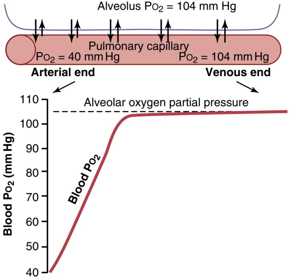
Figure 41-1 Uptake of oxygen by the pulmonary capillary blood. (Data from Milhorn HT Jr, Pulley PE Jr: A theoretical study of pulmonary capillary gas exchange and venous admixture. Biophys J 8:337, 1968.)
شکل 41-1 جذب اکسیژن توسط خون مویرگی ریوی. (دادهها از Milhorn HT Jr، Pulley PE Jr: مطالعه نظری تبادل گاز مویرگی ریوی و مخلوط وریدی. Biophys J 8:337، 1968.)
TRANSPORT OF OXYGEN IN ARTERIAL BLOOD
About 98% of the blood that enters the left atrium from the lungs has just passed through the alveolar capillaries and has become oxygenated up to a Po2 of about 104 mm Hg. Another 2% of the blood has passed from the aorta through the bronchial circulation, which supplies mainly the deep tissues of the lungs and is not exposed to lung air. This blood flow is called shunt flow, meaning that blood is shunted past the gas exchange areas. On leaving the lungs, the Po2 of the shunt blood is approximately that of normal systemic venous blood-about 40 mm Hg. When this blood combines in the pulmonary veins with the oxygenated blood from the alveolar capillaries, this so-called venous admixture of blood causes the Po2 of the blood entering the left heart and pumped into the aorta to fall to about 95 mm Hg. These changes in blood Po2 at different points in the circulatory system are shown in Figure 41-2.
انتقال اکسیژن در خون شریانی
حدود 98 درصد خونی که از ریهها وارد دهلیز چپ میشود، به تازگی از مویرگهای آلوئولی عبور کرده و تا Po2 حدود 104 میلی متر جیوه اکسیژن دار شده است. 2٪ دیگر از خون از آئورت از طریق گردش خون برونش عبور کرده است، که عمدتاً بافتهای عمیق ریهها را تامین میکند و در معرض هوای ریه قرار نمیگیرد. به این جریان خون، جریان شنت میگویند، به این معنی که خون از نواحی تبادل گاز عبور میکند. هنگام خروج از ریهها، Po2 خون شانت تقریباً برابر با خون وریدی سیستمیک طبیعی است – حدود 40 میلی متر جیوه. هنگامیکه این خون در سیاهرگهای ریوی با خون اکسیژندار مویرگهای آلوئولی ترکیب میشود، این ترکیب به اصطلاح وریدی خون باعث میشود که Po2 خون وارد قلب چپ و پمپاژ شده به آئورت تا حدود 95 میلیمتر جیوه کاهش یابد. این تغییرات در Po2 خون در نقاط مختلف سیستم گردش خون در شکل 41-2 نشان داده شده است.
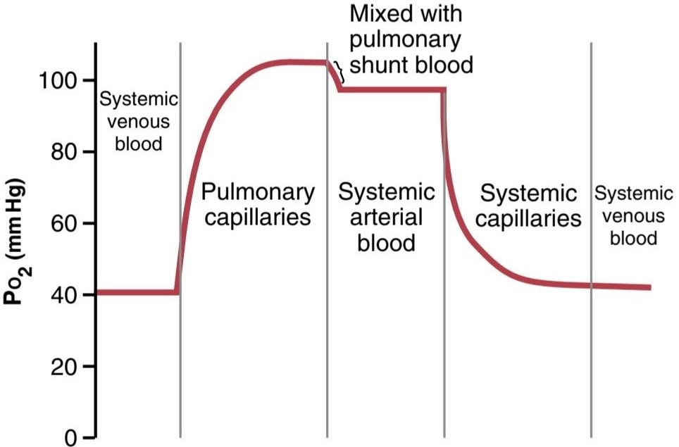
Figure 41-2 Changes in Po2 in the pulmonary capillary blood, systemic arterial blood, and systemic capillary blood demonstrating the effect of venous admixture.
شکل 41-2 تغییرات در Po2 در خون مویرگی ریوی، خون شریانی سیستمیک و خون مویرگی سیستمیک نشان دهنده اثر مخلوط وریدی است.
DIFFUSION OF OXYGEN FROM THE PERIPHERAL CAPILLARIES INTO THE TISSUE FLUID
When the arterial blood reaches the peripheral tissues, its Po2 in the capillaries is still 95 mm Hg. Yet, as shown in Figure 41-3, the Po2 in the interstitial fluid that surrounds the tissue cells averages only 40 mm Hg. Thus, there is a large initial pressure difference that causes O2 to diffuse rapidly from the capillary blood into the tissues- so rapidly that the capillary Po2 falls almost to equal the 40-mm Hg pressure in the interstitium. Therefore, the Po2 of the blood leaving the tissue capillaries and entering the systemic veins is also about 40 mm Hg.
انتشار اکسیژن از مویرگهای محیطی به مایع بافتی
هنگامیکه خون شریانی به بافتهای محیطی میرسد، Po2 آن در مویرگها هنوز 95 میلی متر جیوه است. با این حال، همانطور که در شکل 41-3 نشان داده شده است، Po2 در مایع بینابینی که سلولهای بافت را احاطه کرده است به طور متوسط تنها 40 میلی متر جیوه است. بنابراین، یک اختلاف فشار اولیه بزرگ وجود دارد که باعث میشود O2 به سرعت از خون مویرگی به بافتها منتشر شود – آنقدر سریع که Po2 مویرگی تقریباً با فشار 40 میلیمتر جیوه در بینابینی کاهش مییابد. بنابراین، Po2 خون خروجی از مویرگهای بافتی و ورود به سیاهرگهای سیستمیک نیز حدود 40 میلی متر جیوه است.

Figure 41.3 Diffusion of oxygen from a peripheral tissue capillary to the cells. (Po2 in interstitial fluid = 40 mm Hg; in tissue cells, Pco2 = 23 mm Hg.)
شکل 41.3 انتشار اکسیژن از مویرگ بافت محیطی به سلولها. (Po2 در مایع بینابینی = 40 میلی متر جیوه؛ در سلولهای بافتی، Pco2 = 23 میلی متر جیوه.)
Increasing Blood Flow Raises Interstitial Fluid Po2. If the blood flow through a particular tissue is increased, greater quantities of O2 are transported into the tissue, and the tissue Po2 becomes correspondingly higher. This effect is shown in Figure 41-4. Note that an increase in flow to 400% of normal increases the Po2 from 40 mm Hg (at point A in the figure) to 66 mm Hg (at point B). However, the upper limit to which the Po2 can rise, even with maximal blood flow, is 95 mm Hg because this is the O2 pressure in the arterial blood. Conversely, if blood flow through the tissue decreases, the tissue Po2 also decreases, as shown at point C.
افزایش جریان خون باعث افزایش مایع بینابینی Po2 میشود. اگر جریان خون از طریق یک بافت خاص افزایش یابد، مقادیر بیشتری از O2 به بافت منتقل میشود و Po2 بافت به همان نسبت بالاتر میشود. این اثر در شکل 41-4 نشان داده شده است. توجه داشته باشید که افزایش جریان به 400 درصد نرمال، Po2 را از 40 میلی متر جیوه (در نقطه A در شکل) به 66 میلی متر جیوه (در نقطه B) افزایش میدهد. با این حال، حد بالایی که Po2 میتواند تا آن افزایش یابد، حتی با حداکثر جریان خون، 95 میلی متر جیوه است زیرا این فشار O2 در خون شریانی است. برعکس، اگر جریان خون در بافت کاهش یابد، Po2 بافت نیز کاهش مییابد، همانطور که در نقطه C نشان داده شده است.
Increasing Tissue Metabolism Decreases Interstitial Fluid Po2. If the cells use more O2 for metabolism than nor- mal, the interstitial fluid Po2 is reduced. Figure 41-4 also demonstrates this effect, showing reduced interstitial fluid Po2 when the cellular oxygen consumption is increased and increased Po2 when consumption is decreased.
افزایش متابولیسم بافت، مایع بینابینی Po2 را کاهش میدهد. اگر سلولها بیش از حد معمول از O2 برای متابولیسم استفاده کنند، مایع بینابینی Po2 کاهش مییابد. شکل 41-4 نیز این اثر را نشان میدهد که کاهش Po2 مایع بینابینی را هنگامیکه مصرف اکسیژن سلولی افزایش مییابد و هنگامیکه مصرف کاهش مییابد افزایش مییابد نشان میدهد.
In summary, tissue Po2 is determined by a balance between (1) the rate of O2 transport to the tissues in the blood, and (2) the rate at which the O2 is used by the tissues.
به طور خلاصه، Po2 بافت با تعادل بین (1) سرعت انتقال O2 به بافتها در خون، و (2) سرعت استفاده از O2 توسط بافتها تعیین میشود.

Figure 41-4 Effect of blood flow and rate of oxygen consumption on tissue Poz.
شکل 41-4 اثر جریان خون و میزان مصرف اکسیژن بر بافت پوز.
DIFFUSION OF OXYGEN FROM PERIPHERAL CAPILLARIES TO TISSUE CELLS
Oxygen is always being used by the cells. Therefore, the intracellular Po2 in peripheral tissues remains lower than the Po2 in peripheral capillaries. Also, in many cases, there is considerable physical distance between the capillaries and cells. Therefore, the normal intracellular Po2 ranges from as low as 5 mm Hg to as high as 40 mm Hg, averaging (by direct measurement in experimental animals) 23 mm Hg. Because only 1 to 3 mm Hg of O2 pressure is normally required for full support of the chemical processes that use oxygen in the cell, even this low intracellular Po2 of 23 mm Hg is more than adequate and provides a large safety factor.
انتشار اکسیژن از مویرگهای محیطی به سلولهای بافتی
اکسیژن همیشه توسط سلولها استفاده میشود. بنابراین، Po2 داخل سلولی در بافتهای محیطی کمتر از Po2 در مویرگهای محیطی باقی میماند. همچنین در بسیاری از موارد فاصله فیزیکی قابل توجهی بین مویرگها و سلولها وجود دارد. بنابراین، Po2 داخل سلولی طبیعی از حداقل 5 میلی متر جیوه تا 40 میلی متر جیوه، به طور متوسط (با اندازه گیری مستقیم در حیوانات آزمایشی) 23 میلی متر جیوه است. از آنجا که به طور معمول تنها 1 تا 3 میلیمتر جیوه فشار O2 برای پشتیبانی کامل از فرآیندهای شیمیایی که از اکسیژن در سلول استفاده میکنند مورد نیاز است، حتی این Po2 درون سلولی کم 23 میلیمتر جیوه نیز بیش از اندازه کافی است و یک فاکتور ایمنی بزرگ را فراهم میکند.
DIFFUSION OF CO2 FROM PERIPHERAL TISSUE CELLS INTO CAPILLARIES AND FROM PULMONARY CAPILLARIES INTO ALVEOLI
When O2 is used by the cells, virtually all of it becomes CO2, and this transformation increases the intracellular Pco2; because of this elevated tissue cell Pco2, CO2 diffuses from the cells into the capillaries and is then carried by the blood to the lungs. In the lungs, it diffuses from the pulmonary capillaries into the alveoli and is expired.
انتشار CO2 از سلولهای بافت محیطی به مویرگها و از مویرگهای ریوی به آلوئولها
هنگامیکه O2 توسط سلولها استفاده میشود، تقریباً تمام آن به CO2 تبدیل میشود و این تغییر شکل Pco2 داخل سلولی را افزایش میدهد. به دلیل این سلول بافتی بالا Pco2، CO2 از سلولها به داخل مویرگها پخش میشود و سپس توسط خون به ریهها منتقل میشود. در ریهها از مویرگهای ریوی به آلوئولها منتشر میشود و منقضی میشود.
Thus, at each point in the gas transport chain, CO2 diffuses in the direction exactly opposite to the diffusion of O2. Yet, there is one major difference between diffusion of CO2 and of O2-CO2 can diffuse about 20 times as rapidly as O2. Therefore, the pressure differences required to cause CO2 diffusion are, in each case, far less than the pressure differences required to cause O2 diffusion. The CO2 pressures are approximately the following:
بنابراین، در هر نقطه از زنجیره انتقال گاز، CO2 در جهت دقیقا مخالف انتشار O2 منتشر میشود. با این حال، یک تفاوت عمده بین انتشار CO2 وجود دارد و O2-CO2 میتواند حدود 20 برابر سریعتر از O2 منتشر شود. بنابراین، اختلاف فشار مورد نیاز برای ایجاد انتشار CO2، در هر مورد، بسیار کمتر از اختلاف فشار لازم برای ایجاد انتشار O2 است. فشار CO2 تقریباً به شرح زیر است:
1. Intracellular PCO2, 46 mm Hg; interstitial Pco2, 45 mm Hg. Thus, there is only a 1 mm Hg pressure differential, as shown in Figure 41-5.
1. PCO2 داخل سلولی، 46 میلی متر جیوه; Pco2 بینابینی، 45 میلی متر جیوه. بنابراین، همانطور که در شکل 41-5 نشان داده شده است، تنها یک اختلاف فشار 1 میلی متر جیوه وجود دارد.
2. Pco, of the arterial blood entering the tissues, 40 mm Hg; Pco, of the venous blood leaving the tis- sues, 45 mm Hg. Thus, as shown in Figure 41-5, the tissue capillary blood comes almost exactly to equilibrium with the interstitial Pco, of 45 mm Hg.
2. Pco، از خون شریانی که وارد بافتها میشود، 40 میلی متر جیوه. Pco، از خون وریدی که از بافتها خارج میشود، 45 میلی متر جیوه. بنابراین، همانطور که در شکل 41-5 نشان داده شده است، خون مویرگی بافت تقریباً دقیقاً با Pco بینابینی 45 میلی متر جیوه به تعادل میرسد.
3. Pco2 of the blood entering the pulmonary capillaries at the arterial end, 45 mm Hg; Pco2 of the alveolar air, 40 mm Hg. Thus, only a 5 mm Hg pressure difference causes all the required CO2 diffusion out of the pulmonary capillaries into the alveoli. Furthermore, as shown in Figure 41-6, the Pco, of the pulmonary capillary blood falls to almost exactly equal the alveolar Pco, of 40 mm Hg before it has passed more than about one-third of the distance through the capillaries. This is the same effect that was observed earlier for O, diffusion, except that it is in the opposite direction.
3. Pco2 خون ورودی به مویرگهای ریوی در انتهای شریانی، 45 میلی متر جیوه. Pco2 هوای آلوئولی 40 میلی متر جیوه. بنابراین، تنها اختلاف فشار 5 میلی متر جیوه باعث انتشار تمام CO2 مورد نیاز از مویرگهای ریوی به داخل آلوئول میشود. علاوه بر این، همانطور که در شکل 41-6 نشان داده شده است، Pco خون مویرگی ریوی تقریباً دقیقاً برابر با Pco آلوئولی است، 40 میلی متر جیوه قبل از اینکه بیش از حدود یک سوم فاصله را از مویرگها عبور دهد. این همان اثری است که قبلاً برای انتشار O مشاهده شد، با این تفاوت که در جهت مخالف است.

Figure 41-5 Uptake of carbon dioxide by the blood in the tissue capillaries. (Pco2 in tissue cells = 46 mm Hg; in interstitial fluid, Pco2- 45 mm Hg.)
شکل 41-5 جذب دی اکسید کربن توسط خون در مویرگهای بافتی. (Pco2 در سلولهای بافت = 46 میلی متر جیوه، در مایع بینابینی، Pco2– 45 میلی متر جیوه.)

Figure 41-6 Diffusion of carbon dioxide from the pulmonary blood into the alveolus. (Data from Milhorn HT Jr, Pulley PE Jr: A theoretical study of pulmonary capillary gas exchange and venous admixture. Biophys J 8:337, 1968.)
شکل 41-6 انتشار دی اکسید کربن از خون ریوی به آلوئول. (دادهها از Milhorn HT Jr، Pulley PE Jr: مطالعه نظری تبادل گاز مویرگی ریوی و مخلوط وریدی. Biophys J 8:337، 1968.)
Effect of Tissue Metabolism and Tissue Blood Flow Rate on Interstitial Pco2. Tissue capillary blood flow and tissue metabolism affect the Pco, in ways exactly oppo- site to their effect on tissue Po2. Figure 41-7 shows these effects, as follows:
اثر متابولیسم بافت و سرعت جریان خون بافت بر Pco2 بینابینی. جریان خون مویرگی بافتی و متابولیسم بافت بر Pco تأثیر میگذارد، به روشهایی که دقیقاً مخالف تأثیر آنها بر روی بافت Po2 است. شکل 41-7 این اثرات را به شرح زیر نشان میدهد:
1. A decrease in blood flow from normal (point A) to one-quarter normal (point B) increases peripheral tis- sue Pco, from the normal value of 45 mm Hg to an elevated level of 60 mm Hg. Conversely, increasing the blood flow to six times normal (point C) decreases the interstitial Pco, from the normal value of 45 to 41 mm Hg, almost equal to the Pco, in the arterial blood (40 mm Hg) entering the tissue capillaries.
1. کاهش جریان خون از نرمال (نقطه A) به یک چهارم نرمال (نقطه B) Pco بافت محیطی را از مقدار طبیعی 45 میلی متر جیوه به سطح 60 میلی متر جیوه افزایش میدهد. برعکس، افزایش جریان خون به شش برابر طبیعی (نقطه C) باعث کاهش Pco بینابینی میشود، از مقدار طبیعی 45 به 41 میلی متر جیوه، تقریبا برابر با Pco، در خون شریانی (40 میلی متر جیوه) که وارد مویرگهای بافت میشود.
2. Note also that a 10-fold increase in tissue metabolic rate greatly elevates the interstitial fluid Pco, at all rates of blood flow, whereas decreasing the metabolism to one-quarter normal causes the interstitial fluid Pco, to fall to about 41 mm Hg, closely approaching that of the arterial blood, 40 mm Hg.
2. همچنین توجه داشته باشید که افزایش 10 برابری در نرخ متابولیک بافتی، Pco مایع بین بافتی را در تمام سرعتهای جریان خون بسیار بالا میبرد، در حالی که کاهش متابولیسم به یک چهارم طبیعی باعث میشود که Pco مایع بین بافتی به حدود 41 میلی متر جیوه، نزدیک به خون شریانی، 40 میلی متر جیوه، کاهش یابد.

Figure 41-7 Effect of blood flow and metabolic rate on peripheral tissue Pco2.
شکل 41-7 اثر جریان خون و نرخ متابولیک بر Pco2 بافت محیطی.
ROLE OF HEMOGLOBIN IN OXYGEN TRANSPORT
Normally, about 97% of the oxygen transported from the lungs to the tissues is carried in chemical combination with hemoglobin in the red blood cells. The remaining 3% is transported in the dissolved state in the water of the plasma and blood cells. Thus, under normal conditions, oxygen is carried to the tissues almost entirely by hemoglobin.
نقش هموگلوبین در حمل و نقل اکسیژن
به طور معمول، حدود 97 درصد از اکسیژن منتقل شده از ریهها به بافتها در ترکیب شیمیایی با هموگلوبین در گلبولهای قرمز خون حمل میشود. 3 درصد باقیمانده در حالت محلول در آب پلاسما و سلولهای خونی منتقل میشود. بنابراین، در شرایط عادی، اکسیژن تقریباً به طور کامل توسط هموگلوبین به بافتها منتقل میشود.
Reversible Combination of O2 With Hemoglobin
The chemistry of hemoglobin is presented in Chapter 33, where we pointed out that the O2 molecule combines loosely and reversibly with the heme portion of hemoglobin. When Po2 is high, as in the pulmonary capillaries, O2 binds with hemoglobin, but when Po2 is low, as in the tissue capillaries, O2 is released from hemoglobin. This is the basis for almost all O2 transport from the lungs to the tissues.
ترکیب برگشت پذیر O2 با هموگلوبین
شیمیهموگلوبین در فصل 33 ارائه شده است، جایی که ما اشاره کردیم که مولکول O2 به طور شل و برگشت پذیر با بخش هموگلوبین ترکیب میشود. هنگامیکه PO2 بالا است، مانند مویرگهای ریوی، O2 با هموگلوبین متصل میشود، اما زمانی که Po2 کم است، مانند مویرگهای بافت، O2 از هموگلوبین آزاد میشود. این اساس تقریباً تمام انتقال O2 از ریهها به بافتها است.
Oxygen-Hemoglobin Dissociation Curve. Figure 41-8 shows the O2-hemoglobin dissociation curve, which demonstrates a progressive increase in the percentage of hemoglobin bound with O2 as blood Po2 increases, called the percent saturation of hemoglobin. Because the blood leaving the lungs and entering the systemic arteries usually has a Po2 of about 95 mm Hg, it can be seen from the dissociation curve that the usual O2 saturation of systemic arterial blood averages 97%. Conversely, in normal venous blood returning from the peripheral tissues, the Po2 is about 40 mm Hg, and the saturation of hemoglobin averages 75%.
منحنی تفکیک اکسیژن-هموگلوبین. شکل 41-8 منحنی تفکیک O2-هموگلوبین را نشان میدهد که افزایش تدریجی درصد هموگلوبین متصل به O2 را با افزایش Po2 خون نشان میدهد که به آن درصد اشباع هموگلوبین میگویند. از آنجایی که خونی که از ریهها خارج میشود و وارد شریانهای سیستمیک میشود معمولاً دارای Po2 در حدود 95 میلی متر جیوه است، از منحنی تفکیک میتوان دریافت که اشباع معمول O2 خون شریانی سیستمیک به طور متوسط 97٪ است. برعکس، در خون وریدی طبیعی که از بافتهای محیطی برمیگردد، Po2 حدود 40 میلی متر جیوه است و اشباع هموگلوبین به طور متوسط 75 درصد است.
Maximum Amount of Oxygen That Can Combine With the Hemoglobin of the Blood. The blood of a normal person contains about 15 grams of hemoglobin in each 100 ml of blood, and each gram of hemoglobin can bind with a maximum of 1.34 ml of O2 (1.39 ml when the hemoglobin is chemically pure, but impurities such as methemoglobin reduce this). Therefore, 15 times 1.34 equals 20.1, which means that on average, the 15 grams of hemoglobin in 100 ml of blood can combine with a total of about 20 ml of O2 if the hemoglobin is 100% saturated. This is usually expressed as 20 volume percent. The O2- hemoglobin dissociation curve for the normal person can also be expressed in terms of volume percent of O2, as shown by the far right scale in Figure 41-8, instead of percent saturation of hemoglobin.
حداکثر مقدار اکسیژنی که میتواند با هموگلوبین خون ترکیب شود. خون یک فرد عادی در هر 100 میلی لیتر خون حاوی حدود 15 گرم هموگلوبین است و هر گرم هموگلوبین میتواند حداکثر با 1.34 میلی لیتر O2 متصل شود (1.39 میلی لیتر زمانی که هموگلوبین از نظر شیمیایی خالص است، اما ناخالصیهایی مانند متهموگلوبین این میزان را کاهش میدهد). بنابراین، 15 ضربدر 1.34 برابر با 20.1 است، به این معنی که به طور متوسط، 15 گرم هموگلوبین در 100 میلی لیتر خون میتواند در مجموع با حدود 20 میلی لیتر O2 ترکیب شود، اگر هموگلوبین 100٪ اشباع باشد. این معمولاً به صورت 20 درصد حجمیبیان میشود. منحنی تفکیک هموگلوبین O2 برای افراد عادی را میتوان بر حسب درصد حجمیO2 نیز بیان کرد، همانطور که با مقیاس سمت راست در شکل 41-8 نشان داده شده است، به جای درصد اشباع هموگلوبین.
Amount of Oxygen Released From Hemoglobin When Systemic Arterial Blood Flows Through Tissues. The total quantity of O2 bound with hemoglobin in normal systemic arterial blood, which is 97% saturated, is about 19.4 ml/100 ml of blood, as shown in Figure 41-9. On passing through the tissue capillaries, this amount is reduced, on average, to 14.4 ml (Po2 of 40 mm Hg, 75% saturated hemoglobin). Thus, under normal conditions, about 5 ml of O2 are transported from the lungs to the tissues by each 100 ml of blood flow.
مقدار اکسیژن آزاد شده از هموگلوبین هنگام جریان خون شریانی سیستمیک از طریق بافتها. مقدار کل O2 متصل به هموگلوبین در خون شریانی سیستمیک نرمال، که 97 درصد اشباع است، حدود 19.4 میلی لیتر در 100 میلی لیتر خون است، همانطور که در شکل 41-9 نشان داده شده است. با عبور از مویرگهای بافت، این مقدار به طور متوسط به 14.4 میلی لیتر کاهش مییابد (Po2 از 40 میلی متر جیوه، 75٪ هموگلوبین اشباع). بنابراین، در شرایط عادی، با هر 100 میلی لیتر جریان خون، حدود 5 میلی لیتر O2 از ریهها به بافتها منتقل میشود.
Transport of Oxygen Is Markedly Increased During Strenuous Exercise. During heavy exercise, the muscle cells use O2 at a rapid rate, which in extreme cases can cause the muscle interstitial fluid Po2 to fall from the normal 40 mm Hg to as low as 15 mm Hg. At this low pressure, only 4.4 ml of O2 remains bound with the hemoglobin in each 100 ml of blood, as shown in Figure 41-9. Thus, 19.4 4.4 ml, or 15 ml, is the quantity of O2 that is actually delivered to the tissues by each 100 ml of blood flow, meaning that three times as much O2 as normal is delivered in each volume of blood that passes through the tissues. Keep in mind that the cardiac output can in- crease to six to seven times normal in well-trained marathon runners. Thus, multiplying the increase in cardiac output (6- to 7-fold) by the increase in O2 transport in each volume of blood (3-fold) gives a 20-fold increase in O2 transport to the tissues. We will see later in the chapter that several other factors facilitate delivery of O2 into muscles during exercise, so muscle tissue Po2 often falls just slightly below normal, even during very strenuous exercise.
حمل و نقل اکسیژن در طول ورزش شدید به طور قابل توجهی افزایش مییابد. در طول تمرینات سنگین، سلولهای ماهیچهای از O2 با سرعت زیادی استفاده میکنند، که در موارد شدید میتواند باعث شود که مایع بینابینی ماهیچهای Po2 از 40 میلیمتر جیوه معمولی به 15 میلیمتر جیوه کاهش یابد. در این فشار کم، همانطور که در شکل 41-9 نشان داده شده است، تنها 4.4 میلی لیتر O2 در هر 100 میلی لیتر خون با هموگلوبین متصل میماند. بنابراین، 19.4 4.4 میلی لیتر یا 15 میلی لیتر، مقدار O2 است که در واقع با هر 100 میلی لیتر جریان خون به بافتها میرسد، به این معنی که در هر حجم خونی که از بافتها میگذرد، سه برابر O2 طبیعی تحویل داده میشود. به خاطر داشته باشید که برون ده قلبی در دوندگان ماراتن که به خوبی آموزش دیده اند میتواند به شش تا هفت برابر طبیعی افزایش یابد. بنابراین، ضرب افزایش برون ده قلبی (6 تا 7 برابر) در افزایش انتقال O2 در هر حجم خون (3 برابر) باعث افزایش 20 برابری انتقال O2 به بافتها میشود. بعداً در این فصل خواهیم دید که چندین عامل دیگر تحویل O2 به ماهیچهها را در طول تمرین تسهیل میکنند، بنابراین بافت عضلانی Po2 اغلب کمیکمتر از حد نرمال میافتد، حتی در حین ورزش بسیار شدید.
Utilization Coefficient. The percentage of the blood that gives up its O2 as it passes through the tissue capillaries is called the utilization coefficient. The normal value for this is about 25%, as is evident from the preceding discussion-that is, 25% of the oxygenated hemoglobin gives its O2 to the tissues. During strenuous exercise, the utilization coefficient in the entire body can increase to 75% to 85%. In local tissue areas where blood flow is extremely slow or the metabolic rate is very high, utilization coefficients approaching 100% have been recorded—that is, essentially all the O2 is given to the tissues.
ضریب بهره برداری. درصد خونی که با عبور از مویرگهای بافتی O2 خود را از دست میدهد، ضریب استفاده نامیده میشود. مقدار طبیعی برای این حدود 25٪ است، همانطور که از بحث قبل مشهود است – یعنی 25٪ از هموگلوبین اکسیژن دار O2 خود را به بافتها میدهد. در طول ورزش شدید، ضریب استفاده در کل بدن میتواند به 75٪ تا 85٪ افزایش یابد. در نواحی بافت محلی که جریان خون بسیار کند است یا سرعت متابولیسم بسیار بالاست، ضرایب استفاده نزدیک به 100% ثبت شده است – یعنی اساساً تمام O2 به بافتها داده میشود.

Figure 41-8 Oxygen-hemoglobin dissociation curve.
شکل 41-8 منحنی تفکیک اکسیژن-هموگلوبین.
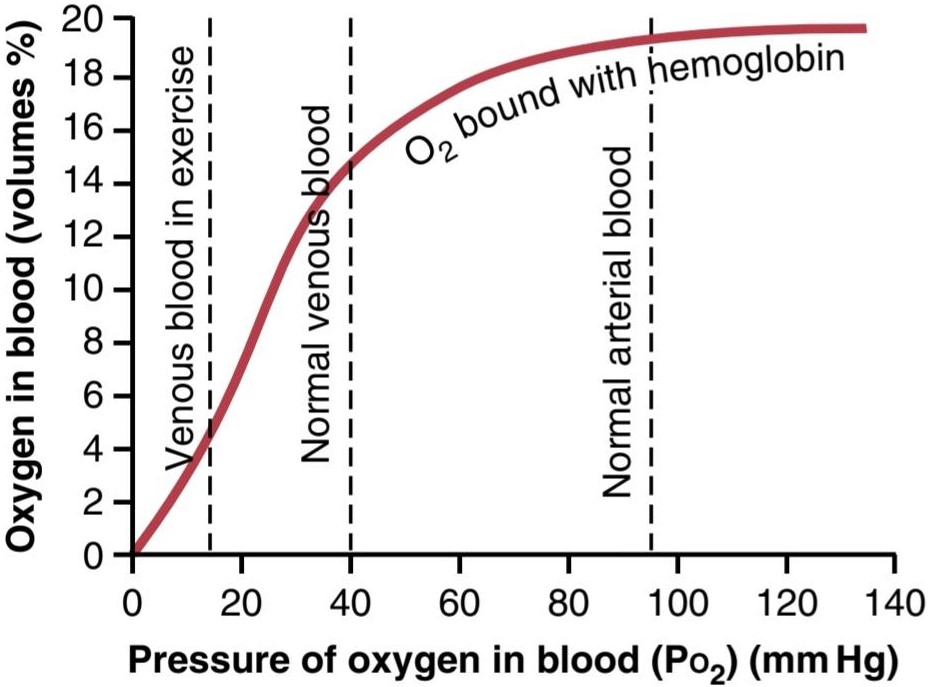
Figure 41-9 Effect of blood PO2 on the quantity of oxygen bound with hemoglobin in each 100 ml of blood.
شکل 41-9 اثر PO2 خون بر روی مقدار اکسیژن متصل به هموگلوبین در هر 100 میلی لیتر خون.
Hemoglobin “Buffers” Tissue Po2
Although hemoglobin is necessary for the transport of O2 to the tissues, it performs another function essential to life. This is its function as a tissue oxygen buffer system. That is, the hemoglobin in the blood is mainly responsible for stabilizing the Po2 in the tissues, which can be explained as follows.
هموگلوبین “بافر” بافت Po2
اگرچه هموگلوبین برای انتقال O2 به بافتها ضروری است، اما عملکرد دیگری را انجام میدهد که برای زندگی ضروری است. این عملکرد آن به عنوان یک سیستم بافر اکسیژن بافتی است. یعنی هموگلوبین خون عمدتاً مسئول تثبیت Po2 در بافتها است که میتوان آن را به شرح زیر توضیح داد.
Hemoglobin Helps Maintain Nearly Constant Po2 in the Tissues. Under basal conditions, the tissues re- quire about 5 ml of O2 from each 100 ml of blood passing through the tissue capillaries. Referring to the O2- hemoglobin dissociation curve in Figure 41-9, note that for the normal 5 ml of O2 to be released per 100 ml of blood flow, the Po2 must fall to about 40 mm Hg. Therefore, the tissue Po2 normally cannot rise above this 40 mm Hg level because, if it did, the amount of O2 needed by the tissues would not be released from the hemoglobin. In this way, the hemoglobin normally sets an upper limit on the Po2 in the tissues at about 40 mm Hg.
هموگلوبین به حفظ تقریباً ثابت Po2 در بافت کمک میکند. در شرایط پایه، بافتها به حدود 5 میلی لیتر O2 از هر 100 میلی لیتر خونی که از مویرگهای بافت میگذرد نیاز دارند. با اشاره به منحنی تفکیک هموگلوبین O2 در شکل 41-9، توجه داشته باشید که برای آزاد شدن 5 میلی لیتر طبیعی O2 در هر 100 میلی لیتر جریان خون، Po2 باید به حدود 40 میلی متر جیوه کاهش یابد. بنابراین، Po2 بافت به طور معمول نمیتواند از این سطح 40 میلی متر جیوه بالاتر برود، زیرا در این صورت، مقدار O2 مورد نیاز بافتها از هموگلوبین آزاد نمیشود. به این ترتیب، هموگلوبین به طور معمول یک حد بالایی برای Po2 در بافتها در حدود 40 میلی متر جیوه تعیین میکند.
Conversely, during heavy exercise, extra amounts of O2 (as much as 20 times normal) must be delivered from the hemoglobin to the tissues. However, this delivery of extra O2 can be achieved with little further decrease in tissue Po2 because of (1) the steep slope of the dissociation curve, and (2) the increase in tissue blood flow caused by the decreased Po2. That is, a very small fall in Po2 causes large amounts of extra O2 to be released from hemoglobin. Thus, hemoglobin in the blood automatically delivers O2 to the tissues at a pressure that is held rather tightly between about 15 and 40 mm Hg.
برعکس، در طول ورزش سنگین، مقدار اضافی O2 (به اندازه 20 برابر طبیعی) باید از هموگلوبین به بافتها منتقل شود. با این حال، این تحویل O2 اضافی را میتوان با کاهش بیشتر در Po2 بافت به دلیل (1) شیب تند منحنی تفکیک، و (2) افزایش در جریان خون بافت ناشی از کاهش Po2 به دست آورد. یعنی کاهش بسیار اندک Po2 باعث آزاد شدن مقادیر زیادی O2 اضافی از هموگلوبین میشود. بنابراین، هموگلوبین در خون به طور خودکار O2 را با فشاری که بین 15 تا 40 میلی متر جیوه نگه داشته میشود، به بافتها میرساند.
When Atmospheric Oxygen Concentration Changes Markedly, the Buffer Effect of Hemoglobin Still Maintains Almost Constant Tissue Po22. The normal Po2 in the alveoli is about 104 mm Hg, but as one ascends a mountain or ascends in an airplane, the Po2 can easily fall to less than half this amount. Alternatively, when one enters areas of compressed air, such as deep in the sea or in pressurized chambers, the Po2 may rise to 10 times this level. Even so, the tissue Po2 changes little.
هنگامیکه غلظت اکسیژن اتمسفر به طور قابل توجهی تغییر میکند، اثر بافر هموگلوبین همچنان بافت Po22 را تقریباً ثابت نگه میدارد. PO2 طبیعی در آلوئولها حدود 104 میلی متر جیوه است، اما با بالا رفتن از کوه یا بالا رفتن در هواپیما، Po2 به راحتی میتواند به کمتر از نصف این مقدار سقوط کند. از طرف دیگر، هنگامیکه فرد وارد مناطقی از هوای فشرده میشود، مانند اعماق دریا یا اتاقهای تحت فشار، Po2 ممکن است تا 10 برابر این سطح افزایش یابد. با این حال، بافت Po2 کمیتغییر میکند.
It can be seen from the oxygen-hemoglobin dissociation curve in Figure 41-8 that when the alveolar Po2 is decreased to as low as 60 mm Hg, the arterial hemoglobin is still 89% saturated with O2-only 8% below the normal saturation of 97%. Furthermore, the tissues still remove about 5 ml of O2 from each 100 ml of blood passing through the tissues. To remove this O2, the Po2 of the venous blood falls to 35 mm Hg-only 5 mm Hg below the normal value of 40 mm Hg. Thus, the tissue Po2 hardly changes, despite the marked fall in alveolar Po2 from 104 to 60 mm Hg.
میتوان از منحنی تفکیک اکسیژن-هموگلوبین در شکل 41-8 مشاهده کرد که وقتی Po2 آلوئولی تا 60 میلی متر جیوه کاهش مییابد، هموگلوبین شریانی هنوز 89٪ با O2 اشباع است – فقط 8٪ زیر اشباع طبیعی 97٪. علاوه بر این، بافتها هنوز حدود 5 میلی لیتر O2 را از هر 100 میلی لیتر خون عبوری از بافتها حذف میکنند. برای حذف این O2، Po2 خون وریدی به 35 میلیمتر جیوه – فقط 5 میلیمتر جیوه کمتر از مقدار طبیعی 40 میلیمتر جیوه میرسد. بنابراین، بافت Po2 به سختی تغییر میکند، علیرغم کاهش مشخص در Po2 آلوئولی از 104 به 60 میلیمتر جیوه.
Conversely, when the alveolar Po2 rises as high as 500 mm Hg, the maximum O2 saturation of hemoglobin can never rise above 100%, which is only 3% above the normal level of 97%. Only a small amount of additional O2 dis- solves in the fluid of the blood, as will be discussed subsequently. Then, when the blood passes through the tissue capillaries and loses several milliliters of O2 to the tissues, this reduces the Po2 of the capillary blood to a value only a few milliliters higher than the normal 40 mm Hg. Consequently, the level of alveolar O2 may vary greatly-from 60 to more than 500 mm Hg Po2-and still the Po2 in the peripheral tissues does not vary more than a few milliliters from normal, demonstrating beautifully the tissue “oxygen buffer” function of the blood hemoglobin system.
برعکس، وقتی Po2 آلوئولی تا 500 میلیمتر جیوه بالا میرود، حداکثر اشباع O2 هموگلوبین هرگز نمیتواند از 100 درصد بالاتر برود که تنها 3 درصد بالاتر از سطح طبیعی 97 درصد است. همانطور که در ادامه صحبت خواهد شد، تنها مقدار کمیO2 اضافی در مایع خون حل میشود. سپس، هنگامیکه خون از مویرگهای بافتی عبور میکند و چندین میلیلیتر O2 به بافتها از دست میدهد، Po2 خون مویرگی را به مقداری که تنها چند میلیلیتر بالاتر از 40 میلیمتر جیوه طبیعی است کاهش میدهد. در نتیجه، سطح O2 آلوئولی ممکن است بسیار متفاوت باشد – از 60 تا بیش از 500 میلیمتر جیوه Po2 – و هنوز هم Po2 در بافتهای محیطی بیش از چند میلیلیتر از نرمال تغییر نمیکند که به زیبایی عملکرد «بافر اکسیژن» بافتی سیستم هموگلوبین خون را نشان میدهد.
Factors That Shift the Oxygen- Hemoglobin Dissociation Curve-Their Importance for Oxygen Transport
The O2-hemoglobin dissociation curves of Figures 41-8 and 41-9 are for normal average blood. However, several factors can displace the dissociation curve in one direction or the other, as shown in Figure 41-10. This figure shows that when the blood becomes slightly acidic, with the pH decreasing from the normal value of 7.4 to 7.2, the O2- hemoglobin dissociation curve shifts, on average, about 15% to the right. Conversely, an increase in pH from the normal 7.4 to 7.6 shifts the curve a similar amount to the left.
عواملی که منحنی تفکیک اکسیژن – هموگلوبین را تغییر میدهند – اهمیت آنها برای انتقال اکسیژن
منحنیهای تفکیک O2-هموگلوبین در شکلهای 41-8 و 41-9 برای خون متوسط طبیعی است. با این حال، عوامل متعددی میتوانند منحنی تفکیک را در یک جهت یا جهت دیگر جابجا کنند، همانطور که در شکل 41-10 نشان داده شده است. این شکل نشان میدهد که وقتی خون کمیاسیدی میشود و pH از مقدار طبیعی 7.4 به 7.2 کاهش مییابد، منحنی تفکیک هموگلوبین O2 به طور متوسط حدود 15٪ به سمت راست تغییر میکند. برعکس، افزایش pH از 7.4 معمولی به 7.6 منحنی را به میزان مشابهی به سمت چپ تغییر میدهد.
In addition to pH changes, several other factors are known to shift the curve. Three of these, which shift the curve to the right are the following: (1) increased CO2 concentration; (2) increased blood temperature; and (3) increased 2,3-biphosphoglycerate (BPG), a metabolically important phosphate compound present in the blood in different concentrations under different metabolic conditions.
علاوه بر تغییرات pH، چندین عامل دیگر برای تغییر منحنی شناخته شده است. سه مورد از این موارد که منحنی را به سمت راست تغییر میدهند عبارتند از: (1) افزایش غلظت CO2. (2) افزایش دمای خون. و (3) افزایش 2،3-biphosphoglycerate (BPG)، یک ترکیب فسفات متابولیکی مهم موجود در خون در غلظتهای مختلف تحت شرایط متابولیک مختلف.
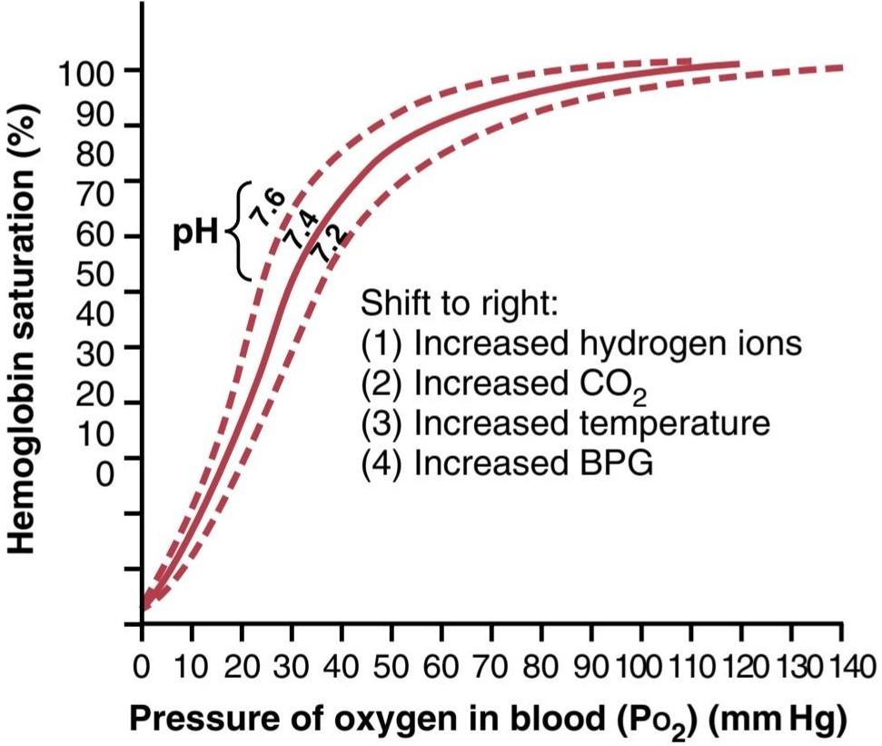
Figure 41-10 Shift of the oxygen-hemoglobin dissociation curve to the right caused by an increase in H+ concentration (decrease in pH). BPG, 2,3-Biphosphoglycerate.
شکل 41-10 تغییر منحنی تفکیک اکسیژن-هموگلوبین به سمت راست ناشی از افزایش غلظت +H (کاهش pH). BPG، 2،3-Biphosphoglycerate.
Increased Delivery of Oxygen to Tissues When CO2 and H+ Shift the Oxygen- Hemoglobin Dissociation Curve-the Bohr Effect
A shift of the oxygen-hemoglobin dissociation curve to the right in response to increases in blood CO2 and H+ levels has a significant effect by enhancing the release of O2 from the blood in the tissues and enhancing oxygenation of the blood in the lungs. This is called the Bohr effect, which can be explained as follows. As the blood passes through the tissues, CO2 diffuses from tissue cells into the blood. This diffusion increases the blood Pco2, which in turn raises blood H2CO3 (carbonic acid) and H+ concentration. These effects shift the O2-hemoglobin dissociation curve to the right and downward, as shown in Figure 41-10, forcing O2 away from the hemoglobin and therefore delivering increased amounts of O2 to the tissues.
افزایش تحویل اکسیژن به بافتها هنگامیکه CO2 و +H منحنی تفکیک اکسیژن – هموگلوبین را تغییر میدهند – اثر بور
تغییر منحنی تفکیک اکسیژن-هموگلوبین به سمت راست در پاسخ به افزایش سطح CO2 و +H خون با افزایش آزادسازی O2 از خون در بافتها و افزایش اکسیژن رسانی خون در ریهها، تأثیر قابل توجهی دارد. این اثر بور نامیده میشود که میتوان آن را به صورت زیر توضیح داد. با عبور خون از بافتها، CO2 از سلولهای بافتی به داخل خون پخش میشود. این انتشار باعث افزایش Pco2 خون میشود که به نوبه خود غلظت H2CO3 (اسید کربنیک) و +H را افزایش میدهد. این اثرات منحنی تفکیک O2-هموگلوبین را به سمت راست و پایین تغییر میدهد، همانطور که در شکل 41-10 نشان داده شده است، O2 را از هموگلوبین دور میکند و بنابراین مقادیر افزایش یافته O2 را به بافتها میرساند.
Exactly the opposite effects occur in the lungs, where CO2 diffuses from the blood into alveoli. This diffusion reduces blood Pco2 and H+ concentration, shifting the O2-hemoglobin dissociation curve to the left and upward. Therefore, the quantity of O2 that binds with the hemoglobin at any given alveolar Po2 becomes considerably increased, thus allowing greater O2 transport to the tissues.
دقیقاً اثرات معکوس در ریهها رخ میدهد، جایی که CO2 از خون به آلوئولها پخش میشود. این انتشار غلظت Pco2 و +H خون را کاهش میدهد و منحنی تفکیک O2-هموگلوبین را به سمت چپ و به بالا تغییر میدهد. بنابراین، مقدار O2 که با هموگلوبین در هر Po2 آلوئولی خاص متصل میشود، به طور قابل توجهی افزایش مییابد، بنابراین امکان انتقال بیشتر O2 به بافتها را فراهم میکند.
Effect of BPG to Cause Rightward Shift of the Oxygen-Hemoglobin Dissociation Curve
The normal BPG in the blood always keeps the O2- hemoglobin dissociation curve shifted slightly to the right. In hypoxic conditions that last longer than a few hours, the quantity of BPG in the blood increases considerably, thus shifting the O2-hemoglobin dissociation curve even farther to the right. This shift causes O2 to be released to the tissues at as much as 10 mm Hg higher tissue O2 pressure than would be the case without this increased BPG. Therefore, under some conditions, the BPG mechanism can be important for adaptation to hypoxia, especially to hypoxia caused by poor tissue blood flow.
تأثیر BPG در ایجاد تغییر اکسیژن-هموگلوبین به سمت راست منحنی تفکیک
BPG طبیعی در خون همیشه منحنی تفکیک هموگلوبین O2 را کمیبه سمت راست تغییر میدهد. در شرایط هیپوکسیک که بیش از چند ساعت طول میکشد، مقدار BPG در خون به طور قابل توجهی افزایش مییابد، بنابراین منحنی تفکیک O2-هموگلوبین حتی بیشتر به سمت راست منتقل میشود. این تغییر باعث میشود که O2 با فشار 10 میلی متر جیوه بالاتر از O2 بافتی که بدون این افزایش BPG اتفاق میافتد به بافتها آزاد شود. بنابراین، تحت برخی شرایط، مکانیسم BPG میتواند برای سازگاری با هیپوکسی، به ویژه هیپوکسی ناشی از جریان خون ضعیف بافت، مهم باشد.
Rightward Shift of the Oxygen- Hemoglobin Dissociation Curve During Exercise
During exercise, several factors shift the dissociation curve considerably to the right, thus delivering extra amounts of O2 to the active, exercising muscle fibers. The exercising muscles, in turn, release large quantities of CO2; this and several other acids released by the muscles increase the H+ concentration in the muscle capillary blood. In addition, the temperature of the muscle often rises 2° to 3°C, which can increase O2 delivery to the muscle fibers even more. All these factors act together to shift the oxygen-hemoglobin dissociation curve of the muscle capillary blood considerably to the right. This rightward shift of the curve forces O2 to be released from blood hemoglobin to the muscle at Po2 levels as great as 40 mm Hg, even when 70% of the O2 has already been removed from the hemoglobin. Then, in the lungs, the shift occurs in the opposite direction, allowing extra amounts of O2 to be picked up from alveoli.
تغییر منحنی تفکیک اکسیژن-هموگلوبین به سمت راست در حین ورزش
در طول تمرین، عوامل متعددی منحنی تفکیک را به طور قابل توجهی به سمت راست تغییر میدهند، بنابراین مقادیر اضافی O2 را به فیبرهای عضلانی فعال و فعال میرساند. ماهیچههای تمرین کننده به نوبه خود مقادیر زیادی CO2 آزاد میکنند. این و چندین اسید دیگر که توسط ماهیچهها آزاد میشوند، غلظت +H را در خون مویرگی عضله افزایش میدهند. علاوه بر این، دمای عضله اغلب 2 تا 3 درجه سانتیگراد افزایش مییابد که میتواند تحویل O2 به فیبرهای عضلانی را حتی بیشتر افزایش دهد. همه این عوامل با هم عمل میکنند تا منحنی تفکیک اکسیژن-هموگلوبین خون مویرگی عضلانی را به طور قابل توجهی به سمت راست تغییر دهند. این تغییر منحنی به سمت راست، O2 را مجبور میکند تا از هموگلوبین خون به عضله در سطوح Po2 تا 40 میلی متر جیوه آزاد شود، حتی زمانی که 70٪ از O2 قبلاً از هموگلوبین خارج شده باشد. سپس، در ریهها، جابجایی در جهت مخالف اتفاق میافتد و اجازه میدهد مقادیر اضافی O2 از آلوئولها برداشت شود.
METABOLIC USE OF OXYGEN BY CELLS
Effect of Intracellular Po2 on Oxygen Usage Rate. Only a minute level of O2 pressure is required in the cells for normal intracellular chemical reactions to take place. The reason for this phenomenon is that the respiratory enzyme systems of the cell, discussed in Chapter 68, have been configured so that when the cellular Po2 is more than 1 mm Hg, O2 availability is no longer a limiting factor in the rates of the chemical reactions. Instead, the main limiting factor is the concentration of adenosine diphosphate (ADP) in the cells. This effect is demonstrated in Figure 41-11, which shows the relationship between intracellular Po2 and the O2 usage rate at different concentrations of ADP. Note that whenever the intracellular Po2 is above 1 mm Hg, O2 usage rate becomes constant for any given concentration of ADP in the cell. Conversely, when the ADP concentration is altered, the rate of O2 usage changes in proportion to the change in ADP concentration.
استفاده متابولیکی از اکسیژن توسط سلولها
اثر Po2 داخل سلولی بر میزان مصرف اکسیژن. فقط یک دقیقه فشار O2 در سلولها برای انجام واکنشهای شیمیایی درون سلولی طبیعی لازم است. دلیل این پدیده این است که سیستمهای آنزیمیتنفسی سلول، که در فصل 68 مورد بحث قرار گرفت، به گونه ای پیکربندی شده اند که وقتی Po2 سلولی بیش از 1 میلی متر جیوه است، در دسترس بودن O2 دیگر یک عامل محدود کننده در سرعت واکنشهای شیمیایی نیست. در عوض، عامل محدود کننده اصلی غلظت آدنوزین دی فسفات (ADP) در سلولها است. این اثر در شکل 41-11 نشان داده شده است که رابطه بین Po2 داخل سلولی و میزان مصرف O2 را در غلظتهای مختلف ADP نشان میدهد. توجه داشته باشید که هر زمان Po2 داخل سلولی بالاتر از 1 میلی متر جیوه باشد، میزان مصرف O2 برای هر غلظت معینی از ADP در سلول ثابت میشود. برعکس، زمانی که غلظت ADP تغییر میکند، میزان مصرف O2 متناسب با تغییر غلظت ADP تغییر میکند.
As explained in Chapter 3, when adenosine triphosphate (ATP) is used in the cells to provide energy, it is converted into ADP. The increasing concentration of ADP increases metabolic usage of O2 as it combines with the various cell nutrients, releasing energy that reconverts the ADP back to ATP. Under normal operating conditions, the rate of O2 usage by the cells is controlled ultimately by the rate of energy expenditure within the cells-that is, by the rate at which ADP is formed from ATP.
همانطور که در فصل 3 توضیح داده شد، هنگامیکه آدنوزین تری فسفات (ATP) در سلولها برای تامین انرژی استفاده میشود، به ADP تبدیل میشود. افزایش غلظت ADP استفاده متابولیکی O2 را افزایش میدهد زیرا با مواد مغذی مختلف سلول ترکیب میشود و انرژی آزاد میکند که ADP را دوباره به ATP تبدیل میکند. در شرایط عملیاتی عادی، نرخ استفاده از O2 توسط سلولها در نهایت توسط نرخ مصرف انرژی در سلولها کنترل میشود، یعنی با سرعتی که ADP از ATP تشکیل میشود.
Effect of Diffusion Distance From the Capillary to the Cell on Oxygen Usage. Tissue cells are seldom more than 50 micrometers away from a capillary, and O2 normally can diffuse readily enough from the capillary to the cell to supply all the required amounts of O, for metabolism. However, occasionally, cells are located farther from the capillaries, and the rate of O2 diffusion to these cells can become so low that intracellular Po2 falls below the critical level required to maintain maximal intracellular metabolism. Thus, under these conditions, O, usage by the cells is diffusion-limited and is no longer determined by the amount of ADP formed in the cells. However, this situation almost never occurs, except in pathological states.
تأثیر فاصله انتشار از مویرگ تا سلول بر مصرف اکسیژن. سلولهای بافتی به ندرت بیش از 50 میکرومتر از مویرگ فاصله دارند و O2 به طور معمول میتواند به راحتی از مویرگ به سلول پخش شود تا تمام مقادیر مورد نیاز O را برای متابولیسم تامین کند. با این حال، گاهی اوقات، سلولها دورتر از مویرگها قرار میگیرند و سرعت انتشار O2 به این سلولها میتواند آنقدر کم شود که Po2 داخل سلولی به زیر سطح بحرانی مورد نیاز برای حفظ حداکثر متابولیسم داخل سلولی میرسد. بنابراین، تحت این شرایط، استفاده از O توسط سلولها محدود به انتشار است و دیگر با مقدار ADP تشکیل شده در سلولها تعیین نمیشود. با این حال، این وضعیت تقریبا هرگز اتفاق نمیافتد، مگر در حالتهای پاتولوژیک.
Effect of Blood Flow on Metabolic Use of Oxygen. The total amount of O2 available each minute for use in any given tissue is determined by (1) the quantity of O2 that can be transported to the tissue in each 100 ml of blood, and (2) the rate of blood flow. If blood flow rate falls to zero, the amount of available O2 also falls to zero. Thus, there are times when blood flow rate through a tissue can be so low that tissue Po2 falls below the critical 1 mm Hg required for intracellular metabolism. Under these conditions, the rate of tissue usage of O2 is blood flow-limited. Neither diffusion-limited nor blood flow- limited oxygen states can continue for long, however, be- cause the cells receive less O2 than is required to continue the life of the cells.
تأثیر جریان خون بر مصرف متابولیک اکسیژن. مقدار کل O2 موجود در هر دقیقه برای استفاده در هر بافت معین با (1) مقدار O2 که میتواند در هر 100 میلی لیتر خون به بافت منتقل شود و (2) سرعت جریان خون تعیین میشود. اگر سرعت جریان خون به صفر برسد، مقدار O2 موجود نیز به صفر میرسد. بنابراین، مواقعی وجود دارد که سرعت جریان خون از طریق بافت میتواند آنقدر پایین باشد که Po2 بافت به زیر 1 میلیمتر جیوه حیاتی مورد نیاز برای متابولیسم داخل سلولی میرسد. تحت این شرایط، میزان استفاده از بافت O2 از نظر جریان خون محدود است. با این حال، نه حالتهای اکسیژن با انتشار محدود و نه جریان خون محدود میتواند برای مدت طولانی ادامه یابد، زیرا سلولها O2 کمتری از آنچه برای ادامه حیات سلولها لازم است دریافت میکنند.
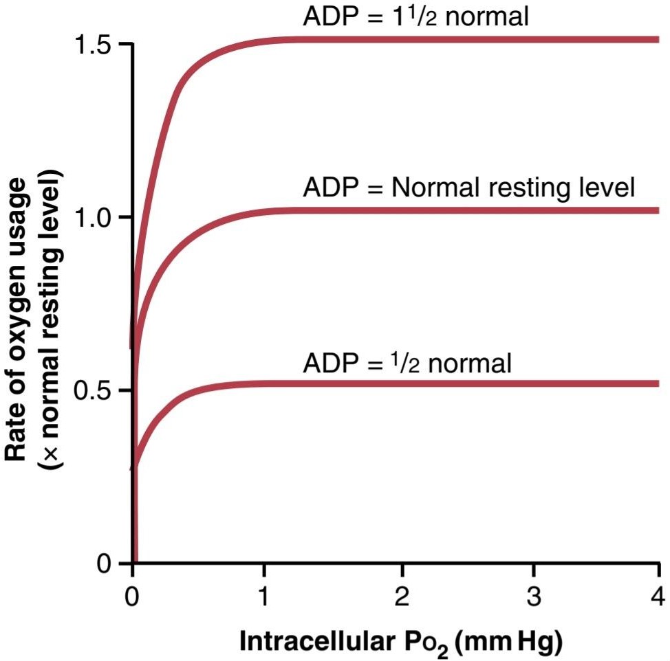
Figure 41-11 Effect of intracellular adenosine diphosphate (ADP) and Po2 on rate of oxygen usage by the cells. Note that as long as the intracellular Po2 remains above 1 mm Hg, the controlling factor for the rate of oxygen usage is the intracellular concentration of ADP.
شکل 41-11 اثر آدنوزین دی فسفات داخل سلولی (ADP) و Po2 بر میزان مصرف اکسیژن توسط سلولها. توجه داشته باشید که تا زمانی که Po2 درون سلولی بالای 1 میلی متر جیوه باقی بماند، عامل کنترل کننده برای میزان مصرف اکسیژن، غلظت درون سلولی ADP است.
Transport of Oxygen in the Dissolved State
At the normal arterial Po2 of 95 mm Hg, about 0.29 ml of O, is dissolved in every 100 ml of water in the blood, and when the Po2 of the blood falls to the normal 40 mm Hg in the tissue capillaries, only 0.12 ml of O2 remains dissolved. In other words, 0.17 ml of O2 is normally transported in the dissolved state to the tissues by each 100 ml of arterial blood flow. This value compares with almost 5 ml of O2 transported by the red blood cell hemoglobin. Therefore, the amount of O2 transported to the tissues in the dissolved state is normally slight, only about 3% of the total, as com- pared with 97% transported by the hemoglobin.
انتقال اکسیژن در حالت محلول
در Po2 شریان طبیعی 95 میلی متر جیوه، حدود 0.29 میلی لیتر O در هر 100 میلی لیتر آب در خون حل میشود و زمانی که Po2 خون به 40 میلی متر جیوه طبیعی در مویرگهای بافت میرسد، تنها 0.12 میلی لیتر O2 حل شده باقی میماند. به عبارت دیگر، 0.17 میلی لیتر O2 به طور معمول در حالت محلول با هر 100 میلی لیتر جریان خون شریانی به بافتها منتقل میشود. این مقدار با تقریباً 5 میلی لیتر O2 منتقل شده توسط هموگلوبین گلبول قرمز مقایسه میشود. بنابراین، مقدار O2 منتقل شده به بافتها در حالت محلول معمولاً ناچیز است، فقط حدود 3٪ از کل، در مقایسه با 97٪ منتقل شده توسط هموگلوبین.
During strenuous exercise, when hemoglobin release of O2 to the tissues increases by another 3-fold, the relative quantity of O2 transported in the dissolved state falls to as little as 1.5%. However, if a person breathes O2 at very high alveolar Po2 levels, the amount transported in the dissolved state can become much greater, sometimes so much so that a serious excess of O2 occurs in the tissues, and so-called O2 poisoning ensues. This condition often leads to brain convulsions and even death, as discussed in detail in Chapter 45 in relation to the high-pressure breathing of O, among deep-sea divers.
در طول ورزش شدید، هنگامیکه آزادسازی هموگلوبین O2 به بافتها تا 3 برابر افزایش مییابد، مقدار نسبی O2 منتقل شده در حالت محلول به 1.5٪ کاهش مییابد. با این حال، اگر فردی O2 را در سطوح بسیار بالای Po2 آلوئولی تنفس کند، مقدار انتقال یافته در حالت محلول میتواند بسیار بیشتر شود، گاهی اوقات به حدی که مقدار زیادی O2 در بافتها ایجاد میشود و به اصطلاح مسمومیت O2 ایجاد میشود. این وضعیت اغلب منجر به تشنج مغزی و حتی مرگ میشود، همانطور که در فصل 45 در رابطه با تنفس پرفشار O در میان غواصان در اعماق دریا به تفصیل بحث شد.
Combination of Hemoglobin With Carbon Monoxide Displacement of O2
Carbon monoxide (CO) combines with hemoglobin at the same point on the hemoglobin molecule as O2; it can therefore displace O2 from the hemoglobin and decrease the O2 carrying capacity of blood. Furthermore, it binds with about 250 times as much tenacity as O2, which is demonstrated by the CO- hemoglobin dissociation curve in Figure 41-12. This curve is almost identical to the O2-hemoglobin dissociation curve except that the CO partial pressures, shown on the abscissa, are at a level 250 of those for the O2-hemoglobin dissociation curve of Figure 41-8. Therefore, a CO partial pressure of only 0.4 mm Hg in the alveoli, so that of normal alveolar O2 (100 mm Hg Po2), allows the CO to compete equally with the O2 for combination with the hemoglobin and causes 50% of the hemoglobin in the blood to become bound with CO instead of with O2. Therefore, a CO pressure of only 0.6 mm Hg (a volume concentration < one part/1000 in air) can be lethal.
ترکیب هموگلوبین با جابجایی مونوکسید کربن O2
مونوکسید کربن (CO) با هموگلوبین در همان نقطه روی مولکول هموگلوبین با O2 ترکیب میشود. بنابراین میتواند O2 را از هموگلوبین جابجا کند و ظرفیت حمل O2 خون را کاهش دهد. علاوه بر این، با استحکامیحدود 250 برابر O2 متصل میشود که با منحنی تفکیک هموگلوبین CO در شکل 41-12 نشان داده شده است. این منحنی تقریباً مشابه منحنی تفکیک هموگلوبین O2 است با این تفاوت که فشارهای جزئی CO، نشان داده شده روی آبسیسا، در سطح 250 منحنی تفکیک هموگلوبین O2 در شکل 41-8 است. بنابراین، فشار جزئی CO فقط 0.4 میلی متر جیوه در آلوئولها، به طوری که O2 آلوئولی طبیعی (100 میلی متر جیوه Po2)، به CO اجازه میدهد تا به طور مساوی با O2 برای ترکیب با هموگلوبین رقابت کند و باعث میشود 50٪ از هموگلوبین خون به جای با O2 به CO متصل شود. بنابراین، فشار CO تنها 0.6 میلی متر جیوه (غلظت حجمی< یک قسمت در 1000 در هوا) میتواند کشنده باشد.
Even though the O2 content of blood is greatly reduced in CO poisoning, the Po, of the blood may be normal. This situation makes exposure to CO especially dangerous because the blood is bright red, and there are no obvious signs of hypoxemia, such as a bluish color of the fingertips or lips (cyanosis). Also, Po2 is not reduced, and the feed- back mechanism that usually stimulates an increased respiration rate in response to lack of O2 (usually reflected by a low Po2) is absent. Because the brain is one of the first organs affected by lack of oxygen, the person may become disoriented and unconscious before becoming aware of the danger.
حتی اگر مقدار O2 خون در مسمومیت با CO به شدت کاهش مییابد، Po خون ممکن است طبیعی باشد. این وضعیت قرار گرفتن در معرض CO را به ویژه خطرناک میکند زیرا خون قرمز روشن است و هیچ نشانه آشکاری از هیپوکسمیمانند رنگ آبی نوک انگشتان یا لبها (سیانوز) وجود ندارد. همچنین، Po2 کاهش نمییابد و مکانیسم بازخوردی که معمولاً باعث افزایش سرعت تنفس در پاسخ به کمبود O2 میشود (معمولاً توسط Po2 کم منعکس میشود) وجود ندارد. از آنجایی که مغز یکی از اولین اندامهایی است که تحت تاثیر کمبود اکسیژن قرار میگیرد، ممکن است فرد قبل از آگاهی از خطر دچار سردرگمیو بیهوشی شود.
A patient severely poisoned with CO can be treated by administering pure O2 because O2 at high alveolar pressure can displace CO rapidly from its combination with hemoglobin. The patient can also benefit from simultaneous ad- ministration of 5% CO2 because this strongly stimulates the respiratory center, which increases alveolar ventilation and reduces alveolar CO. With intensive O2 and CO2 therapy, CO can be removed from the blood as much as 10 times as rapidly as without therapy.
بیماری که به شدت با CO مسموم شده است را میتوان با تجویز O2 خالص درمان کرد زیرا O2 در فشار آلوئولی بالا میتواند CO را به سرعت از ترکیب آن با هموگلوبین جابجا کند. بیمار همچنین میتواند از تجویز همزمان 5% CO2 بهره مند شود زیرا این امر به شدت مرکز تنفسی را تحریک میکند که تهویه آلوئولی را افزایش میدهد و CO2 آلوئولی را کاهش میدهد.

Figure 41-12 Carbon monoxide-hemoglobin dissociation curve. Note the extremely low carbon monoxide pressures at which carbon monoxide combines with hemoglobin.
شکل 41-12 منحنی تفکیک مونوکسید کربن – هموگلوبین. به فشارهای بسیار کم مونوکسید کربن که در آن مونوکسید کربن با هموگلوبین ترکیب میشود توجه کنید.
TRANSPORT OF CO2 IN BLOOD
Transport of CO2 by the blood is not nearly as problematical as transport of O2 is because even in the most abnormal conditions, CO, can usually be transported in far greater quantities than can O2. However, the amount of CO2 in the blood has a lot to do with the acid-base balance of the body fluids, which is discussed in Chapter 31. Under normal resting conditions, an average of 4 ml of CO2 are transported from the tissues to the lungs in each 100 ml of blood.
انتقال CO2 در خون
انتقال CO2 توسط خون تقریباً به اندازه انتقال O2 مشکل ساز نیست، زیرا حتی در غیر طبیعی ترین شرایط، CO2 معمولاً میتواند در مقادیر بسیار بیشتری نسبت به O2 منتقل شود. با این حال، مقدار CO2 در خون ارتباط زیادی با تعادل اسید و باز مایعات بدن دارد که در فصل 31 مورد بحث قرار گرفته است. در شرایط استراحت عادی، در هر 100 میلی لیتر خون به طور متوسط 4 میلی لیتر CO2 از بافتها به ریهها منتقل میشود.
CHEMICAL FORMS IN WHICH CO2 IS TRANSPORTED
To begin the process of CO2 transport, CO, diffuses out of the tissue cells in the dissolved molecular CO2 form. On entering the tissue capillaries, the CO2 initiates a host of almost instantaneous physical and chemical reactions, shown in Figure 41-13, which are essential for CO2 transport.
اشکال شیمیایی که در آنها CO2 منتقل میشود
برای شروع فرآیند انتقال CO2، CO، به شکل محلول مولکولی CO2 از سلولهای بافت منتشر میشود. با ورود به مویرگهای بافتی، CO2 یک میزبان از واکنشهای فیزیکی و شیمیایی تقریباً آنی را آغاز میکند، که در شکل 41-13 نشان داده شده است، که برای انتقال CO2 ضروری هستند.
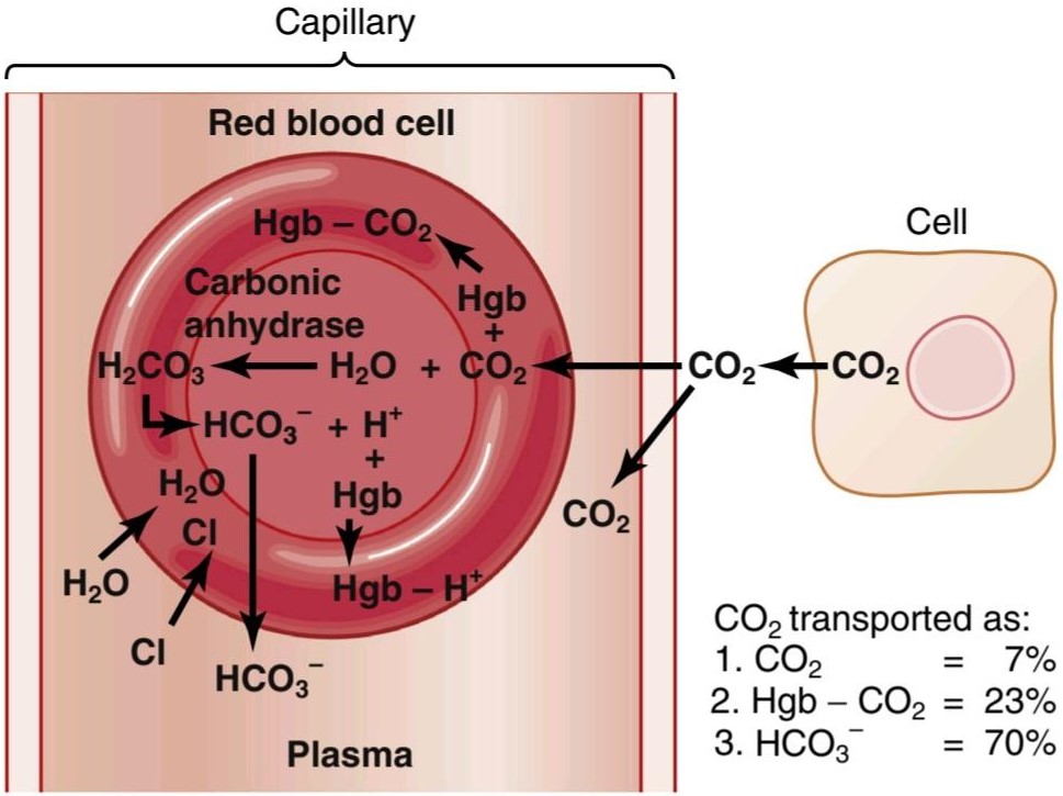
Figure 41-13 Transport of carbon dioxide in the blood. Hgb, Hemoglobin.
شکل 41-13 انتقال دی اکسید کربن در خون. Hgb، هموگلوبین.
Transport of CO2 in a Dissolved State
A small portion of the CO2 is transported in the dissolved state to the lungs. Recall that the Pco, of venous blood is 45 mm Hg and that of arterial blood is 40 mm Hg. The amount of CO2 dissolved in the fluid of the blood at 45 mm Hg is about 2.7 ml/dl (2.7 volume percent). The amount dissolved at 40 mm Hg is about 2.4 ml, or a difference of 0.3 ml. Therefore, only about 0.3 ml of CO2 is transported in the dissolved form by each 100 ml of blood flow. This is about 7% of all the CO2 normally transported.
انتقال CO2 در حالت محلول
بخش کوچکی از CO2 در حالت محلول به ریهها منتقل میشود. به یاد داشته باشید که Pco خون وریدی 45 میلی متر جیوه و خون شریانی 40 میلی متر جیوه است. مقدار CO2 حل شده در مایع خون در 45 میلی متر جیوه حدود 2.7 میلی لیتر در دسی لیتر (2.7 درصد حجمی) است. مقدار حل شده در 40 میلی متر جیوه حدود 2.4 میلی لیتر یا اختلاف 0.3 میلی لیتر است. بنابراین، تنها حدود 0.3 میلی لیتر CO2 به شکل محلول توسط هر 100 میلی لیتر جریان خون منتقل میشود. این حدود 7 درصد از کل CO2 به طور معمول منتقل میشود.
Transport of CO2 in the Form of Bicarbonate lon
Carbonic Anhydrase Catalyzes the Reaction of CO2 With Water in Red Blood Cells. The dissolved CO2 in the blood reacts with water to form carbonic acid. This reaction would occur much too slowly to be of importance were it not for the fact that there is an enzyme called carbonic anhydrase inside the red blood cells, which catalyzes the reaction between CO2 and water and accelerates its reaction rate by about 5000-fold. Therefore, instead of requiring many seconds or minutes to occur, as is true in the plasma, the reaction occurs so rapidly in red blood cells that it reaches almost complete equilibrium within a small fraction of a second. This phenomenon allows tremendous amounts of CO2 to react with the red blood cell water, even before the blood leaves the tissue capillaries.
انتقال CO2 به شکل یون بیکربنات
کربنیک انیدراز واکنش CO2 با آب در گلبولهای قرمز خون را کاتالیز میکند. CO2 محلول در خون با آب واکنش داده و اسید کربنیک تشکیل میدهد. اگر آنزیمیبه نام آنهیدراز کربنیک در داخل گلبولهای قرمز وجود نداشته باشد که واکنش بین CO2 و آب را کاتالیز میکند و سرعت واکنش آن را حدود 5000 برابر تسریع میکند، این واکنش بسیار آهسته رخ میدهد و اهمیت چندانی ندارد. بنابراین، به جای نیاز به چندین ثانیه یا دقیقه برای وقوع، همانطور که در پلاسما صادق است، واکنش آنقدر سریع در گلبولهای قرمز رخ میدهد که در کسری کوچک از ثانیه تقریباً به تعادل کامل میرسد. این پدیده اجازه میدهد تا مقادیر زیادی از CO2 با آب گلبول قرمز، حتی قبل از خروج خون از مویرگهای بافت، واکنش نشان دهد.
Dissociation of Carbonic Acid Into Bicarbonate and Hydrogen lons. In another fraction of a second, the carbonic acid formed in red cells (H2CO3) dissociates into H+ and bicarbonate ions (H+ and HCO3-). Most of the H+ ions then combine with hemoglobin in the red blood cells because the hemoglobin protein is a powerful acid-base buffer. In turn, many of the HCO3- ions diffuse from the red blood cells into the plasma while chloride ions diffuse into the red blood cells to take their place. This diffusion is made possible by the presence of a special bicarbonate- chloride carrier protein in the red blood cell membrane that shuttles these two ions in opposite directions at rapid velocities. Thus, the chloride content of venous red blood cells is greater than that of arterial red blood cells, a phenomenon called the chloride shift.
تفکیک اسید کربنیک به لونهای بی کربنات و هیدروژن. در کسری دیگر از ثانیه، اسید کربنیک تشکیل شده در گلبولهای قرمز (H2CO3) به یونهای +H و بی کربنات (+H و -HCO3) تجزیه میشود. سپس اکثر یونهای +H با هموگلوبین در گلبولهای قرمز ترکیب میشوند زیرا پروتئین هموگلوبین یک بافر قوی اسید-باز است. به نوبه خود، بسیاری از یونهای -HCO3 از گلبولهای قرمز خون به پلاسما منتشر میشوند در حالی که یونهای کلرید به گلبولهای قرمز منتشر میشوند تا جای خود را بگیرند. این انتشار با حضور یک پروتئین حامل بی کربنات-کلرید خاص در غشای گلبول قرمز امکان پذیر میشود که این دو یون را در جهت مخالف با سرعتهای سریع جابه جا میکند. بنابراین، محتوای کلرید گلبولهای قرمز وریدی بیشتر از گلبولهای قرمز شریانی است، پدیدهای که به آن تغییر کلرید میگویند.
The reversible combination of CO2 with water in red blood cells under the influence of carbonic anhydrase accounts for about 70% of the CO2 transported from the tissues to the lungs. Thus, this means of transporting CO2 is the most important. Indeed, when a carbonic anhydrase inhibitor (e.g., acetazolamide) is administered to an animal to block the action of carbonic anhydrase in the red blood cells, CO2 transport from the tissues becomes so poor that the tissue Pco, may rise to 80 mm Hg instead of the normal 45 mm Hg.
ترکیب برگشت پذیر CO2 با آب در گلبولهای قرمز تحت تأثیر کربنیک انیدراز حدود 70 درصد از CO2 منتقل شده از بافتها به ریهها را تشکیل میدهد. بنابراین، این وسیله برای انتقال CO2 مهم ترین است. در واقع، هنگامیکه یک مهارکننده کربنیک انیدراز (به عنوان مثال، استازولامید) برای جلوگیری از عملکرد کربنیک انیدراز در گلبولهای قرمز خون به حیوان تجویز میشود، انتقال CO2 از بافتها چنان ضعیف میشود که Pco بافت ممکن است به جای 45 میلیمتر جیوه طبیعی به 80 میلیمتر جیوه برسد.
Transport of CO2 in Combination With Hemoglobin and Plasma Proteins-Carbaminohemoglobin. In addition to reacting with water, CO2 reacts directly with amine radicals of the hemoglobin molecule to form the compound carbaminohemoglobin (CO2Hgb). This combination of CO2 and hemoglobin is a reversible reaction that occurs with a loose bond, so the CO2 is easily released into the alveoli, where the Pco2 is lower than in the pulmonary capillaries.
انتقال CO2 در ترکیب با هموگلوبین و پروتئینهای پلاسما – کربامینو هموگلوبین. CO2 علاوه بر واکنش با آب، مستقیماً با رادیکالهای آمینی مولکول هموگلوبین واکنش میدهد و ترکیب کربامینو هموگلوبین (CO2Hgb) را تشکیل میدهد. این ترکیب CO2 و هموگلوبین یک واکنش برگشت پذیر است که با یک پیوند شل رخ میدهد، بنابراین CO2 به راحتی در آلوئولها آزاد میشود، جایی که Pco2 کمتر از مویرگهای ریوی است.
A small amount of CO2 also reacts in the same way with the plasma proteins in tissue capillaries. This reaction is much less significant for the transport of CO2 because the quantity of these proteins in the blood is only one-fourth as great as the quantity of hemoglobin.
مقدار کمیاز CO2 نیز به همان شیوه با پروتئینهای پلاسما در مویرگهای بافت واکنش نشان میدهد. این واکنش برای انتقال CO2 اهمیت کمتری دارد زیرا مقدار این پروتئینها در خون تنها یک چهارم مقدار هموگلوبین است.
The quantity of CO2 that can be carried from the peripheral tissues to the lungs by carbamino combination with hemoglobin and plasma proteins is about 30% of the total quantity transported that is, normally about 1.5 ml of CO2 in each 100 ml of blood. However, because this reaction is much slower than the reaction of CO2 with water inside the red blood cells, it is doubtful that under normal conditions this carbamino mechanism transports more than 20% of the total CO2.
مقدار CO2 که میتواند از بافتهای محیطی به ریهها توسط ترکیب کربامینو با هموگلوبین و پروتئینهای پلاسما منتقل شود، حدود 30٪ از کل مقدار انتقال یافته است، که به طور معمول حدود 1.5 میلی لیتر CO2 در هر 100 میلی لیتر خون است. با این حال، از آنجایی که این واکنش بسیار کندتر از واکنش CO2 با آب در داخل گلبولهای قرمز است، تردید وجود دارد که در شرایط عادی این مکانیسم کربامینو بیش از 20٪ از کل CO2 را منتقل کند.
CARBON DIOXIDE DISSOCIATION CURVE
The curve shown in Figure 41-14, the CO2 dissociation curve, depicts the dependence of total blood CO2 in all its forms on Pco2. Note that the normal blood Pco2 is within a narrow range of 40 mm Hg in arterial blood and 45 mm Hg in venous blood. Note also that the normal concentration of CO2 in the blood in all its different forms is about 50 volume percent, but only 4 volume percent of this is exchanged during normal transport of CO2 from the tissues to the lungs. That is, the concentration rises to about 52 volume percent as the blood passes through the tissues and falls to about 48 volume percent as it passes through the lungs.
منحنی تجزیه دی اکسید کربن
منحنی نشان داده شده در شکل 41-14، منحنی تفکیک CO2، وابستگی CO2 کل خون را در تمام اشکال آن به Pco2 نشان میدهد. توجه داشته باشید که Pco2 طبیعی خون در محدوده باریک 40 میلی متر جیوه در خون شریانی و 45 میلی متر جیوه در خون وریدی قرار دارد. همچنین توجه داشته باشید که غلظت طبیعی CO2 در خون در تمام اشکال مختلف آن حدود 50 درصد حجمیاست، اما تنها 4 درصد حجمیآن در طی انتقال طبیعی CO2 از بافتها به ریهها مبادله میشود. یعنی با عبور خون از بافتها غلظت آن به حدود 52 درصد حجمیافزایش مییابد و با عبور از ریهها به حدود 48 درصد حجمیکاهش مییابد.

Figure 41-14 Carbon dioxide dissociation curve.
شکل 41-14 منحنی تفکیک دی اکسید کربن.
When Oxygen Binds With Hemoglobin, CO2 Is Released (the Haldane Effect) to Increase CO2 Transport
Earlier in the chapter, we noted that an increase in CO2 in the blood causes O2 to be displaced from the hemoglobin (the Bohr effect), which is an important factor in increasing O2 transport. The reverse is also true-binding of O2 with hemoglobin tends to displace CO2 from the blood. This effect, called the Haldane effect, is quantitatively far more important in promoting CO2 transport than the Bohr effect in promoting O2 transport.
هنگامیکه اکسیژن با هموگلوبین متصل میشود، CO2 آزاد میشود (اثر Haldane) تا انتقال CO2 را افزایش دهد.
در اوایل فصل، ما اشاره کردیم که افزایش CO2 در خون باعث میشود O2 از هموگلوبین (اثر بور) جابجا شود، که عامل مهمیدر افزایش انتقال O2 است. معکوس نیز اتصال واقعی O2 با هموگلوبین است که تمایل به جابجایی CO2 از خون دارد. این اثر که اثر Haldane نامیده میشود، از نظر کمیدر ارتقای انتقال CO2 بسیار مهمتر از اثر Bohr در ترویج انتقال O2 است.
The Haldane effect results from the simple fact that the combination of O2 with hemoglobin in the lungs causes the hemoglobin to become a stronger acid. This displaces CO2 from the blood and into the alveoli in two ways. First, the more highly acidic hemoglobin has less tendency to combine with CO2 to form carbaminohemoglobin, thus displacing much of the CO2 that is present in the carbamino form from the blood. Second, the increased acidity of the hemoglobin also causes it to release an excess of H+, and these ions bind with HCO3 to form carbonic acid, which then dissociates into water and CO2, and the CO2 is released from the blood into the alveoli and, finally, into the air.
اثر Haldane از این واقعیت ساده ناشی میشود که ترکیب O2 با هموگلوبین در ریهها باعث میشود هموگلوبین به اسید قوی تری تبدیل شود. این امر به دو صورت CO2 را از خون و به آلوئولها جابجا میکند. اولاً، هموگلوبین بسیار اسیدیتر تمایل کمتری به ترکیب با CO2 برای تشکیل کربامینو هموگلوبین دارد، بنابراین مقدار زیادی از CO2 موجود در فرم کربوهیدراتی را از خون جابجا میکند. دوم اینکه افزایش اسیدیته هموگلوبین باعث آزاد شدن +H اضافی میشود و این یونها با HCO3 متصل میشوند و اسید کربنیک تشکیل میدهند که سپس به آب و CO2 تجزیه میشود و CO2 از خون به آلوئولها و در نهایت به هوا آزاد میشود.
Figure 41-15 demonstrates quantitatively the significance of the Haldane effect on the transport of CO2 from the tissues to the lungs. This figure shows small portions of two CO2 dissociation curves: (1) when the Po2 is 100 mm Hg, which is the case in the blood capillaries of the lungs; and (2) when the Po2 is 40 mm Hg, which is the case in the tissue capillaries. Point A shows that the normal Pco2 of 45 mm Hg in the tissues causes 52 volume percent of CO2 to combine with the blood. On entering the lungs, the Pco2 falls to 40 mm Hg, and the Po2 rises to 100 mm Hg. If the CO2 dissociation curve did not shift because of the Haldane effect, the CO2 content of the blood would fall only to 50 volume percent, which would be a loss of only 2 volume percent of CO2. However, the increase in Po2 in the lungs lowers the CO2 dissociation curve from the top curve to the lower curve of the figure, so the CO2 con- tent falls to 48 volume percent (point B). This represents an additional two volume percent loss of CO2. Thus, the Haldane effect approximately doubles the amount of CO2 released from the blood in the lungs and approximately doubles the amount of CO2 picked up in the tissues.
شکل 41-15 اهمیت اثر Haldane را بر انتقال CO2 از بافتها به ریهها به صورت کمینشان میدهد. این شکل بخشهای کوچکی از دو منحنی تفکیک CO2 را نشان میدهد: (1) زمانی که Po2 100 میلیمتر جیوه است، که در مویرگهای خونی ریهها وجود دارد. و (2) هنگامیکه Po2 40 میلی متر جیوه است، که در مویرگهای بافتی وجود دارد. نقطه A نشان میدهد که Pco2 طبیعی 45 میلی متر جیوه در بافتها باعث میشود که 52 درصد حجمیCO2 با خون ترکیب شود. با ورود به ریهها، Pco2 به 40 میلی متر جیوه کاهش مییابد و Po2 تا 100 میلی متر جیوه افزایش مییابد. اگر منحنی تفکیک CO2 به دلیل اثر Haldane تغییر نکند، محتوای CO2 خون تنها تا 50 درصد حجمیکاهش مییابد که تنها 2 درصد حجمیCO2 از دست میرود. با این حال، افزایش Po2 در ریهها منحنی تفکیک CO2 را از منحنی بالا به منحنی پایین شکل پایین میآورد، بنابراین محتوای CO2 به 48 درصد حجمی(نقطه B) کاهش مییابد. این نشان دهنده کاهش دو درصد حجمیCO2 اضافی است. بنابراین، اثر Haldane مقدار CO2 آزاد شده از خون در ریهها را تقریبا دو برابر میکند و مقدار CO2 جذب شده در بافتها را تقریبا دو برابر میکند.

Figure 41-15 Portions of the carbon dioxide dissociation curve when the Po2 is 100 or 40 mm Hg. The arrow represents the Haldane effect on the transport of carbon dioxide.
شکل 41-15 بخشهایی از منحنی تفکیک دی اکسید کربن زمانی که Po2 100 یا 40 میلی متر جیوه است. فلش نشان دهنده اثر Haldane در انتقال دی اکسید کربن است.
Change in Blood Acidity During Co2 Transport
The carbonic acid formed when CO2 enters the blood in peripheral tissues decreases blood pH. However, reaction of this acid with the acid-base buffers of the blood prevents the H+ concentration from rising very much (and the pH from falling very much). Ordinarily, arterial blood has a pH of about 7.41 and as the blood acquires CO2 in the tissue capillaries the pH falls to a venous value of about 7.37. In other words, a pH change of 0.04 unit takes place. The reverse occurs when CO2 is released from the blood in the lungs, with the pH rising to the arterial value of 7.41 once again. During heavy exercise or other conditions of high metabolic activity, or when blood flow through the tissues is sluggish, the decrease in pH in the tissue blood (and in the tissues themselves) can be as much as 0.50, about 12 times normal, thus causing significant tissue acidosis.
تغییر در اسیدیته خون در طول انتقال CO2
اسید کربنیک تشکیل شده هنگام ورود CO2 به خون در بافتهای محیطی باعث کاهش pH خون میشود. با این حال، واکنش این اسید با بافرهای اسید-باز خون، از افزایش بسیار زیاد غلظت +H (و کاهش شدید pH) جلوگیری میکند. به طور معمول، خون شریانی دارای pH حدود 7.41 است و زمانی که خون CO2 در مویرگهای بافتی بدست میآورد، pH به مقدار وریدی حدود 7.37 کاهش مییابد. به عبارت دیگر، تغییر pH 0.04 واحد اتفاق میافتد. عکس آن زمانی رخ میدهد که CO2 از خون در ریهها آزاد میشود و PH یک بار دیگر به مقدار شریانی 7.41 افزایش مییابد. در طول ورزش سنگین یا سایر شرایط فعالیت متابولیک بالا، یا زمانی که جریان خون در بافتها کند است، کاهش pH در خون بافت (و در خود بافتها) میتواند تا 0.50، حدود 12 برابر طبیعی باشد، در نتیجه باعث اسیدوز بافتی قابل توجهی میشود.
RESPIRATORY EXCHANGE RATIO
The discerning student will have noted that normal trans- port of O2 from the lungs to the tissues by each 100 ml of blood is about 5 ml, whereas normal transport of CO2 from the tissues to the lungs is about 4 ml. Thus, under normal resting conditions, only about 82% as much CO2 is expired from the lungs as O2 is taken up by the lungs. The ratio of CO2 output to O2 uptake is called the respiratory exchange ratio (R). That is:
نسبت تبادل تنفسی
دانش آموز فهیم متوجه خواهد شد که انتقال طبیعی O2 از ریهها به بافتها توسط هر 100 میلی لیتر خون حدود 5 میلی لیتر است، در حالی که انتقال طبیعی CO2 از بافتها به ریهها حدود 4 میلی لیتر است. بنابراین، در شرایط استراحت عادی، تنها حدود 82 درصد CO2 از ریهها خارج میشود که O2 توسط ریهها جذب میشود. نسبت خروجی CO2 به جذب O2 را نسبت تبادل تنفسی (R) مینامند. یعنی:

The value for R changes under different metabolic conditions. When a person is using carbohydrates exclusively for body metabolism, R rises to 1.00. Conversely, when a person is using fats exclusively for metabolic energy, the R level falls to as low as 0.7. The reason for this difference is that when O2 is metabolized with carbohydrates, one molecule of CO2 is formed for each molecule of O2 consumed; when O2 reacts with fats, a large share of the O2 combines with hydrogen atoms from the fats to form water instead of CO2. In other words, when fats are metabolized, the respiratory quotient of the chemical reactions in the tissues is about 0.70 instead of 1.00. (The tis- sue respiratory quotient is discussed in Chapter 72.) For a person on a normal diet consuming average amounts of carbohydrates, fats, and proteins, the average value for R is considered to be 0.825.
مقدار R تحت شرایط متابولیک مختلف تغییر میکند. هنگامیکه فردی از کربوهیدراتها به طور انحصاری برای متابولیسم بدن استفاده میکند، R به 1.00 افزایش مییابد. برعکس، وقتی فردی از چربیها به طور انحصاری برای انرژی متابولیک استفاده میکند، سطح R به 0.7 کاهش مییابد. دلیل این تفاوت این است که وقتی O2 با کربوهیدرات متابولیزه میشود، به ازای هر مولکول O2 مصرف شده یک مولکول CO2 تشکیل میشود. هنگامیکه O2 با چربیها واکنش میدهد، سهم بزرگی از O2 با اتمهای هیدروژن چربیها ترکیب میشود و به جای CO2 آب تشکیل میدهد. به عبارت دیگر، هنگامیکه چربیها متابولیزه میشوند، ضریب تنفسی واکنشهای شیمیایی در بافتها به جای 1.00، حدود 0.70 است. (ضریب تنفسی بافت در فصل 72 مورد بحث قرار گرفته است.) برای فردی که رژیم غذایی معمولی دارد و مقادیر متوسط کربوهیدرات، چربی و پروتئین مصرف میکند، مقدار متوسط R 0.825 در نظر گرفته میشود.
کلیک کنید «Bibliography: فهرست کتب مربوطه»
Clanton TL, Hogan MC, Gladden LB: Regulation of cellular gas ex- change, oxygen sensing, and metabolic control. Compr Physiol 3:1135, 2013.
Geers C, Gros G: Carbon dioxide transport and carbonic anhydrase in blood and muscle. Physiol Rev 80:681, 2000.
Jensen FB: Red blood cell pH, the Bohr effect, and other oxygenation- linked phenomena in blood O2 and CO2 transport. Acta Physiol Scand 182:215, 2004.
Jensen FB: The dual roles of red blood cells in tissue oxygen deliv- ery: oxygen carriers and regulators of local blood flow. J Exp Biol 212:3387, 2009.
Joyner MJ, Casey DP: Regulation of increased blood flow (hyperemia) to muscles during exercise: a hierarchy of competing physiological needs. Physiol Rev 95:549, 2015.
Maina JN, West JB: Thin and strong! The bioengineering dilemma in the structural and functional design of the blood-gas barrier. Physiol Rev 85:811, 2005.
Mairbäurl H, Weber RE: Oxygen transport by hemoglobin. Compr Physiol 2:1463, 2012.
Moore LG: Measuring high-altitude adaptation. J Appl Physiol 123:1371, 2017.
Poole DC, Jones AM: Oxygen uptake kinetics. Compr Physiol 2:933, 2012.
Richardson RS: Oxygen transport and utilization: an integration of the muscle systems. Adv Physiol Educ 27:183, 2003.
Rossiter HB: Exercise: Kinetic considerations for gas exchange. Compr Physiol. 1:203, 2011.
Tsai AG, Johnson PC, Intaglietta M: Oxygen gradients in the microcir- culation. Physiol Rev 83:933, 2003.
🚀 با ما همراه شوید!
تازهترین مطالب و آموزشهای مغز و اعصاب را از دست ندهید. با فالو کردن کانال تلگرام آیندهنگاران مغز، از ما حمایت کنید!



