
فیزیولوژی پزشکی گایتون و هال؛ تنظیم تنفس


دعای مطالعه [ نمایش ]
بِسْمِ الله الرَّحْمنِ الرَّحیمِ
اَللّهُمَّ اَخْرِجْنى مِنْ ظُلُماتِ الْوَهْمِ
خدايا مرا بيرون آور از تاريكىهاى وهم،
وَ اَكْرِمْنى بِنُورِ الْفَهْمِ
و به نور فهم گرامى ام بدار،
اَللّهُمَّ افْتَحْ عَلَيْنا اَبْوابَ رَحْمَتِكَ
خدايا درهاى رحمتت را به روى ما بگشا،
وَانْشُرْ عَلَيْنا خَزائِنَ عُلُومِكَ بِرَحْمَتِكَ يا اَرْحَمَ الرّاحِمينَ
و خزانههاى علومت را بر ما باز كن به امید رحمتت اى مهربانترين مهربانان.
کتاب «فیزیولوژی پزشکی گایتون وهال» بهعنوان یکی از جامعترین و معتبرترین منابع در حوزه علوم پزشکی، همچنان مرجع کلیدی برای درک عملکرد پیچیده بدن انسان است. این اثر با تکیه بر تازهترین پژوهشها و توضیحات دقیق از سازوکارهای فیزیولوژیک، پلی میان علوم پایه پزشکی و کاربردهای بالینی ایجاد میکند و نقشی بیبدیل در آموزش، پژوهش و ارتقای دانش سلامت ایفا مینماید.
ترجمه دقیق و علمیاین شاهکار توسط برند علمیآیندهنگاران مغز به مدیریت داریوش طاهری، دسترسی فارسیزبانان به مرزهای نوین دانش فیزیولوژی را ممکن ساخته و رسالتی علمیبرای ارتقای آموزش پزشکی، فهم عمیقتر سازوکارهای بدن و توسعه روشهای نوین در حوزه سلامت فراهم آورده است.
» Guyton and Hall Textbook of Medical Physiology
»» CHAPTER 42: Regulation of Respiration
The nervous system normally adjusts the rate of alveolar ventilation to meet the demands of the body almost exactly so that the oxygen partial pressure (Po2) and carbon dioxide partial pressure (Pco2) in the arterial blood are hardly altered, even during heavy exercise and most other types of respiratory stress. This chapter describes the function of this neurogenic system for regulation of respiration.
سیستم عصبی به طور معمول میزان تهویه آلوئولی را طوری تنظیم میکند که نیازهای بدن را تقریباً دقیقاً برآورده کند، به طوری که فشار جزئی اکسیژن (Po2) و فشار جزئی دی اکسید کربن (Pco2) در خون شریانی به سختی تغییر میکند، حتی در طول ورزش سنگین و بسیاری از انواع دیگر استرسهای تنفسی. این فصل عملکرد این سیستم عصبی را برای تنظیم تنفس شرح میدهد.
RESPIRATORY CENTER
The respiratory center is composed of several groups of neurons located bilaterally in the medulla oblongata and pons of the brain stem, as shown in Figure 42-1. It is divided into three major collections of neurons: (1) a dorsal respiratory group, located in the dorsal portion of the medulla, which mainly causes inspiration; (2) a ventral respiratory group, located in the ventrolateral part of the medulla, which mainly causes expiration; and (3) the pneumotaxic center, located dorsally in the superior portion of the pons, which mainly controls rate and depth of breathing.
مرکز تنفس
همانطور که در شکل 42-1 نشان داده شده است، مرکز تنفسی از چندین گروه نورون تشکیل شده است که به صورت دو طرفه در بصل النخاع و پونز ساقه مغز قرار دارند. این به سه مجموعه اصلی از نورونها تقسیم میشود: (1) یک گروه تنفسی پشتی، واقع در قسمت پشتی مدولا، که عمدتا باعث دم میشود. (2) یک گروه تنفسی شکمی، واقع در قسمت شکمیجانبی مدولا، که عمدتاً باعث بازدم میشود. و (3) مرکز پنوموتاکسیک، که به صورت پشتی در قسمت فوقانی پونز قرار دارد، که عمدتاً سرعت و عمق تنفس را کنترل میکند.
DORSAL RESPIRATORY GROUP OF NEURONS CONTROLS INSPIRATION AND RESPIRATORY RHYTHM
The dorsal respiratory group of neurons plays a fundamental role in the control of respiration and extends most of the length of the medulla. Most of its neurons are located in the nucleus of the tractus solitarius (NTS), although additional neurons in the adjacent reticular substance of the medulla also play important roles in respiratory control. The NTS is the sensory termination of both the vagal and the glossopharyngeal nerves, which transmit sensory signals into the respiratory center from the following: (1) peripheral chemoreceptors; (2) baroreceptors; (3) receptors in the liver, pancreas, and multiple parts of the gastrointestinal tract; and (4) several types of receptors in the lungs.
گروه نورونهای تنفسی پشتی کنترل کننده دم و ریتم تنفسی
گروه نورونهای تنفسی پشتی نقش اساسی در کنترل تنفس دارند و بیشتر طول مدولا را گسترش میدهند. اکثر نورونهای آن در هسته tractus solitarius (NTS) قرار دارند، اگرچه نورونهای اضافی در ماده شبکه مجاور مدولا نیز نقش مهمیدر کنترل تنفس دارند. NTS خاتمه حسی اعصاب واگ و گلوسوفارنجئال است که سیگنالهای حسی را از موارد زیر به مرکز تنفسی منتقل میکند: (1) گیرندههای شیمیایی محیطی. (2) بارورسپتورها. (3) گیرندهها در کبد، لوزالمعده و بخشهای متعدد دستگاه گوارش. و (4) چندین نوع گیرنده در ریهها.
Rhythmical Inspiratory Discharges From the Dorsal Respiratory Group. The basic rhythm of respiration is generated mainly in the dorsal respiratory group of neurons. Even when all the peripheral nerves entering the medulla have been sectioned, and the brain stem has been transected above and below the medulla, this group of neurons still emits repetitive bursts of inspiratory neuronal action potentials. The basic cause of these repetitive discharges is unknown. In primitive animals, neural networks have been found in which activity of one set of neurons excites a second set, which in turn inhibits the first. Then, after a period of time, the mechanism repeats itself, continuing throughout the life of the animal. Similar networks of neurons are present in the human being, located entirely within the medulla; it probably involves not only the dorsal respiratory group but adjacent areas of the medulla as well and is responsible for the basic rhythm of respiration.
ترشحات ریتمیک دمیاز گروه تنفسی پشتی. ریتم اصلی تنفس عمدتاً در نورونهای گروه تنفسی پشتی ایجاد میشود. حتی زمانی که تمام اعصاب محیطی وارد شده به بصل النخاع برش داده شده اند و ساقه مغز در بالا و پایین مدولا قطع شده است، این گروه از نورونها همچنان انفجارهای تکراری پتانسیلهای عمل عصبی دمیرا منتشر میکنند. علت اصلی این ترشحات مکرر ناشناخته است. در حیوانات ابتدایی، شبکههای عصبی پیدا شدهاند که در آن فعالیت یک مجموعه از نورونها، مجموعه دوم را تحریک میکند، که به نوبه خود مانع اولی میشود. سپس، پس از مدتی، مکانیسم تکرار میشود و در طول زندگی حیوان ادامه مییابد. شبکههای مشابهی از نورونها در انسان وجود دارند که به طور کامل در داخل بصل النخاع قرار دارند. احتمالاً نه تنها گروه تنفسی پشتی بلکه نواحی مجاور مدولا را نیز درگیر میکند و مسئول ریتم اساسی تنفس است.
Inspiratory “ramp” Signal. The nervous signal that is transmitted to the inspiratory muscles, mainly the diaphragm, is not an instantaneous burst of action potentials. Instead, it begins weakly and increases steadily in a ramp manner for about 2 seconds in normal respiration. It then ceases abruptly for approximately the next 3 seconds, which turns off the excitation of the diaphragm and allows elastic recoil of the lungs and chest wall to cause expiration. Next, the inspiratory signal begins again for another cycle; this cycle repeats again and again, with expiration occurring in between. Thus, the inspiratory signal is a ramp signal. The obvious ad- vantage of the ramp is that it causes a steady increase in the volume of the lungs during inspiration, rather than inspiratory gasps.
سیگنال “رمپ” دمی. سیگنال عصبی که به عضلات دمی، عمدتاً دیافراگم منتقل میشود، یک انفجار آنی از پتانسیلهای عمل نیست. در عوض، ضعیف شروع میشود و به طور پیوسته در یک روش شیب دار برای حدود 2 ثانیه در تنفس طبیعی افزایش مییابد. سپس به طور ناگهانی برای تقریباً 3 ثانیه بعدی متوقف میشود، که تحریک دیافراگم را خاموش میکند و اجازه میدهد تا پس زدن الاستیک ریهها و دیواره قفسه سینه باعث بازدم شود. سپس، سیگنال دمیدوباره برای یک چرخه دیگر شروع میشود. این چرخه بارها و بارها تکرار میشود و انقضا در این بین اتفاق میافتد. بنابراین، سیگنال دمییک سیگنال رمپ است. مزیت آشکار رمپ این است که باعث افزایش مداوم حجم ریهها در حین دم، به جای گازهای دمیمیشود.
Two qualities of the inspiratory ramp are controlled, as follows:
دو کیفیت از رمپ دمیکنترل میشود، به شرح زیر:
1. Control of the rate of increase of the ramp signal so that during heavy respiration, the ramp increases rapidly and therefore fills the lungs rapidly.
1. کنترل میزان افزایش سیگنال رمپ به طوری که در هنگام تنفس سنگین، رمپ به سرعت افزایش مییابد و بنابراین به سرعت ریهها را پر میکند.
2. Control of the limiting point at which the ramp suddenly ceases, which is the usual method for controlling the rate of respiration. That is, the earlier the ramp ceases, the shorter the duration of inspiration. This method also shortens the duration of expiration. Thus, the frequency of respiration is increased.
2. کنترل نقطه محدودی که در آن رمپ به طور ناگهانی متوقف میشود، که روش معمول برای کنترل سرعت تنفس است. به این معنا که هر چه رمپ زودتر متوقف شود، مدت دم کوتاهتر است. این روش همچنین مدت زمان انقضا را کوتاه میکند. بنابراین، تعداد دفعات تنفس افزایش مییابد.
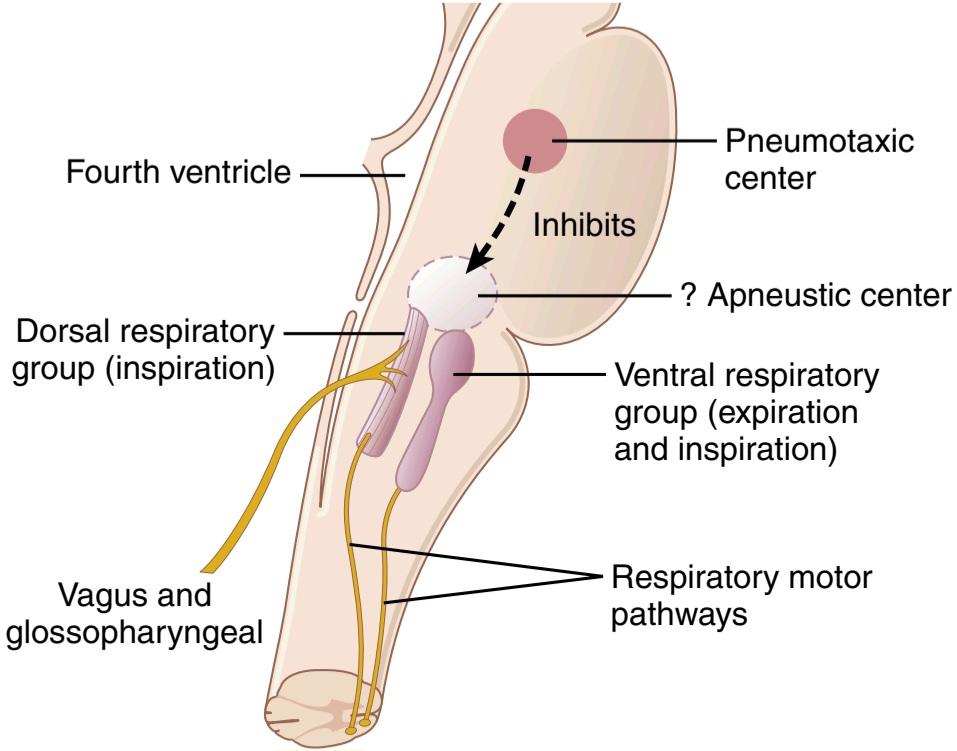
Figure 42-1. Organization of the respiratory center.
شکل 42-1. سازماندهی مرکز تنفسی
PNEUMOTAXIC CENTER LIMITS DURATION OF INSPIRATION AND INCREASES RESPIRATORY RATE
A pneumotaxic center, located dorsally in the nucleus parabrachialis of the upper pons, transmits signals to the inspiratory area. The primary effect of this center is to control the “switch-off” point of the inspiratory ramp, thereby controlling the duration of the filling phase of the lung cycle. When the pneumotaxic signal is strong, inspiration might last for as little as 0.5 second, thus filling the lungs only slightly; when the pneumotaxic signal is weak, inspiration might continue for 5 or more seconds, thus filling the lungs with much greater amounts of air.
مرکز پنوموتاکسیک مدت دم را محدود میکند و تعداد تنفس را افزایش میدهد
یک مرکز پنوموتاکسی که به صورت پشتی در هسته parabrachialis پل بالایی پلک قرار دارد، سیگنالها را به ناحیه دمیمنتقل میکند. اثر اولیه این مرکز کنترل نقطه “خاموش” رمپ دمیاست و در نتیجه مدت مرحله پر شدن چرخه ریه را کنترل میکند. هنگامیکه سیگنال پنوموتاکسی قوی است، دم ممکن است به اندازه 0.5 ثانیه طول بکشد، بنابراین تنها کمیریهها را پر میکند. هنگامیکه سیگنال پنوموتاکسی ضعیف است، دم ممکن است برای 5 ثانیه یا بیشتر ادامه یابد، بنابراین ریهها با مقادیر بسیار بیشتری هوا پر میشود.
The function of the pneumotaxic center is primarily to limit inspiration, which has a secondary effect of increasing the rate of breathing because limitation of inspiration also shortens expiration and the entire period of each respiration. A strong pneumotaxic signal can increase the rate of breathing to 30 to 40 breaths/min, whereas a weak pneumo- taxic signal may reduce the rate to only 3 to 5 breaths/min.
عملکرد مرکز پنوموتاکسیک در درجه اول محدود کردن دم است که یک اثر ثانویه در افزایش سرعت تنفس دارد زیرا محدودیت دم نیز انقضا و کل دوره هر تنفس را کوتاه میکند. یک سیگنال پنوموتاکسی قوی میتواند سرعت تنفس را به 30 تا 40 تنفس در دقیقه افزایش دهد، در حالی که یک سیگنال پنوموتاکسی ضعیف ممکن است سرعت را تنها به 3 تا 5 تنفس در دقیقه کاهش دهد.
VENTRAL RESPIRATORY GROUP OF NEURONS-FUNCTIONS IN BOTH INSPIRATION AND EXPIRATION
Located in each side of the medulla, about 5 millimeters anterior and lateral to the dorsal respiratory group of neurons, is the ventral respiratory group of neurons, found in the nucleus ambiguus rostrally and the nucleus retroambiguus caudally. The function of this neuronal group differs from that of the dorsal respiratory group in several important ways:
گروه نورونهای تنفسی شکمی-عملکرد در هر دو دم و بازدم
در هر طرف بصل النخاع، حدود 5 میلی متر جلو و جانبی از نورونهای گروه تنفسی پشتی، گروه نورونهای تنفسی شکمیقرار دارد که در هسته مبهم به صورت منقاری و هسته رتروامبیگوس به صورت دمییافت میشود. عملکرد این گروه عصبی با عملکرد گروه تنفسی پشتی از چند جهت مهم متفاوت است:
1. The neurons of the ventral respiratory group re- main almost totally inactive during normal quiet respiration. Therefore, normal quiet breathing is caused only by repetitive inspiratory signals from the dorsal respiratory group transmitted mainly to the diaphragm, and expiration results from elastic recoil of the lungs and thoracic cage.
1. نورونهای گروه تنفسی شکمیدر طول تنفس آرام طبیعی تقریباً کاملاً غیرفعال میمانند. بنابراین، تنفس آرام نرمال تنها توسط سیگنالهای دمیمکرر از گروه تنفسی پشتی ایجاد میشود که عمدتاً به دیافراگم منتقل میشود و بازدم ناشی از پسکشی الاستیک ریهها و قفس سینه است.
2. The ventral respiratory neurons do not appear to participate in the basic rhythmical oscillation that controls respiration.
2. به نظر نمیرسد که نورونهای تنفسی شکمیدر نوسانات ریتمیک اساسی که تنفس را کنترل میکند شرکت کنند.
3. When the respiratory drive for increased pulmonary ventilation becomes greater than normal, respiratory signals spill over into the ventral respiratory neurons from the basic oscillating mechanism of the dorsal respiratory area. As a consequence, the ventral respiratory area also contributes extra respiratory drive.
3. هنگامیکه محرک تنفسی برای افزایش تهویه ریوی بیشتر از حد طبیعی میشود، سیگنالهای تنفسی از مکانیسم نوسانی اصلی ناحیه تنفسی پشتی به داخل نورونهای تنفسی شکمیمیریزند. در نتیجه، ناحیه تنفسی شکمینیز به حرکت تنفسی اضافی کمک میکند.
4. Electrical stimulation of a few of the neurons in the ventral group causes inspiration, whereas stimulation of others causes expiration. Therefore, these neurons contribute to both inspiration and expiration. They are especially important in providing the powerful expiratory signals to the abdominal muscles during very heavy expiration. Thus, this area operates more or less as an overdrive mechanism when high levels of pulmonary ventilation are required, especially during heavy exercise.
4. تحریک الکتریکی تعداد کمیاز نورونهای گروه شکمیباعث دم میشود، در حالی که تحریک برخی دیگر باعث بازدم میشود. بنابراین، این نورونها هم به دم و هم در بازدم کمک میکنند. آنها به ویژه در ارائه سیگنالهای بازدمیقدرتمند به عضلات شکم در هنگام بازدم بسیار سنگین اهمیت دارند. بنابراین، زمانی که سطوح بالایی از تهویه ریوی مورد نیاز است، این ناحیه کم و بیش به عنوان یک مکانیسم overdrive عمل میکند، به ویژه در هنگام ورزش سنگین.
LUNG INFLATION SIGNALS LIMIT INSPIRATION-THE HERING-BREUER INFLATION REFLEX
In addition to the central nervous system respiratory control mechanisms operating entirely within the brain stem, sensory nerve signals from the lungs also help control respiration. Most importantly, located in the muscular portions of the walls of the bronchi and bronchioles throughout the lungs are stretch receptors that transmit signals through the vagi into the dorsal respiratory group of neurons when the lungs become overstretched. These signals affect inspiration in much the same way as signals from the pneumotaxic center; that is, when the lungs become overinflated, the stretch receptors activate an appropriate feedback response that “switches off” the inspiratory ramp and thus stops further inspiration. This mechanism is called the Hering-Breuer inflation reflex. This reflex also increases the rate of respiration, as is true for signals from the pneumotaxic center.
سیگنالهای تورم ریه دم را محدود میکند – رفلکس تورم هرینگ-بروئر
علاوه بر مکانیسمهای کنترل تنفسی سیستم عصبی مرکزی که به طور کامل در ساقه مغز عمل میکنند، سیگنالهای عصبی حسی از ریهها نیز به کنترل تنفس کمک میکنند. مهمتر از همه، در قسمتهای عضلانی دیوارههای برونشها و برونشیولها در سراسر ریهها، گیرندههای کششی هستند که سیگنالها را از طریق واگی به گروه تنفسی پشتی نورونها منتقل میکنند که ریهها بیش از حد کشیده میشوند. این سیگنالها مانند سیگنالهای مرکز پنوموتاکسی بر دم تأثیر میگذارند. یعنی وقتی ریهها بیش از حد باد میشوند، گیرندههای کششی پاسخ بازخورد مناسبی را فعال میکنند که سطح شیبدار دمیرا خاموش میکند و بنابراین دمیدن بیشتر را متوقف میکند. این مکانیسم رفلکس تورم هرینگ-بروئر نامیده میشود. این رفلکس همچنین سرعت تنفس را افزایش میدهد، همانطور که برای سیگنالهای مرکز پنوموتاکسیک صادق است.
In humans, the Hering-Breuer reflex probably is not activated until the tidal volume increases to more than three times normal (>1.5 L/breath). Therefore, this reflex appears to be mainly a protective mechanism for preventing excess lung inflation rather than an important factor in normal control of ventilation.
در انسان، رفلکس هرینگ-بروئر احتمالاً تا زمانی که حجم جزر و مدی به بیش از سه برابر نرمال (> 1.5 لیتر در تنفس) افزایش یابد، فعال نمیشود. بنابراین، به نظر میرسد که این رفلکس عمدتاً یک مکانیسم محافظتی برای جلوگیری از تورم اضافی ریه باشد تا یک عامل مهم در کنترل عادی تهویه.
CONTROL OF OVERALL RESPIRATORY CENTER ACTIVITY
Up to this point, we have discussed the basic mechanisms for causing inspiration and expiration, but it is also important to know how the intensity of the respiratory control signals is increased or decreased to match the ventilatory needs of the body. For example, during heavy exercise, the rates of oxygen (O2) usage and carbon dioxide (CO2) formation are often increased to as much as 20 times normal, requiring commensurate increases in pulmonary ventilation. The major purpose of the rest of this chapter is to discuss this control of ventilation in accord with the respiratory needs of the body.
کنترل فعالیت کلی مرکز تنفسی
تا این مرحله، مکانیسمهای اساسی ایجاد دم و بازدم را مورد بحث قرار دادهایم، اما همچنین مهم است که بدانیم چگونه شدت سیگنالهای کنترل تنفسی برای مطابقت با نیازهای تهویه بدن افزایش یا کاهش مییابد. به عنوان مثال، در طول ورزش سنگین، میزان استفاده از اکسیژن (O2) و تشکیل دی اکسید کربن (CO2) اغلب به 20 برابر نرمال افزایش مییابد که نیاز به افزایش متناسب در تهویه ریوی دارد. هدف اصلی بقیه این فصل بحث در مورد این کنترل تهویه مطابق با نیازهای تنفسی بدن است.
CHEMICAL CONTROL OF RESPIRATION
The ultimate goal of respiration is to maintain proper concentrations of O2, CO2, and H+ in the tissues. It is fortunate, therefore, that respiratory activity is highly responsive to changes in each of these substances.
کنترل شیمیایی تنفس
هدف نهایی تنفس حفظ غلظت مناسب O2، CO2 و +H در بافتها است. بنابراین، خوشبختانه فعالیت تنفسی به شدت به تغییرات هر یک از این مواد پاسخ میدهد.
Excess CO2 or excess H+ in the blood mainly act directly on the respiratory center, causing greatly increased strength of both the inspiratory and the expiratory motor signals to the respiratory muscles. Oxygen, in contrast, does not have a major direct effect on the respiratory center of the brain in controlling respiration. Instead, it acts almost entirely on peripheral chemoreceptors located in the carotid and aortic bodies, and these chemoreceptors in turn transmit appropriate nervous signals to the respiratory center for control of respiration.
CO2 اضافی یا +H اضافی در خون عمدتاً مستقیماً روی مرکز تنفسی تأثیر میگذارد و باعث افزایش شدید سیگنالهای حرکتی دمیو بازدمیبه عضلات تنفسی میشود. در مقابل، اکسیژن تأثیر مستقیم عمده ای بر مرکز تنفسی مغز در کنترل تنفس ندارد. در عوض، تقریباً به طور کامل بر روی گیرندههای شیمیایی محیطی واقع در بدن کاروتید و آئورت تأثیر میگذارد و این گیرندههای شیمیایی به نوبه خود سیگنالهای عصبی مناسب را برای کنترل تنفس به مرکز تنفسی منتقل میکنند.
DIRECT CONTROL OF RESPIRATORY CENTER ACTIVITY BY CO2 AND H+
Chemosensitive Area of the Respiratory Center Be- neath the Medulla’s Ventral Surface. We have mainly discussed three areas of the respiratory center-the dorsal respiratory group of neurons, the ventral respiratory group, and the pneumotaxic center. It is believed that none of these is affected directly by changes in blood CO2 or H+ concentration. Instead, an additional neuronal area, a chemosensitive area, shown in Figure 42-2, is located bilaterally, lying only 0.2 millimeter beneath the ventral surface of the medulla. This area is highly sensitive to changes in either blood Pco2 or H+ concentration, and it in turn excites the other portions of the respiratory center.
کنترل مستقیم فعالیت مرکز تنفسی توسط CO2 و +H
ناحیه شیمیایی حساس مرکز تنفسی در زیر سطح شکمیمدولا. ما عمدتاً سه ناحیه از مرکز تنفسی را مورد بحث قرار دادهایم: گروه نورونهای تنفسی پشتی، گروه تنفسی شکمیو مرکز پنوموتاکسیک. اعتقاد بر این است که هیچ یک از اینها مستقیماً تحت تأثیر تغییرات غلظت CO2 یا +H خون قرار نمیگیرند. در عوض، یک ناحیه عصبی اضافی، یک ناحیه حساس شیمیایی، که در شکل 42-2 نشان داده شده است، به صورت دو طرفه قرار دارد و تنها 0.2 میلی متر در زیر سطح شکمیمدولا قرار دارد. این ناحیه نسبت به تغییرات غلظت Pco2 یا +H خون بسیار حساس است و به نوبه خود سایر بخشهای مرکز تنفسی را تحریک میکند.
Excitation of the Chemosensitive Neurons by H+ Is Likely the Primary Stimulus. The sensor neurons in the chemosensitive area are especially excited by H+; in fact, it is believed that H+ may be the only important direct stimulus for these neurons. However, H+ ions do not easily cross the blood-brain barrier. For this reason, changes in H+ concentration in the blood have considerably less effect in stimulating the chemosensitive neurons than changes in blood CO2, even though CO2 is believed to stimulate these neurons secondarily by changing the H+ concentration, as explained in the following section.
تحریک نورونهای حساس شیمیایی توسط +H احتمالاً محرک اولیه است. نورونهای حسگر در ناحیه حساس شیمیایی به ویژه توسط +H برانگیخته میشوند. در واقع، اعتقاد بر این است که +H ممکن است تنها محرک مستقیم مهم برای این نورونها باشد. با این حال، یونهای +H به راحتی از سد خونی مغزی عبور نمیکنند. به همین دلیل، تغییرات در غلظت +H در خون تأثیر قابلتوجهی کمتری در تحریک نورونهای حساس به شیمیایی نسبت به تغییرات CO2 خون دارد، اگرچه اعتقاد بر این است که CO2 این نورونها را بهطور ثانویه با تغییر غلظت +H تحریک میکند، همانطور که در بخش زیر توضیح داده شد.
CO2 Indirectly Stimulates the Chemosensitive Neurons. Although CO2 has little direct effect in stimulating the neurons in the chemosensitive area, it does have a potent indirect effect. It has this effect by reacting with the water of the tissues to form carbonic acid, which dissociates into H+ and HCO3; the H+ then have a potent direct stimulatory effect on respiration. These reactions are shown in Figure 42-2.
CO2 به طور غیر مستقیم نورونهای شیمیایی حساس را تحریک میکند. اگرچه CO2 اثر مستقیم کمیدر تحریک نورونهای ناحیه حساس به شیمیایی دارد، اما اثر غیر مستقیم قوی دارد. این اثر را با واکنش با آب بافتها برای تشکیل اسید کربنیک ایجاد میکند که به +H و HCO3 تجزیه میشود. سپس +H یک اثر تحریکی مستقیم قوی بر روی تنفس دارد. این واکنشها در شکل 42-2 نشان داده شده است.
Why does blood CO2 have a more potent effect in stimulating the chemosensitive neurons than blood H+? The answer is that the blood-brain barrier is not very permeable to H+, but CO2 passes through this barrier almost as if the barrier did not exist. Consequently, when- ever the blood Pco2 increases, so does the Pco2 of both the interstitial fluid of the medulla and the cerebrospinal fluid. In both these fluids, the CO2 immediately reacts with the water to form new H+. Thus, paradoxically, more H+ is released into the respiratory chemosensitive sensory area of the medulla when the blood CO2 concentration increases than when the blood H+ concentration increases. For this reason, respiratory center activity is increased very strongly by changes in blood CO2, a fact that we subsequently discuss quantitatively.
چرا CO2 خون در تحریک نورونهای حساس شیمیایی نسبت به +H خون اثر قوی تری دارد؟ پاسخ این است که سد خونی-مغزی نسبت به +H چندان نفوذپذیر نیست، اما CO2 تقریباً طوری از این سد عبور میکند که گویی سد وجود ندارد. در نتیجه، هر زمان که Pco2 خون افزایش مییابد، Pco2 مایع بینابینی مدولا و مایع مغزی نخاعی نیز افزایش مییابد. در هر دو این مایعات، CO2 بلافاصله با آب واکنش میدهد و +H جدید تشکیل میدهد. بنابراین، به طور متناقض، هنگامیکه غلظت CO2 خون افزایش مییابد، +H بیشتری در ناحیه حسی شیمیایی تنفسی مدولا آزاد میشود تا زمانی که غلظت +H خون افزایش مییابد. به همین دلیل، فعالیت مرکز تنفسی با تغییرات در CO2 خون به شدت افزایش مییابد، این واقعیتی است که ما بعداً به صورت کمیدر مورد آن صحبت میکنیم.
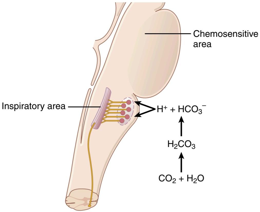
Figure 42-2. Stimulation of the brain stem inspiratory area by signals from the chemosensitive area located bilaterally in the medulla, lying only a fraction of a millimeter beneath the ventral medullary surface. Note also that H+ stimulates the chemosensitive area, but carbon di- oxide in the fluid gives rise to most of the H+.
شکل 42-2. تحریک ناحیه دمیساقه مغز توسط سیگنالهای ناحیه حساس شیمیایی که به صورت دو طرفه در بصل النخاع قرار دارد و تنها کسری از میلی متر زیر سطح مدولاری شکمیقرار دارد. همچنین توجه داشته باشید که +H ناحیه حساس شیمیایی را تحریک میکند، اما دی اکسید کربن موجود در مایع باعث ایجاد بیشتر +H میشود.
Attenuated Stimulatory Effect of CO2 After the First 1 to 2 Days. Excitation of the respiratory center by CO2 is great the first few hours after the blood CO2 first increases, but then it gradually declines over the next 1 to 2 days, decreasing to about one-fifth the initial effect. Part of this decline results from renal readjustment of the H+ concentration in the circulating blood back toward normal after the CO2 first increases the H+ concentration. The kidneys achieve this readjustment by increasing the blood HCO3-, which binds with H+ in the blood and cerebrospinal fluid to reduce their concentrations. But, even more important- ly, over a period of hours, the HCO3- also slowly diffuses through the blood-brain and blood-cerebrospinal fluid barriers and combine directly with H+ adjacent to the respiratory neurons as well, thus reducing the H+ back to near normal. A change in blood CO2 concentration therefore has a potent acute effect on controlling respiratory drive but only a weak chronic effect after a few days’ adaptation.
کاهش اثر تحریکی CO2 پس از 1 تا 2 روز اول. تحریک مرکز تنفسی توسط CO2 در چند ساعت اول پس از افزایش CO2 خون بسیار زیاد است، اما سپس در طی 1 تا 2 روز آینده به تدریج کاهش مییابد و به حدود یک پنجم اثر اولیه کاهش مییابد. بخشی از این کاهش ناشی از تنظیم مجدد کلیوی غلظت +H در خون در گردش به سمت نرمال پس از افزایش CO2 برای اولین بار غلظت +H است. کلیهها با افزایش -HCO3 خون به این تنظیم مجدد دست مییابند که با +H در خون و مایع مغزی نخاعی متصل میشود تا غلظت آنها را کاهش دهد. اما مهمتر از آن، در طی چند ساعت، -HCO3 نیز به آرامیاز طریق سدهای خونی-مغزی و مایع خونی-مغزی نخاعی پخش میشود و مستقیماً با +H مجاور نورونهای تنفسی نیز ترکیب میشود، بنابراین +H را به نزدیک به حالت عادی کاهش میدهد. بنابراین تغییر در غلظت CO2 خون یک اثر حاد قوی بر کنترل حرکت تنفسی دارد اما فقط یک اثر مزمن ضعیف پس از سازگاری چند روزه دارد.
Quantitative Effects of Blood Pco2 and H+ Concentration on Alveolar Ventilation. Figure 42-3 shows quantitatively the approximate effects of blood Pco2 and blood pH (which is an inverse logarithmic measure of H+ concentration) on alveolar ventilation. Note especially the marked increase in ventilation caused by an in- crease in Pco, in the normal range between 35 and 75 mm Hg, which demonstrates the tremendous effect that CO2 changes have in controlling respiration. By contrast, the change in respiration in the normal blood pH range, which is between 7.3 and 7.5, is less than 10% as great.
اثرات کمیغلظت Pco2 و +H خون بر تهویه آلوئولار. شکل 42-3 به صورت کمیاثرات تقریبی Pco2 خون و pH خون (که معیار لگاریتمیمعکوس غلظت +H است) بر تهویه آلوئولی را نشان میدهد. به خصوص به افزایش قابل توجه تهویه ناشی از افزایش Pco، در محدوده طبیعی بین 35 تا 75 میلیمتر جیوه توجه کنید، که نشاندهنده تأثیر فوقالعاده تغییرات CO2 در کنترل تنفس است. در مقابل، تغییر در تنفس در محدوده pH طبیعی خون، که بین 7.3 و 7.5 است، کمتر از 10٪ به همین اندازه است.
Changes in O2 Have Little Direct Effect on Control of the Respiratory Center. Changes in O2 concentration have virtually no direct effect on the respiratory center itself to alter respiratory drive-although O2 changes do have an indirect effect, acting through the peripheral chemoreceptors, as explained in the next section.
تغییرات در O2 تأثیر مستقیم کمیبر کنترل مرکز تنفسی دارد. تغییرات در غلظت O2 عملاً هیچ تأثیر مستقیمیبر خود مرکز تنفسی برای تغییر محرک تنفسی ندارد – اگرچه تغییرات O2 یک اثر غیرمستقیم دارد و از طریق گیرندههای شیمیایی محیطی، همانطور که در بخش بعدی توضیح داده شد، عمل میکند.
We learned in Chapter 41 that the hemoglobin-oxygen buffer system delivers almost exactly normal amounts of O2 to the tissues, even when the pulmonary Po2 changes from a value as low as 60 mm Hg up to a value as high as 1000 mm Hg. Therefore, except under special conditions, adequate delivery of O2 can occur despite changes in lung ventilation ranging from slightly below half-normal to as high as 20 or more times normal. This is not true for CO2 because both the blood and tissue Pco2 change inversely with the rate of pulmonary ventilation; thus, the processes of animal evolution have made CO2 the major controller of respiration, not O2.
ما در فصل 41 یاد گرفتیم که سیستم بافر هموگلوبین-اکسیژن تقریباً دقیقاً مقادیر طبیعی O2 را به بافتها میرساند، حتی زمانی که Po2 ریوی از مقدار کم 60 میلیمتر جیوه به مقدار بالای 1000 میلیمتر جیوه تغییر میکند. بنابراین، به جز در شرایط خاص، تحویل کافی O2 میتواند علیرغم تغییرات در تهویه ریه از کمیکمتر از نیمه نرمال تا 20 یا بیشتر برابر طبیعی رخ دهد. این برای CO2 درست نیست زیرا هم Pco2 خون و هم بافت برعکس با سرعت تهویه ریوی تغییر میکنند. بنابراین، فرآیندهای تکامل حیوانات CO2 را به کنترل کننده اصلی تنفس تبدیل کرده است نه O2.
Yet, for those special conditions in which the tissues get into trouble for lack of O2, the body has a special mechanism for respiratory control located in the peripheral chemoreceptors, outside the brain respiratory center. This mechanism responds when the blood O2 falls too low, mainly below a Po2 of 70 mm Hg, as explained in the next section.
با این حال، برای شرایط خاصی که در آن بافتها به دلیل کمبود اکسیژن دچار مشکل میشوند، بدن مکانیسم خاصی برای کنترل تنفسی دارد که در گیرندههای شیمیایی محیطی، خارج از مرکز تنفسی مغز قرار دارد. همانطور که در بخش بعدی توضیح داده شد، این مکانیسم زمانی پاسخ میدهد که O2 خون خیلی پایین میآید، عمدتاً زیر یک Po2 70 میلیمتر جیوه.
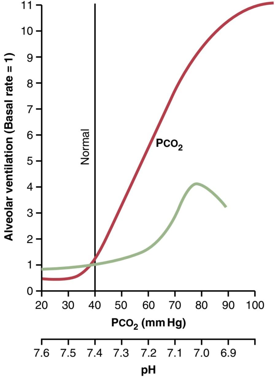
Figure 42-3. Effects of increased arterial blood Pco2 and decreased arterial pH (increased H+ concentration) on the rate of alveolar ventilation.
شکل 42-3. اثرات افزایش Pco2 خون شریانی و کاهش pH شریانی (افزایش غلظت +H) بر سرعت تهویه آلوئولی.
PERIPHERAL CHEMORECEPTOR SYSTEM-ROLE OF OXYGEN IN RESPIRATORY CONTROL
In addition to control of respiratory activity by the respiratory center itself, still another mechanism is available for controlling respiration. This mechanism is the peripheral chemoreceptor system, shown in Figure 42-4. Special nervous chemical receptors, called chemoreceptors, are located in several areas outside the brain. They are especially important for detecting changes in O2 in the blood, although they also respond to a lesser extent to changes in CO2 and H+ concentrations. The chemoreceptors transmit nervous signals to the respiratory center in the brain to help regulate respiratory activity.
سیستم گیرنده شیمیایی محیطی – نقش اکسیژن در کنترل تنفس
علاوه بر کنترل فعالیت تنفسی توسط خود مرکز تنفسی، مکانیسم دیگری نیز برای کنترل تنفس در دسترس است. این مکانیسم سیستم گیرنده شیمیایی محیطی است که در شکل 42-4 نشان داده شده است. گیرندههای شیمیایی عصبی ویژه ای که chemoreceptors نامیده میشوند در چندین ناحیه خارج از مغز قرار دارند. آنها به ویژه برای تشخیص تغییرات O2 در خون مهم هستند، اگرچه آنها همچنین به میزان کمتری به تغییرات در غلظت CO2 و +H پاسخ میدهند. گیرندههای شیمیایی سیگنالهای عصبی را به مرکز تنفسی در مغز منتقل میکنند تا به تنظیم فعالیت تنفسی کمک کنند.
Most of the chemoreceptors are in the carotid bodies. However, a few are also in the aortic bodies, shown in the lower part of Figure 42-4, and a very few are located else- where in association with other arteries of the thoracic and abdominal regions.
اکثر گیرندههای شیمیایی در بدن کاروتید هستند. با این حال، تعداد کمینیز در بدن آئورت هستند، که در قسمت پایینی شکل 42-4 نشان داده شده است، و تعداد بسیار کمیدر جاهای دیگر در ارتباط با سایر شریانهای ناحیه قفسه سینه و شکم قرار دارند.
The carotid bodies are located bilaterally in the bifurcations of the common carotid arteries. Their afferent nerve fibers pass through Hering’s nerves to the Each of the chemoreceptor bodies receives its own special blood supply through a minute artery directly from the adjacent arterial trunk. Blood flow through these bodies is extreme, 20 times the weight of the bodies themselves each minute. Therefore, the percentage of O2 removed from the flowing blood is virtually zero, which means that the chemoreceptors are exposed at all times to arterial blood, not venous blood, and their Po2 values are arterial Po2 values.
اجسام کاروتید به صورت دو طرفه در دو شاخه شدن شریانهای کاروتید مشترک قرار دارند. رشتههای عصبی آوران آنها از طریق اعصاب هرینگ به بدنههای شیمیایی میرسند و هر یک از اجسام گیرندههای شیمیایی خون مخصوص خود را از طریق یک شریان دقیقه مستقیماً از تنه شریانی مجاور دریافت میکنند. جریان خون در این اجسام بسیار زیاد است، 20 برابر وزن خود بدن در هر دقیقه. بنابراین، درصد O2 حذف شده از خون جاری عملاً صفر است، به این معنی که گیرندههای شیمیایی همیشه در معرض خون شریانی هستند، نه خون وریدی، و مقادیر Po2 آنها مقادیر Po2 شریانی است.
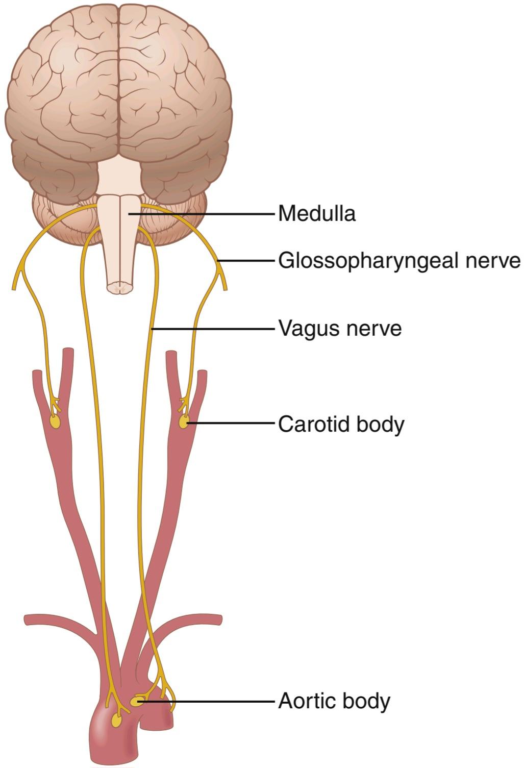
Figure 42-4. Respiratory control by peripheral chemoreceptors in the carotid and aortic bodies.
شکل 42-4. کنترل تنفس توسط گیرندههای شیمیایی محیطی در بدن کاروتید و آئورت.
Decreased Arterial Oxygen Stimulates the Chemoreceptors. When the oxygen concentration in the arterial blood falls below normal, the chemoreceptors become strongly stimulated. This effect is demonstrated in Figure 42-5, which shows the effect of different levels of arterial Po2 on the rate of nerve impulse transmission from a carotid body. Note that the impulse rate is particularly sensitive to changes in arterial Po2 in the range of 60 mm Hg down to 30 mm Hg, a range in which hemoglobin saturation with oxygen decreases rapidly.
کاهش اکسیژن شریانی باعث تحریک گیرندههای شیمیایی میشود. هنگامیکه غلظت اکسیژن در خون شریانی کمتر از حد طبیعی میشود، گیرندههای شیمیایی به شدت تحریک میشوند. این اثر در شکل 42-5 نشان داده شده است که تأثیر سطوح مختلف Po2 شریانی را بر سرعت انتقال تکانههای عصبی از بدن کاروتید نشان میدهد. توجه داشته باشید که نرخ ضربه به ویژه به تغییرات Po2 شریانی در محدوده 60 میلی متر جیوه تا 30 میلی متر جیوه حساس است، محدوده ای که در آن اشباع هموگلوبین با اکسیژن به سرعت کاهش مییابد.
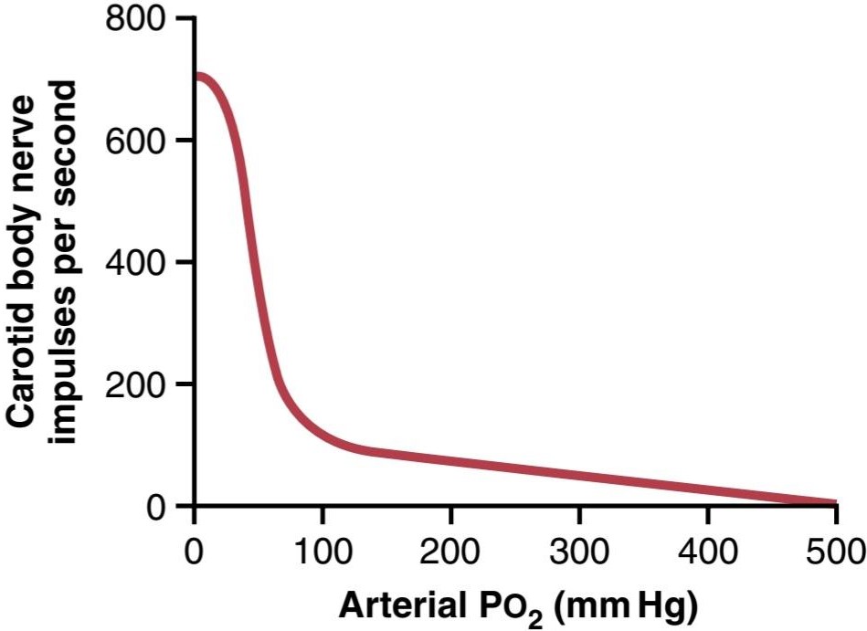
Figure 42-5. Effect of arterial Po2 on impulse rate from the carotid body. glossopharyngeal nerves and then to the dorsal respiratory area of the medulla. The aortic bodies are located along the arch of the aorta; their afferent nerve fibers pass through the vagi, also to the dorsal medullary respiratory area.
شکل 42-5. اثر Po2 شریانی بر میزان ضربه از بدن کاروتید. اعصاب گلوفارنکس و سپس به ناحیه تنفسی پشتی مدولا. اجسام آئورت در امتداد قوس آئورت قرار دارند. رشتههای عصبی آوران آنها از واگی و همچنین به ناحیه تنفسی مدولاری پشتی عبور میکند.
Basic Mechanism of Stimulation of the Chemoreceptors by O2 Deficiency. The exact means whereby low Po2 excites the nerve endings in the carotid and aortic bodies are still not completely understood. However, these bodies have multiple, highly characteristic glandular-like cells, called glomus cells, that synapse directly or indirectly with the nerve endings. Current evidence suggests that these glomus cells function as the chemoreceptors and then stimulate the nerve endings (Figure 42-6). Glomus cells have O2-sensitive potassium channels that are inactivated when blood Po2 decreases markedly. This inactivation causes the cell to depolarize, which in turn opens voltage-gated calcium channels and increases intracellular calcium ion concentration. The increased number of calcium ions stimulates release of a neurotransmitter that activates afferent neurons that send signals to the central nervous system and stimulate respiration. Although early studies suggested that dopamine or acetylcholine might be the main neurotransmitters, more recent studies suggest that during hypoxia, adenosine triphosphate (ATP) may be the key excitatory neurotransmitter released by carotid body glomus cells.
مکانیسم اصلی تحریک گیرندههای شیمیایی در اثر کمبود O2. روش دقیقی که Po2 پایین باعث تحریک انتهای عصب در اجسام کاروتید و آئورت میشود هنوز به طور کامل شناخته نشده است. با این حال، این اجسام دارای سلولهای غدهمانند متعدد و بسیار مشخص هستند که سلولهای گلوموس نامیده میشوند که بهطور مستقیم یا غیرمستقیم با پایانههای عصبی سیناپس میشوند. شواهد فعلی نشان میدهد که این سلولهای گلوموس به عنوان گیرندههای شیمیایی عمل میکنند و سپس انتهای عصبی را تحریک میکنند (شکل 42-6). سلولهای گلوموس دارای کانالهای پتاسیم حساس به O2 هستند که با کاهش قابل توجهی Po2 خون غیر فعال میشوند. این غیرفعال شدن باعث دپلاریزه شدن سلول میشود که به نوبه خود کانالهای کلسیمیبا ولتاژ را باز میکند و غلظت یون کلسیم داخل سلولی را افزایش میدهد. افزایش تعداد یونهای کلسیم باعث آزاد شدن یک انتقالدهنده عصبی میشود که نورونهای آوران را فعال میکند که سیگنالهایی را به سیستم عصبی مرکزی ارسال میکنند و تنفس را تحریک میکنند. اگرچه مطالعات اولیه نشان میدهد که دوپامین یا استیل کولین ممکن است انتقال دهندههای عصبی اصلی باشند، مطالعات جدیدتر نشان میدهد که در طول هیپوکسی، آدنوزین تری فسفات (ATP) ممکن است انتقال دهنده عصبی تحریکی کلیدی باشد که توسط سلولهای گلوموس بدن کاروتید آزاد میشود.
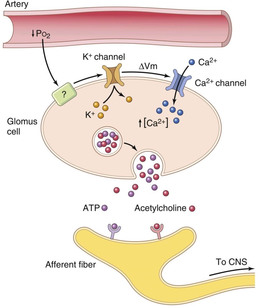
Figure 42-6. Carotid body glomus cell oxygen sensing. When the Poz decreases below around 60 mm Hg, potassium channels close, causing cell depolarization, opening of calcium channels, and increased cytosolic calcium ion concentration. This stimulates transmitter re- lease (adenosine triphosphate [ATP] is likely the most important), which activates afferent fibers that send signals to the central nervous system (CNS) and stimulate respiration. The mechanisms whereby low Poz influences potassium channel activity are still unclear. AVm, Change in membrane voltage.
شکل 42-6. سنجش اکسیژن سلول گلوموس بدن کاروتید. هنگامیکه Poz کمتر از حدود 60 میلی متر جیوه کاهش مییابد، کانالهای پتاسیم بسته میشود و باعث دپلاریزاسیون سلولی، باز شدن کانالهای کلسیم و افزایش غلظت یون کلسیم سیتوزولی میشود. این امر آزادسازی فرستنده را تحریک میکند (آدنوزین تری فسفات [ATP] احتمالاً مهم ترین است)، که فیبرهای آوران را فعال میکند که سیگنالها را به سیستم عصبی مرکزی (CNS) میفرستند و تنفس را تحریک میکنند. مکانیسمهایی که به موجب آن Poz کم بر فعالیت کانال پتاسیم تأثیر میگذارد هنوز نامشخص است. AVm، تغییر در ولتاژ غشا.
Increased CO2 and H+ Concentration Stimulates the Chemoreceptors. An increase in CO2 or H+ concentration also excites the chemoreceptors and, in this way, in- directly increases respiratory activity. However, the direct effects of both these factors in the respiratory center are much more powerful than their effects mediated through the chemoreceptors (about seven times as powerful). Yet, there is one difference between the peripheral and central effects of CO2-the stimulation via the peripheral chemoreceptors occurs as much as five times as rapidly as central stimulation, so the peripheral chemoreceptors might be especially important in increasing the rapidity of response to CO2 at the onset of exercise.
افزایش غلظت CO2 و +H باعث تحریک گیرندههای شیمیایی میشود. افزایش غلظت CO2 یا+H نیز گیرندههای شیمیایی را تحریک میکند و به این ترتیب به طور مستقیم فعالیت تنفسی را افزایش میدهد. با این حال، اثرات مستقیم هر دوی این عوامل در مرکز تنفسی بسیار قویتر از تأثیرات آنها است که از طریق گیرندههای شیمیایی انجام میشود (حدود هفت برابر قویتر). با این حال، یک تفاوت بین اثرات محیطی و مرکزی CO2 وجود دارد – تحریک از طریق گیرندههای شیمیایی محیطی به اندازه پنج برابر سریعتر از تحریک مرکزی رخ میدهد، بنابراین گیرندههای شیمیایی محیطی ممکن است در افزایش سرعت پاسخ به CO2 در شروع تمرین اهمیت ویژه ای داشته باشند.
Effect of Low Arterial Po2 to Stimulate Alveolar Ventilation When Arterial CO2 and H+ Concentrations Remain Normal
Figure 42-7 shows the effect of low arterial Po2 on alveolar ventilation when the Pco, and H+ concentrations are kept constant at their normal levels. In other words, in this figure, only the ventilatory drive caused by low O2 on the chemoreceptors is active. Figure 42-7 shows almost no effect on ventilation as long as the arterial Po2 remains greater than 100 mm Hg. However, at pressures lower than 100 mm Hg, ventilation approximately doubles when the arterial Po, falls to 60 mm Hg and can increase as much as fivefold at very low Po2 values. Under these conditions, low arterial Po, obviously drives the ventilatory process quite strongly.
اثر Po2 شریانی کم برای تحریک تهویه آلوئولی هنگامیکه غلظت CO2 شریانی و +H طبیعی باقی بماند
شکل 42-7 اثر Po2 شریانی کم را بر تهویه آلوئولی نشان میدهد، زمانی که غلظت Pco و +H در سطوح طبیعی خود ثابت نگه داشته میشوند. به عبارت دیگر، در این شکل تنها محرک تهویه ناشی از اکسیژن کم روی گیرندههای شیمیایی فعال است. شکل 42-7 تا زمانی که Po2 شریانی بیشتر از 100 میلی متر جیوه باقی بماند تقریباً هیچ تأثیری بر تهویه نشان نمیدهد. با این حال، در فشارهای کمتر از 100 میلیمتر جیوه، تهویه زمانی که پو شریانی به 60 میلیمتر جیوه میرسد، تقریباً دو برابر میشود و در مقادیر بسیار کم Po2 میتواند تا 5 برابر افزایش یابد. تحت این شرایط، Po شریانی پایین، بدیهی است که فرآیند تهویه را به شدت هدایت میکند.
Because the effect of hypoxia on ventilation is modest for Po2 values greater than 60 to 80 mm Hg, the Pco, and H+ responses are mainly responsible for regulating ventilation in healthy humans at sea level.
از آنجایی که اثر هیپوکسی بر تهویه برای مقادیر Po2 بیشتر از 60 تا 80 میلیمتر جیوه متوسط است، پاسخهای Pco و +H عمدتاً مسئول تنظیم تهویه در انسانهای سالم در سطح دریا هستند.
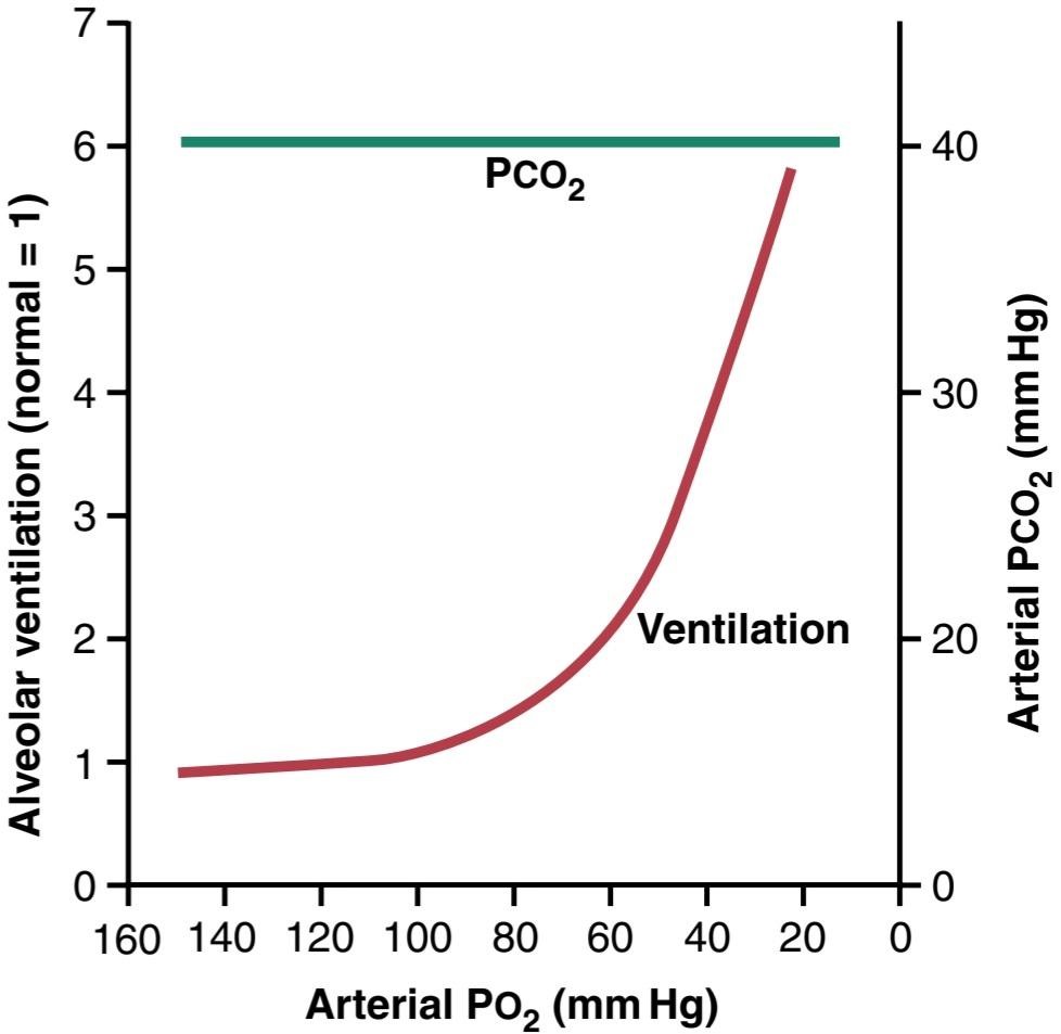
Figure 42-7. The lower red curve demonstrates the effect of different levels of arterial Po2 on alveolar ventilation, showing a 6-fold increase in ventilation as the Po2 decreases from the normal level of 100 mm Hg to 20 mm Hg. The upper green line shows that the arterial Pco2 was kept at a constant level during the measurements of this study; pH also was kept constant.
شکل 42-7. منحنی قرمز پایین، تأثیر سطوح مختلف Po2 شریانی را بر تهویه آلوئولی نشان میدهد، که افزایش 6 برابری تهویه را نشان میدهد زیرا Po2 از سطح طبیعی 100 میلیمتر جیوه به 20 میلیمتر جیوه کاهش مییابد. خط سبز بالا نشان میدهد که Pco2 شریانی در طول اندازه گیریهای این مطالعه در یک سطح ثابت نگه داشته شد. pH نیز ثابت نگه داشته شد.
Chronic Breathing of Low O2 Stimulates Respiration Even More-The Phenomenon of “Acclimatization”
Mountain climbers have found that when they ascend a mountain slowly, over a period of days rather than a period of hours, they breathe much more deeply and therefore can withstand far lower atmospheric O2 con- centrations than when they ascend rapidly. This phenomenon is called acclimatization.
تنفس مزمن O2 کم تنفس را حتی بیشتر تحریک میکند – پدیده “همکاری”
کوهنوردان دریافتهاند که وقتی از یک کوه به آرامیبالا میروند، در طی چند روز به جای چند ساعت، نفس عمیقتری میکشند و بنابراین میتوانند در مقایسه با زمانی که با سرعت بالا میروند، غلظت O2 جوی بسیار کمتری را تحمل کنند. به این پدیده سازگاری میگویند.
The reason for acclimatization is that within 2 to 3 days, the respiratory center in the brain stem loses about 80% of its sensitivity to changes in Pco, and H+. Therefore, the excess ventilatory blow-off of CO2 that normally would inhibit an increase in respiration fails to occur, and low O2 can drive the respiratory system to a much higher level of alveolar ventilation than under acute conditions. Instead of the 70% increase in ventilation that might occur after acute exposure to low O2, the alveolar ventilation often increases by 400% to 500% after 2 to 3 days of low O2, which helps immensely in supplying additional O2 to the mountain climber.
دلیل سازگاری این است که در عرض 2 تا 3 روز، مرکز تنفسی در ساقه مغز حدود 80 درصد از حساسیت خود را نسبت به تغییرات Pco و +H از دست میدهد. بنابراین، دمش اضافی CO2 که معمولاً مانع از افزایش تنفس میشود، رخ نمیدهد و O2 کم میتواند سیستم تنفسی را به سطح بسیار بالاتری از تهویه آلوئولی نسبت به شرایط حاد سوق دهد. به جای افزایش 70 درصدی تهویه که ممکن است پس از مواجهه حاد با اکسیژن کم اتفاق بیفتد، تهویه آلوئولی اغلب 400 تا 500 درصد پس از 2 تا 3 روز کم اکسیژن افزایش مییابد، که به تأمین اکسیژن اضافی به کوهنورد کمک زیادی میکند.
Composite Effects of Pco2, pH, and Po2 on Alveolar Ventilation
Figure 42-8 gives a quick overview of the manner in which Po2, PcO2, and pH together affect alveolar ventilation. To understand this diagram, first observe the four red curves. These curves were recorded at different levels of arterial Po2-40, 50, 60, and 100 mm Hg. For each of these curves, the Pco, was changed from lower to higher levels. Thus, this family of red curves represents the combined effects of alveolar Pco2 and Po2 on ventilation.
اثرات ترکیبی Pco2، pH و Po2 بر تهویه آلوئولی
شکل 42-8 یک نمای کلی از روشی را ارائه میدهد که در آن Po2، PcO2 و pH با هم بر تهویه آلوئولی تأثیر میگذارند. برای درک این نمودار، ابتدا چهار منحنی قرمز را مشاهده کنید. این منحنیها در سطوح مختلف Po2-40 شریانی، 50، 60 و 100 میلی متر جیوه ثبت شد. برای هر یک از این منحنیها، Pco از سطوح پایین تر به سطوح بالاتر تغییر یافت. بنابراین، این خانواده از منحنیهای قرمز نشان دهنده اثرات ترکیبی Pco2 و Po2 آلوئولی بر روی تهویه است.
Now observe the green curves. Whereas the red curves were measured at a blood pH of 7.4, the green curves were measured at a pH of 7.3. We now have two families of curves representing the combined effects of Pco2 and Po2 on ventilation at two different pH values. Still other families of curves would be displaced to the right at higher pH and displaced to the left at lower pH. Therefore, using this diagram, one can predict the level of alveolar ventilation for most combinations of alveolar PCO2, alveolar Po2, and arterial pH.
حالا منحنیهای سبز را مشاهده کنید. در حالی که منحنیهای قرمز در pH خون 7.4 اندازه گیری شدند، منحنیهای سبز در pH 7.3 اندازه گیری شدند. ما اکنون دو خانواده منحنی داریم که اثرات ترکیبی Pco2 و Po2 را بر روی تهویه در دو مقدار pH مختلف نشان میدهند. هنوز خانوادههای دیگر منحنیها در pH بالاتر به سمت راست و در pH پایین تر به سمت چپ جابه جا میشوند. بنابراین با استفاده از این نمودار میتوان سطح تهویه آلوئولی را برای اکثر ترکیبات PCO2 آلوئولی، Po2 آلوئولی و pH شریانی پیش بینی کرد.

Figure 42-8. Composite diagram showing the interrelated effects of Pcoz, Poz, and pH on alveolar ventilation. (Data from Cunningham DJC, Lloyd BB: The Regulation of Human Respiration. Oxford: Blackwell Scientific, 1963.)
شکل 42-8. نمودار ترکیبی که اثرات مرتبط Pcoz، Poz و pH را بر تهویه آلوئولی نشان میدهد. (دادهها از کانینگهام دی جی سی، لوید بی بی: تنظیم تنفس انسان. آکسفورد: بلک ول علمی، 1963.)
REGULATION OF RESPIRATION DURING EXERCISE
During strenuous exercise, O2 consumption and CO2 formation can increase as much as 20-fold. Yet, in the healthy athlete, as illustrated in Figure 42-9, alveolar ventilation ordinarily increases almost exactly in step with the increased level of oxygen metabolism. The arterial Po2, Pco2, and pH remain almost exactly normal.
تنظیم تنفس در حین ورزش
در طول ورزش شدید، مصرف O2 و تشکیل CO2 میتواند تا 20 برابر افزایش یابد. با این حال، همانطور که در شکل 42-9 نشان داده شده است، در ورزشکار سالم، تهویه آلوئولی معمولاً دقیقاً همگام با افزایش سطح متابولیسم اکسیژن افزایش مییابد. Po2، Pco2 و pH شریانی تقریباً کاملاً طبیعی هستند.
In trying to analyze what causes the increased ventilation during exercise, one is tempted to ascribe this increased ventilation to increases in blood CO2 and H+, plus a decrease in blood O2. However, measurements of arterial Pco2, pH, and Po, show that none of these values changes significantly during exercise, so none of them becomes abnormal enough to stimulate respiration as vigorously as observed during strenuous exercise. There- fore, what causes intense ventilation during exercise? At least one effect seems to be predominant. The brain, on transmitting motor impulses to the exercising muscles, is believed to transmit collateral impulses into the brain stem at the same time to excite the respiratory center. This action is analogous to the stimulation of the vasomotor center of the brain stem during exercise that causes a simultaneous increase in arterial pressure.
در تلاش برای تجزیه و تحلیل آنچه باعث افزایش تهویه در حین ورزش میشود، وسوسه میشود که این افزایش تهویه را به افزایش CO2 خون و +H و کاهش O2 خون نسبت دهد. با این حال، اندازه گیری Pco2 شریانی، pH و Po، نشان میدهد که هیچ یک از این مقادیر به طور قابل توجهی در طول ورزش تغییر نمیکند، بنابراین هیچ یک از آنها به اندازه کافی غیر طبیعی نمیشوند تا تنفس را به شدت تحریک کنند که در طول ورزش شدید مشاهده میشود. بنابراین، چه چیزی باعث تهویه شدید در حین ورزش میشود؟ به نظر میرسد حداقل یک اثر غالب است. اعتقاد بر این است که مغز هنگام انتقال تکانههای حرکتی به عضلات در حال تمرین، تکانههای جانبی را همزمان به ساقه مغز منتقل میکند تا مرکز تنفسی را تحریک کند. این عمل مشابه تحریک مرکز وازوموتور ساقه مغز در حین ورزش است که باعث افزایش همزمان فشار شریانی میشود.
Actually, when a person begins to exercise, a large share of the total increase in ventilation begins immediately on initiation of the exercise, before any blood chemicals have had time to change. It is likely that most of the increase in respiration results from neurogenic signals transmitted directly into the brain stem respiratory center at the same time that signals go to the body muscles to cause muscle contraction.
در واقع، زمانی که فرد شروع به ورزش میکند، سهم زیادی از افزایش کل تهویه بلافاصله با شروع ورزش شروع میشود، قبل از اینکه مواد شیمیایی خون زمان تغییر را داشته باشند. این احتمال وجود دارد که بیشتر افزایش تنفس ناشی از سیگنالهای عصبی است که مستقیماً به مرکز تنفسی ساقه مغز منتقل میشوند و همزمان سیگنالها به عضلات بدن میروند تا باعث انقباض عضلانی شوند.
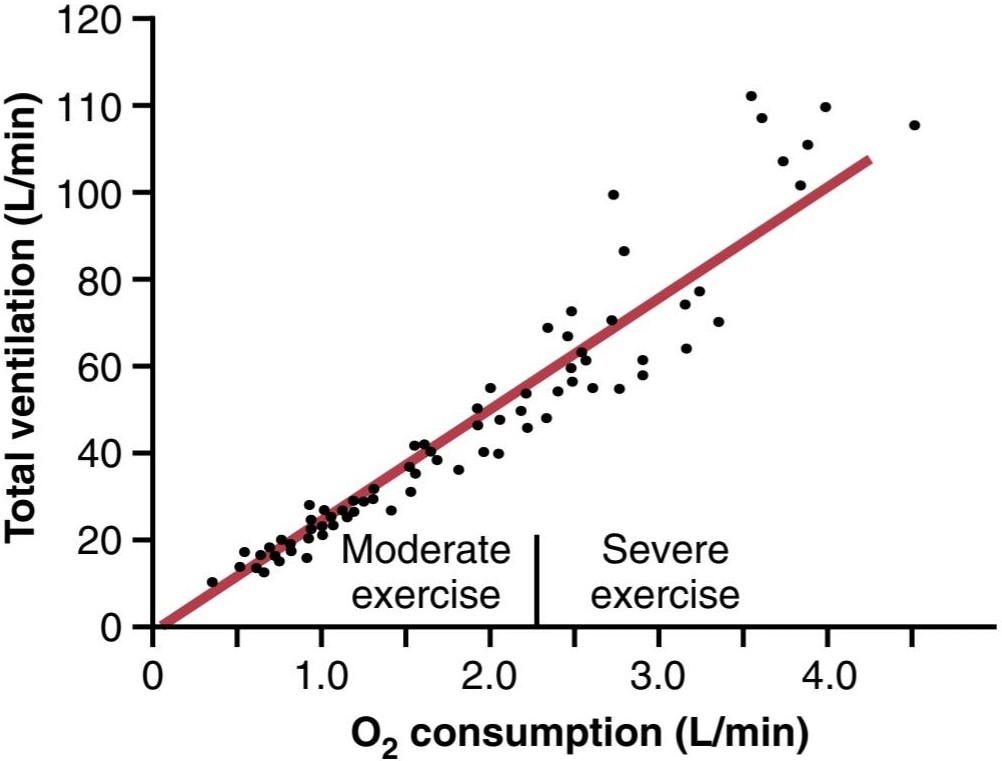
Figure 42-9. Effect of moderate and severe exercise on oxygen consumption and ventilatory rate. (From Gray JS: Pulmonary Ventilation and Its Physiological Regulation. Springfield, IL: Charles C Thomas, 1950.)
شکل 42-9. تأثیر ورزش متوسط و شدید بر مصرف اکسیژن و سرعت تهویه. (از گری JS: تهویه ریوی و تنظیم فیزیولوژیکی آن. اسپرینگفیلد، IL: چارلز سی توماس، 1950.)
Interrelationship Between Chemical and Nervous Factors in Controlling Respiration During Exercise. When a person exercises, direct nervous signals presumably stimulate the respiratory center by almost the proper amount to supply the extra O2 required for exercise and to blow off extra CO2. Occasionally, however, the nervous respiratory control signals are too strong or too weak. Chemical factors then play a significant role in bringing about the final adjustment of respiration required to keep the O2, CO2, and H+ concentrations of the body fluids as nearly normal as possible.
رابطه متقابل بین عوامل شیمیایی و عصبی در کنترل تنفس در حین ورزش. هنگامیکه فردی ورزش میکند، احتمالاً سیگنالهای عصبی مستقیم مرکز تنفس را تقریباً به مقدار مناسب تحریک میکند تا اکسیژن اضافی مورد نیاز برای ورزش را تأمین کند و CO2 اضافی را دفع کند. با این حال، گاهی اوقات سیگنالهای کنترل تنفسی عصبی خیلی قوی یا خیلی ضعیف هستند. سپس عوامل شیمیایی نقش مهمیدر تنظیم نهایی تنفس مورد نیاز برای حفظ غلظت O2، CO2 و +H مایعات بدن تا حد امکان تقریباً نرمال ایفا میکنند.
This process is demonstrated in Figure 42-10. The lower curve shows changes in alveolar ventilation during 1 minute of exercise, and the upper curve shows changes in arterial Pco2. Note that at the onset of exercise, the alveolar ventilation increases almost instantaneously, without an initial increase in arterial Pco,. In fact, this increase in ventilation is usually great enough so that at first it actually decreases arterial Pco2 below normal, as shown in the figure. The presumed reason why the ventilation forges ahead of the buildup of blood CO2 is that the brain provides an “anticipatory” stimulation of respiration at the onset of exercise, causing extra alveolar ventilation even before it is necessary. However, after 30 to 40 seconds, the amount of CO2 released into the blood from the active muscles approximately matches the increased rate of ventilation, and the arterial Pco, returns essentially to normal, even as the exercise continues. This is shown toward the end of 1 minute of exercise in the figure.
این فرآیند در شکل 42-10 نشان داده شده است. منحنی پایینی تغییرات تهویه آلوئولی را در طول 1 دقیقه ورزش نشان میدهد و منحنی بالایی تغییرات Pco2 شریانی را نشان میدهد. توجه داشته باشید که در شروع ورزش، تهویه آلوئولی تقریباً بلافاصله افزایش مییابد، بدون افزایش اولیه در Pco شریانی،. در واقع، این افزایش در تهویه معمولاً به اندازهای زیاد است که در ابتدا، همان طور که در شکل نشان داده شده است، Pco2 شریانی را کمتر از حد طبیعی کاهش میدهد. دلیل احتمالی اینکه چرا تهویه قبل از تجمع CO2 خون ایجاد میشود این است که مغز در شروع ورزش یک تحریک “پیش بینی کننده” تنفس را ارائه میدهد و حتی قبل از اینکه لازم باشد باعث ایجاد تهویه اضافی آلوئولی میشود. با این حال، پس از 30 تا 40 ثانیه، مقدار CO2 آزاد شده در خون از عضلات فعال تقریباً با افزایش سرعت تهویه مطابقت دارد و Pco شریانی، حتی با ادامه تمرین، اساساً به حالت عادی باز میگردد. این در پایان 1 دقیقه تمرین در شکل نشان داده شده است.
Figure 42-11 summarizes the control of respiration during exercise in another way, this time more quantitatively. The lower curve of this figure shows the effect of different levels of arterial Pco2 on alveolar ventilation when the body is at rest-that is, not exercising. The upper curve shows the approximate shift of this ventilatory curve caused by neurogenic drive from the respiratory center that occurs during heavy exercise. The points indicated on the two curves show the arterial Pco, first in the resting state and then in the exercising state. Note in both cases that the Pco, is at the normal level of 40 mm Hg. In other words, the neurogenic factor shifts the curve about 20-fold in the upward direction, so ventilation almost matches the rate of CO2 release, thus keeping arterial Pco2 near its normal value. The upper curve of Figure 42-11 also shows that if during exercise the arterial Pco, does change from its normal value of 40 mm Hg, it has an extra stimulatory effect on ventilation at a Pco2 value greater than 40 mm Hg and a depressant effect at a PCO2 value less than 40 mm Hg.
شکل 42-11 کنترل تنفس در حین ورزش را به روشی دیگر، این بار به صورت کمی، خلاصه میکند. منحنی پایینی این شکل تأثیر سطوح مختلف Pco2 شریانی را بر تهویه آلوئولی در زمانی که بدن در حالت استراحت است، یعنی ورزش نمیکند، نشان میدهد. منحنی بالایی جابجایی تقریبی این منحنی تهویه را نشان میدهد که ناشی از حرکت عصبی از مرکز تنفسی است که در طول ورزش سنگین رخ میدهد. نقاط نشان داده شده در دو منحنی Pco شریانی را ابتدا در حالت استراحت و سپس در حالت ورزش نشان میدهد. در هر دو مورد توجه داشته باشید که Pco در سطح نرمال 40 میلی متر جیوه است. به عبارت دیگر، عامل نوروژنیک منحنی را حدود 20 برابر در جهت بالا تغییر میدهد، بنابراین تهویه تقریباً با سرعت انتشار CO2 مطابقت دارد، بنابراین Pco2 شریانی را نزدیک به مقدار طبیعی خود نگه میدارد. منحنی بالای شکل 42-11 همچنین نشان میدهد که اگر در حین ورزش، Pco شریانی از مقدار طبیعی آن 40 میلیمتر جیوه تغییر کند، در مقدار Pco2 بیشتر از 40 میلیمتر جیوه، اثر تحریککنندهای اضافی روی تهویه و در مقدار PCO2 کمتر از 40 میلیمتر جیوه اثر کاهنده دارد.
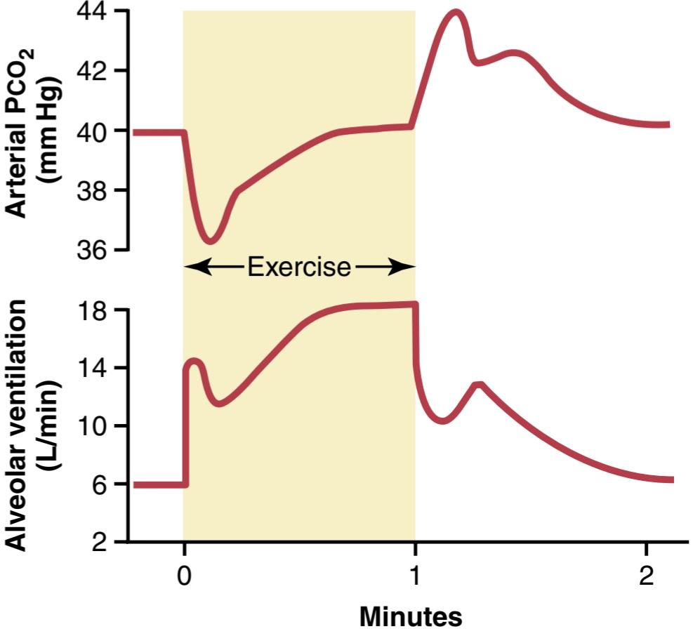
Figure 42-10. Changes in alveolar ventilation (bottom curve) and arterial Pco2 (top curve) during a 1-minute period of exercise and also after termination of exercise. (Data from Bainton CR: Effect of speed vs. grade and shivering on ventilation in dogs during active exercise. J Appl Physiol 33:778, 1972.)
شکل 42-10. تغییرات در تهویه آلوئولی (منحنی پایین) و Pco2 شریانی (منحنی بالا) در طول یک دوره 1 دقیقه ای ورزش و همچنین پس از پایان تمرین. (دادههای Bainton CR: تأثیر سرعت در مقابل درجه و لرز بر تهویه در سگها در طول ورزش فعال. J Appl Physiol 33:778، 1972.)
Neurogenic Control of Ventilation During Exercise May Be Partly a Learned Response. Many experiments suggest that the brain’s ability to shift the ventilatory response curve during exercise, as shown in Figure 42-11, is at least partly a learned response. That is, with repeated periods of exercise, the brain becomes progressively more able to provide the proper signals required to keep the blood Pco, at its normal level. Also, there is reason to believe that even the cerebral cortex is involved in this learning because experiments that block only the cortex also block the learned response.
کنترل نوروژنیک تهویه در حین ورزش ممکن است تا حدی یک پاسخ آموخته شده باشد. بسیاری از آزمایشها نشان میدهند که توانایی مغز برای تغییر منحنی پاسخ تهویه در طول ورزش، همانطور که در شکل 42-11 نشان داده شده است، حداقل تا حدی یک پاسخ آموخته شده است. یعنی با دورههای مکرر ورزش، مغز به تدریج قادر به ارائه سیگنالهای مناسب برای حفظ PCO خون در سطح طبیعی خود میشود. همچنین، دلیلی وجود دارد که باور کنیم حتی قشر مغز نیز در این یادگیری دخیل است، زیرا آزمایشهایی که فقط قشر مغز را مسدود میکنند، پاسخ آموخته شده را نیز مسدود میکنند.
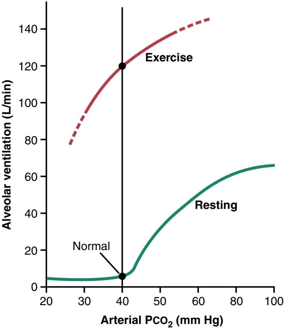
Figure 42-11. Approximate effect of maximum exercise in an athlete to shift the alveolar Pco-ventilation response curve to a level much higher than normal. The shift, believed to be caused by neurogenic factors, is almost exactly the right amount to maintain arterial Pco2 at the normal level of 40 mm Hg in the resting state and during heavy exercise.
شکل 42-11. تأثیر تقریبی حداکثر تمرین در یک ورزشکار برای تغییر منحنی پاسخ Pco-تهویه آلوئولی به سطحی بسیار بالاتر از حد طبیعی. این تغییر که اعتقاد بر این است که توسط عوامل نوروژنیک ایجاد میشود، تقریباً دقیقاً مقدار مناسبی برای حفظ Pco2 شریانی در سطح نرمال 40 میلی متر جیوه در حالت استراحت و در طول ورزش سنگین است.
Other Factors That Affect Respiration
Effect of Irritant Receptors in the Airways. The epithelium of the trachea, bronchi, and bronchioles is supplied with sensory nerve endings called pulmonary irritant receptors that are stimulated by many factors. These receptors initiate coughing and sneezing, as discussed in Chapter 40. They may also cause bronchial constriction in persons with diseases such as asthma and emphysema.
سایر عوامل موثر بر تنفس
اثر گیرندههای تحریک کننده در مجاری هوایی. اپیتلیوم نای، برونشها و برونشیولها با پایانههای عصبی حسی به نام گیرندههای تحریک کننده ریوی تامین میشود که توسط عوامل بسیاری تحریک میشوند. این گیرندهها همانطور که در فصل 40 بحث شد شروع کننده سرفه و عطسه هستند. همچنین ممکن است باعث انقباض برونش در افراد مبتلا به بیماریهایی مانند آسم و آمفیزم شوند.
Function of Lung J Receptors. A few sensory nerve endings have been described in the alveolar walls in juxtaposition to the pulmonary capillaries-hence, the name J receptors. They are stimulated especially when the pulmonary capillaries become engorged with blood or when pulmonary edema occurs in conditions such as congestive heart failure. Although the functional role of the J receptors is not clear, their excitation may give the person a feeling of dyspnea.
عملکرد گیرندههای J ریه. چند پایانه عصبی حسی در دیواره آلوئول در کنار مویرگهای ریوی توصیف شده است – از این رو، گیرندههای J نامیده میشوند. آنها به ویژه هنگامیکه مویرگهای ریوی با خون انباشته میشوند یا زمانی که ادم ریوی در شرایطی مانند نارسایی احتقانی قلب رخ میدهد، تحریک میشوند. اگرچه نقش عملکردی گیرندههای J مشخص نیست، اما تحریک آنها ممکن است به فرد احساس تنگی نفس بدهد.
Brain Edema Depresses the Respiratory Center. The activity of the respiratory center may be depressed or even inactivated by acute brain edema resulting from a brain concussion. For example, the head might be struck against some solid object, after which the damaged brain tissues swell, compressing the cerebral arteries against the cranial vault and thus partially blocking the cerebral blood supply.
ادم مغزی مرکز تنفس را تحت فشار قرار میدهد. فعالیت مرکز تنفس ممکن است در اثر ادم مغزی حاد ناشی از ضربه مغزی کاهش یابد یا حتی غیرفعال شود. به عنوان مثال، ممکن است سر به جسم جامد برخورد کند، پس از آن بافتهای آسیبدیده مغز متورم میشوند، شریانهای مغزی را در برابر طاق جمجمه فشرده میکند و بنابراین تا حدی جریان خون مغز را مسدود میکند.
Occasionally, respiratory depression resulting from brain edema can be relieved temporarily by intravenous injection of a hypertonic solution, such as a highly concentrated mannitol solution. These solutions osmotically re- move some of the fluids of the brain, thus relieving intracranial pressure and sometimes re-establishing respiration within a few minutes.
گاهی اوقات، افسردگی تنفسی ناشی از ادم مغزی را میتوان به طور موقت با تزریق داخل وریدی محلول هیپرتونیک، مانند محلول مانیتول بسیار غلیظ، تسکین داد. این محلولها به صورت اسمزی برخی از مایعات مغز را خارج میکنند، بنابراین فشار داخل جمجمه را کاهش میدهند و گاهی اوقات تنفس را در عرض چند دقیقه دوباره برقرار میکنند.
Overdosage of Anesthetics and Narcotics. Perhaps the most prevalent cause of respiratory depression and respiratory arrest is overdosage with anesthetics or narcotics. For example, sodium pentobarbital depresses the respiratory center considerably more than many other anesthetics, such as halothane. At one time, morphine was used as an anesthetic, but this drug is now used only as an adjunct to an- esthetics because it greatly depresses the respiratory center while having less ability to anesthetize the cerebral cortex.
مصرف بیش از حد داروهای بیهوشی و مخدر. شاید شایع ترین علت افسردگی و ایست تنفسی مصرف بیش از حد داروهای بیهوش کننده یا مخدر باشد. به عنوان مثال، سدیم پنتوباربیتال مرکز تنفس را به طور قابل توجهی بیش از بسیاری از داروهای بیهوشی دیگر مانندهالوتان کاهش میدهد. زمانی از مورفین به عنوان بیهوشی استفاده میشد، اما این دارو در حال حاضر تنها به عنوان مکملی برای بیهوشی استفاده میشود زیرا مرکز تنفسی را به شدت تحت فشار قرار میدهد در حالی که توانایی کمتری در بیهوش کردن قشر مغز دارد.
Because of their capacity to cause respiratory depression, opioids are responsible for a high proportion of fatal drug overdoses around the world. In the United States, approximately 70,000 people died from drug overdose in 2017, largely due to respiratory arrest.
به دلیل توانایی آنها در ایجاد افسردگی تنفسی، مواد افیونی مسئول بخش بالایی از مصرف بیش از حد مواد مخدر کشنده در سراسر جهان هستند. در ایالات متحده، تقریباً 70000 نفر در سال 2017 بر اثر مصرف بیش از حد مواد مخدر جان خود را از دست دادند که عمدتاً به دلیل ایست تنفسی بود.
Periodic Breathing. An abnormality of respiration called periodic breathing occurs in several disease conditions. The person breathes deeply for a short interval and then breathes slightly or not at all for an additional interval, with the cycle repeating itself over and over. One type of periodic breathing, Cheyne-Stokes breathing, is characterized by slowly waxing and waning respiration occurring about every 40 to 60 seconds, as illustrated in Figure 42-12.
تنفس دوره ای. یک ناهنجاری تنفسی به نام تنفس دوره ای در چندین بیماری رخ میدهد. فرد برای مدت کوتاهی نفس عمیق میکشد و سپس برای یک بازه زمانی دیگر کمینفس میکشد یا اصلا نفس نمیکشد و این چرخه بارها و بارها تکرار میشود. یکی از انواع تنفس دوره ای، تنفس Cheyne-Stokes، با اپیلاسیون آهسته و کاهش تنفس مشخص میشود که تقریباً هر 40 تا 60 ثانیه اتفاق میافتد، همانطور که در شکل 42-12 نشان داده شده است.
Basic Mechanism of Cheyne Stokes Breathing. The basic cause of Cheyne-Stokes breathing is the following. When a person overbreathes, thus blowing off too much CO2 from the pulmonary blood while at the same time increasing blood O2, it takes several seconds before the changed pulmonary blood can be transported to the brain and inhibit the excess ventilation. By this time, the per- son has already overventilated for an extra few seconds. Therefore, when the overventilated blood finally reaches the brain respiratory center, the center becomes depressed to an excessive amount, at which point the opposite cycle begins that is, CO2 increases, and O2 decreases in the alveoli. Again, it takes a few seconds before the brain can respond to these new changes. When the brain does respond, the person breathes hard once again and the cycle repeats.
مکانیسم اساسی تنفس چین استوکس. علت اصلی تنفس چین استوکس موارد زیر است. هنگامیکه فردی بیش از حد تنفس میکند، بنابراین CO2 بیش از حد از خون ریوی خارج میشود و در عین حال O2 خون را افزایش میدهد، چند ثانیه طول میکشد تا خون ریوی تغییر یافته بتواند به مغز منتقل شود و تهویه اضافی را مهار کند. در این زمان، فرد قبلاً برای چند ثانیه اضافی بیش از حد تهویه کرده است. بنابراین، هنگامیکه خون بیش از حد تهویه شده در نهایت به مرکز تنفسی مغز میرسد، مرکز تا حد زیادی افسرده میشود، در این مرحله چرخه مخالف شروع میشود، یعنی CO2 افزایش مییابد و O2 در آلوئولها کاهش مییابد. باز هم، چند ثانیه طول میکشد تا مغز بتواند به این تغییرات جدید پاسخ دهد. هنگامیکه مغز پاسخ میدهد، فرد یک بار دیگر به سختی نفس میکشد و چرخه تکرار میشود.
The basic cause of Cheyne-Stokes breathing occurs in eve- ryone. However, under normal conditions, this mechanism is highly damped. That is, the fluids of the blood and respiratory center control areas have large amounts of dissolved and chem- ically bound CO2 and O2. Therefore, normally, the lungs cannot build up enough extra CO2 or depress the O2 sufficiently in a few seconds to cause the next cycle of the periodic breathing. However, under two separate conditions, the damping factors can be overridden, and Cheyne Stokes breathing does occur:
علت اصلی تنفس چین استوکس در همه رخ میدهد. با این حال، در شرایط عادی، این مکانیسم بسیار میرایی است. یعنی مایعات خون و نواحی کنترل مرکز تنفسی دارای مقادیر زیادی CO2 و O2 محلول و متصل به مواد شیمیایی هستند. بنابراین، به طور معمول، ریهها نمیتوانند به اندازه کافی CO2 اضافی ایجاد کنند یا O2 را به اندازه کافی در چند ثانیه کاهش دهند که باعث چرخه بعدی تنفس دوره ای شود. با این حال، تحت دو شرایط جداگانه، عوامل میرایی را میتوان نادیده گرفت و تنفس چین استوکس رخ میدهد:
1. When a long delay occurs for transport of blood from the lungs to the brain, changes in CO2 and O2 in the alveoli can continue for many more seconds than usual. Under these conditions, the storage capacities of the alveoli and pulmonary blood for these gases are exceeded; then, after a few more seconds, the periodic respiratory drive becomes extreme and Cheyne-Stokes breathing begins. This type of Cheyne-Stokes breathing often occurs in patients with severe cardiac failure because blood flow is slow, thus de- laying the transport of blood gases from the lungs to the brain. In patients with chronic heart failure, Cheyne-Stokes breathing can sometimes occur on and off for months.
1. هنگامیکه یک تاخیر طولانی برای انتقال خون از ریهها به مغز اتفاق میافتد، تغییرات CO2 و O2 در آلوئولها میتواند برای ثانیههای بیشتر از حد معمول ادامه یابد. تحت این شرایط، ظرفیت ذخیره سازی آلوئولها و خون ریوی برای این گازها فراتر میرود. سپس، پس از چند ثانیه دیگر، رانش تناوبی تنفسی شدید میشود و تنفس چین استوکس شروع میشود. این نوع تنفس چین استوکس اغلب در بیماران مبتلا به نارسایی شدید قلبی رخ میدهد زیرا جریان خون کند است و بنابراین انتقال گازهای خون از ریهها به مغز را به تاخیر میاندازد. در بیماران مبتلا به نارسایی مزمن قلبی، تنفس چین استوکسمیتواند برای ماهها خاموش و روشن شود.
2. A second cause of Cheyne-Stokes breathing is increased negative feedback gain in the respiratory control areas, which means that a change in blood CO2 or O2 causes a far greater change in ventilation than normally. For example, instead of the normal two- to threefold increase in ventilation that occurs when the Pco, rises 3 mm Hg, the same 3-mm Hg rise might increase ventilation by 10- to 20-fold. The brain feedback tendency for periodic breathing is now strong enough to cause Cheyne-Stokes breathing without extra blood flow delay between the lungs and brain. This type of Cheyne-Stokes breathing occurs mainly in patients with damage to the respiratory centers of the brain. The brain damage often turns off the respiratory drive entirely for a few seconds, and then an extra-intense increase in blood CO2 turns it back on with great force. Cheyne-Stokes breathing of this type is frequently a prelude to death from brain malfunction. Typical records of changes in pulmonary and respiratory center Pco, during Cheyne-Stokes breathing are shown in Figure 42-12. Note that the Pco2 of the pulmonary blood changes in advance of the Pco2 of the respiratory neurons. However, the depth of respiration corresponds with the Pco, in the brain, not with the Pco, in the pulmonary blood where the ventilation is occurring.
2. علت دوم تنفس چین استوکس افزایش بازخورد منفی در نواحی کنترل تنفسی است، به این معنی که تغییر در CO2 یا O2 خون باعث تغییر بسیار بیشتر از حالت عادی در تهویه میشود. به عنوان مثال، به جای افزایش طبیعی دو تا سه برابری در تهویه که زمانی رخ میدهد که Pco، 3 میلی متر جیوه افزایش مییابد، همان افزایش 3 میلی متر جیوه ممکن است تهویه را 10 تا 20 برابر افزایش دهد. تمایل به بازخورد مغز برای تنفس دوره ای اکنون به اندازه کافی قوی است که باعث تنفس چین استوکس بدون تاخیر جریان خون اضافی بین ریهها و مغز میشود. این نوع تنفس چین استوکس عمدتا در بیماران مبتلا به آسیب به مراکز تنفسی مغز رخ میدهد. آسیب مغزی اغلب باعث خاموش شدن کامل درایو تنفسی برای چند ثانیه میشود و سپس افزایش شدید CO2 خون آن را با نیروی زیادی دوباره روشن میکند. تنفس چین استوکس از این نوع اغلب مقدمه ای برای مرگ در اثر سوء عملکرد مغز است. سوابق معمول تغییرات در Pco مرکز ریوی و تنفسی، در طول تنفس چین استوکس در شکل 42-12 نشان داده شده است. توجه داشته باشید که Pco2 خون ریوی قبل از Pco2 نورونهای تنفسی تغییر میکند. با این حال، عمق تنفس مربوط به Pco است، در مغز، نه با Pco، در خون ریوی که در آن تهویه انجام میشود.
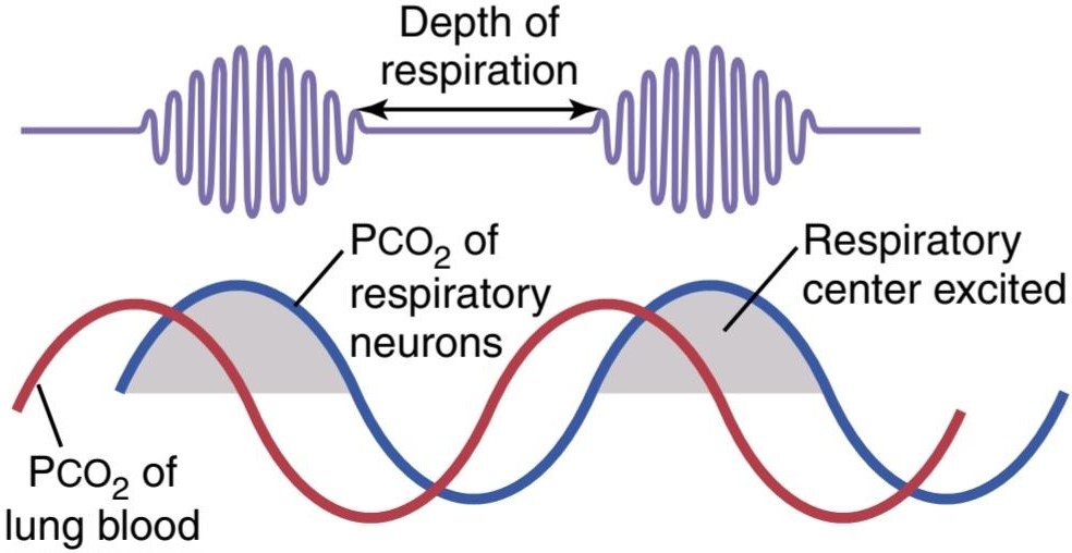
Figure 42-12. Cheyne-Stokes breathing, showing changing Pcoz in the pulmonary blood (red line) and delayed changes in the Pco2 of the fluids of the respiratory center (blue line).
شکل 42-12. تنفس شین-استوکس، که نشاندهندهی تغییرات Pcoz در خون ریوی (خط قرمز) و تغییرات تأخیری در Pco2 مایعات مرکز تنفسی (خط آبی) است.
Sleep Apnea
The term apnea means absence of spontaneous breathing. Occasional apneas occur during normal sleep, but in persons with sleep apnea, the frequency and duration are greatly increased, with episodes of apnea lasting for 10 seconds or longer and occurring 300 to 500 times each night. Sleep apneas can be caused by obstruction of the upper airways, especially the pharynx, or by an impaired central nervous system respiratory drive.
آپنه خواب
اصطلاح آپنه به معنای عدم وجود تنفس خود به خودی است. آپنه گاه به گاه در طول خواب طبیعی رخ میدهد، اما در افراد مبتلا به آپنه خواب، دفعات و مدت آن به شدت افزایش مییابد، به طوری که دورههای آپنه 10 ثانیه یا بیشتر طول میکشد و 300 تا 500 بار در هر شب رخ میدهد. آپنه خواب میتواند در اثر انسداد راههای هوایی فوقانی، به ویژه حلق، یا اختلال در حرکت تنفسی سیستم عصبی مرکزی ایجاد شود.
Obstructive Sleep Apnea Is Caused by Blockage of the Upper Airway. The muscles of the pharynx normally keep this passage open to allow air to flow into the lungs during inspiration. During sleep, these muscles usually relax, but the airway passage remains open enough to permit adequate airflow. Some people have an especially narrow passage, and relaxation of these muscles during sleep causes the pharynx to close completely so that air cannot flow into the lungs.
آپنه انسدادی خواب به دلیل انسداد راه هوایی فوقانی ایجاد میشود. ماهیچههای حلق معمولاً این مسیر را باز نگه میدارند تا در طول دم، هوا به داخل ریهها جریان یابد. در طول خواب، این ماهیچهها معمولاً شل میشوند، اما راه هوایی به اندازه کافی باز میماند تا جریان هوای کافی را فراهم کند. برخی از افراد دارای مجرای باریکی هستند و شل شدن این ماهیچهها در هنگام خواب باعث میشود که حلق به طور کامل بسته شود و هوا نتواند به داخل ریهها جریان یابد.
In persons with sleep apnea, loud snoring and labored breathing occur soon after falling asleep. The snoring proceeds, often becoming louder, and is then interrupted by a long silent period during which no breathing (apnea) occurs. These periods of apnea result in significant decreases in Po2 and increases in Pco2, which greatly stimulate respiration. This stimulation, in turn, causes sudden attempts to breathe, which result in loud snorts and gasps followed by snoring and repeated episodes of apnea. The periods of apnea and labored breathing are repeated several hundred times during the night, resulting in fragmented restless sleep. Therefore, patients with sleep apnea usually have excessive daytime drowsiness, as well as other disorders, including increased sympathetic activity, high heart rate, pulmonary and systemic hypertension, and a greatly elevated risk for cardiovascular disease.
در افراد مبتلا به آپنه خواب، خروپف بلند و تنفس سخت بلافاصله پس از به خواب رفتن رخ میدهد. خروپف ادامه مییابد، اغلب بلندتر میشود، و سپس با یک دوره خاموش طولانی که طی آن هیچ تنفسی (آپنه) رخ نمیدهد، قطع میشود. این دورههای آپنه منجر به کاهش قابل توجهی در Po2 و افزایش Pco2 میشود که تنفس را به شدت تحریک میکند. این تحریک به نوبه خود باعث تلاشهای ناگهانی برای نفس کشیدن میشود که منجر به خرخرهای بلند و نفس نفس زدن میشود و به دنبال آن خروپف و دورههای مکرر آپنه ایجاد میشود. دورههای آپنه و تنفس سخت چندین صد بار در طول شب تکرار میشوند و در نتیجه خواب بیقرار تکه تکه میشوند. بنابراین، بیماران مبتلا به آپنه خواب معمولاً دارای خواب آلودگی بیش از حد در طول روز و همچنین سایر اختلالات از جمله افزایش فعالیت سمپاتیک، ضربان قلب بالا، فشار خون ریوی و سیستمیک و افزایش خطر ابتلا به بیماریهای قلبی عروقی هستند.
Obstructive sleep apnea usually occurs in older obese persons in whom there is increased fat deposition in the soft tissues of the pharynx or compression of the pharynx due to excessive fat masses in the neck. In a few individuals, sleep apnea may be associated with nasal obstruction, a very large tongue, enlarged tonsils, or certain shapes of the palate that greatly increase resistance to the flow of air to the lungs during inspiration. The most common treatments of obstructive sleep apnea include the following: (1) surgery to remove excess fat tissue at the back of the throat (a procedure called uvulopalatopharyngoplasty), remove enlarged tonsils or adenoids, or create an opening in the trachea (tracheostomy) to bypass the obstructed airway during sleep; and (2) nasal ventilation with continuous positive airway pressure (CPAP).
آپنه انسدادی خواب معمولاً در افراد چاق مسنتر رخ میدهد که در آنها رسوب چربی در بافتهای نرم حلق یا فشردگی حلق به دلیل تودههای چربی بیش از حد در گردن افزایش مییابد. در تعداد کمیاز افراد، آپنه خواب ممکن است با انسداد بینی، زبان بسیار بزرگ، لوزههای بزرگ یا اشکال خاصی از کام همراه باشد که مقاومت در برابر جریان هوا به ریهها در طول دم را به شدت افزایش میدهد. رایجترین درمانهای آپنه انسدادی خواب شامل موارد زیر است: (1) جراحی برای برداشتن بافت چربی اضافی در پشت گلو (روشی به نام uvulopalatopharyngoplasty)، برداشتن لوزههای بزرگ شده یا آدنوئیدها، یا ایجاد سوراخ در نای (تراکوستومی) برای دور زدن راه هوایی مسدود شده در طول خواب. و (2) تهویه بینی با فشار مثبت مداوم راه هوایی (CPAP).
“Central” Sleep Apnea Occurs When the Neural Drive to Respiratory Muscles Is Transiently Abolished. In a few persons with sleep apnea, the central nervous system drive to the ventilatory muscles transiently ceases. Disorders that can cause cessation of the ventilatory drive during sleep include damage to the central respiratory centers or abnormalities of the respiratory neuromuscular apparatus. Patients affected by central sleep apnea may have decreased ventilation, even when they are awake, although they are fully capable of normal voluntary breathing. During sleep, their breathing disorders usually worsen, resulting in more frequent episodes of apnea that decrease Po2 and increase Pco, until a critical level is reached that eventually stimulates respiration. These transient instabilities of respiration cause restless sleep and clinical features similar to those observed in people with obstructive sleep apnea.
آپنه خواب “مرکزی” زمانی رخ میدهد که حرکت عصبی به عضلات تنفسی به طور موقت از بین برود. در تعداد کمیاز افراد مبتلا به آپنه خواب، حرکت سیستم عصبی مرکزی به سمت عضلات تهویه به طور موقت متوقف میشود. اختلالاتی که میتواند باعث توقف حرکت تهویه در طول خواب شود شامل آسیب به مراکز تنفسی مرکزی یا ناهنجاریهای دستگاه عصبی عضلانی تنفسی است. بیماران مبتلا به آپنه مرکزی خواب ممکن است حتی زمانی که بیدار هستند کاهش تهویه داشته باشند، اگرچه آنها کاملاً قادر به تنفس ارادی طبیعی هستند. در طول خواب، اختلالات تنفسی آنها معمولا بدتر میشود و در نتیجه دورههای مکرر آپنه که باعث کاهش Po2 و افزایش Pco میشود، تا زمانی که به سطح بحرانی برسد که در نهایت تنفس را تحریک میکند، میشود. این بی ثباتیهای گذرا تنفس باعث خواب بی قرار و ویژگیهای بالینی مشابه آنچه در افراد مبتلا به آپنه انسدادی خواب مشاهده میشود، میشود.
In most patients, the cause of central sleep apnea is unknown, although instability of the respiratory drive can result from strokes or other disorders that make the respiratory centers of the brain less responsive to the stimulatory effects of CO2 and H+. Patients with this disease are extremely sensitive to even small doses of sedatives or narcotics, which further reduce the responsiveness of the respiratory centers to the stimulatory effects of CO2. Medications that stimulate the respiratory centers can sometimes be helpful, but ventilation with CPAP at night is usually necessary.
در اکثر بیماران، علت آپنه خواب مرکزی ناشناخته است، اگرچه بی ثباتی درایو تنفسی میتواند ناشی از سکته مغزی یا سایر اختلالات باشد که باعث میشود مراکز تنفسی مغز کمتر به اثرات تحریکی CO2 و +H پاسخ دهند. بیماران مبتلا به این بیماری حتی به دوزهای کمیاز آرامبخشها یا مواد مخدر حساس هستند، که باعث کاهش بیشتر پاسخ دهی مراکز تنفسی به اثرات تحریکی CO2 میشود. داروهایی که مراکز تنفسی را تحریک میکنند گاهی اوقات میتوانند مفید باشند، اما تهویه با CPAP در شب معمولاً ضروری است.
In some cases, sleep apnea may be caused by a combination of obstructive and central mechanisms. This “mixed” type of sleep apnea is estimated to account for approximately 15% of all sleep apnea cases, whereas pure “central” sleep apnea accounts for less than 1% of cases. The most common cause of sleep apnea is obstruction of the upper airway.
در برخی موارد، آپنه خواب ممکن است به دلیل ترکیبی از مکانیسمهای انسدادی و مرکزی ایجاد شود. تخمین زده میشود که این نوع “مخلوط” از آپنه خواب تقریباً 15٪ از تمام موارد آپنه خواب را تشکیل میدهد، در حالی که آپنه خواب “مرکزی” خالص کمتر از 1٪ موارد را تشکیل میدهد. شایع ترین علت آپنه خواب انسداد راه هوایی فوقانی است.
Voluntary Control of Respiration
Thus far, we have discussed mainly the involuntary system for control of respiration. However, we all know that respiration can be controlled voluntarily for short periods, and that a person can hyperventilate or hypoventilate to such an extent that serious derangements in Pco2, pH, and Po2 can occur in the blood. In fact, the world record for duration of voluntary breath-holding (apnea) under static resting conditions (and not hyperventilating with pure oxygen before the attempt) is reported to be 11 minutes and 54 seconds. Hyperventilation with pure oxygen and expelling large amounts of CO2 before the apnea attempt has permit- ted individuals to hold their breath underwater for over 24 minutes. Ultra-elite apnea competitors are able to suppress respiratory urges to the point where oxygen saturations fall to as low as about 50%, and unconsciousness limits the du- ration of breath-holding.
کنترل داوطلبانه تنفس
تا اینجا به طور عمده سیستم غیرارادی کنترل تنفس را مورد بحث قرار داده ایم. با این حال، همه ما میدانیم که تنفس را میتوان بهطور داوطلبانه برای دورههای کوتاهی کنترل کرد و فرد میتواند آنقدر تهویه یا هیپوونتیله کند که اختلالات جدی در Pco2، pH و Po2 در خون رخ دهد. در واقع، رکورد جهانی مدت حبس داوطلبانه نفس (آپنه) در شرایط استراحت ایستا (و عدم تهویه بیش از حد با اکسیژن خالص قبل از تلاش) 11 دقیقه و 54 ثانیه گزارش شده است. تهویه بیش از حد با اکسیژن خالص و دفع مقادیر زیادی CO2 قبل از اقدام به آپنه به افراد اجازه میدهد تا نفس خود را به مدت بیش از 24 دقیقه زیر آب نگه دارند. رقبای فوقالعاده آپنه میتوانند میلهای تنفسی را تا حدی سرکوب کنند که اشباع اکسیژن تا حدود ۵۰ درصد کاهش یابد و بیهوشی مدت زمان حبس نفس را محدود کند.
کلیک کنید «Bibliography: فهرست کتب مربوطه»
Bain AR, Drvis I, Dujic Z, MacLeod DB, Ainslie PN: Physiology of static breath holding in elite apneists. Exp Physiol 103:635, 2018.
Chang AJ: Acute oxygen sensing by the carotid body: from mitochon- dria to plasma membrane. J Appl Physiol 123:1335, 2017.
Chowdhuri S, Badr MS: control of ventilation in health and disease. Chest. 151:917, 2017.
Guyenet PG, Abbott SB, Stornetta RL: The respiratory chemore- ception conundrum: light at the end of the tunnel? Brain Res 1511:126, 2013.
Guyenet PG, Bayliss DA, Stornetta RL, et al: Proton detection and breathing regulation by the retrotrapezoid nucleus. J Physiol 594:1529, 2016.
Guyenet PG, Bayliss DA: Neural control of breathing and CO2 homeo- stasis. Neuron 87:946, 2015.
Hilaire G, Pasaro R: Genesis and control of the respiratory rhythm in adult mammals. News Physiol Sci 18:23, 2003.
Hoiland RL, Fisher JA, Ainslie PN: Regulation of the cerebral circula- tion by arterial carbon dioxide. Compr Physiol 9:1101, 2019.
Hoiland RL, Howe CA, Coombs GB, Ainslie PN: Ventilatory and cere- brovascular regulation and integration at high-altitude. Clin Auton Res 28:423, 2018.
Javaheri S, Barbe F, Campos-Rodriguez F, Dempsey JA, et al. Sleep ap- nea: types, mechanisms, and clinical cardiovascular consequences. J Am Coll Cardiol 69:841, 2017.
Nurse CA, Piskuric NA: Signal processing at mammalian carotid body chemoreceptors. Semin Cell Dev Biol 24:22, 2013.
Plataki M, Sands SA, Malhotra A: Clinical consequences of altered chemoreflex control. Respir Physiol Neurobiol 189:354, 2013.
Prabhakar NR, Semenza GL: Oxygen sensing and homeostasis. Physi- ology (Bethesda) 30:340, 2015.
Ramirez JM, Doi A, Garcia AJ 3rd, et al: The cellular building blocks of breathing. Compr Physiol 2:2683, 2012.
Stuth EA, Stucke AG, Zuperku EJ: Effects of anesthetics, sedatives, and opioids on ventilatory control. Compr Physiol 2:2281, 2012.
Veasey SC, Rosen IM: Obstructive sleep apnea in adults. N Engl J Med 380:1442, 2019.
Wilson RJ, Teppema UJ: Integration of central and peripheral respiratory chemoreflexes. Compr Physiol 6:1005, 2016.
🚀 با ما همراه شوید!
تازهترین مطالب و آموزشهای مغز و اعصاب را از دست ندهید. با فالو کردن کانال تلگرام آیندهنگاران مغز، از ما حمایت کنید!



