
فیزیولوژی پزشکی گایتون و هال؛ نارسایی تنفسی- پاتوفیزیولوژی، تشخیص، اکسیژن درمانی


دعای مطالعه [ نمایش ]
بِسْمِ الله الرَّحْمنِ الرَّحیمِ
اَللّهُمَّ اَخْرِجْنى مِنْ ظُلُماتِ الْوَهْمِ
خدايا مرا بيرون آور از تاريكىهاى وهم،
وَ اَكْرِمْنى بِنُورِ الْفَهْمِ
و به نور فهم گرامى ام بدار،
اَللّهُمَّ افْتَحْ عَلَيْنا اَبْوابَ رَحْمَتِكَ
خدايا درهاى رحمتت را به روى ما بگشا،
وَانْشُرْ عَلَيْنا خَزائِنَ عُلُومِكَ بِرَحْمَتِكَ يا اَرْحَمَ الرّاحِمينَ
و خزانههاى علومت را بر ما باز كن به امید رحمتت اى مهربانترين مهربانان.
کتاب «فیزیولوژی پزشکی گایتون وهال» بهعنوان یکی از جامعترین و معتبرترین منابع در حوزه علوم پزشکی، همچنان مرجع کلیدی برای درک عملکرد پیچیده بدن انسان است. این اثر با تکیه بر تازهترین پژوهشها و توضیحات دقیق از سازوکارهای فیزیولوژیک، پلی میان علوم پایه پزشکی و کاربردهای بالینی ایجاد میکند و نقشی بیبدیل در آموزش، پژوهش و ارتقای دانش سلامت ایفا مینماید.
ترجمه دقیق و علمیاین شاهکار توسط برند علمیآیندهنگاران مغز به مدیریت داریوش طاهری، دسترسی فارسیزبانان به مرزهای نوین دانش فیزیولوژی را ممکن ساخته و رسالتی علمیبرای ارتقای آموزش پزشکی، فهم عمیقتر سازوکارهای بدن و توسعه روشهای نوین در حوزه سلامت فراهم آورده است.
» کتاب فیزیولوژی پزشکی گایتون وهال
» » فصل ۴۳: نارسایی تنفسی- پاتوفیزیولوژی، تشخیص، اکسیژن درمانی
در حال ویرایش
» Guyton and Hall Textbook of Medical Physiology
»» CHAPTER 43: Respiratory Insufficiency-Pathophysiology, Diagnosis, Oxygen Therapy

Diagnosis and treatment of most respiratory disorders depend heavily on understanding the basic physiological principles of respiration and gas exchange. Some respiratory diseases result from inadequate ventilation. Others are caused by abnormalities of diffusion through the pulmonary membrane or abnormal blood transport of gases between the lungs and tissues. Therapy is often entirely different for these diseases, so it is not satisfactory simply to make a diagnosis of “respiratory insufficiency.
تشخیص و درمان اکثر اختلالات تنفسی به شدت به درک اصول اولیه فیزیولوژیک تنفس و تبادل گاز بستگی دارد. برخی از بیماریهای تنفسی ناشی از تهویه ناکافی است. برخی دیگر به دلیل ناهنجاریهای انتشار از طریق غشای ریوی یا انتقال خون غیرطبیعی گازها بین ریهها و بافتها ایجاد میشوند. درمان اغلب برای این بیماریها کاملاً متفاوت است، بنابراین صرفاً تشخیص “نارسایی تنفسی” رضایت بخش نیست.
USEFUL METHODS FOR STUDYING RESPIRATORY ABNORMALITIES
In the previous few chapters, we discussed several methods for studying respiratory abnormalities, including measuring vital capacity, tidal air, functional residual capacity, dead space, physiologic shunt, and physiological dead space. This array of measurements is only part of the armamentarium of the clinical pulmonary physiologist. Some other tools are described here.
روشهای مفید برای مطالعه ناهنجاریهای تنفسی
در چند فصل قبل، چندین روش برای مطالعه ناهنجاریهای تنفسی، از جمله اندازهگیری ظرفیت حیاتی، هوای جزر و مدی، ظرفیت باقیمانده عملکردی، فضای مرده، شنت فیزیولوژیک و فضای مرده فیزیولوژیکی مورد بحث قرار گرفتیم. این مجموعه اندازه گیری تنها بخشی از آرمنتاریوم فیزیولوژیست ریه بالینی است. برخی از ابزارهای دیگر در اینجا توضیح داده شده است.
STUDY OF BLOOD GASES AND BLOOD PH
Among the most fundamental of all tests of pulmonary performance are determinations of the blood partial pressure of oxygen (Po2), carbon dioxide (CO2), and pH. It is often important to make these measurements rapidly as an aid in determining appropriate therapy for acute respiratory distress or acute abnormalities of acid-base balance. The following simple and rapid methods have been developed to make these measurements within minutes, using no more than a few drops of blood.
مطالعه گازهای خون و PH خون
در میان اساسی ترین تستهای عملکرد ریوی، تعیین فشار جزئی خون اکسیژن (Po2)، دی اکسید کربن (CO2) و pH است. اغلب مهم است که این اندازهگیریها بهسرعت بهعنوان کمکی در تعیین درمان مناسب برای دیسترس تنفسی حاد یا ناهنجاریهای حاد تعادل اسید-باز انجام شود. روشهای ساده و سریع زیر برای انجام این اندازهگیریها در عرض چند دقیقه و با استفاده از بیش از چند قطره خون ایجاد شدهاند.
Determination of Blood pH. Blood pH is measured using a glass pH electrode of the type commonly used in chemical laboratories. However, the electrodes used for this purpose are miniaturized. The voltage generated by the glass electrode is a direct measure of pH and is generally read directly from a voltmeter scale, or it is recorded on a chart.
تعیین PH خون. pH خون با استفاده از الکترود pH شیشه ای از نوع معمول در آزمایشگاههای شیمیایی اندازه گیری میشود. با این حال، الکترودهایی که برای این منظور استفاده میشوند، کوچک هستند. ولتاژ تولید شده توسط الکترود شیشه ای اندازه گیری مستقیم pH است و عموماً مستقیماً از مقیاس ولت متر خوانده میشود یا بر روی نمودار ثبت میشود.
Determination of Blood CO2. A glass electrode pH meter can also be used to determine blood CO2. When a weak solution of sodium bicarbonate is exposed to CO2 gas, the CO2 dissolves in the solution until an equilibrium state is reached. In this equilibrium state, the pH of the solution is a function of the CO2 and bicarbonate ion (HCO3-) concentrations in accordance with the Henderson-Hasselbalch equation explained in Chapter 31:
تعیین CO2 خون. همچنین میتوان از pH متر الکترود شیشه ای برای تعیین CO2 خون استفاده کرد. هنگامیکه محلول ضعیفی از بی کربنات سدیم در معرض گاز CO2 قرار میگیرد، CO2 در محلول حل میشود تا زمانی که به حالت تعادل برسد. در این حالت تعادل، pH محلول تابعی از غلظت CO2 و یون بی کربنات (-HCO3) مطابق با معادله هندرسون-هاسلبالچ است که در فصل 31 توضیح داده شده است:
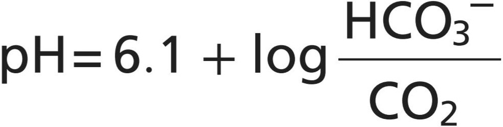
When the glass electrode is used to measure CO2 in blood, a miniature glass electrode is surrounded by a thin plastic membrane. A solution of sodium bicarbonate of known concentration is in the space between the electrode and plastic membrane. Blood is then super- fused onto the outer surface of the plastic membrane, allowing CO2 to diffuse from the blood into the bicarbonate solution. Only a drop or so of blood is required. Next, the pH is measured by the glass electrode, and the CO2 is calculated using the formula that was previously provided.
هنگامیکه از الکترود شیشه ای برای اندازه گیری CO2 در خون استفاده میشود، یک الکترود شیشه ای مینیاتوری توسط یک غشای پلاستیکی نازک احاطه میشود. محلولی از بی کربنات سدیم با غلظت مشخص در فضای بین الکترود و غشای پلاستیکی قرار دارد. سپس خون روی سطح بیرونی غشای پلاستیکی سوپرفیوژن میشود و اجازه میدهد تا CO2 از خون به محلول بیکربنات پخش شود. فقط یک قطره خون لازم است. سپس pH توسط الکترود شیشه ای اندازه گیری میشود و CO2 با استفاده از فرمولی که قبلا ارائه شده است محاسبه میشود.
Determination of Blood Po2. The concentration of O2 in a fluid can be measured by a technique called polarography. Electric current is made to flow between a small negative electrode and the solution. If the volt- age of the electrode is more than -0.6 volt different from the voltage of the solution, O2 will deposit on the electrode. Furthermore, the rate of current flow through the electrode will be directly proportional to the concentration of O2 (and therefore to PO2 as well). In practice, a negative platinum electrode with a surface area of about 1 square millimeter is used, and this electrode is separated from the blood by a thin plastic membrane that allows diffusion of O2 but not diffusion of proteins or other substances that will “poison” the electrode.
تعیین Po2 خون. غلظت O2 در یک مایع را میتوان با تکنیکی به نام پلاروگرافی اندازه گیری کرد. جریان الکتریکی بین یک الکترود منفی کوچک و محلول جریان مییابد. اگر ولتاژ الکترود بیش از -0.6 ولت با ولتاژ محلول متفاوت باشد، O2 روی الکترود رسوب میکند. علاوه بر این، نرخ جریان جریان از طریق الکترود مستقیماً با غلظت O2 (و بنابراین با PO2) متناسب خواهد بود. در عمل، از یک الکترود پلاتین منفی با مساحت سطح حدود 1 میلی متر مربع استفاده میشود و این الکترود توسط یک غشای پلاستیکی نازک از خون جدا میشود که اجازه انتشار O2 را میدهد اما انتشار پروتئینها یا مواد دیگری که الکترود را “مسموم” میکند، نمیدهد.
Often, all three of the measuring devices for pH, CO2, and Po2 are built into the same apparatus, and all these measurements can be made within a minute or so using a single droplet-sized sample of blood. Thus, changes in the blood gas levels and pH can be followed almost moment by moment at the bedside.
اغلب، هر سه دستگاه اندازهگیری pH، CO2 و Po2 در یک دستگاه تعبیه شدهاند، و همه این اندازهگیریها را میتوان در عرض یک دقیقه یا بیشتر با استفاده از یک نمونه خون به اندازه یک قطره انجام داد. بنابراین، تغییرات در سطح گاز خون و pH را میتوان تقریبا لحظه به لحظه در کنار تخت دنبال کرد.
MEASUREMENT OF MAXIMUM EXPIRATORY FLOW
In many respiratory diseases, particularly in asthma, the resistance to airflow becomes especially great during expiration, sometimes causing tremendous difficulty in breathing. This condition has led to the concept called maximum expiratory flow, which can be defined as follows. When a person expires with great force, the expiratory airflow reaches a maximum flow beyond which the flow cannot be increased any more, even with greatly increased additional force. This is the maximum expiratory flow. The maximum expiratory flow is much greater when the lungs are filled with a large volume of air than when they are almost empty. These principles can be understood by referring to Figure 43-1.
اندازه گیری حداکثر جریان بازدم
در بسیاری از بیماریهای تنفسی، بهویژه در آسم، مقاومت در برابر جریان هوا بهویژه در طول بازدم زیاد میشود و گاهی اوقات باعث ایجاد مشکل شدید در تنفس میشود. این وضعیت منجر به مفهومیبه نام حداکثر جریان بازدمیشده است که میتوان آن را به صورت زیر تعریف کرد. وقتی فردی با نیروی زیاد منقضی میشود، جریان هوای بازدمیبه حداکثر جریان میرسد که بیش از آن نمیتوان جریان را بیشتر کرد، حتی با افزایش شدید نیروی اضافی. این حداکثر جریان بازدمیاست. حداکثر جریان بازدم زمانی که ریهها با حجم زیادی از هوا پر میشوند بسیار بیشتر از زمانی است که تقریباً خالی هستند. این اصول را میتوان با مراجعه به شکل 43-1 درک کرد.
Figure 43-14 shows the effect of increased pressure applied to the outsides of the alveoli and air passageways caused by compressing the chest cage. The arrows indicate that the same pressure compresses the outsides of the alveoli and bronchioles. Therefore, not only does this pressure force air from the alveoli toward the bronchioles, but it also tends to collapse the bronchioles at the same time, which will oppose movement of air to the exterior. Once the bronchioles have almost completely collapsed, further expiratory force can still increase the alveolar pressure greatly, but it also increases the degree of bronchiolar collapse and airway resistance by an equal amount, thus preventing further increase in flow. Therefore, beyond a critical degree of expiratory force, a maximum expiratory flow has been reached.
شکل 43-14 اثر افزایش فشار اعمال شده به خارج از آلوئولها و مجاری هوا را نشان میدهد که ناشی از فشرده سازی قفس قفسه سینه است. فلشها نشان میدهند که فشار یکسان، بیرون آلوئولها و برونشیولها را فشرده میکند. بنابراین، این فشار نه تنها هوا را از آلوئولها به سمت نایژهها میکشد، بلکه تمایل به فروپاشی همزمان برونشیولها را نیز دارد که با حرکت هوا به بیرون مخالفت میکند. هنگامیکه نایژهها تقریباً به طور کامل فرو ریختند، نیروی بازدمیبیشتر همچنان میتواند فشار آلوئولی را تا حد زیادی افزایش دهد، اما همچنین درجه فروپاشی نایژه و مقاومت راه هوایی را به میزان مساوی افزایش میدهد، بنابراین از افزایش بیشتر جریان جلوگیری میکند. بنابراین، فراتر از درجه بحرانی نیروی بازدم، حداکثر جریان بازدمیحاصل شده است.
Figure 43-1B shows the effect of different degrees of lung collapse (and therefore also of bronchiolar collapse) on the maximum expiratory flow. The curve recorded in this section shows the maximum expiratory flow at all levels of lung volume after a healthy person first inhales as much air as possible and then expires with maximum expiratory effort until he or she can expire at no greater rate. Note that the person quickly reaches a maximum expiratory airflow of more than 400 L/min. However, regardless of how much additional expiratory effort the person exerts, this is still the maximum flow rate that he or she can achieve.
شکل 43-1B تأثیر درجات مختلف فروپاشی ریه (و بنابراین فروپاشی برونشیولار) را بر حداکثر جریان بازدمینشان میدهد. منحنی ثبت شده در این بخش، حداکثر جریان بازدمیرا در تمام سطوح حجم ریه نشان میدهد، پس از اینکه یک فرد سالم ابتدا تا حد امکان هوا را استنشاق کرده و سپس با حداکثر تلاش بازدمیمنقضی میشود تا زمانی که نتواند با سرعت بیشتری منقضی شود. توجه داشته باشید که فرد به سرعت به حداکثر جریان هوای بازدمیبیش از 400 لیتر در دقیقه میرسد. با این حال، صرف نظر از اینکه فرد چقدر تلاش بازدمیاضافی انجام میدهد، هنوز هم این حداکثر سرعت جریانی است که میتواند به دست آورد.
Note also that as the lung volume becomes smaller, the maximum expiratory flow rate also becomes less. The main reason for this phenomenon is that in the enlarged lung, the bronchi and bronchioles are held open partially by way of elastic pull on their outsides by lung structural elements. However, as the lung becomes smaller, these structures are relaxed so that the bronchi and bronchioles are collapsed more easily by external chest pressure, thus progressively reducing the maximum expiratory flow rate as well.
همچنین توجه داشته باشید که با کوچکتر شدن حجم ریه، حداکثر سرعت جریان بازدم نیز کمتر میشود. دلیل اصلی این پدیده این است که در ریه بزرگ شده، برونشها و برونشیولها تا حدی از طریق کشش الاستیک در قسمت بیرونی خود توسط عناصر ساختاری ریه باز نگه داشته میشوند. با این حال، با کوچکتر شدن ریه، این ساختارها شل میشوند به طوری که برونشها و برونشیولها با فشار خارجی قفسه سینه راحتتر فرو میروند، بنابراین به تدریج حداکثر جریان بازدمینیز کاهش مییابد.
Abnormalities of the Maximum Expiratory Flow- Volume Curve. Figure 43-2 shows the normal maximum expiratory flow-volume curve, along with two additional flow-volume curves recorded in two types of lung dis- eases: constricted lungs and partial airway obstruction. Note that the constricted lungs have both reduced total lung capacity (TLC) and reduced residual volume (RV). Furthermore, because the lung cannot expand to a nor- mal maximum volume, even with the greatest possible expiratory effort, the maximal expiratory flow cannot rise to equal that of the normal curve. Constricted lung diseases include fibrotic diseases of the lung, such as tuberculosis and silicosis, and diseases that constrict the chest cage, such as kyphosis, scoliosis, and fibrotic pleurisy.
ناهنجاریهای منحنی حداکثر جریان بازدمی-حجم. شکل 43-2 منحنی حداکثر حجم جریان بازدمیطبیعی را به همراه دو منحنی حجم جریان اضافی که در دو نوع بیماری ریوی ثبت شده است نشان میدهد: ریههای منقبض و انسداد جزئی راه هوایی. توجه داشته باشید که ریههای منقبض هم ظرفیت کلی ریه (TLC) و هم حجم باقیمانده (RV) کاهش یافته است. علاوه بر این، از آنجایی که ریه نمیتواند به حداکثر حجم طبیعی منبسط شود، حتی با بیشترین تلاش بازدمیممکن، حداکثر جریان بازدمینمیتواند برابر با منحنی طبیعی باشد. بیماریهای منقبض ریه شامل بیماریهای فیبروتیک ریه مانند سل و سیلیکوزیس و بیماریهایی که قفس سینه را منقبض میکنند مانند کیفوز، اسکولیوز و جنب فیبروتیک میباشد.
In diseases with airway obstruction, it is usually much more difficult to expire than to inspire because the closing tendency of the airways is greatly increased by the extra positive pressure required in the chest to cause expiration. By contrast, the extra negative pleural pressure that occurs during inspiration actually “pulls” the airways open at the same time that it expands the alveoli. Therefore, air tends to enter the lung easily but then becomes trapped in the lungs. Over a period of months or years, this effect increases both the TLC and RV, as shown by the green curve in Figure 43-2. Also, because of the obstruction of the airways, and because they collapse more easily than normal airways, the maximum expiratory flow rate is greatly reduced.
در بیماریهای همراه با انسداد راه هوایی، معمولاً منقضی کردن آن بسیار دشوارتر از دمیدن است، زیرا تمایل به بسته شدن راههای هوایی به دلیل فشار مثبت اضافی مورد نیاز در قفسه سینه برای ایجاد بازدم بسیار افزایش مییابد. در مقابل، فشار منفی اضافی پلورال که در طول دم ایجاد میشود، در واقع همزمان با گسترش آلوئولها، راههای هوایی را باز میکند. بنابراین، هوا تمایل دارد به راحتی وارد ریه شود اما سپس در ریهها به دام میافتد. در طی یک دوره چند ماهه یا سالی، این اثر هر دو TLC و RV را افزایش میدهد، همانطور که منحنی سبز در شکل 43-2 نشان داده شده است. همچنین به دلیل انسداد راههای هوایی و به دلیل اینکه راحتتر از راههای هوایی معمولی فرو میروند، حداکثر سرعت بازدم به شدت کاهش مییابد.
The classic disease that causes severe airway obstruction is asthma. Serious airway obstruction also occurs in some stages of emphysema.
بیماری کلاسیکی که باعث انسداد شدید راه هوایی میشود، آسم است. انسداد جدی راه هوایی نیز در برخی از مراحل آمفیزم رخ میدهد.
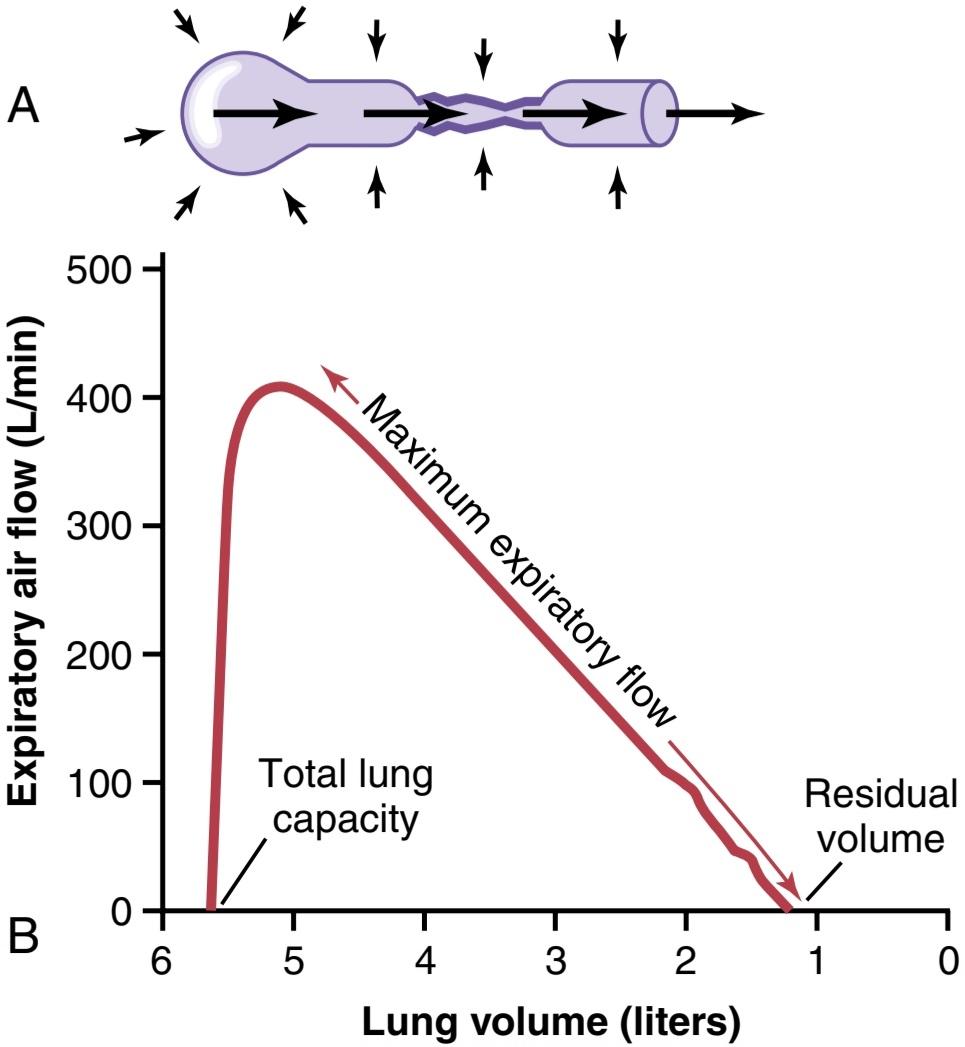
Figure 43-1. A, Collapse of the respiratory passageway during maxi- mum expiratory effort, an effect that limits expiratory flow rate. B, Effect of lung volume on the maximum expiratory airflow, showing decreasing maximum expiratory airflow as the lung volume becomes smaller.
شکل 43-1. الف، فروپاشی مجرای تنفسی در طول حداکثر تلاش بازدمی، اثری که سرعت جریان بازدم را محدود میکند. ب، اثر حجم ریه بر حداکثر جریان هوای بازدمی، نشان دهنده کاهش حداکثر جریان هوای بازدمیبا کوچکتر شدن حجم ریه است.

Figure 43-2. Effect of two respiratory abnormalities-constricted lungs and airway obstruction on the maximum expiratory flow- volume curve. RV, Residual volume; TLC, total lung capacity.
شکل 43-2. اثر دو ناهنجاری تنفسی منقبض ریه و انسداد راه هوایی بر حداکثر منحنی جریان بازدمیحجم. RV، حجم باقیمانده؛ TLC، ظرفیت کل ریه.
FORCED EXPIRATORY VITAL CAPACITY AND FORCED EXPIRATORY VOLUME
Another useful clinical pulmonary test, and one that is also easy to perform, is to record the forced expiratory vital capacity (FVC) on a spirometer. Such a recording is shown in Figure 43-3A for a person with normal lungs and in Figure 43-3B for a person with partial airway obstruction. In performing the FVC maneuver, the person first inspires maximally to the TLC and then exhales into the spirometer with maximum expiratory effort as rapidly and as completely as possible. The total distance of the downslope of the lung volume record represents the FVC, as shown in the figure.
ظرفیت حیاتی بازدم اجباری و حجم بازدم اجباری
یکی دیگر از آزمایشهای مفید بالینی ریوی، که انجام آن نیز آسان است، ثبت ظرفیت حیاتی بازدم اجباری (FVC) روی یک اسپیرومتر است. چنین ضبطی در شکل 43-3A برای فردی با ریههای طبیعی و در شکل 43-3B برای فردی با انسداد جزئی راه هوایی نشان داده شده است. در انجام مانور FVC، فرد ابتدا حداکثر دم را به TLC میدهد و سپس با حداکثر تلاش بازدمیبا حداکثر سرعت و تا حد امکان به داخل اسپیرومتر بازدم میکند. فاصله کل شیب پایین رکورد حجم ریه نشان دهنده FVC است، همانطور که در شکل نشان داده شده است.
Now, study the difference between the two records for (1) normal lungs and (2) partial airway obstruction. The total volume changes of the FVCs are not greatly different, indicating only a moderate difference in basic lung volumes in the two persons. There is, however, a major difference in the amounts of air that these persons can expire each second, especially during the first second. Therefore, it is customary to compare the recorded forced expiratory volume during the first second (FEV1) with the normal value. In the normal person (see Figure 43-3A), the percentage of the FVC that is expired in the first second divided by the total FVC (FEV1/FVC%) is 80%. However, note in Figure 43-3B that with airway obstruction, this value decreases to only 47%. In persons with serious airway obstruction, as often occurs with acute asthma, this value can decrease to less than 20%.
اکنون، تفاوت بین دو رکورد (1) ریههای طبیعی و (2) انسداد جزئی راه هوایی را مطالعه کنید. تغییرات حجم کل FVCها تفاوت زیادی ندارند، که تنها تفاوت متوسطی در حجم پایه ریه در دو نفر را نشان میدهد. با این حال، تفاوت عمدهای در مقدار هوایی وجود دارد که این افراد میتوانند در هر ثانیه منقضی شوند، به خصوص در ثانیه اول. بنابراین، مرسوم است که حجم بازدم اجباری ثبت شده در طول ثانیه اول (FEV1) با مقدار طبیعی مقایسه شود. در افراد عادی (شکل 43-3A را ببینید)، درصد FVC که در ثانیه اول منقضی میشود تقسیم بر کل FVC (FEV1/FVC٪) 80٪ است. با این حال، در شکل 43-3B توجه کنید که با انسداد راه هوایی، این مقدار تنها به 47 درصد کاهش مییابد. در افراد مبتلا به انسداد جدی راه هوایی، همانطور که اغلب با آسم حاد اتفاق میافتد، این مقدار میتواند به کمتر از 20٪ کاهش یابد.

Figure 43-3. Recordings during the forced vital capacity maneuver in a healthy person (A) and in a person with partial airway obstruction (B). (The “zero” on the volume scale is residual volume.) FEV1, Forced expiratory volume during the first second; FVC, forced expiratory vital capacity.
شکل 43-3. ضبط در طول مانور ظرفیت حیاتی اجباری در یک فرد سالم (A) و در یک فرد با انسداد جزئی راه هوایی (B). («صفر» در مقیاس حجم، حجم باقیمانده است.) FEV1، حجم بازدم اجباری در طول ثانیه اول. FVC، ظرفیت حیاتی بازدمیاجباری.
PATHOPHYSIOLOGY OF SPECIFIC PULMONARY ABNORMALITIES CHRONIC PULMONARY EMPHYSEMA
The term pulmonary emphysema literally means excess air in the lungs. However, this term is usually used to describe a complex obstructive and destructive process of the lungs caused by many years of smoking. It results from the following major pathophysiological changes in the lungs:
پاتوفیزیولوژی ناهنجاریهای خاص ریوی آمفیزم مزمن ریوی
اصطلاح آمفیزم ریوی در لغت به معنای هوای اضافی در ریهها است. با این حال، این اصطلاح معمولاً برای توصیف یک فرآیند پیچیده انسدادی و مخرب ریهها که در اثر چندین سال استعمال دخانیات ایجاد میشود، استفاده میشود. این ناشی از تغییرات پاتوفیزیولوژیک عمده زیر در ریهها است:
1. Chronic infection, caused by inhaling smoke or other substances that irritate the bronchi and bronchioles. The chronic infection seriously deranges the normal protective mechanisms of the airways, including partial paralysis of the cilia of the respiratory epithelium, an effect caused by nicotine. As a result, mucus cannot be moved easily out of the passageways. Also, stimulation of excess mucus secretion occurs, which further exacerbates the condition. There is also inhibition of the alveolar macrophages, so they become less effective in com- bating infection.
1. عفونت مزمن، ناشی از استنشاق دود یا سایر موادی که نایژهها و نایژهها را تحریک میکند. عفونت مزمن به طور جدی مکانیسمهای حفاظتی طبیعی راههای هوایی، از جمله فلج جزئی مژکهای اپیتلیوم تنفسی، اثری که توسط نیکوتین ایجاد میشود، مختل میکند. در نتیجه، مخاط را نمیتوان به راحتی از مسیرهای عبور خارج کرد. همچنین، تحریک ترشح بیش از حد موکوس رخ میدهد که این وضعیت را بیشتر تشدید میکند. همچنین مهار ماکروفاژهای آلوئولی وجود دارد، بنابراین آنها در مبارزه با عفونت کمتر موثر میشوند.
2. The infection, excess mucus, and inflammatory edema of the bronchiolar epithelium together cause chronic obstruction of many of the smaller airways.
2. عفونت، مخاط اضافی و ادم التهابی اپیتلیوم برونش با هم باعث انسداد مزمن بسیاری از راههای هوایی کوچکتر میشود.
3. The obstruction of the airways makes it especially difficult to expire, thus causing entrapment of air in the alveoli and overstretching them. This effect, combined with the lung infection, causes marked destruction of as much as 50% to 80% of the alveolar walls. Therefore, the final picture of the emphysematous lung is that shown in Figure 43-4 (top) and 43-5.
3. انسداد راههای هوایی انقباض آن را به ویژه دشوار میکند و در نتیجه باعث گیر افتادن هوا در آلوئولها و کشیده شدن بیش از حد آنها میشود. این اثر، همراه با عفونت ریه، باعث تخریب قابل توجه 50٪ تا 80٪ از دیوارههای آلوئول میشود. بنابراین، تصویر نهایی ریه آمفیزماتوز همان است که در شکل 43-4 (بالا) و 43-5 نشان داده شده است.
The physiological effects of chronic emphysema are variable, depending on the severity of the disease and the relative degrees of bronchiolar obstruction versus lung parenchymal destruction. The different abnormalities include the following:
اثرات فیزیولوژیکی آمفیزم مزمن بسته به شدت بیماری و درجات نسبی انسداد برونش در مقابل تخریب پارانشیم ریه متغیر است. ناهنجاریهای مختلف شامل موارد زیر است:
1. The bronchiolar obstruction increases airway resistance and results in greatly increased work of breathing. It is especially difficult for the person to move air through the bronchioles during expiration because the compressive force on the outside of the lung not only compresses the alveoli but also compresses the bronchioles, which further increases their resistance during expiration.
1. انسداد برونشیولار مقاومت راه هوایی را افزایش میدهد و منجر به افزایش شدید کار تنفس میشود. به ویژه برای فرد دشوار است که هوا را از طریق برونشیولها در حین بازدم حرکت دهد، زیرا نیروی فشاری روی خارج از ریه نه تنها آلوئولها را فشرده میکند، بلکه برونشیولها را نیز فشرده میکند، که مقاومت آنها را در طول بازدم بیشتر میکند.
2. The marked loss of alveolar walls greatly decreases the diffusing capacity of the lung. This reduces the ability of the lungs to oxygenate the blood and re- move CO2 from the blood.
2. از دست دادن قابل توجه دیواره آلوئول ظرفیت انتشار ریه را به شدت کاهش میدهد. این باعث کاهش توانایی ریهها برای اکسیژن رسانی به خون و حذف CO2 از خون میشود.
3. The obstructive process is frequently much worse in some parts of the lungs than in other parts, so some portions of the lungs are well ventilated, whereas other portions are poorly ventilated. This situation often causes extremely abnormal ventilation- perfusion ratios, with a very low VA/Q in some parts (physiological shunt), resulting in poor aeration of the blood, and a very high VA/Q in other parts (physiological dead space), resulting in wasted ventilation, with both effects occurring in the same lungs.
3. فرآیند انسداد اغلب در برخی از قسمتهای ریه بسیار بدتر از قسمتهای دیگر است، بنابراین برخی از قسمتهای ریه به خوبی تهویه میشوند، در حالی که بخشهای دیگر تهویه ضعیفی دارند. این وضعیت اغلب باعث نسبت تهویه – پرفیوژن بسیار غیر طبیعی میشود، با VA/Q بسیار کم در برخی قسمتها (شنت فیزیولوژیکی)، که منجر به هوادهی ضعیف خون، و VA/Q بسیار بالا در سایر قسمتها (فضای مرده فیزیولوژیکی)، و در نتیجه تهویه هدر رفته، با هر دو اثر در یک ریه رخ میدهد.
4. Loss of large portions of the alveolar walls also decreases the number of pulmonary capillaries through which blood can pass. As a result, the pulmonary vascular resistance often increases marked- ly, causing pulmonary hypertension, which in turn overloads the right side of the heart and frequently causes right-sided heart failure.
4. از دست دادن بخشهای بزرگی از دیواره آلوئول همچنین تعداد مویرگهای ریوی را که خون میتواند از آنها عبور کند کاهش میدهد. در نتیجه، مقاومت عروق ریوی اغلب به طور قابل توجهی افزایش مییابد و باعث فشار خون ریوی میشود که به نوبه خود باعث اضافه بار سمت راست قلب میشود و اغلب باعث نارسایی سمت راست قلب میشود.
Chronic emphysema usually progresses slowly over many years. Both hypoxia and hypercapnia develop because of hypoventilation of many alveoli plus loss of alveolar walls. The net result of all these effects is severe, prolonged, devastating air hunger that can last for years until the hypoxia and hypercapnia cause death—a high penalty to pay for smoking.
آمفیزم مزمن معمولاً طی سالها به کندی پیشرفت میکند. هیپوکسی و هیپرکاپنیا هر دو به دلیل هیپوونتیلاسیون بسیاری از آلوئولها و از بین رفتن دیواره آلوئول ایجاد میشوند. نتیجه خالص همه این اثرات، گرسنگی شدید، طولانی مدت و ویرانگر هوا است که میتواند سالها ادامه داشته باشد تا زمانی که هیپوکسی وهایپرکاپنی باعث مرگ شود – جریمه بالایی برای کشیدن سیگار.
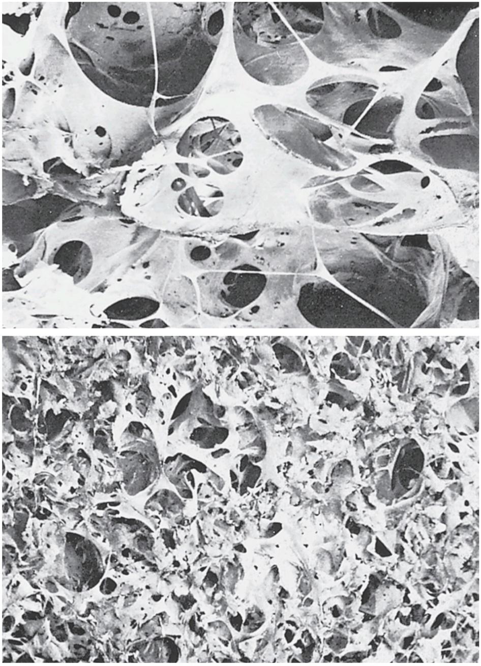
Figure 43-4. Contrast of the emphysematous lung (top) with the normal lung (bottom) showing extensive alveolar destruction in emphysema. (Courtesy Patricia Delaney and the Department of Anatomy, The Medical College of Wisconsin, Milwaukee, WI.)
شکل 43-4. تضاد ریه آمفیزماتوز (بالا) با ریه طبیعی (پایین) که تخریب آلوئولی گسترده را در آمفیزم نشان میدهد. (با احترام به پاتریشیا دلانی و دپارتمان آناتومی، کالج پزشکی ویسکانسین، میلواکی، WI.)
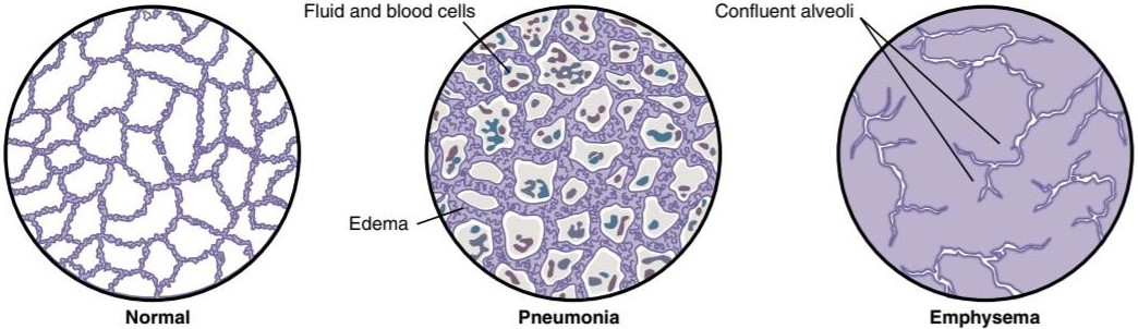
Figure 43-5. Lung alveolar changes in pneumonia and emphysema.
شکل 43-5. تغییرات آلوئولی ریه در پنومونی و آمفیزم.
PNEUMONIA-LUNG INFLAMMATION AND FLUID IN ALVEOLI
The term pneumonia includes any inflammatory condition of the lung in which some or all of the alveoli are filled with fluid and blood cells, as shown in Figure 43-5. A common type of pneumonia is bacterial pneumonia, caused most frequently by pneumococci. This dis- ease begins with infection in the alveoli; the pulmonary membrane becomes inflamed and highly porous so that fluid and even red and white blood cells leak out of the blood into the alveoli. Thus, the infected alveoli become progressively filled with fluid and cells, and the infection spreads by extension of bacteria or virus from alveolus to alveolus. Eventually, large areas of the lungs, sometimes whole lobes or even a whole lung, become “consolidated,” which means that they are filled with fluid and cellular debris.
پنومونی – التهاب ریه و مایع در آلوئول
اصطلاح پنومونی شامل هر گونه بیماری التهابی ریه است که در آن برخی یا همه آلوئولها با مایع و سلولهای خونی پر شده است، همانطور که در شکل 43-5 نشان داده شده است. یک نوع شایع پنومونی، پنومونی باکتریایی است که بیشتر توسط پنوموکوک ایجاد میشود. این بیماری با عفونت در آلوئولها شروع میشود. غشای ریوی ملتهب و بسیار متخلخل میشود به طوری که مایع و حتی گلبولهای قرمز و سفید خون از خون به داخل آلوئولها نشت میکنند. بنابراین، آلوئولهای آلوده به تدریج پر از مایع و سلول میشوند و عفونت با گسترش باکتری یا ویروس از آلوئول به آلوئول گسترش مییابد. در نهایت، نواحی وسیعی از ریهها، گاهی لوبهای کامل یا حتی یک ریه کامل، «تجمیع» میشوند، به این معنی که آنها با مایع و زبالههای سلولی پر میشوند.
In persons with pneumonia, the gas exchange functions of the lungs decline in different stages of the dis- ease. In early stages, the pneumonia process might well be localized to only one lung, with alveolar ventilation being reduced while blood flow through the lung continues normally. This condition causes two major pulmonary abnormalities: (1) reduction in the total available surface area of the respiratory membrane; and (2) a decreased ventilation-perfusion ratio. Both these effects cause hypoxemia (low blood O2) and hypercapnia (high blood CO2).
در افراد مبتلا به پنومونی، عملکرد تبادل گاز ریهها در مراحل مختلف بیماری کاهش مییابد. در مراحل اولیه، فرآیند ذاتالریه ممکن است فقط در یک ریه موضعی داشته باشد، در حالی که جریان خون در ریه به طور طبیعی ادامه مییابد، تهویه آلوئولی کاهش مییابد. این وضعیت باعث دو ناهنجاری عمده ریوی میشود: (1) کاهش در سطح کل موجود در غشای تنفسی. و (2) کاهش نسبت تهویه – پرفیوژن. هر دوی این اثرات باعث هیپوکسمی(کم شدن اکسیژن خون) و هیپرکاپنی (CO2 خون بالا) میشوند.
Figure 43-6 shows the effect of the decreased ventilation-perfusion ratio in pneumonia. The blood passing through the aerated lung becomes 97% saturated with O2, whereas that passing through the unaerated lung is about 60% saturated. Therefore, the average saturation of the blood pumped by the left heart into the aorta is only about 78%, which is far below normal.
شکل 43-6 اثر کاهش نسبت تهویه – پرفیوژن را در پنومونی نشان میدهد. خونی که از ریه هوادهی میگذرد 97 درصد با O2 اشباع میشود، در حالی که خون عبوری از ریه بدون هواده حدود 60 درصد اشباع است. بنابراین، میانگین اشباع خون پمپاژ شده توسط قلب چپ به آئورت تنها حدود 78 درصد است که بسیار کمتر از حد طبیعی است.

Figure 43-6. Effect of pneumonia on percentage saturation of oxy- gen (O2) in the pulmonary artery, the right and left pulmonary veins, and the aorta.
شکل 43-6. تأثیر پنومونی بر درصد اشباع اکسیژن (O2) در شریان ریوی، وریدهای ریوی راست و چپ و آئورت.
ATELECTASIS-COLLAPSE OF THE ALVEOLI
Atelectasis means collapse of the alveoli. It can occur in localized areas of a lung or in an entire lung. Common causes of atelectasis are (1) total obstruction of the airway and (2) lack of surfactant in the fluids lining the alveoli.
آتلکتاز – فروپاشی آلوئول
آتلکتازی به معنای فروپاشی آلوئولها است. این میتواند در نواحی موضعی یک ریه یا در کل ریه رخ دهد. علل شایع آتلکتازی عبارتند از: (1) انسداد کامل راه هوایی و (2) کمبود سورفکتانت در مایعات پوشاننده آلوئول.
Airway Obstruction Causes Lung Collapse. The airway obstruction type of atelectasis usually results from the following: (1) blockage of many small bronchi with mucus; or (2) obstruction of a major bronchus by a large mucous plug or some solid object, such as a tumor. The air entrapped beyond the block is absorbed within minutes to hours by the blood flowing in the pulmonary capillaries. If the lung tissue is pliable enough, this will lead simply to collapse of the alveoli. However, if the lung is rigid be- cause of fibrotic tissue and cannot collapse, absorption of air from the alveoli creates very negative pressures within the alveoli, which pull fluid out of the pulmonary capillaries into the alveoli, thus causing the alveoli to fill completely with edema fluid. This process almost always is the effect that occurs when an entire lung becomes atelectatic, a condition called massive collapse of the lung.
انسداد راه هوایی باعث فروپاشی ریه میشود. نوع انسداد راه هوایی آتلکتازی معمولاً ناشی از موارد زیر است: (1) انسداد بسیاری از برونشهای کوچک همراه با مخاط. یا (2) انسداد یک برونش اصلی توسط یک پلاگ مخاطی بزرگ یا برخی جسم جامد، مانند تومور. هوای محبوس شده در آن سوی بلوک در عرض چند دقیقه تا چند ساعت توسط خونی که در مویرگهای ریوی جریان دارد جذب میشود. اگر بافت ریه به اندازه کافی انعطاف پذیر باشد، این امر به سادگی منجر به فروپاشی آلوئولها میشود. با این حال، اگر ریه به دلیل بافت فیبروتیک سفت باشد و نتواند فرو بریزد، جذب هوا از آلوئولها فشارهای بسیار منفی در آلوئول ایجاد میکند که مایع را از مویرگهای ریوی به داخل آلوئولها میکشد و در نتیجه باعث پر شدن کامل آلوئولها با مایع ادم میشود. این فرآیند تقریباً همیشه اثری است که زمانی رخ میدهد که کل یک ریه آتلکتاتیک میشود، وضعیتی که به آن فروپاشی عظیم ریه میگویند.
The effects on overall pulmonary function caused by massive collapse (atelectasis) of an entire lung are shown in Figure 43-7. Collapse of the lung tissue not only occludes the alveoli but also almost always increases the resistance to blood flow through the pulmonary vessels of the collapsed lung. This resistance increase occurs partially because of the lung collapse, which compresses and folds the vessels as the volume of the lung decreases. In addition, hypoxia in the collapsed alveoli causes additional vasoconstriction, as explained in Chapter 39.
اثرات بر عملکرد کلی ریوی ناشی از فروپاشی عظیم (آتلکتازی) کل ریه در شکل 43-7 نشان داده شده است. فروپاشی بافت ریه نه تنها آلوئولها را مسدود میکند، بلکه تقریباً همیشه مقاومت در برابر جریان خون از طریق عروق ریوی ریه از بین رفته را افزایش میدهد. این افزایش مقاومت تا حدی به دلیل فروپاشی ریه رخ میدهد، که با کاهش حجم ریه، عروق را فشرده و تا میکند. علاوه بر این، هیپوکسی در آلوئولهای فرو ریخته باعث انقباض عروق اضافی میشود، همانطور که در فصل 39 توضیح داده شد.
Because of the vascular constriction, blood flow through the atelectatic lung is greatly reduced. Fortunately, most of the blood is routed through the ventilated lung and therefore becomes well aerated. In the situation shown in Figure 43-7, five-sixths of the blood passes through the aerated lung, and only onesixth passes through the unaerated lung. As a result, the overall ventilation-perfusion ratio is only moderately compromised, so the aortic blood has only mild O, desaturation, despite total loss of ventilation in an entire lung.
به دلیل انقباض عروقی، جریان خون از طریق ریه آتلکتاتیک تا حد زیادی کاهش مییابد. خوشبختانه، بیشتر خون از طریق ریه تهویه شده هدایت میشود و بنابراین به خوبی هوادهی میشود. در وضعیت نشان داده شده در شکل 43-7، پنج ششم خون از ریه هوادهی شده و تنها یک ششم از ریه بدون هوادهی عبور میکند. در نتیجه، نسبت کلی تهویه به پرفیوژن فقط به میزان متوسطی به خطر میافتد، بنابراین خون آئورت با وجود از دست دادن تهویه در کل ریه، فقط O خفیف دارد، غیراشباع.
Lack of “Surfactant” as a Cause of Lung Collapse. The secretion and function of surfactant in the alveoli were discussed in Chapter 38. Surfactant is secreted by special alveolar epithelial cells into the fluids that coat the inside surface of the alveoli. The surfactant in turn decreases the surface tension in the alveoli by 2- to 10-fold, which normally plays a major role in preventing alveolar collapse. However, in several conditions, such as in hyaline mem- brane disease (also called respiratory distress syndrome), which often occurs in newborn premature babies, the quantity of surfactant secreted by the alveoli is so greatly depressed that the surface tension of the alveolar fluid be- comes several times greater than normal. This surfactant deficiency causes a serious tendency for the lungs of these babies to collapse or to become filled with fluid. As explained in Chapter 38, many of these infants die of suffocation when large portions of the lungs become atelectatic.
فقدان “سورفکتانت” به عنوان علت فروپاشی ریه. ترشح و عملکرد سورفکتانت در آلوئولها در فصل 38 مورد بحث قرار گرفت. سورفکتانت توسط سلولهای اپیتلیال آلوئولی ویژه به مایعاتی که سطح داخلی آلوئولها را میپوشانند ترشح میشود. سورفکتانت به نوبه خود کشش سطحی آلوئولها را 2 تا 10 برابر کاهش میدهد که معمولاً نقش مهمیدر جلوگیری از فروپاشی آلوئول ایفا میکند. با این حال، در شرایط متعددی، مانند بیماری غشای هیالین (که به آن سندرم زجر تنفسی نیز میگویند)، که اغلب در نوزادان نارس رخ میدهد، مقدار سورفکتانت ترشح شده از آلوئولها به حدی کاهش مییابد که کشش سطحی مایع آلوئولی چندین برابر بیشتر از حد طبیعی میشود. این کمبود سورفکتانت باعث تمایل جدی ریههای این نوزادان به فروپاشی یا پر شدن از مایع میشود. همانطور که در فصل 38 توضیح داده شد، بسیاری از این نوزادان در اثر خفگی میمیرند که بخشهای بزرگی از ریهها آتلکتاتیک میشوند.

Figure 43-7. Effect of atelectasis on aortic blood oxygen (O2) saturation.
شکل 43-7. اثر آتلکتازی بر اشباع اکسیژن خون آئورت (O2).
ASTHMA-SPASMODIC CONTRACTION OF SMOOTH MUSCLES IN BRONCHIOLES
Asthma is characterized by spastic contraction of the smooth muscle in the bronchioles, which partially obstructs the bronchioles and causes extremely difficult breathing. The prevalence of asthma has been increasing and affects 7% to 8% of all people in the United States, with even higher rates in some groups such as non-Hispanic blacks. The World Health Organization estimates that over 235 million people worldwide suffer from asthma, although some estimates of asthma prevalence are as high as 339 million people.
انقباض آسم – اسپاسم عضلات صاف در برونش
آسم با انقباض اسپاستیک ماهیچه صاف در برونشیلها مشخص میشود که تا حدی باعث انسداد نایژهها میشود و باعث تنفس بسیار دشوار میشود. شیوع آسم در حال افزایش بوده است و 7 تا 8 درصد از کل مردم ایالات متحده را تحت تاثیر قرار میدهد و در برخی از گروهها مانند سیاه پوستان غیر اسپانیایی این میزان حتی بالاتر است. سازمان بهداشت جهانی تخمین میزند که بیش از 235 میلیون نفر در سراسر جهان از آسم رنج میبرند، اگرچه برخی تخمینها از شیوع آسم به 339 میلیون نفر میرسد.
The usual cause of asthma is contractile hypersensitivity of the bronchioles in response to foreign substances in the air. In about 70% of patients younger than 30 years, the asthma is caused by allergic hypersensitivity, especially sensitivity to plant pollens. In older people, the cause is almost always hypersensitivity to nonallergenic types of irritants in the air, such as irritants in smog.
علت معمول آسم، حساسیت بیش از حد انقباضی برونشیولها در پاسخ به مواد خارجی موجود در هوا است. در حدود 70 درصد از بیماران زیر 30 سال، آسم ناشی از حساسیت بیش از حد آلرژیک، به ویژه حساسیت به گرده گیاهان است. در افراد مسن، علت تقریباً همیشه حساسیت مفرط به انواع غیر حساسیت زا از محرکهای موجود در هوا، مانند محرکهای موجود در مه دود است.
The typical allergic person tends to form abnormally large amounts of immunoglobulin E (IgE) antibodies, and these antibodies cause allergic reactions when they react with the specific antigens that have caused them to develop in the first place, as explained in Chapter 35. In persons with asthma, these antibodies are mainly attached to mast cells that are present in the lung interstitium in close association with the bronchioles and small bronchi. When an asthmatic person breathes in pollen to which he or she is sensitive (i.e., to which the person has developed IgE antibodies), the pollen reacts with the mast cell-attached antibodies and causes the mast cells to release several different substances. Among them are the following: (1) histamine; (2) slow-reacting substance of anaphylaxis (which is a mixture of leukotrienes); (3) eosinophilic chemotactic factor; and (4) bradykinin. The combined effects of all these factors, especially the slow- reacting substance of anaphylaxis, are to produce the following: (1) localized edema in the walls of the small bronchioles, as well as secretion of thick mucus into the bronchiolar lumens; and (2) spasm of the bronchiolar smooth muscle. Therefore, the airway resistance increases greatly.
افراد آلرژیک معمولی تمایل دارند مقادیر غیرطبیعی زیادی از آنتی بادیهای ایمونوگلوبولین E (IgE) تشکیل دهند و این آنتی بادیها هنگامیکه با آنتی ژنهای خاصی که در وهله اول باعث ایجاد آنها شده است واکنش نشان میدهند، همانطور که در فصل 35 توضیح داده شد. برونشیولها و برونشهای کوچک. هنگامیکه یک فرد مبتلا به آسم گردههایی را تنفس میکند که به آن حساس است (یعنی فرد به آن آنتی بادیهای IgE تولید کرده است)، گرده با آنتی بادیهای متصل به ماست سل واکنش میدهد و باعث میشود که ماست سلها چندین ماده مختلف را آزاد کنند. از جمله آنها میتوان به موارد زیر اشاره کرد: (1) هیستامین. (2) ماده آنافیلاکسی با واکنش کند (که مخلوطی از لکوترینها است). (3) عامل کموتاکتیک ائوزینوفیلیک. و (4) برادی کینین. اثرات ترکیبی همه این عوامل، به ویژه ماده با واکنش کند آنافیلاکسی، باعث ایجاد موارد زیر میشود: (1) ادم موضعی در دیوارههای برونشیولهای کوچک، و همچنین ترشح مخاط غلیظ در لومنهای برونش. و (2) اسپاسم عضله صاف برونش. بنابراین مقاومت راه هوایی به شدت افزایش مییابد.
As discussed earlier in this chapter, the bronchiolar diameter becomes reduced more during expiration than during inspiration in persons with asthma as a result of bronchiolar collapse during expiratory effort that compresses the outsides of the bronchioles. Because the bronchioles of the asthmatic lungs are already partially occluded, further occlusion resulting from the external pressure creates especially severe obstruction during expiration. That is, the asthmatic person often can inspire quite adequately but has great difficulty expiring. Clinical measurements show (1) greatly reduced maximum expiratory rate, and (2) reduced timed expiratory volume. Also, all this together results in dyspnea, or “air hunger,” discussed later in this chapter.
همانطور که قبلاً در این فصل مورد بحث قرار گرفت، قطر نایژه در افراد مبتلا به آسم در طی بازدم بیشتر از هنگام دم کاهش مییابد که در نتیجه فروپاشی نایژه در طی تلاش بازدمیکه قسمتهای بیرونی برونشیولها را فشرده میکند، کاهش مییابد. از آنجایی که برونشیولهای ریههای مبتلا به آسم تا حدی مسدود شده اند، انسداد بیشتر ناشی از فشار خارجی باعث ایجاد انسداد شدید در طول بازدم میشود. به این معنا که فرد مبتلا به آسم اغلب میتواند به اندازه کافی الهام بخشد اما در انقضا مشکل زیادی دارد. اندازهگیریهای بالینی نشان میدهد (1) حداکثر سرعت بازدم را به میزان زیادی کاهش میدهد، و (2) حجم بازدم را کاهش میدهد. همچنین، همه اینها با هم منجر به تنگی نفس یا “گرسنگی هوا” میشود که بعداً در این فصل مورد بحث قرار میگیرد.
The functional residual capacity and residual volume of the lung become especially increased during an acute asthma attack because of the difficulty in expiring air from the lungs. Also, over a period of years, the chest cage becomes permanently enlarged, causing a so-called barrel chest, and both the functional residual capacity and lung residual volume become permanently increased.
ظرفیت باقیمانده عملکردی و حجم باقیمانده ریه به ویژه در طول حمله حاد آسم به دلیل مشکل در خروج هوا از ریهها افزایش مییابد. همچنین در طی چند سال، قفس سینه به طور دائم بزرگ میشود و به اصطلاح سینه بشکه ای ایجاد میکند و ظرفیت باقیمانده عملکردی و حجم باقیمانده ریه به طور دائم افزایش مییابد.
TUBERCULOSIS
In tuberculosis, the tubercle bacilli cause a peculiar tissue reaction in the lungs, including (1) invasion of the infected tissue by macrophages, and (2) “walling off” of the lesion by fibrous tissue to form the so- called tubercle. This walling-off process helps limit further transmission of the tubercle bacilli in the lungs and therefore is part of the protective process against extension of the infection. However, in about 3% of people in whom tuberculosis develops, if the disease is not treated, the walling-off process fails, and tubercle bacilli spread throughout the lungs, often causing extreme destruction of lung tissue, with formation of large abscess cavities.
سل
در سل، باسیلهای سل باعث واکنش بافتی خاص در ریهها میشوند، از جمله (1) حمله ماکروفاژها به بافت آلوده، و (2) “دیواره کردن” ضایعه توسط بافت فیبری برای تشکیل به اصطلاح سل. این فرآیند جداسازی به محدود کردن انتقال بیشتر باسیل سل در ریهها کمک میکند و بنابراین بخشی از فرآیند محافظتی در برابر گسترش عفونت است. با این حال، در حدود 3٪ از افرادی که سل در آنها ایجاد میشود، اگر بیماری درمان نشود، فرآیند جداسازی با شکست مواجه میشود و باسیلهای سل در سراسر ریهها پخش میشوند و اغلب باعث تخریب شدید بافت ریه و تشکیل حفرههای آبسه بزرگ میشوند.
Thus, tuberculosis in its late stages is characterized by many areas of fibrosis throughout the lungs, as well as by reduced total amount of functional lung tissue. These effects cause the following: (1) increased “work” on the part of the respiratory muscles to cause pulmonary ventilation and reduced vital capacity and breathing capacity; (2) reduced total respiratory membrane surface area and increased thickness of the respiratory membrane, causing progressively diminished pulmonary diffusing capacity; and (3) abnormal ventilation-perfusion ratio in the lungs, further reducing overall pulmonary diffusion of O2 and CO2.
بنابراین، سل در مراحل پایانی خود با بسیاری از نواحی فیبروز در سراسر ریهها و همچنین با کاهش مقدار کل بافت عملکردی ریه مشخص میشود. این اثرات باعث موارد زیر میشود: (1) افزایش “کار” در بخشی از ماهیچههای تنفسی برای ایجاد تهویه ریوی و کاهش ظرفیت حیاتی و ظرفیت تنفس. (2) کاهش سطح کل غشای تنفسی و افزایش ضخامت غشای تنفسی، که باعث کاهش تدریجی ظرفیت انتشار ریوی میشود. و (3) نسبت تهویه-پرفیوژن غیرطبیعی در ریهها، انتشار کلی O2 و CO2 در ریوی را بیشتر کاهش میدهد.
HYPOXIA AND OXYGEN THERAPY
Almost any of the conditions discussed in the past few sections of this chapter can cause serious cellular hypoxia throughout the body. Sometimes O2 therapy is of great value, other times it is of moderate value, and at still other times it is of almost no value. Therefore, it is important to understand the different types of hypoxia, and then we can discuss the physiological principles of oxygen therapy. The following is a descriptive classification of the causes of hypoxia:
هیپوکسی و اکسیژن درمانی
تقریباً هر یک از شرایطی که در چند بخش گذشته این فصل مورد بحث قرار گرفت، میتواند باعث هیپوکسی سلولی جدی در سراسر بدن شود. گاهی اوقات O2 درمانی ارزش زیادی دارد، گاهی اوقات ارزش متوسطی دارد و در موارد دیگر تقریباً هیچ ارزشی ندارد. بنابراین، درک انواع مختلف هیپوکسی مهم است و سپس میتوانیم اصول فیزیولوژیکی اکسیژن درمانی را مورد بحث قرار دهیم. در زیر یک طبقه بندی توصیفی از علل هیپوکسی وجود دارد:
1. Inadequate oxygenation of the blood in the lungs because of extrinsic reasons
a. Deficiency of O2 in the atmosphere
b. Hypoventilation (neuromuscular disorders)
1. اکسیژن رسانی ناکافی خون در ریهها به دلایل بیرونی
الف کمبود O2 در جو
ب هیپوونتیلاسیون (اختلالات عصبی عضلانی)
2. Pulmonary disease
a. Hypoventilation caused by increased airway resistance or decreased pulmonary compliance
b. Abnormal alveolar ventilation-perfusion ratio (including increased physiological dead space or increased physiological shunt)
c. Diminished respiratory membrane diffusion
2. بیماری ریوی
الف هیپوونتیلاسیون ناشی از افزایش مقاومت راه هوایی یا کاهش انطباق ریوی
ب نسبت غیر طبیعی تهویه به پرفیوژن آلوئولی (شامل افزایش فضای مرده فیزیولوژیکی یا افزایش شانت فیزیولوژیکی)
ج کاهش انتشار غشای تنفسی
3. Venous-to-arterial shunts (right-to-left cardiac shunts)
3. شانت وریدی به شریانی (شنت قلبی از راست به چپ)
4. Inadequate O2 transport to the tissues by the blood
a. Anemia or abnormal hemoglobin
b. General circulatory deficiency
c. Localized circulatory deficiency (peripheral, cerebral, coronary vessels)
d. Tissue edema
4. انتقال ناکافی O2 به بافتها توسط خون
الف کم خونی یا هموگلوبین غیر طبیعی
ب کمبود گردش خون عمومی
ج کمبود گردش خون موضعی (رگهای محیطی، مغزی، کرونری)
د ادم بافتی
5. Inadequate tissue capability of using O2
a. Poisoning of cellular oxidation enzymes
b. Diminished cellular metabolic capacity for using oxygen because of toxicity, vitamin deficiency, or other factors
5. قابلیت بافت ناکافی استفاده از O2
الف مسمومیت با آنزیمهای اکسیداسیون سلولی
ب کاهش ظرفیت متابولیک سلولی برای استفاده از اکسیژن به دلیل سمیت، کمبود ویتامین یا عوامل دیگر
This classification of the types of hypoxia is mainly self- evident from the discussions earlier in the chapter. Only one type of hypoxia in the classification needs further elaboration-the hypoxia caused by inadequate capability of the body’s tissue cells to use O2.
این طبقهبندی انواع هیپوکسی عمدتاً از بحثهای قبلی در فصل مشخص است. تنها یک نوع هیپوکسی در طبقه بندی نیاز به توضیح بیشتر دارد – هیپوکسی ناشی از توانایی ناکافی سلولهای بافت بدن برای استفاده از O2.
Inadequate Tissue Capability to Use Oxygen. The classic cause of inability of the tissues to use O2 is cyanide poisoning, in which the action of the enzyme cytochrome oxidase is blocked by cyanide to such an extent that the tissues simply cannot use O2, even when plenty is avail- able. Also, deficiencies of some of the tissue cellular oxidative enzymes or of other elements in the tissue oxidative system can lead to this type of hypoxia. A special example occurs in the disease beriberi, in which several important steps in tissue utilization of oxygen and the formation of CO2 are compromised because of vitamin B deficiency.
قابلیت ناکافی بافت برای استفاده از اکسیژن. علت کلاسیک ناتوانی بافتها در استفاده از O2 مسمومیت با سیانید است که در آن عمل آنزیم سیتوکروم اکسیداز توسط سیانید به حدی مسدود میشود که بافتها به سادگی نمیتوانند از O2 استفاده کنند، حتی اگر مقدار زیادی در دسترس باشد. همچنین کمبود برخی از آنزیمهای اکسیداتیو سلولی بافتی یا سایر عناصر در سیستم اکسیداتیو بافت میتواند منجر به این نوع هیپوکسی شود. یک مثال خاص در بیماری بری بری رخ میدهد که در آن چندین مرحله مهم در استفاده بافتی از اکسیژن و تشکیل CO2 به دلیل کمبود ویتامین B به خطر میافتد.
Effects of Hypoxia on the Body. Hypoxia, if severe enough, can cause death of cells throughout the body, but in less severe degrees, it mainly causes (1) depressed mental activity, sometimes culminating in coma, and (2) reduced work capacity of the muscles. These effects are specifically discussed in Chapter 44 in relation to high- altitude physiology.
اثرات هیپوکسی بر بدن. هیپوکسی، اگر به اندازه کافی شدید باشد، میتواند باعث مرگ سلولها در سراسر بدن شود، اما در درجات شدیدتر، عمدتاً باعث (1) فعالیت ذهنی افسرده، گاهی اوقات به کما میشود، و (2) کاهش ظرفیت کار عضلات. این اثرات به طور خاص در فصل 44 در رابطه با فیزیولوژی ارتفاعات مورد بحث قرار گرفته است.
OXYGEN THERAPY IN DIFFERENT TYPES OF HYPOXIA
O2 can be administered by the following: (1) placing the patient’s head in a “tent” that contains air fortified with O2; (2) allowing the patient to breathe pure O2 or high concentrations of O2 from a mask; or (3) administering O2 through an intranasal tube.
اکسیژن درمانی در انواع مختلف هیپوکسی
O2 را میتوان با موارد زیر تجویز کرد: (1) قرار دادن سر بیمار در یک “چادر” که حاوی هوای غنی شده با O2 است. (2) اجازه دادن به بیمار برای تنفس O2 خالص یا غلظت بالای O2 از ماسک. یا (3) تجویز O2 از طریق یک لوله داخل بینی.
Recalling the basic physiological principles of the different types of hypoxia, one can readily decide when O2 therapy will be of value and, if so, how valuable.
با یادآوری اصول اولیه فیزیولوژیکی انواع مختلف هیپوکسی، میتوان به آسانی تصمیم گرفت که درمان O2 چه زمانی ارزشمند است و اگر چنین است، چقدر ارزشمند است.
In atmospheric hypoxia, O2 therapy can completely correct the depressed O2 level in the inspired gases and, therefore, provide 100% effective therapy.
در هیپوکسی اتمسفر، O2 درمانی میتواند سطح O2 کاهش یافته را در گازهای الهام گرفته به طور کامل اصلاح کند و بنابراین، درمان 100٪ موثر را ارائه میدهد.
In hypoventilation hypoxia, a person breathing 100% O2 can move five times as much O2 into the alveoli with each breath as when breathing normal air. Therefore, here again, O2 therapy can be extremely beneficial. However, this O2 therapy provides no benefit for the excess blood CO2 also caused by the hypoventilation.
در هیپوکسی هیپوونتیلاسیون، فردی که 100% O2 تنفس میکند، میتواند پنج برابر بیشتر از زمانی که هوای معمولی تنفس میکند، با هر بار تنفس، O2 را به داخل آلوئولها منتقل کند. بنابراین، در اینجا دوباره، O2 درمانی میتواند بسیار مفید باشد. با این حال، این درمان O2 هیچ فایده ای برای CO2 اضافی خون که در اثر هیپوونتیلاسیون نیز ایجاد میشود، ندارد.
In hypoxia caused by impaired alveolar membrane diffusion, essentially the same result occurs as in hypoventilation hypoxia because O2 therapy can increase the Po2 in the lung alveoli from the normal value of about 100 mm Hg to as high as 600 mm Hg. This action raises the O2 pressure gradient for diffusion of oxygen from the alveoli to the blood from the normal value of 60 mm Hg to as high as 560 mm Hg, an increase of more than 800%. This highly beneficial effect of O, therapy in diffusion hypoxia is demonstrated in Figure 43-8, which shows that the pulmonary blood in this patient with pulmonary edema picks up O2 three to four times as rapidly as would occur with no therapy.
در هیپوکسی ناشی از اختلال در انتشار غشای آلوئولی، اساساً همان نتیجه در هیپوکسی هیپوونتیلاسیون رخ میدهد، زیرا درمان O2 میتواند Po2 را در آلوئولهای ریه از مقدار طبیعی حدود 100 میلی متر جیوه به 600 میلی متر جیوه افزایش دهد. این عمل گرادیان فشار O2 برای انتشار اکسیژن از آلوئولها به خون را از مقدار طبیعی 60 میلی متر جیوه به 560 میلی متر جیوه افزایش میدهد که افزایش بیش از 800٪ است. این اثر بسیار سودمند درمان O، در هیپوکسی انتشار در شکل 43-8 نشان داده شده است، که نشان میدهد که خون ریوی در این بیمار مبتلا به ادم ریوی سه تا چهار برابر سریعتر از آنچه بدون درمان اتفاق میافتد، O2 را جذب میکند.
In hypoxia caused by anemia, abnormal hemoglobin transport of O2, circulatory deficiency, or physiological shunt, O, therapy is of much less value because normal O2 is already available in the alveoli. Instead, the problem is that one or more of the mechanisms for transporting oxygen from the lungs to the tissues are deficient. Even so, a small amount of extra O2, between 7% and 30%, can be transported in the dissolved state in the blood when alveolar O2 is increased to maximum, even though the amount transported by the hemoglobin is hardly altered. This small amount of extra O, may be the difference between life and death.
در هیپوکسی ناشی از کم خونی، انتقال غیرطبیعی هموگلوبین O2، کمبود گردش خون یا شانت فیزیولوژیکی O، درمان از ارزش بسیار کمتری برخوردار است زیرا O2 طبیعی از قبل در آلوئولها موجود است. در عوض، مشکل این است که یک یا چند مکانیسم برای انتقال اکسیژن از ریهها به بافتها کمبود دارند. با این حال، مقدار کمیاز O2 اضافی، بین 7٪ تا 30٪، میتواند در حالت محلول در خون منتقل شود، زمانی که O2 آلوئولی به حداکثر افزایش یابد، حتی اگر مقدار انتقال یافته توسط هموگلوبین به سختی تغییر کند. این مقدار اندک O اضافی، ممکن است تفاوت بین زندگی و مرگ باشد.
In the different types of hypoxia caused by inadequate tissue use of O2, there is no abnormality of O2 pickup by the lungs or of transport to the tissues. Instead, the tissue metabolic enzyme system is simply incapable of using the O2 that is delivered. Therefore, O2 therapy provides no measurable benefit.
در انواع مختلف هیپوکسی ناشی از استفاده ناکافی از O2 در بافت، هیچ اختلالی در برداشت O2 توسط ریهها یا انتقال به بافتها وجود ندارد. در عوض، سیستم آنزیمیمتابولیک بافت به سادگی قادر به استفاده از O2 است که تحویل داده میشود. بنابراین، O2 درمانی هیچ سود قابل اندازه گیری ندارد.

Figure 43-8. Absorption of oxygen into the pulmonary capillary blood in pulmonary edema, with and without oxygen tent therapy.
شکل 43-8. جذب اکسیژن در خون مویرگی ریوی در ادم ریوی، با و بدون درمان چادر اکسیژن.
CYANOSIS
The term cyanosis means blueness of the skin; its cause is excessive amounts of deoxygenated hemoglobin in the skin blood vessels, especially in the capillaries. This deoxygenated hemoglobin has an intense dark blue-purple color that is transmitted through the skin.
سیانوز
اصطلاح سیانوز به معنای آبی شدن پوست است. علت آن مقادیر بیش از حد هموگلوبین بدون اکسیژن در رگهای خونی پوست، به ویژه در مویرگها است. این هموگلوبین بدون اکسیژن دارای رنگ آبی تیره مایل به بنفش است که از طریق پوست منتقل میشود.
In general, definite cyanosis appears whenever the arterial blood contains more than 5 grams of deoxygenated hemoglobin in each 100 ml of blood. A person with anemia almost never becomes cyanotic because there is not enough hemoglobin for 5 grams to be deoxygenated in 100 ml of arterial blood. Conversely, in a person with excess red blood cells, as in polycythemia vera, the great excess of available hemoglobin that can become deoxygenated leads frequently to cyanosis, even under other- wise normal conditions.
به طور کلی، سیانوز قطعی زمانی ظاهر میشود که خون شریانی حاوی بیش از 5 گرم هموگلوبین بدون اکسیژن در هر 100 میلی لیتر خون باشد. یک فرد مبتلا به کم خونی تقریباً هرگز سیانوتیک نمیشود زیرا هموگلوبین کافی برای 5 گرم برای اکسیژن زدایی در 100 میلی لیتر خون شریانی وجود ندارد. برعکس، در فردی که دارای گلبولهای قرمز اضافی است، مانند پلی سیتمیورا، هموگلوبین بیش از حد موجود که میتواند اکسیژن زدا شود، اغلب منجر به سیانوز میشود، حتی در شرایط عادی دیگر.
HYPERCAPNIA-EXCESS CARBON DIOXIDE IN THE BODY FLUIDS
One might suspect, on first thought, that any respiratory condition that causes hypoxia would also cause hypercapnia. However, hypercapnia usually occurs in association with hypoxia only when the hypoxia is caused by hypoventilation or circulatory deficiency for the following reasons.
هایپرکاپنیا-دی اکسید کربن اضافی در مایعات بدن
ممکن است در ابتدا تصور شود که هر بیماری تنفسی که باعث هیپوکسی شود میتواند باعثهایپرکاپنی نیز شود. با این حال، هیپرکاپنی معمولاً همراه با هیپوکسی تنها زمانی رخ میدهد که هیپوکسی ناشی از هیپوونتیلاسیون یا کمبود گردش خون به دلایل زیر باشد.
Hypoxia caused by too little O2 in the air, too little hemoglobin, or poisoning of the oxidative enzymes involves only the availability of O2 or use of O2 by the tissues. Therefore, it is readily understandable that hypercapnia is not associated with these types of hypoxia.
هیپوکسی ناشی از O2 بسیار کم در هوا، هموگلوبین بسیار کم، یا مسمومیت با آنزیمهای اکسیداتیو تنها شامل در دسترس بودن O2 یا استفاده از O2 توسط بافتها است. بنابراین، به راحتی قابل درک است که هیپرکاپنیا با این نوع هیپوکسی همراه نیست.
In hypoxia resulting from poor diffusion through the pulmonary membrane or the tissues, serious hypercapnia usually does not occur at the same time because CO2 diffuses 20 times as rapidly as O2. If hypercapnia does begin to occur, this immediately stimulates pulmonary ventilation, which corrects the hypercapnia but not necessarily the hypoxia.
در هیپوکسی ناشی از انتشار ضعیف از طریق غشای ریوی یا بافتها، هیپرکاپنی جدی معمولاً همزمان رخ نمیدهد زیرا CO2 20 برابر سریعتر از O2 منتشر میشود. اگر هیپرکاپنیا شروع به رخ دادن کند، این بلافاصله تهویه ریوی را تحریک میکند، که هیپرکاپنی را اصلاح میکند اما لزوماً هیپوکسی را اصلاح نمیکند.
Conversely, in hypoxia caused by hypoventilation, CO2 transfer between the alveoli and the atmosphere is affected as much as O2 transfer. Hypercapnia then occurs along with the hypoxia. In circulatory deficiency, diminished flow of blood decreases CO2 removal from the tis- sues, resulting in tissue hypercapnia in addition to tissue hypoxia. However, the transport capacity of the blood for CO2 is more than three times that for O2, and thus the resulting tissue hypercapnia is much less than the tissue hypoxia.
برعکس، در هیپوکسی ناشی از هیپوونتیلاسیون، انتقال CO2 بین آلوئولها و جو به اندازه انتقال O2 تحت تاثیر قرار میگیرد. سپس هیپرکاپنی همراه با هیپوکسی رخ میدهد. در کمبود گردش خون، کاهش جریان خون، حذف CO2 از بافتها را کاهش میدهد و در نتیجه هیپرکاپنی بافتی علاوه بر هیپوکسی بافتی ایجاد میشود. با این حال، ظرفیت انتقال خون برای CO2 بیش از سه برابر O2 است و بنابراین هیپرکاپنی بافتی بسیار کمتر از هیپوکسی بافتی است.
When the alveolar Pco2 rises above about 60 to 75 mm Hg, an otherwise normal person by then is breathing about as rapidly and deeply as he or she can, and air hunger, also called dyspnea, becomes severe.
هنگامیکه Pco2 آلوئولی از حدود 60 تا 75 میلیمتر جیوه بالاتر میرود، یک فرد عادی در آن زمان تقریباً تا آنجا که میتواند سریع و عمیق نفس میکشد و گرسنگی هوا که تنگی نفس نیز نامیده میشود، شدید میشود.
If the Pco, rises to 80 to 100 mm Hg, the person becomes lethargic and sometimes even semicomatose. Anesthesia and death can result when the Pco2 rises to 120 to 150 mm Hg. At these higher levels of Pco2, the excess CO2 now begins to depress respiration rather than stimulate it, thus causing a vicious circle: (1) more CO2, (2) further decrease in respiration, (3) then more CO2, and so forth-culminating rapidly in a respiratory death.
اگر Pco به 80 تا 100 میلیمتر جیوه برسد، فرد دچار بیحالی و گاهی اوقات نیمهکوماتوز میشود. زمانی که Pco2 به 120 تا 150 میلی متر جیوه برسد، بیهوشی و مرگ ممکن است رخ دهد. در این سطوح بالاتر Pco2، CO2 اضافی اکنون به جای تحریک تنفس، شروع به کاهش تنفس میکند، در نتیجه باعث ایجاد یک دایره باطل میشود: (1) CO2 بیشتر، (2) کاهش بیشتر در تنفس، (3) سپس CO2 بیشتر، و به همین ترتیب به سرعت در مرگ تنفسی به اوج خود میرسد.
DYSPNEA
Dyspnea means mental anguish associated with inability to ventilate enough to satisfy the demand for air. A common synonym is air hunger.
تنگی نفس
تنگی نفس به معنای اضطراب روانی است که با ناتوانی در تهویه کافی برای ارضای نیاز هوا همراه است. مترادف رایج گرسنگی هوا است.
At least three factors often enter into the development of the sensation of dyspnea: (1) abnormality of respiratory gases in the body fluids, especially hypercapnia and, to a much less extent, hypoxia; (2) the amount of work that must be performed by the respiratory muscles to provide adequate ventilation; and (3) state of mind.
حداقل سه عامل اغلب در ایجاد احساس تنگی نفس دخیل هستند: (1) غیر طبیعی بودن گازهای تنفسی در مایعات بدن، به ویژه هیپرکاپنیا و، تا حد بسیار کمتر، هیپوکسی. (2) مقدار کاری که باید توسط ماهیچههای تنفسی انجام شود تا تهویه کافی فراهم شود. و (3) وضعیت ذهنی.
A person becomes especially dyspneic from excess buildup of CO2 in the body fluids. At times, however, the levels of both CO2 and O2 in the body fluids are normal but, to attain this normality of the respiratory gases, the person has to breathe forcefully. In these cases, the forceful activity of the respiratory muscles frequently gives the person a sensation of dyspnea.
فرد به خصوص از تجمع بیش از حد CO2 در مایعات بدن دچار تنگی نفس میشود. با این حال، گاهی اوقات، سطح هر دو CO2 و O2 در مایعات بدن طبیعی است، اما برای رسیدن به این طبیعی بودن گازهای تنفسی، فرد باید به شدت نفس بکشد. در این موارد، فعالیت شدید عضلات تنفسی اغلب احساس تنگی نفس را در فرد ایجاد میکند.
Most people have the sensation of severe dyspnea after only 1 to 2 minutes of voluntary breath-holding (apnea). However, as discussed in Chapter 42, some individuals can train themselves to suppress respiratory urges for more than 10 minutes, despite buildup of CO2 and very low O2 in the body fluids.
بیشتر افراد تنها پس از 1 تا 2 دقیقه حبس داوطلبانه نفس (آپنه) احساس تنگی نفس شدید میکنند. با این حال، همانطور که در فصل 42 مورد بحث قرار گرفت، برخی از افراد میتوانند با وجود تجمع CO2 و O2 بسیار کم در مایعات بدن، خود را برای سرکوب کردن فشارهای تنفسی برای بیش از 10 دقیقه تمرین دهند.
Finally, dyspnea may be experienced because of an abnormal state of mind, even though the person’s respiratory functions, as well as CO2 and O2 in the body fluids, may be normal. This condition is called neurogenic dyspnea or emotional dyspnea. For example, almost anyone momentarily thinking about the act of breathing may suddenly start taking breaths a little more deeply than ordinarily because of a feeling of mild dyspnea. This feeling is greatly enhanced in people who have a psychological fear of not being able to receive a sufficient quantity of air, such as when entering a small or crowded room.
در نهایت، تنگی نفس ممکن است به دلیل یک وضعیت ذهنی غیرطبیعی تجربه شود، حتی اگر عملکرد تنفسی فرد، و همچنین CO2 و O2 در مایعات بدن، طبیعی باشد. به این حالت تنگی نفس عصبی یا تنگی نفس عاطفی میگویند. برای مثال، تقریباً هر کسی که لحظهای به عمل تنفس فکر میکند، ممکن است بهدلیل احساس تنگی نفس خفیف، ناگهان کمیعمیقتر از حد معمول نفس بکشد. این احساس در افرادی که ترس روانی از عدم دریافت مقدار کافی هوا دارند، مانند هنگام ورود به یک اتاق کوچک یا شلوغ، به شدت افزایش مییابد.
ARTIFICIAL RESPIRATION
Resuscitator. Many types of respiratory resuscitators are available, and each has its own characteristic principles of operation. The resuscitator shown in Figure 43-9A consists of a tank supply of O2 or air, a mechanism for applying intermittent positive pressure and, with some machines, negative pressure as well, and a mask that fits over the face of the patient or a connector for joining the equipment to an endotracheal tube. This apparatus forces air through the mask or endotracheal tube into the lungs of the patient during the positive-pressure cycle of the resuscitator and then usually allows the air to flow passively out of the lungs during the remainder of the cycle.
تنفس مصنوعی
احیاگر. انواع بسیاری از احیاگرهای تنفسی در دسترس هستند و هر کدام اصول عملکردی خاص خود را دارند. احیاگر نشان داده شده در شکل 43-9A شامل یک مخزن O2 یا هوا، مکانیزمیبرای اعمال فشار مثبت متناوب و در برخی از ماشینها، فشار منفی نیز میباشد، و ماسکی که روی صورت بیمار قرار میگیرد یا یک رابط برای اتصال تجهیزات به لوله داخل تراشه. این دستگاه هوا را از طریق ماسک یا لوله داخل تراشه به داخل ریههای بیمار در طول چرخه فشار مثبت احیاگر میفرستد و سپس معمولاً به هوا اجازه میدهد تا در طول باقیمانده چرخه به طور غیر فعال از ریهها خارج شود.
Earlier resuscitators often caused damage to the lungs because of excessive positive pressure. Their usage was at one time greatly decried. However, resuscitators now have adjustable positive-pressure limits that are commonly set at 12 to 15 cm H2O pressure for normal lungs, but sometimes much higher for noncompliant lungs.
احیاگرهای اولیه اغلب به دلیل فشار مثبت بیش از حد باعث آسیب به ریهها میشدند. استفاده از آنها در یک زمان به شدت مورد انتقاد قرار گرفت. با این حال، احیاگرها اکنون محدودیتهای فشار مثبت قابل تنظیمیدارند که معمولاً برای ریههای معمولی فشار H2O 12 تا 15 سانتیمتر تعیین میشود، اما گاهی اوقات برای ریههای ناسازگار بسیار بالاتر است.
Tank Respirator (the “Iron Lung”). Figure 43-9B shows the tank respirator with a patient’s body inside the tank and the head protruding through a flexible but airtight collar. At the end of the tank, opposite the patient’s head, a motor-driven leather diaphragm moves back and forth with sufficient excursion to raise and lower the pressure inside the tank. As the leather diaphragm moves inward, positive pressure develops around the body and causes expiration; as the diaphragm moves outward, negative pressure causes inspiration. Check valves on the respirator control the positive and negative pressures. Ordinarily, these pressures are adjusted so that the negative pressure that causes inspiration falls to -10 to -20 cm H2O, and the positive pressure rises to 0 to +5 cm H2O.
ماسک تنفسی مخزن (“ریه آهنی”). شکل 43-9B ماسک تنفسی مخزن را با بدن بیمار در داخل مخزن و سر بیرون زده از یک یقه انعطاف پذیر اما هوابند نشان میدهد. در انتهای مخزن، روبروی سر بیمار، یک دیافراگم چرمیموتوری با حرکت کافی به جلو و عقب حرکت میکند تا فشار داخل مخزن را بالا و پایین کند. همانطور که دیافراگم چرمیبه سمت داخل حرکت میکند، فشار مثبت در اطراف بدن ایجاد میشود و باعث بازدم میشود. همانطور که دیافراگم به سمت بیرون حرکت میکند، فشار منفی باعث الهام میشود. شیرهای چک روی ماسک تنفسی فشارهای مثبت و منفی را کنترل میکنند. معمولاً این فشارها طوری تنظیم میشوند که فشار منفی که باعث دمیدن میشود به 10- تا 20- سانتی متر H2O میرسد و فشار مثبت به 0 تا 5+ سانتی متر H2O افزایش مییابد.
Use of the tank respirator is now largely obsolete in modern medicine with the development of superior mechanical ventilators that push air into the airways with positive pressure.
استفاده از ماسک تنفسی مخزن در حال حاضر در پزشکی مدرن با توسعه ونتیلاتورهای مکانیکی برتر که هوا را با فشار مثبت به مجاری تنفسی میراند تا حد زیادی منسوخ شده است.

Figure 43-9. A, Resuscitator. B, Tank respirator.
شکل 43-9. الف، احیاگر. ب، ماسک تنفسی مخزن.
Effect of the Resuscitator and the Tank Respirator on Venous Return. When air is forced into the lungs under positive pressure by a resuscitator, or when the pressure around the patient’s body is reduced by the tank respirator, the pressure inside the lungs becomes greater than pressure everywhere else in the body. Flow of blood into the chest and heart from the peripheral veins becomes impeded. As a result, use of excessive pressures with the resuscitator or tank respirator can reduce the cardiac out- put, sometimes to lethal levels. For example, continuous exposure for more than a few minutes to more than 30- mm Hg positive pressure in the lungs can cause death be- cause of inadequate venous return to the heart.
اثر احیاگر و ماسک تنفسی مخزن بر بازگشت وریدی. هنگامیکه هوا تحت فشار مثبت توسط یک احیاگر به داخل ریهها وارد میشود، یا زمانی که فشار اطراف بدن بیمار توسط دستگاه تنفس مخزن کاهش مییابد، فشار داخل ریهها از فشار در هر جای دیگر بدن بیشتر میشود. جریان خون از وریدهای محیطی به قفسه سینه و قلب مختل میشود. در نتیجه، استفاده از فشار بیش از حد با احیاگر یا ماسک تنفسی مخزن میتواند برون ده قلبی را کاهش دهد، گاهی اوقات تا سطوح کشنده. برای مثال، قرار گرفتن مداوم در معرض بیش از چند دقیقه با فشار مثبت بیش از 30 میلیمتر جیوه در ریهها میتواند باعث مرگ به دلیل بازگشت ناکافی وریدی به قلب شود.
کلیک کنید «Bibliography: فهرست کتب مربوطه»
Barnes PJ: Targeting cytokines to treat asthma and chronic obstructive pulmonary disease. Nat Rev Immunol 18:454, 2018.
Barnes PJ: Cellular and molecular mechanisms of asthma and COPD. Clin Sci (Lond) 131:1541, 2017.
Celli BR: Pharmacological therapy of COPD: reasons for optimism. Chest 154:1404, 2018.
Centers for Disease Control and Prevention, Asthma Surveillance Data. (accessed June 22, 2019).https://www.cdc.gov/asthma/asthmadata.htm.
Furin J, Cox H, Pai M: Tuberculosis. Lancet 393:1642, 2019.
Hogg JC, Paré PD, Hackett TL: The contribution of small airway ob- struction to the pathogenesis of chronic obstructive pulmonary dis- ease. Physiol Rev 97:529, 2017.
Krishnamoorthy N, Abdulnour RE, Walker KH, Engstrom BD, Levy BD: Specialized proresolving mediators in innate and adaptive immune responses in airway diseases. Physiol Rev 98:1335, 2018.
Marini JJ: Evolving concepts for safer ventilation. Crit Care. 2019 Jun 14;23(Suppl 1):114. https://doi.org/10.1186/s13054-019-2406-9.
Marini JJ: Acute Lobar Atelectasis. Chest 155:1049, 2019.
Martinez FD: Early-life origins of chronic obstructive pulmonary dis- ease. N Engl J Med 375:871, 2016.
Papi A, Brightling C, Pedersen SE, Reddel HK: Asthma. Lancet 391:783, 2018.
Quinton U, Walkey AJ, Mizgerd JP: Integrative physiology of pneu- monia. Physiol Rev 98:1417, 2018.
Pavord ID, Beasley R, Agusti A et al: After asthma: redefining airways diseases. Lancet 391:350, 2018.
Rabe KF, Watz H: Chronic obstructive pulmonary disease. Lancet 389:1931, 2017.
Raju S, Ghosh S, Mehta AC: Chest CT signs in pulmonary disease: a pictorial review. Chest 151:1356, 2017.
Schraufnagel DE, Balmes JR, Cowl CT, et al: Air pollution and non- communicable diseases: a review by the Forum of International Respiratory Societies’ Environmental Committee, part 1: The dam- aging effects of air pollution. Chest 155:409, 2019.
Sheel AW, Romer LM: Ventilation and respiratory mechanics. Compr Physiol 2:1093, 2012.
Suki B, Sato S, Parameswaran H, et al: Emphysema and mechani- cal stress-induced lung remodeling. Physiology (Bethesda) 28:404, 2013.
Veasey SC, Rosen IM: Obstructive sleep apnea in adults. N Engl J Med 380:1442, 2019.
Zumla A, Raviglione M, Hafner R, von Reyn CF: Tuberculosis. N Engl J Med 368:745, 2013.
🚀 با ما همراه شوید!
تازهترین مطالب و آموزشهای مغز و اعصاب را از دست ندهید. با فالو کردن کانال تلگرام آیندهنگاران مغز، از ما حمایت کنید!


