
فیزیولوژی پزشکی گایتون و هال؛ نقش کلیهها در کنترل طولانی مدت فشار شریانی و فشار خون بالا


دعای مطالعه [ نمایش ]
بِسْمِ الله الرَّحْمنِ الرَّحیمِ
اَللّهُمَّ اَخْرِجْنى مِنْ ظُلُماتِ الْوَهْمِ
خدايا مرا بيرون آور از تاريكىهاى وهم،
وَ اَكْرِمْنى بِنُورِ الْفَهْمِ
و به نور فهم گرامى ام بدار،
اَللّهُمَّ افْتَحْ عَلَيْنا اَبْوابَ رَحْمَتِكَ
خدايا درهاى رحمتت را به روى ما بگشا،
وَانْشُرْ عَلَيْنا خَزائِنَ عُلُومِكَ بِرَحْمَتِكَ يا اَرْحَمَ الرّاحِمينَ
و خزانههاى علومت را بر ما باز كن به امید رحمتت اى مهربانترين مهربانان.
کتاب «فیزیولوژی پزشکی گایتون و هال» بهعنوان یکی از جامعترین و معتبرترین منابع در حوزه علوم پزشکی، همچنان مرجع کلیدی برای درک عملکرد پیچیده بدن انسان است. این اثر با تکیه بر تازهترین پژوهشها و توضیحات دقیق از سازوکارهای فیزیولوژیک، پلی میان علوم پایه پزشکی و کاربردهای بالینی ایجاد میکند و نقشی بیبدیل در آموزش، پژوهش و ارتقای دانش سلامت ایفا مینماید.
ترجمه دقیق و علمی این شاهکار توسط برند علمی آیندهنگاران مغز به مدیریت داریوش طاهری، دسترسی فارسیزبانان به مرزهای نوین دانش فیزیولوژی را ممکن ساخته و رسالتی علمی برای ارتقای آموزش پزشکی، فهم عمیقتر سازوکارهای بدن و توسعه روشهای نوین در حوزه سلامت فراهم آورده است.
» کتاب فیزیولوژی پزشکی گایتون و هال
» » فصل ۱۹: نقش کلیهها در کنترل طولانی مدت فشار شریانی و فشار خون بالا: سیستم یکپارچه برای تنظیم فشار شریانی
در حال ویرایش
» Guyton and Hall Textbook of Medical Physiology
»» CHAPTER 19: Role of the Kidneys in Long-Term Control of Arterial Pressure and in Hypertension: The Integrated System for Arterial Pressure Regulation

In addition to the rapidly acting mechanisms for regulation of arterial pressure discussed in Chapter 18, the body also has powerful mechanisms for regulating arterial pressure week after week and month after month. This long-term control of arterial pressure is closely intertwined with homeostasis of body fluid volume, which is determined by the balance between fluid intake and output. For long-term survival, fluid intake and output must be precisely balanced, a task that is performed by multiple nervous and hormonal controls and by local control systems in the kidneys that regulate their excretion of salt and water. In this chapter, we discuss these renal-body fluid systems that play a major role in long-term blood pressure regulation.
علاوه بر مکانیسمهای سریع عمل کننده برای تنظیم فشار شریانی که در فصل 18 مورد بحث قرار گرفت، بدن همچنین مکانیسمهای قدرتمندی برای تنظیم فشار شریانی هفته به هفته و ماه به ماه دارد. این کنترل طولانی مدت فشار شریانی با هموستاز حجم مایع بدن که با تعادل بین دریافت و خروج مایعات تعیین می شود، ارتباط نزدیکی دارد. برای بقای طولانیمدت، مصرف و خروج مایعات باید دقیقاً متعادل باشد، وظیفهای که توسط کنترلهای عصبی و هورمونی متعدد و سیستمهای کنترل موضعی در کلیهها که دفع نمک و آب را تنظیم میکنند، انجام میشود. در این فصل، ما در مورد این سیستم های مایع کلیه-بدن بحث می کنیم که نقش عمده ای در تنظیم طولانی مدت فشار خون دارند.
RENAL-BODY FLUID SYSTEM FOR ARTERIAL PRESSURE CONTROL
The renal-body fluid system for arterial pressure control acts slowly but powerfully, as follows. If blood volume increases and vascular capacitance is not altered, arterial pressure will also increase. The rising pressure, in turn, causes the kidneys to excrete the excess volume, thus returning the pressure back toward normal.
سیستم مایعات کلیه بدن برای کنترل فشار شریانی
سیستم مایع کلیه-بدن برای کنترل فشار شریانی به آرامی اما قدرتمند عمل می کند، به شرح زیر است. اگر حجم خون افزایش یابد و ظرفیت عروقی تغییر نکند، فشار شریانی نیز افزایش می یابد. افزایش فشار به نوبه خود باعث می شود کلیه ها حجم اضافی را دفع کنند و در نتیجه فشار را به حالت عادی برمی گرداند.
In the phylogenetic history of animal development, this renal-body fluid system for pressure control is a primitive one. It is fully operative in one of the lowest of vertebrates, the hagfish. This animal has a low arterial pressure, only 8 to 14 mm Hg, and this pressure increases almost directly in proportion to its blood volume. The hagfish continually drinks sea water, which is absorbed into its blood, increasing the blood volume and blood pressure. However, when the pressure rises too high, the kidney excretes the excess volume into the urine and relieves the pressure. At low pressure, the kidney excretes less fluid than is ingested. Therefore, because the hagfish continues to drink, extra- cellular fluid volume, blood volume, and pressure all build up again to the higher levels.
در تاریخچه فیلوژنتیک رشد حیوانات، این سیستم مایع کلیه-بدن برای کنترل فشار یک سیستم ابتدایی است. در یکی از پست ترین مهره داران، ماهی هاگ، کاملاً عمل می کند. این حیوان فشار شریانی پایینی دارد و فقط 8 تا 14 میلی متر جیوه دارد و این فشار تقریباً مستقیماً متناسب با حجم خونش افزایش می یابد. ماهی هاگ به طور مداوم آب دریا را می نوشد که در خون آن جذب می شود و حجم خون و فشار خون را افزایش می دهد. با این حال، هنگامی که فشار بیش از حد بالا می رود، کلیه حجم اضافی را به ادرار دفع می کند و فشار را کاهش می دهد. در فشار کم، کلیه مایع کمتری نسبت به آنچه بلعیده می شود دفع می کند. بنابراین، از آنجایی که ماهی هاگ به نوشیدن ادامه می دهد، حجم مایع خارج سلولی، حجم خون و فشار دوباره به سطوح بالاتر افزایش می یابد.
This primitive mechanism of pressure control has survived throughout the ages, but with the addition of multiple nervous system, hormones, and local control systems that also contribute to the regulation of salt and water excretion. In humans, kidney output of water and salt is just as sensitive-if not more so-to pressure changes as in the hagfish. Indeed, an increase in arterial pressure in the human of only a few millimeters of Hg can double the renal output of water, a phenomenon called pressure diuresis, as well as double the output of salt, called pressure natriuresis.
این مکانیسم ابتدایی کنترل فشار در طول اعصار باقی مانده است، اما با افزودن چندین سیستم عصبی، هورمونها و سیستمهای کنترل موضعی که همچنین به تنظیم دفع نمک و آب کمک میکنند. در انسان، خروجی آب و نمک کلیه به همان اندازه حساس است، اگر نه بیشتر، به تغییرات فشار مانند ماهی هاگفیش. در واقع، افزایش فشار شریانی در انسان به اندازه چند میلیمتر جیوه میتواند خروجی کلیوی آب را دو برابر کند، پدیدهای که دیورز فشاری نامیده میشود، و همچنین خروجی نمک را دو برابر میکند که به آن ناتریورز فشاری میگویند.
In humans, just as in the hagfish, the renal-body fluid system for arterial pressure control is a fundamental mechanism for long-term arterial pressure control. However, through the stages of evolution, multiple refinements have been added to make this system much more precise in its control. An especially important refinement, as discussed later, has been the addition of the reninangiotensin mechanism.
در انسان، درست مانند ماهی هاگ، سیستم مایع کلیه-بدن برای کنترل فشار شریانی یک مکانیسم اساسی برای کنترل طولانی مدت فشار شریانی است. با این حال، در طی مراحل تکامل، اصلاحات متعددی اضافه شده است تا این سیستم در کنترل آن بسیار دقیق تر شود. یک اصلاح مهم، همانطور که بعداً مورد بحث قرار گرفت، افزودن مکانیسم رنین آنژیوتانسین است.
QUANTITATION OF PRESSURE DIURESIS AS A BASIS FOR ARTERIAL PRESSURE CONTROL
Figure 19-1 shows the approximate average effect of different arterial pressure levels on the renal output of salt and water by an isolated kidney, demonstrating markedly increased urine output as the pressure rises. This increased urinary output is the phenomenon of pressure diuresis. The curve in this figure is called a renal urinary output curve or a renal function curve. In humans, at an arterial pressure of 50 mm Hg, the urine output is essentially zero. At 100 mm Hg, it is normal and, at 200 mm Hg, it is 4 to 6 times normal. Furthermore, not only does increasing the arterial pressure increase urine volume output, but it also causes an approximately equal increase in sodium output, which is the phenomenon of pressure natriuresis.
کمی سازی دیورز فشار به عنوان پایه ای برای کنترل فشار شریانی
شکل 1-19 میانگین تقریبی اثر سطوح مختلف فشار شریانی را بر برون ده کلیوی نمک و آب توسط یک کلیه جدا شده نشان می دهد، که نشان دهنده افزایش قابل توجه برون ده ادرار با افزایش فشار است. این افزایش برون ده ادراری پدیده دیورز فشاری است. منحنی در این شکل منحنی برون ده ادراری کلیوی یا منحنی عملکرد کلیه نامیده می شود. در انسان، در فشار شریانی 50 میلی متر جیوه، خروجی ادرار اساساً صفر است. در 100 میلی متر جیوه، طبیعی است و در 200 میلی متر جیوه، 4 تا 6 برابر طبیعی است. علاوه بر این، نه تنها افزایش فشار شریانی باعث افزایش حجم ادرار می شود، بلکه باعث افزایش تقریباً برابری در خروجی سدیم می شود که پدیده ناتریورز فشاری است.
Experiment Demonstrating the Renal-Body Fluid System for Arterial Pressure Control. Figure 19-2 shows the results of an experiment in dogs in which all the nervous reflex mechanisms for blood pressure control were first blocked. Then, the arterial pressure was suddenly elevated by infusing about 400 ml of blood intravenously. Note the rapid increase in cardiac output to about double normal and the increase in mean arterial pressure to 205 mm Hg, 115 mm Hg above its resting level. Shown by the middle curve is the effect of this increased arterial pressure on urine output, which increased 12-fold. Along with this tremendous loss of fluid in the urine, both the cardiac output and arterial pressure returned to normal during the subsequent hour. Thus, one sees an extreme capability of the kidneys to eliminate excess fluid volume from the body in response to high arterial pressure and, in so doing, to return the arterial pressure back to normal.
آزمایشی که سیستم مایع کلیه-بدنی را برای کنترل فشار شریانی نشان می دهد. شکل 19-2 نتایج آزمایشی را در سگ ها نشان می دهد که در آن ابتدا تمام مکانیسم های بازتاب عصبی برای کنترل فشار خون مسدود شده بودند. سپس با تزریق وریدی حدود 400 میلی لیتر خون، فشار شریانی به طور ناگهانی افزایش یافت. به افزایش سریع برون ده قلبی تا حدود دو برابر نرمال و افزایش فشار متوسط شریانی به 205 میلی متر جیوه، 115 میلی متر جیوه بالاتر از سطح استراحت توجه کنید. با منحنی میانی نشان داده شده است که تأثیر این افزایش فشار شریانی بر برون ده ادرار است که 12 برابر افزایش یافته است. همراه با این از دست دادن شدید مایع در ادرار، برون ده قلبی و فشار شریانی در طول ساعت بعدی به حالت عادی بازگشت. بنابراین، می بینیم که کلیه ها می توانند حجم مایع اضافی را از بدن در پاسخ به فشار شریانی بالا از بین ببرند و با انجام این کار، فشار شریانی را به حالت عادی برگردانند.

Figure 19-1. A typical arterial pressure-renal urinary output curve measured in a perfused isolated kidney, showing pressure diuresis when the arterial pressure rises above normal (point A) to approxi- mately 150 mm Hg (point B). The equilibrium point A describes the level to which the arterial pressure will be regulated if intake is not altered. (Note that the small portion of the salt and water intake that is lost from the body through nonrenal routes is ignored in this and similar figures in this chapter.)
شکل 19-1. یک منحنی معمولی فشار شریانی-برون ده ادرار کلیوی که در کلیه ایزوله پرفیوژن اندازه گیری می شود، زمانی که فشار شریانی بالاتر از حد طبیعی (نقطه A) تا حدود 150 میلی متر جیوه (نقطه B) افزایش می یابد، دیورز فشار را نشان می دهد. نقطه تعادل A سطحی را که فشار شریانی در آن تغییر نمیکند، توصیف میکند. (توجه داشته باشید که بخش کوچکی از نمک و آب دریافتی که از طریق راههای غیرکلیوی از بدن دفع میشود، در این و شکلهای مشابه در این فصل نادیده گرفته شده است.)
Renal-Body Fluid Mechanism Provides Nearly Infinite Feedback Gain for Long-term Arterial Pressure Control. Figure 19-1 shows the relationship of the following: (1) the renal output curve for water and salt in response to rising arterial pressure; and (2) the line that represents the net water and salt intake. Over a long period, the water and salt output must equal the intake. Furthermore, the only point on the graph in Figure 19-1 at which output equals intake is where the two curves intersect, called the equilibrium point (point A). Let us see what happens if the arterial pressure increases above or decreases below the equilibrium point.
مکانیسم مایع کلیه بدن، بازخورد تقریبا بی نهایتی را برای کنترل طولانی مدت فشار شریانی فراهم می کند. شکل 19-1 رابطه موارد زیر را نشان می دهد: (1) منحنی خروجی کلیه برای آب و نمک در پاسخ به افزایش فشار شریانی. و (2) خطی که نشان دهنده خالص مصرف آب و نمک است. در طول یک دوره طولانی، آب و نمک خروجی باید با مقدار مصرفی برابر باشد. علاوه بر این، تنها نقطه ای از نمودار در شکل 19-1 که در آن خروجی برابر با ورودی است، محل تلاقی دو منحنی است که به آن نقطه تعادل (نقطه A) می گویند. بیایید ببینیم اگر فشار شریانی بالاتر از نقطه تعادل یا کاهش یابد چه اتفاقی میافتد.
First, assume that the arterial pressure rises to 150 mm Hg (point B). At this level, the renal output of water and salt is almost three times as great as intake. Therefore, the body loses fluid, the blood volume decreases, and the arterial pressure decreases. Furthermore, this negative balance of fluid will not cease until the pressure falls all the way back exactly to the equilibrium level. Even when the arterial pressure is only a few mm Hg greater than the equilibrium level, there still is slightly more loss of water and salt than intake, so the pressure continues to fall that last few mm Hg until the pressure eventually returns to the equilibrium point.
ابتدا فرض کنید که فشار شریانی به 150 میلی متر جیوه (نقطه B) می رسد. در این سطح، خروجی کلیوی آب و نمک تقریباً سه برابر بیشتر از میزان دریافتی است. بنابراین، بدن مایعات را از دست می دهد، حجم خون کاهش می یابد و فشار شریانی کاهش می یابد. علاوه بر این، این تعادل منفی سیال تا زمانی که فشار به طور کامل به سطح تعادل کاهش پیدا نکند، متوقف نخواهد شد. حتی زمانی که فشار شریانی تنها چند میلیمتر جیوه بیشتر از سطح تعادل باشد، باز هم مقدار کمی بیشتر از میزان آب و نمک دریافتی از دست میرود، بنابراین فشار به همان چند میلیمتر جیوه آخرین کاهش مییابد تا زمانی که فشار در نهایت به نقطه تعادل بازگردد.
If the arterial pressure falls below the equilibrium point, the intake of water and salt is greater than the output. Therefore, body fluid volume increases, blood volume increases, and the arterial pressure rises until once again it returns to the equilibrium point. This return of the arterial pressure always back to the equilibrium point is known as the near-infinite feedback gain principle for control of arterial pressure by the renal-body fluid mechanism.
اگر فشار شریانی کمتر از نقطه تعادل باشد، دریافت آب و نمک بیشتر از خروجی است. بنابراین، حجم مایع بدن افزایش می یابد، حجم خون افزایش می یابد و فشار شریانی افزایش می یابد تا یک بار دیگر به نقطه تعادل بازگردد. این بازگشت فشار شریانی همیشه به نقطه تعادل به عنوان اصل افزایش بازخورد تقریبا بی نهایت برای کنترل فشار شریانی توسط مکانیسم مایع کلیه-بدن شناخته می شود.

Figure 19-2. Increases in cardiac output, urinary output, and arterial pressure caused by increased blood volume in dogs whose nervous pressure control mechanisms had been blocked. This figure shows return of arterial pressure to normal after about 1 hour of fluid loss into the urine. (Courtesy Dr. William Dobbs.)
شکل 19-2. افزایش برون ده قلبی، برون ده ادرار و فشار شریانی ناشی از افزایش حجم خون در سگ هایی که مکانیسم های کنترل فشار عصبی آنها مسدود شده بود. این شکل بازگشت فشار شریانی به حالت طبیعی را پس از حدود 1 ساعت از دست دادن مایع در ادرار نشان می دهد. (با احترام از دکتر ویلیام دابز.)
Two Key Determinants of Long-Term Arterial Pressure. In Figure 19-1, one can also see that two basic long-term factors determine the long-term arterial pressure level. As long as the two curves representing the renal output of salt and water and the intake of salt and water remain exactly as they are shown in Figure 19-1, the mean arterial pressure level will eventually readjust to 100 mm Hg, which is the pressure level depicted by the equilibrium point of this figure. Furthermore, there are only two ways in which the pressure of this equilibrium point can be changed from the 100 mm Hg level. One is by shifting the pressure level of the renal output curve for salt and water, and the other is by changing the level of the water and salt intake line. Therefore, expressed simply, the two primary determinants of the long-term arterial pressure level are as follows:
دو عامل کلیدی تعیین کننده فشار شریانی طولانی مدت. در شکل 19-1، همچنین می توان مشاهده کرد که دو عامل اساسی دراز مدت، سطح فشار شریانی بلند مدت را تعیین می کنند. تا زمانی که دو منحنی نشان دهنده خروجی کلیوی نمک و آب و دریافت نمک و آب دقیقاً همانطور که در شکل 1-19 نشان داده شده است باقی می مانند، سطح فشار متوسط شریانی در نهایت به 100 میلی متر جیوه تنظیم می شود که سطح فشاری است که با نقطه تعادل این شکل نشان داده شده است. علاوه بر این، تنها به دو روش می توان فشار این نقطه تعادل را از سطح 100 میلی متر جیوه تغییر داد. یکی با تغییر سطح فشار منحنی خروجی کلیه برای نمک و آب و دیگری با تغییر سطح خط آب و نمک مصرفی است. بنابراین، به بیان ساده، دو عامل اصلی تعیین کننده سطح فشار شریانی طولانی مدت به شرح زیر است:
1. The degree of pressure shift of the renal output curve for water and salt
2. The level of the water and salt intake
1. درجه تغییر فشار منحنی خروجی کلیه برای آب و نمک
2. سطح آب و نمک مصرفی
Operation of these two determinants in the control of arterial pressure is demonstrated in Figure 19-3. In Figure 19-3A, some abnormality of the kidneys has caused the renal output curve to shift 50 mm Hg in the highpressure direction (to the right). Note that the equilibrium point has also shifted to 50 mm Hg higher than normal. Therefore, one can state that if the renal output curve shifts to a new pressure level, the arterial pressure will follow to this new pressure level within a few days.
عملکرد این دو عامل تعیین کننده در کنترل فشار شریانی در شکل 19-3 نشان داده شده است. در شکل 19-3A، برخی ناهنجاری های کلیه ها باعث شده است که منحنی برون ده کلیوی 50 میلی متر جیوه در جهت فشار بالا (به سمت راست) جابجا شود. توجه داشته باشید که نقطه تعادل نیز به 50 میلی متر جیوه بالاتر از حد نرمال تغییر کرده است. بنابراین، می توان بیان کرد که اگر منحنی برون ده کلیوی به یک سطح فشار جدید تغییر کند، فشار شریانی در عرض چند روز به این سطح فشار جدید می رسد.
Figure 19-3B shows how a change in the level of salt and water intake also can change the arterial pressure. In this case, the intake level has increased fourfold, and the equilibrium point has shifted to a pressure level of 160 mm Hg, 60 mm Hg above the normal level. Conversely, a decrease in the intake level would reduce the arterial pressure.
شکل 19-3B نشان می دهد که چگونه تغییر در سطح نمک و آب مصرفی می تواند فشار شریانی را نیز تغییر دهد. در این حالت، سطح ورودی چهار برابر شده است و نقطه تعادل به سطح فشار 160 میلیمتر جیوه، 60 میلیمتر جیوه بالاتر از سطح طبیعی تغییر کرده است. برعکس، کاهش سطح دریافتی باعث کاهش فشار شریانی می شود.
Thus, it is impossible to change the long-term mean arterial pressure level to a new value without changing one or both of the two basic determinants of long-term arterial pressure, either (1) the level of salt and water intake or (2) the degree of shift of the renal function curve along the pressure axis. However, if either of these is changed, one finds the arterial pressure thereafter to be regulated at a new pressure level, the arterial pressure at which the two new curves intersect.
بنابراین، تغییر سطح فشار متوسط طولانی مدت شریانی به یک مقدار جدید بدون تغییر یکی از دو عامل اصلی تعیین کننده فشار شریانی طولانی مدت، یا (1) سطح نمک و آب مصرفی یا (2) درجه تغییر منحنی عملکرد کلیه در امتداد محور فشار، غیرممکن است. با این حال، اگر هر یک از اینها تغییر کند، فشار شریانی پس از آن در یک سطح فشار جدید تنظیم می شود، فشار شریانی که در آن دو منحنی جدید تلاقی می کنند.
In most people, however, the renal function curve is much steeper than that shown in Figure 19-3, and changes in salt intake have only a modest effect on arterial pressure, as discussed in the next section.
با این حال، در بیشتر افراد، منحنی عملکرد کلیوی بسیار تندتر از آن چیزی است که در شکل 19-3 نشان داده شده است، و تغییرات در مصرف نمک، همان طور که در بخش بعدی مورد بحث قرار گرفت، تنها تأثیر متوسطی بر فشار شریانی دارد.
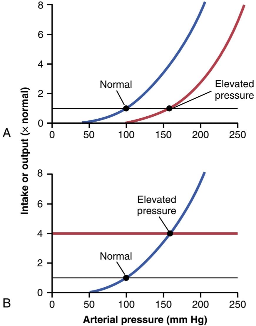
Figure 19-3. Two ways in which the arterial pressure can be increased. A, By shifting the renal output curve in the right-hand direc- tion toward a higher pressure level or by increasing the intake level of salt and water (B).
شکل 19-3. به دو روش می توان فشار شریانی را افزایش داد. A، با تغییر منحنی خروجی کلیه در جهت سمت راست به سمت یک سطح فشار بالاتر یا با افزایش سطح دریافت نمک و آب (B).
Chronic Renal Output Curve Much Steeper Than the Acute Curve. An important characteristic of pressure natriuresis (and pressure diuresis) is that chronic changes in arterial pressure, lasting for days or months, have a much greater effect on the renal output of salt and water than that observed during acute changes in pressure (Figure 19-4). Thus, when the kidneys are functioning normally, the chronic renal output curve is much steeper than the acute curve.
منحنی برون ده مزمن کلیوی بسیار تندتر از منحنی حاد. یکی از ویژگی های مهم ناتریورز فشاری (و دیورز فشاری) این است که تغییرات مزمن فشار شریانی، که برای روزها یا ماه ها طول می کشد، تأثیر بسیار بیشتری بر خروجی نمک و آب کلیوی نسبت به تغییرات حاد فشار دارد (شکل 19-4). بنابراین، زمانی که کلیه ها به طور طبیعی کار می کنند، منحنی برون ده کلیوی مزمن بسیار تندتر از منحنی حاد است.
The powerful effects of chronic increases in arterial pressure on urine output occur because increased pressure not only has direct hemodynamic effects on the kidney to increase excretion, but also has indirect effects mediated by nervous and hormonal changes that occur when blood pressure is increased. For example, increased arterial pressure decreases activity of the sympathetic nervous system, partly through the baro- receptor reflex mechanisms discussed in Chapter 18, and by reducing formation of various hormones such as angiotensin II and aldosterone that tend to reduce salt and water excretion by the kidneys. Reduced activity of these antinatriuretic systems therefore amplifies the effectiveness of pressure natriuresis and diuresis in rais- ing salt and water excretion during chronic increases in arterial pressure (see Chapters 28 and 30 for further discussion).
اثرات قدرتمند افزایش مزمن فشار شریانی بر برون ده ادرار به این دلیل رخ می دهد که افزایش فشار نه تنها اثرات همودینامیک مستقیم بر کلیه برای افزایش دفع دارد، بلکه اثرات غیرمستقیم با واسطه تغییرات عصبی و هورمونی دارد که هنگام افزایش فشار خون رخ می دهد. به عنوان مثال، افزایش فشار شریانی فعالیت سیستم عصبی سمپاتیک را تا حدی از طریق مکانیسمهای بازتابی گیرنده بارو که در فصل 18 مورد بحث قرار گرفت و با کاهش تشکیل هورمونهای مختلف مانند آنژیوتانسین II و آلدوسترون که تمایل به کاهش دفع نمک و آب توسط کلیهها را دارند، کاهش میدهد. بنابراین، کاهش فعالیت این سیستم های ضد ناتریورتیک، اثربخشی ناتریورز فشاری و دیورز را در افزایش دفع نمک و آب در طول افزایش مزمن فشار شریانی تقویت می کند (برای بحث بیشتر به فصل های 28 و 30 مراجعه کنید).
Conversely, when blood pressure is reduced, the sympathetic nervous system is activated, and formation of antinatriuretic hormones is increased, adding to the direct effects of reduced pressure to decrease renal out- put of salt and water. This combination of direct effects of pressure on the kidneys and indirect effects of pressure on the sympathetic nervous system and various hormone systems make pressure natriuresis and diuresis extremely powerful factors for long-term control of arterial pressure and body fluid volumes.
برعکس، وقتی فشار خون کاهش مییابد، سیستم عصبی سمپاتیک فعال میشود و تشکیل هورمونهای ضد ناتریورتیک افزایش مییابد که به اثرات مستقیم کاهش فشار برای کاهش برون ده نمک و آب کلیوی میافزاید. این ترکیب از اثرات مستقیم فشار بر کلیه ها و اثرات غیرمستقیم فشار بر سیستم عصبی سمپاتیک و سیستم های هورمونی مختلف، ناتریورز فشار و دیورز را به عوامل بسیار قدرتمندی برای کنترل طولانی مدت فشار شریانی و حجم مایعات بدن تبدیل می کند.
The importance of neural and hormonal influences on pressure natriuresis is especially evident during chronic changes in sodium intake. If the kidneys and nervous and hormonal mechanisms are functioning normally, chronic increases in intakes of salt and water to as high as six times normal are usually associated with little effect on arterial pressure. Note that the blood pressure equilibrium point B on the curve is nearly the same as point A, the equilibrium point at normal salt intake. Conversely, decreases in salt and water intake to as low as one-sixth normal typically have little effect on arterial pressure. Thus, many per- sons are said to be salt-insensitive because large variations in salt intake do not change blood pressure more than a few mm Hg.
اهمیت تأثیرات عصبی و هورمونی بر ناتریورز فشاری به ویژه در طول تغییرات مزمن در مصرف سدیم مشهود است. اگر کلیه ها و مکانیسم های عصبی و هورمونی به طور طبیعی کار کنند، افزایش مزمن دریافت نمک و آب تا شش برابر نرمال معمولاً با تأثیر کمی بر فشار شریانی همراه است. توجه داشته باشید که نقطه تعادل فشار خون B روی منحنی تقریباً مشابه نقطه A است، نقطه تعادل در مصرف معمولی نمک. برعکس، کاهش مصرف نمک و آب به کمتر از یک ششم نرمال معمولاً تأثیر کمی بر فشار شریانی دارد. بنابراین گفته می شود که بسیاری از افراد به نمک حساس هستند زیرا تغییرات زیاد در مصرف نمک فشار خون را بیش از چند میلی متر جیوه تغییر نمی دهد.
Persons with kidney injury or excessive secretion of antinatriuretic hormones such as angiotensin II or aldo- sterone, however, may be salt-sensitive, with an attenuated renal output curve similar to the acute curve shown in Figure 19-4. In these cases, even moderate increases in salt intake may cause significant increases in arterial pressure.
افراد مبتلا به آسیب کلیوی یا ترشح بیش از حد هورمون های ضد ناتریورتیک مانند آنژیوتانسین II یا آلدوسترون، با این حال، ممکن است حساس به نمک باشند، با منحنی برون ده کلیه ضعیف شده مشابه منحنی حاد نشان داده شده در شکل 19-4. در این موارد، حتی افزایش متوسط در مصرف نمک ممکن است باعث افزایش قابل توجه فشار شریانی شود.
Some of the factors that cause blood pressure to be salt-sensitive include loss of functional nephrons due to kidney injury and excessive formation of antinatriuretic hormones such as angiotensin II or aldosterone. For example, surgical reduction of kidney mass or injury to the kidney due to hypertension, diabetes, or various kidney diseases all cause blood pressure to be more sensitive to changes in salt intake. In these cases, greater than normal increases in arterial pressure are required to raise renal output sufficiently to maintain a balance between the intake and output of salt and water.
برخی از عواملی که باعث می شوند فشار خون به نمک حساس شود عبارتند از از دست دادن نفرون های عملکردی به دلیل آسیب کلیه و تشکیل بیش از حد هورمون های ضد ناتریورتیک مانند آنژیوتانسین II یا آلدوسترون. به عنوان مثال، کاهش توده کلیه با جراحی یا آسیب به کلیه به دلیل فشار خون بالا، دیابت یا بیماری های کلیوی مختلف، همگی باعث می شوند فشار خون نسبت به تغییرات در مصرف نمک حساس تر شود. در این موارد، افزایش بیش از حد طبیعی در فشار شریانی برای بالا بردن برون ده کلیه به اندازه کافی برای حفظ تعادل بین دریافتی و خروجی نمک و آب مورد نیاز است.
There is evidence that long-term high salt intake, lasting for several years, may actually damage the kidneys and eventually makes blood pressure more salt-sensitive. We will discuss salt sensitivity of blood pressure in patients with hypertension later in this chapter.
شواهدی وجود دارد که نشان میدهد مصرف طولانیمدت نمک، که برای چندین سال طول میکشد، ممکن است به کلیهها آسیب برساند و در نهایت فشار خون را به نمک حساستر کند. در ادامه این فصل در مورد حساسیت فشار خون به نمک در بیماران مبتلا به فشار خون بالا بحث خواهیم کرد.

Figure 19-4. Acute and chronic renal output curves. Under steady- state conditions, the renal output of salt and water is equal to intake of salt and water. Points A and B represent the equilibrium points for long-term regulation of arterial pressure when salt intake is normal or six times normal, respectively. Because of the steepness of the chronic renal output curve, increased salt intake normally causes only small changes in arterial pressure. In persons with impaired kidney function, the steepness of the renal output curve may be reduced, similar to the acute curve, resulting in increased sensitivity of arterial pres- sure to changes in salt intake.
شکل 19-4. منحنی های برون ده کلیوی حاد و مزمن. در شرایط پایدار، برون ده نمک و آب کلیه برابر با دریافت نمک و آب است. نقاط A و B به ترتیب نشان دهنده نقاط تعادل برای تنظیم طولانی مدت فشار شریانی هنگامی که مصرف نمک طبیعی یا شش برابر نرمال است. به دلیل شیب زیاد منحنی برون ده کلیوی مزمن، افزایش مصرف نمک به طور معمول تنها باعث تغییرات جزئی در فشار شریانی می شود. در افراد مبتلا به اختلال عملکرد کلیه، شیب منحنی برون ده کلیه ممکن است مشابه منحنی حاد کاهش یابد و در نتیجه حساسیت فشار شریانی به تغییرات در مصرف نمک افزایش یابد.
Failure of Increased Total Peripheral Resistance to Elevate Long-Term Level of Arterial Pressure if Fluid Intake and Renal Function Do Not Change
Recalling the basic equation for arterial pressure—arterial pressure equals cardiac output times total peripheral resistance-it is clear that an increase in total peripheral resistance should elevate the arterial pressure. Indeed, when the total peripheral resistance is acutely increased, the arterial pressure does rise immediately. Yet, if the kidneys continue to function normally, the acute rise in arterial pressure usually is not maintained. Instead, the arterial pressure returns all the way to normal within about 1 or 2 days. Why?
عدم افزایش مقاومت کلی محیطی برای بالا بردن سطح طولانی مدت فشار شریانی در صورت عدم تغییر مصرف مایعات و عملکرد کلیه
با یادآوری معادله اصلی فشار شریانی – فشار شریانی برابر برون ده قلبی ضربدر کل مقاومت محیطی است – واضح است که افزایش مقاومت محیطی کل باید فشار شریانی را افزایش دهد. در واقع، هنگامی که مقاومت کلی محیطی به شدت افزایش می یابد، فشار شریانی بلافاصله افزایش می یابد. با این حال، اگر کلیه ها به عملکرد طبیعی خود ادامه دهند، افزایش شدید فشار شریانی معمولاً حفظ نمی شود. در عوض، فشار شریانی در عرض حدود 1 یا 2 روز به حالت عادی باز می گردد. چرا؟
The reason for this phenomenon is that increasing vascular resistance everywhere else in the body besides in the kidneys does not change the equilibrium point for blood pressure control as dictated by the kidneys (see Figures 19-1 and 19-3). Instead, the kidneys immediately begin to respond to the high arterial pressure, causing pres- sure diuresis and pressure natriuresis. Within hours, large amounts of salt and water are lost from the body; this process continues until the arterial pressure returns to the equilibrium pressure level. At this point, blood pressure is normalized, and extracellular fluid volume and blood volume are decreased to levels below normal.
دلیل این پدیده این است که افزایش مقاومت عروقی در همه جای بدن به غیر از کلیه ها، نقطه تعادل کنترل فشار خون را که توسط کلیه ها دیکته می شود تغییر نمی دهد (شکل های 19-1 و 19-3 را ببینید). در عوض، کلیهها بلافاصله شروع به واکنش به فشار شریانی بالا میکنند که باعث دیورز فشاری و ناتریورز فشاری میشود. در عرض چند ساعت، مقادیر زیادی نمک و آب از بدن از بین می رود. این فرآیند تا زمانی ادامه می یابد که فشار شریانی به سطح فشار تعادل بازگردد. در این مرحله، فشار خون نرمال می شود و حجم مایع خارج سلولی و حجم خون به سطوح پایین تر از حد طبیعی کاهش می یابد.
Figure 19-5 shows the approximate cardiac outputs and arterial pressures in different clinical conditions in which the long-term total peripheral resistance is much less than or much greater than normal, but kidney excretion of salt and water is normal. Note in all these different clinical conditions that the arterial pressure is also normal.
شکل 19-5 برون ده تقریبی قلب و فشارهای شریانی را در شرایط بالینی مختلف نشان می دهد که در آن مقاومت کلی محیطی طولانی مدت بسیار کمتر یا بسیار بیشتر از حد طبیعی است، اما دفع نمک و آب از کلیه طبیعی است. در تمام این شرایط بالینی مختلف توجه داشته باشید که فشار شریانی نیز طبیعی است.
A word of caution is necessary at this point in our discussion. Often, when the total peripheral resistance increases, this also increases the intrarenal vascular resistance at the same time, which alters the function of the kidney and can cause hypertension by shifting the renal function curve to a higher pressure level, as shown in Figure 19-34. We will see an example of this mechanism later in this chapter when we discuss hypertension caused by vasoconstrictor mechanisms. However, it is the increase in renal resistance that is the culprit, not the increased total peripheral resistance-an important distinction.
در این مرحله از بحث ما یک کلمه احتیاط ضروری است. اغلب، هنگامی که مقاومت کلی محیطی افزایش مییابد، مقاومت عروق داخل کلیوی را نیز افزایش میدهد، که عملکرد کلیه را تغییر میدهد و میتواند با تغییر منحنی عملکرد کلیوی به سطح فشار بالاتر، باعث فشار خون بالا شود، همانطور که در شکل 19-34 نشان داده شده است. نمونهای از این مکانیسم را بعداً در این فصل خواهیم دید وقتی که فشار خون بالا ناشی از مکانیسمهای منقبض کننده عروق را مورد بحث قرار میدهیم. با این حال، این افزایش مقاومت کلیوی است که مقصر است، نه افزایش مقاومت کلی محیطی – یک تمایز مهم.

Figure 19-5. Relationships of total peripheral resistance to the long- term levels of arterial pressure and cardiac output in different clinical abnormalities. In these conditions, the kidneys were functioning normally. Note that changing the whole-body total peripheral resistance caused equal and opposite changes in cardiac output but, in all cases, had no effect on arterial pressure. AV, Arteriovenous. (Modified from Guyton AC: Arterial Pressure and Hypertension. Philadelphia: WB Saunders, 1980.)
شکل 19-5. رابطه مقاومت کلی محیطی با سطوح طولانی مدت فشار شریانی و برون ده قلبی در ناهنجاری های بالینی مختلف. در این شرایط، کلیه ها به طور طبیعی کار می کردند. توجه داشته باشید که تغییر مقاومت محیطی کل بدن باعث تغییرات مساوی و معکوس در برون ده قلبی شد، اما در همه موارد، تاثیری بر فشار شریانی نداشت. AV، شریانی وریدی. (اصلاح شده از Guyton AC: Arerial Pressure and Hypertension. Philadelphia: WB Saunders, 1980.)
Increased Fluid Volume Can Elevate Arterial Pressure by Increasing Cardiac Output or Total Peripheral Resistance.
The overall mechanism whereby increased extracellular fluid volume may elevate arterial pressure, if vascular capacity is not simultaneously increased, is shown in Figure 19-6. The sequential events are as follows: (1) increased extracellular fluid volume, which (2) increases the blood volume, which (3) increases the mean circulatory filling pressure, which (4) increases venous return of blood to the heart, which (5) increases cardiac output, which (6) increases arterial pres- sure. The increased arterial pressure, in turn, increases the renal excretion of salt and water and may return extracellular fluid volume to nearly normal if kidney function is normal and vascular capacity is unaltered.
افزایش حجم مایع می تواند فشار شریانی را با افزایش برون ده قلبی یا مقاومت کلی محیطی افزایش دهد.
مکانیسم کلی که به موجب آن افزایش حجم مایع خارج سلولی ممکن است فشار شریانی را افزایش دهد، اگر ظرفیت عروقی به طور همزمان افزایش نیابد، در شکل 19-6 نشان داده شده است. رویدادهای متوالی به شرح زیر است: (1) افزایش حجم مایع خارج سلولی، که (2) حجم خون را افزایش می دهد، (3) فشار متوسط پر شدن گردش خون را افزایش می دهد، که (4) بازگشت وریدی خون به قلب را افزایش می دهد، که (5) برون ده قلبی را افزایش می دهد، که (6) فشار شریانی را افزایش می دهد. افزایش فشار شریانی، به نوبه خود، دفع کلیوی نمک و آب را افزایش می دهد و اگر عملکرد کلیه ها نرمال باشد و ظرفیت عروقی تغییر نکند، ممکن است حجم مایع خارج سلولی را تقریباً به حالت طبیعی برگرداند.
Note especially in this case the two ways in which an increase in cardiac output can increase the arterial pressure. One of these is the direct effect of increased cardiac output to increase the pressure, and the other is an indirect effect to raise total peripheral vascular resistance through autoregulation of blood flow. The second effect can be explained as follows.
به خصوص در این مورد به دو روشی که افزایش برون ده قلبی می تواند فشار شریانی را افزایش دهد توجه کنید. یکی از اینها اثر مستقیم افزایش برون ده قلبی برای افزایش فشار است و دیگری اثر غیرمستقیم برای افزایش مقاومت عروق محیطی کل از طریق تنظیم خودکار جریان خون است. اثر دوم را می توان به صورت زیر توضیح داد.
Referring to Chapter 17, let us recall that whenever an excess amount of blood flows through a tissue, the local tissue vasculature constricts and decreases the blood flow back toward normal. This phenomenon is called autoregulation, which simply means regulation of blood flow by the tissue itself. When increased blood volume raises the cardiac output, blood flow tends to increase in all tissues of the body; if the increased blood flow exceeds the metabolic needs of the tissues, the autoregulation mechanisms constricts blood vessels all over the body, which in turn increases the total peripheral resistance.
با مراجعه به فصل 17، به یاد بیاوریم که هر گاه مقدار زیادی خون در بافتی جریان یابد، عروق بافتی منقبض شده و جریان خون را به حالت عادی کاهش می دهد. این پدیده خود تنظیم نامیده می شود که به سادگی به معنای تنظیم جریان خون توسط خود بافت است. هنگامی که افزایش حجم خون برون ده قلبی را افزایش می دهد، جریان خون در تمام بافت های بدن افزایش می یابد. اگر افزایش جریان خون بیش از نیازهای متابولیکی بافتها باشد، مکانیسمهای خودتنظیمی عروق خونی را در سراسر بدن منقبض میکند که به نوبه خود مقاومت کلی محیطی را افزایش میدهد.
Finally, because arterial pressure is equal to cardiac output times total peripheral resistance, the secondary increase in total peripheral resistance that results from the autoregulation mechanism helps increase the arterial pressure. For example, only a 5% to 10% increase in cardiac output can increase the arterial pressure from the normal mean arterial pressure of 100 mm Hg up to 150 mm Hg when accompanied by an increase in total peripheral resistance due to tissue blood flow autoregulation or other factors that cause vasoconstriction. The slight increase in cardiac output is often not measurable.
در نهایت، از آنجایی که فشار شریانی برابر برون ده قلبی ضربدر مقاومت محیطی کل است، افزایش ثانویه در مقاومت کلی محیطی که از مکانیسم تنظیم خودکار ناشی می شود به افزایش فشار شریانی کمک می کند. به عنوان مثال، تنها یک افزایش 5 تا 10 درصدی در برون ده قلبی می تواند فشار شریانی را از فشار شریانی متوسط 100 میلی متر جیوه تا 150 میلی متر جیوه افزایش دهد، در صورتی که با افزایش مقاومت کلی محیطی به دلیل تنظیم خود تنظیم جریان خون بافتی یا سایر عواملی که باعث انقباض عروق می شوند، همراه باشد. افزایش جزئی در برون ده قلبی اغلب قابل اندازه گیری نیست.
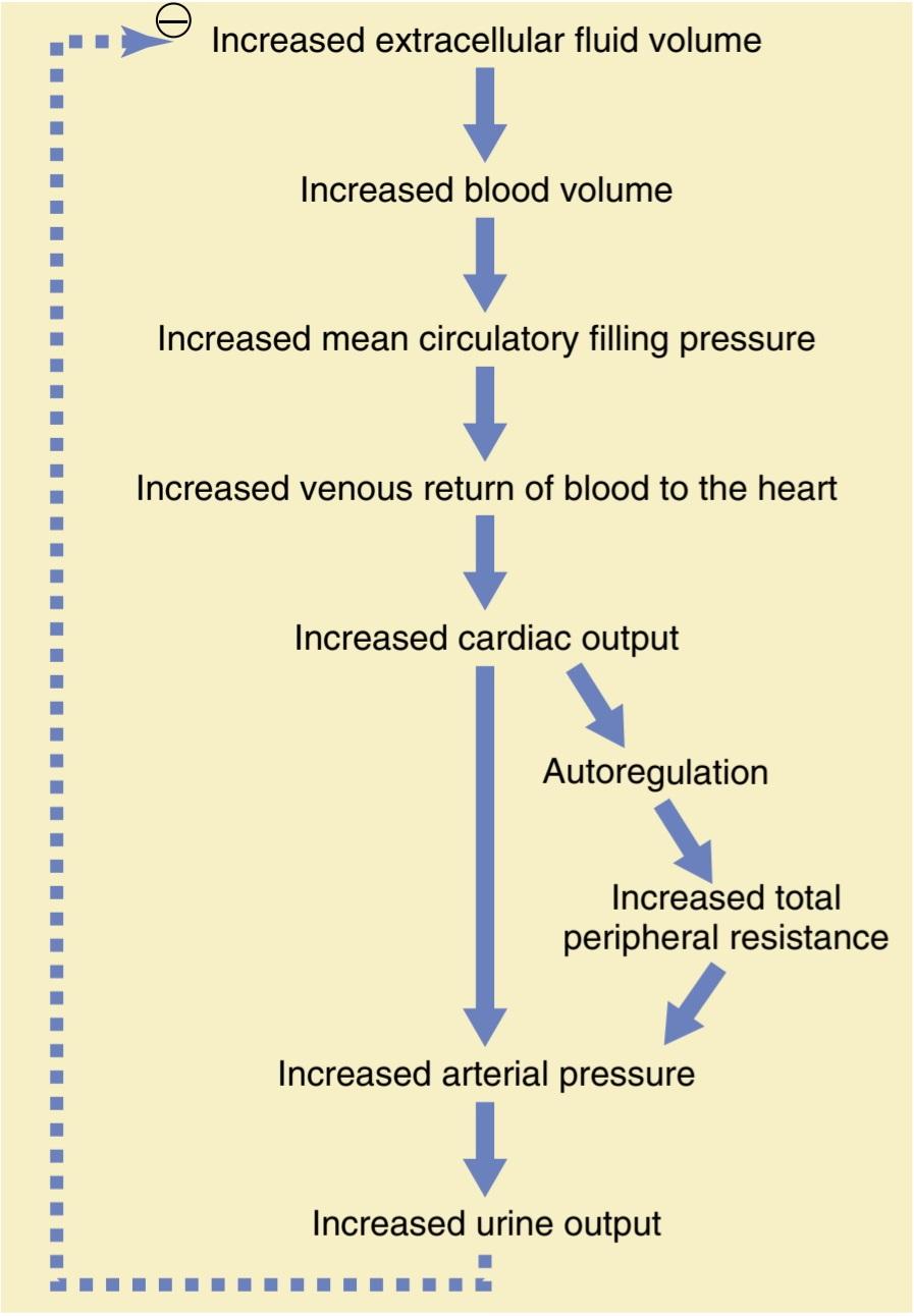
Figure 19-6. Sequential steps whereby increased extracellular fluid volume increases the arterial pressure. Note especially that increased cardiac output has both a direct effect to increase arterial pressure and an indirect effect by first increasing the total peripheral resistance.
شکل 19-6. مراحل متوالی که به موجب آن افزایش حجم مایع خارج سلولی باعث افزایش فشار شریانی می شود. به ویژه توجه داشته باشید که افزایش برون ده قلبی با افزایش مقاومت کلی محیطی هم تأثیر مستقیم بر افزایش فشار شریانی دارد و هم تأثیر غیرمستقیم دارد.
Importance of Salt (NaCl) in the Renal- Body Fluid Schema for Arterial Pressure Regulation
Although the discussions thus far have emphasized the importance of volume in regulation of arterial pressure, experimental studies have shown that an increase in salt intake is far more likely to elevate the arterial pressure, especially in people who are salt-sensitive, than is an increase in water intake. The reason for this finding is that pure water is normally excreted by the kidneys almost as rapidly as it is ingested, but salt is not excreted so easily. As salt accumulates in the body, it also indirectly increases the extracellular fluid volume for two basic reasons:
اهمیت نمک (NaCl) در طرح کلیوی- مایع بدن برای تنظیم فشار شریانی
اگرچه بحثهای تا کنون بر اهمیت حجم در تنظیم فشار شریانی تاکید کردهاند، مطالعات تجربی نشان دادهاند که افزایش در مصرف نمک به احتمال زیاد باعث افزایش فشار شریانی، بهویژه در افرادی که به نمک حساس هستند، بیشتر از افزایش مصرف آب است. دلیل این یافته این است که آب خالص معمولاً تقریباً به همان سرعتی که بلعیده می شود توسط کلیه ها دفع می شود، اما نمک به این راحتی دفع نمی شود. همانطور که نمک در بدن تجمع می یابد، به دو دلیل اساسی به طور غیرمستقیم حجم مایع خارج سلولی را افزایش می دهد:
1. Although some additional sodium may be stored in the tissues when salt accumulates in the body, excess salt in the extracellular fluid increases the fluid osmolality. The increased osmolarity stimulates the thirst center in the brain, making the person drink extra amounts of water to return the extracellular salt concentration to normal and increasing the ex- tracellular fluid volume.
1. اگرچه ممکن است مقداری سدیم اضافی در بافتها ذخیره شود، اما نمک اضافی در مایع خارج سلولی اسمولالیته مایع را افزایش میدهد. افزایش اسمولاریته مرکز تشنگی در مغز را تحریک می کند و باعث می شود که فرد مقادیر بیشتری آب بنوشد تا غلظت نمک خارج سلولی به حالت عادی برگردد و حجم مایع خارج سلولی افزایش یابد.
2. The increase in osmolality caused by the excess salt in the extracellular fluid also stimulates the hypo- thalamic-posterior pituitary gland secretory mechanism to secrete increased quantities of antidiuretic hormone (discussed in Chapter 29). The antidiuretic hormone then causes the kidneys to reabsorb greatly increased quantities of water from the renal tubular fluid, thereby diminishing the excreted volume of urine but increasing the extracellular fluid volume.
2. افزایش اسمولالیته ناشی از نمک اضافی در مایع خارج سلولی همچنین مکانیسم ترشحی غده هیپوفیز هیپوتالاموس خلفی را تحریک می کند تا مقادیر بیشتری از هورمون ضد ادرار ترشح کند (در فصل 29 بحث شد). سپس هورمون ضد دیورتیک باعث میشود کلیهها مقادیر زیادی آب را از مایع لولهای کلیوی بازجذب کنند، در نتیجه حجم ادرار دفع شده کاهش مییابد اما حجم مایع خارج سلولی افزایش مییابد.
Thus, the amount of salt that accumulates in the body is an important determinant of the extracellular fluid volume. Relatively small increases in extracellular fluid and blood volume can often increase the arterial pressure substantially. This is true, however, only if the excess salt accumulation leads to an increase in blood volume and if vascular capacity is not simultaneously increased. As discussed previously, increasing salt intake in the absence of impaired kidney function or excessive formation of antinatriuretic hormones usually does not increase arterial pressure much because the kidneys rapidly eliminate the excess salt, and blood volume is hardly altered.
بنابراین، مقدار نمکی که در بدن انباشته می شود، تعیین کننده مهم حجم مایع خارج سلولی است. افزایش نسبتاً کوچک در مایع خارج سلولی و حجم خون اغلب می تواند فشار شریانی را به میزان قابل توجهی افزایش دهد. این درست است، با این حال، تنها در صورتی که تجمع نمک اضافی منجر به افزایش حجم خون شود و ظرفیت عروقی به طور همزمان افزایش نیابد. همانطور که قبلاً بحث شد، افزایش مصرف نمک در غیاب اختلال عملکرد کلیه یا تشکیل بیش از حد هورمون های ضد ناتریورتیک معمولاً فشار شریانی را زیاد افزایش نمی دهد زیرا کلیه ها به سرعت نمک اضافی را از بین می برند و حجم خون به سختی تغییر می کند.
CHRONIC HYPERTENSION (HIGH BLOOD PRESSURE) CAUSED BY IMPAIRED RENAL FUNCTION
When a person is said to have chronic hypertension (or high blood pressure), this means that his or her mean arterial pressure is greater than the upper range of the accepted normal measure. A mean arterial pressure greater than 110 mm Hg (normal is ~90 mm Hg) is considered to be hypertensive. (This level of mean pressure occurs when the diastolic blood pressure is greater than ~90 mm Hg and the systolic pressure is greater than ≈135 mm Hg.) In persons with severe hypertension, the mean arterial pressure can rise to 150 to 170 mm Hg, with diastolic pressure as high as 130 mm Hg and systolic pressure occasionally as high as 250 mm Hg.
فشار خون مزمن (فشار خون بالا) ناشی از اختلال در عملکرد کلیه
هنگامی که گفته می شود فردی دارای فشار خون مزمن (یا فشار خون بالا) است، به این معنی است که فشار شریانی متوسط او بیشتر از محدوده بالای اندازه گیری طبیعی پذیرفته شده است. فشار متوسط شریانی بیش از 110 میلی متر جیوه (طبیعی 90 میلی متر جیوه است) به عنوان فشار خون بالا در نظر گرفته می شود. (این سطح از فشار متوسط زمانی اتفاق میافتد که فشار خون دیاستولیک بیشتر از 90 میلیمتر جیوه و فشار سیستولیک بیشتر از 135 میلیمتر جیوه باشد.) در افراد مبتلا به فشار خون شدید، فشار متوسط شریانی میتواند به 150 تا 170 میلیمتر جیوه، با فشار دیاستولیک به 130 میلیمتر گرم Hg و فشار سیستولیک به 130 میلیگرم Hg5 برسد.
Even moderate elevation of arterial pressure leads to shortened life expectancy. At severely high pressuresthat is, mean arterial pressures 50% or more above normal-a person can expect to live no more than a few more years unless appropriately treated. The lethal effects of hypertension are caused mainly in three ways:
حتی افزایش متوسط فشار شریانی منجر به کاهش طول عمر می شود. در فشارهای شدیداً بالا – یعنی فشارهای شریانی متوسط 50 درصد یا بیشتر از حد طبیعی – یک فرد نمی تواند انتظار داشته باشد که بیش از چند سال بیشتر زندگی کند مگر اینکه به طور مناسب درمان شود. اثرات کشنده فشار خون بالا عمدتاً به سه طریق ایجاد می شود:
1. Excess workload on the heart leads to early heart failure and coronary heart disease, often causing death as a result of a heart attack.
1. بار کاری بیش از حد بر روی قلب منجر به نارسایی اولیه قلبی و بیماری عروق کرونر قلب می شود که اغلب باعث مرگ در نتیجه حمله قلبی می شود.
2. The high pressure frequently damages a major blood vessel in the brain, followed by death of major portions of the brain; this occurrence is a cerebral infarct. Clinically, it is called a stroke. Depending on which part of the brain is involved, a stroke can be fatal or cause paralysis, dementia, blindness, or multiple other serious brain disorders.
2. فشار بالا اغلب به یک رگ خونی اصلی در مغز آسیب می رساند و به دنبال آن بخش های عمده ای از مغز می میرند. این اتفاق یک انفارکتوس مغزی است. از نظر بالینی به آن سکته مغزی می گویند. بسته به اینکه کدام قسمت از مغز درگیر است، سکته مغزی می تواند کشنده باشد یا باعث فلج، زوال عقل، کوری یا چندین اختلال جدی مغزی شود.
3. High pressure almost always causes injury in the kidneys, producing many areas of renal destruction and, eventually, kidney failure, uremia, and death.
3. فشار بالا تقریباً همیشه باعث آسیب در کلیه ها می شود و مناطق زیادی از تخریب کلیه و در نهایت نارسایی کلیه، اورمی و مرگ را ایجاد می کند.
Lessons learned from the type of hypertension called volume-loading hypertension have been crucial in understanding the role of the renal-body fluid volume mechanism for arterial pressure regulation. Volume- loading hypertension means hypertension caused by excess accumulation of extracellular fluid in the body, some examples of which follow.
درس های آموخته شده از نوع فشار خون به نام فشار خون بارگذاری حجمی در درک نقش مکانیسم حجم مایع کلیه-بدن برای تنظیم فشار شریانی بسیار مهم بوده است. فشار خون حجم دهنده به معنای فشار خون بالا ناشی از تجمع بیش از حد مایع خارج سلولی در بدن است که چند نمونه از آن در ادامه می آید.
Experimental Volume-Loading. Hypertension Caused by Reduced Kidney Mass and Increased Salt Intake. Figure 19-7 shows a typical experiment demonstrating volume-loading hypertension in a group of dogs with 70% of their kidney mass removed. At the first circled point on the curve, the two poles of one of the kidneys were removed, and at the second circled point, the entire opposite kidney was removed, leaving the an- imals with only 30% of their normal renal mass. Note that removal of this amount of kidney mass increased the arterial pressure by an average of only 6 mm Hg. Then, the dogs were given salt solution to drink instead of water. Because salt solution fails to quench the thirst, the dogs drank two to four times the normal amounts of volume, and within a few days, their average arterial pressure rose to about 40 mm Hg above normal. After 2 weeks, the dogs were given tap water again instead of salt solution; the pressure returned to normal within 2 days. Finally, at the end of the experiment, the dogs were given salt solution again, and this time the pressure rose much more rapidly to a high level, again demonstrating volume-loading hypertension.
بارگذاری حجم آزمایشی. فشار خون بالا ناشی از کاهش توده کلیه و افزایش مصرف نمک است. شکل 19-7 یک آزمایش معمولی را نشان می دهد که فشار خون با حجم بار را در گروهی از سگ ها نشان می دهد که 70 درصد توده کلیه آنها برداشته شده است. در اولین نقطه دایره شده روی منحنی، دو قطب یکی از کلیه ها برداشته شد و در نقطه دایره دوم، کل کلیه مقابل برداشته شد و حیوانات تنها 30 درصد از جرم کلیوی طبیعی خود را داشتند. توجه داشته باشید که حذف این مقدار از توده کلیه فشار شریانی را به طور متوسط تنها 6 میلی متر جیوه افزایش می دهد. سپس به سگ ها به جای آب محلول نمک داده شد. از آنجایی که محلول نمک نمی تواند تشنگی را برطرف کند، سگ ها دو تا چهار برابر حجم طبیعی نوشیدنی نوشیدند و در عرض چند روز، میانگین فشار شریانی آنها به حدود 40 میلی متر جیوه بالاتر از حد طبیعی رسید. پس از 2 هفته، به سگ ها دوباره به جای محلول نمک آب لوله کشی داده شد. فشار در عرض 2 روز به حالت عادی بازگشت. در پایان، در پایان آزمایش، دوباره به سگها محلول نمک داده شد و این بار فشار بسیار سریعتر به سطح بالایی افزایش یافت و دوباره فشار خون بالا را نشان داد.
If one considers again the basic determinants of long- term arterial pressure regulation, it is apparent why hypertension occurred in the volume-loading experiment illustrated in Figure 19-7. First, reduction of the kidney mass to 30% of normal greatly reduced the ability of the kidneys to excrete salt and water. Therefore, salt and water accumulated in the body and, in a few days, raised the arterial pressure high enough to excrete the excess salt and water intake.
اگر یک بار دیگر عوامل تعیین کننده اصلی تنظیم فشار شریانی طولانی مدت را در نظر بگیریم، واضح است که چرا فشار خون بالا در آزمایش بارگذاری حجمی نشان داده شده در شکل 19-7 رخ داده است. اول، کاهش توده کلیه به 30 درصد از حد طبیعی، توانایی کلیه ها برای دفع نمک و آب را به شدت کاهش داد. بنابراین نمک و آب در بدن انباشته شد و در عرض چند روز فشار شریانی را به اندازه ای بالا برد که نمک و آب اضافی دریافتی را دفع کند.

Figure 19-7. The average effect on arterial pressure of drinking 0.9% saline solution (0.9% NaCI) instead of water in dogs with 70% of their renal tissue removed. (Modified from Langston JB, Guyton AC, Douglas BH, et al: Effect of changes in salt intake on arterial pressure and renal function in partially nephrectomized dogs. Circ Res 12:508, 1963.)
شکل 19-7. تأثیر متوسط بر فشار شریانی نوشیدن 0.9٪ محلول نمکی (0.9٪ NaCI) به جای آب در سگ هایی که 70٪ از بافت کلیه آنها برداشته شده است. (اصلاح شده از Langston JB، Guyton AC، Douglas BH، و همکاران: تأثیر تغییرات در مصرف نمک بر فشار شریانی و عملکرد کلیه در سگهای نیمهنفرکتومی شده. Circ Res 12:508، 1963.)
Sequential Changes in Circulatory Function During Development of Volume-Loading Hypertension. It is especially instructive to study the sequential changes in circulatory function during progressive development of volume-loading hypertension (Figure 19-8). A week or so before the point labeled “0” days, the kidney mass had already been decreased to only 30% of normal. Then, at this point, the intake of salt and water was increased to about six times normal and kept at this high intake thereafter. The acute effect was to increase extracellular fluid volume, blood volume, and cardiac output to 20% to 40% above normal. Simultaneously, the arterial pressure began to rise but not nearly so much at first as the fluid volumes and cardiac output. The reason for this slower rise in pressure can be discerned by studying the total peripheral resistance curve, which shows an initial decrease in total peripheral resistance. This decrease was caused by the baroreceptor mechanism discussed in Chapter 18, which transiently attenuated the rise in pressure. However, after 2 to 4 days, the baroreceptors adapted (reset) and were no longer able to prevent the rise in pressure. At this time, the arterial pressure had risen almost to its full height because of the increase in cardiac output, even though the total peripheral resistance was still almost at the normal level.
تغییرات متوالی در عملکرد گردش خون در طول توسعه فشار خون با حجم بار. مطالعه تغییرات متوالی در عملکرد گردش خون در طول توسعه پیشرونده فشار خون با حجم بالا به ویژه آموزنده است (شکل 19-8). یک هفته یا بیشتر قبل از نقطه با برچسب “0” روز، توده کلیه در حال حاضر تنها به 30٪ از حد طبیعی کاهش یافته بود. سپس، در این مرحله، دریافت نمک و آب به حدود شش برابر نرمال افزایش یافت و پس از آن در این میزان مصرف بالا نگه داشت. اثر حاد افزایش حجم مایع خارج سلولی، حجم خون و برون ده قلبی به 20 تا 40 درصد بالاتر از حد طبیعی بود. به طور همزمان، فشار شریانی شروع به افزایش کرد، اما در ابتدا نه به اندازه حجم مایع و برون ده قلبی. دلیل این افزایش آهستهتر فشار را میتوان با مطالعه منحنی مقاومت محیطی کل، که کاهش اولیه در مقاومت کلی محیطی را نشان میدهد، تشخیص داد. این کاهش توسط مکانیسم بارورسپتور مورد بحث در فصل 18 ایجاد شد که به طور موقت افزایش فشار را کاهش داد. با این حال، پس از 2 تا 4 روز، بارورسپتورها سازگار شدند (تنظیم مجدد) و دیگر قادر به جلوگیری از افزایش فشار نبودند. در این زمان، فشار شریانی به دلیل افزایش برون ده قلبی تقریباً به ارتفاع کامل خود افزایش یافته بود، حتی اگر مقاومت کلی محیطی هنوز تقریباً در سطح طبیعی بود.
After these early acute changes in the circulatory variables had occurred, more prolonged secondary changes occurred during the next few weeks. Especially important was a progressive increase in total peripheral resistance, while at the same time the cardiac output decreased back toward normal, at least partly as a result of the long-term blood flow autoregulation mechanism discussed in Chapter 17 and earlier in this chapter. That is, after the cardiac output had risen to a high level and had initiated the hypertension, the excess blood flow through the tissues then caused progressive constriction of the local arterioles, thus returning the local blood flow in the body tissues and also the cardiac output toward normal while simultaneously causing a secondary increase in total peripheral resistance.
پس از این تغییرات حاد اولیه در متغیرهای گردش خون، تغییرات ثانویه طولانیتر در طی چند هفته آینده رخ داد. به ویژه افزایش تدریجی مقاومت کلی محیطی مهم بود، در حالی که در همان زمان برون ده قلبی به سمت نرمال کاهش یافت، حداقل تا حدی در نتیجه مکانیسم خودتنظیم جریان خون طولانی مدت که در فصل 17 و قبل از آن در این فصل مورد بحث قرار گرفت. یعنی پس از اینکه برون ده قلب به سطح بالایی رسید و فشار خون را شروع کرد، جریان خون اضافی در بافت ها باعث انقباض تدریجی شریان های موضعی شد و در نتیجه جریان خون موضعی در بافت های بدن و همچنین برون ده قلبی را به حالت طبیعی برمی گرداند و همزمان باعث افزایش مقاومت ثانویه می شود.
Note that the extracellular fluid volume and blood volume also returned toward normal along with the decrease in cardiac output. This outcome resulted from two factors. First, the increase in arteriolar resistance decreased the capillary pressure, which allowed the fluid in the tissue spaces to be absorbed back into the blood. Second, the elevated arterial pressure now caused the kidneys to excrete the excess volume of fluid that had initially accumulated in the body.
توجه داشته باشید که حجم مایع خارج سلولی و حجم خون نیز همراه با کاهش برون ده قلبی به حالت عادی بازگشت. این نتیجه ناشی از دو عامل بود. اولاً، افزایش مقاومت شریانی باعث کاهش فشار مویرگی شد که به مایع موجود در فضاهای بافتی اجازه میداد دوباره به خون جذب شود. دوم، فشار شریانی بالا اکنون باعث میشود کلیهها حجم اضافی مایعی را که در ابتدا در بدن انباشته شده بود دفع کنند.
Several weeks after the initial onset of volume loading, the following effects were found:
1. Hypertension
2. Marked increase in total peripheral resistance
3. Almost complete return of the extracellular fluid volume, blood volume, and cardiac output back to normal.
چندین هفته پس از شروع اولیه بارگذاری حجم، اثرات زیر مشاهده شد:
1. فشار خون بالا
2. افزایش قابل توجه در مقاومت کلی محیطی
3. بازگشت تقریباً کامل حجم مایع خارج سلولی، حجم خون و برون ده قلبی به حالت عادی.
Therefore, we can divide volume-loading hypertension into two sequential stages. The first stage results from increased fluid volume causing increased cardiac output. This increase in cardiac output mediates the hypertension. The second stage in volume-loading hypertension is characterized by high blood pressure and high total peripheral resistance but return of the cardiac output so close to normal that the usual measuring techniques frequently cannot detect an abnormally elevated cardiac output.
بنابراین، ما می توانیم فشار خون بارگذاری حجمی را به دو مرحله متوالی تقسیم کنیم. مرحله اول ناشی از افزایش حجم مایع است که باعث افزایش برون ده قلبی می شود. این افزایش برون ده قلبی واسطه فشار خون بالا می شود. مرحله دوم در پرفشاری خون با بارگذاری حجمی با فشار خون بالا و مقاومت کلی محیطی بالا مشخص می شود، اما برون ده قلبی به حدی نزدیک به نرمال است که روش های معمول اندازه گیری اغلب نمی توانند برون ده قلبی افزایش یافته غیرطبیعی را تشخیص دهند.
Thus, the increased total peripheral resistance in volume-loading hypertension occurs after the hypertension has developed and, therefore, is secondary to the hypertension rather than being the cause of the hypertension.
بنابراین، افزایش مقاومت کلی محیطی در پرفشاری خون بارگذاری حجمی پس از ایجاد فشار خون بالا اتفاق میافتد و بنابراین، به جای اینکه علت فشار خون بالا باشد، ثانویه به فشار خون بالا است.

Figure 19-8. Progressive changes in important circulatory system variables during the first few weeks of volume-loading hypertension. Note especially the initial increase in cardiac output as the basic cause of the hypertension. Subsequently, the autoregulation mechanism returns the cardiac output almost to normal while simultaneously causing a secondary increase in total peripheral resistance. (Modified from Guyton AC: Arterial Pressure and Hypertension. Philadelphia: WB Saunders, 1980.)
شکل 19-8. تغییرات پیشرونده در متغیرهای مهم سیستم گردش خون در طول چند هفته اول فشار خون با حجم بالا. به ویژه به افزایش اولیه برون ده قلبی به عنوان علت اصلی فشار خون بالا توجه کنید. متعاقباً، مکانیسم خودتنظیمی برون ده قلبی را تقریباً به حالت عادی برمی گرداند در حالی که به طور همزمان باعث افزایش ثانویه در مقاومت کلی محیطی می شود. (اصلاح شده از Guyton AC: Arerial Pressure and Hypertension. Philadelphia: WB Saunders, 1980.)
Volume-Loading Hypertension in Patients Who Have No Kidneys but Are Being Maintained With an Artificial Kidney
When a patient is maintained with an artificial kidney, it is especially important to keep the patient’s body fluid volume at a normal level by removing the appropriate amount of water and salt each time the patient undergoes dialysis. If this step is not performed, and extracellular fluid volume is allowed to increase, hypertension almost invariably develops in exactly the same way as shown in Figure 19-8. That is, the cardiac output increases at first and causes hypertension. Then, the autoregulation mechanism returns the cardiac output back toward normal while causing a secondary increase in total peripheral resistance. Therefore, in the end, the hypertension appears to be a high peripheral resistance type of hypertension, although the initial cause is excess volume accumulation.
فشار خون با حجم بالا در بیمارانی که کلیه ندارند اما با کلیه مصنوعی نگهداری می شوند
هنگامی که بیمار با کلیه مصنوعی نگهداری می شود، حفظ حجم مایع بدن بیمار در سطح نرمال با حذف مقدار مناسب آب و نمک در هر بار دیالیز بسیار مهم است. اگر این مرحله انجام نشود و حجم مایع خارج سلولی افزایش یابد، فشار خون بالا تقریباً همیشه به همان شکلی که در شکل 19-8 نشان داده شده است، ایجاد می شود. یعنی در ابتدا برون ده قلبی افزایش می یابد و باعث فشار خون می شود. سپس، مکانیسم خودتنظیمی، برون ده قلبی را به حالت عادی برمی گرداند در حالی که باعث افزایش ثانویه در مقاومت کلی محیطی می شود. بنابراین، در نهایت، فشار خون بالا به نظر می رسد یک نوع فشار خون با مقاومت محیطی بالا باشد، اگرچه علت اولیه تجمع حجم اضافی است.
Hypertension Caused by Excess Aldosterone
Another type of volume-loading hypertension is caused by excess aldosterone in the body or, occasionally, by excesses of other types of steroids. A small tumor in one of the adrenal glands occasionally secretes large quantities of aldosterone, which is the condition called primary aldosteronism. As discussed in Chapters 28 and 30, aldosterone increases the rate of salt and water reabsorption by the tubules of the kidneys, thereby increasing blood volume, extracellular fluid volume, and arterial pressure. If salt intake is increased at the same time, the hypertension becomes even greater. Furthermore, if the condition persists for months or years, the excess arterial pressure often causes pathological changes in the kidneys that make the kidneys retain even more salt and water in addition to that caused directly by the aldosterone. Therefore, the hypertension often finally becomes severe to the point of being lethal.
فشار خون بالا ناشی از آلدوسترون اضافی
نوع دیگری از پرفشاری خون بارگذاری حجمی ناشی از آلدوسترون اضافی در بدن یا، گاهی اوقات، بیش از حد سایر انواع استروئیدها است. تومور کوچک در یکی از غدد فوق کلیوی گهگاه مقادیر زیادی آلدوسترون ترشح می کند که به آن آلدوسترونیسم اولیه گفته می شود. همانطور که در فصلهای 28 و 30 بحث شد، آلدوسترون سرعت بازجذب نمک و آب توسط لولههای کلیه را افزایش میدهد و در نتیجه حجم خون، حجم مایع خارج سلولی و فشار شریانی را افزایش میدهد. اگر مصرف نمک همزمان افزایش یابد، فشار خون حتی بیشتر می شود. علاوه بر این، اگر این بیماری برای ماهها یا سالها ادامه پیدا کند، فشار بیش از حد شریانی اغلب باعث تغییرات پاتولوژیک در کلیهها میشود که باعث میشود کلیهها نمک و آب بیشتری را علاوه بر آن چیزی که مستقیماً توسط آلدوسترون ایجاد میشود، حفظ کنند. بنابراین، فشار خون بالا اغلب در نهایت تا مرز کشنده شدن شدید می شود.
Here again, in the early stages of this type of hypertension, cardiac output is often increased but, in later stages, the cardiac output generally returns almost to normal while total peripheral resistance becomes secondarily elevated, as explained earlier in the chapter for primary volume-loading hypertension.
در اینجا نیز، در مراحل اولیه این نوع فشار خون، برون ده قلبی اغلب افزایش می یابد، اما در مراحل بعدی، برون ده قلبی به طور کلی تقریباً به حالت عادی باز می گردد در حالی که مقاومت کلی محیطی به طور ثانویه افزایش می یابد، همانطور که قبلا در فصل برای فشار خون با حجم اولیه توضیح داده شد.
ROLE OF THE RENIN-ANGIOTENSIN SYSTEM IN ARTERIAL PRESSURE CONTROL
Aside from the capability of the kidneys to control arterial pressure through changes in extracellular fluid volume, the kidneys also have another powerful mechanism for controlling pressure, the renin-angiotensin system.
نقش سیستم رنین-آنژیوتانسین در کنترل فشار شریانی
جدای از توانایی کلیه ها برای کنترل فشار شریانی از طریق تغییر در حجم مایع خارج سلولی، کلیه ها همچنین مکانیسم قدرتمند دیگری برای کنترل فشار دارند، سیستم رنین-آنژیوتانسین.
Renin is a protein enzyme released by the kidneys when the arterial pressure falls too low. In turn, it raises the arterial pressure in several ways, thus helping correct the initial fall in pressure.
رنین یک آنزیم پروتئینی است که با کاهش بیش از حد فشار شریانی توسط کلیه ها آزاد می شود. به نوبه خود، فشار شریانی را به طرق مختلف افزایش می دهد، بنابراین به اصلاح افت اولیه فشار کمک می کند.
COMPONENTS OF THE RENIN- ANGIOTENSIN SYSTEM
Figure 19-9 shows the main functional steps whereby the renin-angiotensin system helps regulate arterial pressure. Renin is synthesized and stored in the juxtaglomerular cells (JG cells) of the kidneys. The JG cells are modified smooth muscle cells located mainly in the walls of the afferent arterioles immediately proximal to the glomeruli. Multiple factors control renin secretion, including the sympathetic nervous system, various hormones, and local autacoids such as prostaglandins, nitric oxide, and endothelin. When the arterial pressure falls, the JG cells release renin by at least three major mechanisms:
اجزای سیستم رنین- آنژیوتانسین
شکل 19-9 مراحل عملکردی اصلی را نشان می دهد که به موجب آن سیستم رنین-آنژیوتانسین به تنظیم فشار شریانی کمک می کند. رنین سنتز شده و در سلول های juxtaglomerular (سلول های JG) کلیه ها ذخیره می شود. سلول های JG سلول های عضله صاف اصلاح شده هستند که عمدتاً در دیواره شریان های آوران بلافاصله نزدیک به گلومرول ها قرار دارند. عوامل متعددی ترشح رنین را کنترل می کنند، از جمله سیستم عصبی سمپاتیک، هورمون های مختلف و اوتاکوئیدهای موضعی مانند پروستاگلاندین ها، اکسید نیتریک و اندوتلین. هنگامی که فشار شریانی کاهش می یابد، سلول های JG رنین را حداقل با سه مکانیسم اصلی آزاد می کنند:
1. Pressure-sensitive baroreceptors in the JG cells respond to decreased arterial pressure with increased release of renin.
1. بارورسپتورهای حساس به فشار در سلول های JG به کاهش فشار شریانی با افزایش آزادسازی رنین پاسخ می دهند.
2. Decreased sodium chloride delivery to the macula densa cells in the early distal tubule stimulates renin release (discussed further in Chapter 27)
2. کاهش تحویل کلرید سدیم به سلولهای ماکولا دنسا در لولههای دیستال اولیه، آزادسازی رنین را تحریک میکند (در فصل 27 بیشتر بحث شده است).
3. Increased sympathetic nervous system activity stimulates renin release by activating beta-adrenergic receptors in the JG cells. Sympathetic stimulation also activates alpha-adrenergic receptors, which can increase renal sodium chloride reabsorption and reduce the glomerular filtration rate in cases of strong sympathetic activation. Increased renal sympathetic activity also enhances the sensitivity of renal baroreceptor and macula densa mechanisms for renin release.
3. افزایش فعالیت سیستم عصبی سمپاتیک با فعال کردن گیرنده های بتا آدرنرژیک در سلول های JG باعث تحریک ترشح رنین می شود. تحریک سمپاتیک همچنین گیرنده های آلفا آدرنرژیک را فعال می کند، که می تواند بازجذب کلرید سدیم کلیوی را افزایش دهد و در موارد فعال شدن قوی سمپاتیک، میزان فیلتراسیون گلومرولی را کاهش دهد. افزایش فعالیت سمپاتیک کلیه نیز حساسیت بارورسپتورهای کلیوی و مکانیسم های ماکولا دنسا را برای آزادسازی رنین افزایش می دهد.
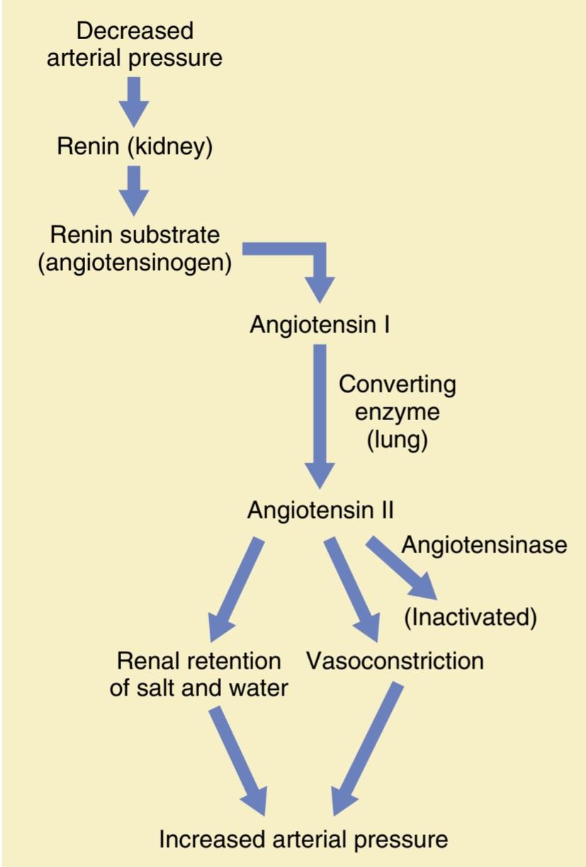
Figure 19-9. The renin-angiotensin vasoconstrictor mechanism for arterial pressure control.
شکل 19-9. مکانیسم منقبض کننده عروق رنین-آنژیوتانسین برای کنترل فشار شریانی
Most of the renin enters the renal blood and then passes out of the kidneys to circulate throughout the entire body. However, small amounts of the renin do remain in the local fluids of the kidney and initiate several intrarenal functions. Renin itself is an enzyme, not a vasoactive substance.
بیشتر رنین وارد خون کلیه می شود و سپس از کلیه ها خارج می شود تا در کل بدن گردش کند. با این حال، مقادیر کمی از رنین در مایعات موضعی کلیه باقی می ماند و چندین عملکرد داخل کلیوی را آغاز می کند. رنین خود یک آنزیم است نه یک ماده وازواکتیو.
As shown in Figure 19-9, renin acts enzymatically on another plasma protein, a globulin called renin substrate (or angiotensinogen), to release a 10-amino acid peptide, angiotensin I. Angiotensin I has mild vasoconstrictor properties but not enough to cause significant changes in circulatory function. The renin persists in the blood for 30 to 60 minutes and continues to cause formation of still more angiotensin I during this entire time.
همانطور که در شکل 19-9 نشان داده شده است، رنین به صورت آنزیمی روی یک پروتئین پلاسما دیگر، گلوبولینی به نام سوبسترای رنین (یا آنژیوتانسینوژن) عمل می کند تا یک پپتید 10 اسید آمینه به نام آنژیوتانسین I آزاد کند. آنژیوتانسین I دارای خاصیت منقبض کننده عروق خفیف است اما به اندازه ای نیست که تغییرات قابل توجهی در عملکرد گردش خون ایجاد کند. رنین به مدت 30 تا 60 دقیقه در خون باقی می ماند و همچنان باعث تشکیل آنژیوتانسین I در تمام این مدت می شود.
Within a few seconds to minutes after the formation of angiotensin I, two additional amino acids are split from the angiotensin I to form the 8-amino acid peptide angiotensin II. This conversion occurs to a great extent in the lungs while the blood flows through the small vessels of the lungs, catalyzed by an enzyme called angiotensin- converting enzyme (ACE) that is present in the endothelium of the lung vessels. Other tissues such as the kidneys and blood vessels also contain ACE and therefore form angiotensin II locally.
در عرض چند ثانیه تا چند دقیقه پس از تشکیل آنژیوتانسین I، دو اسید آمینه اضافی از آنژیوتانسین I جدا می شود تا پپتید 8 آمینو اسید آنژیوتانسین II را تشکیل دهد. این تبدیل تا حد زیادی در ریه ها اتفاق می افتد در حالی که خون در رگ های کوچک ریه جریان می یابد و توسط آنزیمی به نام آنزیم تبدیل کننده آنژیوتانسین (ACE) که در اندوتلیوم عروق ریه وجود دارد کاتالیز می شود. سایر بافت ها مانند کلیه ها و رگ های خونی نیز حاوی ACE هستند و بنابراین به صورت موضعی آنژیوتانسین II را تشکیل می دهند.
Angiotensin II is an extremely powerful vasoconstrictor, and it affects circulatory function in other ways as well. However, it persists in the blood only for 1 or 2 minutes because it is rapidly inactivated by multiple blood and tissue enzymes collectively called angiotensinases.
آنژیوتانسین II یک تنگ کننده عروق بسیار قوی است و به طرق دیگر نیز بر عملکرد گردش خون تأثیر می گذارد. با این حال، تنها به مدت 1 یا 2 دقیقه در خون باقی می ماند زیرا به سرعت توسط چندین آنزیم خون و بافت که مجموعاً آنژیوتانسیناز نامیده می شوند غیرفعال می شود.
Angiotensin II has two principal effects that can elevate arterial pressure. The first of these, vasoconstriction in many areas of the body, occurs rapidly. Vasoconstriction occurs intensely in the arterioles and less so in the veins. Constriction of the arterioles increases the total peripheral resistance, thereby raising the arterial pressure, as demonstrated at the bottom of Figure 19-9. Also, the mild constriction of the veins promotes increased venous return of blood to the heart, thereby helping the heart pump against the increasing pressure.
آنژیوتانسین II دو اثر اصلی دارد که می تواند فشار شریانی را افزایش دهد. اولین مورد، انقباض عروق در بسیاری از نواحی بدن، به سرعت رخ می دهد. انقباض عروق به شدت در شریان ها و کمتر در سیاهرگ ها رخ می دهد. انقباض شریان ها مقاومت کلی محیطی را افزایش می دهد و در نتیجه فشار شریانی را افزایش می دهد، همانطور که در پایین شکل 19-9 نشان داده شده است. همچنین، انقباض خفیف سیاهرگها باعث افزایش بازگشت وریدی خون به قلب میشود و در نتیجه به پمپاژ قلب در برابر فشار فزاینده کمک میکند.
The second principal means whereby angiotensin II increases the arterial pressure is decreased excretion of salt and water by the kidneys due to stimulation of aldoste- rone secretion, as well as direct effects on the kidneys. The salt and water retention by the kidneys slowly increases the extracellular fluid volume, which then increases the arterial pressure during subsequent hours and days. This long-term effect, through the direct and indirect actions of angiotensin II on the kidneys, is even more powerful than the acute vasoconstrictor mechanism in eventually raising the arterial pressure.
دومین وسیله اصلی که به موجب آن آنژیوتانسین II باعث افزایش فشار شریانی می شود، کاهش دفع نمک و آب توسط کلیه ها به دلیل تحریک ترشح آلدوسترون و همچنین اثرات مستقیم بر کلیه ها است. احتباس نمک و آب توسط کلیه ها به آرامی حجم مایع خارج سلولی را افزایش می دهد که سپس فشار شریانی را در ساعات و روزهای بعدی افزایش می دهد. این اثر طولانی مدت، از طریق اعمال مستقیم و غیرمستقیم آنژیوتانسین II بر کلیه ها، حتی از مکانیسم منقبض کننده عروق حاد در افزایش در نهایت فشار شریانی قوی تر است.
Rapidity and Intensity of the Vasoconstrictor Pressure Response to the Renin-Angiotensin System
Figure 19-10 shows an experiment demonstrating the effect of hemorrhage on the arterial pressure under two separate conditions: (1) with the renin-angiotensin system functioning; and (2) after blocking the system with a renin-blocking antibody. Note that after hemorrhage- enough to cause acute decrease of the arterial pressure to 50 mm Hg-the arterial pressure rose back to 83 mm Hg when the renin-angiotensin system was functional. Conversely, it rose to only 60 mm Hg when the reninangiotensin system was blocked. This phenomenon shows that the renin-angiotensin system is powerful enough to return the arterial pressure at least halfway back to normal within a few minutes after severe hemorrhage. Therefore, this system can be of lifesaving service to the body, especially in circulatory shock.
سرعت و شدت پاسخ فشار منقبض کننده عروق به سیستم رنین-آنژیوتانسین
شکل 19-10 آزمایشی را نشان می دهد که اثر خونریزی بر فشار شریانی را تحت دو شرایط جداگانه نشان می دهد: (1) با عملکرد سیستم رنین-آنژیوتانسین. و (2) پس از مسدود کردن سیستم با آنتی بادی مسدود کننده رنین. توجه داشته باشید که پس از خونریزی – به اندازه ای که باعث کاهش حاد فشار شریانی به 50 میلی متر جیوه شود – هنگامی که سیستم رنین-آنژیوتانسین کار می کرد، فشار شریانی به 83 میلی متر جیوه افزایش یافت. برعکس، زمانی که سیستم رنین آنژیوتانسین مسدود شد، تنها به 60 میلی متر جیوه رسید. این پدیده نشان میدهد که سیستم رنین-آنژیوتانسین به اندازهای قدرتمند است که فشار شریانی را در عرض چند دقیقه پس از خونریزی شدید حداقل تا نیمه به حالت عادی بازگرداند. بنابراین، این سیستم می تواند به بدن کمک کند، به ویژه در شوک گردش خون.
Note also that the renin-angiotensin vasoconstrictor system requires about 20 minutes to become fully active. Therefore, it is somewhat slower for blood pressure control than the nervous reflexes and sympathetic norepinephrine-epinephrine system.
همچنین توجه داشته باشید که سیستم منقبض کننده عروق رنین-آنژیوتانسین به حدود 20 دقیقه نیاز دارد تا کاملا فعال شود. بنابراین، برای کنترل فشار خون تا حدودی کندتر از رفلکس های عصبی و سیستم سمپاتیک نوراپی نفرین-اپی نفرین است.

Figure 19-10. The pressure-compensating effect of the reninangiotensin vasoconstrictor system after severe hemorrhage. (Drawn from experiments by Dr. Royce Brough.)
شکل 19-10. اثر جبران کننده فشار سیستم منقبض کننده عروق رنین آنژیوتانسین پس از خونریزی شدید. (برگرفته از آزمایشات دکتر رویس برو.)
Angiotensin II Causes Renal Retention of Salt and Water-An Important Means for Long-Term Control of Arterial Pressure
آنژیوتانسین II باعث احتباس کلیوی نمک و آب می شود – وسیله ای مهم برای کنترل طولانی مدت فشار شریانی
Angiotensin II causes the kidneys to retain both salt and water in two major ways:
1. Angiotensin II acts directly on the kidneys to cause salt and water retention.
2. Angiotensin II stimulates the adrenal glands to secrete aldosterone, and the aldosterone in turn increases salt and water reabsorption by the kidney tubules.
آنژیوتانسین II باعث میشود کلیهها نمک و آب را به دو طریق حفظ کنند:
1. آنژیوتانسین II مستقیماً روی کلیه ها عمل می کند و باعث احتباس نمک و آب می شود.
2. آنژیوتانسین II غدد فوق کلیوی را برای ترشح آلدوسترون تحریک می کند و آلدوسترون نیز به نوبه خود جذب مجدد نمک و آب توسط لوله های کلیه را افزایش می دهد.
Thus, whenever excess amounts of angiotensin II circulate in the blood, the entire long-term renal-body fluid mechanism for arterial pressure control automatically becomes set to a higher arterial pressure level than normal.
بنابراین، هر زمان که مقادیر اضافی آنژیوتانسین II در خون گردش کند، کل مکانیسم طولانی مدت مایع کلیه-بدن برای کنترل فشار شریانی به طور خودکار به سطح فشار شریانی بالاتر از حد طبیعی تنظیم می شود.
Mechanisms of the Direct Renal Effects of Angiotensin Il to Cause Renal Retention of Salt and Water. Angiotensin has several direct renal effects that make the kidneys retain salt and water. One major effect is to constrict the renal arterioles, especially the glomerular efferent arterioles, thereby diminishing blood flow through the kidneys. The slow flow of blood reduces the pressure in the peritubular capillaries, which increases reabsorption of fluid from the tubules. Angiotensin II also has important direct actions on the tubular cells to increase tubular reabsorption of sodium and water, as discussed in Chapter 28. The combined effects of angiotensin II can sometimes decrease urine output to less than one-fifth normal.
مکانیسمهای اثرات مستقیم کلیوی آنژیوتانسین ایل برای ایجاد احتباس کلیوی نمک و آب. آنژیوتانسین دارای چندین اثر مستقیم کلیوی است که باعث می شود کلیه ها نمک و آب را حفظ کنند. یکی از تأثیرات مهم منقبض کردن شریانهای کلیوی، بهویژه شریانهای وابران گلومرولی، در نتیجه کاهش جریان خون از طریق کلیهها است. جریان آهسته خون باعث کاهش فشار در مویرگ های اطراف لوله می شود که باعث افزایش بازجذب مایع از لوله ها می شود. آنژیوتانسین II همچنین اثرات مستقیم مهمی بر روی سلول های لوله ای برای افزایش جذب مجدد سدیم و آب در لوله دارد، همانطور که در فصل 28 بحث شد. اثرات ترکیبی آنژیوتانسین II گاهی اوقات می تواند برون ده ادرار را به کمتر از یک پنجم طبیعی کاهش دهد.
Angiotensin II Increases Kidney Salt and Water Retention by Stimulating Aldosterone. Angiotensin II is also one of the most powerful stimulators of aldosterone secretion by the adrenal glands, as we shall discuss in relation to body fluid regulation in Chapter 30 and in relation to adrenal gland function in Chapter 78. Therefore, when the renin-angiotensin system becomes activated, the rate of aldosterone secretion usually also increases; an important subsequent function of aldoster- one is to cause marked increase in sodium reabsorption by the kidney tubules, thus increasing the total body ex- tracellular fluid sodium and, as already explained, extra- cellular fluid volume. Thus, both the direct effect of an- giotensin II on the kidneys and its effect acting through aldosterone are important in long-term arterial pressure control.
آنژیوتانسین II با تحریک آلدوسترون احتباس نمک و آب کلیه را افزایش می دهد. آنژیوتانسین II همچنین یکی از قویترین محرکهای ترشح آلدوسترون توسط غدد فوق کلیوی است، همانطور که در رابطه با تنظیم مایع بدن در فصل 30 و در رابطه با عملکرد غده آدرنال در فصل 78 بحث خواهیم کرد. یکی از عملکردهای مهم بعدی آلدوستر وان ایجاد افزایش قابل توجه در بازجذب سدیم توسط لوله های کلیه است، در نتیجه کل سدیم مایع خارج سلولی بدن و، همانطور که قبلا توضیح داده شد، حجم مایع خارج سلولی افزایش می یابد. بنابراین، هم اثر مستقیم آنژیوتانسین II بر کلیه ها و هم اثر آن که از طریق آلدوسترون عمل می کند در کنترل طولانی مدت فشار شریانی مهم هستند.
Quantitative Analysis of Arterial Pressure Changes Caused by Angiotensin II. Figure 19-11 shows a quantitative analysis of the effect of angiotensin in arterial pres- sure control. This figure shows two renal function curves, as well as a line depicting a normal level of sodium intake. The left-hand renal function curve is that measured in dogs whose renin-angiotensin system had been blocked by an ACE inhibitor drug that blocks the conversion of angioten- sin I to angiotensin II. The right-hand curve was measured in dogs infused continuously with angiotensin II at a level about 2.5 times the normal rate of angiotensin formation in the blood. Note the shift of the renal output curve toward higher pressure levels under the influence of angiotensin II. This shift is caused by the direct effects of angiotensin II on the kidney and the indirect effect acting through aldoster- one secretion, as explained earlier.
تجزیه و تحلیل کمی تغییرات فشار شریانی ناشی از آنژیوتانسین II. شکل 11-19 تجزیه و تحلیل کمی از اثر آنژیوتانسین در کنترل فشار شریانی را نشان می دهد. این شکل دو منحنی عملکرد کلیه و همچنین خطی را نشان می دهد که سطح طبیعی مصرف سدیم را نشان می دهد. منحنی عملکرد کلیه سمت چپ در سگهایی است که سیستم رنین-آنژیوتانسین آنها توسط یک داروی مهارکننده ACE مسدود شده است که تبدیل آنژیوتانسین I به آنژیوتانسین II را مسدود میکند. منحنی دست راست در سگهایی که به طور مداوم با آنژیوتانسین II تزریق میشدند در سطحی حدود 2.5 برابر نرخ طبیعی تشکیل آنژیوتانسین در خون اندازهگیری شد. به تغییر منحنی برون ده کلیه به سمت سطوح فشار بالاتر تحت تأثیر آنژیوتانسین II توجه کنید. همانطور که قبلا توضیح داده شد، این تغییر به دلیل اثرات مستقیم آنژیوتانسین II بر کلیه و اثر غیرمستقیم آن از طریق ترشح آلدوسترون ایجاد می شود.
Finally, note the two equilibrium points, one for zero angiotensin showing an arterial pressure level of 75 mm Hg, and one for elevated angiotensin showing a pressure level of 115 mm Hg. Therefore, the effect of angiotensin to cause renal retention of salt and water can have a powerful effect in promoting chronic elevation of the arterial pressure.
در نهایت، به دو نقطه تعادل توجه کنید، یکی برای آنژیوتانسین صفر که سطح فشار شریانی 75 میلی متر جیوه را نشان می دهد و دیگری برای آنژیوتانسین بالا که سطح فشار 115 میلی متر جیوه را نشان می دهد. بنابراین، اثر آنژیوتانسین در ایجاد احتباس کلیوی نمک و آب میتواند تأثیر قدرتمندی در افزایش مزمن فشار شریانی داشته باشد.

Figure 19-11. The effect of two angiotensin II levels in the blood on the renal output curve showing regulation of the arterial pressure at an equilibrium point of 75 mm Hg, when the angiotensin II level is low, and at 115 mm Hg, when the angiotensin II level is high.
شکل 19-11. اثر دو سطح آنژیوتانسین II در خون بر روی منحنی برون ده کلیوی که تنظیم فشار شریانی را در نقطه تعادل 75 میلیمتر جیوه، زمانی که سطح آنژیوتانسین II پایین است، و در 115 میلیمتر جیوه، زمانی که سطح آنژیوتانسین II بالا است، نشان میدهد.
Role of the Renin-Angiotensin System in Maintaining a Normal Arterial Pressure Despite Large Variations in Salt Intake
One of the most important functions of the renin- angiotensin system is to allow a person to ingest very small or very large amounts of salt without causing major changes in extracellular fluid volume or arterial pressure. This function is explained by Figure 19-12, which shows that the initial effect of increased salt intake is to elevate the extracellular fluid volume, which tends to elevate the arterial pressure. Multiple effects of increased salt intake, including small increases in arterial pressure and pressure- independent effects, reduce the rate of renin secretion and angiotensin II formation, which then helps eliminate the additional salt with minimal increases in extracellular fluid volume or arterial pressure. Thus, the renin-angiotensin system is an automatic feedback mechanism that helps maintain the arterial pressure at or near the normal level, even when salt intake is increased. When salt intake is decreased below normal, exactly opposite effects take place.
نقش سیستم رنین-آنژیوتانسین در حفظ فشار شریانی طبیعی علیرغم تغییرات زیاد در مصرف نمک
یکی از مهم ترین عملکردهای سیستم رنین آنژیوتانسین این است که به فرد اجازه می دهد مقادیر بسیار کم یا بسیار زیاد نمک را بدون ایجاد تغییرات عمده در حجم مایع خارج سلولی یا فشار شریانی بخورد. این عملکرد در شکل 12-19 توضیح داده شده است که نشان می دهد اثر اولیه افزایش مصرف نمک افزایش حجم مایع خارج سلولی است که تمایل به افزایش فشار شریانی دارد. اثرات متعدد افزایش مصرف نمک، از جمله افزایش اندک در فشار شریانی و اثرات مستقل از فشار، سرعت ترشح رنین و تشکیل آنژیوتانسین II را کاهش می دهد، که سپس به حذف نمک اضافی با حداقل افزایش در حجم مایع خارج سلولی یا فشار شریانی کمک می کند. بنابراین، سیستم رنین آنژیوتانسین یک مکانیسم بازخورد خودکار است که به حفظ فشار شریانی در سطح نرمال یا نزدیک به آن کمک میکند، حتی زمانی که مصرف نمک افزایش مییابد. هنگامی که مصرف نمک کمتر از حد طبیعی باشد، دقیقاً اثرات معکوس رخ می دهد.
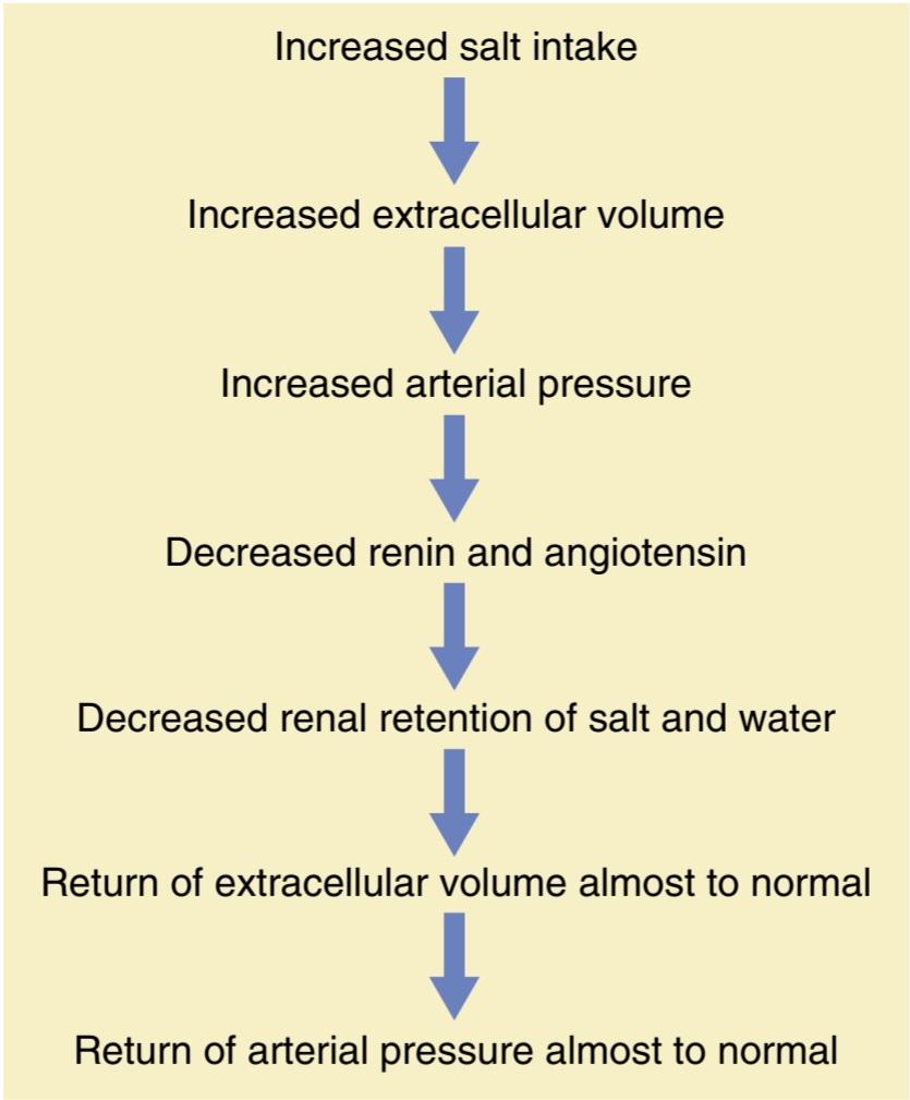
Figure 19-12. Sequential events whereby increased salt intake in- creases the arterial pressure, but feedback decrease in activity of the renin angiotensin system returns the arterial pressure almost to the normal level.
شکل 19-12. رویدادهای متوالی که به موجب آن افزایش مصرف نمک، فشار شریانی را افزایش میدهد، اما کاهش بازخورد در فعالیت سیستم رنین آنژیوتانسین، فشار شریانی را تقریباً به سطح طبیعی برمیگرداند.
To emphasize the efficacy of the renin-angiotensin system in controlling arterial pressure, when the system functions normally, the pressure usually rises no more than 4 to 6 mm Hg in response to as much as a 100-fold increase in salt intake (Figure 19-13). Conversely, when the usual suppression of angiotensin formation is prevented due to continuous infusion of small amounts of angiotensin II so that blood levels cannot decrease, the same increase in salt intake may cause the pressure to rise by 40 mm Hg or more (see Figure 19-13). When salt intake is reduced to as low as one-tenth normal, arterial pressure barely changes as long as the renin-angiotensin system functions normally. However, when angiotensin II formation is blocked with an ACE inhibitor, blood pres- sure decreases markedly as salt intake is reduced (see Figure 19-13). Thus, the renin-angiotensin system is perhaps the body’s most powerful system for accommodating wide variations in salt intake with minimal changes in arterial pressure.
برای تاکید بر اثربخشی سیستم رنین-آنژیوتانسین در کنترل فشار شریانی، زمانی که سیستم به طور طبیعی کار میکند، فشار معمولاً در پاسخ به افزایش ۱۰۰ برابری نمک مصرفی، بیش از ۴ تا ۶ میلیمتر جیوه افزایش نمییابد (شکل ۱۹-۱۳). برعکس، وقتی از سرکوب معمول تشکیل آنژیوتانسین به دلیل تزریق مداوم مقادیر کمی آنژیوتانسین II جلوگیری میشود، به طوری که سطح خون نمیتواند کاهش یابد، همین افزایش در مصرف نمک ممکن است باعث افزایش فشار 40 میلیمتر جیوه یا بیشتر شود (شکل 19-13 را ببینید). هنگامی که مصرف نمک به یک دهم نرمال کاهش می یابد، فشار شریانی به سختی تغییر می کند تا زمانی که سیستم رنین-آنژیوتانسین به طور طبیعی کار کند. با این حال، هنگامی که تشکیل آنژیوتانسین II با یک مهارکننده ACE مسدود می شود، فشار خون به طور قابل توجهی کاهش می یابد زیرا مصرف نمک کاهش می یابد (شکل 19-13 را ببینید). بنابراین، سیستم رنین-آنژیوتانسین شاید قوی ترین سیستم بدن برای سازگاری با تغییرات گسترده در مصرف نمک با حداقل تغییرات در فشار شریانی باشد.
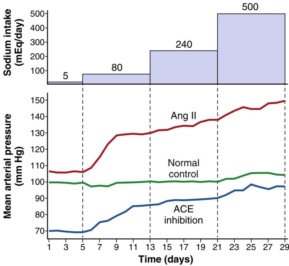
Figure 19-13. Changes in mean arterial pressure during chronic changes in sodium intake in normal control dogs and in dogs treated with an angiotensin-converting enzyme (ACE) inhibitor to block angiotensin II (Ang II) formation or infused with Ang II to prevent Ang II from being suppressed. Sodium intake was raised in steps from a low level of 5 mmol/day to 80, 240, and 500 mmol/day for 8 days at each level. (Modified from Hall JE, Guyton AC, Smith MJ Jr, et al: Blood pressure and renal function during chronic changes in sodium intake: role of angiotensin. Am J Physiol 239:F271, 1980.)
شکل 19-13. تغییرات در فشار متوسط شریانی در طول تغییرات مزمن در مصرف سدیم در سگهای کنترل عادی و در سگهایی که تحت درمان با مهارکننده آنزیم مبدل آنژیوتانسین (ACE) برای جلوگیری از تشکیل آنژیوتانسین II (Ang II) یا تزریق آنگ II برای جلوگیری از سرکوب آنژیوتانسین II هستند. مصرف سدیم به صورت پلکانی از سطح پایین 5 میلی مول در روز به 80، 240 و 500 میلی مول در روز به مدت 8 روز در هر سطح افزایش یافت. (اصلاح شده از Hall JE، Guyton AC، Smith MJ Jr، و همکاران: فشار خون و عملکرد کلیه در طول تغییرات مزمن در مصرف سدیم: نقش آنژیوتانسین. Am J Physiol 239:F271، 1980.)
HYPERTENSION CAUSED BY RENIN- SECRETING TUMOR OR RENAL ISCHEMIA Occasionally, a tumor of the renin-secreting JG cells occurs and secretes large quantities of renin, causing formation of large amounts of angiotensin II. In all patients in whom this phenomenon has occurred, severe hypertension has developed. Also, when large amounts of angiotensin II are infused continuously for days or weeks into animals, similar severe long-term hypertension develops.
فشار خون بالا ناشی از تومور ترشح کننده رنین یا ایسکمی کلیوی. گاهی اوقات، توموری در سلول های JG ترشح کننده رنین ایجاد می شود و مقادیر زیادی رنین ترشح می کند و باعث تشکیل مقادیر زیادی آنژیوتانسین II می شود. در تمام بیمارانی که این پدیده در آنها رخ داده است، فشار خون شدید ایجاد شده است. همچنین، هنگامی که مقادیر زیادی آنژیوتانسین II به طور مداوم برای روزها یا هفته ها به حیوانات تزریق می شود، فشار خون طولانی مدت مشابهی ایجاد می شود.
We have already noted that angiotensin II can increase the arterial pressure in two ways:
1. By constricting the arterioles throughout the entire body, thereby increasing the total peripheral resistance and arterial pressure; this effect occurs within seconds after one begins to infuse large amounts of angiotensin II.
2. By causing the kidneys to retain salt and water; over a period of days, even moderate amounts of angiotensin II can cause causes hypertension through its renal actions, the principal cause of the long-term elevation of arterial pressure.
قبلاً متذکر شدیم که آنژیوتانسین II می تواند فشار شریانی را به دو طریق افزایش دهد:
1. با منقبض کردن شریان ها در سراسر بدن، در نتیجه افزایش مقاومت کلی محیطی و فشار شریانی. این اثر در عرض چند ثانیه پس از شروع تزریق مقادیر زیادی آنژیوتانسین II رخ می دهد.
2. با نگه داشتن کلیه ها نمک و آب. در طی چند روز، حتی مقادیر متوسط آنژیوتانسین II می تواند از طریق اعمال کلیوی خود باعث ایجاد فشار خون بالا شود که علت اصلی افزایش طولانی مدت فشار شریانی است.
One-Kidney Goldblatt Hypertension. When one kidney is removed, and a constrictor is placed on the renal artery of the remaining kidney, as shown in Figure 19- 14, the immediate effect is greatly reduced pressure in the renal artery beyond the constrictor, as demonstrated by the dashed curve in the figure Then, within seconds or minutes, the systemic arterial pressure begins to rise and continues to rise for several days. The pressure usually rises rapidly for the first hour or so, and this effect is followed by a slower additional rise during the next several days. When the systemic arterial pressure reaches its new stable pressure level, the renal arterial pressure distal to the constriction (the dashed curve in the figure) will have returned almost all the way back to normal. The hypertension produced in this way is called one-kidney Goldblatt hypertension in honor of Harry Goldblatt, who first studied the important quantitative features of hypertension caused by renal artery constriction.
فشار خون گلدبلات یک کلیه. همانطور که در شکل 19-14 نشان داده شده است، هنگامی که یکی از کلیه ها برداشته می شود، و یک منقبض کننده روی شریان کلیوی کلیه باقی مانده قرار می گیرد، اثر فوری کاهش شدید فشار در شریان کلیوی فراتر از منقبض کننده است، همانطور که با منحنی شکسته در شکل نشان داده شده است، سپس در عرض چند ثانیه یا چند دقیقه، فشار شریانی سیستمیک برای چند روز شروع به افزایش می کند و به افزایش ادامه می دهد. فشار معمولاً در حدود یک ساعت اول به سرعت افزایش می یابد و این اثر با افزایش آهسته تری در طی چند روز آینده دنبال می شود. هنگامی که فشار شریانی سیستمیک به سطح فشار پایدار جدید خود می رسد، فشار شریانی کلیوی دیستال به انقباض (منحنی چین خورده در شکل) تقریباً در تمام طول مسیر به حالت عادی باز می گردد. به افتخار هری گلدبلات، که برای اولین بار ویژگیهای کمی مهم فشار خون ناشی از انقباض شریان کلیوی را مورد مطالعه قرار داد، فشار خون یک کلیه که به این روش ایجاد میشود، پرفشاری گلدبلات یک کلیه نامیده میشود.
The early rise in arterial pressure in Goldblatt hyper- tension is caused by the renin-angiotensin vasoconstrictor mechanism. That is, because of poor blood flow through the kidney after acute constriction of the renal artery, large quantities of renin are secreted by the kidney, as demonstrated by the lowermost curve in Figure 19-14, and this action increases angiotensin II and aldosterone levels in the blood. The angiotensin II, in turn, raises the arterial pressure acutely. The secretion of renin rises to a peak in about 1 or 2 hours but returns nearly to normal in 5 to 7 days because the renal arterial pressure by that time has also risen back to normal, so the kidney is no longer ischemic.
افزایش زودهنگام فشار شریانی در فشار خون گلدبلات ناشی از مکانیسم منقبض کننده عروق رنین-آنژیوتانسین است. یعنی به دلیل جریان ضعیف خون از طریق کلیه پس از انقباض حاد شریان کلیوی، مقادیر زیادی رنین از کلیه ترشح می شود، همانطور که در پایین ترین منحنی در شکل 19-14 نشان داده شده است، و این عمل باعث افزایش سطح آنژیوتانسین II و آلدوسترون در خون می شود. آنژیوتانسین II به نوبه خود فشار شریانی را به شدت افزایش می دهد. ترشح رنین در حدود 1 یا 2 ساعت به اوج خود می رسد اما در عرض 5 تا 7 روز تقریباً به حالت طبیعی باز می گردد زیرا فشار شریانی کلیوی نیز در آن زمان به حالت عادی برگشته است، بنابراین کلیه دیگر ایسکمیک نیست.

Figure 19-14. Effect of placing a constricting clamp on the renal artery of one kidney after the other kidney has been removed. Note the changes in systemic arterial pressure, renal artery pressure distal to the clamp, and rate of renin secretion. The resulting hypertension is called one-kidney Goldblatt hypertension.
شکل 19-14. اثر قرار دادن گیره منقبض کننده بر روی شریان کلیوی یک کلیه پس از برداشتن کلیه دیگر. به تغییرات فشار شریانی سیستمیک، فشار شریان کلیوی دیستال گیره و سرعت ترشح رنین توجه کنید. فشار خون ناشی از آن فشار خون گلدبلات یک کلیه نامیده می شود.
The second rise in arterial pressure is caused by retention of salt and water by the constricted kidney, which is also stimulated by angiotensin II and aldosterone. In 5 to 7 days, the body fluid volume increases enough to raise the arterial pressure to its new sustained level. The quantitative value of this sustained pressure level is determined by the degree of constriction of the renal artery. That is, the aortic pressure must rise enough so that renal arterial pressure distal to the constrictor is high enough to cause normal urine output.
دومین افزایش فشار شریانی به دلیل احتباس نمک و آب توسط کلیه منقبض است که توسط آنژیوتانسین II و آلدوسترون نیز تحریک می شود. در 5 تا 7 روز، حجم مایع بدن به اندازه ای افزایش می یابد که فشار شریانی را به سطح پایدار جدید خود برساند. مقدار کمی این سطح فشار پایدار با درجه انقباض شریان کلیوی تعیین می شود. به این معنا که فشار آئورت باید به اندازهای افزایش یابد که فشار شریانی کلیوی دیستال به منقبض کننده به اندازهای بالا باشد که باعث برون ده ادرار طبیعی شود.
A similar scenario occurs in patients with stenosis of the renal artery of a single remaining kidney, as sometimes occurs after a person receives a kidney transplant. Also, functional or pathological increases in resistance of the renal arterioles, due to atherosclerosis or excessive levels of vasoconstrictors, can cause hypertension through the same mechanisms as constriction of the main renal artery.
سناریوی مشابهی در بیماران مبتلا به تنگی شریان کلیوی یک کلیه باقیمانده رخ میدهد، همانطور که گاهی اوقات پس از دریافت پیوند کلیه اتفاق میافتد. همچنین افزایش عملکردی یا پاتولوژیک مقاومت شریانهای کلیوی به دلیل تصلب شرایین یا سطوح بیش از حد منقبضکنندههای عروقی، میتواند از طریق مکانیسمهایی مانند انقباض شریان اصلی کلیوی باعث افزایش فشار خون شود.
Two-Kidney Goldblatt Hypertension. Hypertension also can result when the artery to only one kidney is constricted while the artery to the other kidney is normal. The constricted kidney secretes renin and also retains salt and water because of decreased renal arterial pressure in this kidney. Then, the “normal” opposite kidney retains salt and water because of the renin produced by the ischemic kidney. This renin causes increased formation of angiotensin II and aldosterone, both of which circulate to the opposite kidney and cause it also to retain salt and water. Thus, both kidneys-but for different reasons-be- come salt and water retainers. Consequently, hypertension develops.
فشار خون گلدبلات دو کلیه. فشار خون بالا همچنین می تواند زمانی ایجاد شود که شریان تنها یک کلیه منقبض شود در حالی که شریان کلیه دیگر طبیعی است. کلیه منقبض شده رنین ترشح می کند و همچنین نمک و آب را به دلیل کاهش فشار شریانی کلیه در این کلیه حفظ می کند. سپس، کلیه “عادی” مقابل نمک و آب را به دلیل رنین تولید شده توسط کلیه ایسکمیک حفظ می کند. این رنین باعث افزایش تشکیل آنژیوتانسین II و آلدوسترون می شود که هر دو به کلیه مخالف گردش می کنند و باعث حفظ نمک و آب در آن می شوند. بنابراین، هر دو کلیه – اما به دلایل مختلف – نگهدارنده نمک و آب می شوند. در نتیجه، فشار خون بالا ایجاد می شود.
The clinical counterpart of two-kidney Goldblatt hypertension occurs when there is stenosis of a single renal artery-for example, caused by atherosclerosis-in a person who has two kidneys.
همتای بالینی پرفشاری خون گلدبلات دو کلیه زمانی رخ می دهد که تنگی یک شریان کلیوی منفرد – به عنوان مثال، ناشی از تصلب شرایین – در فردی که دارای دو کلیه است، رخ می دهد.
Hypertension Caused by Diseased Kidneys That Secrete Renin Chronically. Often, patchy areas of one or both kidneys are diseased and become ischemic because of local vascular constrictions or infarctions, whereas other areas of the kidneys are normal. When this situation occurs, almost identical effects occur as in the two- kidney type of Goldblatt hypertension. That is, the patchy ischemic kidney tissue secretes renin, which, in turn-by acting through the formation of angiotensin II-causes the remaining kidney mass also to retain salt and water. One of the most common causes of renal hypertension, especially in older persons, is this patchy ischemic kidney disease.
فشار خون بالا ناشی از کلیه های بیمار که رنین را به طور مزمن ترشح می کنند. اغلب، نواحی تکهای یک یا هر دو کلیه به دلیل انقباضات عروقی یا انفارکتوس موضعی بیمار بوده و ایسکمیک میشوند، در حالی که سایر نواحی کلیهها طبیعی هستند. هنگامی که این وضعیت رخ می دهد، اثرات تقریباً مشابهی مانند فشار خون بالا در نوع دو کلیه ای گلدبلات رخ می دهد. یعنی بافت ایسکمیک تکه تکه کلیه رنین ترشح می کند که به نوبه خود از طریق تشکیل آنژیوتانسین II عمل می کند و باعث می شود که توده کلیه باقی مانده نیز نمک و آب را حفظ کند. یکی از شایع ترین علل فشار خون کلیوی، به ویژه در افراد مسن، این بیماری ایسکمیک کلیه است.
Other Types of Hypertension Caused by Combinations of Volume Loading and Vasoconstriction
انواع دیگر فشار خون ناشی از ترکیبی از بارگذاری حجم و انقباض عروق
Hypertension in the Upper Part of the Body Caused by Coarctation of the Aorta. One out of every few thousand babies is born with pathological constriction or blockage of the aorta at a point beyond the aortic arterial branches to the head and arms but proximal to the renal arteries, a condition called coarctation of the aorta. When this occurs, blood flow to the lower body is carried by multiple small collateral arteries in the body wall, with much vascular resistance between the upper aorta and lower aorta. As a consequence, the arterial pressure in the upper part of the body may be 40% to 50% higher than that in the lower body.
فشار خون بالا در قسمت فوقانی بدن ناشی از کوآرکتاسیون آئورت. از هر چند هزار نوزاد، یک نوزاد با انقباض یا انسداد پاتولوژیک آئورت در نقطهای فراتر از شاخههای شریانی آئورت به سر و بازوها، اما نزدیک به شریانهای کلیوی متولد میشود، وضعیتی که کوآرکتاسیون آئورت نامیده میشود. هنگامی که این اتفاق می افتد، جریان خون به پایین بدن توسط چندین شریان جانبی کوچک در دیواره بدن، با مقاومت عروقی زیادی بین آئورت فوقانی و آئورت تحتانی حمل می شود. در نتیجه، فشار شریانی در قسمت بالایی بدن ممکن است 40٪ تا 50٪ بیشتر از فشار در قسمت پایین بدن باشد.
The mechanism of this upper body hypertension is al- most identical to that of one-kidney Goldblatt hypertension. That is, when a constrictor is placed on the aorta above the renal arteries, the blood pressure in both kidneys falls at first, renin is secreted, angiotensin and aldosterone are formed, and hypertension occurs in the upper body. The arterial pressure in the lower body at the level of the kidneys rises approximately to normal, but high pressure persists in the upper body. The kidneys are no longer ischemic, and thus secretion of renin and formation of angiotensin and aldosterone return to nearly normal. Likewise, in coarctation of the aorta, the arterial pressure in the lower body is usually almost normal, whereas the pressure in the upper body is far higher than normal.
مکانیسم این پرفشاری خون بالاتنه تقریباً مشابه فشار خون گلدبلات یک کلیه است. یعنی وقتی یک منقبض کننده روی آئورت بالای شریان های کلیوی قرار می گیرد، ابتدا فشار خون در هر دو کلیه کاهش می یابد، رنین ترشح می شود، آنژیوتانسین و آلدوسترون تشکیل می شود و فشار خون بالا در قسمت بالایی بدن ایجاد می شود. فشار شریانی در قسمت پایین بدن در سطح کلیه ها تقریباً تا حد طبیعی افزایش می یابد، اما فشار بالا در قسمت فوقانی بدن ادامه می یابد. کلیه ها دیگر ایسکمیک نیستند و بنابراین ترشح رنین و تشکیل آنژیوتانسین و آلدوسترون تقریباً به حالت طبیعی باز می گردد. به همین ترتیب، در کوآرکتاسیون آئورت، فشار شریانی در قسمت تحتانی بدن معمولاً تقریباً طبیعی است، در حالی که فشار در قسمت فوقانی بدن بسیار بیشتر از حد طبیعی است.
Role of Autoregulation in Hypertension Caused by Aortic Coarctation. A significant feature of hypertension caused by aortic coarctation is that blood flow in the arms, where the pressure may be 40% to 60% above normal, is almost exactly normal. Also, blood flow in the legs, where the pressure is not elevated, is almost exactly normal. How could this be, with the pressure in the upper body 40% to 60% greater than in the lower body? There are no differences in vasoconstrictor substances in the blood of the up- per and lower body because the same blood flows to both areas. Likewise, the nervous system innervates both areas of the circulation similarly, so there is no reason to believe that there is a difference in nervous control of the blood vessels. The main reason is that long-term autoregulation develops so nearly completely that the local blood flow control mechanisms have compensated almost 100% for the differences in pressure. The result is that in both the highpressure area and low-pressure area, the local blood flow is controlled by local autoregulatory mechanisms almost exactly in accord with the needs of the tissue and not in accord with the level of the pressure.
نقش خودتنظیمی در فشار خون بالا ناشی از کوآرکتاسیون آئورت. یکی از ویژگی های مهم فشار خون بالا ناشی از کوآرکتاسیون آئورت این است که جریان خون در بازوها، جایی که فشار ممکن است 40 تا 60 درصد بالاتر از حد طبیعی باشد، تقریباً کاملاً طبیعی است. همچنین، جریان خون در پاها، جایی که فشار بالا نیست، تقریباً کاملاً طبیعی است. چگونه ممکن است این اتفاق بیفتد، در حالی که فشار در قسمت بالایی بدن 40٪ تا 60٪ بیشتر از قسمت پایین بدن است؟ هیچ تفاوتی در مواد منقبض کننده عروق در خون بالا و پایین بدن وجود ندارد زیرا خون یکسان به هر دو ناحیه جریان دارد. به همین ترتیب، سیستم عصبی هر دو ناحیه گردش خون را به طور مشابه عصب دهی می کند، بنابراین دلیلی وجود ندارد که باور کنیم در کنترل عصبی رگ های خونی تفاوت وجود دارد. دلیل اصلی این است که خودتنظیمی طولانی مدت تقریباً به طور کامل توسعه می یابد که مکانیسم های کنترل جریان خون محلی تقریباً 100٪ تفاوت فشار را جبران کرده است. نتیجه این است که هم در ناحیه پرفشار و هم در ناحیه کم فشار، جریان خون موضعی توسط مکانیسم های خودتنظیمی موضعی تقریباً دقیقاً مطابق با نیازهای بافت و نه مطابق با سطح فشار کنترل می شود.
Hypertension in Preeclampsia (Toxemia of Pregnancy). A syndrome called preeclampsia (also called toxemia of pregnancy) develops in approximately 5% to 10% of expectant mothers. One of the manifestations of preeclampsia is hypertension that usually subsides after delivery of the baby. Although the precise causes of preeclampsia are not completely understood, ischemia of the placenta and subsequent release by the placenta of toxic factors are believed to play a role in causing many of the manifestations of this disorder, including hypertension in the mother. Substances released by the ischemic placenta, in turn, cause dysfunction of vascular endothelial cells throughout the body, including the blood vessels of the kidneys. This endothelial dysfunction decreases release of nitric oxide and other vasodilator substances, causing vasoconstriction, decreased rate of fluid filtration from the glomeruli into the renal tubules, impaired renal pressure natriuresis, and the development of hypertension.
فشار خون بالا در پره اکلامپسی (سموم بارداری). سندرمی به نام پره اکلامپسی (که سموم بارداری نیز نامیده می شود) تقریباً در 5 تا 10 درصد از مادران باردار ایجاد می شود. یکی از تظاهرات پره اکلامپسی فشار خون بالا است که معمولا پس از زایمان کاهش می یابد. اگرچه علل دقیق پره اکلامپسی به طور کامل شناخته نشده است، اعتقاد بر این است که ایسکمی جفت و انتشار متعاقب آن عوامل سمی توسط جفت در ایجاد بسیاری از تظاهرات این اختلال از جمله فشار خون بالا در مادر نقش دارد. موادی که توسط جفت ایسکمیک آزاد می شوند، به نوبه خود باعث اختلال در عملکرد سلول های اندوتلیال عروقی در سراسر بدن، از جمله رگ های خونی کلیه ها می شوند. این اختلال عملکرد اندوتلیال باعث کاهش انتشار اکسید نیتریک و سایر مواد گشادکننده عروق می شود و باعث انقباض عروق، کاهش سرعت فیلتراسیون مایع از گلومرول ها به داخل لوله های کلیوی، اختلال در ناتریورز فشار کلیه و ایجاد فشار خون می شود.
Another pathological abnormality that may contribute to hypertension in preeclampsia is thickening of the kidney glomerular membranes (perhaps caused by an auto- immune process), which also reduces the glomerular fluid filtration rate. For obvious reasons, the arterial pressure level required to cause normal formation of urine becomes elevated, and the long-term level of arterial pressure be- comes correspondingly elevated. These patients are especially prone to extra degrees of hypertension when they have excess salt intake.
یکی دیگر از ناهنجاری های پاتولوژیک که ممکن است به فشار خون در پره اکلامپسی کمک کند، ضخیم شدن غشای گلومرولی کلیه است (شاید ناشی از یک فرآیند خودایمنی باشد)، که همچنین میزان فیلتراسیون مایع گلومرولی را کاهش می دهد. به دلایل واضح، سطح فشار شریانی مورد نیاز برای ایجاد ادرار طبیعی افزایش مییابد و سطح فشار شریانی درازمدت به همان نسبت بالا میرود. این بیماران به ویژه در صورت مصرف بیش از حد نمک مستعد ابتلا به درجات اضافی فشار خون هستند.
Neurogenic Hypertension. Acute neurogenic hypertension can be caused by strong stimulation of the sympathetic nervous system. For example, when a person becomes excited for any reason or during states of anxiety, the sympathetic system becomes excessively stimulated, peripheral vasoconstriction occurs everywhere in the body, and acute hypertension ensues.
فشار خون عصبی. فشار خون حاد عصبی می تواند در اثر تحریک شدید سیستم عصبی سمپاتیک ایجاد شود. به عنوان مثال، هنگامی که فردی به هر دلیلی یا در طول حالت های اضطراب هیجان زده می شود، سیستم سمپاتیک بیش از حد تحریک می شود، انقباض عروق محیطی در همه جای بدن رخ می دهد و فشار خون حاد ایجاد می شود.
Another type of acute neurogenic hypertension occurs when the nerves leading from the baroreceptors are cut or when the tractus solitarius is destroyed in each side of the medulla oblongata. These are the areas where the nerves from the carotid and aortic baroreceptors connect in the brain stem. The sudden cessation of normal nerve signals from the baroreceptors has the same effect on the nervous pressure control mechanisms as a sudden reduction of the arterial pressure in the aorta and carotid arteries. That is, loss of the normal inhibitory effect on the vasomotor center caused by normal baroreceptor nervous signals allows the vasomotor center suddenly to become extremely active and the mean arterial pressure to increase from 100 mm Hg to as high as 160 mm Hg. The pressure returns to nearly normal within about 2 days because the response of the vaso- motor center to the absent baroreceptor signal fades away, which is called central resetting of the baroreceptor pressure control mechanism. Therefore, the neurogenic hypertension caused by sectioning the baroreceptor nerves is mainly an acute type of hypertension, not a chronic type.
نوع دیگری از فشار خون حاد عصبی زمانی رخ می دهد که اعصاب منتهی به گیرنده های فشاری بریده شوند یا زمانی که tractus solitarius در هر طرف بصل النخاع از بین می رود. اینها مناطقی هستند که اعصاب بارورسپتورهای کاروتید و آئورت در ساقه مغز به هم متصل می شوند. قطع ناگهانی سیگنالهای عصبی طبیعی از گیرندههای فشاری همان تأثیری را بر مکانیسمهای کنترل فشار عصبی دارد که کاهش ناگهانی فشار شریانی در آئورت و شریانهای کاروتید. یعنی از دست دادن اثر مهاری طبیعی بر روی مرکز وازوموتور ناشی از سیگنالهای عصبی بارورسپتور طبیعی باعث میشود که مرکز وازوموتور به طور ناگهانی بسیار فعال شود و فشار متوسط شریانی از 100 میلیمتر جیوه به 160 میلیمتر جیوه افزایش یابد. فشار در عرض 2 روز تقریباً به حالت عادی برمیگردد، زیرا پاسخ مرکز وازوموتور به سیگنال گیرنده فشاری که وجود ندارد از بین میرود که به آن تنظیم مجدد مرکزی مکانیسم کنترل فشار گیرنده فشار میگویند. بنابراین، فشار خون عصبی ناشی از برش دادن اعصاب بارورسپتور عمدتاً یک نوع حاد فشار خون است، نه یک نوع مزمن.
The sympathetic nervous system also plays an important role in some forms of chronic hypertension, in large part by activation of the renal sympathetic nerves. For example, excess weight gain and obesity often lead to activation of the sympathetic nervous system, which in turn stimulates the renal sympathetic nerves, impairs renal pressure natriuresis, and causes chronic hypertension. These abnormalities appear to play a major role in a large percentage of patients with primary (essential) hypertension, as discussed later.
سیستم عصبی سمپاتیک همچنین نقش مهمی در برخی از اشکال فشار خون مزمن دارد که تا حد زیادی با فعال شدن اعصاب سمپاتیک کلیوی انجام می شود. به عنوان مثال، افزایش وزن بیش از حد و چاقی اغلب منجر به فعال شدن سیستم عصبی سمپاتیک می شود که به نوبه خود اعصاب سمپاتیک کلیه را تحریک می کند، ناتریورزیس فشار کلیه را مختل می کند و باعث فشار خون مزمن می شود. به نظر می رسد که این ناهنجاری ها نقش عمده ای در درصد زیادی از بیماران مبتلا به فشار خون اولیه (ضروری) دارند، همانطور که بعداً بحث شد.
Genetic Causes of Hypertension. Spontaneous hereditary hypertension has been observed in several strains of animals, including different strains of rats and rabbits and at least one strain of dogs. In the strain of rats that has been studied to the greatest extent, the Okamoto spontaneously hypertensive rat, there is evidence that in early develop-ment of the hypertension, the sympathetic nervous system is considerably more active than in normal rats. In the later stages of this type of hypertension, structural changes have been observed in the nephrons of the kidneys: (1) increased preglomerular renal arterial resistance; and (2) decreased permeability of the glomerular membranes. These structural changes could also contribute to the long-term continuance of the hypertension. In other strains of hypertensive rats, impaired renal function also has been observed.
علل ژنتیکی فشار خون بالا. فشار خون ارثی خود به خودی در چندین گونه از حیوانات، از جمله سویه های مختلف موش و خرگوش و حداقل یک سویه از سگ مشاهده شده است. در سویه موشهایی که تا حد زیادی مورد مطالعه قرار گرفتهاند، موشهای Okamoto خود به خود پرفشاری خون، شواهدی وجود دارد که در شروع اولیه فشار خون بالا، سیستم عصبی سمپاتیک به طور قابلتوجهی فعالتر از موشهای معمولی است. در مراحل بعدی این نوع فشار خون، تغییرات ساختاری در نفرون کلیه ها مشاهده شده است: (1) افزایش مقاومت شریانی کلیوی پرگلومرولی. و (2) کاهش نفوذپذیری غشاهای گلومرولی. این تغییرات ساختاری همچنین می تواند به تداوم طولانی مدت فشار خون کمک کند. در سویه های دیگر موش های با فشار خون بالا، اختلال در عملکرد کلیه نیز مشاهده شده است.
In humans, several different gene mutations have been identified that can cause hypertension. These forms of hy- pertension are called monogenic hypertension because they are caused by mutation of a single gene. An interesting feature of these genetic disorders is that they all cause impaired kidney function, either by increased resistance of the renal arterioles or by excessive salt and water reabsorption by the renal tubules. In some cases, the increased reabsorption is due to gene mutations that directly increase the transport of sodium or chloride in the renal tubular epithelial cells. In other cases, the gene mutations cause increased synthesis or activity of hormones that stimulate renal tubular salt and water reabsorption. Thus, in all monogenic hypertensive disorders discovered thus far, the final common pathway to hypertension appears to be impaired kidney function. Mo- nogenic hypertension, however, is rare, and all the known forms together account for less than 1% of cases of human hypertension.
در انسان، چندین جهش ژنی مختلف شناسایی شده است که می تواند باعث افزایش فشار خون شود. این اشکال پرفشاری خون را پرفشاری خون تک ژنی می نامند زیرا در اثر جهش یک ژن ایجاد می شوند. یکی از ویژگیهای جالب این اختلالات ژنتیکی این است که همگی باعث اختلال در عملکرد کلیه میشوند، یا با افزایش مقاومت شریانهای کلیوی یا با بازجذب بیش از حد نمک و آب توسط لولههای کلیوی. در برخی موارد، افزایش بازجذب به دلیل جهشهای ژنی است که مستقیماً انتقال سدیم یا کلرید را در سلولهای اپیتلیال لولههای کلیوی افزایش میدهد. در موارد دیگر، جهشهای ژنی باعث افزایش سنتز یا فعالیت هورمونهایی میشوند که باعث تحریک جذب مجدد نمک و آب در لولههای کلیوی میشوند. بنابراین، در تمام اختلالات پرفشاری خون تک ژنی که تاکنون کشف شده است، به نظر می رسد آخرین مسیر مشترک برای فشار خون بالا، اختلال در عملکرد کلیه باشد. با این حال، پرفشاری خون مونوژنیک نادر است و همه اشکال شناخته شده با هم کمتر از 1٪ موارد فشار خون انسانی را تشکیل می دهند.
PRIMARY (ESSENTIAL) HYPERTENSION
About 90% to 95% of all people who have hypertension are said to have primary hypertension, also referred to as essential hypertension by many clinicians. These terms simply mean that the hypertension is of unknown origin, in contrast to the forms of hypertension that are secondary to known causes, such as renal artery stenosis or mono- genic forms of hypertension.
فشار خون اولیه (ضروری).
گفته می شود که حدود 90 تا 95 درصد از تمام افرادی که فشار خون بالا دارند، فشار خون اولیه دارند که بسیاری از پزشکان از آن به عنوان فشار خون ضروری نیز یاد می کنند. این عبارات به سادگی به این معنی است که فشار خون بالا منشأ ناشناخته ای دارد، برخلاف اشکال فشار خون بالا که ثانویه به علل شناخته شده هستند، مانند تنگی شریان کلیوی یا اشکال تک ژنی فشار خون بالا.
In most patients, excess weight gain and a sedentary lifestyle appear to play a major role in causing primary hypertension. Most patients with hypertension are overweight, and studies of different populations have suggested that excess adiposity may account for as much as 65% to 75% of the risk for developing primary hypertension. Clinical studies have clearly shown the value of weight loss for reducing blood pressure in most patients with hypertension, and clinical guidelines for treating hypertension recommend increased physical activity and weight loss as a first step in treating most patients with hypertension.
در اکثر بیماران، افزایش وزن بیش از حد و سبک زندگی بی تحرک به نظر می رسد که نقش عمده ای در ایجاد فشار خون اولیه دارند. اکثر بیماران مبتلا به فشار خون بالا اضافه وزن دارند و مطالعات روی جمعیت های مختلف نشان داده است که چاقی بیش از حد ممکن است 65 تا 75 درصد از خطر ابتلا به فشار خون اولیه را تشکیل دهد. مطالعات بالینی به وضوح ارزش کاهش وزن را برای کاهش فشار خون در اکثر بیماران مبتلا به فشار خون نشان داده است و دستورالعمل های بالینی برای درمان فشار خون بالا، افزایش فعالیت بدنی و کاهش وزن را به عنوان اولین قدم در درمان بیشتر بیماران مبتلا به فشار خون بالا توصیه می کند.
The following characteristics of primary hypertension, among others, are caused by excess weight gain and obesity:
ویژگی های زیر فشار خون اولیه، از جمله، ناشی از افزایش وزن و چاقی است:
1. Cardiac output is increased in part because of the additional blood flow required for the extra adipose tissue. However, blood flow in the heart, kidneys, gastrointestinal tract, and skeletal muscle also increases with weight gain because of increased metabolic rate and growth of the organs and tissues in response to their increased metabolic demands. As the hypertension is sustained for many months and years, total peripheral vascular resistance may be increased.
1. برون ده قلبی تا حدی به دلیل جریان خون اضافی مورد نیاز برای بافت چربی اضافی افزایش می یابد. با این حال، جریان خون در قلب، کلیهها، دستگاه گوارش و ماهیچههای اسکلتی نیز با افزایش وزن به دلیل افزایش سرعت متابولیک و رشد اندامها و بافتها در پاسخ به افزایش تقاضای متابولیک آنها افزایش مییابد. از آنجایی که فشار خون بالا برای چندین ماه و سال ادامه دارد، مقاومت عروق محیطی کل ممکن است افزایش یابد.
2. Sympathetic nerve activity, especially in the kidneys, is increased in overweight patients. The causes of increased sympathetic activity in obese persons are not fully understood, but studies have suggested that hormones such as leptin that are released from fat cells may directly stimulate multiple regions of the hypothalamus, which in turn have an excitatory influence on the vasomotor centers of the brain medulla. There is also evidence for reduced sensitivity of arterial baroreceptors in buffering increases in arterial pressure, as well as activation of chemoreceptors in obese persons, especially in those who also have obstructive sleep apnea.
2. فعالیت عصب سمپاتیک به ویژه در کلیه ها در بیماران دارای اضافه وزن افزایش می یابد. علل افزایش فعالیت سمپاتیک در افراد چاق به طور کامل شناخته نشده است، اما مطالعات نشان داده اند که هورمون هایی مانند لپتین که از سلول های چربی آزاد می شوند ممکن است مستقیماً مناطق متعددی از هیپوتالاموس را تحریک کنند که به نوبه خود تأثیر تحریکی بر مراکز وازوموتور بصل النخاع مغز دارند. همچنین شواهدی مبنی بر کاهش حساسیت بارورسپتورهای شریانی در افزایش بافری در فشار شریانی و همچنین فعال شدن گیرنده های شیمیایی در افراد چاق، به ویژه در افرادی که آپنه انسدادی خواب نیز دارند، وجود دارد.
3. Angiotensin II and aldosterone levels are in- creased in many obese patients. This increase may be caused partly by increased sympathetic nerve stimulation, which increases renin release by the kidneys and therefore formation of angiotensin II, which in turn stimulates the adrenal gland to secrete aldosterone.
3. سطح آنژیوتانسین II و آلدوسترون در بسیاری از بیماران چاق افزایش می یابد. این افزایش ممکن است تا حدی ناشی از افزایش تحریک عصب سمپاتیک باشد که باعث افزایش ترشح رنین توسط کلیه ها و در نتیجه تشکیل آنژیوتانسین II می شود که به نوبه خود غده فوق کلیوی را برای ترشح آلدوسترون تحریک می کند.
4. The renal-pressure natriuresis mechanism is impaired, and the kidneys will not excrete adequate amounts of salt and water unless the arterial pressure is high or kidney function is somehow improved. If mean arterial pressure in the essential hypertensive person is 150 mm Hg, acute reduction of mean arterial pressure to the normal value of 100 mm Hg (but without otherwise altering renal function except for the decreased pressure) will cause almost total anuria. The person will then retain salt and water until the pressure rises back to the elevated value of 150 mm Hg. Chronic reductions in arterial pressure with effective antihypertensive therapies, however, usually do not cause marked salt and water retention by the kidneys because these therapies also improve renal pressure natriuresis, as discussed later.
4. مکانیسم ناتریورز فشار کلیه مختل شده است و کلیه ها مقادیر کافی نمک و آب دفع نمی کنند مگر اینکه فشار شریانی بالا باشد یا عملکرد کلیه به نحوی بهبود یابد. اگر فشار متوسط شریانی در فرد مبتلا به فشار خون اساسی 150 میلی متر جیوه باشد، کاهش حاد فشار متوسط شریانی به مقدار طبیعی 100 میلی متر جیوه (اما بدون تغییر عملکرد کلیوی به جز کاهش فشار) باعث آنوری تقریباً کل می شود. سپس فرد نمک و آب را تا زمانی که فشار به مقدار بالای 150 میلیمتر جیوه افزایش یابد، حفظ میکند. با این حال، کاهش مزمن فشار شریانی با درمانهای موثر ضد فشار خون، معمولاً باعث احتباس شدید نمک و آب توسط کلیهها نمیشود، زیرا این درمانها همچنین ناتریورز فشار کلیه را بهبود میبخشند، همانطور که بعداً بحث شد.
Experimental studies in obese animals and obese patients have suggested that impaired renal pressure natriuresis in obesity hypertension is caused mainly by increased renal tubular reabsorption of salt and water due to increased sympathetic nerve activity and increased levels of angiotensin II and aldosterone. However, if hypertension is not effectively treated, there may also be vascular damage in the kidneys that can reduce glomerular filtration rate and increase the severity of hypertension. Eventually, uncontrolled hypertension associated with obesity and associated metabolic disorders can lead to severe vascular injury and complete loss of kidney function.
مطالعات تجربی بر روی حیوانات چاق و بیماران چاق نشان داده است که اختلال ناتریورز فشار کلیوی در فشار خون چاقی عمدتاً به دلیل افزایش بازجذب لولهای کلیوی نمک و آب به دلیل افزایش فعالیت عصب سمپاتیک و افزایش سطح آنژیوتانسین II و آلدوسترون ایجاد میشود. با این حال، اگر فشار خون بالا به طور موثر درمان نشود، ممکن است آسیب عروقی در کلیه ها نیز ایجاد شود که می تواند میزان فیلتراسیون گلومرولار را کاهش دهد و شدت فشار خون را افزایش دهد. در نهایت، فشار خون کنترل نشده همراه با چاقی و اختلالات متابولیک مرتبط می تواند منجر به آسیب شدید عروقی و از دست دادن کامل عملکرد کلیه شود.
Graphic Analysis of Arterial Pressure Control in Essential Hypertension. Figure 19-15 is a graphic analysis of essential hypertension. The curves of this figure are called sodium-loading renal function curves because the arterial pressure in each case is increased very slowly, over many days or weeks, by gradually increasing the level of sodium intake. The sodium-loading type of curve can be determined by increasing the level of sodium intake to a new level every few days and then waiting for the renal output of sodium to come into balance with the intake and, at the same time, recording the changes in arterial pressure.
تجزیه و تحلیل گرافیکی کنترل فشار شریانی در فشار خون ضروری. شکل 19-15 تجزیه و تحلیل گرافیکی فشار خون اساسی است. منحنی های این شکل را منحنی های عملکرد کلیوی بارگیری سدیم می نامند زیرا فشار شریانی در هر مورد بسیار آهسته و در طی چند روز یا چند هفته با افزایش تدریجی سطح دریافت سدیم افزایش می یابد. نوع منحنی بارگیری سدیم را می توان با افزایش سطح دریافتی سدیم به یک سطح جدید هر چند روز یکبار و سپس منتظر ماندن برای متعادل شدن برون ده کلیوی سدیم با مصرف و در عین حال ثبت تغییرات فشار شریانی تعیین کرد.
When this procedure is used in patients with essential hypertension, two types of curves, shown at the right in Figure 19-15, can be recorded; one is called (1) saltinsensitive hypertension and the other (2) salt-sensitive hypertension. Note in both cases that the curves are shifted to the right, to a higher pressure level than for people with normal arterial pressure. In the case of the person with salt-insensitive essential hypertension, the arterial pressure does not increase significantly when changing from a normal salt intake to a high salt intake. However, in patients who have salt-sensitive essential hypertension, the high salt intake significantly exacerbates the hypertension.
هنگامی که این روش در بیماران مبتلا به فشار خون اساسی استفاده می شود، دو نوع منحنی، که در سمت راست در شکل 19-15 نشان داده شده است، قابل ثبت است. یکی (1) فشار خون غیر حساس به نمک و دیگری (2) فشار خون حساس به نمک نامیده می شود. در هر دو مورد توجه داشته باشید که منحنیها به سمت راست منتقل میشوند، به سطح فشار بالاتری نسبت به افراد با فشار شریانی طبیعی. در مورد فرد مبتلا به پرفشاری خون ضروری حساس به نمک، فشار شریانی هنگام تغییر از مصرف نمک معمولی به مصرف نمک زیاد، به میزان قابل توجهی افزایش نمی یابد. با این حال، در بیمارانی که فشار خون ضروری حساس به نمک دارند، مصرف زیاد نمک به طور قابل توجهی فشار خون را تشدید می کند.
Two additional points should be emphasized. First, salt sensitivity of blood pressure is not an all-or-none finding—it is quantitative, with some individuals being more salt-sensitive than others. Second, salt sensitivity of blood pressure is not a fixed characteristic; instead, blood pressure usually becomes more salt sensitive as a person ages, especially after 50 or 60 years of age, when the number of functions units (nephrons) in the kidneys begins to decrease gradually.
دو نکته اضافی باید مورد تاکید قرار گیرد. اولاً، حساسیت فشار خون به نمک یک یافته همه یا هیچ نیست – این یافته کمی است و برخی افراد نسبت به سایرین به نمک حساستر هستند. دوم، حساسیت فشار خون به نمک یک مشخصه ثابت نیست. در عوض، فشار خون معمولاً با افزایش سن به نمک حساستر میشود، بهویژه پس از 50 یا 60 سالگی، زمانی که تعداد واحدهای عملکردی (نفرونها) در کلیهها شروع به کاهش تدریجی میکند.
The reason for the difference between salt-insensitive essential hypertension and salt-sensitive hypertension is presumably related to structural or functional differences in the kidneys of these two types of hypertensive patients. For example, salt-sensitive hypertension may occur with different types of chronic renal disease because of the gradual loss of nephrons or because of normal aging, as discussed in Chapter 32. Abnormal function of the renin-angiotensin system can also cause arterial pressure to become salt-sensitive, as discussed previously in this chapter.
دلیل تفاوت فشار خون ضروری حساس به نمک و پرفشاری خون حساس به نمک احتمالاً مربوط به تفاوت های ساختاری یا عملکردی در کلیه این دو نوع از بیماران مبتلا به فشار خون بالا است. به عنوان مثال، فشار خون حساس به نمک ممکن است با انواع مختلف بیماری مزمن کلیوی به دلیل از دست دادن تدریجی نفرون ها یا به دلیل پیری طبیعی رخ دهد، همانطور که در فصل 32 بحث شد. عملکرد غیر طبیعی سیستم رنین-آنژیوتانسین همچنین می تواند باعث شود فشار شریانی به نمک حساس شود، همانطور که قبلا در این فصل بحث شد.

Figure 19-15. Analysis of arterial pressure regulation in (1) saltinsensitive essential hypertension and (2) saltsensitive essential hypertension. (Modified from Guyton AC, Coleman TG, Young DB, et al: Salt balance and long-term blood pressure control. Annu Rev Med 31:15, 1980.)
شکل 19-15. تجزیه و تحلیل تنظیم فشار شریانی در (1) فشار خون ضروری غیر حساس به نمک و (2) فشار خون ضروری حساس به نمک. (اصلاح شده از Guyton AC، Coleman TG، Young DB، و همکاران: تعادل نمک و کنترل طولانی مدت فشار خون. Annu Rev Med 31:15، 1980.)
Treatment of Essential Hypertension. As a first step, current guidelines for treating hypertension recommend lifestyle modifications aimed at increasing physical activity and weight loss in most patients. Unfortunately, many patients are unable to lose weight, and pharmacological treatment with antihypertensive drugs must be initiated. Two general classes of drugs are used to treat hypertension: (1)vasodilator drugs, which increase renal blood flow and glomerular filtration rate; and (2) natriuretic or diuretic drugs, which decrease tubular reabsorption of salt and water.
درمان فشار خون ضروری. به عنوان اولین گام، دستورالعمل های فعلی برای درمان فشار خون بالا، اصلاح شیوه زندگی را با هدف افزایش فعالیت بدنی و کاهش وزن در اکثر بیماران توصیه می کند. متأسفانه بسیاری از بیماران قادر به کاهش وزن نیستند و باید درمان دارویی با داروهای ضد فشار خون آغاز شود. دو دسته کلی از داروها برای درمان فشار خون بالا استفاده می شوند: (1) داروهای گشادکننده عروق که جریان خون کلیوی و میزان فیلتراسیون گلومرولی را افزایش می دهند. و (2) داروهای ناتریورتیک یا دیورتیک که بازجذب لوله ای نمک و آب را کاهش می دهند.
Vasodilator drugs usually cause vasodilation in many other tissues of the body, as well as in the kidneys. Different vasodilators act in one of the following ways: (1) by inhibiting sympathetic nervous signals to the kidneys or by blocking the action of the sympathetic transmitter substance on the renal vasculature and renal tubules; (2) by directly relaxing the smooth muscle of the renal vasculature; or (3) by blocking the action of the reninangiotensin-aldosterone system on the renal vasculature or renal tubules.
داروهای گشادکننده عروق معمولاً باعث گشاد شدن عروق در بسیاری از بافت های دیگر بدن و همچنین در کلیه ها می شوند. گشادکنندههای عروق مختلف به یکی از روشهای زیر عمل میکنند: (1) با مهار سیگنالهای عصبی سمپاتیک به کلیهها یا با مسدود کردن عمل ماده انتقالدهنده سمپاتیک روی عروق کلیوی و لولههای کلیوی. (2) با شل کردن مستقیم ماهیچه صاف عروق کلیوی. یا (3) با مسدود کردن عملکرد سیستم رنین آنژیوتانسین-آلدوسترون روی عروق کلیوی یا لوله های کلیوی.
Drugs that reduce the reabsorption of salt and water by the renal tubules include, in particular, drugs that block active transport of sodium through the tubular wall; this blockage in turn also prevents the reabsorption of water, as explained earlier in the chapter. These natriuretic or diuretic drugs are discussed in greater detail in Chapter 32.
داروهایی که جذب مجدد نمک و آب توسط لوله های کلیوی را کاهش می دهند، به ویژه داروهایی هستند که انتقال فعال سدیم را از طریق دیواره لوله ای مسدود می کنند. همانطور که قبلا در فصل توضیح داده شد، این انسداد نیز به نوبه خود از جذب مجدد آب جلوگیری می کند. این داروهای ناتریورتیک یا دیورتیک با جزئیات بیشتر در فصل 32 مورد بحث قرار گرفته اند.
SUMMARY OF INTEGRATED MULTIFACETED SYSTEMS FOR ARTERIAL PRESSURE REGULATION
It is clear that arterial pressure is regulated not by a single pressure controlling system but instead by several interrelated systems, each of which performs a specific function. For example, when a person bleeds so severely that the pressure falls suddenly, two problems confront the pressure control system. The first is survival; the arterial pressure must be rapidly returned to a high enough level that the person can live through the acute episode. The second is to return the blood volume and arterial pressure eventually to their normal levels so that the circulatory system can reestablish full normality, not merely back to the levels required for survival.
خلاصه ای از سیستم های چند وجهی یکپارچه برای تنظیم فشارشریانی
واضح است که فشار شریانی توسط یک سیستم کنترل فشار منفرد تنظیم نمی شود، بلکه توسط چندین سیستم مرتبط به هم، که هر یک عملکرد خاصی را انجام می دهند، تنظیم می شود. به عنوان مثال، هنگامی که فردی به شدت خونریزی می کند که فشار به طور ناگهانی کاهش می یابد، سیستم کنترل فشار با دو مشکل مواجه می شود. اولین مورد بقا است. فشار شریانی باید به سرعت به سطح بالایی برگردد که فرد بتواند دوره حاد را پشت سر بگذارد. دوم این است که حجم خون و فشار شریانی را در نهایت به سطح طبیعی خود بازگردانیم تا سیستم گردش خون بتواند به حالت عادی بازگردد، نه اینکه صرفاً به سطوح مورد نیاز برای بقا برگردد.
In Chapter 18, we saw that the first line of defense against acute changes in arterial pressure is the nervous control system. In this chapter, we have emphasized a second line of defense achieved mainly by kidney mechanisms for the long-term control of arterial pressure. However, there are other pieces to the puzzle. Figure 19-16 helps put these pieces together.
در فصل 18 دیدیم که اولین خط دفاعی در برابر تغییرات حاد فشار شریانی، سیستم کنترل عصبی است. در این فصل، ما بر خط دوم دفاعی تأکید کردهایم که عمدتاً توسط مکانیسمهای کلیه برای کنترل طولانیمدت فشار شریانی به دست میآید. با این حال، قطعات دیگری نیز در این پازل وجود دارد. شکل 19-16 به کنار هم قرار دادن این قطعات کمک می کند.
Figure 19-16 shows the approximate immediate (seconds and minutes) and long-term (hours and days) control responses, expressed as feedback gain, of eight arterial pressure control mechanisms. These mechanisms can be divided into three groups: (1) those that react rapidly, within seconds or minutes; (2) those that respond over an intermediate time period-that is, minutes or hours; and (3) those that provide long-term arterial pressure regulation for days, months, and years.
شکل 16-19 پاسخ های کنترلی تقریبی (ثانیه و دقیقه) و طولانی مدت (ساعت ها و روزها) را نشان می دهد که به صورت افزایش بازخورد هشت مکانیسم کنترل فشار شریانی بیان می شود. این مکانیسم ها را می توان به سه گروه تقسیم کرد: (1) آنهایی که به سرعت، در عرض چند ثانیه یا چند دقیقه واکنش نشان می دهند. (2) آنهایی که در یک بازه زمانی میانی – یعنی دقیقه یا ساعت پاسخ می دهند. و (3) آنهایی که تنظیم طولانی مدت فشار شریانی را برای روزها، ماهها و سالها فراهم می کنند.

Figure 19-16. Approximate potency of various arterial pressure control mechanisms at different time intervals after the onset of a disturbance to the arterial pressure. Note especially the near-infinite gain (-) of the renal body fluid pressure control mechanism that occurs after a few weeks’ time. CNS, Central nervous system. (Modified from Guyton AC: Arterial Pressure and Hypertension. Philadelphia: WB Saunders, 1980.)
شکل 19-16. قدرت تقریبی مکانیسم های کنترل فشار شریانی مختلف در فواصل زمانی مختلف پس از شروع اختلال در فشار شریانی. به ویژه به افزایش تقریبا بی نهایت (-) مکانیسم کنترل فشار مایع بدن کلیه که پس از چند هفته رخ می دهد توجه کنید. CNS، سیستم عصبی مرکزی. (اصلاح شده از Guyton AC: Arerial Pressure and Hypertension. Philadelphia: WB Saunders, 1980.)
Arterial Pressure Control Mechanisms That Act Within Seconds or Minutes. The rapidly acting pressure control mechanisms are almost entirely acute nervous reflexes or other autonomic nervous system responses. Note in Figure 19-16 the three mechanisms that show responses within seconds: (1) the baroreceptor feedback mechanism; (2) the central nervous system ischemic mechanism; and (3) the chemoreceptor mechanism. Not only do these mechanisms begin to react within seconds, but they are also powerful. After any acute fall in pressure, as might be caused by severe hemorrhage, the nervous mechanisms combine to cause the following: (1) constriction of the veins and transfer of blood into the heart; (2) increased heart rate and contractility of the heart to provide greater pumping capability by the heart; and (3) constriction of most peripheral arterioles. All these effects occur almost instantly to raise the arterial pressure back into a survival range.
مکانیسم های کنترل فشار شریانی که در عرض چند ثانیه یا چند دقیقه عمل می کنند. مکانیسم های کنترل فشار سریع تقریباً به طور کامل رفلکس های عصبی حاد یا سایر پاسخ های سیستم عصبی خودمختار هستند. در شکل 16-19 به سه مکانیسمی که پاسخ ها را در عرض چند ثانیه نشان می دهند توجه کنید: (1) مکانیسم بازخورد بارورسپتور. (2) مکانیسم ایسکمیک سیستم عصبی مرکزی. و (3) مکانیسم گیرنده شیمیایی. این مکانیسم ها نه تنها در عرض چند ثانیه شروع به واکنش می کنند، بلکه قدرتمند نیز هستند. پس از هر افت حاد فشار، که ممکن است در اثر خونریزی شدید ایجاد شود، مکانیسمهای عصبی با هم ترکیب میشوند و باعث ایجاد موارد زیر میشوند: (1) انقباض وریدها و انتقال خون به قلب. (2) افزایش ضربان قلب و انقباض قلب برای ایجاد قابلیت پمپاژ بیشتر توسط قلب. و (3) تنگی بیشتر شریان های محیطی. همه این اثرات تقریباً فوراً برای بالا بردن فشار شریانی به محدوده بقا رخ می دهند.
When the pressure suddenly rises too high, as might occur in response to a rapid transfusion of excess blood, the same control mechanisms operate in the reverse direction, again returning the pressure back toward normal.
هنگامی که فشار ناگهان بسیار بالا می رود، همانطور که ممکن است در پاسخ به انتقال سریع خون اضافی رخ دهد، مکانیسم های کنترلی مشابه در جهت معکوس عمل می کنند و دوباره فشار را به حالت عادی برمی گرداند.
Arterial Pressure Control Mechanisms That Act After Many Minutes. Several pressure control mechanisms exhibit significant responses only after a few minutes fol- lowing an acute arterial pressure change. Three of these mechanisms, shown in Figure 19-16, are as follows: (1) the renin-angiotensin vasoconstrictor mechanism; (2) stress relaxation of the vasculature; and (3) shift of fluid through the tissue capillary walls in and out of the circula- tion to readjust the blood volume as needed.
مکانیسم های کنترل فشار شریانی که بعد از چند دقیقه عمل می کنند. چندین مکانیسم کنترل فشار تنها پس از چند دقیقه پس از تغییر فشار شریانی حاد، پاسخهای قابل توجهی را نشان میدهند. سه تا از این مکانیسم ها، که در شکل 16-19 نشان داده شده اند، به شرح زیر هستند: (1) مکانیسم منقبض کننده عروق رنین-آنژیوتانسین. (2) آرامش استرس عروق. و (3) انتقال مایع از طریق دیواره های مویرگ بافت به داخل و خارج از گردش خون برای تنظیم مجدد حجم خون در صورت نیاز.
We have already described at length the role of the renin-angiotensin vasoconstrictor system to provide a semiacute means for increasing the arterial pressure when necessary. The stress relaxation mechanism is demonstrated by the following example. When the pres- sure in the blood vessels becomes too high, they become stretched more and more for minutes or hours; as a result, the pressure in the vessels falls toward normal. This continuing stretch of the vessels, called stress relaxation, can serve as an intermediate-term pressure “buffer”
ما قبلاً نقش سیستم منقبض کننده عروق رنین-آنژیوتانسین را برای ارائه یک وسیله نیمه حاد برای افزایش فشار شریانی در مواقع لزوم شرح داده ایم. مکانیسم کاهش استرس با مثال زیر نشان داده شده است. هنگامی که فشار در رگهای خونی بیش از حد بالا میرود، برای چند دقیقه یا چند ساعت بیشتر و بیشتر کشیده میشوند. در نتیجه، فشار در عروق به سمت نرمال کاهش می یابد. این کشش مداوم عروق، به نام آرام سازی استرس، می تواند به عنوان یک “بافر” فشار میان مدت عمل کند.
The capillary fluid shift mechanism means simply that whenever capillary pressure falls too low, fluid is absorbed from the tissues through the capillary membranes and into the circulation, thus building up the blood volume and increasing the pressure in the circulation. Conversely, when the capillary pressure rises too high, fluid is lost out of the circulation into the tissues, thus reducing the blood volume, as well as virtually all the pressures throughout the circulation.
مکانیسم جابجایی مایع مویرگی به سادگی به این معنی است که هرگاه فشار مویرگی خیلی کم شود، مایع از بافت ها از طریق غشای مویرگی جذب شده و وارد گردش خون می شود، بنابراین حجم خون افزایش می یابد و فشار در گردش خون افزایش می یابد. برعکس، زمانی که فشار مویرگی بیش از حد بالا می رود، مایع از گردش خون به بافت ها از دست می رود، بنابراین حجم خون و همچنین تقریباً تمام فشارها در سراسر گردش خون کاهش می یابد.
These three intermediate mechanisms mostly become activated within 30 minutes to several hours. During this time, the nervous mechanisms usually become less and less effective, illustrating the importance of these non- nervous, intermediate-term pressure control measures.
این سه مکانیسم میانی عمدتاً در عرض 30 دقیقه تا چند ساعت فعال می شوند. در طول این مدت، مکانیسمهای عصبی معمولاً کمتر و کمتر مؤثر میشوند، که اهمیت این اقدامات کنترل فشار میانمدت غیرعصبی را نشان میدهد.
Long-Term Mechanisms for Arterial Pressure Regulation. The goal of this chapter has been to explain the role of the kidneys in long-term control of arterial pressure. To the far right in Figure 19-16 is shown the renal-blood volume pressure control mechanism, which is the same as the renal-body fluid pressure control mechanism, demonstrating that it takes a few hours to begin showing significant response. Yet, it eventually develops a feedback gain for control of arterial pressure that is nearly equal to infinity. This means that this mechanism can eventually return the arterial pressure nearly all the way back, not merely partway back, to the pressure level that provides normal output of salt and water by the kidneys.
مکانیسم های بلند مدت برای تنظیم فشار شریانی. هدف این فصل توضیح نقش کلیه ها در کنترل طولانی مدت فشار شریانی بوده است. در سمت راست در شکل 16-19 مکانیسم کنترل فشار حجم خون کلیوی نشان داده شده است، که همان مکانیسم کنترل فشار مایع کلیه-بدن است و نشان می دهد که چند ساعت طول می کشد تا واکنش قابل توجهی نشان داده شود. با این حال، در نهایت یک بازخورد برای کنترل فشار شریانی ایجاد می کند که تقریباً برابر با بی نهایت است. این بدان معناست که این مکانیسم در نهایت میتواند فشار شریانی را تقریباً تا آخر، نه فقط تا حدی، به سطح فشاری برگرداند که خروجی طبیعی نمک و آب توسط کلیهها را فراهم میکند.
Many factors can affect the pressure-regulating level of the renal-body fluid mechanism. One of these, shown in Figure 19-16, is aldosterone. A decrease in arterial pressure leads within minutes to an increase in aldosterone secretion and, over the next hour or days, this effect plays an important role in modifying the pressure control char- acteristics of the renal-body fluid mechanism.
عوامل زیادی می توانند بر سطح تنظیم فشار مکانیسم مایع کلیه-بدن تأثیر بگذارند. یکی از اینها که در شکل 19-16 نشان داده شده است، آلدوسترون است. کاهش فشار شریانی در عرض چند دقیقه منجر به افزایش ترشح آلدوسترون میشود و در طی یک ساعت یا روزهای بعد، این اثر نقش مهمی در اصلاح ویژگیهای کنترل فشار مکانیزم مایع کلیه-بدن ایفا میکند.
Especially important is the interaction of the reninangiotensin system with the aldosterone and renal fluid mechanisms. For example, a person’s salt intake varies tremendously from one day to another. We have seen in this chapter that salt intake can decrease to as little as onetenth normal or can increase to 10 to 15 times normal and yet the regulated level of the mean arterial pressure will change only a few mm Hg if the renin-angiotensin- aldosterone system is fully operative. However, without a functional renin-angiotensin-aldosterone system, blood pressure becomes very sensitive to changes in salt intake.
به ویژه برهمکنش سیستم رنین آنژیوتانسین با آلدوسترون و مکانیسم های مایع کلیوی مهم است. به عنوان مثال، میزان نمک مصرفی یک فرد از یک روز به روز دیگر بسیار متفاوت است. در این فصل دیدیم که مصرف نمک می تواند تا یک دهم نرمال کاهش یابد یا می تواند به 10 تا 15 برابر طبیعی افزایش یابد و با این حال سطح تنظیم شده فشار شریانی متوسط تنها چند میلی متر جیوه تغییر می کند اگر سیستم رنین-آنژیوتانسین-آلدوسترون به طور کامل عمل کند. با این حال، بدون سیستم عملکردی رنین-آنژیوتانسین-آلدوسترون، فشار خون به تغییرات در مصرف نمک بسیار حساس می شود.
Thus, arterial pressure control begins with the lifesaving measures of the nervous pressure controls, then continues with the sustaining characteristics of the intermediate pressure controls and, finally, is stabilized at the long-term pressure level by the renal-body fluid mechanism. This long-term mechanism, in turn, has multiple interactions with the renin-angiotensin-aldosterone system, the nervous system, and several other factors that provide special blood pressure control capabilities for special purposes.
بنابراین، کنترل فشار شریانی با اقدامات نجاتدهنده کنترلهای فشار عصبی آغاز میشود، سپس با ویژگیهای پایدار کنترلهای فشار میانی ادامه مییابد و در نهایت، در سطح فشار طولانیمدت توسط مکانیزم مایع کلیه-بدن تثبیت میشود. این مکانیسم طولانی مدت، به نوبه خود، برهمکنش های متعددی با سیستم رنین-آنژیوتانسین-آلدوسترون، سیستم عصبی و چندین عامل دیگر دارد که قابلیت های ویژه کنترل فشار خون را برای اهداف خاص فراهم می کند.
کلیک کنید «Bibliography: فهرست کتب مربوطه»
Acelajado MC, Hughes ZH, Oparil S, Calhoun DA: Treatment of resist- ant and refractory hypertension. Circ Res 124:1061, 2019. Brands MW:
Chronic blood pressure control. Compr Physiol 2:2481, 2012.
Coffman TM: The inextricable role of the kidney in hypertension. J Clin Invest 124:2341, 2014.
Colafella KMM, Denton KM: Sex-specific differences in hypertension and associated cardiovascular disease. Nat Rev Nephrol 14:185, 2018.
Cowley AW: Long-term control of arterial blood pressure. Physiol Rev 72:231, 1992.
Guyton AC: Arterial Pressure and hypertension. Philadelphia: WB Saunders, 1980.
Hall JE, Granger JP, do Carmo JM, et al: Hypertension: physiology and pathophysiology. Compr Physiol 2:2393, 2012.
Hall JE, do Carmo JM, da Silva AA, et al: Obesity-induced hyperten- sion: interaction of neurohumoral and renal mechanisms. Circ Res 116:991, 2015.
Hall JE, do Carmo JM, da Silva AA, et al: Obesity, kidney dysfunction and hypertension: mechanistic links. Nat Rev Nephrol 15:367, 2019.
Lohmeier TE, Hall JE: Device-based neuromodulation for resistant hypertension therapy. Circ Res 124:1071, 2019.
Lifton RP, Gharavi AG, Geller DS: Molecular mechanisms of human hypertension. Cell 104:545, 2001.
Oparil S, Schmieder RE: New approaches in the treatment of hyper- tension. Circ Res 116:1074, 2015.
Rana S, Lemoine E, Granger J, Karumanchi SA: Preeclampsia. Circ Res 124:1094, 2019.
Rossier BC, Bochud M, Devuyst O: The hypertension pandemic: an evolutionary perspective. Physiology (Bethesda) 32:112, 2017.
Whelton PK, Carey RM, Aronow WS, et al: Guideline for the Prevention, Detection, Evaluation, and Management of High Blood Pressure in Adults: Executive Summary: A Report of the American College of Cardiology/American Heart Association Task Force on Clinical Practice Guidelines. Hypertension 71:1269, 2018.
🚀 با ما همراه شوید!
تازهترین مطالب و آموزشهای مغز و اعصاب را از دست ندهید. با فالو کردن کانال تلگرام آیندهنگاران مغز، از ما حمایت کنید!



