اعصاب جمجمهای؛ عصب جمجمهای V: عصب سه قلو


دعای مطالعه [ نمایش ]
بِسْمِ الله الرَّحْمنِ الرَّحیمِ
اَللّهُمَّ اَخْرِجْنى مِنْ ظُلُماتِ الْوَهْمِ
خدايا مرا بيرون آور از تاريكىهاى وهم،
وَ اَكْرِمْنى بِنُورِ الْفَهْمِ
و به نور فهم گرامى ام بدار،
اَللّهُمَّ افْتَحْ عَلَيْنا اَبْوابَ رَحْمَتِكَ
خدايا درهاى رحمتت را به روى ما بگشا،
وَانْشُرْ عَلَيْنا خَزائِنَ عُلُومِكَ بِرَحْمَتِكَ يا اَرْحَمَ الرّاحِمينَ
و خزانههاى علومت را بر ما باز كن به امید رحمتت اى مهربانترين مهربانان.
آیندهنگاران مغز: «ما مغز را میشناسیم، تا آینده را بسازیم.»
کتاب «نوروآناتومی بالینی اسنل» اثر دکتر ریچارد اس. اسنل، با ویرایش جدید دکتر رایان اسپلیتگربر، همچنان بهعنوان مرجع طلایی این حوزه شناخته میشود. این اثر با تلفیق تازهترین پژوهشها و کاربردهای بالینی، پلی استوار میان دانش پایه و عمل پزشکی بنا میکند.
تلاشهای علمی و سالها تدریس ارزشمند استاد گرانقدر دکتر محمدتقی جغتایی الهامبخش ترجمه و گسترش این دانش در ایران بوده است.
ترجمه دقیق این کتاب توسط برند علمی آیندهنگاران مغز به مدیریت داریوش طاهری، امکان دسترسی فارسیزبانان به مرزهای نوین علوم اعصاب را فراهم کرده و رسالتی علمی در جهت شناخت عمیقتر مغز و ترسیم آیندهای روشنتر بر عهده گرفته است.
»» فصل ۱۱: هستههای اعصاب جمجمهای؛ بخش پنجم مبانی و اصول
»» Chapter 11: Cranial Nerve Nuclei
TRIGEMINAL NERVE (CRANIAL NERVE V)
The trigeminal nerve is the largest cranial nerve and contains both sensory and motor fibers (Fig. 11-7). It is the sensory nerve to the greater part of the head and the motor nerve to several muscles, including the muscles of mastication.
عصب سه قلو (عصب جمجمهای V)
عصب سه قلو بزرگترین عصب جمجمهای است و شامل هر دو رشته حسی و حرکتی است (شکل 11-7). این عصب حسی به قسمت بزرگتر سر و عصب حرکتی به چندین عضله، از جمله عضلات جونده است.
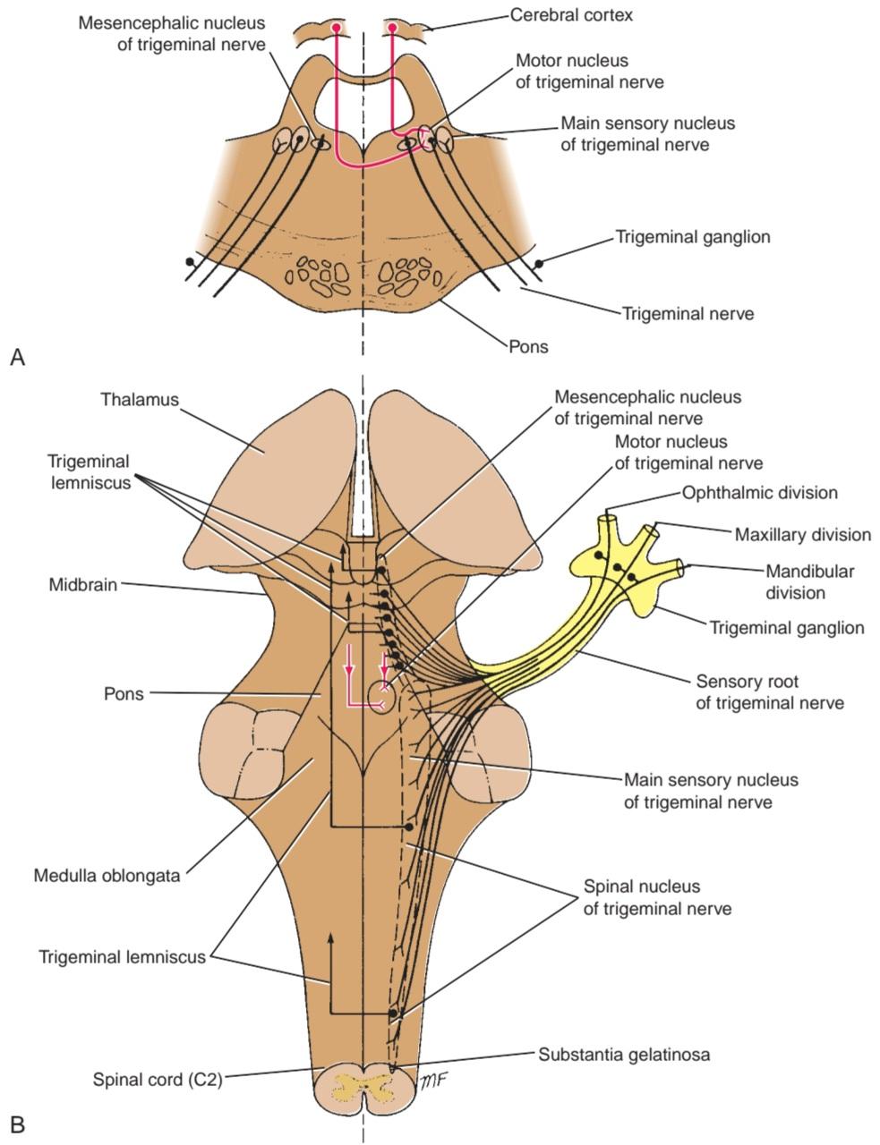
Figure 11-7 A: Trigeminal nerve nuclei seen in a coronal section of the pons. B: Trigeminal nerve nuclei in the brainstem and their central connections.
شکل ۱۱-۷ الف: هستههای عصب سهقلو که در برش کرونال پل مغزی دیده میشوند. ب: هستههای عصب سهقلو در ساقه مغز و اتصالات مرکزی آنها.
Trigeminal Nerve Nuclei
The trigeminal nerve has four nuclei: (1) the main sensory nucleus, (2) the spinal nucleus, (3) the mesen- cephalic nucleus, and (4) the motor nucleus.
هستههای عصب سهقلو
عصب سهقلو چهار هسته دارد: (1) هسته حسی اصلی، (2) هسته نخاعی، (3) هسته مزانسفال و (4) هسته حرکتی.
Main Sensory Nucleus
The main sensory nucleus lies in the posterior part of the pons, lateral to the motor nucleus (see Fig. 11-7A). It is continuous below with the spinal nucleus.
هسته حسی اصلی
هسته حسی اصلی در قسمت خلفی پل مغزی، در سمت خارجی هسته حرکتی قرار دارد (شکل 11-7A را ببینید). این هسته در پایین با هسته نخاعی در یک راستا قرار دارد.
spinal Nucleus
The spinal nucleus is continuous superiorly with the main sensory nucleus in the pons and extends inferiorly through the whole length of the medulla oblongata and into the upper part of the spinal cord as far as the second cervical segment (see Fig. 11-7B).
هسته نخاعی
هسته نخاعی از بالا در امتداد هسته حسی اصلی در پل مغزی قرار دارد و از پایین در تمام طول بصل النخاع و در قسمت فوقانی نخاع تا دومین قطعه گردنی امتداد مییابد (شکل 11-7B را ببینید).
Mesencephalic Nucleus
The mesencephalic nucleus is composed of a column of unipolar nerve cells situated in the lateral part of the gray matter around the cerebral aqueduct. It extends inferiorly into the pons as far as the main sensory nucleus (see Fig. 11-7).
هسته مزانسفالیک
هسته مزانسفالیک از ستونی از سلولهای عصبی تکقطبی تشکیل شده است که در قسمت جانبی ماده خاکستری اطراف مجرای مغزی قرار دارد. این هسته از پایین به داخل پل مغزی تا هسته حسی اصلی امتداد مییابد (شکل 11-7 را ببینید).
Motor Nucleus
The motor nucleus is situated in the pons, medial to the main sensory nucleus (see Fig. 11-7).
هسته حرکتی
هسته حرکتی در پل مغزی، در قسمت میانی هسته حسی اصلی قرار دارد (شکل 11-7 را ببینید).
Trigeminal Nerve Sensory Components
The sensations of pain, temperature, touch, and pressure from the skin of the face and mucous membranes travel along axons whose cell bodies are situated in the semilunar or trigeminal sensory ganglion (see Fig. 11-7B). The central processes of these cells form the large sensory root of the trigeminal nerve. About half the fibers divide into ascending and descending branches when they enter the pons; the remainder ascend or descend without division. The ascending branches terminate in the main sensory nucleus, and the descending branches terminate in the spinal nucleus. The sensations of touch and pressure are conveyed by nerve fibers that terminate in the main sensory nucleus. The sensations of pain and temperature pass to the spinal nucleus. The sensory fibers from the ophthalmic division of the trigeminal nerve terminate in the inferior part of the spinal nucleus; fibers from the maxillary division terminate in the middle of the spinal nucleus; and fibers from the mandibular division end in the superior part of the spinal nucleus.
اجزای حسی عصب سه قلو
حس درد، دما، لمس و فشار از پوست صورت و غشاهای مخاطی در امتداد آکسونهایی حرکت میکنند که جسم سلولی آنها در گانگلیون حسی نیمه هلالی یا سه قلو قرار دارد (شکل 11-7B را ببینید). زوائد مرکزی این سلولها ریشه حسی بزرگ عصب سه قلو را تشکیل میدهند. حدود نیمی از فیبرها هنگام ورود به پل مغزی به شاخههای صعودی و نزولی تقسیم میشوند. بقیه بدون تقسیم شدن، صعودی یا نزولی میشوند. شاخههای صعودی در هسته حسی اصلی و شاخههای نزولی در هسته نخاعی خاتمه مییابند. حس لمس و فشار توسط فیبرهای عصبی که در هسته حسی اصلی خاتمه مییابند، منتقل میشود. حس درد و دما به هسته نخاعی منتقل میشود. فیبرهای حسی از شاخه چشمی عصب سه قلو در قسمت تحتانی هسته نخاعی خاتمه مییابند. فیبرهای شاخه فک بالا در وسط هسته نخاعی خاتمه مییابند. و فیبرهای شاخه فک پایین در قسمت فوقانی هسته نخاعی خاتمه مییابند.
Proprioceptive impulses from the muscles of mastication and from the facial and extraocular muscles are carried by fibers in the sensory root of the trigeminal nerve that have bypassed the semilunar or trigeminal ganglion. The fibers’ cells of origin are the unipolar cells of the mesencephalic nucleus.
تکانههای حس عمقی از عضلات جونده و از عضلات صورت و خارج چشمی توسط فیبرهایی در ریشه حسی عصب سه قلو که از گانگلیون نیمه هلالی یا سه قلو عبور کردهاند، حمل میشوند. سلولهای منشأ این فیبرها، سلولهای تک قطبی هسته مزانسفال هستند.
The axons of the neurons in the main sensory and spinal nuclei and the central processes of the cells in the mesencephalic nucleus now cross the median plane and ascend as the trigeminal lemniscus to terminate on the nerve cells of the ventral posteromedial nucleus of the thalamus. The axons of these cells now travel through the internal capsule to the postcentral gyrus (areas 3, 1, and 2) of the cerebral cortex.
آکسونهای نورونهای هستههای حسی و نخاعی اصلی و زوائد مرکزی سلولهای هسته مزانسفال اکنون از صفحه میانی عبور کرده و به عنوان لمنیسکوس سه قلو صعود میکنند تا به سلولهای عصبی هسته خلفی میانی شکمی تالاموس ختم شوند. آکسونهای این سلولها اکنون از طریق کپسول داخلی به شکنج پس مرکزی (نواحی ۳، ۱ و ۲) قشر مغز میروند.
Trigeminal Nerve Motor Component
The motor nucleus receives corticonuclear fibers from both cerebral hemispheres (see Fig. 11-7). It also receives fibers from the reticular formation, the red nucleus, the tectum, and the medial longitudinal fasciculus. In addition, it receives fibers from the mesencephalic nucleus, thereby forming a monosynaptic reflex arc.
جزء حرکتی عصب سه قلو
هسته حرکتی، فیبرهای قشری-هستهای را از هر دو نیمکره مغزی دریافت میکند (شکل 11-7 را ببینید). همچنین فیبرهایی از تشکیلات مشبک، هسته قرمز، تکتوم و فاسیکول طولی داخلی دریافت میکند. علاوه بر این، فیبرهایی از هسته مزانسفال دریافت میکند و در نتیجه یک قوس رفلکس تک سیناپسی تشکیل میدهد.
The cells of the motor nucleus give rise to the axons that form the motor root. The motor nucleus supplies the muscles of mastication, the tensor tympani, the tensor veli palatini, and the mylohyoid and the anterior belly of the digastric muscle.
سلولهای هسته حرکتی، آکسونهایی را ایجاد میکنند که ریشه حرکتی را تشکیل میدهند. هسته حرکتی، عضلات جونده، عضله کشنده صماخ، عضله کشنده نرم کامی و عضله میلوهیوئید و شکم قدامی عضله دو معدهای را تغذیه میکند.
Trigeminal Nerve Course
The trigeminal nerve leaves the anterior aspect of the pons as a small motor root and a large sensory root. The nerve passes forward out of the posterior cranial fossa and rests on the upper surface of the apex of the petrous part of the temporal bone in the middle cranial fossa. The large sensory root now expands to form the crescent-shaped trigeminal ganglion, which lies within a pouch of dura mater called the trigeminal or Meckel cave. The ophthalmic, maxillary, and mandibular nerves arise from the anterior border of the ganglion (Fig. 11-8). The ophthalmic nerve (V1) contains only sensory fibers and leaves the skull through the superior orbital fissure to enter the orbital cavity. The maxillary nerve (V2) also contains only sensory fibers and leaves the skull through the foramen rotundum. The mandibular nerve (V3) contains both sensory and motor fibers and leaves the skull through the foramen ovale.
مسیر عصب سهقلو
عصب سهقلو از قسمت قدامی پل مغزی به صورت یک ریشه حرکتی کوچک و یک ریشه حسی بزرگ خارج میشود. این عصب از حفره جمجمهای خلفی به سمت جلو عبور میکند و در حفره جمجمهای میانی روی سطح بالایی رأس قسمت خارهای استخوان گیجگاهی قرار میگیرد. ریشه حسی بزرگ اکنون گسترش مییابد و گانگلیون سهقلو هلالی شکل را تشکیل میدهد که در داخل کیسهای از سختشامه به نام غار سهقلو یا مکل قرار دارد. اعصاب چشمی، فکی و فکی از لبه قدامی گانگلیون منشأ میگیرند (شکل 11-8). عصب چشمی (V1) فقط حاوی فیبرهای حسی است و از طریق شکاف مداری فوقانی جمجمه را ترک میکند تا وارد حفره مداری شود. عصب فکی (V2) نیز فقط حاوی فیبرهای حسی است و از طریق سوراخ گرد جمجمه را ترک میکند. عصب فکی (V3) حاوی فیبرهای حسی و حرکتی است و از طریق سوراخ بیضی جمجمه را ترک میکند.
The sensory fibers to the skin of the face from each division supply a distinct zone (Fig. 11-9), there being little or no overlap of the dermatomes (compared with the overlap of the dermatomes of the spinal nerves). As noted previously, the motor fibers in the mandibular division are mainly distributed to muscles of mastication.
فیبرهای حسی پوست صورت از هر بخش، ناحیهی مشخصی را عصبدهی میکنند (شکل 11-9)، در مقایسه با همپوشانی درماتومهای اعصاب نخاعی، همپوشانی بسیار کم یا هیچ همپوشانی وجود ندارد. همانطور که قبلاً اشاره شد، فیبرهای حرکتی در بخش فک پایین عمدتاً در عضلات جونده توزیع شدهاند.
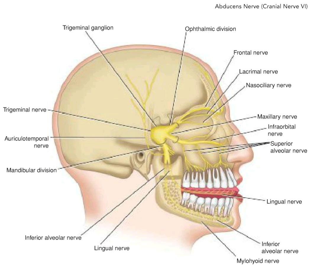
Figure 11-8 Distribution of the trigeminal nerve.
شکل ۱۱-۸ توزیع عصب سه قلو.
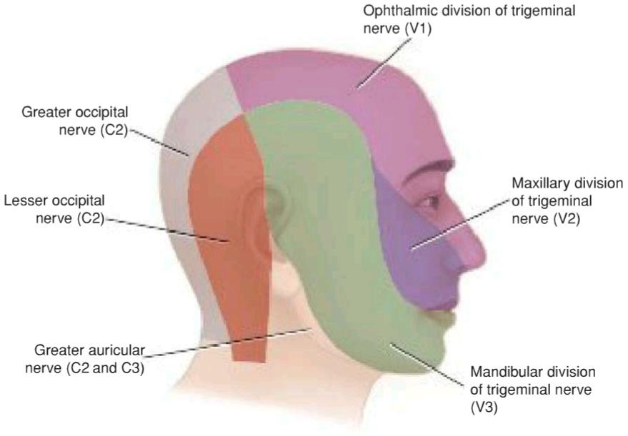
Figure 11-9 Sensory nerve supply to the skin of the head and neck. Note that the skin over the angle of the jaw is supplied by the great auricular nerve (C2 and C3) and not by branches of the trigeminal nerve.
شکل ۱۱-۹ عصب رسانی حسی به پوست سر و گردن. توجه داشته باشید که پوست روی زاویه فک توسط عصب گوش بزرگ (C2 و C3) عصب رسانی میشود و نه توسط شاخههای عصب سه قلو.
«فصل یازدهم کتاب نورواناتومی بالینی اسنل»
🚀 با ما همراه شوید!
تازهترین مطالب و آموزشهای مغز و اعصاب را از دست ندهید. با فالو کردن کانال تلگرام آیندهنگاران مغز، از ما حمایت کنید!