
اعصاب جمجمهای؛ عصب جمجمهای I: اعصاب بویایی؛ فصل یازدهم نوروآناتومی بالینی اسنل


دعای مطالعه [ نمایش ]
بِسْمِ الله الرَّحْمنِ الرَّحیمِ
اَللّهُمَّ اَخْرِجْنى مِنْ ظُلُماتِ الْوَهْمِ
خدايا مرا بيرون آور از تاريكىهاى وهم،
وَ اَكْرِمْنى بِنُورِ الْفَهْمِ
و به نور فهم گرامى ام بدار،
اَللّهُمَّ افْتَحْ عَلَيْنا اَبْوابَ رَحْمَتِكَ
خدايا درهاى رحمتت را به روى ما بگشا،
وَانْشُرْ عَلَيْنا خَزائِنَ عُلُومِكَ بِرَحْمَتِكَ يا اَرْحَمَ الرّاحِمينَ
و خزانههاى علومت را بر ما باز كن به امید رحمتت اى مهربانترين مهربانان.
آیندهنگاران مغز: «ما مغز را میشناسیم، تا آینده را بسازیم.»
کتاب «نوروآناتومی بالینی اسنل» اثر دکتر ریچارد اس. اسنل، با ویرایش جدید دکتر رایان اسپلیتگربر، همچنان بهعنوان مرجع طلایی این حوزه شناخته میشود. این اثر با تلفیق تازهترین پژوهشها و کاربردهای بالینی، پلی استوار میان دانش پایه و عمل پزشکی بنا میکند.
تلاشهای علمی و سالها تدریس ارزشمند استاد گرانقدر آقای دکتر محمدتقی جغتایی الهامبخش ترجمه و گسترش این دانش در ایران بوده است.
ترجمه دقیق این کتاب توسط برند علمی آیندهنگاران مغز به مدیریت داریوش طاهری، امکان دسترسی فارسیزبانان به مرزهای نوین علوم اعصاب را فراهم کرده و رسالتی علمی در جهت شناخت عمیقتر مغز و ترسیم آیندهای روشنتر بر عهده گرفته است.
»» فصل ۱۱: هستههای اعصاب جمجمهای؛ بخش اول مبانی و اصول
»» Chapter 11: Cranial Nerve Nuclei
CHAPTER OBJECTIVES
• To learn the basic information regarding the motor and sensory nuclei of the cranial nerves, including their locations and central connections
• Understand the functional ramifications of lesions to cranial nerve nuclei versus damage of the cranial nerve proper
اهداف فصل
• یادگیری اطلاعات اولیه در مورد هستههای حرکتی و حسی اعصاب جمجمهای، از جمله مکانها و اتصالات مرکزی آنها
• درک پیامدهای عملکردی ضایعات هستههای اعصاب جمجمهای در مقابل آسیب خود عصب جمجمهای
A 49-year-old man wakes up one morning to find the right side of his face paralyzed. When examined by his local medical practitioner, he is found to have complete paralysis of the entire right side of the face. He is also found to have severe hypertension. The patient talks with slightly slurred speech. The physician tells the patient that he has suffered a mild stroke, and he is admitted to the hospital.
مردی ۴۹ ساله یک روز صبح از خواب بیدار میشود و متوجه میشود که سمت راست صورتش فلج شده است. وقتی پزشک محلی او را معاینه میکند، مشخص میشود که او دچار فلج کامل تمام سمت راست صورت شده است. همچنین مشخص میشود که او فشار خون شدید دارد. بیمار با کمی لکنت زبان صحبت میکند. پزشک به بیمار میگوید که دچار سکته مغزی خفیف شده است و او را در بیمارستان بستری میکند.
The patient is later seen by a neurologist who disagrees with the diagnosis. The original physician grouped together the facial paralysis, the slurred speech, and the hypertension and, in the absence of other findings, made the incorrect diagnosis of cerebral hemorrhage. A lesion of the corticonuclear fibers on one side of the brain will cause paralysis only of the muscles of the lower part of the opposite side of the face. This patient has complete paralysis of the entire right side of the face, which could only be caused by a lesion of the lower motor neuron. The correct diagnosis was Bell palsy, an inflammation of the connective tissue sheath of the facial nerve, which temporarily interfered with the functions of the axons of the right facial nerve. This case provides a good example of how knowledge of the central connections of a cranial nerve enables a physician to make the correct diagnosis.
بیمار بعداً توسط یک متخصص مغز و اعصاب ویزیت میشود که با تشخیص مخالف است. پزشک اولیه فلج صورت، لکنت زبان و فشار خون بالا را با هم ترکیب کرد و در غیاب سایر یافتهها، تشخیص نادرست خونریزی مغزی را مطرح کرد. ضایعه فیبرهای قشر هستهای در یک طرف مغز فقط باعث فلج عضلات قسمت تحتانی طرف مقابل صورت میشود. این بیمار فلج کامل کل سمت راست صورت دارد که فقط میتواند ناشی از ضایعه نورون حرکتی تحتانی باشد. تشخیص صحیح فلج بل بود، التهاب غلاف بافت همبند عصب صورت که به طور موقت در عملکرد آکسونهای عصب صورت راست اختلال ایجاد میکند. این مورد مثال خوبی از چگونگی توانایی پزشک در تشخیص صحیح با آگاهی از اتصالات مرکزی یک عصب جمجمهای ارائه میدهد.
The cranial nerves are commonly damaged by trauma or disease, and testing for their integrity forms part of every physical examination.
اعصاب جمجمهای معمولاً در اثر ضربه یا بیماری آسیب میبینند و آزمایش سلامت آنها بخشی از هر معاینه فیزیکی است.
CRANIAL NERVES
The 12 pairs of cranial nerves (CNs) leave the brain and pass through foramina and fissures in the skull. All the nerves are distributed in the head and neck, except CN X, which also supplies structures in the thorax and abdomen. The CNs are named as follows:
اعصاب جمجمهای
۱۲ جفت عصب جمجمهای (CN) از مغز خارج شده و از سوراخها و شکافهای جمجمه عبور میکنند. همه اعصاب در سر و گردن توزیع شدهاند، به جز عصب X که به ساختارهای قفسه سینه و شکم نیز عصب میدهد. اعصاب جمجمهای به شرح زیر نامگذاری شدهاند:
1. Olfactory
2. Optic
3. Oculomotor
4. Trochlear
5. Trigeminal
6. Abducens
7. Facial
8. Vestibulocochlear
9. Glossopharyngeal
10. Vagus
11. Accessory
12. Hypoglossal
۱. بویایی
۲. بینایی
۳. حرکتی چشمی
۴. حلزونی
۵. سه قلو
۶. ابدوسنس
۷. صورتی
۸. دهلیزی-حلقی
۹. زبانی-حلقی
۱۰. واگ
۱۱. فرعی
۱۲. زیرزبانی
See Atlas Plates 1, 6, and 8.
به صفحات اطلس ۱، ۶ و ۸ مراجعه کنید.
CRANIAL NERVE ORGANIZATION
The olfactory, optic, and vestibulocochlear nerves are entirely sensory. The oculomotor, trochlear, abducens, accessory, and hypoglossal nerves are entirely motor. The trigeminal, facial, glossopharyngeal, and vagus nerves are both sensory and motor nerves. The letter symbols commonly used to indicate the functional components of each cranial nerve are shown in Table 11-1. The cranial nerves have central motor and/or sensory nuclei within the brain and peripheral nerve fibers that emerge from the brain and exit from the skull to reach their effector or sensory organs.
سازمان اعصاب جمجمهای
اعصاب بویایی، بینایی و دهلیزی-حلقی کاملاً حسی هستند. اعصاب چشمی-حرکتی، قرقرهای، ابدوسنس، فرعی و زیرزبانی کاملاً حرکتی هستند. اعصاب سهقلو، صورتی، زبانی-حلقی و واگ هم اعصاب حسی و هم حرکتی هستند. نمادهای حروف که معمولاً برای نشان دادن اجزای عملکردی هر عصب جمجمهای استفاده میشوند، در جدول 11-1 نشان داده شدهاند. اعصاب جمجمهای دارای هستههای مرکزی حرکتی و/یا حسی در مغز و فیبرهای عصبی محیطی هستند که از مغز خارج شده و از جمجمه خارج میشوند تا به اندامهای مؤثر یا حسی خود برسند.
The different components of the cranial nerves, their functions, and the openings in the skull through which the nerves leave the cranial cavity are summarized in Table 11-2.
اجزای مختلف اعصاب جمجمهای، عملکرد آنها و دهانههای جمجمه که اعصاب از طریق آنها حفره جمجمه را ترک میکنند، در جدول 11-2 خلاصه شده است.
Table 11-1 The Letter Symbols Commonly Used to Indicate the Functional Components of Each Cranial Nerve
جدول ۱۱-۱ نمادهای حروف که معمولاً برای نشان دادن اجزای عملکردی هر عصب جمجمهای استفاده میشوند
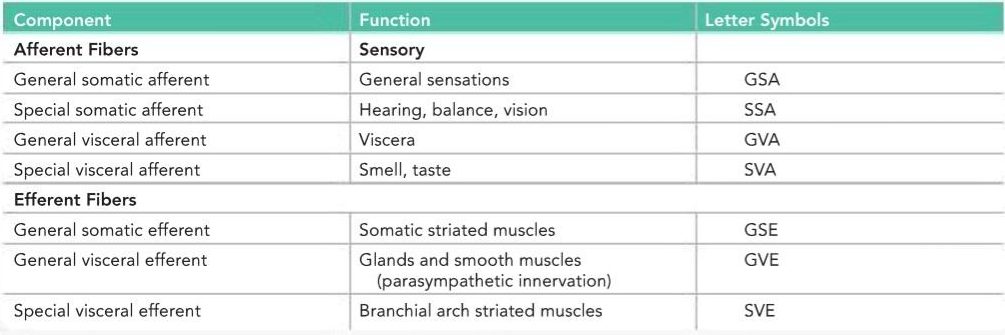
Table 11-2 Cranial Nerves
جدول ۱۱-۲ اعصاب جمجمهای


a The letter symbols are explained in Table 11-1.
b The trigeminal nerve also carries proprioceptive impulses from the muscles of mastication and the facial and extraocular muscles.
الف) نمادهای حروف در جدول 11-1 توضیح داده شدهاند.
ب) عصب سه قلو همچنین تکانههای حس عمقی را از عضلات جونده و عضلات صورت و خارج چشمی منتقل میکند.
Cranial Nerve Motor Nuclei
The motor nuclei of the cranial nerves receive impulses from the cerebral cortex through the corticonuclear (corticobulbar) fibers. These fibers originate from the pyramidal cells in the inferior part of the precentral gyrus (area 4) and from the adjacent part of the postcentral gyrus. The corticonuclear fibers descend through the corona radiata and the genu of the internal capsule. They pass through the midbrain just medial to the corticospinal fibers in the basis pedunculi and end by synapsing either directly with the lower motor neurons within the cranial nerve nuclei or indirectly through the internuncial neurons. The corticonuclear fibers thus constitute the first-order neuron of the descending pathway, the internuncial neuron constitutes the second- order neuron, and the lower motor neuron constitutes the third-order neuron.
هستههای حرکتی اعصاب جمجمهای
هستههای حرکتی اعصاب جمجمهای از طریق فیبرهای قشری-هستهای (قشری-بولبار) تکانهها را از قشر مغز دریافت میکنند. این فیبرها از سلولهای هرمی در قسمت تحتانی شکنج پیشمرکزی (ناحیه ۴) و از قسمت مجاور شکنج پسمرکزی سرچشمه میگیرند. فیبرهای قشری-هستهای از طریق تاج شعاعی و زانوی کپسول داخلی پایین میآیند. آنها از مغز میانی درست در قسمت میانی فیبرهای قشری-نخاعی در پایههای پایه عبور میکنند و با سیناپس مستقیم با نورونهای حرکتی تحتانی در هستههای عصب جمجمهای یا غیرمستقیم از طریق نورونهای بینحلقهای به پایان میرسند. بنابراین، فیبرهای قشری-هستهای نورون مرتبه اول مسیر نزولی را تشکیل میدهند، نورون بینحلقهای نورون مرتبه دوم را تشکیل میدهد و نورون حرکتی تحتانی نورون مرتبه سوم را تشکیل میدهد.
The majority of the corticonuclear fibers to the motor cranial nerve nuclei cross the median plane before reaching the nuclei. Bilateral connections are present for all the cranial motor nuclei except for part of the facial nucleus that supplies the muscles of the lower part of the face and a part of the hypoglossal nucleus that supplies the genioglossus muscle.
اکثر فیبرهای قشری-هستهای به هستههای عصب حرکتی جمجمهای قبل از رسیدن به هستهها از صفحه میانی عبور میکنند. اتصالات دو طرفه برای همه هستههای حرکتی جمجمهای وجود دارد، به جز بخشی از هسته صورت که عضلات قسمت تحتانی صورت را تغذیه میکند و بخشی از هسته زیرزبانی که عضله جنیوگلوس را تغذیه میکند.
Somatic Motor and Branchiomotor Nuclei
The somatic motor and branchiomotor nerve fibers of a cranial nerve are the axons of nerve cells situated within
the brain. These nerve cell groups form motor nuclei and innervate striated muscle. Each nerve cell with its processes is referred to as a lower motor neuron. Such a nerve cell is, therefore, equivalent to the motor cells in the anterior gray columns of the spinal cord.
هستههای حرکتی سوماتیک و شاخهای حرکتی
فیبرهای عصبی حرکتی سوماتیک و شاخهای حرکتی یک عصب جمجمهای، آکسونهای سلولهای عصبی واقع در مغز هستند. این گروههای سلولهای عصبی هستههای حرکتی را تشکیل میدهند و عضله مخطط را عصبدهی میکنند. هر سلول عصبی با زوائدش به عنوان یک نورون حرکتی تحتانی شناخته میشود. بنابراین، چنین سلول عصبی معادل سلولهای حرکتی در ستونهای خاکستری قدامی نخاع است.
General Visceral Motor Nuclei
The general visceral motor nuclei form the cranial outflow of the parasympathetic portion of the autonomic nervous system. They are the Edinger-Westphal nucleus of the oculomotor nerve, the superior salivatory and lacrimal nuclei of the facial nerve, the inferior salivatory nucleus of the glossopharyngeal nerve, and the dorsal motor nucleus of the vagus. These nuclei receive numerous afferent fibers, including descending pathways from the hypothalamus.
هستههای حرکتی عمومی احشایی
هستههای حرکتی عمومی احشایی، خروجی جمجمهای بخش پاراسمپاتیک سیستم عصبی خودکار را تشکیل میدهند. آنها هسته ادینگر-وستفال عصب چشمی، هستههای بزاقی فوقانی و اشکی عصب صورتی، هسته بزاقی تحتانی عصب زبانی-حلقی و هسته حرکتی پشتی عصب واگ هستند. این هستهها فیبرهای آوران متعددی از جمله مسیرهای نزولی از هیپوتالاموس را دریافت میکنند.
Cranial Nerve Sensory Nuclei
Sensory nuclei of the cranial nerves include somatic and visceral afferent nuclei. The sensory or afferent parts of a cranial nerve are the axons of nerve cells outside the brain and are situated in ganglia on the nerve trunks (equivalent to posterior root ganglion of a spinal nerve) or may be situated in a sensory organ, such as the nose, eye, or ear. These cells and their processes form the first-order neuron. The central processes of these cells enter the brain and terminate by synapsing with cells forming the sensory nuclei. These cells and their processes form the second-order neuron. Axons from these nuclear cells now cross the midline and ascend to other sensory nuclei, such as the thalamus, where they synapse. The nerve cells of these nuclei form the third-order neuron, and their axons terminate in the cerebral cortex.
هستههای حسی عصب جمجمهای
هستههای حسی اعصاب جمجمهای شامل هستههای آوران سوماتیک و احشایی هستند. بخشهای حسی یا آوران یک عصب جمجمهای، آکسونهای سلولهای عصبی خارج از مغز هستند و در گانگلیونهای روی تنههای عصبی (معادل گانگلیون ریشه خلفی یک عصب نخاعی) قرار دارند یا ممکن است در یک اندام حسی مانند بینی، چشم یا گوش قرار داشته باشند. این سلولها و زوائد آنها نورون درجه اول را تشکیل میدهند. زوائد مرکزی این سلولها وارد مغز میشوند و با سیناپس با سلولهای تشکیلدهنده هستههای حسی خاتمه مییابند. این سلولها و زوائد آنها نورون درجه دوم را تشکیل میدهند. آکسونهای این سلولهای هستهای اکنون از خط میانی عبور کرده و به هستههای حسی دیگر مانند تالاموس صعود میکنند، جایی که سیناپس میکنند. سلولهای عصبی این هستهها نورون درجه سوم را تشکیل میدهند و آکسونهای آنها در قشر مغز خاتمه مییابند.
OLFACTORY NERVES (CRANIAL NERVE I)
The olfactory nerves arise from the olfactory receptor nerve cells in the olfactory mucous membrane located in the upper part of the nasal cavity above the level of the superior concha (Fig. 11-1). The olfactory receptor cells are scattered among supporting cells. Each receptor cell consists of a small bipolar nerve cell with a coarse peripheral process that passes to the surface of the membrane and a fine central process. From the coarse peripheral process, a number of short cilia arise, the olfactory hairs, which project into the mucus covering the surface of the mucous membrane. These projecting hairs react to odors in the air and stimulate the olfactory cells.
اعصاب بویایی (عصب جمجمهای I)
اعصاب بویایی از سلولهای عصبی گیرنده بویایی در غشای مخاطی بویایی واقع در قسمت فوقانی حفره بینی، بالاتر از سطح شاخک فوقانی (شکل 11-1) منشأ میگیرند. سلولهای گیرنده بویایی در بین سلولهای پشتیبان پراکنده شدهاند. هر سلول گیرنده شامل یک سلول عصبی دو قطبی کوچک با یک زائده محیطی درشت است که به سطح غشا میرود و یک زائده مرکزی ظریف. از زائده محیطی درشت، تعدادی مژک کوتاه، موهای بویایی، بیرون میآیند که به داخل مخاط پوشاننده سطح غشای مخاطی بیرون میآیند. این موهای بیرون زده به بوهای موجود در هوا واکنش نشان میدهند و سلولهای بویایی را تحریک میکنند.
The fine central processes form the olfactory nerve fibers (see Fig. 11-1A,B). Bundles of these nerve fibers pass through the openings of the cribriform plate of the ethmoid bone to enter the olfactory bulb. The olfactory nerve fibers are unmyelinated and are covered with Schwann cells.
زوائد مرکزی ظریف، فیبرهای عصبی بویایی را تشکیل میدهند (به شکل 11-1A,B مراجعه کنید). دستههایی از این فیبرهای عصبی از طریق دهانههای صفحه مشبک استخوان اتموئید عبور میکنند تا وارد پیاز بویایی شوند. رشتههای عصبی بویایی فاقد میلین هستند و توسط سلولهای شوان پوشیده شدهاند.

Figure 11-1 A: Distribution of olfactory nerves on the lateral wall of the nose. B: Connections between the olfactory cells and the neurons of the olfactory bulb. C: Connections between the olfactory cell and the rest of the olfactory system.
شکل ۱۱-۱ الف: توزیع اعصاب بویایی در دیواره جانبی بینی. ب: ارتباطات بین سلولهای بویایی و نورونهای پیاز بویایی. ج: ارتباطات بین سلول بویایی و بقیه سیستم بویایی.
Olfactory Bulb
This ovoid structure possesses several types of nerve cells, the largest of which is the mitral cell (see Fig. 11-1C). The incoming olfactory nerve fibers synapse with the dendrites of the mitral cells and form rounded areas known as synaptic glomeruli. Smaller nerve cells, called tufted cells and granular cells, also synapse with the mitral cells. The olfactory bulb, in addition, receives axons from the contralateral olfactory bulb through the olfactory tract.
پیاز بویایی
این ساختار تخممرغی شکل دارای چندین نوع سلول عصبی است که بزرگترین آنها سلول میترال است (شکل 11-1C را ببینید). فیبرهای عصبی بویایی ورودی با دندریتهای سلولهای میترال سیناپس میکنند و نواحی گردی به نام گلومرولهای سیناپسی تشکیل میدهند. سلولهای عصبی کوچکتر، به نام سلولهای تافتدار و سلولهای دانهدار، نیز با سلولهای میترال سیناپس میکنند. پیاز بویایی، علاوه بر این، آکسونها را از پیاز بویایی طرف مقابل از طریق دستگاه بویایی دریافت میکند.
Olfactory Tract
This narrow band of white matter runs from the posterior end of the olfactory bulb beneath the inferior surface of the frontal lobe of the brain (see Fig. 11-1B). It consists of the axons of the mitral and tufted cells of the bulb and some centrifugal fibers from the opposite olfactory bulb.
مسیر بویایی
این نوار باریک از ماده سفید از انتهای خلفی پیاز بویایی در زیر سطح تحتانی لوب پیشانی مغز امتداد دارد (شکل 11-1B را ببینید). این نوار شامل آکسونهای سلولهای میترال و تافتدار پیاز و برخی از الیاف گریز از مرکز از پیاز بویایی مقابل است.
As the olfactory tract reaches the anterior perforated substance, it divides into medial and lateral olfactory striae. The lateral stria carries the axons to the olfactory area of the cerebral cortex, namely, the periamygdaloid and prepiriform areas (see Fig. 11-1C). The medial olfactory stria carries the fibers that cross the median plane in the anterior commissure to pass to the olfactory bulb of the opposite side.
وقتی مجرای بویایی به ماده سوراخدار قدامی میرسد، به دو نوار بویایی میانی و جانبی تقسیم میشود. نوار بویایی جانبی، آکسونها را به ناحیه بویایی قشر مغز، یعنی نواحی پریآمیگدالوئید و پریپیریفورم (شکل 11-1C را ببینید) حمل میکند. نوار بویایی میانی، فیبرهایی را حمل میکند که از صفحه میانی در رابط قدامی عبور میکنند تا به پیاز بویایی طرف مقابل برسند.
The periamygdaloid and prepiriform areas of the cerebral cortex are often known as the primary olfactory cortex. The entorhinal area (area 28) of the parahippocampal gyrus, which receives numerous connections from the primary olfactory cortex, is called the secondary olfactory cortex. These areas of the cortex are responsible for the appreciation of olfactory sensations. Note that in contrast to all other sensory pathways, the olfactory afferent pathway has only two neurons and reaches the cerebral cortex without synapsing in one of the thalamic nuclei.
نواحی پریآمیگدالوئید و پریپیریفورم قشر مغز اغلب به عنوان قشر بویایی اولیه شناخته میشوند. ناحیه انتورینال (ناحیه 28) شکنج پاراهیپوکامپ، که اتصالات متعددی را از قشر بویایی اولیه دریافت میکند، قشر بویایی ثانویه نامیده میشود. این نواحی قشر مغز مسئول درک احساسات بویایی هستند. توجه داشته باشید که برخلاف سایر مسیرهای حسی، مسیر آوران بویایی تنها دو نورون دارد و بدون سیناپس در یکی از هستههای تالاموس به قشر مغز میرسد.
The primary olfactory cortex sends nerve fibers to many other centers within the brain to establish connections for emotional and autonomic responses to olfactory sensations.
قشر بویایی اولیه، رشتههای عصبی را به بسیاری از مراکز دیگر در مغز میفرستد تا ارتباطاتی را برای پاسخهای عاطفی و خودکار به حسهای بویایی برقرار کند.
«فصل یازدهم کتاب نورواناتومی بالینی اسنل»
🚀 با ما همراه شوید!
تازهترین مطالب و آموزشهای مغز و اعصاب را از دست ندهید. با فالو کردن کانال تلگرام آیندهنگاران مغز، از ما حمایت کنید!


